EP3912578A1 - Temperaturüberwachung von ösophagusgewebe - Google Patents
Temperaturüberwachung von ösophagusgewebe Download PDFInfo
- Publication number
- EP3912578A1 EP3912578A1 EP21174452.9A EP21174452A EP3912578A1 EP 3912578 A1 EP3912578 A1 EP 3912578A1 EP 21174452 A EP21174452 A EP 21174452A EP 3912578 A1 EP3912578 A1 EP 3912578A1
- Authority
- EP
- European Patent Office
- Prior art keywords
- temperature
- processor
- ablation
- tissue
- numerical value
- Prior art date
- Legal status (The legal status is an assumption and is not a legal conclusion. Google has not performed a legal analysis and makes no representation as to the accuracy of the status listed.)
- Withdrawn
Links
Images


Classifications
-
- A—HUMAN NECESSITIES
- A61—MEDICAL OR VETERINARY SCIENCE; HYGIENE
- A61B—DIAGNOSIS; SURGERY; IDENTIFICATION
- A61B5/00—Measuring for diagnostic purposes; Identification of persons
- A61B5/01—Measuring temperature of body parts ; Diagnostic temperature sensing, e.g. for malignant or inflamed tissue
-
- A—HUMAN NECESSITIES
- A61—MEDICAL OR VETERINARY SCIENCE; HYGIENE
- A61B—DIAGNOSIS; SURGERY; IDENTIFICATION
- A61B18/00—Surgical instruments, devices or methods for transferring non-mechanical forms of energy to or from the body
- A61B18/04—Surgical instruments, devices or methods for transferring non-mechanical forms of energy to or from the body by heating
- A61B18/12—Surgical instruments, devices or methods for transferring non-mechanical forms of energy to or from the body by heating by passing a current through the tissue to be heated, e.g. high-frequency current
- A61B18/14—Probes or electrodes therefor
- A61B18/1492—Probes or electrodes therefor having a flexible, catheter-like structure, e.g. for heart ablation
-
- A—HUMAN NECESSITIES
- A61—MEDICAL OR VETERINARY SCIENCE; HYGIENE
- A61B—DIAGNOSIS; SURGERY; IDENTIFICATION
- A61B18/00—Surgical instruments, devices or methods for transferring non-mechanical forms of energy to or from the body
- A61B18/04—Surgical instruments, devices or methods for transferring non-mechanical forms of energy to or from the body by heating
- A61B18/12—Surgical instruments, devices or methods for transferring non-mechanical forms of energy to or from the body by heating by passing a current through the tissue to be heated, e.g. high-frequency current
- A61B18/1206—Generators therefor
-
- A—HUMAN NECESSITIES
- A61—MEDICAL OR VETERINARY SCIENCE; HYGIENE
- A61B—DIAGNOSIS; SURGERY; IDENTIFICATION
- A61B90/00—Instruments, implements or accessories specially adapted for surgery or diagnosis and not covered by any of the groups A61B1/00 - A61B50/00, e.g. for luxation treatment or for protecting wound edges
- A61B90/36—Image-producing devices or illumination devices not otherwise provided for
- A61B90/361—Image-producing devices, e.g. surgical cameras
-
- A—HUMAN NECESSITIES
- A61—MEDICAL OR VETERINARY SCIENCE; HYGIENE
- A61B—DIAGNOSIS; SURGERY; IDENTIFICATION
- A61B90/00—Instruments, implements or accessories specially adapted for surgery or diagnosis and not covered by any of the groups A61B1/00 - A61B50/00, e.g. for luxation treatment or for protecting wound edges
- A61B90/36—Image-producing devices or illumination devices not otherwise provided for
- A61B90/37—Surgical systems with images on a monitor during operation
-
- A—HUMAN NECESSITIES
- A61—MEDICAL OR VETERINARY SCIENCE; HYGIENE
- A61B—DIAGNOSIS; SURGERY; IDENTIFICATION
- A61B18/00—Surgical instruments, devices or methods for transferring non-mechanical forms of energy to or from the body
- A61B2018/00053—Mechanical features of the instrument of device
- A61B2018/00214—Expandable means emitting energy, e.g. by elements carried thereon
- A61B2018/0022—Balloons
-
- A—HUMAN NECESSITIES
- A61—MEDICAL OR VETERINARY SCIENCE; HYGIENE
- A61B—DIAGNOSIS; SURGERY; IDENTIFICATION
- A61B18/00—Surgical instruments, devices or methods for transferring non-mechanical forms of energy to or from the body
- A61B2018/00315—Surgical instruments, devices or methods for transferring non-mechanical forms of energy to or from the body for treatment of particular body parts
- A61B2018/00345—Vascular system
- A61B2018/00351—Heart
- A61B2018/00375—Ostium, e.g. ostium of pulmonary vein or artery
-
- A—HUMAN NECESSITIES
- A61—MEDICAL OR VETERINARY SCIENCE; HYGIENE
- A61B—DIAGNOSIS; SURGERY; IDENTIFICATION
- A61B18/00—Surgical instruments, devices or methods for transferring non-mechanical forms of energy to or from the body
- A61B2018/00315—Surgical instruments, devices or methods for transferring non-mechanical forms of energy to or from the body for treatment of particular body parts
- A61B2018/00482—Digestive system
- A61B2018/00488—Esophagus
-
- A—HUMAN NECESSITIES
- A61—MEDICAL OR VETERINARY SCIENCE; HYGIENE
- A61B—DIAGNOSIS; SURGERY; IDENTIFICATION
- A61B18/00—Surgical instruments, devices or methods for transferring non-mechanical forms of energy to or from the body
- A61B2018/00571—Surgical instruments, devices or methods for transferring non-mechanical forms of energy to or from the body for achieving a particular surgical effect
- A61B2018/00577—Ablation
-
- A—HUMAN NECESSITIES
- A61—MEDICAL OR VETERINARY SCIENCE; HYGIENE
- A61B—DIAGNOSIS; SURGERY; IDENTIFICATION
- A61B18/00—Surgical instruments, devices or methods for transferring non-mechanical forms of energy to or from the body
- A61B2018/00636—Sensing and controlling the application of energy
- A61B2018/00666—Sensing and controlling the application of energy using a threshold value
-
- A—HUMAN NECESSITIES
- A61—MEDICAL OR VETERINARY SCIENCE; HYGIENE
- A61B—DIAGNOSIS; SURGERY; IDENTIFICATION
- A61B18/00—Surgical instruments, devices or methods for transferring non-mechanical forms of energy to or from the body
- A61B2018/00636—Sensing and controlling the application of energy
- A61B2018/00696—Controlled or regulated parameters
- A61B2018/00702—Power or energy
- A61B2018/00708—Power or energy switching the power on or off
-
- A—HUMAN NECESSITIES
- A61—MEDICAL OR VETERINARY SCIENCE; HYGIENE
- A61B—DIAGNOSIS; SURGERY; IDENTIFICATION
- A61B18/00—Surgical instruments, devices or methods for transferring non-mechanical forms of energy to or from the body
- A61B2018/00636—Sensing and controlling the application of energy
- A61B2018/00696—Controlled or regulated parameters
- A61B2018/00714—Temperature
-
- A—HUMAN NECESSITIES
- A61—MEDICAL OR VETERINARY SCIENCE; HYGIENE
- A61B—DIAGNOSIS; SURGERY; IDENTIFICATION
- A61B18/00—Surgical instruments, devices or methods for transferring non-mechanical forms of energy to or from the body
- A61B2018/00636—Sensing and controlling the application of energy
- A61B2018/00773—Sensed parameters
- A61B2018/00791—Temperature
-
- G—PHYSICS
- G06—COMPUTING; CALCULATING OR COUNTING
- G06V—IMAGE OR VIDEO RECOGNITION OR UNDERSTANDING
- G06V20/00—Scenes; Scene-specific elements
- G06V20/60—Type of objects
- G06V20/62—Text, e.g. of license plates, overlay texts or captions on TV images
-
- G—PHYSICS
- G06—COMPUTING; CALCULATING OR COUNTING
- G06V—IMAGE OR VIDEO RECOGNITION OR UNDERSTANDING
- G06V30/00—Character recognition; Recognising digital ink; Document-oriented image-based pattern recognition
- G06V30/10—Character recognition
-
- G—PHYSICS
- G06—COMPUTING; CALCULATING OR COUNTING
- G06V—IMAGE OR VIDEO RECOGNITION OR UNDERSTANDING
- G06V30/00—Character recognition; Recognising digital ink; Document-oriented image-based pattern recognition
- G06V30/10—Character recognition
- G06V30/14—Image acquisition
Definitions
- the present invention relates generally to cardiac ablation, and specifically to monitoring esophageal-tissue temperature during ablation.
- U.S. Patent 9,033,968 describes a method and system for increasing safety of cardiac ablation procedures using a computer-based system that monitors the esophageal temperature, the system comprising an esophageal temperature sensing means, typically on a probe inserted into the esophagus.
- the computer-based system activates different levels of alarm(s), and/or initiates ablation energy interrupt based on pre-defined programmed values.
- U.S. Patent 8,971,997 describes an endoscopic infrared fiber-optic device able to monitor esophageal temperature during an ablation/cryoablation procedure over a volume of interest to sense whether the temperature is too high or too low.
- the device may include a plurality of optical fibers each with a wide-angle lens collectively disposed circumferentially and longitudinally to cover the volume of interest, as the particular region over which undesirable temperature may not be known beforehand.
- the device may include an embedded array of infrared sensors extending sufficiently to encompass a volume of interest. The device may be used as part of a feedback control to regulate and stop operation of the ablation/cryoablation procedure to prevent vessel damage.
- An ablation system includes an ablation catheter that has an array of ablation elements and a location element, an esophageal probe also including a location element, and an interface unit that provides energy to the ablation catheter.
- the distance between the location elements can be used by the system to set or modify one or more system parameters.
- a system of the present invention preferably uses a temperature threshold for a temperature detected using a thermocouple on the esophageal probe.
- An embodiment of the present invention that is described hereinafter provides an apparatus including a camera and a processor.
- the camera is configured to capture images of a display of a temperature measurement system that displays a tissue temperature.
- the processor is configured to analyze the captured images to extract a numerical value of the tissue temperature displayed by the temperature measurement system, and initiate an action responsively to the extracted numerical value.
- the tissue temperature includes a temperature of an esophagus of a patient undergoing a cardiac ablation procedure
- the processor is configured to initiate termination of the cardiac ablation procedure
- the processor is configured to provide the extracted numerical value of the tissue temperature for display by another system.
- the processor is configured to analyze the captured images by performing image processing over a region of interest (ROI) in the captured images.
- ROI region of interest
- the temperature measurement system displays the tissue temperature using alphanumeric characters, and the processor is configured to extract the numerical value by recognizing the alphanumeric characters in the images.
- the temperature measurement system displays the tissue temperature using an analog graphic display, and the processor is configured to extract the numerical value by analyzing the analog graphic display in the images.
- the processor is configured to issue a triggering signal in response to the extracted temperature deviating from a prespecified limit.
- the processor is further configured to calculate a rate of change of the tissue temperature, and to initiate the action in response to the calculated rate of change.
- the processor is configured to issue a triggering signal in response to the rate of change deviating from a prespecified limit.
- the processor is included in an RF generator and is configured to initiate the action by changing an output power of the RF generator output power.
- a method including, using a camera, capturing images of a display of a temperature measurement system that displays a tissue temperature.
- the captured images are analyzed to extract a numerical value of the tissue temperature displayed by the temperature measurement system.
- An action is initiated responsively to the extracted numerical value.
- An anatomic relationship between target tissue undergoing ablation and nearby unrelated tissue can cause problems in invasive ablation of the target tissue, such as unintentional overheating of the nearby unrelated tissue.
- the esophagus lies posterior to the left atrium and leads a variable course relative to the left atrium, adjacent to the right or left pulmonary vein or the posterior wall of the heart.
- RF radiofrequency
- cryoablation may potentially cause collateral damage by accidently cryoablating an esophageal-tissue.
- a third-party system i.e., a system distinct from the ablation system
- a third-party system can be used for esophagus temperature monitoring.
- Such a system typically provides a numerical display of the esophagus temperature, or uses other types of graphical means to display the temperature, such as an analog scale or analog-like display.
- the physician performing the ablation, or an assistant can monitor the third-party system display while performing the ablation.
- the user may abort the ablation, to prevent damage to the esophagus.
- an apparatus comprises a camera used to observe and acquire an image of the third-party display that includes a region of interest (ROI) comprising displayed esophageal temperature.
- ROI region of interest
- a processor comprised/used in the apparatus analyzes the ROI, using image processing techniques, to identify the displayed esophageal temperature (e.g., to extract a numerical temperature value included in the ROI).
- the processor initiates an action responsively to the extracted numerical value. For example, the processor may check the identified temperature to determine if the temperature of the unrelated tissue deviates beyond prespecified temperature limits, or if the rate of change of the temperature deviates beyond a prespecified allowable rate (i.e., temperature and/or rate of change of the temperature deviating from a prespecified limit).
- a prespecified allowable rate i.e., temperature and/or rate of change of the temperature deviating from a prespecified limit.
- the processor checks if a temperature threshold has been exceeded, or if the rate of increase of temperature is too high. In case of cryoablation, the processor checks if the temperature fell below an allowed value or the rate of fall of temperature is too high.
- the processor in response to determining that a temperature deviation is occurring, the processor outputs a triggering signal.
- the triggering signal is received by an RF generator control unit, which in turn terminates the ablation responsively to receiving the triggering signal.
- the processor is comprised in the RF generator and initiates an action comprising changing a setting of the RF generator, including terminating the ablation by shutting off or minimizing the power outputted by the RF generator.
- the disclosed monitoring apparatus provides the identified (e.g., extracted) temperature, and optionally its calculated rate of change, for display by the ablation system. That way, the physician is better aware in real-time to risks of collateral damage from the ablation.
- the physician may be informed by various audiovisual means, such as changing the ablation display colors and/or by using sounding alerts included in the ablation system.
- the processor is programmed in software containing a particular algorithm that enables the processor to conduct each of the processor related steps and functions outlined above.
- ablative treatments may be made safer.
- Fig. 1 is a schematic, pictorial illustration of a catheter-based cardiac radiofrequency (RF) ablation system 20 comprising an automated esophageal-tissue monitoring apparatus, in accordance with an exemplary embodiment of the present invention.
- System 20 comprises a catheter 21, wherein, as seen in inset 25, a distal end 22a of shaft 22 of catheter 21 is inserted through a sheath 23 into a heart 26 of a patient 28 lying on a table 29.
- distal end 22a comprises a magnetic sensor 39, contained within distal end 22a just proximally to a radiofrequency ablative balloon 40.
- Sensor 39 is used by system 20 to navigate the catheter to a target position.
- the disclosed monitoring technique can be applied with any other navigational solution, such as based on electrical impedance signals, or even be applied with catheters that do not include or are not positioned using a navigational means.
- the disclosed monitoring technique can be used with any invasive ablation device, and in particular with any type of ablation catheter.
- catheter 21 The proximal end of catheter 21 is connected to a control console 24.
- catheter 21 may be used for any suitable therapeutic and/or diagnostic purpose, such as electrical ablation using an RF generator 42 comprised in console 24 and/or sensing of tissue in heart 26.
- RF generator 42 comprised in console 24
- sensing of tissue in heart 26 may be any suitable therapeutic and/or diagnostic purpose.
- the disclosed technique is focused on monitoring a therapeutic procedure.
- console 24 receives signals from magnetic sensor 39 in response to magnetic fields from external field generators 36, for example, for the purpose of measuring the position of ablation balloon 40 in the heart 26 and, optionally, presenting the tracked position on a display 27.
- Magnetic field generators 36 are placed at known positions external to patient 28, e.g., below patient table 29.
- Console 24 also comprises a driver circuit 34, configured to drive magnetic field generators 36.
- position signals received from position sensor 39 are indicative of the position of ablation balloon 40 in the coordinate system of position tracking and ablation system 20.
- the method of position sensing using external magnetic fields is implemented in various medical applications, for example, in the CARTOTM system, produced by Biosense-Webster Inc. (Irvine, California), and is described in detail in U.S. Patents 5,391,199 , 6,690, 963 , 6,484,118 , 6,239,724 , 6,618,612 and 6,332,089 , in PCT Patent Publication WO 96/05768 , and in U.S. Patent Application Publications 2002/0065455 A1 , 2003/0120150 A1 and 2004/0068178 A1 , whose disclosures are all incorporated herein by reference.
- Physician 30 navigates the distal end of shaft 22 to a target location in heart 26 by manipulating shaft 22 using a manipulator 32 near the proximal end of the catheter and/or deflection from the sheath 23.
- the balloon 40 may be proximate the esophagus 48 as explained in greater detail subsequently.
- balloon 40 is maintained in a collapsed configuration by sheath 23.
- sheath 23 By containing balloon 40 in a collapsed configuration, sheath 23 also serves to minimize vascular trauma along the way to target location.
- Control console 24 comprises a processor 41, typically a general-purpose computer, with suitable front end and interface circuits 38 for receiving signals from catheter 21, as well as for applying ablative treatment via catheter 21 in heart 26 and for controlling the other components of system 20.
- processor 41 typically a general-purpose computer
- suitable front end and interface circuits 38 for receiving signals from catheter 21, as well as for applying ablative treatment via catheter 21 in heart 26 and for controlling the other components of system 20.
- a camera 55 is positioned to acquire images of a third-party monitor 57 in real time, wherein monitor 57 displays esophageal-tissue temperature information inside an ROI 59, during the ongoing ablation.
- the acquired images are sent, e.g., wirelessly, to processor 41, which uses an algorithm to analyze the images, using imaging processing techniques, so as to identify in ROI 59 of the images an esophageal tissue temperature, and subsequently to calculate a rate of change of the temperature.
- camera 55 is connected directly, either with a cable or wirelessly (e.g., by a Bluetooth link), to a control circuitry of RF generator 42.
- processor 41 is configured to compare the temperature to a threshold value and compare the rate of change of the temperature to an allowable rate, both of which being prespecified. If the temperature exceeds the threshold and/or exceeds the allowable rate, processor 41 triggers a control unit 60 of RF generator 42 of system 20 to responsively terminate ablation, for example by control unit 60 switching a relay on an RF power line. In other exemplary embodiments, however, an indication from camera 55 may be directly transmitted to and trigger control unit 60 of RF generator 42.
- processor 41 shows on display 27 (e.g., CARTO ablation system display) the extracted esophageal-tissue temperature and the calculated rate of change of the temperature and informs the physician by various means, such as changing display colors and sounding alerts, that the ablation had to be terminated automatically, due to one of the aforementioned thermal hazards.
- display 27 e.g., CARTO ablation system display
- Processor 41 typically comprises a general-purpose computer with software programmed to carry out the functions described herein.
- the software may be downloaded to the computer in electronic form, over a network, for example, or it may, alternatively or additionally, be provided and/or stored on non-transitory tangible media, such as magnetic, optical, or electronic memory.
- processor 41 runs a dedicated algorithm as disclosed herein, including in Fig. 3 , which enables processor 41 to perform the disclosed steps, as further described below.
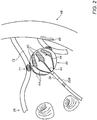
- Fig. 2 is a schematic, pictorial illustration showing ablation balloon 40 of Fig. 1 positioned at an ostium 71 of a pulmonary vein 72 in the left atrium of heart 26 in vicinity of esophagus 48, in accordance with an exemplary embodiment of the present invention.
- Balloon 40 comprises multiple electrodes 44 that are distributed around its outer surface. As seen, some of electrodes 44 face the wall of esophagus 48 and are at close proximity to the wall tissue. Balloon 40 also comprises temperature sensors 45, wherein each temperature sensor 45 is in proximity to an electrode 44.
- esophageal wall tissue 49 is particularly vulnerable to being overheated during an ablation.
- esophageal wall tissue 49 at risk comprises a segment of the esophageal wall facing the posterior side of ostium 71.
- the disclosed apparatus presents on CARTO® display 27 an anatomy similar to the anatomy shown in Fig. 2 with the identified temperature of esophageal wall tissue at risk 49 overlaid on the anatomy.
- system 20 may comprise other sorts of ablation devices, such as a circular multi-electrode catheter (e.g., the Lasso@ catheter made by Biosense Webster Inc.) or a multi-branch multi-electrode catheter (e.g., PentaRay® made by Biosense Webster Inc.).
- ablation devices such as a circular multi-electrode catheter (e.g., the Lasso@ catheter made by Biosense Webster Inc.) or a multi-branch multi-electrode catheter (e.g., PentaRay® made by Biosense Webster Inc.).
- the disclosed treatment method may utilize devices based on laser ablative power, such as a laser ablation balloon that is fitted to the catheter distal end. Laser power would then be terminated by a control unit analogous to control unit 60 to avoid causing collateral thermal damage.
- laser power would then be terminated by a control unit analogous to control unit 60 to avoid causing collateral thermal damage.
- Fig. 3 is a flow chart that schematically illustrates a cardiac ablation procedure aided by the automated esophageal tissue monitoring apparatus of Fig. 1 , in accordance with an exemplary embodiment of the present invention.
- the procedure begins at an image acquisition step 90, in which camera 55 acquires (e.g., captures) video or still images of a display of a third-party temperature measurement system that show a tissue temperature, such as an esophageal-tissue temperature measured during cardiac ablation.
- image acquisition step 90 in which camera 55 acquires (e.g., captures) video or still images of a display of a third-party temperature measurement system that show a tissue temperature, such as an esophageal-tissue temperature measured during cardiac ablation.
- Processor 41 receives the images and, using an algorithm, extracts an ROI of the image that contains the temperature information (e.g., contains a numerical value or an analog scale), at an image ROI extraction step 92.
- processor 41 applies image processing to the extracted ROI to identify the temperature value (e.g., performs optical character recognition (OCR) if it is a numerical display, or other image processing if it is some analog scale or analog-like display).
- OCR optical character recognition
- processor 41 identifies, from the image ROI, a temperature of an esophageal tissue 49 at risk, as well as calculates a rate of change of the temperature, at a temperature identification step 94.
- processor 41 outputs a current (e.g., real time) identified temperature and calculated rate of change of the temperature of esophageal tissue 49 to CARTO® display 27.
- the display may be alphanumeric and/or an analog graphical information which can be overlaid on a presented anatomy.
- Processor 41 compares the temperature to a threshold value and compares the rate of change of the temperature to an allowable rate, which are both prespecified.
- a temperature checking step 98 if temperature exceeds threshold processor 41 triggers control unit 60, by issuing a triggering signal at a triggering step 101, to terminate the ablation.
- control unit 60 terminates the ablation, at an ablation termination step 102.
- the rate of change typically, an increase
- processor 41 triggers control unit 60 to terminate the ablation.
- an alerting step 104 the physician 30 is alerted by audiovisual means, as described above, that the system has automatically terminated ablation.
- Fig. 3 The example flow chart shown in Fig. 3 is shown here purely for the sake of conceptual clarity. In alternative exemplary embodiments, the disclosed technique may use different and/or additional steps, such as, for example, monitoring each electrode 44 temperature using the corresponding temperature sensor 45 and modifying treatment accordingly.
Applications Claiming Priority (1)
| Application Number | Priority Date | Filing Date | Title |
|---|---|---|---|
| US16/877,737 US20210361352A1 (en) | 2020-05-19 | 2020-05-19 | Esophageal-tissue temperature monitoring |
Publications (1)
| Publication Number | Publication Date |
|---|---|
| EP3912578A1 true EP3912578A1 (de) | 2021-11-24 |
Family
ID=76325333
Family Applications (1)
| Application Number | Title | Priority Date | Filing Date |
|---|---|---|---|
| EP21174452.9A Withdrawn EP3912578A1 (de) | 2020-05-19 | 2021-05-18 | Temperaturüberwachung von ösophagusgewebe |
Country Status (5)
| Country | Link |
|---|---|
| US (1) | US20210361352A1 (de) |
| EP (1) | EP3912578A1 (de) |
| JP (1) | JP2021180842A (de) |
| CN (1) | CN113679356A (de) |
| IL (1) | IL282749A (de) |
Citations (15)
| Publication number | Priority date | Publication date | Assignee | Title |
|---|---|---|---|---|
| US5391199A (en) | 1993-07-20 | 1995-02-21 | Biosense, Inc. | Apparatus and method for treating cardiac arrhythmias |
| WO1996005768A1 (en) | 1994-08-19 | 1996-02-29 | Biosense, Inc. | Medical diagnosis, treatment and imaging systems |
| US6239724B1 (en) | 1997-12-30 | 2001-05-29 | Remon Medical Technologies, Ltd. | System and method for telemetrically providing intrabody spatial position |
| US6332089B1 (en) | 1996-02-15 | 2001-12-18 | Biosense, Inc. | Medical procedures and apparatus using intrabody probes |
| US20020065455A1 (en) | 1995-01-24 | 2002-05-30 | Shlomo Ben-Haim | Medical diagnosis, treatment and imaging systems |
| US6484118B1 (en) | 2000-07-20 | 2002-11-19 | Biosense, Inc. | Electromagnetic position single axis system |
| US20030120150A1 (en) | 2001-12-21 | 2003-06-26 | Assaf Govari | Wireless position sensor |
| US6618612B1 (en) | 1996-02-15 | 2003-09-09 | Biosense, Inc. | Independently positionable transducers for location system |
| US20040068178A1 (en) | 2002-09-17 | 2004-04-08 | Assaf Govari | High-gradient recursive locating system |
| US20060106375A1 (en) | 2004-11-15 | 2006-05-18 | Werneth Randell L | Ablation system with feedback |
| US8971997B2 (en) | 2008-03-24 | 2015-03-03 | The Regents Of The University Of Michigan | Non-contact infrared fiber-optic device for measuring temperature in a vessel |
| US9033968B1 (en) | 2011-12-19 | 2015-05-19 | Abl Technologies, Llc | Methods and systems of temperature based alarms, esophageal cooling and/or automatic interrupt (shut-off) during a cardic ablation procedure |
| WO2019005501A1 (en) * | 2017-06-30 | 2019-01-03 | Cryterion Medical, Inc. | GRAPHIC DISPLAY FOR INTRAVASCULAR CATHETER SYSTEM |
| CN110203570A (zh) * | 2019-05-20 | 2019-09-06 | 国电南瑞科技股份有限公司 | 一种冷藏集装箱图像采集装置 |
| EP3560417A1 (de) * | 2018-04-27 | 2019-10-30 | VascoMed GmbH | Ösophagussonde und system |
Family Cites Families (4)
| Publication number | Priority date | Publication date | Assignee | Title |
|---|---|---|---|---|
| KR20100029235A (ko) * | 2007-06-08 | 2010-03-16 | 싸이노슈어, 인코포레이티드 | 외과용 도파관 |
| KR20140013124A (ko) * | 2012-07-03 | 2014-02-05 | 삼성전자주식회사 | 측정장치의 측정결과를 정보시스템으로 전송하기 위한 방법, 장치 그리고 시스템 |
| JP6596436B2 (ja) * | 2013-10-18 | 2019-10-23 | ジーバ メディカル, インコーポレイテッド | 多嚢胞性卵巣症候群の治療のための方法およびシステム |
| US10028658B2 (en) * | 2013-12-30 | 2018-07-24 | Welch Allyn, Inc. | Imager for medical device |
-
2020
- 2020-05-19 US US16/877,737 patent/US20210361352A1/en not_active Abandoned
-
2021
- 2021-04-28 IL IL282749A patent/IL282749A/en unknown
- 2021-05-18 EP EP21174452.9A patent/EP3912578A1/de not_active Withdrawn
- 2021-05-18 JP JP2021083797A patent/JP2021180842A/ja active Pending
- 2021-05-19 CN CN202110547119.0A patent/CN113679356A/zh active Pending
Patent Citations (16)
| Publication number | Priority date | Publication date | Assignee | Title |
|---|---|---|---|---|
| US5391199A (en) | 1993-07-20 | 1995-02-21 | Biosense, Inc. | Apparatus and method for treating cardiac arrhythmias |
| WO1996005768A1 (en) | 1994-08-19 | 1996-02-29 | Biosense, Inc. | Medical diagnosis, treatment and imaging systems |
| US6690963B2 (en) | 1995-01-24 | 2004-02-10 | Biosense, Inc. | System for determining the location and orientation of an invasive medical instrument |
| US20020065455A1 (en) | 1995-01-24 | 2002-05-30 | Shlomo Ben-Haim | Medical diagnosis, treatment and imaging systems |
| US6332089B1 (en) | 1996-02-15 | 2001-12-18 | Biosense, Inc. | Medical procedures and apparatus using intrabody probes |
| US6618612B1 (en) | 1996-02-15 | 2003-09-09 | Biosense, Inc. | Independently positionable transducers for location system |
| US6239724B1 (en) | 1997-12-30 | 2001-05-29 | Remon Medical Technologies, Ltd. | System and method for telemetrically providing intrabody spatial position |
| US6484118B1 (en) | 2000-07-20 | 2002-11-19 | Biosense, Inc. | Electromagnetic position single axis system |
| US20030120150A1 (en) | 2001-12-21 | 2003-06-26 | Assaf Govari | Wireless position sensor |
| US20040068178A1 (en) | 2002-09-17 | 2004-04-08 | Assaf Govari | High-gradient recursive locating system |
| US20060106375A1 (en) | 2004-11-15 | 2006-05-18 | Werneth Randell L | Ablation system with feedback |
| US8971997B2 (en) | 2008-03-24 | 2015-03-03 | The Regents Of The University Of Michigan | Non-contact infrared fiber-optic device for measuring temperature in a vessel |
| US9033968B1 (en) | 2011-12-19 | 2015-05-19 | Abl Technologies, Llc | Methods and systems of temperature based alarms, esophageal cooling and/or automatic interrupt (shut-off) during a cardic ablation procedure |
| WO2019005501A1 (en) * | 2017-06-30 | 2019-01-03 | Cryterion Medical, Inc. | GRAPHIC DISPLAY FOR INTRAVASCULAR CATHETER SYSTEM |
| EP3560417A1 (de) * | 2018-04-27 | 2019-10-30 | VascoMed GmbH | Ösophagussonde und system |
| CN110203570A (zh) * | 2019-05-20 | 2019-09-06 | 国电南瑞科技股份有限公司 | 一种冷藏集装箱图像采集装置 |
Also Published As
| Publication number | Publication date |
|---|---|
| US20210361352A1 (en) | 2021-11-25 |
| CN113679356A (zh) | 2021-11-23 |
| JP2021180842A (ja) | 2021-11-25 |
| IL282749A (en) | 2021-12-01 |
Similar Documents
| Publication | Publication Date | Title |
|---|---|---|
| EP3777743B1 (de) | Dynamische ablation und abtastung entsprechend des kontakts von segmentierten elektroden | |
| EP2682064B1 (de) | Echtzeitbeurteilung der Ablation von Elektrokardiogrammsignalen | |
| CN108498090B (zh) | 根据电极信号突出显示电极图像 | |
| EP2201890B1 (de) | Katheteranzeige mit Spitzenwinkel- und Druckanzeige | |
| US11295835B2 (en) | System and method for interactive event timeline | |
| CN112472276A (zh) | 用于消融系统的图形用户界面 | |
| CN113017823A (zh) | 具有用作消融电极的多个感测电极的导管 | |
| AU2018203282A1 (en) | System and method for glass state view in real-time three-dimensional (3d) cardiac imaging | |
| CN111973272A (zh) | 指示电极接触 | |
| JP2016087464A (ja) | 電気生理学的マップのリアルタイム着色 | |
| JP2017113568A (ja) | アブレーション中の温度の推定 | |
| CN108403208B (zh) | 组织厚度的估计 | |
| JP2019076731A (ja) | 送信コイルを備えた食道プローブ | |
| EP3912578A1 (de) | Temperaturüberwachung von ösophagusgewebe | |
| JP6576707B2 (ja) | 補助的な手動ゼロ合わせ可視化 | |
| CN111281528A (zh) | 半自动消融系统 | |
| RU2747354C1 (ru) | Визуализация траектории перемещения катетера | |
| EP3878390B1 (de) | System zur lückendetektion in ablationslinien | |
| CN115804606A (zh) | 远侧端部组件指南 | |
| EP4295798A1 (de) | Grafischer kontaktqualitätsindikator für ballonkatheternavigation | |
| CN115381551A (zh) | 基于手势选择导管部分 | |
| CN117084775A (zh) | Ire的基于阻抗的消融指数 |
Legal Events
| Date | Code | Title | Description |
|---|---|---|---|
| PUAI | Public reference made under article 153(3) epc to a published international application that has entered the european phase |
Free format text: ORIGINAL CODE: 0009012 |
|
| STAA | Information on the status of an ep patent application or granted ep patent |
Free format text: STATUS: THE APPLICATION HAS BEEN PUBLISHED |
|
| AK | Designated contracting states |
Kind code of ref document: A1 Designated state(s): AL AT BE BG CH CY CZ DE DK EE ES FI FR GB GR HR HU IE IS IT LI LT LU LV MC MK MT NL NO PL PT RO RS SE SI SK SM TR |
|
| B565 | Issuance of search results under rule 164(2) epc |
Effective date: 20211015 |
|
| STAA | Information on the status of an ep patent application or granted ep patent |
Free format text: STATUS: REQUEST FOR EXAMINATION WAS MADE |
|
| 17P | Request for examination filed |
Effective date: 20220113 |
|
| RBV | Designated contracting states (corrected) |
Designated state(s): AL AT BE BG CH CY CZ DE DK EE ES FI FR GB GR HR HU IE IS IT LI LT LU LV MC MK MT NL NO PL PT RO RS SE SI SK SM TR |
|
| RAP3 | Party data changed (applicant data changed or rights of an application transferred) |
Owner name: BIOSENSE WEBSTER (ISRAEL) LTD. |
|
| STAA | Information on the status of an ep patent application or granted ep patent |
Free format text: STATUS: THE APPLICATION HAS BEEN WITHDRAWN |
|
| 18W | Application withdrawn |
Effective date: 20230905 |