EP3903714A1 - Registration of surgical tool with reference array tracked by cameras of an extended reality headset for assisted navigation during surgery - Google Patents
Registration of surgical tool with reference array tracked by cameras of an extended reality headset for assisted navigation during surgery Download PDFInfo
- Publication number
- EP3903714A1 EP3903714A1 EP21170961.3A EP21170961A EP3903714A1 EP 3903714 A1 EP3903714 A1 EP 3903714A1 EP 21170961 A EP21170961 A EP 21170961A EP 3903714 A1 EP3903714 A1 EP 3903714A1
- Authority
- EP
- European Patent Office
- Prior art keywords
- headset
- reference array
- tracking
- surgical
- user
- Prior art date
- Legal status (The legal status is an assumption and is not a legal conclusion. Google has not performed a legal analysis and makes no representation as to the accuracy of the status listed.)
- Granted
Links
Images










Classifications
-
- A—HUMAN NECESSITIES
- A61—MEDICAL OR VETERINARY SCIENCE; HYGIENE
- A61B—DIAGNOSIS; SURGERY; IDENTIFICATION
- A61B34/00—Computer-aided surgery; Manipulators or robots specially adapted for use in surgery
- A61B34/20—Surgical navigation systems; Devices for tracking or guiding surgical instruments, e.g. for frameless stereotaxis
-
- A—HUMAN NECESSITIES
- A61—MEDICAL OR VETERINARY SCIENCE; HYGIENE
- A61B—DIAGNOSIS; SURGERY; IDENTIFICATION
- A61B34/00—Computer-aided surgery; Manipulators or robots specially adapted for use in surgery
- A61B34/10—Computer-aided planning, simulation or modelling of surgical operations
-
- A—HUMAN NECESSITIES
- A61—MEDICAL OR VETERINARY SCIENCE; HYGIENE
- A61B—DIAGNOSIS; SURGERY; IDENTIFICATION
- A61B34/00—Computer-aided surgery; Manipulators or robots specially adapted for use in surgery
- A61B34/25—User interfaces for surgical systems
-
- A—HUMAN NECESSITIES
- A61—MEDICAL OR VETERINARY SCIENCE; HYGIENE
- A61B—DIAGNOSIS; SURGERY; IDENTIFICATION
- A61B34/00—Computer-aided surgery; Manipulators or robots specially adapted for use in surgery
- A61B34/30—Surgical robots
-
- A—HUMAN NECESSITIES
- A61—MEDICAL OR VETERINARY SCIENCE; HYGIENE
- A61B—DIAGNOSIS; SURGERY; IDENTIFICATION
- A61B34/00—Computer-aided surgery; Manipulators or robots specially adapted for use in surgery
- A61B34/70—Manipulators specially adapted for use in surgery
-
- G—PHYSICS
- G06—COMPUTING OR CALCULATING; COUNTING
- G06F—ELECTRIC DIGITAL DATA PROCESSING
- G06F3/00—Input arrangements for transferring data to be processed into a form capable of being handled by the computer; Output arrangements for transferring data from processing unit to output unit, e.g. interface arrangements
- G06F3/01—Input arrangements or combined input and output arrangements for interaction between user and computer
- G06F3/011—Arrangements for interaction with the human body, e.g. for user immersion in virtual reality
-
- G—PHYSICS
- G06—COMPUTING OR CALCULATING; COUNTING
- G06T—IMAGE DATA PROCESSING OR GENERATION, IN GENERAL
- G06T11/00—Two-dimensional [2D] image generation
-
- G—PHYSICS
- G06—COMPUTING OR CALCULATING; COUNTING
- G06T—IMAGE DATA PROCESSING OR GENERATION, IN GENERAL
- G06T7/00—Image analysis
- G06T7/20—Analysis of motion
-
- A—HUMAN NECESSITIES
- A61—MEDICAL OR VETERINARY SCIENCE; HYGIENE
- A61B—DIAGNOSIS; SURGERY; IDENTIFICATION
- A61B17/00—Surgical instruments, devices or methods
- A61B2017/00017—Electrical control of surgical instruments
- A61B2017/00207—Electrical control of surgical instruments with hand gesture control or hand gesture recognition
-
- A—HUMAN NECESSITIES
- A61—MEDICAL OR VETERINARY SCIENCE; HYGIENE
- A61B—DIAGNOSIS; SURGERY; IDENTIFICATION
- A61B34/00—Computer-aided surgery; Manipulators or robots specially adapted for use in surgery
- A61B34/10—Computer-aided planning, simulation or modelling of surgical operations
- A61B2034/107—Visualisation of planned trajectories or target regions
-
- A—HUMAN NECESSITIES
- A61—MEDICAL OR VETERINARY SCIENCE; HYGIENE
- A61B—DIAGNOSIS; SURGERY; IDENTIFICATION
- A61B34/00—Computer-aided surgery; Manipulators or robots specially adapted for use in surgery
- A61B34/10—Computer-aided planning, simulation or modelling of surgical operations
- A61B2034/108—Computer aided selection or customisation of medical implants or cutting guides
-
- A—HUMAN NECESSITIES
- A61—MEDICAL OR VETERINARY SCIENCE; HYGIENE
- A61B—DIAGNOSIS; SURGERY; IDENTIFICATION
- A61B34/00—Computer-aided surgery; Manipulators or robots specially adapted for use in surgery
- A61B34/20—Surgical navigation systems; Devices for tracking or guiding surgical instruments, e.g. for frameless stereotaxis
- A61B2034/2046—Tracking techniques
- A61B2034/2055—Optical tracking systems
- A61B2034/2057—Details of tracking cameras
-
- A—HUMAN NECESSITIES
- A61—MEDICAL OR VETERINARY SCIENCE; HYGIENE
- A61B—DIAGNOSIS; SURGERY; IDENTIFICATION
- A61B34/00—Computer-aided surgery; Manipulators or robots specially adapted for use in surgery
- A61B34/20—Surgical navigation systems; Devices for tracking or guiding surgical instruments, e.g. for frameless stereotaxis
- A61B2034/2046—Tracking techniques
- A61B2034/2059—Mechanical position encoders
-
- A—HUMAN NECESSITIES
- A61—MEDICAL OR VETERINARY SCIENCE; HYGIENE
- A61B—DIAGNOSIS; SURGERY; IDENTIFICATION
- A61B34/00—Computer-aided surgery; Manipulators or robots specially adapted for use in surgery
- A61B34/20—Surgical navigation systems; Devices for tracking or guiding surgical instruments, e.g. for frameless stereotaxis
- A61B2034/2046—Tracking techniques
- A61B2034/2065—Tracking using image or pattern recognition
-
- A—HUMAN NECESSITIES
- A61—MEDICAL OR VETERINARY SCIENCE; HYGIENE
- A61B—DIAGNOSIS; SURGERY; IDENTIFICATION
- A61B34/00—Computer-aided surgery; Manipulators or robots specially adapted for use in surgery
- A61B34/20—Surgical navigation systems; Devices for tracking or guiding surgical instruments, e.g. for frameless stereotaxis
- A61B2034/2072—Reference field transducer attached to an instrument or patient
-
- A—HUMAN NECESSITIES
- A61—MEDICAL OR VETERINARY SCIENCE; HYGIENE
- A61B—DIAGNOSIS; SURGERY; IDENTIFICATION
- A61B34/00—Computer-aided surgery; Manipulators or robots specially adapted for use in surgery
- A61B34/25—User interfaces for surgical systems
- A61B2034/252—User interfaces for surgical systems indicating steps of a surgical procedure
-
- A—HUMAN NECESSITIES
- A61—MEDICAL OR VETERINARY SCIENCE; HYGIENE
- A61B—DIAGNOSIS; SURGERY; IDENTIFICATION
- A61B90/00—Instruments, implements or accessories specially adapted for surgery or diagnosis and not covered by any of the groups A61B1/00 - A61B50/00, e.g. for luxation treatment or for protecting wound edges
- A61B90/08—Accessories or related features not otherwise provided for
- A61B2090/0807—Indication means
-
- A—HUMAN NECESSITIES
- A61—MEDICAL OR VETERINARY SCIENCE; HYGIENE
- A61B—DIAGNOSIS; SURGERY; IDENTIFICATION
- A61B90/00—Instruments, implements or accessories specially adapted for surgery or diagnosis and not covered by any of the groups A61B1/00 - A61B50/00, e.g. for luxation treatment or for protecting wound edges
- A61B90/36—Image-producing devices or illumination devices not otherwise provided for
- A61B2090/363—Use of fiducial points
-
- A—HUMAN NECESSITIES
- A61—MEDICAL OR VETERINARY SCIENCE; HYGIENE
- A61B—DIAGNOSIS; SURGERY; IDENTIFICATION
- A61B90/00—Instruments, implements or accessories specially adapted for surgery or diagnosis and not covered by any of the groups A61B1/00 - A61B50/00, e.g. for luxation treatment or for protecting wound edges
- A61B90/36—Image-producing devices or illumination devices not otherwise provided for
- A61B2090/364—Correlation of different images or relation of image positions in respect to the body
- A61B2090/365—Correlation of different images or relation of image positions in respect to the body augmented reality, i.e. correlating a live optical image with another image
-
- A—HUMAN NECESSITIES
- A61—MEDICAL OR VETERINARY SCIENCE; HYGIENE
- A61B—DIAGNOSIS; SURGERY; IDENTIFICATION
- A61B90/00—Instruments, implements or accessories specially adapted for surgery or diagnosis and not covered by any of the groups A61B1/00 - A61B50/00, e.g. for luxation treatment or for protecting wound edges
- A61B90/36—Image-producing devices or illumination devices not otherwise provided for
- A61B90/37—Surgical systems with images on a monitor during operation
- A61B2090/372—Details of monitor hardware
-
- A—HUMAN NECESSITIES
- A61—MEDICAL OR VETERINARY SCIENCE; HYGIENE
- A61B—DIAGNOSIS; SURGERY; IDENTIFICATION
- A61B90/00—Instruments, implements or accessories specially adapted for surgery or diagnosis and not covered by any of the groups A61B1/00 - A61B50/00, e.g. for luxation treatment or for protecting wound edges
- A61B90/36—Image-producing devices or illumination devices not otherwise provided for
- A61B90/37—Surgical systems with images on a monitor during operation
- A61B2090/376—Surgical systems with images on a monitor during operation using X-rays, e.g. fluoroscopy
-
- A—HUMAN NECESSITIES
- A61—MEDICAL OR VETERINARY SCIENCE; HYGIENE
- A61B—DIAGNOSIS; SURGERY; IDENTIFICATION
- A61B90/00—Instruments, implements or accessories specially adapted for surgery or diagnosis and not covered by any of the groups A61B1/00 - A61B50/00, e.g. for luxation treatment or for protecting wound edges
- A61B90/36—Image-producing devices or illumination devices not otherwise provided for
- A61B90/37—Surgical systems with images on a monitor during operation
- A61B2090/376—Surgical systems with images on a monitor during operation using X-rays, e.g. fluoroscopy
- A61B2090/3762—Surgical systems with images on a monitor during operation using X-rays, e.g. fluoroscopy using computed tomography systems [CT]
-
- A—HUMAN NECESSITIES
- A61—MEDICAL OR VETERINARY SCIENCE; HYGIENE
- A61B—DIAGNOSIS; SURGERY; IDENTIFICATION
- A61B90/00—Instruments, implements or accessories specially adapted for surgery or diagnosis and not covered by any of the groups A61B1/00 - A61B50/00, e.g. for luxation treatment or for protecting wound edges
- A61B90/36—Image-producing devices or illumination devices not otherwise provided for
- A61B90/37—Surgical systems with images on a monitor during operation
- A61B2090/376—Surgical systems with images on a monitor during operation using X-rays, e.g. fluoroscopy
- A61B2090/3762—Surgical systems with images on a monitor during operation using X-rays, e.g. fluoroscopy using computed tomography systems [CT]
- A61B2090/3764—Surgical systems with images on a monitor during operation using X-rays, e.g. fluoroscopy using computed tomography systems [CT] with a rotating C-arm having a cone beam emitting source
-
- A—HUMAN NECESSITIES
- A61—MEDICAL OR VETERINARY SCIENCE; HYGIENE
- A61B—DIAGNOSIS; SURGERY; IDENTIFICATION
- A61B90/00—Instruments, implements or accessories specially adapted for surgery or diagnosis and not covered by any of the groups A61B1/00 - A61B50/00, e.g. for luxation treatment or for protecting wound edges
- A61B90/39—Markers, e.g. radio-opaque or breast lesions markers
- A61B2090/3983—Reference marker arrangements for use with image guided surgery
-
- A—HUMAN NECESSITIES
- A61—MEDICAL OR VETERINARY SCIENCE; HYGIENE
- A61B—DIAGNOSIS; SURGERY; IDENTIFICATION
- A61B90/00—Instruments, implements or accessories specially adapted for surgery or diagnosis and not covered by any of the groups A61B1/00 - A61B50/00, e.g. for luxation treatment or for protecting wound edges
- A61B90/50—Supports for surgical instruments, e.g. articulated arms
- A61B2090/502—Headgear, e.g. helmet, spectacles
-
- G—PHYSICS
- G02—OPTICS
- G02B—OPTICAL ELEMENTS, SYSTEMS OR APPARATUS
- G02B27/00—Optical systems or apparatus not provided for by any of the groups G02B1/00 - G02B26/00, G02B30/00
- G02B27/01—Head-up displays
- G02B27/0101—Head-up displays characterised by optical features
- G02B2027/0138—Head-up displays characterised by optical features comprising image capture systems, e.g. camera
-
- G—PHYSICS
- G02—OPTICS
- G02B—OPTICAL ELEMENTS, SYSTEMS OR APPARATUS
- G02B27/00—Optical systems or apparatus not provided for by any of the groups G02B1/00 - G02B26/00, G02B30/00
- G02B27/01—Head-up displays
- G02B27/017—Head mounted
- G02B27/0172—Head mounted characterised by optical features
-
- G—PHYSICS
- G06—COMPUTING OR CALCULATING; COUNTING
- G06T—IMAGE DATA PROCESSING OR GENERATION, IN GENERAL
- G06T2200/00—Indexing scheme for image data processing or generation, in general
- G06T2200/24—Indexing scheme for image data processing or generation, in general involving graphical user interfaces [GUIs]
-
- G—PHYSICS
- G06—COMPUTING OR CALCULATING; COUNTING
- G06T—IMAGE DATA PROCESSING OR GENERATION, IN GENERAL
- G06T2207/00—Indexing scheme for image analysis or image enhancement
- G06T2207/30—Subject of image; Context of image processing
- G06T2207/30244—Camera pose
Definitions
- the present disclosure relates to medical devices and systems, and more particularly, camera tracking systems used for computer assisted navigation during surgery.
- Computer assisted navigation during surgery can provide a surgeon with computerized visualization of the present pose of a surgical tool relative to medical images of a patient's anatomy.
- Camera tracking systems for computer assisted navigation typically use a set of cameras to track a set of fiducials attached to a surgical tool which is being positioned by a surgeon or other user during surgery.
- the set of fiducials also referred to as a dynamic reference array or dynamic reference base (DRB) allows the camera tracking system to determine a pose of the surgical tool relative to anatomical structure within a medical image and relative to patient for display to the surgeon. The surgeon can thereby use the real-time pose feedback to navigate the surgical tool during a surgical procedure.
- DRB dynamic reference array
- Extended reality (XR) headsets are being combined with camera tracking systems to enable surgeons to see the real-time pose feedback as graphical objects overlaid on the patient.
- XR Extended reality
- a XR headset is used to assist with registering characteristics of a surgical tool with a reference array that is identified by a camera tracking system.
- a representation of the characteristics can be displayed to the XR headset to enable the user to confirm correctness of the registration process.
- Using the XR headset during the registration process can provide a more intuitive, time efficient and reliable process for surgeons and other medical personnel (users) to register surgical tools with a camera tracking system before and/or during a surgical procedure.
- a camera tracking system for computer assisted navigation during surgery is configured to identify a reference array which is being tracked by a set of tracking cameras attached to an XR headset.
- the camera tracking system determines whether the reference array is registered as being paired with characteristics of one of a plurality of surgical tools defined in a surgical tool database. Based on the reference array being determined to not be registered and receiving user input, the camera tracking system registers the reference array as paired with characteristics of one of the plurality of surgical tools selected based on the user input.
- the camera tracking system then provides a representation of the characteristics to a display device of the XR headset for display to the user.
- An extended reality (XR) headset is operatively connected to the surgical system and configured to provide an interactive environment through which a surgeon, assistant, and/or other personnel can view and select among patient images, view and select among computer generated surgery navigation information, and/or control surgical equipment in the operating room.
- the XR headset may be configured to augment a real-world scene with computer generated XR images.
- the XR headset may be configured to provide an augmented reality (AR) viewing environment by displaying the computer generated XR images on a see-through display screen that allows light from the real-world scene to pass therethrough for combined viewing by the user.
- AR augmented reality
- the XR headset may be configured to provide a virtual reality (VR) viewing environment by preventing or substantially preventing light from the real-world scene from being directly viewed by the user while the user is viewing the computer generated AR images on a display screen.
- An XR headset can be configured to provide both AR and VR viewing environments.
- both AR and VR viewing environments are provided by lateral bands of substantially differing opacity arranged between the see-through display screen and the real-world scene, so that a VR viewing environment is provided for XR images aligned with a high opacity band and an AR viewing environment is provided for XR images aligned with the low opacity band.
- both AR and VR viewing environments are provided by computer adjustable control of an opacity filter that variably constrains how much light from the real-world scene passes through a see-through display screen for combining with the XR images viewed by the user.
- the XR headset can also be referred to as an AR headset or a VR headset.
- FIG. 1 illustrates an embodiment of a surgical system 2 according to some embodiments of the present disclosure.
- a three-dimensional (“3D") image scan may be taken of a planned surgical area of a patient using, e.g., the C-Arm imaging device 104 of Fig. 10 or O-Arm imaging device 106 of Fig. 11 , or from another medical imaging device such as a computed tomography (CT) image or MRI.
- CT computed tomography
- This scan can be taken pre-operatively (e.g. few weeks before procedure, most common) or intra-operatively.
- any known 3D or 2D image scan may be used in accordance with various embodiments of the surgical system 2.
- the image scan is sent to a computer platform in communication with the surgical system 2, such as the computer platform 910 of the surgical system 900 ( Fig. 9 ) which may include the camera tracking system component 6, the surgical robot 4 (e.g., robot 2 in Fig. 1 ), imaging devices (e.g., C-Arm 104, O-Arm 106, etc.), and an image database 950 for storing image scans of patients.
- the surgical robot 4 e.g., robot 2 in Fig. 1
- imaging devices e.g., C-Arm 104, O-Arm 106, etc.
- an image database 950 for storing image scans of patients.
- a surgeon reviewing the image scan(s) on a display device of the computer platform 910 ( Fig. 9 ) generates a surgical plan defining a target pose for a surgical tool to be used during a surgical procedure on an anatomical structure of the patient.
- Example surgical tools can include, without limitation, drills, screw drivers, saws, retractors, and implants such as a screws, spacers, interbody fusion devices, plates, rods, etc.
- the surgical plan defining the target plane is planned on the 3D image scan displayed on a display device.
- the term “pose” refers to the position and/or the rotational angle of one object (e.g., dynamic reference array, end effector, surgical tool, anatomical structure, etc.) relative to another object and/or to a defined coordinate system.
- a pose may therefore be defined based on only the multidimensional position of one object relative to another object and/or to a defined coordinate system, only on the multidimensional rotational angles of the object relative to another object and/or to a defined coordinate system, or on a combination of the multidimensional position and the multidimensional rotational angles.
- the term “pose” therefore is used to refer to position, rotational angle, or combination thereof.
- surgical system 2 of Figure 1 can assist surgeons during medical procedures by, for example, holding tools, aligning tools, using tools, guiding tools, and/or positioning tools for use.
- surgical system 2 includes a surgical robot 4 and a camera tracking system component 6.
- the ability to mechanically couple surgical robot 4 and camera tracking system component 6 can allow for surgical system 2 to maneuver and move as a single unit, and allow surgical system 2 to have a small footprint in an area, allow easier movement through narrow passages and around turns, and allow storage within a smaller area.
- a surgical procedure may begin with the surgical system 2 moving from medical storage to a medical procedure room.
- the surgical system 2 may be maneuvered through doorways, halls, and elevators to reach a medical procedure room.
- the surgical system 2 may be physically separated into two separate and distinct systems, the surgical robot 4 and the camera tracking system component 6.
- Surgical robot 4 may be positioned adjacent the patient at any suitable location to properly assist medical personnel.
- Camera tracking system component 6 may be positioned at the base of the patient, at the patient shoulders, or any other location suitable to track the present pose and movement of the pose of tracks portions of the surgical robot 4 and the patient.
- Surgical robot 4 and camera tracking system component 6 may be powered by an onboard power source and/or plugged into an external wall outlet.
- Surgical robot 4 may be used to assist a surgeon by holding and/or using tools during a medical procedure. To properly utilize and hold tools, surgical robot 4 may rely on a plurality of motors, computers, and/or actuators to function properly. Illustrated in Figure 1 , robot body 8 may act as the structure in which the plurality of motors, computers, and/or actuators may be secured within surgical robot 4. Robot body 8 may also provide support for robot telescoping support arm 16. The size of robot body 8 may provide a solid platform supporting attached components, and may house, conceal, and protect the plurality of motors, computers, and/or actuators that may operate attached components.
- Robot base 10 may act as a lower support for surgical robot 4.
- robot base 10 may support robot body 8 and may attach robot body 8 to a plurality of powered wheels 12. This attachment to wheels may allow robot body 8 to move in space efficiently.
- Robot base 10 may run the length and width of robot body 8.
- Robot base 10 may be about two inches to about 10 inches tall.
- Robot base 10 may cover, protect, and support powered wheels 12.
- At least one powered wheel 12 may be attached to robot base 10.
- Powered wheels 12 may attach to robot base 10 at any location. Each individual powered wheel 12 may rotate about a vertical axis in any direction.
- a motor may be disposed above, within, or adjacent to powered wheel 12. This motor may allow for surgical system 2 to maneuver into any location and stabilize and/or level surgical system 2.
- a rod, located within or adjacent to powered wheel 12, may be pressed into a surface by the motor.
- the rod not pictured, may be made of any suitable metal to lift surgical system 2.
- the rod may lift powered wheel 10, which may lift surgical system 2, to any height required to level or otherwise fix the orientation of the surgical system 2 in relation to a patient.
- the weight of surgical system 2 supported through small contact areas by the rod on each wheel, prevents surgical system 2 from moving during a medical procedure. This rigid positioning may prevent objects and/or people from moving surgical system 2 by accident.
- Moving surgical system 2 may be facilitated using robot railing 14.
- Robot railing 14 provides a person with the ability to move surgical system 2 without grasping robot body 8. As illustrated in Figure 1 , robot railing 14 may run the length of robot body 8, shorter than robot body 8, and/or may run longer the length of robot body 8. Robot railing 14 may further provide protection to robot body 8, preventing objects and or personnel from touching, hitting, or bumping into robot body 8.
- Robot body 8 may provide support for a Selective Compliance Articulated Robot Arm, hereafter referred to as a "SCARA.”
- a SCARA 24 may be beneficial to use within the surgical system 2 due to the repeatability and compactness of the robotic arm. The compactness of a SCARA may provide additional space within a medical procedure, which may allow medical professionals to perform medical procedures free of excess clutter and confining areas.
- SCARA 24 may comprise robot telescoping support 16, robot support arm 18, and/or robot arm 20.
- Robot telescoping support 16 may be disposed along robot body 8. As illustrated in Figure 1 , robot telescoping support 16 may provide support for the SCARA 24 and display 34. In some embodiments, robot telescoping support 16 may extend and contract in a vertical direction.
- the body of robot telescoping support 16 may be any width and/or height configured to support the stress and weight placed upon it.
- medical personnel may move SCARA 24 through a command submitted by the medical personnel.
- the command may originate from input received on display 34, a tablet, and/or an XR headset (e.g., headset 920 in Fig. 9 ) as will be explained in further detail below.
- the XR headset may eliminate the need for medical personnel to refer to any other display such as the display 34 or a tablet, which enables the SCARA 24 to be configured without the display 34 and/or the tablet.
- the command may be generated by the depression of a switch and/or the depression of a plurality of switches, and/or may be generated based on a hand gesture command and/or voice command that is sensed by the XR headset as will be explained in further detail below.
- an activation assembly 60 may include a switch and/or a plurality of switches.
- the activation assembly 60 may be operable to transmit a move command to the SCARA 24 allowing an operator to manually manipulate the SCARA 24.
- the switch, or plurality of switches When the switch, or plurality of switches, is depressed the medical personnel may have the ability to move SCARA 24 through applied hand movements.
- an operator may control movement of the SCARA 24 through hand gesture commands and/or voice commands that are sensed by the XR headset as will be explained in further detail below.
- the SCARA 24 may lock in place to prevent accidental movement by personnel and/or other objects. By locking in place, the SCARA 24 provides a solid platform through which the end effector 26 can guide a surgical tool during a medical procedure.
- Robot support arm 18 can be connected to robot telescoping support 16 by various mechanisms. In some embodiments, best seen in Figures 1 and 2 , robot support arm 18 rotates in any direction in regard to robot telescoping support 16. Robot support arm 18 may rotate three hundred and sixty degrees around robot telescoping support 16. Robot arm 20 may connect to robot support arm 18 at any suitable location and by various mechanisms that enable rotation in any direction relative to robot support arm 18. In one embodiment, the robot arm 20 can rotate three hundred and sixty degrees relative to the robot support arm 18. This free rotation allows an operator to position robot arm 20 according to a surgical plan.
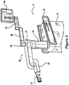
- the end effector 26 shown in Figures 4 and 5 may attach to robot arm 20 in any suitable location.
- the end effector 26 can be configured to attach to an end effector coupler 22 of the robot arm 20 positioned by the surgical robot 4.
- the example end effector 26 includes a tubular guide that guides movement of an inserted surgical tool relative to an anatomical structure on which a surgical procedure is to be performed.
- a dynamic reference array 52 is attached to the end effector 26.
- Dynamic reference arrays also referred to as “DRAs” and “reference arrays” herein, can be rigid bodies, markers, or other indicia which may be attached or formed on one or more XR headsets being worn by personnel in the operating room, the end effector, the surgical robot, a surgical tool in a navigated surgical procedure, and an anatomical structure (e.g., bone) of a patient.
- the computer platform 910 in combination with the camera tracking system component 6 or other 3D localization system are configured to track in real-time the pose (e.g., positions and rotational orientations) of the DRA.
- the DRA can include fiducials, such as the illustrated arrangement of balls. This tracking of 3D coordinates of the DRA can allow the surgical system 2 to determine the pose of the DRA in any multidimensional space in relation to the target anatomical structure of the patient 50 in Figure 5 .
- a light indicator 28 may be positioned on top of the SCARA 24.
- Light indicator 28 may illuminate as any type of light to indicate "conditions" in which surgical system 2 is currently operating.
- the light may be produced by LED bulbs, which may form a ring around light indicator 28.
- Light indicator 28 may comprise a fully permeable material that can let light shine through the entirety of light indicator 28.
- Light indicator 28 may be attached to lower display support 30.
- Lower display support 30, as illustrated in Figure 2 may allow an operator to maneuver display 34 to any suitable location.
- Lower display support 30 may attach to light indicator 28 by any suitable mechanism.
- lower display support 30 may rotate about light indicator 28 or be rigidly attached thereto.
- Upper display support 32 may attach to lower display support 30 by any suitable mechanism.
- a tablet may be used in conjunction with display 34 and/or without display 34.
- the tablet may be disposed on upper display support 32, in place of display 34, and may be removable from upper display support 32 during a medical operation.
- the tablet may communicate with display 34.
- the tablet may be able to connect to surgical robot 4 by any suitable wireless and/or wired connection.
- the tablet may be able to program and/or control surgical system 2 during a medical operation. When controlling surgical system 2 with the tablet, all input and output commands may be duplicated on display 34.
- the use of a tablet may allow an operator to manipulate surgical robot 4 without having to move around patient 50 and/or to surgical robot 4.
- a surgeon and/or other personnel can wear XR headsets that may be used in conjunction with display 34 and/or a tablet or the XR head(s) may eliminate the need for use of the display 34 and/or tablet.
- camera tracking system component 6 works in conjunction with surgical robot 4 through wired or wireless communication networks.
- camera tracking system component 6 can include some similar components to the surgical robot 4.
- camera body 36 may provide the functionality found in robot body 8.
- Robot body 8 may provide an auxiliary tracking bar upon which cameras 46 are mounted.
- the structure within robot body 8 may also provide support for the electronics, communication devices, and power supplies used to operate camera tracking system component 6.
- Camera body 36 may be made of the same material as robot body 8.
- Camera tracking system component 6 may communicate directly to an XR headset, tablet and/or display 34 by a wireless and/or wired network to enable the XR headset, tablet and/or display 34 to control the functions of camera tracking system component 6.
- Camera body 36 is supported by camera base 38.
- Camera base 38 may function as robot base 10.
- camera base 38 may be wider than robot base 10.
- the width of camera base 38 may allow for camera tracking system component 6 to connect with surgical robot 4.
- the width of camera base 38 may be large enough to fit outside robot base 10.
- the additional width of camera base 38 may allow surgical system 2 additional maneuverability and support for surgical system 2.
- a plurality of powered wheels 12 may attach to camera base 38.
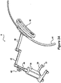
- Powered wheel 12 may allow camera tracking system component 6 to stabilize and level or set fixed orientation in regards to patient 50, similar to the operation of robot base 10 and powered wheels 12. This stabilization may prevent camera tracking system component 6 from moving during a medical procedure and may keep cameras 46 on the auxiliary tracking bar from losing track of a DRA connected to an XR headset and/or the surgical robot 4, and/or losing track of one or more DRAs 52 connected to an anatomical structure 54 and/or tool 58 within a designated area 56 as shown in Figures 3A and 5 .
- This stability and maintenance of tracking enhances the ability of surgical robot 4 to operate effectively with camera tracking system component 6.
- the wide camera base 38 may provide additional support to camera tracking system component 6. Specifically, a wide camera base 38 may prevent camera tracking system component 6 from tipping over when cameras 46 is disposed over a patient, as illustrated in Figures 3A and 5 .
- Camera telescoping support 40 may support cameras 46 on the auxiliary tracking bar. In some embodiments, telescoping support 40 moves cameras 46 higher or lower in the vertical direction.
- Camera handle 48 may be attached to camera telescoping support 40 at any suitable location and configured to allow an operator to move camera tracking system component 6 into a planned position before a medical operation. In some embodiments, camera handle 48 is used to lower and raise camera telescoping support 40. Camera handle 48 may perform the raising and lowering of camera telescoping support 40 through the depression of a button, switch, lever, and/or any combination thereof.
- Lower camera support arm 42 may attach to camera telescoping support 40 at any suitable location, in embodiments, as illustrated in Figure 1 , lower camera support arm 42 may rotate three hundred and sixty degrees around telescoping support 40. This free rotation may allow an operator to position cameras 46 in any suitable location.
- Lower camera support arm 42 may connect to telescoping support 40 by any suitable mechanism.
- Lower camera support arm 42 may be used to provide support for cameras 46.
- Cameras 46 may be attached to lower camera support arm 42 by any suitable mechanism.
- Cameras 46 may pivot in any direction at the attachment area between cameras 46 and lower camera support arm 42.
- a curved rail 44 may be disposed on lower camera support arm 42.
- Curved rail 44 may be disposed at any suitable location on lower camera support arm 42. As illustrated in Figure 3A , curved rail 44 may attach to lower camera support arm 42 by any suitable mechanism. Curved rail 44 may be of any suitable shape, a suitable shape may be a crescent, circular, oval, elliptical, and/or any combination thereof. Cameras 46 may be moveably disposed along curved rail 44. Cameras 46 may attach to curved rail 44 by, for example, rollers, brackets, braces, motors, and/or any combination thereof. Motors and rollers, not illustrated, may be used to move cameras 46 along curved rail 44.
- the motors may responsively move cameras 46 along curved rail 44. This motorized movement may allow cameras 46 to move to a new position that is no longer obstructed by the object without moving camera tracking system component 6. While cameras 46 is obstructed from viewing one or more tracked DRAs, camera tracking system component 6 may send a stop signal to a surgical robot 4, XR headset, display 34, and/or a tablet. The stop signal may prevent SCARA 24 from moving until cameras 46 has reacquired tracked DRAs 52 and/or can warn an operator wearing the XR headset and/or viewing the display 34 and/or the tablet. This SCARA 24 can be configured to respond to receipt of a stop signal by stopping further movement of the base and/or end effector coupler 22 until the camera tracking system can resume tracking of DRAs.
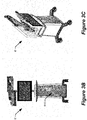
- Figures 3B and 3C illustrate a front view and isometric view of another camera tracking system component 6' which may be used with the surgical system of Figure 1 or may be used independent of a surgical robot.
- the camera tracking system component 6' may be used for providing navigated surgery without use of robotic guidance.
- One of the differences between the camera tracking system component 6' of Figures 3B and 3C and the camera tracking system component 6 of Figure 3A is that the camera tracking system component 6' of Figures 3B and 3C includes a housing that transports the computer platform 910.
- the computer platform 910 can be configured to perform camera tracking operations to track DRAs, perform navigated surgery operations that provide surgical navigation information to a display device, e.g., XR headset and/or other display device, and perform other computational operations disclosed herein.
- the computer platform 910 can therefore include a navigation computer, such as one or more of the navigation computers of Figures 14 .
- Figure 6 illustrates a block diagram view of the components of the surgical system of Figure 5 used for the medical operation.
- the tracking cameras 46 on the auxiliary tracking bar has a navigation field-of-view 600 in which the pose (e.g., position and orientation) of the reference array 602 attached to the patient, the reference array 604 attached to the surgical instrument, and the robot arm 20 are tracked.
- the tracking cameras 46 may be part of the camera tracking system component 6' of Figures 3B and 3C , which includes the computer platform 910 configured to perform the operations described below.
- the reference arrays enable tracking by reflecting light in known patterns, which are decoded to determine their respective poses by the tracking subsystem of the surgical robot 4.
- a responsive notification may temporarily halt further movement of the robot arm 20 and surgical robot 4, display a warning on the display 34, and/or provide an audible warning to medical personnel.
- the display 34 is accessible to the surgeon 610 and assistant 612 but viewing requires a head to be turned away from the patient and for eye focus to be changed to a different distance and location.
- the navigation software may be controlled by a tech personnel 614 based on vocal instructions from the surgeon.
- Figure 7 illustrates various display screens that may be displayed on the display 34 of Figures 5 and 6 by the surgical robot 4 when using a navigation function of the surgical system 2.
- the display screens can include, without limitation, patient radiographs with overlaid graphical representations of models of instruments that are positioned in the display screens relative to the anatomical structure based on a developed surgical plan and/or based on poses of tracked reference arrays, various user selectable menus for controlling different stages of the surgical procedure and dimension parameters of a virtually projected implant (e.g. length, width, and/or diameter).
- processing components e.g., computer platform 910 and associated software described below are provided that enable pre-operatively planning of a surgical procedure, e.g., implant placement, and electronic transfer of the plan to computer platform 910 to provide navigation information to one or more users during the planned surgical procedure.
- a surgical procedure e.g., implant placement
- electronic transfer of the plan to computer platform 910 to provide navigation information to one or more users during the planned surgical procedure.
- various processing components e.g., computer platform 910 and associated software described below are provided that enable pre-operatively planning of a surgical procedure, e.g., implant placement, and electronic transfer of the plan to the surgical robot 4.
- the surgical robot 4 uses the plan to guide the robot arm 20 and connected end effector 26 to provide a target pose for a surgical tool relative to a patient anatomical structure for a step of the planned surgical procedure.
- Various embodiments below are directed to using one or more XR headsets that can be worn by the surgeon 610, the assistant 612, and/or other medical personnel to provide an improved user interface for receiving information from and/or providing control commands to the surgical robot, the camera tracking system component 6/6', and/or other medical equipment in the operating room.
- FIG. 8 illustrates a block diagram of some electrical components of the surgical robot 4 according to some embodiments of the present disclosure.
- a load cell (not shown) may be configured to track force applied to end effector coupler 22.
- the load cell may communicate with a plurality of motors 850, 851, 852, 853, and/or 854.
- Controller 846 may take the force information from load cell and process it with a switch algorithm. The switch algorithm is used by the controller 846 to control a motor driver 842.
- the motor driver 842 controls operation of one or more of the motors 850, 851, 852, 853, and 854.
- Motor driver 842 may direct a specific motor to produce, for example, an equal amount of force measured by load cell through the motor.
- the force produced may come from a plurality of motors, e.g., 850-854, as directed by controller 846.
- motor driver 842 may receive input from controller 846.
- Controller 846 may receive information from load cell as to the direction of force sensed by load cell. Controller 846 may process this information using a motion controller algorithm. The algorithm may be used to provide information to specific motor drivers 842. To replicate the direction of force, controller 846 may activate and/or deactivate certain motor drivers 842.
- Controller 846 may control one or more motors, e.g. one or more of 850-854, to induce motion of end effector 26 in the direction of force sensed by load cell.
- This force-controlled motion may allow an operator to move SCARA 24 and end effector 26 effortlessly and/or with very little resistance. Movement of end effector 26 can be performed to position end effector 26 in any suitable pose (i.e., location and angular orientation relative to defined three-dimensional (3D) orthogonal reference axes) for use by medical personnel.
- Activation assembly 60 may form of a bracelet that wraps around end effector coupler 22.
- the activation assembly 60 may be located on any part of SCARA 24, any part of end effector coupler 22, may be worn by medical personnel (and communicate wirelessly), and/or any combination thereof.
- Activation assembly 60 may comprise of a primary button and a secondary button.
- Depressing primary button may allow an operator to move SCARA 24 and end effector coupler 22.
- SCARA 24 and end effector coupler 22 may not move until an operator programs surgical robot 4 to move SCARA 24 and end effector coupler 22, or is moved using primary button.
- it may require the depression of at least two non-adjacent primary activation switches before SCARA 24 and end effector coupler 22 will respond to operator commands. Depression of at least two primary activation switches may prevent the accidental movement of SCARA 24 and end effector coupler 22 during a medical procedure.
- load cell may measure the force magnitude and/or direction exerted upon end effector coupler 22 by an operator, i.e. medical personnel. This information may be transferred to one or more motors, e.g. one or more of 850-854, within SCARA 24 that may be used to move SCARA 24 and end effector coupler 22. Information as to the magnitude and direction of force measured by load cell may cause the one or more motors, e.g. one or more of 850-854, to move SCARA 24 and end effector coupler 22 in the same direction as sensed by the load cell.
- motors e.g. one or more of 850-854
- This force-controlled movement may allow the operator to move SCARA 24 and end effector coupler 22 easily and without large amounts of exertion due to the motors moving SCARA 24 and end effector coupler 22 at the same time the operator is moving SCARA 24 and end effector coupler 22.
- a secondary button may be used by an operator as a "selection" device.
- surgical robot 4 may notify medical personnel to certain conditions by the XR headset(s) 920, display 34 and/or light indicator 28.
- the XR headset(s) 920 are each configured to display images on a see-through display screen to form an extended reality image that is overlaid on real-world objects viewable through the see-through display screen.
- Medical personnel may be prompted by surgical robot 4 to select a function, mode, and/or asses the condition of surgical system 2.
- Depressing secondary button a single time may activate certain functions, modes, and/or acknowledge information communicated to medical personnel through the XR headset(s) 920, display 34 and/or light indicator 28.
- depressing the secondary button multiple times in rapid succession may activate additional functions, modes, and/or select information communicated to medical personnel through the XR headset(s) 920, display 34 and/or light indicator 28.
- electrical components of the surgical robot 4 include platform subsystem 802, computer subsystem 820, motion control subsystem 840, and tracking subsystem 830.
- Platform subsystem 802 includes battery 806, power distribution module 804, connector panel 808, and charging station 810.
- Computer subsystem 820 includes computer 822, display 824, and speaker 826.
- Motion control subsystem 840 includes driver circuit 842, motors 850, 851, 852, 853, 854, stabilizers 855, 856, 857, 858, end effector connector 844, and controller 846.
- Tracking subsystem 830 includes position sensor 832 and camera converter 834.
- Surgical robot 4 may also include a removable foot pedal 880 and removable tablet computer 890.
- Input power is supplied to surgical robot 4 via a power source which may be provided to power distribution module 804.
- Power distribution module 804 receives input power and is configured to generate different power supply voltages that are provided to other modules, components, and subsystems of surgical robot 4.
- Power distribution module 804 may be configured to provide different voltage supplies to connector panel 808, which may be provided to other components such as computer 822, display 824, speaker 826, driver 842 to, for example, power motors 850-854 and end effector coupler 844, and provided to camera converter 834 and other components for surgical robot 4.
- Power distribution module 804 may also be connected to battery 806, which serves as temporary power source in the event that power distribution module 804 does not receive power from an input power. At other times, power distribution module 804 may serve to charge battery 806.
- Connector panel 808 may serve to connect different devices and components to surgical robot 4 and/or associated components and modules.
- Connector panel 808 may contain one or more ports that receive lines or connections from different components.
- connector panel 808 may have a ground terminal port that may ground surgical robot 4 to other equipment, a port to connect foot pedal 880, a port to connect to tracking subsystem 830, which may include position sensor 832, camera converter 834, and DRA tracking cameras 870.
- Connector panel 808 may also include other ports to allow USB, Ethernet, HDMI communications to other components, such as computer 822.
- the connector panel 808 can include a wired and/or wireless interface for operatively connecting one or more XR headsets 920 to the tracking subsystem 830 and/or the computer subsystem 820.
- Control panel 816 may provide various buttons or indicators that control operation of surgical robot 4 and/or provide information from surgical robot 4 for observation by an operator.
- control panel 816 may include buttons to power on or off surgical robot 4, lift or lower vertical column 16, and lift or lower stabilizers 855-858 that may be designed to engage casters 12 to lock surgical robot 4 from physically moving.
- Other buttons may stop surgical robot 4 in the event of an emergency, which may remove all motor power and apply mechanical brakes to stop all motion from occurring.
- Control panel 816 may also have indicators notifying the operator of certain system conditions such as a line power indicator or status of charge for battery 806.
- one or more XR headsets 920 may communicate, e.g. via the connector panel 808, to control operation of the surgical robot 4 and/or to received and display information generated by surgical robot 4 for observation by persons wearing the XR headsets 920.
- Computer 822 of computer subsystem 820 includes an operating system and software to operate assigned functions of surgical robot 4.
- Computer 822 may receive and process information from other components (for example, tracking subsystem 830, platform subsystem 802, and/or motion control subsystem 840) in order to display information to the operator.
- computer subsystem 820 may provide output through the speaker 826 for the operator.
- the speaker may be part of the surgical robot, part of an XR headset 920, or within another component of the surgical system 2.
- the display 824 may correspond to the display 34 shown in Figures 1 and 2 .
- Tracking subsystem 830 may include position sensor 832 and camera converter 834. Tracking subsystem 830 may correspond to the camera tracking system component 6 of Figure 3 .
- the DRA tracking cameras 870 operate with the position sensor 832 to determine the pose of DRAs 52. This tracking may be conducted in a manner consistent with the present disclosure including the use of infrared or visible light technology that tracks the location of active or passive elements of DRAs 52, such as LEDs or reflective fiducials (also called markers), respectively.
- the tracking subsystem 830 and the computer subsystem 820 can be included in the computer platform 910, which can be transported by the camera tracking system component 6' of Figures 3A and 3B .
- the tracking subsystem 830 can be configured to determine the poses, e.g., location and angular orientation of the tracked DRAs.
- the computer platform 910 can also include a navigation controller that is configured to use the determined poses to provide navigation information to users that guides their movement of tracked tools relative to position-registered patient images and/or tracked anatomical structures during a planned surgical procedure.
- the computer platform 910 can display information on the display of Figures 3B and 3C and/or to one or more XR headsets 920.
- the computer platform 910 when used with a surgical robot, can be configured to communicate with the computer subsystem 820 and other subsystems of Figure 8 to control movement of the end effector 26.
- the computer platform 910 can generate a graphical representation of a patient's anatomical structure, surgical tool, user's hand, etc. with a displayed size, shape, color, and/or pose that is controlled based on the determined pose(s) of one or more the tracked DRAs, and which the graphical representation that is displayed can be dynamically modified to track changes in the determined poses over time.
- Motion control subsystem 840 may be configured to physically move vertical column 16, upper arm 18, lower arm 20, or rotate end effector coupler 22.
- the physical movement may be conducted through the use of one or more motors 850-854.
- motor 850 may be configured to vertically lift or lower vertical column 16.
- Motor 851 may be configured to laterally move upper arm 18 around a point of engagement with vertical column 16 as shown in Figure 2 .
- Motor 852 may be configured to laterally move lower arm 20 around a point of engagement with upper arm 18 as shown in Figure 2 .
- Motors 853 and 854 may be configured to move end effector coupler 22 to provide translational movement and rotation along in about three-dimensional axes.
- the computer platform 910 shown in Figure 9 can provide control input to the controller 846 that guides movement of the end effector coupler 22 to position a passive end effector, which is connected thereto, with a planned pose (i.e., location and angular orientation relative to defined 3D orthogonal reference axes) relative to an anatomical structure that is to be operated on during a planned surgical procedure.
- Motion control subsystem 840 may be configured to measure position of the end effector coupler 22 and/or the end effector 26 using integrated position sensors (e.g. encoders).
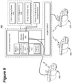
- Figure 9 illustrates a block diagram of components of a surgical system that includes imaging devices (e.g., C-Arm 104, O-Arm 106, etc.) connected to a computer platform 910 which can be operationally connected to a camera tracking system component 6 ( Fig. 3A ) or 6' ( Figs. 3B,3C ) and/or to surgical robot 4 according to some embodiments of the present disclosure.
- imaging devices e.g., C-Arm 104, O-Arm 106, etc.
- a computer platform 910 which can be operationally connected to a camera tracking system component 6 ( Fig. 3A ) or 6' ( Figs. 3B,3C ) and/or to surgical robot 4 according to some embodiments of the present disclosure.
- a camera tracking system component 6 Fig. 3A
- 6' Figs. 3B,3C
- the computer platform 910 includes a display 912, at least one processor circuit 914 (also referred to as a processor for brevity), at least one memory circuit 916 (also referred to as a memory for brevity) containing computer readable program code 918, and at least one network interface 902 (also referred to as a network interface for brevity).
- the display 912 may be part of an XR headset 920 in accordance with some embodiments of the present disclosure.
- the network interface 902 can be configured to connect to a C-Arm imaging device 104 in Figure 10 , an O-Arm imaging device 106 in Figure 11 , another medical imaging device, an image database 950 containing patient medical images, components of the surgical robot 4, and/or other electronic equipment.
- the display 912 When used with a surgical robot 4, the display 912 may correspond to the display 34 of Figure 2 and/or the tablet 890 of Figure 8 and/or the XR headset 920 that is operatively connected to the surgical robot 4, the network interface 902 may correspond to the platform network interface 812 of Figure 8 , and the processor 914 may correspond to the computer 822 of Figure 8 .
- the network interface 902 of the XR headset 920 may be configured to communicate through a wired network, e.g., thin wire ethernet, and/or through wireless RF transceiver link according to one or more wireless communication protocols, e.g., WLAN, 3GPP 4G and/or 5G (New Radio) cellular communication standards, etc.
- wireless communication protocols e.g., WLAN, 3GPP 4G and/or 5G (New Radio) cellular communication standards, etc.
- the processor 914 may include one or more data processing circuits, such as a general purpose and/or special purpose processor, e.g., microprocessor and/or digital signal processor.
- the processor 914 is configured to execute the computer readable program code 918 in the memory 916 to perform operations, which may include some or all of the operations described herein as being performed for surgery planning, navigated surgery, and/or robotic surgery.
- the computer platform 910 can be configured to provide surgery planning functionality.
- the processor 914 can operate to display on the display device 912 and/or on the XR headset 920 an image of an anatomical structure, e.g., vertebra, that is received from one of the imaging devices 104 and 106 and/or from the image database 950 through the network interface 920.
- the processor 914 receives an operator's definition of where the anatomical structure shown in one or more images is to have a surgical procedure, e.g., screw placement, such as by the operator touch selecting locations on the display 912 for planned procedures or using a mouse-based cursor to define locations for planned procedures.
- the XR headset can be configured to sense in gesture-based commands formed by the wearer and/or sense voice based commands spoken by the wearer, which can be used to control selection among menu items and/or control how objects are displayed on the XR headset 920 as will be explained in further detail below.
- the computer platform 910 can be configured to enable anatomy measurement, which can be particularly useful for knee surgery, like measurement of various angles determining center of hip, center of angles, natural landmarks (e.g. transepicondylar line, Whitesides line, posterior condylar line), etc. Some measurements can be automatic while some others can involve human input or assistance.
- the computer platform 910 may be configured to allow an operator to input a choice of the correct implant for a patient, including choice of size and alignment.
- the computer platform 910 may be configured to perform automatic or semi-automatic (involving human input) segmentation (image processing) for CT images or other medical images.
- the surgical plan for a patient may be stored in a cloud-based server, which may correspond to database 950, for retrieval by the surgical robot 4.
- a surgeon may choose which cut to make (e.g. posterior femur, proximal tibia etc.) using a computer screen (e.g. touchscreen) or extended reality (XR) interaction (e.g., hand gesture based commands and/or voice based commands) via, e.g., the XR headset 920.
- the computer platform 910 can generate navigation information which provides visual guidance to the surgeon for performing the surgical procedure.
- the computer platform 910 can provide guidance that allows the surgical robot 4 to automatically move the end effector 26 to a target pose so that the surgical tool is aligned with a target location to perform the surgical procedure on an anatomical structure.
- the surgical system 900 can use two DRAs to track patient anatomy position, such as one connected to patient tibia and one connected to patient femur.
- the system 900 may use standard navigated instruments for the registration and checks (e.g. a pointer similar to the one used in Globus ExcelsiusGPS system for spine surgery).
- a particularly challenging task in navigated surgery is how to plan the position of an implant in spine, knee, and other anatomical structures where surgeons struggle to perform the task on a computer screen which is a 2D representation of the 3D anatomical structure.
- the system 900 could address this problem by using the XR headset 920 to display a three-dimensional (3D) computer generated representations of the anatomical structure and a candidate implant device.
- the computer generated representations are scaled and posed relative to each other on the display screen under guidance of the computer platform 910 and which can be manipulated by a surgeon while viewed through the XR headset 920.
- a surgeon may, for example, manipulate the displayed computer-generated representations of the anatomical structure, the implant, a surgical tool, etc., using hand gesture based commands and/or voice based commands that are sensed by the XR headset 920.
- a surgeon can view a displayed virtual handle on a virtual implant, and can manipulate (e.g., grab and move) the virtual handle to move the virtual implant to a desired pose and adjust a planned implant placement relative to a graphical representation of an anatomical structure.
- the computer platform 910 could display navigation information through the XR headset 920 that facilitates the surgeon's ability to more accurately follow the surgical plan to insert the implant and/or to perform another surgical procedure on the anatomical structure.
- the progress of bone removal e.g., depth of cut
- Other features that may be displayed through the XR headset 920 can include, without limitation, gap or ligament balance along a range of joint motion, contact line on the implant along the range of joint motion, ligament tension and/or laxity through color or other graphical renderings, etc.
- the computer platform 910 can allow planning for use of standard surgical tools and/or implants, e.g., posterior stabilized implants and cruciate retaining implants, cemented and cementless implants, revision systems for surgeries related to, for example, total or partial knee and/or hip replacement and/or trauma.
- standard surgical tools and/or implants e.g., posterior stabilized implants and cruciate retaining implants, cemented and cementless implants, revision systems for surgeries related to, for example, total or partial knee and/or hip replacement and/or trauma.
- An automated imaging system can be used in conjunction with the computer platform 910 to acquire pre-operative, intra-operative, post-operative, and/or real-time image data of an anatomical structure.
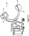
- Example automated imaging systems are illustrated in Figures 10 and 11 .
- the automated imaging system is a C-arm 104 ( Fig. 10 ) imaging device or an O-arm® 106 ( Fig. 11 ).
- O-arm® is copyrighted by Medtronic Navigation, Inc. having a place of business in Louisville, Colo., USA). It may be desirable to take x-rays of a patient from a number of different positions, without the need for frequent manual repositioning of the patient which may be required in an x-ray system.
- C-arm 104 x-ray diagnostic equipment may solve the problems of frequent manual repositioning and may be well known in the medical art of surgical and other interventional procedures.
- a C-arm includes an elongated C-shaped member terminating in opposing distal ends 112 of the "C" shape.
- C-shaped member is attached to an x-ray source 114 and an image receptor 116.
- the space within C-arm 104 of the arm provides room for the physician to attend to the patient substantially free of interference from the x-ray support structure.
- the C-arm is mounted to enable rotational movement of the arm in two degrees of freedom, (i.e. about two perpendicular axes in a spherical motion).
- C-arm is slidably mounted to an x-ray support structure, which allows orbiting rotational movement of the C-arm about its center of curvature, which may permit selective orientation of x-ray source 114 and image receptor 116 vertically and/or horizontally.
- the C-arm may also be laterally rotatable, (i.e. in a perpendicular direction relative to the orbiting direction to enable selectively adjustable positioning of x-ray source 114 and image receptor 116 relative to both the width and length of the patient).
- Spherically rotational aspects of the C-arm apparatus allow physicians to take x-rays of the patient at an optimal angle as determined with respect to the particular anatomical condition being imaged.
- the O-arm® 106 illustrated in Figure 11 includes a gantry housing 124 which may enclose an image capturing portion, not illustrated.
- the image capturing portion includes an x-ray source and/or emission portion and an x-ray receiving and/or image receiving portion, which may be disposed about one hundred and eighty degrees from each other and mounted on a rotor (not illustrated) relative to a track of the image capturing portion.
- the image capturing portion may be operable to rotate three hundred and sixty degrees during image acquisition.
- the image capturing portion may rotate around a central point and/or axis, allowing image data of the patient to be acquired from multiple directions or in multiple planes.
- the O-arm® 106 with the gantry housing 124 has a central opening for positioning around an object to be imaged, a source of radiation that is rotatable around the interior of gantry housing 124, which may be adapted to project radiation from a plurality of different projection angles.
- a detector system is adapted to detect the radiation at each projection angle to acquire object images from multiple projection planes in a quasi-simultaneous manner.
- the gantry may be attached to a support structure O-arm® support structure, such as a wheeled mobile cart with wheels, in a cantilevered fashion.
- a positioning unit translates and/or tilts the gantry to a planned position and orientation, preferably under control of a computerized motion control system.
- the gantry may include a source and detector disposed opposite one another on the gantry.
- the source and detector may be secured to a motorized rotor, which may rotate the source and detector around the interior of the gantry in coordination with one another.
- the source may be pulsed at multiple positions and orientations over a partial and/or full three hundred and sixty degree rotation for multiplanar imaging of a targeted object located inside the gantry.
- the gantry may further comprise a rail and bearing system for guiding the rotor as it rotates, which may carry the source and detector.
- Both and/or either O-arm® 106 and C-arm 104 may be used as automated imaging system to scan a patient and send information to the surgical system 2.
- Images captured by an imaging system can be displayed on the XR headset 920 and/or another display device of the computer platform 910, the surgical robot 4, and/or another component of the surgical system 900.
- the XR headset 920 may be connected to one or more of the imaging devices 104 and/or 106 and/or to the image database 950, e.g., via the computer platform 910, to display images therefrom.
- a user may provide control inputs through the XR headset 920, e.g., gesture and/or voice based commands, to control operation of one or more of the imaging devices 104 and/or 106 and/or the image database 950.
- Figure 12 illustrates a block diagram view of the components of a surgical system that include a pair of XR headsets 920 (head-mounted displays HMD1 and HMD2), which may correspond to the XR headset 920 shown in Figure 13 and operate in accordance with some embodiments of the present disclosure.
- XR headsets 920 head-mounted displays HMD1 and HMD2
- the assistant 612 and surgeon 610 are both wearing the XR headsets 920, respectively. It is optional for the assistant 612 to wear the XR headset 920.
- the XR headsets 920 are configured to provide an interactive environment through which the wearers can view and interact with information related to a surgical procedure as will be described further below. This interactive XR based environment may eliminate a need for the tech personnel 614 shown in Figure 6 to be present in the operating room and may eliminate a need for use of the display 34 shown in Figure 6 .
- Each XR headset 920 can include one or more cameras that are configured to provide an additional source of tracking of DRAs or other reference arrays attached to surgical tools, a patient's anatomical structure, the end effector 26, and/or other equipment.
- XR headset 920 has a field-of-view (FOV) 1202 for tracking DRAs and other objects
- XR headset 920 has a FOV 1212 partially overlapping FOV 1202 for tracking DRAs and other objects
- the tracking cameras 46 has another FOV 600 partially overlapping FOVs 1202 and 1212 for tracking DRAs and other objects.
- FOV field-of-view
- the tracking subsystem 830 and/or navigation controller 828 can continue to track the object seamlessly without loss of navigation. Additionally, if there is partial occlusion of the DRA from the perspective of one camera, but the entire DRA is visible via multiple camera sources, the tracking inputs of the cameras can be merged to continue navigation of the DRA.
- One of the XR headsets and/or the tracking cameras 46 may view and track the DRA on another one of the XR headsets to enable the computer platform 910 ( Figs.
- the tracking subsystem 830 and/or another computing component to determine the pose of the DRA relative to one or more defined coordinate systems, e.g., of the XR headsets 920, the tracking cameras 46, and/or another coordinate system defined for the patient, table, and/or room.
- one or more defined coordinate systems e.g., of the XR headsets 920, the tracking cameras 46, and/or another coordinate system defined for the patient, table, and/or room.
- the XR headsets 920 can be operatively connected to view video, pictures, and/or other received information and/or to provide commands that control various equipment in the surgical room, including but not limited to neuromonitoring, microscopes, video cameras, and anesthesia systems. Data from the various equipment may be processed and displayed within the headset, for example the display of patient vitals or the microscope feed.
- FIG. 13 illustrates an XR headset 920 which is configured in accordance with some embodiments of the present disclosure.
- the XR headset includes a headband 1306 configured to secure the XR headset to a wearer's head, an electronic component enclosure 1304 supported by the headband 1306, and a display screen 1302 that extends laterally across and downward from the electronic component enclosure 1304.
- the display screen 1302 may be a see-through LCD display device or a semi-reflective lens that reflects images projected by a display device toward the wearer's eyes.
- a set of DRA fiducials 1310 e.g., dots, are painted or attached in a spaced apart known arranged on one or both sides of the headset.
- the DRA on the headset enables the tracking cameras on the auxiliary tracking bar to track pose of the headset 920 and/or enables another XR headset to track pose of the headset 920.
- the display screen 1302 operates as a see-through display screen, also referred to as a combiner, that reflects light from display panels of a display device toward the user's eyes.
- the display panels can be located between the electronic component enclosure and the user's head, and angled to project virtual content toward the display screen 1302 for reflection toward the user's eyes.
- the display screen 1302 is semi-transparent and semi-reflective allowing the user to see reflected virtual content superimposed on the user's view of a real-world scene.
- the display screen 1302 may have different opacity regions, such as the illustrated upper laterally band which has a higher opacity than the lower laterally band.
- Opacity of the display screen 1302 may be electronically controlled to regulate how much light from the real-world scene passes through to the user's eyes.
- a high opacity configuration of the display screen 1302 results in high-contrast virtual images overlaid on a dim view of the real-world scene.
- a low opacity configuration of the display screen 1302 can result in more faint virtual images overlaid on a clearer view of the real-world scene.
- the opacity may be controlled by applying an opaque material on a surface of the display screen 1302.
- the surgical system includes an XR headset 920 and an XR headset controller, e.g., controller 1430 in Fig. 14 .
- the XR headset 920 is configured to be worn by a user during a surgical procedure and has a see-through display screen 1302 that is configured to display an XR image and to allow at least a portion of a real-world scene to pass therethrough for viewing by the user.
- the XR headset 920 also includes an opacity filter positioned between at least one of the user's eyes and the real-world scene when the see-through display screen 1302 is viewed by the user.
- the opacity filter is configured to provide opaqueness to light from the real-world scene.
- the XR headset controller is configured to communicate with a navigation controller, e.g., controller(s) 828A, 828B, and/or 828C in Fig. 14 , to receive navigation information from the navigation controller which provides guidance to the user during the surgical procedure on an anatomical structure, and is further configured to generate the XR image based on the navigation information for display on the see-through display screen 1302.
- a navigation controller e.g., controller(s) 828A, 828B, and/or 828C in Fig. 14
- the XR headset controller is configured to communicate with a navigation controller, e.g., controller(s) 828A, 828B, and/or 828C in Fig. 14 , to receive navigation information from the navigation controller which provides guidance to the user during the surgical procedure on an anatomical structure, and is further configured to generate the XR image based on the navigation information for display on the see-through display screen 1302.
- Opacity of the display screen 1302 may be configured as a gradient having a more continuously changing opacity with distance downward from a top portion of the display screen 1302.
- the gradient's darkest point can be located at the top portion of the display screen 1302, and gradually becoming less opaque further down on the display screen 1302 until the opacity is transparent or not present.
- the gradient can change from about 90% opacity to entirely transparent approximately at the mid-eye level of the display screen 1302. With the headset properly calibrated and positioned, the mid-eye level can correspond to the point where the user would look straight out, and the end of the gradient would be located at the "horizon" line of the eye. The darker portion of the gradient will allow crisp, clear visuals of the virtual content and help to block the intrusive brightness of the overhead operating room lights.
- an opacity filter in this manner enables the XR headset 920 to provide virtual reality (VR) capabilities, by substantially or entirely blocking light from the real-world scene, along an upper portion of the display screen 1302 and to provide AR capabilities along a middle or lower portion of the display screen 1302. This allows the user to have the semi-translucence of AR where needed and allowing clear optics of the patient anatomy during procedures.
- Configuring the display screen 1302 as a gradient instead of as a more constant opacity band can enable the wearer to experience a more natural transition between a more VR type view to a more AR type view without experiencing abrupt changes in brightness of the real-world scene and depth of view that may otherwise strain the eyes such as during more rapid shifting between upward and downward views.
- the display panels and display screen 1302 can be configured to provide a wide field of view see-through XR display system. In one example configuration they provide an 80° diagonal field-of-view (FOV) with 55° of vertical coverage for a user to view virtual content. Other diagonal FOV angles and vertical coverage angles can be provided through different size display panels, different curvature lens, and/or different distances and angular orientations between the display panels and curved display screen 1302.
- FOV field-of-view
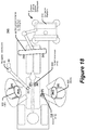
- Figure 14 illustrates electrical components of the XR headset 920 that can be operatively connected to the computer platform 910, to one or more of the imaging devices, such as the C-arm imaging device 104, the O-arm imaging device 106, and/or the image database 950, and/or to the surgical robot 800 in accordance with various embodiments of the present disclosure.
- the imaging devices such as the C-arm imaging device 104, the O-arm imaging device 106, and/or the image database 950, and/or to the surgical robot 800 in accordance with various embodiments of the present disclosure.
- the XR headset 920 provides an improved human interface for performing navigated surgical procedures.
- the XR headset 920 can be configured to provide functionalities, e.g., via the computer platform 910, that include without limitation any one or more of: identification of hand gesture based commands and/or voice based commands, display XR graphical objects on a display device 1450.
- the display device 1450 may be a video projector, flat panel display, etc., which projects the displayed XR graphical objects onto the display screen 1302.
- the user can view the XR graphical objects as an overlay anchored to particular real-world objects viewed through the display screen 1302 ( Fig. 13 ).
- the XR headset 920 may additionally or alternatively be configured to display on the display screen 1450 video feeds from cameras mounted to one or more XR headsets 920 and other cameras.
- Electrical components of the XR headset 920 can include a plurality of cameras 1440, a microphone 1442, a gesture sensor 1444, a pose sensor (e.g., inertial measurement unit (IMU)) 1446, a display module 1448 containing the display device 1450, and a wireless/wired communication interface 1452.
- the cameras 1440 of the XR headset may be visible light capturing cameras, near infrared capturing cameras, or a combination of both.
- the cameras 1440 may be configured operate as the gesture sensor 1444 by capturing for identification user hand gestures performed within the field of view of the camera(s) 1440.
- the gesture sensor 1444 may be a proximity sensor and/or a touch sensor that senses hand gestures performed proximately to the gesture sensor 1444 and/or senses physical contact, e.g. tapping on the sensor or the enclosure 1304.
- the pose sensor 1446 e.g., IMU, may include a multi-axis accelerometer, a tilt sensor, and/or another sensor that can sense rotation and/or acceleration of the XR headset 920 along one or more defined coordinate axes. Some or all of these electrical components may be contained in the component enclosure 1304 or may be contained in another enclosure configured to be worn elsewhere, such as on the hip or shoulder.
- the surgical system 2 includes a camera tracking system component 6/6' and a tracking subsystem 830 which may be part of the computer platform 910.
- the surgical system may include imaging devices (e.g., C-arm 104, O-arm 106, and/or image database 950) and/or a surgical robot 4.
- the tracking subsystem 830 is configured to determine a pose of DRAs attached to an anatomical structure, an end effector, a surgical tool, etc.
- a navigation controller 828 is configured to determine a target pose for the surgical tool relative to an anatomical structure based on a surgical plan, e.g., from a surgical planning function performed by the computer platform 910 of Fig.
- the navigation controller 828 may be further configured to generate steering information based on the target pose for the surgical tool, the pose of the anatomical structure, and the pose of the surgical tool and/or the end effector, where the steering information indicates where the surgical tool and/or the end effector of a surgical robot should be moved to perform the surgical plan.
- Various of the cameras 1440 of the XR headset 920 may be connected to the camera tracking system component 6/6' to track poses of DRAs, user's hand(s), etc.
- the electrical components of the XR headset 920 can be operatively connected to the electrical components of the computer platform 910 through a wired/wireless interface 1452.
- the electrical components of the XR headset 920 may be operatively connected, e.g., through the computer platform 910 or directly connected, to various imaging devices, e.g., the C-arm imaging device 104, the I/O-arm imaging device 106, the image database 950, and/or to other medical equipment through the wired/wireless interface 1452.
- the surgical system 2 further includes at least one XR headset controller 1430 (also referred to as "XR headset controller” for brevity) that may reside in the XR headset 920, the computer platform 910, and/or in another system component connected via wired cables and/or wireless communication links.
- XR headset controller 1430 is configured to receive navigation information from the navigation controller 828 which provides guidance to the user during the surgical procedure on an anatomical structure, and is configured to generate an XR image based on the navigation information for display on the display device 1450 for projection on the see-through display screen 1302.
- the configuration of the display device 1450 relative to the display screen (also referred to as "see-through display screen ”) 1302 is configured to display XR images in a manner such that when the user wearing the XR headset 920 looks through the display screen 1302 the XR images appear to be in the real world.
- the display screen 1302 can be positioned by the headband 1306 in front of the user's eyes.
- the XR headset controller 1430 can be within a housing that is configured to be worn on a user's head or elsewhere on the user's body while viewing the display screen 1302 or may be remotely located from the user viewing the display screen 1302 while being communicatively connected to the display screen 1302.
- the XR headset controller 1430 can be configured to operationally process signaling from the cameras 1440, the microphone 142, and/or the pose sensor 1446, and is connected to display XR images on the display device 1450 for user viewing on the display screen 1302.
- the XR headset controller 1430 illustrated as a circuit block within the XR headset 920 is to be understood as being operationally connected to other illustrated components of the XR headset 920 but not necessarily residing within a common housing (e.g., the electronic component enclosure 1304 of Fig. 13 ) or being otherwise transportable by the user.
- the XR headset controller 1430 may reside within the computer platform 910 which, in turn, may reside within a housing of the computer tracking system component 6' shown in Figures 3B and 3C .
- Figure 15 illustrates a block diagram showing arrange of optical components of the XR headset 920 in accordance with some embodiments of the present disclosure.
- the display device 1450 is configured to display XR images generated by the XR headset controller 1430, light from which is projected as XR images 1500 toward the display screen 1302.
- the display screen 1302 is configured to combine light of the XR images 1500 and light from the real-world scene 1502 into a combined augmented view 1504 that is directed to the user's eye(s) 1510.
- the display screen 1302 configured in this manner operates as a see-through display screen.
- the XR headset 920 can include any plural number of tracking cameras 1440.
- the cameras 1440 may be visible light capturing cameras, near infrared capturing cameras, or a combination of both.
- the XR headset operations can display both 2D images and 3D models on the display screen 1302.
- the 2D images may preferably be displayed in a more opaque band of the display screen 1302 (upper band) and the 3D model may be more preferably displayed in the more transparent band of the display screen 1302, otherwise known as the environmental region (bottom band). Below the lower band where the display screen 1302 ends the wearer has an unobstructed view of the surgical room.
- XR content is display on the display screen 1302 may be fluidic. It is possible that where the 3D content is displayed moves to the opaque band depending on the position of the headset relative to the content, and where 2D content is displayed can be placed in the transparent band and stabilized to the real world.
- the entire display screen 1302 may be darkened under electronic control to convert the headset into virtual reality for surgical planning or completely transparent during the medical procedure.
- the XR headset 920 and associated operations not only support navigated procedures, but also can be performed in conjunction with robotically assisted procedures.
- Figure 16 illustrates an example view through the display screen 1302 of the XR headset 920 for providing navigation assistance to a user who is manipulating a surgical tool 1602 during a medical procedure in accordance with some embodiments of the present disclosure.
- a graphical representation 1600 of the tool can be displayed in 2D and/or 3D images in relation to a graphical representation 1610 of the anatomical structure.
- the user can use the viewed graphical representations to adjust a trajectory 1620 of the surgical tool 1602, which can be illustrated as extending from the graphical representation 2000 of the tool through the graphical representation 1610 of the anatomical structure.
- the XR headset 920 may also display textual information and other objects 1640.
- the dashed line 1650 extending across the viewed display screen represents an example division between different opacity level upper and lower bands.
- XR images virtual content
- Other types of XR images can include, but are not limited to any one or more of:
- Figure 17 illustrates example configuration of an auxiliary tracking bar 46 having two pairs of stereo tracking cameras configured in accordance with some embodiments of the present disclosure.
- the auxiliary tracking bar 46 is part of the camera tracking system component of Figures 3A , 3B, and 3C .
- the stereo tracking cameras include a stereo pair of spaced apart visible light capturing cameras and another stereo pair of spaced apart near infrared capturing cameras, in accordance with one embodiment.
- only one stereo pair of visible light capturing cameras or only one stereo pair of near infrared capture cameras can used in the auxiliary tracking bar 46. Any plural number of near infrared and/or visible light cameras can be used.
- navigated surgery can include computer vision tracking and determination of pose (e.g., position and orientation in a six degree-of-freedom coordinate system) of surgical instruments, such as by determining pose of attached DRAs that include spaced apart fiducials, e.g., disks or spheres, arranged in a manner known to the camera tracking system.
- the computer vision uses spaced apart tracking cameras, e.g., stereo cameras, that are configured to capture near infrared and/or visible light.
- Computer operations may combine (chain) measured poses in ways that can improve optimization of one or more of the above three parameters by incorporating additional tracking cameras mounted to one or more XR headsets.
- a stereo pair of visible light tracking cameras and another stereo pair of near infrared tracking cameras can be attached to the auxiliary tracking bar of the camera tracking system component in accordance with some embodiments of the present disclosure.
- Operational algorithms are disclosed that analyze the pose of DRAs that are fully observed or partially observed (e.g., when less than all of the fiducials of a DRA are viewed by a pair of stereo cameras), and combine the observed poses or partial poses in ways that can improve accuracy, robustness, and/or ergonomics during navigated surgery.
- the XR headset may be configured to augment a real-world scene with computer generated XR images.
- the XR headset may be configured to provide an XR viewing environment by displaying the computer generated XR images on a see-through display screen that allows light from the real-world scene to pass therethrough for combined viewing by the user.
- the XR headset may be configured to provide a VR viewing environment by preventing or substantially preventing light from the real-world scene from being directly viewed by the user along the viewing path of the displayed XR images.
- An XR headset can be configured to provide both AR and VR viewing environments.
- both AR and VR viewing environments are provided by lateral bands of substantially differing opacity arranged between the see-through display screen and the real-world scene, so that a VR viewing environment is provided for XR images aligned with a high opacity band and an AR viewing environment is provided for XR images aligned with the low opacity band.
- both AR and VR viewing environments are provided by computer adjustable control of an opacity filter that variably constrains how much light from the real-world scene passes through a see-through display screen for combining with the XR images viewed by the user.
- the XR headset can also be referred to as an AR headset or a VR headset.
- the XR headset can include near infrared tracking cameras and/or visible light tracking cameras that are configured to track fiducials of DRAs connected to surgical instruments, patient anatomy, other XR headset(s), and/or a robotic end effector.
- near infrared tracking and/or visible light tracking on the XR headset provides additional tracking volume coverage beyond what cameras on a single auxiliary tracking bar can provide. Adding near infrared tracking cameras to the existing auxiliary tracking bar allows for the headset location to be tracked more robustly but less accurately than in visible light.
- Mechanically calibrating the visible and near infrared tracking coordinate systems enables the coordinate systems to be aligned sufficiently to perform 3D DRA fiducials triangulation operations using stereo matching to jointly identify pose of the DRA fiducials between the visible and near infrared tracking coordinate systems.
- Using both visible and near infrared tracking coordinate systems can enable any one or more of: (a) identifying tools that would not be identified using a single coordinate system; (b) increased pose tracking accuracy; (c) enabling a wider range of motion without losing tracking of surgical instruments, patient anatomy, and/or a robotic end effector; and (d) naturally track an XR headset in the same coordinate system as the navigated surgical instruments.
- Figure 18 illustrates a block diagram view of the components of a surgical system that include tracking cameras in a pair of XR headsets 920 (head-mounted displays HMD1 and HMD2) and tracking cameras in a camera tracking bar in the camera tracking system component 6' which houses the computer platform 910.
- the computer platform 910 can include the tracking subsystem 830, the navigation controller 828, and the XR headset controller 1430 as was earlier shown in Figure 14 .
- a surgeon and an assistant are both wearing XR headsets HMD1 920 and HMD2 920, respectively, each if which includes tracking cameras that may be configured as shown in Figure 13 . It is optional for the assistant to wear the XR headset HMD2 920.
- the combination of XR headsets HMD1 920 and HMD2 920 and the tracking cameras 46 on the auxiliary tracking bar can, in operation with the computer platform 910, more robustly track the example objects of a patient reference array (R), robotic end effector (E), and surgical tool (T) or instrument.
- R patient reference array
- E robotic end effector
- T surgical tool
- the overlapping views from different perspectives that are provided by the XR headsets HMD1 920 and HMD2 920 and the tracking cameras 46 on the auxiliary tracking bar are shown in Figure 12 .
- T N 2 S is calibrated: T N 3 A ; T N V , where the term "T" is defined as a six degree-of-freedom (6 DOF) homogeneous transformation between the two indicated coordinates systems.
- T N 2 S is a 6 DOF homogeneous transformation between the visible light coordinate system of the primary headset HMD1920 and the NIR coordinate system of the primary headset HMD1 920.
- the XR headsets HMD1 920 and HMD2 920 have passive visible light fiducials painted or otherwise attached to them (coordinate systems S and A), such as the reference array fiducials 1310 shown in Figure 13 .
- the tracking cameras are spatially calibrated to these passive fiducials (coordinate systems N2 and N3).
- the cameras on the XR headset HMD1 920 and HMD2 920 and the tracking cameras 46 on the auxiliary tracking bar have partially overlapping field of views. If one or more of the cameras on the XR headset HMD1 920 are obstructed from viewing a DRA attached to a tracked object, e.g., a tracked tool (T), but the DRA is in view of the cameras of the other XR headset HMD2 920 and/or the tracking cameras 46 on the auxiliary tracking bar, the computer platform 910 can continue to track the DRA seamlessly without loss of navigation.
- a tracked object e.g., a tracked tool (T)
- the tracking inputs of the cameras can be merged to continue navigation of the DRA.
- the various coordinate systems can be chained together by virtue of independent observations the various camera systems provided by the XR headsets HMD1 920 and HMD2 920 and the tracking cameras 46 on the auxiliary tracking bar.
- each of the XR headsets HMD1 920 and HMD2 920 may require virtual augmentation of the robotic end effector (E). While one XR headset HMD1 920 (N2) and the tracking cameras 46 on the auxiliary tracking bar (N) are able to see (E), perhaps the other XR headset HMD2 920 (N3) cannot.
- the location of (E) with respect to (N3) can still be computed via one of several different operational methods. Operations according to one embodiment performing chaining of poses from a patient reference (R).
- the chains can be arbitrarily long and are enabled by having more than one stereo camera system (e.g., N, N2, N3).
- the camera tracking system can be configured to receive tracking information related to tracked objects from a first tracking camera (e.g., N3) and a second tracking camera (e.g., N2) during a surgical procedure.
- the camera tracking system can determine a first pose transform (e.g., T N 3 R ) between a first object (e.g., R) coordinate system and the first tracking camera (e.g., N3) coordinate system based on first object tracking information from the first tracking camera (e.g., N3) which indicates pose of the first object (e.g., R).
- the camera tracking system can determine a second pose transform (e.g., T R N 2 ) between the first object (e.g., R) coordinate system and the second tracking camera (e.g., N2) coordinate system based on first object tracking information from the second tracking camera (e.g., N2) which indicates pose of the first object (e.g., R).
- the camera tracking system can determine a third pose transform (e.g., T N 2 E ) between a second object (e.g., E) coordinate system and the second tracking camera (e.g., N2) coordinate system based on second object tracking information from the second tracking camera (e.g., N2) which indicates pose of the second object (e.g., E).
- the camera tracking system can determine a fourth pose transform (e. g., T N 3 E ) between the second object (e.g., E) coordinate system and the first tracking camera (e.g., N3) coordinate system based on combining the first, second, and third pose transforms.
- the camera system can further determine pose of the second object (e.g., E) and the first tracking camera system (e.g., N3) coordinate system based on processing the tracking information through the fourth pose transform.
- the camera tracking system is capable of determining the pose of the second object (e.g., E) relative to first tracking camera (e.g., N3) when the first camera is blocked from seeing the second object (e.g., E).
- the camera tracking system is further configured to determine the fourth pose transform (e.g. , T N 3 E ) between the second object (e.g., E) coordinate system and the first tracking camera (e.g., N3) coordinate system without use of any tracking information from the first tracking camera (e.g., N3) indicating pose of the second object (e.g., E).
- the camera tracking system may achieve higher tracking accuracy by merging synchronized imagery from multiple camera systems.
- the camera tracking system can determine pose of the second object (e.g., E) relative to first tracking camera (e.g., N3) by merging synchronized imagery of the second object (e.g., E) from multiple perspectives (first and second tracking cameras), and can use weighting which can be determined based on accuracy specs of the respective cameras.
- the camera tracking system can be further configured to determine the fourth pose transform (e.g., T N 3 E ) between the second object (e.g., E) coordinate system and the first tracking camera (e.g., N3) coordinate system based on second object tracking information from the first tracking camera (e.g., N3) which indicates pose of the second object (e.g., E) and further based on a result of the combining of the first, second, and third pose transforms.
- the fourth pose transform e.g., T N 3 E
- the first tracking camera e.g., N3 coordinate system
- the surgical system may be configured to display on the see-through display screen of an XR headset an XR image having a pose that is determined based on the fourth pose transform.
- the camera tracking system may be further configured to generate the XR image as a graphical representation of the second object (e.g., E) that is posed on the see-through display screen based on processing through the fourth pose transform the first object tracking information from the first and second tracking cameras and the second object tracking information from the second tracking camera.
- the camera tracking system can include a navigation controller 828 communicatively connected to the first tracking camera (e.g., N3) and the second tracking camera (e.g., N2) to receive the tracking information and configured to perform the determination of the first, second, third, and fourth pose transforms.
- a navigation controller 828 communicatively connected to the first tracking camera (e.g., N3) and the second tracking camera (e.g., N2) to receive the tracking information and configured to perform the determination of the first, second, third, and fourth pose transforms.
- the camera tracking system can simultaneously track the poses of surgical tools which are being held or supported within the field-of-view of a set of tracking cameras, and can resume tracking of a surgical tool when it is moved from outside to inside that field-of-view, e.g., after being picked-up again.
- Many surgical tools require software configuration to track properly.
- the camera tracking system tracks poses of the reference arrays attached to or on the surgical tools, the camera tracking system should be informed of which surgical tool characteristics are registered to which of the tracked reference arrays.
- the navigation controller 828 ( Fig. 14 ) can thereby operate with knowledge of the particular characteristics of the surgical tool.
- registration of surgical tool characteristics of the distance from an identified reference array to a tip of the surgical tool enables the navigation controller 828 to navigate a surgeon's movement of the tool tip during a surgical procedure.
- registration of a direction of curvature of a surgical tool relative to an identified reference array enables the navigation controller 828 to display an accurate graphical representation of the surgical tool through the XR headset 920 and accurately posed relative to a tracked anatomical structure during the surgical procedure.
- the registration process is also referred to as a pairing process during which a user holds a surgical tool having a reference array in the field-of-view of the set of tracking cameras for identification of the reference array, and the user then define characteristics of that surgical tool, in accordance with some embodiments.
- the registration process is repeated for each combination of reference array and surgical tool characteristics that will be tracked during a surgical procedure, and can be further repeated when a reference array is detached from one type of surgical tool and attached to a different type of surgical tool.
- Some further embodiments of the present disclosure are directed to using an XR headset during a registration process to register an identified reference array to characteristics of a surgical tool, and to display a representation of those characteristics through the XR headset so that a user can verify correctness of the registration.
- Using the XR headset during the registration process can provide a more intuitive, time efficient and reliable process for surgeons and other medical personnel (users) to register surgical tools with a camera tracking system before and/or during a surgical procedure.
- the registration process may be automatically initiated responsive to a reference array being brought into the field-of-view of a set of tracking cameras and the camera tracking system determining that the identified reference array has not yet been registered as being paired with characteristics of a surgical tool.
- Figure 19 illustrates a graphical representation 1910 of surgical tool characteristics that can be displayed by the XR headset when a reference array 1902 is being registered to characteristics of a surgical tool 1900 for use in computer assisted navigation of the surgical tool 1900 during surgery, in accordance with some embodiments of the present disclosure.
- Figure 20 illustrates a flowchart of registration operations performed by the camera tracking system (e.g., system 6 in Fig. 18 ), and which may provide the graphical representation of the surgical tool characteristics in Figure 19 , in accordance with some embodiments of the present disclosure.
- the example XR headset includes a set of tracking cameras which may, for example, be contained in the electronic component enclosure 1304 of the XR headset 920 as shown in Figure 13 .
- the camera tracking system e.g., system 6 in Fig. 18 ) identifies 2000 a reference array 1902 which is tracked by the set of tracking cameras, such as by identifying reference array in video streams from the tracking cameras.
- reference arrays can be uniquely identified based on the differing orientations between the sets of fiducials forming respective reference arrays.
- the camera tracking system determines 2002 whether the identified reference array 1902 is registered as being paired with characteristics of one of a plurality of surgical tools defined in a surgical tool database.
- the camera tracking system Based on the reference array 1902 being determined 2002 to not be registered and based on receiving user input, the camera tracking system registers 2004 the reference array 1902 as being paired with characteristics of the surgical tool 1900 which is selected among the plurality of surgical tools based on the user input. The camera tracking system then provides 2006 a representation of the characteristics to a display device of the XR headset 920 for display to the user.
- a user can initiate registration by raising the surgical tool 1900 into the field-of-view of the set of tracking cameras attached to the XR headset 920.
- the camera tracking system can cause a visual prompt to be displayed through the XR headset 920 which prompts the user to perform registration of the reference array to characteristics of the associated surgical tool.
- the camera tracking system receives video from the tracking cameras, identifies 2000 the reference array based on the spacing and relative, and determines 2002 that the identified reference array has not yet been registered as being paired with any defined characteristics of a surgical tool defined in the surgical tool database.
- the camera tracking system receives user input identifying characteristics of the surgical tool, and registers 2004 the reference array as being paired with characteristics of the surgical tool 1900.
- the camera tracking system receives user input identifying characteristics of the surgical tool, and registers 2004 the reference array as being paired with characteristics of the surgical tool 1900. The camera tracking system.
- Example surgical tool characteristics can include, without limitation, structural and/or operational characteristics of a drill, saw, screw driver, retractor, and implant such as a screw, spacer, interbody fusion device, plate, rod, etc
- the camera tracking system can facilitate the user's definition of the surgical tool characteristics that are to be registered with the identified reference array 1902, by providing 2008 a list of the surgical tools in the database for display through the XR headset 920 for user selection among to be registered with the reference array 1902. For example, based on the reference array 1902 being determined 2002 to not be registered, the camera tracking system can provide to the display device of the XR headset 920 a list of at least some of the plurality of the surgical tools defined in the surgical tool database for the user to select one of the surgical tools to be registered as paired with the reference array 1902.
- the tip of the surgical tool 1900 curves off to one side and requires a user to select one of four modes displayed as a list 1910 during registration based on which direction the tool tip is curving with respect to the reference array 1902.
- the four modes include Mode A corresponding to +X curvature, Mode B corresponding to +Z curvature, Mode C corresponding to -X curvature, and Mode D corresponding to -Z curvature.
- Use of the XR headset 920 during such surgical tool registration can intuitively facilitate the user's registration of the surgical tool characteristics as well as provide safeguards to ensure the correct characteristics are registered to the correct reference array 1902.
- the camera tracking system may be configured to track movement of a user's hand and to identify hand gestures as user input during the tool registration process. For example, in the example of Figure 19 , a user may be able to virtually touch-select one of the modes (Mode A-D) displayed in the virtual space to have the surgical tool characteristics which have been defined for the selected mode to become registered to the reference array 1902. More particularly, the camera tracking system may be configured to determine the user's selection among the displayed list 1910, based on tracking information from the set of tracking cameras indicating pose of a hand of the user in XR space relative to the displayed list 1910.
- Mode A-D the modes
- the camera tracking system may be configured to determine the user's selection among the displayed list 1910, based on tracking information from the set of tracking cameras indicating pose of a hand of the user in XR space relative to the displayed list 1910.
- the camera tracking system may display the characteristics and other information so that it appears near to the surgical tool 1900 when viewed by the surgeon through the XR headset 920, such as illustrated by the listing 1910 that is graphically displayed adjacent to the surgical tool 1900 when viewed by the surgeon through the XR headset 920.
- the camera tracking system receives tracking information from the set of tracking cameras indicating pose of the reference array 1902 relative to the XR headset 920, and determines a pose for how the representation of the characteristics (e.g., tool information) is to be displayed relative to the reference array based on the tracking information.
- the camera tracking system controls the XR headset 920 to display the representation of the characteristics with the determined pose relative to the reference array 1902.
- the camera tracking system can display visual feedback of the registered characteristics to enable the surgeon to verify accuracy of registration. Displaying informational description about the registered surgical tool 1900 provides one level of verification to ensure that the correct surgical tool has been registered. Another level of verification can include displaying a 2D or 3D graphical representation of the surgical tool characteristics that have been, or are being, registered to the reference array 1902.
- Figure 21 illustrates a graphical representation 2112 of a registered actively-used surgical tool 1900 which is displayed by the XR headset 920, in accordance with some embodiments of the present disclosure.
- Figure 22 illustrates a flowchart of operations performed by the camera tracking system to provide the graphical representation 2112 of the registered actively-used surgical tool 1900 in Figure 21 , in accordance with some embodiments of the present disclosure.
- the camera tracking system obtains 2200 a graphical representation 2112 of the surgical tool 1900 in at least 2-dimensions (i.e., a 2D model or 3D model), and receives 2202 tracking information from the set of tracking cameras indicating pose of the reference array 1902 relative to the XR headset 920.
- the camera tracking system determines 2204 a pose for the graphical representation 2112 based on the tracking information, and provides 2206 the graphical representation 2112 as the representation of the characteristics to the display device of the XR headset 920 for display with the determined pose.
- the camera tracking system has obtained a 3D representation 2112 of the registered surgical tool 1900, and displays the 3D representation 2112 in a feedback window 2110 which is adjacent to the surgical tool 1900 and with a pose that matches a current pose of the surgical tool 1900.
- a surgeon holding the surgical tool 1900 in front of the XR headset 920 is thereby shown which characteristics of a particular one of the surgical tools that have been registered to the reference array 1902, which enables the surgeon to visually compare the displayed 3D representation 2112 to the physical surgical tool 1900 to verify the correctness of the registration.
- These operations can, for example, enable a surgeon to confirm that the correct mode among the list 1910 ( Fig. 19 ) has been selected by comparing the tool tip bend direction shown in the graphical representation 2112 to that of the physical surgical tool 1900.
- the camera tracking system may determine how accurately, i.e., a measure of tracking accuracy (also called tracking quality), the reference array is being tracked by the tracking cameras.
- the tracking accuracy can become degraded when the tracking cameras are not positioned properly relative to the surgical tool 2100, when a location of one or more of the fiducials has been moved through deformation of the fiducials support structure, and/or when a fiducial is damaged and/or has become covered by bodily fluid or other material. It can therefore be important to enable a surgeon to be able to observe the tracking accuracy while viewing the surgical tool 2100 through the XR headset 920.
- Figure 23 illustrates a graphical representation 2312 of tracking accuracy information displayed by the XR headset 920 for a registered reference array 2102 while a user is viewing the surgical tool 2100, in accordance with some embodiments of the present disclosure.
- Figure 24 illustrates a flowchart of operations performed by the camera tracking system to provide the graphical representation 2312 of the tracking accuracy information in Figure 23 , in accordance with some embodiments of the present disclosure.
- the camera tracking system obtains 2400 tracking accuracy information characterizing accuracy at which the set of tracking cameras is presently tracking pose of the reference array 2102.
- the camera tracking system then provides 2402 an indication 2312 of the tracking accuracy information to the display device of the XR headset for display to the user.
- the camera tracking system has displayed the tracking accuracy in the form of a graphical sliding scale within a tracking quality window 2310 viewable through the XR headset 920, although other graphical indications, textual descriptions (e.g., poorly-tracked, well-tracked, fully-tracked), and/or colored graphical indicia could be displayed.
- the tracking accuracy may be determined based on how closely the measured positions of the fiducials forming the reference array 2102 match the positions of the fiducials that have been earlier defined for the reference array 2102 (e.g., a defined model for the identified reference array 2101), and/or based on how well the shape of the fiducials identified in video from the tracking cameras fits a defined shape of the fiducials (e.g., a defined ellipse shape).
- the tracking accuracy may be determined based on comparing a pose of the reference array 2102 to an expected pose when a defined location (e.g., tip) of the surgical tool is touched to a known location (e.g., a calibration divot on another registered reference array).
- the graphical representation of the tracking accuracy is generated to visually indicate the determined or accuracy at which the tracking cameras are tracking the reference array 2102.
- the camera tracking system may be configured to display other information associated with the surgical tool 2100.
- surgical tools can operate properly during surgical procedure, such as by not being structurally bent or lacking proper range of extension and/or angular motion, they can be regularly verified. For example, whether a surgical tool has a bent tip can be determined by positioning the tool tip at a location known to the camera tracking system, such as a known calibration divot, to confirm that the tracked reference array has an expected pose while the tool tip is at the known location.
- the results of this verification process can be stored in a surgical database, and displayed near the surgical tool to ensure that users are notified if the tool should not be used during a surgical procedure and/or whether enough time has transpired since a last verification such that the surgical tool should be re-verified before use.
- the camera tracking system may be configured to obtain a last verified date which indicates when the surgical tool 2100 was last verified to not have structural deformation, and to provide an indication of the last verified date to the display device of the XR headset 920 for display to the user.
- Such graphical characteristics of a surgical tool can be selectively displayed depending upon whether the surgical tool is within an inspection region that has been defined relative to location XR headset 920. For example, when the surgeon holds the surgical tool so that it is above the surgeon's chest or neck within a defined inspection region, the camera tracking system can respond by displaying the registered characteristics of the surgical tool. In contrast, while the surgeon holds the surgical tool below the inspection region the camera tracking system can prevent the display of the registered characteristics of the surgical tool.
- the camera tracking system may compare a shape of a portion of the tool (e.g., shape of the shaft of tool 2100) to a defined template shape for the tool 2100. Differences in the compared shape that exceed a defined threshold may cause a notification to be displayed through the XR headset 920. For example, a graphical highlight may be displayed overlapping a portion of the shaft of tool 2100 that appears to deviate from the defined template shape, in order to intuitively notify the user of a potential problem which may necessitate replacement of the shaft and/or the tool 2100.
- Figure 26 illustrates a flowchart of operations performed by the camera tracking system to selectively display characteristics of a surgical tool registered with a reference array depending upon whether the reference array is within an inspection region defined relative to the XR headset, in accordance with some embodiments of the present disclosure.
- the camera tracking system is configured to determine 2600 when the reference array has been brought within an inspection region which is defined relative to location of the XR headset. Responsive to determining that the reference array has been brought within the inspection region, the camera tracking system initiates 2602 the providing of the representation of the characteristics to the display device of the XR headset for display to the user. In contrast, responsive to determining that the reference array has exited the inspection region, the camera tracking system ceases 2604 any ongoing providing of the representation of the characteristics to the display device of the XR headset for display to the user.
- FIG. 25 illustrates a flowchart of operations performed by the camera tracking system to selectively allow user registration of a reference array depending upon whether the reference array is within an inspection region defined relative to the XR headset, in accordance with some embodiments of the present disclosure.
- the camera tracking system is configured to determine 2500 when the reference array has been brought within an inspection region which is defined relative to location of the XR headset. Responsive to determining that the reference array has been brought within the inspection region, the camera tracking system initiates 2502 the determination of whether the reference array is registered as being paired with characteristics of one of a plurality of surgical tools defined in the surgical tool database. In contrast, the camera tracking system is configured to limit 2504 operation to receive user input to register the reference array as paired with characteristics of one of the plurality of surgical tools defined in the surgical tool database, to occur only while the reference array remains within the inspection region.
- one or more of the fiducials which are arranged to form a reference array can become obscured or otherwise not viewable by the set of tracking cameras because, for example, it has been damaged during a surgical procedure and/or has become covered by bodily fluid or other material.
- Some embodiments are directed to displaying information that shows which fiducials in a reference array are being tracked by the tracking cameras. For example, when tracking fails because one or more fiducials are occluded, the remaining fiducials can be reported as strays.
- the camera tracking system can identify poses (e.g., position) of some of the fiducials but does not have enough information to match the identified fiducials to one of the plurality of predefined reference arrays.
- the poses of the stray fiducials can still be used to inform the surgeon as to which of the fiducials are being tracked.
- Virtual representations of each tracked fiducial can be displayed through the XR headset at their poses (e.g., positions) in the real world as viewed through the XR headset. In this manner, the surgeon will know which of the fiducials are occluded or need adjustment or replacement.
- a surgical tool 2100 having five fiducials forming the reference array 2102 is viewed through the XR headset 920 with graphical indicia 2700, 2702, 2704, 2706 that are displayed at least partially overlapping identified poses of four of the fiducials of the reference array 2102 and another graphical indicia 2710 that is displayed at least partially overlapping an estimated pose of a missing fiducial, in accordance with some embodiments of the present disclosure.
- Figure 28 illustrates a flowchart of operations performed by the camera tracking system to display the graphical indicia relative to the identified and missing fiducials, such as shown in Figure 27 , in accordance with some embodiments of the present disclosure.
- the camera tracking system is further configured to identify 2800 poses of a plurality of fiducials among the reference array 2102. For each of the plurality of fiducials among the reference array 2102 having the identified poses, the camera tracking system provides 2802 a graphical indicia (e.g., 2700, 2702, 2704, and 2706) for display by the display device of the XR headset 920 at least partially overlapped with the identified pose of the fiducial.
- a graphical indicia e.g., 2700, 2702, 2704, and 2706
- the camera tracking system may be further configured to respond to when one of the fiducials in the reference array 2102 is a missing fiducial not having an identifiable pose, by estimating 2804 a pose of the missing fiducial relative to the plurality of fiducials having the identified poses, and providing 2804 another graphical indicia 2710 for display by the display device of the XR headset 920 at least partially overlapped with the estimated pose of the missing fiducial.
- a process to estimate 2804 the pose of the missing fiducial may be performed based on comparing the presently identified poses of the fiducials 2700-2706 to a defined template of the relative poses of the fiducials of the identified reference array 2102, to estimate a present pose of the missing fiducial.
- a surgeon can intuitively identify which fiducials are being properly tracked by the tracking cameras and can also identify which, if any fiducials, are not being properly tracked.
- the missing fiducial is excessively obscured by body fluid or other material, the surgeon can remedy the situation to enable accurate tracking of the reference array 2102.
- Some further embodiments are directed to avoiding interpretation of a user's hand movements while holding a surgical tool as being a user's attempt to provide input to a hand-tracking input interface. This can be particularly important while a surgeon is holding a surgical tool because the tracking cameras may not maintain a sufficient view of the hands during movement of the surgical tool, and which can result in inaccurate tracking of the hands and cause accidental interactions with the user interface.
- a hand-user-interface exclusion zone is defined relative to the reference array. While a user's hand is determined to be at least partially within the hand-user-interface exclusion zone, the hand tracking information is not used as input from the user to a user-interface.
- Figure 29 illustrates a flowchart of operations performed by the camera tracking system to selectively enable receipt of hand gesture input based on whether the user's hand is at least partially within a hand-user-interface exclusion zone determined relative to the reference array, in accordance with some embodiments of the present disclosure.
- Figure 30 illustrates a hand-user-interface exclusion zone 3000 that is determined relative to the reference array 2102 connected to the surgical tool 2100, in accordance with some embodiments of the present disclosure.
- the camera tracking system is configured to receive 2900 tracking information from the set of tracking cameras indicating pose of the reference array 2102 relative to the XR headset and pose of a hand of the user.
- the camera tracking system determines 2902 the hand-user-interface exclusion zone 3000 relative to the pose of the reference array 2102 based on the tracking information.
- the camera tracking system enables 2904 receipt of hand gesture input from the user based on the tracked pose of the hand indicated by the tracking information while the user's hand is determined to be entirely outside the hand-user-interface exclusion zone 3000.
- the camera tracking system disables 2906 receipt of hand gesture input from the user based on the tracked pose of the hand indicated by the tracking information while the user's hand is determined to be at least partially within the hand-user-interface exclusion zone 3000.
- the camera tracking system may interpret movement of the user's hand as gesture input being provided to, for example, select among the displayed list 1910 of modes that are displayed as shown in Figure 19 in virtual space adjacent to the surgical tool 1900.
- the camera tracking system prevents interpretation of movement of the user's hand as a gesture input to the system and would thereby, for example, not enable a user to use a hand gesture to select among the displayed list 1910 of modes.
- the set of tracking cameras are separate and spaced apart from the XR headset while the camera tracking system is receiving the tracking information from the set of tracking cameras.
- Various operations disclosed herein for pose chaining may be used to track poses of a reference array and/or a user's hand.
- the terms “comprise”, “comprising”, “comprises”, “include”, “including”, “includes”, “have”, “has”, “having”, or variants thereof are open-ended, and include one or more stated features, integers, elements, steps, components or functions but does not preclude the presence or addition of one or more other features, integers, elements, steps, components, functions or groups thereof.
- the common abbreviation “e.g.”, which derives from the Latin phrase “exempli gratia” may be used to introduce or specify a general example or examples of a previously mentioned item, and is not intended to be limiting of such item.
- the common abbreviation “i.e.”, which derives from the Latin phrase “id est,” may be used to specify a particular item from a more general recitation.
- Example embodiments are described herein with reference to block diagrams and/or flowchart illustrations of computer-implemented methods, apparatus (systems and/or devices) and/or computer program products. It is understood that a block of the block diagrams and/or flowchart illustrations, and combinations of blocks in the block diagrams and/or flowchart illustrations, can be implemented by computer program instructions that are performed by one or more computer circuits.
- These computer program instructions may be provided to a processor circuit of a general purpose computer circuit, special purpose computer circuit, and/or other programmable data processing circuit to produce a machine, such that the instructions, which execute via the processor of the computer and/or other programmable data processing apparatus, transform and control transistors, values stored in memory locations, and other hardware components within such circuitry to implement the functions/acts specified in the block diagrams and/or flowchart block or blocks, and thereby create means (functionality) and/or structure for implementing the functions/acts specified in the block diagrams and/or flowchart block(s).
Landscapes
- Engineering & Computer Science (AREA)
- Health & Medical Sciences (AREA)
- Surgery (AREA)
- Life Sciences & Earth Sciences (AREA)
- Animal Behavior & Ethology (AREA)
- Veterinary Medicine (AREA)
- Biomedical Technology (AREA)
- Heart & Thoracic Surgery (AREA)
- Medical Informatics (AREA)
- Molecular Biology (AREA)
- Nuclear Medicine, Radiotherapy & Molecular Imaging (AREA)
- General Health & Medical Sciences (AREA)
- Public Health (AREA)
- Robotics (AREA)
- Theoretical Computer Science (AREA)
- Physics & Mathematics (AREA)
- General Physics & Mathematics (AREA)
- General Engineering & Computer Science (AREA)
- Human Computer Interaction (AREA)
- Multimedia (AREA)
- Computer Vision & Pattern Recognition (AREA)
- Manipulator (AREA)
Abstract
Description
- The present disclosure relates to medical devices and systems, and more particularly, camera tracking systems used for computer assisted navigation during surgery.
- Computer assisted navigation during surgery can provide a surgeon with computerized visualization of the present pose of a surgical tool relative to medical images of a patient's anatomy. Camera tracking systems for computer assisted navigation typically use a set of cameras to track a set of fiducials attached to a surgical tool which is being positioned by a surgeon or other user during surgery. The set of fiducials, also referred to as a dynamic reference array or dynamic reference base (DRB) allows the camera tracking system to determine a pose of the surgical tool relative to anatomical structure within a medical image and relative to patient for display to the surgeon. The surgeon can thereby use the real-time pose feedback to navigate the surgical tool during a surgical procedure.
- Extended reality (XR) headsets are being combined with camera tracking systems to enable surgeons to see the real-time pose feedback as graphical objects overlaid on the patient. There is a continuing need to enable a surgeon wearing an XR headset to be able to work with a myriad of types of surgical tools while reducing error in tool selection and while reducing interruption of a surgeon's concentration during a surgical procedure.
- Various embodiments disclosed herein are directed to improvements in computer assisted navigation during surgery. A XR headset is used to assist with registering characteristics of a surgical tool with a reference array that is identified by a camera tracking system. A representation of the characteristics can be displayed to the XR headset to enable the user to confirm correctness of the registration process. Using the XR headset during the registration process can provide a more intuitive, time efficient and reliable process for surgeons and other medical personnel (users) to register surgical tools with a camera tracking system before and/or during a surgical procedure.
- In one embodiment, a camera tracking system for computer assisted navigation during surgery is configured to identify a reference array which is being tracked by a set of tracking cameras attached to an XR headset. The camera tracking system determines whether the reference array is registered as being paired with characteristics of one of a plurality of surgical tools defined in a surgical tool database. Based on the reference array being determined to not be registered and receiving user input, the camera tracking system registers the reference array as paired with characteristics of one of the plurality of surgical tools selected based on the user input. The camera tracking system then provides a representation of the characteristics to a display device of the XR headset for display to the user.
- Related computer program products for and methods by a camera tracking system are disclosed.
- Other camera tracking systems, computer program products, and methods according to embodiments will be or become apparent to one with skill in the art upon review of the following drawings and detailed description. It is intended that all such camera tracking systems, computer program products, and methods be included within this description, be within the scope of the present disclosure, and be protected by the accompanying claims. Moreover, it is intended that all embodiments disclosed herein can be implemented separately or combined in any way and/or combination.
- The accompanying drawings, which are included to provide a further understanding of the disclosure and are incorporated in a constitute a part of this application, illustrate certain non-limiting embodiments of inventive concepts. In the drawings:
-
Figure 1 illustrates an embodiment of a surgical system according to some embodiments of the present disclosure; -
Figure 2 illustrates a surgical robot component of the surgical system ofFigure 1 according to some embodiments of the present disclosure; -
Figure 3A illustrates a camera tracking system component of the surgical system ofFigure 1 according to some embodiments of the present disclosure; -
Figures 3B and 3C illustrate a front view and isometric view of another camera tracking system component which may be used with the surgical system ofFigure 1 according to some embodiments of the present disclosure; -
Figure 4 illustrates an embodiment of an end effector that is connectable to a robot arm and configured according to some embodiments of the present disclosure; -
Figure 5 illustrates a medical operation in which a surgical robot and a camera system are disposed around a patient; -
Figure 6 illustrates a block diagram view of the components of the surgical system ofFigure 5 being used for a medical operation; -
Figure 7 illustrates various display screens that may be displayed on the display ofFigures 5 and6 when using a navigation function of the surgical system; -
Figure 8 illustrates a block diagram of some electrical components of a surgical robot according to some embodiments of the present disclosure; -
Figure 9 illustrates a block diagram of components of a surgical system that includes imaging devices connected to a computer platform which can be operationally connected to a camera tracking system and/or surgical robot according to some embodiments of the present disclosure; -
Figure 10 illustrates an embodiment of a C-Arm imaging device that can be used in combination with the surgical robot in accordance with some embodiments of the present disclosure; -
Figure 11 illustrates an embodiment of an O-Arm imaging device that can be used in combination with the surgical robot in accordance with some embodiments of the present disclosure; -
Figure 12 illustrates a block diagram view of the components of a surgical system that includes a pair of XR headsets and an auxiliary tracking bar which operate in accordance with some embodiments of the present disclosure; -
Figure 13 illustrates an XR headset which is configured in accordance with some embodiments of the present disclosure; -
Figure 14 illustrates electrical components of the XR headset that can be operatively connected to a computer platform, imaging device(s), and/or a surgical robot in accordance with some embodiments of the present disclosure; -
Figure 15 illustrates a block diagram showing arrangement of optical components of the XR headset in accordance with some embodiments of the present disclosure; -
Figure 16 illustrates an example view through the display screen of an XR headset for providing navigation assistance to manipulate a surgical tool during a medical procedure in accordance with some embodiments of the present disclosure; -
Figure 17 illustrates an example configuration of an auxiliary tracking bar having two pairs of stereo cameras configured in accordance with some embodiments of the present disclosure; -
Figure 18 illustrates a block diagram view of the components of a surgical system that includes tracking cameras in a pair of XR headsets and in an auxiliary tracking bar which collectively operate in accordance with some embodiments of the present disclosure; -
Figure 19 illustrates a graphical representation of surgical tool characteristics displayed by the XR headset when a reference array is being registered to a surgical tool for use in computer assisted navigation of the surgical tool during surgery, in accordance with some embodiments of the present disclosure; -
Figure 20 illustrates a flowchart of registration operations performed by the camera tracking system to provide the graphical representation of the surgical tool characteristics inFigure 19 , in accordance with some embodiments of the present disclosure; -
Figure 21 illustrates a graphical representation of a registered actively-used surgical tool which is displayed by the XR headset, in accordance with some embodiments of the present disclosu re; -
Figure 22 illustrates a flowchart of operations performed by the camera tracking system to provide the graphical representation of the registered actively-used surgical tool inFigure 21 , in accordance with some embodiments of the present disclosure; -
Figure 23 illustrates a graphical representation of tracking accuracy information displayed by the XR headset for a registered reference array while a user is viewing the surgical tool, in accordance with some embodiments of the present disclosure; -
Figure 24 illustrates a flowchart of operations performed by the camera tracking system to provide the graphical representation of the tracking accuracy information inFigure 23 , in accordance with some embodiments of the present disclosure; -
Figure 25 illustrates a flowchart of operations performed by the camera tracking system to selectively allow user registration of a reference array depending upon whether the reference array is within an inspection region defined relative to the XR headset, in accordance with some embodiments of the present disclosure; -
Figure 26 illustrates a flowchart of operations performed by the camera tracking system to selectively display characteristics of a surgical tool registered with a reference array depending upon whether the reference array is within an inspection region defined relative to the XR headset, in accordance with some embodiments of the present disclosure; -
Figure 27 illustrates a surgical tool viewed through the XR headset with graphical indicia displayed at least partially overlapping identified poses of fiducials of the reference array and another graphical indicia displayed at least partially overlapping an estimated pose of a missing fiducial, in accordance with some embodiments of the present disclosure; -
Figure 28 illustrates a flowchart of operations performed by the camera tracking system to display the graphical indicia relative to the identified and missing fiducials, such as shown inFigure 27 , in accordance with some embodiments of the present disclosure; -
Figure 29 illustrates a flowchart of operations performed by the camera tracking system to selectively enable receipt of hand gesture input based on whether the user's hand is at least partially within a hand-user-interface exclusion zone determined relative to the reference array, in accordance with some embodiments of the present disclosure; and -
Figure 30 illustrates a hand-user-interface exclusion zone determined relative to the reference array in accordance with some embodiments of the present disclosure. - Inventive concepts will now be described more fully hereinafter with reference to the accompanying drawings, in which examples of embodiments of inventive concepts are shown. Inventive concepts may, however, be embodied in many different forms and should not be construed as limited to the embodiments set forth herein. Rather, these embodiments are provided so that this disclosure will be thorough and complete, and will fully convey the scope of various present inventive concepts to those skilled in the art. It should also be noted that these embodiments are not mutually exclusive. Components from one embodiment may be tacitly assumed to be present or used in another embodiment.
- Various embodiments disclosed herein are directed to improvements in computer assisted navigation during surgery. An extended reality (XR) headset is operatively connected to the surgical system and configured to provide an interactive environment through which a surgeon, assistant, and/or other personnel can view and select among patient images, view and select among computer generated surgery navigation information, and/or control surgical equipment in the operating room. As will be explained below, the XR headset may be configured to augment a real-world scene with computer generated XR images. The XR headset may be configured to provide an augmented reality (AR) viewing environment by displaying the computer generated XR images on a see-through display screen that allows light from the real-world scene to pass therethrough for combined viewing by the user. Alternatively, the XR headset may be configured to provide a virtual reality (VR) viewing environment by preventing or substantially preventing light from the real-world scene from being directly viewed by the user while the user is viewing the computer generated AR images on a display screen. An XR headset can be configured to provide both AR and VR viewing environments. In one embodiment, both AR and VR viewing environments are provided by lateral bands of substantially differing opacity arranged between the see-through display screen and the real-world scene, so that a VR viewing environment is provided for XR images aligned with a high opacity band and an AR viewing environment is provided for XR images aligned with the low opacity band. In another embodiment, both AR and VR viewing environments are provided by computer adjustable control of an opacity filter that variably constrains how much light from the real-world scene passes through a see-through display screen for combining with the XR images viewed by the user. Thus, the XR headset can also be referred to as an AR headset or a VR headset.
-
Figure 1 illustrates an embodiment of asurgical system 2 according to some embodiments of the present disclosure. Prior to performance of an orthopedic or other surgical procedure, a three-dimensional ("3D") image scan may be taken of a planned surgical area of a patient using, e.g., the C-Arm imaging device 104 ofFig. 10 or O-Arm imaging device 106 ofFig. 11 , or from another medical imaging device such as a computed tomography (CT) image or MRI. This scan can be taken pre-operatively (e.g. few weeks before procedure, most common) or intra-operatively. However, any known 3D or 2D image scan may be used in accordance with various embodiments of thesurgical system 2. The image scan is sent to a computer platform in communication with thesurgical system 2, such as thecomputer platform 910 of the surgical system 900 (Fig. 9 ) which may include the cameratracking system component 6, the surgical robot 4 (e.g.,robot 2 inFig. 1 ), imaging devices (e.g., C-Arm 104, O-Arm 106, etc.), and animage database 950 for storing image scans of patients. A surgeon reviewing the image scan(s) on a display device of the computer platform 910 (Fig. 9 ) generates a surgical plan defining a target pose for a surgical tool to be used during a surgical procedure on an anatomical structure of the patient. Example surgical tools, also referred to as tools, can include, without limitation, drills, screw drivers, saws, retractors, and implants such as a screws, spacers, interbody fusion devices, plates, rods, etc. In some embodiments, the surgical plan defining the target plane is planned on the 3D image scan displayed on a display device. - As used herein, the term "pose" refers to the position and/or the rotational angle of one object (e.g., dynamic reference array, end effector, surgical tool, anatomical structure, etc.) relative to another object and/or to a defined coordinate system. A pose may therefore be defined based on only the multidimensional position of one object relative to another object and/or to a defined coordinate system, only on the multidimensional rotational angles of the object relative to another object and/or to a defined coordinate system, or on a combination of the multidimensional position and the multidimensional rotational angles. The term "pose" therefore is used to refer to position, rotational angle, or combination thereof.
- The
surgical system 2 ofFigure 1 can assist surgeons during medical procedures by, for example, holding tools, aligning tools, using tools, guiding tools, and/or positioning tools for use. In some embodiments,surgical system 2 includes asurgical robot 4 and a cameratracking system component 6. The ability to mechanically couplesurgical robot 4 and cameratracking system component 6 can allow forsurgical system 2 to maneuver and move as a single unit, and allowsurgical system 2 to have a small footprint in an area, allow easier movement through narrow passages and around turns, and allow storage within a smaller area. - A surgical procedure may begin with the
surgical system 2 moving from medical storage to a medical procedure room. Thesurgical system 2 may be maneuvered through doorways, halls, and elevators to reach a medical procedure room. Within the room, thesurgical system 2 may be physically separated into two separate and distinct systems, thesurgical robot 4 and the cameratracking system component 6.Surgical robot 4 may be positioned adjacent the patient at any suitable location to properly assist medical personnel. Cameratracking system component 6 may be positioned at the base of the patient, at the patient shoulders, or any other location suitable to track the present pose and movement of the pose of tracks portions of thesurgical robot 4 and the patient.Surgical robot 4 and cameratracking system component 6 may be powered by an onboard power source and/or plugged into an external wall outlet. -
Surgical robot 4 may be used to assist a surgeon by holding and/or using tools during a medical procedure. To properly utilize and hold tools,surgical robot 4 may rely on a plurality of motors, computers, and/or actuators to function properly. Illustrated inFigure 1 ,robot body 8 may act as the structure in which the plurality of motors, computers, and/or actuators may be secured withinsurgical robot 4.Robot body 8 may also provide support for robottelescoping support arm 16. The size ofrobot body 8 may provide a solid platform supporting attached components, and may house, conceal, and protect the plurality of motors, computers, and/or actuators that may operate attached components. -
Robot base 10 may act as a lower support forsurgical robot 4. In some embodiments,robot base 10 may supportrobot body 8 and may attachrobot body 8 to a plurality ofpowered wheels 12. This attachment to wheels may allowrobot body 8 to move in space efficiently.Robot base 10 may run the length and width ofrobot body 8.Robot base 10 may be about two inches to about 10 inches tall.Robot base 10 may cover, protect, and support poweredwheels 12. - In some embodiments, as illustrated in
Figure 1 , at least onepowered wheel 12 may be attached torobot base 10.Powered wheels 12 may attach torobot base 10 at any location. Each individual poweredwheel 12 may rotate about a vertical axis in any direction. A motor may be disposed above, within, or adjacent topowered wheel 12. This motor may allow forsurgical system 2 to maneuver into any location and stabilize and/or levelsurgical system 2. A rod, located within or adjacent topowered wheel 12, may be pressed into a surface by the motor. The rod, not pictured, may be made of any suitable metal to liftsurgical system 2. The rod may lift poweredwheel 10, which may liftsurgical system 2, to any height required to level or otherwise fix the orientation of thesurgical system 2 in relation to a patient. The weight ofsurgical system 2, supported through small contact areas by the rod on each wheel, preventssurgical system 2 from moving during a medical procedure. This rigid positioning may prevent objects and/or people from movingsurgical system 2 by accident. - Moving
surgical system 2 may be facilitated usingrobot railing 14.Robot railing 14 provides a person with the ability to movesurgical system 2 without graspingrobot body 8. As illustrated inFigure 1 ,robot railing 14 may run the length ofrobot body 8, shorter thanrobot body 8, and/or may run longer the length ofrobot body 8.Robot railing 14 may further provide protection torobot body 8, preventing objects and or personnel from touching, hitting, or bumping intorobot body 8. -
Robot body 8 may provide support for a Selective Compliance Articulated Robot Arm, hereafter referred to as a "SCARA." ASCARA 24 may be beneficial to use within thesurgical system 2 due to the repeatability and compactness of the robotic arm. The compactness of a SCARA may provide additional space within a medical procedure, which may allow medical professionals to perform medical procedures free of excess clutter and confining areas.SCARA 24 may compriserobot telescoping support 16,robot support arm 18, and/orrobot arm 20.Robot telescoping support 16 may be disposed alongrobot body 8. As illustrated inFigure 1 ,robot telescoping support 16 may provide support for theSCARA 24 anddisplay 34. In some embodiments,robot telescoping support 16 may extend and contract in a vertical direction. The body ofrobot telescoping support 16 may be any width and/or height configured to support the stress and weight placed upon it. - In some embodiments, medical personnel may move
SCARA 24 through a command submitted by the medical personnel. The command may originate from input received ondisplay 34, a tablet, and/or an XR headset (e.g.,headset 920 inFig. 9 ) as will be explained in further detail below. The XR headset may eliminate the need for medical personnel to refer to any other display such as thedisplay 34 or a tablet, which enables theSCARA 24 to be configured without thedisplay 34 and/or the tablet. The command may be generated by the depression of a switch and/or the depression of a plurality of switches, and/or may be generated based on a hand gesture command and/or voice command that is sensed by the XR headset as will be explained in further detail below. - As shown in
Figure 5 , anactivation assembly 60 may include a switch and/or a plurality of switches. Theactivation assembly 60 may be operable to transmit a move command to theSCARA 24 allowing an operator to manually manipulate theSCARA 24. When the switch, or plurality of switches, is depressed the medical personnel may have the ability to moveSCARA 24 through applied hand movements. Alternatively or additionally, an operator may control movement of theSCARA 24 through hand gesture commands and/or voice commands that are sensed by the XR headset as will be explained in further detail below. Additionally, when theSCARA 24 is not receiving a command to move, theSCARA 24 may lock in place to prevent accidental movement by personnel and/or other objects. By locking in place, theSCARA 24 provides a solid platform through which theend effector 26 can guide a surgical tool during a medical procedure. -
Robot support arm 18 can be connected torobot telescoping support 16 by various mechanisms. In some embodiments, best seen inFigures 1 and2 ,robot support arm 18 rotates in any direction in regard torobot telescoping support 16.Robot support arm 18 may rotate three hundred and sixty degrees aroundrobot telescoping support 16.Robot arm 20 may connect torobot support arm 18 at any suitable location and by various mechanisms that enable rotation in any direction relative torobot support arm 18. In one embodiment, therobot arm 20 can rotate three hundred and sixty degrees relative to therobot support arm 18. This free rotation allows an operator to positionrobot arm 20 according to a surgical plan. - The
end effector 26 shown inFigures 4 and5 may attach torobot arm 20 in any suitable location. Theend effector 26 can be configured to attach to anend effector coupler 22 of therobot arm 20 positioned by thesurgical robot 4. Theexample end effector 26 includes a tubular guide that guides movement of an inserted surgical tool relative to an anatomical structure on which a surgical procedure is to be performed. - In some embodiments, a
dynamic reference array 52 is attached to theend effector 26. Dynamic reference arrays, also referred to as "DRAs" and "reference arrays" herein, can be rigid bodies, markers, or other indicia which may be attached or formed on one or more XR headsets being worn by personnel in the operating room, the end effector, the surgical robot, a surgical tool in a navigated surgical procedure, and an anatomical structure (e.g., bone) of a patient. Thecomputer platform 910 in combination with the cameratracking system component 6 or other 3D localization system are configured to track in real-time the pose (e.g., positions and rotational orientations) of the DRA. The DRA can include fiducials, such as the illustrated arrangement of balls. This tracking of 3D coordinates of the DRA can allow thesurgical system 2 to determine the pose of the DRA in any multidimensional space in relation to the target anatomical structure of the patient 50 inFigure 5 . - As illustrated in
Figure 1 , alight indicator 28 may be positioned on top of theSCARA 24.Light indicator 28 may illuminate as any type of light to indicate "conditions" in whichsurgical system 2 is currently operating. In some embodiments, the light may be produced by LED bulbs, which may form a ring aroundlight indicator 28.Light indicator 28 may comprise a fully permeable material that can let light shine through the entirety oflight indicator 28.Light indicator 28 may be attached tolower display support 30.Lower display support 30, as illustrated inFigure 2 may allow an operator to maneuverdisplay 34 to any suitable location.Lower display support 30 may attach tolight indicator 28 by any suitable mechanism. In some embodiments,lower display support 30 may rotate aboutlight indicator 28 or be rigidly attached thereto.Upper display support 32 may attach tolower display support 30 by any suitable mechanism. - In some embodiments, a tablet may be used in conjunction with
display 34 and/or withoutdisplay 34. The tablet may be disposed onupper display support 32, in place ofdisplay 34, and may be removable fromupper display support 32 during a medical operation. In addition the tablet may communicate withdisplay 34. The tablet may be able to connect tosurgical robot 4 by any suitable wireless and/or wired connection. In some embodiments, the tablet may be able to program and/or controlsurgical system 2 during a medical operation. When controllingsurgical system 2 with the tablet, all input and output commands may be duplicated ondisplay 34. The use of a tablet may allow an operator to manipulatesurgical robot 4 without having to move aroundpatient 50 and/or tosurgical robot 4. - As will be explained below, in some embodiments a surgeon and/or other personnel can wear XR headsets that may be used in conjunction with
display 34 and/or a tablet or the XR head(s) may eliminate the need for use of thedisplay 34 and/or tablet. - As illustrated in
Figures 3A and5 , cameratracking system component 6 works in conjunction withsurgical robot 4 through wired or wireless communication networks. Referring toFigures 1 ,3 and5 , cameratracking system component 6 can include some similar components to thesurgical robot 4. For example,camera body 36 may provide the functionality found inrobot body 8.Robot body 8 may provide an auxiliary tracking bar upon whichcameras 46 are mounted. The structure withinrobot body 8 may also provide support for the electronics, communication devices, and power supplies used to operate cameratracking system component 6.Camera body 36 may be made of the same material asrobot body 8. Cameratracking system component 6 may communicate directly to an XR headset, tablet and/ordisplay 34 by a wireless and/or wired network to enable the XR headset, tablet and/ordisplay 34 to control the functions of cameratracking system component 6. -
Camera body 36 is supported bycamera base 38.Camera base 38 may function asrobot base 10. In the embodiment ofFigure 1 ,camera base 38 may be wider thanrobot base 10. The width ofcamera base 38 may allow for cameratracking system component 6 to connect withsurgical robot 4. As illustrated inFigure 1 , the width ofcamera base 38 may be large enough to fitoutside robot base 10. When cameratracking system component 6 andsurgical robot 4 are connected, the additional width ofcamera base 38 may allowsurgical system 2 additional maneuverability and support forsurgical system 2. - As with
robot base 10, a plurality ofpowered wheels 12 may attach tocamera base 38.Powered wheel 12 may allow cameratracking system component 6 to stabilize and level or set fixed orientation in regards topatient 50, similar to the operation ofrobot base 10 andpowered wheels 12. This stabilization may prevent cameratracking system component 6 from moving during a medical procedure and may keepcameras 46 on the auxiliary tracking bar from losing track of a DRA connected to an XR headset and/or thesurgical robot 4, and/or losing track of one or more DRAs 52 connected to ananatomical structure 54 and/ortool 58 within a designatedarea 56 as shown inFigures 3A and5 . This stability and maintenance of tracking enhances the ability ofsurgical robot 4 to operate effectively with cameratracking system component 6. Additionally, thewide camera base 38 may provide additional support to cameratracking system component 6. Specifically, awide camera base 38 may prevent cameratracking system component 6 from tipping over whencameras 46 is disposed over a patient, as illustrated inFigures 3A and5 . -
Camera telescoping support 40 may supportcameras 46 on the auxiliary tracking bar. In some embodiments, telescopingsupport 40moves cameras 46 higher or lower in the vertical direction. Camera handle 48 may be attached tocamera telescoping support 40 at any suitable location and configured to allow an operator to move cameratracking system component 6 into a planned position before a medical operation. In some embodiments, camera handle 48 is used to lower and raisecamera telescoping support 40. Camera handle 48 may perform the raising and lowering ofcamera telescoping support 40 through the depression of a button, switch, lever, and/or any combination thereof. - Lower
camera support arm 42 may attach tocamera telescoping support 40 at any suitable location, in embodiments, as illustrated inFigure 1 , lowercamera support arm 42 may rotate three hundred and sixty degrees around telescopingsupport 40. This free rotation may allow an operator to positioncameras 46 in any suitable location. Lowercamera support arm 42 may connect to telescopingsupport 40 by any suitable mechanism. Lowercamera support arm 42 may be used to provide support forcameras 46.Cameras 46 may be attached to lowercamera support arm 42 by any suitable mechanism.Cameras 46 may pivot in any direction at the attachment area betweencameras 46 and lowercamera support arm 42. In embodiments acurved rail 44 may be disposed on lowercamera support arm 42. -
Curved rail 44 may be disposed at any suitable location on lowercamera support arm 42. As illustrated inFigure 3A ,curved rail 44 may attach to lowercamera support arm 42 by any suitable mechanism.Curved rail 44 may be of any suitable shape, a suitable shape may be a crescent, circular, oval, elliptical, and/or any combination thereof.Cameras 46 may be moveably disposed alongcurved rail 44.Cameras 46 may attach tocurved rail 44 by, for example, rollers, brackets, braces, motors, and/or any combination thereof. Motors and rollers, not illustrated, may be used to movecameras 46 alongcurved rail 44. As illustrated inFigure 3A , during a medical procedure, if an object preventscameras 46 from viewing one or more DRAs being tracked, the motors may responsively movecameras 46 alongcurved rail 44. This motorized movement may allowcameras 46 to move to a new position that is no longer obstructed by the object without moving cameratracking system component 6. Whilecameras 46 is obstructed from viewing one or more tracked DRAs, cameratracking system component 6 may send a stop signal to asurgical robot 4, XR headset,display 34, and/or a tablet. The stop signal may preventSCARA 24 from moving untilcameras 46 has reacquired tracked DRAs 52 and/or can warn an operator wearing the XR headset and/or viewing thedisplay 34 and/or the tablet. ThisSCARA 24 can be configured to respond to receipt of a stop signal by stopping further movement of the base and/orend effector coupler 22 until the camera tracking system can resume tracking of DRAs. -
Figures 3B and 3C illustrate a front view and isometric view of another camera tracking system component 6' which may be used with the surgical system ofFigure 1 or may be used independent of a surgical robot. For example, the camera tracking system component 6' may be used for providing navigated surgery without use of robotic guidance. One of the differences between the camera tracking system component 6' ofFigures 3B and 3C and the cameratracking system component 6 ofFigure 3A , is that the camera tracking system component 6' ofFigures 3B and 3C includes a housing that transports thecomputer platform 910. Thecomputer platform 910 can be configured to perform camera tracking operations to track DRAs, perform navigated surgery operations that provide surgical navigation information to a display device, e.g., XR headset and/or other display device, and perform other computational operations disclosed herein. Thecomputer platform 910 can therefore include a navigation computer, such as one or more of the navigation computers ofFigures 14 . -
Figure 6 illustrates a block diagram view of the components of the surgical system ofFigure 5 used for the medical operation. Referring toFigure 6 , the trackingcameras 46 on the auxiliary tracking bar has a navigation field-of-view 600 in which the pose (e.g., position and orientation) of thereference array 602 attached to the patient, thereference array 604 attached to the surgical instrument, and therobot arm 20 are tracked. The trackingcameras 46 may be part of the camera tracking system component 6' ofFigures 3B and 3C , which includes thecomputer platform 910 configured to perform the operations described below. The reference arrays enable tracking by reflecting light in known patterns, which are decoded to determine their respective poses by the tracking subsystem of thesurgical robot 4. If the line-of-sight between thepatient reference array 602 and thetracking cameras 46 in the auxiliary tracking bar is blocked (for example, by a medical personnel, instrument, etc.), further navigation of the surgical instrument may not be able to be performed and a responsive notification may temporarily halt further movement of therobot arm 20 andsurgical robot 4, display a warning on thedisplay 34, and/or provide an audible warning to medical personnel. Thedisplay 34 is accessible to thesurgeon 610 andassistant 612 but viewing requires a head to be turned away from the patient and for eye focus to be changed to a different distance and location. The navigation software may be controlled by atech personnel 614 based on vocal instructions from the surgeon. -
Figure 7 illustrates various display screens that may be displayed on thedisplay 34 ofFigures 5 and6 by thesurgical robot 4 when using a navigation function of thesurgical system 2. The display screens can include, without limitation, patient radiographs with overlaid graphical representations of models of instruments that are positioned in the display screens relative to the anatomical structure based on a developed surgical plan and/or based on poses of tracked reference arrays, various user selectable menus for controlling different stages of the surgical procedure and dimension parameters of a virtually projected implant (e.g. length, width, and/or diameter). - For navigated surgery, various processing components (e.g., computer platform 910) and associated software described below are provided that enable pre-operatively planning of a surgical procedure, e.g., implant placement, and electronic transfer of the plan to
computer platform 910 to provide navigation information to one or more users during the planned surgical procedure. - For robotic navigation, various processing components (e.g., computer platform 910) and associated software described below are provided that enable pre-operatively planning of a surgical procedure, e.g., implant placement, and electronic transfer of the plan to the
surgical robot 4. Thesurgical robot 4 uses the plan to guide therobot arm 20 andconnected end effector 26 to provide a target pose for a surgical tool relative to a patient anatomical structure for a step of the planned surgical procedure. - Various embodiments below are directed to using one or more XR headsets that can be worn by the
surgeon 610, theassistant 612, and/or other medical personnel to provide an improved user interface for receiving information from and/or providing control commands to the surgical robot, the cameratracking system component 6/6', and/or other medical equipment in the operating room. -
Figure 8 illustrates a block diagram of some electrical components of thesurgical robot 4 according to some embodiments of the present disclosure. Referring toFigure 8 , a load cell (not shown) may be configured to track force applied to endeffector coupler 22. In some embodiments the load cell may communicate with a plurality ofmotors controller 846.Controller 846 may take the force information from load cell and process it with a switch algorithm. The switch algorithm is used by thecontroller 846 to control amotor driver 842. Themotor driver 842 controls operation of one or more of themotors Motor driver 842 may direct a specific motor to produce, for example, an equal amount of force measured by load cell through the motor. In some embodiments, the force produced may come from a plurality of motors, e.g., 850-854, as directed bycontroller 846. Additionally,motor driver 842 may receive input fromcontroller 846.Controller 846 may receive information from load cell as to the direction of force sensed by load cell.Controller 846 may process this information using a motion controller algorithm. The algorithm may be used to provide information tospecific motor drivers 842. To replicate the direction of force,controller 846 may activate and/or deactivatecertain motor drivers 842.Controller 846 may control one or more motors, e.g. one or more of 850-854, to induce motion ofend effector 26 in the direction of force sensed by load cell. This force-controlled motion may allow an operator to moveSCARA 24 andend effector 26 effortlessly and/or with very little resistance. Movement ofend effector 26 can be performed to positionend effector 26 in any suitable pose (i.e., location and angular orientation relative to defined three-dimensional (3D) orthogonal reference axes) for use by medical personnel. -
Activation assembly 60, best illustrated inFigure 5 , may form of a bracelet that wraps aroundend effector coupler 22. Theactivation assembly 60 may be located on any part ofSCARA 24, any part ofend effector coupler 22, may be worn by medical personnel (and communicate wirelessly), and/or any combination thereof.Activation assembly 60 may comprise of a primary button and a secondary button. - Depressing primary button may allow an operator to move
SCARA 24 andend effector coupler 22. According to one embodiment, once set in place,SCARA 24 andend effector coupler 22 may not move until an operator programssurgical robot 4 to moveSCARA 24 andend effector coupler 22, or is moved using primary button. In some examples, it may require the depression of at least two non-adjacent primary activation switches beforeSCARA 24 andend effector coupler 22 will respond to operator commands. Depression of at least two primary activation switches may prevent the accidental movement ofSCARA 24 andend effector coupler 22 during a medical procedure. - Activated by primary button, load cell may measure the force magnitude and/or direction exerted upon
end effector coupler 22 by an operator, i.e. medical personnel. This information may be transferred to one or more motors, e.g. one or more of 850-854, withinSCARA 24 that may be used to moveSCARA 24 andend effector coupler 22. Information as to the magnitude and direction of force measured by load cell may cause the one or more motors, e.g. one or more of 850-854, to moveSCARA 24 andend effector coupler 22 in the same direction as sensed by the load cell. This force-controlled movement may allow the operator to moveSCARA 24 andend effector coupler 22 easily and without large amounts of exertion due to themotors moving SCARA 24 andend effector coupler 22 at the same time the operator is movingSCARA 24 andend effector coupler 22. - In some examples, a secondary button may be used by an operator as a "selection" device. During a medical operation,
surgical robot 4 may notify medical personnel to certain conditions by the XR headset(s) 920,display 34 and/orlight indicator 28. The XR headset(s) 920 are each configured to display images on a see-through display screen to form an extended reality image that is overlaid on real-world objects viewable through the see-through display screen. Medical personnel may be prompted bysurgical robot 4 to select a function, mode, and/or asses the condition ofsurgical system 2. Depressing secondary button a single time may activate certain functions, modes, and/or acknowledge information communicated to medical personnel through the XR headset(s) 920,display 34 and/orlight indicator 28. Additionally, depressing the secondary button multiple times in rapid succession may activate additional functions, modes, and/or select information communicated to medical personnel through the XR headset(s) 920,display 34 and/orlight indicator 28. - With further reference to
Figure 8 , electrical components of thesurgical robot 4 includeplatform subsystem 802,computer subsystem 820,motion control subsystem 840, andtracking subsystem 830.Platform subsystem 802 includesbattery 806,power distribution module 804,connector panel 808, and chargingstation 810.Computer subsystem 820 includescomputer 822,display 824, andspeaker 826.Motion control subsystem 840 includesdriver circuit 842,motors stabilizers end effector connector 844, andcontroller 846.Tracking subsystem 830 includesposition sensor 832 andcamera converter 834.Surgical robot 4 may also include aremovable foot pedal 880 andremovable tablet computer 890. - Input power is supplied to
surgical robot 4 via a power source which may be provided topower distribution module 804.Power distribution module 804 receives input power and is configured to generate different power supply voltages that are provided to other modules, components, and subsystems ofsurgical robot 4.Power distribution module 804 may be configured to provide different voltage supplies toconnector panel 808, which may be provided to other components such ascomputer 822,display 824,speaker 826,driver 842 to, for example, power motors 850-854 andend effector coupler 844, and provided tocamera converter 834 and other components forsurgical robot 4.Power distribution module 804 may also be connected tobattery 806, which serves as temporary power source in the event thatpower distribution module 804 does not receive power from an input power. At other times,power distribution module 804 may serve to chargebattery 806. -
Connector panel 808 may serve to connect different devices and components tosurgical robot 4 and/or associated components and modules.Connector panel 808 may contain one or more ports that receive lines or connections from different components. For example,connector panel 808 may have a ground terminal port that may groundsurgical robot 4 to other equipment, a port to connectfoot pedal 880, a port to connect to trackingsubsystem 830, which may includeposition sensor 832,camera converter 834, andDRA tracking cameras 870.Connector panel 808 may also include other ports to allow USB, Ethernet, HDMI communications to other components, such ascomputer 822. In accordance with some embodiments, theconnector panel 808 can include a wired and/or wireless interface for operatively connecting one ormore XR headsets 920 to thetracking subsystem 830 and/or thecomputer subsystem 820. -
Control panel 816 may provide various buttons or indicators that control operation ofsurgical robot 4 and/or provide information fromsurgical robot 4 for observation by an operator. For example,control panel 816 may include buttons to power on or offsurgical robot 4, lift or lowervertical column 16, and lift or lower stabilizers 855-858 that may be designed to engagecasters 12 to locksurgical robot 4 from physically moving. Other buttons may stopsurgical robot 4 in the event of an emergency, which may remove all motor power and apply mechanical brakes to stop all motion from occurring.Control panel 816 may also have indicators notifying the operator of certain system conditions such as a line power indicator or status of charge forbattery 806. In accordance with some embodiments, one ormore XR headsets 920 may communicate, e.g. via theconnector panel 808, to control operation of thesurgical robot 4 and/or to received and display information generated bysurgical robot 4 for observation by persons wearing theXR headsets 920. -
Computer 822 ofcomputer subsystem 820 includes an operating system and software to operate assigned functions ofsurgical robot 4.Computer 822 may receive and process information from other components (for example,tracking subsystem 830,platform subsystem 802, and/or motion control subsystem 840) in order to display information to the operator. Further,computer subsystem 820 may provide output through thespeaker 826 for the operator. The speaker may be part of the surgical robot, part of anXR headset 920, or within another component of thesurgical system 2. Thedisplay 824 may correspond to thedisplay 34 shown inFigures 1 and2 . -
Tracking subsystem 830 may includeposition sensor 832 andcamera converter 834.Tracking subsystem 830 may correspond to the cameratracking system component 6 ofFigure 3 . TheDRA tracking cameras 870 operate with theposition sensor 832 to determine the pose of DRAs 52. This tracking may be conducted in a manner consistent with the present disclosure including the use of infrared or visible light technology that tracks the location of active or passive elements of DRAs 52, such as LEDs or reflective fiducials (also called markers), respectively. - Functional operations of the
tracking subsystem 830 and thecomputer subsystem 820 can be included in thecomputer platform 910, which can be transported by the camera tracking system component 6' ofFigures 3A and3B . Thetracking subsystem 830 can be configured to determine the poses, e.g., location and angular orientation of the tracked DRAs. Thecomputer platform 910 can also include a navigation controller that is configured to use the determined poses to provide navigation information to users that guides their movement of tracked tools relative to position-registered patient images and/or tracked anatomical structures during a planned surgical procedure. Thecomputer platform 910 can display information on the display ofFigures 3B and 3C and/or to one ormore XR headsets 920. Thecomputer platform 910, when used with a surgical robot, can be configured to communicate with thecomputer subsystem 820 and other subsystems ofFigure 8 to control movement of theend effector 26. For example, as will be explained below thecomputer platform 910 can generate a graphical representation of a patient's anatomical structure, surgical tool, user's hand, etc. with a displayed size, shape, color, and/or pose that is controlled based on the determined pose(s) of one or more the tracked DRAs, and which the graphical representation that is displayed can be dynamically modified to track changes in the determined poses over time. -
Motion control subsystem 840 may be configured to physically movevertical column 16,upper arm 18,lower arm 20, or rotateend effector coupler 22. The physical movement may be conducted through the use of one or more motors 850-854. For example,motor 850 may be configured to vertically lift or lowervertical column 16.Motor 851 may be configured to laterally moveupper arm 18 around a point of engagement withvertical column 16 as shown inFigure 2 .Motor 852 may be configured to laterally movelower arm 20 around a point of engagement withupper arm 18 as shown inFigure 2 .Motors end effector coupler 22 to provide translational movement and rotation along in about three-dimensional axes. Thecomputer platform 910 shown inFigure 9 can provide control input to thecontroller 846 that guides movement of theend effector coupler 22 to position a passive end effector, which is connected thereto, with a planned pose (i.e., location and angular orientation relative to defined 3D orthogonal reference axes) relative to an anatomical structure that is to be operated on during a planned surgical procedure.Motion control subsystem 840 may be configured to measure position of theend effector coupler 22 and/or theend effector 26 using integrated position sensors (e.g. encoders). -
Figure 9 illustrates a block diagram of components of a surgical system that includes imaging devices (e.g., C-Arm 104, O-Arm 106, etc.) connected to acomputer platform 910 which can be operationally connected to a camera tracking system component 6 (Fig. 3A ) or 6' (Figs. 3B,3C ) and/or tosurgical robot 4 according to some embodiments of the present disclosure. Alternatively, at least some operations disclosed herein as being performed by thecomputer platform 910 may additionally or alternatively be performed by components of a surgical system. - Referring to
Figure 9 , thecomputer platform 910 includes a display 912, at least one processor circuit 914 (also referred to as a processor for brevity), at least one memory circuit 916 (also referred to as a memory for brevity) containing computerreadable program code 918, and at least one network interface 902 (also referred to as a network interface for brevity). The display 912 may be part of anXR headset 920 in accordance with some embodiments of the present disclosure. Thenetwork interface 902 can be configured to connect to a C-Arm imaging device 104 inFigure 10 , an O-Arm imaging device 106 inFigure 11 , another medical imaging device, animage database 950 containing patient medical images, components of thesurgical robot 4, and/or other electronic equipment. - When used with a
surgical robot 4, the display 912 may correspond to thedisplay 34 ofFigure 2 and/or thetablet 890 ofFigure 8 and/or theXR headset 920 that is operatively connected to thesurgical robot 4, thenetwork interface 902 may correspond to theplatform network interface 812 ofFigure 8 , and theprocessor 914 may correspond to thecomputer 822 ofFigure 8 . Thenetwork interface 902 of theXR headset 920 may be configured to communicate through a wired network, e.g., thin wire ethernet, and/or through wireless RF transceiver link according to one or more wireless communication protocols, e.g., WLAN, 3GPP 4G and/or 5G (New Radio) cellular communication standards, etc. - The
processor 914 may include one or more data processing circuits, such as a general purpose and/or special purpose processor, e.g., microprocessor and/or digital signal processor. Theprocessor 914 is configured to execute the computerreadable program code 918 in thememory 916 to perform operations, which may include some or all of the operations described herein as being performed for surgery planning, navigated surgery, and/or robotic surgery. - The
computer platform 910 can be configured to provide surgery planning functionality. Theprocessor 914 can operate to display on the display device 912 and/or on theXR headset 920 an image of an anatomical structure, e.g., vertebra, that is received from one of theimaging devices image database 950 through thenetwork interface 920. Theprocessor 914 receives an operator's definition of where the anatomical structure shown in one or more images is to have a surgical procedure, e.g., screw placement, such as by the operator touch selecting locations on the display 912 for planned procedures or using a mouse-based cursor to define locations for planned procedures. When the image is displayed in theXR headset 920, the XR headset can be configured to sense in gesture-based commands formed by the wearer and/or sense voice based commands spoken by the wearer, which can be used to control selection among menu items and/or control how objects are displayed on theXR headset 920 as will be explained in further detail below. - The
computer platform 910 can be configured to enable anatomy measurement, which can be particularly useful for knee surgery, like measurement of various angles determining center of hip, center of angles, natural landmarks (e.g. transepicondylar line, Whitesides line, posterior condylar line), etc. Some measurements can be automatic while some others can involve human input or assistance. Thecomputer platform 910 may be configured to allow an operator to input a choice of the correct implant for a patient, including choice of size and alignment. Thecomputer platform 910 may be configured to perform automatic or semi-automatic (involving human input) segmentation (image processing) for CT images or other medical images. The surgical plan for a patient may be stored in a cloud-based server, which may correspond todatabase 950, for retrieval by thesurgical robot 4. - During orthopedic surgery, for example, a surgeon may choose which cut to make (e.g. posterior femur, proximal tibia etc.) using a computer screen (e.g. touchscreen) or extended reality (XR) interaction (e.g., hand gesture based commands and/or voice based commands) via, e.g., the
XR headset 920. Thecomputer platform 910 can generate navigation information which provides visual guidance to the surgeon for performing the surgical procedure. When used with thesurgical robot 4, thecomputer platform 910 can provide guidance that allows thesurgical robot 4 to automatically move theend effector 26 to a target pose so that the surgical tool is aligned with a target location to perform the surgical procedure on an anatomical structure. - In some embodiments, the
surgical system 900 can use two DRAs to track patient anatomy position, such as one connected to patient tibia and one connected to patient femur. Thesystem 900 may use standard navigated instruments for the registration and checks (e.g. a pointer similar to the one used in Globus ExcelsiusGPS system for spine surgery). - A particularly challenging task in navigated surgery is how to plan the position of an implant in spine, knee, and other anatomical structures where surgeons struggle to perform the task on a computer screen which is a 2D representation of the 3D anatomical structure. The
system 900 could address this problem by using theXR headset 920 to display a three-dimensional (3D) computer generated representations of the anatomical structure and a candidate implant device. The computer generated representations are scaled and posed relative to each other on the display screen under guidance of thecomputer platform 910 and which can be manipulated by a surgeon while viewed through theXR headset 920. A surgeon may, for example, manipulate the displayed computer-generated representations of the anatomical structure, the implant, a surgical tool, etc., using hand gesture based commands and/or voice based commands that are sensed by theXR headset 920. - For example, a surgeon can view a displayed virtual handle on a virtual implant, and can manipulate (e.g., grab and move) the virtual handle to move the virtual implant to a desired pose and adjust a planned implant placement relative to a graphical representation of an anatomical structure. Afterward, during surgery, the
computer platform 910 could display navigation information through theXR headset 920 that facilitates the surgeon's ability to more accurately follow the surgical plan to insert the implant and/or to perform another surgical procedure on the anatomical structure. When the surgical procedure involves bone removal, the progress of bone removal, e.g., depth of cut, can be displayed in real-time through theXR headset 920. Other features that may be displayed through theXR headset 920 can include, without limitation, gap or ligament balance along a range of joint motion, contact line on the implant along the range of joint motion, ligament tension and/or laxity through color or other graphical renderings, etc. - The
computer platform 910, in some embodiments, can allow planning for use of standard surgical tools and/or implants, e.g., posterior stabilized implants and cruciate retaining implants, cemented and cementless implants, revision systems for surgeries related to, for example, total or partial knee and/or hip replacement and/or trauma. - An automated imaging system can be used in conjunction with the
computer platform 910 to acquire pre-operative, intra-operative, post-operative, and/or real-time image data of an anatomical structure. Example automated imaging systems are illustrated inFigures 10 and11 . In some embodiments, the automated imaging system is a C-arm 104 (Fig. 10 ) imaging device or an O-arm® 106 (Fig. 11 ). (O-arm® is copyrighted by Medtronic Navigation, Inc. having a place of business in Louisville, Colo., USA). It may be desirable to take x-rays of a patient from a number of different positions, without the need for frequent manual repositioning of the patient which may be required in an x-ray system. C-arm 104 x-ray diagnostic equipment may solve the problems of frequent manual repositioning and may be well known in the medical art of surgical and other interventional procedures. As illustrated inFigure 10 , a C-arm includes an elongated C-shaped member terminating in opposing distal ends 112 of the "C" shape. C-shaped member is attached to an x-ray source 114 and animage receptor 116. The space within C-arm 104 of the arm provides room for the physician to attend to the patient substantially free of interference from the x-ray support structure. - The C-arm is mounted to enable rotational movement of the arm in two degrees of freedom, (i.e. about two perpendicular axes in a spherical motion). C-arm is slidably mounted to an x-ray support structure, which allows orbiting rotational movement of the C-arm about its center of curvature, which may permit selective orientation of x-ray source 114 and
image receptor 116 vertically and/or horizontally. The C-arm may also be laterally rotatable, (i.e. in a perpendicular direction relative to the orbiting direction to enable selectively adjustable positioning of x-ray source 114 andimage receptor 116 relative to both the width and length of the patient). Spherically rotational aspects of the C-arm apparatus allow physicians to take x-rays of the patient at an optimal angle as determined with respect to the particular anatomical condition being imaged. - The O-
arm® 106 illustrated inFigure 11 includes agantry housing 124 which may enclose an image capturing portion, not illustrated. The image capturing portion includes an x-ray source and/or emission portion and an x-ray receiving and/or image receiving portion, which may be disposed about one hundred and eighty degrees from each other and mounted on a rotor (not illustrated) relative to a track of the image capturing portion. The image capturing portion may be operable to rotate three hundred and sixty degrees during image acquisition. The image capturing portion may rotate around a central point and/or axis, allowing image data of the patient to be acquired from multiple directions or in multiple planes. - The O-
arm® 106 with thegantry housing 124 has a central opening for positioning around an object to be imaged, a source of radiation that is rotatable around the interior ofgantry housing 124, which may be adapted to project radiation from a plurality of different projection angles. A detector system is adapted to detect the radiation at each projection angle to acquire object images from multiple projection planes in a quasi-simultaneous manner. The gantry may be attached to a support structure O-arm® support structure, such as a wheeled mobile cart with wheels, in a cantilevered fashion. A positioning unit translates and/or tilts the gantry to a planned position and orientation, preferably under control of a computerized motion control system. The gantry may include a source and detector disposed opposite one another on the gantry. The source and detector may be secured to a motorized rotor, which may rotate the source and detector around the interior of the gantry in coordination with one another. The source may be pulsed at multiple positions and orientations over a partial and/or full three hundred and sixty degree rotation for multiplanar imaging of a targeted object located inside the gantry. The gantry may further comprise a rail and bearing system for guiding the rotor as it rotates, which may carry the source and detector. Both and/or either O-arm® 106 and C-arm 104 may be used as automated imaging system to scan a patient and send information to thesurgical system 2. - Images captured by an imaging system can be displayed on the
XR headset 920 and/or another display device of thecomputer platform 910, thesurgical robot 4, and/or another component of thesurgical system 900. TheXR headset 920 may be connected to one or more of theimaging devices 104 and/or 106 and/or to theimage database 950, e.g., via thecomputer platform 910, to display images therefrom. A user may provide control inputs through theXR headset 920, e.g., gesture and/or voice based commands, to control operation of one or more of theimaging devices 104 and/or 106 and/or theimage database 950. -
Figure 12 illustrates a block diagram view of the components of a surgical system that include a pair of XR headsets 920 (head-mounted displays HMD1 and HMD2), which may correspond to theXR headset 920 shown inFigure 13 and operate in accordance with some embodiments of the present disclosure. - Referring to the example scenario of
Figure 12 , theassistant 612 andsurgeon 610 are both wearing theXR headsets 920, respectively. It is optional for the assistant 612 to wear theXR headset 920. TheXR headsets 920 are configured to provide an interactive environment through which the wearers can view and interact with information related to a surgical procedure as will be described further below. This interactive XR based environment may eliminate a need for thetech personnel 614 shown inFigure 6 to be present in the operating room and may eliminate a need for use of thedisplay 34 shown inFigure 6 . EachXR headset 920 can include one or more cameras that are configured to provide an additional source of tracking of DRAs or other reference arrays attached to surgical tools, a patient's anatomical structure, theend effector 26, and/or other equipment. In the example ofFigure 12 ,XR headset 920 has a field-of-view (FOV) 1202 for tracking DRAs and other objects,XR headset 920 has aFOV 1212 partially overlappingFOV 1202 for tracking DRAs and other objects, and thetracking cameras 46 has anotherFOV 600 partially overlappingFOVs - If one or more cameras is obstructed from viewing a DRA attached to a tracked object, e.g., a surgical tool, but the DRA is in view of one or more other cameras the
tracking subsystem 830 and/ornavigation controller 828 can continue to track the object seamlessly without loss of navigation. Additionally, if there is partial occlusion of the DRA from the perspective of one camera, but the entire DRA is visible via multiple camera sources, the tracking inputs of the cameras can be merged to continue navigation of the DRA. One of the XR headsets and/or thetracking cameras 46 may view and track the DRA on another one of the XR headsets to enable the computer platform 910 (Figs. 9 and14 ), thetracking subsystem 830, and/or another computing component to determine the pose of the DRA relative to one or more defined coordinate systems, e.g., of theXR headsets 920, the trackingcameras 46, and/or another coordinate system defined for the patient, table, and/or room. - The
XR headsets 920 can be operatively connected to view video, pictures, and/or other received information and/or to provide commands that control various equipment in the surgical room, including but not limited to neuromonitoring, microscopes, video cameras, and anesthesia systems. Data from the various equipment may be processed and displayed within the headset, for example the display of patient vitals or the microscope feed. -
Figure 13 illustrates anXR headset 920 which is configured in accordance with some embodiments of the present disclosure. The XR headset includes aheadband 1306 configured to secure the XR headset to a wearer's head, anelectronic component enclosure 1304 supported by theheadband 1306, and adisplay screen 1302 that extends laterally across and downward from theelectronic component enclosure 1304. Thedisplay screen 1302 may be a see-through LCD display device or a semi-reflective lens that reflects images projected by a display device toward the wearer's eyes. A set ofDRA fiducials 1310, e.g., dots, are painted or attached in a spaced apart known arranged on one or both sides of the headset. The DRA on the headset enables the tracking cameras on the auxiliary tracking bar to track pose of theheadset 920 and/or enables another XR headset to track pose of theheadset 920. - The
display screen 1302 operates as a see-through display screen, also referred to as a combiner, that reflects light from display panels of a display device toward the user's eyes. The display panels can be located between the electronic component enclosure and the user's head, and angled to project virtual content toward thedisplay screen 1302 for reflection toward the user's eyes. Thedisplay screen 1302 is semi-transparent and semi-reflective allowing the user to see reflected virtual content superimposed on the user's view of a real-world scene. Thedisplay screen 1302 may have different opacity regions, such as the illustrated upper laterally band which has a higher opacity than the lower laterally band. Opacity of thedisplay screen 1302 may be electronically controlled to regulate how much light from the real-world scene passes through to the user's eyes. A high opacity configuration of thedisplay screen 1302 results in high-contrast virtual images overlaid on a dim view of the real-world scene. A low opacity configuration of thedisplay screen 1302 can result in more faint virtual images overlaid on a clearer view of the real-world scene. The opacity may be controlled by applying an opaque material on a surface of thedisplay screen 1302. - According to some embodiments, the surgical system includes an
XR headset 920 and an XR headset controller, e.g.,controller 1430 inFig. 14 . TheXR headset 920 is configured to be worn by a user during a surgical procedure and has a see-throughdisplay screen 1302 that is configured to display an XR image and to allow at least a portion of a real-world scene to pass therethrough for viewing by the user. TheXR headset 920 also includes an opacity filter positioned between at least one of the user's eyes and the real-world scene when the see-throughdisplay screen 1302 is viewed by the user. The opacity filter is configured to provide opaqueness to light from the real-world scene. The XR headset controller is configured to communicate with a navigation controller, e.g., controller(s) 828A, 828B, and/or 828C inFig. 14 , to receive navigation information from the navigation controller which provides guidance to the user during the surgical procedure on an anatomical structure, and is further configured to generate the XR image based on the navigation information for display on the see-throughdisplay screen 1302. - Opacity of the
display screen 1302 may be configured as a gradient having a more continuously changing opacity with distance downward from a top portion of thedisplay screen 1302. The gradient's darkest point can be located at the top portion of thedisplay screen 1302, and gradually becoming less opaque further down on thedisplay screen 1302 until the opacity is transparent or not present. In an example further embodiment, the gradient can change from about 90% opacity to entirely transparent approximately at the mid-eye level of thedisplay screen 1302. With the headset properly calibrated and positioned, the mid-eye level can correspond to the point where the user would look straight out, and the end of the gradient would be located at the "horizon" line of the eye. The darker portion of the gradient will allow crisp, clear visuals of the virtual content and help to block the intrusive brightness of the overhead operating room lights. - Using an opacity filter in this manner enables the
XR headset 920 to provide virtual reality (VR) capabilities, by substantially or entirely blocking light from the real-world scene, along an upper portion of thedisplay screen 1302 and to provide AR capabilities along a middle or lower portion of thedisplay screen 1302. This allows the user to have the semi-translucence of AR where needed and allowing clear optics of the patient anatomy during procedures. Configuring thedisplay screen 1302 as a gradient instead of as a more constant opacity band can enable the wearer to experience a more natural transition between a more VR type view to a more AR type view without experiencing abrupt changes in brightness of the real-world scene and depth of view that may otherwise strain the eyes such as during more rapid shifting between upward and downward views. - The display panels and
display screen 1302 can be configured to provide a wide field of view see-through XR display system. In one example configuration they provide an 80° diagonal field-of-view (FOV) with 55° of vertical coverage for a user to view virtual content. Other diagonal FOV angles and vertical coverage angles can be provided through different size display panels, different curvature lens, and/or different distances and angular orientations between the display panels andcurved display screen 1302. -
Figure 14 illustrates electrical components of theXR headset 920 that can be operatively connected to thecomputer platform 910, to one or more of the imaging devices, such as the C-arm imaging device 104, the O-arm imaging device 106, and/or theimage database 950, and/or to the surgical robot 800 in accordance with various embodiments of the present disclosure. - The
XR headset 920 provides an improved human interface for performing navigated surgical procedures. TheXR headset 920 can be configured to provide functionalities, e.g., via thecomputer platform 910, that include without limitation any one or more of: identification of hand gesture based commands and/or voice based commands, display XR graphical objects on adisplay device 1450. Thedisplay device 1450 may be a video projector, flat panel display, etc., which projects the displayed XR graphical objects onto thedisplay screen 1302. The user can view the XR graphical objects as an overlay anchored to particular real-world objects viewed through the display screen 1302 (Fig. 13 ). TheXR headset 920 may additionally or alternatively be configured to display on thedisplay screen 1450 video feeds from cameras mounted to one ormore XR headsets 920 and other cameras. - Electrical components of the
XR headset 920 can include a plurality ofcameras 1440, amicrophone 1442, agesture sensor 1444, a pose sensor (e.g., inertial measurement unit (IMU)) 1446, adisplay module 1448 containing thedisplay device 1450, and a wireless/wired communication interface 1452. As will be explained below, thecameras 1440 of the XR headset may be visible light capturing cameras, near infrared capturing cameras, or a combination of both. - The
cameras 1440 may be configured operate as thegesture sensor 1444 by capturing for identification user hand gestures performed within the field of view of the camera(s) 1440. Alternatively thegesture sensor 1444 may be a proximity sensor and/or a touch sensor that senses hand gestures performed proximately to thegesture sensor 1444 and/or senses physical contact, e.g. tapping on the sensor or theenclosure 1304. Thepose sensor 1446, e.g., IMU, may include a multi-axis accelerometer, a tilt sensor, and/or another sensor that can sense rotation and/or acceleration of theXR headset 920 along one or more defined coordinate axes. Some or all of these electrical components may be contained in thecomponent enclosure 1304 or may be contained in another enclosure configured to be worn elsewhere, such as on the hip or shoulder. - As explained above, the
surgical system 2 includes a cameratracking system component 6/6' and atracking subsystem 830 which may be part of thecomputer platform 910. The surgical system may include imaging devices (e.g., C-arm 104, O-arm 106, and/or image database 950) and/or asurgical robot 4. Thetracking subsystem 830 is configured to determine a pose of DRAs attached to an anatomical structure, an end effector, a surgical tool, etc. Anavigation controller 828 is configured to determine a target pose for the surgical tool relative to an anatomical structure based on a surgical plan, e.g., from a surgical planning function performed by thecomputer platform 910 ofFig. 9 , defining where a surgical procedure is to be performed using the surgical tool on the anatomical structure and based on a pose of the anatomical structure determined by thetracking subsystem 830. Thenavigation controller 828 may be further configured to generate steering information based on the target pose for the surgical tool, the pose of the anatomical structure, and the pose of the surgical tool and/or the end effector, where the steering information indicates where the surgical tool and/or the end effector of a surgical robot should be moved to perform the surgical plan. Various of thecameras 1440 of theXR headset 920 may be connected to the cameratracking system component 6/6' to track poses of DRAs, user's hand(s), etc. - The electrical components of the
XR headset 920 can be operatively connected to the electrical components of thecomputer platform 910 through a wired/wireless interface 1452. The electrical components of theXR headset 920 may be operatively connected, e.g., through thecomputer platform 910 or directly connected, to various imaging devices, e.g., the C-arm imaging device 104, the I/O-arm imaging device 106, theimage database 950, and/or to other medical equipment through the wired/wireless interface 1452. - The
surgical system 2 further includes at least one XR headset controller 1430 (also referred to as "XR headset controller" for brevity) that may reside in theXR headset 920, thecomputer platform 910, and/or in another system component connected via wired cables and/or wireless communication links. Various functionality is provided by software executed by theXR headset controller 1430. TheXR headset controller 1430 is configured to receive navigation information from thenavigation controller 828 which provides guidance to the user during the surgical procedure on an anatomical structure, and is configured to generate an XR image based on the navigation information for display on thedisplay device 1450 for projection on the see-throughdisplay screen 1302. - The configuration of the
display device 1450 relative to the display screen (also referred to as "see-through display screen ") 1302 is configured to display XR images in a manner such that when the user wearing theXR headset 920 looks through thedisplay screen 1302 the XR images appear to be in the real world. Thedisplay screen 1302 can be positioned by theheadband 1306 in front of the user's eyes. - The
XR headset controller 1430 can be within a housing that is configured to be worn on a user's head or elsewhere on the user's body while viewing thedisplay screen 1302 or may be remotely located from the user viewing thedisplay screen 1302 while being communicatively connected to thedisplay screen 1302. TheXR headset controller 1430 can be configured to operationally process signaling from thecameras 1440, the microphone 142, and/or thepose sensor 1446, and is connected to display XR images on thedisplay device 1450 for user viewing on thedisplay screen 1302. Thus, theXR headset controller 1430 illustrated as a circuit block within theXR headset 920 is to be understood as being operationally connected to other illustrated components of theXR headset 920 but not necessarily residing within a common housing (e.g., theelectronic component enclosure 1304 ofFig. 13 ) or being otherwise transportable by the user. For example, theXR headset controller 1430 may reside within thecomputer platform 910 which, in turn, may reside within a housing of the computer tracking system component 6' shown inFigures 3B and 3C . -
Figure 15 illustrates a block diagram showing arrange of optical components of theXR headset 920 in accordance with some embodiments of the present disclosure. Referring toFigure 15 , thedisplay device 1450 is configured to display XR images generated by theXR headset controller 1430, light from which is projected asXR images 1500 toward thedisplay screen 1302. Thedisplay screen 1302 is configured to combine light of theXR images 1500 and light from the real-world scene 1502 into a combinedaugmented view 1504 that is directed to the user's eye(s) 1510. Thedisplay screen 1302 configured in this manner operates as a see-through display screen. TheXR headset 920 can include any plural number oftracking cameras 1440. Thecameras 1440 may be visible light capturing cameras, near infrared capturing cameras, or a combination of both. - The XR headset operations can display both 2D images and 3D models on the
display screen 1302. The 2D images may preferably be displayed in a more opaque band of the display screen 1302 (upper band) and the 3D model may be more preferably displayed in the more transparent band of thedisplay screen 1302, otherwise known as the environmental region (bottom band). Below the lower band where thedisplay screen 1302 ends the wearer has an unobstructed view of the surgical room. It is noted that where XR content is display on thedisplay screen 1302 may be fluidic. It is possible that where the 3D content is displayed moves to the opaque band depending on the position of the headset relative to the content, and where 2D content is displayed can be placed in the transparent band and stabilized to the real world. Additionally, theentire display screen 1302 may be darkened under electronic control to convert the headset into virtual reality for surgical planning or completely transparent during the medical procedure. As explained above, theXR headset 920 and associated operations not only support navigated procedures, but also can be performed in conjunction with robotically assisted procedures. -
Figure 16 illustrates an example view through thedisplay screen 1302 of theXR headset 920 for providing navigation assistance to a user who is manipulating asurgical tool 1602 during a medical procedure in accordance with some embodiments of the present disclosure. Referring toFigure 16 , when thesurgical tool 1602 is brought in vicinity of a tracked anatomical structure so thatdynamic reference arrays surgical tool 1602, become within the field of view of the cameras 1440 (Fig. 15 ) and/or 46 (Fig. 6 ), agraphical representation 1600 of the tool can be displayed in 2D and/or 3D images in relation to agraphical representation 1610 of the anatomical structure. The user can use the viewed graphical representations to adjust atrajectory 1620 of thesurgical tool 1602, which can be illustrated as extending from thegraphical representation 2000 of the tool through thegraphical representation 1610 of the anatomical structure. TheXR headset 920 may also display textual information andother objects 1640. The dashedline 1650 extending across the viewed display screen represents an example division between different opacity level upper and lower bands. - Other types of XR images (virtual content) that can be displayed on the
display screen 1302 can include, but are not limited to any one or more of: - 1) 2D Axial, Sagittal and/or Coronal views of patient anatomy;
- 2) overlay of planned vs currently tracked tool and surgical implant locations;
- 3) gallery of preoperative images;
- 4) video feeds from microscopes and other similar systems or remote video conferencing;
- 5) options and configuration settings and buttons;
- 6) floating 3D models of patient anatomy with surgical planning information;
- 7) real-time tracking of surgical instruments relative to floating patient anatomy;
- 8) augmented overlay of patient anatomy with instructions and guidance; and
- 9) augmented overlay of surgical equipment.
-
Figure 17 illustrates example configuration of anauxiliary tracking bar 46 having two pairs of stereo tracking cameras configured in accordance with some embodiments of the present disclosure. Theauxiliary tracking bar 46 is part of the camera tracking system component ofFigures 3A ,3B, and 3C . The stereo tracking cameras include a stereo pair of spaced apart visible light capturing cameras and another stereo pair of spaced apart near infrared capturing cameras, in accordance with one embodiment. Alternatively, only one stereo pair of visible light capturing cameras or only one stereo pair of near infrared capture cameras can used in theauxiliary tracking bar 46. Any plural number of near infrared and/or visible light cameras can be used. - As explained above, navigated surgery can include computer vision tracking and determination of pose (e.g., position and orientation in a six degree-of-freedom coordinate system) of surgical instruments, such as by determining pose of attached DRAs that include spaced apart fiducials, e.g., disks or spheres, arranged in a manner known to the camera tracking system. The computer vision uses spaced apart tracking cameras, e.g., stereo cameras, that are configured to capture near infrared and/or visible light. In this scenario, there are three parameters jointly competing for optimization: (1) accuracy, (2) robustness, and (3) user ergonomics during a surgical procedure.
- Computer operations may combine (chain) measured poses in ways that can improve optimization of one or more of the above three parameters by incorporating additional tracking cameras mounted to one or more XR headsets. As shown in
Figure 17 , a stereo pair of visible light tracking cameras and another stereo pair of near infrared tracking cameras can be attached to the auxiliary tracking bar of the camera tracking system component in accordance with some embodiments of the present disclosure. Operational algorithms are disclosed that analyze the pose of DRAs that are fully observed or partially observed (e.g., when less than all of the fiducials of a DRA are viewed by a pair of stereo cameras), and combine the observed poses or partial poses in ways that can improve accuracy, robustness, and/or ergonomics during navigated surgery. - As explained above, the XR headset may be configured to augment a real-world scene with computer generated XR images. The XR headset may be configured to provide an XR viewing environment by displaying the computer generated XR images on a see-through display screen that allows light from the real-world scene to pass therethrough for combined viewing by the user. Alternatively, the XR headset may be configured to provide a VR viewing environment by preventing or substantially preventing light from the real-world scene from being directly viewed by the user along the viewing path of the displayed XR images. An XR headset can be configured to provide both AR and VR viewing environments. In one embodiment, both AR and VR viewing environments are provided by lateral bands of substantially differing opacity arranged between the see-through display screen and the real-world scene, so that a VR viewing environment is provided for XR images aligned with a high opacity band and an AR viewing environment is provided for XR images aligned with the low opacity band. In another embodiment, both AR and VR viewing environments are provided by computer adjustable control of an opacity filter that variably constrains how much light from the real-world scene passes through a see-through display screen for combining with the XR images viewed by the user. Thus, the XR headset can also be referred to as an AR headset or a VR headset.
- As was also explained above, the XR headset can include near infrared tracking cameras and/or visible light tracking cameras that are configured to track fiducials of DRAs connected to surgical instruments, patient anatomy, other XR headset(s), and/or a robotic end effector. Using near infrared tracking and/or visible light tracking on the XR headset provides additional tracking volume coverage beyond what cameras on a single auxiliary tracking bar can provide. Adding near infrared tracking cameras to the existing auxiliary tracking bar allows for the headset location to be tracked more robustly but less accurately than in visible light. Mechanically calibrating the visible and near infrared tracking coordinate systems enables the coordinate systems to be aligned sufficiently to perform 3D DRA fiducials triangulation operations using stereo matching to jointly identify pose of the DRA fiducials between the visible and near infrared tracking coordinate systems. Using both visible and near infrared tracking coordinate systems can enable any one or more of: (a) identifying tools that would not be identified using a single coordinate system; (b) increased pose tracking accuracy; (c) enabling a wider range of motion without losing tracking of surgical instruments, patient anatomy, and/or a robotic end effector; and (d) naturally track an XR headset in the same coordinate system as the navigated surgical instruments.
-
Figure 18 illustrates a block diagram view of the components of a surgical system that include tracking cameras in a pair of XR headsets 920 (head-mounted displays HMD1 and HMD2) and tracking cameras in a camera tracking bar in the camera tracking system component 6' which houses thecomputer platform 910. Thecomputer platform 910 can include thetracking subsystem 830, thenavigation controller 828, and theXR headset controller 1430 as was earlier shown inFigure 14 . - Referring to the surgical system of
Figure 18 , a surgeon and an assistant are both wearing XR headsets HMD1 920 andHMD2 920, respectively, each if which includes tracking cameras that may be configured as shown inFigure 13 . It is optional for the assistant to wear theXR headset HMD2 920. - The combination of XR headsets HMD1 920 and HMD2 920 and the
tracking cameras 46 on the auxiliary tracking bar can, in operation with thecomputer platform 910, more robustly track the example objects of a patient reference array (R), robotic end effector (E), and surgical tool (T) or instrument. The overlapping views from different perspectives that are provided by the XR headsets HMD1 920 and HMD2 920 and thetracking cameras 46 on the auxiliary tracking bar are shown inFigure 12 . - Each of the items labeled in
Figure 18 represent a unique coordinate system. Descriptions of the coordinate system labels are as follows: - A = visible light coordinate system of
second headset HMD2 920; - N3 = near infra-red (NIR) coordinate system of
second headset HMD2 920; - S = visible light coordinate system of
primary headset HMD1 920; - N2 = NIR coordinate system of the
primary headset HMD1 920; - N = NIR coordinate system of the
auxiliary navigation bar 46; - V = visible light coordinate system of the
auxiliary navigation bar 46; - R = NIR coordinate system of a patient reference
fiducial array 602; - T = NIR coordinate system of a tracked
tool 604; - E = NIR coordinate system of a tracked robot end effector on
robotic arm 20; and - W = Inertially navigated world coordinate system with stable gravity vector.
- The spatial relationships of some of these labeled objects (and by extension, coordinate systems) can be measured and calibrated during the manufacturing process, when the equipment is installed in an operating room, and/or before a surgical procedure is to be performed. In the disclosed system, the following coordinate systems are calibrated:


primary headset HMD1 920. - In one embodiment, the XR headsets HMD1 920 and
HMD2 920 have passive visible light fiducials painted or otherwise attached to them (coordinate systems S and A), such as thereference array fiducials 1310 shown inFigure 13 . The tracking cameras are spatially calibrated to these passive fiducials (coordinate systems N2 and N3). - As explained above, the cameras on the
XR headset HMD1 920 and HMD2 920 and thetracking cameras 46 on the auxiliary tracking bar have partially overlapping field of views. If one or more of the cameras on theXR headset HMD1 920 are obstructed from viewing a DRA attached to a tracked object, e.g., a tracked tool (T), but the DRA is in view of the cameras of the otherXR headset HMD2 920 and/or thetracking cameras 46 on the auxiliary tracking bar, thecomputer platform 910 can continue to track the DRA seamlessly without loss of navigation. Additionally, if there is partial occlusion of the DRA from the perspective of the cameras on theXR headset HMD1 920, but the entire DRA is visible via cameras of the otherXR headset HMD2 920 and/or thetracking cameras 46 on the auxiliary tracking bar, the tracking inputs of the cameras can be merged to continue navigation of the DRA. - More particularly, the various coordinate systems can be chained together by virtue of independent observations the various camera systems provided by the XR headsets HMD1 920 and HMD2 920 and the
tracking cameras 46 on the auxiliary tracking bar. For example, each of the XR headsets HMD1 920 andHMD2 920 may require virtual augmentation of the robotic end effector (E). While one XR headset HMD1 920 (N2) and thetracking cameras 46 on the auxiliary tracking bar (N) are able to see (E), perhaps the other XR headset HMD2 920 (N3) cannot. The location of (E) with respect to (N3) can still be computed via one of several different operational methods. Operations according to one embodiment performing chaining of poses from a patient reference (R). If the patient reference (R) is seen by (N3) and either one of (N) or (N2), the pose of (E) with respect to (N3) can be solved directly by either one of the following two equations:

- They key to this pose chaining is that the relationship between the frames at the end of each chain are inferred (circled and transported below). The chains can be arbitrarily long and are enabled by having more than one stereo camera system (e.g., N, N2, N3).
- The camera tracking system can be configured to receive tracking information related to tracked objects from a first tracking camera (e.g., N3) and a second tracking camera (e.g., N2) during a surgical procedure. The camera tracking system can determine a first pose transform (e.g.,


- In some further embodiments, the camera system can further determine pose of the second object (e.g., E) and the first tracking camera system (e.g., N3) coordinate system based on processing the tracking information through the fourth pose transform.
- Because of the overlapping field of views of the various camera systems, the camera tracking system is capable of determining the pose of the second object (e.g., E) relative to first tracking camera (e.g., N3) when the first camera is blocked from seeing the second object (e.g., E). For example, in some embodiments the camera tracking system is further configured to determine the fourth pose transform (e.g.,

- The camera tracking system may achieve higher tracking accuracy by merging synchronized imagery from multiple camera systems. For example, the camera tracking system can determine pose of the second object (e.g., E) relative to first tracking camera (e.g., N3) by merging synchronized imagery of the second object (e.g., E) from multiple perspectives (first and second tracking cameras), and can use weighting which can be determined based on accuracy specs of the respective cameras. More particularly, the camera tracking system can be further configured to determine the fourth pose transform (e.g.,

- The surgical system may be configured to display on the see-through display screen of an XR headset an XR image having a pose that is determined based on the fourth pose transform. The camera tracking system may be further configured to generate the XR image as a graphical representation of the second object (e.g., E) that is posed on the see-through display screen based on processing through the fourth pose transform the first object tracking information from the first and second tracking cameras and the second object tracking information from the second tracking camera.
- As explained above, the camera tracking system can include a
navigation controller 828 communicatively connected to the first tracking camera (e.g., N3) and the second tracking camera (e.g., N2) to receive the tracking information and configured to perform the determination of the first, second, third, and fourth pose transforms. - During a surgical procedure, the camera tracking system can simultaneously track the poses of surgical tools which are being held or supported within the field-of-view of a set of tracking cameras, and can resume tracking of a surgical tool when it is moved from outside to inside that field-of-view, e.g., after being picked-up again. Many surgical tools require software configuration to track properly. Because the camera tracking system tracks poses of the reference arrays attached to or on the surgical tools, the camera tracking system should be informed of which surgical tool characteristics are registered to which of the tracked reference arrays. The navigation controller 828 (
Fig. 14 ) can thereby operate with knowledge of the particular characteristics of the surgical tool. For example, registration of surgical tool characteristics of the distance from an identified reference array to a tip of the surgical tool enables thenavigation controller 828 to navigate a surgeon's movement of the tool tip during a surgical procedure. Similarly, registration of a direction of curvature of a surgical tool relative to an identified reference array enables thenavigation controller 828 to display an accurate graphical representation of the surgical tool through theXR headset 920 and accurately posed relative to a tracked anatomical structure during the surgical procedure. - The registration process is also referred to as a pairing process during which a user holds a surgical tool having a reference array in the field-of-view of the set of tracking cameras for identification of the reference array, and the user then define characteristics of that surgical tool, in accordance with some embodiments. The registration process is repeated for each combination of reference array and surgical tool characteristics that will be tracked during a surgical procedure, and can be further repeated when a reference array is detached from one type of surgical tool and attached to a different type of surgical tool. It can be important to enable a surgeon or other medical personnel wearing an XR headset to be able to time efficiently perform registration processes for a set of surgical tools, to assist the surgeon with avoiding making errors when registering surgical tool characteristics with reference arrays, and to reduce interruption of a surgeon's concentration before and during a surgical procedure.
- Some further embodiments of the present disclosure are directed to using an XR headset during a registration process to register an identified reference array to characteristics of a surgical tool, and to display a representation of those characteristics through the XR headset so that a user can verify correctness of the registration. Using the XR headset during the registration process can provide a more intuitive, time efficient and reliable process for surgeons and other medical personnel (users) to register surgical tools with a camera tracking system before and/or during a surgical procedure.
- The registration process may be automatically initiated responsive to a reference array being brought into the field-of-view of a set of tracking cameras and the camera tracking system determining that the identified reference array has not yet been registered as being paired with characteristics of a surgical tool.
Figure 19 illustrates agraphical representation 1910 of surgical tool characteristics that can be displayed by the XR headset when areference array 1902 is being registered to characteristics of asurgical tool 1900 for use in computer assisted navigation of thesurgical tool 1900 during surgery, in accordance with some embodiments of the present disclosure.Figure 20 illustrates a flowchart of registration operations performed by the camera tracking system (e.g.,system 6 inFig. 18 ), and which may provide the graphical representation of the surgical tool characteristics inFigure 19 , in accordance with some embodiments of the present disclosure. - Referring to
Figures 19 and 20 , the example XR headset includes a set of tracking cameras which may, for example, be contained in theelectronic component enclosure 1304 of theXR headset 920 as shown inFigure 13 . The camera tracking system (e.g.,system 6 inFig. 18 ) identifies 2000 areference array 1902 which is tracked by the set of tracking cameras, such as by identifying reference array in video streams from the tracking cameras. As explained above, reference arrays can be uniquely identified based on the differing orientations between the sets of fiducials forming respective reference arrays. The camera tracking system determines 2002 whether the identifiedreference array 1902 is registered as being paired with characteristics of one of a plurality of surgical tools defined in a surgical tool database. Based on thereference array 1902 being determined 2002 to not be registered and based on receiving user input, the camera tracking system registers 2004 thereference array 1902 as being paired with characteristics of thesurgical tool 1900 which is selected among the plurality of surgical tools based on the user input. The camera tracking system then provides 2006 a representation of the characteristics to a display device of theXR headset 920 for display to the user. - Thus, in the example illustration of
Figure 19 , a user can initiate registration by raising thesurgical tool 1900 into the field-of-view of the set of tracking cameras attached to theXR headset 920. When an unregistered reference array comes into the field-of-view of the tracking cameras, the camera tracking system can cause a visual prompt to be displayed through theXR headset 920 which prompts the user to perform registration of the reference array to characteristics of the associated surgical tool. The camera tracking system receives video from the tracking cameras, identifies 2000 the reference array based on the spacing and relative, and determines 2002 that the identified reference array has not yet been registered as being paired with any defined characteristics of a surgical tool defined in the surgical tool database. The camera tracking system receives user input identifying characteristics of the surgical tool, and registers 2004 the reference array as being paired with characteristics of thesurgical tool 1900. The camera tracking system. - Example surgical tool characteristics can include, without limitation, structural and/or operational characteristics of a drill, saw, screw driver, retractor, and implant such as a screw, spacer, interbody fusion device, plate, rod, etc
- The camera tracking system can facilitate the user's definition of the surgical tool characteristics that are to be registered with the identified
reference array 1902, by providing 2008 a list of the surgical tools in the database for display through theXR headset 920 for user selection among to be registered with thereference array 1902. For example, based on thereference array 1902 being determined 2002 to not be registered, the camera tracking system can provide to the display device of the XR headset 920 a list of at least some of the plurality of the surgical tools defined in the surgical tool database for the user to select one of the surgical tools to be registered as paired with thereference array 1902. - Some surgical tools are asymmetric and require additional characteristic configuration during registration. In the particular example of
Figure 19 , the tip of thesurgical tool 1900 curves off to one side and requires a user to select one of four modes displayed as alist 1910 during registration based on which direction the tool tip is curving with respect to thereference array 1902. The four modes include Mode A corresponding to +X curvature, Mode B corresponding to +Z curvature, Mode C corresponding to -X curvature, and Mode D corresponding to -Z curvature. Use of theXR headset 920 during such surgical tool registration can intuitively facilitate the user's registration of the surgical tool characteristics as well as provide safeguards to ensure the correct characteristics are registered to thecorrect reference array 1902. - The camera tracking system may be configured to track movement of a user's hand and to identify hand gestures as user input during the tool registration process. For example, in the example of
Figure 19 , a user may be able to virtually touch-select one of the modes (Mode A-D) displayed in the virtual space to have the surgical tool characteristics which have been defined for the selected mode to become registered to thereference array 1902. More particularly, the camera tracking system may be configured to determine the user's selection among the displayedlist 1910, based on tracking information from the set of tracking cameras indicating pose of a hand of the user in XR space relative to the displayedlist 1910. - The camera tracking system may display the characteristics and other information so that it appears near to the
surgical tool 1900 when viewed by the surgeon through theXR headset 920, such as illustrated by thelisting 1910 that is graphically displayed adjacent to thesurgical tool 1900 when viewed by the surgeon through theXR headset 920. In one embodiment, the camera tracking system receives tracking information from the set of tracking cameras indicating pose of thereference array 1902 relative to theXR headset 920, and determines a pose for how the representation of the characteristics (e.g., tool information) is to be displayed relative to the reference array based on the tracking information. The camera tracking system then controls theXR headset 920 to display the representation of the characteristics with the determined pose relative to thereference array 1902. - Once the
surgical tool 1900 has been registered to thereference array 1902, the camera tracking system can display visual feedback of the registered characteristics to enable the surgeon to verify accuracy of registration. Displaying informational description about the registeredsurgical tool 1900 provides one level of verification to ensure that the correct surgical tool has been registered. Another level of verification can include displaying a 2D or 3D graphical representation of the surgical tool characteristics that have been, or are being, registered to thereference array 1902. -
Figure 21 illustrates agraphical representation 2112 of a registered actively-usedsurgical tool 1900 which is displayed by theXR headset 920, in accordance with some embodiments of the present disclosure.Figure 22 illustrates a flowchart of operations performed by the camera tracking system to provide thegraphical representation 2112 of the registered actively-usedsurgical tool 1900 inFigure 21 , in accordance with some embodiments of the present disclosure. - Referring to
Figures 21 and 22 , the camera tracking system obtains 2200 agraphical representation 2112 of thesurgical tool 1900 in at least 2-dimensions (i.e., a 2D model or 3D model), and receives 2202 tracking information from the set of tracking cameras indicating pose of thereference array 1902 relative to theXR headset 920. The camera tracking system determines 2204 a pose for thegraphical representation 2112 based on the tracking information, and provides 2206 thegraphical representation 2112 as the representation of the characteristics to the display device of theXR headset 920 for display with the determined pose. - In the illustrated example of
Figure 21 , the camera tracking system has obtained a3D representation 2112 of the registeredsurgical tool 1900, and displays the3D representation 2112 in afeedback window 2110 which is adjacent to thesurgical tool 1900 and with a pose that matches a current pose of thesurgical tool 1900. A surgeon holding thesurgical tool 1900 in front of theXR headset 920 is thereby shown which characteristics of a particular one of the surgical tools that have been registered to thereference array 1902, which enables the surgeon to visually compare the displayed3D representation 2112 to the physicalsurgical tool 1900 to verify the correctness of the registration. These operations can, for example, enable a surgeon to confirm that the correct mode among the list 1910 (Fig. 19 ) has been selected by comparing the tool tip bend direction shown in thegraphical representation 2112 to that of the physicalsurgical tool 1900. - When the set of tracking cameras identifies a reference array connected to a surgical tool, the camera tracking system may determine how accurately, i.e., a measure of tracking accuracy (also called tracking quality), the reference array is being tracked by the tracking cameras. The tracking accuracy can become degraded when the tracking cameras are not positioned properly relative to the
surgical tool 2100, when a location of one or more of the fiducials has been moved through deformation of the fiducials support structure, and/or when a fiducial is damaged and/or has become covered by bodily fluid or other material. It can therefore be important to enable a surgeon to be able to observe the tracking accuracy while viewing thesurgical tool 2100 through theXR headset 920. -
Figure 23 illustrates agraphical representation 2312 of tracking accuracy information displayed by theXR headset 920 for a registeredreference array 2102 while a user is viewing thesurgical tool 2100, in accordance with some embodiments of the present disclosure.Figure 24 illustrates a flowchart of operations performed by the camera tracking system to provide thegraphical representation 2312 of the tracking accuracy information inFigure 23 , in accordance with some embodiments of the present disclosure. - Referring to
Figures 23 and 24 , the camera tracking system obtains 2400 tracking accuracy information characterizing accuracy at which the set of tracking cameras is presently tracking pose of thereference array 2102. The camera tracking system then provides 2402 anindication 2312 of the tracking accuracy information to the display device of the XR headset for display to the user. In the illustrative example ofFigure 23 , the camera tracking system has displayed the tracking accuracy in the form of a graphical sliding scale within a trackingquality window 2310 viewable through theXR headset 920, although other graphical indications, textual descriptions (e.g., poorly-tracked, well-tracked, fully-tracked), and/or colored graphical indicia could be displayed. - The tracking accuracy may be determined based on how closely the measured positions of the fiducials forming the
reference array 2102 match the positions of the fiducials that have been earlier defined for the reference array 2102 (e.g., a defined model for the identified reference array 2101), and/or based on how well the shape of the fiducials identified in video from the tracking cameras fits a defined shape of the fiducials (e.g., a defined ellipse shape). Alternatively or additionally, the tracking accuracy may be determined based on comparing a pose of thereference array 2102 to an expected pose when a defined location (e.g., tip) of the surgical tool is touched to a known location (e.g., a calibration divot on another registered reference array). The graphical representation of the tracking accuracy is generated to visually indicate the determined or accuracy at which the tracking cameras are tracking thereference array 2102. - The camera tracking system may be configured to display other information associated with the
surgical tool 2100. To ensure that surgical tools can operate properly during surgical procedure, such as by not being structurally bent or lacking proper range of extension and/or angular motion, they can be regularly verified. For example, whether a surgical tool has a bent tip can be determined by positioning the tool tip at a location known to the camera tracking system, such as a known calibration divot, to confirm that the tracked reference array has an expected pose while the tool tip is at the known location. The results of this verification process can be stored in a surgical database, and displayed near the surgical tool to ensure that users are notified if the tool should not be used during a surgical procedure and/or whether enough time has transpired since a last verification such that the surgical tool should be re-verified before use. The camera tracking system may be configured to obtain a last verified date which indicates when thesurgical tool 2100 was last verified to not have structural deformation, and to provide an indication of the last verified date to the display device of theXR headset 920 for display to the user. - Such graphical characteristics of a surgical tool can be selectively displayed depending upon whether the surgical tool is within an inspection region that has been defined relative to
location XR headset 920. For example, when the surgeon holds the surgical tool so that it is above the surgeon's chest or neck within a defined inspection region, the camera tracking system can respond by displaying the registered characteristics of the surgical tool. In contrast, while the surgeon holds the surgical tool below the inspection region the camera tracking system can prevent the display of the registered characteristics of the surgical tool. This enables the surgeon to quickly inspect displayed characteristics that have been registered with a particular surgical tool by holding within the inspection region, and then having those display characteristics disappear from view as the surgical tools moved outside the inspection region in order to, for example, avoid interfering with the surgeon's view of a surgical site during use of the tool in a surgical procedure. - The camera tracking system may compare a shape of a portion of the tool (e.g., shape of the shaft of tool 2100) to a defined template shape for the
tool 2100. Differences in the compared shape that exceed a defined threshold may cause a notification to be displayed through theXR headset 920. For example, a graphical highlight may be displayed overlapping a portion of the shaft oftool 2100 that appears to deviate from the defined template shape, in order to intuitively notify the user of a potential problem which may necessitate replacement of the shaft and/or thetool 2100. -
Figure 26 illustrates a flowchart of operations performed by the camera tracking system to selectively display characteristics of a surgical tool registered with a reference array depending upon whether the reference array is within an inspection region defined relative to the XR headset, in accordance with some embodiments of the present disclosure. - Referring to
Figure 26 , the camera tracking system is configured to determine 2600 when the reference array has been brought within an inspection region which is defined relative to location of the XR headset. Responsive to determining that the reference array has been brought within the inspection region, the camera tracking system initiates 2602 the providing of the representation of the characteristics to the display device of the XR headset for display to the user. In contrast, responsive to determining that the reference array has exited the inspection region, the camera tracking system ceases 2604 any ongoing providing of the representation of the characteristics to the display device of the XR headset for display to the user. - An inspection region can be similarly defined to selectively enable a registration process to be performed for a surgical tool.
Figure 25 illustrates a flowchart of operations performed by the camera tracking system to selectively allow user registration of a reference array depending upon whether the reference array is within an inspection region defined relative to the XR headset, in accordance with some embodiments of the present disclosure. - Referring to
Figure 25 , the camera tracking system is configured to determine 2500 when the reference array has been brought within an inspection region which is defined relative to location of the XR headset. Responsive to determining that the reference array has been brought within the inspection region, the camera tracking system initiates 2502 the determination of whether the reference array is registered as being paired with characteristics of one of a plurality of surgical tools defined in the surgical tool database. In contrast, the camera tracking system is configured to limit 2504 operation to receive user input to register the reference array as paired with characteristics of one of the plurality of surgical tools defined in the surgical tool database, to occur only while the reference array remains within the inspection region. - During a surgical procedure, one or more of the fiducials which are arranged to form a reference array can become obscured or otherwise not viewable by the set of tracking cameras because, for example, it has been damaged during a surgical procedure and/or has become covered by bodily fluid or other material. Some embodiments are directed to displaying information that shows which fiducials in a reference array are being tracked by the tracking cameras. For example, when tracking fails because one or more fiducials are occluded, the remaining fiducials can be reported as strays. In this case, the camera tracking system can identify poses (e.g., position) of some of the fiducials but does not have enough information to match the identified fiducials to one of the plurality of predefined reference arrays. However, the poses of the stray fiducials can still be used to inform the surgeon as to which of the fiducials are being tracked. Virtual representations of each tracked fiducial can be displayed through the XR headset at their poses (e.g., positions) in the real world as viewed through the XR headset. In this manner, the surgeon will know which of the fiducials are occluded or need adjustment or replacement.
- Referring to the example embodiment of
Figure 27 , asurgical tool 2100 having five fiducials forming thereference array 2102 is viewed through theXR headset 920 withgraphical indicia reference array 2102 and anothergraphical indicia 2710 that is displayed at least partially overlapping an estimated pose of a missing fiducial, in accordance with some embodiments of the present disclosure. -
Figure 28 illustrates a flowchart of operations performed by the camera tracking system to display the graphical indicia relative to the identified and missing fiducials, such as shown inFigure 27 , in accordance with some embodiments of the present disclosure. - Referring to
Figures 27 and 28 , the camera tracking system is further configured to identify 2800 poses of a plurality of fiducials among thereference array 2102. For each of the plurality of fiducials among thereference array 2102 having the identified poses, the camera tracking system provides 2802 a graphical indicia (e.g., 2700, 2702, 2704, and 2706) for display by the display device of theXR headset 920 at least partially overlapped with the identified pose of the fiducial. The camera tracking system may be further configured to respond to when one of the fiducials in thereference array 2102 is a missing fiducial not having an identifiable pose, by estimating 2804 a pose of the missing fiducial relative to the plurality of fiducials having the identified poses, and providing 2804 anothergraphical indicia 2710 for display by the display device of theXR headset 920 at least partially overlapped with the estimated pose of the missing fiducial. A process to estimate 2804 the pose of the missing fiducial may be performed based on comparing the presently identified poses of the fiducials 2700-2706 to a defined template of the relative poses of the fiducials of the identifiedreference array 2102, to estimate a present pose of the missing fiducial. In this manner, a surgeon can intuitively identify which fiducials are being properly tracked by the tracking cameras and can also identify which, if any fiducials, are not being properly tracked. When, for example, the missing fiducial is excessively obscured by body fluid or other material, the surgeon can remedy the situation to enable accurate tracking of thereference array 2102. - Some further embodiments are directed to avoiding interpretation of a user's hand movements while holding a surgical tool as being a user's attempt to provide input to a hand-tracking input interface. This can be particularly important while a surgeon is holding a surgical tool because the tracking cameras may not maintain a sufficient view of the hands during movement of the surgical tool, and which can result in inaccurate tracking of the hands and cause accidental interactions with the user interface. In accordance with these further embodiments, a hand-user-interface exclusion zone is defined relative to the reference array. While a user's hand is determined to be at least partially within the hand-user-interface exclusion zone, the hand tracking information is not used as input from the user to a user-interface.
-
Figure 29 illustrates a flowchart of operations performed by the camera tracking system to selectively enable receipt of hand gesture input based on whether the user's hand is at least partially within a hand-user-interface exclusion zone determined relative to the reference array, in accordance with some embodiments of the present disclosure.Figure 30 illustrates a hand-user-interface exclusion zone 3000 that is determined relative to thereference array 2102 connected to thesurgical tool 2100, in accordance with some embodiments of the present disclosure. - Referring to
Figure 29 , the camera tracking system is configured to receive 2900 tracking information from the set of tracking cameras indicating pose of thereference array 2102 relative to the XR headset and pose of a hand of the user. The camera tracking system determines 2902 the hand-user-interface exclusion zone 3000 relative to the pose of thereference array 2102 based on the tracking information. The camera tracking system enables 2904 receipt of hand gesture input from the user based on the tracked pose of the hand indicated by the tracking information while the user's hand is determined to be entirely outside the hand-user-interface exclusion zone 3000. In contrast, the camera tracking system disables 2906 receipt of hand gesture input from the user based on the tracked pose of the hand indicated by the tracking information while the user's hand is determined to be at least partially within the hand-user-interface exclusion zone 3000. Thus, while the user's hand is outside theexclusion zone 3000 the camera tracking system may interpret movement of the user's hand as gesture input being provided to, for example, select among the displayedlist 1910 of modes that are displayed as shown inFigure 19 in virtual space adjacent to thesurgical tool 1900. In contrast, while the user's hand is at least partially within theexclusion zone 3000, such as while holding thesurgical tool 2100, the camera tracking system prevents interpretation of movement of the user's hand as a gesture input to the system and would thereby, for example, not enable a user to use a hand gesture to select among the displayedlist 1910 of modes. - Although various embodiments have been described in the context of using a set of tracking cameras on and XR headset, these and other embodiments can be used with any form of tracking cameras such as the set of tracking cameras on an auxiliary tracking bar and/or on another XR headset. Thus, in some embodiments, the set of tracking cameras are separate and spaced apart from the XR headset while the camera tracking system is receiving the tracking information from the set of tracking cameras. Various operations disclosed herein for pose chaining may be used to track poses of a reference array and/or a user's hand.
- In the above-description of various embodiments of present inventive concepts, it is to be understood that the terminology used herein is for the purpose of describing particular embodiments only and is not intended to be limiting of present inventive concepts. Unless otherwise defined, all terms (including technical and scientific terms) used herein have the same meaning as commonly understood by one of ordinary skill in the art to which present inventive concepts belongs. It will be further understood that terms, such as those defined in commonly used dictionaries, should be interpreted as having a meaning that is consistent with their meaning in the context of this specification and the relevant art and will not be interpreted in an idealized or overly formal sense expressly so defined herein.
- When an element is referred to as being "connected", "coupled", "responsive", or variants thereof to another element, it can be directly connected, coupled, or responsive to the other element or intervening elements may be present. In contrast, when an element is referred to as being "directly connected", "directly coupled", "directly responsive", or variants thereof to another element, there are no intervening elements present. Like numbers refer to like elements throughout. Furthermore, "coupled", "connected", "responsive", or variants thereof as used herein may include wirelessly coupled, connected, or responsive. As used herein, the singular forms "a", "an" and "the" are intended to include the plural forms as well, unless the context clearly indicates otherwise. Well-known functions or constructions may not be described in detail for brevity and/or clarity. The term "and/or" includes any and all combinations of one or more of the associated listed items.
- It will be understood that although the terms first, second, third, etc. may be used herein to describe various elements/operations, these elements/operations should not be limited by these terms. These terms are only used to distinguish one element/operation from another element/operation. Thus, a first element/operation in some embodiments could be termed a second element/operation in other embodiments without departing from the teachings of present inventive concepts. The same reference numerals or the same reference designators denote the same or similar elements throughout the specification.
- As used herein, the terms "comprise", "comprising", "comprises", "include", "including", "includes", "have", "has", "having", or variants thereof are open-ended, and include one or more stated features, integers, elements, steps, components or functions but does not preclude the presence or addition of one or more other features, integers, elements, steps, components, functions or groups thereof. Furthermore, as used herein, the common abbreviation "e.g.", which derives from the Latin phrase "exempli gratia," may be used to introduce or specify a general example or examples of a previously mentioned item, and is not intended to be limiting of such item. The common abbreviation "i.e.", which derives from the Latin phrase "id est," may be used to specify a particular item from a more general recitation.
- Example embodiments are described herein with reference to block diagrams and/or flowchart illustrations of computer-implemented methods, apparatus (systems and/or devices) and/or computer program products. It is understood that a block of the block diagrams and/or flowchart illustrations, and combinations of blocks in the block diagrams and/or flowchart illustrations, can be implemented by computer program instructions that are performed by one or more computer circuits. These computer program instructions may be provided to a processor circuit of a general purpose computer circuit, special purpose computer circuit, and/or other programmable data processing circuit to produce a machine, such that the instructions, which execute via the processor of the computer and/or other programmable data processing apparatus, transform and control transistors, values stored in memory locations, and other hardware components within such circuitry to implement the functions/acts specified in the block diagrams and/or flowchart block or blocks, and thereby create means (functionality) and/or structure for implementing the functions/acts specified in the block diagrams and/or flowchart block(s).
- These computer program instructions may also be stored in a tangible computer-readable medium that can direct a computer or other programmable data processing apparatus to function in a particular manner, such that the instructions stored in the computer-readable medium produce an article of manufacture including instructions which implement the functions/acts specified in the block diagrams and/or flowchart block or blocks. Accordingly, embodiments of present inventive concepts may be embodied in hardware and/or in software (including firmware, resident software, micro-code, etc.) that runs on a processor such as a digital signal processor, which may collectively be referred to as "circuitry," "a module" or variants thereof.
- It should also be noted that in some alternate implementations, the functions/acts noted in the blocks may occur out of the order noted in the flowcharts. For example, two blocks shown in succession may in fact be executed substantially concurrently or the blocks may sometimes be executed in the reverse order, depending upon the functionality/acts involved. Moreover, the functionality of a given block of the flowcharts and/or block diagrams may be separated into multiple blocks and/or the functionality of two or more blocks of the flowcharts and/or block diagrams may be at least partially integrated. Finally, other blocks may be added/inserted between the blocks that are illustrated, and/or blocks/operations may be omitted without departing from the scope of inventive concepts. Moreover, although some of the diagrams include arrows on communication paths to show a primary direction of communication, it is to be understood that communication may occur in the opposite direction to the depicted arrows.
- Many variations and modifications can be made to the embodiments without substantially departing from the principles of the present inventive concepts. All such variations and modifications are intended to be included herein within the scope of present inventive concepts. Accordingly, the above disclosed subject matter is to be considered illustrative, and not restrictive, and the appended examples of embodiments are intended to cover all such modifications, enhancements, and other embodiments, which fall within the spirit and scope of present inventive concepts. Thus, to the maximum extent allowed by law, the scope of present inventive concepts are to be determined by the broadest permissible interpretation of the present disclosure including the following examples of embodiments and their equivalents, and shall not be restricted or limited by the foregoing detailed description.
- The invention could be defined inter alia by the following examples:
- 1. A camera tracking system for computer assisted navigation during surgery, the camera tracking system configured to:
- identify a reference array tracked by a set of tracking cameras attached to an extended reality (XR) headset;
- determine whether the reference array is registered as being paired with characteristics of one of a plurality of surgical tools defined in a surgical tool database;
- based on the reference array being determined to not be registered and receiving user input, register the reference array as paired with characteristics of one of the plurality of surgical tools selected based on the user input; and
- provide a representation of the characteristics to a display device of the XR headset for display to the user.
- 2. The camera tracking system of Example 1, further configured to:
based on the reference array being determined to not be registered, provide to the display device a list of at least some of the plurality of the surgical tools defined in the surgical tool database for the user to select the one of the surgical tools to be registered as paired with the reference array. - 3. The camera tracking system of Example 2, further configured to:
determine the user's selection among the displayed list of the one of the surgical tools to be registered as paired with the reference array, based on tracking information from the set of tracking cameras indicating pose of a hand of the user in XR space relative to the displayed list. - 4. The camera tracking system of Example 1, further configured to:
- receive tracking information from the set of tracking cameras indicating pose of the reference array relative to the XR headset;
- determine a pose for where the representation of the characteristics is to be displayed relative to the reference array based on the tracking information; and
- control the XR headset to display the representation of the characteristics with the determined pose relative to the reference array.
- 5. The camera tracking system of Example 1, further configured to:
- obtain tracking accuracy information characterizing accuracy at which the set of tracking cameras is presently tracking pose of the reference array; and
- provide an indication of the tracking accuracy information to the display device of the XR headset for display to the user.
- 6. The camera tracking system of Example 1, further configured to:
- obtain a graphical representation of the one of the plurality of surgical tools in at least 2-dimensions;
- receive tracking information from the set of tracking cameras indicating pose of the reference array relative to the XR headset;
- determine a pose for the graphical representation based on the tracking information; and
- provide the graphical representation as the representation of the characteristics to the display device of the XR headset for display with the determined pose.
- 7. The camera tracking system of Example 1, further configured to:
- obtain a last verified date which indicates when the one of the surgical tools was last verified to not have structural deformation; and
- provide an indication of the last verified date to the display device of the XR headset for display to the user.
- 8. The camera tracking system of Example 1, further configured to:
- determine when the reference array has been brought within an inspection region which is defined relative to location of the XR headset;
- responsive to determining that the reference array has been brought within the inspection region, initiate the determination of whether the reference array is registered as being paired with characteristics of one of a plurality of surgical tools defined in the surgical tool database; and
- limit operation to receive user input to register the reference array as paired with characteristics of one of the plurality of surgical tools defined in the surgical tool database, to occur only while the reference array remains within the inspection region.
- 9. The camera tracking system of Example 1, further configured to:
- determine when the reference array has been brought within an inspection region which is defined relative to location of the XR headset;
- responsive to determining that the reference array has been brought within the inspection region, initiate the providing of the representation of the characteristics to the display device of the XR headset for display to the user; and
- responsive to determining that the reference array has exited the inspection region, cease any ongoing providing of the representation of the characteristics to the display device of the XR headset for display to the user.
- 10. The camera tracking system of Example 1, further configured to:
- identify poses of a plurality of fiducials among the reference array; and
- for each of the plurality of fiducials among the reference array having the identified poses, providing a graphical indicia for display by the display device at least partially overlapped with the identified pose of the fiducial.
- 11. The camera tracking system of Example 10, further configured to:
when one of the fiducials in the reference array is a missing fiducial not having an identifiable pose, estimating a pose of the missing fiducial relative to the plurality of fiducials having the identified poses, and providing another graphical indicia for display by the display device at least partially overlapped with the estimated pose of the missing fiducial. - 12. The camera tracking system of Example 1, further configured to:
- receive tracking information from the set of tracking cameras indicating pose of the reference array relative to the XR headset and pose of a hand of the user;
- determine a hand-user-interface exclusion zone relative to the pose of the reference array based on the tracking information;
- enable receipt of hand gesture input from the user based on the tracked pose of the hand indicated by the tracking information while the user's hand is determined to be entirely outside the hand-user-interface exclusion zone; and
- disable receipt of hand gesture input from the user based on the tracked pose of the hand indicated by the tracking information while the user's hand is determined to be at least partially within the hand-user-interface exclusion zone.
- 13. A computer program product comprising a non-transitory computer readable medium storing program code executable by at least one processor of a camera tracking system for computer assisted navigation surgery to:
- identify a reference array tracked by a set of tracking cameras attached to an extended reality (XR) headset;
- determine whether the reference array is registered as being paired with characteristics of one of a plurality of surgical tools defined in a surgical tool database;
- based on the reference array being determined to not be registered and receiving user input, register the reference array as paired with characteristics of one of the plurality of surgical tools selected based on the user input; and
- provide a representation of the characteristics to a display device of the XR headset for display to the user.
- 14. The computer program product of Example 13, wherein the program code executable by the at least one processor of the camera tracking system is further configured to:
based on the reference array being determined to not be registered, provide to the display device a list of at least some of the plurality of the surgical tools defined in the surgical tool database for the user to select the one of the surgical tools to be registered as paired with the reference array. - 15. The computer program product of Example 13, wherein the program code executable by the at least one processor of the camera tracking system is further configured to:
- receive tracking information from the set of tracking cameras indicating pose of the reference array relative to the XR headset;
- determine a pose for where the representation of the characteristics is to be displayed relative to the reference array based on the tracking information; and
- control the XR headset to display the representation of the characteristics with the determined pose relative to the reference array.
- 16. The computer program product of Example 13, wherein the program code executable by the at least one processor of the camera tracking system is further configured to:
- obtain tracking accuracy information characterizing accuracy at which the set of tracking cameras is presently tracking pose of the reference array; and
- provide an indication of the tracking accuracy information to the display device of the XR headset for display to the user.
- 17. The computer program product of Example 13, wherein the program code executable by the at least one processor of the camera tracking system is further configured to:
- obtain a graphical representation of the one of the plurality of surgical tools in at least 2-dimensions;
- receive tracking information from the set of tracking cameras indicating pose of the reference array relative to the XR headset;
- determine a pose for the graphical representation based on the tracking information; and
- provide the graphical representation as the representation of the characteristics to the display device of the XR headset for display with the determined pose.
- 18. The computer program product of Example 13, wherein the program code executable by the at least one processor of the camera tracking system is further configured to:
- determine when the reference array has been brought within an inspection region which is defined relative to location of the XR headset;
- responsive to determining that the reference array has been brought within the inspection region, initiate the determination of whether the reference array is registered as being paired with characteristics of one of a plurality of surgical tools defined in the surgical tool database; and
- limit operation to receive user input to register the reference array as paired with characteristics of one of the plurality of surgical tools defined in the surgical tool database, to occur only while the reference array remains within the inspection region.
- 19. The computer program product of Example 13, wherein the program code executable by the at least one processor of the camera tracking system is further configured to:
- identify poses of a plurality of fiducials among the reference array; and
- for each of the plurality of fiducials among the reference array having the identified poses, providing a graphical indicia for display by the display device at least partially overlapped with the identified pose of the fiducial.
- 20. A method by a camera tracking system for computer assisted navigation during surgery, the method comprising:
- identifying a reference array tracked by a set of tracking cameras attached to an extended reality (XR) headset;
- determining whether the reference array is registered as being paired with characteristics of one of a plurality of surgical tools defined in a surgical tool database;
- based on the reference array being determined to not be registered and receiving user input, registering the reference array as paired with characteristics of one of the plurality of surgical tools selected based on the user input; and
- providing a representation of the characteristics to a display device of the XR headset for display to the user.
Claims (12)
- A camera tracking system for computer assisted navigation during surgery, the camera tracking system configured to:identify a reference array tracked by a set of tracking cameras attached to an extended reality (XR) headset;determine whether the reference array is registered as being paired with characteristics of one of a plurality of surgical tools defined in a surgical tool database;based on the reference array being determined to not be registered and receiving user input, register the reference array as paired with characteristics of one of the plurality of surgical tools selected based on the user input; andprovide a representation of the characteristics to a display device of the XR headset for display to the user.
- The camera tracking system of Claim 1, further configured to:
based on the reference array being determined to not be registered, provide to the display device a list of at least some of the plurality of the surgical tools defined in the surgical tool database for the user to select the one of the surgical tools to be registered as paired with the reference array. - The camera tracking system of Claim 2, further configured to:
determine the user's selection among the displayed list of the one of the surgical tools to be registered as paired with the reference array, based on tracking information from the set of tracking cameras indicating pose of a hand of the user in XR space relative to the displayed list. - The camera tracking system of Claim 1, further configured to:receive tracking information from the set of tracking cameras indicating pose of the reference array relative to the XR headset;determine a pose for where the representation of the characteristics is to be displayed relative to the reference array based on the tracking information; andcontrol the XR headset to display the representation of the characteristics with the determined pose relative to the reference array.
- The camera tracking system of Claim 1, further configured to:obtain tracking accuracy information characterizing accuracy at which the set of tracking cameras is presently tracking pose of the reference array; andprovide an indication of the tracking accuracy information to the display device of the XR headset for display to the user.
- The camera tracking system of Claim 1, further configured to:obtain a graphical representation of the one of the plurality of surgical tools in at least 2-dimensions;receive tracking information from the set of tracking cameras indicating pose of the reference array relative to the XR headset;determine a pose for the graphical representation based on the tracking information; andprovide the graphical representation as the representation of the characteristics to the display device of the XR headset for display with the determined pose.
- The camera tracking system of Claim 1, further configured to:obtain a last verified date which indicates when the one of the surgical tools was last verified to not have structural deformation; andprovide an indication of the last verified date to the display device of the XR headset for display to the user.
- The camera tracking system of Claim 1, further configured to:determine when the reference array has been brought within an inspection region which is defined relative to location of the XR headset;responsive to determining that the reference array has been brought within the inspection region, initiate the determination of whether the reference array is registered as being paired with characteristics of one of a plurality of surgical tools defined in the surgical tool database; andlimit operation to receive user input to register the reference array as paired with characteristics of one of the plurality of surgical tools defined in the surgical tool database, to occur only while the reference array remains within the inspection region.
- The camera tracking system of Claim 1, further configured to:determine when the reference array has been brought within an inspection region which is defined relative to location of the XR headset;responsive to determining that the reference array has been brought within the inspection region, initiate the providing of the representation of the characteristics to the display device of the XR headset for display to the user; andresponsive to determining that the reference array has exited the inspection region, cease any ongoing providing of the representation of the characteristics to the display device of the XR headset for display to the user.
- The camera tracking system of Claim 1, further configured to:identify poses of a plurality of fiducials among the reference array; andfor each of the plurality of fiducials among the reference array having the identified poses, providing a graphical indicia for display by the display device at least partially overlapped with the identified pose of the fiducial.
- The camera tracking system of Claim 10, further configured to:
when one of the fiducials in the reference array is a missing fiducial not having an identifiable pose, estimating a pose of the missing fiducial relative to the plurality of fiducials having the identified poses, and providing another graphical indicia for display by the display device at least partially overlapped with the estimated pose of the missing fiducial. - The camera tracking system of Claim 1, further configured to:receive tracking information from the set of tracking cameras indicating pose of the reference array relative to the XR headset and pose of a hand of the user;determine a hand-user-interface exclusion zone relative to the pose of the reference array based on the tracking information;enable receipt of hand gesture input from the user based on the tracked pose of the hand indicated by the tracking information while the user's hand is determined to be entirely outside the hand-user-interface exclusion zone; anddisable receipt of hand gesture input from the user based on the tracked pose of the hand indicated by the tracking information while the user's hand is determined to be at least partially within the hand-user-interface exclusion zone.
Applications Claiming Priority (1)
| Application Number | Priority Date | Filing Date | Title |
|---|---|---|---|
| US16/861,741 US11607277B2 (en) | 2020-04-29 | 2020-04-29 | Registration of surgical tool with reference array tracked by cameras of an extended reality headset for assisted navigation during surgery |
Publications (2)
| Publication Number | Publication Date |
|---|---|
| EP3903714A1 true EP3903714A1 (en) | 2021-11-03 |
| EP3903714B1 EP3903714B1 (en) | 2024-02-21 |
Family
ID=75728722
Family Applications (1)
| Application Number | Title | Priority Date | Filing Date |
|---|---|---|---|
| EP21170961.3A Active EP3903714B1 (en) | 2020-04-29 | 2021-04-28 | Registration of surgical tool with reference array tracked by cameras of an extended reality headset for assisted navigation during surgery |
Country Status (4)
| Country | Link |
|---|---|
| US (3) | US11607277B2 (en) |
| EP (1) | EP3903714B1 (en) |
| JP (1) | JP7216764B2 (en) |
| CN (1) | CN113558762B (en) |
Cited By (1)
| Publication number | Priority date | Publication date | Assignee | Title |
|---|---|---|---|---|
| WO2026022811A1 (en) * | 2024-07-26 | 2026-01-29 | Mazor Robotics Ltd. | Robotic arm hand-guided motion control bracelet and methods for using the same |
Families Citing this family (31)
| Publication number | Priority date | Publication date | Assignee | Title |
|---|---|---|---|---|
| FR2955481B1 (en) | 2010-01-27 | 2013-06-14 | Tornier Sa | DEVICE AND METHOD FOR GLENOIDAL CHARACTERIZATION OF PROSTHETIC OR RESURFACING OMOPLATE |
| EP3628245B1 (en) | 2013-10-10 | 2025-01-29 | Stryker European Operations Limited | Method for designing and producing a shoulder surgery guide |
| GB2536650A (en) | 2015-03-24 | 2016-09-28 | Augmedics Ltd | Method and system for combining video-based and optic-based augmented reality in a near eye display |
| US12521201B2 (en) | 2017-12-07 | 2026-01-13 | Augmedics Ltd. | Spinous process clamp |
| US12458411B2 (en) | 2017-12-07 | 2025-11-04 | Augmedics Ltd. | Spinous process clamp |
| US11980507B2 (en) | 2018-05-02 | 2024-05-14 | Augmedics Ltd. | Registration of a fiducial marker for an augmented reality system |
| WO2019245868A1 (en) | 2018-06-19 | 2019-12-26 | Tornier, Inc. | Automated instrument or component assistance using mixed reality in orthopedic surgical procedures |
| US11766296B2 (en) | 2018-11-26 | 2023-09-26 | Augmedics Ltd. | Tracking system for image-guided surgery |
| WO2020197422A2 (en) * | 2019-03-22 | 2020-10-01 | Hamad Medical Corporation | System and methods for tele-collaboration in minimally invasive surgeries |
| EP3968886A1 (en) | 2019-05-14 | 2022-03-23 | Howmedica Osteonics Corporation | Bone wall tracking and guidance for orthopedic implant placement |
| US12178666B2 (en) | 2019-07-29 | 2024-12-31 | Augmedics Ltd. | Fiducial marker |
| US12042234B2 (en) | 2019-11-26 | 2024-07-23 | Howmedica Osteonics Corp. | Tracking surgical pin |
| US12465374B2 (en) | 2019-12-18 | 2025-11-11 | Howmedica Osteonics Corp. | Surgical guidance for surgical tools |
| US11382712B2 (en) | 2019-12-22 | 2022-07-12 | Augmedics Ltd. | Mirroring in image guided surgery |
| WO2021221749A1 (en) | 2020-04-29 | 2021-11-04 | Howmedica Osteonics Corp. | Mixed-reality humeral-head sizing and placement |
| US11389252B2 (en) | 2020-06-15 | 2022-07-19 | Augmedics Ltd. | Rotating marker for image guided surgery |
| US20240024065A1 (en) * | 2020-08-10 | 2024-01-25 | Orcana Inc. | Method and Platform for Portable Remote Support During Medical Device Implantation Using Augmented Reality Devices |
| US20220068506A1 (en) * | 2020-08-27 | 2022-03-03 | Asensus Surgical Us, Inc. | Tele-collaboration during robotic surgical procedures |
| US12239385B2 (en) | 2020-09-09 | 2025-03-04 | Augmedics Ltd. | Universal tool adapter |
| US12502163B2 (en) | 2020-09-09 | 2025-12-23 | Augmedics Ltd. | Universal tool adapter for image-guided surgery |
| EP4280991B1 (en) * | 2021-01-19 | 2025-01-08 | Covidien LP | System and method for transparent overlay in surgical robotic system |
| US12251167B2 (en) * | 2021-05-24 | 2025-03-18 | Biosense Webster (Israel) Ltd. | Gesture based selection of portion of catheter |
| EP4370057A4 (en) * | 2021-07-13 | 2025-08-13 | Proprio Inc | Methods and systems for displaying pre-operative and intra-operative image data of a scene |
| US12150821B2 (en) | 2021-07-29 | 2024-11-26 | Augmedics Ltd. | Rotating marker and adapter for image-guided surgery |
| EP4388734A4 (en) | 2021-08-18 | 2025-05-07 | Augmedics Ltd. | Stereoscopic display and digital loupe for augmented-reality near-eye display |
| EP4385448A4 (en) * | 2021-09-14 | 2024-12-04 | Wuhan United Imaging Healthcare Surgical Technology Co., Ltd. | METHOD AND SYSTEM FOR CALCULATING A SPATIAL RECORDING POSE OF A SURGICAL ROBOT |
| US20240346749A1 (en) * | 2021-11-24 | 2024-10-17 | Chimeraxr Incorporated | Systems, methods and apparatus for universal tracking and sensing for use in digitally simulated environments |
| CN114569250B (en) * | 2022-02-21 | 2023-11-17 | 北京唯迈医疗设备有限公司 | Main end control system of interventional robot operated by gestures |
| WO2023203521A1 (en) | 2022-04-21 | 2023-10-26 | Augmedics Ltd. | Systems and methods for medical image visualization |
| TWI790181B (en) * | 2022-08-03 | 2023-01-11 | 國立陽明交通大學 | Surgical robot system |
| EP4587881A1 (en) | 2022-09-13 | 2025-07-23 | Augmedics Ltd. | Augmented reality eyewear for image-guided medical intervention |
Citations (4)
| Publication number | Priority date | Publication date | Assignee | Title |
|---|---|---|---|---|
| WO2015143508A1 (en) * | 2014-03-27 | 2015-10-01 | Bresmedical Pty Limited | Computer aided surgical navigation and planning in implantology |
| EP3025665A1 (en) * | 2014-11-26 | 2016-06-01 | MASMEC S.p.A. | Computer-assisted system for guiding a surgical/diagnostic instrument in the body of a patient |
| US20170238996A1 (en) * | 2016-02-24 | 2017-08-24 | General Electric Company | Medical tracking sensor assembly |
| US20190099225A1 (en) * | 2017-10-02 | 2019-04-04 | Robin Elizabeth McKenzie TODD | User interface system and methods for overlaying surgical video output |
Family Cites Families (341)
| Publication number | Priority date | Publication date | Assignee | Title |
|---|---|---|---|---|
| US4722056A (en) | 1986-02-18 | 1988-01-26 | Trustees Of Dartmouth College | Reference display systems for superimposing a tomagraphic image onto the focal plane of an operating microscope |
| US5417210A (en) | 1992-05-27 | 1995-05-23 | International Business Machines Corporation | System and method for augmentation of endoscopic surgery |
| DE69424733T2 (en) | 1993-04-20 | 2001-02-01 | General Electric Co., Schenectady | GRAPHIC DIGITAL PROCESSING SYSTEM AND REAL-TIME VIDEO SYSTEM FOR IMPROVING THE REPRESENTATION OF BODY STRUCTURES DURING SURGICAL INTERVENTION. |
| US5961456A (en) | 1993-05-12 | 1999-10-05 | Gildenberg; Philip L. | System and method for displaying concurrent video and reconstructed surgical views |
| US5526812A (en) | 1993-06-21 | 1996-06-18 | General Electric Company | Display system for enhancing visualization of body structures during medical procedures |
| US6226548B1 (en) | 1997-09-24 | 2001-05-01 | Surgical Navigation Technologies, Inc. | Percutaneous registration apparatus and method for use in computer-assisted surgical navigation |
| WO1999023524A1 (en) | 1997-10-30 | 1999-05-14 | The Microoptical Corporation | Eyeglass interface system |
| US6470207B1 (en) | 1999-03-23 | 2002-10-22 | Surgical Navigation Technologies, Inc. | Navigational guidance via computer-assisted fluoroscopic imaging |
| US6301495B1 (en) | 1999-04-27 | 2001-10-09 | International Business Machines Corporation | System and method for intra-operative, image-based, interactive verification of a pre-operative surgical plan |
| US6503195B1 (en) | 1999-05-24 | 2003-01-07 | University Of North Carolina At Chapel Hill | Methods and systems for real-time structured light depth extraction and endoscope using real-time structured light depth extraction |
| US6139544A (en) | 1999-05-26 | 2000-10-31 | Endocare, Inc. | Computer guided cryosurgery |
| US6379302B1 (en) | 1999-10-28 | 2002-04-30 | Surgical Navigation Technologies Inc. | Navigation information overlay onto ultrasound imagery |
| US6725080B2 (en) | 2000-03-01 | 2004-04-20 | Surgical Navigation Technologies, Inc. | Multiple cannula image guided tool for image guided procedures |
| US6535756B1 (en) | 2000-04-07 | 2003-03-18 | Surgical Navigation Technologies, Inc. | Trajectory storage apparatus and method for surgical navigation system |
| US6614453B1 (en) | 2000-05-05 | 2003-09-02 | Koninklijke Philips Electronics, N.V. | Method and apparatus for medical image display for surgical tool planning and navigation in clinical environments |
| EP1328208A1 (en) | 2000-10-23 | 2003-07-23 | Deutsches Krebsforschungszentrum Stiftung des öffentlichen Rechts | Method, device and navigation aid for navigation during medical interventions |
| US7176936B2 (en) | 2001-03-27 | 2007-02-13 | Siemens Corporate Research, Inc. | Augmented reality guided instrument positioning with modulated guiding graphics |
| US7605826B2 (en) | 2001-03-27 | 2009-10-20 | Siemens Corporate Research, Inc. | Augmented reality guided instrument positioning with depth determining graphics |
| US6856324B2 (en) | 2001-03-27 | 2005-02-15 | Siemens Corporate Research, Inc. | Augmented reality guided instrument positioning with guiding graphics |
| US6919867B2 (en) | 2001-03-29 | 2005-07-19 | Siemens Corporate Research, Inc. | Method and apparatus for augmented reality visualization |
| CA2486525C (en) | 2001-06-13 | 2009-02-24 | Volume Interactions Pte. Ltd. | A guide system and a probe therefor |
| ES2292593T3 (en) | 2001-06-13 | 2008-03-16 | Volume Interactions Pte. Ltd. | GUIDING SYSTEM |
| ES2203577T3 (en) | 2001-12-18 | 2004-04-16 | Brainlab Ag | SUPERPOSITION OF SCANNER OR X-RAY IMAGE DATA OF A PATIENT, AND SURFACE VIDEO IMAGES. |
| US20030210812A1 (en) | 2002-02-26 | 2003-11-13 | Ali Khamene | Apparatus and method for surgical navigation |
| US6947786B2 (en) | 2002-02-28 | 2005-09-20 | Surgical Navigation Technologies, Inc. | Method and apparatus for perspective inversion |
| US20030179308A1 (en) | 2002-03-19 | 2003-09-25 | Lucia Zamorano | Augmented tracking using video, computed data and/or sensing technologies |
| US6867753B2 (en) | 2002-10-28 | 2005-03-15 | University Of Washington | Virtual image registration in augmented display field |
| US7599730B2 (en) | 2002-11-19 | 2009-10-06 | Medtronic Navigation, Inc. | Navigation system for cardiac therapies |
| US7542791B2 (en) | 2003-01-30 | 2009-06-02 | Medtronic Navigation, Inc. | Method and apparatus for preplanning a surgical procedure |
| US20050267354A1 (en) * | 2003-02-04 | 2005-12-01 | Joel Marquart | System and method for providing computer assistance with spinal fixation procedures |
| US7570791B2 (en) | 2003-04-25 | 2009-08-04 | Medtronic Navigation, Inc. | Method and apparatus for performing 2D to 3D registration |
| AU2004203173A1 (en) | 2003-07-14 | 2005-02-03 | Sunnybrook And Women's College And Health Sciences Centre | Optical image-based position tracking for magnetic resonance imaging |
| US7835778B2 (en) | 2003-10-16 | 2010-11-16 | Medtronic Navigation, Inc. | Method and apparatus for surgical navigation of a multiple piece construct for implantation |
| US7774044B2 (en) | 2004-02-17 | 2010-08-10 | Siemens Medical Solutions Usa, Inc. | System and method for augmented reality navigation in a medical intervention procedure |
| EP1723605A1 (en) | 2004-03-12 | 2006-11-22 | Bracco Imaging, S.P.A. | Accuracy evaluation of video-based augmented reality enhanced surgical navigation systems |
| US9681925B2 (en) | 2004-04-21 | 2017-06-20 | Siemens Medical Solutions Usa, Inc. | Method for augmented reality instrument placement using an image based navigation system |
| US7831294B2 (en) | 2004-10-07 | 2010-11-09 | Stereotaxis, Inc. | System and method of surgical imagining with anatomical overlay for navigation of surgical devices |
| US9492241B2 (en) | 2005-01-13 | 2016-11-15 | Mazor Robotics Ltd. | Image guided robotic system for keyhole neurosurgery |
| US20060176242A1 (en) | 2005-02-08 | 2006-08-10 | Blue Belt Technologies, Inc. | Augmented reality device and method |
| WO2006087663A1 (en) | 2005-02-17 | 2006-08-24 | Koninklijke Philips Electronics N.V. | Autostereoscopic display |
| EP1861035A1 (en) | 2005-03-11 | 2007-12-05 | Bracco Imaging S.P.A. | Methods and apparati for surgical navigation and visualization with microscope |
| WO2006111965A2 (en) | 2005-04-20 | 2006-10-26 | Visionsense Ltd. | System and method for producing an augmented image of an organ of a patient |
| EP2289452A3 (en) | 2005-06-06 | 2015-12-30 | Intuitive Surgical Operations, Inc. | Laparoscopic ultrasound robotic surgical system |
| US9289267B2 (en) | 2005-06-14 | 2016-03-22 | Siemens Medical Solutions Usa, Inc. | Method and apparatus for minimally invasive surgery using endoscopes |
| US20070167702A1 (en) | 2005-12-30 | 2007-07-19 | Intuitive Surgical Inc. | Medical robotic system providing three-dimensional telestration |
| US20070248261A1 (en) | 2005-12-31 | 2007-10-25 | Bracco Imaging, S.P.A. | Systems and methods for collaborative interactive visualization of 3D data sets over a network ("DextroNet") |
| US20070238981A1 (en) | 2006-03-13 | 2007-10-11 | Bracco Imaging Spa | Methods and apparatuses for recording and reviewing surgical navigation processes |
| US20070236514A1 (en) | 2006-03-29 | 2007-10-11 | Bracco Imaging Spa | Methods and Apparatuses for Stereoscopic Image Guided Surgical Navigation |
| WO2007115826A2 (en) | 2006-04-12 | 2007-10-18 | Nassir Navab | Virtual penetrating mirror device for visualizing of virtual objects within an augmented reality environment |
| ATE499894T1 (en) | 2006-05-04 | 2011-03-15 | Navab Nassir | INTERACTIVE VIRTUAL MIRROR DEVICE FOR VISUALIZING VIRTUAL OBJECTS IN ENDOSCOPIC APPLICATIONS |
| US8532741B2 (en) | 2006-09-08 | 2013-09-10 | Medtronic, Inc. | Method and apparatus to optimize electrode placement for neurological stimulation |
| US20080123910A1 (en) | 2006-09-19 | 2008-05-29 | Bracco Imaging Spa | Method and system for providing accuracy evaluation of image guided surgery |
| US8660635B2 (en) | 2006-09-29 | 2014-02-25 | Medtronic, Inc. | Method and apparatus for optimizing a computer assisted surgical procedure |
| US8358818B2 (en) | 2006-11-16 | 2013-01-22 | Vanderbilt University | Apparatus and methods of compensating for organ deformation, registration of internal structures to images, and applications of same |
| US20080183188A1 (en) | 2007-01-25 | 2008-07-31 | Warsaw Orthopedic, Inc. | Integrated Surgical Navigational and Neuromonitoring System |
| US20080183068A1 (en) | 2007-01-25 | 2008-07-31 | Warsaw Orthopedic, Inc. | Integrated Visualization of Surgical Navigational and Neural Monitoring Information |
| US8374673B2 (en) | 2007-01-25 | 2013-02-12 | Warsaw Orthopedic, Inc. | Integrated surgical navigational and neuromonitoring system having automated surgical assistance and control |
| US20080183074A1 (en) | 2007-01-25 | 2008-07-31 | Warsaw Orthopedic, Inc. | Method and apparatus for coordinated display of anatomical and neuromonitoring information |
| US7987001B2 (en) | 2007-01-25 | 2011-07-26 | Warsaw Orthopedic, Inc. | Surgical navigational and neuromonitoring instrument |
| US8672836B2 (en) | 2007-01-31 | 2014-03-18 | The Penn State Research Foundation | Method and apparatus for continuous guidance of endoscopy |
| US20080243142A1 (en) | 2007-02-20 | 2008-10-02 | Gildenberg Philip L | Videotactic and audiotactic assisted surgical methods and procedures |
| FR2917598B1 (en) | 2007-06-19 | 2010-04-02 | Medtech | MULTI-APPLICATIVE ROBOTIC PLATFORM FOR NEUROSURGERY AND METHOD OF RECALING |
| GB0712690D0 (en) | 2007-06-29 | 2007-08-08 | Imp Innovations Ltd | Imagee processing |
| US8532734B2 (en) | 2008-04-18 | 2013-09-10 | Regents Of The University Of Minnesota | Method and apparatus for mapping a structure |
| US8169468B2 (en) | 2008-04-26 | 2012-05-01 | Intuitive Surgical Operations, Inc. | Augmented stereoscopic visualization for a surgical robot |
| US20100030061A1 (en) | 2008-07-31 | 2010-02-04 | Canfield Monte R | Navigation system for cardiac therapies using gating |
| JP5762962B2 (en) | 2008-10-20 | 2015-08-12 | ザ・ジョンズ・ホプキンス・ユニバーシティ | Environmental characteristic estimation and image display |
| EP2403408B1 (en) | 2009-03-06 | 2017-11-15 | Koninklijke Philips N.V. | Medical viewing system for displaying a region of interest on medical images |
| CN107510506A (en) | 2009-03-24 | 2017-12-26 | 伊顿株式会社 | Utilize the surgical robot system and its control method of augmented reality |
| RU2535605C2 (en) | 2009-05-28 | 2014-12-20 | Конинклейке Филипс Электроникс Н.В. | Recalibration of pre-recorded images during interventions using needle device |
| DE102009025077A1 (en) | 2009-06-10 | 2010-12-16 | Karl Storz Gmbh & Co. Kg | System for orientation support and representation of an instrument in the interior of an examination object, in particular in the human body |
| US8504136B1 (en) | 2009-10-06 | 2013-08-06 | University Of South Florida | See-through abdomen display for minimally invasive surgery |
| US8784443B2 (en) | 2009-10-20 | 2014-07-22 | Truevision Systems, Inc. | Real-time surgical reference indicium apparatus and methods for astigmatism correction |
| US20120059378A1 (en) | 2009-11-25 | 2012-03-08 | James David Farrell | Efficient Sculpting System |
| US8938301B2 (en) | 2010-01-06 | 2015-01-20 | Evoke Neuroscience, Inc. | Headgear with displaceable sensors for electrophysiology measurement and training |
| RU2556593C2 (en) | 2010-01-13 | 2015-07-10 | Конинклейке Филипс Электроникс Н.В. | Image integration based superposition and navigation for endoscopic surgery |
| WO2011127379A2 (en) | 2010-04-09 | 2011-10-13 | University Of Florida Research Foundation Inc. | Interactive mixed reality system and uses thereof |
| CA2797302C (en) | 2010-04-28 | 2019-01-15 | Ryerson University | System and methods for intraoperative guidance feedback |
| US8842893B2 (en) | 2010-04-30 | 2014-09-23 | Medtronic Navigation, Inc. | Method and apparatus for image-based navigation |
| US8945140B2 (en) | 2010-06-18 | 2015-02-03 | Vantage Surgical Systems, Inc. | Surgical procedures using instrument to boundary spacing information extracted from real-time diagnostic scan data |
| US10456209B2 (en) | 2010-10-13 | 2019-10-29 | Gholam A. Peyman | Remote laser treatment system with dynamic imaging |
| US8325873B2 (en) | 2010-10-20 | 2012-12-04 | Medtronic Navigation, Inc. | Selected image acquisition technique to optimize patient model construction |
| EP2452649A1 (en) | 2010-11-12 | 2012-05-16 | Deutsches Krebsforschungszentrum Stiftung des Öffentlichen Rechts | Visualization of anatomical data by augmented reality |
| US9439556B2 (en) | 2010-12-10 | 2016-09-13 | Wayne State University | Intelligent autonomous camera control for robotics with medical, military, and space applications |
| US20120203067A1 (en) | 2011-02-04 | 2012-08-09 | The Penn State Research Foundation | Method and device for determining the location of an endoscope |
| JP6092124B2 (en) | 2011-02-07 | 2017-03-08 | コーニンクレッカ フィリップス エヌ ヴェKoninklijke Philips N.V. | Medical imaging device providing image description to assist in precise positioning of the interventional device in a vascular interventional procedure |
| WO2012112907A2 (en) | 2011-02-17 | 2012-08-23 | Dartmouth College | System and method for providing registration between breast shapes before and during surgery |
| WO2012149548A2 (en) | 2011-04-29 | 2012-11-01 | The Johns Hopkins University | System and method for tracking and navigation |
| US9265468B2 (en) | 2011-05-11 | 2016-02-23 | Broncus Medical, Inc. | Fluoroscopy-based surgical device tracking method |
| DE102011079561B4 (en) | 2011-07-21 | 2018-10-18 | Siemens Healthcare Gmbh | Method and X-ray device for timely presentation of a moving section of a body, computer program and data carrier |
| EP2736410A1 (en) | 2011-07-29 | 2014-06-04 | Koninklijke Philips N.V. | Accurate visualization of soft tissue motion on x-ray |
| US9123155B2 (en) | 2011-08-09 | 2015-09-01 | Covidien Lp | Apparatus and method for using augmented reality vision system in surgical procedures |
| US9554866B2 (en) | 2011-08-09 | 2017-01-31 | Covidien Lp | Apparatus and method for using a remote control system in surgical procedures |
| EP2744389B1 (en) | 2011-08-21 | 2019-10-30 | M.S.T. Medical Surgery Technologies Ltd. | Device for assisting laparoscopic surgery - rule based approach |
| US9795282B2 (en) | 2011-09-20 | 2017-10-24 | M.S.T. Medical Surgery Technologies Ltd | Device and method for maneuvering endoscope |
| JP2015502807A (en) | 2011-12-01 | 2015-01-29 | ネオコード インコーポレイテッドNeochord,Inc. | Surgical navigation for heart valve leaflet repair |
| EP2793680A2 (en) | 2011-12-21 | 2014-10-29 | Koninklijke Philips N.V. | Overlay and motion compensation of structures from volumetric modalities onto video of an uncalibrated endoscope |
| US8891847B2 (en) | 2012-01-23 | 2014-11-18 | Medtronic Navigation, Inc. | Automatic implant detection from image artifacts |
| US20130211232A1 (en) | 2012-02-01 | 2013-08-15 | The Johns Hopkins University | Arthroscopic Surgical Planning and Execution with 3D Imaging |
| US9451915B1 (en) | 2012-02-29 | 2016-09-27 | Google Inc. | Performance of a diagnostic procedure using a wearable computing device |
| US10758209B2 (en) | 2012-03-09 | 2020-09-01 | The Johns Hopkins University | Photoacoustic tracking and registration in interventional ultrasound |
| US20130267838A1 (en) | 2012-04-09 | 2013-10-10 | Board Of Regents, The University Of Texas System | Augmented Reality System for Use in Medical Procedures |
| CN104582622B (en) | 2012-04-16 | 2017-10-13 | 儿童国家医疗中心 | Dual Mode Stereoscopic Imaging System for Tracking and Control in Surgical and Interventional Procedures |
| RU2668490C2 (en) | 2012-04-19 | 2018-10-01 | Конинклейке Филипс Н.В. | Guidance tools to manually steer endoscope using pre-operative and intra-operative 3d images |
| EP2660776A1 (en) * | 2012-05-01 | 2013-11-06 | Universität Bern | Image distortion correction and robust phantom detection |
| KR101711358B1 (en) | 2012-05-02 | 2017-02-28 | 엠파이어 테크놀로지 디벨롭먼트 엘엘씨 | Four dimensional image registration using dynamical model for augmented reality in medical applications |
| US11871901B2 (en) | 2012-05-20 | 2024-01-16 | Cilag Gmbh International | Method for situational awareness for surgical network or surgical network connected device capable of adjusting function based on a sensed situation or usage |
| KR101427730B1 (en) | 2012-05-25 | 2014-09-23 | 주식회사 고영테크놀러지 | Camera registration method for augmented reality of surgical navigation system |
| US20190029765A1 (en) * | 2012-06-21 | 2019-01-31 | Globus Medical, Inc. | Surgical robotic systems providing transfer of registration and related methods and computer program products |
| EP2866638B1 (en) | 2012-06-28 | 2019-12-04 | Koninklijke Philips N.V. | Enhanced visualization of blood vessels using a robotically steered endoscope |
| BR112014032136A2 (en) | 2012-06-28 | 2017-06-27 | Koninklijke Philips Nv | medical imaging system, portable video display device for medical imaging, and method for medical imaging |
| WO2014013393A2 (en) | 2012-07-17 | 2014-01-23 | Koninklijke Philips N.V. | Imaging system and method for enabling instrument guidance |
| US20140022283A1 (en) | 2012-07-20 | 2014-01-23 | University Health Network | Augmented reality apparatus |
| US20120302875A1 (en) | 2012-08-08 | 2012-11-29 | Gregory Allen Kohring | System and method for inserting intracranial catheters |
| WO2014036034A1 (en) | 2012-08-27 | 2014-03-06 | University Of Houston | Robotic device and system software, hardware and methods of use for image-guided and robot-assisted surgery |
| US20140081659A1 (en) | 2012-09-17 | 2014-03-20 | Depuy Orthopaedics, Inc. | Systems and methods for surgical and interventional planning, support, post-operative follow-up, and functional recovery tracking |
| US9563266B2 (en) | 2012-09-27 | 2017-02-07 | Immersivetouch, Inc. | Haptic augmented and virtual reality system for simulation of surgical procedures |
| US20150238276A1 (en) | 2012-09-30 | 2015-08-27 | M.S.T. Medical Surgery Technologies Ltd. | Device and method for assisting laparoscopic surgery - directing and maneuvering articulating tool |
| EP2732761A1 (en) | 2012-11-14 | 2014-05-21 | Hill-Rom Services, Inc. | Augmented reality system in the patient care environment |
| US8948935B1 (en) | 2013-01-02 | 2015-02-03 | Google Inc. | Providing a medical support device via an unmanned aerial vehicle |
| US10736497B2 (en) | 2013-03-11 | 2020-08-11 | Institut Hospitalo-Universitaire De Chirurgie Mini-Invasive Guidee Par L'image | Anatomical site relocalisation using dual data synchronisation |
| KR20140112207A (en) | 2013-03-13 | 2014-09-23 | 삼성전자주식회사 | Augmented reality imaging display system and surgical robot system comprising the same |
| US9014851B2 (en) * | 2013-03-15 | 2015-04-21 | Hansen Medical, Inc. | Systems and methods for tracking robotically controlled medical instruments |
| EP2981205A4 (en) | 2013-04-04 | 2017-02-15 | Children's National Medical Center | Device and method for generating composite images for endoscopic surgery of moving and deformable anatomy |
| US20150084990A1 (en) | 2013-04-07 | 2015-03-26 | Laor Consulting Llc | Augmented reality medical procedure aid |
| KR102171873B1 (en) | 2013-05-08 | 2020-10-29 | 삼성전자주식회사 | Haptic glove and Surgical robot system |
| US10228242B2 (en) | 2013-07-12 | 2019-03-12 | Magic Leap, Inc. | Method and system for determining user input based on gesture |
| JP6252004B2 (en) | 2013-07-16 | 2017-12-27 | セイコーエプソン株式会社 | Information processing apparatus, information processing method, and information processing system |
| KR101536115B1 (en) | 2013-08-26 | 2015-07-14 | 재단법인대구경북과학기술원 | Method for operating surgical navigational system and surgical navigational system |
| WO2015042120A1 (en) | 2013-09-18 | 2015-03-26 | Richard Awdeh | Surgical navigation system and method |
| JP2016534813A (en) | 2013-10-31 | 2016-11-10 | ボストン サイエンティフィック ニューロモデュレイション コーポレイション | System for incorporating lead information from images |
| US9949637B1 (en) | 2013-11-25 | 2018-04-24 | Verily Life Sciences Llc | Fluorescent imaging on a head-mountable device |
| EP3086736B8 (en) | 2013-12-20 | 2020-04-01 | Koninklijke Philips N.V. | System for tracking a penetrating instrument |
| US11103122B2 (en) | 2014-07-15 | 2021-08-31 | Mentor Acquisition One, Llc | Content presentation in head worn computing |
| US20160019715A1 (en) | 2014-07-15 | 2016-01-21 | Osterhout Group, Inc. | Content presentation in head worn computing |
| WO2015110859A1 (en) | 2014-01-21 | 2015-07-30 | Trophy | Method for implant surgery using augmented visualization |
| US10772684B2 (en) | 2014-02-11 | 2020-09-15 | Koninklijke Philips N.V. | Spatial visualization of internal mammary artery during minimally invasive bypass surgery |
| DE102014203097A1 (en) | 2014-02-20 | 2015-08-20 | Siemens Aktiengesellschaft | A method of assisting navigation of an endoscopic device |
| WO2015126466A1 (en) | 2014-02-21 | 2015-08-27 | The University Of Akron | Imaging and display system for guiding medical interventions |
| JP6264087B2 (en) | 2014-02-21 | 2018-01-24 | ソニー株式会社 | Display control device, display device, and display control system |
| US10152796B2 (en) | 2014-02-24 | 2018-12-11 | H. Lee Moffitt Cancer Center And Research Institute, Inc. | Methods and systems for performing segmentation and registration of images using neutrosophic similarity scores |
| US9280825B2 (en) | 2014-03-10 | 2016-03-08 | Sony Corporation | Image processing system with registration mechanism and method of operation thereof |
| DE102014204702A1 (en) | 2014-03-13 | 2015-09-17 | Siemens Aktiengesellschaft | A navigation aid for introducing an elongated medical penetration element into a patient |
| US10567660B2 (en) | 2014-03-14 | 2020-02-18 | Brainlab Ag | Overlay of anatomical information in a microscope image |
| WO2015145395A1 (en) | 2014-03-28 | 2015-10-01 | Alma Mater Studiorum - Universita' Di Bologna | Augmented reality glasses for medical applications and corresponding augmented reality system |
| DE102014206004A1 (en) | 2014-03-31 | 2015-10-01 | Siemens Aktiengesellschaft | Triangulation-based depth and surface visualization |
| CN105992996B (en) | 2014-04-04 | 2019-11-26 | 外科手术室公司 | Dynamic and interactive navigation in surgical environment |
| CN104146767A (en) * | 2014-04-24 | 2014-11-19 | 薛青 | Intraoperative navigation method and system for assisting in surgery |
| KR101570857B1 (en) | 2014-04-29 | 2015-11-24 | 큐렉소 주식회사 | Apparatus for adjusting robot surgery plans |
| EP4295801A3 (en) | 2014-05-05 | 2024-03-20 | Vicarious Surgical Inc. | Virtual reality surgical device |
| US10595716B2 (en) | 2014-05-09 | 2020-03-24 | X-Biomedical Inc. | Portable surgical methods, systems, and apparatus |
| GB201408582D0 (en) | 2014-05-14 | 2014-06-25 | Ucl Business Plc | A system and method for computer-assisted planning of a trajectory for a surgical insertion into a skull |
| US9835862B1 (en) | 2014-05-15 | 2017-12-05 | Google Llc | Graphic interface for real-time vision enhancement |
| US20170099479A1 (en) | 2014-05-20 | 2017-04-06 | University Of Washington Through Its Center For Commercialization | Systems and methods for mediated-reality surgical visualization |
| WO2015189839A1 (en) | 2014-06-08 | 2015-12-17 | M.S.T. Medical Surgery Technologies Ltd. | Device and method for assisting laparoscopic surgery utilizing a touch screen |
| US20150366628A1 (en) | 2014-06-18 | 2015-12-24 | Covidien Lp | Augmented surgical reality environment system |
| WO2016005988A1 (en) | 2014-07-10 | 2016-01-14 | M.S.T. Medical Surgery Technologies Ltd. | Improved interface for laparoscopic surgeries - movement gestures |
| US20160015469A1 (en) | 2014-07-17 | 2016-01-21 | Kyphon Sarl | Surgical tissue recognition and navigation apparatus and method |
| EP3179947B1 (en) | 2014-07-25 | 2019-06-26 | Covidien LP | An augmented surgical reality environment for a robotic surgical system |
| EP3179948B1 (en) | 2014-07-25 | 2022-04-27 | Covidien LP | Augmented surgical reality environment |
| US9707400B2 (en) | 2014-08-15 | 2017-07-18 | Medtronic, Inc. | Systems, methods, and interfaces for configuring cardiac therapy |
| US9547940B1 (en) | 2014-09-12 | 2017-01-17 | University Of South Florida | Systems and methods for providing augmented reality in minimally invasive surgery |
| JP6574939B2 (en) | 2014-09-16 | 2019-09-18 | ソニー株式会社 | Display control device, display control method, display control system, and head-mounted display |
| WO2016048911A1 (en) | 2014-09-22 | 2016-03-31 | Invuity, Inc. | Hyperspectral imager |
| EP4635432A3 (en) * | 2014-09-24 | 2026-02-18 | 7D Surgical ULC | Tracking marker support structure and surface registration methods employing the same for performing navigated surgical procedures |
| JP6480694B2 (en) | 2014-09-30 | 2019-03-13 | 京セラ株式会社 | Surgery support device and program |
| US9833254B1 (en) | 2014-10-03 | 2017-12-05 | Verily Life Sciences Llc | Controlled dissection of biological tissue |
| US9486128B1 (en) | 2014-10-03 | 2016-11-08 | Verily Life Sciences Llc | Sensing and avoiding surgical equipment |
| EP3009097A1 (en) | 2014-10-17 | 2016-04-20 | Imactis | Method for navigating a surgical instrument |
| WO2016061699A1 (en) | 2014-10-23 | 2016-04-28 | Orpyx Medical Technologies Inc. | Foot gesture-based control device |
| US10499992B2 (en) | 2014-10-30 | 2019-12-10 | Edda Technology, Inc. | Method and system for estimating a deflated lung shape for video assisted thoracic surgery in augmented and mixed reality |
| GB201501157D0 (en) | 2015-01-23 | 2015-03-11 | Scopis Gmbh | Instrument guidance system for sinus surgery |
| EP3221809A1 (en) | 2014-11-18 | 2017-09-27 | Koninklijke Philips N.V. | User guidance system and method, use of an augmented reality device |
| US10383654B2 (en) | 2014-11-20 | 2019-08-20 | Ohio State Innovation Foundation | Methods and systems for performing navigation-assisted medical procedures |
| US9839365B1 (en) | 2014-11-24 | 2017-12-12 | Verily Life Sciences Llc | Applications of vasculature mapping using laser speckle imaging |
| US9298884B1 (en) | 2014-12-17 | 2016-03-29 | Vitaax Llc | Remote instruction and monitoring of health care |
| US9645379B2 (en) | 2014-12-29 | 2017-05-09 | Novartis Ag | Magnification in ophthalmic procedures and associated devices, systems, and methods |
| US10154239B2 (en) | 2014-12-30 | 2018-12-11 | Onpoint Medical, Inc. | Image-guided surgery with surface reconstruction and augmented reality visualization |
| US9538962B1 (en) | 2014-12-31 | 2017-01-10 | Verily Life Sciences Llc | Heads-up displays for augmented reality network in a medical environment |
| US9931040B2 (en) | 2015-01-14 | 2018-04-03 | Verily Life Sciences Llc | Applications of hyperspectral laser speckle imaging |
| US10013808B2 (en) | 2015-02-03 | 2018-07-03 | Globus Medical, Inc. | Surgeon head-mounted display apparatuses |
| JP6336929B2 (en) | 2015-02-16 | 2018-06-06 | 富士フイルム株式会社 | Virtual object display device, method, program, and system |
| US10908681B2 (en) | 2015-02-20 | 2021-02-02 | Covidien Lp | Operating room and surgical site awareness |
| MX2017011316A (en) | 2015-03-01 | 2019-01-21 | Aris Md Inc | Reality-augmented morphological procedure. |
| WO2016139638A1 (en) | 2015-03-05 | 2016-09-09 | Atracsys Sàrl | Redundant reciprocal tracking system |
| KR101734094B1 (en) | 2015-03-09 | 2017-05-11 | 국립암센터 | Augmented Reality Image Projection System |
| US20160324580A1 (en) | 2015-03-23 | 2016-11-10 | Justin Esterberg | Systems and methods for assisted surgical navigation |
| GB2536650A (en) | 2015-03-24 | 2016-09-28 | Augmedics Ltd | Method and system for combining video-based and optic-based augmented reality in a near eye display |
| WO2016154571A1 (en) | 2015-03-25 | 2016-09-29 | Zaxis Labs | System and method for medical procedure planning |
| USRE49930E1 (en) | 2015-03-26 | 2024-04-23 | Universidade De Coimbra | Methods and systems for computer-aided surgery using intra-operative video acquired by a free moving camera |
| US20160287337A1 (en) | 2015-03-31 | 2016-10-06 | Luke J. Aram | Orthopaedic surgical system and method for patient-specific surgical procedure |
| US20180140362A1 (en) | 2015-04-07 | 2018-05-24 | King Abdullah University Of Science And Technology | Method, apparatus, and system for utilizing augmented reality to improve surgery |
| US9436993B1 (en) | 2015-04-17 | 2016-09-06 | Clear Guide Medical, Inc | System and method for fused image based navigation with late marker placement |
| US9788756B2 (en) | 2015-04-20 | 2017-10-17 | Medtronic, Inc. | Systems, devices, methods, and computer-readable storage facilitating locating an implantable medical device within a body |
| GB201506842D0 (en) | 2015-04-22 | 2015-06-03 | Ucl Business Plc And Schooling Steven | Locally rigid vessel based registration for laparoscopic liver surgery |
| US10235737B2 (en) | 2015-05-11 | 2019-03-19 | Elwha Llc | Interactive surgical drape, system, and related methods |
| US9970955B1 (en) | 2015-05-26 | 2018-05-15 | Verily Life Sciences Llc | Methods for depth estimation in laser speckle imaging |
| US10197816B2 (en) | 2015-05-26 | 2019-02-05 | Lumenis Ltd. | Laser safety glasses with an improved imaging system |
| WO2016210340A1 (en) | 2015-06-26 | 2016-12-29 | Li-Cor, Inc. | Fluorescence biopsy specimen imager and methods |
| CN107708568B (en) | 2015-06-30 | 2020-11-20 | 佳能美国公司 | Registration of fiducial markers, systems and methods |
| US10543045B2 (en) | 2015-07-13 | 2020-01-28 | Synaptive Medical (Barbados) Inc. | System and method for providing a contour video with a 3D surface in a medical navigation system |
| US10176642B2 (en) | 2015-07-17 | 2019-01-08 | Bao Tran | Systems and methods for computer assisted operation |
| CA2976516C (en) | 2015-07-27 | 2022-11-22 | Synaptive Medical (Barbados) Inc. | Navigational feedback for intraoperative waypoint |
| DE102015215587A1 (en) | 2015-08-14 | 2017-02-16 | Nt-Trading Gmbh & Co. Kg | Method for producing a bone replacement piece |
| US10973587B2 (en) | 2015-08-19 | 2021-04-13 | Brainlab Ag | Reference array holder |
| US10105187B2 (en) | 2015-08-27 | 2018-10-23 | Medtronic, Inc. | Systems, apparatus, methods and computer-readable storage media facilitating surgical procedures utilizing augmented reality |
| WO2017044965A1 (en) | 2015-09-10 | 2017-03-16 | Duke University | Systems and methods for arbitrary viewpoint robotic manipulation and robotic surgical assistance |
| US10092361B2 (en) | 2015-09-11 | 2018-10-09 | AOD Holdings, LLC | Intraoperative systems and methods for determining and providing for display a virtual image overlaid onto a visual image of a bone |
| WO2017049381A1 (en) | 2015-09-24 | 2017-03-30 | Synaptive Medical (Barbados) Inc. | Motorized full field adaptive microscope |
| DK178899B1 (en) | 2015-10-09 | 2017-05-08 | 3Dintegrated Aps | A depiction system |
| BR112018007473A2 (en) | 2015-10-14 | 2018-10-23 | Surgical Theater LLC | augmented reality surgical navigation |
| JP6712453B2 (en) | 2015-10-16 | 2020-06-24 | 富士フイルム株式会社 | Augmented reality providing system and method, information processing device, and program |
| WO2017070275A1 (en) | 2015-10-23 | 2017-04-27 | Covidien Lp | Surgical system for detecting gradual changes in perfusion |
| US10195076B2 (en) | 2015-10-23 | 2019-02-05 | Eye Labs, LLC | Head-mounted device providing diagnosis and treatment and multisensory experience |
| CN114795472B (en) | 2015-10-28 | 2024-11-08 | 安多卓思公司 | Apparatus and method for tracking the position of an endoscope within a patient's body |
| CN108352195A (en) | 2015-10-29 | 2018-07-31 | 皇家飞利浦有限公司 | Remote assistance work station, method and system with the user interface for placing task via augmented reality glasses remote assistance space |
| CA3004167C (en) | 2015-11-03 | 2019-02-05 | Synaptive Medical (Barbados) Inc. | Dual zoom and dual field-of-view microscope |
| AU2016348368A1 (en) | 2015-11-04 | 2018-06-07 | Illusio, Inc. | Augmented reality imaging system for cosmetic surgical procedures |
| JP2019502419A (en) | 2015-11-05 | 2019-01-31 | コヴィディエン リミテッド パートナーシップ | System and method for detection of subsurface blood |
| CN109310363B (en) | 2015-11-07 | 2022-08-02 | 普渡研究基金会 | Intraoperative photoacoustic navigation device and method |
| DE102015222782A1 (en) | 2015-11-18 | 2017-05-18 | Sirona Dental Systems Gmbh | Method for visualizing a dental situation |
| ITUB20155830A1 (en) | 2015-11-23 | 2017-05-23 | R A W Srl | "NAVIGATION, TRACKING AND GUIDE SYSTEM FOR THE POSITIONING OF OPERATOR INSTRUMENTS" |
| US10052170B2 (en) | 2015-12-18 | 2018-08-21 | MediLux Capitol Holdings, S.A.R.L. | Mixed reality imaging system, apparatus and surgical suite |
| DE102015226669B4 (en) | 2015-12-23 | 2022-07-28 | Siemens Healthcare Gmbh | Method and system for outputting augmented reality information |
| CN108475436A (en) | 2015-12-29 | 2018-08-31 | 皇家飞利浦有限公司 | Use the registration of the surgical operation image capture device of profile mark |
| US10646289B2 (en) * | 2015-12-29 | 2020-05-12 | Koninklijke Philips N.V. | System, controller and method using virtual reality device for robotic surgery |
| WO2017117517A1 (en) | 2015-12-30 | 2017-07-06 | The Johns Hopkins University | System and method for medical imaging |
| US10426339B2 (en) | 2016-01-13 | 2019-10-01 | Novartis Ag | Apparatuses and methods for parameter adjustment in surgical procedures |
| US9925013B2 (en) | 2016-01-14 | 2018-03-27 | Synaptive Medical (Barbados) Inc. | System and method for configuring positions in a surgical positioning system |
| AU2017224228B2 (en) | 2016-02-26 | 2022-03-24 | Think Surgical, Inc. | Method and system for guiding user positioning of a robot |
| WO2017144934A1 (en) * | 2016-02-26 | 2017-08-31 | Trophy | Guided surgery apparatus and method |
| US20190090955A1 (en) | 2016-03-01 | 2019-03-28 | Mirus Llc | Systems and methods for position and orientation tracking of anatomy and surgical instruments |
| US20190206134A1 (en) | 2016-03-01 | 2019-07-04 | ARIS MD, Inc. | Systems and methods for rendering immersive environments |
| US10788672B2 (en) | 2016-03-01 | 2020-09-29 | Mirus Llc | Augmented visualization during surgery |
| US20170256095A1 (en) | 2016-03-02 | 2017-09-07 | Siemens Medical Solutions Usa, Inc. | Blocking screen in Augmented Reality |
| WO2017151999A1 (en) | 2016-03-04 | 2017-09-08 | Covidien Lp | Virtual and/or augmented reality to provide physical interaction training with a surgical robot |
| US9861446B2 (en) | 2016-03-12 | 2018-01-09 | Philipp K. Lang | Devices and methods for surgery |
| CN109069208B (en) | 2016-03-14 | 2023-02-28 | 穆罕默德·R·马赫福兹 | Ultra-Wideband Positioning for Wireless Ultrasound Tracking and Communication |
| CN108882854B (en) | 2016-03-21 | 2022-05-24 | 华盛顿大学 | Virtual reality or augmented reality visualization of 3D medical images |
| WO2017173518A1 (en) | 2016-04-05 | 2017-10-12 | Synaptive Medical (Barbados) Inc. | Multi-metric surgery simulator and methods |
| JP6698824B2 (en) | 2016-04-11 | 2020-05-27 | 富士フイルム株式会社 | Image display control device, method and program |
| US20170296292A1 (en) | 2016-04-16 | 2017-10-19 | Eitezaz MAHMOOD | Systems and Methods for Surgical Imaging |
| US20190117190A1 (en) | 2016-04-19 | 2019-04-25 | Koninklijke Philips N.V. | Ultrasound imaging probe positioning |
| US10194990B2 (en) | 2016-04-27 | 2019-02-05 | Arthrology Consulting, Llc | Method for augmenting a surgical field with virtual guidance content |
| US10191615B2 (en) | 2016-04-28 | 2019-01-29 | Medtronic Navigation, Inc. | Method and apparatus for image-based navigation |
| WO2017192781A1 (en) | 2016-05-03 | 2017-11-09 | Affera, Inc. | Anatomical model displaying |
| US10578852B2 (en) | 2016-05-05 | 2020-03-03 | Robert D. Watson | Surgical stereoscopic visualization system with movable head mounted display |
| US10433915B2 (en) | 2016-05-16 | 2019-10-08 | TrackX Technology, LLC | System and method for image localization of effecters during a medical procedure |
| CN106296805B (en) | 2016-06-06 | 2019-02-26 | 厦门铭微科技有限公司 | A kind of augmented reality human body positioning navigation method and device based on Real-time Feedback |
| WO2017220165A1 (en) | 2016-06-24 | 2017-12-28 | Rwe International Se | Augmented reality system |
| KR20180001292A (en) * | 2016-06-27 | 2018-01-04 | 에스프린팅솔루션 주식회사 | Method and system for registering plurality of devices |
| US10375799B2 (en) * | 2016-07-08 | 2019-08-06 | Ana Catarina da Silva Carvalho | Lighting commanding method and an assymetrical gesture decoding device to command a lighting apparatus |
| EP3525173A4 (en) | 2016-07-11 | 2020-07-08 | Taiwan Main Orthopaedic Biotechnology Co., Ltd. | Image reality augmentation method and surgical guide of applying same to wearable glasses |
| US10610318B2 (en) | 2016-07-25 | 2020-04-07 | General Electric Company | Augmented reality catheter interface |
| US10398514B2 (en) | 2016-08-16 | 2019-09-03 | Insight Medical Systems, Inc. | Systems and methods for sensory augmentation in medical procedures |
| US10687901B2 (en) | 2016-08-17 | 2020-06-23 | Synaptive Medical (Barbados) Inc. | Methods and systems for registration of virtual space with real space in an augmented reality system |
| DE202017104953U1 (en) | 2016-08-18 | 2017-12-04 | Google Inc. | Processing fundus images using machine learning models |
| US11666407B2 (en) | 2016-09-07 | 2023-06-06 | Intellijoint Surgical Inc. | Systems and methods for surgical navigation, including image-guided navigation of a patient's head |
| WO2018052966A1 (en) | 2016-09-16 | 2018-03-22 | Zimmer, Inc. | Augmented reality surgical technique guidance |
| KR101687821B1 (en) | 2016-09-22 | 2016-12-20 | 장원석 | Method for dental surgery using augmented reality |
| US11839433B2 (en) | 2016-09-22 | 2023-12-12 | Medtronic Navigation, Inc. | System for guided procedures |
| KR102265060B1 (en) | 2016-10-03 | 2021-06-16 | 버브 서지컬 인크. | Immersive 3D display for robotic surgery |
| US20180092698A1 (en) | 2016-10-04 | 2018-04-05 | WortheeMed, Inc. | Enhanced Reality Medical Guidance Systems and Methods of Use |
| US20180116732A1 (en) | 2016-10-18 | 2018-05-03 | The Board Of Trustees Of The Leland Stanford Junior University | Real-time Three Dimensional Display of Flexible Needles Using Augmented Reality |
| KR101837301B1 (en) | 2016-10-28 | 2018-03-12 | 경북대학교 산학협력단 | Surgical navigation system |
| US10231784B2 (en) | 2016-10-28 | 2019-03-19 | Medtronic Ardian Luxembourg S.A.R.L. | Methods and systems for optimizing perivascular neuromodulation therapy using computational fluid dynamics |
| US10275927B2 (en) | 2016-11-16 | 2019-04-30 | Terarecon, Inc. | System and method for three-dimensional printing, holographic and virtual reality rendering from medical image processing |
| US10918445B2 (en) | 2016-12-19 | 2021-02-16 | Ethicon Llc | Surgical system with augmented reality display |
| US10499997B2 (en) | 2017-01-03 | 2019-12-10 | Mako Surgical Corp. | Systems and methods for surgical navigation |
| US10412377B2 (en) | 2017-01-11 | 2019-09-10 | Koninklijke Philips N.V. | Augmented display device for use in a medical imaging laboratory |
| CA3045780A1 (en) | 2017-01-30 | 2018-08-02 | Novartis Ag | Systems and method for augmented reality ophthalmic surgical microscope projection |
| US10643360B2 (en) | 2017-02-10 | 2020-05-05 | Arizona Board Of Regents On Behalf Of Arizona State University | Real-time medical image visualization systems and related methods |
| CA2958163C (en) | 2017-02-15 | 2019-02-12 | Synaptive Medical (Barbados) Inc. | Digitally enhanced surgical instruments |
| US11158415B2 (en) | 2017-02-16 | 2021-10-26 | Mako Surgical Corporation | Surgical procedure planning system with multiple feedback loops |
| US10010379B1 (en) | 2017-02-21 | 2018-07-03 | Novarad Corporation | Augmented reality viewing and tagging for medical procedures |
| KR101891704B1 (en) | 2017-02-28 | 2018-08-24 | 메디컬아이피 주식회사 | Method and apparatus for controlling 3D medical image |
| US11460915B2 (en) * | 2017-03-10 | 2022-10-04 | Brainlab Ag | Medical augmented reality navigation |
| EP4278956B1 (en) | 2017-03-10 | 2025-02-19 | Biomet Manufacturing, LLC | Augmented reality supported knee surgery |
| US20180263727A1 (en) | 2017-03-15 | 2018-09-20 | Coredent Advancements, LLC | Surgical Guide and Associated Process for Dental Implants |
| TWI634870B (en) | 2017-03-20 | 2018-09-11 | 承鋆生醫股份有限公司 | Image registration and augmented reality system and method augmented reality thereof |
| US9892564B1 (en) | 2017-03-30 | 2018-02-13 | Novarad Corporation | Augmenting real-time views of a patient with three-dimensional data |
| WO2018187626A1 (en) | 2017-04-05 | 2018-10-11 | Sensus Healthcare, Inc. | Augmented reality glasses to help doctors visualize radiation patterns and overall tumor shape/size |
| EP3609424B1 (en) | 2017-04-14 | 2026-01-14 | Stryker Corporation | Surgical systems for facilitating ad-hoc intraoperative planning of surgical procedures |
| DE102017108235A1 (en) | 2017-04-18 | 2018-10-18 | Bredent Medical Gmbh & Co. Kg | Glasses with at least a partially transparent screen and method of operating glasses |
| JP7429120B2 (en) | 2017-04-20 | 2024-02-07 | ザ クリーヴランド クリニック ファウンデーション | Non-vascular percutaneous procedure system and method for holographic image guidance |
| US10716643B2 (en) | 2017-05-05 | 2020-07-21 | OrbisMV LLC | Surgical projection system and method |
| US10667869B2 (en) | 2017-05-17 | 2020-06-02 | General Electric Company | Guidance system for needle procedures |
| US10874327B2 (en) | 2017-05-19 | 2020-12-29 | Covidien Lp | Systems and methods for tracking and imaging a treatment probe having an integrated sensor |
| US10432913B2 (en) | 2017-05-31 | 2019-10-01 | Proximie, Inc. | Systems and methods for determining three dimensional measurements in telemedicine application |
| US10610179B2 (en) | 2017-06-05 | 2020-04-07 | Biosense Webster (Israel) Ltd. | Augmented reality goggles having X-ray protection |
| EP3412242A1 (en) | 2017-06-09 | 2018-12-12 | Siemens Healthcare GmbH | Dispensing of position information relating to a medical instrument |
| US10242292B2 (en) | 2017-06-13 | 2019-03-26 | Digital Surgery Limited | Surgical simulation for training detection and classification neural networks |
| US10390891B2 (en) | 2017-06-13 | 2019-08-27 | Biosense Webster (Israel) Ltd. | Hologram lens for positioning an orthopedic implant |
| US20180360655A1 (en) | 2017-06-16 | 2018-12-20 | Michael S. Berlin | Methods and systems for oct guided glaucoma surgery |
| US11229496B2 (en) | 2017-06-22 | 2022-01-25 | Navlab Holdings Ii, Llc | Systems and methods of providing assistance to a surgeon for minimizing errors during a surgical procedure |
| US11589933B2 (en) | 2017-06-29 | 2023-02-28 | Ix Innovation Llc | Guiding a robotic surgical system to perform a surgical procedure |
| US11071593B2 (en) | 2017-07-14 | 2021-07-27 | Synaptive Medical Inc. | Methods and systems for providing visuospatial information |
| US11166764B2 (en) | 2017-07-27 | 2021-11-09 | Carlsmed, Inc. | Systems and methods for assisting and augmenting surgical procedures |
| WO2019021236A1 (en) | 2017-07-28 | 2019-01-31 | Edda Technology, Inc. | Method and system for surgical planning in a mixed reality environment |
| US11432877B2 (en) | 2017-08-02 | 2022-09-06 | Medtech S.A. | Surgical field camera system that only uses images from cameras with an unobstructed sight line for tracking |
| JP6967906B2 (en) | 2017-08-04 | 2021-11-17 | Hoya株式会社 | Endoscope shape display device and endoscope system |
| US11202652B2 (en) * | 2017-08-11 | 2021-12-21 | Canon U.S.A., Inc. | Registration and motion compensation for patient-mounted needle guide |
| EP3445048B8 (en) | 2017-08-15 | 2026-01-14 | Augmedics, Inc. | A graphical user interface for a surgical navigation system for providing an augmented reality image during operation |
| EP4245250A3 (en) | 2017-08-15 | 2023-09-27 | Holo Surgical Inc. | Surgical navigation system for providing an augmented reality image during operation |
| EP3336848B1 (en) | 2017-08-15 | 2023-09-27 | Siemens Healthcare GmbH | Method for operating a medical imaging device and medical imaging device |
| US11432879B2 (en) | 2017-08-18 | 2022-09-06 | Siemens Healthcare Gmbh | Method and apparatus for wide area multi-body 6D pose tracking system |
| US10987016B2 (en) | 2017-08-23 | 2021-04-27 | The Boeing Company | Visualization system for deep brain stimulation |
| US10607420B2 (en) | 2017-08-30 | 2020-03-31 | Dermagenesis, Llc | Methods of using an imaging apparatus in augmented reality, in medical imaging and nonmedical imaging |
| EP3449830B1 (en) | 2017-08-31 | 2020-01-29 | Siemens Healthcare GmbH | Control of a medical imaging device |
| US10861236B2 (en) | 2017-09-08 | 2020-12-08 | Surgical Theater, Inc. | Dual mode augmented reality surgical system and method |
| US10265138B2 (en) | 2017-09-18 | 2019-04-23 | MediVis, Inc. | Methods and systems for generating and using 3D images in surgical settings |
| US10874460B2 (en) | 2017-09-29 | 2020-12-29 | K2M, Inc. | Systems and methods for modeling spines and treating spines based on spine models |
| AU2018345760A1 (en) | 2017-10-02 | 2020-01-30 | Mcginley Engineered Solutions, Llc | Surgical instrument with real time navigation assistance |
| EP3467785A1 (en) | 2017-10-06 | 2019-04-10 | Thomson Licensing | A method and apparatus for encoding a point cloud representing three-dimensional objects |
| US11801098B2 (en) | 2017-10-30 | 2023-10-31 | Cilag Gmbh International | Method of hub communication with surgical instrument systems |
| US11911045B2 (en) | 2017-10-30 | 2024-02-27 | Cllag GmbH International | Method for operating a powered articulating multi-clip applier |
| US11272985B2 (en) | 2017-11-14 | 2022-03-15 | Stryker Corporation | Patient-specific preoperative planning simulation techniques |
| US11103314B2 (en) | 2017-11-24 | 2021-08-31 | Synaptive Medical Inc. | Methods and devices for tracking objects by surgical navigation systems |
| US11617492B2 (en) | 2017-11-27 | 2023-04-04 | Optecks, Llc | Medical three-dimensional (3D) scanning and mapping system |
| US12396656B2 (en) | 2017-12-04 | 2025-08-26 | Bard Access Systems, Inc. | Systems and methods for visualizing anatomy, locating medical devices, or placing medical devices |
| US20190175059A1 (en) * | 2017-12-07 | 2019-06-13 | Medtronic Xomed, Inc. | System and Method for Assisting Visualization During a Procedure |
| US20190192230A1 (en) * | 2017-12-12 | 2019-06-27 | Holo Surgical Inc. | Method for patient registration, calibration, and real-time augmented reality image display during surgery |
| US11179200B2 (en) | 2017-12-15 | 2021-11-23 | Medtronic, Inc. | Augmented reality solution to disrupt, transform and enhance cardiovascular surgical and/or procedural mapping navigation and diagnostics |
| US10413363B2 (en) | 2017-12-15 | 2019-09-17 | Medtronic, Inc. | Augmented reality solution to optimize the directional approach and therapy delivery of interventional cardiology tools |
| US11058497B2 (en) | 2017-12-26 | 2021-07-13 | Biosense Webster (Israel) Ltd. | Use of augmented reality to assist navigation during medical procedures |
| US11937769B2 (en) | 2017-12-28 | 2024-03-26 | Cilag Gmbh International | Method of hub communication, processing, storage and display |
| US11857152B2 (en) | 2017-12-28 | 2024-01-02 | Cilag Gmbh International | Surgical hub spatial awareness to determine devices in operating theater |
| US11424027B2 (en) | 2017-12-28 | 2022-08-23 | Cilag Gmbh International | Method for operating surgical instrument systems |
| US11896443B2 (en) | 2017-12-28 | 2024-02-13 | Cilag Gmbh International | Control of a surgical system through a surgical barrier |
| US11998193B2 (en) | 2017-12-28 | 2024-06-04 | Cilag Gmbh International | Method for usage of the shroud as an aspect of sensing or controlling a powered surgical device, and a control algorithm to adjust its default operation |
| US10621733B2 (en) | 2017-12-31 | 2020-04-14 | Google Llc | Enhanced visualization of breathing or heartbeat of an infant or other monitored subject |
| EP3509013B1 (en) | 2018-01-04 | 2025-12-24 | Augmedics Inc. | Identification of a predefined object in a set of images from a medical image scanner during a surgical procedure |
| US10535427B2 (en) | 2018-01-10 | 2020-01-14 | Medtronic, Inc. | System for planning implantation of a cranially mounted medical device |
| US11547481B2 (en) | 2018-01-11 | 2023-01-10 | Covidien Lp | Systems and methods for laparoscopic planning and navigation |
| US20190223746A1 (en) | 2018-01-25 | 2019-07-25 | NeuroSteer Ltd. | Systems and Methods for Analyzing Brain Activity and Applications Pertaining to Augmented Reality |
| US20190239850A1 (en) | 2018-02-06 | 2019-08-08 | Steven Philip Dalvin | Augmented/mixed reality system and method for the guidance of a medical exam |
| US20190251551A1 (en) * | 2018-02-14 | 2019-08-15 | Blockchain Goose Inc. | Systems, apparatuses, and methods for assessing, managing, presenting and indicating the value of a set of digital assets |
| US20190254753A1 (en) | 2018-02-19 | 2019-08-22 | Globus Medical, Inc. | Augmented reality navigation systems for use with robotic surgical systems and methods of their use |
| EP3540494B1 (en) | 2018-03-16 | 2022-11-23 | Leica Instruments (Singapore) Pte. Ltd. | Augmented reality surgical microscope and microscopy method |
| CN112040875A (en) | 2018-04-06 | 2020-12-04 | 美敦力公司 | Image-based navigation systems and methods of using the same |
| US20220117755A1 (en) * | 2019-02-05 | 2022-04-21 | Smith & Nephew, Inc. | Computer-assisted arthroplasty system to improve patellar performance |
-
2020
- 2020-04-29 US US16/861,741 patent/US11607277B2/en active Active
-
2021
- 2021-04-27 JP JP2021074796A patent/JP7216764B2/en active Active
- 2021-04-28 CN CN202110466291.3A patent/CN113558762B/en active Active
- 2021-04-28 EP EP21170961.3A patent/EP3903714B1/en active Active
-
2023
- 2023-02-21 US US18/171,731 patent/US12484971B2/en active Active
-
2025
- 2025-12-01 US US19/404,606 patent/US20260083516A1/en active Pending
Patent Citations (4)
| Publication number | Priority date | Publication date | Assignee | Title |
|---|---|---|---|---|
| WO2015143508A1 (en) * | 2014-03-27 | 2015-10-01 | Bresmedical Pty Limited | Computer aided surgical navigation and planning in implantology |
| EP3025665A1 (en) * | 2014-11-26 | 2016-06-01 | MASMEC S.p.A. | Computer-assisted system for guiding a surgical/diagnostic instrument in the body of a patient |
| US20170238996A1 (en) * | 2016-02-24 | 2017-08-24 | General Electric Company | Medical tracking sensor assembly |
| US20190099225A1 (en) * | 2017-10-02 | 2019-04-04 | Robin Elizabeth McKenzie TODD | User interface system and methods for overlaying surgical video output |
Cited By (1)
| Publication number | Priority date | Publication date | Assignee | Title |
|---|---|---|---|---|
| WO2026022811A1 (en) * | 2024-07-26 | 2026-01-29 | Mazor Robotics Ltd. | Robotic arm hand-guided motion control bracelet and methods for using the same |
Also Published As
| Publication number | Publication date |
|---|---|
| EP3903714B1 (en) | 2024-02-21 |
| US20260083516A1 (en) | 2026-03-26 |
| JP7216764B2 (en) | 2023-02-01 |
| US11607277B2 (en) | 2023-03-21 |
| US12484971B2 (en) | 2025-12-02 |
| CN113558762A (en) | 2021-10-29 |
| JP2021171657A (en) | 2021-11-01 |
| US20230200917A1 (en) | 2023-06-29 |
| US20210338337A1 (en) | 2021-11-04 |
| CN113558762B (en) | 2024-12-17 |
Similar Documents
| Publication | Publication Date | Title |
|---|---|---|
| US12484971B2 (en) | Registration of surgical tool with reference array tracked by cameras of an extended reality headset for assisted navigation during surgery | |
| US12310678B2 (en) | Pose measurement chaining for extended reality surgical navigation in visible and near infrared spectrums | |
| US12295798B2 (en) | Displaying a virtual model of a planned instrument attachment to ensure correct selection of physical instrument attachment | |
| US11839435B2 (en) | Extended reality headset tool tracking and control | |
| EP3861957B1 (en) | Extended reality visualization of optical tool tracking volume for computer assisted navigation in surgery | |
| US20250169894A1 (en) | Extended reality instrument interaction zone for navigated robotic surgery | |
| US11382713B2 (en) | Navigated surgical system with eye to XR headset display calibration | |
| EP3907986A1 (en) | Leveraging two-dimensional digital imaging and communication in medicine imagery in three-dimensional extended reality applications | |
| EP3861956A1 (en) | Extended reality instrument interaction zone for navigated robotic surgery | |
| HK40053541A (en) | Registration of surgical tool with reference array tracked by cameras of an extended reality headset for assisted navigation during surgery | |
| HK40053541B (en) | Registration of surgical tool with reference array tracked by cameras of an extended reality headset for assisted navigation during surgery | |
| HK40049857A (en) | Pose measurement chaining for extended reality surgical navigation in visible and near infrared spectrums | |
| HK40049289A (en) | Displaying a virtual model of a planned instrument attachment to ensure correct selection of physical instrument attachment | |
| HK40049293A (en) | Extended reality visualization of optical tool tracking volume for computer assisted navigation in surgery | |
| HK40049293B (en) | Extended reality visualization of optical tool tracking volume for computer assisted navigation in surgery | |
| HK40049294A (en) | Extended reality instrument interaction zone for navigated robotic surgery |
Legal Events
| Date | Code | Title | Description |
|---|---|---|---|
| PUAI | Public reference made under article 153(3) epc to a published international application that has entered the european phase |
Free format text: ORIGINAL CODE: 0009012 |
|
| STAA | Information on the status of an ep patent application or granted ep patent |
Free format text: STATUS: REQUEST FOR EXAMINATION WAS MADE |
|
| 17P | Request for examination filed |
Effective date: 20210428 |
|
| AK | Designated contracting states |
Kind code of ref document: A1 Designated state(s): AL AT BE BG CH CY CZ DE DK EE ES FI FR GB GR HR HU IE IS IT LI LT LU LV MC MK MT NL NO PL PT RO RS SE SI SK SM TR |
|
| B565 | Issuance of search results under rule 164(2) epc |
Effective date: 20210930 |
|
| REG | Reference to a national code |
Ref country code: HK Ref legal event code: DE Ref document number: 40053541 Country of ref document: HK |
|
| GRAP | Despatch of communication of intention to grant a patent |
Free format text: ORIGINAL CODE: EPIDOSNIGR1 |
|
| STAA | Information on the status of an ep patent application or granted ep patent |
Free format text: STATUS: GRANT OF PATENT IS INTENDED |
|
| RIC1 | Information provided on ipc code assigned before grant |
Ipc: A61B 90/50 20160101ALN20230914BHEP Ipc: A61B 34/00 20160101ALN20230914BHEP Ipc: A61B 17/00 20060101ALN20230914BHEP Ipc: A61B 90/00 20160101ALN20230914BHEP Ipc: A61B 34/30 20160101ALI20230914BHEP Ipc: A61B 34/20 20160101AFI20230914BHEP |
|
| INTG | Intention to grant announced |
Effective date: 20231004 |
|
| GRAS | Grant fee paid |
Free format text: ORIGINAL CODE: EPIDOSNIGR3 |
|
| GRAA | (expected) grant |
Free format text: ORIGINAL CODE: 0009210 |
|
| STAA | Information on the status of an ep patent application or granted ep patent |
Free format text: STATUS: THE PATENT HAS BEEN GRANTED |
|
| AK | Designated contracting states |
Kind code of ref document: B1 Designated state(s): AL AT BE BG CH CY CZ DE DK EE ES FI FR GB GR HR HU IE IS IT LI LT LU LV MC MK MT NL NO PL PT RO RS SE SI SK SM TR |
|
| REG | Reference to a national code |
Ref country code: GB Ref legal event code: FG4D |
|
| REG | Reference to a national code |
Ref country code: CH Ref legal event code: EP |
|
| REG | Reference to a national code |
Ref country code: DE Ref legal event code: R096 Ref document number: 602021009502 Country of ref document: DE |
|
| REG | Reference to a national code |
Ref country code: IE Ref legal event code: FG4D |
|
| REG | Reference to a national code |
Ref country code: LT Ref legal event code: MG9D |
|
| REG | Reference to a national code |
Ref country code: NL Ref legal event code: MP Effective date: 20240221 |
|
| PG25 | Lapsed in a contracting state [announced via postgrant information from national office to epo] |
Ref country code: IS Free format text: LAPSE BECAUSE OF FAILURE TO SUBMIT A TRANSLATION OF THE DESCRIPTION OR TO PAY THE FEE WITHIN THE PRESCRIBED TIME-LIMIT Effective date: 20240621 |
|
| PG25 | Lapsed in a contracting state [announced via postgrant information from national office to epo] |
Ref country code: LT Free format text: LAPSE BECAUSE OF FAILURE TO SUBMIT A TRANSLATION OF THE DESCRIPTION OR TO PAY THE FEE WITHIN THE PRESCRIBED TIME-LIMIT Effective date: 20240221 |
|
| PG25 | Lapsed in a contracting state [announced via postgrant information from national office to epo] |
Ref country code: GR Free format text: LAPSE BECAUSE OF FAILURE TO SUBMIT A TRANSLATION OF THE DESCRIPTION OR TO PAY THE FEE WITHIN THE PRESCRIBED TIME-LIMIT Effective date: 20240522 |
|
| REG | Reference to a national code |
Ref country code: AT Ref legal event code: MK05 Ref document number: 1658419 Country of ref document: AT Kind code of ref document: T Effective date: 20240221 |
|
| PG25 | Lapsed in a contracting state [announced via postgrant information from national office to epo] |
Ref country code: NL Free format text: LAPSE BECAUSE OF FAILURE TO SUBMIT A TRANSLATION OF THE DESCRIPTION OR TO PAY THE FEE WITHIN THE PRESCRIBED TIME-LIMIT Effective date: 20240221 Ref country code: RS Free format text: LAPSE BECAUSE OF FAILURE TO SUBMIT A TRANSLATION OF THE DESCRIPTION OR TO PAY THE FEE WITHIN THE PRESCRIBED TIME-LIMIT Effective date: 20240521 Ref country code: HR Free format text: LAPSE BECAUSE OF FAILURE TO SUBMIT A TRANSLATION OF THE DESCRIPTION OR TO PAY THE FEE WITHIN THE PRESCRIBED TIME-LIMIT Effective date: 20240221 |
|
| PG25 | Lapsed in a contracting state [announced via postgrant information from national office to epo] |
Ref country code: ES Free format text: LAPSE BECAUSE OF FAILURE TO SUBMIT A TRANSLATION OF THE DESCRIPTION OR TO PAY THE FEE WITHIN THE PRESCRIBED TIME-LIMIT Effective date: 20240221 |
|
| PG25 | Lapsed in a contracting state [announced via postgrant information from national office to epo] |
Ref country code: AT Free format text: LAPSE BECAUSE OF FAILURE TO SUBMIT A TRANSLATION OF THE DESCRIPTION OR TO PAY THE FEE WITHIN THE PRESCRIBED TIME-LIMIT Effective date: 20240221 |
|
| PG25 | Lapsed in a contracting state [announced via postgrant information from national office to epo] |
Ref country code: RS Free format text: LAPSE BECAUSE OF FAILURE TO SUBMIT A TRANSLATION OF THE DESCRIPTION OR TO PAY THE FEE WITHIN THE PRESCRIBED TIME-LIMIT Effective date: 20240521 Ref country code: NO Free format text: LAPSE BECAUSE OF FAILURE TO SUBMIT A TRANSLATION OF THE DESCRIPTION OR TO PAY THE FEE WITHIN THE PRESCRIBED TIME-LIMIT Effective date: 20240521 Ref country code: NL Free format text: LAPSE BECAUSE OF FAILURE TO SUBMIT A TRANSLATION OF THE DESCRIPTION OR TO PAY THE FEE WITHIN THE PRESCRIBED TIME-LIMIT Effective date: 20240221 Ref country code: LT Free format text: LAPSE BECAUSE OF FAILURE TO SUBMIT A TRANSLATION OF THE DESCRIPTION OR TO PAY THE FEE WITHIN THE PRESCRIBED TIME-LIMIT Effective date: 20240221 Ref country code: IS Free format text: LAPSE BECAUSE OF FAILURE TO SUBMIT A TRANSLATION OF THE DESCRIPTION OR TO PAY THE FEE WITHIN THE PRESCRIBED TIME-LIMIT Effective date: 20240621 Ref country code: HR Free format text: LAPSE BECAUSE OF FAILURE TO SUBMIT A TRANSLATION OF THE DESCRIPTION OR TO PAY THE FEE WITHIN THE PRESCRIBED TIME-LIMIT Effective date: 20240221 Ref country code: GR Free format text: LAPSE BECAUSE OF FAILURE TO SUBMIT A TRANSLATION OF THE DESCRIPTION OR TO PAY THE FEE WITHIN THE PRESCRIBED TIME-LIMIT Effective date: 20240522 Ref country code: FI Free format text: LAPSE BECAUSE OF FAILURE TO SUBMIT A TRANSLATION OF THE DESCRIPTION OR TO PAY THE FEE WITHIN THE PRESCRIBED TIME-LIMIT Effective date: 20240221 Ref country code: ES Free format text: LAPSE BECAUSE OF FAILURE TO SUBMIT A TRANSLATION OF THE DESCRIPTION OR TO PAY THE FEE WITHIN THE PRESCRIBED TIME-LIMIT Effective date: 20240221 Ref country code: BG Free format text: LAPSE BECAUSE OF FAILURE TO SUBMIT A TRANSLATION OF THE DESCRIPTION OR TO PAY THE FEE WITHIN THE PRESCRIBED TIME-LIMIT Effective date: 20240221 Ref country code: AT Free format text: LAPSE BECAUSE OF FAILURE TO SUBMIT A TRANSLATION OF THE DESCRIPTION OR TO PAY THE FEE WITHIN THE PRESCRIBED TIME-LIMIT Effective date: 20240221 |
|
| PG25 | Lapsed in a contracting state [announced via postgrant information from national office to epo] |
Ref country code: PT Free format text: LAPSE BECAUSE OF FAILURE TO SUBMIT A TRANSLATION OF THE DESCRIPTION OR TO PAY THE FEE WITHIN THE PRESCRIBED TIME-LIMIT Effective date: 20240621 Ref country code: PL Free format text: LAPSE BECAUSE OF FAILURE TO SUBMIT A TRANSLATION OF THE DESCRIPTION OR TO PAY THE FEE WITHIN THE PRESCRIBED TIME-LIMIT Effective date: 20240221 |
|
| PG25 | Lapsed in a contracting state [announced via postgrant information from national office to epo] |
Ref country code: SE Free format text: LAPSE BECAUSE OF FAILURE TO SUBMIT A TRANSLATION OF THE DESCRIPTION OR TO PAY THE FEE WITHIN THE PRESCRIBED TIME-LIMIT Effective date: 20240221 Ref country code: PT Free format text: LAPSE BECAUSE OF FAILURE TO SUBMIT A TRANSLATION OF THE DESCRIPTION OR TO PAY THE FEE WITHIN THE PRESCRIBED TIME-LIMIT Effective date: 20240621 Ref country code: PL Free format text: LAPSE BECAUSE OF FAILURE TO SUBMIT A TRANSLATION OF THE DESCRIPTION OR TO PAY THE FEE WITHIN THE PRESCRIBED TIME-LIMIT Effective date: 20240221 Ref country code: LV Free format text: LAPSE BECAUSE OF FAILURE TO SUBMIT A TRANSLATION OF THE DESCRIPTION OR TO PAY THE FEE WITHIN THE PRESCRIBED TIME-LIMIT Effective date: 20240221 |
|
| PG25 | Lapsed in a contracting state [announced via postgrant information from national office to epo] |
Ref country code: DK Free format text: LAPSE BECAUSE OF FAILURE TO SUBMIT A TRANSLATION OF THE DESCRIPTION OR TO PAY THE FEE WITHIN THE PRESCRIBED TIME-LIMIT Effective date: 20240221 |
|
| PG25 | Lapsed in a contracting state [announced via postgrant information from national office to epo] |
Ref country code: SM Free format text: LAPSE BECAUSE OF FAILURE TO SUBMIT A TRANSLATION OF THE DESCRIPTION OR TO PAY THE FEE WITHIN THE PRESCRIBED TIME-LIMIT Effective date: 20240221 |
|
| PG25 | Lapsed in a contracting state [announced via postgrant information from national office to epo] |
Ref country code: CZ Free format text: LAPSE BECAUSE OF FAILURE TO SUBMIT A TRANSLATION OF THE DESCRIPTION OR TO PAY THE FEE WITHIN THE PRESCRIBED TIME-LIMIT Effective date: 20240221 Ref country code: EE Free format text: LAPSE BECAUSE OF FAILURE TO SUBMIT A TRANSLATION OF THE DESCRIPTION OR TO PAY THE FEE WITHIN THE PRESCRIBED TIME-LIMIT Effective date: 20240221 |
|
| PG25 | Lapsed in a contracting state [announced via postgrant information from national office to epo] |
Ref country code: SK Free format text: LAPSE BECAUSE OF FAILURE TO SUBMIT A TRANSLATION OF THE DESCRIPTION OR TO PAY THE FEE WITHIN THE PRESCRIBED TIME-LIMIT Effective date: 20240221 |
|
| PG25 | Lapsed in a contracting state [announced via postgrant information from national office to epo] |
Ref country code: SM Free format text: LAPSE BECAUSE OF FAILURE TO SUBMIT A TRANSLATION OF THE DESCRIPTION OR TO PAY THE FEE WITHIN THE PRESCRIBED TIME-LIMIT Effective date: 20240221 Ref country code: SK Free format text: LAPSE BECAUSE OF FAILURE TO SUBMIT A TRANSLATION OF THE DESCRIPTION OR TO PAY THE FEE WITHIN THE PRESCRIBED TIME-LIMIT Effective date: 20240221 Ref country code: RO Free format text: LAPSE BECAUSE OF FAILURE TO SUBMIT A TRANSLATION OF THE DESCRIPTION OR TO PAY THE FEE WITHIN THE PRESCRIBED TIME-LIMIT Effective date: 20240221 Ref country code: EE Free format text: LAPSE BECAUSE OF FAILURE TO SUBMIT A TRANSLATION OF THE DESCRIPTION OR TO PAY THE FEE WITHIN THE PRESCRIBED TIME-LIMIT Effective date: 20240221 Ref country code: DK Free format text: LAPSE BECAUSE OF FAILURE TO SUBMIT A TRANSLATION OF THE DESCRIPTION OR TO PAY THE FEE WITHIN THE PRESCRIBED TIME-LIMIT Effective date: 20240221 Ref country code: CZ Free format text: LAPSE BECAUSE OF FAILURE TO SUBMIT A TRANSLATION OF THE DESCRIPTION OR TO PAY THE FEE WITHIN THE PRESCRIBED TIME-LIMIT Effective date: 20240221 |
|
| PG25 | Lapsed in a contracting state [announced via postgrant information from national office to epo] |
Ref country code: MC Free format text: LAPSE BECAUSE OF FAILURE TO SUBMIT A TRANSLATION OF THE DESCRIPTION OR TO PAY THE FEE WITHIN THE PRESCRIBED TIME-LIMIT Effective date: 20240221 |
|
| REG | Reference to a national code |
Ref country code: DE Ref legal event code: R097 Ref document number: 602021009502 Country of ref document: DE |
|
| PG25 | Lapsed in a contracting state [announced via postgrant information from national office to epo] |
Ref country code: MC Free format text: LAPSE BECAUSE OF FAILURE TO SUBMIT A TRANSLATION OF THE DESCRIPTION OR TO PAY THE FEE WITHIN THE PRESCRIBED TIME-LIMIT Effective date: 20240221 |
|
| REG | Reference to a national code |
Ref country code: CH Ref legal event code: PL |
|
| PG25 | Lapsed in a contracting state [announced via postgrant information from national office to epo] |
Ref country code: IT Free format text: LAPSE BECAUSE OF FAILURE TO SUBMIT A TRANSLATION OF THE DESCRIPTION OR TO PAY THE FEE WITHIN THE PRESCRIBED TIME-LIMIT Effective date: 20240221 |
|
| PG25 | Lapsed in a contracting state [announced via postgrant information from national office to epo] |
Ref country code: LU Free format text: LAPSE BECAUSE OF NON-PAYMENT OF DUE FEES Effective date: 20240428 |
|
| PLBE | No opposition filed within time limit |
Free format text: ORIGINAL CODE: 0009261 |
|
| STAA | Information on the status of an ep patent application or granted ep patent |
Free format text: STATUS: NO OPPOSITION FILED WITHIN TIME LIMIT |
|
| REG | Reference to a national code |
Ref country code: BE Ref legal event code: MM Effective date: 20240430 |
|
| PG25 | Lapsed in a contracting state [announced via postgrant information from national office to epo] |
Ref country code: LU Free format text: LAPSE BECAUSE OF NON-PAYMENT OF DUE FEES Effective date: 20240428 Ref country code: IT Free format text: LAPSE BECAUSE OF FAILURE TO SUBMIT A TRANSLATION OF THE DESCRIPTION OR TO PAY THE FEE WITHIN THE PRESCRIBED TIME-LIMIT Effective date: 20240221 |
|
| PG25 | Lapsed in a contracting state [announced via postgrant information from national office to epo] |
Ref country code: BE Free format text: LAPSE BECAUSE OF NON-PAYMENT OF DUE FEES Effective date: 20240430 |
|
| PG25 | Lapsed in a contracting state [announced via postgrant information from national office to epo] |
Ref country code: FR Free format text: LAPSE BECAUSE OF NON-PAYMENT OF DUE FEES Effective date: 20240430 |
|
| 26N | No opposition filed |
Effective date: 20241122 |
|
| PG25 | Lapsed in a contracting state [announced via postgrant information from national office to epo] |
Ref country code: FR Free format text: LAPSE BECAUSE OF NON-PAYMENT OF DUE FEES Effective date: 20240430 Ref country code: BE Free format text: LAPSE BECAUSE OF NON-PAYMENT OF DUE FEES Effective date: 20240430 Ref country code: CH Free format text: LAPSE BECAUSE OF NON-PAYMENT OF DUE FEES Effective date: 20240430 |
|
| PG25 | Lapsed in a contracting state [announced via postgrant information from national office to epo] |
Ref country code: IE Free format text: LAPSE BECAUSE OF NON-PAYMENT OF DUE FEES Effective date: 20240428 |
|
| PG25 | Lapsed in a contracting state [announced via postgrant information from national office to epo] |
Ref country code: SI Free format text: LAPSE BECAUSE OF FAILURE TO SUBMIT A TRANSLATION OF THE DESCRIPTION OR TO PAY THE FEE WITHIN THE PRESCRIBED TIME-LIMIT Effective date: 20240221 |
|
| PGFP | Annual fee paid to national office [announced via postgrant information from national office to epo] |
Ref country code: DE Payment date: 20250305 Year of fee payment: 5 |
|
| PG25 | Lapsed in a contracting state [announced via postgrant information from national office to epo] |
Ref country code: CY Free format text: LAPSE BECAUSE OF FAILURE TO SUBMIT A TRANSLATION OF THE DESCRIPTION OR TO PAY THE FEE WITHIN THE PRESCRIBED TIME-LIMIT; INVALID AB INITIO Effective date: 20210428 |
|
| PG25 | Lapsed in a contracting state [announced via postgrant information from national office to epo] |
Ref country code: HU Free format text: LAPSE BECAUSE OF FAILURE TO SUBMIT A TRANSLATION OF THE DESCRIPTION OR TO PAY THE FEE WITHIN THE PRESCRIBED TIME-LIMIT; INVALID AB INITIO Effective date: 20210428 |
|
| PGFP | Annual fee paid to national office [announced via postgrant information from national office to epo] |
Ref country code: GB Payment date: 20260312 Year of fee payment: 6 |