EP3900529A1 - Non-human animal having human il-34, and use therefor - Google Patents
Non-human animal having human il-34, and use therefor Download PDFInfo
- Publication number
- EP3900529A1 EP3900529A1 EP19914084.9A EP19914084A EP3900529A1 EP 3900529 A1 EP3900529 A1 EP 3900529A1 EP 19914084 A EP19914084 A EP 19914084A EP 3900529 A1 EP3900529 A1 EP 3900529A1
- Authority
- EP
- European Patent Office
- Prior art keywords
- human
- microglia
- human animal
- animal
- hiv
- Prior art date
- Legal status (The legal status is an assumption and is not a legal conclusion. Google has not performed a legal analysis and makes no representation as to the accuracy of the status listed.)
- Granted
Links
Images









Classifications
-
- A—HUMAN NECESSITIES
- A01—AGRICULTURE; FORESTRY; ANIMAL HUSBANDRY; HUNTING; TRAPPING; FISHING
- A01K—ANIMAL HUSBANDRY; AVICULTURE; APICULTURE; PISCICULTURE; FISHING; REARING OR BREEDING ANIMALS, NOT OTHERWISE PROVIDED FOR; NEW BREEDS OF ANIMALS
- A01K67/00—Rearing or breeding animals, not otherwise provided for; New or modified breeds of animals
- A01K67/027—New or modified breeds of vertebrates
-
- A—HUMAN NECESSITIES
- A01—AGRICULTURE; FORESTRY; ANIMAL HUSBANDRY; HUNTING; TRAPPING; FISHING
- A01K—ANIMAL HUSBANDRY; AVICULTURE; APICULTURE; PISCICULTURE; FISHING; REARING OR BREEDING ANIMALS, NOT OTHERWISE PROVIDED FOR; NEW BREEDS OF ANIMALS
- A01K67/00—Rearing or breeding animals, not otherwise provided for; New or modified breeds of animals
- A01K67/027—New or modified breeds of vertebrates
- A01K67/0271—Chimeric vertebrates, e.g. comprising exogenous cells
-
- A—HUMAN NECESSITIES
- A01—AGRICULTURE; FORESTRY; ANIMAL HUSBANDRY; HUNTING; TRAPPING; FISHING
- A01K—ANIMAL HUSBANDRY; AVICULTURE; APICULTURE; PISCICULTURE; FISHING; REARING OR BREEDING ANIMALS, NOT OTHERWISE PROVIDED FOR; NEW BREEDS OF ANIMALS
- A01K67/00—Rearing or breeding animals, not otherwise provided for; New or modified breeds of animals
- A01K67/027—New or modified breeds of vertebrates
- A01K67/0275—Genetically modified vertebrates, e.g. transgenic
-
- C—CHEMISTRY; METALLURGY
- C07—ORGANIC CHEMISTRY
- C07K—PEPTIDES
- C07K14/00—Peptides having more than 20 amino acids; Gastrins; Somatostatins; Melanotropins; Derivatives thereof
- C07K14/435—Peptides having more than 20 amino acids; Gastrins; Somatostatins; Melanotropins; Derivatives thereof from animals; from humans
- C07K14/52—Cytokines; Lymphokines; Interferons
- C07K14/54—Interleukins [IL]
-
- C—CHEMISTRY; METALLURGY
- C12—BIOCHEMISTRY; BEER; SPIRITS; WINE; VINEGAR; MICROBIOLOGY; ENZYMOLOGY; MUTATION OR GENETIC ENGINEERING
- C12N—MICROORGANISMS OR ENZYMES; COMPOSITIONS THEREOF; PROPAGATING, PRESERVING, OR MAINTAINING MICROORGANISMS; MUTATION OR GENETIC ENGINEERING; CULTURE MEDIA
- C12N5/00—Undifferentiated human, animal or plant cells, e.g. cell lines; Tissues; Cultivation or maintenance thereof; Culture media therefor
- C12N5/06—Animal cells or tissues; Human cells or tissues
- C12N5/0602—Vertebrate cells
- C12N5/0618—Cells of the nervous system
- C12N5/0622—Glial cells, e.g. astrocytes, oligodendrocytes; Schwann cells
-
- A—HUMAN NECESSITIES
- A01—AGRICULTURE; FORESTRY; ANIMAL HUSBANDRY; HUNTING; TRAPPING; FISHING
- A01K—ANIMAL HUSBANDRY; AVICULTURE; APICULTURE; PISCICULTURE; FISHING; REARING OR BREEDING ANIMALS, NOT OTHERWISE PROVIDED FOR; NEW BREEDS OF ANIMALS
- A01K2207/00—Modified animals
- A01K2207/12—Animals modified by administration of exogenous cells
-
- A—HUMAN NECESSITIES
- A01—AGRICULTURE; FORESTRY; ANIMAL HUSBANDRY; HUNTING; TRAPPING; FISHING
- A01K—ANIMAL HUSBANDRY; AVICULTURE; APICULTURE; PISCICULTURE; FISHING; REARING OR BREEDING ANIMALS, NOT OTHERWISE PROVIDED FOR; NEW BREEDS OF ANIMALS
- A01K2207/00—Modified animals
- A01K2207/15—Humanized animals
-
- A—HUMAN NECESSITIES
- A01—AGRICULTURE; FORESTRY; ANIMAL HUSBANDRY; HUNTING; TRAPPING; FISHING
- A01K—ANIMAL HUSBANDRY; AVICULTURE; APICULTURE; PISCICULTURE; FISHING; REARING OR BREEDING ANIMALS, NOT OTHERWISE PROVIDED FOR; NEW BREEDS OF ANIMALS
- A01K2217/00—Genetically modified animals
- A01K2217/05—Animals comprising random inserted nucleic acids (transgenic)
- A01K2217/052—Animals comprising random inserted nucleic acids (transgenic) inducing gain of function
-
- A—HUMAN NECESSITIES
- A01—AGRICULTURE; FORESTRY; ANIMAL HUSBANDRY; HUNTING; TRAPPING; FISHING
- A01K—ANIMAL HUSBANDRY; AVICULTURE; APICULTURE; PISCICULTURE; FISHING; REARING OR BREEDING ANIMALS, NOT OTHERWISE PROVIDED FOR; NEW BREEDS OF ANIMALS
- A01K2217/00—Genetically modified animals
- A01K2217/15—Animals comprising multiple alterations of the genome, by transgenesis or homologous recombination, e.g. obtained by cross-breeding
-
- A—HUMAN NECESSITIES
- A01—AGRICULTURE; FORESTRY; ANIMAL HUSBANDRY; HUNTING; TRAPPING; FISHING
- A01K—ANIMAL HUSBANDRY; AVICULTURE; APICULTURE; PISCICULTURE; FISHING; REARING OR BREEDING ANIMALS, NOT OTHERWISE PROVIDED FOR; NEW BREEDS OF ANIMALS
- A01K2227/00—Animals characterised by species
- A01K2227/10—Mammal
- A01K2227/105—Murine
-
- A—HUMAN NECESSITIES
- A01—AGRICULTURE; FORESTRY; ANIMAL HUSBANDRY; HUNTING; TRAPPING; FISHING
- A01K—ANIMAL HUSBANDRY; AVICULTURE; APICULTURE; PISCICULTURE; FISHING; REARING OR BREEDING ANIMALS, NOT OTHERWISE PROVIDED FOR; NEW BREEDS OF ANIMALS
- A01K2267/00—Animals characterised by purpose
- A01K2267/03—Animal model, e.g. for test or diseases
-
- A—HUMAN NECESSITIES
- A01—AGRICULTURE; FORESTRY; ANIMAL HUSBANDRY; HUNTING; TRAPPING; FISHING
- A01K—ANIMAL HUSBANDRY; AVICULTURE; APICULTURE; PISCICULTURE; FISHING; REARING OR BREEDING ANIMALS, NOT OTHERWISE PROVIDED FOR; NEW BREEDS OF ANIMALS
- A01K2267/00—Animals characterised by purpose
- A01K2267/03—Animal model, e.g. for test or diseases
- A01K2267/0337—Animal models for infectious diseases
Definitions
- the present invention relates to a non-human animal having human IL-34 and the use thereof.
- the present invention particularly relates to a non-human animal having human IL-34, a method for producing human microglia using the non-human animal, and human microglia obtained from the non-human animal.
- the present invention relates to a method for producing a non-human animal having human microglia, and a method for producing an HIV-infected non-human animal.
- Microglia are central nervous system (CNS)-resident macrophages, and contribute to brain development and immune defense.
- CNS central nervous system
- HSCs hematopoietic stem cells
- monocytes monocytes
- HSCs derived from a donor were shown to produce microglia-like cells in mice depleted of endogenous microglia by the expression of microglia-specific suicide genes, radiation, or chemical substances (for example, Non-Patent Literature 1).
- Non-Patent Literature 2 In an experiment in which human CD34 + cells were transplanted into immunodeficient mice, only a few human microglia were confirmed (for example, Non-Patent Literature 2).
- Non-Patent Literature 3 and Non-Patent Literature 4 mice in which human immune cells have been stably reconstructed by transfer of HSC (for example, Non-Patent Literature 3 and Non-Patent Literature 4) or human fetal liver and thymus (for example, Non-Patent Literature 5) into NOD/ScidIL2Rg-/-(NSG) mice and the like.
- HSC for example, Non-Patent Literature 3 and Non-Patent Literature 4
- human fetal liver and thymus for example, Non-Patent Literature 5
- mice of the related art cannot retain human microglia, or can retain only a small number thereof, and thus have not been suitable for simulating HIV infection and the like in humans.
- the present invention includes the following aspects.
- the present invention also includes the following aspects.
- a non-human animal having large retention numbers of human microglia and a method for producing the same are provided.
- a method for using the non-human animal is also provided.
- IL-34" means an IL-34 protein
- IL-34 gene means a gene encoding the amino acid sequence of IL-34.
- gene means a polynucleotide including at least one open reading frame encoding a specific protein, and may include both exons and introns.
- human IL-34 activity means an activity that induces differentiation of human monocytes and macrophages in the body of a mouse in a case of introduction into an immunodeficient mouse.
- Human IL-34 activity includes an activity of inducing human microglia from human HSC in the body of an immunodeficient mouse.
- sequence identity (or homology) between amino acid sequences or base sequences is obtained as a ratio of matched amino acids or bases to the entire amino acid sequence or the entire base sequence excluding gaps in the obtained alignment by juxtaposing two amino acid sequences or base sequences while inputting gaps in portions corresponding to insertions and deletions so that the corresponding amino acids or bases are most matched.
- Sequence identity between amino acid sequences or between base sequences can be obtained using various homology search software known in the technical field. For example, a value of sequence identity of amino acid sequences can be obtained by calculation based on alignment obtained by known homology search software BLASTP, and a value of sequence identity of base sequences can be obtained by calculation based on alignment obtained by known homology search software BLASTN.
- stringent conditions include methods described in Molecular Cloning-A LABORATORY MANUAL THIRD EDITION (Sambrook et al., Cold Spring Harbor Laboratory Press ).
- Examples of stringent conditions include conditions for hybridization by performing incubation at 42 to 70°C for several hours to overnight in a hybridization buffer consisting of 6 ⁇ SSC (composition of 20 ⁇ SSC: 3 M sodium chloride, 0.3 M citric acid solution, pH 7.0), 5 ⁇ Denhardt's solution (composition of 100 ⁇ Denhardt's solution: 2% by mass bovine serum albumin, 2% by mass Ficoll, 2% by mass polyvinylpyrrolidone), 0.5% by mass SDS, 0.1 mg/mL salmon sperm DNA, and 50% formamide.
- a washing buffer to be used for washing after incubation a 0.1% by mass SDS-containing 1 ⁇ SSC solution is preferable, and a 0.1 % by mass SDS-containing 0.1 ⁇ SSC
- immunodeficient non-human animal means a non-human animal having one or more of a lack of functional immune cells such as T cells and B cells; DNA repair defects; defects in reconstitution of genes encoding an antigen specific receptor on lymphocytes; and lack of immune functional molecules such as IgM, IgG1, IgG2a, IgG2b, IgG3, and IgA.
- An immunodeficient non-human animal may have any of the above or other defects that result in abnormal immune function in the non-human animal.
- operably linked which is used with regard to polynucleotides means that a first base sequence is located sufficiently close to a second base sequence, and the first base sequence may affect the second base sequence or a region under control of the second base sequence.
- operably linked to a promoter means that a polynucleotide is linked to be expressed under the control of the promoter.
- the phase a "promoter can function" means that a polynucleotide operably linked to the promoter can be expressed in cells of a target non-human animal.
- an "expressible state” means that a polynucleotide can be transcribed in a cell into which the polynucleotide has been introduced.
- an "expression vector” means a vector including a target polynucleotide, which is the vector including a system that enables expression of a target polynucleotide in a cell into which the vector has been introduced.
- the term “marker” means a protein that is specifically expressed in a specific type of cell.
- the “marker” is preferably a protein present on a cell surface.
- a first aspect of the present invention is a non-human animal having human interleukin-34 (IL-34) in the body thereof.
- Human IL-34 includes, for example, the following.
- the human IL-34 gene includes, for example, the following.
- the number of amino acids to be deleted, substituted, added, or inserted is not particularly limited as long as the resulting polypeptide has human IL-34 activity.
- the number of bases to be deleted, substituted, added, or inserted is not particularly limited as long as the resulting polynucleotide encodes a polypeptide having human IL-34 activity.
- the number of amino acids or bases to be deleted, substituted, added, or inserted may be, for example, 1 to 80, is preferably 1 to 60, and is more preferably 1 to 50. Examples thereof include 1 to 30, 1 to 20, 1 to 10, 1 to 5, 1 to 3, 1 or 2, and the like.
- sequence identity is not particularly limited as long as it is 80% or more.
- sequence identity is preferably 85% or more, is more preferably 90% or more, is still more preferably 95% or more, and is particularly preferably 97% or more.
- the amino acid sequence of positions 1 to 20 is a signal peptide. Since a signal peptide is cleaved upon extracellular secretion, mature human IL-34 is composed of the amino acid sequence of positions 21 to 242 in the amino acid sequence set forth in SEQ ID NO: 2.
- human IL-34 also includes the following.
- the number of amino acids to be deleted, substituted, added, or inserted in the above (12) may be the same as in the case of the above (2).
- the numerical value of sequence identity in the above (13) may be the same as in the case of the above (3).
- Examples of the human IL-34 gene also includes a polynucleotide encoding a polypeptide in which a signal peptide for extracellular secretion is linked to the N-terminal side of the polypeptide of any of the above (11) to (13).
- the signal peptide is not limited to the signal peptide of human IL-34, and may be a signal peptide of another protein. Examples of such signal peptides include, for example, signal peptides of cytokines other than IL-34.
- the non-human animal is not particularly limited as long as it is a non-human animal, but is preferably a mammal.
- non-human animals include non-human primates (monkeys, chimpanzees, gorillas, and the like), rodents (mice, rats, guinea pigs, and the like), dogs, cats, rabbits, cows, pigs, horses, goats, sheep, and the like, but examples are not limited thereto.
- rodents are preferable and mice are more preferable because they are easily available, and testing thereon is easy.
- the non-human animal is preferably an immunodeficient non-human animal since human cells can be engrafted thereto.
- immunodeficient mice can be characterized by one or more defects in genes involved in an immune function such as Rag1 and Rag2 (for example, Oettinger, M. A et al., Science, 248: 1517-1523, 1990 ; Schatz, D. G. et al., Cell, 59: 1035-1048, 1989 ).
- Immunodeficient mice can have any of the above or other defects that result in abnormal immune function in the mouse.
- immunodeficient mice examples include, but are not limited to, NOG mice (NOD. Cg-Prkdc scid Il2rg tm1Sug /ShiJic), NSG mice (NOD. Cg-Prkdc scid Il2rg tm1Wjl /SzJ), NOD/ShiJcl mice, and the like. Immunodeficient mice are commercially available, and such commercially available immunodeficient mice can be used without particular limitation.
- the non-human animal of the present embodiment is characterized by having human IL-34 in the body.
- methods for producing a non-human animal having human IL-34 in the body include a method of administering human IL-34 to a non-human animal, a method of introducing the human IL-34 gene into a non-human animal, a method of transferring cells that secrete human IL-34 into a non-human animal, and the like.
- an administration method is not particularly limited as long as human IL-34 is retained in the non-human animal, but parenteral administration is preferable.
- routes of parenteral administration include intramuscular injection, subcutaneous injection, intravascular injection, and the like.
- Administration of human IL-34 may be a single dose or multiple doses, as long as human IL-34 is retained in the non-human animal.
- Whether or not a non-human animal has human IL-34 in the body can be confirmed by collecting plasma from the non-human animal and measuring a human IL-34 concentration in the plasma.
- a method for measuring a human IL-34 concentration in plasma is not particularly limited, but examples thereof include immunochemical methods using anti-human IL-34 antibodies. Examples of such methods include an ELISA method, an EIA method, a RIA method, a Western blotting method, and the like.
- a commercially available ELISA kit for measuring human IL-34 or the like may be used.
- a concentration of human IL-34 in plasma is, for example, 20 pg/mL or more, is preferably 30 pg/mL or more, is more preferably 50 pg/mL or more, is still more preferably 80 pg/mL or more, and is particularly preferably 100 pg/mL or more.
- An upper limit of the concentration of human IL-34 in plasma is not particularly limited, and examples thereof include 1000 pg/mL or less, 800 pg/mL or less, or 700 pg/mL or less.
- the human IL-34 gene When the human IL-34 gene is introduced into a non-human animal, it is preferable that the human IL-34 be operably linked downstream of a promoter that can function in the non-human animal as an introduction target.
- promoters that can function in mammals include a cytomegalovirus (CMV) promoter, an SR ⁇ promoter, an SV40 early promoter, an LTR of retrovirus, a Rous sarcoma virus (RSV) promoter, a herpes simplex virus thymidine kinase (HSV-TK) promoter, an EF1 ⁇ promoter, a metallothionein promoter, a heat-shock promoter, and the like, but examples are not limited thereto.
- CMV cytomegalovirus
- SR ⁇ promoter an SV40 early promoter
- LTR of retrovirus a Rous sarcoma virus (RSV) promoter
- RSV-TK Rous sarcoma virus
- the human IL-34 gene is introduced into the non-human animal in an expressible state, for example, in the form of an expression vector.
- the expression vector may contain, in addition to the human IL-34 gene and promoter, control sequences such as enhancers, poly A addition signals, and terminators; and marker genes such as drug resistance genes.
- the type of the vector is not particularly limited, and a commonly used expression vector can be used without particular limitation.
- the vector may be linear or circular, and may be a non-viral vector such as a plasmid, a viral vector (for example, a retrovirus vector such as a lentiviral vector), or may be a transposon vector.
- a method for introducing the human IL-34 gene into non-human animals is not particularly limited, and methods generally used for producing transgenic animals can be applied.
- methods for introducing a human IL-34 gene into a non-human animal include a method of introducing an expression vector containing the human IL-34 gene into a fertilized egg of the non-human animal as an introduction target by microinjection and the like.
- the non-human animal is a mouse, for example, a fertilized egg obtained by mating the NOG mouse (NOD. Cg-Prkdc scid Il2rg tm1Sug /ShiJic) and NOD/ShiJcl mouse may be exemplified, but examples are not limited thereto.
- a fertilized egg into which a human IL-34 gene has been introduced is cultured at 37°C for about 18 to 24 hours, and then transplanted and implanted in the uterus of a foster mother to allow giving birth to offspring, and thereby a non-human animal having a human IL-34 gene can be obtained.
- non-human animal obtained as described above has a human IL-34 gene or not can be confirmed by extracting genomic DNA from a sample collected from the non-human animal and performing PCR or the like.
- whether the non-human animal expresses a human IL-34 gene or not can be determined by extracting RNA from a sample collected from the non-human animal and performing RT-PCR or the like, or it can be confirmed by performing in situ hybridization and the like using tissue samples collected from non-human animals.
- it can be confirmed by detecting human IL-34 in a sample collected from the non-human animal using an anti-human IL-34 antibody (for example, an ELISA method, an EIA method, a RIA method, a Western blot method, an EIA method, a RIA method, immunohistological staining, and the like).
- a non-human animal into which a human IL-34 gene has been introduced preferably secretes human IL-34.
- Secretion of human IL-34 by the non-human animal mainly refers to release of human IL-34 from the cells of the non-human animal into the body fluid (blood, tissue fluid, lymph, and the like). Whether or not the non-human animal secretes human IL-34 can be confirmed by measuring a human IL-34 concentration in the plasma collected from the non-human animal. The same examples as described above may be exemplified for a method for measuring the human IL-34 concentration in plasma.
- human IL-34-secreting cells include cells derived from human organs and blood, cancer cells, and the like.
- Examples of cells derived from human organs include cells derived from the spleen, thymus, liver, small intestine, large intestine, prostate gland, lungs, heart, brain, kidneys, testes, uterus, and the like.
- Examples of cells derived from human blood include blood cells and the like contained in the peripheral blood mononuclear cell fraction.
- cells secreting human IL-34 can be selected from cell lines established from these human cells, and can be used as human IL-34-secreting cells for transfer to non-human animals.
- cells of non-human animals into which the human IL-34 gene has been introduced may be used as human IL-34-secreting cells.
- the cells of the non-human animal are preferably cells of a non-human animal belonging to the same species as the non-human animal to which cells are to be transferred.
- the non-human animal to which cells are to be transferred is a mouse, it is preferable that the cells into which the human IL-34 gene is transferred be mouse cells.
- Non-human animal cells into which the human IL-34 gene is introduced are not particularly limited, but examples thereof include cells derived from organs or blood or cell lines thereof, hematopoietic stem cells (such as CD34-positive hematopoietic stem cells), cancer cells, and the like. Examples of cells derived from the organs and blood of the non-human animals include cells of non-human animals derived from organs and blood fractions similar to those exemplified for the human IL-34-secreting cells. As described above, the human IL-34 gene is introduced into the non-human animal cells in an expressible state, for example, in the form of an expression vector.
- the method for introducing the human IL-34 gene into non-human animal cells is not particularly limited, and a method generally used as a gene transfer method can be applied. Examples of such methods include viral infection, lipofection, microinjection, calcium phosphate, DEAE-dextran, electroporation, transposon, and particle gun methods, and the like, but examples are not limited thereto.
- human cell line or the human IL-34 transgenic cell secretes human IL-34 can be confirmed by measuring a IL-34 concentration in a culture solution of the cells or cell lines or the human IL-34 transgenic cells.
- Examples of methods for measuring a human IL-34 concentration in the culture solution include the same as those described above.
- the method for transferring human IL-34-secreting cells to non-human animals is not particularly limited, and methods generally used for transferring cells to non-human animals can be applied.
- methods for transferring human IL-34-secreting cells to non-human animals include a method of administering human 1L-34-secreting cells in the spleen, in the liver, subcutaneously, or intravenously according to the type of cells used, and the like.
- a non-human animal transfected with human IL-34-secreting cells preferably secretes human IL-34.
- Secretion of human IL-34 by the non-human animal mainly refers to release of human IL-34 from the human IL-34-secreting cells transferred to the non-human animal into the body fluid (blood, tissue fluid, lymph, and the like). Whether or not the non-human animal secretes human IL-34 can be confirmed by measuring a human IL-34 concentration in the plasma collected from the non-human animal. The same examples as described above may be exemplified for a method for measuring the human IL-34 concentration in plasma.
- examples of methods for producing a non-human animal of the present embodiment include a method in which a non-human animal is directly inoculated and infected with a virus such as a lentivirus or adenovirus incorporating a human IL-34 gene for secretion of human IL-34. Such method may be included in the methods of introducing the human IL-34 gene into a non-human animal described above.
- the non-human animal of the present embodiment is preferably a non-human animal having a human IL-34 gene, and is more preferably a non-human animal secreting human IL-34.
- a non-human animal has a human IL-34 gene and secretes human IL-34
- examples of a concentration of human IL-34 in plasma include the same concentrations as described above.
- human CD34-positive hematopoietic stem cells By transplanting human CD34-positive hematopoietic stem cells into a non-human animal having human IL-34, human microglia are induced from the CD34-positive hematopoietic stem cells in the body of the non-human animal.
- a non-human animal having human microglia By transplanting human CD34-positive hematopoietic stem cells into a non-human animal having human IL-34, a non-human animal having human microglia can be produced.
- the non-human animal of the present embodiment may be a non-human animal in which human CD34-positive hematopoietic stem cells have been transplanted to a non-human animal having human IL-34.
- the non-human animal of the present embodiment may be a non-human animal having human IL-34 and human microglia.
- the present invention provides a method for producing a non-human animal having human microglia, including transplanting a human CD34-positive hematopoietic stem cell into a non-human animal having human IL-34 in the body thereof.
- Human CD34-positive hematopoietic stem cells can be obtained from human cord blood, bone marrow, blood, and the like.
- a method for obtaining human CD34-positive hematopoietic stem cells from these samples is not particularly limited, and examples thereof include a method in which, after density gradient centrifugation of these samples, CD34-positive cells are isolated by a magnetic bead method using an anti-human CD34 antibody. The purity of the obtained human CD34-positive hematopoietic stem cells may be confirmed by flow cytometry or the like.
- a method for transplanting human CD34-positive hematopoietic stem cells into a non-human animal is not particularly limited, and methods generally used for transplantation of hematopoietic stem cells can be applied.
- methods for transplanting human CD34-positive hematopoietic stem cells into a non-human animal include a method of administering human CD34-positive hematopoietic stem cells into the liver or a vein after whole-body treatment of postnatal non-human animals (for example, postnatal day 0 to 1) with radiation.
- the number of human CD34-positive hematopoietic stem cells to be transplanted is not particularly limited, but, for example, 10 3 or more is preferable, and 10 4 or more is more preferable.
- An upper limit of the number of human CD34-positive hematopoietic stem cells to be transplanted is not particularly limited, and is, for example, 10 10 or less, 10 9 or less, 10 8 or less, and the like.
- Whether non-human animals have had human CD34-positive hematopoietic stem cells transplanted thereinto can be determined by analyzing a blood sample or a spleen tissue sample of the non-human animal by flow cytometry and the like using an antibody against a human immune cell marker (CD45, CD3, CD19, CD8, CD14, and the like).
- a human immune cell marker CD45, CD3, CD19, CD8, CD14, and the like.
- human CD34-positive hematopoietic stem cells When human CD34-positive hematopoietic stem cells are transplanted into a non-human animal having human IL-34, human microglia are differentiated and induced from CD34-positive hematopoietic stem cells in the non-human animal body, and a non-human animal having human microglia can be obtained. Whether or not a non-human animal has human microglia can be confirmed by immunohistological staining using an antibody against a marker specific for human microglia (for example, double-positive for HLA-DR and Iba1).
- a site where human microglia are present is not particularly limited, but because microglia are usually present in the central nervous system, also in the non-human animal, the human microglia is preferably present in the central nervous system, and more preferably in the brain.
- the human microglia possessed by the non-human animal of the present embodiment preferably express at least one gene selected from the group consisting of CD74, b2m, AIF1, CD14, CD68, CSF1R, ITGAM (CD11b), P2RY12, CX3CR1, TREM2, TMEM119, CCL2, TNF, HGH (IL-6), CXCL8, IL-10, IL- 1a, CXCL10, PU.1 (SPI1), ETV5, and APOE. These genes are known as markers of classical macrophages/microglia.
- human microglia possessed by the non-human animal of the present embodiment express a part or all of the genes described in Tables 2A to 2H to be shown later.
- human microglia possessed by the non-human animal of the present embodiment preferably secrete at least one cytokine selected from the group consisting of CCL2, TNF, HGH (IL-6), CXCL8, IL-10, IL-la, and CXCL10.
- non-human animals having human IL-34 can have larger numbers of human microglia compared to non-human animals not having human IL-34. For this reason, human immunodeficiency virus (HIV) infection in the human brain can be appropriately simulated by infecting a non-human animal having human IL-34 and human microglia with HIV.
- HIV human immunodeficiency virus
- the non-human animal of the present embodiment may be a non-human animal having human IL-34 infected with HIV.
- Such non-human animal is preferably a non-human animal having human IL-34 and human microglia.
- the present invention also provides a method for producing an HIV-infected non-human animal, the method including producing a non-human animal having human microglia (the non-human animal has human IL-34 and human microglia) according to the method described above, and then infecting the non-human animal having human microglia with HIV.
- the HIV used for infection may be either HIV-1 or HIV-2.
- a method of infection of non-human animals with HIV is not particularly limited, and commonly used infection methods can be applied. Examples of methods of infecting non-human animals with HIV include a method of intraperitoneally administering HIV and the like.
- a viral load used for infection is not particularly limited as long as HIV infection can be established, but examples thereof include 500 to 5000 TCID 50 , 700 to 3000 TCID 50 is preferable, and a specific example thereof includes 1000 TCID 50 .
- Whether or not HIV infection is established in a non-human animal can be confirmed by measuring an amount of HIV in peripheral blood collected from the non-human animal. For example, when an amount of HIV is detected at about 10 6 RNA copies/mL in peripheral blood, it can be determined that HIV infection has been established.
- HIV infection of the brain can be confirmed by performing immunostaining targeting HIV-specific proteins or RNA, in situ RNA hybridization, or the like in brain tissue sections of the non-human animal. It is preferable that the non-human animal of the present embodiment have HIV infecting the brain.
- the non-human animal of the present embodiment can appropriately reconstitute the human hemolymph system by having human IL-34 in the body.
- reconstitution of human microglia in the brain which was difficult in non-human animals such as immunodeficient mice of the related art, becomes possible.
- the non-human animal of the present embodiment can be used to produce a model for infection of a virus such as HIV having microglia as the main reservoir.
- the non-human animal of the present embodiment can be used for elucidating the viral infection mechanism targeting microglia, and for screening and evaluating a therapeutic drug for viral infection.
- the non-human animal of the present embodiment can be used for elucidating the mechanism of central nervous system diseases mediated by microglia and for screening and evaluating a therapeutic drug.
- a second aspect of the present invention is a method for producing human microglia, which includes obtaining human microglia from the non-human animal of the first aspect.
- the present invention provides a method of producing human microglia using the non-human animal of the first aspect, and human microglia obtained by such production method.
- the step (c) can be carried out by isolating human microglia from a blood sample or brain tissue of the non-human animal obtained in the step (b).
- microglia can be isolated from the sample by performing density gradient centrifugation, a magnetic bead method using an antibody against a human microglia specific marker, or the like.
- density gradient centrifugation may be performed after suspending and homogenizing brain tissue in an appropriate buffer solution (for example, phosphate-buffered saline, and the like).
- human microglia-specific markers include HLA-DR, Ibal, CD74, b2m, AIF1, CD14, CD68, CSF1R, ITGAM (CD11b), P2RY12, CX3CR1, TREM2, TMEM119, CCL2, TNF, HGH (IL -6), CXCL8, IL-10, IL-la, CXCL10, PU.1 (SPI1), ETV5, and APOE, but examples are not limited thereto.
- the human microglia obtained by the production method of the present embodiment can be used for elucidating the function of human microglia, elucidating the mechanism of virus infection targeting human microglia, evaluating drugs targeting human microglia, and the like.
- mice are used as non-human animals, but the embodiments of the present invention are not limited to mice.
- Other materials and methods are not limited to those described in the following examples.
- NOG NOD Cg-Pfkdc scid Il2g tm1Sug /Jic
- NOD/ShiJcl NOD mice were used. NOG mice were bred under specific pathogen-free conditions at the Central Institute for Experimental Animals (CIEA). NOD/ShiJcl mice were purchased from CLEA Japan, Inc. (Tokyo, Japan).
- a linear DNA vector containing human IL-34 (hIL-34) cDNA (Origene Technologies, Inc., Rockville, MD, USA) under the control of a CMV-promoter was microinjected into fertilized eggs obtained by crossing NOG mice with NOD mice.
- Three mice (# 11, # 13, and # 24) among the obtained 26 mice weanlings were positive by polymerase chain reaction (PCR) amplifying hIL-34 cDNA.
- PCR polymerase chain reaction
- Transgenic expression of human IL-34 was evaluated by quantifying human IL-34 in mouse plasma (1:10 dilution) using human IL-34 ELISA quantification set (R & D systems, MN, USA) according to the manufacturer's instructions. Absorbance at 450 nm was measured with SpectraMax M3 (Molecular Devices, USA).
- RNA isolation from spleen, lung, liver, kidney, intestine, skin, and brain tissue each tissue was homogenized in a Trizol solution using Qiagen Tissue Lyzer II (Valencia, CA), and RNA was extracted by a phenol-chloroform method.
- Synthesis of cDNA from RNA was performed using Verso cDNA Synthesis Kit (Thermo Scientific, Vilnius, Lithuania) according to the manufacturer's instructions, and amplification of cDNA was performed with ABI Step One Plus real-time PCR device (Applied Biosystems, MA, USA) using TaqMan detection chemistry.
- Human IL-34 (Hs01050926_m1) was used as a housekeeping gene. Real-time PCR settings were as follows: 40 cycles of 2 minutes at 50°C, 10 minutes at 95°C, and 15 seconds at 95°C; and 1 minute at 60°C. A fold change in a relative amount of each target gene mRNA to GAPDH was obtained between the humanized NOG-hIL-34 Tg mouse group (transgenic group) and the humanized mouse group (control group).
- human CD34 + hematopoietic stem cells were obtained from cord blood of healthy full-term neonates.
- Cord blood in a leukapheresis medium MP Biomedicals, Santa, ANA, CA, USA
- buffy coats were harvested to enrich for CD34 + cells using immunomagnetic beads according to the manufacturer's instructions (CD34 + selection kit; Miltenyi Biotec Inc., Auburn, CA).
- a level of purity of the isolated CD34 + cells was evaluated by flow cytometry.
- CD34 + HSCs were used as they were, or were stored in liquid nitrogen using a freezing medium containing 50% bovine serum albumin (Sigma-Aldrich, St Louis, MO, USA), 40% Iscove's modified Dulbecco's medium (GIBCO, Life technologies, Carlsbad, CA, USA), and 10% dimethyl sulfoxide (DMSO; Sigma-Aldrich St Louis, MO, USA).
- a freezing medium containing 50% bovine serum albumin (Sigma-Aldrich, St Louis, MO, USA), 40% Iscove's modified Dulbecco's medium (GIBCO, Life technologies, Carlsbad, CA, USA), and 10% dimethyl sulfoxide (DMSO; Sigma-Aldrich St Louis, MO, USA).
- NOG-hIL-34 Tg mice were bred in a facility under specific pathogen-free conditions (SPF) of the University of Kansas Medical Center (UNMC).
- SPF pathogen-free conditions
- UNMC University of Kansas Medical Center
- 1 Gy RS 2000 X-ray irradiator, Rad Source Technologies, Inc., Suwanee, Ga., USA
- a total of 19 human cell-reconstituted animals were used in the following experiments. Engraftment of human leukocytes was examined by analyzing blood samples from facial veins 12 weeks after engraftment using flow cytometry.
- EDTA ethylenediaminetetraacetic acid
- Hemocytes and splenocytes were resuspended in a FACS buffer solution (phosphate-buffered saline containing 2% FBS), and incubated for 30 minutes at 4°C with a cocktail (CD45 + fluorescein isothiocyanate (FITC, BD Biosciences, USA); CD3 + Alexa Fluor 700 (BD, BD Biosciences, USA), CD19 + Brilliant Violet 650 (BD Biosciences, USA); CD4 + allophycocyanin (APC, BD Biosciences, USA); CD8 + Brilliant Violet TM 421 (BV 421, BD Biosciences, USA); and CD14 + PE (BD Biosciences, USA) of antibodies against human immune cell markers.
- FITC fluorescein isothiocyanate
- CD3 + Alexa Fluor 700 BD, BD Biosciences, USA
- CD19 + Brilliant Violet 650 BD Biosciences, USA
- CD4 + allophycocyanin APC, BD Biosciences, USA
- RBCs were lysed by a FACS lysis solution (BD biosciences, USA). Stained cells were washed with a FACS buffer solution and immobilized with 2% paraformaldehyde. Data collection was performed using the acquisition software, FACS Diva v6 (BD Biosciences, USA), implemented on the BD LSR 2 flow cytometer, and data were analyzed using FLOW JO analysis software v 10.2 (Tree Star, USA; www.flowjo.com). Gates were assigned according to the appropriate control population.
- RNA copies in mouse plasma was obtained using the COBAS Amplicor System v 1.5 kit (Roche Molecular Diagnostics, Pleasanton, CA, USA).
- Expression of HIV-1 group-specific antigen (gag) RNA in the brain was analyzed as described above with the ABI Step One Plus real-time PCR device (Applied Biosystems, MA, USA) using TaqMan detection chemistry.
- Primers and probes used for the second round of PCR were as follows: antisense: 5'-ATCTGGGCCTGGTGCAATAGG-3' (SEQ ID NO: 3); sense: 5'-ACATCAAGCAGCCATGCAAAAT-3' (SEQ ID NO: 4) (Invitrogen, Life technologies, Pittsburgh, PA, USA); and TaqMan probe: FAM-CATCAATGAGGAAGCTGCAGAATGGGATAGA-TAMRA (SEQ ID NO: 5) (Applied Biosystems, Foster City, CA, USA).
- endogenous mouse GAPDH Mm9999991515_g1
- RNAScope Advanced Cell Diagnostics, Hayward, CA
- a channel 1 antisense HIV-1 Clade B target probe that contains 78 probe pairs targeting HIV-1 base pairs 854 to 8291 was used. Positive expression was indicated by the presence of brown spots in infected cells.
- a tissue immunohistochemistry test was performed on HIV-1 p24 (1:20; Dako, Carpenteria, CA, USA), CD4 (1:100; Abcam, Cambridge, MA, USA), CD8 (1:100; Abcam, Cambridge, MA, USA), and HLA-DR (1:100; Novus Biologicals, Littleton, CO, USA) using EXPOSE Mouse and Rabbit Specific HRP/DAB Detection IHC Kit (Abcam, Cambridge, MA, USA) according to the manufacturer's instructions.
- Tissue (spleen, lung, liver, kidney, heart, skin, and left hemisphere of brain) was immobilized with 4% paraformaldehyde for 24 hours at room temperature, and then embedded in paraffin.
- Antigen retrieval of paraffin-embedded tissue sections with a 5 ⁇ m thickness was performed using Declere/trilogy Solution (Sigma-Aldrich, St Louis, Mo, USA) according to the manufacturer's instructions.
- An immunohistochemistry test was performed using the EXPOSE Mouse and Rabbit Specific HRP/DAB Detection 1HC Kit (Abcam, Cambridge, Mass, USA) according to the manufacturer's instructions.
- HLA-DR (1:100; Novus Biologicals, Littleton, CO, USA
- CD14 (1:500; Abcam, Cambridge, Mass, USA
- CD68 (1:100; specific to humans, Dako, Carpenteria, CA, USA)
- CD163 (1:100; Thermoscientific, Rockford, IL, USA
- CD68 (1:100; specific to mice, LifeSpan BioSciences, Inc., Seattle, WA, USA
- Iba-1 (1:500; Wako life sciences, Richmond, VA, USA.
- Nuclei were counterstained with Mayer's hematoxylin, and bright field images were taken using 20 ⁇ and 40 ⁇ objective lenses by Nuance Multispectral Tissue Imaging system (CRi, Woburn, MA).
- HLA-DR stained sections were scanned using a high-resolution scanner (Ventana Medical Systems, Inc., Oro Valley, AZ, USA). HLA-DR stained brain sections were analyzed using DEFINIENS Tissue Studio (registered mark) software (Definiens AG, Kunststoff, Germany; www.definiens.com/).
- mice mouse anti-human HLA-DR (1:100; Novus Biologicals, Littleton, CO, USA); HIV-1 p24 (1:20; Dako, Carpenteria, CA, USA); anti-synaptophysin (1:800; H.
- the frontal cortex of brain tissue (four uninfected humanized transgenic mice, four HIV-1-infected transgenic mice, and four NOG non-humanized control mice) was rapidly frozen in liquid nitrogen and stored at -80°C.
- Tissue RNA was subjected to RNA cleanup using RNeasy mini columns, and DNase was removed using RNase-free DNase set (Qiagen, CA, USA).
- RNase-free DNase set Qiagen, CA, USA.
- the sample was deep-sequenced at 100 bp/read, ⁇ 40 million read/sample using Illumina HiSeq 2500 Sequence Analyzer (Illumina, Inc., San Diego, Calif., USA).
- Reads were trimmed using the fqtrim (ccb.jhu.edu/software/fqtrim/) software to remove ambiguous bases from the reads. Before and after trimming, quality was evaluated for each sample using FASTQC. Reads were aligned to mouse reference genome, GRCm38. p3 (https://uswest.ensembl.org/index.html) by STAR-2.5.3a (https://github.com/alexdobin/STAR) using default parameters, and then quantified by RSEM 1.2.21 (deweylab.github.io/RSEM/) using Ensemble annotations. The abundance of genes and transcripts was measured as Transcripts Per Kilobase Million (TPM) values.
- TPM Transcripts Per Kilobase Million
- the TPM was calculated in consideration of the sequence depth in order to more easily compare a percentage of reads mapped to the gene for each sample after normalizing the length of the gene.
- the same pipeline was used to further align reads unmapped for the humanized NOG-hIL-34 sample with respect to the human reference genome GrCh37 (uswest.ensembl.org/index.html), and comparative analysis was performed between uninfected samples and HV-infected samples. Reads that did not match human were further aligned to the HIV genome using STAR and quantified by RSEM. Count and expression data were filtered to exclude genes that do not encode proteins.
- a subset of these filtered genes was used for (1) analysis of differential expression of various genes between sample groups in R statistical software environment using the package of Bioconductor, (2) identification of pathways using Ingenuity pathway analysis (www.qiagenbioinformatics.com), and (3) analysis of biological processes and molecular functions using Gene Analytics (ga.genecards.org/).
- NOD. Cg-PrkdC scid Il2rg tm1Sug human IL-34 transgenic mice
- NOD. Cg-Prkdc scid Il2rg tm1Sug Tg CMV-IL34
- NOG-hIL-34 mice were identified by amplifying hIL-34 (358 bp) transcripts by PCR analysis of ear DNA.
- IL-34 in mouse tissues including brain was confirmed by analysis by RT-PCR, ELISA, and RNAscope ( Figs. 1A to ID and Fig. 2 ). Humanization of NOG-hIL-34 mice followed a standard method of intrahepatic transplantation of human CD34 + HSCs at the time of birth (CD34 - NSG).
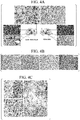
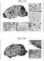
- Human microglia were widely distributed throughout the mouse brain area ( Fig. 4C and Fig. 5 ). Although different types of human microglia were found, most of them were branched microglia, and some were microglia having immature compact amoeba-like forms ( Fig. 4D ). A percentage of human microglia in the total microglia population was calculated by counting HLA-DR + /Iba1 + double-positive human microglia and Iba1 + mouse microglia, and a percentage of human microglia was a maximum of 80% of total microglia in certain brain areas ( Fig. 4E, Fig. 4F , and Fig. 6 ).
- HLA-DR + /Iba1 + positive cells A percentage of HLA-DR + /Iba1 + positive cells was high in the olfactory bulb (OB, 59.3 ⁇ 15.4%), cortical area (CTX), striatum (STR), and hippocampus (HC, 48.3 ⁇ 34.2%); and was low in the brain stem ((BS, 28.4 ⁇ 12.5%)) and in the midbrain (MB, 29.5 ⁇ 15.7%). Interaction of the mouse CNS with human microglia showed normal astrocyte behavior and neuronal integrity ( Fig. 7 ).
- mice were infected with HIV-1 by intraperitoneal injection of 1000 TCID 50 virus, a viral load reached 10 6 RNA copies/mL in peripheral blood ( FIGs. 8A to 8D ).
- Strong infection to human microglia was easily detected in the brain ( Fig. 9A , Figs. 10A to 10C , and Fig. 11 ).
- Infected cells were found in several mouse brain areas, and the largest number of infected cells was found in OB, HC, and CTX.
- RNAscope technology it was possible to clearly see infected human cells and extracellular virus particles released from the infected cells. Reactive astrocytic cells were easily detected in the HIV-infected microglia or at the vicinity thereof ( Fig. 9 ).
- the brains of CD34-NOG-hIL-34 mice had a HIV virus load of 3 to 4 log 10 -fold (10 6 vs. 10 2 ) higher than that of the CD34-NSG model which was reconstructed with a human immune system having a comparable viral load in the periphery ( Fig. 9C and Table 1).
- Table 1 Mouse numbers HIV-1 RNA 6 weeks 3475 810600 3476 333900 3478 190680 3479 310800 3473 703500 3485 378000 3 weeks 3463 ⁇ 20 3464 2184 3474 1646 3471 8841 3469 220500 3467 ⁇ 20
- HIV-1-related genes such as gag, nef, and env which were not detected in the brain of CD34-NSG mice significantly increased ( Colby, D., et al. HIV RNA REBOUND POSTINTERRUPTION IN PERSONS SUPPRESSED IN FIEBIG I ACUTE HIV. in Conference on Retroviruses and Opportunistic Infections (IAS-USA, Seattle, Washington, 2017 )) ( Fig. 9D ).
- cytokines secreted by microglia such as CCL2, TNF, HGF (IL6), CXC18 (IL8), IL-10, IL1A, and CXC110 were present.
- PU. 1 (SPI 1), ETV5, and APOE which are important transcription factors for microglia maintenance and functions, were also recognized (Table 2).
- human-specific genes (687 genes) that showed different differential expression from the comparison between the HIV-infected CD34-NOG-hIL-34 mice and the uninfected mice, most of the genes (426 genes) showed a significant downregulation in the HIV-infected mice ( Figs. 13A and 13B ).
- Most of the downregulated genes were genes involving EIF2 signaling and oxidative phosphorylation.
- upregulated genes were genes involving interferon signaling, pattern recognition receptors, toll-like receptor signaling, and death receptor signaling ( Figs. 12B to 12E ).
- Tables 2A to 2H List of genes expressed by microglia
- a non-human animal that can retain a large number of human microglia, and a method for producing the same are provided.
- a method for using the non-human animal is also provided.
Landscapes
- Life Sciences & Earth Sciences (AREA)
- Health & Medical Sciences (AREA)
- Environmental Sciences (AREA)
- Engineering & Computer Science (AREA)
- Zoology (AREA)
- Chemical & Material Sciences (AREA)
- Biomedical Technology (AREA)
- Biotechnology (AREA)
- Animal Behavior & Ethology (AREA)
- Animal Husbandry (AREA)
- Biodiversity & Conservation Biology (AREA)
- Organic Chemistry (AREA)
- General Health & Medical Sciences (AREA)
- Genetics & Genomics (AREA)
- Wood Science & Technology (AREA)
- Bioinformatics & Cheminformatics (AREA)
- Cell Biology (AREA)
- Biochemistry (AREA)
- Veterinary Medicine (AREA)
- General Engineering & Computer Science (AREA)
- Microbiology (AREA)
- Neurosurgery (AREA)
- Neurology (AREA)
- Toxicology (AREA)
- Proteomics, Peptides & Aminoacids (AREA)
- Molecular Biology (AREA)
- Medicinal Chemistry (AREA)
- Biophysics (AREA)
- Gastroenterology & Hepatology (AREA)
- Micro-Organisms Or Cultivation Processes Thereof (AREA)
- Medicines Containing Material From Animals Or Micro-Organisms (AREA)
Abstract
Description
- The present invention relates to a non-human animal having human IL-34 and the use thereof. The present invention particularly relates to a non-human animal having human IL-34, a method for producing human microglia using the non-human animal, and human microglia obtained from the non-human animal. In addition, the present invention relates to a method for producing a non-human animal having human microglia, and a method for producing an HIV-infected non-human animal.
- Priority is claimed on
U.S. Patent Application No. 62/797,369, filed January 28, 2019 - Microglia are central nervous system (CNS)-resident macrophages, and contribute to brain development and immune defense. However, the precise origin, development, and specific markers of human microglia have been subjects of debate. In the adult brain, it is unclear whether microglia are derived only from cells present in the brain from a fetal developmental stage, or whether microglia flow into the CNS from hematopoietic stem cells (HSCs) or monocytes, under both normal or pathological conditions. Monocytes enter the parenchyma of the CNS and can be changed to cells that are morphologically similar to microglia. However, such changes occur only under certain conditions, and it has been discussed whether these cells are genuinely microglia. In mouse-to-mouse transplantation experiments, HSCs derived from a donor were shown to produce microglia-like cells in mice depleted of endogenous microglia by the expression of microglia-specific suicide genes, radiation, or chemical substances (for example, Non-Patent Literature 1).
- On the other hand, attempts to efficiently generate human microglia in the brains of immunodeficient mice have not been successful. In an experiment in which human CD34+ cells were transplanted into immunodeficient mice, only a few human microglia were confirmed (for example, Non-Patent Literature 2).
- Existing humanized mice are mice in which human immune cells have been stably reconstructed by transfer of HSC (for example,
Non-Patent Literature 3 and Non-Patent Literature 4) or human fetal liver and thymus (for example, Non-Patent Literature 5) into NOD/ScidIL2Rg-/-(NSG) mice and the like. However, in the mouse brain, the number and distribution of macrophages in the vicinity of meninges and blood vessels are low, and hardly any microglia have been recognized (Non-PatentLiterature 4 and Non-Patent Literature 6). Since microglial cells are the main target of HIV-1 and the main reservoir of HIV-1 in the CNS, humanized mouse models currently usable for analysis of intracerebral microglial cells are insufficient for simulating HIV-1 infection in the CNS in humans. - Capotondo, A., et al. Brain conditioning is instrumental for successful microglia reconstitution following hematopoietic stem cell transplantation. Proc Natl Acad Sci U S A 109, 15018-15023 (2012).
- Asheuer, M., et al. Human CD34+ cells differentiate into microglia and express recombinant therapeutic protein. Proc Natl Acad Sci U S A 101, 3557-3562 (2004).
- Arainga, M., Su, H., Poluektova, L.Y., Gorantla, S. & Gendelman, H.E. HIV-1 cellular and tissue replication patterns in infected humanized mice. ).
- Gorantla, S., et al. Links between progressive HIV-1 infection of humanized mice and viral neuropathogenesis. Am J Pathol 177, 2938-2949 (2010).
- Denton, P.W. & Garcia, J.V. Novel humanized murine models for HIV research. Curr HIV/).
- Dash, P.K., et al. Loss of neuronal integrity during progressive HIV-1 infection of humanized mice. The Journal of neuroscience: the official journal of the Society for Neuroscience 31, 3148-3157 (2011).
- As described above, humanized mice of the related art cannot retain human microglia, or can retain only a small number thereof, and thus have not been suitable for simulating HIV infection and the like in humans.
- An object of the present invention is to provide a non-human animal that retain a large number of human microglia, and a method for producing the same. Another object of the present invention is to provide a method for using the non-human animal.
- The present invention includes the following aspects.
- [1] A non-human animal having human interleukin-34 (IL-34) in the body thereof.
- [2] The non-human animal according to [1], to which a human CD34-positive hematopoietic stem cell is transplanted.
- [3] The non-human animal according to [1] or [2], in which human microglia are present in the brain.
- [4] The non-human animal according to [3], in which the human microglia express at least one gene selected from the group consisting of CD74, b2m, AIF1, CD14, CD68, CSF1R, ITGAM (CD11b), P2RY12, CX3CR1, TREM2, TMEM119, CCL2, TNF, HGH (IL-6), CXCL8, IL-10, IL-la, CXCL10, PU.1 (SPI1), ETV5, and APOE.
- [5] The non-human animal according to [3] or [4], in which the human microglia secrete at least one cytokine selected from the group consisting of CCL2, TNF, HGH (IL-6), CXCL8, IL-10, IL-la, and CXCL10.
- [6] The non-human animal according to any one of [1] to [5], which is infected with the human immunodeficiency virus (HIV).
- [7] A method for producing human microglia, including obtaining human microglia from the non-human animal according to any one of [3] to [6].
- [8] A method for producing a non-human animal having human microglia, including transplanting human CD34-positive hematopoietic stem cells into a non-human animal having human IL-34 in the body thereof.
- [9] The method for producing a non-human animal having human microglia according to [8], in which the non-human animal having human IL-34 in the body is an immunodeficient non-human animal.
- [10] The method for producing a non-human animal having human microglia according to [8] or [9], in which the human microglia are present in the brain.
- [11] The method for producing a non-human animal having human microglia according to any one of [8] to [10], in which the human microglia express at least one gene selected from the group consisting of CD74, b2m, AIF1, CD14, CD68, CSF1R, ITGAM (CD11b), P2RY12, CX3CR1, TREM2, TMEM119, CCL2, TNF, HGH (IL-6), CXCL8, IL-10, IL-1a, CXCL10, PU.1 (SPI1), ETV5, and APOE.
- [12] The method for producing a non-human animal having human microglia according to any one of [8] to [11], in which the human microglia secrete at least one cytokine selected from the group consisting of CCL2, TNF, HGH (IL-6), CXCL8, IL-10, IL-1a, and CXCL10.
- [13] A method for producing an HIV-infected non-human animal, including producing a non-human animal having human microglia by the method for producing a non-human animal having human microglia according to any one of [8] to [12], and then infecting the non-human animal having human microglia with HIV.
- The present invention also includes the following aspects.
- [14] The non-human animal according to any one of [1] to [6], which secretes human IL-34.
- [15] The non-human animal according to [14], which has a human IL-34 gene.
- [16] The non-human animal according to [14] or [15], which has human microglia.
- [17] The non-human animal according to any one of [1] to [6] and [14] to [16], which is a rodent.
- [18] The non-human animal according to [17], which is a mouse.
- [19] A method for producing the non-human animal having human microglia according to any one of [7] to [12], in which the non-human animal having human IL-34 in the body has a human IL-34 gene.
- [20] A method for producing the non-human animal having human microglia according to any one of [7] to [12] and [19], in which the non-human animal having human IL-34 in the body secretes human IL-34.
- [21] A method for producing the non-human animal having human microglia according to any one of [7] to [12], [19], and [20], in which the non-human animal is a rodent.
- [22] A method for producing the non-human animal having human microglia according to [21], in which the rodent is a mouse.
- According to the present invention, a non-human animal having large retention numbers of human microglia and a method for producing the same are provided. A method for using the non-human animal is also provided.
-
-
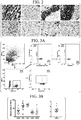
Figs. 1A to ID are diagrams showing the production of NOD. Cg-PrkdcscidIl2rgtm1Sug Tg (CMV-IL-34) 1/Jic (NOG-hIL-34) mice, and an outline of characterization.Fig. 1A shows a vector introduced into NOG-hIL-34 transgenic mice for producing NOD. Cg-PrkdcscidIl2rgtm1Sug Tg (CMV-IL-34) 1/Jic mice. A vector containing hIL-34 and a transgene (Tg) is inserted downstream of a CMV promoter. -
Fig. 1B shows results of PCR analysis using DNA extracted from the ears of homozygous mice. In NOG-hIL-34 transgenic mice (IL-34+/+), a band was not detected in control non-transgenic NOG mice in which the band of hIL-34 (358 bp) was amplified. -
Fig. 1C shows the results of real-time PCR using RNA extracted from the brain, spleen, lung, kidney, liver, and skin of NOG-hIL-34 mice. In NOG-hIL-34 (IL-34), the expression of hIL-34 in all tissues was confirmed in contrast to in NOG mice (Control). - Fig. ID shows the results of ELISA that confirmed hIL-34 expression in plasma.
-
Fig. 2 shows the results of RNAScope assay showing expression of human IL-34 in different regions of a mouse brain. RNAScope assay was performed using Hs-IL-34-NoXMm, an antisense probe targeting 38 to 1774 bp of human IL-34, and human IL-34 was detected as a single brown dot or cluster of dots in brain sections with a thickness of 5 µm. In NOG mice, no signal indicating the presence of human IL-34 was confirmed. 40 × magnification. -
Figs. 3A to 3B show differentiation and proliferation of human peripheral blood lymphocytes in CD34-NOG-hIL-34 mice.Fig. 3A shows the results of FACS analysis of peripheral blood. By gating to the CD45 lymphocyte population, representative plots of differentiated 45 (CD45)+, and human clusters of human CD3, CD19, and CD14 cells can be confirm. -
Fig. 3B shows a percentage of human cell subpopulations in the peripheral blood of reconstituted mice. Each symbol represents an individual mouse. -
Figs. 4A to 4F show the presence of human microglial cells in the brains of CD45-NOG-hIL-34 mice.Fig. 4A is a diagram comparing tissue macrophage reconstitution between CD34-NOG-hIL-34 and CD34-NSG mice. Brain sections were stained for HLA-DR, and liver and spleen were stained for CD68. -
Fig. 4B shows human cells stained with human P2RY127, CD14, CD68, and CD163 antibodies in the brain. -
Fig. 4C shows an enlarged view of the olfactory bulb (OB, 20 ×), cortex (CTX, 20 ×), and hippocampus (HC, 10 ×) from the brains of CD34-NOG-hIL-34 mice in which HLA-DR was stained. -
Fig. 4D is a diagram showing microglia morphology at higher magnification. -
Fig. 4E is a confocal image of the brain for which HLA-DR and Iba-1 were stained. -
Fig. 4F shows a percentage of HLA-DR/Iba-1 double-positive human microglial cells in all Iba-1+ cells. Brain stem (BS), Midbrain (MB), Cerebellum (CB), Cerebral cortex (CC), Hippocampus (HC), and Olfactory bulb (OB). -
Figs. 5A to 5B show the reconstitution of human microglia in a mouse brain. Paraffin-embedded brain sections with a thickness of 5 µm were stained for HLA-DR.Fig. 5A shows a representative CD34-NOG-hIL-34 mouse brain at 2 × magnification. An overall image of human microglial distribution in the entire mouse brain area can be confirmed. Olfactory bulb (OB), Cerebral cortex (CTX), Hippocampus (HC), Midbrain (MB), Cerebellum (CB), Striatum (STR), Hippocampus (HC), Substantia nigra (SN), Thalamus (TH), and Brain stem (BS). The image was taken with a Ventana iScan HT at 200 × original magnification. A magnified view (20 × objective lens) of the brain region (OB, CTX, and HC inFig. 4C ) shows HLA-DR+ cells having the morphology of microglia. -
Fig. 5B shows a representative CD34-NSG mouse brain section (2 ×) showing the absence of human microglial cells. A small number of HLA-DR+ cells found in the meninges and perivascular area (boxed area) are shown in an enlarged view (20 ×). -
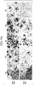
Fig. 6 shows the distribution of human and mouse glial cells in the mouse brain. Sagittal sections of paraffin-embedded brain tissue stained for human MHC class II (HLA-DR) and microglial cells (Iba-1) are shown. Original confocal images were collected with a Zeiss 710 system at 630 × magnification. -
Fig. 7 shows the interaction of mouse neural cells with human microglial cells. Sagittal sections of paraffin-embedded brain tissue stained for human MHC class II (HLA-DR) and mouse neuron marker (MAP-2), neurofilament H (NF-H) or synaptophysin (SYN) are shown. 400 × original magnification. -
Figs. 8A to 8D show establishment of systemic HIV infection in CD34-NOG-hIL-34 mice.Fig. 8A shows a viral load of peripheral blood obtained by a COBAS Amplicor System. Each symbol represents an individual infected mouse. -
Fig. 8B shows the results of flow cytometric analysis of splenocytes for human CD4- and CD8-positive T lymphocytes. A percentage of CD4- and CD8-positive cells is shown. -
Fig. 8C shows the results of flow cytometric analysis of splenocytes for human CD4 and CD8-positive T lymphocytes. A CD4/CD8 ratio in an HIV+ group (n = 12) and control group (n = 7) in spleen is shown. -
Fig. 8D shows the results of immunohistological analysis of spleen sections showing the presence of HLA-DR+ cells and HIV-1-infected cells stained for HIV-1 p24. RNAScope assay was performed using V-HIV1-Clade-B (ACD cat #416111), an antisense probe targeting HIV-1 at 854 to 8291 bp, and HIV-1 RNA was detected as a single brown dot or cluster of dots in spleen sections with a thickness of 5 µm. In uninfected mice, no signal corresponding to the presence of viral RNA was detected. Images were taken with a Nuance multiplex system at 200 × original magnification. -
Figs. 9A to 9D show HIV infection in a humanized mouse brain.Fig. 9A shows the results of immunohistological analysis of brain regions showing the presence of HIV-1 p24+ infected cells. RNAScope assay was performed using V-HIV1-Clade-B, an antisense probe, and HIV-1 RNA was detected as a single brown dot or cluster of dots. 200 × original magnification. -
Fig. 9B shows the results of immunofluorescence staining of mouse astrocytes (GFAP) in HIV-1-infected mouse brain near human microglial cells (HLA-DR+), and HIV-1 p24-positive human microglia. 400 × original magnification. -
Fig. 9C shows comparison results of viral RNA levels in the brain of CD34-NSG mice and CD34-NOG-hIL-34 mice, which were obtained by semi-nested RT-PCR. -
Fig. 9D shows an expression level of each HIV-1 gene in an infected mouse brain. RNAseq reads were aligned to an HIV-1ADA sequence. -
Figs. 10A to 10C are diagrams showing the distribution of human immune cells in HIV-infected humanized NOG hIL-34 mice.Fig. 10A shows the results of staining a paraffin-embedded sagittal section with a 5 µm thickness of brain for human-specific immune cell markers (CD4 and CD8). Images were taken with a Nuance multiplex system at 200 × original magnification. -
Fig. 10B shows quantification results of human CD4 and CD8 T cells. For HIV-infected mice (n = 4) and control mice (n = 3), at least 2 sections were counted per mouse. Black bars indicate target mice, and gray bars indicate HIV-infected mice. -
Fig. 10C shows a CD4/CD8 ratio. For HIV-infected mice (n = 4) and control mice (n = 3), at least 2 sections were counted per mouse. -
Fig. 11 shows a representative CD34-NOG-hIL-34 mouse brain showing an overview of HIV-1 infection across a mouse brain area stained for HIV-1 p24. The image was taken at 2 ×magnification 2. -
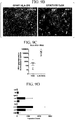
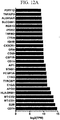
Figs. 12A to 12E show changes in transcripts in CD34-NOG-hIL-34 brain tissue. Differentially expressed genes (DEG) having p < 0.05 in Transcripts Per Kilobase Million (TPM) values are shown. 687 DEG human genes were found by alignment of the gene to the human genome (h19) by comparing uninfected CD34-NOG-hIL-34 mice to infected CD34-NOG-hIL-34 mice. Among them, 261 genes were upregulated by HIV infection, and 426 genes were downregulated by HIV infection.Fig. 12A shows a top list of classical microglial markers expressed in a CD34-NOG-IL-34 mouse brain. -
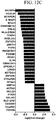
Fig. 12B and12C show top rankings of log-fold changes in HIV infection-related genes differentially expressed in HIV-uninfected CD34-NOG-IL-34 and HIV-infected CD34-NOG-IL-34 in human microglia and brain.Fig. 12B shows DEG upregulated with HIV-infected CD34-NOG-IL-34. -
Fig. 12C shows DEG downregulated with HIV-infected CD34-NOG-IL-34. -
Fig. 12D shows that human genes (261) upregulated in HIV infection are associated with interferon signaling, PRP, and TLR signaling. -
Fig. 12E shows that human genes (426) downregulated in HIV infection are closely linked to EIF2 signaling and oxidative phosphorylation pathways. -
Fig. 13A is a pie chart showing the breakdown of 687 DEG human genes found by alignment of the gene to the human genome (h19) by comparing HIV-uninfected CD34-NOG-hIL-34 mice to HIV-infected CD34-NOG-hIL-34 mice. 261 genes were upregulated by HIV infection, and 426 genes were downregulated by HIV infection. -
Fig. 13B is a Volcano plot of total DEG (human) created for HIV-uninfected CD34-NOG-hIL-34 mice versus HIV-infected CD34-NOG-hIL-34 mice. X and Y axes showlog 2 fold change and log 10 (p value), respectively. Blue dots indicate upregulated genes, green dots indicate downregulated genes, and black dots indicate neutrally regulated genes. - In the present specification, unless otherwise specified, "IL-34" means an IL-34 protein, and an "IL-34 gene" means a gene encoding the amino acid sequence of IL-34. The term "gene" means a polynucleotide including at least one open reading frame encoding a specific protein, and may include both exons and introns.
- In the present specification, "human IL-34 activity" means an activity that induces differentiation of human monocytes and macrophages in the body of a mouse in a case of introduction into an immunodeficient mouse. Human IL-34 activity includes an activity of inducing human microglia from human HSC in the body of an immunodeficient mouse.
- In the present specification, sequence identity (or homology) between amino acid sequences or base sequences is obtained as a ratio of matched amino acids or bases to the entire amino acid sequence or the entire base sequence excluding gaps in the obtained alignment by juxtaposing two amino acid sequences or base sequences while inputting gaps in portions corresponding to insertions and deletions so that the corresponding amino acids or bases are most matched. Sequence identity between amino acid sequences or between base sequences can be obtained using various homology search software known in the technical field. For example, a value of sequence identity of amino acid sequences can be obtained by calculation based on alignment obtained by known homology search software BLASTP, and a value of sequence identity of base sequences can be obtained by calculation based on alignment obtained by known homology search software BLASTN.
- In the present specification, examples of "stringent conditions" include methods described in Molecular Cloning-A LABORATORY MANUAL THIRD EDITION (Sambrook et al., Cold Spring Harbor Laboratory Press). Examples of stringent conditions include conditions for hybridization by performing incubation at 42 to 70°C for several hours to overnight in a hybridization buffer consisting of 6 × SSC (composition of 20 × SSC: 3 M sodium chloride, 0.3 M citric acid solution, pH 7.0), 5 × Denhardt's solution (composition of 100 × Denhardt's solution: 2% by mass bovine serum albumin, 2% by mass Ficoll, 2% by mass polyvinylpyrrolidone), 0.5% by mass SDS, 0.1 mg/mL salmon sperm DNA, and 50% formamide. As a washing buffer to be used for washing after incubation, a 0.1% by mass SDS-containing 1 × SSC solution is preferable, and a 0.1 % by mass SDS-containing 0.1 × SSC solution is more preferable.
- The term "immunodeficient non-human animal" means a non-human animal having one or more of a lack of functional immune cells such as T cells and B cells; DNA repair defects; defects in reconstitution of genes encoding an antigen specific receptor on lymphocytes; and lack of immune functional molecules such as IgM, IgG1, IgG2a, IgG2b, IgG3, and IgA. An immunodeficient non-human animal may have any of the above or other defects that result in abnormal immune function in the non-human animal.
- In the present specification, the phrase "operably linked," which is used with regard to polynucleotides means that a first base sequence is located sufficiently close to a second base sequence, and the first base sequence may affect the second base sequence or a region under control of the second base sequence. For example, the phrase "operably linked to a promoter" means that a polynucleotide is linked to be expressed under the control of the promoter.
- In the present specification, the phase a "promoter can function" means that a polynucleotide operably linked to the promoter can be expressed in cells of a target non-human animal.
- In the present specification, the term an "expressible state" means that a polynucleotide can be transcribed in a cell into which the polynucleotide has been introduced.
- In the present specification, the term an "expression vector" means a vector including a target polynucleotide, which is the vector including a system that enables expression of a target polynucleotide in a cell into which the vector has been introduced.
- In the present specification, the term "marker" means a protein that is specifically expressed in a specific type of cell. The "marker" is preferably a protein present on a cell surface.
- A first aspect of the present invention is a non-human animal having human interleukin-34 (IL-34) in the body thereof.
-
- IL-34 is a type of cytokine that promotes differentiation and survival of monocytes and macrophages via a colony-stimulating factor-1 receptor (CSF1R).
- Gene sequences and amino acid sequences of human IL-34 are known, and their sequence information can be obtained from known databases such as GenBank. Examples of gene sequences and amino acid sequences of human IL-34 include the sequences registered as Accession No. NM_152456.2 (SEQ ID NOS: 1 and 2) in GenBank. Human IL-34 is not limited to those having the above sequences, and includes homologs (orthologs, paralogs) and variants thereof.
- Human IL-34 includes, for example, the following.
- (1) A polypeptide including an amino acid sequence set forth in SEQ ID NO: 2.
- (2) A polypeptide consisting of an amino acid sequence in which one or more amino acids have been deleted, substituted, added, or inserted in an amino acid sequence set forth in SEQ ID NO: 2, and having human IL-34 activity.
- (3) A polypeptide consisting of an amino acid sequence having 80% or more sequence identity with an amino acid sequence set forth in SEQ ID NO: 2, and having human IL-34 activity.
- The human IL-34 gene includes, for example, the following.
- (4) A polynucleotide encoding a polypeptide including an amino acid sequence set forth in SEQ ID NO: 2.
- (5) A polynucleotide encoding a polypeptide consisting of an amino acid sequence in which one or more amino acids have been deleted, substituted, added, or inserted in an amino acid sequence set forth in SEQ ID NO: 2, and having human IL-34 activity.
- (6) A polynucleotide encoding a polypeptide consisting of an amino acid sequence having 80% or more sequence identity with an amino acid sequence set forth in SEQ ID NO: 2, and having human IL-34 activity.
- (7) A polynucleotide including a base sequence set forth in SEQ ID NO: 1.
- (8) A polynucleotide consisting of a base acid sequence in which one or more bases have been deleted, substituted, added, or inserted in a base acid sequence set forth in SEQ ID NO: 1, and encoding a polypeptide having human IL-34 activity.
- (9) A polynucleotide consisting of a base acid sequence having 80% or more sequence identity with a base sequence set forth in SEQ ID NO: 1, and encoding a polypeptide having human IL-34 activity.
- (10) A polynucleotide that hybridizes with a polynucleotide consisting of a base sequence set forth in SEQ ID NO: 1 under stringent conditions, and encoding a polypeptide having human IL-34 activity.
- In the above (2) and (5), the number of amino acids to be deleted, substituted, added, or inserted is not particularly limited as long as the resulting polypeptide has human IL-34 activity. In the above (8), the number of bases to be deleted, substituted, added, or inserted is not particularly limited as long as the resulting polynucleotide encodes a polypeptide having human IL-34 activity. The number of amino acids or bases to be deleted, substituted, added, or inserted may be, for example, 1 to 80, is preferably 1 to 60, and is more preferably 1 to 50. Examples thereof include 1 to 30, 1 to 20, 1 to 10, 1 to 5, 1 to 3, 1 or 2, and the like.
- In the above (3), (6) or (9), the sequence identity is not particularly limited as long as it is 80% or more. The sequence identity is preferably 85% or more, is more preferably 90% or more, is still more preferably 95% or more, and is particularly preferably 97% or more.
- In the amino acid sequence set forth in SEQ ID NO: 2, the amino acid sequence of
positions 1 to 20 is a signal peptide. Since a signal peptide is cleaved upon extracellular secretion, mature human IL-34 is composed of the amino acid sequence of positions 21 to 242 in the amino acid sequence set forth in SEQ ID NO: 2. - Accordingly, human IL-34 also includes the following.
- (11) A polypeptide including an amino acid sequence of positions 21 to 242 in an amino acid sequence set forth in SEQ ID NO: 2.
- (12) A polypeptide consisting of an amino acid sequence in which one or more amino acids have been deleted, substituted, added, or inserted in an amino acid sequence of positions 21 to 242 in an amino acid sequence set forth in SEQ ID NO: 2, and having human IL-34 activity.
- (13) A polypeptide consisting of an amino acid sequence having 80% or more sequence identity with an amino acid sequence of positions 21 to 242 in an amino acid sequence set forth in SEQ ID NO: 2, and having human IL-34 activity.
- The number of amino acids to be deleted, substituted, added, or inserted in the above (12) may be the same as in the case of the above (2). The numerical value of sequence identity in the above (13) may be the same as in the case of the above (3).
- Examples of the human IL-34 gene also includes a polynucleotide encoding a polypeptide in which a signal peptide for extracellular secretion is linked to the N-terminal side of the polypeptide of any of the above (11) to (13). The signal peptide is not limited to the signal peptide of human IL-34, and may be a signal peptide of another protein. Examples of such signal peptides include, for example, signal peptides of cytokines other than IL-34.
- The non-human animal is not particularly limited as long as it is a non-human animal, but is preferably a mammal. Examples of non-human animals include non-human primates (monkeys, chimpanzees, gorillas, and the like), rodents (mice, rats, guinea pigs, and the like), dogs, cats, rabbits, cows, pigs, horses, goats, sheep, and the like, but examples are not limited thereto. Among them, rodents are preferable and mice are more preferable because they are easily available, and testing thereon is easy.
- The non-human animal is preferably an immunodeficient non-human animal since human cells can be engrafted thereto. When the non-human animal is a mouse, immunodeficient mice can be characterized by one or more defects in genes involved in an immune function such as Rag1 and Rag2 (for example, Oettinger, M. A et al., Science, 248: 1517-1523, 1990; Schatz, D. G. et al., Cell, 59: 1035-1048, 1989). Immunodeficient mice can have any of the above or other defects that result in abnormal immune function in the mouse.
- Examples of immunodeficient mice include, but are not limited to, NOG mice (NOD. Cg-PrkdcscidIl2rgtm1Sug/ShiJic), NSG mice (NOD. Cg-PrkdcscidIl2rgtm1Wjl/SzJ), NOD/ShiJcl mice, and the like. Immunodeficient mice are commercially available, and such commercially available immunodeficient mice can be used without particular limitation.
- The non-human animal of the present embodiment is characterized by having human IL-34 in the body. Examples of methods for producing a non-human animal having human IL-34 in the body include a method of administering human IL-34 to a non-human animal, a method of introducing the human IL-34 gene into a non-human animal, a method of transferring cells that secrete human IL-34 into a non-human animal, and the like.
- When human IL-34 is administered to a non-human animal, an administration method is not particularly limited as long as human IL-34 is retained in the non-human animal, but parenteral administration is preferable. Examples of routes of parenteral administration include intramuscular injection, subcutaneous injection, intravascular injection, and the like. Administration of human IL-34 may be a single dose or multiple doses, as long as human IL-34 is retained in the non-human animal.
- Whether or not a non-human animal has human IL-34 in the body can be confirmed by collecting plasma from the non-human animal and measuring a human IL-34 concentration in the plasma. A method for measuring a human IL-34 concentration in plasma is not particularly limited, but examples thereof include immunochemical methods using anti-human IL-34 antibodies. Examples of such methods include an ELISA method, an EIA method, a RIA method, a Western blotting method, and the like. For measurement of human IL-34 in plasma, a commercially available ELISA kit for measuring human IL-34 or the like may be used.
- A concentration of human IL-34 in plasma is, for example, 20 pg/mL or more, is preferably 30 pg/mL or more, is more preferably 50 pg/mL or more, is still more preferably 80 pg/mL or more, and is particularly preferably 100 pg/mL or more. An upper limit of the concentration of human IL-34 in plasma is not particularly limited, and examples thereof include 1000 pg/mL or less, 800 pg/mL or less, or 700 pg/mL or less.
- When the human IL-34 gene is introduced into a non-human animal, it is preferable that the human IL-34 be operably linked downstream of a promoter that can function in the non-human animal as an introduction target. Examples of promoters that can function in mammals include a cytomegalovirus (CMV) promoter, an SRα promoter, an SV40 early promoter, an LTR of retrovirus, a Rous sarcoma virus (RSV) promoter, a herpes simplex virus thymidine kinase (HSV-TK) promoter, an EF1α promoter, a metallothionein promoter, a heat-shock promoter, and the like, but examples are not limited thereto.
- The human IL-34 gene is introduced into the non-human animal in an expressible state, for example, in the form of an expression vector. The expression vector may contain, in addition to the human IL-34 gene and promoter, control sequences such as enhancers, poly A addition signals, and terminators; and marker genes such as drug resistance genes.
- The type of the vector is not particularly limited, and a commonly used expression vector can be used without particular limitation. The vector may be linear or circular, and may be a non-viral vector such as a plasmid, a viral vector (for example, a retrovirus vector such as a lentiviral vector), or may be a transposon vector.
- A method for introducing the human IL-34 gene into non-human animals is not particularly limited, and methods generally used for producing transgenic animals can be applied. Examples of methods for introducing a human IL-34 gene into a non-human animal include a method of introducing an expression vector containing the human IL-34 gene into a fertilized egg of the non-human animal as an introduction target by microinjection and the like. When the non-human animal is a mouse, for example, a fertilized egg obtained by mating the NOG mouse (NOD. Cg-PrkdcscidIl2rgtm1Sug/ShiJic) and NOD/ShiJcl mouse may be exemplified, but examples are not limited thereto.
- A fertilized egg into which a human IL-34 gene has been introduced is cultured at 37°C for about 18 to 24 hours, and then transplanted and implanted in the uterus of a foster mother to allow giving birth to offspring, and thereby a non-human animal having a human IL-34 gene can be obtained.
- Whether the non-human animal obtained as described above has a human IL-34 gene or not can be confirmed by extracting genomic DNA from a sample collected from the non-human animal and performing PCR or the like.
- In addition, whether the non-human animal expresses a human IL-34 gene or not can be determined by extracting RNA from a sample collected from the non-human animal and performing RT-PCR or the like, or it can be confirmed by performing in situ hybridization and the like using tissue samples collected from non-human animals. Alternatively, it can be confirmed by detecting human IL-34 in a sample collected from the non-human animal using an anti-human IL-34 antibody (for example, an ELISA method, an EIA method, a RIA method, a Western blot method, an EIA method, a RIA method, immunohistological staining, and the like).
- A non-human animal into which a human IL-34 gene has been introduced preferably secretes human IL-34. Secretion of human IL-34 by the non-human animal mainly refers to release of human IL-34 from the cells of the non-human animal into the body fluid (blood, tissue fluid, lymph, and the like). Whether or not the non-human animal secretes human IL-34 can be confirmed by measuring a human IL-34 concentration in the plasma collected from the non-human animal. The same examples as described above may be exemplified for a method for measuring the human IL-34 concentration in plasma.
- In a case where a cell that secretes human IL-34 (human IL-34-secreting cell) is transferred to a non-human animal, examples of human IL-34-secreting cells include cells derived from human organs and blood, cancer cells, and the like. Examples of cells derived from human organs include cells derived from the spleen, thymus, liver, small intestine, large intestine, prostate gland, lungs, heart, brain, kidneys, testes, uterus, and the like. Examples of cells derived from human blood include blood cells and the like contained in the peripheral blood mononuclear cell fraction. For example, cells secreting human IL-34 can be selected from cell lines established from these human cells, and can be used as human IL-34-secreting cells for transfer to non-human animals. Alternatively, cells of non-human animals into which the human IL-34 gene has been introduced may be used as human IL-34-secreting cells. The cells of the non-human animal are preferably cells of a non-human animal belonging to the same species as the non-human animal to which cells are to be transferred. For example, if the non-human animal to which cells are to be transferred is a mouse, it is preferable that the cells into which the human IL-34 gene is transferred be mouse cells. Non-human animal cells into which the human IL-34 gene is introduced are not particularly limited, but examples thereof include cells derived from organs or blood or cell lines thereof, hematopoietic stem cells (such as CD34-positive hematopoietic stem cells), cancer cells, and the like. Examples of cells derived from the organs and blood of the non-human animals include cells of non-human animals derived from organs and blood fractions similar to those exemplified for the human IL-34-secreting cells. As described above, the human IL-34 gene is introduced into the non-human animal cells in an expressible state, for example, in the form of an expression vector. The method for introducing the human IL-34 gene into non-human animal cells is not particularly limited, and a method generally used as a gene transfer method can be applied. Examples of such methods include viral infection, lipofection, microinjection, calcium phosphate, DEAE-dextran, electroporation, transposon, and particle gun methods, and the like, but examples are not limited thereto.
- Whether the human cell line or the human IL-34 transgenic cell secretes human IL-34 can be confirmed by measuring a IL-34 concentration in a culture solution of the cells or cell lines or the human IL-34 transgenic cells. Examples of methods for measuring a human IL-34 concentration in the culture solution include the same as those described above.
- The method for transferring human IL-34-secreting cells to non-human animals is not particularly limited, and methods generally used for transferring cells to non-human animals can be applied. Examples of methods for transferring human IL-34-secreting cells to non-human animals include a method of administering human 1L-34-secreting cells in the spleen, in the liver, subcutaneously, or intravenously according to the type of cells used, and the like.
- A non-human animal transfected with human IL-34-secreting cells preferably secretes human IL-34. Secretion of human IL-34 by the non-human animal mainly refers to release of human IL-34 from the human IL-34-secreting cells transferred to the non-human animal into the body fluid (blood, tissue fluid, lymph, and the like). Whether or not the non-human animal secretes human IL-34 can be confirmed by measuring a human IL-34 concentration in the plasma collected from the non-human animal. The same examples as described above may be exemplified for a method for measuring the human IL-34 concentration in plasma.
- In addition, examples of methods for producing a non-human animal of the present embodiment include a method in which a non-human animal is directly inoculated and infected with a virus such as a lentivirus or adenovirus incorporating a human IL-34 gene for secretion of human IL-34. Such method may be included in the methods of introducing the human IL-34 gene into a non-human animal described above.
- The non-human animal of the present embodiment is preferably a non-human animal having a human IL-34 gene, and is more preferably a non-human animal secreting human IL-34. When a non-human animal has a human IL-34 gene and secretes human IL-34, examples of a concentration of human IL-34 in plasma include the same concentrations as described above.
- By transplanting human CD34-positive hematopoietic stem cells into a non-human animal having human IL-34, human microglia are induced from the CD34-positive hematopoietic stem cells in the body of the non-human animal. In other words, by transplanting human CD34-positive hematopoietic stem cells into a non-human animal having human IL-34, a non-human animal having human microglia can be produced.
- Accordingly, the non-human animal of the present embodiment may be a non-human animal in which human CD34-positive hematopoietic stem cells have been transplanted to a non-human animal having human IL-34. In addition, the non-human animal of the present embodiment may be a non-human animal having human IL-34 and human microglia.
- Furthermore, in one embodiment, the present invention provides a method for producing a non-human animal having human microglia, including transplanting a human CD34-positive hematopoietic stem cell into a non-human animal having human IL-34 in the body thereof.
- Human CD34-positive hematopoietic stem cells can be obtained from human cord blood, bone marrow, blood, and the like. A method for obtaining human CD34-positive hematopoietic stem cells from these samples is not particularly limited, and examples thereof include a method in which, after density gradient centrifugation of these samples, CD34-positive cells are isolated by a magnetic bead method using an anti-human CD34 antibody. The purity of the obtained human CD34-positive hematopoietic stem cells may be confirmed by flow cytometry or the like.
- A method for transplanting human CD34-positive hematopoietic stem cells into a non-human animal is not particularly limited, and methods generally used for transplantation of hematopoietic stem cells can be applied. Examples of methods for transplanting human CD34-positive hematopoietic stem cells into a non-human animal include a method of administering human CD34-positive hematopoietic stem cells into the liver or a vein after whole-body treatment of postnatal non-human animals (for example,
postnatal day 0 to 1) with radiation. The number of human CD34-positive hematopoietic stem cells to be transplanted is not particularly limited, but, for example, 103 or more is preferable, and 104 or more is more preferable. An upper limit of the number of human CD34-positive hematopoietic stem cells to be transplanted is not particularly limited, and is, for example, 1010 or less, 109 or less, 108 or less, and the like. - Whether non-human animals have had human CD34-positive hematopoietic stem cells transplanted thereinto can be determined by analyzing a blood sample or a spleen tissue sample of the non-human animal by flow cytometry and the like using an antibody against a human immune cell marker (CD45, CD3, CD19, CD8, CD14, and the like).
- When human CD34-positive hematopoietic stem cells are transplanted into a non-human animal having human IL-34, human microglia are differentiated and induced from CD34-positive hematopoietic stem cells in the non-human animal body, and a non-human animal having human microglia can be obtained. Whether or not a non-human animal has human microglia can be confirmed by immunohistological staining using an antibody against a marker specific for human microglia (for example, double-positive for HLA-DR and Iba1).
- In non-human animals having human IL-34, a site where human microglia are present is not particularly limited, but because microglia are usually present in the central nervous system, also in the non-human animal, the human microglia is preferably present in the central nervous system, and more preferably in the brain.
- The human microglia possessed by the non-human animal of the present embodiment preferably express at least one gene selected from the group consisting of CD74, b2m, AIF1, CD14, CD68, CSF1R, ITGAM (CD11b), P2RY12, CX3CR1, TREM2, TMEM119, CCL2, TNF, HGH (IL-6), CXCL8, IL-10, IL- 1a, CXCL10, PU.1 (SPI1), ETV5, and APOE. These genes are known as markers of classical macrophages/microglia. In addition, it is preferable that human microglia possessed by the non-human animal of the present embodiment express a part or all of the genes described in Tables 2A to 2H to be shown later.
- In addition, human microglia possessed by the non-human animal of the present embodiment preferably secrete at least one cytokine selected from the group consisting of CCL2, TNF, HGH (IL-6), CXCL8, IL-10, IL-la, and CXCL10.
- As shown in the examples below, non-human animals having human IL-34 can have larger numbers of human microglia compared to non-human animals not having human IL-34. For this reason, human immunodeficiency virus (HIV) infection in the human brain can be appropriately simulated by infecting a non-human animal having human IL-34 and human microglia with HIV.
- Accordingly, the non-human animal of the present embodiment may be a non-human animal having human IL-34 infected with HIV. Such non-human animal is preferably a non-human animal having human IL-34 and human microglia.
- In one embodiment, the present invention also provides a method for producing an HIV-infected non-human animal, the method including producing a non-human animal having human microglia (the non-human animal has human IL-34 and human microglia) according to the method described above, and then infecting the non-human animal having human microglia with HIV.
- The HIV used for infection may be either HIV-1 or HIV-2. A method of infection of non-human animals with HIV is not particularly limited, and commonly used infection methods can be applied. Examples of methods of infecting non-human animals with HIV include a method of intraperitoneally administering HIV and the like. A viral load used for infection is not particularly limited as long as HIV infection can be established, but examples thereof include 500 to 5000 TCID50, 700 to 3000 TCID50 is preferable, and a specific example thereof includes 1000 TCID50.
- Whether or not HIV infection is established in a non-human animal can be confirmed by measuring an amount of HIV in peripheral blood collected from the non-human animal. For example, when an amount of HIV is detected at about 106 RNA copies/mL in peripheral blood, it can be determined that HIV infection has been established. In addition, HIV infection of the brain can be confirmed by performing immunostaining targeting HIV-specific proteins or RNA, in situ RNA hybridization, or the like in brain tissue sections of the non-human animal. It is preferable that the non-human animal of the present embodiment have HIV infecting the brain.
- The non-human animal of the present embodiment can appropriately reconstitute the human hemolymph system by having human IL-34 in the body. In particular, reconstitution of human microglia in the brain, which was difficult in non-human animals such as immunodeficient mice of the related art, becomes possible. For this reason, the non-human animal of the present embodiment can be used to produce a model for infection of a virus such as HIV having microglia as the main reservoir.
- The non-human animal of the present embodiment can be used for elucidating the viral infection mechanism targeting microglia, and for screening and evaluating a therapeutic drug for viral infection. In addition, the non-human animal of the present embodiment can be used for elucidating the mechanism of central nervous system diseases mediated by microglia and for screening and evaluating a therapeutic drug.
- A second aspect of the present invention is a method for producing human microglia, which includes obtaining human microglia from the non-human animal of the first aspect.
- As described above, since the non-human animal of the first aspect can retain many human microglia, it can be used for producing human microglia. Therefore, in one aspect, the present invention provides a method of producing human microglia using the non-human animal of the first aspect, and human microglia obtained by such production method.
- The method for producing human microglia of the present embodiment can include the following steps:
- a step (a) of administering human IL-34 or introducing a human IL-34 gene into a non-human animal to obtain a non-human animal having human IL-34 in the body;
- a step (b) of implanting human CD34-positive hematopoietic stem cells into the non-human animal obtained in the step (a) to obtain a non-human animal having human microglia; and
- a step (c) of obtaining human microglia from the non-human animal obtained in the step (b).
- The steps (a) and (b) can be carried out as described above in the section "[Non-human animal having IL-34]."
- The step (c) can be carried out by isolating human microglia from a blood sample or brain tissue of the non-human animal obtained in the step (b). For example, microglia can be isolated from the sample by performing density gradient centrifugation, a magnetic bead method using an antibody against a human microglia specific marker, or the like. In a case where human microglia are isolated from brain tissue, density gradient centrifugation may be performed after suspending and homogenizing brain tissue in an appropriate buffer solution (for example, phosphate-buffered saline, and the like).
- Examples of human microglia-specific markers include HLA-DR, Ibal, CD74, b2m, AIF1, CD14, CD68, CSF1R, ITGAM (CD11b), P2RY12, CX3CR1, TREM2, TMEM119, CCL2, TNF, HGH (IL -6), CXCL8, IL-10, IL-la, CXCL10, PU.1 (SPI1), ETV5, and APOE, but examples are not limited thereto.
- The human microglia obtained by the production method of the present embodiment can be used for elucidating the function of human microglia, elucidating the mechanism of virus infection targeting human microglia, evaluating drugs targeting human microglia, and the like.
- The present invention will be described based on examples. However, the embodiments of the present invention are not limited to the description of these examples. In the following examples, mice are used as non-human animals, but the embodiments of the present invention are not limited to mice. Other materials and methods are not limited to those described in the following examples.
- NOG (NOD Cg-PfkdcscidIl2gtm1Sug/Jic) and NOD/ShiJcl (NOD) were used. NOG mice were bred under specific pathogen-free conditions at the Central Institute for Experimental Animals (CIEA). NOD/ShiJcl mice were purchased from CLEA Japan, Inc. (Tokyo, Japan).
- For the production of transgenic NOG mice expressing human IL-34, a linear DNA vector (pCMV6-XL4) containing human IL-34 (hIL-34) cDNA (Origene Technologies, Inc., Rockville, MD, USA) under the control of a CMV-promoter was microinjected into fertilized eggs obtained by crossing NOG mice with NOD mice. Three mice (# 11, # 13, and # 24) among the obtained 26 mice weanlings were positive by polymerase chain reaction (PCR) amplifying hIL-34 cDNA.
- Transgenic expression of human IL-34 was evaluated by quantifying human IL-34 in mouse plasma (1:10 dilution) using human IL-34 ELISA quantification set (R & D systems, MN, USA) according to the manufacturer's instructions. Absorbance at 450 nm was measured with SpectraMax M3 (Molecular Devices, USA).
- For RNA isolation from spleen, lung, liver, kidney, intestine, skin, and brain tissue, each tissue was homogenized in a Trizol solution using Qiagen Tissue Lyzer II (Valencia, CA), and RNA was extracted by a phenol-chloroform method. Synthesis of cDNA from RNA was performed using Verso cDNA Synthesis Kit (Thermo Scientific, Vilnius, Lithuania) according to the manufacturer's instructions, and amplification of cDNA was performed with ABI Step One Plus real-time PCR device (Applied Biosystems, MA, USA) using TaqMan detection chemistry. Expression of human IL-34 (Hs01050926_m1) in samples of humanized NOG-hIL-34 Tg mice was compared to that in samples of humanized mice. Human GAPDH (Hs03929097_g1) was used as a housekeeping gene. Real-time PCR settings were as follows: 40 cycles of 2 minutes at 50°C, 10 minutes at 95°C, and 15 seconds at 95°C; and 1 minute at 60°C. A fold change in a relative amount of each target gene mRNA to GAPDH was obtained between the humanized NOG-hIL-34 Tg mouse group (transgenic group) and the humanized mouse group (control group). This was performed using a threshold cycle (CT) and 2-ΔΔCT method (ΔCT = CTIL-34-CTGAPDH, Δ (ΔCT) = ΔCT (transgenic group) - ΔCT (control group)).
- After obtaining written informed consent from parents and approval from the Institutional Review Board of the University of Nebraska Medical Center (UNMC gynecology and obstetrics department), human CD34+ hematopoietic stem cells were obtained from cord blood of healthy full-term neonates. Cord blood in a leukapheresis medium (MP Biomedicals, Santa, ANA, CA, USA) was subjected to density-gradient centrifugation at 300 g for 35 minutes. Thereafter, buffy coats were harvested to enrich for CD34+ cells using immunomagnetic beads according to the manufacturer's instructions (CD34+ selection kit; Miltenyi Biotec Inc., Auburn, CA). A level of purity of the isolated CD34+ cells was evaluated by flow cytometry. CD34+ HSCs were used as they were, or were stored in liquid nitrogen using a freezing medium containing 50% bovine serum albumin (Sigma-Aldrich, St Louis, MO, USA), 40% Iscove's modified Dulbecco's medium (GIBCO, Life technologies, Carlsbad, CA, USA), and 10% dimethyl sulfoxide (DMSO; Sigma-Aldrich St Louis, MO, USA).
- NOG-hIL-34 Tg mice were bred in a facility under specific pathogen-free conditions (SPF) of the University of Nebraska Medical Center (UNMC). Four hours after the neonatal offspring (0 to 1 days after birth) were irradiated with 1 Gy (
RS 2000 X-ray irradiator, Rad Source Technologies, Inc., Suwanee, Ga., USA), the neonatal mice were injected intrahepatically with 1 × 10⁵ human CD34+ HSC. A total of 19 human cell-reconstituted animals were used in the following experiments. Engraftment of human leukocytes was examined by analyzing blood samples from facial veins 12 weeks after engraftment using flow cytometry. - Blood samples were collected with an ethylenediaminetetraacetic acid (EDTA)-containing tube (BD Microtainer, Franklin Lakes, NJ, USA) from facial veins or by direct cardiopuncture after euthanasia, and centrifuged at 1800 rpm for 8 minutes. Splenocytes were harvested by homogenizing spleen tissue and filtering using a 40 u strainer. Hemocytes and splenocytes were resuspended in a FACS buffer solution (phosphate-buffered saline containing 2% FBS), and incubated for 30 minutes at 4°C with a cocktail (CD45+ fluorescein isothiocyanate (FITC, BD Biosciences, USA); CD3+ Alexa Fluor 700 (BD, BD Biosciences, USA), CD19+ Brilliant Violet 650 (BD Biosciences, USA); CD4+ allophycocyanin (APC, BD Biosciences, USA); CD8+ Brilliant Violet TM 421 (
BV 421, BD Biosciences, USA); and CD14+ PE (BD Biosciences, USA) of antibodies against human immune cell markers. RBCs were lysed by a FACS lysis solution (BD biosciences, USA). Stained cells were washed with a FACS buffer solution and immobilized with 2% paraformaldehyde. Data collection was performed using the acquisition software, FACS Diva v6 (BD Biosciences, USA), implemented on theBD LSR 2 flow cytometer, and data were analyzed using FLOW JO analysis software v 10.2 (Tree Star, USA; www.flowjo.com). Gates were assigned according to the appropriate control population. - Mice in which a human blood-lymphatic system was reconstituted and IL-34 expression was positive (6 to 8 months of age) were intraperitoneally infected with a macrophage-tropic HIV-1 ADA strain (n = 12), and were euthanized six weeks after the infection was established.
- Three weeks and six weeks after infection, the number of viral RNA copies in mouse plasma was obtained using the COBAS Amplicor System v 1.5 kit (Roche Molecular Diagnostics, Pleasanton, CA, USA). Expression of HIV-1 group-specific antigen (gag) RNA in the brain was analyzed as described above with the ABI Step One Plus real-time PCR device (Applied Biosystems, MA, USA) using TaqMan detection chemistry. Primers and probes used for the second round of PCR were as follows: antisense: 5'-ATCTGGGCCTGGTGCAATAGG-3' (SEQ ID NO: 3); sense: 5'-ACATCAAGCAGCCATGCAAAAT-3' (SEQ ID NO: 4) (Invitrogen, Life technologies, Pittsburgh, PA, USA); and TaqMan probe: FAM-CATCAATGAGGAAGCTGCAGAATGGGATAGA-TAMRA (SEQ ID NO: 5) (Applied Biosystems, Foster City, CA, USA). After normalization of total RNA expression using endogenous mouse GAPDH (Mm9999991515_g1) transcripts, logarithmic changes in RNA expression were calculated using the ΔΔCT method.
- For morphological detection of HIV RNA copies in spleen and brain tissue, RNAScope (Advanced Cell Diagnostics, Hayward, CA) was performed according to the manufacturer's instructions. A
channel 1 antisense HIV-1 Clade B target probe that contains 78 probe pairs targeting HIV-1 base pairs 854 to 8291 was used. Positive expression was indicated by the presence of brown spots in infected cells. A tissue immunohistochemistry test was performed on HIV-1 p24 (1:20; Dako, Carpenteria, CA, USA), CD4 (1:100; Abcam, Cambridge, MA, USA), CD8 (1:100; Abcam, Cambridge, MA, USA), and HLA-DR (1:100; Novus Biologicals, Littleton, CO, USA) using EXPOSE Mouse and Rabbit Specific HRP/DAB Detection IHC Kit (Abcam, Cambridge, MA, USA) according to the manufacturer's instructions. - Tissue (spleen, lung, liver, kidney, heart, skin, and left hemisphere of brain) was immobilized with 4% paraformaldehyde for 24 hours at room temperature, and then embedded in paraffin. Antigen retrieval of paraffin-embedded tissue sections with a 5 µm thickness was performed using Declere/trilogy Solution (Sigma-Aldrich, St Louis, Mo, USA) according to the manufacturer's instructions. An immunohistochemistry test was performed using the EXPOSE Mouse and Rabbit Specific HRP/DAB Detection 1HC Kit (Abcam, Cambridge, Mass, USA) according to the manufacturer's instructions. Primary antibodies used were as follows: HLA-DR (1:100; Novus Biologicals, Littleton, CO, USA), CD14 (1:500; Abcam, Cambridge, Mass, USA), CD68 (1:100; specific to humans, Dako, Carpenteria, CA, USA), CD163 (1:100; Thermoscientific, Rockford, IL, USA), CD68 (1:100; specific to mice, LifeSpan BioSciences, Inc., Seattle, WA, USA), and Iba-1 (1:500; Wako life sciences, Richmond, VA, USA). Nuclei were counterstained with Mayer's hematoxylin, and bright field images were taken using 20 × and 40 × objective lenses by Nuance Multispectral Tissue Imaging system (CRi, Woburn, MA). For quantification, HLA-DR stained sections were scanned using a high-resolution scanner (Ventana Medical Systems, Inc., Oro Valley, AZ, USA). HLA-DR stained brain sections were analyzed using DEFINIENS Tissue Studio (registered mark) software (Definiens AG, Munich, Germany; www.definiens.com/).
- For the detection of human IL-34, RNAScope (Advanced Cell Diagnostics, Hayward, CA) was performed.
Channel 1 antisense Hs-IL-34-No-XMm containing 20 probe pairs targeting positions 38 to 1774 of human IL-34 was used for single-plex chromogenic assay. Briefly, deparaffinized and dehydrated formalin-fixed paraffin-embedded (FFPE) brain sections with a 5 µm thickness were pretreated in a HybEZ hybridization oven for 10 minutes at room temperature with hydrogen peroxide, 8 minutes at 100°C with a target recovery solution, and 15 minutes at 40°C with protease IV. Hybridization with the target probe, pre-amplification, amplification, and chromogenic detection using DAB were performed in the HybEZ oven at 40°C according to the manufacturer's instructions. Positive expression was indicated by the presence of brown spots in cells. - For immunofluorescent staining of paraffin-embedded tissues, sections were processed to suppress non-specific adsorption of antibodies with 1 × Tris-buffered saline containing 0.5% Tween containing 10% normal goat serum. Primary antibodies were as follows: mouse (Ms) anti-human HLA-DR (1:100; Novus Biologicals, Littleton, CO, USA); HIV-1 p24 (1:20; Dako, Carpenteria, CA, USA); anti-synaptophysin (1:800; H. Abcam Cambridge, MA, USA); rabbit (Rb) anti-MAP2 (1:500; Millipore, Burlington, MA, USA); Rb anti-Neurofilament H (1:400; Millipore, Burlington, MA, USA); polyclonal Rb anti-glial fibrillary acidic protein (1:1000; Dako, Carpenteria, CA, USA); and Rb anti-Ibal (1:500; Wako life sciences, Richmond, VA, USA). Secondary antibodies were as follows: Alexa Fluor 488-conjugated goat anti-Rb IgG (1:200; Invitrogen, Grand Island, NY, USA), and Alexa Fluor 594-conjugated goat anti-Ms IgG (1:200; Invitrogen). Images were taken at 63 × using a Zeiss LSM 710 confocal system (Carl Zeiss Microscopy, Jena, Germany) for immunofluorescence imaging.
- Representative sagittal sections from mice (n = 3) for which double immunostaining of Iba1 and HLA-DR was performed were used to quantify the number of human microglial cells. Using the Nuance Multispectral Tissue Imaging system (CRi, Woburn, MA), at a magnification of 400 x, a minimum of 2 to 4 selected fields of view of the same brain area were counted for Iba+HLADR+ (human microglia) and Iba1+ (mouse microglia).
- For sequencing, the frontal cortex of brain tissue (four uninfected humanized transgenic mice, four HIV-1-infected transgenic mice, and four NOG non-humanized control mice) was rapidly frozen in liquid nitrogen and stored at -80°C. Tissue RNA was subjected to RNA cleanup using RNeasy mini columns, and DNase was removed using RNase-free DNase set (Qiagen, CA, USA). After analysis of nucleic acid integrity, the sample was deep-sequenced at 100 bp/read, ≤ 40 million read/sample using Illumina HiSeq 2500 Sequence Analyzer (Illumina, Inc., San Diego, Calif., USA). Reads were trimmed using the fqtrim (ccb.jhu.edu/software/fqtrim/) software to remove ambiguous bases from the reads. Before and after trimming, quality was evaluated for each sample using FASTQC. Reads were aligned to mouse reference genome, GRCm38. p3 (https://uswest.ensembl.org/index.html) by STAR-2.5.3a (https://github.com/alexdobin/STAR) using default parameters, and then quantified by RSEM 1.2.21 (deweylab.github.io/RSEM/) using Ensemble annotations. The abundance of genes and transcripts was measured as Transcripts Per Kilobase Million (TPM) values. The TPM was calculated in consideration of the sequence depth in order to more easily compare a percentage of reads mapped to the gene for each sample after normalizing the length of the gene. The same pipeline was used to further align reads unmapped for the humanized NOG-hIL-34 sample with respect to the human reference genome GrCh37 (uswest.ensembl.org/index.html), and comparative analysis was performed between uninfected samples and HV-infected samples. Reads that did not match human were further aligned to the HIV genome using STAR and quantified by RSEM. Count and expression data were filtered to exclude genes that do not encode proteins. A subset of these filtered genes was used for (1) analysis of differential expression of various genes between sample groups in R statistical software environment using the package of Bioconductor, (2) identification of pathways using Ingenuity pathway analysis (www.qiagenbioinformatics.com), and (3) analysis of biological processes and molecular functions using Gene Analytics (ga.genecards.org/).
- Data were analyzed and plotted using GraphPad prism 7 (Graphpad, USA), and expressed as mean ± standard error of the mean (SEM). For transcriptome analysis, the data obtained are expressed as the mean ± standard deviation of each group. Student's t test was performed using the package of R/Bioconductor. Benjamini-Hochberg (BH) adjusted p-values were calculated by adjusting a false discovery rate (FDR) by multiple tests. p < 0.05 was perceived to indicate a statistically significant difference.
- By introducing a vector (NOD. Cg-PrkdCscidIl2rgtm1Sug) containing the hIL-34 transgene (Tg) under the control of a CMV promoter into NOG mice, human IL-34 transgenic mice (NOD. Cg-PrkdcscidIl2rgtm1SugTg (CMV-IL34) 1/Jic) were developed with a NOGCIEA background. NOG-hIL-34 mice were identified by amplifying hIL-34 (358 bp) transcripts by PCR analysis of ear DNA. Expression of IL-34 in mouse tissues including brain was confirmed by analysis by RT-PCR, ELISA, and RNAscope (
Figs. 1A to ID andFig. 2 ). Humanization of NOG-hIL-34 mice followed a standard method of intrahepatic transplantation of human CD34+ HSCs at the time of birth (CD34-NSG). - In CD34-NOG-hIL-34 mice, as in the case of CD34-NSG, stable engraftment of the human immune system consisting of human lymphocytes and bone marrow cells was achieved (
Fig. 3 ). Surprisingly, it was recognized that, in the brains of CD34-NOG-hIL-34 mice, the number of human cells having a microglia morphology in which HLA-DR, CD14, CD163, CD68, and P2RY12 were positive significantly increased as compared to human CD34-NSG mice in which such cells were nearly present (Fig. 4A, Fig. 4B , andFig. 5 ). Human microglia-like cells were recognized from 4 months of age (data indicates 6 months of age). Human microglia were widely distributed throughout the mouse brain area (Fig. 4C andFig. 5 ). Although different types of human microglia were found, most of them were branched microglia, and some were microglia having immature compact amoeba-like forms (Fig. 4D ). A percentage of human microglia in the total microglia population was calculated by counting HLA-DR+/Iba1+ double-positive human microglia and Iba1+ mouse microglia, and a percentage of human microglia was a maximum of 80% of total microglia in certain brain areas (Fig. 4E, Fig. 4F , andFig. 6 ). A percentage of HLA-DR+/Iba1+ positive cells was high in the olfactory bulb (OB, 59.3 ± 15.4%), cortical area (CTX), striatum (STR), and hippocampus (HC, 48.3 ± 34.2%); and was low in the brain stem ((BS, 28.4 ± 12.5%)) and in the midbrain (MB, 29.5 ± 15.7%). Interaction of the mouse CNS with human microglia showed normal astrocyte behavior and neuronal integrity (Fig. 7 ). - Next, when mice were infected with HIV-1 by intraperitoneal injection of 1000 TCID50 virus, a viral load reached 106 RNA copies/mL in peripheral blood (
Figs. 8A to 8D ). Strong infection to human microglia was easily detected in the brain (Fig. 9A ,Figs. 10A to 10C , andFig. 11 ). Infected cells were found in several mouse brain areas, and the largest number of infected cells was found in OB, HC, and CTX. By RNAscope technology, it was possible to clearly see infected human cells and extracellular virus particles released from the infected cells. Reactive astrocytic cells were easily detected in the HIV-infected microglia or at the vicinity thereof (Fig. 9 ). The brains of CD34-NOG-hIL-34 mice had a HIV virus load of 3 to 4 log10-fold (106 vs. 102) higher than that of the CD34-NSG model which was reconstructed with a human immune system having a comparable viral load in the periphery (Fig. 9C and Table 1).[Table 1] Mouse numbers HIV-1 RNA 6 weeks 3475 810600 3476 333900 3478 190680 3479 310800 3473 703500 3485 378000 3 weeks 3463 < 20 3464 2184 3474 1646 3471 8841 3469 220500 3467 < 20 - Similarly, in deep sequencing of brain RNA obtained from infected CD34-NOG-hIL-34 mice, HIV-1-related genes such as gag, nef, and env which were not detected in the brain of CD34-NSG mice significantly increased (Colby, D., et al. HIV RNA REBOUND POSTINTERRUPTION IN PERSONS SUPPRESSED IN FIEBIG I ACUTE HIV. in Conference on Retroviruses and Opportunistic Infections (IAS-USA, Seattle, Washington, 2017)) (
Fig. 9D ). All sequenced reads were aligned to the mouse and human reference genomes as described in "[Method]." A total of 82 human myeloid/monocyte/macrophage/microglia-related genes were expressed in human cells of CD34-NOG-hIL-34. The highest expression was MHC class II (CD74) and class I (B2M) (Fig. 12A ). Expression of classical macrophage/microglial markers such as AIF1 (IBA1), CD14, CD68, CSF1R, ITGAM (CD11b), P2RY12, CX3CR1, TREM2, and TMEM119 was observed. Various cytokines secreted by microglia such as CCL2, TNF, HGF (IL6), CXC18 (IL8), IL-10, IL1A, and CXC110 were present. PU. 1 (SPI 1), ETV5, and APOE, which are important transcription factors for microglia maintenance and functions, were also recognized (Table 2). Among the human-specific genes (687 genes) that showed different differential expression from the comparison between the HIV-infected CD34-NOG-hIL-34 mice and the uninfected mice, most of the genes (426 genes) showed a significant downregulation in the HIV-infected mice (Figs. 13A and 13B ). Most of the downregulated genes were genes involving EIF2 signaling and oxidative phosphorylation. On the other hand, upregulated genes were genes involving interferon signaling, pattern recognition receptors, toll-like receptor signaling, and death receptor signaling (Figs. 12B to 12E ). - These findings support the fact that human IL-34 plays an important role in human bone marrow derived-monocytes migrating in a postnatal mouse brain and differentiating into microglia.
-
[Table 2A] Gene TPM Log2 (TPM) B2M 17240.7025 14.0735309 MT-CO1 11927.96 13.5420597 MT-CO2 11092.525 13.4373002 HLA-DRB1 5978.9075 12.5456662 APOE 4766.6875 12.2187713 C1QA 2387.045 11.22101 TYROBP 2290.3425 11.1613476 CTSD 2085.585 11.0262364 FCGR3A 1955.7825 10.9335302 STAB1 1723.205 10.7508786 AIF1 1613.7525 10.6562036 CD14 1081.4125 10.0787012 CSF1R 1034.5725 10.014819 CD68 963.7575 9.91252637 GRN 930.5725 9.86197474 CX3CR1 900.0075 9.81379321 IGHM 862.4175 9.75224264 CTSH 755.695 9.56166027 TREM2 747.2575 9.54546166 ITGB2 723.2775 9.49840546 RGS10 706.555 9.46465806 SLCO2B1 677.38 9.40382158 ALOX5AP 606.6725 9.24477411 TNFAIP2 536.9825 9.06873126 P2RY12 526.465 9.04019381 S100A9 474.6525 8.89072787 OLFML3 441.1 8.78496195 CALR 435.9675 8.76807678 ITGAX 422.0725 8.72134702 ISG15 389.355 8.60494234 CD37 385.3125 8.58988518 CYBB 383.24 8.58210434 SPI1 348.0175 8.44301604 FCGR2A 291.7375 8.18852703 NCF4 284.915 8.15438777 FUS 275.245 8.10457255 CXCL10 272.61 8.09069467 GAS6 259.35 8.01875656 OLR1 259.15 8.01764358 LAIR1 256.285 8.00160523 CD163 255.3425 7.99628987 [Table 2B] Gene TPM Log2 (TPM) TSPO 245.8775 7.94179591 GPR34 234.75 7.87498135 HEXB 233.1925 7.86537758 MX1 232.575 7.86155222 MERTK 228.3775 7.83527671 TMEM173 227.92 7.83238372 CASP4 212.845 7.73365939 CCL2 201.3475 7.65354375 GAL3ST4 195.725 7.61268423 IL18 194.38 7.60273598 BHLHE41 193.9175 7.59929919 SLC2A5 193.68 7.59753117 IRF8 182.91 7.51499014 CPVL 179.2625 7.48592991 HCK 176.9875 7.46750366 ITGAL 171.3725 7.42099181 ANXA11 168.15 7.39360497 P2RY13 161.0875 7.33170074 ITGAM 154.3525 7.27008504 PILRA 150.885 7.23730558 TMEM119 148.245 7.21183964 BLNK 142.8825 7.15868542 TNFRSF1B 135.825 7.08560524 HAVCR2 134.61 7.07264178 PTAFR 134.5925 7.07245421 FPR1 133.11 7.05647515 ATP6V0A1 132.21 7.04668749 GBP2 131.925 7.04357417 TGFBR1 131.4275 7.03812337 ACP5 131.0275 7.03372583 SLC11A1 129.4425 7.01616757 SOD1 128.7525 7.00845664 EBI3 128.75 7.00842862 PTGS 1 127.04 6.98913901 PTOV1 125.205 6.96814837 TLR2 117.9125 6.88157286 PTPRC 116.335 6.86214139 PFKFB3 112.3975 6.81246614 MMP14 111.02 6.79467579 CCND1 108.9075 6.7669595 DOK3 106.0925 6.72917886 [Table 2C] Gene TPM Log2 (TPM) NFKBIA 99.81 6.64111246 IL10RA 98.44 6.62117275 RAB3IL1 98.055 6.61551929 SSBP1 97.6925 6.6101759 ITGB5 96.5875 6.59376459 SLA 94.455 6.56155526 CD86 85.8425 6.42362019 PFDN1 84.91 6.40786257 IL1B 84.065 6.39343336 PTGES2 82.8225 6.37195085 BCL2L1 80.74 6.33521168 ENTPD1 80.0475 6.32278444 CD40 79.7175 6.31682456 CD33 77.4675 6.27551928 MX2 77.445 6.27510019 IFIT1 76.23 6.25228697 TIMP1 76.0125 6.24816478 PIK3AP1 75.8725 6.24550517 CFB 75.225 6.2331403 GBP3 75.205 6.23275668 ETV5 74.4575 6.21834527 VCP 73.4475 6.19864148 GBP5 72.7575 6.18502407 VPS 13C 68.7675 6.10365499 PPARD 65.8475 6.04105676 ACSL1 65.5675 6.03490898 IFIT3 63.87 5.99706655 IGFBP4 60.8375 5.92688896 PLAUR 60.29 5.91384682 PPFIBP2 59.7 5.89965903 CEBPB 59.1525 5.88636724 PABPN1 57.535 5.84636795 PTPN7 57.0725 5.83472386 SLAMF7 55.8325 5.80303325 DKC1 55.4 5.79181407 PLAU 54.55 5.76950729 AGTRAP 54.4375 5.76652891 TLR1 53.77 5.74872957 ABCC5 52.8425 5.72362682 MSRA 52.185 5.70556327 GPR84 50.4775 5.65756855 [Table 2D] Gene TPM Log2 (TPM) TARDBP 49.1525 5.61919289 POLA2 49.1175 5.61816523 CAMK1 48.56 5.60169652 AGPAT1 48.325 5.59469783 SPN 47.8275 5.57976848 RCBTB2 47.1175 5.55819109 SLC7A8 46.82 5.54905303 RILPL2 46.1825 5.52927437 ENPP2 45.835 5.51837777 PIM1 45.4575 5.50644644 APBB3 45.0175 5.49241404 EML1 44.26 5.46793155 NLRP3 43.3325 5.43737757 IVNS1ABP 41.5575 5.37703696 CCR5 41.1875 5.36413466 FZR1 39.9925 5.32165756 MTSS1 39.745 5.31270147 RUNX3 39.235 5.2940693 PSTPIP2 38.9475 5.28345882 RASGRP3 38.945 5.28336621 RSAD2 36.235 5.17931199 SLC31A2 35.5 5.14974712 SESN1 35.4625 5.14822234 PROCR 35.3175 5.14231132 ANG 35.2025 5.13760598 ARHGEF7 35.2025 5.13760598 IFIT2 34.7825 5.12028973 CCL8 33.2775 5.05647515 NPEPPS 32.49 5.02192384 MMP9 32.125 5.00562455 ABCA7 31.3925 4.97234802 LAG3 31.29 4.96762975 APP 31.0925 4.95849472 UBE2E2 30.8825 4.94871764 SLC37A2 30.54 4.93262816 IL10 28.63 4.83945577 DNAJC9 28.5775 4.83680781 MCM3 28.325 4.82400405 HK3 28.2775 4.82158267 RUNX1 27.4 4.77610399 ARHGAP18 27.065 4.75835648 [Table 2E] Gene TPM Log2 (TPM) BATF 27.0175 4.75582228 CRYBB1 26.7625 4.74214099 RAPGEF2 25.5175 4.67341509 IGF1 25.4775 4.67115181 UPP1 25.0775 4.64832163 F13A1 24.7325 4.62833617 JAK2 24.6875 4.62570884 ZDHHC14 24.3075 4.60332962 GPD2 24.2825 4.60184506 GADD45B 23.7875 4.57213175 AGER 23.7425 4.56939995 ADRB2 22.8375 4.51333282 CD69 22.815 4.51191075 TNF 22.81 4.51159454 GCH1 22.705 4.50493813 MCM4 22.3825 4.48429928 CCRL2 22.1975 4.4723253 SUOX 21.75 4.4429435 PDGFC 21.71 4.44028782 CCL20 21.035 4.39471991 CD274 20.58 4.36317108 SPINT1 20.3675 4.348197 CREM 20.2 4.33628339 CD180 19.4075 4.27854238 PROS1 18.995 4.24754781 TRAF1 18.885 4.23916888 GYS1 17.935 4.16470584 PMEPA1 17.57 4.13504229 AKAP10 17.45 4.12515513 SMAD7 17.3775 4.11914864 SNAPC2 17.1475 4.09992635 F11R 17.045 4.09127669 NAMPT 16.79 4.06953033 IL1RN 16.7425 4.06544306 PRIM1 16.6475 4.05723364 NFKBIZ 15.5275 3.95675366 PHYH 15.455 3.95000175 LRRK2 15.4125 3.94602899 MCM6 14.7175 3.87946072 FAM102B 14.24 3.83187724 CABLES 1 13.75 3.78135971 [Table 2G] Gene TPM Log2 (TPM) PDE4B 13.515 3.75648961 ARAP2 13.425 3.74685018 RBL1 13.075 3.70873904 CASP9 13 3.70043972 ILIA 12.9025 3.68957873 SLC6A12 12.57 3.65191274 BIRC3 12.54 3.64846544 EPAS1 12.215 3.61058196 IRAK3 11.9725 3.58165253 GK 11.94 3.57773093 RGS2 11.855 3.56742376 CABLES2 11.5975 3.53574194 CSF1 11.1675 3.48123435 CYSLTR1 10.9875 3.45779126 RAB11FIP1 10.675 3.41616416 MALT1 10.6125 3.40769265 FGD4 10.28 3.36176836 SDC2 10.15 3.34340782 NFRKB 9.51 3.24944534 DUSP16 9.3425 3.22380866 FN1 9.28 3.21412481 FCGR2B 9.12 3.18903382 GCNT1 8.1525 3.02724254 FLNB 7.89 2.9800253 SLC36A1 7.7825 2.96023367 TMEM154 7.285 2.86492897 CD80 6.7825 2.76181714 LAMP3 6.5525 2.71204545 TGM2 5.7175 2.51538446 HELLS 5.54 2.46988598 TET2 4.82 2.26903315 CD300E 4.64 2.21412481 CXCL8/IL8 4.4525 2.15461561 ENC1 4.3725 2.12845838 ZBED4 4.09 2.03210084 RMI1 3.7975 1.92504996 BNIP3 3.4675 1.79389588 MSH2 3.3725 1.75381844 CCR2 3.285 1.71589337 WDHD1 2.8275 1.49952702 POLA1 2.8025 1.48671437 [Table 2H] Gene TPM Log2 (TPM) ZC3H12C 2.62 1.38956681 XYLT1 2.5 1.32192809 IFT57 2.3225 1.2156786 SOCS3 2.1875 1.12928302 GOLM1 2.0575 1.04089243 IRF4 1.765 0.81966818 EXT1 1.675 0.7441611 DCBLD2 1.55 0.63226822 DKK1 1.4425 0.52857132 HGF 1.415 0.50080205 HGF/IL6 1.415 0.50080205 OPHN1 0.87 -0.2009127 ABCD2 0.5925 -0.7551129 SLC7A11 0.4425 -1.1762506 DGKH 0.4375 -1.1926451 - According to the present invention, a non-human animal that can retain a large number of human microglia, and a method for producing the same are provided. A method for using the non-human animal is also provided.
-


Claims (13)
- A non-human animal having human interleukin-34 (IL-34) in the body thereof.
- The non-human animal according to Claim 1, to which a human CD34-positive hematopoietic stem cell is transplanted.
- The non-human animal according to Claim 1 or 2, wherein human microglia are present in the brain.
- The non-human animal according to Claim 3, wherein the human microglia express at least one gene selected from the group consisting of CD74, b2m, AIF1, CD14, CD68, CSF1R, ITGAM (CD11b), P2RY12, CX3CR1, TREM2, TMEM119, CCL2, TNF, HGH (IL-6), CXCL8, IL-10, IL-la, CXCL10, PU.1 (SPI1), ETV5, and APOE.
- The non-human animal according to Claim 3 or 4, wherein the human microglia secrete at least one cytokine selected from the group consisting of CCL2, TNF, HGH (IL-6), CXCL8, IL-10, IL-la, and CXCL10.
- The non-human animal according to any one of Claims 1 to 5, which is infected with the human immunodeficiency virus (HIV).
- A method for producing human microglia, comprising obtaining human microglia from the non-human animal according to any one of Claims 3 to 6.
- A method for producing a non-human animal having human microglia, comprising transplanting a human CD34-positive hematopoietic stem cell into a non-human animal having human IL-34 in the body thereof.
- The method for producing a non-human animal having human microglia according to Claim 8, wherein the non-human animal having human IL-34 in the body is an immunodeficient non-human animal.
- The method for producing a non-human animal having human microglia according to Claim 8 or 9, wherein the human microglia are present in the brain.
- The method for producing a non-human animal having human microglia according to any one of Claims 8 to 10, wherein the human microglia express at least one gene selected from the group consisting of CD74, b2m, AIF1, CD14, CD68, CSF1R, ITGAM (CD11b), P2RY12, CX3CR1, TREM2, TMEM119, CCL2, TNF, HGH (IL-6), CXCL8, IL-10, IL-la, CXCL10, PU.1 (SPI1), ETV5, and APOE.
- The method for producing a non-human animal having human microglia according to any one of Claims 8 to 11, wherein the human microglia secrete at least one cytokine selected from the group consisting of CCL2, TNF, HGH (IL-6), CXCL8, IL-10, IL-la, and CXCL10.
- A method for producing an HIV-infected non-human animal, comprising producing a non-human animal having human microglia by the method for producing a non-human animal having human microglia according to any one of Claims 8 to 12, and then infecting the non-human animal having human microglia with HIV.
Applications Claiming Priority (2)
| Application Number | Priority Date | Filing Date | Title |
|---|---|---|---|
| US201962797369P | 2019-01-28 | 2019-01-28 | |
| PCT/JP2019/011261 WO2020157997A1 (en) | 2019-01-28 | 2019-03-18 | Non-human animal having human il-34, and use therefor |
Publications (3)
| Publication Number | Publication Date |
|---|---|
| EP3900529A1 true EP3900529A1 (en) | 2021-10-27 |
| EP3900529A4 EP3900529A4 (en) | 2022-04-13 |
| EP3900529B1 EP3900529B1 (en) | 2024-01-10 |
Family
ID=71840993
Family Applications (1)
| Application Number | Title | Priority Date | Filing Date |
|---|---|---|---|
| EP19914084.9A Active EP3900529B1 (en) | 2019-01-28 | 2019-03-18 | Rodent having a human il-34 gene and human cd34-positive hematopoietic stem cells, and use therefor |
Country Status (5)
| Country | Link |
|---|---|
| US (1) | US11419317B2 (en) |
| EP (1) | EP3900529B1 (en) |
| JP (1) | JP6886524B2 (en) |
| CN (1) | CN113473849A (en) |
| WO (1) | WO2020157997A1 (en) |
Families Citing this family (1)
| Publication number | Priority date | Publication date | Assignee | Title |
|---|---|---|---|---|
| WO2025042728A1 (en) * | 2023-08-18 | 2025-02-27 | The Jackson Laboratory | Murine models of human development and disease |
Family Cites Families (4)
| Publication number | Priority date | Publication date | Assignee | Title |
|---|---|---|---|---|
| EP1887859B1 (en) * | 2005-05-17 | 2012-12-05 | Universität Leipzig | Animal model for the human immune system, and method for producing the same |
| SG176118A1 (en) * | 2009-06-29 | 2011-12-29 | Qingfeng Chen | Methods of producing humanized non-human mammals |
| JP6851322B2 (en) * | 2015-05-27 | 2021-03-31 | ユーシービー バイオファルマ エスアールエル | How to treat neurological disorders |
| WO2018200669A1 (en) * | 2017-04-25 | 2018-11-01 | Children's Medical Center Corporation | Inducing microglia from hematopoietic and pluripotent stem cells |
-
2019
- 2019-03-18 WO PCT/JP2019/011261 patent/WO2020157997A1/en not_active Ceased
- 2019-03-18 CN CN201980090496.7A patent/CN113473849A/en active Pending
- 2019-03-18 EP EP19914084.9A patent/EP3900529B1/en active Active
- 2019-03-18 JP JP2019544759A patent/JP6886524B2/en active Active
- 2019-03-18 US US16/615,578 patent/US11419317B2/en active Active
Also Published As
| Publication number | Publication date |
|---|---|
| US11419317B2 (en) | 2022-08-23 |
| EP3900529B1 (en) | 2024-01-10 |
| CN113473849A (en) | 2021-10-01 |
| JP6886524B2 (en) | 2021-06-16 |
| EP3900529A4 (en) | 2022-04-13 |
| JPWO2020157997A1 (en) | 2021-02-18 |
| US20210352877A1 (en) | 2021-11-18 |
| WO2020157997A1 (en) | 2020-08-06 |
Similar Documents
| Publication | Publication Date | Title |
|---|---|---|
| Mathews et al. | Human Interleukin-34 facilitates microglia-like cell differentiation and persistent HIV-1 infection in humanized mice | |
| Gorantla et al. | Links between progressive HIV-1 infection of humanized mice and viral neuropathogenesis | |
| Garofalo et al. | Blocking immune cell infiltration of the central nervous system to tame Neuroinflammation in Amyotrophic lateral sclerosis | |
| US20140373187A1 (en) | Fumarylacetoacetate hydrolase (fah)-deficient and immunodeficient rats and uses thereof | |
| Jing et al. | Dedicator of cytokinesis protein 2 couples with lymphoid enhancer–binding factor 1 to regulate expression of CD21 and B-cell differentiation | |
| Hua et al. | Human amniotic mesenchymal stromal cells alleviate acute liver injury by inhibiting the pro-inflammatory response of liver resident macrophage through autophagy | |
| Mei et al. | CD30L is involved in the regulation of the inflammatory response through inducing homing and differentiation of monocytes via CCL2/CCR2 axis and NF-κB pathway in mice with colitis | |
| Claeys et al. | Experimental hepatic encephalopathy causes early but sustained glial transcriptional changes | |
| Nmezi et al. | An oligodendrocyte silencer element underlies the pathogenic impact of lamin B1 structural variants | |
| CN116157529A (en) | Methods of identifying and/or modulating aging | |
| Zanoletti et al. | Cytological, molecular, cytogenetic, and physiological characterization of a novel immortalized human enteric glial cell line | |
| HK1211042A1 (en) | Inflammation-enabling polypeptides and uses thereof | |
| US11419317B2 (en) | Non-human animal having human IL-34 and use Thereof | |
| Wang et al. | Global human myeloid replacement with peripheral progenitors induces interferonopathy and neurodegeneration | |
| US20240148832A1 (en) | Astrocyte Interleukin-3 Reprograms Microglia and Limits Alzheimer`s Disease | |
| US20100310530A1 (en) | Cell Therapy for Brain Tissue Damage | |
| EP4208538A1 (en) | Immune system restoration by cell therapy | |
| Xiong et al. | The experimental research of pregnancy immune tolerance induced by FTY720 via blocking S1P signal transduction pathway | |
| JP2009502159A (en) | Staged expression of snails as a marker of cancer growth and disease based on DNA damage | |
| Alcalde | Deciphering the melanoma pre-metastatic niche at the single cell level | |
| Baum | The Schizophrenia-Associated Gene, CSMD1, Encodes a Brain-Specific Complement Inhibitor | |
| Gätjens-Sanchez | Dissecting Molecular Mechanisms Underlying CNS Trophism of Aggressive B-Cell Lymphomas | |
| Han et al. | Telomerase reverse transcriptase is required for resistance to mycobacterial infection | |
| Jain | Innate Immunity Protein IFITM3 Regulates Alzheimer’s Disease-Associated Microglial Response | |
| Tamura et al. | Loss of newborn neuron function accelerates neuroinflammation and amyloid accumulation via dentate gyrus circuit alteration in a mouse model of Alzheimer’s disease |
Legal Events
| Date | Code | Title | Description |
|---|---|---|---|
| STAA | Information on the status of an ep patent application or granted ep patent |
Free format text: STATUS: THE INTERNATIONAL PUBLICATION HAS BEEN MADE |
|
| PUAI | Public reference made under article 153(3) epc to a published international application that has entered the european phase |
Free format text: ORIGINAL CODE: 0009012 |
|
| STAA | Information on the status of an ep patent application or granted ep patent |
Free format text: STATUS: REQUEST FOR EXAMINATION WAS MADE |
|
| 17P | Request for examination filed |
Effective date: 20210630 |
|
| AK | Designated contracting states |
Kind code of ref document: A1 Designated state(s): AL AT BE BG CH CY CZ DE DK EE ES FI FR GB GR HR HU IE IS IT LI LT LU LV MC MK MT NL NO PL PT RO RS SE SI SK SM TR |
|
| STAA | Information on the status of an ep patent application or granted ep patent |
Free format text: STATUS: EXAMINATION IS IN PROGRESS |
|
| A4 | Supplementary search report drawn up and despatched |
Effective date: 20220316 |
|
| RIC1 | Information provided on ipc code assigned before grant |
Ipc: C07K 14/54 20060101ALI20220310BHEP Ipc: C12N 15/09 20060101ALI20220310BHEP Ipc: C12N 5/0786 20100101ALI20220310BHEP Ipc: A01K 67/027 20060101AFI20220310BHEP |
|
| 17Q | First examination report despatched |
Effective date: 20220328 |
|
| DAV | Request for validation of the european patent (deleted) | ||
| DAX | Request for extension of the european patent (deleted) | ||
| GRAP | Despatch of communication of intention to grant a patent |
Free format text: ORIGINAL CODE: EPIDOSNIGR1 |
|
| STAA | Information on the status of an ep patent application or granted ep patent |
Free format text: STATUS: GRANT OF PATENT IS INTENDED |
|
| INTG | Intention to grant announced |
Effective date: 20231004 |
|
| GRAS | Grant fee paid |
Free format text: ORIGINAL CODE: EPIDOSNIGR3 |
|
| GRAA | (expected) grant |
Free format text: ORIGINAL CODE: 0009210 |
|
| STAA | Information on the status of an ep patent application or granted ep patent |
Free format text: STATUS: THE PATENT HAS BEEN GRANTED |
|
| P01 | Opt-out of the competence of the unified patent court (upc) registered |
Effective date: 20231114 |
|
| AK | Designated contracting states |
Kind code of ref document: B1 Designated state(s): AL AT BE BG CH CY CZ DE DK EE ES FI FR GB GR HR HU IE IS IT LI LT LU LV MC MK MT NL NO PL PT RO RS SE SI SK SM TR |
|
| REG | Reference to a national code |
Ref country code: GB Ref legal event code: FG4D |
|
| REG | Reference to a national code |
Ref country code: CH Ref legal event code: EP |
|
| REG | Reference to a national code |
Ref country code: DE Ref legal event code: R096 Ref document number: 602019045072 Country of ref document: DE |
|
| REG | Reference to a national code |
Ref country code: IE Ref legal event code: FG4D |
|
| REG | Reference to a national code |
Ref country code: LT Ref legal event code: MG9D |
|
| REG | Reference to a national code |
Ref country code: NL Ref legal event code: MP Effective date: 20240110 |
|
| REG | Reference to a national code |
Ref country code: DE Ref legal event code: R081 Ref document number: 602019045072 Country of ref document: DE Owner name: THE BOARD OF REGENTS OF THE UNIVERSITY OF NEBR, US Free format text: FORMER OWNERS: CENTRAL INSTITUTE FOR EXPERIMENTAL ANIMALS, KAWASAKI-SHI, KANAGAWA, JP; THE BOARD OF REGENTS OF THE UNIVERSITY OF NEBRASKA, LINCOLN, NE, US Ref country code: DE Ref legal event code: R081 Ref document number: 602019045072 Country of ref document: DE Owner name: CENTRAL INSTITUTE FOR EXPERIMENTAL MEDICINE AN, JP Free format text: FORMER OWNERS: CENTRAL INSTITUTE FOR EXPERIMENTAL ANIMALS, KAWASAKI-SHI, KANAGAWA, JP; THE BOARD OF REGENTS OF THE UNIVERSITY OF NEBRASKA, LINCOLN, NE, US |
|
| REG | Reference to a national code |
Ref country code: AT Ref legal event code: MK05 Ref document number: 1648119 Country of ref document: AT Kind code of ref document: T Effective date: 20240110 |
|
| RAP4 | Party data changed (patent owner data changed or rights of a patent transferred) |
Owner name: THE BOARD OF REGENTS OF THE UNIVERSITY OF NEBRASKA Owner name: CENTRAL INSTITUTE FOR EXPERIMENTAL MEDICINE AND LIFE SCIENCE |
|
| PG25 | Lapsed in a contracting state [announced via postgrant information from national office to epo] |
Ref country code: NL Free format text: LAPSE BECAUSE OF FAILURE TO SUBMIT A TRANSLATION OF THE DESCRIPTION OR TO PAY THE FEE WITHIN THE PRESCRIBED TIME-LIMIT Effective date: 20240110 |
|
| PG25 | Lapsed in a contracting state [announced via postgrant information from national office to epo] |
Ref country code: NL Free format text: LAPSE BECAUSE OF FAILURE TO SUBMIT A TRANSLATION OF THE DESCRIPTION OR TO PAY THE FEE WITHIN THE PRESCRIBED TIME-LIMIT Effective date: 20240110 |
|
| PG25 | Lapsed in a contracting state [announced via postgrant information from national office to epo] |
Ref country code: IS Free format text: LAPSE BECAUSE OF FAILURE TO SUBMIT A TRANSLATION OF THE DESCRIPTION OR TO PAY THE FEE WITHIN THE PRESCRIBED TIME-LIMIT Effective date: 20240510 |
|
| PG25 | Lapsed in a contracting state [announced via postgrant information from national office to epo] |
Ref country code: LT Free format text: LAPSE BECAUSE OF FAILURE TO SUBMIT A TRANSLATION OF THE DESCRIPTION OR TO PAY THE FEE WITHIN THE PRESCRIBED TIME-LIMIT Effective date: 20240110 |
|
| PG25 | Lapsed in a contracting state [announced via postgrant information from national office to epo] |
Ref country code: GR Free format text: LAPSE BECAUSE OF FAILURE TO SUBMIT A TRANSLATION OF THE DESCRIPTION OR TO PAY THE FEE WITHIN THE PRESCRIBED TIME-LIMIT Effective date: 20240411 |
|
| PG25 | Lapsed in a contracting state [announced via postgrant information from national office to epo] |
Ref country code: HR Free format text: LAPSE BECAUSE OF FAILURE TO SUBMIT A TRANSLATION OF THE DESCRIPTION OR TO PAY THE FEE WITHIN THE PRESCRIBED TIME-LIMIT Effective date: 20240110 Ref country code: RS Free format text: LAPSE BECAUSE OF FAILURE TO SUBMIT A TRANSLATION OF THE DESCRIPTION OR TO PAY THE FEE WITHIN THE PRESCRIBED TIME-LIMIT Effective date: 20240410 |
|
| PG25 | Lapsed in a contracting state [announced via postgrant information from national office to epo] |
Ref country code: ES Free format text: LAPSE BECAUSE OF FAILURE TO SUBMIT A TRANSLATION OF THE DESCRIPTION OR TO PAY THE FEE WITHIN THE PRESCRIBED TIME-LIMIT Effective date: 20240110 |
|
| PG25 | Lapsed in a contracting state [announced via postgrant information from national office to epo] |
Ref country code: AT Free format text: LAPSE BECAUSE OF FAILURE TO SUBMIT A TRANSLATION OF THE DESCRIPTION OR TO PAY THE FEE WITHIN THE PRESCRIBED TIME-LIMIT Effective date: 20240110 |
|
| PG25 | Lapsed in a contracting state [announced via postgrant information from national office to epo] |
Ref country code: RS Free format text: LAPSE BECAUSE OF FAILURE TO SUBMIT A TRANSLATION OF THE DESCRIPTION OR TO PAY THE FEE WITHIN THE PRESCRIBED TIME-LIMIT Effective date: 20240410 Ref country code: NO Free format text: LAPSE BECAUSE OF FAILURE TO SUBMIT A TRANSLATION OF THE DESCRIPTION OR TO PAY THE FEE WITHIN THE PRESCRIBED TIME-LIMIT Effective date: 20240410 Ref country code: LT Free format text: LAPSE BECAUSE OF FAILURE TO SUBMIT A TRANSLATION OF THE DESCRIPTION OR TO PAY THE FEE WITHIN THE PRESCRIBED TIME-LIMIT Effective date: 20240110 Ref country code: IS Free format text: LAPSE BECAUSE OF FAILURE TO SUBMIT A TRANSLATION OF THE DESCRIPTION OR TO PAY THE FEE WITHIN THE PRESCRIBED TIME-LIMIT Effective date: 20240510 Ref country code: HR Free format text: LAPSE BECAUSE OF FAILURE TO SUBMIT A TRANSLATION OF THE DESCRIPTION OR TO PAY THE FEE WITHIN THE PRESCRIBED TIME-LIMIT Effective date: 20240110 Ref country code: GR Free format text: LAPSE BECAUSE OF FAILURE TO SUBMIT A TRANSLATION OF THE DESCRIPTION OR TO PAY THE FEE WITHIN THE PRESCRIBED TIME-LIMIT Effective date: 20240411 Ref country code: ES Free format text: LAPSE BECAUSE OF FAILURE TO SUBMIT A TRANSLATION OF THE DESCRIPTION OR TO PAY THE FEE WITHIN THE PRESCRIBED TIME-LIMIT Effective date: 20240110 Ref country code: BG Free format text: LAPSE BECAUSE OF FAILURE TO SUBMIT A TRANSLATION OF THE DESCRIPTION OR TO PAY THE FEE WITHIN THE PRESCRIBED TIME-LIMIT Effective date: 20240110 Ref country code: AT Free format text: LAPSE BECAUSE OF FAILURE TO SUBMIT A TRANSLATION OF THE DESCRIPTION OR TO PAY THE FEE WITHIN THE PRESCRIBED TIME-LIMIT Effective date: 20240110 |
|
| PG25 | Lapsed in a contracting state [announced via postgrant information from national office to epo] |
Ref country code: PT Free format text: LAPSE BECAUSE OF FAILURE TO SUBMIT A TRANSLATION OF THE DESCRIPTION OR TO PAY THE FEE WITHIN THE PRESCRIBED TIME-LIMIT Effective date: 20240510 Ref country code: PL Free format text: LAPSE BECAUSE OF FAILURE TO SUBMIT A TRANSLATION OF THE DESCRIPTION OR TO PAY THE FEE WITHIN THE PRESCRIBED TIME-LIMIT Effective date: 20240110 |
|
| PG25 | Lapsed in a contracting state [announced via postgrant information from national office to epo] |
Ref country code: SE Free format text: LAPSE BECAUSE OF FAILURE TO SUBMIT A TRANSLATION OF THE DESCRIPTION OR TO PAY THE FEE WITHIN THE PRESCRIBED TIME-LIMIT Effective date: 20240110 Ref country code: PT Free format text: LAPSE BECAUSE OF FAILURE TO SUBMIT A TRANSLATION OF THE DESCRIPTION OR TO PAY THE FEE WITHIN THE PRESCRIBED TIME-LIMIT Effective date: 20240510 Ref country code: PL Free format text: LAPSE BECAUSE OF FAILURE TO SUBMIT A TRANSLATION OF THE DESCRIPTION OR TO PAY THE FEE WITHIN THE PRESCRIBED TIME-LIMIT Effective date: 20240110 Ref country code: LV Free format text: LAPSE BECAUSE OF FAILURE TO SUBMIT A TRANSLATION OF THE DESCRIPTION OR TO PAY THE FEE WITHIN THE PRESCRIBED TIME-LIMIT Effective date: 20240110 |
|
| PG25 | Lapsed in a contracting state [announced via postgrant information from national office to epo] |
Ref country code: DK Free format text: LAPSE BECAUSE OF FAILURE TO SUBMIT A TRANSLATION OF THE DESCRIPTION OR TO PAY THE FEE WITHIN THE PRESCRIBED TIME-LIMIT Effective date: 20240110 |
|
| REG | Reference to a national code |
Ref country code: DE Ref legal event code: R097 Ref document number: 602019045072 Country of ref document: DE |
|
| PG25 | Lapsed in a contracting state [announced via postgrant information from national office to epo] |
Ref country code: SM Free format text: LAPSE BECAUSE OF FAILURE TO SUBMIT A TRANSLATION OF THE DESCRIPTION OR TO PAY THE FEE WITHIN THE PRESCRIBED TIME-LIMIT Effective date: 20240110 |
|
| PG25 | Lapsed in a contracting state [announced via postgrant information from national office to epo] |
Ref country code: CZ Free format text: LAPSE BECAUSE OF FAILURE TO SUBMIT A TRANSLATION OF THE DESCRIPTION OR TO PAY THE FEE WITHIN THE PRESCRIBED TIME-LIMIT Effective date: 20240110 Ref country code: EE Free format text: LAPSE BECAUSE OF FAILURE TO SUBMIT A TRANSLATION OF THE DESCRIPTION OR TO PAY THE FEE WITHIN THE PRESCRIBED TIME-LIMIT Effective date: 20240110 |
|
| PG25 | Lapsed in a contracting state [announced via postgrant information from national office to epo] |
Ref country code: SK Free format text: LAPSE BECAUSE OF FAILURE TO SUBMIT A TRANSLATION OF THE DESCRIPTION OR TO PAY THE FEE WITHIN THE PRESCRIBED TIME-LIMIT Effective date: 20240110 |
|
| PG25 | Lapsed in a contracting state [announced via postgrant information from national office to epo] |
Ref country code: SM Free format text: LAPSE BECAUSE OF FAILURE TO SUBMIT A TRANSLATION OF THE DESCRIPTION OR TO PAY THE FEE WITHIN THE PRESCRIBED TIME-LIMIT Effective date: 20240110 Ref country code: SK Free format text: LAPSE BECAUSE OF FAILURE TO SUBMIT A TRANSLATION OF THE DESCRIPTION OR TO PAY THE FEE WITHIN THE PRESCRIBED TIME-LIMIT Effective date: 20240110 Ref country code: RO Free format text: LAPSE BECAUSE OF FAILURE TO SUBMIT A TRANSLATION OF THE DESCRIPTION OR TO PAY THE FEE WITHIN THE PRESCRIBED TIME-LIMIT Effective date: 20240110 Ref country code: EE Free format text: LAPSE BECAUSE OF FAILURE TO SUBMIT A TRANSLATION OF THE DESCRIPTION OR TO PAY THE FEE WITHIN THE PRESCRIBED TIME-LIMIT Effective date: 20240110 Ref country code: DK Free format text: LAPSE BECAUSE OF FAILURE TO SUBMIT A TRANSLATION OF THE DESCRIPTION OR TO PAY THE FEE WITHIN THE PRESCRIBED TIME-LIMIT Effective date: 20240110 Ref country code: CZ Free format text: LAPSE BECAUSE OF FAILURE TO SUBMIT A TRANSLATION OF THE DESCRIPTION OR TO PAY THE FEE WITHIN THE PRESCRIBED TIME-LIMIT Effective date: 20240110 |
|
| PLBE | No opposition filed within time limit |
Free format text: ORIGINAL CODE: 0009261 |
|
| STAA | Information on the status of an ep patent application or granted ep patent |
Free format text: STATUS: NO OPPOSITION FILED WITHIN TIME LIMIT |
|
| PG25 | Lapsed in a contracting state [announced via postgrant information from national office to epo] |
Ref country code: LU Free format text: LAPSE BECAUSE OF NON-PAYMENT OF DUE FEES Effective date: 20240318 |
|
| PG25 | Lapsed in a contracting state [announced via postgrant information from national office to epo] |
Ref country code: MC Free format text: LAPSE BECAUSE OF FAILURE TO SUBMIT A TRANSLATION OF THE DESCRIPTION OR TO PAY THE FEE WITHIN THE PRESCRIBED TIME-LIMIT Effective date: 20240110 |
|
| PG25 | Lapsed in a contracting state [announced via postgrant information from national office to epo] |
Ref country code: MC Free format text: LAPSE BECAUSE OF FAILURE TO SUBMIT A TRANSLATION OF THE DESCRIPTION OR TO PAY THE FEE WITHIN THE PRESCRIBED TIME-LIMIT Effective date: 20240110 Ref country code: LU Free format text: LAPSE BECAUSE OF NON-PAYMENT OF DUE FEES Effective date: 20240318 |
|
| PG25 | Lapsed in a contracting state [announced via postgrant information from national office to epo] |
Ref country code: IT Free format text: LAPSE BECAUSE OF FAILURE TO SUBMIT A TRANSLATION OF THE DESCRIPTION OR TO PAY THE FEE WITHIN THE PRESCRIBED TIME-LIMIT Effective date: 20240110 |
|
| REG | Reference to a national code |
Ref country code: BE Ref legal event code: MM Effective date: 20240331 |
|
| 26N | No opposition filed |
Effective date: 20241011 |
|
| PG25 | Lapsed in a contracting state [announced via postgrant information from national office to epo] |
Ref country code: IT Free format text: LAPSE BECAUSE OF FAILURE TO SUBMIT A TRANSLATION OF THE DESCRIPTION OR TO PAY THE FEE WITHIN THE PRESCRIBED TIME-LIMIT Effective date: 20240110 |
|
| PG25 | Lapsed in a contracting state [announced via postgrant information from national office to epo] |
Ref country code: BE Free format text: LAPSE BECAUSE OF NON-PAYMENT OF DUE FEES Effective date: 20240331 |
|
| PG25 | Lapsed in a contracting state [announced via postgrant information from national office to epo] |
Ref country code: IE Free format text: LAPSE BECAUSE OF NON-PAYMENT OF DUE FEES Effective date: 20240318 |
|
| PG25 | Lapsed in a contracting state [announced via postgrant information from national office to epo] |
Ref country code: IE Free format text: LAPSE BECAUSE OF NON-PAYMENT OF DUE FEES Effective date: 20240318 Ref country code: BE Free format text: LAPSE BECAUSE OF NON-PAYMENT OF DUE FEES Effective date: 20240331 |
|
| PG25 | Lapsed in a contracting state [announced via postgrant information from national office to epo] |
Ref country code: SI Free format text: LAPSE BECAUSE OF FAILURE TO SUBMIT A TRANSLATION OF THE DESCRIPTION OR TO PAY THE FEE WITHIN THE PRESCRIBED TIME-LIMIT Effective date: 20240110 |
|
| PGFP | Annual fee paid to national office [announced via postgrant information from national office to epo] |
Ref country code: CH Payment date: 20250401 Year of fee payment: 7 |
|
| PG25 | Lapsed in a contracting state [announced via postgrant information from national office to epo] |
Ref country code: CY Free format text: LAPSE BECAUSE OF FAILURE TO SUBMIT A TRANSLATION OF THE DESCRIPTION OR TO PAY THE FEE WITHIN THE PRESCRIBED TIME-LIMIT; INVALID AB INITIO Effective date: 20190318 |
|
| PG25 | Lapsed in a contracting state [announced via postgrant information from national office to epo] |
Ref country code: HU Free format text: LAPSE BECAUSE OF FAILURE TO SUBMIT A TRANSLATION OF THE DESCRIPTION OR TO PAY THE FEE WITHIN THE PRESCRIBED TIME-LIMIT; INVALID AB INITIO Effective date: 20190318 |
|
| PG25 | Lapsed in a contracting state [announced via postgrant information from national office to epo] |
Ref country code: FI Free format text: LAPSE BECAUSE OF FAILURE TO SUBMIT A TRANSLATION OF THE DESCRIPTION OR TO PAY THE FEE WITHIN THE PRESCRIBED TIME-LIMIT Effective date: 20240110 |
|
| PG25 | Lapsed in a contracting state [announced via postgrant information from national office to epo] |
Ref country code: TR Free format text: LAPSE BECAUSE OF FAILURE TO SUBMIT A TRANSLATION OF THE DESCRIPTION OR TO PAY THE FEE WITHIN THE PRESCRIBED TIME-LIMIT Effective date: 20240110 |
|
| REG | Reference to a national code |
Ref country code: CH Ref legal event code: U11 Free format text: ST27 STATUS EVENT CODE: U-0-0-U10-U11 (AS PROVIDED BY THE NATIONAL OFFICE) Effective date: 20260401 |
|
| PGFP | Annual fee paid to national office [announced via postgrant information from national office to epo] |
Ref country code: GB Payment date: 20260304 Year of fee payment: 8 |
|
| PGFP | Annual fee paid to national office [announced via postgrant information from national office to epo] |
Ref country code: DE Payment date: 20260320 Year of fee payment: 8 |
|
| PGFP | Annual fee paid to national office [announced via postgrant information from national office to epo] |
Ref country code: FR Payment date: 20260323 Year of fee payment: 8 |