EP3891207B1 - Stereocomplexes for the delivery of anti-cancer agents - Google Patents
Stereocomplexes for the delivery of anti-cancer agents Download PDFInfo
- Publication number
- EP3891207B1 EP3891207B1 EP19833007.8A EP19833007A EP3891207B1 EP 3891207 B1 EP3891207 B1 EP 3891207B1 EP 19833007 A EP19833007 A EP 19833007A EP 3891207 B1 EP3891207 B1 EP 3891207B1
- Authority
- EP
- European Patent Office
- Prior art keywords
- cancer
- stereocomplex
- group
- tumor
- pdla
- Prior art date
- Legal status (The legal status is an assumption and is not a legal conclusion. Google has not performed a legal analysis and makes no representation as to the accuracy of the status listed.)
- Active
Links
- 239000002246 antineoplastic agent Substances 0.000 title claims description 99
- 206010028980 Neoplasm Diseases 0.000 claims description 427
- 229960005558 mertansine Drugs 0.000 claims description 130
- JVTAAEKCZFNVCJ-REOHCLBHSA-N L-lactic acid Chemical compound C[C@H](O)C(O)=O JVTAAEKCZFNVCJ-REOHCLBHSA-N 0.000 claims description 118
- ANZJBCHSOXCCRQ-FKUXLPTCSA-N mertansine Chemical compound CO[C@@H]([C@@]1(O)C[C@H](OC(=O)N1)[C@@H](C)[C@@H]1O[C@@]1(C)[C@@H](OC(=O)[C@H](C)N(C)C(=O)CCS)CC(=O)N1C)\C=C\C=C(C)\CC2=CC(OC)=C(Cl)C1=C2 ANZJBCHSOXCCRQ-FKUXLPTCSA-N 0.000 claims description 104
- ZDZOTLJHXYCWBA-VCVYQWHSSA-N N-debenzoyl-N-(tert-butoxycarbonyl)-10-deacetyltaxol Chemical compound O([C@H]1[C@H]2[C@@](C([C@H](O)C3=C(C)[C@@H](OC(=O)[C@H](O)[C@@H](NC(=O)OC(C)(C)C)C=4C=CC=CC=4)C[C@]1(O)C3(C)C)=O)(C)[C@@H](O)C[C@H]1OC[C@]12OC(=O)C)C(=O)C1=CC=CC=C1 ZDZOTLJHXYCWBA-VCVYQWHSSA-N 0.000 claims description 89
- 229960003668 docetaxel Drugs 0.000 claims description 89
- 229920001432 poly(L-lactide) Polymers 0.000 claims description 78
- 201000011510 cancer Diseases 0.000 claims description 46
- JVTAAEKCZFNVCJ-UWTATZPHSA-N D-lactic acid Chemical group C[C@@H](O)C(O)=O JVTAAEKCZFNVCJ-UWTATZPHSA-N 0.000 claims description 43
- 229920001434 poly(D-lactide) Polymers 0.000 claims description 42
- 230000008685 targeting Effects 0.000 claims description 40
- GKLVYJBZJHMRIY-OUBTZVSYSA-N Technetium-99 Chemical compound [99Tc] GKLVYJBZJHMRIY-OUBTZVSYSA-N 0.000 claims description 31
- WQZGKKKJIJFFOK-GASJEMHNSA-N Glucose Natural products OC[C@H]1OC(O)[C@H](O)[C@@H](O)[C@@H]1O WQZGKKKJIJFFOK-GASJEMHNSA-N 0.000 claims description 27
- 239000012216 imaging agent Substances 0.000 claims description 25
- 229920001223 polyethylene glycol Polymers 0.000 claims description 25
- 239000008103 glucose Substances 0.000 claims description 24
- 229920001515 polyalkylene glycol Polymers 0.000 claims description 23
- 239000002202 Polyethylene glycol Substances 0.000 claims description 22
- XLYOFNOQVPJJNP-UHFFFAOYSA-N water Substances O XLYOFNOQVPJJNP-UHFFFAOYSA-N 0.000 claims description 22
- 239000003886 aromatase inhibitor Substances 0.000 claims description 14
- 206010006187 Breast cancer Diseases 0.000 claims description 13
- 125000002228 disulfide group Chemical group 0.000 claims description 13
- 239000003504 photosensitizing agent Substances 0.000 claims description 13
- 208000026310 Breast neoplasm Diseases 0.000 claims description 12
- FAPWRFPIFSIZLT-UHFFFAOYSA-M Sodium chloride Chemical compound [Na+].[Cl-] FAPWRFPIFSIZLT-UHFFFAOYSA-M 0.000 claims description 12
- 238000002296 dynamic light scattering Methods 0.000 claims description 12
- 239000008194 pharmaceutical composition Substances 0.000 claims description 12
- 241000282414 Homo sapiens Species 0.000 claims description 11
- 206010025323 Lymphomas Diseases 0.000 claims description 11
- 206010061535 Ovarian neoplasm Diseases 0.000 claims description 11
- 229940122815 Aromatase inhibitor Drugs 0.000 claims description 10
- 108010063738 Interleukins Proteins 0.000 claims description 10
- 102000015696 Interleukins Human genes 0.000 claims description 10
- 206010033128 Ovarian cancer Diseases 0.000 claims description 10
- 206010061902 Pancreatic neoplasm Diseases 0.000 claims description 10
- 125000000217 alkyl group Chemical group 0.000 claims description 10
- 230000002280 anti-androgenic effect Effects 0.000 claims description 10
- 239000000051 antiandrogen Substances 0.000 claims description 10
- 201000002528 pancreatic cancer Diseases 0.000 claims description 10
- 239000012661 PARP inhibitor Substances 0.000 claims description 9
- 229940121906 Poly ADP ribose polymerase inhibitor Drugs 0.000 claims description 9
- 210000003743 erythrocyte Anatomy 0.000 claims description 8
- 125000004185 ester group Chemical group 0.000 claims description 8
- 125000005597 hydrazone group Chemical group 0.000 claims description 8
- 239000003909 protein kinase inhibitor Substances 0.000 claims description 8
- 239000012271 PD-L1 inhibitor Substances 0.000 claims description 7
- 125000004036 acetal group Chemical group 0.000 claims description 7
- 125000003368 amide group Chemical group 0.000 claims description 7
- 239000002872 contrast media Substances 0.000 claims description 7
- 239000003937 drug carrier Substances 0.000 claims description 7
- 150000002148 esters Chemical class 0.000 claims description 7
- 150000002303 glucose derivatives Chemical class 0.000 claims description 7
- 125000000879 imine group Chemical group 0.000 claims description 7
- 229940121656 pd-l1 inhibitor Drugs 0.000 claims description 7
- 239000002243 precursor Substances 0.000 claims description 7
- 208000008839 Kidney Neoplasms Diseases 0.000 claims description 6
- 239000012270 PD-1 inhibitor Substances 0.000 claims description 6
- 239000012668 PD-1-inhibitor Substances 0.000 claims description 6
- 229940049937 Pgp inhibitor Drugs 0.000 claims description 6
- 208000000236 Prostatic Neoplasms Diseases 0.000 claims description 6
- MUMGGOZAMZWBJJ-DYKIIFRCSA-N Testostosterone Chemical compound O=C1CC[C@]2(C)[C@H]3CC[C@](C)([C@H](CC4)O)[C@@H]4[C@@H]3CCC2=C1 MUMGGOZAMZWBJJ-DYKIIFRCSA-N 0.000 claims description 6
- 239000002738 chelating agent Substances 0.000 claims description 6
- 239000002748 glycoprotein P inhibitor Substances 0.000 claims description 6
- 208000014018 liver neoplasm Diseases 0.000 claims description 6
- 208000015486 malignant pancreatic neoplasm Diseases 0.000 claims description 6
- 201000001441 melanoma Diseases 0.000 claims description 6
- 238000012634 optical imaging Methods 0.000 claims description 6
- 208000008443 pancreatic carcinoma Diseases 0.000 claims description 6
- 229940121655 pd-1 inhibitor Drugs 0.000 claims description 6
- 239000002096 quantum dot Substances 0.000 claims description 6
- 239000012217 radiopharmaceutical Substances 0.000 claims description 6
- 229940121896 radiopharmaceutical Drugs 0.000 claims description 6
- 230000002799 radiopharmaceutical effect Effects 0.000 claims description 6
- 206010009944 Colon cancer Diseases 0.000 claims description 5
- WQZGKKKJIJFFOK-QTVWNMPRSA-N D-mannopyranose Chemical compound OC[C@H]1OC(O)[C@@H](O)[C@@H](O)[C@@H]1O WQZGKKKJIJFFOK-QTVWNMPRSA-N 0.000 claims description 5
- 108010021625 Immunoglobulin Fragments Proteins 0.000 claims description 5
- 102000008394 Immunoglobulin Fragments Human genes 0.000 claims description 5
- 208000001894 Nasopharyngeal Neoplasms Diseases 0.000 claims description 5
- 206010060862 Prostate cancer Diseases 0.000 claims description 5
- 208000007097 Urinary Bladder Neoplasms Diseases 0.000 claims description 5
- 239000007864 aqueous solution Substances 0.000 claims description 5
- 239000000975 dye Substances 0.000 claims description 5
- 208000014829 head and neck neoplasm Diseases 0.000 claims description 5
- 229940043355 kinase inhibitor Drugs 0.000 claims description 5
- 239000003446 ligand Substances 0.000 claims description 5
- 239000002534 radiation-sensitizing agent Substances 0.000 claims description 5
- 208000000587 small cell lung carcinoma Diseases 0.000 claims description 5
- 235000000346 sugar Nutrition 0.000 claims description 5
- ZGXJTSGNIOSYLO-UHFFFAOYSA-N 88755TAZ87 Chemical compound NCC(=O)CCC(O)=O ZGXJTSGNIOSYLO-UHFFFAOYSA-N 0.000 claims description 4
- 208000003174 Brain Neoplasms Diseases 0.000 claims description 4
- AOJJSUZBOXZQNB-TZSSRYMLSA-N Doxorubicin Chemical compound O([C@H]1C[C@@](O)(CC=2C(O)=C3C(=O)C=4C=CC=C(C=4C(=O)C3=C(O)C=21)OC)C(=O)CO)[C@H]1C[C@H](N)[C@H](O)[C@H](C)O1 AOJJSUZBOXZQNB-TZSSRYMLSA-N 0.000 claims description 4
- BPNZYADGDZPRTK-UDUYQYQQSA-N Exametazime Chemical compound O/N=C(\C)[C@@H](C)NCC(C)(C)CN[C@H](C)C(\C)=N\O BPNZYADGDZPRTK-UDUYQYQQSA-N 0.000 claims description 4
- 206010035226 Plasma cell myeloma Diseases 0.000 claims description 4
- RJKFOVLPORLFTN-LEKSSAKUSA-N Progesterone Chemical compound C1CC2=CC(=O)CC[C@]2(C)[C@@H]2[C@@H]1[C@@H]1CC[C@H](C(=O)C)[C@@]1(C)CC2 RJKFOVLPORLFTN-LEKSSAKUSA-N 0.000 claims description 4
- 206010041067 Small cell lung cancer Diseases 0.000 claims description 4
- TZCXTZWJZNENPQ-UHFFFAOYSA-L barium sulfate Chemical compound [Ba+2].[O-]S([O-])(=O)=O TZCXTZWJZNENPQ-UHFFFAOYSA-L 0.000 claims description 4
- MSWZFWKMSRAUBD-UHFFFAOYSA-N beta-D-galactosamine Natural products NC1C(O)OC(CO)C(O)C1O MSWZFWKMSRAUBD-UHFFFAOYSA-N 0.000 claims description 4
- 208000029742 colonic neoplasm Diseases 0.000 claims description 4
- 229960000221 exametazime Drugs 0.000 claims description 4
- 239000003112 inhibitor Substances 0.000 claims description 4
- 201000007270 liver cancer Diseases 0.000 claims description 4
- JNELGWHKGNBSMD-UHFFFAOYSA-N xanthone Chemical compound C1=CC=C2C(=O)C3=CC=CC=C3OC2=C1 JNELGWHKGNBSMD-UHFFFAOYSA-N 0.000 claims description 4
- YBLJYSTXRAWBBH-ZFYZTMLRSA-N (2s,3r,4s,5s,6r)-6-(hydroxymethyl)-2-methyloxane-2,3,4,5-tetrol Chemical compound C[C@]1(O)O[C@H](CO)[C@@H](O)[C@H](O)[C@H]1O YBLJYSTXRAWBBH-ZFYZTMLRSA-N 0.000 claims description 3
- MSWZFWKMSRAUBD-IVMDWMLBSA-N 2-amino-2-deoxy-D-glucopyranose Chemical compound N[C@H]1C(O)O[C@H](CO)[C@@H](O)[C@@H]1O MSWZFWKMSRAUBD-IVMDWMLBSA-N 0.000 claims description 3
- 206010005003 Bladder cancer Diseases 0.000 claims description 3
- 229910052688 Gadolinium Inorganic materials 0.000 claims description 3
- 108060003951 Immunoglobulin Proteins 0.000 claims description 3
- FBOZXECLQNJBKD-ZDUSSCGKSA-N L-methotrexate Chemical compound C=1N=C2N=C(N)N=C(N)C2=NC=1CN(C)C1=CC=C(C(=O)N[C@@H](CCC(O)=O)C(O)=O)C=C1 FBOZXECLQNJBKD-ZDUSSCGKSA-N 0.000 claims description 3
- 241001529936 Murinae Species 0.000 claims description 3
- 206010061306 Nasopharyngeal cancer Diseases 0.000 claims description 3
- 206010038389 Renal cancer Diseases 0.000 claims description 3
- 208000024313 Testicular Neoplasms Diseases 0.000 claims description 3
- 206010057644 Testis cancer Diseases 0.000 claims description 3
- 208000024770 Thyroid neoplasm Diseases 0.000 claims description 3
- WQZGKKKJIJFFOK-PHYPRBDBSA-N alpha-D-galactose Chemical compound OC[C@H]1O[C@H](O)[C@H](O)[C@@H](O)[C@H]1O WQZGKKKJIJFFOK-PHYPRBDBSA-N 0.000 claims description 3
- DQLATGHUWYMOKM-UHFFFAOYSA-L cisplatin Chemical compound N[Pt](N)(Cl)Cl DQLATGHUWYMOKM-UHFFFAOYSA-L 0.000 claims description 3
- 229960004316 cisplatin Drugs 0.000 claims description 3
- 229940011871 estrogen Drugs 0.000 claims description 3
- 239000000262 estrogen Substances 0.000 claims description 3
- UIWYJDYFSGRHKR-UHFFFAOYSA-N gadolinium atom Chemical compound [Gd] UIWYJDYFSGRHKR-UHFFFAOYSA-N 0.000 claims description 3
- 229930182830 galactose Natural products 0.000 claims description 3
- SDUQYLNIPVEERB-QPPQHZFASA-N gemcitabine Chemical compound O=C1N=C(N)C=CN1[C@H]1C(F)(F)[C@H](O)[C@@H](CO)O1 SDUQYLNIPVEERB-QPPQHZFASA-N 0.000 claims description 3
- 229960005277 gemcitabine Drugs 0.000 claims description 3
- 229960002442 glucosamine Drugs 0.000 claims description 3
- 150000007857 hydrazones Chemical class 0.000 claims description 3
- 102000018358 immunoglobulin Human genes 0.000 claims description 3
- 201000010982 kidney cancer Diseases 0.000 claims description 3
- 208000032839 leukemia Diseases 0.000 claims description 3
- 229960000485 methotrexate Drugs 0.000 claims description 3
- 229960003330 pentetic acid Drugs 0.000 claims description 3
- IEQIEDJGQAUEQZ-UHFFFAOYSA-N phthalocyanine Chemical compound N1C(N=C2C3=CC=CC=C3C(N=C3C4=CC=CC=C4C(=N4)N3)=N2)=C(C=CC=C2)C2=C1N=C1C2=CC=CC=C2C4=N1 IEQIEDJGQAUEQZ-UHFFFAOYSA-N 0.000 claims description 3
- 201000003120 testicular cancer Diseases 0.000 claims description 3
- 229960003604 testosterone Drugs 0.000 claims description 3
- 201000002510 thyroid cancer Diseases 0.000 claims description 3
- 201000005112 urinary bladder cancer Diseases 0.000 claims description 3
- YBLJYSTXRAWBBH-XUUWZHRGSA-N (2r,3r,4s,5s,6r)-6-(hydroxymethyl)-2-methyloxane-2,3,4,5-tetrol Chemical compound C[C@@]1(O)O[C@H](CO)[C@@H](O)[C@H](O)[C@H]1O YBLJYSTXRAWBBH-XUUWZHRGSA-N 0.000 claims description 2
- ZPDFIIGFYAHNSK-CTHHTMFSSA-K 2-[4,10-bis(carboxylatomethyl)-7-[(2r,3s)-1,3,4-trihydroxybutan-2-yl]-1,4,7,10-tetrazacyclododec-1-yl]acetate;gadolinium(3+) Chemical compound [Gd+3].OC[C@@H](O)[C@@H](CO)N1CCN(CC([O-])=O)CCN(CC([O-])=O)CCN(CC([O-])=O)CC1 ZPDFIIGFYAHNSK-CTHHTMFSSA-K 0.000 claims description 2
- PCZHWPSNPWAQNF-LMOVPXPDSA-K 2-[[(2s)-2-[bis(carboxylatomethyl)amino]-3-(4-ethoxyphenyl)propyl]-[2-[bis(carboxylatomethyl)amino]ethyl]amino]acetate;gadolinium(3+);hydron Chemical compound [Gd+3].CCOC1=CC=C(C[C@@H](CN(CCN(CC(O)=O)CC([O-])=O)CC([O-])=O)N(CC(O)=O)CC([O-])=O)C=C1 PCZHWPSNPWAQNF-LMOVPXPDSA-K 0.000 claims description 2
- ZCXUVYAZINUVJD-AHXZWLDOSA-N 2-deoxy-2-((18)F)fluoro-alpha-D-glucose Chemical compound OC[C@H]1O[C@H](O)[C@H]([18F])[C@@H](O)[C@@H]1O ZCXUVYAZINUVJD-AHXZWLDOSA-N 0.000 claims description 2
- RBTBFTRPCNLSDE-UHFFFAOYSA-N 3,7-bis(dimethylamino)phenothiazin-5-ium Chemical compound C1=CC(N(C)C)=CC2=[S+]C3=CC(N(C)C)=CC=C3N=C21 RBTBFTRPCNLSDE-UHFFFAOYSA-N 0.000 claims description 2
- QGJZLNKBHJESQX-UHFFFAOYSA-N 3-Epi-Betulin-Saeure Natural products C1CC(O)C(C)(C)C2CCC3(C)C4(C)CCC5(C(O)=O)CCC(C(=C)C)C5C4CCC3C21C QGJZLNKBHJESQX-UHFFFAOYSA-N 0.000 claims description 2
- IHXWECHPYNPJRR-UHFFFAOYSA-N 3-hydroxycyclobut-2-en-1-one Chemical compound OC1=CC(=O)C1 IHXWECHPYNPJRR-UHFFFAOYSA-N 0.000 claims description 2
- CLOUCVRNYSHRCF-UHFFFAOYSA-N 3beta-Hydroxy-20(29)-Lupen-3,27-oic acid Natural products C1CC(O)C(C)(C)C2CCC3(C)C4(C(O)=O)CCC5(C)CCC(C(=C)C)C5C4CCC3C21C CLOUCVRNYSHRCF-UHFFFAOYSA-N 0.000 claims description 2
- VPBJNBUDASILSQ-INIZCTEOSA-N 5-(3-fluoropropyl)-2-methoxy-n-[[(2s)-1-prop-2-enylpyrrolidin-2-yl]methyl]benzamide Chemical compound COC1=CC=C(CCCF)C=C1C(=O)NC[C@H]1N(CC=C)CCC1 VPBJNBUDASILSQ-INIZCTEOSA-N 0.000 claims description 2
- 108010088751 Albumins Proteins 0.000 claims description 2
- 102000009027 Albumins Human genes 0.000 claims description 2
- 239000012099 Alexa Fluor family Substances 0.000 claims description 2
- APKFDSVGJQXUKY-KKGHZKTASA-N Amphotericin-B Natural products O[C@H]1[C@@H](N)[C@H](O)[C@@H](C)O[C@H]1O[C@H]1C=CC=CC=CC=CC=CC=CC=C[C@H](C)[C@@H](O)[C@@H](C)[C@H](C)OC(=O)C[C@H](O)C[C@H](O)CC[C@@H](O)[C@H](O)C[C@H](O)C[C@](O)(C[C@H](O)[C@H]2C(O)=O)O[C@H]2C1 APKFDSVGJQXUKY-KKGHZKTASA-N 0.000 claims description 2
- DIZWSDNSTNAYHK-XGWVBXMLSA-N Betulinic acid Natural products CC(=C)[C@@H]1C[C@H]([C@H]2CC[C@]3(C)[C@H](CC[C@@H]4[C@@]5(C)CC[C@H](O)C(C)(C)[C@@H]5CC[C@@]34C)[C@@H]12)C(=O)O DIZWSDNSTNAYHK-XGWVBXMLSA-N 0.000 claims description 2
- DQFBYFPFKXHELB-UHFFFAOYSA-N Chalcone Natural products C=1C=CC=CC=1C(=O)C=CC1=CC=CC=C1 DQFBYFPFKXHELB-UHFFFAOYSA-N 0.000 claims description 2
- IGXWBGJHJZYPQS-SSDOTTSWSA-N D-Luciferin Chemical compound OC(=O)[C@H]1CSC(C=2SC3=CC=C(O)C=C3N=2)=N1 IGXWBGJHJZYPQS-SSDOTTSWSA-N 0.000 claims description 2
- CYCGRDQQIOGCKX-UHFFFAOYSA-N Dehydro-luciferin Natural products OC(=O)C1=CSC(C=2SC3=CC(O)=CC=C3N=2)=N1 CYCGRDQQIOGCKX-UHFFFAOYSA-N 0.000 claims description 2
- 208000000461 Esophageal Neoplasms Diseases 0.000 claims description 2
- BJGNCJDXODQBOB-UHFFFAOYSA-N Fivefly Luciferin Natural products OC(=O)C1CSC(C=2SC3=CC(O)=CC=C3N=2)=N1 BJGNCJDXODQBOB-UHFFFAOYSA-N 0.000 claims description 2
- 229930091371 Fructose Natural products 0.000 claims description 2
- 239000005715 Fructose Substances 0.000 claims description 2
- RFSUNEUAIZKAJO-ARQDHWQXSA-N Fructose Chemical compound OC[C@H]1O[C@](O)(CO)[C@@H](O)[C@@H]1O RFSUNEUAIZKAJO-ARQDHWQXSA-N 0.000 claims description 2
- 229920000855 Fucoidan Polymers 0.000 claims description 2
- 208000022072 Gallbladder Neoplasms Diseases 0.000 claims description 2
- UXIGWFXRQKWHHA-UHFFFAOYSA-N Iotalamic acid Chemical compound CNC(=O)C1=C(I)C(NC(C)=O)=C(I)C(C(O)=O)=C1I UXIGWFXRQKWHHA-UHFFFAOYSA-N 0.000 claims description 2
- AMDBBAQNWSUWGN-UHFFFAOYSA-N Ioversol Chemical compound OCCN(C(=O)CO)C1=C(I)C(C(=O)NCC(O)CO)=C(I)C(C(=O)NCC(O)CO)=C1I AMDBBAQNWSUWGN-UHFFFAOYSA-N 0.000 claims description 2
- DDWFXDSYGUXRAY-UHFFFAOYSA-N Luciferin Natural products CCc1c(C)c(CC2NC(=O)C(=C2C=C)C)[nH]c1Cc3[nH]c4C(=C5/NC(CC(=O)O)C(C)C5CC(=O)O)CC(=O)c4c3C DDWFXDSYGUXRAY-UHFFFAOYSA-N 0.000 claims description 2
- 208000003445 Mouth Neoplasms Diseases 0.000 claims description 2
- 229910019142 PO4 Inorganic materials 0.000 claims description 2
- 229930012538 Paclitaxel Natural products 0.000 claims description 2
- 208000009565 Pharyngeal Neoplasms Diseases 0.000 claims description 2
- ABLZXFCXXLZCGV-UHFFFAOYSA-N Phosphorous acid Chemical class OP(O)=O ABLZXFCXXLZCGV-UHFFFAOYSA-N 0.000 claims description 2
- 208000015634 Rectal Neoplasms Diseases 0.000 claims description 2
- PYMYPHUHKUWMLA-LMVFSUKVSA-N Ribose Natural products OC[C@@H](O)[C@@H](O)[C@@H](O)C=O PYMYPHUHKUWMLA-LMVFSUKVSA-N 0.000 claims description 2
- XWHUQXFERLNWEQ-UHFFFAOYSA-N Rosamine Natural products CCC1=CC2CN3CCC4(Nc5ccccc5C4=O)C(C2)(C13)C(=O)OC XWHUQXFERLNWEQ-UHFFFAOYSA-N 0.000 claims description 2
- 208000002495 Uterine Neoplasms Diseases 0.000 claims description 2
- ULHRKLSNHXXJLO-UHFFFAOYSA-L Yo-Pro-1 Chemical compound [I-].[I-].C1=CC=C2C(C=C3N(C4=CC=CC=C4O3)C)=CC=[N+](CCC[N+](C)(C)C)C2=C1 ULHRKLSNHXXJLO-UHFFFAOYSA-L 0.000 claims description 2
- PVNFMCBFDPTNQI-UIBOPQHZSA-N [(1S,2R,5S,6S,16E,18E,20R,21S)-11-chloro-21-hydroxy-12,20-dimethoxy-2,5,9,16-tetramethyl-8,23-dioxo-4,24-dioxa-9,22-diazatetracyclo[19.3.1.110,14.03,5]hexacosa-10,12,14(26),16,18-pentaen-6-yl] acetate [(1S,2R,5S,6S,16E,18E,20R,21S)-11-chloro-21-hydroxy-12,20-dimethoxy-2,5,9,16-tetramethyl-8,23-dioxo-4,24-dioxa-9,22-diazatetracyclo[19.3.1.110,14.03,5]hexacosa-10,12,14(26),16,18-pentaen-6-yl] 3-methylbutanoate [(1S,2R,5S,6S,16E,18E,20R,21S)-11-chloro-21-hydroxy-12,20-dimethoxy-2,5,9,16-tetramethyl-8,23-dioxo-4,24-dioxa-9,22-diazatetracyclo[19.3.1.110,14.03,5]hexacosa-10,12,14(26),16,18-pentaen-6-yl] 2-methylpropanoate [(1S,2R,5S,6S,16E,18E,20R,21S)-11-chloro-21-hydroxy-12,20-dimethoxy-2,5,9,16-tetramethyl-8,23-dioxo-4,24-dioxa-9,22-diazatetracyclo[19.3.1.110,14.03,5]hexacosa-10,12,14(26),16,18-pentaen-6-yl] propanoate Chemical compound CO[C@@H]1\C=C\C=C(C)\Cc2cc(OC)c(Cl)c(c2)N(C)C(=O)C[C@H](OC(C)=O)[C@]2(C)OC2[C@H](C)[C@@H]2C[C@@]1(O)NC(=O)O2.CCC(=O)O[C@H]1CC(=O)N(C)c2cc(C\C(C)=C\C=C\[C@@H](OC)[C@@]3(O)C[C@H](OC(=O)N3)[C@@H](C)C3O[C@@]13C)cc(OC)c2Cl.CO[C@@H]1\C=C\C=C(C)\Cc2cc(OC)c(Cl)c(c2)N(C)C(=O)C[C@H](OC(=O)C(C)C)[C@]2(C)OC2[C@H](C)[C@@H]2C[C@@]1(O)NC(=O)O2.CO[C@@H]1\C=C\C=C(C)\Cc2cc(OC)c(Cl)c(c2)N(C)C(=O)C[C@H](OC(=O)CC(C)C)[C@]2(C)OC2[C@H](C)[C@@H]2C[C@@]1(O)NC(=O)O2 PVNFMCBFDPTNQI-UIBOPQHZSA-N 0.000 claims description 2
- 239000002253 acid Substances 0.000 claims description 2
- HMFHBZSHGGEWLO-UHFFFAOYSA-N alpha-D-Furanose-Ribose Natural products OCC1OC(O)C(O)C1O HMFHBZSHGGEWLO-UHFFFAOYSA-N 0.000 claims description 2
- YVPYQUNUQOZFHG-UHFFFAOYSA-N amidotrizoic acid Chemical compound CC(=O)NC1=C(I)C(NC(C)=O)=C(I)C(C(O)=O)=C1I YVPYQUNUQOZFHG-UHFFFAOYSA-N 0.000 claims description 2
- 229960002749 aminolevulinic acid Drugs 0.000 claims description 2
- QGZKDVFQNNGYKY-BJUDXGSMSA-N ammonia-(13)N Chemical compound [13NH3] QGZKDVFQNNGYKY-BJUDXGSMSA-N 0.000 claims description 2
- APKFDSVGJQXUKY-INPOYWNPSA-N amphotericin B Chemical compound O[C@H]1[C@@H](N)[C@H](O)[C@@H](C)O[C@H]1O[C@H]1/C=C/C=C/C=C/C=C/C=C/C=C/C=C/[C@H](C)[C@@H](O)[C@@H](C)[C@H](C)OC(=O)C[C@H](O)C[C@H](O)CC[C@@H](O)[C@H](O)C[C@H](O)C[C@](O)(C[C@H](O)[C@H]2C(O)=O)O[C@H]2C1 APKFDSVGJQXUKY-INPOYWNPSA-N 0.000 claims description 2
- 229960003942 amphotericin b Drugs 0.000 claims description 2
- QGJZLNKBHJESQX-FZFNOLFKSA-N betulinic acid Chemical compound C1C[C@H](O)C(C)(C)[C@@H]2CC[C@@]3(C)[C@]4(C)CC[C@@]5(C(O)=O)CC[C@@H](C(=C)C)[C@@H]5[C@H]4CC[C@@H]3[C@]21C QGJZLNKBHJESQX-FZFNOLFKSA-N 0.000 claims description 2
- 235000005513 chalcones Nutrition 0.000 claims description 2
- 239000000084 colloidal system Substances 0.000 claims description 2
- 229960005423 diatrizoate Drugs 0.000 claims description 2
- AAOVKJBEBIDNHE-UHFFFAOYSA-N diazepam Chemical compound N=1CC(=O)N(C)C2=CC=C(Cl)C=C2C=1C1=CC=CC=C1 AAOVKJBEBIDNHE-UHFFFAOYSA-N 0.000 claims description 2
- 229960003529 diazepam Drugs 0.000 claims description 2
- PZXJOHSZQAEJFE-UHFFFAOYSA-N dihydrobetulinic acid Natural products C1CC(O)C(C)(C)C2CCC3(C)C4(C)CCC5(C(O)=O)CCC(C(C)C)C5C4CCC3C21C PZXJOHSZQAEJFE-UHFFFAOYSA-N 0.000 claims description 2
- 229960004679 doxorubicin Drugs 0.000 claims description 2
- 201000004101 esophageal cancer Diseases 0.000 claims description 2
- 229940012952 fibrinogen Drugs 0.000 claims description 2
- GNBHRKFJIUUOQI-UHFFFAOYSA-N fluorescein Chemical compound O1C(=O)C2=CC=CC=C2C21C1=CC=C(O)C=C1OC1=CC(O)=CC=C21 GNBHRKFJIUUOQI-UHFFFAOYSA-N 0.000 claims description 2
- MHMNJMPURVTYEJ-UHFFFAOYSA-N fluorescein-5-isothiocyanate Chemical compound O1C(=O)C2=CC(N=C=S)=CC=C2C21C1=CC=C(O)C=C1OC1=CC(O)=CC=C21 MHMNJMPURVTYEJ-UHFFFAOYSA-N 0.000 claims description 2
- MXZROTBGJUUXID-UHFFFAOYSA-K gadobenic acid Chemical compound [H+].[H+].[Gd+3].[O-]C(=O)CN(CC([O-])=O)CCN(CC(=O)[O-])CCN(CC([O-])=O)C(C([O-])=O)COCC1=CC=CC=C1 MXZROTBGJUUXID-UHFFFAOYSA-K 0.000 claims description 2
- 229960003411 gadobutrol Drugs 0.000 claims description 2
- 229960005063 gadodiamide Drugs 0.000 claims description 2
- PIZALBORPSCYJU-QSQMUHTISA-H gadofosveset Chemical compound O.[Na+].[Na+].[Na+].[Gd+3].C1CC(OP([O-])(=O)OC[C@@H](CN(CCN(CC([O-])=O)CC([O-])=O)CC(=O)[O-])N(CC([O-])=O)CC([O-])=O)CCC1(C=1C=CC=CC=1)C1=CC=CC=C1 PIZALBORPSCYJU-QSQMUHTISA-H 0.000 claims description 2
- 229960003935 gadofosveset Drugs 0.000 claims description 2
- 229940005649 gadopentetate Drugs 0.000 claims description 2
- IZOOGPBRAOKZFK-UHFFFAOYSA-K gadopentetate Chemical compound [Gd+3].OC(=O)CN(CC([O-])=O)CCN(CC([O-])=O)CCN(CC(O)=O)CC([O-])=O IZOOGPBRAOKZFK-UHFFFAOYSA-K 0.000 claims description 2
- GFSTXYOTEVLASN-UHFFFAOYSA-K gadoteric acid Chemical compound [Gd+3].OC(=O)CN1CCN(CC([O-])=O)CCN(CC([O-])=O)CCN(CC([O-])=O)CC1 GFSTXYOTEVLASN-UHFFFAOYSA-K 0.000 claims description 2
- 229960005451 gadoteridol Drugs 0.000 claims description 2
- DPNNNPAKRZOSMO-UHFFFAOYSA-K gadoteridol Chemical compound [Gd+3].CC(O)CN1CCN(CC([O-])=O)CCN(CC([O-])=O)CCN(CC([O-])=O)CC1 DPNNNPAKRZOSMO-UHFFFAOYSA-K 0.000 claims description 2
- 229960002059 gadoversetamide Drugs 0.000 claims description 2
- 229940097926 gadoxetate Drugs 0.000 claims description 2
- 201000010175 gallbladder cancer Diseases 0.000 claims description 2
- 201000006585 gastric adenocarcinoma Diseases 0.000 claims description 2
- NBZBKCUXIYYUSX-UHFFFAOYSA-N iminodiacetic acid Chemical compound OC(=O)CNCC(O)=O NBZBKCUXIYYUSX-UHFFFAOYSA-N 0.000 claims description 2
- KHLVKKOJDHCJMG-QDBORUFSSA-L indigo carmine Chemical compound [Na+].[Na+].N/1C2=CC=C(S([O-])(=O)=O)C=C2C(=O)C\1=C1/NC2=CC=C(S(=O)(=O)[O-])C=C2C1=O KHLVKKOJDHCJMG-QDBORUFSSA-L 0.000 claims description 2
- 229960003988 indigo carmine Drugs 0.000 claims description 2
- 235000012738 indigotine Nutrition 0.000 claims description 2
- 239000004179 indigotine Substances 0.000 claims description 2
- 229960004657 indocyanine green Drugs 0.000 claims description 2
- MOFVSTNWEDAEEK-UHFFFAOYSA-M indocyanine green Chemical compound [Na+].[O-]S(=O)(=O)CCCCN1C2=CC=C3C=CC=CC3=C2C(C)(C)C1=CC=CC=CC=CC1=[N+](CCCCS([O-])(=O)=O)C2=CC=C(C=CC=C3)C3=C2C1(C)C MOFVSTNWEDAEEK-UHFFFAOYSA-M 0.000 claims description 2
- PDWUPXJEEYOOTR-JRGAVVOBSA-N iobenguane (131I) Chemical compound NC(N)=NCC1=CC=CC([131I])=C1 PDWUPXJEEYOOTR-JRGAVVOBSA-N 0.000 claims description 2
- 229960004359 iodixanol Drugs 0.000 claims description 2
- NBQNWMBBSKPBAY-UHFFFAOYSA-N iodixanol Chemical compound IC=1C(C(=O)NCC(O)CO)=C(I)C(C(=O)NCC(O)CO)=C(I)C=1N(C(=O)C)CC(O)CN(C(C)=O)C1=C(I)C(C(=O)NCC(O)CO)=C(I)C(C(=O)NCC(O)CO)=C1I NBQNWMBBSKPBAY-UHFFFAOYSA-N 0.000 claims description 2
- HXWLAJVUJSVENX-HFIFKADTSA-N ioflupane I(123) Chemical compound C1([C@H]2C[C@@H]3CC[C@@H](N3CCCF)[C@H]2C(=O)OC)=CC=C([123I])C=C1 HXWLAJVUJSVENX-HFIFKADTSA-N 0.000 claims description 2
- 229960001025 iohexol Drugs 0.000 claims description 2
- NTHXOOBQLCIOLC-UHFFFAOYSA-N iohexol Chemical compound OCC(O)CN(C(=O)C)C1=C(I)C(C(=O)NCC(O)CO)=C(I)C(C(=O)NCC(O)CO)=C1I NTHXOOBQLCIOLC-UHFFFAOYSA-N 0.000 claims description 2
- 229960004647 iopamidol Drugs 0.000 claims description 2
- XQZXYNRDCRIARQ-LURJTMIESA-N iopamidol Chemical compound C[C@H](O)C(=O)NC1=C(I)C(C(=O)NC(CO)CO)=C(I)C(C(=O)NC(CO)CO)=C1I XQZXYNRDCRIARQ-LURJTMIESA-N 0.000 claims description 2
- 229960002603 iopromide Drugs 0.000 claims description 2
- DGAIEPBNLOQYER-UHFFFAOYSA-N iopromide Chemical compound COCC(=O)NC1=C(I)C(C(=O)NCC(O)CO)=C(I)C(C(=O)N(C)CC(O)CO)=C1I DGAIEPBNLOQYER-UHFFFAOYSA-N 0.000 claims description 2
- 229940029378 iothalamate Drugs 0.000 claims description 2
- 229960004537 ioversol Drugs 0.000 claims description 2
- 229940029407 ioxaglate Drugs 0.000 claims description 2
- TYYBFXNZMFNZJT-UHFFFAOYSA-N ioxaglic acid Chemical compound CNC(=O)C1=C(I)C(N(C)C(C)=O)=C(I)C(C(=O)NCC(=O)NC=2C(=C(C(=O)NCCO)C(I)=C(C(O)=O)C=2I)I)=C1I TYYBFXNZMFNZJT-UHFFFAOYSA-N 0.000 claims description 2
- 229960002611 ioxilan Drugs 0.000 claims description 2
- UUMLTINZBQPNGF-UHFFFAOYSA-N ioxilan Chemical compound OCC(O)CN(C(=O)C)C1=C(I)C(C(=O)NCCO)=C(I)C(C(=O)NCC(O)CO)=C1I UUMLTINZBQPNGF-UHFFFAOYSA-N 0.000 claims description 2
- 208000012987 lip and oral cavity carcinoma Diseases 0.000 claims description 2
- 208000026037 malignant tumor of neck Diseases 0.000 claims description 2
- 229910052751 metal Inorganic materials 0.000 claims description 2
- 239000002184 metal Substances 0.000 claims description 2
- 229960000907 methylthioninium chloride Drugs 0.000 claims description 2
- 229960004712 metrizoic acid Drugs 0.000 claims description 2
- GGGDNPWHMNJRFN-UHFFFAOYSA-N metrizoic acid Chemical compound CC(=O)N(C)C1=C(I)C(NC(C)=O)=C(I)C(C(O)=O)=C1I GGGDNPWHMNJRFN-UHFFFAOYSA-N 0.000 claims description 2
- 239000004005 microsphere Substances 0.000 claims description 2
- 201000000050 myeloid neoplasm Diseases 0.000 claims description 2
- MQYXUWHLBZFQQO-UHFFFAOYSA-N nepehinol Natural products C1CC(O)C(C)(C)C2CCC3(C)C4(C)CCC5(C)CCC(C(=C)C)C5C4CCC3C21C MQYXUWHLBZFQQO-UHFFFAOYSA-N 0.000 claims description 2
- 229960000988 nystatin Drugs 0.000 claims description 2
- VQOXZBDYSJBXMA-NQTDYLQESA-N nystatin A1 Chemical compound O[C@H]1[C@@H](N)[C@H](O)[C@@H](C)O[C@H]1O[C@H]1/C=C/C=C/C=C/C=C/CC/C=C/C=C/[C@H](C)[C@@H](O)[C@@H](C)[C@H](C)OC(=O)C[C@H](O)C[C@H](O)C[C@H](O)CC[C@@H](O)[C@H](O)C[C@](O)(C[C@H](O)[C@H]2C(O)=O)O[C@H]2C1 VQOXZBDYSJBXMA-NQTDYLQESA-N 0.000 claims description 2
- 229960002700 octreotide Drugs 0.000 claims description 2
- 229960001592 paclitaxel Drugs 0.000 claims description 2
- 201000008006 pharynx cancer Diseases 0.000 claims description 2
- 235000021317 phosphate Nutrition 0.000 claims description 2
- 150000003013 phosphoric acid derivatives Chemical class 0.000 claims description 2
- 229920001481 poly(stearyl methacrylate) Polymers 0.000 claims description 2
- OLBCVFGFOZPWHH-UHFFFAOYSA-N propofol Chemical compound CC(C)C1=CC=CC(C(C)C)=C1O OLBCVFGFOZPWHH-UHFFFAOYSA-N 0.000 claims description 2
- 229960004134 propofol Drugs 0.000 claims description 2
- 206010038038 rectal cancer Diseases 0.000 claims description 2
- 201000001275 rectum cancer Diseases 0.000 claims description 2
- 239000011775 sodium fluoride Substances 0.000 claims description 2
- PUZPDOWCWNUUKD-UHFFFAOYSA-M sodium fluoride Inorganic materials [F-].[Na+] PUZPDOWCWNUUKD-UHFFFAOYSA-M 0.000 claims description 2
- RCINICONZNJXQF-MZXODVADSA-N taxol Chemical compound O([C@@H]1[C@@]2(C[C@@H](C(C)=C(C2(C)C)[C@H](C([C@]2(C)[C@@H](O)C[C@H]3OC[C@]3([C@H]21)OC(C)=O)=O)OC(=O)C)OC(=O)[C@H](O)[C@@H](NC(=O)C=1C=CC=CC=1)C=1C=CC=CC=1)O)C(=O)C1=CC=CC=C1 RCINICONZNJXQF-MZXODVADSA-N 0.000 claims description 2
- KXVADGBQPMPMIQ-UHFFFAOYSA-M tetramethylrosamine chloride Chemical compound [Cl-].C=12C=CC(=[N+](C)C)C=C2OC2=CC(N(C)C)=CC=C2C=1C1=CC=CC=C1 KXVADGBQPMPMIQ-UHFFFAOYSA-M 0.000 claims description 2
- 229960004113 tetrofosmin Drugs 0.000 claims description 2
- MPLHNVLQVRSVEE-UHFFFAOYSA-N texas red Chemical compound [O-]S(=O)(=O)C1=CC(S(Cl)(=O)=O)=CC=C1C(C1=CC=2CCCN3CCCC(C=23)=C1O1)=C2C1=C(CCC1)C3=[N+]1CCCC3=C2 MPLHNVLQVRSVEE-UHFFFAOYSA-N 0.000 claims description 2
- ACOJCCLIDPZYJC-UHFFFAOYSA-M thiazole orange Chemical compound CC1=CC=C(S([O-])(=O)=O)C=C1.C1=CC=C2C(C=C3N(C4=CC=CC=C4S3)C)=CC=[N+](C)C2=C1 ACOJCCLIDPZYJC-UHFFFAOYSA-M 0.000 claims description 2
- ANRHNWWPFJCPAZ-UHFFFAOYSA-M thionine Chemical compound [Cl-].C1=CC(N)=CC2=[S+]C3=CC(N)=CC=C3N=C21 ANRHNWWPFJCPAZ-UHFFFAOYSA-M 0.000 claims description 2
- DQFBYFPFKXHELB-VAWYXSNFSA-N trans-chalcone Chemical compound C=1C=CC=CC=1C(=O)\C=C\C1=CC=CC=C1 DQFBYFPFKXHELB-VAWYXSNFSA-N 0.000 claims description 2
- 206010046766 uterine cancer Diseases 0.000 claims description 2
- JFCFGYGEYRIEBE-YVLHJLIDSA-N wob38vs2ni Chemical compound CO[C@@H]([C@@]1(O)C[C@H](OC(=O)N1)[C@@H](C)[C@@H]1O[C@@]1(C)[C@@H](OC(=O)[C@H](C)N(C)C(=O)CCC(C)(C)S)CC(=O)N1C)\C=C\C=C(C)\CC2=CC(OC)=C(Cl)C1=C2 JFCFGYGEYRIEBE-YVLHJLIDSA-N 0.000 claims description 2
- RBMHUYBJIYNRLY-UHFFFAOYSA-N 2-[(1-carboxy-1-hydroxyethyl)-hydroxyphosphoryl]-2-hydroxypropanoic acid Chemical compound OC(=O)C(O)(C)P(O)(=O)C(C)(O)C(O)=O RBMHUYBJIYNRLY-UHFFFAOYSA-N 0.000 claims 12
- 101000623895 Bos taurus Mucin-15 Proteins 0.000 claims 4
- GHASVSINZRGABV-UHFFFAOYSA-N Fluorouracil Chemical compound FC1=CNC(=O)NC1=O GHASVSINZRGABV-UHFFFAOYSA-N 0.000 claims 1
- WQABCVAJNWAXTE-UHFFFAOYSA-N dimercaprol Chemical compound OCC(S)CS WQABCVAJNWAXTE-UHFFFAOYSA-N 0.000 claims 1
- 229960002949 fluorouracil Drugs 0.000 claims 1
- CJJCPDZKQKUXSS-JMSAOHGTSA-N fuculose Chemical compound C[C@@H]1OC(O)(CO)[C@H](O)[C@@H]1O CJJCPDZKQKUXSS-JMSAOHGTSA-N 0.000 claims 1
- HZHFFEYYPYZMNU-UHFFFAOYSA-K gadodiamide Chemical compound [Gd+3].CNC(=O)CN(CC([O-])=O)CCN(CC([O-])=O)CCN(CC([O-])=O)CC(=O)NC HZHFFEYYPYZMNU-UHFFFAOYSA-K 0.000 claims 1
- HBEAOBRDTOXWRZ-UHFFFAOYSA-K gadoversetamide Chemical compound [Gd+3].COCCNC(=O)CN(CC([O-])=O)CCN(CC([O-])=O)CCN(CC([O-])=O)CC(=O)NCCOC HBEAOBRDTOXWRZ-UHFFFAOYSA-K 0.000 claims 1
- 229960005205 prednisolone Drugs 0.000 claims 1
- OIGNJSKKLXVSLS-VWUMJDOOSA-N prednisolone Chemical compound O=C1C=C[C@]2(C)[C@H]3[C@@H](O)C[C@](C)([C@@](CC4)(O)C(=O)CO)[C@@H]4[C@@H]3CCC2=C1 OIGNJSKKLXVSLS-VWUMJDOOSA-N 0.000 claims 1
- 229960004618 prednisone Drugs 0.000 claims 1
- XOFYZVNMUHMLCC-ZPOLXVRWSA-N prednisone Chemical compound O=C1C=C[C@]2(C)[C@H]3C(=O)C[C@](C)([C@@](CC4)(O)C(=O)CO)[C@@H]4[C@@H]3CCC2=C1 XOFYZVNMUHMLCC-ZPOLXVRWSA-N 0.000 claims 1
- 229960003387 progesterone Drugs 0.000 claims 1
- 239000000186 progesterone Substances 0.000 claims 1
- 125000000548 ribosyl group Chemical group C1([C@H](O)[C@H](O)[C@H](O1)CO)* 0.000 claims 1
- 241000699670 Mus sp. Species 0.000 description 178
- 238000011282 treatment Methods 0.000 description 163
- 239000000203 mixture Substances 0.000 description 93
- 238000002347 injection Methods 0.000 description 73
- 239000007924 injection Substances 0.000 description 73
- 210000004027 cell Anatomy 0.000 description 47
- 210000003462 vein Anatomy 0.000 description 45
- 239000006285 cell suspension Substances 0.000 description 44
- 230000037396 body weight Effects 0.000 description 43
- 238000000034 method Methods 0.000 description 41
- 238000007920 subcutaneous administration Methods 0.000 description 41
- 125000005647 linker group Chemical group 0.000 description 37
- 230000015572 biosynthetic process Effects 0.000 description 36
- 230000005917 in vivo anti-tumor Effects 0.000 description 36
- 230000008859 change Effects 0.000 description 33
- 239000000243 solution Substances 0.000 description 33
- VHYFNPMBLIVWCW-UHFFFAOYSA-N 4-Dimethylaminopyridine Chemical compound CN(C)C1=CC=NC=C1 VHYFNPMBLIVWCW-UHFFFAOYSA-N 0.000 description 30
- RTZKZFJDLAIYFH-UHFFFAOYSA-N Diethyl ether Chemical compound CCOCC RTZKZFJDLAIYFH-UHFFFAOYSA-N 0.000 description 30
- RWSXRVCMGQZWBV-WDSKDSINSA-N glutathione Chemical compound OC(=O)[C@@H](N)CCC(=O)N[C@@H](CS)C(=O)NCC(O)=O RWSXRVCMGQZWBV-WDSKDSINSA-N 0.000 description 30
- YMWUJEATGCHHMB-UHFFFAOYSA-N Dichloromethane Chemical compound ClCCl YMWUJEATGCHHMB-UHFFFAOYSA-N 0.000 description 29
- 238000003786 synthesis reaction Methods 0.000 description 29
- 229940002612 prodrug Drugs 0.000 description 28
- 239000000651 prodrug Substances 0.000 description 28
- 231100000419 toxicity Toxicity 0.000 description 27
- 230000001988 toxicity Effects 0.000 description 27
- 230000000259 anti-tumor effect Effects 0.000 description 26
- 238000006243 chemical reaction Methods 0.000 description 26
- IAZDPXIOMUYVGZ-UHFFFAOYSA-N Dimethylsulphoxide Chemical compound CS(C)=O IAZDPXIOMUYVGZ-UHFFFAOYSA-N 0.000 description 24
- 229940079593 drug Drugs 0.000 description 21
- 239000003814 drug Substances 0.000 description 21
- OKKJLVBELUTLKV-UHFFFAOYSA-N Methanol Chemical compound OC OKKJLVBELUTLKV-UHFFFAOYSA-N 0.000 description 18
- YXFVVABEGXRONW-UHFFFAOYSA-N Toluene Chemical compound CC1=CC=CC=C1 YXFVVABEGXRONW-UHFFFAOYSA-N 0.000 description 18
- -1 nitro, amino Chemical group 0.000 description 18
- 238000009472 formulation Methods 0.000 description 17
- 238000010253 intravenous injection Methods 0.000 description 17
- 210000001519 tissue Anatomy 0.000 description 17
- WPYMKLBDIGXBTP-UHFFFAOYSA-N benzoic acid Chemical compound OC(=O)C1=CC=CC=C1 WPYMKLBDIGXBTP-UHFFFAOYSA-N 0.000 description 16
- 125000003178 carboxy group Chemical group [H]OC(*)=O 0.000 description 16
- 229960000549 4-dimethylaminophenol Drugs 0.000 description 15
- WEVYAHXRMPXWCK-UHFFFAOYSA-N Acetonitrile Chemical compound CC#N WEVYAHXRMPXWCK-UHFFFAOYSA-N 0.000 description 15
- 241000699666 Mus <mouse, genus> Species 0.000 description 15
- 150000001875 compounds Chemical class 0.000 description 15
- 229960003180 glutathione Drugs 0.000 description 15
- 238000000502 dialysis Methods 0.000 description 14
- 239000000463 material Substances 0.000 description 14
- 239000002245 particle Substances 0.000 description 14
- 230000000694 effects Effects 0.000 description 13
- 239000002953 phosphate buffered saline Substances 0.000 description 13
- 230000002829 reductive effect Effects 0.000 description 13
- 238000005160 1H NMR spectroscopy Methods 0.000 description 12
- QTBSBXVTEAMEQO-UHFFFAOYSA-N Acetic acid Chemical compound CC(O)=O QTBSBXVTEAMEQO-UHFFFAOYSA-N 0.000 description 12
- 239000003795 chemical substances by application Substances 0.000 description 12
- 208000002154 non-small cell lung carcinoma Diseases 0.000 description 12
- 238000003756 stirring Methods 0.000 description 12
- 210000004881 tumor cell Anatomy 0.000 description 12
- JUJWROOIHBZHMG-UHFFFAOYSA-N Pyridine Chemical compound C1=CC=NC=C1 JUJWROOIHBZHMG-UHFFFAOYSA-N 0.000 description 11
- 239000012299 nitrogen atmosphere Substances 0.000 description 11
- 239000012071 phase Substances 0.000 description 11
- 229920000642 polymer Polymers 0.000 description 11
- 238000012360 testing method Methods 0.000 description 11
- 208000029729 tumor suppressor gene on chromosome 11 Diseases 0.000 description 11
- 125000003118 aryl group Chemical group 0.000 description 10
- 238000003776 cleavage reaction Methods 0.000 description 10
- DDRJAANPRJIHGJ-UHFFFAOYSA-N creatinine Chemical compound CN1CC(=O)NC1=N DDRJAANPRJIHGJ-UHFFFAOYSA-N 0.000 description 10
- 230000003247 decreasing effect Effects 0.000 description 10
- 125000000524 functional group Chemical group 0.000 description 10
- 229920000747 poly(lactic acid) Polymers 0.000 description 10
- 238000001556 precipitation Methods 0.000 description 10
- 230000007017 scission Effects 0.000 description 10
- 239000002671 adjuvant Substances 0.000 description 9
- OVBPIULPVIDEAO-LBPRGKRZSA-N folic acid Chemical compound C=1N=C2NC(N)=NC(=O)C2=NC=1CNC1=CC=C(C(=O)N[C@@H](CCC(O)=O)C(O)=O)C=C1 OVBPIULPVIDEAO-LBPRGKRZSA-N 0.000 description 9
- 238000004108 freeze drying Methods 0.000 description 9
- 208000020816 lung neoplasm Diseases 0.000 description 9
- 229940124302 mTOR inhibitor Drugs 0.000 description 9
- 239000003628 mammalian target of rapamycin inhibitor Substances 0.000 description 9
- 125000002496 methyl group Chemical group [H]C([H])([H])* 0.000 description 9
- 238000002360 preparation method Methods 0.000 description 9
- 239000002904 solvent Substances 0.000 description 9
- 239000005711 Benzoic acid Substances 0.000 description 8
- XSQUKJJJFZCRTK-UHFFFAOYSA-N Urea Chemical compound NC(N)=O XSQUKJJJFZCRTK-UHFFFAOYSA-N 0.000 description 8
- 235000010233 benzoic acid Nutrition 0.000 description 8
- 235000019152 folic acid Nutrition 0.000 description 8
- 239000011724 folic acid Substances 0.000 description 8
- 230000006870 function Effects 0.000 description 8
- 125000002887 hydroxy group Chemical group [H]O* 0.000 description 8
- 208000037841 lung tumor Diseases 0.000 description 8
- 238000002844 melting Methods 0.000 description 8
- 230000008018 melting Effects 0.000 description 8
- 239000004626 polylactic acid Substances 0.000 description 8
- 210000004369 blood Anatomy 0.000 description 7
- 239000008280 blood Substances 0.000 description 7
- 239000000460 chlorine Substances 0.000 description 7
- 229940127089 cytotoxic agent Drugs 0.000 description 7
- 238000000113 differential scanning calorimetry Methods 0.000 description 7
- 229940014144 folate Drugs 0.000 description 7
- 230000003993 interaction Effects 0.000 description 7
- 210000000265 leukocyte Anatomy 0.000 description 7
- 229920001428 methoxy poly(ethylene glycol)-block-poly(l-lactic acid) Polymers 0.000 description 7
- 239000002105 nanoparticle Substances 0.000 description 7
- 239000000843 powder Substances 0.000 description 7
- 230000009467 reduction Effects 0.000 description 7
- 239000011780 sodium chloride Substances 0.000 description 7
- 102100036475 Alanine aminotransferase 1 Human genes 0.000 description 6
- 108010082126 Alanine transaminase Proteins 0.000 description 6
- 108010003415 Aspartate Aminotransferases Proteins 0.000 description 6
- 102000004625 Aspartate Aminotransferases Human genes 0.000 description 6
- 108010024636 Glutathione Proteins 0.000 description 6
- 241001465754 Metazoa Species 0.000 description 6
- 208000006265 Renal cell carcinoma Diseases 0.000 description 6
- HEMHJVSKTPXQMS-UHFFFAOYSA-M Sodium hydroxide Chemical compound [OH-].[Na+] HEMHJVSKTPXQMS-UHFFFAOYSA-M 0.000 description 6
- 239000012822 autophagy inhibitor Substances 0.000 description 6
- 239000004202 carbamide Substances 0.000 description 6
- 230000007423 decrease Effects 0.000 description 6
- 238000001914 filtration Methods 0.000 description 6
- 230000002209 hydrophobic effect Effects 0.000 description 6
- JVTAAEKCZFNVCJ-UHFFFAOYSA-N lactic acid Chemical compound CC(O)C(O)=O JVTAAEKCZFNVCJ-UHFFFAOYSA-N 0.000 description 6
- 239000008176 lyophilized powder Substances 0.000 description 6
- 210000000056 organ Anatomy 0.000 description 6
- 239000012264 purified product Substances 0.000 description 6
- 238000001959 radiotherapy Methods 0.000 description 6
- 239000007858 starting material Substances 0.000 description 6
- DJBRKGZFUXKLKO-UHFFFAOYSA-N 3-(pyridin-2-yldisulfanyl)propanoic acid Chemical compound OC(=O)CCSSC1=CC=CC=N1 DJBRKGZFUXKLKO-UHFFFAOYSA-N 0.000 description 5
- 102100040678 Programmed cell death protein 1 Human genes 0.000 description 5
- 101710089372 Programmed cell death protein 1 Proteins 0.000 description 5
- 208000005718 Stomach Neoplasms Diseases 0.000 description 5
- 210000000481 breast Anatomy 0.000 description 5
- 230000015556 catabolic process Effects 0.000 description 5
- 229940109239 creatinine Drugs 0.000 description 5
- 238000006731 degradation reaction Methods 0.000 description 5
- 206010017758 gastric cancer Diseases 0.000 description 5
- 238000005227 gel permeation chromatography Methods 0.000 description 5
- 208000005017 glioblastoma Diseases 0.000 description 5
- 238000004128 high performance liquid chromatography Methods 0.000 description 5
- 210000000987 immune system Anatomy 0.000 description 5
- 229940047122 interleukins Drugs 0.000 description 5
- 210000004072 lung Anatomy 0.000 description 5
- 210000001165 lymph node Anatomy 0.000 description 5
- 239000002609 medium Substances 0.000 description 5
- 239000003960 organic solvent Substances 0.000 description 5
- 239000000047 product Substances 0.000 description 5
- 235000018102 proteins Nutrition 0.000 description 5
- 102000004169 proteins and genes Human genes 0.000 description 5
- 108090000623 proteins and genes Proteins 0.000 description 5
- 201000011549 stomach cancer Diseases 0.000 description 5
- 210000002536 stromal cell Anatomy 0.000 description 5
- 230000001225 therapeutic effect Effects 0.000 description 5
- CXQWRCVTCMQVQX-LSDHHAIUSA-N (+)-taxifolin Chemical compound C1([C@@H]2[C@H](C(C3=C(O)C=C(O)C=C3O2)=O)O)=CC=C(O)C(O)=C1 CXQWRCVTCMQVQX-LSDHHAIUSA-N 0.000 description 4
- BXTJCSYMGFJEID-XMTADJHZSA-N (2s)-2-[[(2r,3r)-3-[(2s)-1-[(3r,4s,5s)-4-[[(2s)-2-[[(2s)-2-[6-[3-[(2r)-2-amino-2-carboxyethyl]sulfanyl-2,5-dioxopyrrolidin-1-yl]hexanoyl-methylamino]-3-methylbutanoyl]amino]-3-methylbutanoyl]-methylamino]-3-methoxy-5-methylheptanoyl]pyrrolidin-2-yl]-3-met Chemical compound C([C@H](NC(=O)[C@H](C)[C@@H](OC)[C@@H]1CCCN1C(=O)C[C@H]([C@H]([C@@H](C)CC)N(C)C(=O)[C@@H](NC(=O)[C@H](C(C)C)N(C)C(=O)CCCCCN1C(C(SC[C@H](N)C(O)=O)CC1=O)=O)C(C)C)OC)C(O)=O)C1=CC=CC=C1 BXTJCSYMGFJEID-XMTADJHZSA-N 0.000 description 4
- WOXQFPPKZBOSTN-UHFFFAOYSA-N 4-hydroxybutanehydrazide Chemical compound NNC(=O)CCCO WOXQFPPKZBOSTN-UHFFFAOYSA-N 0.000 description 4
- WFDIJRYMOXRFFG-UHFFFAOYSA-N Acetic anhydride Chemical compound CC(=O)OC(C)=O WFDIJRYMOXRFFG-UHFFFAOYSA-N 0.000 description 4
- 102000002260 Alkaline Phosphatase Human genes 0.000 description 4
- 108020004774 Alkaline Phosphatase Proteins 0.000 description 4
- IYMAXBFPHPZYIK-BQBZGAKWSA-N Arg-Gly-Asp Chemical compound NC(N)=NCCC[C@H](N)C(=O)NCC(=O)N[C@@H](CC(O)=O)C(O)=O IYMAXBFPHPZYIK-BQBZGAKWSA-N 0.000 description 4
- 230000005778 DNA damage Effects 0.000 description 4
- 231100000277 DNA damage Toxicity 0.000 description 4
- 102000010834 Extracellular Matrix Proteins Human genes 0.000 description 4
- 108010037362 Extracellular Matrix Proteins Proteins 0.000 description 4
- 206010016654 Fibrosis Diseases 0.000 description 4
- WZUVPPKBWHMQCE-UHFFFAOYSA-N Haematoxylin Chemical compound C12=CC(O)=C(O)C=C2CC2(O)C1C1=CC=C(O)C(O)=C1OC2 WZUVPPKBWHMQCE-UHFFFAOYSA-N 0.000 description 4
- 102000001253 Protein Kinase Human genes 0.000 description 4
- REFJWTPEDVJJIY-UHFFFAOYSA-N Quercetin Chemical compound C=1C(O)=CC(O)=C(C(C=2O)=O)C=1OC=2C1=CC=C(O)C(O)=C1 REFJWTPEDVJJIY-UHFFFAOYSA-N 0.000 description 4
- NKANXQFJJICGDU-QPLCGJKRSA-N Tamoxifen Chemical compound C=1C=CC=CC=1C(/CC)=C(C=1C=CC(OCCN(C)C)=CC=1)/C1=CC=CC=C1 NKANXQFJJICGDU-QPLCGJKRSA-N 0.000 description 4
- HEDRZPFGACZZDS-MICDWDOJSA-N Trichloro(2H)methane Chemical compound [2H]C(Cl)(Cl)Cl HEDRZPFGACZZDS-MICDWDOJSA-N 0.000 description 4
- 229940030495 antiandrogen sex hormone and modulator of the genital system Drugs 0.000 description 4
- 229940046844 aromatase inhibitors Drugs 0.000 description 4
- 230000004900 autophagic degradation Effects 0.000 description 4
- WQZGKKKJIJFFOK-VFUOTHLCSA-N beta-D-glucose Chemical compound OC[C@H]1O[C@@H](O)[C@H](O)[C@@H](O)[C@@H]1O WQZGKKKJIJFFOK-VFUOTHLCSA-N 0.000 description 4
- 238000004820 blood count Methods 0.000 description 4
- 238000011088 calibration curve Methods 0.000 description 4
- 230000009918 complex formation Effects 0.000 description 4
- 208000037265 diseases, disorders, signs and symptoms Diseases 0.000 description 4
- 210000002744 extracellular matrix Anatomy 0.000 description 4
- 230000004761 fibrosis Effects 0.000 description 4
- 229910052731 fluorine Inorganic materials 0.000 description 4
- 230000012010 growth Effects 0.000 description 4
- 230000003301 hydrolyzing effect Effects 0.000 description 4
- 238000003384 imaging method Methods 0.000 description 4
- 102000006495 integrins Human genes 0.000 description 4
- 108010044426 integrins Proteins 0.000 description 4
- 210000003734 kidney Anatomy 0.000 description 4
- 231100000053 low toxicity Toxicity 0.000 description 4
- 230000007246 mechanism Effects 0.000 description 4
- 239000012528 membrane Substances 0.000 description 4
- BDAGIHXWWSANSR-UHFFFAOYSA-N methanoic acid Natural products OC=O BDAGIHXWWSANSR-UHFFFAOYSA-N 0.000 description 4
- 238000002156 mixing Methods 0.000 description 4
- 230000004048 modification Effects 0.000 description 4
- 238000012986 modification Methods 0.000 description 4
- 230000001936 parietal effect Effects 0.000 description 4
- 239000002244 precipitate Substances 0.000 description 4
- 230000002028 premature Effects 0.000 description 4
- 230000008569 process Effects 0.000 description 4
- 108090000765 processed proteins & peptides Proteins 0.000 description 4
- 108060006633 protein kinase Proteins 0.000 description 4
- UMJSCPRVCHMLSP-UHFFFAOYSA-N pyridine Natural products COC1=CC=CN=C1 UMJSCPRVCHMLSP-UHFFFAOYSA-N 0.000 description 4
- 239000011541 reaction mixture Substances 0.000 description 4
- 230000009885 systemic effect Effects 0.000 description 4
- FALRKNHUBBKYCC-UHFFFAOYSA-N 2-(chloromethyl)pyridine-3-carbonitrile Chemical compound ClCC1=NC=CC=C1C#N FALRKNHUBBKYCC-UHFFFAOYSA-N 0.000 description 3
- 102000008096 B7-H1 Antigen Human genes 0.000 description 3
- 108010074708 B7-H1 Antigen Proteins 0.000 description 3
- UHOVQNZJYSORNB-UHFFFAOYSA-N Benzene Chemical compound C1=CC=CC=C1 UHOVQNZJYSORNB-UHFFFAOYSA-N 0.000 description 3
- 208000001333 Colorectal Neoplasms Diseases 0.000 description 3
- 102000004127 Cytokines Human genes 0.000 description 3
- 108090000695 Cytokines Proteins 0.000 description 3
- 229930182843 D-Lactic acid Natural products 0.000 description 3
- 108020004414 DNA Proteins 0.000 description 3
- QOSSAOTZNIDXMA-UHFFFAOYSA-N Dicylcohexylcarbodiimide Chemical compound C1CCCCC1N=C=NC1CCCCC1 QOSSAOTZNIDXMA-UHFFFAOYSA-N 0.000 description 3
- 102000004190 Enzymes Human genes 0.000 description 3
- 108090000790 Enzymes Proteins 0.000 description 3
- LFQSCWFLJHTTHZ-UHFFFAOYSA-N Ethanol Chemical compound CCO LFQSCWFLJHTTHZ-UHFFFAOYSA-N 0.000 description 3
- 241000124008 Mammalia Species 0.000 description 3
- 239000000637 Melanocyte-Stimulating Hormone Substances 0.000 description 3
- 108010007013 Melanocyte-Stimulating Hormones Proteins 0.000 description 3
- 241000699660 Mus musculus Species 0.000 description 3
- 239000007832 Na2SO4 Substances 0.000 description 3
- 206010039491 Sarcoma Diseases 0.000 description 3
- PMZURENOXWZQFD-UHFFFAOYSA-L Sodium Sulfate Chemical compound [Na+].[Na+].[O-]S([O-])(=O)=O PMZURENOXWZQFD-UHFFFAOYSA-L 0.000 description 3
- 208000003721 Triple Negative Breast Neoplasms Diseases 0.000 description 3
- 230000000996 additive effect Effects 0.000 description 3
- 239000003098 androgen Substances 0.000 description 3
- 230000002300 anti-fibrosis Effects 0.000 description 3
- 229940045988 antineoplastic drug protein kinase inhibitors Drugs 0.000 description 3
- 238000001574 biopsy Methods 0.000 description 3
- 150000001720 carbohydrates Chemical class 0.000 description 3
- 229910052799 carbon Inorganic materials 0.000 description 3
- 238000002648 combination therapy Methods 0.000 description 3
- 230000021615 conjugation Effects 0.000 description 3
- 238000013270 controlled release Methods 0.000 description 3
- 230000006378 damage Effects 0.000 description 3
- 239000008367 deionised water Substances 0.000 description 3
- 229910021641 deionized water Inorganic materials 0.000 description 3
- 239000008121 dextrose Substances 0.000 description 3
- 238000002059 diagnostic imaging Methods 0.000 description 3
- 201000010099 disease Diseases 0.000 description 3
- 239000000839 emulsion Substances 0.000 description 3
- XMOCLSLCDHWDHP-IUODEOHRSA-N epi-Gallocatechin Chemical compound C1([C@H]2OC3=CC(O)=CC(O)=C3C[C@H]2O)=CC(O)=C(O)C(O)=C1 XMOCLSLCDHWDHP-IUODEOHRSA-N 0.000 description 3
- 230000002496 gastric effect Effects 0.000 description 3
- 125000002791 glucosyl group Chemical group C1([C@H](O)[C@@H](O)[C@H](O)[C@H](O1)CO)* 0.000 description 3
- 210000003128 head Anatomy 0.000 description 3
- 229940088597 hormone Drugs 0.000 description 3
- 239000005556 hormone Substances 0.000 description 3
- 230000002401 inhibitory effect Effects 0.000 description 3
- 239000003999 initiator Substances 0.000 description 3
- 238000001990 intravenous administration Methods 0.000 description 3
- MWDZOUNAPSSOEL-UHFFFAOYSA-N kaempferol Natural products OC1=C(C(=O)c2cc(O)cc(O)c2O1)c3ccc(O)cc3 MWDZOUNAPSSOEL-UHFFFAOYSA-N 0.000 description 3
- 210000004185 liver Anatomy 0.000 description 3
- 230000003211 malignant effect Effects 0.000 description 3
- 238000005259 measurement Methods 0.000 description 3
- 238000011580 nude mouse model Methods 0.000 description 3
- 239000000546 pharmaceutical excipient Substances 0.000 description 3
- 230000002265 prevention Effects 0.000 description 3
- 238000000746 purification Methods 0.000 description 3
- 238000007151 ring opening polymerisation reaction Methods 0.000 description 3
- 238000002390 rotary evaporation Methods 0.000 description 3
- 229910052938 sodium sulfate Inorganic materials 0.000 description 3
- 229950007213 spartalizumab Drugs 0.000 description 3
- 239000000126 substance Substances 0.000 description 3
- 229940014800 succinic anhydride Drugs 0.000 description 3
- 208000024891 symptom Diseases 0.000 description 3
- KSBAEPSJVUENNK-UHFFFAOYSA-L tin(ii) 2-ethylhexanoate Chemical compound [Sn+2].CCCCC(CC)C([O-])=O.CCCCC(CC)C([O-])=O KSBAEPSJVUENNK-UHFFFAOYSA-L 0.000 description 3
- 208000022679 triple-negative breast carcinoma Diseases 0.000 description 3
- 239000003981 vehicle Substances 0.000 description 3
- XMAYWYJOQHXEEK-OZXSUGGESA-N (2R,4S)-ketoconazole Chemical compound C1CN(C(=O)C)CCN1C(C=C1)=CC=C1OC[C@@H]1O[C@@](CN2C=NC=C2)(C=2C(=CC(Cl)=CC=2)Cl)OC1 XMAYWYJOQHXEEK-OZXSUGGESA-N 0.000 description 2
- MQSMWZHHUGSULF-QNGWXLTQSA-N (2s)-n-benzyl-3-(4-chlorophenyl)-n-(1,5-dipyridin-4-ylpentan-3-yl)-2-[methyl-[2-oxo-2-(3,4,5-trimethoxyphenyl)acetyl]amino]propanamide Chemical compound COC1=C(OC)C(OC)=CC(C(=O)C(=O)N(C)[C@@H](CC=2C=CC(Cl)=CC=2)C(=O)N(CC=2C=CC=CC=2)C(CCC=2C=CN=CC=2)CCC=2C=CN=CC=2)=C1 MQSMWZHHUGSULF-QNGWXLTQSA-N 0.000 description 2
- DKVSUQWCZQBWCP-QAGGRKNESA-N (8R,9S,10R,13S,14S)-10,13-dimethyl-9,10,11,12,13,14,15,16-octahydro-3H-cyclopenta[alpha]phenanthrene-3,17(8H)-dione Natural products O=C1C=C[C@]2(C)[C@H]3CC[C@](C)(C(CC4)=O)[C@@H]4[C@@H]3C=CC2=C1 DKVSUQWCZQBWCP-QAGGRKNESA-N 0.000 description 2
- IAKHMKGGTNLKSZ-INIZCTEOSA-N (S)-colchicine Chemical compound C1([C@@H](NC(C)=O)CC2)=CC(=O)C(OC)=CC=C1C1=C2C=C(OC)C(OC)=C1OC IAKHMKGGTNLKSZ-INIZCTEOSA-N 0.000 description 2
- FTVWIRXFELQLPI-ZDUSSCGKSA-N (S)-naringenin Chemical compound C1=CC(O)=CC=C1[C@H]1OC2=CC(O)=CC(O)=C2C(=O)C1 FTVWIRXFELQLPI-ZDUSSCGKSA-N 0.000 description 2
- DKIDEFUBRARXTE-UHFFFAOYSA-N 3-mercaptopropanoic acid Chemical compound OC(=O)CCS DKIDEFUBRARXTE-UHFFFAOYSA-N 0.000 description 2
- FSASIHFSFGAIJM-UHFFFAOYSA-N 3-methyladenine Chemical group CN1C=NC(N)=C2N=CN=C12 FSASIHFSFGAIJM-UHFFFAOYSA-N 0.000 description 2
- OSWFIVFLDKOXQC-UHFFFAOYSA-N 4-(3-methoxyphenyl)aniline Chemical compound COC1=CC=CC(C=2C=CC(N)=CC=2)=C1 OSWFIVFLDKOXQC-UHFFFAOYSA-N 0.000 description 2
- JJTUDXZGHPGLLC-IMJSIDKUSA-N 4511-42-6 Chemical compound C[C@@H]1OC(=O)[C@H](C)OC1=O JJTUDXZGHPGLLC-IMJSIDKUSA-N 0.000 description 2
- BUROJSBIWGDYCN-GAUTUEMISA-N AP 23573 Chemical compound C1C[C@@H](OP(C)(C)=O)[C@H](OC)C[C@@H]1C[C@@H](C)[C@H]1OC(=O)[C@@H]2CCCCN2C(=O)C(=O)[C@](O)(O2)[C@H](C)CC[C@H]2C[C@H](OC)/C(C)=C/C=C/C=C/[C@@H](C)C[C@@H](C)C(=O)[C@H](OC)[C@H](O)/C(C)=C/[C@@H](C)C(=O)C1 BUROJSBIWGDYCN-GAUTUEMISA-N 0.000 description 2
- 208000031261 Acute myeloid leukaemia Diseases 0.000 description 2
- USFZMSVCRYTOJT-UHFFFAOYSA-N Ammonium acetate Chemical compound N.CC(O)=O USFZMSVCRYTOJT-UHFFFAOYSA-N 0.000 description 2
- 239000005695 Ammonium acetate Substances 0.000 description 2
- 108091023037 Aptamer Proteins 0.000 description 2
- BFYIZQONLCFLEV-DAELLWKTSA-N Aromasine Chemical compound O=C1C=C[C@]2(C)[C@H]3CC[C@](C)(C(CC4)=O)[C@@H]4[C@@H]3CC(=C)C2=C1 BFYIZQONLCFLEV-DAELLWKTSA-N 0.000 description 2
- 102000014654 Aromatase Human genes 0.000 description 2
- 108010078554 Aromatase Proteins 0.000 description 2
- IJGRMHOSHXDMSA-UHFFFAOYSA-N Atomic nitrogen Chemical compound N#N IJGRMHOSHXDMSA-UHFFFAOYSA-N 0.000 description 2
- 208000010839 B-cell chronic lymphocytic leukemia Diseases 0.000 description 2
- 208000031648 Body Weight Changes Diseases 0.000 description 2
- 206010055113 Breast cancer metastatic Diseases 0.000 description 2
- 201000009030 Carcinoma Diseases 0.000 description 2
- 230000004543 DNA replication Effects 0.000 description 2
- ULGZDMOVFRHVEP-RWJQBGPGSA-N Erythromycin Chemical compound O([C@@H]1[C@@H](C)C(=O)O[C@@H]([C@@]([C@H](O)[C@@H](C)C(=O)[C@H](C)C[C@@](C)(O)[C@H](O[C@H]2[C@@H]([C@H](C[C@@H](C)O2)N(C)C)O)[C@H]1C)(C)O)CC)[C@H]1C[C@@](C)(OC)[C@@H](O)[C@H](C)O1 ULGZDMOVFRHVEP-RWJQBGPGSA-N 0.000 description 2
- PIICEJLVQHRZGT-UHFFFAOYSA-N Ethylenediamine Chemical compound NCCN PIICEJLVQHRZGT-UHFFFAOYSA-N 0.000 description 2
- VWUXBMIQPBEWFH-WCCTWKNTSA-N Fulvestrant Chemical compound OC1=CC=C2[C@H]3CC[C@](C)([C@H](CC4)O)[C@@H]4[C@@H]3[C@H](CCCCCCCCCS(=O)CCCC(F)(F)C(F)(F)F)CC2=C1 VWUXBMIQPBEWFH-WCCTWKNTSA-N 0.000 description 2
- 102100041003 Glutamate carboxypeptidase 2 Human genes 0.000 description 2
- 102000001554 Hemoglobins Human genes 0.000 description 2
- 108010054147 Hemoglobins Proteins 0.000 description 2
- 206010019695 Hepatic neoplasm Diseases 0.000 description 2
- 208000017604 Hodgkin disease Diseases 0.000 description 2
- 208000021519 Hodgkin lymphoma Diseases 0.000 description 2
- 208000010747 Hodgkins lymphoma Diseases 0.000 description 2
- 241000282412 Homo Species 0.000 description 2
- 101000892862 Homo sapiens Glutamate carboxypeptidase 2 Proteins 0.000 description 2
- 102000014150 Interferons Human genes 0.000 description 2
- 108010050904 Interferons Proteins 0.000 description 2
- XMOCLSLCDHWDHP-UHFFFAOYSA-N L-Epigallocatechin Natural products OC1CC2=C(O)C=C(O)C=C2OC1C1=CC(O)=C(O)C(O)=C1 XMOCLSLCDHWDHP-UHFFFAOYSA-N 0.000 description 2
- 108010000817 Leuprolide Proteins 0.000 description 2
- 208000031422 Lymphocytic Chronic B-Cell Leukemia Diseases 0.000 description 2
- 208000034578 Multiple myelomas Diseases 0.000 description 2
- 208000033776 Myeloid Acute Leukemia Diseases 0.000 description 2
- JGFZNNIVVJXRND-UHFFFAOYSA-N N,N-Diisopropylethylamine (DIPEA) Chemical compound CCN(C(C)C)C(C)C JGFZNNIVVJXRND-UHFFFAOYSA-N 0.000 description 2
- FXHOOIRPVKKKFG-UHFFFAOYSA-N N,N-Dimethylacetamide Chemical compound CN(C)C(C)=O FXHOOIRPVKKKFG-UHFFFAOYSA-N 0.000 description 2
- UFWIBTONFRDIAS-UHFFFAOYSA-N Naphthalene Chemical compound C1=CC=CC2=CC=CC=C21 UFWIBTONFRDIAS-UHFFFAOYSA-N 0.000 description 2
- 101710179684 Poly [ADP-ribose] polymerase Proteins 0.000 description 2
- 102100023712 Poly [ADP-ribose] polymerase 1 Human genes 0.000 description 2
- ZVOLCUVKHLEPEV-UHFFFAOYSA-N Quercetagetin Natural products C1=C(O)C(O)=CC=C1C1=C(O)C(=O)C2=C(O)C(O)=C(O)C=C2O1 ZVOLCUVKHLEPEV-UHFFFAOYSA-N 0.000 description 2
- HWTZYBCRDDUBJY-UHFFFAOYSA-N Rhynchosin Natural products C1=C(O)C(O)=CC=C1C1=C(O)C(=O)C2=CC(O)=C(O)C=C2O1 HWTZYBCRDDUBJY-UHFFFAOYSA-N 0.000 description 2
- 108010080361 Sodium-Glucose Transport Proteins Proteins 0.000 description 2
- 210000001744 T-lymphocyte Anatomy 0.000 description 2
- 102000013530 TOR Serine-Threonine Kinases Human genes 0.000 description 2
- 108010065917 TOR Serine-Threonine Kinases Proteins 0.000 description 2
- 208000025865 Ulcer Diseases 0.000 description 2
- 239000000654 additive Substances 0.000 description 2
- 239000000556 agonist Substances 0.000 description 2
- 108700025316 aldesleukin Proteins 0.000 description 2
- 125000003545 alkoxy group Chemical group 0.000 description 2
- 229960003437 aminoglutethimide Drugs 0.000 description 2
- ROBVIMPUHSLWNV-UHFFFAOYSA-N aminoglutethimide Chemical group C=1C=C(N)C=CC=1C1(CC)CCC(=O)NC1=O ROBVIMPUHSLWNV-UHFFFAOYSA-N 0.000 description 2
- 229940043376 ammonium acetate Drugs 0.000 description 2
- 235000019257 ammonium acetate Nutrition 0.000 description 2
- 229960002932 anastrozole Drugs 0.000 description 2
- YBBLVLTVTVSKRW-UHFFFAOYSA-N anastrozole Chemical compound N#CC(C)(C)C1=CC(C(C)(C#N)C)=CC(CN2N=CN=C2)=C1 YBBLVLTVTVSKRW-UHFFFAOYSA-N 0.000 description 2
- 230000008485 antagonism Effects 0.000 description 2
- 230000001093 anti-cancer Effects 0.000 description 2
- 239000004599 antimicrobial Substances 0.000 description 2
- 229940041181 antineoplastic drug Drugs 0.000 description 2
- 210000000702 aorta abdominal Anatomy 0.000 description 2
- 238000013459 approach Methods 0.000 description 2
- 229960003852 atezolizumab Drugs 0.000 description 2
- 238000004630 atomic force microscopy Methods 0.000 description 2
- QVGXLLKOCUKJST-UHFFFAOYSA-N atomic oxygen Chemical compound [O] QVGXLLKOCUKJST-UHFFFAOYSA-N 0.000 description 2
- 229950002916 avelumab Drugs 0.000 description 2
- 230000008901 benefit Effects 0.000 description 2
- CGVWPQOFHSAKRR-NDEPHWFRSA-N biricodar Chemical compound COC1=C(OC)C(OC)=CC(C(=O)C(=O)N2[C@@H](CCCC2)C(=O)OC(CCCC=2C=NC=CC=2)CCCC=2C=NC=CC=2)=C1 CGVWPQOFHSAKRR-NDEPHWFRSA-N 0.000 description 2
- 230000004579 body weight change Effects 0.000 description 2
- 239000000872 buffer Substances 0.000 description 2
- 125000004432 carbon atom Chemical group C* 0.000 description 2
- 150000001732 carboxylic acid derivatives Chemical class 0.000 description 2
- 239000000969 carrier Substances 0.000 description 2
- 210000003164 cauda equina Anatomy 0.000 description 2
- 230000030833 cell death Effects 0.000 description 2
- 229960005395 cetuximab Drugs 0.000 description 2
- 238000002512 chemotherapy Methods 0.000 description 2
- 229910052801 chlorine Inorganic materials 0.000 description 2
- HVYWMOMLDIMFJA-DPAQBDIFSA-N cholesterol Chemical compound C1C=C2C[C@@H](O)CC[C@]2(C)[C@@H]2[C@@H]1[C@@H]1CC[C@H]([C@H](C)CCCC(C)C)[C@@]1(C)CC2 HVYWMOMLDIMFJA-DPAQBDIFSA-N 0.000 description 2
- 208000032852 chronic lymphocytic leukemia Diseases 0.000 description 2
- 238000002591 computed tomography Methods 0.000 description 2
- 239000012043 crude product Substances 0.000 description 2
- 230000001186 cumulative effect Effects 0.000 description 2
- 231100000433 cytotoxic Toxicity 0.000 description 2
- 230000001472 cytotoxic effect Effects 0.000 description 2
- 229940022769 d- lactic acid Drugs 0.000 description 2
- ZQSIJRDFPHDXIC-UHFFFAOYSA-N daidzein Chemical compound C1=CC(O)=CC=C1C1=COC2=CC(O)=CC=C2C1=O ZQSIJRDFPHDXIC-UHFFFAOYSA-N 0.000 description 2
- 230000034994 death Effects 0.000 description 2
- 208000028919 diffuse intrinsic pontine glioma Diseases 0.000 description 2
- 238000009792 diffusion process Methods 0.000 description 2
- KQNGHARGJDXHKF-UHFFFAOYSA-N dihydrotamarixetin Natural products C1=C(O)C(OC)=CC=C1C1C(O)C(=O)C2=C(O)C=C(O)C=C2O1 KQNGHARGJDXHKF-UHFFFAOYSA-N 0.000 description 2
- 239000000890 drug combination Substances 0.000 description 2
- 229950009791 durvalumab Drugs 0.000 description 2
- YQGOJNYOYNNSMM-UHFFFAOYSA-N eosin Chemical compound [Na+].OC(=O)C1=CC=CC=C1C1=C2C=C(Br)C(=O)C(Br)=C2OC2=C(Br)C(O)=C(Br)C=C21 YQGOJNYOYNNSMM-UHFFFAOYSA-N 0.000 description 2
- DZYNKLUGCOSVKS-UHFFFAOYSA-N epigallocatechin Natural products OC1Cc2cc(O)cc(O)c2OC1c3cc(O)c(O)c(O)c3 DZYNKLUGCOSVKS-UHFFFAOYSA-N 0.000 description 2
- 125000001495 ethyl group Chemical group [H]C([H])([H])C([H])([H])* 0.000 description 2
- 229960000255 exemestane Drugs 0.000 description 2
- 102000006815 folate receptor Human genes 0.000 description 2
- 108020005243 folate receptor Proteins 0.000 description 2
- 235000019253 formic acid Nutrition 0.000 description 2
- 239000012737 fresh medium Substances 0.000 description 2
- 229960002258 fulvestrant Drugs 0.000 description 2
- 239000007789 gas Substances 0.000 description 2
- 230000002068 genetic effect Effects 0.000 description 2
- 230000009931 harmful effect Effects 0.000 description 2
- 238000003306 harvesting Methods 0.000 description 2
- 210000002216 heart Anatomy 0.000 description 2
- 206010073071 hepatocellular carcinoma Diseases 0.000 description 2
- 231100000844 hepatocellular carcinoma Toxicity 0.000 description 2
- 125000005842 heteroatom Chemical group 0.000 description 2
- LZYXPFZBAZTOCH-UHFFFAOYSA-N hexyl 5-amino-4-oxopentanoate;hydron;chloride Chemical compound Cl.CCCCCCOC(=O)CCC(=O)CN LZYXPFZBAZTOCH-UHFFFAOYSA-N 0.000 description 2
- 238000007489 histopathology method Methods 0.000 description 2
- JYGXADMDTFJGBT-VWUMJDOOSA-N hydrocortisone Chemical compound O=C1CC[C@]2(C)[C@H]3[C@@H](O)C[C@](C)([C@@](CC4)(O)C(=O)CO)[C@@H]4[C@@H]3CCC2=C1 JYGXADMDTFJGBT-VWUMJDOOSA-N 0.000 description 2
- 238000009169 immunotherapy Methods 0.000 description 2
- 230000006872 improvement Effects 0.000 description 2
- 238000001727 in vivo Methods 0.000 description 2
- 230000008595 infiltration Effects 0.000 description 2
- 238000001764 infiltration Methods 0.000 description 2
- 230000002452 interceptive effect Effects 0.000 description 2
- 230000001788 irregular Effects 0.000 description 2
- IYRMWMYZSQPJKC-UHFFFAOYSA-N kaempferol Chemical compound C1=CC(O)=CC=C1C1=C(O)C(=O)C2=C(O)C=C(O)C=C2O1 IYRMWMYZSQPJKC-UHFFFAOYSA-N 0.000 description 2
- 229960004125 ketoconazole Drugs 0.000 description 2
- 229960003881 letrozole Drugs 0.000 description 2
- HPJKCIUCZWXJDR-UHFFFAOYSA-N letrozole Chemical compound C1=CC(C#N)=CC=C1C(N1N=CN=C1)C1=CC=C(C#N)C=C1 HPJKCIUCZWXJDR-UHFFFAOYSA-N 0.000 description 2
- GFIJNRVAKGFPGQ-LIJARHBVSA-N leuprolide Chemical compound CCNC(=O)[C@@H]1CCCN1C(=O)[C@H](CCCNC(N)=N)NC(=O)[C@H](CC(C)C)NC(=O)[C@@H](CC(C)C)NC(=O)[C@@H](NC(=O)[C@H](CO)NC(=O)[C@H](CC=1C2=CC=CC=C2NC=1)NC(=O)[C@H](CC=1N=CNC=1)NC(=O)[C@H]1NC(=O)CC1)CC1=CC=C(O)C=C1 GFIJNRVAKGFPGQ-LIJARHBVSA-N 0.000 description 2
- 229960004338 leuprorelin Drugs 0.000 description 2
- 239000007788 liquid Substances 0.000 description 2
- 201000005243 lung squamous cell carcinoma Diseases 0.000 description 2
- 210000003712 lysosome Anatomy 0.000 description 2
- 230000001868 lysosomic effect Effects 0.000 description 2
- 210000002540 macrophage Anatomy 0.000 description 2
- 238000002595 magnetic resonance imaging Methods 0.000 description 2
- 230000014759 maintenance of location Effects 0.000 description 2
- 230000036210 malignancy Effects 0.000 description 2
- KZMACGJDUUWFCH-UHFFFAOYSA-O malvidin Chemical compound COC1=C(O)C(OC)=CC(C=2C(=CC=3C(O)=CC(O)=CC=3[O+]=2)O)=C1 KZMACGJDUUWFCH-UHFFFAOYSA-O 0.000 description 2
- 238000004519 manufacturing process Methods 0.000 description 2
- 230000001394 metastastic effect Effects 0.000 description 2
- 206010061289 metastatic neoplasm Diseases 0.000 description 2
- 229950005674 modotuximab Drugs 0.000 description 2
- 238000002625 monoclonal antibody therapy Methods 0.000 description 2
- 230000035772 mutation Effects 0.000 description 2
- WGEYAGZBLYNDFV-UHFFFAOYSA-N naringenin Natural products C1(=O)C2=C(O)C=C(O)C=C2OC(C1)C1=CC=C(CC1)O WGEYAGZBLYNDFV-UHFFFAOYSA-N 0.000 description 2
- 235000007625 naringenin Nutrition 0.000 description 2
- 229940117954 naringenin Drugs 0.000 description 2
- 208000025402 neoplasm of esophagus Diseases 0.000 description 2
- 210000004126 nerve fiber Anatomy 0.000 description 2
- PCHKPVIQAHNQLW-CQSZACIVSA-N niraparib Chemical group N1=C2C(C(=O)N)=CC=CC2=CN1C(C=C1)=CC=C1[C@@H]1CCCNC1 PCHKPVIQAHNQLW-CQSZACIVSA-N 0.000 description 2
- 229950011068 niraparib Drugs 0.000 description 2
- 229960003301 nivolumab Drugs 0.000 description 2
- 231100001095 no nephrotoxicity Toxicity 0.000 description 2
- 231100000252 nontoxic Toxicity 0.000 description 2
- 230000003000 nontoxic effect Effects 0.000 description 2
- 229960003347 obinutuzumab Drugs 0.000 description 2
- 229920001542 oligosaccharide Polymers 0.000 description 2
- 150000002482 oligosaccharides Chemical class 0.000 description 2
- 239000012074 organic phase Substances 0.000 description 2
- 230000002611 ovarian Effects 0.000 description 2
- 229910052760 oxygen Inorganic materials 0.000 description 2
- 239000001301 oxygen Substances 0.000 description 2
- 230000037361 pathway Effects 0.000 description 2
- 229960002621 pembrolizumab Drugs 0.000 description 2
- 230000035699 permeability Effects 0.000 description 2
- 230000010399 physical interaction Effects 0.000 description 2
- 229950010773 pidilizumab Drugs 0.000 description 2
- BASFCYQUMIYNBI-UHFFFAOYSA-N platinum Chemical compound [Pt] BASFCYQUMIYNBI-UHFFFAOYSA-N 0.000 description 2
- 229920000728 polyester Polymers 0.000 description 2
- 238000006116 polymerization reaction Methods 0.000 description 2
- 239000000244 polyoxyethylene sorbitan monooleate Substances 0.000 description 2
- 235000010482 polyoxyethylene sorbitan monooleate Nutrition 0.000 description 2
- 229920001451 polypropylene glycol Polymers 0.000 description 2
- 229920000053 polysorbate 80 Polymers 0.000 description 2
- 229940068968 polysorbate 80 Drugs 0.000 description 2
- 239000003755 preservative agent Substances 0.000 description 2
- 230000035755 proliferation Effects 0.000 description 2
- 208000023958 prostate neoplasm Diseases 0.000 description 2
- 235000005875 quercetin Nutrition 0.000 description 2
- 229960001285 quercetin Drugs 0.000 description 2
- LOUPRKONTZGTKE-LHHVKLHASA-N quinidine Chemical compound C([C@H]([C@H](C1)C=C)C2)C[N@@]1[C@H]2[C@@H](O)C1=CC=NC2=CC=C(OC)C=C21 LOUPRKONTZGTKE-LHHVKLHASA-N 0.000 description 2
- 229960002633 ramucirumab Drugs 0.000 description 2
- ZAHRKKWIAAJSAO-UHFFFAOYSA-N rapamycin Natural products COCC(O)C(=C/C(C)C(=O)CC(OC(=O)C1CCCCN1C(=O)C(=O)C2(O)OC(CC(OC)C(=CC=CC=CC(C)CC(C)C(=O)C)C)CCC2C)C(C)CC3CCC(O)C(C3)OC)C ZAHRKKWIAAJSAO-UHFFFAOYSA-N 0.000 description 2
- 230000000306 recurrent effect Effects 0.000 description 2
- 229960001302 ridaforolimus Drugs 0.000 description 2
- 239000000523 sample Substances 0.000 description 2
- 239000004065 semiconductor Substances 0.000 description 2
- 230000035945 sensitivity Effects 0.000 description 2
- BNRNXUUZRGQAQC-UHFFFAOYSA-N sildenafil Chemical compound CCCC1=NN(C)C(C(N2)=O)=C1N=C2C(C(=CC=1)OCC)=CC=1S(=O)(=O)N1CCN(C)CC1 BNRNXUUZRGQAQC-UHFFFAOYSA-N 0.000 description 2
- 238000010898 silica gel chromatography Methods 0.000 description 2
- 229960002930 sirolimus Drugs 0.000 description 2
- QFJCIRLUMZQUOT-HPLJOQBZSA-N sirolimus Chemical compound C1C[C@@H](O)[C@H](OC)C[C@@H]1C[C@@H](C)[C@H]1OC(=O)[C@@H]2CCCCN2C(=O)C(=O)[C@](O)(O2)[C@H](C)CC[C@H]2C[C@H](OC)/C(C)=C/C=C/C=C/[C@@H](C)C[C@@H](C)C(=O)[C@H](OC)[C@H](O)/C(C)=C/[C@@H](C)C(=O)C1 QFJCIRLUMZQUOT-HPLJOQBZSA-N 0.000 description 2
- 210000000952 spleen Anatomy 0.000 description 2
- 206010041823 squamous cell carcinoma Diseases 0.000 description 2
- 239000003270 steroid hormone Substances 0.000 description 2
- 230000001629 suppression Effects 0.000 description 2
- 239000000725 suspension Substances 0.000 description 2
- 230000008961 swelling Effects 0.000 description 2
- 230000002195 synergetic effect Effects 0.000 description 2
- 229960001603 tamoxifen Drugs 0.000 description 2
- RMMXLENWKUUMAY-UHFFFAOYSA-N telmisartan Chemical compound CCCC1=NC2=C(C)C=C(C=3N(C4=CC=CC=C4N=3)C)C=C2N1CC(C=C1)=CC=C1C1=CC=CC=C1C(O)=O RMMXLENWKUUMAY-UHFFFAOYSA-N 0.000 description 2
- 238000002560 therapeutic procedure Methods 0.000 description 2
- 231100000331 toxic Toxicity 0.000 description 2
- 230000002588 toxic effect Effects 0.000 description 2
- 230000004614 tumor growth Effects 0.000 description 2
- 230000036269 ulceration Effects 0.000 description 2
- 238000002604 ultrasonography Methods 0.000 description 2
- 210000005166 vasculature Anatomy 0.000 description 2
- YTZALCGQUPRCGW-ZSFNYQMMSA-N verteporfin Chemical compound N1C(C=C2C(=C(CCC(O)=O)C(C=C3C(CCC(=O)OC)=C(C)C(N3)=C3)=N2)C)=C(C=C)C(C)=C1C=C1C2=CC=C(C(=O)OC)[C@@H](C(=O)OC)[C@@]2(C)C3=N1 YTZALCGQUPRCGW-ZSFNYQMMSA-N 0.000 description 2
- 230000003442 weekly effect Effects 0.000 description 2
- 229950007157 zolbetuximab Drugs 0.000 description 2
- 229950005752 zosuquidar Drugs 0.000 description 2
- AHOUBRCZNHFOSL-YOEHRIQHSA-N (+)-Casbol Chemical compound C1=CC(F)=CC=C1[C@H]1[C@H](COC=2C=C3OCOC3=CC=2)CNCC1 AHOUBRCZNHFOSL-YOEHRIQHSA-N 0.000 description 1
- XMOCLSLCDHWDHP-SWLSCSKDSA-N (+)-Epigallocatechin Natural products C1([C@H]2OC3=CC(O)=CC(O)=C3C[C@@H]2O)=CC(O)=C(O)C(O)=C1 XMOCLSLCDHWDHP-SWLSCSKDSA-N 0.000 description 1
- PFTAWBLQPZVEMU-DZGCQCFKSA-N (+)-catechin Chemical compound C1([C@H]2OC3=CC(O)=CC(O)=C3C[C@@H]2O)=CC=C(O)C(O)=C1 PFTAWBLQPZVEMU-DZGCQCFKSA-N 0.000 description 1
- LSHVYAFMTMFKBA-PZJWPPBQSA-N (+)-catechin-3-O-gallate Chemical compound O([C@H]1CC2=C(O)C=C(C=C2O[C@@H]1C=1C=C(O)C(O)=CC=1)O)C(=O)C1=CC(O)=C(O)C(O)=C1 LSHVYAFMTMFKBA-PZJWPPBQSA-N 0.000 description 1
- PFTAWBLQPZVEMU-ZFWWWQNUSA-N (+)-epicatechin Natural products C1([C@@H]2OC3=CC(O)=CC(O)=C3C[C@@H]2O)=CC=C(O)C(O)=C1 PFTAWBLQPZVEMU-ZFWWWQNUSA-N 0.000 description 1
- HMJIYCCIJYRONP-UHFFFAOYSA-N (+-)-Isradipine Chemical compound COC(=O)C1=C(C)NC(C)=C(C(=O)OC(C)C)C1C1=CC=CC2=NON=C12 HMJIYCCIJYRONP-UHFFFAOYSA-N 0.000 description 1
- PFTAWBLQPZVEMU-UKRRQHHQSA-N (-)-epicatechin Chemical compound C1([C@H]2OC3=CC(O)=CC(O)=C3C[C@H]2O)=CC=C(O)C(O)=C1 PFTAWBLQPZVEMU-UKRRQHHQSA-N 0.000 description 1
- ZBRAJOQFSNYJMF-SFHVURJKSA-N (1s)-1-[6,7-bis(difluoromethoxy)naphthalen-2-yl]-2-methyl-1-(2h-triazol-4-yl)propan-1-ol Chemical compound C1([C@](O)(C(C)C)C=2C=C3C=C(OC(F)F)C(OC(F)F)=CC3=CC=2)=CNN=N1 ZBRAJOQFSNYJMF-SFHVURJKSA-N 0.000 description 1
- NQUUPTGRJYIXSL-YPDXTJLXSA-N (2R)-3-[(3R)-1-[3-[2-[2-[2-[2-[2-[2-[2-[2-[3-[[(2S)-1-[[(2S)-1-[4-[[(6S,6aS)-3-[5-[[(6aS)-2-methoxy-8-methyl-11-oxo-6a,7-dihydropyrrolo[2,1-c][1,4]benzodiazepin-3-yl]oxy]pentoxy]-6-hydroxy-2-methoxy-8-methyl-11-oxo-6a,7-dihydro-6H-pyrrolo[2,1-c][1,4]benzodiazepine-5-carbonyl]oxymethyl]anilino]-1-oxopropan-2-yl]amino]-3-methyl-1-oxobutan-2-yl]amino]-3-oxopropoxy]ethoxy]ethoxy]ethoxy]ethoxy]ethoxy]ethoxy]ethoxy]ethylamino]-3-oxopropyl]-2,5-dioxopyrrolidin-3-yl]sulfanyl-2-aminopropanoic acid Chemical compound COc1cc2c(cc1OCCCCCOc1cc3N([C@@H](O)[C@@H]4CC(C)=CN4C(=O)c3cc1OC)C(=O)OCc1ccc(NC(=O)[C@H](C)NC(=O)[C@@H](NC(=O)CCOCCOCCOCCOCCOCCOCCOCCOCCNC(=O)CCN3C(=O)C[C@@H](SC[C@H](N)C(O)=O)C3=O)C(C)C)cc1)N=C[C@@H]1CC(C)=CN1C2=O NQUUPTGRJYIXSL-YPDXTJLXSA-N 0.000 description 1
- MFZSNESUTRVBQX-XEURHVNRSA-N (2S)-2-amino-6-[4-[[3-[[(2S)-1-[[(1S,2R,3S,5S,6S,16E,18E,20R,21S)-11-chloro-21-hydroxy-12,20-dimethoxy-2,5,9,16-tetramethyl-8,23-dioxo-4,24-dioxa-9,22-diazatetracyclo[19.3.1.110,14.03,5]hexacosa-10,12,14(26),16,18-pentaen-6-yl]oxy]-1-oxopropan-2-yl]-methylamino]-3-oxopropyl]disulfanyl]pentanoylamino]hexanoic acid Chemical compound CO[C@@H]1\C=C\C=C(C)\Cc2cc(OC)c(Cl)c(c2)N(C)C(=O)C[C@H](OC(=O)[C@H](C)N(C)C(=O)CCSSC(C)CCC(=O)NCCCC[C@H](N)C(O)=O)[C@]2(C)O[C@H]2[C@H](C)[C@@H]2C[C@@]1(O)NC(=O)O2 MFZSNESUTRVBQX-XEURHVNRSA-N 0.000 description 1
- ATNMPCPGYQSWBN-REOHCLBHSA-N (3s)-3-amino-4-chloro-4-oxobutanoic acid Chemical compound ClC(=O)[C@@H](N)CC(O)=O ATNMPCPGYQSWBN-REOHCLBHSA-N 0.000 description 1
- UTVXFQMLZSPQLB-DHVFOXMCSA-N (3s,4r,5s,6s)-2-amino-6-methyloxane-3,4,5-triol Chemical compound C[C@@H]1OC(N)[C@@H](O)[C@H](O)[C@@H]1O UTVXFQMLZSPQLB-DHVFOXMCSA-N 0.000 description 1
- SVJMLYUFVDMUHP-MGBGTMOVSA-N (4R)-2,6-dimethyl-4-(3-nitrophenyl)-1,4-dihydropyridine-3,5-dicarboxylic acid O5-[3-(4,4-diphenyl-1-piperidinyl)propyl] ester O3-methyl ester Chemical compound C1([C@H]2C(=C(C)NC(C)=C2C(=O)OC)C(=O)OCCCN2CCC(CC2)(C=2C=CC=CC=2)C=2C=CC=CC=2)=CC=CC([N+]([O-])=O)=C1 SVJMLYUFVDMUHP-MGBGTMOVSA-N 0.000 description 1
- SPDQRCUBFSRAFI-DOMZBBRYSA-N (8s)-9-[(5-chloropyridin-3-yl)methyl]-2-[(3r)-3-methylmorpholin-4-yl]-8-(trifluoromethyl)-7,8-dihydro-6h-pyrimido[1,2-a]pyrimidin-4-one Chemical compound C[C@@H]1COCCN1C1=CC(=O)N(CC[C@H](N2CC=3C=C(Cl)C=NC=3)C(F)(F)F)C2=N1 SPDQRCUBFSRAFI-DOMZBBRYSA-N 0.000 description 1
- IATKKATWPOVYCC-VMXHOPILSA-N (8s,9s,10r,13s,14s)-10,13-dimethyl-2,3,6,7,8,9,11,12,14,15,16,17-dodecahydro-1h-cyclopenta[a]phenanthrene Chemical compound C1CC2=CCCC[C@]2(C)[C@@H]2[C@@H]1[C@@H]1CCC[C@@]1(C)CC2 IATKKATWPOVYCC-VMXHOPILSA-N 0.000 description 1
- LKJPYSCBVHEWIU-KRWDZBQOSA-N (R)-bicalutamide Chemical compound C([C@@](O)(C)C(=O)NC=1C=C(C(C#N)=CC=1)C(F)(F)F)S(=O)(=O)C1=CC=C(F)C=C1 LKJPYSCBVHEWIU-KRWDZBQOSA-N 0.000 description 1
- WHTVZRBIWZFKQO-AWEZNQCLSA-N (S)-chloroquine Chemical compound ClC1=CC=C2C(N[C@@H](C)CCCN(CC)CC)=CC=NC2=C1 WHTVZRBIWZFKQO-AWEZNQCLSA-N 0.000 description 1
- KQZLRWGGWXJPOS-NLFPWZOASA-N 1-[(1R)-1-(2,4-dichlorophenyl)ethyl]-6-[(4S,5R)-4-[(2S)-2-(hydroxymethyl)pyrrolidin-1-yl]-5-methylcyclohexen-1-yl]pyrazolo[3,4-b]pyrazine-3-carbonitrile Chemical compound ClC1=C(C=CC(=C1)Cl)[C@@H](C)N1N=C(C=2C1=NC(=CN=2)C1=CC[C@@H]([C@@H](C1)C)N1[C@@H](CCC1)CO)C#N KQZLRWGGWXJPOS-NLFPWZOASA-N 0.000 description 1
- WZZBNLYBHUDSHF-DHLKQENFSA-N 1-[(3s,4s)-4-[8-(2-chloro-4-pyrimidin-2-yloxyphenyl)-7-fluoro-2-methylimidazo[4,5-c]quinolin-1-yl]-3-fluoropiperidin-1-yl]-2-hydroxyethanone Chemical compound CC1=NC2=CN=C3C=C(F)C(C=4C(=CC(OC=5N=CC=CN=5)=CC=4)Cl)=CC3=C2N1[C@H]1CCN(C(=O)CO)C[C@@H]1F WZZBNLYBHUDSHF-DHLKQENFSA-N 0.000 description 1
- AWNXKZVIZARMME-UHFFFAOYSA-N 1-[[5-[2-[(2-chloropyridin-4-yl)amino]pyrimidin-4-yl]-4-(cyclopropylmethyl)pyrimidin-2-yl]amino]-2-methylpropan-2-ol Chemical compound N=1C(NCC(C)(O)C)=NC=C(C=2N=C(NC=3C=C(Cl)N=CC=3)N=CC=2)C=1CC1CC1 AWNXKZVIZARMME-UHFFFAOYSA-N 0.000 description 1
- ZOHXWSHGANNQGO-DSIKUUPMSA-N 1-amino-4-[[5-[[(2S)-1-[[(1S,2R,3S,5S,6S,16E,18E,20R,21S)-11-chloro-21-hydroxy-12,20-dimethoxy-2,5,9,16-tetramethyl-8,23-dioxo-4,24-dioxa-9,22-diazatetracyclo[19.3.1.110,14.03,5]hexacosa-10,12,14(26),16,18-pentaen-6-yl]oxy]-1-oxopropan-2-yl]-methylamino]-2-methyl-5-oxopentan-2-yl]disulfanyl]-1-oxobutane-2-sulfonic acid Chemical compound CO[C@@H]([C@@]1(O)C[C@H](OC(=O)N1)[C@@H](C)[C@@H]1O[C@@]1(C)[C@@H](OC(=O)[C@H](C)N(C)C(=O)CCC(C)(C)SSCCC(C(N)=O)S(O)(=O)=O)CC(=O)N1C)\C=C\C=C(C)\CC2=CC(OC)=C(Cl)C1=C2 ZOHXWSHGANNQGO-DSIKUUPMSA-N 0.000 description 1
- CTLOSZHDGZLOQE-UHFFFAOYSA-N 14-methoxy-9-[(4-methylpiperazin-1-yl)methyl]-9,19-diazapentacyclo[10.7.0.02,6.07,11.013,18]nonadeca-1(12),2(6),7(11),13(18),14,16-hexaene-8,10-dione Chemical compound O=C1C2=C3C=4C(OC)=CC=CC=4NC3=C3CCCC3=C2C(=O)N1CN1CCN(C)CC1 CTLOSZHDGZLOQE-UHFFFAOYSA-N 0.000 description 1
- SGTNSNPWRIOYBX-UHFFFAOYSA-N 2-(3,4-dimethoxyphenyl)-5-{[2-(3,4-dimethoxyphenyl)ethyl](methyl)amino}-2-(propan-2-yl)pentanenitrile Chemical group C1=C(OC)C(OC)=CC=C1CCN(C)CCCC(C#N)(C(C)C)C1=CC=C(OC)C(OC)=C1 SGTNSNPWRIOYBX-UHFFFAOYSA-N 0.000 description 1
- NEXGBSJERNQRSV-UHFFFAOYSA-N 2-[5-bromo-2-(3,4,5-trimethoxyanilino)pyrimidin-4-yl]oxy-N-methylbenzamide Chemical compound BrC=1C(=NC(=NC=1)NC1=CC(=C(C(=C1)OC)OC)OC)OC1=C(C(=O)NC)C=CC=C1 NEXGBSJERNQRSV-UHFFFAOYSA-N 0.000 description 1
- RTQWWZBSTRGEAV-PKHIMPSTSA-N 2-[[(2s)-2-[bis(carboxymethyl)amino]-3-[4-(methylcarbamoylamino)phenyl]propyl]-[2-[bis(carboxymethyl)amino]propyl]amino]acetic acid Chemical compound CNC(=O)NC1=CC=C(C[C@@H](CN(CC(C)N(CC(O)=O)CC(O)=O)CC(O)=O)N(CC(O)=O)CC(O)=O)C=C1 RTQWWZBSTRGEAV-PKHIMPSTSA-N 0.000 description 1
- NFPWGFSRWDHFFT-UHFFFAOYSA-N 2-[bis[2-[carboxymethyl-[2-(methylamino)-2-oxoethyl]amino]ethyl]amino]acetic acid gadolinium Chemical compound [Gd].CNC(=O)CN(CCN(CCN(CC(O)=O)CC(=O)NC)CC(O)=O)CC(O)=O NFPWGFSRWDHFFT-UHFFFAOYSA-N 0.000 description 1
- MSWZFWKMSRAUBD-GASJEMHNSA-N 2-amino-2-deoxy-D-galactopyranose Chemical compound N[C@H]1C(O)O[C@H](CO)[C@H](O)[C@@H]1O MSWZFWKMSRAUBD-GASJEMHNSA-N 0.000 description 1
- MSWZFWKMSRAUBD-CBPJZXOFSA-N 2-amino-2-deoxy-D-mannopyranose Chemical compound N[C@@H]1C(O)O[C@H](CO)[C@@H](O)[C@@H]1O MSWZFWKMSRAUBD-CBPJZXOFSA-N 0.000 description 1
- WAVYAFBQOXCGSZ-UHFFFAOYSA-N 2-fluoropyrimidine Chemical group FC1=NC=CC=N1 WAVYAFBQOXCGSZ-UHFFFAOYSA-N 0.000 description 1
- 102100027324 2-hydroxyacyl-CoA lyase 1 Human genes 0.000 description 1
- JUSFANSTBFGBAF-IRXDYDNUSA-N 3-[2,4-bis[(3s)-3-methylmorpholin-4-yl]pyrido[2,3-d]pyrimidin-7-yl]-n-methylbenzamide Chemical compound CNC(=O)C1=CC=CC(C=2N=C3N=C(N=C(C3=CC=2)N2[C@H](COCC2)C)N2[C@H](COCC2)C)=C1 JUSFANSTBFGBAF-IRXDYDNUSA-N 0.000 description 1
- GSCPDZHWVNUUFI-UHFFFAOYSA-N 3-aminobenzamide Chemical compound NC(=O)C1=CC=CC(N)=C1 GSCPDZHWVNUUFI-UHFFFAOYSA-N 0.000 description 1
- CLPFFLWZZBQMAO-UHFFFAOYSA-N 4-(5,6,7,8-tetrahydroimidazo[1,5-a]pyridin-5-yl)benzonitrile Chemical compound C1=CC(C#N)=CC=C1C1N2C=NC=C2CCC1 CLPFFLWZZBQMAO-UHFFFAOYSA-N 0.000 description 1
- XXSDLQLNIVFIJI-UHFFFAOYSA-N 4-(cyclopropylmethyl)-5-[2-(pyridin-4-ylamino)pyrimidin-4-yl]pyrimidin-2-amine Chemical compound N=1C(N)=NC=C(C=2N=C(NC=3C=CN=CC=3)N=CC=2)C=1CC1CC1 XXSDLQLNIVFIJI-UHFFFAOYSA-N 0.000 description 1
- RMYLFPZPIDTXQK-UHFFFAOYSA-N 4-[[[1-(2-fluorophenyl)cyclopentyl]amino]methyl]-2-[(4-methylpiperazin-1-yl)methyl]phenol Chemical compound C1CN(C)CCN1CC1=CC(CNC2(CCCC2)C=2C(=CC=CC=2)F)=CC=C1O RMYLFPZPIDTXQK-UHFFFAOYSA-N 0.000 description 1
- ORRZGUBHBVWWOP-UHFFFAOYSA-N 4-ethyl-4-methylpiperidine-2,6-dione Chemical compound CCC1(C)CC(=O)NC(=O)C1 ORRZGUBHBVWWOP-UHFFFAOYSA-N 0.000 description 1
- MDOJTZQKHMAPBK-UHFFFAOYSA-N 4-iodo-3-nitrobenzamide Chemical compound NC(=O)C1=CC=C(I)C([N+]([O-])=O)=C1 MDOJTZQKHMAPBK-UHFFFAOYSA-N 0.000 description 1
- GZSOSUNBTXMUFQ-NJGQXECBSA-N 5,7,3'-Trihydroxy-4'-methoxyflavone 7-O-rutinoside Natural products O(C[C@@H]1[C@@H](O)[C@H](O)[C@@H](O)[C@H](Oc2cc(O)c3C(=O)C=C(c4cc(O)c(OC)cc4)Oc3c2)O1)[C@H]1[C@H](O)[C@@H](O)[C@@H](O)[C@H](C)O1 GZSOSUNBTXMUFQ-NJGQXECBSA-N 0.000 description 1
- GYLDXIAOMVERTK-UHFFFAOYSA-N 5-(4-amino-1-propan-2-yl-3-pyrazolo[3,4-d]pyrimidinyl)-1,3-benzoxazol-2-amine Chemical compound C12=C(N)N=CN=C2N(C(C)C)N=C1C1=CC=C(OC(N)=N2)C2=C1 GYLDXIAOMVERTK-UHFFFAOYSA-N 0.000 description 1
- SUBDBMMJDZJVOS-UHFFFAOYSA-N 5-methoxy-2-{[(4-methoxy-3,5-dimethylpyridin-2-yl)methyl]sulfinyl}-1H-benzimidazole Chemical compound N=1C2=CC(OC)=CC=C2NC=1S(=O)CC1=NC=C(C)C(OC)=C1C SUBDBMMJDZJVOS-UHFFFAOYSA-N 0.000 description 1
- RZTAMFZIAATZDJ-HNNXBMFYSA-N 5-o-ethyl 3-o-methyl (4s)-4-(2,3-dichlorophenyl)-2,6-dimethyl-1,4-dihydropyridine-3,5-dicarboxylate Chemical compound CCOC(=O)C1=C(C)NC(C)=C(C(=O)OC)[C@@H]1C1=CC=CC(Cl)=C1Cl RZTAMFZIAATZDJ-HNNXBMFYSA-N 0.000 description 1
- AWIVHRPYFSSVOG-UHFFFAOYSA-N 6-fluoro-n-[(4-fluorophenyl)methyl]quinazolin-4-amine Chemical compound C1=CC(F)=CC=C1CNC1=NC=NC2=CC=C(F)C=C12 AWIVHRPYFSSVOG-UHFFFAOYSA-N 0.000 description 1
- WLCZTRVUXYALDD-IBGZPJMESA-N 7-[[(2s)-2,6-bis(2-methoxyethoxycarbonylamino)hexanoyl]amino]heptoxy-methylphosphinic acid Chemical compound COCCOC(=O)NCCCC[C@H](NC(=O)OCCOC)C(=O)NCCCCCCCOP(C)(O)=O WLCZTRVUXYALDD-IBGZPJMESA-N 0.000 description 1
- HBAQYPYDRFILMT-UHFFFAOYSA-N 8-[3-(1-cyclopropylpyrazol-4-yl)-1H-pyrazolo[4,3-d]pyrimidin-5-yl]-3-methyl-3,8-diazabicyclo[3.2.1]octan-2-one Chemical class C1(CC1)N1N=CC(=C1)C1=NNC2=C1N=C(N=C2)N1C2C(N(CC1CC2)C)=O HBAQYPYDRFILMT-UHFFFAOYSA-N 0.000 description 1
- KVLFRAWTRWDEDF-IRXDYDNUSA-N AZD-8055 Chemical compound C1=C(CO)C(OC)=CC=C1C1=CC=C(C(=NC(=N2)N3[C@H](COCC3)C)N3[C@H](COCC3)C)C2=N1 KVLFRAWTRWDEDF-IRXDYDNUSA-N 0.000 description 1
- 208000024893 Acute lymphoblastic leukemia Diseases 0.000 description 1
- 208000014697 Acute lymphocytic leukaemia Diseases 0.000 description 1
- GUBGYTABKSRVRQ-XLOQQCSPSA-N Alpha-Lactose Chemical compound O[C@@H]1[C@@H](O)[C@@H](O)[C@@H](CO)O[C@H]1O[C@@H]1[C@@H](CO)O[C@H](O)[C@H](O)[C@H]1O GUBGYTABKSRVRQ-XLOQQCSPSA-N 0.000 description 1
- 206010073478 Anaplastic large-cell lymphoma Diseases 0.000 description 1
- 229940127512 Androgen Synthesis Inhibitors Drugs 0.000 description 1
- 201000003076 Angiosarcoma Diseases 0.000 description 1
- 206010003571 Astrocytoma Diseases 0.000 description 1
- 206010065869 Astrocytoma, low grade Diseases 0.000 description 1
- 239000005485 Azilsartan Substances 0.000 description 1
- MLDQJTXFUGDVEO-UHFFFAOYSA-N BAY-43-9006 Chemical compound C1=NC(C(=O)NC)=CC(OC=2C=CC(NC(=O)NC=3C=C(C(Cl)=CC=3)C(F)(F)F)=CC=2)=C1 MLDQJTXFUGDVEO-UHFFFAOYSA-N 0.000 description 1
- 208000032791 BCR-ABL1 positive chronic myelogenous leukemia Diseases 0.000 description 1
- 108700020463 BRCA1 Proteins 0.000 description 1
- 102000036365 BRCA1 Human genes 0.000 description 1
- 101150072950 BRCA1 gene Proteins 0.000 description 1
- 102000052609 BRCA2 Human genes 0.000 description 1
- 108700020462 BRCA2 Proteins 0.000 description 1
- 241000894006 Bacteria Species 0.000 description 1
- 102000004506 Blood Proteins Human genes 0.000 description 1
- 108010017384 Blood Proteins Proteins 0.000 description 1
- 241000283690 Bos taurus Species 0.000 description 1
- 101150008921 Brca2 gene Proteins 0.000 description 1
- 108010037003 Buserelin Proteins 0.000 description 1
- 239000002083 C09CA01 - Losartan Substances 0.000 description 1
- 239000002080 C09CA02 - Eprosartan Substances 0.000 description 1
- 239000004072 C09CA03 - Valsartan Substances 0.000 description 1
- 239000002947 C09CA04 - Irbesartan Substances 0.000 description 1
- 239000002053 C09CA06 - Candesartan Substances 0.000 description 1
- 239000005537 C09CA07 - Telmisartan Substances 0.000 description 1
- KSFOVUSSGSKXFI-GAQDCDSVSA-N CC1=C/2NC(\C=C3/N=C(/C=C4\N\C(=C/C5=N/C(=C\2)/C(C=C)=C5C)C(C=C)=C4C)C(C)=C3CCC(O)=O)=C1CCC(O)=O Chemical compound CC1=C/2NC(\C=C3/N=C(/C=C4\N\C(=C/C5=N/C(=C\2)/C(C=C)=C5C)C(C=C)=C4C)C(C)=C3CCC(O)=O)=C1CCC(O)=O KSFOVUSSGSKXFI-GAQDCDSVSA-N 0.000 description 1
- VSEIDZLLWQQJGK-CHOZPQDDSA-N CCC1=C(C)C2=N\C\1=C/C1=C(C)C(C(O)=O)=C(N1)\C(CC(=O)N[C@@H](CC(O)=O)C(O)=O)=C1/N=C(/C=C3\N/C(=C\2)C(C=C)=C3C)[C@@H](C)[C@@H]1CCC(O)=O Chemical compound CCC1=C(C)C2=N\C\1=C/C1=C(C)C(C(O)=O)=C(N1)\C(CC(=O)N[C@@H](CC(O)=O)C(O)=O)=C1/N=C(/C=C3\N/C(=C\2)C(C=C)=C3C)[C@@H](C)[C@@H]1CCC(O)=O VSEIDZLLWQQJGK-CHOZPQDDSA-N 0.000 description 1
- 210000001239 CD8-positive, alpha-beta cytotoxic T lymphocyte Anatomy 0.000 description 1
- 229940121707 Calmodulin antagonist Drugs 0.000 description 1
- 241000282472 Canis lupus familiaris Species 0.000 description 1
- OKTJSMMVPCPJKN-UHFFFAOYSA-N Carbon Chemical compound [C] OKTJSMMVPCPJKN-UHFFFAOYSA-N 0.000 description 1
- 102000000844 Cell Surface Receptors Human genes 0.000 description 1
- 108010001857 Cell Surface Receptors Proteins 0.000 description 1
- ZAMOUSCENKQFHK-UHFFFAOYSA-N Chlorine atom Chemical compound [Cl] ZAMOUSCENKQFHK-UHFFFAOYSA-N 0.000 description 1
- 208000010833 Chronic myeloid leukaemia Diseases 0.000 description 1
- 208000030808 Clear cell renal carcinoma Diseases 0.000 description 1
- GDLIGKIOYRNHDA-UHFFFAOYSA-N Clomipramine Chemical compound C1CC2=CC=C(Cl)C=C2N(CCCN(C)C)C2=CC=CC=C21 GDLIGKIOYRNHDA-UHFFFAOYSA-N 0.000 description 1
- 206010052358 Colorectal cancer metastatic Diseases 0.000 description 1
- RYGMFSIKBFXOCR-UHFFFAOYSA-N Copper Chemical compound [Cu] RYGMFSIKBFXOCR-UHFFFAOYSA-N 0.000 description 1
- PMATZTZNYRCHOR-CGLBZJNRSA-N Cyclosporin A Chemical compound CC[C@@H]1NC(=O)[C@H]([C@H](O)[C@H](C)C\C=C\C)N(C)C(=O)[C@H](C(C)C)N(C)C(=O)[C@H](CC(C)C)N(C)C(=O)[C@H](CC(C)C)N(C)C(=O)[C@@H](C)NC(=O)[C@H](C)NC(=O)[C@H](CC(C)C)N(C)C(=O)[C@H](C(C)C)NC(=O)[C@H](CC(C)C)N(C)C(=O)CN(C)C1=O PMATZTZNYRCHOR-CGLBZJNRSA-N 0.000 description 1
- 108010036949 Cyclosporine Proteins 0.000 description 1
- HMFHBZSHGGEWLO-SOOFDHNKSA-N D-ribofuranose Chemical compound OC[C@H]1OC(O)[C@H](O)[C@@H]1O HMFHBZSHGGEWLO-SOOFDHNKSA-N 0.000 description 1
- 230000006820 DNA synthesis Effects 0.000 description 1
- ZBNZXTGUTAYRHI-UHFFFAOYSA-N Dasatinib Chemical compound C=1C(N2CCN(CCO)CC2)=NC(C)=NC=1NC(S1)=NC=C1C(=O)NC1=C(C)C=CC=C1Cl ZBNZXTGUTAYRHI-UHFFFAOYSA-N 0.000 description 1
- GCPYCNBGGPHOBD-UHFFFAOYSA-N Delphinidin Natural products OC1=Cc2c(O)cc(O)cc2OC1=C3C=C(O)C(=O)C(=C3)O GCPYCNBGGPHOBD-UHFFFAOYSA-N 0.000 description 1
- BWGNESOTFCXPMA-UHFFFAOYSA-N Dihydrogen disulfide Chemical compound SS BWGNESOTFCXPMA-UHFFFAOYSA-N 0.000 description 1
- MYMOFIZGZYHOMD-UHFFFAOYSA-N Dioxygen Chemical compound O=O MYMOFIZGZYHOMD-UHFFFAOYSA-N 0.000 description 1
- 229940126626 Ektomab Drugs 0.000 description 1
- DWAWDSVKAUWFHC-UHFFFAOYSA-N Emopamil Chemical compound C=1C=CC=CC=1C(C(C)C)(C#N)CCCN(C)CCC1=CC=CC=C1 DWAWDSVKAUWFHC-UHFFFAOYSA-N 0.000 description 1
- 241000283086 Equidae Species 0.000 description 1
- IAYPIBMASNFSPL-UHFFFAOYSA-N Ethylene oxide Chemical compound C1CO1 IAYPIBMASNFSPL-UHFFFAOYSA-N 0.000 description 1
- HKVAMNSJSFKALM-GKUWKFKPSA-N Everolimus Chemical compound C1C[C@@H](OCCO)[C@H](OC)C[C@@H]1C[C@@H](C)[C@H]1OC(=O)[C@@H]2CCCCN2C(=O)C(=O)[C@](O)(O2)[C@H](C)CC[C@H]2C[C@H](OC)/C(C)=C/C=C/C=C/[C@@H](C)C[C@@H](C)C(=O)[C@H](OC)[C@H](O)/C(C)=C/[C@@H](C)C(=O)C1 HKVAMNSJSFKALM-GKUWKFKPSA-N 0.000 description 1
- 229940126611 FBTA05 Drugs 0.000 description 1
- 201000001342 Fallopian tube cancer Diseases 0.000 description 1
- 208000013452 Fallopian tube neoplasm Diseases 0.000 description 1
- 102000016627 Fanconi Anemia Complementation Group N protein Human genes 0.000 description 1
- 108010067741 Fanconi Anemia Complementation Group N protein Proteins 0.000 description 1
- 241000282326 Felis catus Species 0.000 description 1
- TZXKOCQBRNJULO-UHFFFAOYSA-N Ferriprox Chemical compound CC1=C(O)C(=O)C=CN1C TZXKOCQBRNJULO-UHFFFAOYSA-N 0.000 description 1
- 241000233866 Fungi Species 0.000 description 1
- XQLWNAFCTODIRK-UHFFFAOYSA-N Gallopamil Chemical compound C1=C(OC)C(OC)=CC=C1CCN(C)CCCC(C#N)(C(C)C)C1=CC(OC)=C(OC)C(OC)=C1 XQLWNAFCTODIRK-UHFFFAOYSA-N 0.000 description 1
- 206010017993 Gastrointestinal neoplasms Diseases 0.000 description 1
- 206010051066 Gastrointestinal stromal tumour Diseases 0.000 description 1
- 208000032612 Glial tumor Diseases 0.000 description 1
- 206010018338 Glioma Diseases 0.000 description 1
- 108091052347 Glucose transporter family Proteins 0.000 description 1
- 102000042092 Glucose transporter family Human genes 0.000 description 1
- 239000000579 Gonadotropin-Releasing Hormone Substances 0.000 description 1
- 108010069236 Goserelin Proteins 0.000 description 1
- BLCLNMBMMGCOAS-URPVMXJPSA-N Goserelin Chemical compound C([C@@H](C(=O)N[C@H](COC(C)(C)C)C(=O)N[C@@H](CC(C)C)C(=O)N[C@@H](CCCN=C(N)N)C(=O)N1[C@@H](CCC1)C(=O)NNC(N)=O)NC(=O)[C@H](CO)NC(=O)[C@H](CC=1C2=CC=CC=C2NC=1)NC(=O)[C@H](CC=1NC=NC=1)NC(=O)[C@H]1NC(=O)CC1)C1=CC=C(O)C=C1 BLCLNMBMMGCOAS-URPVMXJPSA-N 0.000 description 1
- 208000001258 Hemangiosarcoma Diseases 0.000 description 1
- 208000002250 Hematologic Neoplasms Diseases 0.000 description 1
- 101001009252 Homo sapiens 2-hydroxyacyl-CoA lyase 1 Proteins 0.000 description 1
- 101000868279 Homo sapiens Leukocyte surface antigen CD47 Proteins 0.000 description 1
- 101000904173 Homo sapiens Progonadoliberin-1 Proteins 0.000 description 1
- 206010020843 Hyperthermia Diseases 0.000 description 1
- 229940076838 Immune checkpoint inhibitor Drugs 0.000 description 1
- 102000037984 Inhibitory immune checkpoint proteins Human genes 0.000 description 1
- 108091008026 Inhibitory immune checkpoint proteins Proteins 0.000 description 1
- 108010002350 Interleukin-2 Proteins 0.000 description 1
- 208000007766 Kaposi sarcoma Diseases 0.000 description 1
- PWKSKIMOESPYIA-BYPYZUCNSA-N L-N-acetyl-Cysteine Chemical compound CC(=O)N[C@@H](CS)C(O)=O PWKSKIMOESPYIA-BYPYZUCNSA-N 0.000 description 1
- QZNPNKJXABGCRC-LFRDXLMFSA-N L-fuculose Chemical compound C[C@H](O)[C@@H](O)[C@@H](O)C(=O)CO QZNPNKJXABGCRC-LFRDXLMFSA-N 0.000 description 1
- WZNJWVWKTVETCG-YFKPBYRVSA-N L-mimosine Chemical compound OC(=O)[C@@H](N)CN1C=CC(=O)C(O)=C1 WZNJWVWKTVETCG-YFKPBYRVSA-N 0.000 description 1
- 239000005517 L01XE01 - Imatinib Substances 0.000 description 1
- 239000005411 L01XE02 - Gefitinib Substances 0.000 description 1
- 239000005551 L01XE03 - Erlotinib Substances 0.000 description 1
- 239000002147 L01XE04 - Sunitinib Substances 0.000 description 1
- 239000005511 L01XE05 - Sorafenib Substances 0.000 description 1
- 239000002067 L01XE06 - Dasatinib Substances 0.000 description 1
- 239000002136 L01XE07 - Lapatinib Substances 0.000 description 1
- 239000005536 L01XE08 - Nilotinib Substances 0.000 description 1
- 239000003798 L01XE11 - Pazopanib Substances 0.000 description 1
- 239000002118 L01XE12 - Vandetanib Substances 0.000 description 1
- 239000002145 L01XE14 - Bosutinib Substances 0.000 description 1
- 239000002146 L01XE16 - Crizotinib Substances 0.000 description 1
- 239000002144 L01XE18 - Ruxolitinib Substances 0.000 description 1
- 239000002177 L01XE27 - Ibrutinib Substances 0.000 description 1
- CZQHHVNHHHRRDU-UHFFFAOYSA-N LY294002 Chemical compound C1=CC=C2C(=O)C=C(N3CCOCC3)OC2=C1C1=CC=CC=C1 CZQHHVNHHHRRDU-UHFFFAOYSA-N 0.000 description 1
- GUBGYTABKSRVRQ-QKKXKWKRSA-N Lactose Natural products OC[C@H]1O[C@@H](O[C@H]2[C@H](O)[C@@H](O)C(O)O[C@@H]2CO)[C@H](O)[C@@H](O)[C@H]1O GUBGYTABKSRVRQ-QKKXKWKRSA-N 0.000 description 1
- 208000031671 Large B-Cell Diffuse Lymphoma Diseases 0.000 description 1
- 208000032004 Large-Cell Anaplastic Lymphoma Diseases 0.000 description 1
- 108090001090 Lectins Proteins 0.000 description 1
- 102000004856 Lectins Human genes 0.000 description 1
- 102100032913 Leukocyte surface antigen CD47 Human genes 0.000 description 1
- 102000004882 Lipase Human genes 0.000 description 1
- 108090001060 Lipase Proteins 0.000 description 1
- 239000004367 Lipase Substances 0.000 description 1
- 206010058467 Lung neoplasm malignant Diseases 0.000 description 1
- 208000009018 Medullary thyroid cancer Diseases 0.000 description 1
- MZSGWZGPESCJAN-MOBFUUNNSA-N Melitric acid A Natural products O([C@@H](C(=O)O)Cc1cc(O)c(O)cc1)C(=O)/C=C/c1cc(O)c(O/C(/C(=O)O)=C/c2cc(O)c(O)cc2)cc1 MZSGWZGPESCJAN-MOBFUUNNSA-N 0.000 description 1
- 108010047230 Member 1 Subfamily B ATP Binding Cassette Transporter Proteins 0.000 description 1
- 206010027476 Metastases Diseases 0.000 description 1
- 206010059282 Metastases to central nervous system Diseases 0.000 description 1
- 206010027480 Metastatic malignant melanoma Diseases 0.000 description 1
- 239000005462 Mubritinib Substances 0.000 description 1
- 208000033761 Myelogenous Chronic BCR-ABL Positive Leukemia Diseases 0.000 description 1
- 208000033833 Myelomonocytic Chronic Leukemia Diseases 0.000 description 1
- 208000037538 Myelomonocytic Juvenile Leukemia Diseases 0.000 description 1
- IKMDFBPHZNJCSN-UHFFFAOYSA-N Myricetin Chemical compound C=1C(O)=CC(O)=C(C(C=2O)=O)C=1OC=2C1=CC(O)=C(O)C(O)=C1 IKMDFBPHZNJCSN-UHFFFAOYSA-N 0.000 description 1
- OVBPIULPVIDEAO-UHFFFAOYSA-N N-Pteroyl-L-glutaminsaeure Natural products C=1N=C2NC(N)=NC(=O)C2=NC=1CNC1=CC=C(C(=O)NC(CCC(O)=O)C(O)=O)C=C1 OVBPIULPVIDEAO-UHFFFAOYSA-N 0.000 description 1
- LYPFDBRUNKHDGX-SOGSVHMOSA-N N1C2=CC=C1\C(=C1\C=CC(=N1)\C(=C1\C=C/C(/N1)=C(/C1=N/C(/CC1)=C2/C1=CC(O)=CC=C1)C1=CC(O)=CC=C1)\C1=CC(O)=CC=C1)C1=CC(O)=CC=C1 Chemical compound N1C2=CC=C1\C(=C1\C=CC(=N1)\C(=C1\C=C/C(/N1)=C(/C1=N/C(/CC1)=C2/C1=CC(O)=CC=C1)C1=CC(O)=CC=C1)\C1=CC(O)=CC=C1)C1=CC(O)=CC=C1 LYPFDBRUNKHDGX-SOGSVHMOSA-N 0.000 description 1
- 108010021717 Nafarelin Proteins 0.000 description 1
- 206010029260 Neuroblastoma Diseases 0.000 description 1
- ZBBHBTPTTSWHBA-UHFFFAOYSA-N Nicardipine Chemical compound COC(=O)C1=C(C)NC(C)=C(C(=O)OCCN(C)CC=2C=CC=CC=2)C1C1=CC=CC([N+]([O-])=O)=C1 ZBBHBTPTTSWHBA-UHFFFAOYSA-N 0.000 description 1
- 208000015914 Non-Hodgkin lymphomas Diseases 0.000 description 1
- 239000005480 Olmesartan Substances 0.000 description 1
- 208000007571 Ovarian Epithelial Carcinoma Diseases 0.000 description 1
- 206010049082 Pancreatic mass Diseases 0.000 description 1
- AHOUBRCZNHFOSL-UHFFFAOYSA-N Paroxetine hydrochloride Natural products C1=CC(F)=CC=C1C1C(COC=2C=C3OCOC3=CC=2)CNCC1 AHOUBRCZNHFOSL-UHFFFAOYSA-N 0.000 description 1
- OAICVXFJPJFONN-UHFFFAOYSA-N Phosphorus Chemical compound [P] OAICVXFJPJFONN-UHFFFAOYSA-N 0.000 description 1
- 102000012338 Poly(ADP-ribose) Polymerases Human genes 0.000 description 1
- 108010061844 Poly(ADP-ribose) Polymerases Proteins 0.000 description 1
- 229920000776 Poly(Adenosine diphosphate-ribose) polymerase Polymers 0.000 description 1
- 208000006664 Precursor Cell Lymphoblastic Leukemia-Lymphoma Diseases 0.000 description 1
- 208000026149 Primary peritoneal carcinoma Diseases 0.000 description 1
- 102100024028 Progonadoliberin-1 Human genes 0.000 description 1
- GOOHAUXETOMSMM-UHFFFAOYSA-N Propylene oxide Chemical compound CC1CO1 GOOHAUXETOMSMM-UHFFFAOYSA-N 0.000 description 1
- YJDYDFNKCBANTM-QCWCSKBGSA-N SDZ PSC 833 Chemical compound C\C=C\C[C@@H](C)C(=O)[C@@H]1N(C)C(=O)[C@H](C(C)C)N(C)C(=O)[C@H](CC(C)C)N(C)C(=O)[C@H](CC(C)C)N(C)C(=O)[C@@H](C)NC(=O)[C@H](C)NC(=O)[C@H](CC(C)C)N(C)C(=O)[C@H](C(C)C)NC(=O)[C@H](CC(C)C)N(C)C(=O)CN(C)C(=O)[C@H](C(C)C)NC1=O YJDYDFNKCBANTM-QCWCSKBGSA-N 0.000 description 1
- LOGJQOUIVKBFGH-UHFFFAOYSA-N SU6656 Chemical compound C1CCCC(N2)=C1C=C2C=C1C(=O)NC2=CC=C(S(=O)(=O)N(C)C)C=C21 LOGJQOUIVKBFGH-UHFFFAOYSA-N 0.000 description 1
- 108010086019 Secretin Proteins 0.000 description 1
- 102100037505 Secretin Human genes 0.000 description 1
- MTCFGRXMJLQNBG-UHFFFAOYSA-N Serine Natural products OCC(N)C(O)=O MTCFGRXMJLQNBG-UHFFFAOYSA-N 0.000 description 1
- UIIMBOGNXHQVGW-UHFFFAOYSA-M Sodium bicarbonate Chemical class [Na+].OC([O-])=O UIIMBOGNXHQVGW-UHFFFAOYSA-M 0.000 description 1
- 102000000070 Sodium-Glucose Transport Proteins Human genes 0.000 description 1
- 208000021712 Soft tissue sarcoma Diseases 0.000 description 1
- 101000857870 Squalus acanthias Gonadoliberin Proteins 0.000 description 1
- 208000000102 Squamous Cell Carcinoma of Head and Neck Diseases 0.000 description 1
- 108010091105 Subfamily B ATP Binding Cassette Transporter Proteins 0.000 description 1
- 102000018075 Subfamily B ATP Binding Cassette Transporter Human genes 0.000 description 1
- 241000282887 Suidae Species 0.000 description 1
- NINIDFKCEFEMDL-UHFFFAOYSA-N Sulfur Chemical compound [S] NINIDFKCEFEMDL-UHFFFAOYSA-N 0.000 description 1
- 101000996723 Sus scrofa Gonadotropin-releasing hormone receptor Proteins 0.000 description 1
- 241000689006 Syntrophorhabdaceae Species 0.000 description 1
- 230000006052 T cell proliferation Effects 0.000 description 1
- LGGHDPFKSSRQNS-UHFFFAOYSA-N Tariquidar Chemical compound C1=CC=CC2=CC(C(=O)NC3=CC(OC)=C(OC)C=C3C(=O)NC3=CC=C(C=C3)CCN3CCC=4C=C(C(=CC=4C3)OC)OC)=CN=C21 LGGHDPFKSSRQNS-UHFFFAOYSA-N 0.000 description 1
- CBPNZQVSJQDFBE-FUXHJELOSA-N Temsirolimus Chemical compound C1C[C@@H](OC(=O)C(C)(CO)CO)[C@H](OC)C[C@@H]1C[C@@H](C)[C@H]1OC(=O)[C@@H]2CCCCN2C(=O)C(=O)[C@](O)(O2)[C@H](C)CC[C@H]2C[C@H](OC)/C(C)=C/C=C/C=C/[C@@H](C)C[C@@H](C)C(=O)[C@H](OC)[C@H](O)/C(C)=C/[C@@H](C)C(=O)C1 CBPNZQVSJQDFBE-FUXHJELOSA-N 0.000 description 1
- AYFVYJQAPQTCCC-UHFFFAOYSA-N Threonine Natural products CC(O)C(N)C(O)=O AYFVYJQAPQTCCC-UHFFFAOYSA-N 0.000 description 1
- 239000004473 Threonine Substances 0.000 description 1
- IWEQQRMGNVVKQW-OQKDUQJOSA-N Toremifene citrate Chemical compound OC(=O)CC(O)(C(O)=O)CC(O)=O.C1=CC(OCCN(C)C)=CC=C1C(\C=1C=CC=CC=1)=C(\CCCl)C1=CC=CC=C1 IWEQQRMGNVVKQW-OQKDUQJOSA-N 0.000 description 1
- 102000004338 Transferrin Human genes 0.000 description 1
- 108090000901 Transferrin Proteins 0.000 description 1
- 108010050144 Triptorelin Pamoate Proteins 0.000 description 1
- 230000006682 Warburg effect Effects 0.000 description 1
- 238000002441 X-ray diffraction Methods 0.000 description 1
- HCHKCACWOHOZIP-UHFFFAOYSA-N Zinc Chemical compound [Zn] HCHKCACWOHOZIP-UHFFFAOYSA-N 0.000 description 1
- PTFCDOFLOPIGGS-UHFFFAOYSA-N Zinc dication Chemical compound [Zn+2] PTFCDOFLOPIGGS-UHFFFAOYSA-N 0.000 description 1
- IEDXPSOJFSVCKU-HOKPPMCLSA-N [4-[[(2S)-5-(carbamoylamino)-2-[[(2S)-2-[6-(2,5-dioxopyrrolidin-1-yl)hexanoylamino]-3-methylbutanoyl]amino]pentanoyl]amino]phenyl]methyl N-[(2S)-1-[[(2S)-1-[[(3R,4S,5S)-1-[(2S)-2-[(1R,2R)-3-[[(1S,2R)-1-hydroxy-1-phenylpropan-2-yl]amino]-1-methoxy-2-methyl-3-oxopropyl]pyrrolidin-1-yl]-3-methoxy-5-methyl-1-oxoheptan-4-yl]-methylamino]-3-methyl-1-oxobutan-2-yl]amino]-3-methyl-1-oxobutan-2-yl]-N-methylcarbamate Chemical compound CC[C@H](C)[C@@H]([C@@H](CC(=O)N1CCC[C@H]1[C@H](OC)[C@@H](C)C(=O)N[C@H](C)[C@@H](O)c1ccccc1)OC)N(C)C(=O)[C@@H](NC(=O)[C@H](C(C)C)N(C)C(=O)OCc1ccc(NC(=O)[C@H](CCCNC(N)=O)NC(=O)[C@@H](NC(=O)CCCCCN2C(=O)CCC2=O)C(C)C)cc1)C(C)C IEDXPSOJFSVCKU-HOKPPMCLSA-N 0.000 description 1
- HKGATZAPXCCEJR-OWRSNIELSA-N [4-[[(2s)-2-[[(2s)-2-[3-[2-[2-[2-[2-[2-[2-[2-[2-[3-(2,5-dioxopyrrol-1-yl)propanoylamino]ethoxy]ethoxy]ethoxy]ethoxy]ethoxy]ethoxy]ethoxy]ethoxy]propanoylamino]-3-methylbutanoyl]amino]propanoyl]amino]phenyl]methyl (6s,6as)-3-[5-[[(6as)-2-methoxy-8-methyl-1 Chemical compound N([C@H](C(=O)N[C@@H](C)C(=O)NC1=CC=C(C=C1)COC(=O)N1C=2C=C(C(=CC=2C(=O)N2C=C(C)C[C@H]2[C@@H]1O)OC)OCCCCCOC1=CC2=C(C(N3C=C(C)C[C@H]3C=N2)=O)C=C1OC)C(C)C)C(=O)CCOCCOCCOCCOCCOCCOCCOCCOCCNC(=O)CCN1C(=O)C=CC1=O HKGATZAPXCCEJR-OWRSNIELSA-N 0.000 description 1
- 229950005186 abagovomab Drugs 0.000 description 1
- GZOSMCIZMLWJML-VJLLXTKPSA-N abiraterone Chemical compound C([C@H]1[C@H]2[C@@H]([C@]3(CC[C@H](O)CC3=CC2)C)CC[C@@]11C)C=C1C1=CC=CN=C1 GZOSMCIZMLWJML-VJLLXTKPSA-N 0.000 description 1
- 229960000853 abiraterone Drugs 0.000 description 1
- 229960004103 abiraterone acetate Drugs 0.000 description 1
- UVIQSJCZCSLXRZ-UBUQANBQSA-N abiraterone acetate Chemical compound C([C@@H]1[C@]2(C)CC[C@@H]3[C@@]4(C)CC[C@@H](CC4=CC[C@H]31)OC(=O)C)C=C2C1=CC=CN=C1 UVIQSJCZCSLXRZ-UBUQANBQSA-N 0.000 description 1
- 229950005008 abituzumab Drugs 0.000 description 1
- 230000002159 abnormal effect Effects 0.000 description 1
- 238000010521 absorption reaction Methods 0.000 description 1
- 108010052004 acetyl-2-naphthylalanyl-3-chlorophenylalanyl-1-oxohexadecyl-seryl-4-aminophenylalanyl(hydroorotyl)-4-aminophenylalanyl(carbamoyl)-leucyl-ILys-prolyl-alaninamide Proteins 0.000 description 1
- 229960004308 acetylcysteine Drugs 0.000 description 1
- 230000009471 action Effects 0.000 description 1
- 239000004480 active ingredient Substances 0.000 description 1
- 229960002964 adalimumab Drugs 0.000 description 1
- 230000002411 adverse Effects 0.000 description 1
- 229950008459 alacizumab pegol Drugs 0.000 description 1
- 230000001476 alcoholic effect Effects 0.000 description 1
- 150000001299 aldehydes Chemical class 0.000 description 1
- 229960005310 aldesleukin Drugs 0.000 description 1
- 239000012670 alkaline solution Substances 0.000 description 1
- 125000003342 alkenyl group Chemical group 0.000 description 1
- 230000002152 alkylating effect Effects 0.000 description 1
- 125000000304 alkynyl group Chemical group 0.000 description 1
- SRVFFFJZQVENJC-IHRRRGAJSA-N aloxistatin Chemical compound CCOC(=O)[C@H]1O[C@@H]1C(=O)N[C@@H](CC(C)C)C(=O)NCCC(C)C SRVFFFJZQVENJC-IHRRRGAJSA-N 0.000 description 1
- 229950001537 amatuximab Drugs 0.000 description 1
- IYIKLHRQXLHMJQ-UHFFFAOYSA-N amiodarone Chemical compound CCCCC=1OC2=CC=CC=C2C=1C(=O)C1=CC(I)=C(OCCN(CC)CC)C(I)=C1 IYIKLHRQXLHMJQ-UHFFFAOYSA-N 0.000 description 1
- 229960005260 amiodarone Drugs 0.000 description 1
- 229950006061 anatumomab mafenatox Drugs 0.000 description 1
- 229950004189 andecaliximab Drugs 0.000 description 1
- 229940030486 androgens Drugs 0.000 description 1
- AEMFNILZOJDQLW-QAGGRKNESA-N androst-4-ene-3,17-dione Chemical compound O=C1CC[C@]2(C)[C@H]3CC[C@](C)(C(CC4)=O)[C@@H]4[C@@H]3CCC2=C1 AEMFNILZOJDQLW-QAGGRKNESA-N 0.000 description 1
- 229960005471 androstenedione Drugs 0.000 description 1
- AEMFNILZOJDQLW-UHFFFAOYSA-N androstenedione Natural products O=C1CCC2(C)C3CCC(C)(C(CC4)=O)C4C3CCC2=C1 AEMFNILZOJDQLW-UHFFFAOYSA-N 0.000 description 1
- 229940035674 anesthetics Drugs 0.000 description 1
- 229950006588 anetumab ravtansine Drugs 0.000 description 1
- 238000002583 angiography Methods 0.000 description 1
- 229940125364 angiotensin receptor blocker Drugs 0.000 description 1
- 150000008064 anhydrides Chemical class 0.000 description 1
- 239000002260 anti-inflammatory agent Substances 0.000 description 1
- 229940121363 anti-inflammatory agent Drugs 0.000 description 1
- 239000000427 antigen Substances 0.000 description 1
- 108091007433 antigens Proteins 0.000 description 1
- 102000036639 antigens Human genes 0.000 description 1
- 239000003963 antioxidant agent Substances 0.000 description 1
- 235000006708 antioxidants Nutrition 0.000 description 1
- 229950007511 apalutamide Drugs 0.000 description 1
- HJBWBFZLDZWPHF-UHFFFAOYSA-N apalutamide Chemical compound C1=C(F)C(C(=O)NC)=CC=C1N1C2(CCC2)C(=O)N(C=2C=C(C(C#N)=NC=2)C(F)(F)F)C1=S HJBWBFZLDZWPHF-UHFFFAOYSA-N 0.000 description 1
- 229950003145 apolizumab Drugs 0.000 description 1
- 230000006907 apoptotic process Effects 0.000 description 1
- 239000008365 aqueous carrier Substances 0.000 description 1
- 125000001204 arachidyl group Chemical group [H]C([*])([H])C([H])([H])C([H])([H])C([H])([H])C([H])([H])C([H])([H])C([H])([H])C([H])([H])C([H])([H])C([H])([H])C([H])([H])C([H])([H])C([H])([H])C([H])([H])C([H])([H])C([H])([H])C([H])([H])C([H])([H])C([H])([H])C([H])([H])[H] 0.000 description 1
- 229950005725 arcitumomab Drugs 0.000 description 1
- 108010072041 arginyl-glycyl-aspartic acid Proteins 0.000 description 1
- 229950000847 ascrinvacumab Drugs 0.000 description 1
- 229960003005 axitinib Drugs 0.000 description 1
- RITAVMQDGBJQJZ-FMIVXFBMSA-N axitinib Chemical compound CNC(=O)C1=CC=CC=C1SC1=CC=C(C(\C=C\C=2N=CC=CC=2)=NN2)C2=C1 RITAVMQDGBJQJZ-FMIVXFBMSA-N 0.000 description 1
- 229960002731 azilsartan Drugs 0.000 description 1
- KGSXMPPBFPAXLY-UHFFFAOYSA-N azilsartan Chemical compound CCOC1=NC2=CC=CC(C(O)=O)=C2N1CC(C=C1)=CC=C1C1=CC=CC=C1C1=NOC(=O)N1 KGSXMPPBFPAXLY-UHFFFAOYSA-N 0.000 description 1
- 229940052375 azintuxizumab vedotin Drugs 0.000 description 1
- 210000003719 b-lymphocyte Anatomy 0.000 description 1
- XDHNQDDQEHDUTM-JQWOJBOSSA-N bafilomycin A1 Chemical compound CO[C@H]1\C=C\C=C(C)\C[C@H](C)[C@H](O)[C@H](C)\C=C(/C)\C=C(OC)\C(=O)O[C@@H]1[C@@H](C)[C@@H](O)[C@H](C)[C@]1(O)O[C@H](C(C)C)[C@@H](C)[C@H](O)C1 XDHNQDDQEHDUTM-JQWOJBOSSA-N 0.000 description 1
- XDHNQDDQEHDUTM-ZGOPVUMHSA-N bafilomycin A1 Natural products CO[C@H]1C=CC=C(C)C[C@H](C)[C@H](O)[C@H](C)C=C(C)C=C(OC)C(=O)O[C@@H]1[C@@H](C)[C@@H](O)[C@H](C)[C@]1(O)O[C@H](C(C)C)[C@@H](C)[C@H](O)C1 XDHNQDDQEHDUTM-ZGOPVUMHSA-N 0.000 description 1
- XDHNQDDQEHDUTM-UHFFFAOYSA-N bafliomycin A1 Natural products COC1C=CC=C(C)CC(C)C(O)C(C)C=C(C)C=C(OC)C(=O)OC1C(C)C(O)C(C)C1(O)OC(C(C)C)C(C)C(O)C1 XDHNQDDQEHDUTM-UHFFFAOYSA-N 0.000 description 1
- 229950007843 bavituximab Drugs 0.000 description 1
- 230000006399 behavior Effects 0.000 description 1
- 229940018964 belantamab mafodotin Drugs 0.000 description 1
- 229960003270 belimumab Drugs 0.000 description 1
- 229950009566 bemarituzumab Drugs 0.000 description 1
- 229960000750 bemegride Drugs 0.000 description 1
- UIEATEWHFDRYRU-UHFFFAOYSA-N bepridil Chemical compound C1CCCN1C(COCC(C)C)CN(C=1C=CC=CC=1)CC1=CC=CC=C1 UIEATEWHFDRYRU-UHFFFAOYSA-N 0.000 description 1
- 229960003665 bepridil Drugs 0.000 description 1
- 229950010559 besilesomab Drugs 0.000 description 1
- 229960000397 bevacizumab Drugs 0.000 description 1
- 229960000997 bicalutamide Drugs 0.000 description 1
- 230000008033 biological extinction Effects 0.000 description 1
- 230000033228 biological regulation Effects 0.000 description 1
- 229950005124 biricodar Drugs 0.000 description 1
- 229960005522 bivatuzumab mertansine Drugs 0.000 description 1
- 230000000903 blocking effect Effects 0.000 description 1
- 230000017531 blood circulation Effects 0.000 description 1
- IUEWAGVJRJORLA-HZPDHXFCSA-N bmn-673 Chemical compound CN1N=CN=C1[C@H]1C(NNC(=O)C2=CC(F)=C3)=C2C3=N[C@@H]1C1=CC=C(F)C=C1 IUEWAGVJRJORLA-HZPDHXFCSA-N 0.000 description 1
- 229960003065 bosentan Drugs 0.000 description 1
- GJPICJJJRGTNOD-UHFFFAOYSA-N bosentan Chemical compound COC1=CC=CC=C1OC(C(=NC(=N1)C=2N=CC=CN=2)OCCO)=C1NS(=O)(=O)C1=CC=C(C(C)(C)C)C=C1 GJPICJJJRGTNOD-UHFFFAOYSA-N 0.000 description 1
- 229960003736 bosutinib Drugs 0.000 description 1
- UBPYILGKFZZVDX-UHFFFAOYSA-N bosutinib Chemical compound C1=C(Cl)C(OC)=CC(NC=2C3=CC(OC)=C(OCCCN4CCN(C)CC4)C=C3N=CC=2C#N)=C1Cl UBPYILGKFZZVDX-UHFFFAOYSA-N 0.000 description 1
- 229960000455 brentuximab vedotin Drugs 0.000 description 1
- 229910052794 bromium Inorganic materials 0.000 description 1
- 229950001478 brontictuzumab Drugs 0.000 description 1
- 239000008366 buffered solution Substances 0.000 description 1
- 229960002719 buserelin Drugs 0.000 description 1
- CUWODFFVMXJOKD-UVLQAERKSA-N buserelin Chemical compound CCNC(=O)[C@@H]1CCCN1C(=O)[C@H](CCCN=C(N)N)NC(=O)[C@H](CC(C)C)NC(=O)[C@@H](COC(C)(C)C)NC(=O)[C@@H](NC(=O)[C@H](CO)NC(=O)[C@H](CC=1C2=CC=CC=C2NC=1)NC(=O)[C@H](CC=1NC=NC=1)NC(=O)[C@H]1NC(=O)CC1)CC1=CC=C(O)C=C1 CUWODFFVMXJOKD-UVLQAERKSA-N 0.000 description 1
- 229940126608 cBR96-doxorubicin immunoconjugate Drugs 0.000 description 1
- 229950010831 cabiralizumab Drugs 0.000 description 1
- BPKIGYQJPYCAOW-FFJTTWKXSA-I calcium;potassium;disodium;(2s)-2-hydroxypropanoate;dichloride;dihydroxide;hydrate Chemical compound O.[OH-].[OH-].[Na+].[Na+].[Cl-].[Cl-].[K+].[Ca+2].C[C@H](O)C([O-])=O BPKIGYQJPYCAOW-FFJTTWKXSA-I 0.000 description 1
- 229950007712 camrelizumab Drugs 0.000 description 1
- 230000009702 cancer cell proliferation Effects 0.000 description 1
- 230000005907 cancer growth Effects 0.000 description 1
- 230000000711 cancerogenic effect Effects 0.000 description 1
- 229960000932 candesartan Drugs 0.000 description 1
- SGZAIDDFHDDFJU-UHFFFAOYSA-N candesartan Chemical compound CCOC1=NC2=CC=CC(C(O)=O)=C2N1CC(C=C1)=CC=C1C1=CC=CC=C1C1=NN=N[N]1 SGZAIDDFHDDFJU-UHFFFAOYSA-N 0.000 description 1
- 229950007296 cantuzumab mertansine Drugs 0.000 description 1
- 229950011547 cantuzumab ravtansine Drugs 0.000 description 1
- 235000014633 carbohydrates Nutrition 0.000 description 1
- 125000002915 carbonyl group Chemical group [*:2]C([*:1])=O 0.000 description 1
- 231100000315 carcinogenic Toxicity 0.000 description 1
- 229950005629 carotuximab Drugs 0.000 description 1
- ADRVNXBAWSRFAJ-UHFFFAOYSA-N catechin Natural products OC1Cc2cc(O)cc(O)c2OC1c3ccc(O)c(O)c3 ADRVNXBAWSRFAJ-UHFFFAOYSA-N 0.000 description 1
- 235000005487 catechin Nutrition 0.000 description 1
- 238000004113 cell culture Methods 0.000 description 1
- 230000032823 cell division Effects 0.000 description 1
- 230000004663 cell proliferation Effects 0.000 description 1
- 230000001413 cellular effect Effects 0.000 description 1
- 230000004700 cellular uptake Effects 0.000 description 1
- 229940121420 cemiplimab Drugs 0.000 description 1
- 229950002256 cergutuzumab amunaleukin Drugs 0.000 description 1
- 229940067219 cetrelimab Drugs 0.000 description 1
- 238000012512 characterization method Methods 0.000 description 1
- IHOVFYSQUDPMCN-DBEBIPAYSA-N chembl444172 Chemical compound C([C@H](COC=1C2=CC=CN=C2C=CC=1)O)N(CC1)CCN1[C@@H]1C2=CC=CC=C2[C@H]2C(F)(F)[C@H]2C2=CC=CC=C12 IHOVFYSQUDPMCN-DBEBIPAYSA-N 0.000 description 1
- 239000003638 chemical reducing agent Substances 0.000 description 1
- 229960003677 chloroquine Drugs 0.000 description 1
- WHTVZRBIWZFKQO-UHFFFAOYSA-N chloroquine Natural products ClC1=CC=C2C(NC(C)CCCN(CC)CC)=CC=NC2=C1 WHTVZRBIWZFKQO-UHFFFAOYSA-N 0.000 description 1
- 235000012000 cholesterol Nutrition 0.000 description 1
- 238000002046 chromoendoscopy Methods 0.000 description 1
- 230000001684 chronic effect Effects 0.000 description 1
- 201000010902 chronic myelomonocytic leukemia Diseases 0.000 description 1
- 229950001002 cianidanol Drugs 0.000 description 1
- 229940070039 cibisatamab Drugs 0.000 description 1
- SCKYRAXSEDYPSA-UHFFFAOYSA-N ciclopirox Chemical compound ON1C(=O)C=C(C)C=C1C1CCCCC1 SCKYRAXSEDYPSA-UHFFFAOYSA-N 0.000 description 1
- 229960003749 ciclopirox Drugs 0.000 description 1
- 229960001265 ciclosporin Drugs 0.000 description 1
- LOUPRKONTZGTKE-UHFFFAOYSA-N cinchonine Natural products C1C(C(C2)C=C)CCN2C1C(O)C1=CC=NC2=CC=C(OC)C=C21 LOUPRKONTZGTKE-UHFFFAOYSA-N 0.000 description 1
- 230000004087 circulation Effects 0.000 description 1
- NJMYODHXAKYRHW-DVZOWYKESA-N cis-flupenthixol Chemical compound C1CN(CCO)CCN1CC\C=C\1C2=CC(C(F)(F)F)=CC=C2SC2=CC=CC=C2/1 NJMYODHXAKYRHW-DVZOWYKESA-N 0.000 description 1
- 229950010905 citatuzumab bogatox Drugs 0.000 description 1
- 229950006647 cixutumumab Drugs 0.000 description 1
- AGOYDEPGAOXOCK-KCBOHYOISA-N clarithromycin Chemical compound O([C@@H]1[C@@H](C)C(=O)O[C@@H]([C@@]([C@H](O)[C@@H](C)C(=O)[C@H](C)C[C@](C)([C@H](O[C@H]2[C@@H]([C@H](C[C@@H](C)O2)N(C)C)O)[C@H]1C)OC)(C)O)CC)[C@H]1C[C@@](C)(OC)[C@@H](O)[C@H](C)O1 AGOYDEPGAOXOCK-KCBOHYOISA-N 0.000 description 1
- 229960002626 clarithromycin Drugs 0.000 description 1
- 206010073251 clear cell renal cell carcinoma Diseases 0.000 description 1
- 229950002595 clivatuzumab tetraxetan Drugs 0.000 description 1
- 229960004606 clomipramine Drugs 0.000 description 1
- 229960001184 clopenthixol Drugs 0.000 description 1
- 229960002271 cobimetinib Drugs 0.000 description 1
- RESIMIUSNACMNW-BXRWSSRYSA-N cobimetinib fumarate Chemical compound OC(=O)\C=C\C(O)=O.C1C(O)([C@H]2NCCCC2)CN1C(=O)C1=CC=C(F)C(F)=C1NC1=CC=C(I)C=C1F.C1C(O)([C@H]2NCCCC2)CN1C(=O)C1=CC=C(F)C(F)=C1NC1=CC=C(I)C=C1F RESIMIUSNACMNW-BXRWSSRYSA-N 0.000 description 1
- 229950007906 codrituzumab Drugs 0.000 description 1
- 229950009660 cofetuzumab pelidotin Drugs 0.000 description 1
- 229960001338 colchicine Drugs 0.000 description 1
- 210000001072 colon Anatomy 0.000 description 1
- 229950005458 coltuximab ravtansine Drugs 0.000 description 1
- 230000000295 complement effect Effects 0.000 description 1
- 229940125877 compound 31 Drugs 0.000 description 1
- 229950007276 conatumumab Drugs 0.000 description 1
- 239000012141 concentrate Substances 0.000 description 1
- 238000009833 condensation Methods 0.000 description 1
- 230000005494 condensation Effects 0.000 description 1
- 229920000547 conjugated polymer Polymers 0.000 description 1
- 210000001608 connective tissue cell Anatomy 0.000 description 1
- 229920001577 copolymer Polymers 0.000 description 1
- 229910052802 copper Inorganic materials 0.000 description 1
- 239000010949 copper Substances 0.000 description 1
- 239000003246 corticosteroid Substances 0.000 description 1
- 229960001334 corticosteroids Drugs 0.000 description 1
- 229960005061 crizotinib Drugs 0.000 description 1
- KTEIFNKAUNYNJU-GFCCVEGCSA-N crizotinib Chemical compound O([C@H](C)C=1C(=C(F)C=CC=1Cl)Cl)C(C(=NC=1)N)=CC=1C(=C1)C=NN1C1CCNCC1 KTEIFNKAUNYNJU-GFCCVEGCSA-N 0.000 description 1
- 229940085936 cusatuzumab Drugs 0.000 description 1
- 125000004122 cyclic group Chemical group 0.000 description 1
- 229930182912 cyclosporin Natural products 0.000 description 1
- 208000031513 cyst Diseases 0.000 description 1
- 210000004292 cytoskeleton Anatomy 0.000 description 1
- 210000000172 cytosol Anatomy 0.000 description 1
- 231100000135 cytotoxicity Toxicity 0.000 description 1
- 230000003013 cytotoxicity Effects 0.000 description 1
- 229950007409 dacetuzumab Drugs 0.000 description 1
- 229950006418 dactolisib Drugs 0.000 description 1
- JOGKUKXHTYWRGZ-UHFFFAOYSA-N dactolisib Chemical compound O=C1N(C)C2=CN=C3C=CC(C=4C=C5C=CC=CC5=NC=4)=CC3=C2N1C1=CC=C(C(C)(C)C#N)C=C1 JOGKUKXHTYWRGZ-UHFFFAOYSA-N 0.000 description 1
- 235000007240 daidzein Nutrition 0.000 description 1
- 229960002482 dalotuzumab Drugs 0.000 description 1
- 229960002204 daratumumab Drugs 0.000 description 1
- 229950001379 darolutamide Drugs 0.000 description 1
- 229960002448 dasatinib Drugs 0.000 description 1
- 125000002704 decyl group Chemical group [H]C([H])([H])C([H])([H])C([H])([H])C([H])([H])C([H])([H])C([H])([H])C([H])([H])C([H])([H])C([H])([H])C([H])([H])* 0.000 description 1
- 229960003266 deferiprone Drugs 0.000 description 1
- MEUCPCLKGZSHTA-XYAYPHGZSA-N degarelix Chemical compound C([C@H](C(=O)N[C@@H](CC(C)C)C(=O)N[C@@H](CCCCNC(C)C)C(=O)N1[C@@H](CCC1)C(=O)N[C@H](C)C(N)=O)NC(=O)[C@H](CC=1C=CC(NC(=O)[C@H]2NC(=O)NC(=O)C2)=CC=1)NC(=O)[C@H](CO)NC(=O)[C@@H](CC=1C=NC=CC=1)NC(=O)[C@@H](CC=1C=CC(Cl)=CC=1)NC(=O)[C@@H](CC=1C=C2C=CC=CC2=CC=1)NC(C)=O)C1=CC=C(NC(N)=O)C=C1 MEUCPCLKGZSHTA-XYAYPHGZSA-N 0.000 description 1
- 239000007857 degradation product Substances 0.000 description 1
- 230000003111 delayed effect Effects 0.000 description 1
- 230000002939 deleterious effect Effects 0.000 description 1
- 235000007242 delphinidin Nutrition 0.000 description 1
- JKHRCGUTYDNCLE-UHFFFAOYSA-O delphinidin Chemical compound [O+]=1C2=CC(O)=CC(O)=C2C=C(O)C=1C1=CC(O)=C(O)C(O)=C1 JKHRCGUTYDNCLE-UHFFFAOYSA-O 0.000 description 1
- 229950007998 demcizumab Drugs 0.000 description 1
- 229950004079 denintuzumab mafodotin Drugs 0.000 description 1
- 229950008925 depatuxizumab mafodotin Drugs 0.000 description 1
- 230000001419 dependent effect Effects 0.000 description 1
- 229940126610 derlotuximab biotin Drugs 0.000 description 1
- 238000001514 detection method Methods 0.000 description 1
- 229950008962 detumomab Drugs 0.000 description 1
- 229950002422 dexniguldipine Drugs 0.000 description 1
- SGTNSNPWRIOYBX-HHHXNRCGSA-N dexverapamil Chemical compound C1=C(OC)C(OC)=CC=C1CCN(C)CCC[C@@](C#N)(C(C)C)C1=CC=C(OC)C(OC)=C1 SGTNSNPWRIOYBX-HHHXNRCGSA-N 0.000 description 1
- 229950005878 dexverapamil Drugs 0.000 description 1
- 206010012818 diffuse large B-cell lymphoma Diseases 0.000 description 1
- 208000026144 diffuse midline glioma, H3 K27M-mutant Diseases 0.000 description 1
- 208000024558 digestive system cancer Diseases 0.000 description 1
- HSUGRBWQSSZJOP-RTWAWAEBSA-N diltiazem Chemical compound C1=CC(OC)=CC=C1[C@H]1[C@@H](OC(C)=O)C(=O)N(CCN(C)C)C2=CC=CC=C2S1 HSUGRBWQSSZJOP-RTWAWAEBSA-N 0.000 description 1
- 229960004166 diltiazem Drugs 0.000 description 1
- 239000003085 diluting agent Substances 0.000 description 1
- 229960004497 dinutuximab Drugs 0.000 description 1
- PVBALTLWZVEAIO-UHFFFAOYSA-N diodone Chemical compound OC(=O)CN1C=C(I)C(=O)C(I)=C1 PVBALTLWZVEAIO-UHFFFAOYSA-N 0.000 description 1
- 229960001845 diodone Drugs 0.000 description 1
- GZSOSUNBTXMUFQ-YFAPSIMESA-N diosmin Chemical compound C1=C(O)C(OC)=CC=C1C(OC1=C2)=CC(=O)C1=C(O)C=C2O[C@H]1[C@H](O)[C@@H](O)[C@H](O)[C@@H](CO[C@H]2[C@@H]([C@H](O)[C@@H](O)[C@H](C)O2)O)O1 GZSOSUNBTXMUFQ-YFAPSIMESA-N 0.000 description 1
- 229960004352 diosmin Drugs 0.000 description 1
- IGBKNLGEMMEWKD-UHFFFAOYSA-N diosmin Natural products COc1ccc(cc1)C2=C(O)C(=O)c3c(O)cc(OC4OC(COC5OC(C)C(O)C(O)C5O)C(O)C(O)C4O)cc3O2 IGBKNLGEMMEWKD-UHFFFAOYSA-N 0.000 description 1
- 229910001882 dioxygen Inorganic materials 0.000 description 1
- LOKCTEFSRHRXRJ-UHFFFAOYSA-I dipotassium trisodium dihydrogen phosphate hydrogen phosphate dichloride Chemical compound P(=O)(O)(O)[O-].[K+].P(=O)(O)([O-])[O-].[Na+].[Na+].[Cl-].[K+].[Cl-].[Na+] LOKCTEFSRHRXRJ-UHFFFAOYSA-I 0.000 description 1
- 150000002016 disaccharides Chemical class 0.000 description 1
- RMDMBHQVNHQDDD-VFWKRBOSSA-L disodium;(2e,4e,6e,8e,10e,12e,14e)-2,6,11,15-tetramethylhexadeca-2,4,6,8,10,12,14-heptaenedioate Chemical compound [Na+].[Na+].[O-]C(=O)C(/C)=C/C=C/C(/C)=C/C=C/C=C(\C)/C=C/C=C(\C)C([O-])=O RMDMBHQVNHQDDD-VFWKRBOSSA-L 0.000 description 1
- 208000035475 disorder Diseases 0.000 description 1
- 150000004625 docetaxel anhydrous derivatives Chemical class 0.000 description 1
- 229950009964 drozitumab Drugs 0.000 description 1
- 238000001035 drying Methods 0.000 description 1
- 229950006432 duligotuzumab Drugs 0.000 description 1
- 229940057045 duvortuxizumab Drugs 0.000 description 1
- 229950000006 ecromeximab Drugs 0.000 description 1
- 229960001776 edrecolomab Drugs 0.000 description 1
- 239000003792 electrolyte Substances 0.000 description 1
- 229950002519 elgemtumab Drugs 0.000 description 1
- 229960004137 elotuzumab Drugs 0.000 description 1
- 229950004647 emactuzumab Drugs 0.000 description 1
- 229950004255 emibetuzumab Drugs 0.000 description 1
- 229950009967 emopamil Drugs 0.000 description 1
- 229950003048 enavatuzumab Drugs 0.000 description 1
- 230000012202 endocytosis Effects 0.000 description 1
- 238000001839 endoscopy Methods 0.000 description 1
- 210000002889 endothelial cell Anatomy 0.000 description 1
- 229950004930 enfortumab vedotin Drugs 0.000 description 1
- 230000002708 enhancing effect Effects 0.000 description 1
- 229950004270 enoblituzumab Drugs 0.000 description 1
- 229950010640 ensituximab Drugs 0.000 description 1
- 229950000521 entrectinib Drugs 0.000 description 1
- 229960004671 enzalutamide Drugs 0.000 description 1
- WXCXUHSOUPDCQV-UHFFFAOYSA-N enzalutamide Chemical compound C1=C(F)C(C(=O)NC)=CC=C1N1C(C)(C)C(=O)N(C=2C=C(C(C#N)=CC=2)C(F)(F)F)C1=S WXCXUHSOUPDCQV-UHFFFAOYSA-N 0.000 description 1
- 230000002255 enzymatic effect Effects 0.000 description 1
- LPTRNLNOHUVQMS-UHFFFAOYSA-N epicatechin Natural products Cc1cc(O)cc2OC(C(O)Cc12)c1ccc(O)c(O)c1 LPTRNLNOHUVQMS-UHFFFAOYSA-N 0.000 description 1
- 235000012734 epicatechin Nutrition 0.000 description 1
- 210000002919 epithelial cell Anatomy 0.000 description 1
- 229950009760 epratuzumab Drugs 0.000 description 1
- 229960004563 eprosartan Drugs 0.000 description 1
- OROAFUQRIXKEMV-LDADJPATSA-N eprosartan Chemical compound C=1C=C(C(O)=O)C=CC=1CN1C(CCCC)=NC=C1\C=C(C(O)=O)/CC1=CC=CS1 OROAFUQRIXKEMV-LDADJPATSA-N 0.000 description 1
- ADFCQWZHKCXPAJ-GFCCVEGCSA-N equol Chemical compound C1=CC(O)=CC=C1[C@@H]1CC2=CC=C(O)C=C2OC1 ADFCQWZHKCXPAJ-GFCCVEGCSA-N 0.000 description 1
- 235000019126 equol Nutrition 0.000 description 1
- TUJPOVKMHCLXEL-UHFFFAOYSA-N eriodictyol Natural products C1C(=O)C2=CC(O)=CC(O)=C2OC1C1=CC=C(O)C(O)=C1 TUJPOVKMHCLXEL-UHFFFAOYSA-N 0.000 description 1
- SBHXYTNGIZCORC-ZDUSSCGKSA-N eriodictyol Chemical compound C1([C@@H]2CC(=O)C3=C(O)C=C(C=C3O2)O)=CC=C(O)C(O)=C1 SBHXYTNGIZCORC-ZDUSSCGKSA-N 0.000 description 1
- 235000011797 eriodictyol Nutrition 0.000 description 1
- SBHXYTNGIZCORC-UHFFFAOYSA-N eriodyctiol Natural products O1C2=CC(O)=CC(O)=C2C(=O)CC1C1=CC=C(O)C(O)=C1 SBHXYTNGIZCORC-UHFFFAOYSA-N 0.000 description 1
- 229960001433 erlotinib Drugs 0.000 description 1
- AAKJLRGGTJKAMG-UHFFFAOYSA-N erlotinib Chemical compound C=12C=C(OCCOC)C(OCCOC)=CC2=NC=NC=1NC1=CC=CC(C#C)=C1 AAKJLRGGTJKAMG-UHFFFAOYSA-N 0.000 description 1
- 229950008579 ertumaxomab Drugs 0.000 description 1
- 229960003276 erythromycin Drugs 0.000 description 1
- 210000003238 esophagus Anatomy 0.000 description 1
- 229950009569 etaracizumab Drugs 0.000 description 1
- 238000001704 evaporation Methods 0.000 description 1
- 230000008020 evaporation Effects 0.000 description 1
- 229960005167 everolimus Drugs 0.000 description 1
- 230000003090 exacerbative effect Effects 0.000 description 1
- 230000007717 exclusion Effects 0.000 description 1
- 229950011548 fadrozole Drugs 0.000 description 1
- 229940116862 faricimab Drugs 0.000 description 1
- 229950009929 farletuzumab Drugs 0.000 description 1
- 229960003580 felodipine Drugs 0.000 description 1
- 229950002846 ficlatuzumab Drugs 0.000 description 1
- 229950008085 figitumumab Drugs 0.000 description 1
- 239000012467 final product Substances 0.000 description 1
- 229940002006 firmagon Drugs 0.000 description 1
- 229950010320 flanvotumab Drugs 0.000 description 1
- 229930003935 flavonoid Natural products 0.000 description 1
- 150000002215 flavonoids Chemical class 0.000 description 1
- 235000017173 flavonoids Nutrition 0.000 description 1
- 229940121282 flotetuzumab Drugs 0.000 description 1
- 239000012530 fluid Substances 0.000 description 1
- 229960002419 flupentixol Drugs 0.000 description 1
- 229960002074 flutamide Drugs 0.000 description 1
- MKXKFYHWDHIYRV-UHFFFAOYSA-N flutamide Chemical compound CC(C)C(=O)NC1=CC=C([N+]([O-])=O)C(C(F)(F)F)=C1 MKXKFYHWDHIYRV-UHFFFAOYSA-N 0.000 description 1
- 229960000304 folic acid Drugs 0.000 description 1
- 150000002224 folic acids Chemical class 0.000 description 1
- 229960004421 formestane Drugs 0.000 description 1
- OSVMTWJCGUFAOD-KZQROQTASA-N formestane Chemical compound O=C1CC[C@]2(C)[C@H]3CC[C@](C)(C(CC4)=O)[C@@H]4[C@@H]3CCC2=C1O OSVMTWJCGUFAOD-KZQROQTASA-N 0.000 description 1
- 229950005309 fostamatinib Drugs 0.000 description 1
- GKDRMWXFWHEQQT-UHFFFAOYSA-N fostamatinib Chemical compound COC1=C(OC)C(OC)=CC(NC=2N=C(NC=3N=C4N(COP(O)(O)=O)C(=O)C(C)(C)OC4=CC=3)C(F)=CN=2)=C1 GKDRMWXFWHEQQT-UHFFFAOYSA-N 0.000 description 1
- 239000003517 fume Substances 0.000 description 1
- 229950002140 futuximab Drugs 0.000 description 1
- 229950001109 galiximab Drugs 0.000 description 1
- 229960000457 gallopamil Drugs 0.000 description 1
- 229940121448 gancotamab Drugs 0.000 description 1
- 229950004896 ganitumab Drugs 0.000 description 1
- 201000007492 gastroesophageal junction adenocarcinoma Diseases 0.000 description 1
- 201000011243 gastrointestinal stromal tumor Diseases 0.000 description 1
- 201000010231 gastrointestinal system cancer Diseases 0.000 description 1
- 210000001035 gastrointestinal tract Anatomy 0.000 description 1
- 229960002584 gefitinib Drugs 0.000 description 1
- XGALLCVXEZPNRQ-UHFFFAOYSA-N gefitinib Chemical compound C=12C=C(OCCCN3CCOCC3)C(OC)=CC2=NC=NC=1NC1=CC=C(F)C(Cl)=C1 XGALLCVXEZPNRQ-UHFFFAOYSA-N 0.000 description 1
- 229960003297 gemtuzumab ozogamicin Drugs 0.000 description 1
- 239000003193 general anesthetic agent Substances 0.000 description 1
- 229940045109 genistein Drugs 0.000 description 1
- 235000006539 genistein Nutrition 0.000 description 1
- TZBJGXHYKVUXJN-UHFFFAOYSA-N genistein Natural products C1=CC(O)=CC=C1C1=COC2=CC(O)=CC(O)=C2C1=O TZBJGXHYKVUXJN-UHFFFAOYSA-N 0.000 description 1
- ZCOLJUOHXJRHDI-CMWLGVBASA-N genistein 7-O-beta-D-glucoside Chemical compound O[C@@H]1[C@@H](O)[C@H](O)[C@@H](CO)O[C@H]1OC1=CC(O)=C2C(=O)C(C=3C=CC(O)=CC=3)=COC2=C1 ZCOLJUOHXJRHDI-CMWLGVBASA-N 0.000 description 1
- 229950002026 girentuximab Drugs 0.000 description 1
- 229950009672 glembatumumab vedotin Drugs 0.000 description 1
- XLXSAKCOAKORKW-AQJXLSMYSA-N gonadorelin Chemical compound C([C@@H](C(=O)NCC(=O)N[C@@H](CC(C)C)C(=O)N[C@@H](CCCNC(N)=N)C(=O)N1[C@@H](CCC1)C(=O)NCC(N)=O)NC(=O)[C@H](CO)NC(=O)[C@H](CC=1C2=CC=CC=C2NC=1)NC(=O)[C@H](CC=1N=CNC=1)NC(=O)[C@H]1NC(=O)CC1)C1=CC=C(O)C=C1 XLXSAKCOAKORKW-AQJXLSMYSA-N 0.000 description 1
- XLXSAKCOAKORKW-UHFFFAOYSA-N gonadorelin Chemical compound C1CCC(C(=O)NCC(N)=O)N1C(=O)C(CCCN=C(N)N)NC(=O)C(CC(C)C)NC(=O)CNC(=O)C(NC(=O)C(CO)NC(=O)C(CC=1C2=CC=CC=C2NC=1)NC(=O)C(CC=1NC=NC=1)NC(=O)C1NC(=O)CC1)CC1=CC=C(O)C=C1 XLXSAKCOAKORKW-UHFFFAOYSA-N 0.000 description 1
- 229940035638 gonadotropin-releasing hormone Drugs 0.000 description 1
- 229960002913 goserelin Drugs 0.000 description 1
- 239000003102 growth factor Substances 0.000 description 1
- WIHZLLGSGQNAGK-UHFFFAOYSA-N hafnium(4+);oxygen(2-) Chemical compound [O-2].[O-2].[Hf+4] WIHZLLGSGQNAGK-UHFFFAOYSA-N 0.000 description 1
- 201000009277 hairy cell leukemia Diseases 0.000 description 1
- 150000004820 halides Chemical class 0.000 description 1
- 229910052736 halogen Inorganic materials 0.000 description 1
- 150000002367 halogens Chemical class 0.000 description 1
- 201000010536 head and neck cancer Diseases 0.000 description 1
- 201000000459 head and neck squamous cell carcinoma Diseases 0.000 description 1
- 230000002489 hematologic effect Effects 0.000 description 1
- 125000003187 heptyl group Chemical group [H]C([*])([H])C([H])([H])C([H])([H])C([H])([H])C([H])([H])C([H])([H])C([H])([H])[H] 0.000 description 1
- AIONOLUJZLIMTK-AWEZNQCLSA-N hesperetin Chemical compound C1=C(O)C(OC)=CC=C1[C@H]1OC2=CC(O)=CC(O)=C2C(=O)C1 AIONOLUJZLIMTK-AWEZNQCLSA-N 0.000 description 1
- 229960001587 hesperetin Drugs 0.000 description 1
- VUYDGVRIQRPHFX-UHFFFAOYSA-N hesperidin Natural products COc1cc(ccc1O)C2CC(=O)c3c(O)cc(OC4OC(COC5OC(O)C(O)C(O)C5O)C(O)C(O)C4O)cc3O2 VUYDGVRIQRPHFX-UHFFFAOYSA-N 0.000 description 1
- 125000001072 heteroaryl group Chemical group 0.000 description 1
- IPCSVZSSVZVIGE-UHFFFAOYSA-N hexadecanoic acid Chemical group CCCCCCCCCCCCCCCC(O)=O IPCSVZSSVZVIGE-UHFFFAOYSA-N 0.000 description 1
- 125000004051 hexyl group Chemical group [H]C([H])([H])C([H])([H])C([H])([H])C([H])([H])C([H])([H])C([H])([H])* 0.000 description 1
- 231100000086 high toxicity Toxicity 0.000 description 1
- 229960002193 histrelin Drugs 0.000 description 1
- 108700020746 histrelin Proteins 0.000 description 1
- HHXHVIJIIXKSOE-QILQGKCVSA-N histrelin Chemical compound CCNC(=O)[C@@H]1CCCN1C(=O)[C@H](CCCNC(N)=N)NC(=O)[C@H](CC(C)C)NC(=O)[C@H](NC(=O)[C@H](CC=1C=CC(O)=CC=1)NC(=O)[C@H](CO)NC(=O)[C@H](CC=1C2=CC=CC=C2NC=1)NC(=O)[C@H](CC=1N=CNC=1)NC(=O)[C@H]1NC(=O)CC1)CC(N=C1)=CN1CC1=CC=CC=C1 HHXHVIJIIXKSOE-QILQGKCVSA-N 0.000 description 1
- 230000036571 hydration Effects 0.000 description 1
- 238000006703 hydration reaction Methods 0.000 description 1
- 150000002430 hydrocarbons Chemical group 0.000 description 1
- 229960000890 hydrocortisone Drugs 0.000 description 1
- 239000000017 hydrogel Substances 0.000 description 1
- 239000001257 hydrogen Substances 0.000 description 1
- 229910052739 hydrogen Inorganic materials 0.000 description 1
- XXSMGPRMXLTPCZ-UHFFFAOYSA-N hydroxychloroquine Chemical compound ClC1=CC=C2C(NC(C)CCCN(CCO)CC)=CC=NC2=C1 XXSMGPRMXLTPCZ-UHFFFAOYSA-N 0.000 description 1
- 229960004171 hydroxychloroquine Drugs 0.000 description 1
- 230000003166 hypermetabolic effect Effects 0.000 description 1
- 230000036031 hyperthermia Effects 0.000 description 1
- 229960001001 ibritumomab tiuxetan Drugs 0.000 description 1
- 229960001507 ibrutinib Drugs 0.000 description 1
- XYFPWWZEPKGCCK-GOSISDBHSA-N ibrutinib Chemical compound C1=2C(N)=NC=NC=2N([C@H]2CN(CCC2)C(=O)C=C)N=C1C(C=C1)=CC=C1OC1=CC=CC=C1 XYFPWWZEPKGCCK-GOSISDBHSA-N 0.000 description 1
- 229950006359 icrucumab Drugs 0.000 description 1
- 229950009630 iladatuzumab vedotin Drugs 0.000 description 1
- 229950003680 imalumab Drugs 0.000 description 1
- 229960002411 imatinib Drugs 0.000 description 1
- KTUFNOKKBVMGRW-UHFFFAOYSA-N imatinib Chemical compound C1CN(C)CCN1CC1=CC=C(C(=O)NC=2C=C(NC=3N=C(C=CN=3)C=3C=NC=CC=3)C(C)=CC=2)C=C1 KTUFNOKKBVMGRW-UHFFFAOYSA-N 0.000 description 1
- 229950005646 imgatuzumab Drugs 0.000 description 1
- 239000012729 immediate-release (IR) formulation Substances 0.000 description 1
- 210000002865 immune cell Anatomy 0.000 description 1
- 239000012274 immune-checkpoint protein inhibitor Substances 0.000 description 1
- 238000000338 in vitro Methods 0.000 description 1
- 229950011428 indatuximab ravtansine Drugs 0.000 description 1
- ADFCQWZHKCXPAJ-UHFFFAOYSA-N indofine Natural products C1=CC(O)=CC=C1C1CC2=CC=C(O)C=C2OC1 ADFCQWZHKCXPAJ-UHFFFAOYSA-N 0.000 description 1
- 229950000932 indusatumab vedotin Drugs 0.000 description 1
- 229950005015 inebilizumab Drugs 0.000 description 1
- 239000011261 inert gas Substances 0.000 description 1
- 230000005764 inhibitory process Effects 0.000 description 1
- 229950002133 iniparib Drugs 0.000 description 1
- 229940079322 interferon Drugs 0.000 description 1
- 229940047124 interferons Drugs 0.000 description 1
- 229950001014 intetumumab Drugs 0.000 description 1
- 230000007154 intracellular accumulation Effects 0.000 description 1
- 230000010039 intracellular degradation Effects 0.000 description 1
- 230000003834 intracellular effect Effects 0.000 description 1
- 229910052740 iodine Inorganic materials 0.000 description 1
- 230000005865 ionizing radiation Effects 0.000 description 1
- 229960005386 ipilimumab Drugs 0.000 description 1
- 229950010939 iratumumab Drugs 0.000 description 1
- 229960002198 irbesartan Drugs 0.000 description 1
- YCPOHTHPUREGFM-UHFFFAOYSA-N irbesartan Chemical compound O=C1N(CC=2C=CC(=CC=2)C=2C(=CC=CC=2)C=2[N]N=NN=2)C(CCCC)=NC21CCCC2 YCPOHTHPUREGFM-UHFFFAOYSA-N 0.000 description 1
- 230000002427 irreversible effect Effects 0.000 description 1
- 229950007752 isatuximab Drugs 0.000 description 1
- 125000000959 isobutyl group Chemical group [H]C([H])([H])C([H])(C([H])([H])[H])C([H])([H])* 0.000 description 1
- 125000001449 isopropyl group Chemical group [H]C([H])([H])C([H])(*)C([H])([H])[H] 0.000 description 1
- 229960004427 isradipine Drugs 0.000 description 1
- 229950009645 istiratumab Drugs 0.000 description 1
- 201000005992 juvenile myelomonocytic leukemia Diseases 0.000 description 1
- 150000002576 ketones Chemical class 0.000 description 1
- WZNJWVWKTVETCG-UHFFFAOYSA-N kojic acid Natural products OC(=O)C(N)CN1C=CC(=O)C(O)=C1 WZNJWVWKTVETCG-UHFFFAOYSA-N 0.000 description 1
- 229950000518 labetuzumab Drugs 0.000 description 1
- 229940057958 lacnotuzumab Drugs 0.000 description 1
- 239000004310 lactic acid Substances 0.000 description 1
- 235000014655 lactic acid Nutrition 0.000 description 1
- JJTUDXZGHPGLLC-UHFFFAOYSA-N lactide Chemical compound CC1OC(=O)C(C)OC1=O JJTUDXZGHPGLLC-UHFFFAOYSA-N 0.000 description 1
- 239000008101 lactose Substances 0.000 description 1
- 229950009648 ladiratuzumab vedotin Drugs 0.000 description 1
- 229950010652 laniquidar Drugs 0.000 description 1
- TULGGJGJQXESOO-UHFFFAOYSA-N laniquidar Chemical compound C12=CC=CC=C2CCN2C(C(=O)OC)=CN=C2C1=C1CCN(CCC=2C=CC(OCC=3N=C4C=CC=CC4=CC=3)=CC=2)CC1 TULGGJGJQXESOO-UHFFFAOYSA-N 0.000 description 1
- 229960003174 lansoprazole Drugs 0.000 description 1
- MJIHNNLFOKEZEW-UHFFFAOYSA-N lansoprazole Chemical compound CC1=C(OCC(F)(F)F)C=CN=C1CS(=O)C1=NC2=CC=CC=C2N1 MJIHNNLFOKEZEW-UHFFFAOYSA-N 0.000 description 1
- 229960004891 lapatinib Drugs 0.000 description 1
- BCFGMOOMADDAQU-UHFFFAOYSA-N lapatinib Chemical compound O1C(CNCCS(=O)(=O)C)=CC=C1C1=CC=C(N=CN=C2NC=3C=C(Cl)C(OCC=4C=C(F)C=CC=4)=CC=3)C2=C1 BCFGMOOMADDAQU-UHFFFAOYSA-N 0.000 description 1
- 239000002523 lectin Substances 0.000 description 1
- 229960003784 lenvatinib Drugs 0.000 description 1
- WOSKHXYHFSIKNG-UHFFFAOYSA-N lenvatinib Chemical compound C=12C=C(C(N)=O)C(OC)=CC2=NC=CC=1OC(C=C1Cl)=CC=C1NC(=O)NC1CC1 WOSKHXYHFSIKNG-UHFFFAOYSA-N 0.000 description 1
- 229950007439 lenzilumab Drugs 0.000 description 1
- 229940118199 levulan Drugs 0.000 description 1
- 229950002884 lexatumumab Drugs 0.000 description 1
- 229950004529 lifastuzumab vedotin Drugs 0.000 description 1
- 125000002463 lignoceryl group Chemical group [H]C([*])([H])C([H])([H])C([H])([H])C([H])([H])C([H])([H])C([H])([H])C([H])([H])C([H])([H])C([H])([H])C([H])([H])C([H])([H])C([H])([H])C([H])([H])C([H])([H])C([H])([H])C([H])([H])C([H])([H])C([H])([H])C([H])([H])C([H])([H])C([H])([H])C([H])([H])C([H])([H])C([H])([H])[H] 0.000 description 1
- 229950001237 lilotomab Drugs 0.000 description 1
- 229940126616 lilotomab satetraxetan Drugs 0.000 description 1
- 230000000670 limiting effect Effects 0.000 description 1
- 229950002950 lintuzumab Drugs 0.000 description 1
- 235000019421 lipase Nutrition 0.000 description 1
- 229950011263 lirilumab Drugs 0.000 description 1
- 244000144972 livestock Species 0.000 description 1
- 229950009758 loncastuximab tesirine Drugs 0.000 description 1
- 229950003526 lorvotuzumab mertansine Drugs 0.000 description 1
- 229960004773 losartan Drugs 0.000 description 1
- KJJZZJSZUJXYEA-UHFFFAOYSA-N losartan Chemical compound CCCCC1=NC(Cl)=C(CO)N1CC1=CC=C(C=2C(=CC=CC=2)C=2[N]N=NN=2)C=C1 KJJZZJSZUJXYEA-UHFFFAOYSA-N 0.000 description 1
- 229940059386 losatuxizumab vedotin Drugs 0.000 description 1
- FBQPGGIHOFZRGH-UHFFFAOYSA-N lucanthone Chemical compound S1C2=CC=CC=C2C(=O)C2=C1C(C)=CC=C2NCCN(CC)CC FBQPGGIHOFZRGH-UHFFFAOYSA-N 0.000 description 1
- 229950005239 lucanthone Drugs 0.000 description 1
- 229950004563 lucatumumab Drugs 0.000 description 1
- 229950000128 lumiliximab Drugs 0.000 description 1
- 229950010079 lumretuzumab Drugs 0.000 description 1
- 201000005202 lung cancer Diseases 0.000 description 1
- IQPNAANSBPBGFQ-UHFFFAOYSA-N luteolin Chemical compound C=1C(O)=CC(O)=C(C(C=2)=O)C=1OC=2C1=CC=C(O)C(O)=C1 IQPNAANSBPBGFQ-UHFFFAOYSA-N 0.000 description 1
- LRDGATPGVJTWLJ-UHFFFAOYSA-N luteolin Natural products OC1=CC(O)=CC(C=2OC3=CC(O)=CC(O)=C3C(=O)C=2)=C1 LRDGATPGVJTWLJ-UHFFFAOYSA-N 0.000 description 1
- 235000009498 luteolin Nutrition 0.000 description 1
- HAVFFEMDLROBGI-UHFFFAOYSA-N m8926c7ilx Chemical compound C1CC(O)CCN1CC1=CC=C(OC=2C3=C(C(NN=C33)=O)C=CC=2)C3=C1 HAVFFEMDLROBGI-UHFFFAOYSA-N 0.000 description 1
- 229920001427 mPEG Polymers 0.000 description 1
- 239000006249 magnetic particle Substances 0.000 description 1
- 235000009584 malvidin Nutrition 0.000 description 1
- 229950001869 mapatumumab Drugs 0.000 description 1
- 229950003135 margetuximab Drugs 0.000 description 1
- 229950008001 matuzumab Drugs 0.000 description 1
- 230000010534 mechanism of action Effects 0.000 description 1
- 208000023356 medullary thyroid gland carcinoma Diseases 0.000 description 1
- 229960004296 megestrol acetate Drugs 0.000 description 1
- RQZAXGRLVPAYTJ-GQFGMJRRSA-N megestrol acetate Chemical compound C1=C(C)C2=CC(=O)CC[C@]2(C)[C@@H]2[C@@H]1[C@@H]1CC[C@@](C(C)=O)(OC(=O)C)[C@@]1(C)CC2 RQZAXGRLVPAYTJ-GQFGMJRRSA-N 0.000 description 1
- 239000002207 metabolite Substances 0.000 description 1
- 229910021645 metal ion Inorganic materials 0.000 description 1
- 230000009401 metastasis Effects 0.000 description 1
- 208000021039 metastatic melanoma Diseases 0.000 description 1
- 208000010658 metastatic prostate carcinoma Diseases 0.000 description 1
- YUUAYBAIHCDHHD-UHFFFAOYSA-N methyl 5-aminolevulinate Chemical compound COC(=O)CCC(=O)CN YUUAYBAIHCDHHD-UHFFFAOYSA-N 0.000 description 1
- HOVAGTYPODGVJG-ZFYZTMLRSA-N methyl alpha-D-glucopyranoside Chemical compound CO[C@H]1O[C@H](CO)[C@@H](O)[C@H](O)[C@H]1O HOVAGTYPODGVJG-ZFYZTMLRSA-N 0.000 description 1
- HOVAGTYPODGVJG-UHFFFAOYSA-N methyl beta-galactoside Natural products COC1OC(CO)C(O)C(O)C1O HOVAGTYPODGVJG-UHFFFAOYSA-N 0.000 description 1
- 229960000282 metronidazole Drugs 0.000 description 1
- VAOCPAMSLUNLGC-UHFFFAOYSA-N metronidazole Chemical compound CC1=NC=C([N+]([O-])=O)N1CCO VAOCPAMSLUNLGC-UHFFFAOYSA-N 0.000 description 1
- 239000000693 micelle Substances 0.000 description 1
- 229950003734 milatuzumab Drugs 0.000 description 1
- 229950002289 mimosine Drugs 0.000 description 1
- 229950000035 mirvetuximab soravtansine Drugs 0.000 description 1
- 229950010514 misonidazole Drugs 0.000 description 1
- OBBCSXFCDPPXOL-UHFFFAOYSA-N misonidazole Chemical compound COCC(O)CN1C=CN=C1[N+]([O-])=O OBBCSXFCDPPXOL-UHFFFAOYSA-N 0.000 description 1
- 210000003470 mitochondria Anatomy 0.000 description 1
- 229950003063 mitumomab Drugs 0.000 description 1
- 239000003607 modifier Substances 0.000 description 1
- 229950007699 mogamulizumab Drugs 0.000 description 1
- 229950001907 monalizumab Drugs 0.000 description 1
- 239000000178 monomer Substances 0.000 description 1
- 150000002772 monosaccharides Chemical class 0.000 description 1
- 229950009794 mosunetuzumab Drugs 0.000 description 1
- WIQKYZYFTAEWBF-UHFFFAOYSA-L motexafin lutetium hydrate Chemical compound O.[Lu+3].CC([O-])=O.CC([O-])=O.C1=C([N-]2)C(CC)=C(CC)C2=CC(C(=C2C)CCCO)=NC2=CN=C2C=C(OCCOCCOCCOC)C(OCCOCCOCCOC)=CC2=NC=C2C(C)=C(CCCO)C1=N2 WIQKYZYFTAEWBF-UHFFFAOYSA-L 0.000 description 1
- 210000000214 mouth Anatomy 0.000 description 1
- 229950000720 moxetumomab pasudotox Drugs 0.000 description 1
- 229950002212 mubritinib Drugs 0.000 description 1
- ZTFBIUXIQYRUNT-MDWZMJQESA-N mubritinib Chemical compound C1=CC(C(F)(F)F)=CC=C1\C=C\C1=NC(COC=2C=CC(CCCCN3N=NC=C3)=CC=2)=CO1 ZTFBIUXIQYRUNT-MDWZMJQESA-N 0.000 description 1
- 206010028537 myelofibrosis Diseases 0.000 description 1
- PCOBUQBNVYZTBU-UHFFFAOYSA-N myricetin Natural products OC1=C(O)C(O)=CC(C=2OC3=CC(O)=C(O)C(O)=C3C(=O)C=2)=C1 PCOBUQBNVYZTBU-UHFFFAOYSA-N 0.000 description 1
- 235000007743 myricetin Nutrition 0.000 description 1
- 229940116852 myricetin Drugs 0.000 description 1
- 125000001421 myristyl group Chemical group [H]C([*])([H])C([H])([H])C([H])([H])C([H])([H])C([H])([H])C([H])([H])C([H])([H])C([H])([H])C([H])([H])C([H])([H])C([H])([H])C([H])([H])C([H])([H])C([H])([H])[H] 0.000 description 1
- JTUYDBHQGOZPQQ-UHFFFAOYSA-N n-(7-chloroquinolin-4-yl)-n'-[2-[(7-chloroquinolin-4-yl)amino]ethyl]-n'-methylethane-1,2-diamine;trihydrochloride Chemical compound Cl.Cl.Cl.ClC1=CC=C2C(NCCN(CCNC=3C4=CC=C(Cl)C=C4N=CC=3)C)=CC=NC2=C1 JTUYDBHQGOZPQQ-UHFFFAOYSA-N 0.000 description 1
- BLIJXOOIHRSQRB-PXYINDEMSA-N n-[(2s)-1-[3-(3-chloro-4-cyanophenyl)pyrazol-1-yl]propan-2-yl]-5-(1-hydroxyethyl)-1h-pyrazole-3-carboxamide Chemical compound C([C@H](C)NC(=O)C=1NN=C(C=1)C(C)O)N(N=1)C=CC=1C1=CC=C(C#N)C(Cl)=C1 BLIJXOOIHRSQRB-PXYINDEMSA-N 0.000 description 1
- KKISLZKMBSCLSS-UHFFFAOYSA-N n-[3-[[5-cyclopropyl-2-[(2-methyl-3,4-dihydro-1h-isoquinolin-6-yl)amino]pyrimidin-4-yl]amino]propyl]cyclobutanecarboxamide Chemical compound C=1C=C2CN(C)CCC2=CC=1NC(N=C1NCCCNC(=O)C2CCC2)=NC=C1C1CC1 KKISLZKMBSCLSS-UHFFFAOYSA-N 0.000 description 1
- OSFCMRGOZNQUSW-UHFFFAOYSA-N n-[4-[2-(6,7-dimethoxy-3,4-dihydro-1h-isoquinolin-2-yl)ethyl]phenyl]-5-methoxy-9-oxo-10h-acridine-4-carboxamide Chemical compound N1C2=C(OC)C=CC=C2C(=O)C2=C1C(C(=O)NC1=CC=C(C=C1)CCN1CCC=3C=C(C(=CC=3C1)OC)OC)=CC=C2 OSFCMRGOZNQUSW-UHFFFAOYSA-N 0.000 description 1
- HAYYBYPASCDWEQ-UHFFFAOYSA-N n-[5-[(3,5-difluorophenyl)methyl]-1h-indazol-3-yl]-4-(4-methylpiperazin-1-yl)-2-(oxan-4-ylamino)benzamide Chemical compound C1CN(C)CCN1C(C=C1NC2CCOCC2)=CC=C1C(=O)NC(C1=C2)=NNC1=CC=C2CC1=CC(F)=CC(F)=C1 HAYYBYPASCDWEQ-UHFFFAOYSA-N 0.000 description 1
- 125000004108 n-butyl group Chemical group [H]C([H])([H])C([H])([H])C([H])([H])C([H])([H])* 0.000 description 1
- 125000004123 n-propyl group Chemical group [H]C([H])([H])C([H])([H])C([H])([H])* 0.000 description 1
- 229950003027 nacolomab tafenatox Drugs 0.000 description 1
- 229960002333 nafarelin Drugs 0.000 description 1
- RWHUEXWOYVBUCI-ITQXDASVSA-N nafarelin Chemical compound C([C@@H](C(=O)N[C@H](CC=1C=C2C=CC=CC2=CC=1)C(=O)N[C@@H](CC(C)C)C(=O)N[C@@H](CCCN=C(N)N)C(=O)N1[C@@H](CCC1)C(=O)NCC(N)=O)NC(=O)[C@H](CO)NC(=O)[C@H](CC=1C2=CC=CC=C2NC=1)NC(=O)[C@H](CC=1NC=NC=1)NC(=O)[C@H]1NC(=O)CC1)C1=CC=C(O)C=C1 RWHUEXWOYVBUCI-ITQXDASVSA-N 0.000 description 1
- 239000002539 nanocarrier Substances 0.000 description 1
- 229950009793 naptumomab estafenatox Drugs 0.000 description 1
- 229950008353 narnatumab Drugs 0.000 description 1
- 229950005790 navicixizumab Drugs 0.000 description 1
- 229940121585 naxitamab Drugs 0.000 description 1
- 238000003333 near-infrared imaging Methods 0.000 description 1
- 229960000513 necitumumab Drugs 0.000 description 1
- 210000003739 neck Anatomy 0.000 description 1
- 230000003985 negative regulation of transmembrane transport Effects 0.000 description 1
- 229950002697 nesvacumab Drugs 0.000 description 1
- 201000011519 neuroendocrine tumor Diseases 0.000 description 1
- 230000007935 neutral effect Effects 0.000 description 1
- 229960001783 nicardipine Drugs 0.000 description 1
- HYIMSNHJOBLJNT-UHFFFAOYSA-N nifedipine Chemical compound COC(=O)C1=C(C)NC(C)=C(C(=O)OC)C1C1=CC=CC=C1[N+]([O-])=O HYIMSNHJOBLJNT-UHFFFAOYSA-N 0.000 description 1
- 229960001597 nifedipine Drugs 0.000 description 1
- 229960001346 nilotinib Drugs 0.000 description 1
- HHZIURLSWUIHRB-UHFFFAOYSA-N nilotinib Chemical compound C1=NC(C)=CN1C1=CC(NC(=O)C=2C=C(NC=3N=C(C=CN=3)C=3C=NC=CC=3)C(C)=CC=2)=CC(C(F)(F)F)=C1 HHZIURLSWUIHRB-UHFFFAOYSA-N 0.000 description 1
- 229960002653 nilutamide Drugs 0.000 description 1
- XWXYUMMDTVBTOU-UHFFFAOYSA-N nilutamide Chemical compound O=C1C(C)(C)NC(=O)N1C1=CC=C([N+]([O-])=O)C(C(F)(F)F)=C1 XWXYUMMDTVBTOU-UHFFFAOYSA-N 0.000 description 1
- 229950010203 nimotuzumab Drugs 0.000 description 1
- 229960004378 nintedanib Drugs 0.000 description 1
- XZXHXSATPCNXJR-ZIADKAODSA-N nintedanib Chemical compound O=C1NC2=CC(C(=O)OC)=CC=C2\C1=C(C=1C=CC=CC=1)\NC(C=C1)=CC=C1N(C)C(=O)CN1CCN(C)CC1 XZXHXSATPCNXJR-ZIADKAODSA-N 0.000 description 1
- 229910052757 nitrogen Inorganic materials 0.000 description 1
- 239000012457 nonaqueous media Substances 0.000 description 1
- 201000011330 nonpapillary renal cell carcinoma Diseases 0.000 description 1
- 239000000346 nonvolatile oil Substances 0.000 description 1
- 239000002405 nuclear magnetic resonance imaging agent Substances 0.000 description 1
- 235000015097 nutrients Nutrition 0.000 description 1
- 229950009090 ocaratuzumab Drugs 0.000 description 1
- 125000002347 octyl group Chemical group [H]C([*])([H])C([H])([H])C([H])([H])C([H])([H])C([H])([H])C([H])([H])C([H])([H])C([H])([H])[H] 0.000 description 1
- 229960002450 ofatumumab Drugs 0.000 description 1
- 229960000572 olaparib Drugs 0.000 description 1
- FAQDUNYVKQKNLD-UHFFFAOYSA-N olaparib Chemical compound FC1=CC=C(CC2=C3[CH]C=CC=C3C(=O)N=N2)C=C1C(=O)N(CC1)CCN1C(=O)C1CC1 FAQDUNYVKQKNLD-UHFFFAOYSA-N 0.000 description 1
- 229950008516 olaratumab Drugs 0.000 description 1
- 229940059392 oleclumab Drugs 0.000 description 1
- ZQPPMHVWECSIRJ-KTKRTIGZSA-N oleic acid Chemical group CCCCCCCC\C=C/CCCCCCCC(O)=O ZQPPMHVWECSIRJ-KTKRTIGZSA-N 0.000 description 1
- 229960005117 olmesartan Drugs 0.000 description 1
- VTRAEEWXHOVJFV-UHFFFAOYSA-N olmesartan Chemical compound CCCC1=NC(C(C)(C)O)=C(C(O)=O)N1CC1=CC=C(C=2C(=CC=CC=2)C=2NN=NN=2)C=C1 VTRAEEWXHOVJFV-UHFFFAOYSA-N 0.000 description 1
- 229960000381 omeprazole Drugs 0.000 description 1
- 229950000846 onartuzumab Drugs 0.000 description 1
- 229950002104 ontuxizumab Drugs 0.000 description 1
- 229940127240 opiate Drugs 0.000 description 1
- 229950009057 oportuzumab monatox Drugs 0.000 description 1
- 230000014207 opsonization Effects 0.000 description 1
- 229950007283 oregovomab Drugs 0.000 description 1
- 210000003463 organelle Anatomy 0.000 description 1
- 125000000962 organic group Chemical group 0.000 description 1
- 229950000121 otlertuzumab Drugs 0.000 description 1
- DRKHJSDSSUXYTE-UHFFFAOYSA-L oxidanium;2-[bis[2-[carboxylatomethyl-[2-(2-methoxyethylamino)-2-oxoethyl]amino]ethyl]amino]acetate;gadolinium(3+) Chemical compound [OH3+].[Gd+3].COCCNC(=O)CN(CC([O-])=O)CCN(CC([O-])=O)CCN(CC([O-])=O)CC(=O)NCCOC DRKHJSDSSUXYTE-UHFFFAOYSA-L 0.000 description 1
- 230000000242 pagocytic effect Effects 0.000 description 1
- 125000000913 palmityl group Chemical group [H]C([*])([H])C([H])([H])C([H])([H])C([H])([H])C([H])([H])C([H])([H])C([H])([H])C([H])([H])C([H])([H])C([H])([H])C([H])([H])C([H])([H])C([H])([H])C([H])([H])C([H])([H])C([H])([H])[H] 0.000 description 1
- 229950003481 pamrevlumab Drugs 0.000 description 1
- 201000002094 pancreatic adenocarcinoma Diseases 0.000 description 1
- 229960001972 panitumumab Drugs 0.000 description 1
- 229940126618 pankomab Drugs 0.000 description 1
- 229960002296 paroxetine Drugs 0.000 description 1
- 229950004260 parsatuzumab Drugs 0.000 description 1
- 229950000037 pasotuxizumab Drugs 0.000 description 1
- 230000007170 pathology Effects 0.000 description 1
- 229950010966 patritumab Drugs 0.000 description 1
- 229960000639 pazopanib Drugs 0.000 description 1
- CUIHSIWYWATEQL-UHFFFAOYSA-N pazopanib Chemical compound C1=CC2=C(C)N(C)N=C2C=C1N(C)C(N=1)=CC=NC=1NC1=CC=C(C)C(S(N)(=O)=O)=C1 CUIHSIWYWATEQL-UHFFFAOYSA-N 0.000 description 1
- 229960003407 pegaptanib Drugs 0.000 description 1
- HKUHOPQRJKPJCJ-UHFFFAOYSA-N pelargonidin Natural products OC1=Cc2c(O)cc(O)cc2OC1c1ccc(O)cc1 HKUHOPQRJKPJCJ-UHFFFAOYSA-N 0.000 description 1
- 235000006251 pelargonidin Nutrition 0.000 description 1
- XVFMGWDSJLBXDZ-UHFFFAOYSA-O pelargonidin Chemical compound C1=CC(O)=CC=C1C(C(=C1)O)=[O+]C2=C1C(O)=CC(O)=C2 XVFMGWDSJLBXDZ-UHFFFAOYSA-O 0.000 description 1
- 229960005570 pemtumomab Drugs 0.000 description 1
- 125000001147 pentyl group Chemical group C(CCCC)* 0.000 description 1
- 229930015721 peonidin Natural products 0.000 description 1
- 235000006404 peonidin Nutrition 0.000 description 1
- XFDQJKDGGOEYPI-UHFFFAOYSA-O peonidin Chemical compound C1=C(O)C(OC)=CC(C=2C(=CC=3C(O)=CC(O)=CC=3[O+]=2)O)=C1 XFDQJKDGGOEYPI-UHFFFAOYSA-O 0.000 description 1
- 108010091212 pepstatin Proteins 0.000 description 1
- FAXGPCHRFPCXOO-LXTPJMTPSA-N pepstatin A Chemical compound OC(=O)C[C@H](O)[C@H](CC(C)C)NC(=O)[C@H](C)NC(=O)C[C@H](O)[C@H](CC(C)C)NC(=O)[C@H](C(C)C)NC(=O)[C@H](C(C)C)NC(=O)CC(C)C FAXGPCHRFPCXOO-LXTPJMTPSA-N 0.000 description 1
- NJCBUSHGCBERSK-UHFFFAOYSA-N perfluoropentane Chemical compound FC(F)(F)C(F)(F)C(F)(F)C(F)(F)C(F)(F)F NJCBUSHGCBERSK-UHFFFAOYSA-N 0.000 description 1
- 229960002087 pertuzumab Drugs 0.000 description 1
- 229930015717 petunidin Natural products 0.000 description 1
- 235000006384 petunidin Nutrition 0.000 description 1
- AFOLOMGWVXKIQL-UHFFFAOYSA-O petunidin Chemical compound OC1=C(O)C(OC)=CC(C=2C(=CC=3C(O)=CC(O)=CC=3[O+]=2)O)=C1 AFOLOMGWVXKIQL-UHFFFAOYSA-O 0.000 description 1
- 239000008363 phosphate buffer Substances 0.000 description 1
- 229910052698 phosphorus Inorganic materials 0.000 description 1
- 239000011574 phosphorus Substances 0.000 description 1
- 229940109328 photofrin Drugs 0.000 description 1
- 238000007626 photothermal therapy Methods 0.000 description 1
- MCTOGTBIQBEIBZ-QHWUVJKOSA-K photrex Chemical compound CCOC(=O)C([C@@]1([C@H]2C)CC)=CC3=C1N([Sn](N14)(Cl)Cl)C2=CC(C(=C2C)CC)=NC2=CC1=C(CC)C(C)=C4C=C1C(CC)=C(C)C3=N1 MCTOGTBIQBEIBZ-QHWUVJKOSA-K 0.000 description 1
- 229950010074 pinatuzumab vedotin Drugs 0.000 description 1
- 230000008884 pinocytosis Effects 0.000 description 1
- 229960003073 pirfenidone Drugs 0.000 description 1
- ISWRGOKTTBVCFA-UHFFFAOYSA-N pirfenidone Chemical compound C1=C(C)C=CC(=O)N1C1=CC=CC=C1 ISWRGOKTTBVCFA-UHFFFAOYSA-N 0.000 description 1
- 150000003057 platinum Chemical class 0.000 description 1
- 229910052697 platinum Inorganic materials 0.000 description 1
- 229950009416 polatuzumab vedotin Drugs 0.000 description 1
- 239000004810 polytetrafluoroethylene Substances 0.000 description 1
- 229920001343 polytetrafluoroethylene Polymers 0.000 description 1
- 150000004032 porphyrins Chemical class 0.000 description 1
- 238000002600 positron emission tomography Methods 0.000 description 1
- 229950009904 pritumumab Drugs 0.000 description 1
- 239000000583 progesterone congener Substances 0.000 description 1
- 230000000750 progressive effect Effects 0.000 description 1
- 229940087463 proleukin Drugs 0.000 description 1
- 230000009696 proliferative response Effects 0.000 description 1
- 125000001436 propyl group Chemical group [H]C([*])([H])C([H])([H])C([H])([H])[H] 0.000 description 1
- 210000002307 prostate Anatomy 0.000 description 1
- 229940126409 proton pump inhibitor Drugs 0.000 description 1
- 239000000612 proton pump inhibitor Substances 0.000 description 1
- 229950003776 protoporphyrin Drugs 0.000 description 1
- 229960001404 quinidine Drugs 0.000 description 1
- 229950011613 racotumomab Drugs 0.000 description 1
- 229950011639 radretumab Drugs 0.000 description 1
- 230000010837 receptor-mediated endocytosis Effects 0.000 description 1
- 102000005962 receptors Human genes 0.000 description 1
- 108020003175 receptors Proteins 0.000 description 1
- 230000022983 regulation of cell cycle Effects 0.000 description 1
- 230000025053 regulation of cell proliferation Effects 0.000 description 1
- 230000026267 regulation of growth Effects 0.000 description 1
- 230000018406 regulation of metabolic process Effects 0.000 description 1
- 230000008439 repair process Effects 0.000 description 1
- 238000011160 research Methods 0.000 description 1
- 238000004007 reversed phase HPLC Methods 0.000 description 1
- 229950003238 rilotumumab Drugs 0.000 description 1
- 229950001808 robatumumab Drugs 0.000 description 1
- 229950005380 rosmantuzumab Drugs 0.000 description 1
- 229950006765 rovalpituzumab tesirine Drugs 0.000 description 1
- 229950004707 rucaparib Drugs 0.000 description 1
- HMABYWSNWIZPAG-UHFFFAOYSA-N rucaparib Chemical compound C1=CC(CNC)=CC=C1C(N1)=C2CCNC(=O)C3=C2C1=CC(F)=C3 HMABYWSNWIZPAG-UHFFFAOYSA-N 0.000 description 1
- 229960000215 ruxolitinib Drugs 0.000 description 1
- HFNKQEVNSGCOJV-OAHLLOKOSA-N ruxolitinib Chemical compound C1([C@@H](CC#N)N2N=CC(=C2)C=2C=3C=CNC=3N=CN=2)CCCC1 HFNKQEVNSGCOJV-OAHLLOKOSA-N 0.000 description 1
- 229950000143 sacituzumab govitecan Drugs 0.000 description 1
- ULRUOUDIQPERIJ-PQURJYPBSA-N sacituzumab govitecan Chemical compound N([C@@H](CCCCN)C(=O)NC1=CC=C(C=C1)COC(=O)O[C@]1(CC)C(=O)OCC2=C1C=C1N(C2=O)CC2=C(C3=CC(O)=CC=C3N=C21)CC)C(=O)COCC(=O)NCCOCCOCCOCCOCCOCCOCCOCCOCCN(N=N1)C=C1CNC(=O)C(CC1)CCC1CN1C(=O)CC(SC[C@H](N)C(O)=O)C1=O ULRUOUDIQPERIJ-PQURJYPBSA-N 0.000 description 1
- 229950000106 samalizumab Drugs 0.000 description 1
- 239000012488 sample solution Substances 0.000 description 1
- 229940121326 samrotamab vedotin Drugs 0.000 description 1
- 229950009216 sapanisertib Drugs 0.000 description 1
- 229930195734 saturated hydrocarbon Natural products 0.000 description 1
- 238000004626 scanning electron microscopy Methods 0.000 description 1
- 229960002101 secretin Drugs 0.000 description 1
- OWMZNFCDEHGFEP-NFBCVYDUSA-N secretin human Chemical compound C([C@@H](C(=O)N[C@H](C(=O)N[C@@H](CO)C(=O)N[C@@H](CCC(O)=O)C(=O)N[C@@H](CC(C)C)C(=O)N[C@@H](CO)C(=O)N[C@@H](CCCNC(N)=N)C(=O)N[C@@H](CC(C)C)C(=O)N[C@@H](CCCNC(N)=N)C(=O)N[C@@H](CCC(O)=O)C(=O)NCC(=O)N[C@@H](C)C(=O)N[C@@H](CCCNC(N)=N)C(=O)N[C@@H](CC(C)C)C(=O)N[C@@H](CCC(N)=O)C(=O)N[C@@H](CCCNC(N)=N)C(=O)N[C@@H](CC(C)C)C(=O)N[C@@H](CC(C)C)C(=O)N[C@@H](CCC(N)=O)C(=O)NCC(=O)N[C@@H](CC(C)C)C(=O)N[C@@H](C(C)C)C(N)=O)[C@@H](C)O)NC(=O)[C@@H](NC(=O)CNC(=O)[C@H](CC(O)=O)NC(=O)[C@H](CO)NC(=O)[C@@H](N)CC=1NC=NC=1)[C@@H](C)O)C1=CC=CC=C1 OWMZNFCDEHGFEP-NFBCVYDUSA-N 0.000 description 1
- 229950008834 seribantumab Drugs 0.000 description 1
- 125000003607 serino group Chemical group [H]N([H])[C@]([H])(C(=O)[*])C(O[H])([H])[H] 0.000 description 1
- 229960002073 sertraline Drugs 0.000 description 1
- VGKDLMBJGBXTGI-SJCJKPOMSA-N sertraline Chemical compound C1([C@@H]2CC[C@@H](C3=CC=CC=C32)NC)=CC=C(Cl)C(Cl)=C1 VGKDLMBJGBXTGI-SJCJKPOMSA-N 0.000 description 1
- 229950001043 seviteronel Drugs 0.000 description 1
- 229950008684 sibrotuzumab Drugs 0.000 description 1
- 229960003310 sildenafil Drugs 0.000 description 1
- 229960003323 siltuximab Drugs 0.000 description 1
- 238000009097 single-agent therapy Methods 0.000 description 1
- 238000002603 single-photon emission computed tomography Methods 0.000 description 1
- 229940121497 sintilimab Drugs 0.000 description 1
- JACPFCQFVIAGDN-UHFFFAOYSA-M sipc iv Chemical compound [OH-].[Si+4].CN(C)CCC[Si](C)(C)[O-].C=1C=CC=C(C(N=C2[N-]C(C3=CC=CC=C32)=N2)=N3)C=1C3=CC([C]1C=CC=CC1=1)=NC=1N=C1[C]3C=CC=CC3=C2[N-]1 JACPFCQFVIAGDN-UHFFFAOYSA-M 0.000 description 1
- 229950007212 sirtratumab vedotin Drugs 0.000 description 1
- 210000003491 skin Anatomy 0.000 description 1
- 201000008261 skin carcinoma Diseases 0.000 description 1
- 229950003763 sofituzumab vedotin Drugs 0.000 description 1
- 239000007787 solid Substances 0.000 description 1
- 229950011267 solitomab Drugs 0.000 description 1
- 239000011877 solvent mixture Substances 0.000 description 1
- 229960003787 sorafenib Drugs 0.000 description 1
- 230000007480 spreading Effects 0.000 description 1
- 238000003892 spreading Methods 0.000 description 1
- 208000017572 squamous cell neoplasm Diseases 0.000 description 1
- 230000006641 stabilisation Effects 0.000 description 1
- 238000011105 stabilization Methods 0.000 description 1
- 239000008223 sterile water Substances 0.000 description 1
- 230000001954 sterilising effect Effects 0.000 description 1
- 238000004659 sterilization and disinfection Methods 0.000 description 1
- 230000003637 steroidlike Effects 0.000 description 1
- 230000004936 stimulating effect Effects 0.000 description 1
- 201000004059 subependymal giant cell astrocytoma Diseases 0.000 description 1
- 238000006467 substitution reaction Methods 0.000 description 1
- 239000000758 substrate Substances 0.000 description 1
- 150000008163 sugars Chemical class 0.000 description 1
- 229910052717 sulfur Inorganic materials 0.000 description 1
- 239000011593 sulfur Substances 0.000 description 1
- 229960001796 sunitinib Drugs 0.000 description 1
- WINHZLLDWRZWRT-ATVHPVEESA-N sunitinib Chemical compound CCN(CC)CCNC(=O)C1=C(C)NC(\C=C/2C3=CC(F)=CC=C3NC\2=O)=C1C WINHZLLDWRZWRT-ATVHPVEESA-N 0.000 description 1
- 239000004094 surface-active agent Substances 0.000 description 1
- 238000001356 surgical procedure Methods 0.000 description 1
- 238000003239 susceptibility assay Methods 0.000 description 1
- 238000013268 sustained release Methods 0.000 description 1
- 239000012730 sustained-release form Substances 0.000 description 1
- 239000011885 synergistic combination Substances 0.000 description 1
- 238000010189 synthetic method Methods 0.000 description 1
- 229950010265 tabalumab Drugs 0.000 description 1
- 229940121503 tafasitamab Drugs 0.000 description 1
- 229950010924 talaporfin Drugs 0.000 description 1
- 229950004550 talazoparib Drugs 0.000 description 1
- 229950007435 tarextumab Drugs 0.000 description 1
- 229940126625 tavolimab Drugs 0.000 description 1
- 229950000864 technetium (99mtc) nofetumomab merpentan Drugs 0.000 description 1
- 229950009177 telisotuzumab vedotin Drugs 0.000 description 1
- 229960005187 telmisartan Drugs 0.000 description 1
- 229960002197 temoporfin Drugs 0.000 description 1
- 229960000235 temsirolimus Drugs 0.000 description 1
- QFJCIRLUMZQUOT-UHFFFAOYSA-N temsirolimus Natural products C1CC(O)C(OC)CC1CC(C)C1OC(=O)C2CCCCN2C(=O)C(=O)C(O)(O2)C(C)CCC2CC(OC)C(C)=CC=CC=CC(C)CC(C)C(=O)C(OC)C(O)C(C)=CC(C)C(=O)C1 QFJCIRLUMZQUOT-UHFFFAOYSA-N 0.000 description 1
- 229950001289 tenatumomab Drugs 0.000 description 1
- 125000000999 tert-butyl group Chemical group [H]C([H])([H])C(*)(C([H])([H])[H])C([H])([H])[H] 0.000 description 1
- 229960005353 testolactone Drugs 0.000 description 1
- BPEWUONYVDABNZ-DZBHQSCQSA-N testolactone Chemical compound O=C1C=C[C@]2(C)[C@H]3CC[C@](C)(OC(=O)CC4)[C@@H]4[C@@H]3CCC2=C1 BPEWUONYVDABNZ-DZBHQSCQSA-N 0.000 description 1
- 239000002562 thickening agent Substances 0.000 description 1
- 150000003573 thiols Chemical class 0.000 description 1
- 229950004742 tigatuzumab Drugs 0.000 description 1
- 229950004026 timcodar Drugs 0.000 description 1
- 229940060249 timigutuzumab Drugs 0.000 description 1
- SYRHIZPPCHMRIT-UHFFFAOYSA-N tin(4+) Chemical compound [Sn+4] SYRHIZPPCHMRIT-UHFFFAOYSA-N 0.000 description 1
- 229950002376 tirapazamine Drugs 0.000 description 1
- QVMPZNRFXAKISM-UHFFFAOYSA-N tirapazamine Chemical compound C1=CC=C2[N+]([O-])=NC(=N)N(O)C2=C1 QVMPZNRFXAKISM-UHFFFAOYSA-N 0.000 description 1
- 229950004269 tisotumab vedotin Drugs 0.000 description 1
- 230000017423 tissue regeneration Effects 0.000 description 1
- 229960005026 toremifene Drugs 0.000 description 1
- XFCLJVABOIYOMF-QPLCGJKRSA-N toremifene Chemical compound C1=CC(OCCN(C)C)=CC=C1C(\C=1C=CC=CC=1)=C(\CCCl)C1=CC=CC=C1 XFCLJVABOIYOMF-QPLCGJKRSA-N 0.000 description 1
- 229960004167 toremifene citrate Drugs 0.000 description 1
- 229960005267 tositumomab Drugs 0.000 description 1
- 229950005808 tovetumab Drugs 0.000 description 1
- 239000012581 transferrin Substances 0.000 description 1
- 230000032895 transmembrane transport Effects 0.000 description 1
- 238000004627 transmission electron microscopy Methods 0.000 description 1
- 229960000575 trastuzumab Drugs 0.000 description 1
- 229940049679 trastuzumab deruxtecan Drugs 0.000 description 1
- 229960001612 trastuzumab emtansine Drugs 0.000 description 1
- 238000011269 treatment regimen Methods 0.000 description 1
- 229950007217 tremelimumab Drugs 0.000 description 1
- ZEWQUBUPAILYHI-UHFFFAOYSA-N trifluoperazine Chemical compound C1CN(C)CCN1CCCN1C2=CC(C(F)(F)F)=CC=C2SC2=CC=CC=C21 ZEWQUBUPAILYHI-UHFFFAOYSA-N 0.000 description 1
- 229960002324 trifluoperazine Drugs 0.000 description 1
- XSCGXQMFQXDFCW-UHFFFAOYSA-N triflupromazine Chemical compound C1=C(C(F)(F)F)C=C2N(CCCN(C)C)C3=CC=CC=C3SC2=C1 XSCGXQMFQXDFCW-UHFFFAOYSA-N 0.000 description 1
- 229960003904 triflupromazine Drugs 0.000 description 1
- 229960004824 triptorelin Drugs 0.000 description 1
- VXKHXGOKWPXYNA-PGBVPBMZSA-N triptorelin Chemical compound C([C@@H](C(=O)N[C@H](CC=1C2=CC=CC=C2NC=1)C(=O)N[C@@H](CC(C)C)C(=O)N[C@@H](CCCNC(N)=N)C(=O)N1[C@@H](CCC1)C(=O)NCC(N)=O)NC(=O)[C@H](CO)NC(=O)[C@H](CC=1C2=CC=CC=C2NC=1)NC(=O)[C@H](CC=1N=CNC=1)NC(=O)[C@H]1NC(=O)CC1)C1=CC=C(O)C=C1 VXKHXGOKWPXYNA-PGBVPBMZSA-N 0.000 description 1
- 229950003364 tucotuzumab celmoleukin Drugs 0.000 description 1
- 108700008509 tucotuzumab celmoleukin Proteins 0.000 description 1
- 230000005747 tumor angiogenesis Effects 0.000 description 1
- 230000005748 tumor development Effects 0.000 description 1
- 229950004593 ublituximab Drugs 0.000 description 1
- 229950010095 ulocuplumab Drugs 0.000 description 1
- 238000000825 ultraviolet detection Methods 0.000 description 1
- 229950005972 urelumab Drugs 0.000 description 1
- 210000003932 urinary bladder Anatomy 0.000 description 1
- 238000007487 urography Methods 0.000 description 1
- 229950003520 utomilumab Drugs 0.000 description 1
- 229950001694 vadastuximab talirine Drugs 0.000 description 1
- BNJNAEJASPUJTO-DUOHOMBCSA-N vadastuximab talirine Chemical compound COc1ccc(cc1)C2=CN3[C@@H](C2)C=Nc4cc(OCCCOc5cc6N=C[C@@H]7CC(=CN7C(=O)c6cc5OC)c8ccc(NC(=O)[C@H](C)NC(=O)[C@@H](NC(=O)CCCCCN9C(=O)C[C@@H](SC[C@H](N)C(=O)O)C9=O)C(C)C)cc8)c(OC)cc4C3=O BNJNAEJASPUJTO-DUOHOMBCSA-N 0.000 description 1
- 229960004699 valsartan Drugs 0.000 description 1
- SJSNUMAYCRRIOM-QFIPXVFZSA-N valsartan Chemical compound C1=CC(CN(C(=O)CCCC)[C@@H](C(C)C)C(O)=O)=CC=C1C1=CC=CC=C1C1=NN=N[N]1 SJSNUMAYCRRIOM-QFIPXVFZSA-N 0.000 description 1
- 229960000241 vandetanib Drugs 0.000 description 1
- UHTHHESEBZOYNR-UHFFFAOYSA-N vandetanib Chemical compound COC1=CC(C(/N=CN2)=N/C=3C(=CC(Br)=CC=3)F)=C2C=C1OCC1CCN(C)CC1 UHTHHESEBZOYNR-UHFFFAOYSA-N 0.000 description 1
- 229950001876 vandortuzumab vedotin Drugs 0.000 description 1
- 229950008718 vantictumab Drugs 0.000 description 1
- 229950000449 vanucizumab Drugs 0.000 description 1
- 229940061162 varisacumab Drugs 0.000 description 1
- 229950001067 varlilumab Drugs 0.000 description 1
- 229950011257 veliparib Drugs 0.000 description 1
- JNAHVYVRKWKWKQ-CYBMUJFWSA-N veliparib Chemical compound N=1C2=CC=CC(C(N)=O)=C2NC=1[C@@]1(C)CCCN1 JNAHVYVRKWKWKQ-CYBMUJFWSA-N 0.000 description 1
- 229950000815 veltuzumab Drugs 0.000 description 1
- 229960003862 vemurafenib Drugs 0.000 description 1
- GPXBXXGIAQBQNI-UHFFFAOYSA-N vemurafenib Chemical compound CCCS(=O)(=O)NC1=CC=C(F)C(C(=O)C=2C3=CC(=CN=C3NC=2)C=2C=CC(Cl)=CC=2)=C1F GPXBXXGIAQBQNI-UHFFFAOYSA-N 0.000 description 1
- 229960001722 verapamil Drugs 0.000 description 1
- 229960003895 verteporfin Drugs 0.000 description 1
- 229950010789 vesencumab Drugs 0.000 description 1
- 229940061392 visudyne Drugs 0.000 description 1
- 229950001212 volociximab Drugs 0.000 description 1
- 229940061144 vonlerolizumab Drugs 0.000 description 1
- 229960001771 vorozole Drugs 0.000 description 1
- XLMPPFTZALNBFS-INIZCTEOSA-N vorozole Chemical compound C1([C@@H](C2=CC=C3N=NN(C3=C2)C)N2N=CN=C2)=CC=C(Cl)C=C1 XLMPPFTZALNBFS-INIZCTEOSA-N 0.000 description 1
- 229950003511 votumumab Drugs 0.000 description 1
- 238000005406 washing Methods 0.000 description 1
- QDLHCMPXEPAAMD-QAIWCSMKSA-N wortmannin Chemical compound C1([C@]2(C)C3=C(C4=O)OC=C3C(=O)O[C@@H]2COC)=C4[C@@H]2CCC(=O)[C@@]2(C)C[C@H]1OC(C)=O QDLHCMPXEPAAMD-QAIWCSMKSA-N 0.000 description 1
- QDLHCMPXEPAAMD-UHFFFAOYSA-N wortmannin Natural products COCC1OC(=O)C2=COC(C3=O)=C2C1(C)C1=C3C2CCC(=O)C2(C)CC1OC(C)=O QDLHCMPXEPAAMD-UHFFFAOYSA-N 0.000 description 1
- 229950008250 zalutumumab Drugs 0.000 description 1
- 229950007155 zenocutuzumab Drugs 0.000 description 1
- 229910052725 zinc Inorganic materials 0.000 description 1
- 239000011701 zinc Substances 0.000 description 1
- IHOVFYSQUDPMCN-QKUIIBHLSA-N zosuquidar Chemical compound C([C@H](COC=1C2=CC=CN=C2C=CC=1)O)N(CC1)CCN1C1C2=CC=CC=C2C2C(F)(F)C2C2=CC=CC=C12 IHOVFYSQUDPMCN-QKUIIBHLSA-N 0.000 description 1
- WFPIAZLQTJBIFN-DVZOWYKESA-N zuclopenthixol Chemical compound C1CN(CCO)CCN1CC\C=C\1C2=CC(Cl)=CC=C2SC2=CC=CC=C2/1 WFPIAZLQTJBIFN-DVZOWYKESA-N 0.000 description 1
Images


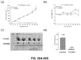


Classifications
-
- A—HUMAN NECESSITIES
- A61—MEDICAL OR VETERINARY SCIENCE; HYGIENE
- A61K—PREPARATIONS FOR MEDICAL, DENTAL OR TOILETRY PURPOSES
- A61K47/00—Medicinal preparations characterised by the non-active ingredients used, e.g. carriers or inert additives; Targeting or modifying agents chemically bound to the active ingredient
- A61K47/50—Medicinal preparations characterised by the non-active ingredients used, e.g. carriers or inert additives; Targeting or modifying agents chemically bound to the active ingredient the non-active ingredient being chemically bound to the active ingredient, e.g. polymer-drug conjugates
- A61K47/51—Medicinal preparations characterised by the non-active ingredients used, e.g. carriers or inert additives; Targeting or modifying agents chemically bound to the active ingredient the non-active ingredient being chemically bound to the active ingredient, e.g. polymer-drug conjugates the non-active ingredient being a modifying agent
- A61K47/56—Medicinal preparations characterised by the non-active ingredients used, e.g. carriers or inert additives; Targeting or modifying agents chemically bound to the active ingredient the non-active ingredient being chemically bound to the active ingredient, e.g. polymer-drug conjugates the non-active ingredient being a modifying agent the modifying agent being an organic macromolecular compound, e.g. an oligomeric, polymeric or dendrimeric molecule
- A61K47/59—Medicinal preparations characterised by the non-active ingredients used, e.g. carriers or inert additives; Targeting or modifying agents chemically bound to the active ingredient the non-active ingredient being chemically bound to the active ingredient, e.g. polymer-drug conjugates the non-active ingredient being a modifying agent the modifying agent being an organic macromolecular compound, e.g. an oligomeric, polymeric or dendrimeric molecule obtained otherwise than by reactions only involving carbon-to-carbon unsaturated bonds, e.g. polyureas or polyurethanes
- A61K47/593—Polyesters, e.g. PLGA or polylactide-co-glycolide
-
- C—CHEMISTRY; METALLURGY
- C08—ORGANIC MACROMOLECULAR COMPOUNDS; THEIR PREPARATION OR CHEMICAL WORKING-UP; COMPOSITIONS BASED THEREON
- C08G—MACROMOLECULAR COMPOUNDS OBTAINED OTHERWISE THAN BY REACTIONS ONLY INVOLVING UNSATURATED CARBON-TO-CARBON BONDS
- C08G63/00—Macromolecular compounds obtained by reactions forming a carboxylic ester link in the main chain of the macromolecule
- C08G63/66—Polyesters containing oxygen in the form of ether groups
- C08G63/664—Polyesters containing oxygen in the form of ether groups derived from hydroxy carboxylic acids
-
- A—HUMAN NECESSITIES
- A61—MEDICAL OR VETERINARY SCIENCE; HYGIENE
- A61K—PREPARATIONS FOR MEDICAL, DENTAL OR TOILETRY PURPOSES
- A61K31/00—Medicinal preparations containing organic active ingredients
- A61K31/045—Hydroxy compounds, e.g. alcohols; Salts thereof, e.g. alcoholates
- A61K31/05—Phenols
-
- A—HUMAN NECESSITIES
- A61—MEDICAL OR VETERINARY SCIENCE; HYGIENE
- A61K—PREPARATIONS FOR MEDICAL, DENTAL OR TOILETRY PURPOSES
- A61K31/00—Medicinal preparations containing organic active ingredients
- A61K31/16—Amides, e.g. hydroxamic acids
- A61K31/165—Amides, e.g. hydroxamic acids having aromatic rings, e.g. colchicine, atenolol, progabide
-
- A—HUMAN NECESSITIES
- A61—MEDICAL OR VETERINARY SCIENCE; HYGIENE
- A61K—PREPARATIONS FOR MEDICAL, DENTAL OR TOILETRY PURPOSES
- A61K31/00—Medicinal preparations containing organic active ingredients
- A61K31/33—Heterocyclic compounds
- A61K31/335—Heterocyclic compounds having oxygen as the only ring hetero atom, e.g. fungichromin
- A61K31/337—Heterocyclic compounds having oxygen as the only ring hetero atom, e.g. fungichromin having four-membered rings, e.g. taxol
-
- A—HUMAN NECESSITIES
- A61—MEDICAL OR VETERINARY SCIENCE; HYGIENE
- A61K—PREPARATIONS FOR MEDICAL, DENTAL OR TOILETRY PURPOSES
- A61K31/00—Medicinal preparations containing organic active ingredients
- A61K31/33—Heterocyclic compounds
- A61K31/335—Heterocyclic compounds having oxygen as the only ring hetero atom, e.g. fungichromin
- A61K31/35—Heterocyclic compounds having oxygen as the only ring hetero atom, e.g. fungichromin having six-membered rings with one oxygen as the only ring hetero atom
- A61K31/352—Heterocyclic compounds having oxygen as the only ring hetero atom, e.g. fungichromin having six-membered rings with one oxygen as the only ring hetero atom condensed with carbocyclic rings, e.g. methantheline
- A61K31/353—3,4-Dihydrobenzopyrans, e.g. chroman, catechin
-
- A—HUMAN NECESSITIES
- A61—MEDICAL OR VETERINARY SCIENCE; HYGIENE
- A61K—PREPARATIONS FOR MEDICAL, DENTAL OR TOILETRY PURPOSES
- A61K31/00—Medicinal preparations containing organic active ingredients
- A61K31/33—Heterocyclic compounds
- A61K31/395—Heterocyclic compounds having nitrogen as a ring hetero atom, e.g. guanethidine or rifamycins
- A61K31/41—Heterocyclic compounds having nitrogen as a ring hetero atom, e.g. guanethidine or rifamycins having five-membered rings with two or more ring hetero atoms, at least one of which being nitrogen, e.g. tetrazole
- A61K31/4164—1,3-Diazoles
- A61K31/4178—1,3-Diazoles not condensed 1,3-diazoles and containing further heterocyclic rings, e.g. pilocarpine, nitrofurantoin
-
- A—HUMAN NECESSITIES
- A61—MEDICAL OR VETERINARY SCIENCE; HYGIENE
- A61K—PREPARATIONS FOR MEDICAL, DENTAL OR TOILETRY PURPOSES
- A61K31/00—Medicinal preparations containing organic active ingredients
- A61K31/33—Heterocyclic compounds
- A61K31/395—Heterocyclic compounds having nitrogen as a ring hetero atom, e.g. guanethidine or rifamycins
- A61K31/41—Heterocyclic compounds having nitrogen as a ring hetero atom, e.g. guanethidine or rifamycins having five-membered rings with two or more ring hetero atoms, at least one of which being nitrogen, e.g. tetrazole
- A61K31/4164—1,3-Diazoles
- A61K31/4184—1,3-Diazoles condensed with carbocyclic rings, e.g. benzimidazoles
-
- A—HUMAN NECESSITIES
- A61—MEDICAL OR VETERINARY SCIENCE; HYGIENE
- A61K—PREPARATIONS FOR MEDICAL, DENTAL OR TOILETRY PURPOSES
- A61K31/00—Medicinal preparations containing organic active ingredients
- A61K31/33—Heterocyclic compounds
- A61K31/395—Heterocyclic compounds having nitrogen as a ring hetero atom, e.g. guanethidine or rifamycins
- A61K31/41—Heterocyclic compounds having nitrogen as a ring hetero atom, e.g. guanethidine or rifamycins having five-membered rings with two or more ring hetero atoms, at least one of which being nitrogen, e.g. tetrazole
- A61K31/4245—Oxadiazoles
-
- A—HUMAN NECESSITIES
- A61—MEDICAL OR VETERINARY SCIENCE; HYGIENE
- A61K—PREPARATIONS FOR MEDICAL, DENTAL OR TOILETRY PURPOSES
- A61K31/00—Medicinal preparations containing organic active ingredients
- A61K31/33—Heterocyclic compounds
- A61K31/395—Heterocyclic compounds having nitrogen as a ring hetero atom, e.g. guanethidine or rifamycins
- A61K31/435—Heterocyclic compounds having nitrogen as a ring hetero atom, e.g. guanethidine or rifamycins having six-membered rings with one nitrogen as the only ring hetero atom
- A61K31/44—Non condensed pyridines; Hydrogenated derivatives thereof
- A61K31/4412—Non condensed pyridines; Hydrogenated derivatives thereof having oxo groups directly attached to the heterocyclic ring
-
- A—HUMAN NECESSITIES
- A61—MEDICAL OR VETERINARY SCIENCE; HYGIENE
- A61K—PREPARATIONS FOR MEDICAL, DENTAL OR TOILETRY PURPOSES
- A61K31/00—Medicinal preparations containing organic active ingredients
- A61K31/33—Heterocyclic compounds
- A61K31/395—Heterocyclic compounds having nitrogen as a ring hetero atom, e.g. guanethidine or rifamycins
- A61K31/435—Heterocyclic compounds having nitrogen as a ring hetero atom, e.g. guanethidine or rifamycins having six-membered rings with one nitrogen as the only ring hetero atom
- A61K31/44—Non condensed pyridines; Hydrogenated derivatives thereof
- A61K31/4418—Non condensed pyridines; Hydrogenated derivatives thereof having a carbocyclic group directly attached to the heterocyclic ring, e.g. cyproheptadine
-
- A—HUMAN NECESSITIES
- A61—MEDICAL OR VETERINARY SCIENCE; HYGIENE
- A61K—PREPARATIONS FOR MEDICAL, DENTAL OR TOILETRY PURPOSES
- A61K31/00—Medicinal preparations containing organic active ingredients
- A61K31/33—Heterocyclic compounds
- A61K31/395—Heterocyclic compounds having nitrogen as a ring hetero atom, e.g. guanethidine or rifamycins
- A61K31/495—Heterocyclic compounds having nitrogen as a ring hetero atom, e.g. guanethidine or rifamycins having six-membered rings with two or more nitrogen atoms as the only ring heteroatoms, e.g. piperazine or tetrazines
- A61K31/496—Non-condensed piperazines containing further heterocyclic rings, e.g. rifampin, thiothixene or sparfloxacin
-
- A—HUMAN NECESSITIES
- A61—MEDICAL OR VETERINARY SCIENCE; HYGIENE
- A61K—PREPARATIONS FOR MEDICAL, DENTAL OR TOILETRY PURPOSES
- A61K31/00—Medicinal preparations containing organic active ingredients
- A61K31/33—Heterocyclic compounds
- A61K31/395—Heterocyclic compounds having nitrogen as a ring hetero atom, e.g. guanethidine or rifamycins
- A61K31/495—Heterocyclic compounds having nitrogen as a ring hetero atom, e.g. guanethidine or rifamycins having six-membered rings with two or more nitrogen atoms as the only ring heteroatoms, e.g. piperazine or tetrazines
- A61K31/505—Pyrimidines; Hydrogenated pyrimidines, e.g. trimethoprim
- A61K31/506—Pyrimidines; Hydrogenated pyrimidines, e.g. trimethoprim not condensed and containing further heterocyclic rings
-
- A—HUMAN NECESSITIES
- A61—MEDICAL OR VETERINARY SCIENCE; HYGIENE
- A61K—PREPARATIONS FOR MEDICAL, DENTAL OR TOILETRY PURPOSES
- A61K31/00—Medicinal preparations containing organic active ingredients
- A61K31/33—Heterocyclic compounds
- A61K31/395—Heterocyclic compounds having nitrogen as a ring hetero atom, e.g. guanethidine or rifamycins
- A61K31/495—Heterocyclic compounds having nitrogen as a ring hetero atom, e.g. guanethidine or rifamycins having six-membered rings with two or more nitrogen atoms as the only ring heteroatoms, e.g. piperazine or tetrazines
- A61K31/505—Pyrimidines; Hydrogenated pyrimidines, e.g. trimethoprim
- A61K31/513—Pyrimidines; Hydrogenated pyrimidines, e.g. trimethoprim having oxo groups directly attached to the heterocyclic ring, e.g. cytosine
-
- A—HUMAN NECESSITIES
- A61—MEDICAL OR VETERINARY SCIENCE; HYGIENE
- A61K—PREPARATIONS FOR MEDICAL, DENTAL OR TOILETRY PURPOSES
- A61K31/00—Medicinal preparations containing organic active ingredients
- A61K31/33—Heterocyclic compounds
- A61K31/395—Heterocyclic compounds having nitrogen as a ring hetero atom, e.g. guanethidine or rifamycins
- A61K31/495—Heterocyclic compounds having nitrogen as a ring hetero atom, e.g. guanethidine or rifamycins having six-membered rings with two or more nitrogen atoms as the only ring heteroatoms, e.g. piperazine or tetrazines
- A61K31/505—Pyrimidines; Hydrogenated pyrimidines, e.g. trimethoprim
- A61K31/519—Pyrimidines; Hydrogenated pyrimidines, e.g. trimethoprim ortho- or peri-condensed with heterocyclic rings
-
- A—HUMAN NECESSITIES
- A61—MEDICAL OR VETERINARY SCIENCE; HYGIENE
- A61K—PREPARATIONS FOR MEDICAL, DENTAL OR TOILETRY PURPOSES
- A61K31/00—Medicinal preparations containing organic active ingredients
- A61K31/33—Heterocyclic compounds
- A61K31/395—Heterocyclic compounds having nitrogen as a ring hetero atom, e.g. guanethidine or rifamycins
- A61K31/535—Heterocyclic compounds having nitrogen as a ring hetero atom, e.g. guanethidine or rifamycins having six-membered rings with at least one nitrogen and one oxygen as the ring hetero atoms, e.g. 1,2-oxazines
- A61K31/5365—Heterocyclic compounds having nitrogen as a ring hetero atom, e.g. guanethidine or rifamycins having six-membered rings with at least one nitrogen and one oxygen as the ring hetero atoms, e.g. 1,2-oxazines ortho- or peri-condensed with heterocyclic ring systems
-
- A—HUMAN NECESSITIES
- A61—MEDICAL OR VETERINARY SCIENCE; HYGIENE
- A61K—PREPARATIONS FOR MEDICAL, DENTAL OR TOILETRY PURPOSES
- A61K31/00—Medicinal preparations containing organic active ingredients
- A61K31/33—Heterocyclic compounds
- A61K31/395—Heterocyclic compounds having nitrogen as a ring hetero atom, e.g. guanethidine or rifamycins
- A61K31/535—Heterocyclic compounds having nitrogen as a ring hetero atom, e.g. guanethidine or rifamycins having six-membered rings with at least one nitrogen and one oxygen as the ring hetero atoms, e.g. 1,2-oxazines
- A61K31/537—Heterocyclic compounds having nitrogen as a ring hetero atom, e.g. guanethidine or rifamycins having six-membered rings with at least one nitrogen and one oxygen as the ring hetero atoms, e.g. 1,2-oxazines spiro-condensed or forming part of bridged ring systems
-
- A—HUMAN NECESSITIES
- A61—MEDICAL OR VETERINARY SCIENCE; HYGIENE
- A61K—PREPARATIONS FOR MEDICAL, DENTAL OR TOILETRY PURPOSES
- A61K31/00—Medicinal preparations containing organic active ingredients
- A61K31/33—Heterocyclic compounds
- A61K31/395—Heterocyclic compounds having nitrogen as a ring hetero atom, e.g. guanethidine or rifamycins
- A61K31/55—Heterocyclic compounds having nitrogen as a ring hetero atom, e.g. guanethidine or rifamycins having seven-membered rings, e.g. azelastine, pentylenetetrazole
- A61K31/551—Heterocyclic compounds having nitrogen as a ring hetero atom, e.g. guanethidine or rifamycins having seven-membered rings, e.g. azelastine, pentylenetetrazole having two nitrogen atoms, e.g. dilazep
- A61K31/5513—1,4-Benzodiazepines, e.g. diazepam or clozapine
-
- A—HUMAN NECESSITIES
- A61—MEDICAL OR VETERINARY SCIENCE; HYGIENE
- A61K—PREPARATIONS FOR MEDICAL, DENTAL OR TOILETRY PURPOSES
- A61K31/00—Medicinal preparations containing organic active ingredients
- A61K31/56—Compounds containing cyclopenta[a]hydrophenanthrene ring systems; Derivatives thereof, e.g. steroids
- A61K31/57—Compounds containing cyclopenta[a]hydrophenanthrene ring systems; Derivatives thereof, e.g. steroids substituted in position 17 beta by a chain of two carbon atoms, e.g. pregnane or progesterone
- A61K31/573—Compounds containing cyclopenta[a]hydrophenanthrene ring systems; Derivatives thereof, e.g. steroids substituted in position 17 beta by a chain of two carbon atoms, e.g. pregnane or progesterone substituted in position 21, e.g. cortisone, dexamethasone, prednisone or aldosterone
-
- A—HUMAN NECESSITIES
- A61—MEDICAL OR VETERINARY SCIENCE; HYGIENE
- A61K—PREPARATIONS FOR MEDICAL, DENTAL OR TOILETRY PURPOSES
- A61K31/00—Medicinal preparations containing organic active ingredients
- A61K31/70—Carbohydrates; Sugars; Derivatives thereof
- A61K31/7028—Compounds having saccharide radicals attached to non-saccharide compounds by glycosidic linkages
- A61K31/7034—Compounds having saccharide radicals attached to non-saccharide compounds by glycosidic linkages attached to a carbocyclic compound, e.g. phloridzin
- A61K31/704—Compounds having saccharide radicals attached to non-saccharide compounds by glycosidic linkages attached to a carbocyclic compound, e.g. phloridzin attached to a condensed carbocyclic ring system, e.g. sennosides, thiocolchicosides, escin, daunorubicin
-
- A—HUMAN NECESSITIES
- A61—MEDICAL OR VETERINARY SCIENCE; HYGIENE
- A61K—PREPARATIONS FOR MEDICAL, DENTAL OR TOILETRY PURPOSES
- A61K31/00—Medicinal preparations containing organic active ingredients
- A61K31/70—Carbohydrates; Sugars; Derivatives thereof
- A61K31/7042—Compounds having saccharide radicals and heterocyclic rings
- A61K31/7048—Compounds having saccharide radicals and heterocyclic rings having oxygen as a ring hetero atom, e.g. leucoglucosan, hesperidin, erythromycin, nystatin, digitoxin or digoxin
-
- A—HUMAN NECESSITIES
- A61—MEDICAL OR VETERINARY SCIENCE; HYGIENE
- A61K—PREPARATIONS FOR MEDICAL, DENTAL OR TOILETRY PURPOSES
- A61K31/00—Medicinal preparations containing organic active ingredients
- A61K31/70—Carbohydrates; Sugars; Derivatives thereof
- A61K31/7042—Compounds having saccharide radicals and heterocyclic rings
- A61K31/7052—Compounds having saccharide radicals and heterocyclic rings having nitrogen as a ring hetero atom, e.g. nucleosides, nucleotides
- A61K31/706—Compounds having saccharide radicals and heterocyclic rings having nitrogen as a ring hetero atom, e.g. nucleosides, nucleotides containing six-membered rings with nitrogen as a ring hetero atom
- A61K31/7064—Compounds having saccharide radicals and heterocyclic rings having nitrogen as a ring hetero atom, e.g. nucleosides, nucleotides containing six-membered rings with nitrogen as a ring hetero atom containing condensed or non-condensed pyrimidines
- A61K31/7068—Compounds having saccharide radicals and heterocyclic rings having nitrogen as a ring hetero atom, e.g. nucleosides, nucleotides containing six-membered rings with nitrogen as a ring hetero atom containing condensed or non-condensed pyrimidines having oxo groups directly attached to the pyrimidine ring, e.g. cytidine, cytidylic acid
-
- A—HUMAN NECESSITIES
- A61—MEDICAL OR VETERINARY SCIENCE; HYGIENE
- A61K—PREPARATIONS FOR MEDICAL, DENTAL OR TOILETRY PURPOSES
- A61K33/00—Medicinal preparations containing inorganic active ingredients
- A61K33/24—Heavy metals; Compounds thereof
- A61K33/243—Platinum; Compounds thereof
-
- A—HUMAN NECESSITIES
- A61—MEDICAL OR VETERINARY SCIENCE; HYGIENE
- A61K—PREPARATIONS FOR MEDICAL, DENTAL OR TOILETRY PURPOSES
- A61K47/00—Medicinal preparations characterised by the non-active ingredients used, e.g. carriers or inert additives; Targeting or modifying agents chemically bound to the active ingredient
- A61K47/50—Medicinal preparations characterised by the non-active ingredients used, e.g. carriers or inert additives; Targeting or modifying agents chemically bound to the active ingredient the non-active ingredient being chemically bound to the active ingredient, e.g. polymer-drug conjugates
- A61K47/51—Medicinal preparations characterised by the non-active ingredients used, e.g. carriers or inert additives; Targeting or modifying agents chemically bound to the active ingredient the non-active ingredient being chemically bound to the active ingredient, e.g. polymer-drug conjugates the non-active ingredient being a modifying agent
- A61K47/54—Medicinal preparations characterised by the non-active ingredients used, e.g. carriers or inert additives; Targeting or modifying agents chemically bound to the active ingredient the non-active ingredient being chemically bound to the active ingredient, e.g. polymer-drug conjugates the non-active ingredient being a modifying agent the modifying agent being an organic compound
- A61K47/549—Sugars, nucleosides, nucleotides or nucleic acids
-
- A—HUMAN NECESSITIES
- A61—MEDICAL OR VETERINARY SCIENCE; HYGIENE
- A61K—PREPARATIONS FOR MEDICAL, DENTAL OR TOILETRY PURPOSES
- A61K47/00—Medicinal preparations characterised by the non-active ingredients used, e.g. carriers or inert additives; Targeting or modifying agents chemically bound to the active ingredient
- A61K47/50—Medicinal preparations characterised by the non-active ingredients used, e.g. carriers or inert additives; Targeting or modifying agents chemically bound to the active ingredient the non-active ingredient being chemically bound to the active ingredient, e.g. polymer-drug conjugates
- A61K47/51—Medicinal preparations characterised by the non-active ingredients used, e.g. carriers or inert additives; Targeting or modifying agents chemically bound to the active ingredient the non-active ingredient being chemically bound to the active ingredient, e.g. polymer-drug conjugates the non-active ingredient being a modifying agent
- A61K47/54—Medicinal preparations characterised by the non-active ingredients used, e.g. carriers or inert additives; Targeting or modifying agents chemically bound to the active ingredient the non-active ingredient being chemically bound to the active ingredient, e.g. polymer-drug conjugates the non-active ingredient being a modifying agent the modifying agent being an organic compound
- A61K47/55—Medicinal preparations characterised by the non-active ingredients used, e.g. carriers or inert additives; Targeting or modifying agents chemically bound to the active ingredient the non-active ingredient being chemically bound to the active ingredient, e.g. polymer-drug conjugates the non-active ingredient being a modifying agent the modifying agent being an organic compound the modifying agent being also a pharmacologically or therapeutically active agent, i.e. the entire conjugate being a codrug, i.e. a dimer, oligomer or polymer of pharmacologically or therapeutically active compounds
- A61K47/551—Medicinal preparations characterised by the non-active ingredients used, e.g. carriers or inert additives; Targeting or modifying agents chemically bound to the active ingredient the non-active ingredient being chemically bound to the active ingredient, e.g. polymer-drug conjugates the non-active ingredient being a modifying agent the modifying agent being an organic compound the modifying agent being also a pharmacologically or therapeutically active agent, i.e. the entire conjugate being a codrug, i.e. a dimer, oligomer or polymer of pharmacologically or therapeutically active compounds one of the codrug's components being a vitamin, e.g. niacinamide, vitamin B3, cobalamin, vitamin B12, folate, vitamin A or retinoic acid
-
- A—HUMAN NECESSITIES
- A61—MEDICAL OR VETERINARY SCIENCE; HYGIENE
- A61K—PREPARATIONS FOR MEDICAL, DENTAL OR TOILETRY PURPOSES
- A61K47/00—Medicinal preparations characterised by the non-active ingredients used, e.g. carriers or inert additives; Targeting or modifying agents chemically bound to the active ingredient
- A61K47/50—Medicinal preparations characterised by the non-active ingredients used, e.g. carriers or inert additives; Targeting or modifying agents chemically bound to the active ingredient the non-active ingredient being chemically bound to the active ingredient, e.g. polymer-drug conjugates
- A61K47/51—Medicinal preparations characterised by the non-active ingredients used, e.g. carriers or inert additives; Targeting or modifying agents chemically bound to the active ingredient the non-active ingredient being chemically bound to the active ingredient, e.g. polymer-drug conjugates the non-active ingredient being a modifying agent
- A61K47/56—Medicinal preparations characterised by the non-active ingredients used, e.g. carriers or inert additives; Targeting or modifying agents chemically bound to the active ingredient the non-active ingredient being chemically bound to the active ingredient, e.g. polymer-drug conjugates the non-active ingredient being a modifying agent the modifying agent being an organic macromolecular compound, e.g. an oligomeric, polymeric or dendrimeric molecule
- A61K47/59—Medicinal preparations characterised by the non-active ingredients used, e.g. carriers or inert additives; Targeting or modifying agents chemically bound to the active ingredient the non-active ingredient being chemically bound to the active ingredient, e.g. polymer-drug conjugates the non-active ingredient being a modifying agent the modifying agent being an organic macromolecular compound, e.g. an oligomeric, polymeric or dendrimeric molecule obtained otherwise than by reactions only involving carbon-to-carbon unsaturated bonds, e.g. polyureas or polyurethanes
- A61K47/60—Medicinal preparations characterised by the non-active ingredients used, e.g. carriers or inert additives; Targeting or modifying agents chemically bound to the active ingredient the non-active ingredient being chemically bound to the active ingredient, e.g. polymer-drug conjugates the non-active ingredient being a modifying agent the modifying agent being an organic macromolecular compound, e.g. an oligomeric, polymeric or dendrimeric molecule obtained otherwise than by reactions only involving carbon-to-carbon unsaturated bonds, e.g. polyureas or polyurethanes the organic macromolecular compound being a polyoxyalkylene oligomer, polymer or dendrimer, e.g. PEG, PPG, PEO or polyglycerol
-
- A—HUMAN NECESSITIES
- A61—MEDICAL OR VETERINARY SCIENCE; HYGIENE
- A61K—PREPARATIONS FOR MEDICAL, DENTAL OR TOILETRY PURPOSES
- A61K47/00—Medicinal preparations characterised by the non-active ingredients used, e.g. carriers or inert additives; Targeting or modifying agents chemically bound to the active ingredient
- A61K47/50—Medicinal preparations characterised by the non-active ingredients used, e.g. carriers or inert additives; Targeting or modifying agents chemically bound to the active ingredient the non-active ingredient being chemically bound to the active ingredient, e.g. polymer-drug conjugates
- A61K47/51—Medicinal preparations characterised by the non-active ingredients used, e.g. carriers or inert additives; Targeting or modifying agents chemically bound to the active ingredient the non-active ingredient being chemically bound to the active ingredient, e.g. polymer-drug conjugates the non-active ingredient being a modifying agent
- A61K47/62—Medicinal preparations characterised by the non-active ingredients used, e.g. carriers or inert additives; Targeting or modifying agents chemically bound to the active ingredient the non-active ingredient being chemically bound to the active ingredient, e.g. polymer-drug conjugates the non-active ingredient being a modifying agent the modifying agent being a protein, peptide or polyamino acid
- A61K47/64—Drug-peptide, drug-protein or drug-polyamino acid conjugates, i.e. the modifying agent being a peptide, protein or polyamino acid which is covalently bonded or complexed to a therapeutically active agent
-
- A—HUMAN NECESSITIES
- A61—MEDICAL OR VETERINARY SCIENCE; HYGIENE
- A61K—PREPARATIONS FOR MEDICAL, DENTAL OR TOILETRY PURPOSES
- A61K47/00—Medicinal preparations characterised by the non-active ingredients used, e.g. carriers or inert additives; Targeting or modifying agents chemically bound to the active ingredient
- A61K47/50—Medicinal preparations characterised by the non-active ingredients used, e.g. carriers or inert additives; Targeting or modifying agents chemically bound to the active ingredient the non-active ingredient being chemically bound to the active ingredient, e.g. polymer-drug conjugates
- A61K47/69—Medicinal preparations characterised by the non-active ingredients used, e.g. carriers or inert additives; Targeting or modifying agents chemically bound to the active ingredient the non-active ingredient being chemically bound to the active ingredient, e.g. polymer-drug conjugates the conjugate being characterised by physical or galenical forms, e.g. emulsion, particle, inclusion complex, stent or kit
- A61K47/6921—Medicinal preparations characterised by the non-active ingredients used, e.g. carriers or inert additives; Targeting or modifying agents chemically bound to the active ingredient the non-active ingredient being chemically bound to the active ingredient, e.g. polymer-drug conjugates the conjugate being characterised by physical or galenical forms, e.g. emulsion, particle, inclusion complex, stent or kit the form being a particulate, a powder, an adsorbate, a bead or a sphere
- A61K47/6927—Medicinal preparations characterised by the non-active ingredients used, e.g. carriers or inert additives; Targeting or modifying agents chemically bound to the active ingredient the non-active ingredient being chemically bound to the active ingredient, e.g. polymer-drug conjugates the conjugate being characterised by physical or galenical forms, e.g. emulsion, particle, inclusion complex, stent or kit the form being a particulate, a powder, an adsorbate, a bead or a sphere the form being a solid microparticle having no hollow or gas-filled cores
- A61K47/6929—Medicinal preparations characterised by the non-active ingredients used, e.g. carriers or inert additives; Targeting or modifying agents chemically bound to the active ingredient the non-active ingredient being chemically bound to the active ingredient, e.g. polymer-drug conjugates the conjugate being characterised by physical or galenical forms, e.g. emulsion, particle, inclusion complex, stent or kit the form being a particulate, a powder, an adsorbate, a bead or a sphere the form being a solid microparticle having no hollow or gas-filled cores the form being a nanoparticle, e.g. an immuno-nanoparticle
- A61K47/6931—Medicinal preparations characterised by the non-active ingredients used, e.g. carriers or inert additives; Targeting or modifying agents chemically bound to the active ingredient the non-active ingredient being chemically bound to the active ingredient, e.g. polymer-drug conjugates the conjugate being characterised by physical or galenical forms, e.g. emulsion, particle, inclusion complex, stent or kit the form being a particulate, a powder, an adsorbate, a bead or a sphere the form being a solid microparticle having no hollow or gas-filled cores the form being a nanoparticle, e.g. an immuno-nanoparticle the material constituting the nanoparticle being a polymer
- A61K47/6935—Medicinal preparations characterised by the non-active ingredients used, e.g. carriers or inert additives; Targeting or modifying agents chemically bound to the active ingredient the non-active ingredient being chemically bound to the active ingredient, e.g. polymer-drug conjugates the conjugate being characterised by physical or galenical forms, e.g. emulsion, particle, inclusion complex, stent or kit the form being a particulate, a powder, an adsorbate, a bead or a sphere the form being a solid microparticle having no hollow or gas-filled cores the form being a nanoparticle, e.g. an immuno-nanoparticle the material constituting the nanoparticle being a polymer the polymer being obtained otherwise than by reactions involving carbon to carbon unsaturated bonds, e.g. polyesters, polyamides or polyglycerol
- A61K47/6937—Medicinal preparations characterised by the non-active ingredients used, e.g. carriers or inert additives; Targeting or modifying agents chemically bound to the active ingredient the non-active ingredient being chemically bound to the active ingredient, e.g. polymer-drug conjugates the conjugate being characterised by physical or galenical forms, e.g. emulsion, particle, inclusion complex, stent or kit the form being a particulate, a powder, an adsorbate, a bead or a sphere the form being a solid microparticle having no hollow or gas-filled cores the form being a nanoparticle, e.g. an immuno-nanoparticle the material constituting the nanoparticle being a polymer the polymer being obtained otherwise than by reactions involving carbon to carbon unsaturated bonds, e.g. polyesters, polyamides or polyglycerol the polymer being PLGA, PLA or polyglycolic acid
-
- A—HUMAN NECESSITIES
- A61—MEDICAL OR VETERINARY SCIENCE; HYGIENE
- A61K—PREPARATIONS FOR MEDICAL, DENTAL OR TOILETRY PURPOSES
- A61K51/00—Preparations containing radioactive substances for use in therapy or testing in vivo
- A61K51/02—Preparations containing radioactive substances for use in therapy or testing in vivo characterised by the carrier, i.e. characterised by the agent or material covalently linked or complexing the radioactive nucleus
- A61K51/04—Organic compounds
- A61K51/06—Macromolecular compounds, carriers being organic macromolecular compounds, i.e. organic oligomeric, polymeric, dendrimeric molecules
- A61K51/065—Macromolecular compounds, carriers being organic macromolecular compounds, i.e. organic oligomeric, polymeric, dendrimeric molecules conjugates with carriers being macromolecules
-
- A—HUMAN NECESSITIES
- A61—MEDICAL OR VETERINARY SCIENCE; HYGIENE
- A61P—SPECIFIC THERAPEUTIC ACTIVITY OF CHEMICAL COMPOUNDS OR MEDICINAL PREPARATIONS
- A61P35/00—Antineoplastic agents
-
- C—CHEMISTRY; METALLURGY
- C08—ORGANIC MACROMOLECULAR COMPOUNDS; THEIR PREPARATION OR CHEMICAL WORKING-UP; COMPOSITIONS BASED THEREON
- C08G—MACROMOLECULAR COMPOUNDS OBTAINED OTHERWISE THAN BY REACTIONS ONLY INVOLVING UNSATURATED CARBON-TO-CARBON BONDS
- C08G63/00—Macromolecular compounds obtained by reactions forming a carboxylic ester link in the main chain of the macromolecule
- C08G63/02—Polyesters derived from hydroxycarboxylic acids or from polycarboxylic acids and polyhydroxy compounds
- C08G63/06—Polyesters derived from hydroxycarboxylic acids or from polycarboxylic acids and polyhydroxy compounds derived from hydroxycarboxylic acids
- C08G63/08—Lactones or lactides
-
- C—CHEMISTRY; METALLURGY
- C08—ORGANIC MACROMOLECULAR COMPOUNDS; THEIR PREPARATION OR CHEMICAL WORKING-UP; COMPOSITIONS BASED THEREON
- C08G—MACROMOLECULAR COMPOUNDS OBTAINED OTHERWISE THAN BY REACTIONS ONLY INVOLVING UNSATURATED CARBON-TO-CARBON BONDS
- C08G63/00—Macromolecular compounds obtained by reactions forming a carboxylic ester link in the main chain of the macromolecule
- C08G63/91—Polymers modified by chemical after-treatment
- C08G63/912—Polymers modified by chemical after-treatment derived from hydroxycarboxylic acids
-
- C—CHEMISTRY; METALLURGY
- C08—ORGANIC MACROMOLECULAR COMPOUNDS; THEIR PREPARATION OR CHEMICAL WORKING-UP; COMPOSITIONS BASED THEREON
- C08G—MACROMOLECULAR COMPOUNDS OBTAINED OTHERWISE THAN BY REACTIONS ONLY INVOLVING UNSATURATED CARBON-TO-CARBON BONDS
- C08G65/00—Macromolecular compounds obtained by reactions forming an ether link in the main chain of the macromolecule
- C08G65/02—Macromolecular compounds obtained by reactions forming an ether link in the main chain of the macromolecule from cyclic ethers by opening of the heterocyclic ring
- C08G65/32—Polymers modified by chemical after-treatment
- C08G65/329—Polymers modified by chemical after-treatment with organic compounds
- C08G65/331—Polymers modified by chemical after-treatment with organic compounds containing oxygen
- C08G65/332—Polymers modified by chemical after-treatment with organic compounds containing oxygen containing carboxyl groups, or halides, or esters thereof
-
- C—CHEMISTRY; METALLURGY
- C08—ORGANIC MACROMOLECULAR COMPOUNDS; THEIR PREPARATION OR CHEMICAL WORKING-UP; COMPOSITIONS BASED THEREON
- C08L—COMPOSITIONS OF MACROMOLECULAR COMPOUNDS
- C08L67/00—Compositions of polyesters obtained by reactions forming a carboxylic ester link in the main chain; Compositions of derivatives of such polymers
- C08L67/04—Polyesters derived from hydroxycarboxylic acids, e.g. lactones
Definitions
- hydrophobic drugs to the appropriate tissues in the body has long been a challenge for medical researchers, who must maximize biocompatibility while minimizing toxicity.
- An ideal delivery vehicle would avoid premature release of its cargo, thereby delivering a larger dose of the drug to the effective site.
- Chemotherapeutic agents especially those with low molecular weights, can enter all cell types via random diffusion, which both decreases their availability at tumor sites and leads to systemic side effects. Random diffusion may further result in rapid cellular uptake rather than extended therapeutic effect.
- filtration by the kidneys can rapidly remove drugs from the bloodstream.
- chemotherapeutic agent or combination of chemotherapeutic agents can be selected to treat a subject's specific tumor(s) more effectively than a general course of chemotherapy.
- the chemotherapeutic agents could be selected based on tests such as biopsies, cell culture, and susceptibility assays rather than conducting expensive genetic tumor profiles.
- chemotherapeutic agents may display toxic side effects and combined side effects of two or more chemotherapeutic agents may prove to be intolerable for subjects.
- CN105031672 describes polylactic acid stereocomplex magnetic nano micelle with high stability and less proneness to leakage of magnetic particles and can be used for preparing drug controlled-release nano-carriers or novel magnetic resonance imaging contrast agents.
- What is needed is a method for treating cancer or reducing tumor size in a subject that minimizes toxicity and is biocompatible, that offers targeted delivery of anti-cancer agents via polymeric drug conjugates or similar means to tumor cells without adversely affecting surrounding tissue, that exhibits controlled, sustained release rates for the anti-cancer agents, and that allows for synergistic combination of two or more anti-cancer agents without a concomitant increase in side effects.
- the method could also be customized for individual patients.
- stereocomplexes for the delivery of one or more anti-cancer agents.
- the stereocomplexes exhibit low toxicity and are biodegradable while also providing for controlled release of one or more anti-cancer agents at tumor sites.
- the stereocomplexes can be designed such that the anti-cancer agents operate synergistically and may optionally include additional targeting groups and functionalities.
- the stereocomplexes disclosed herein can be combined with pharmaceutically-acceptable carriers and/or excipients to form pharmaceutical compositions. By varying the amount of each anti-cancer agent in the stereocomplex, specific types of tumors and cancer cell lines can be treated.
- compositions described herein may optionally contain one or more targeting groups, where the targeting group may or may not be present.
- the term "about” is used to provide flexibility to a numerical range endpoint by providing that a given value may be “a little above” or “a little below” the endpoint without affecting the desired result.
- “about” refers to a range extending from 10% below the numerical value to 10% above the numerical value. For example, if the numerical value is 10, “about 10” means between 9 and 11 inclusive of the endpoints 9 and 11.
- Admixing or "admixture” refers to a combination of two components together when there is no chemical reaction or physical interaction.
- the terms “admixing” and “admixture” can also include the chemical interaction or physical interaction among any of the components described herein upon mixing to produce the composition.
- the components can be admixed alone, in water, in another solvent, or in a combination of solvents.
- solid tumor is an abnormal mass of tissue that usually does not contain cysts or liquid areas. Solid tumors may be benign (not cancer), or malignant (cancer). Different types of solid tumors are named for the type of cells that form them. Examples of solid tumors are sarcomas, carcinomas, and lymphomas.
- subject as defined herein is any organism in need of cancer treatment and/or prevention.
- the subject is a mammal including, but not limited to, humans, domesticated animals (e.g., dogs, cats, horses), livestock (e.g., cows, pigs), and wild animals.
- treat as used herein is defined as maintaining or reducing the symptoms of a pre-existing condition.
- compositions described herein are used to treat cancer.
- prevent is defined as eliminating or reducing the likelihood of occurrence of one or more symptoms of a disease or disorder.
- the compositions described herein can be used to prevent the regrowth of tumor cells or reduce the rate of regrowth of tumor cells.
- inhibitor is the ability of the compounds described herein to completely eliminate an activity or reduce the activity when compared to the same activity in the absence of the compound.
- the compositions described herein can be used to inhibit the growth and/or spread of cancer in the body of a subject.
- Biodegradable materials are capable of being decomposed by bacteria, fungi, or other organisms, or by enzymes in the body of a subject.
- Biocompatible materials are materials that perform their desired functions without eliciting harmful or deleterious changes to the subject in which they are implanted or to which they are applied, either locally or systemically.
- the compositions disclosed herein are biocompatible.
- toxicity refers to harmful effects a substance has on an organism such as a human or mammal, or on cells within that organism.
- a compound or composition with high toxicity would be unsuitable for use as a medical treatment, while a compound or composition with low toxicity would be acceptable for use as a medical treatment.
- the compounds and compositions disclosed herein exhibit low toxicity.
- alkyl group as used herein is a branched or unbranched saturated hydrocarbon group of 1 to 25 carbon atoms, such as methyl, ethyl, n -propyl, isopropyl, n -butyl, isobutyl, t -butyl, pentyl, hexyl, heptyl, octyl, decyl, tetradecyl, hexadecyl, eicosyl, tetracosyl, and the like.
- longer chain alkyl groups include, but are not limited to, an oleate group or a palmitate group.
- a "lower alkyl” group is an alkyl group containing from one to six carbon atoms.
- aryl group as used herein is any carbon-based aromatic group including, but not limited to, benzene, naphthalene, etc.
- aryl group also includes "heteroaryl group,” which is defined as an aromatic group that has at least one heteroatom incorporated within the ring of the aromatic group. Examples of heteroatoms include, but are not limited to, nitrogen, oxygen, sulfur, and phosphorus.
- the aryl group can be substituted or unsubstituted.
- the aryl group if substituted, can be substituted with one or more groups including, but not limited to, alkyl, alkynyl, alkenyl, aryl, halide, nitro, amino, ester, ketone, aldehyde, hydroxyl, carboxylic acid, or alkoxy.
- alkoxy group as used herein is defined as RO-, where R is an alkyl group or aryl group defined herein.
- halogenated group is any organic group such as, for example, an alkyl group or aryl group that possesses at least one halogen (F, Cl, Br, I).
- references in the specification and concluding claims to parts by weight, of a particular element in a composition or article denote the weight relationship between the element or component and any other elements or components in the composition or article for which a part by weight is expressed.
- X and Y are present at a weight ratio of 2:5, and are present in such a ratio regardless of whether additional components are contained in the compound.
- a weight percent of a component is based on the total weight of the formulation or composition in which the component is included.
- each of the combinations A + E, A + F, B + D, B + E, B + F, C + D, C + E, and C + F is specifically contemplated and should be considered from disclosure of A, B, and C; D, E, and F; and the example combination of A + D.
- any subset or combination of these is also specifically contemplated and disclosed.
- the sub-group of A + E, B + F, and C + E is specifically contemplated and should be considered from disclosure of A, B, and C; D, E, and F; and the example combination of A + D.
- This concept applies to all aspects of the disclosure including, but not limited to, steps in methods of making and using the disclosed compositions.
- steps in methods of making and using the disclosed compositions are a variety of additional steps that can be performed with any specific embodiment or combination of embodiments of the disclosed methods, each such combination is specifically contemplated and should be considered disclosed.
- stereocomplexes described herein are useful in delivering one or more anti-cancer agents to a subject.
- the stereocomplexes are composed at least two components, each component having a hydrophilic group, an isotactic polylactic acid moiety, a linker, and an anti-cancer agent.
- stereocomplexes having the following components: X 1 -Y 1 -L 1 -Z 1 (I) X 2 -Y 2 -L 2 -Z 2 (II) wherein X 1 and X 2 are hydrophilic groups; Y 1 and Y 2 are PDLA or PLLA; L 1 and L 2 are cleavable linkers, Z 1 is an anti-cancer agent, Z 2 is an anti-cancer agent or imaging agent, wherein when Z 2 is an anti-cancer agent, Z 1 and Z 2 are different anti-cancer agents, and wherein (1) when Y 1 is PDLA then Y 2 is PLLA, and when Y 1 is PLLA then Y 2 is PDLA and (2) the ratio of the total number of D-lactic acid units in the stereocomplex to the total number of L-lactic acid units in the stereocomplex is from 0.9:1.1 to 1.1:0.9.
- stereocomplexes form when the PDLA and PLLA units present in components (I) and (II) form an extensive 3-dimensional network driven by hydrogen bonding.
- the stereocomplexes described herein have enhanced properties such as tensile strength, Young's modulus, and elongation at break compared to either component (I) or component (II) alone.
- the stereocomplexes have high stability and high resistance to hydrolytic degradation, which eliminates the premature release of drugs (i.e., before the stereocomplexes reach their target tissue) and increases the circulation time of the stereocomplexes in the blood.
- Fig. 1 shows a schematic illustration of an exemplary stereocomplex with two different drugs based on the stereocomplexation between PDLA and PLLA.
- Fig. 2 shows a schematic illustration of exemplary polymer conjugated drugs for stereocomplexation.
- hydrophilic elements project into solution while hydrophobic elements cluster at the particle core ( Fig. 2A ).
- Examples anti-cancer drugs conjugated to hydrophobic parts with cleavable linkers are provided in Fig. 2B . Referring to Fig.
- mertansine (DM1) is linked to a carrier with a disulfide bond (D-DM1) and docetaxel (DTX) is linked to a carrier with a hydrazone bond, ester bond, or disulfide bond (L-DTX).
- the components used to produce the stereocomplexes disclosed herein include a hydrophilic group.
- X 1 and X 2 in components (I) and (II) are different hydrophilic groups.
- X 1 and X 2 are the same hydrophilic group.
- X 1 and X 2 are each a polyalkylene glycol.
- Polyalkylene glycol refers to a condensation polymer of ethylene oxide or propylene oxide and water. Polyalkylene glycols are typically colorless liquids with high molecular weights and are soluble in water as well as some organic solvents.
- the hydrophilic group in the stereocomplexes disclosed herein is a polyalkylene glycol.
- the polyalkylene glycol is polyethylene glycol and/or polypropylene glycol.
- the polyalkylene glycol is monomethoxy polyethylene glycol.
- the generic structure of a polyalkylene glycol is as follows:
- Table 1 Polyalkylene Glycols Compound Name R 1 R 2 R 3 Polyethylene glycol H H OH Polypropylene glycol H CH 3 OH Monomethoxy polyethylene glycol CH 3 H OH
- polyalkylene glycols are of low enough molecular weight that the chemical nature of the end groups (usually, but not always, hydroxyls) still affects the performance of the polymers.
- polyalkylene glycols can modify the viscosity of the stereocomplexes disclosed herein and may aid in the formation of emulsions.
- polyalkylene glycols are biocompatible and/or biodegradable.
- the polyalkylene glycols and/or other hydrophilic groups used herein are non-toxic.
- X 1 and X 2 in components (I) and (II) are polyalkylene glycols
- X 1 and X 2 have molecular weights of from about 1,000 Da to about 5,000 Da, or from 1,500 Da to 4,500 Da, or from 2,000 Da to 4,000 Da, or have molecular weights of about 1,000 Da, 1,500 Da, 2,000 Da, 2,500 Da, 3,000 Da, 3,500 Da, 4,000 Da, 4,500 Da, or about 5,000 Da, where any value can be a lower and upper end-point of a range (e.g., 2,000 Da to 4,000 Da).
- X 1 and X 2 in components (I) and (II) of the stereocomplexes disclosed herein are each monomethoxy polyethylene glycol having molecular weights of from about 1,000 to about 5,000 Da, or from 1,500 Da to 4,500 Da, or from 2,000 Da to 4,000 Da, or have molecular weights of about 1,000 Da, 1,500 Da, 2,000 Da, 2,500 Da, 3,000 Da, 3,500 Da, 4,000 Da, 4,500 Da, or about 5,000 Da, where any value can be a lower and upper end-point of a range (e.g., 2,000 Da to 4,000 Da).
- X 1 and X 2 in components (I) and (II) of the stereocomplexes disclosed herein are each monomethoxy polyethylene glycol having the same molecular weight.
- Polylactic acid is a polyester derived from lactic acid.
- the polyester is composed of lactic acid units depicted in the structure below, where m indicates the number of lactic acid units.
- the lactic acid unit has one chiral center, indicated by the asterisk (*) in the structure below, where m is the number of lactic acid units:
- Polylactic acid polymerization can begin from D or L lactic acid or a mixture thereof, or lactide, a cyclic diester. Properties of polylactic acid can be fine-tuned by controlling the ratio of D to L enantiomers used in the polymerization, and polylactic acid polymers can also be synthesized using starting materials that are only D or only L rather than a mixture of the two. Polylactic acid prepared from only D starting materials is referred to as poly-D-lactide (PDLA) (i.e., only composed of D-lactic acid units); conversely, polylactic acid prepared from only L starting materials is poly-L-lactide (or PLLA) (i.e., only composed of L-lactic acid units).
- PDLA poly-D-lactide
- PLLA poly-L-lactide
- a D-lactic acid unit or an L-lactic acid unit refers to the monomer units within the polylactic acid polymers described herein, wherein a D-lactic acid unit is derived from the D-lactic acid or D-lactide starting material, and an L-lactic acid unit is derived from the L-lactic acid or L-lactide starting material, as shown in Table 2: Table 2: Starting Materials for PDLA and PLLA
- the polylactic acid in components (I) and (II) is represented by Y 1 and Y 2 , where when Y 1 is PDLA then Y 2 is PLLA or, in the alternative, when Y 1 is PLLA then Y 2 is PDLA.
- the ratio of the total number of D-lactic acid units in the stereocomplex to the number of L-iactic acid units in the stereocomplex is from 0.9:1.1 to 1.1:0.9. In another aspect, the ratio of the total number of D-lactic acid units to the number of L-iactic acid units is 0.9:1.1, 0.95:1.05, 1:1, 1.05:0.95, or 1.1:0.9. In one aspect, the ratio of the total number of D-lactic acid units to the number of L-iactic acid units approaches 1:1. Put another way, in some aspects, the total number of D-lactic acid units and L-lactic acid units in the stereocomplexes are approximately equal.
- PDLA and PLLA are present in components (I) and (II); however, as will be discussed in greater detail below, additional components can be used to prepare the stereocomplexes herein that include PDLA or PLLA. These components add to the total number of D-lactic acid units or L-lactic acid units present in the stereocomplex.
- PDLA and PLLA present in components (I) and (II) has a molecular weight of from about 700 Da to about 5,000 Da, or about 750 Da to 4000 Da, or about 1,000 Da to about 3,000 Da. Further in this aspect, PDLA and PLLA has a molecular weight of about 700 Da, 750 Da, 800 Da, 900 Da, 1,000 Da, 1,250 Da, 1,500 Da, 2,000 Da, 2,500 Da, 3,000 Da, 3,500 Da, 4,000 Da, 4,500 Da, or 5,000 Da, where any value can be a lower and upper end-point of a range (e.g., 1,000 Da to 3,000 Da). In another aspect, PDLA and PLLA present in components (I) and (II) have equal molecular weights or approximately equal molecular weights.
- the number of D-lactic acid units present in PDLA and L-lactic acid units present in PLLA in components (I) and (II) is from 10, 15, 20, 25, 30, 35, 40, 45, 50, 55, 60, 65, 70, 75, 80, 85, 90, 95, or 100, where any value can be a lower and upper end-point of a range (e.g., 10 to 60).
- PDLA and PLLA present in components (I) and (II) have the same number of D-lactic acid units and L-lactic acid units, respectively.
- the hydrophilic group in components (I) and (II) is covalently bonded to PDLA or PLLA.
- the hydrophilic group is monomethoxy polyethylene glycol
- the terminal hydroxyl group can react with the terminal carboxyl group of PDLA or PLLA to form a new ester.
- Exemplary methods for bonding the hydrophilic group to PDLA or PLLA are provided in the Examples.
- the stereocomplexes disclosed herein include a cleavable linker.
- the cleavable linker includes at least one cleavable group so that upon cleavage the anti-cancer-agent is released. Cleavage of the cleavable group can occur by, for example, enzymatically, hydrolytically, or with a change in pH.
- L 1 and L 2 present in components (I) and (I) are different cleavable linkers. Further in this aspect, L 1 and L 2 can exhibit different cleavage rates (e.g., hydrolytic, enzymatic, pH) due to the presence of different cleavable groups and can thus release their linked anti-cancer agents at different, controlled rates.
- the selection of the cleavable linker and rate of hydrolytic degradation of the linker can reduce the required concentration of the anti-cancer agents or can enhance the synergistic effects of the anti-cancer agents present in the stereocomplex.
- L 1 and L 2 can be selected to achieve a sequential or concomitant release of two different anti-cancer agents.
- L 1 and L 2 are the same cleavable linker.
- L 1 and L 2 independently includes a cleavable group including, but not limited to, a disulfide group, an ester group, a hydrazone group, an acetal group, an imine group, a ⁇ -thiopropionate group, an amide group, or any combination thereof.
- the cleavable linker molecule can include one or more of these groups.
- the cleavable linker can also include additional functional groups so that the cleavable linker can be covalently bonded to PDLA or PLLA.
- the cleavable linker includes a functional group that can react with a terminal hydroxyl group of PDLA or PLLA to produce a new covalent bond.
- the cleavable linker can include a carboxyl group (e.g., carboxylic acid, ester, anhydride) that reacts with the hydroxyl group of PDLA or PLLA.
- a carboxyl group e.g., carboxylic acid, ester, anhydride
- Exemplary methods for bonding the cleavable linker to PDLA or PLLA are provided in the Examples and figures herein.
- a disulfide group is a functional group with the structure (-S-S-). Upon cleavage of the disulfide group, the anti-cancer agent is released.
- L 1 or L 2 or both L 1 and L 2 of the stereocomplexes disclosed herein include a disulfide group.
- disulfide groups are glutathione redox sensitive. Glutathione (GSH) in cancer cells is implicated in the regulation of carcinogenic mechanisms; sensitivity against cytotoxic drugs, ionizing radiations, and some cytokines; DNA synthesis; and cell proliferation and death. GSH can cleave disulfide bonds in order to release the anti-cancer agent present in the stereocomplex to produce the corresponding thiol. Representative linkers with a disulfide group are provided herein.
- an ester group is a functional group with the following structure
- L 1 or L 2 or both L 1 and L 2 of the stereocomplexes disclosed herein include an ester group. Upon cleavage of the ester group, the anti-cancer agent is released. Exemplary methods for preparing and using a cleavable linker with an ester group are provided in the Examples and figures herein.
- a hydrazone group is a functional group with the following structure, where R and R' can be the same or different:
- L 1 or L 2 or both L 1 and L 2 of the stereocomplexes disclosed herein include a hydrazone group. Upon cleavage of the hydrazone group, the anti-cancer agent is released. Exemplary methods for preparing and using a cleavable linker with a hydrazone group are provided in the Examples and figures herein.
- an acetal group is a functional group with the following structure, where R, R', and R" can be the same or different:
- L 1 or L 2 or both L 1 and L 2 of the stereocomplexes disclosed herein include an acetal group. Upon cleavage of the acetal group, the anti-cancer agent is released.
- an imine group is a functional group with the following structure:
- L 1 or L 2 or both L 1 and L 2 of the stereocomplexes disclosed herein include an imine group. Upon cleavage of the imine group, the anti-cancer agent is released.
- a ⁇ -thioprapionate group is a functional group with the following structure, where R and R' can be the same or different:
- L 1 or L 2 or both L 1 and L 2 of the stereocomplexes disclosed herein include a ⁇ -thiopropionate group. Upon cleavage of the ⁇ -thiopropionate group, the anti-cancer agent is released.
- an amide group is a functional group with the following structure:
- L 1 or L 2 or both L 1 and L 2 of the stereocomplexes disclosed herein include an amide group. Upon cleavage of the amide group, the anti-cancer agent is released
- the stereocomplexes described herein include two or more anti-cancer agents.
- an "anti-cancer agent” is a compound used to kill cancer cells in the body of a subject, to slow the growth of cancer in a subject, to keep cancer from spreading in a subject, or to prevent the return of a tumor that has been surgically removed.
- Anti-cancer agents may operate by a variety of methods including, but not limited to, by alkylating DNA (which can interfere with coiling and recognition by DNA replication enzymes), by interfering with the production of DNA, by interfering with the production of proteins in cancer cells, by preventing cancer cells from dividing, or by slowing the growth of a cancer that depends on hormones.
- the anti-cancer agent is covalently bonded to the cleavable linker.
- the relative amount of each anti-cancer agents present in the stereocomplex can be varied to achieve additive and/or synergistic therapeutic effects with specific types of cancer. This feature of the stereocomplexes is described in greater detail below.
- the anti-cancer agent is paclitaxel, doxorubicin, gemcitabine, cisplatin, methotrexate, 5-fluorouricil, betulinic acid, amphotericin B, diazepam, nystatin, propofol, testosterone, docetaxel, a maytansinoid, a PD-1 inhibitor, a PD-L1 inhibitor, a protein kinase inhibitor, a P-glycoprotein inhibitor, an autophage inhibitor, a PARP inhibitor, an aromatase inhibitor, a monoclonal antibody, a photosensitizer, a radiosensitizer, an interleukin, an antiandrogen, or any combination thereof.
- the anti-cancer agent when it is a maytansinoid, it can be ansamitocin, mertansine (DM1), ravtansine, or another maytansinoid.
- an anti-cancer agent can fall into multiple of the above categories at the same time.
- an aromatase inhibitor can also be an antiandrogen, or a PD-1 inhibitor can also be a monoclonal antibody.
- the anti-cancer agent is a PD-1 inhibitor or a PD-L1 inhibitor.
- PD-1 inhibitors and PD-L1 inhibitors are immune checkpoint inhibitors that inhibit the association of programmed death-ligand 1 (PD-L1) with programmed cell death protein 1 (PD-1). This protein-ligand interaction is involved in the suppression of the immune system in certain types of cancer.
- the compositions disclosed herein include PD-1 and/or PD-L1 inhibitors.
- the PD-1 inhibitor can be pembrolizumab, nivolumab, pidilizumab, AMP-224, AMP-514, or PDR001.
- the PD-L1 inhibitor can be atezolizumab, avelumab, durvalumab, or BMS0936559.
- T-cell function signals are reduced, thereby preventing the immune system from attacking the tumor cell.
- blocking of this interaction allows the immune system to target the tumor cell.
- advanced melanoma, non-small cell lung cancer, renal cell carcinoma, bladder, cancer, Hodgkin's lymphoma, and other cancers can be treated by PD-1 and PD-L1 inhibitors.
- the anti-cancer agent is a monoclonal antibody.
- monoclonal antibody therapy monoclonal antibodies bind monospecifically to target cells and/or proteins, stimulating a subject's immune system to attack those cells.
- monoclonal antibody therapy is used in conjunction with radiotherapy.
- the compositions disclosed herein include monoclonal antibodies.
- Monoclonal antibodies may be murine (suffix -omab), chimeric (suffix - ximab), humanized (suffiz -zumab), or human (suffix -umab).
- the monoclonal antibody is ramucirumab, 3F8, 8H9, Abagovomab, Abituzumab, Adalimumab, Afutuzumab, Alacizumab pegol, Amatuximab, Anatumomab mafenatox, Andecaliximab, Anetumab ravtansine, Apolizumab, Arcitumomab, Ascrinvacumab, Atezolizumab, Avelumab, Azintuxizumab vedotin, Bavituximab, BCD-100, Belantamab mafodotin, Belimumab, Bemarituzumab, Besilesomab, Bevacizumab, Bivatuzumab mertansine, Brentuximab vedotin, Brontictuzumab, Cabiralizumab, Camidaniumab tes
- monoclonal antibodies can be used to treat advanced malignancies and lymphomas such as non-Hodgkins lymphoma as well as neuroblastoma, sarcoma, metastatic brain cancers, ovarian cancer, prostate cancer, breast cancers including triple-negative breast cancer, lymphoma, non-small cell lung carcinoma, gastric cancer, gastroesophageal junction adenocarcinoma, hematological cancers, melanoma, squamous cell carcinoma, Hodgkin's lymphoma, anaplastic large-cell lymphoma, pancreatic cancer, acute lymphoblastic leukemia, acute myeloid leukemia, hepatocellular carcinoma, colorectal cancer, angiosarcoma, head and neck cancer, ovarian cancer, solid tumors, multiple myeloma, glioblastoma, testicular cancer, B-cell malignancies, urotnelial cancer, chronic lymphocytic leukemia, adenocor
- the anti-cancer agent is a photosensitizer.
- Photosensitizers are used in conjunction with light and molecular oxygen to elicit cell death.
- the compositions disclosed herein include photosensitizers. Without wishing to be bound by theory, first a photosensitizer is administered in the absence of light until the photosensitizer reaches a critical concentration in the tissue to be treated. Following this, the photosensitizer is activated by exposure to light at a level sufficient to activate the photosensitizer while minimizing damage to nearby healthy tissue.
- malignant cancers of the head and neck, lung, bladder, and skin may be particularly susceptible to photosensitizers.
- the photosensitizer can be a porphyrin, a chlorine, or a dye.
- the photosensitizer is 5-aminolevulinic acid (Levulan), silicon phthalocyanine Pc 4, naphthalocyanines, metallo-naphthalocyanines, tin (IV) purpurins, copper octaethylbenzochlorin, zinc (II) purpurins, m-tetrahydroxyphenylchlorin, mono-L-aspartyl chlorine e6, Allumera, Photofrin, Visudyne (Verteporfin), Foscan, Metvix, Hexvix, Cysview, Laserphyrin, Antrin, Photochlor, Photosens, Photrex, Purlytin, Lutex, Lumacan, Cevira, Visonac, BF-200 ALA, Amphinez, azadipyrromethenes, zinc phthalocyanine, or another photosensitizer.
- Levulan 5-aminolevulinic acid
- silicon phthalocyanine Pc 4
- the anti-cancer agent is a protein kinase inhibitor.
- Protein kinase inhibitors block the action of one or more protein kinases. Protein kinases may be overexpressed in certain types of cancer.
- the compositions disclosed herein include one or more protein kinase inhibitors.
- the protein kinase inhibitor can be afatanib, axitinib, bosutinib, cetuximab, cobimetinib, crizotinib, cabozanitinib, dasatinib, entrectinib, erlotinib, fostamatinib, gefitinib, ibrutinib, imatinib, lapatinib, lenvatinib, mubritinib, nilotinib, pazopanib, pegaptanib, ruxolitinib, sorafenib, sunitinib, SU6656, vandetanib, vemurafenib, or another protein kinase inhibitor.
- protein kinase inhibitors are particularly useful against non-small cell lung cancer, renal cell carcinoma, chronic myoleginous leukemia, advanced melanoma, metastatic medullary thyroid cancer, neruoblastoma, colorectal cancer, breast cancer, thyroid cancer, renal cancer, myelofibrosis, renal cell carcinoma, or gastrointestinal stromal tumors.
- the anti-cancer agent can be a p-glycoprotein inhibitor.
- P-glycoproteins are promiscuous drug efflux pumps and can reduce bioavailability of drugs at tumor sites.
- p-glycoprotein inhibitors can enhance the intracellular accumulation of anti-cancer agents. In one aspect, this can be accomplished by binding to p-glycoprotein transporters, inhibiting transmembrane transport of anti-cancer agents. Inhibition of transmembrane transport may result in increased intracellular concentrations of anti-cancer agent, which ultimately can enhance its cytotoxicity.
- the p-glycoprotein inhibitor is verapamil, cyclosporine, tamoxifen, a calmodulin antagonist, dexverapamil, dexniguldipine, valspodar (PSC 833), biricodar (VX-710), tariquidar (XR9576), zosuquidar (LY335979), laniquidar (R101933), elacridar (GF120918), timcodar (VX-853), taxifolin, naringenin, diosmin, quercetin, diltiazem, bepridil, nicardipine, nifedipine, felodipine, isradipine, trifluoperazine, clopenthixol, trifluopromazine, flupenthixol, emopamil, gallopamil, Ro11-2933, amiodarone, clarithromycin, colchicines, erypha-1-actin
- the anti-cancer agent is an autophagy inhibitor.
- Autophagy as used herein, is a mechanism of intracellular degradation dependent upon lysosomes. Autophagy involves multiple proteins, including some protein kinases. Autophagy inhibitors can target early stages of autophagy (i.e., pathways involved in initial steps of the core autophagy machinery) or can target later stages (i.e., the functions of lysosomes).
- the compositions disclosed herein include one or more autophagy inhibitors.
- the autophagy inhibitor can be 3-methyladenine, wortmannin, LY294002, PT210, GSK-2126548, spautin-1, SAR405, compound 31, VPS34-IN1, PIK-III, compound 6, MRT68921, SBI-0206965, pepstatin A, E64d, bafilomycin A1, clomipramine, lucanthone, chloroquine, hydroxychloroquine, Lys05, ARN5187, compound 30, or another autophagy inhibitor.
- autophagy inhibitors may be useful for treating non-small cell lung cancer, chronic myeloid leukemia, metastatic prostate cancer, castrate refractory prostate cancer, metastatic colorectal cancer, breast cancer, brain metastases, relapsed and refractory multiple myeloma, glioblastoma multiform, and other cancers.
- the anti-cancer agent is a radiosensitizer. Radiosensitizers make tumor cells more sensitive to radiation therapy.
- the compositions disclosed herein include one or more radiosensitizers.
- the radiosensitizer is a fluoropyrimidine, gemcitabine, a platinum analog such as cisplatin, NBTXR3, Nimoral, trans sodium crocetinate, NVX-108, misonidazole, metronidazole, tirapazamine, or another radiosensitizer. Without wishing to be bound by theory, radiosensitizers interfere with the regulation of cell cycle checkpoints in tumor cells, especially those with DNA damage caused by radiation therapy.
- radiosensitizers may crosslink DNA strands, exacerbating DNA damage caused by radiation therapy.
- radiosensitizers may be particularly useful for soft tissue sarcoma of the extremities and trunk wall, hepatocellular carcinoma, prostate cancer, squamous cell cancer of the oral cavity, squamous cell carcinoma of the head and neck, and glioblastoma.
- the anti-cancer agent is a PARP inhibitor.
- PARP inhibitors act against the enzyme poly ADP ribose polymerase.
- the compositions disclosed herein include one or more PARP inhibitors. Without wishing to be bound by theory, PARP inhibitors block PARP activity, preventing the repair of DNA damage, and may also localize PARP proteins at sites of DNA damage, which blocks DNA replication and is thus cytotoxic.
- PARP inhibitors are effective against recurrent platinum-sensitive ovarian cancer, tumors with BRCA1, BRCA2, or PALB2 mutations, PTEN-defective tumors (e.g., certain prostate cancers), fast-growing tumors that are low in oxygen, epithelial ovarian cancer, fallopian tube cancer, primary peritoneal cancer, squamous cell lung cancer, hematological malignancies, advanced or recurrent solid tumors, non-small cell lung cancer, triple-negative breast cancer, colorectal cancer, metastatic breast and ovarian cancer, and metastatic melanoma.
- the PARP inhibitor is MK-4827 (also known as niraparib), rucaparib, iniparib, talazoparib, olaparib, veliparib, CEP 9722, E7016, BGB2-290, 3-aminobenzamide, or another PARP inhibitor.
- the anti-cancer agent is an interleukin.
- Interleukins are cytokines, or signal molecules, typically expressed by white blood cells.
- externally synthesized interleukins can be used as cancer treatments.
- the compositions disclosed herein include one or more interleukins.
- the interleukin can be PROLEUKIN ® (also known as IL-2 and aldesleukin) or another interleukin.
- interleukins may aid in encouraging the growth of killer T cells and other immune cells, thereby enhancing the function of a subject's immune system as it relates to emerging tumor cells.
- interleukins may be effective against kidney cancers and melanoma.
- the anti-cancer agent is a mTOR inhibitor.
- mTOR inhibitors are drugs that inhibit the mechanistic target of rapamycin.
- mTOR is a serine/threonine-specific protein kinase and is important for regulation of metabolism, growth, and cell proliferation.
- the compositions disclosed herein include one or more mTOR inhibitors.
- the mTOR inhibitor can be rapamycin, sirolimus, temsirolimus, everolimus, ridaforolimus, deforolimus, dactolisib, sapanisertib, AZD8055, AZD2014, or another mTOR inhibitor.
- mTOR inhibitors act against T-cell proliferation and proliferative responses induced by various cytokines, including processes related to tumor angiogenesis.
- certain mTOR inhibitors may be primarily effective against tumors with specific genetic determinants or mutations.
- mTOR inhibitors may be particularly useful against renal cell carcinoma, subependymal giant cell astrocytoma, progressive neuroendocrine tumors of pancreatic origin, or advanced breast cancer.
- mTOR inhibitors can be used as monotherapy for disease stabilization or as part of combination therapy for many cancer types.
- the anti-cancer agent is an aromatase inhibitor.
- Aromatase inhibitors are useful in the treatment and prevention of breast and ovarian cancers, especially in postmenopausal women, high-risk women, and women with hormonesensitive tumors.
- the compositions disclosed herein include one or more aromatase inhibitors.
- aromatase inhibitors block the conversion of various precursors, including androstenedione and testosterone.
- the aromatase inhibitor is an irreversible steroidal inhibitor, which can act by forming a permanent bond with the aromatase enzyme.
- the aromatase inhibitor is a nonsteroidal inhibitor, which reversibly competes with substrates for the aromatase enzyme.
- the specific mechanism of action of the aromatase inhibitor may be unknown.
- the aromatase inhibitor can be aminoglutethimide, testolactone, anastrozole, letrozole, exemestane, vorozole, formestane, fadrozole, 1,4,6-androstatrien-3,17-dione, 4-androstene,3,6,17-trione, or another aromatase inhibitor.
- the anti-cancer agent is an antiandrogen.
- Antiandrogens, or androgen synthesis inhibitors prevent the biosynthesis of androgen hormones.
- the compositions disclosed herein include one or more antiandrogens.
- antiandrogens can act at a variety of different steps in the androgen synthesis pathway including, but not limited to, inhibiting the conversion of cholesterol into a steroid hormone precursor, or inhibiting the conversion of pregnane steroids into androgens.
- the antiandrogen can be aminoglutethimide (which also acts as an aromatase inhibitor), ketoconazole, abiraterone acetate, seviteronel, or another antiandrogen.
- imaging agent refers to a compound or composition that enhances contrast, visibility, or another property during a medical imaging procedure such as, for example, an X-ray, computed tomography and single photon emission computed tomography, an ultrasound, an MRI (magnetic resonance imaging), a nuclear medical procedure (including positron emission tomography and related techniques), optical imaging, near infrared imaging, angiography, venography, endoscopy, voiding cystourethrogaphy, hysterosalpingogram, intravenous urography, or another medical imaging procedure.
- Imaging agents are generally non-toxic and stable in vivo. Ideal imaging agents should rapidly clear from the bloodstream and bind to and accumulate in specific target tissues.
- Z 2 in component (II) is an imaging agent, where the imaging agent is covalently bonded to component (II).
- the stereocomplex is composed of the following components: X 1 -Y 1 -L 1 -Z 1 (I) X 2 -Y 2 -L 2 -Z 2 (II) wherein X 1 and X 2 are hydrophilic groups; Y 1 and Y 2 are PDLA or PLLA; L 1 and L 2 are cleavable linkers, Z 1 is an anti-cancer agent, Z 2 is an imaging agent, and wherein (1) when Y 1 is PDLA then Y 2 is PLLA, and when Y 1 is PLLA then Y 2 is PDLA and (2) the ratio of the total number of D-lactic acid units in the stereocomplex to the total number of L-lactic acid units in the stereocomplex is from 0.9:1.1 to 1.1:0.9.
- the stereocomplex is composed of the following components: X 1 -Y 1 -L 1 -Z 1 (I) X 2 -Y 2 -L 2 -Z 2 (II) X 5 -Y 5 -L 5 -Z 5 (IX) wherein X 1 , X 2 , and X 5 are hydrophilic groups; Y 1 , Y 2 , and Y 5 are PDLA or PLLA; L 1 , L 2 , and L 5 are cleavable linkers, Z 1 and Z 5 are different anti-cancer agents, Z 2 is an imaging agent, and wherein the ratio of the total number of D-lactic acid units in the stereocomplex to the total number of L-lactic acid units in the stereocomplex is from 0.9:1.1 to 1.1:0.9.
- the imaging agent can be a radiopharmaceutical such as 11 C-L-methyl-methionine, 18 F-fluorodeoxyglucose, 18 F-sodium fluoride, 18 F fluorochoilne, 18 F desmethoxyfallypride, 57 Ga-Ga 3+ , 68 Ga-dotatoc, 68 Ga-PSMA, 111 In-diethylenetriaminepentaacetic acid, 111 In-lekuocytes, 111 In-platelets, 111 Inpenetreotide, 111 In-octreotide, 123 I-iodide, 123 I-o-iodohippurate, 123 I-m-iodobenzylguanidine, 123 I-FP-CIT, 125 I-fibrinogen, 131 I-iodide, 131 I-m-iodobenzylguanidine, 81 Kr m -gas, 81 Kr m -
- the imaging agent can be a radiocontrast agent.
- the imaging agent can be an iodinated contrast agent and can be ionic, such as, for example, diatrizoate, metrizoate, iothalamate, or ioxaglate, or can be non-ionic such as, for example, iopamidol, iohexol, ioxilan, iopromide, iodixanol, ioversol, or can be another iodinated contrast agent.
- the imaging agent can be based on barium sulfate or can be a gadolinium-based contrast agent such as, for example, gadoterate, gadodiamide, gadobenate, gadopentetate, gadoteridol, gadofosveset, gadoversetamide, gadoxetate, gadobutrol, or another gadolinium chelating agent.
- gadolinium-based contrast agent such as, for example, gadoterate, gadodiamide, gadobenate, gadopentetate, gadoteridol, gadofosveset, gadoversetamide, gadoxetate, gadobutrol, or another gadolinium chelating agent.
- the imaging agent can be an optical imaging agent useful for fluorescence, chromoendoscopy, or another optical imaging technique.
- the imaging agent can be methylene blue, indigo carmine, or another nonspecific dye.
- the imaging agent can be a fluorophore such as, for example, fluorescein isothiocyanate, indocyanine green, rosamine, BODIPY (boron-dipyrromethane) derivatives, chalcone, xanthone, oxazole yellow, thiazole orange, fluorescein, luciferin, Texas red, squaraine, a porphyrine, a phthalocyanine, a polymethine cyanine dye (e.g., Cy3, Cy5, Cy5.5, Cy7), an Alexa fluor, or a precursor molecule (e.g., 5-aminlevulinic acid) for a fluorescent metabolite (e.g., protoporphyr
- a “quantum dot,” as referred to herein, is a nanoparticle made from a semiconductor material. Quantum dots have properties that differ from larger semiconductor particles and materials; these properties are tunable with size and shape of the particles. Quantum dots may be useful in medical imaging.
- the imaging agents useful herein can include quantum dots which may be uncoated or which can be coated or encapsulated with a polymer or hydrogel. In one aspect, quantum dots have high extinction coefficients and are useful in fluorescence-based imaging techniques.
- stereocomplexes can include one or more additional components to perform functions such as maintain stereocomplex formation with different anti-cancer agent ratios, or to add one or more additional anti-cancer agents (aside from Z 1 and Z 2 ) to the stereocomplexes, to target a specific cell or tissue type, or to perform another function.
- stereocomplexes further having the additional component (VII): X 3 -Y 3 (VII) wherein X 3 is a hydrophilic group as described previously and Y 3 is PDLA or PLLA.
- equimolar amounts of components (I) and (II) can be used.
- the desired ratio of one anti-cancer agent to a second anti-cancer agent depends on the type of cancer being treated in the subject.
- component (VII) allows for modification of the molar ratio of anti-cancer agents present in the stereocomplex while maintaining the optimal ratio of D-lactic acid units and L-lactic acid units for stereocomplex formation.
- the scheme below is provided to demonstrate how to produce a stereocomplex Z 1 : Z 2 ratio of 2:1.
- components (I), (II), and (VII) are admixed, where the number of D-lactic acid units in component (I) is equal to or approximately equal to the sum of the L-lactic acid units present in components (II) and (VII).
- component (VII) that is added with a reduction in component (II)
- PDLA or PLLA present in component (VII) has a molecular weight of from about 700 Da to about 5,000 Da, or about 750 Da to 4000 Da, or about 1,000 Da to about 3,000 Da. Further in this aspect, PDLA and PLLA has a molecular weight of about 700 Da, 750 Da, 800 Da, 900 Da, 1,000 Da, 1,250 Da, 1,500 Da, 2,000 Da, 2,500 Da, 3,000 Da, 3,500 Da, 4,000 Da, 4,500 Da, or 5,000 Da, where any value can be a lower and upper end-point of a range (e.g., 1,000 Da to 3,000 Da). In another aspect, PDLA or PLLA present in component (VII) has approximately the same molecular weight as that of PDLA and PLLA present in components (I) and (II).
- the number of D-lactic acid units present in PDLA or L-lactic acid units present in PLLA in component (VII) is from 10, 15, 20, 25, 30, 35, 40, 45, 50, 55, 60, 65, 70, 75, 80, 85, 90, 95, or 100, where any value can be a lower and upper end-point of a range (e.g., 10 to 60).
- PDLA or PLLA present in component (VII) has the same number of D-lactic acid units and L-lactic acid units as that of PDLA and PLLA present in components (I) and (II).
- X 3 in component (VII) is a polyalkylene glycol having molecular weight of from about 1,000 Da to about 5,000 Da, or from 1,500 Da to 4,500 Da, or from 2,000 Da to 4,000 Da, or have molecular weights of about 1,000 Da, 1,500 Da, 2,000 Da, 2,500 Da, 3,000 Da, 3,500 Da, 4,000 Da, 4,500 Da, or about 5,000 Da, where any value can be a lower and upper end-point of a range (e.g., 2,000 Da to 4,000 Da).
- X 3 in component (VII) is monomethoxy polyethylene glycol having a molecular weight of from about 1,000 to about 5,000 Da, or from 1,500 Da to 4,500 Da, or from 2,000 Da to 4,000 Da, or have molecular weights of about 1,000 Da, 1,500 Da, 2,000 Da, 2,500 Da, 3,000 Da, 3,500 Da, 4,000 Da, 4,500 Da, or about 5,000 Da, where any value can be a lower and upper end-point of a range (e.g., 2,000 Da to 4,000 Da).
- X 1 , X 2 , and X 3 in components (I), (II), and (VII) of the stereocomplexes disclosed herein are each monomethoxy polyethylene glycol having the same molecular weight.
- the stereocomplexes disclosed herein can also include a component (VIII): TA-X 4 -Y 4 (VIII) wherein X 4 is a hydrophilic group as discussed previously; Y 4 is PDLA or PLLA; and TA is a targeting agent or targeting group.
- the stereocomplexes include two or more components (VIII) with different targeting groups.
- the targeting group TA is covalently bonded to the hydrophilic group X 4 .
- X 4 can be a polyalkylene glycol having a molecular weight of from about 1,000 Da to about 5,000 Da. Further in this aspect, the molecular weight of X 4 is greater than the molecular weight of X 1 and X 2 in components (I) and (II), respectively.
- X 4 in component (VIII) is a polyalkylene glycol having molecular weight of from about 1,000 Da to about 5,000 Da, or from 1,500 Da to 4,500 Da, or from 2,000 Da to 4,000 Da, or have molecular weights of about 1,000 Da, 1,500 Da, 2,000 Da, 2,500 Da, 3,000 Da, 3,500 Da, 4,000 Da, 4,500 Da, or about 5,000 Da, where any value can be a lower and upper end-point of a range (e.g., 2,000 Da to 4,000 Da).
- X 4 in component (VIII) is polyethylene glycol having a molecular weight of from about 1,000 to about 5,000 Da of from about 1,000 Da to about 5,000 Da, or from 1,500 Da to 4,500 Da, or from 2,000 Da to 4,000 Da, or have molecular weights of about 1,000 Da, 1,500 Da, 2,000 Da, 2,500 Da, 3,000 Da, 3,500 Da, 4,000 Da, 4,500 Da, or about 5,000 Da, where any value can be a lower and upper end-point of a range (e.g., 2,000 Da to 4,000 Da).
- PDLA or PLLA present in component (VIII) has a molecular weight of from about 700 Da to about 5,000 Da, or about 750 Da to 4000 Da, or about 1,000 Da to about 3,000 Da. Further in this aspect, PDLA and PLLA has a molecular weight of about 700 Da, 750 Da, 800 Da, 900 Da, 1,000 Da, 1,250 Da, 1,500 Da, 2,000 Da, 2,500 Da, 3,000 Da, 3,500 Da, 4,000 Da, 4,500 Da, or 5,000 Da, where any value can be a lower and upper end-point of a range (e.g., 1,000 Da to 3,000 Da). In another aspect, PDLA or PLLA present in component (VIII) has approximately the same molecular weight as that of PDLA and PLLA present in components (I) and (II).
- the number of D-lactic acid units present in PDLA or L-lactic acid units present in PLLA in component (VIII) is from 10, 15, 20, 25, 30, 35, 40, 45, 50, 55, 60, 65, 70, 75, 80, 85, 90, 95, or 100, where any value can be a lower and upper end-point of a range (e.g., 10 to 60).
- PDLA or PLLA present in component (VII) has the same number of D-lactic acid units and L-lactic acid units as that of PDLA and PLLA present in components (I) and (II).
- a targeting group in compound (VIII) with the stereocomplexes described herein can better localize the anti-cancer agents to specific sites in the body or specific tissue types. Further in these aspects, the targeting group improves the specificity of the stereocomplexes to cancer cells. In a further aspect, such targeting reduces the systemic side effects of the anti-cancer agent.
- the targeting group can be an antibody, antibody fragment, aptamer, peptide, oligosaccharide or other carbohydrate, a lectin, or a similar molecule.
- structures complementary to cell surface antigens or receptors can be used.
- the targeting group is an antibody, an antibody fragment, a saccharide, an epitope-binding peptide, or an aptamer.
- the targeting group can be a monosaccharide, disaccharide, oligosaccharide or a methacryloylated saccharide unit; an antibody such as IgG (rat immunoglobulin) or antibody fragment; a protein such as transferrin or melanocytestimulating hormone (MSH); or a peptide.
- the targeting group can be galactosamine, galactose, glucose, glucosamine, mannosamine, fucosylamine, lactose, a folate derivative, a hormone (e.g., MSH, secretin), an opiate, a monoclonal antibody, or polyclonal antibodies.
- the targeting group can be Fab' from the OV-TL16 antibody specific to CD47 (expressed on the majority of ovarian carcinoma cells) or an antibody towards prostate specific membrane antigen (PSMA).
- the targeting group can be a peptide such as, for example, arginylglycylaspartic acid (RGD), which is a specific sequence recognized by integrins.
- integrins are proteins that function to attach the cytoskeleton to the extracellular matrix (ECM) and that sense whether this adhesion has occurred. Further in this aspect, integrins are involved in cell adherence to the ECM, in apoptosis and its prevention, tissue regeneration, and other processes that are relevant to cancer cell proliferation. In still another aspect, integrins are overexpressed on tumor cells and tumor vasculature.
- an RGD targeting group as used herein will help deliver higher concentrations of the stereocomplexes disclosed herein to tumor tissues while minimizing interaction with nearby healthy cells.
- the RGD targeting group can be linear or cyclic (i.e., cRGD).
- the targeting group can be folic acid or folate.
- folate has a high affinity for the folate receptor, which captures ligands and concentrates them in the cytosol using an endocytosis mechanism.
- the folate receptor is overexpressed on the surface of malignant cancer cells and activated macrophages.
- activated macrophages are found in inflamed tissues and tissues with extensive symptoms of disease.
- using a folate ligand as a targeting group may help localize the stereocomplexes disclosed herein near tumors or other areas of diseased tissue.
- component VIII has the structure wherein n 3 is from 45 to 90;
- the targeting group (TA) in structure XV can be an unsubstituted or substituted sugar.
- sugars useful herein include, but are not limited to, glucose, ribose, galactose, mannose, fructose, fuculose, glucosamine, or fucoidan.
- the targeting group in structure XV is glucose or substituted glucose.
- the targeting group in structure XV is glucose substituted with one or more alkyl groups as defined herein, where one or more the hydroxyl protons of glucose can be substituted with an alkyl group.
- the targeting group in structure XV is glucose substituted with one or more methyl, ethyl, or propyl groups.
- the targeting group in structure XV is glucose substituted with one methyl group. In another aspect, the targeting group in structure XV is glucose where the C1 hydroxyl proton is substituted with a methyl group. In one aspect, the targeting group in structure XV is methyl- ⁇ -glucose or methyl- ⁇ -glucose. In another aspect, the targeting group in structure XV is methyl- ⁇ -glucose, where the methylated glucose moiety is covalently bonded to the carbonyl group in structure XV at the C6 hydroxyl position. This is depicted in Figure 3 .
- cancer cells require more glucose for faster proliferation.
- the transportation of glucose is supported by the GLUT family and SGLT family.
- SGLT transporters are seen in both early and late phases of tumor growth, whereas upregulated GLUT transporters are usually seen in the late phase of tumor development.
- using a sugar such as, for example, glucose or substituted glucose as a targeting group can increase the uptake and infiltration of the stereocomplexes described herein near or within the tumor, which ultimately results in an improvement of therapeutic efficacy.
- the targeting group is a ligand for a cell-surface receptor on a cancer cell or a cell such as, for example, an endothelial cell, that is part of the vasculature of a solid tumor.
- biorecognition of a targeting group at the cell surface results in enhanced uptake of the stereocomplexes through receptor-mediated endocytosis, pinocytosis, or another selective mechanism. In a further aspect, this increased uptake results in an improvement in therapeutic efficacy.
- subcellular targeting to specific organelles can be achieved through the use of specific targeting agents.
- mitochondria can be targeted using positively-charged triphenylphosphonium ions linked to the stereocomplexes disclosed herein as previously described.
- nuclear targeting may be achieved through the use of steroid hormones as targeting groups. Examples of component (VIII) are provided in Fig. 3 .
- component (VIII) allows for modification of the molar ratio of anti-cancer agents present in the stereocomplex while maintaining the optimal ratio of D-lactic acid units and L-lactic acid units for stereocomplex formation.
- the scheme below is provided to demonstrate how to produce a stereocomplex Z 1 : Z 2 ratio of 2:1 using component (VIII), where the total number of D-lactic acid units is equal to the total number of L-lactic acid units.
- components (I), (II), and (VIII) are admixed, where the number of D-lactic acid units in component (I) is equal to or approximately equal to the sum of the L-lactic acid units present in components (II) and (VIII).
- component (VIII) that is added with a reduction in component (II)
- component (VII) can be added to components (I), (II), and (VIII) to produce the stereocomplex.
- the scheme below is provided to demonstrate how to produce a stereocomplex Z 1 : Z 2 ratio of 2:1 using components (VII) and (VIII), where the total number of D-lactic acid units is equal to the total number of L-lactic acid units.
- X 1 -PDLA-L 1 -Z 1 (I) (1 molar equivalent)
- X 2 -PLLA-L 2 -Z 2 (II) (0.5 molar equivalent)
- TA-X 4 -PLLA (VIII) (0.25 molar equivalent)
- the stereocomplexes disclosed herein can include one or more components having formula (IX): X 5 -Y 5 -L 5 -Z 5 (IX) wherein X 5 is a hydrophilic group as described previously; Y 5 is PDLA or PLLA; L 5 is a cleavable linker; and each Z 5 is an anti-cancer agent as described herein, where Z 5 is different from Z 1 and Z 2 .
- X 5 can be a polyalkylene glycol having a molecular weight of from about 1,000 Da to about 5,000 Da.
- X 5 in component (IX) is a polyalkylene glycol having molecular weight of from about 1,000 Da to about 5,000 Da, or from 1,500 Da to 4,500 Da, or from 2,000 Da to 4,000 Da, or have molecular weights of about 1,000 Da, 1,500 Da, 2,000 Da, 2,500 Da, 3,000 Da, 3,500 Da, 4,000 Da, 4,500 Da, or about 5,000 Da, where any value can be a lower and upper end-point of a range (e.g., 2,000 Da to 4,000 Da).
- X 5 in component (IX) is polyethylene glycol having a molecular weight of from about 1,000 to about 5,000 Da, or from 1,500 Da to 4,500 Da, or from 2,000 Da to 4,000 Da, or have molecular weights of about 1,000 Da, 1,500 Da, 2,000 Da, 2,500 Da, 3,000 Da, 3,500 Da, 4,000 Da, 4,500 Da, or about 5,000 Da, where any value can be a lower and upper end-point of a range (e.g., 2,000 Da to 4,000 Da).
- PDLA or PLLA present in component (IX) has a molecular weight of from about 700 Da to about 5,000 Da, or about 750 Da to 4000 Da, or about 1,000 Da to about 3,000 Da. Further in this aspect, PDLA and PLLA has a molecular weight of about 700 Da, 750 Da, 800 Da, 900 Da, 1,000 Da, 1,250 Da, 1,500 Da, 2,000 Da, 2,500 Da, 3,000 Da, 3,500 Da, 4,000 Da, 4,500 Da, or 5,000 Da, where any value can be a lower and upper end-point of a range (e.g., 1,000 Da to 3,000 Da). In another aspect, PDLA or PLLA present in component (IX) has approximately the same molecular weight as that of PDLA and PLLA present in components (I) and (II).
- the number of D-lactic acid units present in PDLA or L-lactic acid units present in PLLA in component (IX) is from 10, 15, 20, 25, 30, 35, 40, 45, 50, 55, 60, 65, 70, 75, 80, 85, 90, 95, or 100, where any value can be a lower and upper end-point of a range (e.g., 10 to 60).
- PDLA or PLLA present in component (IX) has the same number of D-lactic acid units and L-lactic acid units as that of PDLA and PLLA present in components (I) and (II).
- L 5 includes a cleavable group as described herein including, but not limited to, a disulfide group, an ester group, a hydrazone group, an acetal group, an imine group, a ⁇ -thiopropionate group, or an amide group.
- the cleavable linker L 5 can include one or more of these groups.
- the cleavable linker L 5 can also include additional functional groups so that the cleavable linker can be covalently bonded to PDLA or PLLA.
- additional anti-cancer agents can be administered to a subject.
- additional anti-cancer agents can be incorporated into the stereocomplexes described herein.
- component (IX) allows for modification of the molar ratio of anti-cancer agents present in the stereocomplex while maintaining the optimal ratio of D-lactic acid units and L-lactic acid units for stereocomplex formation.
- the scheme below is provided to demonstrate how to produce a stereocomplex Z 1 :Z 2 : Z 5 ratio of 2:1:1 using component (IX), where the total number of D-lactic acid units is equal to the total number of L-lactic acid units.
- components (I), (II), and (IX) are admixed, where the number of D-lactic acid units in component (I) is equal to or approximately equal to the sum of the L-lactic acid units present in components (II) and (VIII).
- component (IX) that is added with a reduction in component (II)
- components (VII) and/or (VIII) can be added to components (I), (II), and (IX) to produce the stereocomplex.
- one or more adjuvants can be incorporated in the stereocomplexes described herein.
- the adjuvant can be admixed with components I and II as described herein to produce a stereocomplex with adjuvant.
- the adjuvant targets stromal cells.
- stromal cells are the connective tissue cells in any organ and collectively form the stroma.
- the interaction of stromal cells and cancer cells plays a role in cancer progression.
- stromal cells may release growth factors that promote cell division, or can provide an extracellular matrix that supports tumor cells.
- stroma-rupturing agents can be used in the compositions disclosed herein.
- the stroma-rupturing agent can be an angiotensin receptor blocker such as, for example, losartan, azilsartan, candesartan, eprosartan, irbesartan, olmesartan, telmisartan, valsartan, or a combination thereof.
- angiotensin receptor blocker such as, for example, losartan, azilsartan, candesartan, eprosartan, irbesartan, olmesartan, telmisartan, valsartan, or a combination thereof.
- the stroma-rupturing agent can be a flavonoid such as, for example, luteolin, quercetin, genistein, catechin, cyaniding, naringenin, delphinidin, malvidin, petunidin, peonidin, pelargonidin, gallocatechin, catechin-3-gallate, epicatechin, epigallocatechin, daidzein, glycetein, equol, kaempherol, myricetin, eriodictyol, hesperitin, taxifolin, or a combination thereof.
- luteolin quercetin
- genistein catechin
- cyaniding naringenin
- delphinidin malvidin
- petunidin petunidin
- peonidin peonidin
- pelargonidin gallocatechin
- catechin-3-gallate epicatechin
- epigallocatechin daidzein
- glycetein
- the adjuvant can target fibrosis and/or cancer-assisted fibrosis.
- fibrosis is a component of the microenvironment of a tumor and can significantly affect behavior of the cancer.
- fibrosis is characterized by infiltration and proliferation of multipotent stromal cells (i.e., mesenchymal cells) in the interstitial space.
- anti-fibrosis agents can be used in the compositions disclosed herein.
- the anti-fibrosis agent can be a pyridine such as, for example, pirfenidone, mimosine, ciclopirox, diodone, bemegride, deferiprone, or a combination thereof.
- the anti-fibrosis agent can be N-acetylcysteine, etanrecept, bosentan, sildenafil, nintedanib, colchicine, or a combination thereof.
- the adjuvant can be an aromatase inhibitors (anastrozole, letrozole, exemestane), estrogen blocker (tamoxifen, toremifene, fulvestrant, fulvestrant), blockers of ovarian function (goserelin, leuprolide), gonadotropinreleasing hormone agonists (buserelin, histrelin, leuprorelin, triptorelin, nafarelin), estrogen modulators (toremifene citrate), progestin therapeutic (megestrol acetate), LHRH agonists (firmagon), androgen-reducing agents (abiraterone, ketoconazole), anti-androgens (flutamide, bicalutamide, nilutamide, enzalutamide, apalutamide, darolutamide), and the like.
- these therapies can be used as adjuvants in the methods disclosed herein.
- immunotherapies can be used as adjuvants in conjunction with the methods disclosed herein.
- these can include immune-suppressing agents including corticosteroids (hydrocortisone), methotrexate, and interferons (e.g., interferon ⁇ -2A, ⁇ -2b, ⁇ -n3, ⁇ -1a, ⁇ -1b, ⁇ -1b, and the like).
- corticosteroids hydrocortisone
- methotrexate methotrexate
- interferons e.g., interferon ⁇ -2A, ⁇ -2b, ⁇ -n3, ⁇ -1a, ⁇ -1b, ⁇ -1b, and the like.
- component (I) has the following features: X 1 is monomethoxy polyethylene glycol having a molecular weight of from about 2,000 Da to about 4,000 Da, a number of L-lactic acid units or D-lactic acid units of from about 15 to about 60, L 1 includes a disulfide group, and Z 1 is mertansine (DM 1 ). Further in this aspect, the components can be abbreviated as L-s-s-DM 1 and/or D-s-s-DM 1 to further specify polymer stereochemistry, linker group, and anti-cancer agent.
- component (I) has the structure (III):
- o in formula (III) is 2.
- X 2 is monomethoxy polyethylene glycol having a molecular weight of from about 2,000 Da to about 4,000 Da, a number of L-lactic acid units or D-lactic acid units from about 15 to about 60, L 2 includes an ester, hydrazone, or disulfide group, and Z 2 is docetaxel.
- docetaxel may be abbreviated as DTX (e.g., L-s-s-DTX would refer to a stereocomplex component with PLLA, a disulfide linkage, and docetaxel).
- component (II) has the structure (IV): wherein n 2 is from 45 to 90;
- p in formula (IV) is 2.
- component (II) has the structure (V): wherein n 2 is from 45 to 90;
- each p is 2, q is 3.
- component (II) has the structure (VI): wherein n 2 is from 45 to 90;
- each p is 2.
- the stereocomplexes described herein can be combined with at least one pharmaceutically-acceptable carrier to produce a pharmaceutical composition.
- the pharmaceutical compositions can be prepared using techniques known in the art.
- the pharmaceutical composition is prepared by admixing the stereocomplexes with a pharmaceutically-acceptable carrier.
- Pharmaceutically-acceptable carriers are known to those skilled in the art. These most typically would be standard carriers for administration to humans and/or other mammals, including solutions such as sterile water, saline, and buffered solutions at physiological pH.
- Molecules intended for pharmaceutical delivery may be formulated in a pharmaceutical composition.
- Pharmaceutical compositions may include carriers, thickeners, diluents, buffers, preservatives, surface active agents, and the like, in addition to the stereocomplexes described herein.
- Pharmaceutical compositions may also include one or more additional active ingredients such as antimicrobial agents, anti-inflammatory agents, anesthetics, and the like.
- the pharmaceutical composition may be administered in a number of ways depending on whether local or systemic treatment is desired, and on the area to be treated. Administration may be parenterally, orally, subcutaneously, intralesionally, intraperitoneally, intravenously, or intramuscularly.
- Preparations for administration include sterile aqueous or non-aqueous solutions, suspensions, and emulsions.
- non-aqueous carrier include alcoholic/aqueous solutions, emulsions, or suspensions, including saline and buffered media.
- Parenteral vehicles if needed for collateral use of the disclosed compositions and methods, include sodium chloride solution, Ringer's dextrose, dextrose and sodium chloride, lactated Ringer's, or fixed oils.
- Intravenous vehicles if needed for collateral use of the disclosed compositions and methods, include fluid and nutrient replenishers, electrolyte replenishers (such as those based on Ringer's dextrose), and the like. Preservatives and other additives may also be present such as, for example, antimicrobials, antioxidants, chelating agents, inert gases, and the like.
- a pharmaceutical composition containing the stereocomplexes described herein and a pharmaceutically acceptable carrier or excipient.
- solutions of PLLA- and PDLA-conjugated polymers and anti-cancer and/or imaging agents, dissolved in compatible organic solvents can be mixed together with stirring and then replaced the solvents with buffer to prepare the stereocomplexes described herein.
- particle size of the stereocomplexes can be characterized using dynamic light scattering.
- melting temperature of the crystalline anti-cancer agents, prepared prodrugs, and stereocomplexes can be characterized using a technique such as, for example, differential scanning calorimetry.
- Non-limiting methods for producing the stereocomplexes is provided in the Examples.
- the stereocomplexes herein are nanoparticles.
- the stereocomplexes have average diameters of from 50 to 500 nm, or from 100 to 400 nm, or from 100 to 200 nm.
- the diameters of the nanoparticles can be measured using dynamic light scattering (DLS), transmission electron microscopy (TEM), scanning electron microscopy (SEM), atomic force microscopy (AFM), photon correlation spectroscopy (PCS), x-ray diffraction (XRD), or other methods.
- DLS dynamic light scattering
- TEM transmission electron microscopy
- SEM scanning electron microscopy
- AFM atomic force microscopy
- PCS photon correlation spectroscopy
- XRD x-ray diffraction
- the stereocomplexes described herein are effective in delivering one, preferably two or more anti-cancer agents to a subject using a single delivery device.
- the selection of the cleavable linker can be varied for each component in the stereocomplex in order to deliver each anti-cancer agent at a specific rate (e.g., immediate release, delayed release, controlled release).
- a specific rate e.g., immediate release, delayed release, controlled release.
- the selection of the anti-cancer agents and cleavable linkers can be fine-tuned to maximize the efficiency of the stereocomplex to treat cancer.
- the stereocomplexes permit the safe delivery of two anti-cancer agents while minimizing unwanted side-effects associated with coadministration of the agents.
- the stereocomplexes permit the delivery of the anti-cancer agents such that the agents synergistically affect one another.
- the stereocomplexes when they are nanoparticles, they have improved tumor targeting by using the "enhanced permeability and retention (EPR) effect.
- EPR enhanced permeability and retention
- the stereocomplexes have an increased resistance to hydrolytic degradation.
- the hydrophilic linker can form a thick and dynamic hydration shell around the nanoparticles that can prevent the absorption of serum proteins on the surface of the nanoparticles.
- the hydrophilic linkers of the stereocomplexes can reduce opsonization and clearance by the mononuclear phagocytic system (MPS), which can extend blood circulation time.
- MPS mononuclear phagocytic system
- the cancer can be pancreatic cancer, non-small cell lung cancer, small cell lung cancer, ovary cancer, nasopharyngeal cancer, breast cancer, ovarian cancer, prostate cancer, colon cancer, gastric adenocarcinoma, head cancer, neck cancer, brain cancer, oral cancer, pharynx cancer, thyroid cancer, esophagus cancer, gall bladder cancer, liver cancer, rectum cancer, kidney cancer, uterine cancer, bladder cancer, testis cancer, lymphoma, myeloma, melanoma, leukemia, or a nonspecified solid tumors.
- stereocomplexes can reduce the weight or volume of an existing tumor from 10% to 100% when compared to a control (i.e., no treatment with stereocomplex).
- the stereocomplexes can reduce the weight or volume of an existing tumor by 10%, 15%, 20%, 25%, 30%, 35%, 40%, 45%, 50%, 55%, 60%, 65%, 70%, 75%, 80%, 85%, 90%, 95%, or 100%, where any value can be a lower and upper end-point of a range (e.g., 30% to 70%, 50% to 90%, etc.).
- the stereocomplexes can eliminate the tumor such that the tumor no longer exists and does not return (i.e., remission).
- the stereocomplexes can prevent the growth of an existing tumor (i.e., suppression).
- the stereocomplexes disclosed herein, alone or combined with pharmaceutically-acceptable carriers or excipients to form pharmaceutical compositions can be administered to a subject in need of cancer treatment via intravenous injection.
- the stereocomplex can be administered to the subject at least once a week, at least two times per week, or at least three times per week.
- the stereocomplex can be administered every two weeks, three weeks, four weeks, six weeks, or eight weeks.
- different populations of stereocomplex can be administered to the subject.
- a first population of stereocomplex composed of components (I) and (II) with certain ratio of anti-cancer agents e.g., Z 1 : Z 2 at 2:1
- a second population e.g., Z 1 : Z 2 at 1:1
- the second population can include a component with a different anti-cancer agent combination (Z 1 : Z 5 or Z 2 : Z 5 ).
- the molar ratio of the anti-cancer agents Z 1 to Z 2 is from 10:1 to 1:10, or is about 10:1, 9:1, 8:1, 7:1, 6:1, 5:1, 4:1, 3:1, 2:1, 1:1, 1:2, 1:3, 1:4, 1:5, 1:6, 1:7, 1:8, 1:9, or 1:10, where any value can be a lower and upper end-point of a range (e.g., 5:1 to 1:5).
- Z 1 in component (I) is mertansine and Z 2 in component (I) is docetaxel.
- the stereocomplex has a molar ratio of mertansine to docetaxel of from about 4:1 to about 1:10, or having a ratio of 4:1, 3:1, 2:1, 1:1, 1:2, 1:3, 1:4, 1:5, 1:6, 1:7, 1:8, 1:9, or 1:10, where any value can be a lower and upper end-point of a range (e.g., 5:1 to 1:5).
- the ratio of mertansine to docetaxel is from about 1:6 to about 1:10.
- the stereocomplex when the stereocomplex includes mertansine and docetaxel as the anti-cancer agents, the molar ratio of each agent can be varied depending upon the cancer to be treated.
- a table providing molar ratios of components described herein to produce stereocomplexes for treating various types of cancer (cell line in parenthesis; components (III), (IV), and (V) defined above).
- the dosage of DM1 administered to the subject in stereocomplex is from about 2 mg/kg to about 5 mg/kg of body weight per single administration, or about 2, about 2.5, about 3, about 3.5, about 4, about 4.5, or about 5 mg/kg, where any value can be a lower and upper end-point of a range (e.g., about 2.5 mg/kg to about 4 mg/kg, about 3.5 mg/kg to about 4.5 mg/kg, etc.).
- the dosage of docetaxel administered to the subject in stereocomplex is from about 12 mg/kg to about 50 mg/kg of body weight per single administration, or about 12, about 15, about 20, about 25, about 30, about 35, about 40, about 45, or about 50 mg/kg, where any value can be a lower and upper end-point of a range (e.g., about 15 mg/kg to about 40 mg/kg, about 25 to about 35 mg/kg, etc.).
- the single unit dosage of DM1 administered to the subject in stereocomplex is from about 0.5 mg/m 2 to about 15 mg/m 2 , where the unit mg/m 2 is the body surface area calculated on height and weight. In another aspect, the single unit dosage of DM1 administered to the subject in stereocomplex is about 0.5 mg/m 2 , 1 mg/m 2 , 1.5 mg/m 2 , 2 mg/m 2 , 2.5 mg/m 2 , 3 mg/m 2 , 3.5 mg/m 2 , 4 mg/m 2 , 4.5 mg/m 2 , 5 mg/m 2 , 5.5 mg/m 2 , 6 mg/m 2 , 6.5 mg/m 2 , 7 mg/m 2 , 7.5 mg/m 2 , 8 mg/m 2 , 8.5 mg/m 2 , 9 mg/m 2 , 9.5 mg/m 2 ,10 mg/m 2 , 10.5 mg/m 2 , 11 mg/m 2 , 11.5 mg/m 2 , 12 mg/m 2
- the single unit dosage of docetaxel administered to the subject in stereocomplex is from about 3 mg/m 2 to about 135 mg/m 2 , where the unit mg/m 2 is the body surface area calculated on height and weight. In another aspect, the single unit dosage of docetaxel administered to the subject in stereocomplex is about 3 mg/m 2 , 5 mg/m 2 , 10 mg/m 2 , 15 mg/m 2 , 20 mg/m 2 , 25 mg/m 2 , 30 mg/m 2 , 35 mg/m 2 , 40 mg/m 2 , 45 mg/m 2 , 50 mg/m 2 , 55 mg/m 2 , 60 mg/m 2 , 65 mg/m 2 , 70 mg/m 2 , 75 mg/m 2 , 80 mg/m 2 , 85 mg/m 2 , 90 mg/m 2 , 95 mg/m 2 , 100 mg/m 2 , 105 mg/m 2 , 110 mg/m 2 , 115 mg/m 2 , 120 mg/m 2
- chemotherapy with the stereocomplexes disclosed herein can be used in combination with one or more other treatment strategies including, but not limited to, surgical excision of all or part of the tumor or affected organ or tissue, radiotherapy, high intensity focused ultrasound, magnetic hyperthermia, photothermal therapy, immunotherapy, or a combination thereof.
- reaction conditions e.g., component concentrations, desired solvents, solvent mixtures, temperatures, pressures, and other reaction ranges and conditions
- mPEG-PDLA copolymer, DM1-SS-COOH, DCC, and DMAP were dissolved in dry dichloromethane and cooled with an ice bath (mPEG-PDLA: DM1-SS-COOH: DCC: DMAP stoichiometric molar ratio: 1: 1: 2: 2).
- the reaction was stirred at 0°C for 48 hours under a nitrogen atmosphere, followed by filtered and concentrated under reduced pressure.
- the DM1 conjugated mPEG-PDLA was recovered by precipitation in cold diethyl ether and dried under vacuum.
- gel permeation chromatography (GPC) with THF as mobile phase was used and yield was calculated to be 64%.
- GPC gel permeation chromatography
- Docetaxel was esterified on 2'-hydroxyl of DTX with LEV to afford the respective ester derivate. Briefly, EDC•HCl and LEV were dissolved in dichloromethane under stirring at 4°C for 30 min. Then dichloromethane solution of DTX and DMAP was added to the reaction (DTX: EDC•HCl: DMAP: LEV stoichiometric molar ratio: 1: 2: 2: 2). The reaction was kept stirring at 4°C under nitrogen atmosphere overnight. After being washed with 0.05 N HCl twice and sat. NaCl once, the organic phase was dried over anhydrous Na 2 SO 4 and concentrated under reduced pressure to give the product with a 77% yield. A schematic for synthesis is shown in Fig. 6 .
- the hydrazone contained derivative of DTX was achieved by the reaction of DTX-LEV and 4-hydroxybutanehydrazide. Briefly, DTX-LEV and 4-hydroxybutanehydrazide were dissolved in anhydrous methanol under stirring at 45°C (DTX-LEV: 4-hydroxybutanehydrazide stoichiometric molar ratio: 1:10). The reaction was performed for 2 hours after addition of acetic acid(10 ⁇ L/mL of reaction solution).
- reaction solution was cooled to room temperature, and washed with saturated NaHCO 3 to remove acetic acid and unreacted 4-hydroxybutanehydrazide, followed by extracting with acetyl acetate, drying over anhydrous NaSO 4 , and concentrating under reduced pressure to give the crude product, which was purified with silica gel column chromatography using CH 2 Cl 2 : MeOH(90:10) as mobile phase with a yield of 72%.
- a schematic for synthesis is shown in Fig. 6 .
- DTX was esterified on the 2'-hydroxyl of DTX with 3-(pyridin-2-yldisulfanyl)propanoic acid to afford the respective ester derivate.
- DTX, 3-(pyridin-2-yldisulfanyl)propanoic acid, CMPI and DMAP were dissolved in anhydrous CH 2 Cl 2 (DTX: 3-(pyridin-2-yldisulfanyl)propanoic acid: CMPI: DMAP stoichiometric molar ratio: 1: 1: 1.2 :2.4).
- the reaction mixture was stirred at 40°C for 1 hour.
- DTX-SS-Pyridine and 3-mercaptopropanoic acid were dissolved in N,N-dimethylacetamide (DTX-SS-Pyridine, 3-mercaptopropanoic acid stoichiometric molar ratio: 1:1.1), followed by addition of acetic acid (10 ⁇ L/mL of reaction solution). After stirring at 35°C for 24 hours under nitrogen atmosphere, the reaction solution was cooled to room temperature and dialyzed against deionized water. After lyophilization, the resulting product was obtained and used in the next step without further purification. The yield was estimated to be approximately 75%. A schematic for synthesis is shown in Fig. 9 .
- HOOC-PEG-PDLA was dissolved in DMF and activated with HBTU for 1 hour under stirring at room temperature, followed by addition of a DMF solution of cRGD and DIEA (HOOC-PEG-PLLA: cRGD: HBTU: DIEA stoichiometric molar ratio: 1: 1.1: 3: 3). After being kept at room temperature for 24 hours under nitrogen atmosphere, the reaction was filtered and recovered by precipitation in ice-cooled diethyl ether. The resultant precipitate was re-dissolved in DMF and dialyzed against water. After lyophilization, the resulting cRGD-amide-PEG-PDLA was obtained with a yield of 75%.
- Fig. 10 shows 1 H NMR of the purified product.
- HOOC-PEG-PL/DLA folate-NH 2 : NHS: EDC•HCl: pyridine stoichiometric molar ratio: 1: 2: 1.5: 1.5).
- the final mixture was dialyzed against DMSO and water, sequentially. After lyophilization, the resulting Folate-amide-PEG-PL/DLA was obtained with a yield of 53%.
- Fig. 11 shows 1 H NMR of the purified product.
- HOOC-PEG-PDLA, methyl ⁇ -D-glucopyranoside, and lipase 435 were suspended in acetonitrile. The mixture was homogenized for 5 days under 50 degree. The enzyme was filtered off, subsequently, the solvent was evaporated. The residue was dissolved in CH 2 Cl 2 , followed by washing with di-water. The organic phase was dried over anhydrous Na 2 SO 4 and concentrated. The resulting Glu-PEG-PDLA conjugate was recovered by precipitation in cold diethyl ether with a yield of 80%.
- Fig. 12 shows 1 H NMR of the purified product.
- mPEG-PDLA-SS-DM1 was dissolved in 0.5mL DMF and 0.5 mL DMSO at a concentration of 20 mg/mL. The solution was added into Di-PBS dropwise. After stirring 1 hour at room temperature, the mixture was transferred to a dialysis membrane (cutoff 3.5K) to remove solvent by dialysis against PBS for two days. After filtering with a 450 nm filter, the size of the D-DM1 formulation was characterized by dynamic light scattering (Zetasizer from Malvern Instruments, Malvern, UK). The concentration of DM1 was tested by HPLC with the help of DTT. Briefly, 100 ⁇ l D-DM1 solution was lyophilized to powder.
- DM1 was evaluated using RP-HPLC system with UV detection at 254 nm using a mixture of acetonitrile and water (v/v, 60/40) as mobile phase.
- mPEG-PLLA-Hydrazone-DTX was dissolved in 1mL DMSO at a final concentration of 30 mg/mL.
- the DMSO solution was added into Di-PBS dropwise. After stirring 1 hour at room temperature, the mixture was transferred to a dialysis membrane (cutoff 3.5K) to remove solvent by dialysis against PBS for two days. After filtering with a 450 nm filter, the size of the L-DTX formulation was characterized by dynamic light scattering (Zetasizer from Malvern Instruments, Malvern, UK). The concentration of DTX was tested by HPLC after alkaline degradation.
- L-DTX sample solution 80 ⁇ L
- 6M NaOH 200 ⁇ L
- water 220 ⁇ L
- 6M formic acid 250 ⁇ L
- the solution containing benzoic acid which was derived from the degradation of DTX in alkaline solution, was used for HPCL.
- the mobile phase was ammonium acetate (20 mM) and methanol in 90:10 ratio.
- the column effluent was detected at 230nm. Benzoic acid standard dissolved in methanol was used to prepare a calibration curve.
- the mass conversion ratio of DTX: benzoic acid is 6.62:1.
- mPEG-PLLA-hydrazone-DTX was dissolved in 2 mL THF at the concentration of 17 mmol.
- mPEG-PDLA-S-S-DM1 with or without mPEG-PDLA was dissolved in 2 mL DMF at the concentration of 2.3 mmol for mPEG-PDLA-SS-DM1 and 13.5mmol for mPEG-PDLA, respectively.
- THF solution and DMF solution After mixing of THF solution and DMF solution, the mixture was stirred at room temperature for 4 h and then was added into Di-PBS dropwise.
- the mixture was transferred to dialysis membrane (cutoff 3.5K) to remove solvent by dialysis against PBS for two days. After filtering with a 450 nm filter, the size of the complex was characterized by dynamic light scattering (Zetasizer from Malvern Instruments, Malvern, UK). The methods used for concentration of DM1 and DTX in complex were the same as the ones used in the prodrug formulations. Reconstitution of freeze-dried powder from the solution of the complex was successful without the help of any lyoprotectant, and the concentration of DTX and DM1 in complex formulation is 6.63 mmol and 0.95 mmol, respectively.
- mPEG-PLLA-hydrazone-DTX was dissolved in 2 mL THF at the concentration of 17 mmol.
- mPEG-PDLA-S-S-DM1 with or without mPEG-PDLA was dissolved in 2 mL acetonitrile at the concentration of 2.3 mmol for mPEG-PDLA-DM1 and 10.5 mmol for mPEG-PDLA, respectively.
- THF solution and acetonitrile solution After mixing of THF solution and acetonitrile solution, the mixture was stirred at room temperature for 4 h and then was added into Di-PBS dropwise. After stirring 1 hour at room temperature, the organic solvents were rotary evaporated under vacuum. After frozen-dry and reconstitution, the concentration of DTX and DM1 in complex is 7.79 mmol and 1.08 mmol, respectively.
- mPEG-PLLA-hydrazone-DTX was dissolved in 2 mL THF at the concentration of 17 mmol.
- mPEG-PDLA-S-S-DM1 and Glu-PEG-PDLA were dissolved in 2 mL DMF at the concentration of 2.3 mmol for mPEG-PDLA-DM1 and 25 mmol for Glu-PEG-PDLA, respectively.
- THF solution and DMF solution After mixing of THF solution and DMF solution, the mixture was stirred at room temperature for 4 h and then was added into Di-PBS dropwise.
- the mixture was transferred to dialysis membrane (cutoff 3.5K) to remove the residual organic solvents by dialysis against PBS for two days. After filtering with a 450 nm filter, the size of the complex was characterized by dynamic light scattering (Zetasizer from Malvern Instruments, Malvern, UK). Reconstitution of freeze-dried powder from the solution of the complex containing glucose was successful without the help of any lyoprotectant, and the concentration of DTX and DM1 in complex formulation is 5.37 mmol and 0.745 mmol, respectively.
- the cumulative release of DTX was measured indirectly by quantifying the content of benzoic acid (one stable final degradation product of DTX) by HPLC.
- the solution withdrawn from release medium was lyophilized, dissolved in 6M NaOH (0.25mL) and incubated at 60°C in a water bath overnight. Finally, 6M formic acid (0.25mL) was added and the mixture was filtrated through a 0.45 ⁇ m PTFE filter for HPLC detection.
- the mobile phase consisted of ammonium acetate (20mM) and methanol in 90:10 ratio. The column effluent was detected at 230nm.
- a benzoic acid standard dissolved in methanol was used to prepare a calibration curve.
- the mass conversion ratio of DTX: benzoic acid is 6.62:1.
- Combination index was calculated for free DM1 and DTX in different cell lines at different ratios of DM1 to DTX.
- Fig. 14A shows particle sizes of prodrug mPEG-PDLA-SS-DM1 and mPEG-PLLA-hydrazone-DTX.
- Fig. 14B shows the particle size of the complex formation produced by dialysis (left panel) and after freeze-drying and reconstitution (right panel). The size of the complex is about 80 nm.
- Fig. 14C shows the particle size of the complex formation produced by using rotary evaporation (left panel) and after freeze-drying and reconstitution (right panel). The size of the complex is about 50 nm.
- Figs. 15A-15B Melting temperatures of prodrugs, precursor molecules and the stereocomplexes were determined using differential scanning calorimetry (DSC). DSC results are shown in Figs. 15A-15B.
- Fig. 15A shows DSC profiles of free DM1 powder (blue line), mPEG-PDLA lyophilized powder (black line), and mPEG-PDLA-DM1 prodrug (red line).
- DM1 has a melting temperature of 177°C
- the PDLA has a melting temperature of about 120°C.
- Fig. 15B shows melting temperature profiles for prodrugs mPEG-PLLA-DTX (black line), mPEG-PDLA-DM1 (red line), and the stereocomplex formed between the two (blue line).
- a new melting temperature of 186°C appears in the DSC profile for the stereocomplex, indicating a strong interaction between PDLA and PLLA during stereocomplexation.
- DM1 Release of DM1 from the stereocomplex over time, with and without glutathione (GSH) was measured at pH 7.4; results are presented in Fig. 16B .
- DM1 is released from the isolated prodrug most quickly in the presence of GSH (squares) and more slowly from the stereocomplex (circles).
- GSH glutathione
- DM1 is essentially not released from either the isolated prodrug or the complex (triangles and inverted triangles at 0% cumulative release).
- conjugation of DM1 with a redox-sensitive disulfide linker prohibits premature release of DM1 without GSH.
- Fig. 18A shows tumor size measurements for a control group (squares) and treatment groups (circles). No significant body weight loss was observed for the treatment group or the control group ( Fig. 18B ). Significant tumor reduction was achieved for the complex group (excised tumors are pictured in Fig. 18C ), with a greater total reduction in tumor weight achieved in the stereocomplex treatment group ( Fig. 18D ).
- Figs. 20A-20B show tumor volume for control group (squares), D-DM1 prodrug group (circles), and stereocomplex treatment group (triangles).
- Fig. 20B shows body weight for control group (squares), D-DM1 prodrug group (circles), and stereocomplex treatment group (triangles).
- Figs. 21A-21 D Results are presented in Figs. 21A-21 D.
- Fig. 21A shows tumor volume for control group (squares) and stereocomplex treatment group (circles), arrows indicate injection dates for treatment group at a dosage of 4 mg/kg DM1 and 28 mg/kg DTX per injection.
- Fig. 21B shows body weight for control group (sqaures) and stereocomplex treatment group (circles).
- Fig. 21C shows excised tumors for the control group (top row) and stereocomplex treatment group (bottom row).
- Fig. 21D shows tumor weight comparison of the control group (left) and stereocomplex treatment group (right).
- Fig. 22A shows that tumor volume increased significantly more with untreated mice (squares), while mice treated with the complex (circles) exhibited lower final tumor volumes (with arrows indicating injection dates at a dosage of 4 mg/kg DM1 and 32 mg/kg DTX per injection).
- Fig. 22B shows the change of body weight for control group and treatment group.
- Fig. 22C shows excised tumors for the control group (top row) and stereocomplex treatment group (bottom row).
- Fig. 22D shows tumor weight comparison of the control group (left) and stereocomplex treatment group (right).
- Fig. 23 shows that tumor volume increased significantly with D-DM1 prodrug treatment group after four injections at 4 mg/kg DM1 once per week, while mice treated with the complex exhibited lower final tumor volume after four injections at 4 mg/kg DM1 and 26 mg/kg DTX once per week.
- NCI-H526 small cell lung tumor model In vivo antitumor efficacy of the complex was assessed in a subcutaneous NCI-H526 small cell lung tumor model.
- NCI-H526 cell suspensions were injected subcutaneously on the backs of mice to establish the tumor model.
- tumor volume reached approximately 100 mm 3
- a mouse After three injections at 3.8 mg/kg DM1 and 32mg/kg DTX once per week, one mouse was tumor-free on the 18 th day, and all mice had no tumors from the 32 nd day.
- Fig. 24A shows tumor size change for the group treated with complex (red line) versus the control group (black line).
- Fig. 24B shows control mice (top row of photos) and treated mice (bottom row of photos) on the 18 th day of the trial.
- Example 16 Antitumor Efficacy and Toxicity in a Non-Small Cell Lung Tumor Model
- Fig. 25A shows that tumor volume increased significantly more with untreated mice (black line), while mice treated with the complex (red line) exhibited lower final tumor volumes after only one injection at 5mg/kg DM1 and 33 mg/kg DTX.
- Fig. 25B shows the change of body weight for control group and treatment group.
- FIG. 25C shows excised tumors for the control group (top row) and stereocomplex treatment group (bottom row), notably, one mouse had no tumor at the end of the trial in the treatment group.
- Fig. 25D shows tumor weight comparison of the control group (left) and stereocomplex treatment group (right).
- Example 17 Antitumor Efficacy and Toxicity in a Triple-Negative Breast Tumor Model
- Fig. 26A shows that tumor volume increased significantly more with untreated mice (squares), while mice treated with the complex (circles) exhibited lower final tumor volumes after only one injection at 2.5mg/kg DM1 and 18 mg/kg DTX.
- Fig. 26B shows the change of body weight for control group and treatment group.
- 26C shows excised tumors for the control group (top row) and stereocomplex treatment group (bottom row), notably, one mouse was tumor-free from the 23 rd day to the end of the trial.
- Fig. 26D shows the tumor weight comparison of the control group (left) and stereocomplex treatment group (right).
- Fig. 28A shows the tumor volume change for the untreated group(black line) and the treatment one with the complex (red line) after two injections (with arrows indicating injection dates at 5mg/kg DM1 and 30mg/kg DTX each injection).
- Fig. 28B shows no difference of body weight change between the control group and treatment group.
- FIG. 28C shows excised tumors for the control group (top row) and stereocomplex treatment group (bottom row), notably, one mouse was tumor-free from the 25 th day, and three mice were tumor-free at the end of the test.
- Fig. 28D shows the tumor weight comparison of the control group (left) and stereocomplex treatment group (right).
- Fig. 29A shows that tumor volume increased significantly more with untreated mice (squares), while mice treated with the complex (circles) exhibited lower final tumor volumes after three injections at 3.6 mg/kg DM1 and 30 mg/kg DTX once per week.
- Fig. 29B shows the change of body weight for the control group and treatment group.
- Fig. 29C shows excised tumors for the control group (top row) and the stereocomplex treatment group (bottom row).
- Fig. 29D shows the tumor weight comparison of the control group (left) and stereocomplex treatment group (right).
- Example 21 Antitumor Efficacy and Toxicity in an Esophagus Tumor Model
- Fig. 30A shows that tumor volume increased significantly with untreated mice (squares), while tumor shrinked quickly with complex treated mice after three injections at 4 mg/kg DM1 and 40 mg/kg DTX once per week.
- Fig. 30B shows the change of body weight for the control group and treatment group.
- FIG. 30C shows excised tumors for the control group (top row) and stereocomplex treatment group (bottom row), notably, one mouse was tumor-free from the 27 th day.
- Fig. 30D shows the tumor weight comparison of the control group (left) and the stereocomplex treatment group (right).
- Fig. 31A shows that tumor volume increased significantly with untreated mice (squares), while mice treated with the complex (circles) exhibited lower final tumor volumes after two injections at 3 mg/kg DM1 and 30 mg/kg DTX once per week.
- Fig. 31B shows the change of body weight for the control group and treatment group.
- Fig. 31C shows excised tumors for the control group (top row) and the stereocomplex treatment group (bottom row).
- Fig. 31D shows the tumor weight comparison of the control group (left) and stereocomplex treatment group (right).
- Fig. 32A shows that tumor volume increased significantly more with untreated mice (squares), while mice treated with the complex (circles) exhibited lower final tumor volumes after three injections at 3.2 mg/kg DM1 and 32 mg/kg DTX once per week.
- Fig. 32B shows the change of body weight for the control group and treatment group.
- Fig. 32C shows excised tumors for the control group (top row) and the stereocomplex treatment group (bottom row).
- Fig. 32D shows the tumor weight comparison of the control group (left) and stereocomplex treatment group (right).
- Fig. 33A shows that tumor volume increased significantly with untreated mice (squares), while the tumor volume fell sharply with complex treated mice after two injections at 5 mg/kg DM1 and 50 mg/kg DTX once per week.
- Fig. 33B shows the change of body weight for the control group and treatment group.
- FIG. 33C shows excised tumors for the control group (top row) and stereocomplex treatment group (bottom row), notably, three mice were tumor-free at the end of the test.
- Fig. 33D shows the tumor weight comparison of the control group (left) and stereocomplex treatment group (right).
- Example 25 Antitumor Efficacy and Toxicity in a Non-Small Cell Lung Tumor Model
- Fig. 34A shows that tumor volume increased significantly more with untreated mice (squares), while mice treated with the complex (circles) exhibited lower final tumor volumes after two injections at 4 mg/kg DM1 and 32 mg/kg DTX once per two weeks.
- Fig. 34B shows the change of body weight for the control group and treatment group.
- Fig. 34C shows excised tumors for the control group (top row) and the stereocomplex treatment group (bottom row).
- Fig. 34D shows the tumor weight comparison of the control group (left) and stereocomplex treatment group (right).
- Fig. 35A shows that tumor volume increased significantly with untreated mice (squares), while mice treated with the complex (circles) exhibited small final tumor volumes after only one injection at 6 mg/kg DM1 and 39 mg/kg DTX.
- Fig. 35B shows the change of body weight for the control group and treatment group.
- Fig. 35C shows excised tumors for the control group (top row) and the stereocomplex treatment group (bottom row).
- Fig. 35D shows tumor weight comparison of the control group (left) and stereocomplex treatment group (right).
- Fig. 36A shows that tumor volume increased significantly with untreated mice (squares), while mice treated with the complex (circles) exhibited small tumor volumes after three injections at 3.8 mg/kg DM1 and 38 mg/kg DTX once per week.
- Fig. 36B shows that the body weight of mice for the control group decreased, while the body weight kept normal in the treatment group.
- Fig. 36C shows excised tumors for the control group (top row) and the stereocomplex treatment group (bottom row).
- Fig. 36D shows the tumor weight comparison of the control group (left) and stereocomplex treatment group (right).
- Example 28 Antitumor Efficacy and Toxicity in a Lymphoma Tumor Model
- Fig. 37A shows tumor size change for the group treated with complex (circles) versus the control group (squares).
- Fig. 37B shows the photos of the control mice (top row) and the treated mice (bottom row) on the 25 th day of the trial.
- mice (4 animals per group) were injected with complex at a single i.v. dose of 5mg/kg DM1 and 32.5mg/kg DTX ,and then were killed on days 3, 7 and 14.
- Blood samples were collected and analyzed for the following general parameters: white blood cell count (WBC); red blood cell count (RBC); hemoglobin concentration (HGB) and platelet count (PLT).
- WBC white blood cell count
- RBC red blood cell count
- HGB hemoglobin concentration
- PLT platelet count
- RBC and HGB showed no statistical difference in all tries. Even lower number of WBC and PLT were observed on day 3, they were all recovered on day 7 and kept normal on day 14, as shown in Fig. 38 .
- Fig. 39 shows the clinical chemistry in nude mice after a single i.v. injection of complex.
- Mice (4 animals per group) were injected with complex at a single i.v. dose of 5mg/kg DM1 and 32.5mg/kg DTX ,and then were killed on days 3, 7 and 14.
- Blood samples were collected and analyzed for the following parameters: alanine aminotransferase (ALT); aspartate aminotransferase (AST); alkaline phosphatase (ALP), creatitine (CREA) and urea (UREA).
- ALT and AST were elevated after injection, but returned to normal on day 14. There is no obvious difference in UREA and CREA, which means no nephrotoxicity at all.
- Example 31 Antitumor Efficacy and Toxicity in a Lymphoma Tumor Model for Complex with Glucose
- Fig. 42A shows tumor size change for the group treated with complex (red line) versus the control group (black line).
- Fig. 42B shows the photos of the control mice (top row) and the complex containing glucose treated mice (bottom row) on the 25 th day of the trial.
- mPEG-PLLA-hydrazone-DTX was dissolved in 2 mL THF, and mPEG-PDLA-S-S-DM1 with mPEG-PDLA was dissolved in 2 mL acetonitrile.
- the two solutions were mixed with one another, and the mixture was stirred at room temperature for 4 h and then was added into Di-PBS dropwise. After stirring 1 hour at room temperature, the organic solvents were rotary evaporated under vacuum. After evaporation, the stereocomplex was freeze-dried, the powder was reconstituted with water and filtered with a 200 nm filter for sterilization.
- the weight percentage of DM1 is about 0.8% and the weight percentage of DTX is about 6%, which means the weight ratio of DTX to DM1 is from 7 to 9.
- the aqueous solution of stereocomplex was mixed with saline (500 mL) for intravenous injection.
- the stereocomplex was administered intravenously to each patient for approximately one hour.
- the stereocomplex was administered about every two weeks after the first treatment.
- the amount of DM1 and DTX administered to each patient below varied.
- the total amount of DM1 was measured, where the unit mg/m 2 is the body surface area calculated on height and weight.
- patient 1 was administered 4 mg/m 2 DM1.
- the total DM1 that was administered is 4.8 mg.
- the weight percentage of DM1 in the stereocomplex is about 0.8 wt% of the stereocomplex, which means the total weight of the stereocomplex administered to the subject is about 600 mg (4.8/0.8).
- the first patient is a 71 year old man with squamous cell lung cancer.
- the lung cancer was classified as stage 3 with hypermetabolic subcarinal node at the right lower lobe.
- the intensity of the subcarinal lymph node was significantly reduced as shown in Fig.43 .
- the patient underwent a right total lobectomy.
- the 38 adjacent lymph nodes that were removed were normal without evidence of tumor on the pathology report, and the cancer was pathologically classified as stage 1. After the surgery, no further treatments were conducted, and the person was normal after a four months follow-up based on PET-CT scan.
- the second patient is a 70 year old man with pancreatic cancer.
- the size of the pancreatic head mass was 3.68cm ⁇ 3.77cm ⁇ 4.26cm as confirmed by MR imaging (axial and coronal plans), and the biopsy revealed adenocarcinoma of pancreas.
- the size of the pancreatic mass was 3.19cm ⁇ 3.27cm confirmed by contrast CT (axial plans), which is a 25% decrease in cross section area.
- CT axial plans
- the surgical specimen revealed a 2cm ⁇ 1.5cm pancreatic head tumor, which means roughly an 80% decrease in cross section area compared to the beginning MR imaging.
- the third patient is 6 year old boy with diffuse intrinsic pontine glioma (DIPG).
- DIPG diffuse intrinsic pontine glioma
- the boy was treated with 30 daily times hyperfractionated radiation therapy at a total dose is 54 Gray (Gy).
- grade 4 diffuse intrinsic pontine glioma was confirmed by stereotactic biopsy.
- sagittal MR of the spine showed multiple large irregular shape masses occupying most of the spinal canal from L1 to S1, and the CSF space was minimally visible.
- MR revealed marked decreased tumor masses in spinal canal between L1 to S1, which is about a 93% decrease of tumor volume in this area. Only residual small masses behind L4 and L5 were noted, where CSF space and cauda equina nerve fibers can be easily identified ( Fig. 44 ).
- the fourth patient is a 70 year old man with non-small cell lung cancer.
- the size of the tumor was 3.3 cm ⁇ 2.9 cm as confirmed by PET/CT.
- the size of the tumor increased to 3.5 cm ⁇ 2.8 cm as confirmed PET/CT.
- the PET/CT tested verified that the long diameter of tumor decreased from 3.5 cm to 2.2 cm in 15 days as shown in Fig. 45 .
- the uptake of mediastinal, hilar and abdominal aorta lymph node changed to normal ( Fig. 46 ).
- the tumor was found to invade the parietal pleural; however, after treatment, tumor size shrank, and the tumor and parietal pleural were found to be completely separated ( Fig. 47 ).
Landscapes
- Health & Medical Sciences (AREA)
- Chemical & Material Sciences (AREA)
- Life Sciences & Earth Sciences (AREA)
- Medicinal Chemistry (AREA)
- Veterinary Medicine (AREA)
- Pharmacology & Pharmacy (AREA)
- Animal Behavior & Ethology (AREA)
- General Health & Medical Sciences (AREA)
- Public Health (AREA)
- Epidemiology (AREA)
- Engineering & Computer Science (AREA)
- Bioinformatics & Cheminformatics (AREA)
- Chemical Kinetics & Catalysis (AREA)
- Organic Chemistry (AREA)
- Polymers & Plastics (AREA)
- Molecular Biology (AREA)
- General Chemical & Material Sciences (AREA)
- Nanotechnology (AREA)
- Proteomics, Peptides & Aminoacids (AREA)
- Nuclear Medicine, Radiotherapy & Molecular Imaging (AREA)
- Biochemistry (AREA)
- Immunology (AREA)
- Inorganic Chemistry (AREA)
- Physics & Mathematics (AREA)
- Optics & Photonics (AREA)
- Pharmaceuticals Containing Other Organic And Inorganic Compounds (AREA)
- Medicinal Preparation (AREA)
- Medicines That Contain Protein Lipid Enzymes And Other Medicines (AREA)
- Medicines Containing Antibodies Or Antigens For Use As Internal Diagnostic Agents (AREA)
Description
- This application claims priority upon
U.S. provisional application Serial Nos. 62/775,076 filed on December 4, 2018 62/893,863 filed August 30, 2019 - Delivery of hydrophobic drugs to the appropriate tissues in the body has long been a challenge for medical researchers, who must maximize biocompatibility while minimizing toxicity. An ideal delivery vehicle would avoid premature release of its cargo, thereby delivering a larger dose of the drug to the effective site. Further, it is highly desirable to avoid affecting non-target tissue in order to maximize treatment of the target area as well as to avoid systemic effects. This is of particular concern in cancer research, where many anti-cancer chemotherapeutic agents are hydrophobic and can have toxic side effects. Chemotherapeutic agents, especially those with low molecular weights, can enter all cell types via random diffusion, which both decreases their availability at tumor sites and leads to systemic side effects. Random diffusion may further result in rapid cellular uptake rather than extended therapeutic effect. Finally, filtration by the kidneys can rapidly remove drugs from the bloodstream.
- Furthermore, personalized cancer treatment is increasingly becoming possible. Using such an approach, a chemotherapeutic agent or combination of chemotherapeutic agents can be selected to treat a subject's specific tumor(s) more effectively than a general course of chemotherapy. Ideally, the chemotherapeutic agents could be selected based on tests such as biopsies, cell culture, and susceptibility assays rather than conducting expensive genetic tumor profiles.
- Further, in some instances, it may be clinically desirable to treat a subject who has cancer with more than one chemotherapeutic agent simultaneously. However, individual chemotherapeutic agents often display toxic side effects and combined side effects of two or more chemotherapeutic agents may prove to be intolerable for subjects.
- Currently, polymeric drug conjugates are receiving a great deal of attention for their desirable properties in treating various forms of cancer, including low toxicity and localized delivery.
CN105031672 describes polylactic acid stereocomplex magnetic nano micelle with high stability and less proneness to leakage of magnetic particles and can be used for preparing drug controlled-release nano-carriers or novel magnetic resonance imaging contrast agents. - Many polymeric drug conjugates have been successfully tested, but tumor cells often develop resistance to therapy with single drugs. Although combination therapies using polymeric drug conjugates have been developed, most of these have yet to be extensively tested in vivo.
- What is needed is a method for treating cancer or reducing tumor size in a subject that minimizes toxicity and is biocompatible, that offers targeted delivery of anti-cancer agents via polymeric drug conjugates or similar means to tumor cells without adversely affecting surrounding tissue, that exhibits controlled, sustained release rates for the anti-cancer agents, and that allows for synergistic combination of two or more anti-cancer agents without a concomitant increase in side effects. Ideally, the method could also be customized for individual patients.
- Disclosed herein are stereocomplexes for the delivery of one or more anti-cancer agents. The stereocomplexes exhibit low toxicity and are biodegradable while also providing for controlled release of one or more anti-cancer agents at tumor sites. The stereocomplexes can be designed such that the anti-cancer agents operate synergistically and may optionally include additional targeting groups and functionalities. The stereocomplexes disclosed herein can be combined with pharmaceutically-acceptable carriers and/or excipients to form pharmaceutical compositions. By varying the amount of each anti-cancer agent in the stereocomplex, specific types of tumors and cancer cell lines can be treated.
- The advantages of the materials, methods, and devices described herein will be set forth in part in the description that follows, or may be learned by practice of the aspects described below. The advantages described below will be realized and attained by means of the elements and combinations particularly pointed out in the appended claims. It is to be understood that both the foregoing general description and the following detailed description are exemplary and explanatory only and are not restrictive.
- The accompanying drawings, which are incorporated in and constitute a part of this specification, illustrate several aspects described below.
-
Figure 1 shows a schematic illustration of the stereocomplexes disclosed herein comprising two different drugs based on the stereocomplexation between PLLA and PDLA. -
Figure 2 shows a schematic illustration of polymer conjugated drugs (PCD) for stereocomplexation. In panel (a), hydrophilic elements project into solution and hydrophobic elements are located at the particle core of the stereocomplexes. In panel (b), examples of anti-cancer drugs conjugated with hydrophobic parts with cleavable linkers are shown. In one example form, mertansine (DM1) is conjugated using a disulfide bond while docetaxel (DTX) is conjugated using a hydrazone bond (linker 1), a disulfide bond (linker 2), or an ester bond (linker 3). -
Figure 3 shows the structures of cRGD-PEG-PDLA, FA-PEG-PLLA, and methyl-α-glucose-PEG-PDLA. -
Figure 4 shows a synthetic scheme for producing mPEG-PDLA-SS-DM1. -
Figure 5 shows 1H NMR of mPEG-PDLA-SS-DM1 in DMSO-d 6. Peaks that are lettered correspond to the same letters on the inset structure; a: -CH- of PDLA; b and c: -CH3 of DM1. -
Figure 6 shows a synthetic scheme for producing mPEG-PLLA-hydrazone-DTX. -
Figure 7 shows 1H NMR of mPEG-PLLA-hydrazone-DTX in DMSO-d 6. Peaks that are lettered correspond to the same letters on the inset structure; a: - NH- of DTX-hydrazone-OH; e: -CH- of PLLA. -
Figure 8 shows 1H NMR of mPEG-PLLA-ester-DTX in CDCl3. Peaks that are lettered correspond to the same letters on the inset structure; c and d: -CH3 of DTX; e: -CH- of PLLA. -
Figure 9 shows a synthetic scheme for producing mPEG-PLLA-SS-DTX. -
Figure 10 shows 1H NMR of cRGD-amide-PEG-PDLA in DMSO-d 6. Peaks that are lettered correspond to the same letters on the inset structure; a: =CH- of cRGD; b: -CH2- of cRGD; c: -CH- of PDLA. -
Figure 11 shows 1H NMR of folate-amide-PEG-PLLA in DMSO-d 6. Peaks that are lettered correspond to the same letters on the inset structure; a: =CH- of folate; b: -CH2- of folate; c: -CH- of PDLA. -
Figure 12 shows 1H NMR of methyl-α-glucose-PEG-PDLA in CDCl3. Peaks that are lettered correspond to the same letters on the inset structure; a: -CH- of PDLA; b: -CH2- of PEG; c: -CH3 of glucose. -
Figure 13 shows the combination index (Cl, displayed on the vertical axis) of free DM1 and DTX in different cell lines at different ratios of DM1 to DTX. Cl is used for quantitative synergy determination of two-drug combinations. Synergism is represented by Cl < 1, an additive effect occurs when Cl = 1, and antagonism occurs when Cl > 1. Cell lines include A549 (adenocarcinomic human alveolar basal epithelia cells; red circles), NCl-H460 (non-small cell lung cancer cells; black squares), MiA PaCa-2 (pancreatic cancer cells, blue triangles), SGC-7901 (gastric cancer cells, teal triangles), and Hep3B2.1-7 (liver cancer cells, pink triangles). -
Fig. 14A shows particle sizes of prodrug mPEG-PDLA-SS-DM1 (D-DM1) and mPEG-PLLA-hydrazone-DTX(L-DTX).Fig. 14B shows the particle size of the complex formation produced by dialysis (left panel) and after freeze-drying and reconstitution (right panel).Fig. 14C shows the particle size of the complex formation produced by using rotary evaporation (left panel) and after freeze-drying and reconstitution (right panel). -
Figs 15A-15B show DSC results evaluating melting temperature (Tm) of the various formulations.Fig. 15A shows free DM1 powder (triangles), mPEG-PDLA-SS-DM1 lyophilized powder (circles), and mPEG-PDLA lyophilized powder (squares).Fig. 15B shows mPEG-PLLA-hydrazone-DTX lyophilized powder (squares), mPEG-PDLA-SS-DM1 lyophilized powder (circles), and a complex lyophilized powder formed from previously mentioned two polymers (triangles). -
Figs 16A-16B show release of drugs from complexes over time.Fig. 16A shows the release of docetaxel over time from the complex at pH 7.4 (squares) and pH 5.5 (circles); this difference is due to the pH sensitivity of the hydrazone linker.Fig. 16B shows the release of DM1 over time from the prodrug D-DM1 formulation and complex at pH 7.4 with and without glutathione (GSH). The complex provides a much slower release than the prodrug with GSH (circles and squares, respectively), while the DM1 conjugation with a redox sensitive disulfide linker prohibits the premature release of DM1 without GSH (complex represented by the inverted triangles, prodrug by the triangles overlapping with the inverted triangles). -
Figure 17A shows the tolerance of the various formulations in mice without tumors with injections given atdays Figure 17B shows the body weight change of mice without tumors in three weeks after complex injections at 3.6mg/kg DM1 once/week for three injections, at 5mg/kg DM1 once/two weeks for two injections and at 7mg/kg DM1 only one injection, respectively. -
Figs. 18A-18D show the in vivo antitumor efficacy of the complex in subcutaneous BGC-823 (gastric) tumor model via intravenous. Human gastric cancer cell suspensions were injected subcutaneously on the backs of mice to establish the tumor model. When tumor volume reached approximately 60 mm3, groups of tumor-bearing mice (n = 5) were injected with the through the tail vein on days indicated by the arrows in panel (a) with stereocomplex (i.e.,days Fig. 18A shows tumor size measurements for a control group (squares) and treatment groups (circles). No significant body weight loss was observed for the treatment group or the control group (Fig. 18B ). Significant tumor reduction was achieved for the group administered the complex (excised tumors are pictured inFig. 18C ), with a greater total reduction in tumor weight achieved in the stereocomplex treatment group (Fig. 18D ). -
Figs. 19A-19B show the in vivo antitumor efficacy of the complex in subcutaneous MIA PaCa-2 (pancreatic) tumor model via intravenous injection. Cell suspensions were injected subcutaneously on the backs of mice to establish the tumor model. When tumor volume reached approximately 140 mm3, groups of tumor bearing mice (n = 5) were injected with the complex through the tail vein once every two weeks. After two injections (i.e.,days Fig. 19A shows tumor size change for the group treated with complex (circles) versus the control group (squares).Fig. 19B shows control mice (top row of photos) and treated mice (bottom row of photos) on the 29th day of the trial. -
Figs. 20A-20B shows a comparison between complex and prodrug for in vivo antitumor efficacy and toxicity in a subcutaneous MIA PaCa-2 when the treatment is delivered via intravenous injection. Cell suspensions were injected subcutaneously on the backs of mice to establish the tumor model. When tumor volume reached approximately 140 mm3, groups of tumor bearing mice (n = 5) were injected with the complex through the tail vein. After four injections of D-DM1 prodrug and two injections for the complex, approximately the same antitumor effects were observed (Fig. 20A , circles and triangles, respectively), but the prodrug treatment induced mouse death and obvious body weight decreases (Fig. 20B , circles), while the complex treatment group showed a body weight increase during the treatment period (Fig. 20B , triangles). -
Figs. 21A-21D show a comparison between complex and control (no complex administration) in vivo antitumor efficacy and toxicity in a subcutaneous Hep 3B2.1-7 (liver) tumor model when the treatment is delivered via intravenous injection. Cell suspensions were injected subcutaneously on the backs of mice to establish the tumor model. When tumor volume reached approximately 130 mm3, groups of tumor bearing mice (n = 5) were injected with the complex through the tail vein.Fig. 21A shows that tumor volume continued to grow for the control group (squares) but was reduced in the complex treatment group (circles).Fig. 21 B shows that body weight for the complex treatment group remained approximately the same throughout the trial (circles) but significantly decreased for the control (squares).Fig. 21C shows excised tumors from the control group (top row) and complex group (bottom row) andFig. 21D shows a comparison of tumor weight between the control group (left bar) and complex group (right bar). -
Figs. 22A-22D show tumor size changes for complex treatment group versus a control group in a subcutaneous HT-29 (colon) tumor model. Cell suspensions were injected subcutaneously on the backs of mice to establish the tumor model and when tumors were approximately 100 mm3, groups of tumor bearing mice (n = 5) were injected with the compositions through the tail vein.Fig. 22A shows that tumor volume increased significantly more with untreated mice (squares), while mice treated with the complex (circles) exhibited lower final tumor volumes (with arrows indicating injection dates).Fig. 22B shows the change of body weight for the control group and treatment group.Fig. 22C shows excised tumors for the control group (top row) and the stereocomplex treatment group (bottom row).Fig. 22D shows the tumor weight comparison of the control group (left) and stereocomplex treatment group (right). -
Fig. 23 shows a comparison between complex and prodrug (D-DM1) for in vivo antitumor efficacy in a subcutaneous CNE (nasopharyngeal) tumor model when the treatment is delivered via intravenous injection. Cell suspensions were injected subcutaneously on the backs of mice to establish the tumor model. When tumor volume reached approximately 100 mm3, groups of tumor bearing mice (n = 5) were injected with the complex through the tail vein. As shown, with the equivalent DM1 dose, tumor volume continued to grow for the prodrug group (squares) but complex treatment showed good tumor growth inhibition efficacy (circles). -
Figs. 24A-24B show the in vivo antitumor efficacy of the complex in subcutaneous NCI-H526 (small-cell lung cancer) tumor model via intravenous injection. Cell suspensions were injected subcutaneously on the backs of mice to establish the tumor model. When tumor volume reached approximately 100 mm3, groups of tumor bearing mice (n = 5) were injected with the complex through the tail vein once every week. After three injections, one mouse was tumor-free on the 18th day, and all mice had no tumors by the 32nd day.Fig. 24A shows tumor size change for the group treated with complex (circles) versus the control group (squares).Fig. 24B shows control mice (top row of photos) and treated mice (bottom row of photos) on the 18th day of the trial. -
Figs. 25A-25D show the in vivo antitumor efficacy of the complex in subcutaneous NCl-H1975 (non-small cell lung cancer) tumor model via intravenous injection. Cell suspensions were injected subcutaneously on the backs of mice to establish the tumor model and when tumors were approximately 130 mm3, groups of tumor bearing mice (n = 5) were injected with the compositions through the tail vein.Fig. 25A shows that tumor volume increased significantly more with untreated mice (squares), while mice treated with the complex (circles) exhibited lower final tumor volumes after only one injection (with arrows indicating injection dates).Fig. 25B shows the change of body weight for the control group and treatment group.Fig. 25C shows excised tumors for the control group (top row) and the stereocomplex treatment group (bottom row).Fig. 25D shows the tumor weight comparison of the control group (left) and stereocomplex treatment group (right). -
Figs. 26A-26D show the in vivo antitumor efficacy of the complex in subcutaneous MDA-MB-231 (triple negative breast cancer) tumor model. Cell suspensions were injected subcutaneously on the backs of mice to establish the tumor model and when tumors were approximately 100 mm3, groups of tumor bearing mice (n = 6) were injected with the compositions through the tail vein.Fig. 26A shows that tumor volume increased significantly more with untreated mice (squares), while mice treated with the complex (circles) exhibited lower final tumor volumes after only one injection.Fig. 26B shows the change of body weight for the control group and the treatment group.Fig. 26C shows excised tumors for the control group (top row) and stereocomplex treatment group (bottom row), notably, one mouse was tumor-free from the 23rd day.Fig. 26D shows the tumor weight comparison of the control group (left) and stereocomplex treatment group (right). -
Figs. 27A-27B show the in vivo antitumor efficacy of the complex in subcutaneous MX-1 (breast) tumor model via intravenous injection. Cell suspensions were injected subcutaneously on the backs of mice to establish the tumor model. When tumor volume reached approximately 530 mm3, groups of tumor bearing mice (n = 5) were injected with the complex through the tail vein. After only one injection, tumor size decreased continuously in the next 20 days, as shown inFig.27A , which demonstrated the efficacy of complex even in large tumors. No body weight loss was observed for this treatment (Fig. 27B ). -
Figs. 28A-28D show the in vivo antitumor efficacy of the complex in subcutaneous MCF-7 (breast) tumor model. Cell suspensions were injected subcutaneously on the backs of mice to establish the tumor model and when tumors were approximately 100 mm3, groups of tumor bearing mice (n = 8) were injected with the compositions through the tail vein.Fig. 28A shows that tumor volume increased significantly more with untreated mice (squares), while mice treated with the complex (circles) exhibited lower final tumor volumes after two injections (with arrows indicating injection dates).Fig. 28B shows the change of body weight for the control group and treatment group.Fig. 28C shows excised tumors for the control group (top row) and stereocomplex treatment group (bottom row), notably, one mouse was tumor-free from the 25th day, and three mice were tumor-free at the end of the test.Fig. 28D shows the tumor weight comparison of the control group (left) and stereocomplex treatment group (right). -
Figs. 29A-29D show the in vivo antitumor efficacy of the complex in subcutaneous RT112 (bladder) tumor model. Cell suspensions were injected subcutaneously on the backs of mice to establish the tumor model and when tumors were approximately 100 mm3, groups of tumor bearing mice (n = 5) were injected with the compositions through the tail vein.Fig. 29A shows that tumor volume increased significantly more with untreated mice (squares), while mice treated with the complex (circles) exhibited lower final tumor volumes after three injections (with arrows indicating injection dates).Fig. 29B shows the change of body weight for the control group and treatment group.Fig. 29C shows excised tumors for the control group (top row) and the stereocomplex treatment group (bottom row).Fig. 29D shows the tumor weight comparison of the control group (left) and stereocomplex treatment group (right). -
Figs. 30A-30D show the in vivo antitumor efficacy of the complex in subcutaneous T.T (esophagus) tumor model. Cell suspensions were injected subcutaneously on the backs of mice to establish the tumor model and when tumors were approximately 110 mm3, groups of tumor bearing mice (n = 5) were injected with the compositions through the tail vein.Fig. 30A shows that tumor volume increased significantly more with untreated mice (squares), while mice treated with the complex (circles) exhibited lower final tumor volumes after three injections (with arrows indicating injection dates).Fig. 30B shows the change of body weight for the control group and treatment group.Fig. 30C shows excised tumors for the control group (top row) and stereocomplex treatment group (bottom row), notably, one mouse was tumor-free from the 27th day.Fig. 30D shows the tumor weight comparison of the control group (left) and the stereocomplex treatment group (right). -
Figs. 31A-31D show the in vivo antitumor efficacy of the complex in subcutaneous U251 (glioblastoma) tumor model. Cell suspensions were injected subcutaneously on the backs of mice to establish the tumor model and when tumors were approximately 150 mm3, groups of tumor bearing mice (n = 5) were injected with the compositions through the tail vein.Fig. 31A shows that tumor volume increased significantly more with untreated mice (squares), while mice treated with the complex (circles) exhibited lower final tumor volumes after two injections (with arrows indicating injection dates).Fig. 31B shows the change of body weight for the control group and treatment group.Fig. 31C shows excised tumors for the control group (top row) and the stereocomplex treatment group (bottom row).Fig. 31D shows the tumor weight comparison of the control group (left) and stereocomplex treatment group (right). -
Figs. 32A-32D show the in vivo antitumor efficacy of the complex in subcutaneous Caki-1(kidney) tumor model. Cell suspensions were injected subcutaneously on the backs of mice to establish the tumor model and when tumors were approximately 170 mm3, groups of tumor bearing mice (n = 5) were injected with the compositions through the tail vein.Fig. 32A shows that tumor volume increased significantly more with untreated mice (squares), while mice treated with the complex (circles) exhibited lower final tumor volumes after three injections (with arrows indicating injection dates).Fig. 32B shows the change of body weight for the control group and treatment group.Fig. 32C shows excised tumors for the control group (top row) and the stereocomplex treatment group (bottom row).Fig. 32D shows the tumor weight comparison of the control group (left) and stereocomplex treatment group (right). -
Figs. 33A-33D show the in vivo antitumor efficacy of the complex in subcutaneous NCI-H522 (Non-small cell lung cancer) tumor model. Cell suspensions were injected subcutaneously on the backs of mice to establish the tumor model and when tumors were approximately 130 mm3, groups of tumor bearing mice (n = 5) were injected with the compositions through the tail vein.Fig. 33A shows that tumor volume increased significantly more with untreated mice (squares), while mice treated with the complex (circles) exhibited lower final tumor volumes after two injections (with arrows indicating injection dates).Fig. 33B shows the change of body weight for the control group and treatment group.Fig. 33C shows excised tumors for the control group (top row) and stereocomplex treatment group (bottom row), notably, three mice were tumor-free at the end of the test.Fig. 33D shows the tumor weight comparison of the control group (left) and stereocomplex treatment group (right). -
Figs. 34A-34D show the in vivo antitumor efficacy of the complex in subcutaneous NCl-H226 (Non-small cell lung cancer) tumor model. Cell suspensions were injected subcutaneously on the backs of mice to establish the tumor model and when tumors were approximately 120 mm3, groups of tumor bearing mice (n = 4) were injected with the compositions through the tail vein.Fig. 34A shows that tumor volume increased significantly more with untreated mice (squares), while mice treated with the complex (circles) exhibited lower final tumor volumes after two injections (with arrows indicating injection dates).Fig. 34B shows the change of body weight for the control group and treatment group.Fig. 34C shows excised tumors for the control group (top row) and the stereocomplex treatment group (bottom row).Fig. 34D shows the tumor weight comparison of the control group (left) and stereocomplex treatment group (right). -
Figs. 35A-35D show the in vivo antitumor efficacy of the complex in subcutaneous Ovcar-3 (Ovarian) tumor model. Cell suspensions were injected subcutaneously on the backs of mice to establish the tumor model and when tumors were approximately 150 mm3, groups of tumor bearing mice (n = 5) were injected with the compositions through the tail vein.Fig. 35A shows that tumor volume increased significantly more with untreated mice (squares), while mice treated with the complex (circles) exhibited lower final tumor volumes after only one injection.Fig. 35B shows the change of body weight for the control group and treatment group.Fig. 35C shows excised tumors for the control group (top row) and the stereocomplex treatment group (bottom row).Fig. 35D shows tumor weight comparison of the control group (left) and stereocomplex treatment group (right). -
Figs. 36A-36D show the in vivo antitumor efficacy of the complex in subcutaneous PC-3 (prostate) tumor model. Cell suspensions were injected subcutaneously on the backs of mice to establish the tumor model and when tumors were approximately 130 mm3, groups of tumor bearing mice (n = 5) were injected with the compositions through the tail vein.Fig. 36A shows that tumor volume increased significantly more with untreated mice (squares), while mice treated with the complex (circles) exhibited lower final tumor volumes after three injections (with arrows indicating injection dates).Fig. 36B shows the change of body weight for the control group and treatment group.Fig. 36C shows excised tumors for the control group (top row) and the stereocomplex treatment group (bottom row).Fig. 36D shows the tumor weight comparison of the control group (left) and stereocomplex treatment group (right). -
Figs. 37A-37B show the in vivo antitumor efficacy of the complex in subcutaneous Raji (lymphoma) tumor model via intravenous injection. Cell suspensions were injected subcutaneously on the backs of mice to establish the tumor model. When tumor volume reached approximately 130 mm3, groups of tumor bearing mice (n = 4) were injected with the complex through the tail vein. After only one injection, three mice were tumor-free on the 15th day, and all mice had no tumors from the 22nd day.Fig. 37A shows tumor size change for the group treated with complex (circles) versus the control group (squares).Fig. 37B shows the photos of the control mice (top row) and the treated mice (bottom row) on the 25th day of the trial. -
Figs. 38A-38B show the blood parameters in nude mice after a single i.v. injection of the complex. Mice (4 animals per group) were injected with the complex at single i.v. at the dose of 5mg/kg DM1 and 32.5mg/kg DTX, and then were killed ondays day 3, they were all recovered onday 7 and kept normal onday 14. -
Fig. 39 shows the clinical chemistry in nude mice after a single i.v. injection of the complex formulation. Mice (4 animals per group) were injected with the complex at single i.v. at the dose of 5mg/kg DM1 and 32.5mg/kg DTX, and then were killed ondays day 14. There is no obvious difference in UREA and CREA, which means no nephrotoxicity at all. -
Figs. 40 and41 show the histopathological analysis of organs for the complex treatment group (Complex) versus the untreated group (Control) and prodrug treatment group (D-DM1) in a CNE (nasopharyngeal) tumor model. Cell suspensions were injected subcutaneously on the backs of mice to establish the tumor model and when tumors were approximately 100 mm3, groups of tumor bearing mice (n = 5) were injected with the compositions through the tail vein weekly for 4 consecutive weeks at doses of 4mg/kg DM1 for D-DM1 group and 4mg/kg DM1 with 26mg/kg DTX for complex group, respectively. After harvesting hearts, kidneys, spleens, lungs and livers, sections were stained with hematoxylin and eosin for observation. Compared with control and D-DM1 treatment, complex treatment did not induce any damages to organs. -
Figs. 42A-42B show the in vivo antitumor efficacy of the complex containing glucose in subcutaneous Raji (lymphoma) tumor model via intravenous injection. Cell suspensions were injected subcutaneously on the backs of mice to establish the tumor model. When tumor volume reached approximately 130 mm3, groups of tumor bearing mice (n = 4) were injected with the complex through the tail vein. After only one injection, three mice were tumor-free on the 15th day, and all mice had no tumors from the 18th day.Fig. 42A shows tumor size change for the group treated with complex (circles) versus the control group (squares).Fig. 42B shows the photos of the control mice (top row) and the complex containing glucose treated mice (bottom row) on the 25th day of the trial. -
Fig. 43 shows the PET/CT images ofpatient 1 before and after treatment with the stereocomplex. By the comparison, the intensity of the subcarinal lymph node was reduced significantly caused due to treatment. -
Fig. 44 shows the sagittal MR imaging ofpatient 3 before and after treatment with the stereocomplex. Before treatment, sagittal MR of the spine showed multiple large irregular shape masses occupying most of the spinal canal from L1 to S1, and the CSF space was minimally visible. After treatment, MR revealed marked decreased tumor masses in spinal canal between L1 to S1. Only residual small masses behind L4 and L5 were noted, where CSF space and cauda equina nerve fibers can be easily identified -
Fig. 45 shows the PET/CT images ofpatient 4 before and after treatment with the stereocomplex. Tumor size decreased due to treatment. -
Fig. 46 shows the PET/CT image ofpatient 4 before and after treatment with the stereocomplex. The intensity of the uptake of mediastinal, hilar and abdominal aorta lymph node was reduced. -
Fig. 47 shows the PET/CT images ofpatient 4 before and after treatment with the stereocomplex. Before treatment, the tumor was found to invade the parietal pleural; however, after treatment, the tumor and parietal pleural were found to be completely separated - Before the present materials, articles, and/or methods are disclosed and described, it is to be understood that the aspects described below are not limited to specific compounds, synthetic methods, or uses, as such may, of course, vary. It is also to be understood that the terminology used herein is for the purpose of describing particular aspects only and is not intended to be limiting.
- In the specification and in the claims that follow, reference will be made to a number of terms that shall be defined to have the following meanings:
It must be noted that, as used in the specification and the appended claims, the singular forms "a," "an," and "the" include plural referents unless the context clearly dictates otherwise. Thus, for example, reference to "an anti-cancer agent" includes mixtures of two or more such anti-cancer agents, and the like. - "Optional" or "optionally" means that the subsequently described event or circumstance can or cannot occur, and that the description includes instances where the event or circumstance occurs and instances where it does not. For example, the compositions described herein may optionally contain one or more targeting groups, where the targeting group may or may not be present.
- As used herein, the term "about" is used to provide flexibility to a numerical range endpoint by providing that a given value may be "a little above" or "a little below" the endpoint without affecting the desired result. For purposes of the present disclosure, "about" refers to a range extending from 10% below the numerical value to 10% above the numerical value. For example, if the numerical value is 10, "about 10" means between 9 and 11 inclusive of the
endpoints - Throughout this specification, unless the context dictates otherwise, the word "comprise," or variations such as "comprises" or "comprising," will be understood to imply the inclusion of a stated integer or step or group of integers or steps, but not the exclusion of any other integer or step or group of integers or steps. It is also contemplated that the term "comprises" and variations thereof can be replaced with other transitional phrases such as "consisting of" and "consisting essentially of."
- "Admixing" or "admixture" refers to a combination of two components together when there is no chemical reaction or physical interaction. The terms "admixing" and "admixture" can also include the chemical interaction or physical interaction among any of the components described herein upon mixing to produce the composition. The components can be admixed alone, in water, in another solvent, or in a combination of solvents.
- The term "solid tumor" as defined herein is an abnormal mass of tissue that usually does not contain cysts or liquid areas. Solid tumors may be benign (not cancer), or malignant (cancer). Different types of solid tumors are named for the type of cells that form them. Examples of solid tumors are sarcomas, carcinomas, and lymphomas.
- The term "subject" as defined herein is any organism in need of cancer treatment and/or prevention. In one aspect, the subject is a mammal including, but not limited to, humans, domesticated animals (e.g., dogs, cats, horses), livestock (e.g., cows, pigs), and wild animals.
- The term "treat" as used herein is defined as maintaining or reducing the symptoms of a pre-existing condition. For example, the compositions described herein are used to treat cancer.
- The term "prevent" as used herein is defined as eliminating or reducing the likelihood of occurrence of one or more symptoms of a disease or disorder. For example, the compositions described herein can be used to prevent the regrowth of tumor cells or reduce the rate of regrowth of tumor cells.
- The term "inhibit" as used herein is the ability of the compounds described herein to completely eliminate an activity or reduce the activity when compared to the same activity in the absence of the compound. For example, the compositions described herein can be used to inhibit the growth and/or spread of cancer in the body of a subject.
- "Biodegradable" materials are capable of being decomposed by bacteria, fungi, or other organisms, or by enzymes in the body of a subject.
- "Biocompatible" materials are materials that perform their desired functions without eliciting harmful or deleterious changes to the subject in which they are implanted or to which they are applied, either locally or systemically. In one aspect, the compositions disclosed herein are biocompatible.
- As used herein, "toxicity" refers to harmful effects a substance has on an organism such as a human or mammal, or on cells within that organism. A compound or composition with high toxicity would be unsuitable for use as a medical treatment, while a compound or composition with low toxicity would be acceptable for use as a medical treatment. In one aspect, the compounds and compositions disclosed herein exhibit low toxicity.
- The term "alkyl group" as used herein is a branched or unbranched saturated hydrocarbon group of 1 to 25 carbon atoms, such as methyl, ethyl, n-propyl, isopropyl, n-butyl, isobutyl, t-butyl, pentyl, hexyl, heptyl, octyl, decyl, tetradecyl, hexadecyl, eicosyl, tetracosyl, and the like. Examples of longer chain alkyl groups include, but are not limited to, an oleate group or a palmitate group. A "lower alkyl" group is an alkyl group containing from one to six carbon atoms.
- The term "aryl group" as used herein is any carbon-based aromatic group including, but not limited to, benzene, naphthalene, etc. The term "aryl group" also includes "heteroaryl group," which is defined as an aromatic group that has at least one heteroatom incorporated within the ring of the aromatic group. Examples of heteroatoms include, but are not limited to, nitrogen, oxygen, sulfur, and phosphorus. The aryl group can be substituted or unsubstituted. The aryl group, if substituted, can be substituted with one or more groups including, but not limited to, alkyl, alkynyl, alkenyl, aryl, halide, nitro, amino, ester, ketone, aldehyde, hydroxyl, carboxylic acid, or alkoxy.
- The term "alkoxy group" as used herein is defined as RO-, where R is an alkyl group or aryl group defined herein.
- The term "halogenated group" is any organic group such as, for example, an alkyl group or aryl group that possesses at least one halogen (F, Cl, Br, I).
- References in the specification and concluding claims to parts by weight, of a particular element in a composition or article, denote the weight relationship between the element or component and any other elements or components in the composition or article for which a part by weight is expressed. Thus, in a compound containing 2 parts by weight of component X and 5 parts by weight of component Y, X and Y are present at a weight ratio of 2:5, and are present in such a ratio regardless of whether additional components are contained in the compound. A weight percent of a component, unless specifically stated to the contrary, is based on the total weight of the formulation or composition in which the component is included.
- As used herein, a plurality of items, structural elements, compositional elements, and/or materials may be presented in a common list for convenience. However, these lists should be construed as though each member of the list is individually identified as a separate and unique member. Thus, no individual member of any such list should be construed as a de facto equivalent of any other member of the same list based solely on its presentation in a common group, without indications to the contrary.
- Concentrations, amounts, and other numerical data may be expressed or presented herein in a range format. It is to be understood that such a range format is used merely for convenience and brevity and thus should be interpreted flexibly to include not only the numerical values explicitly recited as the limits of the range, but also to include all the individual numerical values or sub-ranges encompassed within that range as if each numerical value and sub-range was explicitly recited. As an illustration, a numerical range of "about 1 to about 5" should be interpreted to include not only the explicitly recited values of about 1 to about 5, but also to include individual values and sub-ranges within the indicated range. Thus, included in this numerical range are individual values such as 2, 3, and 4, sub-ranges such as from 1-3, from 2-4, from 3-5, etc., as well as 1, 2, 3, 4, and 5, individually. The same principle applies to ranges reciting only one numerical value as a minimum or a maximum. Furthermore, such an interpretation should apply regardless of the breadth of the range or the characteristics being described.
- Disclosed are materials and components that can be used for, can be used in conjunction with, can be used in preparation for, or are products of the disclosed compositions and methods. These and other materials are disclosed herein, and it is understood that when combinations, subsets, interactions, groups, etc., of these materials are disclosed, that while specific reference to each various individual and collective combination and permutation of these compounds may not be explicitly disclosed, each is specifically contemplated and described herein. For example, if an anti-cancer agent is disclosed and discussed and a number of different linkers are discussed, each and every combination of anti-cancer agent and linker that is possible is specifically contemplated unless specifically indicated to the contrary. For example, if a class of molecules A, B, and C are disclosed, as well as a class of molecules D, E, and F, and an example combination of A + D is disclosed, then even if each is not individually recited, each is individually and collectively contemplated. Thus, in this example, each of the combinations A + E, A + F, B + D, B + E, B + F, C + D, C + E, and C + F is specifically contemplated and should be considered from disclosure of A, B, and C; D, E, and F; and the example combination of A + D. Likewise, any subset or combination of these is also specifically contemplated and disclosed. Thus, for example, the sub-group of A + E, B + F, and C + E is specifically contemplated and should be considered from disclosure of A, B, and C; D, E, and F; and the example combination of A + D. This concept applies to all aspects of the disclosure including, but not limited to, steps in methods of making and using the disclosed compositions. Thus, if there are a variety of additional steps that can be performed with any specific embodiment or combination of embodiments of the disclosed methods, each such combination is specifically contemplated and should be considered disclosed.
- The stereocomplexes described herein are useful in delivering one or more anti-cancer agents to a subject. In one aspect, the stereocomplexes are composed at least two components, each component having a hydrophilic group, an isotactic polylactic acid moiety, a linker, and an anti-cancer agent.
- In one aspect, disclosed herein are stereocomplexes having the following components:
X1-Y1-L1-Z1 (I)
X2-Y2-L2-Z2 (II)
wherein X1 and X2 are hydrophilic groups; Y1 and Y2 are PDLA or PLLA; L1 and L2 are cleavable linkers, Z1 is an anti-cancer agent, Z2 is an anti-cancer agent or imaging agent, wherein when Z2 is an anti-cancer agent, Z1 and Z2 are different anti-cancer agents, and wherein (1) when Y1 is PDLA then Y2 is PLLA, and when Y1 is PLLA then Y2 is PDLA and (2) the ratio of the total number of D-lactic acid units in the stereocomplex to the total number of L-lactic acid units in the stereocomplex is from 0.9:1.1 to 1.1:0.9. - Not wishing to be bound by theory, stereocomplexes form when the PDLA and PLLA units present in components (I) and (II) form an extensive 3-dimensional network driven by hydrogen bonding. The stereocomplexes described herein have enhanced properties such as tensile strength, Young's modulus, and elongation at break compared to either component (I) or component (II) alone. The stereocomplexes have high stability and high resistance to hydrolytic degradation, which eliminates the premature release of drugs (i.e., before the stereocomplexes reach their target tissue) and increases the circulation time of the stereocomplexes in the blood.
-
Fig. 1 shows a schematic illustration of an exemplary stereocomplex with two different drugs based on the stereocomplexation between PDLA and PLLA.Fig. 2 shows a schematic illustration of exemplary polymer conjugated drugs for stereocomplexation. Without wishing to be bound by theory, hydrophilic elements project into solution while hydrophobic elements cluster at the particle core (Fig. 2A ). Examples anti-cancer drugs conjugated to hydrophobic parts with cleavable linkers are provided inFig. 2B . Referring toFig. 2B , mertansine (DM1) is linked to a carrier with a disulfide bond (D-DM1) and docetaxel (DTX) is linked to a carrier with a hydrazone bond, ester bond, or disulfide bond (L-DTX). - Each component used to prepare the stereocomplexes and methods for making and using thereof are described in detail herein.
- The components used to produce the stereocomplexes disclosed herein include a hydrophilic group. In one aspect, X1 and X2 in components (I) and (II) are different hydrophilic groups. In an alternative aspect, X1 and X2 are the same hydrophilic group. In still another aspect, X1 and X2 are each a polyalkylene glycol.
- "Polyalkylene glycol" as used herein refers to a condensation polymer of ethylene oxide or propylene oxide and water. Polyalkylene glycols are typically colorless liquids with high molecular weights and are soluble in water as well as some organic solvents. In one aspect, the hydrophilic group in the stereocomplexes disclosed herein is a polyalkylene glycol. In another aspect, the polyalkylene glycol is polyethylene glycol and/or polypropylene glycol. In another aspect, the polyalkylene glycol is monomethoxy polyethylene glycol. The generic structure of a polyalkylene glycol is as follows:

- Substitutions in selected polyalkylene glycols are provided in Table 1:
Table 1: Polyalkylene Glycols Compound Name R1 R2 R3 Polyethylene glycol H H OH Polypropylene glycol H CH3 OH Monomethoxy polyethylene glycol CH3 H OH - In a further aspect, polyalkylene glycols are of low enough molecular weight that the chemical nature of the end groups (usually, but not always, hydroxyls) still affects the performance of the polymers. In addition to being hydrophilic, polyalkylene glycols can modify the viscosity of the stereocomplexes disclosed herein and may aid in the formation of emulsions. In another aspect, polyalkylene glycols are biocompatible and/or biodegradable. In still another aspect, the polyalkylene glycols and/or other hydrophilic groups used herein are non-toxic.
- In one aspect, when X1 and X2 in components (I) and (II) are polyalkylene glycols, X1 and X2 have molecular weights of from about 1,000 Da to about 5,000 Da, or from 1,500 Da to 4,500 Da, or from 2,000 Da to 4,000 Da, or have molecular weights of about 1,000 Da, 1,500 Da, 2,000 Da, 2,500 Da, 3,000 Da, 3,500 Da, 4,000 Da, 4,500 Da, or about 5,000 Da, where any value can be a lower and upper end-point of a range (e.g., 2,000 Da to 4,000 Da).
- In another aspect, X1 and X2 in components (I) and (II) of the stereocomplexes disclosed herein are each monomethoxy polyethylene glycol having molecular weights of from about 1,000 to about 5,000 Da, or from 1,500 Da to 4,500 Da, or from 2,000 Da to 4,000 Da, or have molecular weights of about 1,000 Da, 1,500 Da, 2,000 Da, 2,500 Da, 3,000 Da, 3,500 Da, 4,000 Da, 4,500 Da, or about 5,000 Da, where any value can be a lower and upper end-point of a range (e.g., 2,000 Da to 4,000 Da). In one aspect, X1 and X2 in components (I) and (II) of the stereocomplexes disclosed herein are each monomethoxy polyethylene glycol having the same molecular weight.
- Polylactic acid is a polyester derived from lactic acid. The polyester is composed of lactic acid units depicted in the structure below, where m indicates the number of lactic acid units. The lactic acid unit has one chiral center, indicated by the asterisk (*) in the structure below, where m is the number of lactic acid units:

- Polylactic acid polymerization can begin from D or L lactic acid or a mixture thereof, or lactide, a cyclic diester. Properties of polylactic acid can be fine-tuned by controlling the ratio of D to L enantiomers used in the polymerization, and polylactic acid polymers can also be synthesized using starting materials that are only D or only L rather than a mixture of the two. Polylactic acid prepared from only D starting materials is referred to as poly-D-lactide (PDLA) (i.e., only composed of D-lactic acid units); conversely, polylactic acid prepared from only L starting materials is poly-L-lactide (or PLLA) (i.e., only composed of L-lactic acid units).
- As used herein, a D-lactic acid unit or an L-lactic acid unit refers to the monomer units within the polylactic acid polymers described herein, wherein a D-lactic acid unit is derived from the D-lactic acid or D-lactide starting material, and an L-lactic acid unit is derived from the L-lactic acid or L-lactide starting material, as shown in Table 2:
Table 2: Starting Materials for PDLA and PLLA 


- The polylactic acid in components (I) and (II) is represented by Y1 and Y2, where when Y1 is PDLA then Y2 is PLLA or, in the alternative, when Y1 is PLLA then Y2 is PDLA.
- In one aspect, the ratio of the total number of D-lactic acid units in the stereocomplex to the number of L-iactic acid units in the stereocomplex is from 0.9:1.1 to 1.1:0.9. In another aspect, the ratio of the total number of D-lactic acid units to the number of L-iactic acid units is 0.9:1.1, 0.95:1.05, 1:1, 1.05:0.95, or 1.1:0.9. In one aspect, the ratio of the total number of D-lactic acid units to the number of L-iactic acid units approaches 1:1. Put another way, in some aspects, the total number of D-lactic acid units and L-lactic acid units in the stereocomplexes are approximately equal.
- PDLA and PLLA are present in components (I) and (II); however, as will be discussed in greater detail below, additional components can be used to prepare the stereocomplexes herein that include PDLA or PLLA. These components add to the total number of D-lactic acid units or L-lactic acid units present in the stereocomplex.
- In one aspect, PDLA and PLLA present in components (I) and (II) has a molecular weight of from about 700 Da to about 5,000 Da, or about 750 Da to 4000 Da, or about 1,000 Da to about 3,000 Da. Further in this aspect, PDLA and PLLA has a molecular weight of about 700 Da, 750 Da, 800 Da, 900 Da, 1,000 Da, 1,250 Da, 1,500 Da, 2,000 Da, 2,500 Da, 3,000 Da, 3,500 Da, 4,000 Da, 4,500 Da, or 5,000 Da, where any value can be a lower and upper end-point of a range (e.g., 1,000 Da to 3,000 Da). In another aspect, PDLA and PLLA present in components (I) and (II) have equal molecular weights or approximately equal molecular weights.
- In another aspect, the number of D-lactic acid units present in PDLA and L-lactic acid units present in PLLA in components (I) and (II) is from 10, 15, 20, 25, 30, 35, 40, 45, 50, 55, 60, 65, 70, 75, 80, 85, 90, 95, or 100, where any value can be a lower and upper end-point of a range (e.g., 10 to 60). In another aspect, PDLA and PLLA present in components (I) and (II) have the same number of D-lactic acid units and L-lactic acid units, respectively.
- The hydrophilic group in components (I) and (II) is covalently bonded to PDLA or PLLA. In one aspect, when the hydrophilic group is monomethoxy polyethylene glycol, the terminal hydroxyl group can react with the terminal carboxyl group of PDLA or PLLA to form a new ester. Exemplary methods for bonding the hydrophilic group to PDLA or PLLA are provided in the Examples.
- In one aspect, the stereocomplexes disclosed herein include a cleavable linker. The cleavable linker includes at least one cleavable group so that upon cleavage the anti-cancer-agent is released. Cleavage of the cleavable group can occur by, for example, enzymatically, hydrolytically, or with a change in pH.
- In one aspect, L1 and L2 present in components (I) and (I) are different cleavable linkers. Further in this aspect, L1 and L2 can exhibit different cleavage rates (e.g., hydrolytic, enzymatic, pH) due to the presence of different cleavable groups and can thus release their linked anti-cancer agents at different, controlled rates. In one aspect, the selection of the cleavable linker and rate of hydrolytic degradation of the linker can reduce the required concentration of the anti-cancer agents or can enhance the synergistic effects of the anti-cancer agents present in the stereocomplex. Thus, L1 and L2 can be selected to achieve a sequential or concomitant release of two different anti-cancer agents. In an alternative aspect, L1 and L2 are the same cleavable linker.
- In one aspect, L1 and L2 independently includes a cleavable group including, but not limited to, a disulfide group, an ester group, a hydrazone group, an acetal group, an imine group, a β-thiopropionate group, an amide group, or any combination thereof. The cleavable linker molecule can include one or more of these groups. The cleavable linker can also include additional functional groups so that the cleavable linker can be covalently bonded to PDLA or PLLA. In one aspect, the cleavable linker includes a functional group that can react with a terminal hydroxyl group of PDLA or PLLA to produce a new covalent bond. For example, the cleavable linker can include a carboxyl group (e.g., carboxylic acid, ester, anhydride) that reacts with the hydroxyl group of PDLA or PLLA. Exemplary methods for bonding the cleavable linker to PDLA or PLLA are provided in the Examples and figures herein.
- As used herein, a disulfide group is a functional group with the structure (-S-S-). Upon cleavage of the disulfide group, the anti-cancer agent is released. In one aspect, L1 or L2 or both L1 and L2 of the stereocomplexes disclosed herein include a disulfide group. Not wishing to be bind by theory, disulfide groups are glutathione redox sensitive. Glutathione (GSH) in cancer cells is implicated in the regulation of carcinogenic mechanisms; sensitivity against cytotoxic drugs, ionizing radiations, and some cytokines; DNA synthesis; and cell proliferation and death. GSH can cleave disulfide bonds in order to release the anti-cancer agent present in the stereocomplex to produce the corresponding thiol. Representative linkers with a disulfide group are provided herein.
- As used herein, an ester group is a functional group with the following structure

- In one aspect, L1 or L2 or both L1 and L2 of the stereocomplexes disclosed herein include an ester group. Upon cleavage of the ester group, the anti-cancer agent is released. Exemplary methods for preparing and using a cleavable linker with an ester group are provided in the Examples and figures herein.
- As used herein, a hydrazone group is a functional group with the following structure, where R and R' can be the same or different:

- In one aspect, L1 or L2 or both L1 and L2 of the stereocomplexes disclosed herein include a hydrazone group. Upon cleavage of the hydrazone group, the anti-cancer agent is released. Exemplary methods for preparing and using a cleavable linker with a hydrazone group are provided in the Examples and figures herein.
- As used herein, an acetal group is a functional group with the following structure, where R, R', and R" can be the same or different:

- In one aspect, L1 or L2 or both L1 and L2 of the stereocomplexes disclosed herein include an acetal group. Upon cleavage of the acetal group, the anti-cancer agent is released.
- As used herein, an imine group is a functional group with the following structure:

- In one aspect, L1 or L2 or both L1 and L2 of the stereocomplexes disclosed herein include an imine group. Upon cleavage of the imine group, the anti-cancer agent is released.
- As used herein, a β-thioprapionate group is a functional group with the following structure, where R and R' can be the same or different:

- In one aspect, L1 or L2 or both L1 and L2 of the stereocomplexes disclosed herein include a β-thiopropionate group. Upon cleavage of the β-thiopropionate group, the anti-cancer agent is released.
- As used herein, an amide group is a functional group with the following structure:

- In one aspect, L1 or L2 or both L1 and L2 of the stereocomplexes disclosed herein include an amide group. Upon cleavage of the amide group, the anti-cancer agent is released
- In one aspect, the stereocomplexes described herein include two or more anti-cancer agents. As used herein, an "anti-cancer agent" is a compound used to kill cancer cells in the body of a subject, to slow the growth of cancer in a subject, to keep cancer from spreading in a subject, or to prevent the return of a tumor that has been surgically removed. Anti-cancer agents may operate by a variety of methods including, but not limited to, by alkylating DNA (which can interfere with coiling and recognition by DNA replication enzymes), by interfering with the production of DNA, by interfering with the production of proteins in cancer cells, by preventing cancer cells from dividing, or by slowing the growth of a cancer that depends on hormones. The anti-cancer agent is covalently bonded to the cleavable linker.
- The relative amount of each anti-cancer agents present in the stereocomplex can be varied to achieve additive and/or synergistic therapeutic effects with specific types of cancer. This feature of the stereocomplexes is described in greater detail below.
- In one aspect, the anti-cancer agent is paclitaxel, doxorubicin, gemcitabine, cisplatin, methotrexate, 5-fluorouricil, betulinic acid, amphotericin B, diazepam, nystatin, propofol, testosterone, docetaxel, a maytansinoid, a PD-1 inhibitor, a PD-L1 inhibitor, a protein kinase inhibitor, a P-glycoprotein inhibitor, an autophage inhibitor, a PARP inhibitor, an aromatase inhibitor, a monoclonal antibody, a photosensitizer, a radiosensitizer, an interleukin, an antiandrogen, or any combination thereof. In one aspect, when the anti-cancer agent is a maytansinoid, it can be ansamitocin, mertansine (DM1), ravtansine, or another maytansinoid. In a further aspect, an anti-cancer agent can fall into multiple of the above categories at the same time. For example, an aromatase inhibitor can also be an antiandrogen, or a PD-1 inhibitor can also be a monoclonal antibody.
- In one aspect, the anti-cancer agent is a PD-1 inhibitor or a PD-L1 inhibitor. PD-1 inhibitors and PD-L1 inhibitors are immune checkpoint inhibitors that inhibit the association of programmed death-ligand 1 (PD-L1) with programmed cell death protein 1 (PD-1). This protein-ligand interaction is involved in the suppression of the immune system in certain types of cancer. In one aspect, the compositions disclosed herein include PD-1 and/or PD-L1 inhibitors. In a further aspect, the PD-1 inhibitor can be pembrolizumab, nivolumab, pidilizumab, AMP-224, AMP-514, or PDR001. In a still further aspect, the PD-L1 inhibitor can be atezolizumab, avelumab, durvalumab, or BMS0936559. Without wishing to be bound by theory, when PD-L1 on a cancer cell interacts with PD-1 on a T-cell, T-cell function signals are reduced, thereby preventing the immune system from attacking the tumor cell. Thus, blocking of this interaction allows the immune system to target the tumor cell. In one aspect, advanced melanoma, non-small cell lung cancer, renal cell carcinoma, bladder, cancer, Hodgkin's lymphoma, and other cancers can be treated by PD-1 and PD-L1 inhibitors.
- In one aspect, the anti-cancer agent is a monoclonal antibody. In monoclonal antibody therapy, monoclonal antibodies bind monospecifically to target cells and/or proteins, stimulating a subject's immune system to attack those cells. In some aspects, monoclonal antibody therapy is used in conjunction with radiotherapy. In one aspect, the compositions disclosed herein include monoclonal antibodies. Monoclonal antibodies may be murine (suffix -omab), chimeric (suffix - ximab), humanized (suffiz -zumab), or human (suffix -umab). In one aspect, the monoclonal antibody is ramucirumab, 3F8, 8H9, Abagovomab, Abituzumab, Adalimumab, Afutuzumab, Alacizumab pegol, Amatuximab, Anatumomab mafenatox, Andecaliximab, Anetumab ravtansine, Apolizumab, Arcitumomab, Ascrinvacumab, Atezolizumab, Avelumab, Azintuxizumab vedotin, Bavituximab, BCD-100, Belantamab mafodotin, Belimumab, Bemarituzumab, Besilesomab, Bevacizumab, Bivatuzumab mertansine, Brentuximab vedotin, Brontictuzumab, Cabiralizumab, Camidaniumab tesirine, Camrelizumab, Cantuzumab mertansine, Cantuzumab ravtansine, Carotuximab, Cantumaxomab, cBR96-doxorubicin immunoconjugate, Cemiplimab, Cergutuzumab amunaleukin, Cetrelimab, Cetuximab, Cibisatamab, Citatuzumab bogatox, Cixutumumab, Clivatuzumab tetraxetan, Codrituzumab, Cofetuzumab pelidotin, Coltuximab ravtansine, Conatumumab, Cusatuzumab, Dacetuzumab, Dalotuzumab, Daratumumab, Demcizumab, Denintuzumab mafodotin, Depatuxizumab mafodotin, Derlotuximab biotin, Detumomab, Dinutuximab, Drozitumab, DS-8201, Duligotuzumab, Durvalumab, Dusitgitumab, Duvortuxizumab, Ecromeximab, Edrecolomab, Elgemtumab, Elotuzumab, Emactuzumab, Emibetuzumab, Enapotomab vedotin, Enavatuzumab, Enfortumab vedotin, Enoblituzumab, Ensituximab, Epratuzumab, Ertumaxomab, Etaracizumab, Faricimab, Farletuzumab, FBTA05, Ficlatuzumab, Figitumumab, Flanvotumab, Flotetuzumab, Futuximab, Galiximab, Gancotamab, Ganitumab, Gatipotozumab, Gemtuzumab ozogamicin, Girentuximab, Glembatumumab vedotin, IBI308, Ibritumomab tiuxetan, Icrucumab, Iladatuzumab vedotin, IMAB362, Imalumab, Imgatuzumab, Indatuximab ravtansine, Indusatumab vedotin, INebilizumab, INtetumumab Ipilimumab, Iratumumab, Isatuximab, Istiratumab, Labetuzumab, Lacnotuzumab, Ladiratuzumab vedotin, Lenzilumab, Lexatumumab, Lifastuzumab vedotin, Loncastuximab tesirine, Losatuxizumab vedotin, Lilotomab satetraxetan, Lintuzumab, Lirilumab, Lorvotuzumab mertansine, Lucatumumab, Lumiliximab, Lumretuzumab, MABp1, Mapatumumab, Margetuximab, Matuzumab, Milatuzumab, Mirvetuximab soravtansine, Mitumomab, Modotuximab, Mogamulizumab, Monalizumab, Mosunetuzumab, Moxetumomab pasudotox, Nacolomab tafenatox, Naptumomab estafenatox, Narnatumab, Navicixizumab, Naxitamab, Necitumumab, Nesvacumab, Nimotuzumab, Nivolumab, Nofetumomab merpentan, Obinutuzumab, Ocaratuzumab, Ofatumumab, Olaratumab, Oleclumab, Onartuzumab, Ontuxizumab, Oportuzumab monatox, ORegovomab, Otlertuzumab, Pamrevlumab, Panitumumab, Pankomab, Parsatuzumab, Pasotuxizumab, Patritumab, PDR001, Pembrolizumab, Pemtumomab, Pertuzumab, Pidilizumab, Pinatuzumab vedotin, Polatuzumab vedotin, Pritumumab, Racotumomab, Radretumab, Ramucirumab, Rilotumumab, Rituxiamab, Robatumumab, Rosmantuzumab, Rovalpituzumab tesirine, Sacituzumab govitecan, Samalizumab, Samrotamab vedotin, Seribantumab, Sibrotuzumab, SGN-CD19A, Siltuximab, Sirtratumab vedotin, Sofituzumab vedotin, Solitomab, Spartalizumab, Tabalumab, Tacatuzumab tetraextan, Tapitumumab paptox, Tarextumab, Tavolimab, Telisotuzumab vedotin, Tenatumomab, Tepotidimab, Tetulomab, TGN1412, Tigatuzumab, Timigutuzumab, Tiragotumab, Tislezlizumab Tisotumab vedotin, TNX-650, Tomuzutuximab, Tovetumab, Trastuzumab, Trastuzumab emtansine, TRBS07, Tremelimumab, Tucotuzumab celmoleukin, Ublituximab, Ulocuplumab, URelumab, Utomilumab, Vadastuximab talirine, Vandortuzumab vedotin, Vantictumab, Vanucizumab, Varisacumab, Varlilumab, Veltuzumab, Vesencumab, Volociximab, Vonlerolizumab, Vorsetuzumab mafodotin, Votumumab, XMAB-5574, Zalutumumab, Zatuximab, Zenocutuzumab, Zolbetuximab, or tositumomab. In another aspect, monoclonal antibodies can be used to treat advanced malignancies and lymphomas such as non-Hodgkins lymphoma as well as neuroblastoma, sarcoma, metastatic brain cancers, ovarian cancer, prostate cancer, breast cancers including triple-negative breast cancer, lymphoma, non-small cell lung carcinoma, gastric cancer, gastroesophageal junction adenocarcinoma, hematological cancers, melanoma, squamous cell carcinoma, Hodgkin's lymphoma, anaplastic large-cell lymphoma, pancreatic cancer, acute lymphoblastic leukemia, acute myeloid leukemia, hepatocellular carcinoma, colorectal cancer, angiosarcoma, head and neck cancer, ovarian cancer, solid tumors, multiple myeloma, glioblastoma, testicular cancer, B-cell malignancies, urotnelial cancer, chronic lymphocytic leukemia, adenocortical carcinoma, acute myelogenous leukemia, clear cell renal cell carcinoma, chronic myelomonocytic leukemia, juvenile myelomonocytic leukemia, small cell lung carcinoma, hairy cell leukemia, renal cell carcinoma, nasopharyngeal cancer, glioma, chronic lymphatic leukemia, diffuse large B-cell lymphoma, and other cancers.
- In one aspect, the anti-cancer agent is a photosensitizer. Photosensitizers are used in conjunction with light and molecular oxygen to elicit cell death. In one aspect, the compositions disclosed herein include photosensitizers. Without wishing to be bound by theory, first a photosensitizer is administered in the absence of light until the photosensitizer reaches a critical concentration in the tissue to be treated. Following this, the photosensitizer is activated by exposure to light at a level sufficient to activate the photosensitizer while minimizing damage to nearby healthy tissue. In a further aspect, malignant cancers of the head and neck, lung, bladder, and skin (including Kaposi's sarcoma and cutaneous non-melanoma skin cancer), metastatic breast cancer, cancers of the gastrointestinal tract, and bladder cancer may be particularly susceptible to photosensitizers. In one aspect, the photosensitizer can be a porphyrin, a chlorine, or a dye. In another aspect, the photosensitizer is 5-aminolevulinic acid (Levulan),
silicon phthalocyanine Pc 4, naphthalocyanines, metallo-naphthalocyanines, tin (IV) purpurins, copper octaethylbenzochlorin, zinc (II) purpurins, m-tetrahydroxyphenylchlorin, mono-L-aspartyl chlorine e6, Allumera, Photofrin, Visudyne (Verteporfin), Foscan, Metvix, Hexvix, Cysview, Laserphyrin, Antrin, Photochlor, Photosens, Photrex, Purlytin, Lutex, Lumacan, Cevira, Visonac, BF-200 ALA, Amphinez, azadipyrromethenes, zinc phthalocyanine, or another photosensitizer. - In one aspect, the anti-cancer agent is a protein kinase inhibitor. Protein kinase inhibitors block the action of one or more protein kinases. Protein kinases may be overexpressed in certain types of cancer. In some aspects, the compositions disclosed herein include one or more protein kinase inhibitors. In a further aspect, the protein kinase inhibitor can be afatanib, axitinib, bosutinib, cetuximab, cobimetinib, crizotinib, cabozanitinib, dasatinib, entrectinib, erlotinib, fostamatinib, gefitinib, ibrutinib, imatinib, lapatinib, lenvatinib, mubritinib, nilotinib, pazopanib, pegaptanib, ruxolitinib, sorafenib, sunitinib, SU6656, vandetanib, vemurafenib, or another protein kinase inhibitor. In some aspects, protein kinase inhibitors are particularly useful against non-small cell lung cancer, renal cell carcinoma, chronic myoleginous leukemia, advanced melanoma, metastatic medullary thyroid cancer, neruoblastoma, colorectal cancer, breast cancer, thyroid cancer, renal cancer, myelofibrosis, renal cell carcinoma, or gastrointestinal stromal tumors.
- In one aspect, the anti-cancer agent can be a p-glycoprotein inhibitor. P-glycoproteins are promiscuous drug efflux pumps and can reduce bioavailability of drugs at tumor sites. Not wishing to be bound by theory, p-glycoprotein inhibitors can enhance the intracellular accumulation of anti-cancer agents. In one aspect, this can be accomplished by binding to p-glycoprotein transporters, inhibiting transmembrane transport of anti-cancer agents. Inhibition of transmembrane transport may result in increased intracellular concentrations of anti-cancer agent, which ultimately can enhance its cytotoxicity. In a further aspect, the p-glycoprotein inhibitor is verapamil, cyclosporine, tamoxifen, a calmodulin antagonist, dexverapamil, dexniguldipine, valspodar (PSC 833), biricodar (VX-710), tariquidar (XR9576), zosuquidar (LY335979), laniquidar (R101933), elacridar (GF120918), timcodar (VX-853), taxifolin, naringenin, diosmin, quercetin, diltiazem, bepridil, nicardipine, nifedipine, felodipine, isradipine, trifluoperazine, clopenthixol, trifluopromazine, flupenthixol, emopamil, gallopamil, Ro11-2933, amiodarone, clarithromycin, colchicines, erythromycin, lansoprazole, omeprazole, another proton-pump inhibitor, paroxetine, sertraline, quinidine, or any combination thereof. In one aspect, p-glycoprotein inhibitors are particularly effective at treating drugresistant cancers, including as part of a combination therapy.
- In one aspect, the anti-cancer agent is an autophagy inhibitor. Autophagy, as used herein, is a mechanism of intracellular degradation dependent upon lysosomes. Autophagy involves multiple proteins, including some protein kinases. Autophagy inhibitors can target early stages of autophagy (i.e., pathways involved in initial steps of the core autophagy machinery) or can target later stages (i.e., the functions of lysosomes). In one aspect, the compositions disclosed herein include one or more autophagy inhibitors. In a further aspect, the autophagy inhibitor can be 3-methyladenine, wortmannin, LY294002, PT210, GSK-2126548, spautin-1, SAR405, compound 31, VPS34-IN1, PIK-III,
compound 6, MRT68921, SBI-0206965, pepstatin A, E64d, bafilomycin A1, clomipramine, lucanthone, chloroquine, hydroxychloroquine, Lys05, ARN5187,compound 30, or another autophagy inhibitor. In a further aspect, autophagy inhibitors may be useful for treating non-small cell lung cancer, chronic myeloid leukemia, metastatic prostate cancer, castrate refractory prostate cancer, metastatic colorectal cancer, breast cancer, brain metastases, relapsed and refractory multiple myeloma, glioblastoma multiform, and other cancers. - In one aspect, the anti-cancer agent is a radiosensitizer. Radiosensitizers make tumor cells more sensitive to radiation therapy. In one aspect, the compositions disclosed herein include one or more radiosensitizers. In one aspect, the radiosensitizer is a fluoropyrimidine, gemcitabine, a platinum analog such as cisplatin, NBTXR3, Nimoral, trans sodium crocetinate, NVX-108, misonidazole, metronidazole, tirapazamine, or another radiosensitizer. Without wishing to be bound by theory, radiosensitizers interfere with the regulation of cell cycle checkpoints in tumor cells, especially those with DNA damage caused by radiation therapy. Some radiosensitizers may crosslink DNA strands, exacerbating DNA damage caused by radiation therapy. In one aspect, radiosensitizers may be particularly useful for soft tissue sarcoma of the extremities and trunk wall, hepatocellular carcinoma, prostate cancer, squamous cell cancer of the oral cavity, squamous cell carcinoma of the head and neck, and glioblastoma.
- In one aspect, the anti-cancer agent is a PARP inhibitor. PARP inhibitors act against the enzyme poly ADP ribose polymerase. In one aspect, the compositions disclosed herein include one or more PARP inhibitors. Without wishing to be bound by theory, PARP inhibitors block PARP activity, preventing the repair of DNA damage, and may also localize PARP proteins at sites of DNA damage, which blocks DNA replication and is thus cytotoxic. In one aspect, PARP inhibitors are effective against recurrent platinum-sensitive ovarian cancer, tumors with BRCA1, BRCA2, or PALB2 mutations, PTEN-defective tumors (e.g., certain prostate cancers), fast-growing tumors that are low in oxygen, epithelial ovarian cancer, fallopian tube cancer, primary peritoneal cancer, squamous cell lung cancer, hematological malignancies, advanced or recurrent solid tumors, non-small cell lung cancer, triple-negative breast cancer, colorectal cancer, metastatic breast and ovarian cancer, and metastatic melanoma. In one aspect, the PARP inhibitor is MK-4827 (also known as niraparib), rucaparib, iniparib, talazoparib, olaparib, veliparib, CEP 9722, E7016, BGB2-290, 3-aminobenzamide, or another PARP inhibitor.
- In one aspect, the anti-cancer agent is an interleukin. Interleukins are cytokines, or signal molecules, typically expressed by white blood cells. In some aspects, externally synthesized interleukins can be used as cancer treatments. In one aspect, the compositions disclosed herein include one or more interleukins. In a further aspect, the interleukin can be PROLEUKIN® (also known as IL-2 and aldesleukin) or another interleukin. Without wishing to be bound by theory, interleukins may aid in encouraging the growth of killer T cells and other immune cells, thereby enhancing the function of a subject's immune system as it relates to emerging tumor cells. In another aspect, interleukins may be effective against kidney cancers and melanoma.
- In one aspect, the anti-cancer agent is a mTOR inhibitor. mTOR inhibitors are drugs that inhibit the mechanistic target of rapamycin. mTOR is a serine/threonine-specific protein kinase and is important for regulation of metabolism, growth, and cell proliferation. In one aspect, the compositions disclosed herein include one or more mTOR inhibitors. In a further aspect, the mTOR inhibitor can be rapamycin, sirolimus, temsirolimus, everolimus, ridaforolimus, deforolimus, dactolisib, sapanisertib, AZD8055, AZD2014, or another mTOR inhibitor. Without wishing to be bound by theory, mTOR inhibitors act against T-cell proliferation and proliferative responses induced by various cytokines, including processes related to tumor angiogenesis. In one aspect, certain mTOR inhibitors may be primarily effective against tumors with specific genetic determinants or mutations. mTOR inhibitors may be particularly useful against renal cell carcinoma, subependymal giant cell astrocytoma, progressive neuroendocrine tumors of pancreatic origin, or advanced breast cancer. In another aspect, mTOR inhibitors can be used as monotherapy for disease stabilization or as part of combination therapy for many cancer types.
- In one aspect, the anti-cancer agent is an aromatase inhibitor. Aromatase inhibitors are useful in the treatment and prevention of breast and ovarian cancers, especially in postmenopausal women, high-risk women, and women with hormonesensitive tumors. In one aspect, the compositions disclosed herein include one or more aromatase inhibitors. Without wishing to be bound by theory, aromatase inhibitors block the conversion of various precursors, including androstenedione and testosterone. In one aspect, the aromatase inhibitor is an irreversible steroidal inhibitor, which can act by forming a permanent bond with the aromatase enzyme. In another aspect, the aromatase inhibitor is a nonsteroidal inhibitor, which reversibly competes with substrates for the aromatase enzyme. In still another aspect, the specific mechanism of action of the aromatase inhibitor may be unknown. In one aspect, the aromatase inhibitor can be aminoglutethimide, testolactone, anastrozole, letrozole, exemestane, vorozole, formestane, fadrozole, 1,4,6-androstatrien-3,17-dione, 4-androstene,3,6,17-trione, or another aromatase inhibitor.
- In one aspect, the anti-cancer agent is an antiandrogen. Antiandrogens, or androgen synthesis inhibitors, prevent the biosynthesis of androgen hormones. In one aspect, the compositions disclosed herein include one or more antiandrogens. Without wishing to be bound by theory, antiandrogens can act at a variety of different steps in the androgen synthesis pathway including, but not limited to, inhibiting the conversion of cholesterol into a steroid hormone precursor, or inhibiting the conversion of pregnane steroids into androgens. In one aspect, the antiandrogen can be aminoglutethimide (which also acts as an aromatase inhibitor), ketoconazole, abiraterone acetate, seviteronel, or another antiandrogen.
- The stereocomplexes disclosed herein may include one or more imaging agents. As used herein, "imaging agent" refers to a compound or composition that enhances contrast, visibility, or another property during a medical imaging procedure such as, for example, an X-ray, computed tomography and single photon emission computed tomography, an ultrasound, an MRI (magnetic resonance imaging), a nuclear medical procedure (including positron emission tomography and related techniques), optical imaging, near infrared imaging, angiography, venography, endoscopy, voiding cystourethrogaphy, hysterosalpingogram, intravenous urography, or another medical imaging procedure. Imaging agents are generally non-toxic and stable in vivo. Ideal imaging agents should rapidly clear from the bloodstream and bind to and accumulate in specific target tissues.
- In one aspect, Z2 in component (II) is an imaging agent, where the imaging agent is covalently bonded to component (II). In another aspect, the stereocomplex is composed of the following components:
X1-Y1-L1-Z1 (I)
X2-Y2-L2-Z2 (II)
wherein X1 and X2 are hydrophilic groups; Y1 and Y2 are PDLA or PLLA; L1 and L2 are cleavable linkers, Z1 is an anti-cancer agent, Z2 is an imaging agent, and wherein (1) when Y1 is PDLA then Y2 is PLLA, and when Y1 is PLLA then Y2 is PDLA and (2) the ratio of the total number of D-lactic acid units in the stereocomplex to the total number of L-lactic acid units in the stereocomplex is from 0.9:1.1 to 1.1:0.9. - In another aspect, the stereocomplex is composed of the following components:
X1-Y1-L1-Z1 (I)
X2-Y2-L2-Z2 (II)
X5-Y5-L5-Z5 (IX)
wherein X1, X2, and X5 are hydrophilic groups; Y1, Y2, and Y5 are PDLA or PLLA; L1, L2, and L5 are cleavable linkers, Z1 and Z5 are different anti-cancer agents, Z2 is an imaging agent, and wherein the ratio of the total number of D-lactic acid units in the stereocomplex to the total number of L-lactic acid units in the stereocomplex is from 0.9:1.1 to 1.1:0.9. - In one aspect, the imaging agent can be a radiopharmaceutical such as 11C-L-methyl-methionine, 18F-fluorodeoxyglucose, 18F-sodium fluoride, 18F fluorochoilne, 18F desmethoxyfallypride, 57Ga-Ga3+, 68Ga-dotatoc, 68Ga-PSMA, 111In-diethylenetriaminepentaacetic acid, 111In-lekuocytes, 111In-platelets, 111Inpenetreotide, 111In-octreotide, 123I-iodide, 123I-o-iodohippurate, 123I-m-iodobenzylguanidine, 123I-FP-CIT, 125I-fibrinogen, 131I-iodide, 131I-m-iodobenzylguanidine, 81Krm-gas, 81Krm-aqueous solution, 13N-ammonia, 15O-water, 75Se-selenorcholesterol, 75Se-seleno-25-homo-tauro-cholate, 120Tl-Tl+, 133Xe-gas, 133Xe in isotonic sodium chloride solution, 99Tcm-pertechnetate, 99Tcm-human albumin including macroaggregates or microspheres, 99Tcm phosphonates and/or phosphates, 99Tcm-diethylenetriaminepentaacetic acid, 99Tcm-dimercaptosuccinic acid, 99Tcm-colloid, 99Tcm-hepatic iminodiacetic acid, 99Tcm whole red blood cells, 99Tcm-mercaptoacetyltriglycine, 99Tcm exametazime including exametazime labeled leucocytes, 99Tcm sesta-methoxy isobutyl isonitrile, 99Tcm IMMU-MN3 murine Fab'-SH antigranulocyte monoclonal antibody fragments, 99Tcm-technegas, 99Tcm human immunoglobulin, 99Tcm-tetrofosmin, 99Tcm-ethyl cysteinate dimer, or another radiopharmaceutical In still another aspect, the radiopharmaceutical is a metal ion accompanied by a chelating agent.
- In another aspect, the imaging agent can be a radiocontrast agent. In a further aspect, the imaging agent can be an iodinated contrast agent and can be ionic, such as, for example, diatrizoate, metrizoate, iothalamate, or ioxaglate, or can be non-ionic such as, for example, iopamidol, iohexol, ioxilan, iopromide, iodixanol, ioversol, or can be another iodinated contrast agent. In a further aspect, the imaging agent can be based on barium sulfate or can be a gadolinium-based contrast agent such as, for example, gadoterate, gadodiamide, gadobenate, gadopentetate, gadoteridol, gadofosveset, gadoversetamide, gadoxetate, gadobutrol, or another gadolinium chelating agent.
- In yet another aspect, the imaging agent can be an optical imaging agent useful for fluorescence, chromoendoscopy, or another optical imaging technique. In a further aspect, the imaging agent can be methylene blue, indigo carmine, or another nonspecific dye. In an alternative aspect, the imaging agent can be a fluorophore such as, for example, fluorescein isothiocyanate, indocyanine green, rosamine, BODIPY (boron-dipyrromethane) derivatives, chalcone, xanthone, oxazole yellow, thiazole orange, fluorescein, luciferin, Texas red, squaraine, a porphyrine, a phthalocyanine, a polymethine cyanine dye (e.g., Cy3, Cy5, Cy5.5, Cy7), an Alexa fluor, or a precursor molecule (e.g., 5-aminlevulinic acid) for a fluorescent metabolite (e.g., protoporphyrin X). In one aspect, the fluorophore can be a metal chelating agent.
- A "quantum dot," as referred to herein, is a nanoparticle made from a semiconductor material. Quantum dots have properties that differ from larger semiconductor particles and materials; these properties are tunable with size and shape of the particles. Quantum dots may be useful in medical imaging. In one aspect, the imaging agents useful herein can include quantum dots which may be uncoated or which can be coated or encapsulated with a polymer or hydrogel. In one aspect, quantum dots have high extinction coefficients and are useful in fluorescence-based imaging techniques.
- In addition to components (I) and (II), additional components can be used to produce the stereocomplexes. In one aspect, the stereocomplexes disclosed herein can include one or more additional components to perform functions such as maintain stereocomplex formation with different anti-cancer agent ratios, or to add one or more additional anti-cancer agents (aside from Z1 and Z2) to the stereocomplexes, to target a specific cell or tissue type, or to perform another function.
- In one aspect, provided herein are stereocomplexes further having the additional component (VII):
X3-Y3 (VII)
wherein X3 is a hydrophilic group as described previously and Y3 is PDLA or PLLA. - In certain aspects, if a 1:1 ratio of two different anti-cancer agents (Z1 and Z2) is required, then equimolar amounts of components (I) and (II) can be used. However, in certain aspects, it is desirable to vary the relative amount of Z1 and Z2. The desired ratio of one anti-cancer agent to a second anti-cancer agent depends on the type of cancer being treated in the subject.
- The inclusion of component (VII) allows for modification of the molar ratio of anti-cancer agents present in the stereocomplex while maintaining the optimal ratio of D-lactic acid units and L-lactic acid units for stereocomplex formation. As an example, the scheme below is provided to demonstrate how to produce a stereocomplex Z1: Z2 ratio of 2:1.
X1-PDLA-L1-Z1 (I) (1 molar equivalent) X2-PLLA-L2-Z2 (II) (0.5 molar equivalent) X3-PLLA (VII) (0.5 molar equivalent) - In this example, components (I), (II), and (VII) are admixed, where the number of D-lactic acid units in component (I) is equal to or approximately equal to the sum of the L-lactic acid units present in components (II) and (VII). Thus, by varying the amount of component (VII) that is added with a reduction in component (II), it is possible to vary the relative amount of Z1 and Z2 present in the stereocomplex and still balance the total number D- and L-lactic acid units in order to produce the stereocomplex (i.e., where the ratio of the total number of D-lactic acid units in the stereocomplex to the total number of L-lactic acid units in the stereocomplex is from 0.9:1.1 to 1.1:0.9).
- In one aspect, PDLA or PLLA present in component (VII) has a molecular weight of from about 700 Da to about 5,000 Da, or about 750 Da to 4000 Da, or about 1,000 Da to about 3,000 Da. Further in this aspect, PDLA and PLLA has a molecular weight of about 700 Da, 750 Da, 800 Da, 900 Da, 1,000 Da, 1,250 Da, 1,500 Da, 2,000 Da, 2,500 Da, 3,000 Da, 3,500 Da, 4,000 Da, 4,500 Da, or 5,000 Da, where any value can be a lower and upper end-point of a range (e.g., 1,000 Da to 3,000 Da). In another aspect, PDLA or PLLA present in component (VII) has approximately the same molecular weight as that of PDLA and PLLA present in components (I) and (II).
- In another aspect, the number of D-lactic acid units present in PDLA or L-lactic acid units present in PLLA in component (VII) is from 10, 15, 20, 25, 30, 35, 40, 45, 50, 55, 60, 65, 70, 75, 80, 85, 90, 95, or 100, where any value can be a lower and upper end-point of a range (e.g., 10 to 60). In another aspect, PDLA or PLLA present in component (VII) has the same number of D-lactic acid units and L-lactic acid units as that of PDLA and PLLA present in components (I) and (II).
- In one aspect, X3 in component (VII) is a polyalkylene glycol having molecular weight of from about 1,000 Da to about 5,000 Da, or from 1,500 Da to 4,500 Da, or from 2,000 Da to 4,000 Da, or have molecular weights of about 1,000 Da, 1,500 Da, 2,000 Da, 2,500 Da, 3,000 Da, 3,500 Da, 4,000 Da, 4,500 Da, or about 5,000 Da, where any value can be a lower and upper end-point of a range (e.g., 2,000 Da to 4,000 Da).
- In another aspect, X3 in component (VII) is monomethoxy polyethylene glycol having a molecular weight of from about 1,000 to about 5,000 Da, or from 1,500 Da to 4,500 Da, or from 2,000 Da to 4,000 Da, or have molecular weights of about 1,000 Da, 1,500 Da, 2,000 Da, 2,500 Da, 3,000 Da, 3,500 Da, 4,000 Da, 4,500 Da, or about 5,000 Da, where any value can be a lower and upper end-point of a range (e.g., 2,000 Da to 4,000 Da). In one aspect, X1, X2, and X3 in components (I), (II), and (VII) of the stereocomplexes disclosed herein are each monomethoxy polyethylene glycol having the same molecular weight.
- In one aspect, the stereocomplexes disclosed herein can also include a component (VIII):
TA-X4-Y4 (VIII)
wherein X4 is a hydrophilic group as discussed previously; Y4 is PDLA or PLLA; and TA is a targeting agent or targeting group. In a further aspect, the stereocomplexes include two or more components (VIII) with different targeting groups. The targeting group TA is covalently bonded to the hydrophilic group X4. - In one aspect, X4 can be a polyalkylene glycol having a molecular weight of from about 1,000 Da to about 5,000 Da. Further in this aspect, the molecular weight of X4 is greater than the molecular weight of X1 and X2 in components (I) and (II), respectively.
- In one aspect, X4 in component (VIII) is a polyalkylene glycol having molecular weight of from about 1,000 Da to about 5,000 Da, or from 1,500 Da to 4,500 Da, or from 2,000 Da to 4,000 Da, or have molecular weights of about 1,000 Da, 1,500 Da, 2,000 Da, 2,500 Da, 3,000 Da, 3,500 Da, 4,000 Da, 4,500 Da, or about 5,000 Da, where any value can be a lower and upper end-point of a range (e.g., 2,000 Da to 4,000 Da).
- In another aspect, X4 in component (VIII) is polyethylene glycol having a molecular weight of from about 1,000 to about 5,000 Da of from about 1,000 Da to about 5,000 Da, or from 1,500 Da to 4,500 Da, or from 2,000 Da to 4,000 Da, or have molecular weights of about 1,000 Da, 1,500 Da, 2,000 Da, 2,500 Da, 3,000 Da, 3,500 Da, 4,000 Da, 4,500 Da, or about 5,000 Da, where any value can be a lower and upper end-point of a range (e.g., 2,000 Da to 4,000 Da).
- In one aspect, PDLA or PLLA present in component (VIII) has a molecular weight of from about 700 Da to about 5,000 Da, or about 750 Da to 4000 Da, or about 1,000 Da to about 3,000 Da. Further in this aspect, PDLA and PLLA has a molecular weight of about 700 Da, 750 Da, 800 Da, 900 Da, 1,000 Da, 1,250 Da, 1,500 Da, 2,000 Da, 2,500 Da, 3,000 Da, 3,500 Da, 4,000 Da, 4,500 Da, or 5,000 Da, where any value can be a lower and upper end-point of a range (e.g., 1,000 Da to 3,000 Da). In another aspect, PDLA or PLLA present in component (VIII) has approximately the same molecular weight as that of PDLA and PLLA present in components (I) and (II).
- In another aspect, the number of D-lactic acid units present in PDLA or L-lactic acid units present in PLLA in component (VIII) is from 10, 15, 20, 25, 30, 35, 40, 45, 50, 55, 60, 65, 70, 75, 80, 85, 90, 95, or 100, where any value can be a lower and upper end-point of a range (e.g., 10 to 60). In another aspect, PDLA or PLLA present in component (VII) has the same number of D-lactic acid units and L-lactic acid units as that of PDLA and PLLA present in components (I) and (II).
- The use of a targeting group in compound (VIII) with the stereocomplexes described herein can better localize the anti-cancer agents to specific sites in the body or specific tissue types. Further in these aspects, the targeting group improves the specificity of the stereocomplexes to cancer cells. In a further aspect, such targeting reduces the systemic side effects of the anti-cancer agent. In one aspect, the targeting group can be an antibody, antibody fragment, aptamer, peptide, oligosaccharide or other carbohydrate, a lectin, or a similar molecule. As targeting groups, structures complementary to cell surface antigens or receptors can be used. In one aspect, the targeting group is an antibody, an antibody fragment, a saccharide, an epitope-binding peptide, or an aptamer. In a further aspect, the targeting group can be a monosaccharide, disaccharide, oligosaccharide or a methacryloylated saccharide unit; an antibody such as IgG (rat immunoglobulin) or antibody fragment; a protein such as transferrin or melanocytestimulating hormone (MSH); or a peptide. In another aspect, the targeting group can be galactosamine, galactose, glucose, glucosamine, mannosamine, fucosylamine, lactose, a folate derivative, a hormone (e.g., MSH, secretin), an opiate, a monoclonal antibody, or polyclonal antibodies. In one aspect, the targeting group can be Fab' from the OV-TL16 antibody specific to CD47 (expressed on the majority of ovarian carcinoma cells) or an antibody towards prostate specific membrane antigen (PSMA).
- In one aspect, the targeting group can be a peptide such as, for example, arginylglycylaspartic acid (RGD), which is a specific sequence recognized by integrins. As used herein, integrins are proteins that function to attach the cytoskeleton to the extracellular matrix (ECM) and that sense whether this adhesion has occurred. Further in this aspect, integrins are involved in cell adherence to the ECM, in apoptosis and its prevention, tissue regeneration, and other processes that are relevant to cancer cell proliferation. In still another aspect, integrins are overexpressed on tumor cells and tumor vasculature. In one aspect, an RGD targeting group as used herein will help deliver higher concentrations of the stereocomplexes disclosed herein to tumor tissues while minimizing interaction with nearby healthy cells. In a further aspect, the RGD targeting group can be linear or cyclic (i.e., cRGD).
- In another aspect, the targeting group can be folic acid or folate. Further in this aspect, folate has a high affinity for the folate receptor, which captures ligands and concentrates them in the cytosol using an endocytosis mechanism. In one aspect, the folate receptor is overexpressed on the surface of malignant cancer cells and activated macrophages. In some aspects, activated macrophages are found in inflamed tissues and tissues with extensive symptoms of disease. Further in this aspect, using a folate ligand as a targeting group may help localize the stereocomplexes disclosed herein near tumors or other areas of diseased tissue.
- In one aspect, component VIII has the structure

- m3 is from 15 to 60; and
- the stereochemistry at Ca is R or S.
- In one aspect, the targeting group (TA) in structure XV can be an unsubstituted or substituted sugar. Examples of sugars useful herein include, but are not limited to, glucose, ribose, galactose, mannose, fructose, fuculose, glucosamine, or fucoidan. In one aspect, the targeting group in structure XV is glucose or substituted glucose. In another aspect, the targeting group in structure XV is glucose substituted with one or more alkyl groups as defined herein, where one or more the hydroxyl protons of glucose can be substituted with an alkyl group. In another aspect, the targeting group in structure XV is glucose substituted with one or more methyl, ethyl, or propyl groups. In another aspect, the targeting group in structure XV is glucose substituted with one methyl group. In another aspect, the targeting group in structure XV is glucose where the C1 hydroxyl proton is substituted with a methyl group. In one aspect, the targeting group in structure XV is methyl-α-glucose or methyl-β-glucose. In another aspect, the targeting group in structure XV is methyl-α-glucose, where the methylated glucose moiety is covalently bonded to the carbonyl group in structure XV at the C6 hydroxyl position. This is depicted in
Figure 3 . - Not wishing to be bound by theory, according to the Warburg effect, cancer cells require more glucose for faster proliferation. In some aspects, the transportation of glucose is supported by the GLUT family and SGLT family. SGLT transporters are seen in both early and late phases of tumor growth, whereas upregulated GLUT transporters are usually seen in the late phase of tumor development. Further in this aspect, using a sugar such as, for example, glucose or substituted glucose as a targeting group can increase the uptake and infiltration of the stereocomplexes described herein near or within the tumor, which ultimately results in an improvement of therapeutic efficacy.
- In one aspect, the targeting group is a ligand for a cell-surface receptor on a cancer cell or a cell such as, for example, an endothelial cell, that is part of the vasculature of a solid tumor. In one aspect, biorecognition of a targeting group at the cell surface results in enhanced uptake of the stereocomplexes through receptor-mediated endocytosis, pinocytosis, or another selective mechanism. In a further aspect, this increased uptake results in an improvement in therapeutic efficacy.
- In another aspect, subcellular targeting to specific organelles can be achieved through the use of specific targeting agents. In a further aspect, mitochondria can be targeted using positively-charged triphenylphosphonium ions linked to the stereocomplexes disclosed herein as previously described. In a related aspect, nuclear targeting may be achieved through the use of steroid hormones as targeting groups. Examples of component (VIII) are provided in
Fig. 3 . - The inclusion of component (VIII) allows for modification of the molar ratio of anti-cancer agents present in the stereocomplex while maintaining the optimal ratio of D-lactic acid units and L-lactic acid units for stereocomplex formation. As an example, the scheme below is provided to demonstrate how to produce a stereocomplex Z1: Z2 ratio of 2:1 using component (VIII), where the total number of D-lactic acid units is equal to the total number of L-lactic acid units.
X1-PDLA-L1-Z1 (I) (1 molar equivalent) X2-PLLA-L2-Z2 (II) (0.5 molar equivalent) TA-X4-PLLA (VIII) (0.5 molar equivalent) - In this example, components (I), (II), and (VIII) are admixed, where the number of D-lactic acid units in component (I) is equal to or approximately equal to the sum of the L-lactic acid units present in components (II) and (VIII). Thus, by varying the amount of component (VIII) that is added with a reduction in component (II), it is possible to vary the relative amount of Z1 and Z2 present in the stereocomplex and still balance the total number D- and L-lactic acid units in order to produce the stereocomplex (i.e., where the ratio of the total number of D-lactic acid units in the stereocomplex to the total number of L-lactic acid units in the stereocomplex is from 0.9:1.1 to 1.1:0.9).
- In other aspects, component (VII) can be added to components (I), (II), and (VIII) to produce the stereocomplex. As an example, the scheme below is provided to demonstrate how to produce a stereocomplex Z1: Z2 ratio of 2:1 using components (VII) and (VIII), where the total number of D-lactic acid units is equal to the total number of L-lactic acid units.
X1-PDLA-L1-Z1 (I) (1 molar equivalent) X2-PLLA-L2-Z2 (II) (0.5 molar equivalent) X3-PLLA (VII) (0.25 molar equivalent) TA-X4-PLLA (VIII) (0.25 molar equivalent) - In another aspect, the stereocomplexes disclosed herein can include one or more components having formula (IX):
X5-Y5-L5-Z5 (IX)
wherein X5 is a hydrophilic group as described previously; Y5 is PDLA or PLLA; L5 is a cleavable linker; and each Z5 is an anti-cancer agent as described herein, where Z5 is different from Z1 and Z2. - In one aspect, X5 can be a polyalkylene glycol having a molecular weight of from about 1,000 Da to about 5,000 Da. In another aspect, X5 in component (IX) is a polyalkylene glycol having molecular weight of from about 1,000 Da to about 5,000 Da, or from 1,500 Da to 4,500 Da, or from 2,000 Da to 4,000 Da, or have molecular weights of about 1,000 Da, 1,500 Da, 2,000 Da, 2,500 Da, 3,000 Da, 3,500 Da, 4,000 Da, 4,500 Da, or about 5,000 Da, where any value can be a lower and upper end-point of a range (e.g., 2,000 Da to 4,000 Da).
- In another aspect, X5 in component (IX) is polyethylene glycol having a molecular weight of from about 1,000 to about 5,000 Da, or from 1,500 Da to 4,500 Da, or from 2,000 Da to 4,000 Da, or have molecular weights of about 1,000 Da, 1,500 Da, 2,000 Da, 2,500 Da, 3,000 Da, 3,500 Da, 4,000 Da, 4,500 Da, or about 5,000 Da, where any value can be a lower and upper end-point of a range (e.g., 2,000 Da to 4,000 Da).
- In one aspect, PDLA or PLLA present in component (IX) has a molecular weight of from about 700 Da to about 5,000 Da, or about 750 Da to 4000 Da, or about 1,000 Da to about 3,000 Da. Further in this aspect, PDLA and PLLA has a molecular weight of about 700 Da, 750 Da, 800 Da, 900 Da, 1,000 Da, 1,250 Da, 1,500 Da, 2,000 Da, 2,500 Da, 3,000 Da, 3,500 Da, 4,000 Da, 4,500 Da, or 5,000 Da, where any value can be a lower and upper end-point of a range (e.g., 1,000 Da to 3,000 Da). In another aspect, PDLA or PLLA present in component (IX) has approximately the same molecular weight as that of PDLA and PLLA present in components (I) and (II).
- In another aspect, the number of D-lactic acid units present in PDLA or L-lactic acid units present in PLLA in component (IX) is from 10, 15, 20, 25, 30, 35, 40, 45, 50, 55, 60, 65, 70, 75, 80, 85, 90, 95, or 100, where any value can be a lower and upper end-point of a range (e.g., 10 to 60). In another aspect, PDLA or PLLA present in component (IX) has the same number of D-lactic acid units and L-lactic acid units as that of PDLA and PLLA present in components (I) and (II).
- In one aspect, L5 includes a cleavable group as described herein including, but not limited to, a disulfide group, an ester group, a hydrazone group, an acetal group, an imine group, a β-thiopropionate group, or an amide group. The cleavable linker L5 can include one or more of these groups. The cleavable linker L5 can also include additional functional groups so that the cleavable linker can be covalently bonded to PDLA or PLLA.
- In some aspects, in this manner, by incorporating one or more components of formula (VIII) into the stereocomplexes, additional anti-cancer agents can be administered to a subject. In one aspect, three, four, or more anti-cancer agents can be incorporated into the stereocomplexes described herein.
- The inclusion of component (IX) allows for modification of the molar ratio of anti-cancer agents present in the stereocomplex while maintaining the optimal ratio of D-lactic acid units and L-lactic acid units for stereocomplex formation. As an example, the scheme below is provided to demonstrate how to produce a stereocomplex Z1:Z2: Z5 ratio of 2:1:1 using component (IX), where the total number of D-lactic acid units is equal to the total number of L-lactic acid units.
X1-PDLA-L1-Z1 (I) (1 molar equivalent) X2-PLLA-L2-Z2 (II) (0.5 molar equivalent) X5-PLLA-L5-Z5 (IX) (0.5 molar equivalent) - In this example, components (I), (II), and (IX) are admixed, where the number of D-lactic acid units in component (I) is equal to or approximately equal to the sum of the L-lactic acid units present in components (II) and (VIII). Thus, by varying the amount of component (IX) that is added with a reduction in component (II), it is possible to vary the relative amount of Z1, Z2, and Z5 present in the stereocomplex and still balance the total number D- and L-lactic acid units in order to produce the stereocomplex (i.e., where the ratio of the total number of D-lactic acid units in the stereocomplex to the total number of L-lactic acid units in the stereocomplex is from 0.9:1.1 to 1.1:0.9).
- In other aspects, components (VII) and/or (VIII) can be added to components (I), (II), and (IX) to produce the stereocomplex.
- In one aspect, one or more adjuvants can be incorporated in the stereocomplexes described herein. For example, the adjuvant can be admixed with components I and II as described herein to produce a stereocomplex with adjuvant.
- In one aspect, the adjuvant targets stromal cells. As used herein, "stromal cells" are the connective tissue cells in any organ and collectively form the stroma. In a further aspect, the interaction of stromal cells and cancer cells plays a role in cancer progression. In still a further aspect, stromal cells may release growth factors that promote cell division, or can provide an extracellular matrix that supports tumor cells. In a further aspect, stroma-rupturing agents can be used in the compositions disclosed herein. In one aspect, the stroma-rupturing agent can be an angiotensin receptor blocker such as, for example, losartan, azilsartan, candesartan, eprosartan, irbesartan, olmesartan, telmisartan, valsartan, or a combination thereof. In another aspect, the stroma-rupturing agent can be a flavonoid such as, for example, luteolin, quercetin, genistein, catechin, cyaniding, naringenin, delphinidin, malvidin, petunidin, peonidin, pelargonidin, gallocatechin, catechin-3-gallate, epicatechin, epigallocatechin, daidzein, glycetein, equol, kaempherol, myricetin, eriodictyol, hesperitin, taxifolin, or a combination thereof.
- In another aspect, the adjuvant can target fibrosis and/or cancer-assisted fibrosis. In a further aspect, fibrosis is a component of the microenvironment of a tumor and can significantly affect behavior of the cancer. In a further aspect, fibrosis is characterized by infiltration and proliferation of multipotent stromal cells (i.e., mesenchymal cells) in the interstitial space. In a further aspect, anti-fibrosis agents can be used in the compositions disclosed herein. In one aspect, the anti-fibrosis agent can be a pyridine such as, for example, pirfenidone, mimosine, ciclopirox, diodone, bemegride, deferiprone, or a combination thereof. In another aspect, the anti-fibrosis agent can be N-acetylcysteine, etanrecept, bosentan, sildenafil, nintedanib, colchicine, or a combination thereof.
- In another aspect, the adjuvant can be an aromatase inhibitors (anastrozole, letrozole, exemestane), estrogen blocker (tamoxifen, toremifene, fulvestrant, fulvestrant), blockers of ovarian function (goserelin, leuprolide), gonadotropinreleasing hormone agonists (buserelin, histrelin, leuprorelin, triptorelin, nafarelin), estrogen modulators (toremifene citrate), progestin therapeutic (megestrol acetate), LHRH agonists (firmagon), androgen-reducing agents (abiraterone, ketoconazole), anti-androgens (flutamide, bicalutamide, nilutamide, enzalutamide, apalutamide, darolutamide), and the like. In some aspects, these therapies can be used as adjuvants in the methods disclosed herein.
- In yet another aspect, immunotherapies can be used as adjuvants in conjunction with the methods disclosed herein. In one aspect, these can include immune-suppressing agents including corticosteroids (hydrocortisone), methotrexate, and interferons (e.g., interferon α-2A, α-2b, α-n3, β-1a, β-1b, γ-1b, and the like).
- In one aspect, disclosed herein are stereocomplexes where component (I) has the following features: X1 is monomethoxy polyethylene glycol having a molecular weight of from about 2,000 Da to about 4,000 Da, a number of L-lactic acid units or D-lactic acid units of from about 15 to about 60, L1 includes a disulfide group, and Z1 is mertansine (DM1). Further in this aspect, the components can be abbreviated as L-s-s-DM1 and/or D-s-s-DM1 to further specify polymer stereochemistry, linker group, and anti-cancer agent.
- In another aspect, disclosed herein are stereocomplexes where component (I) has the structure (III):

- wherein n1 is from 45 to 90;
- m1 is from 15 to 60;
- o is from 1 to 4; and
- the stereochemistry at Ca is R or S.
- In a further aspect, o in formula (III) is 2.
- In another aspect, disclosed herein are stereocomplexes where component (II) has the following features: X2 is monomethoxy polyethylene glycol having a molecular weight of from about 2,000 Da to about 4,000 Da, a number of L-lactic acid units or D-lactic acid units from about 15 to about 60, L2 includes an ester, hydrazone, or disulfide group, and Z2 is docetaxel. In any of these aspects, docetaxel may be abbreviated as DTX (e.g., L-s-s-DTX would refer to a stereocomplex component with PLLA, a disulfide linkage, and docetaxel).
- In another aspect, disclosed herein are stereocomplexes where component (II) has the structure (IV):

- m2 is from 15 to 60;
- p is from 0 to 7; and
- the stereochemistry at Ca is R or S.
- In one aspect, p in formula (IV) is 2.
- In still another aspect, disclosed herein are stereocomplexes where component (II) has the structure (V):

- m2 is from 15 to 60;
- each p is independently from 0 to 7;
- q is from 1 to 7; and
- the stereochemistry at Ca is R or S.
- In a further aspect, each p is 2, q is 3.
- In still another aspect, disclosed herein are stereocomplexes where component (II) has the structure (VI):

- m2 is from 15 to 60;
- each p is independently from 0 to 7;
- the stereochemistry at Ca is R or S.
- In another aspect, in structure (VI), each p is 2.
- The stereocomplexes described herein can be combined with at least one pharmaceutically-acceptable carrier to produce a pharmaceutical composition. The pharmaceutical compositions can be prepared using techniques known in the art. In one aspect, the pharmaceutical composition is prepared by admixing the stereocomplexes with a pharmaceutically-acceptable carrier.
- Pharmaceutically-acceptable carriers are known to those skilled in the art. These most typically would be standard carriers for administration to humans and/or other mammals, including solutions such as sterile water, saline, and buffered solutions at physiological pH.
- Molecules intended for pharmaceutical delivery may be formulated in a pharmaceutical composition. Pharmaceutical compositions may include carriers, thickeners, diluents, buffers, preservatives, surface active agents, and the like, in addition to the stereocomplexes described herein. Pharmaceutical compositions may also include one or more additional active ingredients such as antimicrobial agents, anti-inflammatory agents, anesthetics, and the like.
- The pharmaceutical composition may be administered in a number of ways depending on whether local or systemic treatment is desired, and on the area to be treated. Administration may be parenterally, orally, subcutaneously, intralesionally, intraperitoneally, intravenously, or intramuscularly.
- Preparations for administration include sterile aqueous or non-aqueous solutions, suspensions, and emulsions. Examples of non-aqueous carrier include alcoholic/aqueous solutions, emulsions, or suspensions, including saline and buffered media. Parenteral vehicles, if needed for collateral use of the disclosed compositions and methods, include sodium chloride solution, Ringer's dextrose, dextrose and sodium chloride, lactated Ringer's, or fixed oils. Intravenous vehicles, if needed for collateral use of the disclosed compositions and methods, include fluid and nutrient replenishers, electrolyte replenishers (such as those based on Ringer's dextrose), and the like. Preservatives and other additives may also be present such as, for example, antimicrobials, antioxidants, chelating agents, inert gases, and the like.
- In one aspect, provided herein is a pharmaceutical composition containing the stereocomplexes described herein and a pharmaceutically acceptable carrier or excipient.
- In any of the above aspects, solutions of PLLA- and PDLA-conjugated polymers and anti-cancer and/or imaging agents, dissolved in compatible organic solvents can be mixed together with stirring and then replaced the solvents with buffer to prepare the stereocomplexes described herein. As with the precursor components, in some aspects, particle size of the stereocomplexes can be characterized using dynamic light scattering. In a further aspect, melting temperature of the crystalline anti-cancer agents, prepared prodrugs, and stereocomplexes can be characterized using a technique such as, for example, differential scanning calorimetry. Non-limiting methods for producing the stereocomplexes is provided in the Examples.
- In one aspect, the stereocomplexes herein are nanoparticles. In a further aspect, the stereocomplexes have average diameters of from 50 to 500 nm, or from 100 to 400 nm, or from 100 to 200 nm. In a still further aspect, the diameters of the nanoparticles can be measured using dynamic light scattering (DLS), transmission electron microscopy (TEM), scanning electron microscopy (SEM), atomic force microscopy (AFM), photon correlation spectroscopy (PCS), x-ray diffraction (XRD), or other methods.
- The stereocomplexes described herein are effective in delivering one, preferably two or more anti-cancer agents to a subject using a single delivery device. The selection of the cleavable linker can be varied for each component in the stereocomplex in order to deliver each anti-cancer agent at a specific rate (e.g., immediate release, delayed release, controlled release). Depending upon the type of cancer to be treated, the selection of the anti-cancer agents and cleavable linkers can be fine-tuned to maximize the efficiency of the stereocomplex to treat cancer. As will be demonstrated below, the stereocomplexes permit the safe delivery of two anti-cancer agents while minimizing unwanted side-effects associated with coadministration of the agents. Moreover, the stereocomplexes permit the delivery of the anti-cancer agents such that the agents synergistically affect one another.
- In one aspect, when the stereocomplexes are nanoparticles, they have improved tumor targeting by using the "enhanced permeability and retention (EPR) effect. As used herein, the "enhanced permeability and retention (EPR) effect" refers to the tendency of nanoparticles to accumulate in tumor tissue more so than in healthy tissue. In one aspect, the stereocomplexes disclosed herein, due to their average particle size, tend to accumulate in or near cancer cells, in the absence of or in addition to any specific cellular targeting.
- In another aspect, the stereocomplexes have an increased resistance to hydrolytic degradation. Not wishing to be bound by theory, due to the hydrophilic nature of the stereocomplexes, the hydrophilic linker can form a thick and dynamic hydration shell around the nanoparticles that can prevent the absorption of serum proteins on the surface of the nanoparticles. Additionally, the hydrophilic linkers of the stereocomplexes can reduce opsonization and clearance by the mononuclear phagocytic system (MPS), which can extend blood circulation time.
- In another aspect, provided herein is a method for treating cancer in a subject, the method involving the step of administering the stereocomplexes disclosed herein to the subject. In a further aspect, the cancer can be pancreatic cancer, non-small cell lung cancer, small cell lung cancer, ovary cancer, nasopharyngeal cancer, breast cancer, ovarian cancer, prostate cancer, colon cancer, gastric adenocarcinoma, head cancer, neck cancer, brain cancer, oral cancer, pharynx cancer, thyroid cancer, esophagus cancer, gall bladder cancer, liver cancer, rectum cancer, kidney cancer, uterine cancer, bladder cancer, testis cancer, lymphoma, myeloma, melanoma, leukemia, or a nonspecified solid tumors.
- In an alternative aspect, provided herein is a method for reducing the size of a tumor in a subject, the method involving the step of administering the stereocomplexes disclosed herein to the subject. In one aspect, the stereocomplexes can reduce the weight or volume of an existing tumor from 10% to 100% when compared to a control (i.e., no treatment with stereocomplex). In another aspect, the stereocomplexes can reduce the weight or volume of an existing tumor by 10%, 15%, 20%, 25%, 30%, 35%, 40%, 45%, 50%, 55%, 60%, 65%, 70%, 75%, 80%, 85%, 90%, 95%, or 100%, where any value can be a lower and upper end-point of a range (e.g., 30% to 70%, 50% to 90%, etc.). In one aspect, the stereocomplexes can eliminate the tumor such that the tumor no longer exists and does not return (i.e., remission). In another aspect, the stereocomplexes can prevent the growth of an existing tumor (i.e., suppression).
- In one aspect, the stereocomplexes disclosed herein, alone or combined with pharmaceutically-acceptable carriers or excipients to form pharmaceutical compositions, can be administered to a subject in need of cancer treatment via intravenous injection. In one aspect, the stereocomplex can be administered to the subject at least once a week, at least two times per week, or at least three times per week. In other aspects, the stereocomplex can be administered every two weeks, three weeks, four weeks, six weeks, or eight weeks.
- In another aspect, different populations of stereocomplex can be administered to the subject. For example, a first population of stereocomplex composed of components (I) and (II) with certain ratio of anti-cancer agents (e.g., Z1: Z2 at 2:1) can be prepared and administered, then a second population (e.g., Z1: Z2 at 1:1) can be subsequently administered. Alternatively, the second population can include a component with a different anti-cancer agent combination (Z1: Z5 or Z2: Z5).
- In one aspect, the molar ratio of the anti-cancer agents Z1 to Z2 is from 10:1 to 1:10, or is about 10:1, 9:1, 8:1, 7:1, 6:1, 5:1, 4:1, 3:1, 2:1, 1:1, 1:2, 1:3, 1:4, 1:5, 1:6, 1:7, 1:8, 1:9, or 1:10, where any value can be a lower and upper end-point of a range (e.g., 5:1 to 1:5).
- In one further aspect, Z1 in component (I) is mertansine and Z2 in component (I) is docetaxel. In another aspect, the stereocomplex has a molar ratio of mertansine to docetaxel of from about 4:1 to about 1:10, or having a ratio of 4:1, 3:1, 2:1, 1:1, 1:2, 1:3, 1:4, 1:5, 1:6, 1:7, 1:8, 1:9, or 1:10, where any value can be a lower and upper end-point of a range (e.g., 5:1 to 1:5). In one aspect, the ratio of mertansine to docetaxel is from about 1:6 to about 1:10.
- In another aspect, when the stereocomplex includes mertansine and docetaxel as the anti-cancer agents, the molar ratio of each agent can be varied depending upon the cancer to be treated. Below is a table providing molar ratios of components described herein to produce stereocomplexes for treating various types of cancer (cell line in parenthesis; components (III), (IV), and (V) defined above).
Table 3 Cancer Molar Ratio of Components (III), (IV), and (V) Gastric (SGC-7901) III: IV 1:1 to 1:14 Gastric (SGC-7901) III: V 4:1 to 1:14 Lung (NCI-H460) III: V 1:6 to 1:14 Breast (MCF-7) III: IV 4:1 to 1:1 and 1:8 to 1:14 Breast (MCF-7) III: V 1:4 to 1:6 Lung (A549) III: IV 1:4 to 1:12 Pancreatic (MIAPaCa-2) III: IV 4:1 to 1:14 - In one aspect, the dosage of DM1 administered to the subject in stereocomplex is from about 2 mg/kg to about 5 mg/kg of body weight per single administration, or about 2, about 2.5, about 3, about 3.5, about 4, about 4.5, or about 5 mg/kg, where any value can be a lower and upper end-point of a range (e.g., about 2.5 mg/kg to about 4 mg/kg, about 3.5 mg/kg to about 4.5 mg/kg, etc.). In another aspect, the dosage of docetaxel administered to the subject in stereocomplex is from about 12 mg/kg to about 50 mg/kg of body weight per single administration, or about 12, about 15, about 20, about 25, about 30, about 35, about 40, about 45, or about 50 mg/kg, where any value can be a lower and upper end-point of a range (e.g., about 15 mg/kg to about 40 mg/kg, about 25 to about 35 mg/kg, etc.).
- In one aspect, the single unit dosage of DM1 administered to the subject in stereocomplex is from about 0.5 mg/m2 to about 15 mg/m2, where the unit mg/m2 is the body surface area calculated on height and weight. In another aspect, the single unit dosage of DM1 administered to the subject in stereocomplex is about 0.5 mg/m2, 1 mg/m2, 1.5 mg/m2, 2 mg/m2, 2.5 mg/m2, 3 mg/m2, 3.5 mg/m2, 4 mg/m2, 4.5 mg/m2, 5 mg/m2, 5.5 mg/m2, 6 mg/m2, 6.5 mg/m2, 7 mg/m2, 7.5 mg/m2, 8 mg/m2, 8.5 mg/m2, 9 mg/m2, 9.5 mg/m2,10 mg/m2, 10.5 mg/m2, 11 mg/m2, 11.5 mg/m2, 12 mg/m2, 12.5 mg/m2, 13 mg/m2, 13.5 mg/m2, 14 mg/m2, 14.5 mg/m2, 15 mg/m2, where any value can be a lower and upper end-point of a range (e.g., about 1 mg/m2 to about 6 mg/m2, about 3 mg/m2 to about 5 mg/m2, etc.).
- In one aspect, the single unit dosage of docetaxel administered to the subject in stereocomplex is from about 3 mg/m2 to about 135 mg/m2, where the unit mg/m2 is the body surface area calculated on height and weight. In another aspect, the single unit dosage of docetaxel administered to the subject in stereocomplex is about 3 mg/m2, 5 mg/m2, 10 mg/m2, 15 mg/m2, 20 mg/m2, 25 mg/m2, 30 mg/m2, 35 mg/m2, 40 mg/m2, 45 mg/m2, 50 mg/m2, 55 mg/m2, 60 mg/m2, 65 mg/m2, 70 mg/m2, 75 mg/m2, 80 mg/m2, 85 mg/m2, 90 mg/m2, 95 mg/m2, 100 mg/m2, 105 mg/m2, 110 mg/m2, 115 mg/m2, 120 mg/m2, 125 mg/m2, 130 mg/m2, 135 mg/m2, where any value can be a lower and upper end-point of a range (e.g., about 5 mg/m2 to about 70 mg/m2, about 20 mg/m2 to about 60 mg/m2, etc.).
- In one aspect, chemotherapy with the stereocomplexes disclosed herein can be used in combination with one or more other treatment strategies including, but not limited to, surgical excision of all or part of the tumor or affected organ or tissue, radiotherapy, high intensity focused ultrasound, magnetic hyperthermia, photothermal therapy, immunotherapy, or a combination thereof.
- The following examples are put forth so as to provide those of ordinary skill in the art with a complete disclosure and description of how the compounds, compositions, and methods described and claimed herein are made and evaluated, and are intended to be purely exemplary and are not intended to limit the scope of what the inventors regard as their invention. Efforts have been made to ensure accuracy with respect to numbers (e.g., amounts, temperature, etc.) but some errors and deviations should be accounted for. Unless indicated otherwise, parts are parts by weight, temperature is in °C or is at ambient temperature, and pressure is at or near atmospheric. Numerous variations and combinations of reaction conditions (e.g., component concentrations, desired solvents, solvent mixtures, temperatures, pressures, and other reaction ranges and conditions) can be used to optimize the product purity and yield obtained from the described process. Only reasonable and routine experimentation will be required to optimize such process conditions.
- mPEG-PD/LLA copolymer was synthesized by ring-opening polymerization with mPEG-OH as initiator. Briefly, in a flame-dried and nitrogen-purged flask, distilled mPEG (Mn=2000) and recrystallized D/L-lactide were added under N2 stream. After stannous octoate (in toluene) and toluene were added to the flask sequentially, the sealed flask was maintained at 120°C for 24 h. The synthesized polymer was recovered by precipitation in ice-cooled diethyl ether. The resultant precipitate was filtered and dried under vacuum at room temperature and yield was calculated to be 90%.
- Mertansine (DM1) and 3-(pyridin-2-yldisulfanyl)propanoic acid were dissolved in N, N-dimethylacetamide (DM1, 3-(pyridin-2-yldisulfanyl)propanoic acid stoichiometric molar ratio: 1:2), followed by addition of acetic acid (10µL/mL of reaction solution). After stirred at 35°C for 24 hours under a nitrogen atmosphere, the reaction solution was cooled to room temperature, and then dialyzed against deionized water. After lyophilization, the resulting product was obtained and used in the next step without further purification and yield was calculated to be 88%. A schematic for the synthesis is shown in
Fig. 4 . - mPEG-PDLA copolymer, DM1-SS-COOH, DCC, and DMAP were dissolved in dry dichloromethane and cooled with an ice bath (mPEG-PDLA: DM1-SS-COOH: DCC: DMAP stoichiometric molar ratio: 1: 1: 2: 2). The reaction was stirred at 0°C for 48 hours under a nitrogen atmosphere, followed by filtered and concentrated under reduced pressure. The DM1 conjugated mPEG-PDLA was recovered by precipitation in cold diethyl ether and dried under vacuum. To remove free DM1-SS-COOH, gel permeation chromatography (GPC) with THF as mobile phase was used and yield was calculated to be 64%. A schematic for the synthesis is shown in
Fig. 4 .Fig. 5 shows 1H NMR of the purified product. - Docetaxel (DTX) was esterified on 2'-hydroxyl of DTX with LEV to afford the respective ester derivate. Briefly, EDC•HCl and LEV were dissolved in dichloromethane under stirring at 4°C for 30 min. Then dichloromethane solution of DTX and DMAP was added to the reaction (DTX: EDC•HCl: DMAP: LEV stoichiometric molar ratio: 1: 2: 2: 2). The reaction was kept stirring at 4°C under nitrogen atmosphere overnight. After being washed with 0.05 N HCl twice and sat. NaCl once, the organic phase was dried over anhydrous Na2SO4 and concentrated under reduced pressure to give the product with a 77% yield. A schematic for synthesis is shown in
Fig. 6 . - The hydrazone contained derivative of DTX was achieved by the reaction of DTX-LEV and 4-hydroxybutanehydrazide. Briefly, DTX-LEV and 4-hydroxybutanehydrazide were dissolved in anhydrous methanol under stirring at 45°C (DTX-LEV: 4-hydroxybutanehydrazide stoichiometric molar ratio: 1:10). The reaction was performed for 2 hours after addition of acetic acid(10µL/mL of reaction solution). Then the reaction solution was cooled to room temperature, and washed with saturated NaHCO3 to remove acetic acid and unreacted 4-hydroxybutanehydrazide, followed by extracting with acetyl acetate, drying over anhydrous NaSO4, and concentrating under reduced pressure to give the crude product, which was purified with silica gel column chromatography using CH2Cl2: MeOH(90:10) as mobile phase with a yield of 72%. A schematic for synthesis is shown in
Fig. 6 . - Succinic anhydride, DMAP and mPEG-PLLA were dissolved in dichloromethane followed by addition of TEA (mPEG-PLLA: succinic anhydride: DMAP: TEA stoichiometric molar ratio: 1: 2: 2: 2). After carried out at room temperature for 24 hours, the reaction solution was washed with 0.1M HCl and Diwater twice, respectively, to remove DMAP and unreacted succinic anhydride, then dried over anhydrous Na2SO4 and concentrated under reduced pressure. By precipitation in cold diethyl ether, the resulting mPEG-PLLA-COOH was retrieved with a yield of 78%. A schematic for synthesis is shown in
Fig. 6 . - Distilled mPEG-PLLA-COOH, DTX-hydrazone-OH, DCC, and DMAP were dissolved in dry dichloromethane and cooled with ice bath (mPEG-PLLA-COOH: DTX-hydrazone-OH: DCC: DMAP stoichiometric molar ratio: 1: 1.2: 2: 2). The reaction was stirred at 0°C for 48 hours under nitrogen atmosphere, then filtered and concentrated under reduced pressure. The DTX conjugated mPEG-PLLA was recovered by precipitation in cold diethyl ether and dried under vacuum. The final product was purified by preparative gel permeation chromatography with THF as mobile phase. The yield was calculated to be 60%. A schematic for synthesis is shown in
Fig. 6 .Fig. 7 shows 1H NMR of the purified product. - Distilled mPEG-PLLA-COOH (previously described), DTX, DCC, and DMAP were dissolved in dry dichloromethane and cooled with an ice bath (mPEG-PLLA-COOH: DTX: DCC: DMAP stoichiometric molar ratio: 1: 2: 2: 2). The reaction was stirred at 0°C for 48 hours under a nitrogen atmosphere, then filtered and concentrated under reduced pressure. The DTX conjugated mPEG-PLLA was recovered by precipitation in cold diethyl ether and dried under vacuum. To remove free DTX, GPC with THF as mobile phase was used. The yield was calculated to be 42%.
Fig. 8 shows 1H NMR of the purified product. - DTX was esterified on the 2'-hydroxyl of DTX with 3-(pyridin-2-yldisulfanyl)propanoic acid to afford the respective ester derivate. Briefly, DTX, 3-(pyridin-2-yldisulfanyl)propanoic acid, CMPI and DMAP were dissolved in anhydrous CH2Cl2 (DTX: 3-(pyridin-2-yldisulfanyl)propanoic acid: CMPI: DMAP stoichiometric molar ratio: 1: 1: 1.2 :2.4). The reaction mixture was stirred at 40°C for 1 hour. The resulting reaction solution was concentrated under reduced pressure to give the crude product, which was purified with silica gel column chromatography using CH2Cl2: acetyl acetate (50:50) as mobile phase with a yield of 80%. A schematic for synthesis is shown in
Fig. 9 . - DTX-SS-Pyridine and 3-mercaptopropanoic acid were dissolved in N,N-dimethylacetamide (DTX-SS-Pyridine, 3-mercaptopropanoic acid stoichiometric molar ratio: 1:1.1), followed by addition of acetic acid (10µL/mL of reaction solution). After stirring at 35°C for 24 hours under nitrogen atmosphere, the reaction solution was cooled to room temperature and dialyzed against deionized water. After lyophilization, the resulting product was obtained and used in the next step without further purification. The yield was estimated to be approximately 75%. A schematic for synthesis is shown in
Fig. 9 . - Distilled mPEG-PLLA copolymer, DTX-SS-COOH, DCC, and DMAP were dissolved in dry dichloromethane and cooled with an ice bath (mPEG-PLLA: DTX-SS-COOH: DCC: DMAP stoichiometric molar ratio: 1: 1.2: 2.4: 2.4 ). The reaction was stirred at 0°C to r.t. for 48 hours under nitrogen atmosphere, followed by filtration and concentration under reduced pressure. The resulting mPEG-PLLA-SS-DTX conjugate was recovered by precipitation in cold diethyl ether and dried under vacuum. To remove free DTX-SS-COOH, GPC with THF as mobile phase was used. A schematic for synthesis is shown in
Fig. 9 . - HOOC-PEG-PDLA copolymer was synthesized by ring-opening polymerization with COOH-PEG-OH as initiator. Briefly, in a flame-dried and nitrogen-purged flask, distilled HOOC-PEG (Mn=3500) and recrystallized D-lactide were added under N2 stream. After stannous octoate (in toluene) and toluene were added to the flask sequentially, the sealed flask was maintained at 120°C for 24 hours. The synthesized polymer was recovered by precipitation in ice-cooled diethyl ether. The resultant precipitate was filtered and dried at room temperature under vacuum for a yield of 88%.
- HOOC-PEG-PDLA was dissolved in DMF and activated with HBTU for 1 hour under stirring at room temperature, followed by addition of a DMF solution of cRGD and DIEA (HOOC-PEG-PLLA: cRGD: HBTU: DIEA stoichiometric molar ratio: 1: 1.1: 3: 3). After being kept at room temperature for 24 hours under nitrogen atmosphere, the reaction was filtered and recovered by precipitation in ice-cooled diethyl ether. The resultant precipitate was re-dissolved in DMF and dialyzed against water. After lyophilization, the resulting cRGD-amide-PEG-PDLA was obtained with a yield of 75%.
Fig. 10 shows 1H NMR of the purified product. - Maleimide-PEG-PDLA copolymer was synthesized by ring-opening polymerization with maleimide-PEG-OH as initiator. Briefly, in a flame-dried and nitrogen-purged flask, distilled maleimide-PEG (Mn=3500) and recrystallized D-lactide were added under N2 stream. After stannous octoate (in toluene) and toluene were added to the flask sequentially, the sealed flask was maintained at 120°C for 24 hours. The synthesized polymer was recovered by precipitation in ice-cooled diethyl ether. The resultant precipitate was filtered and dried at room temperature under vacuum. The yield was calculated to be 67%.
- Maleimide-PEG-PDLA and cRGDfc were dissolved in DMF (Maleimide-PEG-PDLA: cRGDfc stoichiometric molar ratio: 1: 1.2). The reaction mixture was stirred at room temperature under nitrogen atmosphere for overnight. The final mixture was dialyzed against deionized water. After lyophilization, the resulting cRGD-Maleimide-PEG-PDLA was obtained.
- Folate was dissolved in DMSO followed by addition of NHS and DCC. After activated for 6 hours at 50°C under nitrogen atmosphere in dark, the DMSO solution of ethane-1,2-diamine and pyridine was added to the reaction mixture (Folate: ethane-1,2-diamine: NHS: DCC: pyridine stoichiometric molar ratio: 1: 2: 2: 2: 1). Then the reaction was allowed to proceed at room temperature for 24 hours. The mixture was filtered, precipitated in ACN, and placed at 4°C overnight before being centrifuged (4000 rpm, 5min). The solid was washed twice with ethanol and dried under vacuum, then used in the next step without further purification. The yield was estimated to be 58%.
- Distilled HOOC-PEG-PL/DLA (previously described), NHS, and EDC•HCl were dissolved in DMSO. The reaction mixture was stirred at room temperature under a nitrogen atmosphere overnight. Then, DMSO solution of folate-NH2 was added to the mixture followed by keeping at room temperature in dark for 24 hours
- (HOOC-PEG-PL/DLA: folate-NH2: NHS: EDC•HCl: pyridine stoichiometric molar ratio: 1: 2: 1.5: 1.5). The final mixture was dialyzed against DMSO and water, sequentially. After lyophilization, the resulting Folate-amide-PEG-PL/DLA was obtained with a yield of 53%.
Fig. 11 shows 1H NMR of the purified product. - HOOC-PEG-PDLA, methyl α-D-glucopyranoside, and lipase 435 were suspended in acetonitrile. The mixture was homogenized for 5 days under 50 degree. The enzyme was filtered off, subsequently, the solvent was evaporated. The residue was dissolved in CH2Cl2, followed by washing with di-water. The organic phase was dried over anhydrous Na2SO4 and concentrated. The resulting Glu-PEG-PDLA conjugate was recovered by precipitation in cold diethyl ether with a yield of 80%.
Fig. 12 shows 1H NMR of the purified product. - mPEG-PDLA-SS-DM1 was dissolved in 0.5mL DMF and 0.5 mL DMSO at a concentration of 20 mg/mL. The solution was added into Di-PBS dropwise. After stirring 1 hour at room temperature, the mixture was transferred to a dialysis membrane (cutoff 3.5K) to remove solvent by dialysis against PBS for two days. After filtering with a 450 nm filter, the size of the D-DM1 formulation was characterized by dynamic light scattering (Zetasizer from Malvern Instruments, Malvern, UK). The concentration of DM1 was tested by HPLC with the help of DTT. Briefly, 100 µl D-DM1 solution was lyophilized to powder. Then, 1mL DMF solution containing 40mM DTT was used to dissolve the powder and sonicated for 30 mins. The content of DM1 was evaluated using RP-HPLC system with UV detection at 254 nm using a mixture of acetonitrile and water (v/v, 60/40) as mobile phase. The standard curve of DM1 was given by Y=14.51448X-15.43867(Y=peak area; X=DM1 concentration; r2=0.99709; 1-50ug/ml)
- mPEG-PLLA-Hydrazone-DTX was dissolved in 1mL DMSO at a final concentration of 30 mg/mL. The DMSO solution was added into Di-PBS dropwise. After stirring 1 hour at room temperature, the mixture was transferred to a dialysis membrane (cutoff 3.5K) to remove solvent by dialysis against PBS for two days. After filtering with a 450 nm filter, the size of the L-DTX formulation was characterized by dynamic light scattering (Zetasizer from Malvern Instruments, Malvern, UK). The concentration of DTX was tested by HPLC after alkaline degradation. Briefly, L-DTX sample solution (80 µL), 6M NaOH (200 µL) and water (220 µL) were added to 15ml centrifuge tube in sequence. The mixture was incubated in water bath at 60°C overnight. Next, 6M formic acid (250 µL) was added and the volume of solution was adjusted to 3 mL with water. The solution containing benzoic acid, which was derived from the degradation of DTX in alkaline solution, was used for HPCL. The mobile phase was ammonium acetate (20 mM) and methanol in 90:10 ratio. The column effluent was detected at 230nm. Benzoic acid standard dissolved in methanol was used to prepare a calibration curve. The mass conversion ratio of DTX: benzoic acid is 6.62:1. The calibration curve of benzoic acid is Y=5.8601X-3.9858 (Y=peak area, X=DTX concentration;r2=0.99871;6.62-132.4µg/mL)
- mPEG-PLLA-hydrazone-DTX was dissolved in 2 mL THF at the concentration of 17 mmol. mPEG-PDLA-S-S-DM1 with or without mPEG-PDLA was dissolved in 2 mL DMF at the concentration of 2.3 mmol for mPEG-PDLA-SS-DM1 and 13.5mmol for mPEG-PDLA, respectively. After mixing of THF solution and DMF solution, the mixture was stirred at room temperature for 4 h and then was added into Di-PBS dropwise. After stirring 1 hour at room temperature in a fume hood to evaporate THF as much as possible, the mixture was transferred to dialysis membrane (cutoff 3.5K) to remove solvent by dialysis against PBS for two days. After filtering with a 450 nm filter, the size of the complex was characterized by dynamic light scattering (Zetasizer from Malvern Instruments, Malvern, UK). The methods used for concentration of DM1 and DTX in complex were the same as the ones used in the prodrug formulations. Reconstitution of freeze-dried powder from the solution of the complex was successful without the help of any lyoprotectant, and the concentration of DTX and DM1 in complex formulation is 6.63 mmol and 0.95 mmol, respectively.
- In a second procedure, mPEG-PLLA-hydrazone-DTX was dissolved in 2 mL THF at the concentration of 17 mmol. mPEG-PDLA-S-S-DM1 with or without mPEG-PDLA was dissolved in 2 mL acetonitrile at the concentration of 2.3 mmol for mPEG-PDLA-DM1 and 10.5 mmol for mPEG-PDLA, respectively. After mixing of THF solution and acetonitrile solution, the mixture was stirred at room temperature for 4 h and then was added into Di-PBS dropwise. After stirring 1 hour at room temperature, the organic solvents were rotary evaporated under vacuum. After frozen-dry and reconstitution, the concentration of DTX and DM1 in complex is 7.79 mmol and 1.08 mmol, respectively.
- mPEG-PLLA-hydrazone-DTX was dissolved in 2 mL THF at the concentration of 17 mmol. mPEG-PDLA-S-S-DM1 and Glu-PEG-PDLA were dissolved in 2 mL DMF at the concentration of 2.3 mmol for mPEG-PDLA-DM1 and 25 mmol for Glu-PEG-PDLA, respectively. After mixing of THF solution and DMF solution, the mixture was stirred at room temperature for 4 h and then was added into Di-PBS dropwise. After stirring 1 hour at room temperature and rotary evaporation under vacuum at room temperature to remove solvent as much as possible, the mixture was transferred to dialysis membrane (cutoff 3.5K) to remove the residual organic solvents by dialysis against PBS for two days. After filtering with a 450 nm filter, the size of the complex was characterized by dynamic light scattering (Zetasizer from Malvern Instruments, Malvern, UK). Reconstitution of freeze-dried powder from the solution of the complex containing glucose was successful without the help of any lyoprotectant, and the concentration of DTX and DM1 in complex formulation is 5.37 mmol and 0.745 mmol, respectively.
- The release of poly-DTX was performed by dialysis in phosphate buffer saline (pH 7.4 and 5.5, containing 0.2%w/v polysorbate 80). Briefly, 1 mL poly-DTX formulation with the concentration of docetaxel adjusted to 3mg/mL with PBS was placed in a dialysis bag (MWCO=3.5k Da), which was sealed. After being immediately immersed in 10mL release medium, the sample was incubated at 37°C. At scheduled time intervals (4, 8, 24, 48h), 1mL of the external release medium was taken out and replenished with an equal volume of fresh medium.
- The cumulative release of DTX was measured indirectly by quantifying the content of benzoic acid (one stable final degradation product of DTX) by HPLC. In brief, the solution withdrawn from release medium was lyophilized, dissolved in 6M NaOH (0.25mL) and incubated at 60°C in a water bath overnight. Finally, 6M formic acid (0.25mL) was added and the mixture was filtrated through a 0.45µm PTFE filter for HPLC detection. The mobile phase consisted of ammonium acetate (20mM) and methanol in 90:10 ratio. The column effluent was detected at 230nm. A benzoic acid standard dissolved in methanol was used to prepare a calibration curve. The mass conversion ratio of DTX: benzoic acid is 6.62:1. The calibration curve of benzoic acid was calculated to have the equation Y=4.92334X-3.53882 (Y=peak area, X=DTX concentration; r2=0.99976) for concentrations ranging from 6.62-132.4 µg/mL.
- The release of poly-DM1 was performed by dialysis in phosphate buffered saline (pH 7.4, containing 0.2%w/v polysorbate 80) with or without 10 mM glutathione (GSH). Briefly, 1 mL of the poly-DM1 formulation with the concentration of DM1 adjusted to 0.5mg/mL by PBS was placed in a dialysis bag (MWCO=3.5k Da), which was sealed. After being immediately immersed in 10mL release medium, the sample was incubated at 37°C. At scheduled time intervals (4, 8, 24, 48h), 1 mL of the external release medium was taken out and replenished with an equal volume of fresh medium. All released samples were freeze-dried and the amount of released DM1 was determined using HPLC measurements as described previously.
- Combination index was calculated for free DM1 and DTX in different cell lines at different ratios of DM1 to DTX. As used herein, "combination index" or "CI" refers quantitative determination of drug combinations. Results are categorized as synergism (condition CI < 1), additive effect (condition CI = 1), and antagonism (condition CI > 1). Tests were conducted in Human adenocarcinomic alveolar basal epithelial cells (A549), non-small cell lung cancer cells (NCI-H460), pancreatic cancer cells (MiA PaCa-2), gastric cancer cells (SGC-7901), and liver cancer cells (Hep3B2.1-7). Combination index results for these five cell lines are shown in
Fig. 13 , with different drug ratios on the horizontal axis and combination index on the vertical axis. - Particle sizes for the stereocomplexes and individual component molecules were determined using dynamic light scattering.
Fig. 14A shows particle sizes of prodrug mPEG-PDLA-SS-DM1 and mPEG-PLLA-hydrazone-DTX.Fig. 14B shows the particle size of the complex formation produced by dialysis (left panel) and after freeze-drying and reconstitution (right panel). The size of the complex is about 80 nm.Fig. 14C shows the particle size of the complex formation produced by using rotary evaporation (left panel) and after freeze-drying and reconstitution (right panel). The size of the complex is about 50 nm. - Melting temperatures of prodrugs, precursor molecules and the stereocomplexes were determined using differential scanning calorimetry (DSC). DSC results are shown in
Figs. 15A-15B. Fig. 15A shows DSC profiles of free DM1 powder (blue line), mPEG-PDLA lyophilized powder (black line), and mPEG-PDLA-DM1 prodrug (red line). DM1 has a melting temperature of 177°C, while the PDLA has a melting temperature of about 120°C.Fig. 15B shows melting temperature profiles for prodrugs mPEG-PLLA-DTX (black line), mPEG-PDLA-DM1 (red line), and the stereocomplex formed between the two (blue line). A new melting temperature of 186°C appears in the DSC profile for the stereocomplex, indicating a strong interaction between PDLA and PLLA during stereocomplexation. - Release of DTX from the stereocomplex over time was measured at pH 7.4 (squares) and 5.5 (circles); results are presented in
Fig. 16A . Conjugation of DTX with the pH-sensitive hydrazone linker provides a faster release of DTX at pH 5.5 than at a pH of 7.4, which is closer to neutral. - Release of DM1 from the stereocomplex over time, with and without glutathione (GSH) was measured at pH 7.4; results are presented in
Fig. 16B . DM1 is released from the isolated prodrug most quickly in the presence of GSH (squares) and more slowly from the stereocomplex (circles). In the absence of glutathione, DM1 is essentially not released from either the isolated prodrug or the complex (triangles and inverted triangles at 0% cumulative release). Thus, conjugation of DM1 with a redox-sensitive disulfide linker prohibits premature release of DM1 without GSH. - Healthy mice (n = 5 for each treatment group) were injected on
days day 4 and treatment of this group was stopped (circles). Body weight decreased slightly, on average, with stereocomplex treatment, but stayed under a 5% change. No tail swelling or ulceration was observed in the stereocomplex treatment group (triangles). Results are presented inFig. 17A . Healthy mice (n=5 for each treatment group) were injected with complex at 3.6mg/kg DM1 once/week for total three injections, at 5mg/kg DM1 once/two weeks for total two injections and at 7mg/kg DM1 only once, respectively. No obvious body weight lose were observed in all treatment group in a 21-day trail, as shown inFig. 17B . - Human gastric cancer cell suspensions (BGC-823) were injected subcutaneously on the backs of mice to establish the tumor model. When tumor volume reached approximately 60 mm3, groups of tumor-bearing mice (n = 5) were injected with the through the tail vein on days indicated by the arrows in panel (a) with stereocomplex(i.e.,
days Fig. 18A shows tumor size measurements for a control group (squares) and treatment groups (circles). No significant body weight loss was observed for the treatment group or the control group (Fig. 18B ). Significant tumor reduction was achieved for the complex group (excised tumors are pictured inFig. 18C ), with a greater total reduction in tumor weight achieved in the stereocomplex treatment group (Fig. 18D ). - In vivo antitumor efficacy of the complex was assessed in a subcutaneous MiA PaCa-2 pancreatic tumor model. MiA PaCa-2 cell suspensions were injected subcutaneously on the backs of mice to establish the tumor model. When tumor volume reached approximately 140 mm3, groups of tumor bearing mice (n = 5) were injected through the tail vein on
day 1 andday 14 of an approximately 40-day trial. After 24 days, one treated mouse was tumor-free, and three total mice were tumor-free after 38 days. After tumor regression, no relapses were observed during the testing period. Tumor size change is presented inFig. 19A and Fig. 19B shows photographs of the mice in the control group (top row) and treatment group (bottom row) on 29th day. - Antitumor efficacy and toxicity for the stereocomplex and DM1-containing prodrug were assessed in a subcutaneous MiaPaCa-2 pancreatic tumor model. MiA PaCa-2 cell suspensions were injected subcutaneously on the backs of mice to establish the tumor model. When tumor volume reached approximately 140 mm3, groups of tumor-bearing mice (n = 5) were injected with treatment through the tail vein. After four injections for the DM1-containing prodrug (D-DM1) and two injections for the stereocomplex, almost the same antitumor effect was observed. However, the D-DM1 treatment induced mouse death and obvious body weight decrease and this treatment group was ended after the 4th injection for humane reasons. Conversely, the stereocomplex treatment group shows a body weight increase during the whole period, indicating general safety of the treatment. Results are presented in
Figs. 20A-20B. Fig. 20A shows tumor volume for control group (squares), D-DM1 prodrug group (circles), and stereocomplex treatment group (triangles).Fig. 20B shows body weight for control group (squares), D-DM1 prodrug group (circles), and stereocomplex treatment group (triangles). - Antitumor efficacy and toxicity for the stereocomplex was assessed in a subcutaneous Hep 3B2.1-7 liver tumor model. Hep 3B2.1 -7 cell suspensions were injected subcutaneously on the backs of mice to establish the tumor model. When tumor volume reached approximately 130 mm3, groups of tumor-bearing mice (n = 5) were injected with a treatment through the tail vein. In this model, the control group's body weight decreased with tumor size increase. Body weight for the stereocomplex treatment group remained normal for the entire test period. After treatment, one mouse from the stereocomplex treatment group was tumor free and the average tumor weight of the stereocomplex treatment group (n = 5) was only 4.5% of that of the control group (n = 3), where two mice died before the end of the trial due to the formation of very large tumors.
- Results are presented in
Figs. 21A-21 D. Fig. 21A shows tumor volume for control group (squares) and stereocomplex treatment group (circles), arrows indicate injection dates for treatment group at a dosage of 4 mg/kg DM1 and 28 mg/kg DTX per injection.Fig. 21B shows body weight for control group (sqaures) and stereocomplex treatment group (circles).Fig. 21C shows excised tumors for the control group (top row) and stereocomplex treatment group (bottom row).Fig. 21D shows tumor weight comparison of the control group (left) and stereocomplex treatment group (right). - Antitumor efficacy and toxicity for the stereocomplex was assessed in a subcutaneous HT-29 colon tumor model. Cell suspensions were injected subcutaneously on the backs of mice to establish the tumor model and when tumors were approximately 100 mm3, groups of tumor bearing mice (n = 5) were injected with the compositions through the tail vein.
Fig. 22A shows that tumor volume increased significantly more with untreated mice (squares), while mice treated with the complex (circles) exhibited lower final tumor volumes (with arrows indicating injection dates at a dosage of 4 mg/kg DM1 and 32 mg/kg DTX per injection).Fig. 22B shows the change of body weight for control group and treatment group.Fig. 22C shows excised tumors for the control group (top row) and stereocomplex treatment group (bottom row).Fig. 22D shows tumor weight comparison of the control group (left) and stereocomplex treatment group (right). - Antitumor efficacy and toxicity for the stereocomplex and DM1-containing prodrug were assessed in a subcutaneous CNE nasopharyngeal tumor model when the treatment is delivered via intravenous injection. Cell suspensions were injected subcutaneously on the backs of mice to establish the tumor model. When tumor volume reached approximately 100 mm3, groups of tumor bearing mice (n = 5) were injected with the complex through the tail vein.
Fig. 23 shows that tumor volume increased significantly with D-DM1 prodrug treatment group after four injections at 4 mg/kg DM1 once per week, while mice treated with the complex exhibited lower final tumor volume after four injections at 4 mg/kg DM1 and 26 mg/kg DTX once per week. - In vivo antitumor efficacy of the complex was assessed in a subcutaneous NCI-H526 small cell lung tumor model. NCI-H526 cell suspensions were injected subcutaneously on the backs of mice to establish the tumor model. When tumor volume reached approximately 100 mm3, groups of tumor bearing mice (n = 5) were injected through the tail vein once every week. After three injections at 3.8 mg/kg DM1 and 32mg/kg DTX once per week, one mouse was tumor-free on the 18th day, and all mice had no tumors from the 32nd day.
Fig. 24A shows tumor size change for the group treated with complex (red line) versus the control group (black line).Fig. 24B shows control mice (top row of photos) and treated mice (bottom row of photos) on the 18th day of the trial. - In vivo antitumor efficacy of the complex was assessed in a subcutaneous NCI-H1975 non-small cell lung tumor model. Cell suspensions were injected subcutaneously on the backs of mice to establish the tumor model and when tumors were approximately 130 mm3, groups of tumor bearing mice (n = 5) were injected with the compositions through the tail vein.
Fig. 25A shows that tumor volume increased significantly more with untreated mice (black line), while mice treated with the complex (red line) exhibited lower final tumor volumes after only one injection at 5mg/kg DM1 and 33 mg/kg DTX.Fig. 25B shows the change of body weight for control group and treatment group.Fig. 25C shows excised tumors for the control group (top row) and stereocomplex treatment group (bottom row), notably, one mouse had no tumor at the end of the trial in the treatment group.Fig. 25D shows tumor weight comparison of the control group (left) and stereocomplex treatment group (right). - In vivo antitumor efficacy of the complex was assessed in a subcutaneous MDA-MB-231 triple negative breast tumor model. Cell suspensions were injected subcutaneously on the backs of mice to establish the tumor model and when tumors were approximately 100 mm3, groups of tumor bearing mice (n = 6) were injected with the compositions through the tail vein.
Fig. 26A shows that tumor volume increased significantly more with untreated mice (squares), while mice treated with the complex (circles) exhibited lower final tumor volumes after only one injection at 2.5mg/kg DM1 and 18 mg/kg DTX.Fig. 26B shows the change of body weight for control group and treatment group.Fig. 26C shows excised tumors for the control group (top row) and stereocomplex treatment group (bottom row), notably, one mouse was tumor-free from the 23rd day to the end of the trial.Fig. 26D shows the tumor weight comparison of the control group (left) and stereocomplex treatment group (right). - In vivo antitumor efficacy of the complex in large tumors was assessed in a subcutaneous MX-1 breast tumor model. Cell suspensions were injected subcutaneously on the backs of mice to establish the tumor model. When tumor volume reached approximately 530 mm3, groups of tumor bearing mice (n = 5) were injected with the complex through the tail vein. After only one injection at 6mg/kg DM1 and 42 mg/kg DTX, tumor size decreased continuously in the next 20 days, as shown in
Fig. 27A , which demonstrated the efficacy of complex even in large tumors. No body weight loss was observed for this treatment (Fig. 27B ). - In vivo antitumor efficacy of the complex was assessed in a subcutaneous MCF-7 breast tumor model. Cell suspensions were injected subcutaneously on the backs of mice to establish the tumor model and when tumors were approximately 100 mm3, groups of tumor bearing mice (n = 8) were injected with the compositions through the tail vein.
Fig. 28A shows the tumor volume change for the untreated group(black line) and the treatment one with the complex (red line) after two injections (with arrows indicating injection dates at 5mg/kg DM1 and 30mg/kg DTX each injection).Fig. 28B shows no difference of body weight change between the control group and treatment group.Fig. 28C shows excised tumors for the control group (top row) and stereocomplex treatment group (bottom row), notably, one mouse was tumor-free from the 25th day, and three mice were tumor-free at the end of the test.Fig. 28D shows the tumor weight comparison of the control group (left) and stereocomplex treatment group (right). - In vivo antitumor efficacy of the complex was assessed in a subcutaneous RT112 bladder tumor model. Cell suspensions were injected subcutaneously on the backs of mice to establish the tumor model and when tumors were approximately 100 mm3, groups of tumor bearing mice (n = 5) were injected with the compositions through the tail vein.
Fig. 29A shows that tumor volume increased significantly more with untreated mice (squares), while mice treated with the complex (circles) exhibited lower final tumor volumes after three injections at 3.6 mg/kg DM1 and 30 mg/kg DTX once per week.Fig. 29B shows the change of body weight for the control group and treatment group.Fig. 29C shows excised tumors for the control group (top row) and the stereocomplex treatment group (bottom row).Fig. 29D shows the tumor weight comparison of the control group (left) and stereocomplex treatment group (right). - In vivo antitumor efficacy of the complex was assessed in a subcutaneous T.T esophagus tumor model. Cell suspensions were injected subcutaneously on the backs of mice to establish the tumor model and when tumors were approximately 110 mm3, groups of tumor bearing mice (n = 5) were injected with the compositions through the tail vein.
Fig. 30A shows that tumor volume increased significantly with untreated mice (squares), while tumor shrinked quickly with complex treated mice after three injections at 4 mg/kg DM1 and 40 mg/kg DTX once per week.Fig. 30B shows the change of body weight for the control group and treatment group.Fig. 30C shows excised tumors for the control group (top row) and stereocomplex treatment group (bottom row), notably, one mouse was tumor-free from the 27th day.Fig. 30D shows the tumor weight comparison of the control group (left) and the stereocomplex treatment group (right). - In vivo antitumor efficacy of the complex was assessed in a subcutaneous U251 globlastoma tumor model. Cell suspensions were injected subcutaneously on the backs of mice to establish the tumor model and when tumors were approximately 150 mm3, groups of tumor bearing mice (n = 5) were injected with the compositions through the tail vein.
Fig. 31A shows that tumor volume increased significantly with untreated mice (squares), while mice treated with the complex (circles) exhibited lower final tumor volumes after two injections at 3 mg/kg DM1 and 30 mg/kg DTX once per week.Fig. 31B shows the change of body weight for the control group and treatment group.Fig. 31C shows excised tumors for the control group (top row) and the stereocomplex treatment group (bottom row).Fig. 31D shows the tumor weight comparison of the control group (left) and stereocomplex treatment group (right). - In vivo antitumor efficacy of the complex was assessed in a subcutaneous Caki-1 kidney tumor model. Cell suspensions were injected subcutaneously on the backs of mice to establish the tumor model and when tumors were approximately 170 mm3, groups of tumor bearing mice (n = 5) were injected with the compositions through the tail vein.
Fig. 32A shows that tumor volume increased significantly more with untreated mice (squares), while mice treated with the complex (circles) exhibited lower final tumor volumes after three injections at 3.2 mg/kg DM1 and 32 mg/kg DTX once per week.Fig. 32B shows the change of body weight for the control group and treatment group.Fig. 32C shows excised tumors for the control group (top row) and the stereocomplex treatment group (bottom row).Fig. 32D shows the tumor weight comparison of the control group (left) and stereocomplex treatment group (right). - In vivo antitumor efficacy of the complex was assessed in a subcutaneous NCI-H522 non-small cell lung tumor model. Cell suspensions were injected subcutaneously on the backs of mice to establish the tumor model and when tumors were approximately 130 mm3, groups of tumor bearing mice (n = 5) were injected with the compositions through the tail vein.
Fig. 33A shows that tumor volume increased significantly with untreated mice (squares), while the tumor volume fell sharply with complex treated mice after two injections at 5 mg/kg DM1 and 50 mg/kg DTX once per week.Fig. 33B shows the change of body weight for the control group and treatment group.Fig. 33C shows excised tumors for the control group (top row) and stereocomplex treatment group (bottom row), notably, three mice were tumor-free at the end of the test.Fig. 33D shows the tumor weight comparison of the control group (left) and stereocomplex treatment group (right). - In vivo antitumor efficacy of the complex was assessed in a subcutaneous NCI-H226 non-small cell lung tumor model. Cell suspensions were injected subcutaneously on the backs of mice to establish the tumor model and when tumors were approximately 120 mm3, groups of tumor bearing mice (n = 4) were injected with the compositions through the tail vein.
Fig. 34A shows that tumor volume increased significantly more with untreated mice (squares), while mice treated with the complex (circles) exhibited lower final tumor volumes after two injections at 4 mg/kg DM1 and 32 mg/kg DTX once per two weeks.Fig. 34B shows the change of body weight for the control group and treatment group.Fig. 34C shows excised tumors for the control group (top row) and the stereocomplex treatment group (bottom row).Fig. 34D shows the tumor weight comparison of the control group (left) and stereocomplex treatment group (right). - In vivo antitumor efficacy of the complex was assessed in a subcutaneous ovcar-3 ovarian tumor model. Cell suspensions were injected subcutaneously on the backs of mice to establish the tumor model and when tumors were approximately 150 mm3, groups of tumor bearing mice (n = 5) were injected with the compositions through the tail vein.
Fig. 35A shows that tumor volume increased significantly with untreated mice (squares), while mice treated with the complex (circles) exhibited small final tumor volumes after only one injection at 6 mg/kg DM1 and 39 mg/kg DTX.Fig. 35B shows the change of body weight for the control group and treatment group.Fig. 35C shows excised tumors for the control group (top row) and the stereocomplex treatment group (bottom row).Fig. 35D shows tumor weight comparison of the control group (left) and stereocomplex treatment group (right). - In vivo antitumor efficacy of the complex was assessed in a subcutaneous PC-3 prostate tumor model. Cell suspensions were injected subcutaneously on the backs of mice to establish the tumor model and when tumors were approximately 130 mm3, groups of tumor bearing mice (n = 5) were injected with the compositions through the tail vein.
Fig. 36A shows that tumor volume increased significantly with untreated mice (squares), while mice treated with the complex (circles) exhibited small tumor volumes after three injections at 3.8 mg/kg DM1 and 38 mg/kg DTX once per week.Fig. 36B shows that the body weight of mice for the control group decreased, while the body weight kept normal in the treatment group.Fig. 36C shows excised tumors for the control group (top row) and the stereocomplex treatment group (bottom row).Fig. 36D shows the tumor weight comparison of the control group (left) and stereocomplex treatment group (right). - In vivo antitumor efficacy of the complex was assessed in a subcutaneous Raji lymphoma tumor model via intravenous injection. Cell suspensions were injected subcutaneously on the backs of mice to establish the tumor model. When tumor volume reached approximately 130 mm3, groups of tumor bearing mice (n = 4) were injected with the complex through the tail vein. After only one injection at 5 mg/kg DM1 and 40 mg/kg DTX, three mice were tumor-free on the 15th day, and all mice had no tumors from the 22nd day.
Fig. 37A shows tumor size change for the group treated with complex (circles) versus the control group (squares).Fig. 37B shows the photos of the control mice (top row) and the treated mice (bottom row) on the 25th day of the trial. - Mice (4 animals per group) were injected with complex at a single i.v. dose of 5mg/kg DM1 and 32.5mg/kg DTX ,and then were killed on
days day 3, they were all recovered onday 7 and kept normal onday 14, as shown inFig. 38 . -
Fig. 39 shows the clinical chemistry in nude mice after a single i.v. injection of complex. Mice (4 animals per group) were injected with complex at a single i.v. dose of 5mg/kg DM1 and 32.5mg/kg DTX ,and then were killed ondays day 14. There is no obvious difference in UREA and CREA, which means no nephrotoxicity at all. - CNE (nasopharyngeal) tumor cell suspensions were injected subcutaneously on the backs of mice to establish the tumor model and when tumors were approximately 100 mm3, groups of tumor bearing mice (n = 5) were injected with the compositions through the tail vein weekly for 4 consecutive weeks at doses of 4mg/kg DM1 for D-DM1 group and 4mg/kg DM1 with 26mg/kg DTX for complex group. After harvesting hearts, kidneys, spleens, lungs and livers, sections were stained with hematoxylin and eosin for observation. Compared with control and D-DM1 treatment, complex treatment did not induce any damages to organs, as shown in
Figs. 40 and41 . - In vivo antitumor efficacy of the complex containing glucose was assessed in a subcutaneous Raji lymphoma tumor model via intravenous injection. Cell suspensions were injected subcutaneously on the backs of mice to establish the tumor model. When tumor volume reached approximately 130 mm3, groups of tumor bearing mice (n = 4) were injected with the complex through the tail vein. After only one injection at 5 mg/kg DM1 and 40 mg/kg DTX, three mice were tumor-free on the 15th day, and all mice had no tumors from the 18th day.
Fig. 42A shows tumor size change for the group treated with complex (red line) versus the control group (black line).Fig. 42B shows the photos of the control mice (top row) and the complex containing glucose treated mice (bottom row) on the 25th day of the trial. - mPEG-PLLA-hydrazone-DTX was dissolved in 2 mL THF, and mPEG-PDLA-S-S-DM1 with mPEG-PDLA was dissolved in 2 mL acetonitrile. The two solutions were mixed with one another, and the mixture was stirred at room temperature for 4 h and then was added into Di-PBS dropwise. After stirring 1 hour at room temperature, the organic solvents were rotary evaporated under vacuum. After evaporation, the stereocomplex was freeze-dried, the powder was reconstituted with water and filtered with a 200 nm filter for sterilization. The weight percentage of DM1 is about 0.8% and the weight percentage of DTX is about 6%, which means the weight ratio of DTX to DM1 is from 7 to 9.
- The aqueous solution of stereocomplex was mixed with saline (500 mL) for intravenous injection. The stereocomplex was administered intravenously to each patient for approximately one hour. The stereocomplex was administered about every two weeks after the first treatment. The amount of DM1 and DTX administered to each patient below varied. The total amount of DM1 was measured, where the unit mg/m2 is the body surface area calculated on height and weight. As noted below,
patient 1 was administered 4 mg/m2 DM1. Thus, if the patient has a body surface area (BSA) of 1.2 m2, the total DM1 that was administered is 4.8 mg. As provided above, the weight percentage of DM1 in the stereocomplex is about 0.8 wt% of the stereocomplex, which means the total weight of the stereocomplex administered to the subject is about 600 mg (4.8/0.8). - The first patient is a 71 year old man with squamous cell lung cancer. From the PET-CT study, AJCC cancer staging, the lung cancer was classified as
stage 3 with hypermetabolic subcarinal node at the right lower lobe. After four treatments (4 mg DM1/m2 per treatment) based on the PET-CT study, the intensity of the subcarinal lymph node was significantly reduced as shown inFig.43 . The patient underwent a right total lobectomy. The 38 adjacent lymph nodes that were removed were normal without evidence of tumor on the pathology report, and the cancer was pathologically classified asstage 1. After the surgery, no further treatments were conducted, and the person was normal after a four months follow-up based on PET-CT scan. - The second patient is a 70 year old man with pancreatic cancer. At the beginning of the study, the size of the pancreatic head mass was 3.68cm × 3.77cm × 4.26cm as confirmed by MR imaging (axial and coronal plans), and the biopsy revealed adenocarcinoma of pancreas. After treatment with the stereocomplex (4 mg DM1/m2), the size of the pancreatic mass was 3.19cm × 3.27cm confirmed by contrast CT (axial plans), which is a 25% decrease in cross section area. After five treatments with the stereocomplex (4 mg DM1/m2 per treatment), the person underwent Whipple procedure. The surgical specimen revealed a 2cm × 1.5cm pancreatic head tumor, which means roughly an 80% decrease in cross section area compared to the beginning MR imaging.
- The third patient is 6 year old boy with diffuse intrinsic pontine glioma (DIPG). At the beginning, the boy was treated with 30 daily times hyperfractionated radiation therapy at a total dose is 54 Gray (Gy). After that leptomeningeal metastasis was found by MR, and
grade 4 diffuse intrinsic pontine glioma was confirmed by stereotactic biopsy. Before treatment with the stereocomplex, sagittal MR of the spine showed multiple large irregular shape masses occupying most of the spinal canal from L1 to S1, and the CSF space was minimally visible. After five treatments with the stereocomplex (10 mg DM1/m2 per treatment), MR revealed marked decreased tumor masses in spinal canal between L1 to S1, which is about a 93% decrease of tumor volume in this area. Only residual small masses behind L4 and L5 were noted, where CSF space and cauda equina nerve fibers can be easily identified (Fig. 44 ). - The fourth patient is a 70 year old man with non-small cell lung cancer. At the beginning of the study, the size of the tumor was 3.3 cm × 2.9 cm as confirmed by PET/CT. Shortly after the first treatment, the size of the tumor increased to 3.5 cm × 2.8 cm as confirmed PET/CT. After two treatments with stereocomplex (12 mg DM1/m2 per treatment), the PET/CT tested verified that the long diameter of tumor decreased from 3.5 cm to 2.2 cm in 15 days as shown in
Fig. 45 . The uptake of mediastinal, hilar and abdominal aorta lymph node changed to normal (Fig. 46 ). Noteworthy, before treatment, the tumor was found to invade the parietal pleural; however, after treatment, tumor size shrank, and the tumor and parietal pleural were found to be completely separated (Fig. 47 ). - Various modification and variations can be made to the materials, methods, and articles described herein. Other aspects of the materials, methods, and articles described herein will be apparent from consideration of the specification and practice of the materials, methods, and articles disclosed herein. It is intended that the specification and examples be considered as exemplary.
Claims (19)
- A stereocomplex comprising the components
X1-Y1-L1-Z1 (I)
wherein
X2-Y2-L2-Z2 (II)
each X1 and X2 is a hydrophilic group;each Y1 and Y2 is PDLA or PLLA;each L1 and L2 is a cleavable linker;Z1 is an anti-cancer agent,Z2 is a different anti-cancer agent; andwherein (1) when Y1 is PDLA then Y2 is PLLA, and when Y1 is PLLA then Y2 is PDLA and (2) the ratio of the total number of D-lactic acid units in the stereocomplex to the total number of L-lactic acid units in the stereocomplex is from 0.9:1.1 to 1.1:0.9. - The stereocomplex of claim 1, wherein X1 and X2 are different hydrophilic groups or are the same hydrophilic group, preferably wherein X1 and X2 are each a polyalkylene glycol, more preferably are each a polyalkylene glycol having a molecular weight from 1,000 Da to 5,000 Da, most preferably are each a polyethylene glycol having a molecular weight from 1,000 Da to 5,000 Da or are each a monomethoxy polyethylene glycol having a molecular weight from 1,000 Da to 5,000 Da.
- The stereocomplex as claimed in claim 1 or 2, wherein PDLA and PLLA has a molecular weight from 700 Da to 5,000 Da.
- The stereocomplex as claimed in any preceding claim, wherein L1 and L2 are different linkers or are the same linker, preferably wherein L1 and L2 are independently containing a disulfide group, an ester group, a hydrazone group, an acetal group, an imine group, a β-thiopropionate group, or an amide group.
- The stereocomplex as claimed in any preceding claim, wherein Z1 and Z2 are independently paclitaxel, doxorubicin, gemcitabine, cisplatin, methotrexate, 5-fluorouracil, betulinic acid, amphotericin B, diazepam, nystatin, propofol, testosterone, estrogen, prednisolone, prednisone, 2,3 mercaptopropanol, progesterone, docetaxel, a maytansinoid, a PD-1 inhibitor, a PD-L1 inhibitor, a protein kinase inhibitor, a P-glycoprotein inhibitor, an autophage inhibitor, a PARP inhibitor, an aromatase inhibitor, a monoclonal antibody, a photosensitizer, a radiosensitizer, an interleukin, an antiandrogen, or any combination thereof, preferably wherein the maytansinoid is ansamitocin, mertansine (DM1) or ravtansine, more preferably wherein Z1 is mertansine and Z2 is docetaxel.
- The stereocomplex as claimed in any preceding claim, wherein the molar ratio of Z1 to Z2 is from 10:1 to 1:10.
- The stereocomplex as claimed in any preceding claim, wherein for component I, X1 is monomethoxy polyethylene glycol having a molecular weight from 2,000 Da to 4,000 Da, the number of L-lactic acid units or D-lactic acid units is from 15 to 60, L1 comprises a disulfide group, and Z1 is mertansine (DM1); preferably wherein component I has the following structure:
 m1 is from 15 to 60;o is from 1 to 4, preferably 2; andthe stereochemistry at Ca is R or S.
 - The stereocomplex as claimed in claim 7, wherein for component II, X2 is monomethoxy polyethylene glycol having a molecular weight from 2,000 Da to 4,000 Da, the number of L-lactic acid units or D-lactic acid units is from 15 to 60, L2 comprises an ester, hydrazone or disulfide group, and Z2 is docetaxel, preferably wherein the molar ratio of mertansine to docetaxel is from 4:1 to 1:10.
- The stereocomplex as claimed in claim 8, wherein component II has the following structure:
 m2 is from 15 to 60;p is from 0 to 7, preferably 2; andthe stereochemistry at Ca is R or S;orwherein component II has the following structure:
 m2 is from 15 to 60;each p is independently from 0 to 7;q is from 1 to 7; andthe stereochemistry at Ca is R or S;preferably wherein each p is 2, and q is 3;orwherein component II has the following structure
 m2 is from 15 to 60;each p is independently from 0 to 7;
the stereochemistry at Ca is R or S; preferably wherein each p is 2. - The stereocomplex as claimed in any preceding claim, wherein the stereocomplex further comprises component VII
X3-Y3 (VII)
whereinX3 is a hydrophilic group, preferably a polyalkylene glycol having a molecular weight from 1,000 Da to 5,000 Da, more preferably a polyethylene glycol having a molecular weight from 1,000 Da to 5,000 Da or a monomethoxy polyethylene glycol having a molecular weight from 1,000 Da to 5,000 Da; andY3 is PDLA or PLLA;Most preferably wherein X3 is monomethoxy polyethylene glycol having a molecular weight from 2,000 Da to 4,000 Da, and the number of L-lactic acid units or D-lactic acid units present in PDLA or PLLA is from 15 to 60. - The stereocomplex as claimed in any preceding claim, wherein the stereocomplex further comprises component VIII
TA-X4-Y4 (VIII)
whereinX4 is a hydrophilic group, preferably a polyalkylene glycol having a molecular weight from 1,000 Da to 5,000 Da, wherein the molecular weight of X4 is greater than the molecular weight of X1 and X2; more preferably a polyethylene glycol having a molecular weight from 1,000 Da to 5,000 Da, wherein the molecular weight of X4 is greater than the molecular weight of X1 and X2;Y4 is PDLA or PLLA; andTA is a targeting group, preferably a ligand;most preferably wherein X4 is polyethylene glycol having a molecular weight from 2,000 Da to 4,000 Da, and the number of L-lactic acid units or D-lactic acid units present in PDLA or PLLA is from 15 to 60. - The stereocomplex as claimed in claim 11, wherein the component VIII has the structure
 m3 is from 15 to 60; andthe stereochemistry at Ca is R or S.
 - The stereocomplex as claimed in claim 11, wherein TA is an unsubstituted or substituted sugar, preferably wherein the sugar is ribose, galactose, mannose, fructose, fuculose, glucosamine, or fucoidan;
or
wherein TA is glucose or substituted glucose, preferably alkyl substituted glucose, more preferably methyl-α-glucose or methyl-β-glucose. - The stereocomplex as claimed in any preceding claim, wherein the stereocomplex further comprises one or more components of formula IX
X5-Y5-L5-Z5 (IX)
whereinX5 is a hydrophilic group;Y5 is PDLA or PLLA;L5 is a cleavable linker; andZ5 is an anti-cancer agent, wherein Z5 is different from Z1 and Z2. - A stereocomplex comprising the components
X1-Y1-L1-Z1 (I)
X2-Y2-L2-Z2 (II)
X5-Y5-L5-Z5 (IX)
whereineach X1 X2 and X5 is a hydrophilic group;each Y1 Y2 and Y5 is PDLA or PLLA;each L1 L2 and L5 is a cleavable linker;Z1 is an anti-cancer agent,Z2 is an imaging agent;Z5 is an anti-cancer agent different from Z1;and wherein (1) when Y1 is PDLA then Y2 is PLLA, and when Y1 is PLLA then Y2 is PDLA and (2) the ratio of the total number of D-lactic acid units in the stereocomplex to the total number of L-lactic acid units in the stereocomplex is from 0.9:1.1 to 1.1:0.9. - The stereocomplex as claimed in claim 15 wherein the imaging agent comprises a radiopharmaceutical, a radiocontrast agent, an optical imaging agent or precursor thereof, a quantum dot, or a combination thereof; preferably wherein the radiopharmaceutical comprises 11C-L-methyl-methionine, 18F-fluorodeoxyglucose, 18F-sodium fluoride, 18F fluorochoilne, 18F desmethoxyfallypride, 67Ga-Ga3+, 68Ga-dotatoc, 68Ga-PSMA, 111Indiethylenetriaminepentaacetic acid, 111In-lekuocytes, 111In-platelets, 111Inpenetreotide, 111In-octreotide, 123I-iodide, 123I-o-iodohippurate, 123I-m-iodobenzylguanidine, 123I-FP-CIT, 125I-fibrinogen, 131I-iodide, 131I-m-iodobenzylguanidine, 81Krm-gas, 81Krm-aqueous solution, 13N-ammonia, 15O-water, 75Se-selenorcholesterol, 75Se-seleno-25-homo-tauro-cholate, 120TI-TI+, 133Xe-gas, 133Xe in isotonic sodium chloride solution, 99Tcm-pertechnetate, 99Tcm-human albumin including macroaggregates or microspheres, 99Tcm phosphonates and/or phosphates, 99Tcm-diethylenetriaminepentaacetic acid, 99Tcm-dimercaptosuccinic acid, 99Tcm-colloid, 99Tcm-hepatic iminodiacetic acid, 99Tcm whole red blood cells, 99 Tc m-mercaptoacetyltriglycine, 99Tcm exametazime including exametazime labeled leucocytes, 99Tcm sesta-methoxy isobutyl isonitrile, 99Tcm IMMU-MN3 murine Fab'-SH antigranulocyte monoclonal antibody fragments, 99Tcm-technegas, 99Tcm human immunoglobulin, 99Tcm-tetrofosmin, 99Tcm-ethyl cysteinate dimer, or another radiopharmaceutical;
orwherein the radiocontrast agent preferably comprises diatrizoate, metrizoate, iothalamate, ioxaglate, iopamidol, iohexol, ioxilan, iopromide, iodixanol, ioversol, another iodinated contrast agent, barium sulfate, gadoterate, gadodiamide, gadobenate, gadopentetate, gadoteridol, gadofosveset, gadoversetamide, gadoxetate, gadobutrol, or another gadolinium chelating agent;orwherein the optical imaging agent or precursor thereof preferably comprises methylene blue, indigo carmine, another nonspecific dye, fluorescein isothiocyanate, indocyanine green, rosamine, BODIPY (boron-dipyrromethane) derivatives, chalcone, xanthone, oxazole yellow, thiazole orange, fluorescein, luciferin, Texas red, squaraine, a porphyrine, a phthalocyanine, a polymethine cyanine dye including Cy3, Cy5, Cy5.5, or Cy7, an Alexa fluor, 5-aminolevulinic acid, a metal chelating agent, or another optical imaging agent. - The stereocomplex as claimed in any preceding claim, wherein the stereocomplex has an average diameter from 50 nm to 200 nm (measured by dynamic light scattering).
- A pharmaceutical composition comprising the stereocomplex as claimed in any preceding claim and a pharmaceutically acceptable carrier.
- The stereocomplex as claimed in any one of claims 1-17 for use in treating cancer in a subject, preferably wherein the cancer is pancreatic cancer, no-small cell lung cancer, small cell lung cancer, ovary cancer, nasopharyngeal cancer, breast cancer, ovarian cancer, prostate cancer, colon cancer, gastric adenocarcinoma, head cancer, neck cancer, brain cancer, oral cancer, pharynx cancer, thyroid cancer, esophagus cancer, gall bladder cancer, liver cancer, rectum cancer, kidney cancer, uterine cancer, bladder cancer, testis cancer, lymphoma, myeloma, melanoma, leukemia, or a nonspecified solid tumor.
Priority Applications (1)
| Application Number | Priority Date | Filing Date | Title |
|---|---|---|---|
| EP22195440.7A EP4137527A1 (en) | 2018-12-04 | 2019-12-03 | Stereocomplexes for the delivery of anti-cancer agents |
Applications Claiming Priority (3)
| Application Number | Priority Date | Filing Date | Title |
|---|---|---|---|
| US201862775076P | 2018-12-04 | 2018-12-04 | |
| US201962893863P | 2019-08-30 | 2019-08-30 | |
| PCT/US2019/064140 WO2020117742A1 (en) | 2018-12-04 | 2019-12-03 | Stereocomplexes for the delivery of anti-cancer agents |
Related Child Applications (2)
| Application Number | Title | Priority Date | Filing Date |
|---|---|---|---|
| EP22195440.7A Division-Into EP4137527A1 (en) | 2018-12-04 | 2019-12-03 | Stereocomplexes for the delivery of anti-cancer agents |
| EP22195440.7A Division EP4137527A1 (en) | 2018-12-04 | 2019-12-03 | Stereocomplexes for the delivery of anti-cancer agents |
Publications (2)
| Publication Number | Publication Date |
|---|---|
| EP3891207A1 EP3891207A1 (en) | 2021-10-13 |
| EP3891207B1 true EP3891207B1 (en) | 2022-11-02 |
Family
ID=69147760
Family Applications (2)
| Application Number | Title | Priority Date | Filing Date |
|---|---|---|---|
| EP22195440.7A Pending EP4137527A1 (en) | 2018-12-04 | 2019-12-03 | Stereocomplexes for the delivery of anti-cancer agents |
| EP19833007.8A Active EP3891207B1 (en) | 2018-12-04 | 2019-12-03 | Stereocomplexes for the delivery of anti-cancer agents |
Family Applications Before (1)
| Application Number | Title | Priority Date | Filing Date |
|---|---|---|---|
| EP22195440.7A Pending EP4137527A1 (en) | 2018-12-04 | 2019-12-03 | Stereocomplexes for the delivery of anti-cancer agents |
Country Status (10)
| Country | Link |
|---|---|
| US (1) | US12029792B2 (en) |
| EP (2) | EP4137527A1 (en) |
| JP (2) | JP2022514463A (en) |
| KR (1) | KR20210107029A (en) |
| CN (2) | CN115671286B (en) |
| AU (1) | AU2019394855A1 (en) |
| CA (1) | CA3121902A1 (en) |
| MX (2) | MX2021006557A (en) |
| TW (1) | TWI744730B (en) |
| WO (1) | WO2020117742A1 (en) |
Families Citing this family (2)
| Publication number | Priority date | Publication date | Assignee | Title |
|---|---|---|---|---|
| CN113289062B (en) * | 2021-05-17 | 2022-08-30 | 辽宁省计划生育科学研究院 | Solvent precipitation type in-situ gel injection implant and application |
| WO2023204581A1 (en) * | 2022-04-20 | 2023-10-26 | 동국대학교 산학협력단 | Three-dimensional hydrogel composite having cancer cell killing effect and method for producing same |
Family Cites Families (26)
| Publication number | Priority date | Publication date | Assignee | Title |
|---|---|---|---|---|
| US6251382B1 (en) | 1998-04-17 | 2001-06-26 | Enzon, Inc. | Biodegradable high molecular weight polymeric linkers and their conjugates |
| CA2385528C (en) | 1999-10-01 | 2013-12-10 | Immunogen, Inc. | Compositions and methods for treating cancer using immunoconjugates and chemotherapeutic agents |
| WO2001038318A1 (en) | 1999-11-24 | 2001-05-31 | Immunogen, Inc. | Cytotoxic agents comprising taxanes and their therapeutic use |
| US6441163B1 (en) | 2001-05-31 | 2002-08-27 | Immunogen, Inc. | Methods for preparation of cytotoxic conjugates of maytansinoids and cell binding agents |
| US7850990B2 (en) | 2001-10-03 | 2010-12-14 | Celator Pharmaceuticals, Inc. | Compositions for delivery of drug combinations |
| AU2002340669A1 (en) | 2001-11-13 | 2003-05-26 | Celator Technologies, Inc. | Lipid carrier compositions with enhanced blood stability |
| US6716821B2 (en) | 2001-12-21 | 2004-04-06 | Immunogen Inc. | Cytotoxic agents bearing a reactive polyethylene glycol moiety, cytotoxic conjugates comprising polyethylene glycol linking groups, and methods of making and using the same |
| US6596757B1 (en) | 2002-05-14 | 2003-07-22 | Immunogen Inc. | Cytotoxic agents comprising polyethylene glycol-containing taxanes and their therapeutic use |
| RU2402548C2 (en) | 2004-05-19 | 2010-10-27 | Медарекс, Инк. | Chemical linkers and conjugates thereof |
| EP1765285A2 (en) * | 2004-06-16 | 2007-03-28 | University of Massachusetts | Poly (lactid acid) copolymer hydrogels and related methods of drug delivery |
| EA200702239A1 (en) | 2005-04-15 | 2008-10-30 | Иммьюноджен, Инк. | DESTRUCTION OF HETEROGENOUS OR MIXED CELL POPULATION IN TUMORS |
| AU2006306108B2 (en) | 2005-10-25 | 2012-10-04 | Celator Pharmaceuticals, Inc. | Fixed ratio drug combination treatments for solid tumors |
| CA2629249C (en) | 2005-11-11 | 2015-05-05 | Boehringer Ingelheim International Gmbh | Combination treatment of cancer comprising egfr/her2 inhibitors |
| TWI412367B (en) | 2006-12-28 | 2013-10-21 | Medarex Llc | Chemical linkers and cleavable substrates and conjugates thereof |
| HRP20220428T3 (en) | 2007-02-16 | 2022-05-27 | Rotalec Ip Holdings Llc | Fixed drug ratios for treatment of hematopoietic cancers and proliferative disorders |
| EP3692988A3 (en) | 2008-03-18 | 2020-10-14 | Genentech, Inc. | Combinations of an anti-her2 antibody-drug conjugate and 5-fu, anti-vegf antibody, carboplatin or abt-869 and methods of use |
| LT2281006T (en) | 2008-04-30 | 2017-11-27 | Immunogen, Inc. | Cross-linkers and their uses |
| CN104244988A (en) | 2012-03-05 | 2014-12-24 | 雷蒙特亚特特拉维夫大学有限公司 | Polymers having therapeutically active agents conjugated thereto, processes of preparing same and uses thereof |
| US9504756B2 (en) | 2012-05-15 | 2016-11-29 | Seattle Genetics, Inc. | Self-stabilizing linker conjugates |
| CN103772686B (en) * | 2012-10-26 | 2015-01-07 | 苏州雷纳药物研发有限公司 | Amphiphilic block copolymer and preparation method thereof, micelle drug delivery system formed by copolymer and anti-tumor drug |
| WO2016145096A1 (en) | 2015-03-09 | 2016-09-15 | The Regents Of The University Of California | Polymer-drug conjugates for combination anticancer therapy |
| CN105031672A (en) * | 2015-07-14 | 2015-11-11 | 同济大学 | Preparation method of polylactic acid stereocomplex magnetic nano micelle |
| KR102420468B1 (en) * | 2016-03-14 | 2022-07-13 | 위스콘신 얼럼나이 리서어치 화운데이션 | Oligolactic acid conjugate and micelles with improved anticancer efficacy |
| KR101949013B1 (en) * | 2016-03-22 | 2019-02-18 | 한양대학교 에리카산학협력단 | Biodegradable anisotropic nanoparticles based on stereocomplexes between enantiomeric poly(lactide)s for controlled release of multiple drugs and methods of preparing thereof |
| CN106139160B (en) | 2016-07-29 | 2019-02-19 | 苏州大学 | The maytansine prodrug of hyaluronic acid derivatization, preparation method and preparing the application in neoplasm targeted therapy drug |
| CN106581691B (en) | 2016-12-04 | 2019-09-10 | 苏州大学 | Restore targeting polyethylene glycol carbonic ester maytansine prodrug micelle, preparation method and the application of response |
-
2019
- 2019-12-03 CN CN202211280058.7A patent/CN115671286B/en active Active
- 2019-12-03 CN CN201980080207.5A patent/CN113260653B/en active Active
- 2019-12-03 US US17/299,561 patent/US12029792B2/en active Active
- 2019-12-03 JP JP2021531881A patent/JP2022514463A/en active Pending
- 2019-12-03 EP EP22195440.7A patent/EP4137527A1/en active Pending
- 2019-12-03 TW TW108144097A patent/TWI744730B/en active
- 2019-12-03 MX MX2021006557A patent/MX2021006557A/en unknown
- 2019-12-03 AU AU2019394855A patent/AU2019394855A1/en active Pending
- 2019-12-03 KR KR1020217020912A patent/KR20210107029A/en unknown
- 2019-12-03 EP EP19833007.8A patent/EP3891207B1/en active Active
- 2019-12-03 WO PCT/US2019/064140 patent/WO2020117742A1/en unknown
- 2019-12-03 CA CA3121902A patent/CA3121902A1/en active Pending
-
2021
- 2021-06-03 MX MX2024004063A patent/MX2024004063A/en unknown
-
2024
- 2024-03-07 JP JP2024034842A patent/JP2024053046A/en active Pending
Also Published As
| Publication number | Publication date |
|---|---|
| MX2021006557A (en) | 2021-09-08 |
| CN115671286A (en) | 2023-02-03 |
| CA3121902A1 (en) | 2020-06-11 |
| MX2024004063A (en) | 2024-05-29 |
| CN113260653A (en) | 2021-08-13 |
| US20220072139A1 (en) | 2022-03-10 |
| CN113260653B (en) | 2022-11-04 |
| US12029792B2 (en) | 2024-07-09 |
| TWI744730B (en) | 2021-11-01 |
| JP2024053046A (en) | 2024-04-12 |
| JP2022514463A (en) | 2022-02-14 |
| EP3891207A1 (en) | 2021-10-13 |
| AU2019394855A1 (en) | 2021-06-24 |
| CN115671286B (en) | 2023-12-08 |
| KR20210107029A (en) | 2021-08-31 |
| WO2020117742A1 (en) | 2020-06-11 |
| EP4137527A1 (en) | 2023-02-22 |
| TW202042843A (en) | 2020-12-01 |
Similar Documents
| Publication | Publication Date | Title |
|---|---|---|
| JP2024053046A (en) | Stereocomplexes for delivery of anticancer agents | |
| EP2242514B1 (en) | Intralymphatic chemotherapy drug carriers | |
| US20090162425A1 (en) | Methods and compositions for inhibiting undesirable cellular proliferation by targeted liposome delivery of active agents | |
| US11612570B2 (en) | Decreased adhesivity receptor-targeted nanoparticles for Fn14-positive tumors | |
| Zhao et al. | A hindsight reflection on the clinical studies of poly (l‐glutamic acid)‐paclitaxel | |
| ES2832802T3 (en) | Systems of directed administration of the specific particulate of a structure | |
| US20230172856A1 (en) | Liposome formulations for treatment of cancers and drug resistance of cancers | |
| Carter et al. | Blood interactions, pharmacokinetics, and depth-dependent ablation of rat mammary tumors with photoactivatable, liposomal doxorubicin | |
| EP4301340A1 (en) | Micelle releasing thermosensitive hydrogels as a therapeutic delivery system | |
| US20240350644A1 (en) | Stereocomplexes for the delivery of anti-cancer agents | |
| US20220249682A1 (en) | Polymeric drug delivery conjugates and methods of making and using thereof | |
| KR102681674B1 (en) | Lipophilic statin composition with improved solubility and permeability and uses thereof | |
| JPWO2020117742A5 (en) | ||
| US20120100218A1 (en) | Intralymphatic Chemotherapy Drug Carriers | |
| EP2878312A1 (en) | Reversible PEGylation of nanocarriers | |
| WO2020158863A1 (en) | Combined drug of anthracycline compound-containing micelle preparation with immunostimulant | |
| CA3164919A1 (en) | Sequential targeting in crosslinking nano-theranostics for treating brain tumors |
Legal Events
| Date | Code | Title | Description |
|---|---|---|---|
| STAA | Information on the status of an ep patent application or granted ep patent |
Free format text: STATUS: UNKNOWN |
|
| STAA | Information on the status of an ep patent application or granted ep patent |
Free format text: STATUS: THE INTERNATIONAL PUBLICATION HAS BEEN MADE |
|
| PUAI | Public reference made under article 153(3) epc to a published international application that has entered the european phase |
Free format text: ORIGINAL CODE: 0009012 |
|
| STAA | Information on the status of an ep patent application or granted ep patent |
Free format text: STATUS: REQUEST FOR EXAMINATION WAS MADE |
|
| 17P | Request for examination filed |
Effective date: 20210701 |
|
| AK | Designated contracting states |
Kind code of ref document: A1 Designated state(s): AL AT BE BG CH CY CZ DE DK EE ES FI FR GB GR HR HU IE IS IT LI LT LU LV MC MK MT NL NO PL PT RO RS SE SI SK SM TR |
|
| GRAP | Despatch of communication of intention to grant a patent |
Free format text: ORIGINAL CODE: EPIDOSNIGR1 |
|
| STAA | Information on the status of an ep patent application or granted ep patent |
Free format text: STATUS: GRANT OF PATENT IS INTENDED |
|
| INTG | Intention to grant announced |
Effective date: 20220621 |
|
| GRAS | Grant fee paid |
Free format text: ORIGINAL CODE: EPIDOSNIGR3 |
|
| GRAA | (expected) grant |
Free format text: ORIGINAL CODE: 0009210 |
|
| STAA | Information on the status of an ep patent application or granted ep patent |
Free format text: STATUS: THE PATENT HAS BEEN GRANTED |
|
| AK | Designated contracting states |
Kind code of ref document: B1 Designated state(s): AL AT BE BG CH CY CZ DE DK EE ES FI FR GB GR HR HU IE IS IT LI LT LU LV MC MK MT NL NO PL PT RO RS SE SI SK SM TR |
|
| REG | Reference to a national code |
Ref country code: GB Ref legal event code: FG4D |
|
| REG | Reference to a national code |
Ref country code: CH Ref legal event code: EP Ref country code: AT Ref legal event code: REF Ref document number: 1528708 Country of ref document: AT Kind code of ref document: T Effective date: 20221115 |
|
| REG | Reference to a national code |
Ref country code: DE Ref legal event code: R096 Ref document number: 602019021542 Country of ref document: DE |
|
| REG | Reference to a national code |
Ref country code: IE Ref legal event code: FG4D |
|
| REG | Reference to a national code |
Ref country code: NL Ref legal event code: FP |
|
| REG | Reference to a national code |
Ref country code: LT Ref legal event code: MG9D |
|
| REG | Reference to a national code |
Ref country code: AT Ref legal event code: MK05 Ref document number: 1528708 Country of ref document: AT Kind code of ref document: T Effective date: 20221102 |
|
| PG25 | Lapsed in a contracting state [announced via postgrant information from national office to epo] |
Ref country code: SE Free format text: LAPSE BECAUSE OF FAILURE TO SUBMIT A TRANSLATION OF THE DESCRIPTION OR TO PAY THE FEE WITHIN THE PRESCRIBED TIME-LIMIT Effective date: 20221102 Ref country code: PT Free format text: LAPSE BECAUSE OF FAILURE TO SUBMIT A TRANSLATION OF THE DESCRIPTION OR TO PAY THE FEE WITHIN THE PRESCRIBED TIME-LIMIT Effective date: 20230302 Ref country code: NO Free format text: LAPSE BECAUSE OF FAILURE TO SUBMIT A TRANSLATION OF THE DESCRIPTION OR TO PAY THE FEE WITHIN THE PRESCRIBED TIME-LIMIT Effective date: 20230202 Ref country code: LT Free format text: LAPSE BECAUSE OF FAILURE TO SUBMIT A TRANSLATION OF THE DESCRIPTION OR TO PAY THE FEE WITHIN THE PRESCRIBED TIME-LIMIT Effective date: 20221102 Ref country code: FI Free format text: LAPSE BECAUSE OF FAILURE TO SUBMIT A TRANSLATION OF THE DESCRIPTION OR TO PAY THE FEE WITHIN THE PRESCRIBED TIME-LIMIT Effective date: 20221102 Ref country code: ES Free format text: LAPSE BECAUSE OF FAILURE TO SUBMIT A TRANSLATION OF THE DESCRIPTION OR TO PAY THE FEE WITHIN THE PRESCRIBED TIME-LIMIT Effective date: 20221102 Ref country code: AT Free format text: LAPSE BECAUSE OF FAILURE TO SUBMIT A TRANSLATION OF THE DESCRIPTION OR TO PAY THE FEE WITHIN THE PRESCRIBED TIME-LIMIT Effective date: 20221102 |
|
| PG25 | Lapsed in a contracting state [announced via postgrant information from national office to epo] |
Ref country code: RS Free format text: LAPSE BECAUSE OF FAILURE TO SUBMIT A TRANSLATION OF THE DESCRIPTION OR TO PAY THE FEE WITHIN THE PRESCRIBED TIME-LIMIT Effective date: 20221102 Ref country code: PL Free format text: LAPSE BECAUSE OF FAILURE TO SUBMIT A TRANSLATION OF THE DESCRIPTION OR TO PAY THE FEE WITHIN THE PRESCRIBED TIME-LIMIT Effective date: 20221102 Ref country code: LV Free format text: LAPSE BECAUSE OF FAILURE TO SUBMIT A TRANSLATION OF THE DESCRIPTION OR TO PAY THE FEE WITHIN THE PRESCRIBED TIME-LIMIT Effective date: 20221102 Ref country code: IS Free format text: LAPSE BECAUSE OF FAILURE TO SUBMIT A TRANSLATION OF THE DESCRIPTION OR TO PAY THE FEE WITHIN THE PRESCRIBED TIME-LIMIT Effective date: 20230302 Ref country code: HR Free format text: LAPSE BECAUSE OF FAILURE TO SUBMIT A TRANSLATION OF THE DESCRIPTION OR TO PAY THE FEE WITHIN THE PRESCRIBED TIME-LIMIT Effective date: 20221102 Ref country code: GR Free format text: LAPSE BECAUSE OF FAILURE TO SUBMIT A TRANSLATION OF THE DESCRIPTION OR TO PAY THE FEE WITHIN THE PRESCRIBED TIME-LIMIT Effective date: 20230203 |
|
| P01 | Opt-out of the competence of the unified patent court (upc) registered |
Effective date: 20230515 |
|
| PG25 | Lapsed in a contracting state [announced via postgrant information from national office to epo] |
Ref country code: SM Free format text: LAPSE BECAUSE OF FAILURE TO SUBMIT A TRANSLATION OF THE DESCRIPTION OR TO PAY THE FEE WITHIN THE PRESCRIBED TIME-LIMIT Effective date: 20221102 Ref country code: RO Free format text: LAPSE BECAUSE OF FAILURE TO SUBMIT A TRANSLATION OF THE DESCRIPTION OR TO PAY THE FEE WITHIN THE PRESCRIBED TIME-LIMIT Effective date: 20221102 Ref country code: EE Free format text: LAPSE BECAUSE OF FAILURE TO SUBMIT A TRANSLATION OF THE DESCRIPTION OR TO PAY THE FEE WITHIN THE PRESCRIBED TIME-LIMIT Effective date: 20221102 Ref country code: DK Free format text: LAPSE BECAUSE OF FAILURE TO SUBMIT A TRANSLATION OF THE DESCRIPTION OR TO PAY THE FEE WITHIN THE PRESCRIBED TIME-LIMIT Effective date: 20221102 Ref country code: CZ Free format text: LAPSE BECAUSE OF FAILURE TO SUBMIT A TRANSLATION OF THE DESCRIPTION OR TO PAY THE FEE WITHIN THE PRESCRIBED TIME-LIMIT Effective date: 20221102 |
|
| REG | Reference to a national code |
Ref country code: DE Ref legal event code: R097 Ref document number: 602019021542 Country of ref document: DE |
|
| PG25 | Lapsed in a contracting state [announced via postgrant information from national office to epo] |
Ref country code: SK Free format text: LAPSE BECAUSE OF FAILURE TO SUBMIT A TRANSLATION OF THE DESCRIPTION OR TO PAY THE FEE WITHIN THE PRESCRIBED TIME-LIMIT Effective date: 20221102 Ref country code: LU Free format text: LAPSE BECAUSE OF NON-PAYMENT OF DUE FEES Effective date: 20221203 Ref country code: AL Free format text: LAPSE BECAUSE OF FAILURE TO SUBMIT A TRANSLATION OF THE DESCRIPTION OR TO PAY THE FEE WITHIN THE PRESCRIBED TIME-LIMIT Effective date: 20221102 |
|
| PLBE | No opposition filed within time limit |
Free format text: ORIGINAL CODE: 0009261 |
|
| STAA | Information on the status of an ep patent application or granted ep patent |
Free format text: STATUS: NO OPPOSITION FILED WITHIN TIME LIMIT |
|
| 26N | No opposition filed |
Effective date: 20230803 |
|
| PG25 | Lapsed in a contracting state [announced via postgrant information from national office to epo] |
Ref country code: SI Free format text: LAPSE BECAUSE OF FAILURE TO SUBMIT A TRANSLATION OF THE DESCRIPTION OR TO PAY THE FEE WITHIN THE PRESCRIBED TIME-LIMIT Effective date: 20221102 |
|
| PGFP | Annual fee paid to national office [announced via postgrant information from national office to epo] |
Ref country code: GB Payment date: 20231218 Year of fee payment: 5 |
|
| PGFP | Annual fee paid to national office [announced via postgrant information from national office to epo] |
Ref country code: NL Payment date: 20231218 Year of fee payment: 5 Ref country code: IE Payment date: 20231219 Year of fee payment: 5 Ref country code: FR Payment date: 20231219 Year of fee payment: 5 Ref country code: DE Payment date: 20231220 Year of fee payment: 5 |
|
| PGFP | Annual fee paid to national office [announced via postgrant information from national office to epo] |
Ref country code: BE Payment date: 20231218 Year of fee payment: 5 |
|
| PG25 | Lapsed in a contracting state [announced via postgrant information from national office to epo] |
Ref country code: CY Free format text: LAPSE BECAUSE OF FAILURE TO SUBMIT A TRANSLATION OF THE DESCRIPTION OR TO PAY THE FEE WITHIN THE PRESCRIBED TIME-LIMIT Effective date: 20221102 |
|
| PGFP | Annual fee paid to national office [announced via postgrant information from national office to epo] |
Ref country code: CH Payment date: 20240101 Year of fee payment: 5 |
|
| PG25 | Lapsed in a contracting state [announced via postgrant information from national office to epo] |
Ref country code: MK Free format text: LAPSE BECAUSE OF FAILURE TO SUBMIT A TRANSLATION OF THE DESCRIPTION OR TO PAY THE FEE WITHIN THE PRESCRIBED TIME-LIMIT Effective date: 20221102 Ref country code: IT Free format text: LAPSE BECAUSE OF FAILURE TO SUBMIT A TRANSLATION OF THE DESCRIPTION OR TO PAY THE FEE WITHIN THE PRESCRIBED TIME-LIMIT Effective date: 20221102 Ref country code: HU Free format text: LAPSE BECAUSE OF FAILURE TO SUBMIT A TRANSLATION OF THE DESCRIPTION OR TO PAY THE FEE WITHIN THE PRESCRIBED TIME-LIMIT; INVALID AB INITIO Effective date: 20191203 |
|
| PG25 | Lapsed in a contracting state [announced via postgrant information from national office to epo] |
Ref country code: MC Free format text: LAPSE BECAUSE OF FAILURE TO SUBMIT A TRANSLATION OF THE DESCRIPTION OR TO PAY THE FEE WITHIN THE PRESCRIBED TIME-LIMIT Effective date: 20221102 |
|
| PG25 | Lapsed in a contracting state [announced via postgrant information from national office to epo] |
Ref country code: MC Free format text: LAPSE BECAUSE OF FAILURE TO SUBMIT A TRANSLATION OF THE DESCRIPTION OR TO PAY THE FEE WITHIN THE PRESCRIBED TIME-LIMIT Effective date: 20221102 |
|
| PG25 | Lapsed in a contracting state [announced via postgrant information from national office to epo] |
Ref country code: BG Free format text: LAPSE BECAUSE OF FAILURE TO SUBMIT A TRANSLATION OF THE DESCRIPTION OR TO PAY THE FEE WITHIN THE PRESCRIBED TIME-LIMIT Effective date: 20221102 |
|
| PG25 | Lapsed in a contracting state [announced via postgrant information from national office to epo] |
Ref country code: MT Free format text: LAPSE BECAUSE OF FAILURE TO SUBMIT A TRANSLATION OF THE DESCRIPTION OR TO PAY THE FEE WITHIN THE PRESCRIBED TIME-LIMIT Effective date: 20221102 |