EP3868305A1 - End-effector for endoscopic surgical instrument - Google Patents
End-effector for endoscopic surgical instrument Download PDFInfo
- Publication number
- EP3868305A1 EP3868305A1 EP20386011.9A EP20386011A EP3868305A1 EP 3868305 A1 EP3868305 A1 EP 3868305A1 EP 20386011 A EP20386011 A EP 20386011A EP 3868305 A1 EP3868305 A1 EP 3868305A1
- Authority
- EP
- European Patent Office
- Prior art keywords
- effector
- bearing stud
- tendons
- tool
- channels
- Prior art date
- Legal status (The legal status is an assumption and is not a legal conclusion. Google has not performed a legal analysis and makes no representation as to the accuracy of the status listed.)
- Withdrawn
Links
Images


Classifications
-
- A—HUMAN NECESSITIES
- A61—MEDICAL OR VETERINARY SCIENCE; HYGIENE
- A61B—DIAGNOSIS; SURGERY; IDENTIFICATION
- A61B17/00—Surgical instruments, devices or methods, e.g. tourniquets
- A61B17/00234—Surgical instruments, devices or methods, e.g. tourniquets for minimally invasive surgery
-
- A—HUMAN NECESSITIES
- A61—MEDICAL OR VETERINARY SCIENCE; HYGIENE
- A61B—DIAGNOSIS; SURGERY; IDENTIFICATION
- A61B17/00—Surgical instruments, devices or methods, e.g. tourniquets
- A61B17/28—Surgical forceps
- A61B17/29—Forceps for use in minimally invasive surgery
-
- A—HUMAN NECESSITIES
- A61—MEDICAL OR VETERINARY SCIENCE; HYGIENE
- A61B—DIAGNOSIS; SURGERY; IDENTIFICATION
- A61B17/00—Surgical instruments, devices or methods, e.g. tourniquets
- A61B17/00234—Surgical instruments, devices or methods, e.g. tourniquets for minimally invasive surgery
- A61B2017/00292—Surgical instruments, devices or methods, e.g. tourniquets for minimally invasive surgery mounted on or guided by flexible, e.g. catheter-like, means
- A61B2017/003—Steerable
- A61B2017/00318—Steering mechanisms
- A61B2017/00323—Cables or rods
- A61B2017/00327—Cables or rods with actuating members moving in opposite directions
-
- A—HUMAN NECESSITIES
- A61—MEDICAL OR VETERINARY SCIENCE; HYGIENE
- A61B—DIAGNOSIS; SURGERY; IDENTIFICATION
- A61B17/00—Surgical instruments, devices or methods, e.g. tourniquets
- A61B2017/00477—Coupling
-
- A—HUMAN NECESSITIES
- A61—MEDICAL OR VETERINARY SCIENCE; HYGIENE
- A61B—DIAGNOSIS; SURGERY; IDENTIFICATION
- A61B17/00—Surgical instruments, devices or methods, e.g. tourniquets
- A61B2017/00526—Methods of manufacturing
-
- A—HUMAN NECESSITIES
- A61—MEDICAL OR VETERINARY SCIENCE; HYGIENE
- A61B—DIAGNOSIS; SURGERY; IDENTIFICATION
- A61B17/00—Surgical instruments, devices or methods, e.g. tourniquets
- A61B2017/00831—Material properties
- A61B2017/0084—Material properties low friction
-
- A—HUMAN NECESSITIES
- A61—MEDICAL OR VETERINARY SCIENCE; HYGIENE
- A61B—DIAGNOSIS; SURGERY; IDENTIFICATION
- A61B17/00—Surgical instruments, devices or methods, e.g. tourniquets
- A61B2017/00831—Material properties
- A61B2017/0084—Material properties low friction
- A61B2017/00845—Material properties low friction of moving parts with respect to each other
Definitions
- This invention relates generally to endoscopic surgery, for example endonasal neurosurgery. More particularly the present invention relates to an end-effector for an endoscopic surgical instrument, an endoscopic surgical instrument comprising the end-effector and a method of manufacturing an end-effector for an endoscopic surgical instrument.
- MIS Minimally Invasive Surgery
- Robotic-Assisted Minimally Invasive Surgery has had a great impact since it allows for precise and accurate motions while reducing the learning curve for the surgeons. This means that more surgeons can perform RAMIS when they might otherwise resort to open surgery.
- robotics With the introduction of robotics into the surgical theatre, a number of conventional specialties, such as urology, gynaecology, abdominal and cardiothoracic surgery, have integrated current robotic technologies into their procedures augmenting the capabilities of the surgeon while improving patient outcomes. Lately, an increasing amount of surgical procedures have deployed or started deploying robotic devices, with neurosurgery being at the forefront of these advancing technologies.
- the present invention attempts to address this desire by providing an end-effector for an endoscopic surgical instrument, an endoscopic surgical instrument comprising the end-effector and a method of manufacturing an end-effector for an endoscopic surgical instrument.
- an end-effector for an endoscopic surgical instrument comprising: a tool configured to interact with tissue; a main body comprising a bearing stud, the tool being connected to the bearing stud; a base comprising a surface facing the bearing stud, the bearing stud and the surface forming a ball joint; and a plurality of tendons connected to the main body so as to control movement of the tool in two degrees of freedom.
- an endoscopic surgical instrument comprising the end-effector.
- a method of manufacturing an end-effector for an endoscopic surgical instrument comprising: providing a tool configured to interact with tissue; connecting the tool to a main body comprising a bearing stud of a ball joint; and connecting a plurality of tendons to the main body to allow controlled movement of the tool in two degrees of freedom.
- Figure 1 shows an end-effector 10.
- the end-effector 10 is for an endoscopic surgical instrument.
- the end-effector 10 comprises a tool 11.
- the tool 11 forms the tip of the end-effector 10.
- the tool 11 is configured to interact with tissue.
- the tool 11 may be configured to grip, pierce, move, cut and/or dissect tissue during a surgical procedure.
- the tool 11 is a gripper 11a.
- the gripper 11a is configured to grip tissue.
- Other types of tools are compatible with the end-effector 10, as shown in Figure 21 , for example.
- the end-effector 10 comprises a ball joint 12.
- the ball joint 12 comprises a bearing stud 14 and a surface 15 facing the bearing stud 14.
- the ball joint 12 is configured to connect the controls of the endoscopic surgical instrument to the tool 11.
- the bearing stud 14 may be held relative to the facing surface 15 only by the tendons 13, which are described in more detail below.
- the ball joint 12 allows movement of the tool 11 (e.g. changes in orientation) relative to the facing surface 15.
- the tool 11 is connected to the bearing stud 14.
- the tool 11 (or at least a part of it) may be fixed relative to the bearing stud 14.
- Figure 1 shows an example in which the tool 11 is a gripper 11a.
- the gripper 11a may comprise an upper part 31 and a bottom part 32.
- the upper part 31 and the bottom part 32 can be moved relative to each other. This allows the gripper 11a to grip tissue during use.
- only one of the upper part 31 and the bottom part 32 is fixed relative to the bearing stud 14.
- Figure 1 shows an embodiment in which the bottom part 32 is connected to the bearing stud 14.
- the upper part 31 is connected to the bearing stud 14 only via the bottom part 32 of the gripper 11a.
- both of the upper part 31 and the bottom part 32 are hinged relative to the bearing stud 14 and can be actuated independently of each other, or in unison.
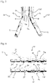
- Figures 4 to 6 show how movement of the tool 11 is controlled using tendons 13.
- the end-effector 10 comprises a plurality of tendons 13.
- the tendons 13 are connected to the bearing stud 14.
- the tendons 13 are configured to control movement of the tool 11 in at least two degrees of freedom.
- the invention employs a tendon-driven mechanism.
- the end-effector By using the tendons 13 to drive the robot, the end-effector has better distal-tip dexterity and force sensing capabilities than concentric-tube robots. Furthermore, breakage and fatigue are more limited.
- FIG. 4 to 6 shows how tendons 13 are used to control movement of the tool 11 in a different degree of freedom.
- Figures 4 and 5 show movement of the tool 11 in degrees of freedom through movement of the ball joint 12.
- Figure 6 shows a further degree of freedom associated with relative movement between the upper part 31 and bottom part 32 of the gripper 11a.
- the arrows show the movement of the tendons 13.
- Figure 2 shows the frames of the degrees of freedom of movement of the tool 11 on the end-effector 10.
- the first two degrees of freedom shown in Figures 4 and 5 have frames located on the middle point at the base of the ball joint 12. These are represented by the axes with subscripts 0 and 1.
- the frame of a third degree of freedom is located on the axis of rotation of the gripper 11a and is shown in Figure 2 with the subscript 2.
- the frame with the axes x 3 , y 3 and z 3 represents movement of the tip of the tool 11.
- the maximum distance that the tip of the tool 11 can cover while being constrained to a fixed point is at least about 10mm and optionally at least about 20mm in each of the y and z axes
- each tendon 13a-d extends through the facing surface 15 to reach the bearing stud 14.
- each tendon 13a-d extends through the bearing stud 14.
- the tendons 13 may pass around the outside of the bearing stud 14.
- Each tendon 13a-d is connected to the main body 18 that comprises the bearing stud 14.
- Figure 4 shows movement of the tool 11 in a degree of freedom which may be referred to as the up-down movement of the tool 11. As shown in Figure 4 , this movement is controlled by tendons 13a, 13b.
- the tendons 13a, 13b terminate at different positions.
- the tendons 13a, 13b shown in Figure 4 actuate the up-down degree of freedom.
- the tendons 13a, 13b terminate at a distal surface 20 (shown in Figure 9 ) of the main body 18 that comprises the bearing stud 14. It is not essential for the tendons 13a, 13b to terminate at the distal surface 20.
- the tendons 13a, 13b may terminate within the channels 19 that extend through the bearing stud 14.
- Figures 23 and 24 show different views of an alternative version of the main body 18 that comprises the bearing stud 14.
- Figures 25 and 26 show different views of a further alternative version of the main body 18 that comprises the bearing stud 14.
- the tendons 13 have termination points 50 at the perimeter wall 21 of the main body 18.
- the tendons 13 may be fixed to the perimeter wall 21 rather than to the bearing stud 14.
- the tendons 13 may extend from the termination points 50 to the exterior of the main body 18.
- the termination points 50 may be formed as holes in the perimeter wall 21 into which the tendons 13 are fixed.
- the manner in which the tendons 13a-d are fixed to their corresponding termination positions is not particularly limited.
- the tendons 13 may have their loose ends tied in a knot (e.g. an eight-figure knot).
- a piece of tubing may be crimped at the end of the tendon 13.
- the tendons 13a, 13b function in an antagonistic fashion. This means that when one of the tendons 13a pulls, the other tendon 13b has released tension (lets go). This means that the tool 11 has a two-way actuation capability (e.g. up-down in the degree of freedom shown in Figure 4 ).
- the top picture shown in Figure 4 shows a situation in which one tendon 13a is pulled and the other tendon 13b is released. This causes the tool 11 to move upwards (in the orientation shown in the Figures).
- the bottom picture of Figure 4 shows the opposite situation in which one tendon 13a is released and the other tendon 13 is pulled. This cause the tool 11 to move downwards.
- the tendons 13 are maintained in tension so as to hold the bearing stud 14 to the facing surface 15.
- the tendons in Figure 4 have termination points at different positions in the up-down direction.
- the tendons 13a, 13b have termination points that are at the same position in the left-right direction.
- the tendons 13a, 13b have termination points that are at the same position in the axial direction.
- Figure 5 shows how two other tendons 13c, 13d work in an antagonistic fashion to control movement of the tool 11.
- the tendons 13c, 13d control the left-right degree of freedom.
- one tendon 13c is pulled and the other tendon 13d is released, resulting in the tool 11 moving right (when looking along the longitudinal direction of the end-effector 10 from the proximal end to the distal end where the tool 11 is positioned).
- the right hand side picture of Figure 5 one tendon 13c is released and the other tendon 13d is pulled. This results in the tool 11 moving to the left.
- the tendons 13c, 13d have termination positions at different points of the left-right direction.
- the tendons 13c, 13d have termination points that are at the same position in the up-down direction.
- the tendons 13c, 13d have termination points that are at the same position in the axial direction.
- the ball joint 12 allows movement of the tool 11 in two degrees of freedom.
- the ball joint 12 rotates over two axes only, the pitch and yaw axes.
- the ball joint 12 is not required to rotate over the roll axis.
- the tendons 13a-d and antagonistic motion constrain rotation of the ball joint 12 over the roll axis.
- the roll motion, as well as the translation of the tool 11, are carried out by a robot arm on which the instrument is mounted.
- the roll motion and/or the translation of the tool 11 may be carried out by the arm of a surgeon performing the procedure.
- the end-effector 10 comprises at least one further tendon 13e, 13f.
- the further tendons 13e, 13f may extend through a channel 19 in the bearing stud 14.
- the further tendons 13e, 13f are different from the other tendons 13a-d in that they are not fixed to the bearing stud 14.
- the further tendons 13e, 13f are connected to the tool 11 for controlling an action of the tool 11 in a further degree of freedom.
- Figure 6 shows the example of a gripper 11a being used as a tool 11.
- the tendons 13e, 13f have termination points 34, 35 on the upper part 31 of the gripper 11a.
- the bottom part 32 of the gripper 11a is coupled with the main body 18 which comprises the bearing stud 14.
- the further tendons 13e, 13f work in an antagonistic fashion.
- one tendon 13e pulls and the other tendon 13f lets go, resulting in the gripper 11a opening (i.e. the distal portion of the upper part 31 moves away from the bottom part 32).
- the gripper 12a closing i.e. the distal portion of the upper part 31 moving towards the bottom part 32.
- the gripper 11a is closed, the distal portion of the upper part 31 comes into contact with the lower part 32.
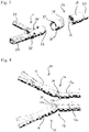
- Figure 3 shows an exploded view of the end-effector 10.
- the end-effector 10 comprises a main body 18 that comprises a bearing stud 14.
- the main body 18 may be formed as a unitary piece of material.
- the bearing stud 14 forms a solid mass of material.
- the bearing stud 14 is a unitary mass of material. This means that it is formed as a single piece, rather than a plurality of components that are later connected together.
- the bearing stud 14 comprises a plurality of channels 19 for accommodating the tendons 13. Apart from the channels 19, the bearing stud 14 is a substantially solid piece of material without other significant holes.
- the bearing stud 14 is relatively dense, having a relatively large amount of material in a relatively small volume.
- the bearing stud 14 is robust and capable of being used to impart larger forces at the tip of the end-effector 10. In contrast, a concentric tube robot cannot apply as much force and is less robust.
- the end-effector 10 By providing the end-effector 10 with the ball joint 12 for allowing movement of the tool 11, the end-effector 10 is more robust and can apply a greater force compared to known devices. Being able to apply a greater force is particularly useful for manipulating tissue with the tool 11 and/or for cutting bone.
- a bone punch 11b is an example of a tool 11 which may be used to cut bone.
- the bearing stud 14 faces the facing surface 15.
- the tendons 13 are configured to constrain the axial position of the bearing stud 14 relative to the surface 15 facing the bearing stud 14.
- the tendons 13 are kept in tension during use. This means that it is not necessary to encapsulate the bearing stud 14. It is not necessary to surround the bearing stud 14 in the radial direction, which helps to reduce the overall diameter of the end-effector 10. This allows the end-effector 10 to be used in smaller spaces, e.g. for endonasal neural surgery.
- the surface 15 facing the bearing stud 14 is coated with a coating configured to affect friction between the proximal end of the bearing stud 14 and the surface 15 facing the bearing stud 14.
- the proximal end of the bearing stud 14 is coated with a coating configured to affect friction between the proximal end of the bearing stud 14 and the surface 15 facing the bearing stud 14.
- the end-effector 10 comprises a frictional component between the surface 15 and the bearing stud 14 so as to affect the effective friction between the bearing stud 14 and the surface 15.
- a frictional component is not shown in the Figures.
- a component may be inserted between the bearing stud 14 and the surface 15 so as to reduce or increase the friction that there would otherwise be if the component were not provided.
- the frictional component may be used to control the level of friction.
- the coating or component can be chosen to control the amount of friction between the bearing stud 14 and the surface 15. Different levels of friction may be desirable for different uses of the end-effector 10 (e.g. when different levels of precision are required). It is also helpful to be able to control the level of friction in order to control the life-time of the end-effector 10.
- the end-effector 10 comprises a base 16.
- the base 16 is formed as a separate component from the main body 18 that comprises the bearing stud 14.
- the tool 11 is manufactured initially as a separate component from the main body 18. The tool 11 may then be connected to the main body 18 during the manufacturing process.
- the tool 11 comprises a connection element 33 that connects the tool 11 to the main body 18.
- the tool 11 is fixedly connected to the main body 18.
- the tool 11 comprises parts that move relative to each other.
- the upper part 31 is formed as a separate component from the bottom part 32.
- the upper part 31 is connected to the bottom part 32 in a way that allows the distal position of the upper part 31 to move relative to the bottom part 32.
- the proximal portion of the upper part 31 is connected via a hinge to the bottom part 32.
- the end-effector 10 is attached to a shaft 23.
- the shaft 23 has an internal channel through which the tendons 13 extend.
- the shaft 23 extends from the end-effector 10 to the other end of the tendons 13 where they are coupled to the controls of the instrument.
- Figure 7 is a close up view of the base 16 when viewed from the distal end.
- the surface 15 that faces the bearing stud 14 is provided at the distal end of the base 16 of the end-effector 10. Accordingly, the facing surface 15 can be seen in Figure 7 .
- the facing surface 15 comprises a plurality of channels 17 through which the tendons 13 extend.
- a channel 17 is provided for each respective tendon 13. The correspondence between the channels 17 and the tendons 13 is indicated by the letters of the reference numerals.
- the facing surface 15 is concave.
- Figure 8 is a close up view of the main body 18 when viewed from the proximal end.
- the bearing stud 14 is at the proximal end of the main body 18. Accordingly, the proximal surface of the bearing stud 14 can be seen in Figure 8 .
- the bearing stud 14 comprises a plurality of channels 19 through which the tendons 13 extend from a proximal end of the bearing stud 14 to a distal end of the bearing stud 14.
- a channel 19 is provided from each respected tendon 13. This is indicated by the letters used in the reference numerals.
- the channels 17, 19 reduce the possibility of the tendons 13 tangling during use.
- the robot has six channels that tendons 13 pass through.
- four of the tendons 13a-d are diametrically positioned on the same radius from the centre.
- the centres of the channels 17a-d, 19a-d are positioned at least 0.5mm from the central axis of the end-effector.
- the centres of the channels 17a-d, 19a-d are positioned at most 2mm from the central axis of the end-effector.
- the centres of the channels 17a-d, 19a-d are positioned about 1mm from the central axis of the end-effector.
- the four tendons 13a-d terminate on the main body 18 (e.g. on the distal end of the bearing stud 14 or at the perimeter wall 21) and control the two first degrees of freedom.
- the remaining two tendons 13e, 13f are for controlling the degree of freedom of the gripper 11a.
- the centres of the channels 17e-f, 19e-f are positioned at least 0.1mm from the central axis of the end-effector.
- the centres of the channels 17e-f, 19e-f are positioned at most 0.5mm from the central axis of the end-effector.
- the centres of the channels 17e-f, 19e-f are positioned about 0.35mm from the central axis of the end-effector.
- the tendons 13e, 13f terminate on the attachments at the upper part 31 of the gripper 11a.
- the difference between the lengths of the tendon 13 on the current joint-space and the desired joint-space is turned into the angle by which the respective motor should rotate in position control.
- Figure 9 is a close up view of the main body 18 when viewed from the distal end. Accordingly, a distal end surface 20 of the bearing stud 14 can be seen. Additionally, a perimeter wall 21 (also indicated in Figure 3 ) is visible. Optionally, the perimeter wall 21 extends distally beyond the bearing stud 14. The perimeter wall 21 may be formed unitarily with the bearing stud 14. In an alternative embodiment, the main body 18 is not provided with such a perimeter wall 21. In such an embodiment, the main body 18 may be formed simply of the bearing stud 14.
- the channels 19 extend from the proximal end of the bearing stud 14 (shown in Figure 8 ) to the distal end of the bearing stud 14 (shown in Figure 9 ).
- the channels 19a-d have a larger dimension at the proximal end of the bearing stud 14 compared to at the distal end of the bearing stud 14.
- the channels 19a-d taper towards a distal end of the bearing stud 14.
- the channels 19a-d may be substantially circular in cross section.
- the channels 19a-d may extend further in the radial direction of the bearing stud 14.
- the extra size of the channels 19a-d at the proximal end of the bearing stud 14 may be achieved by providing an additional hole (e.g.
- the tendons 13a-d can move more easily without bending at extreme angles. This improves the precision with which the movements of the tool 11 can be controlled using the tendons 13a-d.
- Figures 10-16 show alternative arrangements of the tendons 13 within the channels 17, 19.
- Figures 10-16 are close up views of the base 16 showing the surface 15 that faces the bearing stud 14.
- the pattern of channels 17 shown in Figures 10-16 is equally applicable to the channels 19 that extends through the bearing stud 14.
- FIG. 10 shows an example of which 2 channels 17a, 17e are joined together to form a common channel 22.
- two further channels 17b. 17f are joined together to form another common channel 22.
- the further tendons 13e. 13f and their respect channels 17e, 17f, 19e, 19f may not be provided.
- the channels 17e, 17f, 19e, 19f may be provided so that the same main body 18 and base 16 can be used also for tools 11 requiring a further control tendons 13e, 13f.
- the tool 11 could be detached from the main body 18 and replaced with another tool 11 that requires a different number of tendons 13 for control.
- Figure 11 shows an example in which the additional channels 17e, 17f for the further tendons 13e, 13f that control an action of the tool 11 are joined to form a common channel 22.
- the common channel 22 may have a Figure of 8 shape.
- the channels 17e, 17f, 19e, 19f for the further tendons 13e, 13f align in cross-section with a direction of movement of the further degree of movement.
- the further degree of freedom is in the up-down direction. This is because the further degree of freedom is movement of the upper part 31 if the gripper 11a in the up-down direction.
- the further channels 17e, 17f are arranged in the up-down direction.
- the further channels 17e, 17f are aligned with a direction of movement of the left-right degree of freedom. This is also shown in Figures 14 and 15 .
- the further channels 17e, 17f align in cross-section with a direction of movement of one of the two degrees of freedom enabled by the ball joint 12.
- one of the two degrees of freedom enabled by the ball joint 12 aligns with the further degree of freedom associated with the action of the tool 11.
- the action of the tool 11 could be offset from the two degrees of freedom associated with the ball joint 12.
- the channels 17e, 17f are aligned diagonally in the cross-sectional view. This may be particularly appropriate when the further degree of freedom associated with the action of the tool 11 is aligned in the diagonal direction.
- the further channels 17e, 17f may be provided in the diagonal direction (as shown in Figures 12 and 16 ) so as to increase a distance between the edges of the channels 17.
- the channels 17c, 17d associated with the left-right degree of freedom join with the further channels 17e, 17f to form joint channels 22.
- Figures 23 and 24 show different views of an alternative version of the main body 18 that comprises the bearing stud 14.
- Figure 23 is a perspective view.
- Figure 24 is a view from the distal side of the main body 18.
- the channels 19a-d continue through the perimeter wall 21 of the main body. This allows the tendons 13a-d to continue around the outside of the main body 18.
- the tendons 13a-d may be fixed at one end to the perimeter wall 21 at the termination points 50.
- Figures 25 and 26 show different views of a further alternative version of the main body 18 that comprises the bearing stud 14.
- Figure 25 is a perspective view.
- Figure 26 is a view from the distal side of the main body 18.
- the main body 18 is not provided with the channels for the tendons 13a-d.
- the tendons 13a-d extend around the outside of the bearing stud 14. This simplifies the design of the bearing stud 14.
- the tendons 13a-d may be fixed at one end to the perimeter wall 21 at the termination points 50.
- the channels 17, 19 have an internal width of at least 1 and a half times a cross-section dimension of the tendons 13. This reduces the possibility of the tendons 13 getting stuck within the channels 17, 19, thereby restricting their axial movement.
- the channels 17, 19 have an internal width at least two times the cross-sectional dimension of the tendons 13.
- the end-effector 10 has a diameter such that it can fit through a nostril.
- the end-effector 10 has a diameter such that two or three such end-effectors 10 can fit through the same nostril simultaneously.
- the end-effector 10 has a maximum external diameter of at most about 7mm and optionally at most about 5mm.
- the end-effector 10 has a maximum external diameter of about 3.6mm.
- the end-effector 10 has a length of at most about 20mm.
- the end-effector 10 is made of medical grade steel.
- the tendons 13 are made of stainless steel.
- the tool 11, main body 18 and base 16 may be made of plastic.
- the tendons 13 are made of tungsten.
- the tendons 13 have a diameter of at most about 0.5mm and optionally at most about 0.2mm.
- the channels 17, 19 have a diameter of at most about 1mm and optionally at most about 0.5mm.
- the channels 17, 19 have a diameter of at least about 0.2mm and optionally at least about 0.5mm.
- the end-effector 10 is fixed on the shaft 23.
- the shaft 23 may be a stainless steel grade 316 seamless tube shaft.
- the shaft 23 has an outer diameter of at most 7mm, and optionally at most 5mm.
- the shaft 23 has an outer diameter of 3mm.
- the shaft 23 comprises wire-guiding tubes through which the tendons 13 pass.
- the wire-guiding tubes have a diameter of about 0.5mm
- the bearing stud 14 comprises a flat surface 20 at its distal end.
- the tendons 13 are attached at the flat surface 30.
- the bearing stud 14 has a substantially hemispherical shape, with a convex surface at its proximal end (facing the facing surface 15) and a flat surface 20 at its distal end.
- Figure 17 shows an alternative version of the main body 18 in which the bearing stud 14 has a spherical shape.
- the distal end surface 20 of the bearing stud 14 is convex.
- Figure 18 shows an alternative version of the main body 18 in which the channels 19 have a consistent cross-section or width from the proximal end to the distal end of the bearing stud 14.
- Figure 18 shows that the additional holes that can be seen in Figure 8 (and can also be seen in Figures 1 to 6 ) are not provided.
- the base 16 is attached to a shaft 23 through which the tendons 13 extend.
- Figures 19 and 20 show alternative ways of connecting the base 16 of the end-effector 10 to the shaft 23.
- the base 16 comprises a flange 24 at its proximal end. The flange 24 fits around the shaft 23 so as to connect the base 16 to the shaft 23.
- the connection between the end-effector 10 and the shaft 23 can be undone without breaking the end-effector 10. This allows the end-effector 10 to be replaced (e.g. if a different tool 11 is required).
- the connection between the end-effector 10 and the shaft 23 is permanent (e.g. by welding) such that the end-effector 10 cannot be replaced without damaging the end-effector 10 and/or the shaft 23.
- Figure 20 shows an alternative version in which the base 16 comprises a protruding part 25 that extends within the inner channel of the shaft 23.
- the channels 17 extend all the way through the base 16 including the protruding part 25.
- the overall diameter of the protruding part 25 may be so small that the channels 17a-d are exposed to the internal wall of the shaft 23.
- the base 16 is fixed directly onto the shaft 23 without the need for the protruding part 25.
- the base 16 is welded (e.g. laser welded) onto the shaft 23.
- tool 11 can be used with the end-effector 10.
- one tool 11 can be replaced with another without replacing the rest of the end-effector 10 (e.g. the main body 18 and the base 16).
- the whole end-effector 10 is replaced when it is desired to use a different tool 11.
- the left-most tool 11 show in Figure 21 is a gripper 11a as shown in Figures 1-6 .
- the next tool 11 is a bone punch 11b.
- the bone punch 11b has a moving part.
- the bone punch 11b requires relative axial movement between an upper part 36 and a bottom part 37.
- the bone punch 11b is configured to cut bone.
- the relative movement between the upper part 36 and the bottom part 37 of the bone punch 11b can be actuated by use of the further tendons 13e, 13f.
- the bone punch 11b is spring-loaded.
- the middle tool 11 shown in Figure 21 is a flat-long spatula dissector 11c.
- the fourth tool 11 shown in Figure 21 is a round spatula dissector 11d.
- the right-most tool shown in Figure 21 is a ring curette 11e.
- the tendons 13 extend from their termination points (at their distal end) through the shaft 23 to their proximal end. At their proximal end, the tendons 13 are attached so that they can be controlled by motors.
- Figure 22 shows a rerouting mechanism 40 for the tendons 13 at their proximal end.
- the rerouting mechanism 40 comprises a housing 44.
- the housing 44 is produced via additive manufacturing.
- the housing 44 is made of polylactic acid (PLA) plastic.
- the housing 44 comprises a connection point 41 for connecting to the shaft 23.
- the tendons 13 extend through the shaft 23 and through the connection point 41 into the rerouting mechanism 40.
- the rerouting mechanism 40 comprises a plurality of capstans to which the tendons 13 are connected.
- the capstans are connected with stainless steel rods that are then connected to motors for actuating the tendons 13.
- the rerouting mechanism 40 comprises a plurality of pulleys 43.
- the pulleys 43 allow the tendons to be rerouted at different axial positions of the capstans 42.
- the end-effector 10 of the invention can be applied in various different types of surgery.
- the end-effector is suitable for use in neurosurgery.
- the invention is not limited to neurosurgery.
- the end-effector 10 can be used in other types of surgery, such as spinal surgery.
- the invention is described above mainly in the context of an end-effector 10 with six tendons 13.
- a different number of tendons 13 may be provided.
- the end-effector 10 may comprise only four tendons for controlling the two degrees of freedom.
- the end-effector 10 comprises one tendon 13 per degree of freedom.
- the tendon 13 may be looped (e.g. around a capstan), with both ends of the tendon 13 attached at different termination points for controlling movement in the degree of freedom.
- the end-effector 10 may comprise eight tendons 13 (two pairs for the up-down and left-right degrees of freedom and a pair for each part of the gripper 11a).
- the number of tendons 13 is not particularly limited.
- the end-effector 10 is manufactured by additive manufacturing, for example stereolithography, or direct laser metal sintering.
- the printing material is a medical grade steel or an autoclavable resin (of the type used in fluidics, optics and mold-making).
Abstract
An end-effector (10) for an endoscopic surgical instrument, the end-effector comprising:
a tool (11) configured to interact with tissue;
a main body (18) comprising a bearing stud (14), the tool being connected to the bearing stud;
a base (16) comprising a surface (15) facing the bearing stud, the bearing stud and the surface forming a ball joint (12); and
a plurality of tendons connected to the main body so as to control movement of the tool in two degrees of freedom.
Description
- This invention relates generally to endoscopic surgery, for example endonasal neurosurgery. More particularly the present invention relates to an end-effector for an endoscopic surgical instrument, an endoscopic surgical instrument comprising the end-effector and a method of manufacturing an end-effector for an endoscopic surgical instrument.
- Since the early 1980s when it was first introduced, Minimally Invasive Surgery (MIS) has had a great deal of success. Compared to traditional surgery, it requires smaller incisions, which equates to less trauma and thus reduced pain and hospital time, making MIS the standard and established procedure in a number of operations, such as laparoscopic surgery. Albeit their numerous advantages, MIS procedures are ergonomically difficult to perform due to the use of rigid instruments, visuomotor axes misalignment, limited sensory feedback and the need for high dexterity. Those drawbacks led to the development of robotic surgical devices that are now causing a paradigm shift in surgery.
- Robotic-Assisted Minimally Invasive Surgery (RAMIS) has had a great impact since it allows for precise and accurate motions while reducing the learning curve for the surgeons. This means that more surgeons can perform RAMIS when they might otherwise resort to open surgery. With the introduction of robotics into the surgical theatre, a number of conventional specialties, such as urology, gynaecology, abdominal and cardiothoracic surgery, have integrated current robotic technologies into their procedures augmenting the capabilities of the surgeon while improving patient outcomes. Lately, an increasing amount of surgical procedures have deployed or started deploying robotic devices, with neurosurgery being at the forefront of these advancing technologies.
- Due to its delicate subject matter and challenging operations, neurosurgery has been always in need for adapting new techniques and technologies. One such adaptation is surgical robotics, both in brain and spine applications. Although the majority of neurosurgical robots are stereotactic, technological advances in image guidance, endoscopy and laparoscopic instruments have led into the development of robotic tools for minimally invasive neurosurgery. However, the use of robotics in 'keyhole' neurosurgical approaches is still rather limited.
- A number of studies have taken place implementing concentric tube robotic tools. In Burgner, J., Swaney, P.J., Rucker, D.C., Gilbert, H.B., Nill, S.T., Russell, P.T., Weaver, K.D., Webster, R.J.: "A bimanual teleoperated system for endonasal skull base surgery." (In: 2011 IEEE/RSJ international conference on intelligent robots and systems, pp. 2517-2523. IEEE (2011)), a prototype system for bimanual teleoperated endonasal skull base surgery is developed. However, there are still concerns about the distal-end dexterity of this manipulator and its grasping force and/or force-sensing capability.
- It is desirable to provide an end-effector for an endoscopic surgical instrument capable of applying a greater force and/or that is more robust and/or that is capable of more dextrous manipulation of tissue and/or at a smaller size than known devices.
- The present invention attempts to address this desire by providing an end-effector for an endoscopic surgical instrument, an endoscopic surgical instrument comprising the end-effector and a method of manufacturing an end-effector for an endoscopic surgical instrument.
- According to the invention there is provided an end-effector for an endoscopic surgical instrument, the end-effector comprising: a tool configured to interact with tissue; a main body comprising a bearing stud, the tool being connected to the bearing stud; a base comprising a surface facing the bearing stud, the bearing stud and the surface forming a ball joint; and a plurality of tendons connected to the main body so as to control movement of the tool in two degrees of freedom.
- According to the invention there is provided an endoscopic surgical instrument comprising the end-effector.
- According to the invention, there is provided a method of manufacturing an end-effector for an endoscopic surgical instrument, the method comprising: providing a tool configured to interact with tissue; connecting the tool to a main body comprising a bearing stud of a ball joint; and connecting a plurality of tendons to the main body to allow controlled movement of the tool in two degrees of freedom..
- The present invention will now be described, by the way of non-limitative example only, with reference to the accompanying drawings in which:
-
Figure 1 shows an end-effector for an endoscopic surgical instrument according to the present invention; -
Figure 2 shows the axes of degrees of freedom of the end-effector shown inFigure 1 ; -
Figure 3 is an exploded view of the end-effector shown inFigure 1 ; -
Figures 4 to 6 each show how the tendons control a respective degree of freedom of movement of the end-effector; -
Figure 7 shows the surface of the base of the end-effector that faces the bearing stud; -
Figure 8 shows the proximal end of the bearing stud that faces the surface shown inFigure 7 ; -
Figure 9 shows the distal surface of the bearing stud that faces the tool of the end-effector; -
Figures 10 to 16 show alternative arrangements of the channels for the tendons; -
Figures 17 and 18 show variations of the bearing stud; -
Figures 19 and 20 show alternative ways of the base of the end-effector connecting to the shaft; -
Figure 21 shows alternative tools for the end-effector; -
Figure 22 shows a rerouting mechanism for connecting the tendons to motors; -
Figures 23 and 24 show different views of an alternative version of the main body of the end-effector that comprises the bearing stud; and -
Figures 25 and 26 show different views of a further alternative version of the main body of the end-effector. -
Figure 1 shows an end-effector 10. The end-effector 10 is for an endoscopic surgical instrument. As shown inFigure 1 , the end-effector 10 comprises atool 11. Thetool 11 forms the tip of the end-effector 10. Thetool 11 is configured to interact with tissue. For example, thetool 11 may be configured to grip, pierce, move, cut and/or dissect tissue during a surgical procedure. In the example shown inFigure 1 , thetool 11 is a gripper 11a. The gripper 11a is configured to grip tissue. Other types of tools are compatible with the end-effector 10, as shown inFigure 21 , for example. - As shown in
Figure 1 , the end-effector 10 comprises aball joint 12. Theball joint 12 comprises abearing stud 14 and asurface 15 facing thebearing stud 14. Theball joint 12 is configured to connect the controls of the endoscopic surgical instrument to thetool 11. As shown inFigure 1 , in theball joint 12 of the end-effector 10, it is not necessary for thebearing stud 14 to be enclosed in a casing. Instead, thebearing stud 14 may be held relative to the facingsurface 15 only by the tendons 13, which are described in more detail below. Theball joint 12 allows movement of the tool 11 (e.g. changes in orientation) relative to the facingsurface 15. - As shown in
Figure 1 , thetool 11 is connected to thebearing stud 14. For example, the tool 11 (or at least a part of it) may be fixed relative to thebearing stud 14. As mentioned above,Figure 1 shows an example in which thetool 11 is a gripper 11a. The gripper 11a may comprise anupper part 31 and abottom part 32. Theupper part 31 and thebottom part 32 can be moved relative to each other. This allows the gripper 11a to grip tissue during use. Optionally, only one of theupper part 31 and thebottom part 32 is fixed relative to thebearing stud 14. For example,Figure 1 shows an embodiment in which thebottom part 32 is connected to thebearing stud 14. Theupper part 31 is connected to thebearing stud 14 only via thebottom part 32 of the gripper 11a. By providing that one of theupper part 31 and thebottom part 32 is fixed relative to thebearing stud 14, only the other (non-fixed) part is required to be controlled in order to fully activate the gripper 11a. However, this is not necessarily the case. In an alternative embodiment, both of theupper part 31 and thebottom part 32 are hinged relative to thebearing stud 14 and can be actuated independently of each other, or in unison. - By connecting the
tool 11 to thebearing stud 14, movement of the bearingstud 14 relative to the facingsurface 15 results in movement of thetool 11 relative to the facingsurface 15. By controlling the ball joint 12, the movement of thetool 11 at the tip of the end-effector 10 can be controlled in two degrees of freedom. -
Figures 4 to 6 show how movement of thetool 11 is controlled using tendons 13. As shown inFigures 4 to 6 , the end-effector 10 comprises a plurality of tendons 13. The tendons 13 are connected to thebearing stud 14. The tendons 13 are configured to control movement of thetool 11 in at least two degrees of freedom. - The invention employs a tendon-driven mechanism. By using the tendons 13 to drive the robot, the end-effector has better distal-tip dexterity and force sensing capabilities than concentric-tube robots. Furthermore, breakage and fatigue are more limited.
- Each of
Figures 4 to 6 shows how tendons 13 are used to control movement of thetool 11 in a different degree of freedom.Figures 4 and5 show movement of thetool 11 in degrees of freedom through movement of the ball joint 12.Figure 6 shows a further degree of freedom associated with relative movement between theupper part 31 andbottom part 32 of the gripper 11a. InFigures 4 to 6 the arrows show the movement of the tendons 13. -
Figure 2 shows the frames of the degrees of freedom of movement of thetool 11 on the end-effector 10. The first two degrees of freedom shown inFigures 4 and5 have frames located on the middle point at the base of the ball joint 12. These are represented by the axes with subscripts 0 and 1. The frame of a third degree of freedom is located on the axis of rotation of the gripper 11a and is shown inFigure 2 with the subscript 2. InFigure 2 , the frame with the axes x3, y3 and z3 represents movement of the tip of thetool 11. Optionally, the maximum distance that the tip of thetool 11 can cover while being constrained to a fixed point, which is the origin of frames 0 and 1 as shown inFigure 2 , is at least about 10mm and optionally at least about 20mm in each of the y and z axes - Optionally, each
tendon 13a-d extends through the facingsurface 15 to reach thebearing stud 14. As indicated inFigure 4 , optionally eachtendon 13a-d extends through the bearingstud 14. Alternatively, the tendons 13 may pass around the outside of the bearingstud 14. Eachtendon 13a-d is connected to themain body 18 that comprises the bearingstud 14. -
Figure 4 shows movement of thetool 11 in a degree of freedom which may be referred to as the up-down movement of thetool 11. As shown inFigure 4 , this movement is controlled bytendons tendons tendons Figure 4 actuate the up-down degree of freedom. Optionally, thetendons Figure 9 ) of themain body 18 that comprises the bearingstud 14. It is not essential for thetendons distal surface 20. For example, thetendons channels 19 that extend through the bearingstud 14. -
Figures 23 and 24 show different views of an alternative version of themain body 18 that comprises the bearingstud 14.Figures 25 and 26 show different views of a further alternative version of themain body 18 that comprises the bearingstud 14. As shown in the versions shown inFigures 23 to 26 , optionally the tendons 13 havetermination points 50 at theperimeter wall 21 of themain body 18. The tendons 13 may be fixed to theperimeter wall 21 rather than to thebearing stud 14. The tendons 13 may extend from the termination points 50 to the exterior of themain body 18. The termination points 50 may be formed as holes in theperimeter wall 21 into which the tendons 13 are fixed. - The manner in which the
tendons 13a-d are fixed to their corresponding termination positions is not particularly limited. As one example, the tendons 13 may have their loose ends tied in a knot (e.g. an eight-figure knot). Alternatively, a piece of tubing may be crimped at the end of the tendon 13. - As shown in
Figure 4 , thetendons tendons 13a pulls, theother tendon 13b has released tension (lets go). This means that thetool 11 has a two-way actuation capability (e.g. up-down in the degree of freedom shown inFigure 4 ). The top picture shown inFigure 4 shows a situation in which onetendon 13a is pulled and theother tendon 13b is released. This causes thetool 11 to move upwards (in the orientation shown in the Figures). The bottom picture ofFigure 4 shows the opposite situation in which onetendon 13a is released and the other tendon 13 is pulled. This cause thetool 11 to move downwards. The tendons 13 are maintained in tension so as to hold thebearing stud 14 to the facingsurface 15. - As shown in
Figure 4 , the tendons inFigure 4 , thetendons tendons tendons -
Figure 5 shows how twoother tendons tool 11. Thetendons Figure 5 , onetendon 13c is pulled and theother tendon 13d is released, resulting in thetool 11 moving right (when looking along the longitudinal direction of the end-effector 10 from the proximal end to the distal end where thetool 11 is positioned). In the right hand side picture ofFigure 5 , onetendon 13c is released and theother tendon 13d is pulled. This results in thetool 11 moving to the left. - The
tendons tendons tendons - As illustrated in
Figures 4 and5 , the ball joint 12 allows movement of thetool 11 in two degrees of freedom. The ball joint 12 rotates over two axes only, the pitch and yaw axes. The ball joint 12 is not required to rotate over the roll axis. Optionally, thetendons 13a-d and antagonistic motion constrain rotation of the ball joint 12 over the roll axis. - Optionally, the roll motion, as well as the translation of the
tool 11, are carried out by a robot arm on which the instrument is mounted. Alternatively, the roll motion and/or the translation of thetool 11 may be carried out by the arm of a surgeon performing the procedure. - As shown in
Figures 6 , optionally, the end-effector 10 comprises at least onefurther tendon further tendons channel 19 in thebearing stud 14. Thefurther tendons other tendons 13a-d in that they are not fixed to thebearing stud 14. Thefurther tendons tool 11 for controlling an action of thetool 11 in a further degree of freedom. - For example,
Figure 6 shows the example of a gripper 11a being used as atool 11. Thetendons termination points upper part 31 of the gripper 11a. Thebottom part 32 of the gripper 11a is coupled with themain body 18 which comprises the bearingstud 14. Thefurther tendons Figure 6 , onetendon 13e pulls and theother tendon 13f lets go, resulting in the gripper 11a opening (i.e. the distal portion of theupper part 31 moves away from the bottom part 32). In the lower picture ofFigure 6 , onetendon 13e lets go and theother tendon 13f pulls. This results in the gripper 12a closing (i.e. the distal portion of theupper part 31 moving towards the bottom part 32). Optionally, when the gripper 11a is closed, the distal portion of theupper part 31 comes into contact with thelower part 32. -
Figure 3 shows an exploded view of the end-effector 10. As shown inFigure 3 , optionally the end-effector 10 comprises amain body 18 that comprises abearing stud 14. Themain body 18 may be formed as a unitary piece of material. The bearingstud 14 forms a solid mass of material. Optionally, the bearingstud 14 is a unitary mass of material. This means that it is formed as a single piece, rather than a plurality of components that are later connected together. As will be explained in further detail below, the bearingstud 14 comprises a plurality ofchannels 19 for accommodating the tendons 13. Apart from thechannels 19, the bearingstud 14 is a substantially solid piece of material without other significant holes. The bearingstud 14 is relatively dense, having a relatively large amount of material in a relatively small volume. The bearingstud 14 is robust and capable of being used to impart larger forces at the tip of the end-effector 10. In contrast, a concentric tube robot cannot apply as much force and is less robust. - By providing the end-
effector 10 with the ball joint 12 for allowing movement of thetool 11, the end-effector 10 is more robust and can apply a greater force compared to known devices. Being able to apply a greater force is particularly useful for manipulating tissue with thetool 11 and/or for cutting bone. For example, a bone punch 11b is an example of atool 11 which may be used to cut bone. - As shown in
Figures 1-3 , the bearingstud 14 faces the facingsurface 15. Optionally, only the tendons 13 are configured to constrain the axial position of the bearingstud 14 relative to thesurface 15 facing the bearingstud 14. The tendons 13 are kept in tension during use. This means that it is not necessary to encapsulate thebearing stud 14. It is not necessary to surround thebearing stud 14 in the radial direction, which helps to reduce the overall diameter of the end-effector 10. This allows the end-effector 10 to be used in smaller spaces, e.g. for endonasal neural surgery. - Optionally, the
surface 15 facing the bearingstud 14 is coated with a coating configured to affect friction between the proximal end of the bearingstud 14 and thesurface 15 facing the bearingstud 14. Additionally or alternatively, the proximal end of the bearingstud 14 is coated with a coating configured to affect friction between the proximal end of the bearingstud 14 and thesurface 15 facing the bearingstud 14. - Optionally, the end-
effector 10 comprises a frictional component between thesurface 15 and thebearing stud 14 so as to affect the effective friction between the bearingstud 14 and thesurface 15. Such a frictional component is not shown in the Figures. For example, a component may be inserted between the bearingstud 14 and thesurface 15 so as to reduce or increase the friction that there would otherwise be if the component were not provided. The frictional component may be used to control the level of friction. - The coating or component can be chosen to control the amount of friction between the bearing
stud 14 and thesurface 15. Different levels of friction may be desirable for different uses of the end-effector 10 (e.g. when different levels of precision are required). It is also helpful to be able to control the level of friction in order to control the life-time of the end-effector 10. - As show in
Figure 3 , optionally the end-effector 10 comprises abase 16. Thebase 16 is formed as a separate component from themain body 18 that comprises the bearingstud 14. As shown inFigure 3 , optionally thetool 11 is manufactured initially as a separate component from themain body 18. Thetool 11 may then be connected to themain body 18 during the manufacturing process. For example, as shown inFigure 3 optionally thetool 11 comprises aconnection element 33 that connects thetool 11 to themain body 18. Optionally, thetool 11 is fixedly connected to themain body 18. As shown in the example inFigure 3 , optionally, thetool 11 comprises parts that move relative to each other. Optionally, theupper part 31 is formed as a separate component from thebottom part 32. Theupper part 31 is connected to thebottom part 32 in a way that allows the distal position of theupper part 31 to move relative to thebottom part 32. As shown inFigure 3 , optionally the proximal portion of theupper part 31 is connected via a hinge to thebottom part 32. - As shown in
Figures 1-3 , optionally the end-effector 10 is attached to ashaft 23. Theshaft 23 has an internal channel through which the tendons 13 extend. Optionally, theshaft 23 extends from the end-effector 10 to the other end of the tendons 13 where they are coupled to the controls of the instrument. -
Figure 7 is a close up view of the base 16 when viewed from the distal end. Thesurface 15 that faces the bearingstud 14 is provided at the distal end of thebase 16 of the end-effector 10. Accordingly, the facingsurface 15 can be seen inFigure 7 . As shown inFigure 7 , optionally the facingsurface 15 comprises a plurality ofchannels 17 through which the tendons 13 extend. As shown inFigure 7 , optionally achannel 17 is provided for each respective tendon 13. The correspondence between thechannels 17 and the tendons 13 is indicated by the letters of the reference numerals. As can be seen inFigures 1 ,3 and7 , the facingsurface 15 is concave. -
Figure 8 is a close up view of themain body 18 when viewed from the proximal end. The bearingstud 14 is at the proximal end of themain body 18. Accordingly, the proximal surface of the bearingstud 14 can be seen inFigure 8 . As shown inFigure 8 , optionally the bearingstud 14 comprises a plurality ofchannels 19 through which the tendons 13 extend from a proximal end of the bearingstud 14 to a distal end of the bearingstud 14. As shown inFigure 8 , optionally achannel 19 is provided from each respected tendon 13. This is indicated by the letters used in the reference numerals. Thechannels - For each rotational degree of freedom there are two tendons 13 that control the movement of each angle in an agonist-antagonist fashion. The lengths of the tendons 13 are selected by locating the 3D positions of the pass-through
channels tendons 13a-d are diametrically positioned on the same radius from the centre. Optionally, the centres of thechannels 17a-d, 19a-d are positioned at least 0.5mm from the central axis of the end-effector. Optionally, the centres of thechannels 17a-d, 19a-d are positioned at most 2mm from the central axis of the end-effector. Optionally, the centres of thechannels 17a-d, 19a-d are positioned about 1mm from the central axis of the end-effector. - Optionally, the four
tendons 13a-d terminate on the main body 18 (e.g. on the distal end of the bearingstud 14 or at the perimeter wall 21) and control the two first degrees of freedom. The remaining twotendons channels 17e-f, 19e-f are positioned at least 0.1mm from the central axis of the end-effector. Optionally, the centres of thechannels 17e-f, 19e-f are positioned at most 0.5mm from the central axis of the end-effector. Optionally, the centres of thechannels 17e-f, 19e-f are positioned about 0.35mm from the central axis of the end-effector. Thetendons upper part 31 of the gripper 11a. - During use, the difference between the lengths of the tendon 13 on the current joint-space and the desired joint-space is turned into the angle by which the respective motor should rotate in position control.
-
Figure 9 is a close up view of themain body 18 when viewed from the distal end. Accordingly, adistal end surface 20 of the bearingstud 14 can be seen. Additionally, a perimeter wall 21 (also indicated inFigure 3 ) is visible. Optionally, theperimeter wall 21 extends distally beyond the bearingstud 14. Theperimeter wall 21 may be formed unitarily with the bearingstud 14. In an alternative embodiment, themain body 18 is not provided with such aperimeter wall 21. In such an embodiment, themain body 18 may be formed simply of the bearingstud 14. - As can be seen from
Figures 8 and 9 , thechannels 19 extend from the proximal end of the bearing stud 14 (shown inFigure 8 ) to the distal end of the bearing stud 14 (shown inFigure 9 ). - As can be seen from a comparison between
Figures 8 and 9 , thechannels 19a-d have a larger dimension at the proximal end of the bearingstud 14 compared to at the distal end of the bearingstud 14. Optionally, thechannels 19a-d taper towards a distal end of the bearingstud 14. As shown inFigure 9 , optionally at the distal end thechannels 19a-d may be substantially circular in cross section. In contrast, as shown inFigure 8 at the proximal end of the bearingstud 14, thechannels 19a-d may extend further in the radial direction of the bearingstud 14. For example, the extra size of thechannels 19a-d at the proximal end of the bearingstud 14 may be achieved by providing an additional hole (e.g. an additional cylindrical hole) that joins with the main hole in forming thechannels 19a-d. By providing that thechannels 19a-d get smaller towards to distal end of the bearingstud 14, thetendons 13a-d can move more easily without bending at extreme angles. This improves the precision with which the movements of thetool 11 can be controlled using thetendons 13a-d. -
Figures 10-16 show alternative arrangements of the tendons 13 within thechannels Figures 10-16 are close up views of the base 16 showing thesurface 15 that faces the bearingstud 14. However, the pattern ofchannels 17 shown inFigures 10-16 is equally applicable to thechannels 19 that extends through the bearingstud 14. - As shown in
Figures 10 ,11 ,14 ,15 and 16 , optionally at least one pair ofchannels 17 is joined to form acommon channel 22 for a respective pair of tendons 13. For example,Figure 10 shows an example of which 2channels common channel 22. Similarly, twofurther channels 17b. 17f are joined together to form anothercommon channel 22. By joiningchannels 17 together to form acommon channel 22, the amount of cross-sectional space required to accommodate thechannels 17 can be reduced. In particular, when thetool 11 is provided with moving parts requiring tendons 13 for their control, theextra channels common channels 22,. The amount of extra space in the cross-sectional area required to accommodate theextra channels - For
tools 11 that have no moving parts, then thefurther tendons 13e. 13f and theirrespect channels tool 11 is used that does not have moving parts (such that thefurther tendons channels main body 18 andbase 16 can be used also fortools 11 requiring afurther control tendons tool 11 could be detached from themain body 18 and replaced with anothertool 11 that requires a different number of tendons 13 for control. -
Figure 11 shows an example in which theadditional channels further tendons tool 11 are joined to form acommon channel 22. As shownFigures 10 and11 , for example, thecommon channel 22 may have a Figure of 8 shape. - As shown in
Figures 7-11 , optionally, thechannels further tendons Figures 1-3 , the further degree of freedom is in the up-down direction. This is because the further degree of freedom is movement of theupper part 31 if the gripper 11a in the up-down direction. Thefurther channels channels further tendons - However, it is not essential for the
further channels Figure 13 ,m in an alternative embodiment thefurther channels Figures 14 and15 . Optionally, thefurther channels Figures 1-3 , one of the two degrees of freedom enabled by the ball joint 12 aligns with the further degree of freedom associated with the action of thetool 11. However, this is not the necessarily the case. For example, the action of thetool 11 could be offset from the two degrees of freedom associated with the ball joint 12. - As shown in
Figures 12 and16 , optionally, thechannels tool 11 is aligned in the diagonal direction. Alternatively, thefurther channels Figures 12 and16 ) so as to increase a distance between the edges of thechannels 17. - As shown in
Figure 14 , optionally, thechannels further channels joint channels 22. -
Figures 23 and 24 show different views of an alternative version of themain body 18 that comprises the bearingstud 14.Figure 23 is a perspective view.Figure 24 is a view from the distal side of themain body 18. As shown inFigures 23 and 24 , optionally, thechannels 19a-d continue through theperimeter wall 21 of the main body. This allows thetendons 13a-d to continue around the outside of themain body 18. Thetendons 13a-d may be fixed at one end to theperimeter wall 21 at the termination points 50. -
Figures 25 and 26 show different views of a further alternative version of themain body 18 that comprises the bearingstud 14.Figure 25 is a perspective view.Figure 26 is a view from the distal side of themain body 18. As shown inFigures 25 and 26 , optionally, themain body 18 is not provided with the channels for thetendons 13a-d. Instead, thetendons 13a-d extend around the outside of the bearingstud 14. This simplifies the design of the bearingstud 14. Thetendons 13a-d may be fixed at one end to theperimeter wall 21 at the termination points 50. - Optionally, the
channels channels channels - Optionally the end-
effector 10 has a diameter such that it can fit through a nostril. Optionally the end-effector 10 has a diameter such that two or three such end-effectors 10 can fit through the same nostril simultaneously. Optionally, the end-effector 10 has a maximum external diameter of at most about 7mm and optionally at most about 5mm. Optionally, the end-effector 10 has a maximum external diameter of about 3.6mm. Optionally, the end-effector 10 has a length of at most about 20mm. - Optionally, the end-
effector 10 is made of medical grade steel. Optionally, the tendons 13 are made of stainless steel. However, other materials are possible. For example, thetool 11,main body 18 andbase 16 may be made of plastic. Optionally, the tendons 13 are made of tungsten. - Optionally, the tendons 13 have a diameter of at most about 0.5mm and optionally at most about 0.2mm. Optionally, the
channels channels - Optionally, the end-
effector 10 is fixed on theshaft 23. Theshaft 23 may be a stainless steel grade 316 seamless tube shaft. Optionally, theshaft 23 has an outer diameter of at most 7mm, and optionally at most 5mm. Optionally, theshaft 23 has an outer diameter of 3mm. Optionally, theshaft 23 comprises wire-guiding tubes through which the tendons 13 pass. Optionally, the wire-guiding tubes have a diameter of about 0.5mm - As shown in
Figure 9 , optionally, the bearingstud 14 comprises aflat surface 20 at its distal end. Optionally, the tendons 13 are attached at the flat surface 30. Optionally, the bearingstud 14 has a substantially hemispherical shape, with a convex surface at its proximal end (facing the facing surface 15) and aflat surface 20 at its distal end. By providing thebearing stud 14 as a substantially hemispherical object, the end-effector 10 is easier to manufacture. - However, it is not essential for the
bearing stud 14 to have a hemispherical shape.Figure 17 shows an alternative version of themain body 18 in which thebearing stud 14 has a spherical shape. Thedistal end surface 20 of the bearingstud 14 is convex. - As mentioned above, optionally the
channels 19 are wider at the proximal end of the bearingstud 14 compared to at the distal end of the bearingstud 14. However, this is not necessarily the case.Figure 18 shows an alternative version of themain body 18 in which thechannels 19 have a consistent cross-section or width from the proximal end to the distal end of the bearingstud 14. In particular,Figure 18 shows that the additional holes that can be seen inFigure 8 (and can also be seen inFigures 1 to 6 ) are not provided. - As shown in
Figures 1-3 , optionally thebase 16 is attached to ashaft 23 through which the tendons 13 extend.Figures 19 and 20 show alternative ways of connecting thebase 16 of the end-effector 10 to theshaft 23. As shown inFigure 19 , optionally thebase 16 comprises aflange 24 at its proximal end. Theflange 24 fits around theshaft 23 so as to connect the base 16 to theshaft 23. Optionally, the connection between the end-effector 10 and theshaft 23 can be undone without breaking the end-effector 10. This allows the end-effector 10 to be replaced (e.g. if adifferent tool 11 is required). Alternatively, the connection between the end-effector 10 and theshaft 23 is permanent (e.g. by welding) such that the end-effector 10 cannot be replaced without damaging the end-effector 10 and/or theshaft 23. -
Figure 20 shows an alternative version in which thebase 16 comprises a protrudingpart 25 that extends within the inner channel of theshaft 23. As can be seen inFigure 20 , optionally thechannels 17 extend all the way through the base 16 including the protrudingpart 25. The overall diameter of the protrudingpart 25 may be so small that thechannels 17a-d are exposed to the internal wall of theshaft 23. - In an alternative version, the
base 16 is fixed directly onto theshaft 23 without the need for the protrudingpart 25. For example, optionally thebase 16 is welded (e.g. laser welded) onto theshaft 23. - As mentioned above, different types of
tool 11 can be used with the end-effector 10. Optionally, onetool 11 can be replaced with another without replacing the rest of the end-effector 10 (e.g. themain body 18 and the base 16). Alternatively, it may be that the whole end-effector 10 is replaced when it is desired to use adifferent tool 11. - The
left-most tool 11 show inFigure 21 is a gripper 11a as shown inFigures 1-6 . Thenext tool 11 is a bone punch 11b. The bone punch 11b has a moving part. In particular, the bone punch 11b requires relative axial movement between anupper part 36 and abottom part 37. The bone punch 11b is configured to cut bone. The relative movement between theupper part 36 and thebottom part 37 of the bone punch 11b can be actuated by use of thefurther tendons middle tool 11 shown inFigure 21 is a flat-long spatula dissector 11c. Thefourth tool 11 shown inFigure 21 is a round spatula dissector 11d. The right-most tool shown inFigure 21 is a ring curette 11e. - The tendons 13 extend from their termination points (at their distal end) through the
shaft 23 to their proximal end. At their proximal end, the tendons 13 are attached so that they can be controlled by motors.Figure 22 shows arerouting mechanism 40 for the tendons 13 at their proximal end. - As shown in
Figure 22 , optionally, thererouting mechanism 40 comprises ahousing 44. Optionally, thehousing 44 is produced via additive manufacturing. Optionally, thehousing 44 is made of polylactic acid (PLA) plastic. Thehousing 44 comprises aconnection point 41 for connecting to theshaft 23. The tendons 13 extend through theshaft 23 and through theconnection point 41 into thererouting mechanism 40. - As shown in
Figure 22 , thererouting mechanism 40 comprises a plurality of capstans to which the tendons 13 are connected. The capstans, in turn, are connected with stainless steel rods that are then connected to motors for actuating the tendons 13. As shown inFigure 22 , optionally thererouting mechanism 40 comprises a plurality ofpulleys 43. Thepulleys 43 allow the tendons to be rerouted at different axial positions of thecapstans 42. - The end-
effector 10 of the invention can be applied in various different types of surgery. For example, the end-effector is suitable for use in neurosurgery. However, the invention is not limited to neurosurgery. The end-effector 10 can be used in other types of surgery, such as spinal surgery. - The invention is described above mainly in the context of an end-
effector 10 with six tendons 13. However, a different number of tendons 13 may be provided. For example, when thetool 11 does not require activation (e.g. when thetool 11 has no moving parts), then the end-effector 10 may comprise only four tendons for controlling the two degrees of freedom. Optionally, instead of two tendons 13 for each degree of freedom, the end-effector 10 comprises one tendon 13 per degree of freedom. For example, the tendon 13 may be looped (e.g. around a capstan), with both ends of the tendon 13 attached at different termination points for controlling movement in the degree of freedom. As another example, when a gripper 11a having both anupper part 31 and abottom part 32 independently actuatable, then the end-effector 10 may comprise eight tendons 13 (two pairs for the up-down and left-right degrees of freedom and a pair for each part of the gripper 11a). The number of tendons 13 is not particularly limited. - Optionally, the end-
effector 10 is manufactured by additive manufacturing, for example stereolithography, or direct laser metal sintering. Optionally the printing material is a medical grade steel or an autoclavable resin (of the type used in fluidics, optics and mold-making). - The Figures and previous description relate to preferred features by way of illustration only. It should be noted that alternative features of the structures and methods disclosed herein will be readily recognised as possible alternatives. The equivalent described above is by way of example only, and it will be appreciated that it may be modified in several different ways while remaining in the scope of the claims. Features of the different embodiments and arrangements may be combined with each other, except where these are mutually exclusive.
Claims (15)
- An end-effector for an endoscopic surgical instrument, the end-effector comprising:a tool configured to interact with tissue;a main body comprising a bearing stud, the tool being connected to the bearing stud;a base comprising a surface facing the bearing stud, the bearing stud and the surface forming a ball joint; anda plurality of tendons connected to the main body so as to control movement of the tool in two degrees of freedom.
- The end-effector of claim 1, wherein the bearing stud is a unitary mass of material.
- The end-effector of claim 1 or 2, wherein the surface facing the bearing stud and/or a proximal end of the bearing stud is coated with a coating configured to affect friction between the proximal end of the bearing stud and the surface facing the bearing stud.
- The end-effector of any preceding claim, comprising a frictional component between the surface facing the bearing stud and the bearing stud so as to affect effective friction between the bearing stud and the surface facing the bearing stud.
- The end-effector of any preceding claim, wherein only the tendons are configured to constrain an axial position of the bearing stud relative to the surface facing the bearing stud.
- The end-effector of any preceding claim, wherein the bearing stud comprises a plurality of channels through which the tendons extend from a proximal end of the bearing stud to a distal end of the bearing stud.
- The end-effector of claim 6, wherein the channels taper towards the distal end of the bearing stud.
- The end-effector of any preceding claim, comprising at least one further tendon extending through a channel in the bearing stud and connected to the tool for controlling an action of the tool in one further degree of freedom.
- The end-effector of claim 8, wherein at least two of said further tendons are provided, having respective channels that, in cross-section, align with a direction of movement of said one further degree of freedom and/or a direction of movement of one of said two degrees of freedom.
- The end-effector of any of claims 6 to 9, wherein at least one pair of the channels is joined to form a common channel for a respective pair of the tendons.
- The end-effector of any of claims 6 to 10, wherein the channels have an internal width at least one and a half times a cross-sectional dimension of the tendons.
- The end-effector of any preceding claim, wherein the main body comprises a perimeter wall extending distally relative to the bearing stud, wherein the tendons are attached to the perimeter wall.
- The end-effector of any preceding claim, wherein the bearing stud comprises a flat or convex surface at its distal end to which the tendons are attached.
- An endoscopic surgical instrument comprising the end-effector of any preceding claim.
- A method of manufacturing an end-effector for an endoscopic surgical instrument, the method comprising:providing a tool configured to interact with tissue;connecting the tool to a main body comprising a bearing stud of a ball joint; andconnecting a plurality of tendons to the main body to allow controlled movement of the tool in two degrees of freedom.
Priority Applications (7)
| Application Number | Priority Date | Filing Date | Title |
|---|---|---|---|
| EP20386011.9A EP3868305A1 (en) | 2020-02-19 | 2020-02-19 | End-effector for endoscopic surgical instrument |
| PCT/GB2021/050316 WO2021165647A1 (en) | 2020-02-19 | 2021-02-11 | End-effector for endoscopic surgical instrument |
| BR112022016462A BR112022016462A2 (en) | 2020-02-19 | 2021-02-11 | TERMINAL EFFECTOR FOR AN ENDOSCOPIC SURGICAL INSTRUMENT, ENDOSCOPIC SURGICAL INSTRUMENT AND MANUFACTURING METHOD OF A TERMINAL EFFECTOR FOR AN ENDOSCOPIC SURGICAL INSTRUMENT |
| JP2022549761A JP2023517285A (en) | 2020-02-19 | 2021-02-11 | End effector for endoscopic surgical instruments |
| CN202180021897.4A CN115297787A (en) | 2020-02-19 | 2021-02-11 | End effector for endoscopic surgical device |
| EP21705255.4A EP4106636A1 (en) | 2020-02-19 | 2021-02-11 | End-effector for endoscopic surgical instrument |
| US17/819,757 US20230068155A1 (en) | 2020-02-19 | 2022-08-15 | End-effector for endoscopic surgical instrument |
Applications Claiming Priority (1)
| Application Number | Priority Date | Filing Date | Title |
|---|---|---|---|
| EP20386011.9A EP3868305A1 (en) | 2020-02-19 | 2020-02-19 | End-effector for endoscopic surgical instrument |
Publications (1)
| Publication Number | Publication Date |
|---|---|
| EP3868305A1 true EP3868305A1 (en) | 2021-08-25 |
Family
ID=69780129
Family Applications (2)
| Application Number | Title | Priority Date | Filing Date |
|---|---|---|---|
| EP20386011.9A Withdrawn EP3868305A1 (en) | 2020-02-19 | 2020-02-19 | End-effector for endoscopic surgical instrument |
| EP21705255.4A Pending EP4106636A1 (en) | 2020-02-19 | 2021-02-11 | End-effector for endoscopic surgical instrument |
Family Applications After (1)
| Application Number | Title | Priority Date | Filing Date |
|---|---|---|---|
| EP21705255.4A Pending EP4106636A1 (en) | 2020-02-19 | 2021-02-11 | End-effector for endoscopic surgical instrument |
Country Status (6)
| Country | Link |
|---|---|
| US (1) | US20230068155A1 (en) |
| EP (2) | EP3868305A1 (en) |
| JP (1) | JP2023517285A (en) |
| CN (1) | CN115297787A (en) |
| BR (1) | BR112022016462A2 (en) |
| WO (1) | WO2021165647A1 (en) |
Citations (5)
| Publication number | Priority date | Publication date | Assignee | Title |
|---|---|---|---|---|
| CA2271811A1 (en) * | 1998-05-05 | 1999-11-14 | Kassem A. Ashe | Surgical arm and tissue stabilizer |
| US20060058825A1 (en) * | 2004-09-10 | 2006-03-16 | Aloka Co., Ltd. | Ultrasonic surgical apparatus |
| US20080196533A1 (en) * | 2003-11-14 | 2008-08-21 | Massimo Bergamasco | Remotely Actuated Robotic Wrist |
| US20110144656A1 (en) * | 2001-02-15 | 2011-06-16 | Hansen Medical, Inc. | Robotically controlled medical instrument |
| CN109171837A (en) * | 2018-09-30 | 2019-01-11 | 泗洪县正心医疗技术有限公司 | The operation device of multi-direction flexible bending and locking |
-
2020
- 2020-02-19 EP EP20386011.9A patent/EP3868305A1/en not_active Withdrawn
-
2021
- 2021-02-11 JP JP2022549761A patent/JP2023517285A/en active Pending
- 2021-02-11 EP EP21705255.4A patent/EP4106636A1/en active Pending
- 2021-02-11 CN CN202180021897.4A patent/CN115297787A/en active Pending
- 2021-02-11 BR BR112022016462A patent/BR112022016462A2/en unknown
- 2021-02-11 WO PCT/GB2021/050316 patent/WO2021165647A1/en unknown
-
2022
- 2022-08-15 US US17/819,757 patent/US20230068155A1/en active Pending
Patent Citations (5)
| Publication number | Priority date | Publication date | Assignee | Title |
|---|---|---|---|---|
| CA2271811A1 (en) * | 1998-05-05 | 1999-11-14 | Kassem A. Ashe | Surgical arm and tissue stabilizer |
| US20110144656A1 (en) * | 2001-02-15 | 2011-06-16 | Hansen Medical, Inc. | Robotically controlled medical instrument |
| US20080196533A1 (en) * | 2003-11-14 | 2008-08-21 | Massimo Bergamasco | Remotely Actuated Robotic Wrist |
| US20060058825A1 (en) * | 2004-09-10 | 2006-03-16 | Aloka Co., Ltd. | Ultrasonic surgical apparatus |
| CN109171837A (en) * | 2018-09-30 | 2019-01-11 | 泗洪县正心医疗技术有限公司 | The operation device of multi-direction flexible bending and locking |
Non-Patent Citations (1)
| Title |
|---|
| BURGNER, J.SWANEY, P.J.RUCKER, D.C.GILBERT, H.B.NILL, S.T.RUSSELL, P.T.WEAVER, K.D.WEBSTER, R.J.: "2011 IEEE/RSJ international conference on intelligent robots and systems", 2011, IEEE, article "A bimanual teleoperated system for endonasal skull base surgery", pages: 2517 - 2523 |
Also Published As
| Publication number | Publication date |
|---|---|
| CN115297787A (en) | 2022-11-04 |
| EP4106636A1 (en) | 2022-12-28 |
| US20230068155A1 (en) | 2023-03-02 |
| WO2021165647A1 (en) | 2021-08-26 |
| BR112022016462A2 (en) | 2022-10-04 |
| JP2023517285A (en) | 2023-04-25 |
Similar Documents
| Publication | Publication Date | Title |
|---|---|---|
| JP7141500B2 (en) | surgical robot | |
| US5766196A (en) | Surgical instrument with steerable distal end | |
| US5916146A (en) | System for support and actuation with vertebrae in particular for surgical and diagnostic instruments | |
| EP2430991B1 (en) | Medical manipulator | |
| EP2330965B1 (en) | Multi-linked endoscopic device with spherical distal assembly | |
| US10245058B2 (en) | Manipulator for medical use | |
| CN107708597B (en) | Robot for operation | |
| WO2017006377A1 (en) | Surgical robot | |
| US20230190398A1 (en) | Medical devices having three tool members | |
| CN108836483B (en) | Medical treatment tool and surgical system | |
| US11291514B2 (en) | Medical devices having multiple blades and methods of use | |
| JP2021506374A (en) | Medical tool with tension band | |
| US20140243850A1 (en) | Surgical instrument with curved jaws for surgical system | |
| US11324560B2 (en) | Surgical instrument | |
| Carrozza et al. | Micromechatronics in surgery | |
| US11033342B2 (en) | Universal joint for surgical robotics | |
| WO2018174227A1 (en) | Gripping mechanism | |
| JP6861963B2 (en) | Remote control device | |
| EP3868305A1 (en) | End-effector for endoscopic surgical instrument | |
| JP6820636B1 (en) | Surgical tool | |
| US10806489B2 (en) | Systems and methods for performing a surgical procedure | |
| US11234783B2 (en) | Articulated tool positioner for robotic surgery system | |
| JP2001137257A (en) | Medical master-slave system | |
| JP2022524100A (en) | Flexible robotic endoscopy device that can be steered for minimally invasive procedures | |
| Sutar et al. | Design of In-Vivo Robot for Biopsy |
Legal Events
| Date | Code | Title | Description |
|---|---|---|---|
| PUAI | Public reference made under article 153(3) epc to a published international application that has entered the european phase |
Free format text: ORIGINAL CODE: 0009012 |
|
| STAA | Information on the status of an ep patent application or granted ep patent |
Free format text: STATUS: THE APPLICATION HAS BEEN PUBLISHED |
|
| AK | Designated contracting states |
Kind code of ref document: A1 Designated state(s): AL AT BE BG CH CY CZ DE DK EE ES FI FR GB GR HR HU IE IS IT LI LT LU LV MC MK MT NL NO PL PT RO RS SE SI SK SM TR |
|
| RAP3 | Party data changed (applicant data changed or rights of an application transferred) |
Owner name: UCL BUSINESS LTD |
|
| STAA | Information on the status of an ep patent application or granted ep patent |
Free format text: STATUS: THE APPLICATION IS DEEMED TO BE WITHDRAWN |
|
| 18D | Application deemed to be withdrawn |
Effective date: 20220226 |