EP3859004A1 - Platelet targeted treatment - Google Patents
Platelet targeted treatment Download PDFInfo
- Publication number
- EP3859004A1 EP3859004A1 EP21150184.6A EP21150184A EP3859004A1 EP 3859004 A1 EP3859004 A1 EP 3859004A1 EP 21150184 A EP21150184 A EP 21150184A EP 3859004 A1 EP3859004 A1 EP 3859004A1
- Authority
- EP
- European Patent Office
- Prior art keywords
- gene
- stem cell
- hematopoietic stem
- platelet
- expression
- Prior art date
- Legal status (The legal status is an assumption and is not a legal conclusion. Google has not performed a legal analysis and makes no representation as to the accuracy of the status listed.)
- Pending
Links
Images


Classifications
-
- C—CHEMISTRY; METALLURGY
- C07—ORGANIC CHEMISTRY
- C07K—PEPTIDES
- C07K14/00—Peptides having more than 20 amino acids; Gastrins; Somatostatins; Melanotropins; Derivatives thereof
- C07K14/435—Peptides having more than 20 amino acids; Gastrins; Somatostatins; Melanotropins; Derivatives thereof from animals; from humans
- C07K14/745—Blood coagulation or fibrinolysis factors
- C07K14/755—Factors VIII, e.g. factor VIII C (AHF), factor VIII Ag (VWF)
-
- C—CHEMISTRY; METALLURGY
- C07—ORGANIC CHEMISTRY
- C07K—PEPTIDES
- C07K14/00—Peptides having more than 20 amino acids; Gastrins; Somatostatins; Melanotropins; Derivatives thereof
- C07K14/435—Peptides having more than 20 amino acids; Gastrins; Somatostatins; Melanotropins; Derivatives thereof from animals; from humans
- C07K14/705—Receptors; Cell surface antigens; Cell surface determinants
- C07K14/70546—Integrin superfamily
- C07K14/7055—Integrin beta1-subunit-containing molecules, e.g. CD29, CD49
-
- A—HUMAN NECESSITIES
- A61—MEDICAL OR VETERINARY SCIENCE; HYGIENE
- A61K—PREPARATIONS FOR MEDICAL, DENTAL OR TOILETRY PURPOSES
- A61K35/00—Medicinal preparations containing materials or reaction products thereof with undetermined constitution
- A61K35/12—Materials from mammals; Compositions comprising non-specified tissues or cells; Compositions comprising non-embryonic stem cells; Genetically modified cells
- A61K35/28—Bone marrow; Haematopoietic stem cells; Mesenchymal stem cells of any origin, e.g. adipose-derived stem cells
-
- A—HUMAN NECESSITIES
- A61—MEDICAL OR VETERINARY SCIENCE; HYGIENE
- A61K—PREPARATIONS FOR MEDICAL, DENTAL OR TOILETRY PURPOSES
- A61K48/00—Medicinal preparations containing genetic material which is inserted into cells of the living body to treat genetic diseases; Gene therapy
- A61K48/005—Medicinal preparations containing genetic material which is inserted into cells of the living body to treat genetic diseases; Gene therapy characterised by an aspect of the 'active' part of the composition delivered, i.e. the nucleic acid delivered
- A61K48/0058—Nucleic acids adapted for tissue specific expression, e.g. having tissue specific promoters as part of a contruct
-
- A—HUMAN NECESSITIES
- A61—MEDICAL OR VETERINARY SCIENCE; HYGIENE
- A61P—SPECIFIC THERAPEUTIC ACTIVITY OF CHEMICAL COMPOUNDS OR MEDICINAL PREPARATIONS
- A61P7/00—Drugs for disorders of the blood or the extracellular fluid
- A61P7/04—Antihaemorrhagics; Procoagulants; Haemostatic agents; Antifibrinolytic agents
-
- C—CHEMISTRY; METALLURGY
- C12—BIOCHEMISTRY; BEER; SPIRITS; WINE; VINEGAR; MICROBIOLOGY; ENZYMOLOGY; MUTATION OR GENETIC ENGINEERING
- C12N—MICROORGANISMS OR ENZYMES; COMPOSITIONS THEREOF; PROPAGATING, PRESERVING, OR MAINTAINING MICROORGANISMS; MUTATION OR GENETIC ENGINEERING; CULTURE MEDIA
- C12N15/00—Mutation or genetic engineering; DNA or RNA concerning genetic engineering, vectors, e.g. plasmids, or their isolation, preparation or purification; Use of hosts therefor
- C12N15/09—Recombinant DNA-technology
- C12N15/63—Introduction of foreign genetic material using vectors; Vectors; Use of hosts therefor; Regulation of expression
- C12N15/79—Vectors or expression systems specially adapted for eukaryotic hosts
- C12N15/85—Vectors or expression systems specially adapted for eukaryotic hosts for animal cells
-
- A—HUMAN NECESSITIES
- A61—MEDICAL OR VETERINARY SCIENCE; HYGIENE
- A61K—PREPARATIONS FOR MEDICAL, DENTAL OR TOILETRY PURPOSES
- A61K35/00—Medicinal preparations containing materials or reaction products thereof with undetermined constitution
- A61K35/12—Materials from mammals; Compositions comprising non-specified tissues or cells; Compositions comprising non-embryonic stem cells; Genetically modified cells
- A61K2035/124—Materials from mammals; Compositions comprising non-specified tissues or cells; Compositions comprising non-embryonic stem cells; Genetically modified cells the cells being hematopoietic, bone marrow derived or blood cells
Definitions
- the present disclosure relates to compositions and methods for targeting expression of exogenous genes to platelets.
- the present disclosure relates to treatment of hemophilia and other diseases and conditions by targeting expression of exogenous agents (e.g., clotting factors) to platelets.
- exogenous agents e.g., clotting factors
- Hemophilia is a common bleeding disorder (occurring in approximately 1:10,000 males) in which causes severe internal bleeding that often leads to death because the patient's blood doesn't clot normally. Hemophilia usually is inherited with patients displaying severe uncontrollable bleeding events beginning at birth and reoccurring throughout the individual's life. Although there are several types of clotting factors that work together with platelets to help the blood coagulate, people with hemophilia usually have quantitative or qualitative defects in the proteins that encode coagulation factor VIII (hemophilia A) or factor IX (hemophilia B) that prevent normal hemostasis.
- coagulation factor VIII hemophilia A
- factor IX factor IX
- Hematopoietic stem cells differentiate in the bone marrow to form megakaryocytes that mature and break-up into several thousand small fragments known as platelets, which in normal conditions circulate quietly (not interacting with the other blood cells or the vessel wall) in the blood stream for approximately 10 days with the main job to become activated, change shape and stick to damaged blood vessel to repair the injury.
- platelets When blood vessels are injured, clotting factors help platelets stick together to plug cuts and close breaks on the vessels to stop bleeding.
- clotting factor VIII People with hemophilia A are missing or have low levels of clotting factor VIII. About 9 out of 10 people who have hemophilia have type A. People with hemophilia B are missing or have low levels of clotting factor IX. Both clotting factors are normally synthesized in the liver although there are reports that other cell types can be induced to synthesize fully functional forms of recombinant FVIII and FIX proteins.
- Hemophilia can be mild, moderate, or severe, depending on how much normal functional clotting factor is present in the blood. About 7 out of 10 people who have hemophilia A have the severe form of the disorder.
- Hemophilia usually occurs in males because Factors VIII and IX are located on the X chromosome (although with rare exceptions females who inherit a defective X chromosome each from an affected father and mother who is a carrier for the disease). About 1 in 10,000 individuals are born with hemophilia each year all over the world.
- the main treatment for hemophilia is protein replacement therapy.
- Concentrates of clotting factor VIII (for hemophilia A) or clotting factor IX (for hemophilia B) can be isolated from pools of donor blood or recombinant protein that has been prepared from tissue culture cell lines transformed with the normal genes encoding FVIII or FIX that are slowly dripped or injected into a vein at the onset of a serious bleeding event. These infusions help replace the clotting factor that's missing or low.
- Complications of replacement therapy include developing antibodies response to the normal therapeutic protein that is foreign to the patient's immune system (known as inhibitor formation), which ultimately leads to inactivation or destruction of the clotting factor and uncontrolled bleeding in about 30% of patients, developing viral infections from human clotting factors (from blood contaminated with HIV or Hepatitis from infected blood donors especially in third world countries), very expensive costs of the replacement protein which has a very short half-life (days) which requires frequent re-administration to subside a severe vascular injury and damage to joints, muscles, or other parts of the body resulting from delays in treatment.
- the present disclosure relates to compositions and methods for targeting expression of exogenous genes to platelets.
- the present disclosure relates to treatment of hemophilia and other diseases and conditions by targeting expression of exogenous agents (e.g., clotting factors) to platelets.
- exogenous agents e.g., clotting factors
- the present disclosure relates to compositions and clinically relevant methods for hematopoietic stem cell gene therapy where targeting expression of exogenous genes within bone marrow megakaryocytes leads to expression and/or storage of recombinant therapeutic proteins within human platelets.
- the present invention provides a composition comprising an expression vector comprising a) an expression cassette comprising a fragment of the integrin allb gene (ITGA2B) promoter; and an exogenous gene of interest operably linked to the expression cassette.
- an expression vector comprising a) an expression cassette comprising a fragment of the integrin allb gene (ITGA2B) promoter; and an exogenous gene of interest operably linked to the expression cassette.
- the expression cassette comprises, consists essentially of, or consists of a nucleic acid sequence selected from, for example, SEQ ID NOs: 21, 22, or 23, or sequences larger or smaller than SEQ ID NOs:21, 22, 23, or 25 (e.g., 1, 2, 3, 4, 5, 6, 7, 8, 9, 10, 20, 30, 40, 50, 60, 75, 100, 150, 200, 300, 400, 500, 600 or more nucleotides larger or smaller than SEQ ID NOs: 21, 22, 23, and 25) or fragments thereof.
- SEQ ID NOs: 21, 22, or 23, or sequences larger or smaller than SEQ ID NOs:21, 22, 23, or 25 e.g., 1, 2, 3, 4, 5, 6, 7, 8, 9, 10, 20, 30, 40, 50, 60, 75, 100, 150, 200, 300, 400, 500, 600 or more nucleotides larger or smaller than SEQ ID NOs: 21, 22, 23, and 25
- the expression vector further comprises targeting factor (e.g., a fragment of the human Von Willebrand Factor propeptide (VWFpp) operably linked to a D2 domain) (e.g., as described by SEQ ID NO:24 or sequences 1, 2, 3, 4, 5, 6, 7, 8, 9, 10, 20, 30, 40, 50, 60, 75, 100, 150, 200, 300, 400, 500, 600 or more nucleotides larger or smaller than SEQ ID NO: 24 or fragment of SEQ ID NO:24).
- VWFpp Von Willebrand Factor propeptide
- the present invention is not limited to a particular exogenous gene. Examples include, but are not limited to, a human FVIII or FIX gene.
- the vector is a self-inactivating vector, for example, a retroviral vector (e.g., a lentiviral vector).
- hematopoietic stem cell comprising the expression vectors described herein (e.g., an ex vivo stem cell).
- the present invention provides the use of such stem cells to treat diseases and conditions (e.g., hemophilia) in an animal (e.g., a human).
- diseases and conditions e.g., hemophilia
- the present invention also provides a method, comprising: contacting a hematopoietic stem cell with a vector as described herein to generate a modified stem cell under conditions such that a gene (e.g., Factor VIII gene) is expressed in the modified stem cell.
- the method further comprises the step of transferring said modified stem cell into an animal (e.g., human).
- the animal has been diagnosed with hemophilia and the transferring treats or prevents excessive bleeding in the animal.
- the contacting occurs ex vivo.
- the stem cells are mobilized from the animal (e.g., by administration of cytokines, mobilization into peripheral blood, contacting apheresis collection, immune magnetic bead isolation).
- the animal expresses said Factor VIII in platelets.
- the method is repeated (e.g., at regular intervals or when needed).
- gene transfer system refers to any means of delivering a composition comprising a nucleic acid sequence to a cell or tissue.
- gene transfer systems include, but are not limited to, vectors (e.g., retroviral, adenoviral, adeno-associated viral, human artificial chromosomes, and other nucleic acid-based delivery systems), microinjection of naked nucleic acid, polymer-based delivery systems (e.g., liposome-based and metallic particle-based systems), biolistic injection, and the like.
- viral gene transfer system refers to gene transfer systems comprising viral elements (e.g., intact viruses, modified viruses and viral components such as nucleic acids or proteins) to facilitate delivery of the sample to a desired cell or tissue.
- adenovirus gene transfer system refers to gene transfer systems comprising intact or altered viruses belonging to the family Adenoviridae.
- nucleic acid molecule refers to any nucleic acid containing molecule, including but not limited to, DNA or RNA.
- the term encompasses sequences that include any of the known base analogs of DNA and RNA including, but not limited to, 4-acetylcytosine, 8-hydroxy-N6-methyladenosine, aziridinylcytosine, pseudoisocytosine, 5-(carboxyhydroxylmethyl) uracil, 5-fluorouracil, 5-bromouracil, 5-carboxymethylaminomethyl-2-thiouracil, 5-carboxymethylaminomethyluracil, dihydrouracil, inosine, N6-isopentenyladenine, 1-methyladenine, 1-methylpseudouracil, 1-methylguanine, 1-methylinosine, 2,2-dimethylguanine, 2-methyladenine, 2-methylguanine, 3-methylcytosine, 5-methylcytosine, N6-methyladenine,
- gene refers to a nucleic acid (e.g ., DNA) sequence that comprises coding sequences necessary for the production of a polypeptide, precursor, or RNA (e.g ., rRNA, tRNA).
- the polypeptide can be encoded by a full length coding sequence or by any portion of the coding sequence so long as the desired activity or functional properties (e.g ., enzymatic activity, ligand binding, signal transduction, immunogenicity, etc.) of the full-length or fragment are retained.
- the term also encompasses the coding region of a structural gene and the sequences located adjacent to the coding region on both the 5' and 3' ends for a distance of about 1 kb or more on either end such that the gene corresponds to the length of the full-length mRNA. Sequences located 5' of the coding region and present on the mRNA are referred to as 5' non-translated sequences. Sequences located 3' or downstream of the coding region and present on the mRNA are referred to as 3' non-translated sequences.
- the term "gene” encompasses both cDNA and genomic forms of a gene.
- a genomic form or clone of a gene contains the coding region interrupted with non-coding sequences termed "introns” or “intervening regions” or “intervening sequences.”
- Introns are segments of a gene that are transcribed into nuclear RNA (hnRNA); introns may contain regulatory elements such as enhancers. Introns are removed or “spliced out” from the nuclear or primary transcript; introns therefore are absent in the messenger RNA (mRNA) transcript.
- mRNA messenger RNA
- heterologous gene refers to a gene that is not in its natural environment.
- a heterologous gene includes a gene from one species introduced into another species.
- a heterologous gene also includes a gene native to an organism that has been altered in some way (e.g ., mutated, added in multiple copies, linked to non-native regulatory sequences, etc).
- Heterologous genes are distinguished from endogenous genes in that the heterologous gene sequences are typically joined to DNA sequences that are not found naturally associated with the gene sequences in the chromosome or are associated with portions of the chromosome not found in nature ( e.g ., genes expressed in loci where the gene is not normally expressed).
- Promoter/enhancer denotes a segment of DNA which contains sequences capable of providing both promoter and enhancer functions (i.e., the functions provided by a promoter element and an enhancer element, see above for a discussion of these functions).
- the enhancer/promoter may be "endogenous” or “exogenous” or “heterologous.”
- An "endogenous” enhancer/promoter is one that is naturally linked with a given gene in the genome.
- tissue specific or cell specific refers to a regulatory element that is capable of directing selective expression of a nucleotide sequence of interest to a specific type of tissue (e.g., mammary gland) in the relative absence of expression of the same nucleotide sequence(s) of interest in a different type of tissue (e.g., liver).
- Tissue specificity of a regulatory element may be evaluated by, for example, operably linking a reporter gene to a promoter sequence (which is not tissue-specific) and to the regulatory element to generate a reporter construct, introducing the reporter construct into the genome of an animal such that the reporter construct is integrated into every tissue of the resulting transgenic animal, and detecting the expression of the reporter gene (e.g., detecting mRNA, protein, or the activity of a protein encoded by the reporter gene) in different tissues of the transgenic animal.
- the detection of a greater level of expression of the reporter gene in one or more tissues relative to the level of expression of the reporter gene in other tissues shows that the regulatory element is "specific" for the tissues in which greater levels of expression are detected.
- tissue-specific e.g., liver-specific
- tissue-specific does not require that one tissue have extremely high levels of expression and another tissue have no expression. It is sufficient that expression is greater in one tissue than another.
- tissue-specific expression is meant to indicate expression in a single tissue type (e.g., liver) with no detectable expression in other tissues.
- portion or “fragment” when in reference to a nucleotide sequence (as in “a portion or fragment of a given nucleotide sequence”) refers to fragments of that sequence.
- the fragments may range in size from four nucleotides to the entire nucleotide sequence minus one nucleotide (10 nucleotides, 20, 30, 40, 50, 100, 200, etc.).
- fragments comprise a nucleotide sequence (e.g., promoter) that are 1, 2, 3, 4, 5, 6, 7, 8, 9, 10, 11, 12, 13, 14, 15, 16, 17, 18, 19, 20, 30, 40, 50, 100, 150, or 200 nucleotides less than the sequence or subsets thereof (e.g., 31, 32, 33, 34, 35, 35, 36, 37, 38, 39 nucleotides shorter and the like).
- a nucleotide sequence e.g., promoter
- sequences thereof e.g., 31, 32, 33, 34, 35, 35, 36, 37, 38, 39 nucleotides shorter and the like.
- oligonucleotide refers to a short length of single-stranded polynucleotide chain. Oligonucleotides are typically less than 200 residues long ( e.g ., between 15 and 100), however, as used herein, the term is also intended to encompass longer polynucleotide chains. Oligonucleotides are often referred to by their length. For example a 24 residue oligonucleotide is referred to as a "24-mer”. Oligonucleotides can form secondary and tertiary structures by self-hybridizing or by hybridizing to other polynucleotides. Such structures can include, but are not limited to, duplexes, hairpins, cruciforms, bends, and triplexes.
- the terms “complementary” or “complementarity” are used in reference to polynucleotides (i.e ., a sequence of nucleotides) related by the base-pairing rules.
- sequence “5'-A-G-T-3'” is complementary to the sequence “3'-T-C-A-5'.”
- Complementarity may be “partial,” in which only some of the nucleic acids' bases are matched according to the base pairing rules. Or, there may be “complete” or “total” complementarity between the nucleic acids.
- the degree of complementarity between nucleic acid strands has significant effects on the efficiency and strength of hybridization between nucleic acid strands. This is of particular importance in amplification reactions, as well as detection methods that depend upon binding between nucleic acids.
- a partially complementary sequence is a nucleic acid molecule that at least partially inhibits a completely complementary nucleic acid molecule from hybridizing to a target nucleic acid is "substantially homologous.”
- the inhibition of hybridization of the completely complementary sequence to the target sequence may be examined using a hybridization assay (Southern or Northern blot, solution hybridization and the like) under conditions of low stringency.
- a substantially homologous sequence or probe will compete for and inhibit the binding ( i.e ., the hybridization) of a completely homologous nucleic acid molecule to a target under conditions of low stringency.
- low stringency conditions are such that non-specific binding is permitted; low stringency conditions require that the binding of two sequences to one another be a specific ( i.e ., selective) interaction.
- the absence of non-specific binding may be tested by the use of a second target that is substantially non-complementary (e.g ., less than about 30% identity); in the absence of non-specific binding the probe will not hybridize to the second non-complementary target.
- substantially homologous refers to any probe that can hybridize to either or both strands of the double-stranded nucleic acid sequence under conditions of low stringency as described above.
- a gene may produce multiple RNA species that are generated by differential splicing of the primary RNA transcript.
- cDNAs that are splice variants of the same gene will contain regions of sequence identity or complete homology (representing the presence of the same exon or portion of the same exon on both cDNAs) and regions of complete non-identity (for example, representing the presence of exon "A” on cDNA 1 wherein cDNA 2 contains exon "B" instead). Because the two cDNAs contain regions of sequence identity they will both hybridize to a probe derived from the entire gene or portions of the gene containing sequences found on both cDNAs; the two splice variants are therefore substantially homologous to such a probe and to each other.
- substantially homologous refers to any probe that can hybridize (i.e. , it is the complement of) the single-stranded nucleic acid sequence under conditions of low stringency as described above.
- hybridization is used in reference to the pairing of complementary nucleic acids. Hybridization and the strength of hybridization (i.e. , the strength of the association between the nucleic acids) is impacted by such factors as the degree of complementary between the nucleic acids, stringency of the conditions involved, the T m of the formed hybrid, and the G:C ratio within the nucleic acids. A single molecule that contains pairing of complementary nucleic acids within its structure is said to be “self-hybridized.”
- isolated when used in relation to a nucleic acid, as in “an isolated oligonucleotide” or “isolated polynucleotide” refers to a nucleic acid sequence that is identified and separated from at least one component or contaminant with which it is ordinarily associated in its natural source. Isolated nucleic acid is such present in a form or setting that is different from that in which it is found in nature. In contrast, non-isolated nucleic acids as nucleic acids such as DNA and RNA found in the state they exist in nature.
- a given DNA sequence e.g ., a gene
- RNA sequences such as a specific mRNA sequence encoding a specific protein
- isolated nucleic acid encoding a given protein includes, by way of example, such nucleic acid in cells ordinarily expressing the given protein where the nucleic acid is in a chromosomal location different from that of natural cells, or is otherwise flanked by a different nucleic acid sequence than that found in nature.
- the isolated nucleic acid, oligonucleotide, or polynucleotide may be present in single-stranded or double-stranded form.
- the oligonucleotide or polynucleotide will contain at a minimum the sense or coding strand (i.e. , the oligonucleotide or polynucleotide may be single-stranded), but may contain both the sense and anti-sense strands ( i.e. , the oligonucleotide or polynucleotide may be double-stranded).
- the term "purified” or “to purify” refers to the removal of components (e.g ., contaminants) from a sample.
- antibodies are purified by removal of contaminating non-immunoglobulin proteins; they are also purified by the removal of immunoglobulin that does not bind to the target molecule.
- the removal of non-immunoglobulin proteins and/or the removal of immunoglobulins that do not bind to the target molecule results in an increase in the percent of target-reactive immunoglobulins in the sample.
- recombinant polypeptides are expressed in bacterial host cells and the polypeptides are purified by the removal of host cell proteins; the percent of recombinant polypeptides is thereby increased in the sample.
- sample is used in its broadest sense. In one sense, it is meant to include a specimen or culture obtained from any source, as well as biological and environmental samples. Biological samples may be obtained from animals (including humans) and encompass fluids, solids, tissues, and gases. Biological samples include blood products, such as plasma, serum and the like. Environmental samples include environmental material such as surface matter, soil, water, and industrial samples. Such examples are not however to be construed as limiting the sample types applicable to the present invention.
- the term "subject” refers to organisms to be treated by the methods of the present invention. Such organisms preferably include, but are not limited to, mammals (e.g ., murines, simians, equines, bovines, porcines, canines, felines, and the like), and most preferably includes humans.
- the term “subject” generally refers to an individual who will receive or who has received treatment (e.g ., administration of a compound of the present invention and optionally one or more other agents) for a condition characterized by bacterial infection.
- the present disclosure relates to compositions and methods for targeting expression of exogenous genes to platelets.
- the present disclosure relates to treatment of hemophilia and other diseases and conditions by targeting expression of exogenous agents (e.g., clotting factors) to platelets.
- exogenous agents e.g., clotting factors
- Embodiments of the present invention provide compositions and method for directing expression of a heterologous gene to a specific cell type using a cell-specific promoter.
- expression of heterologous or exogenous genes is targeted to a stem cell (e.g., cancer stem cell or hematopoietic stem cell) that in turn expresses the gene of interest in progenitor cells (e.g., platelets).
- stem cell e.g., cancer stem cell or hematopoietic stem cell
- progenitor cells e.g., platelets.
- the compositions and methods find use in the treatment of a variety of disease (e.g., platelet mediated diseases such as hemophilia).
- Certain embodiments of the present invention are illustrated based on treatment of hemophilia with exogenous or heterologous clotting factors, although the present invention is not limited to the treatment of hemophilia or platelet disorders.
- the present disclosure relates to treatment of hemophilia and rare and common inherited bleeding disorders as well as other diseases states that involve platelets (e.g. thrombosis of veins and arteries, immune response, and cancer) and conditions by employing hematopoietic stem cell gene therapy using a fragment of a platelet specific gene promoter to drive expression of proteins only in the platelet lineage and in some circumstances fusion of a signal peptide to the therapeutic molecule to traffic recombinant proteins specifically to platelet ⁇ -granules to induce regulated release of the exogenous agents from activated platelets at the site of injury.
- platelets e.g. thrombosis of veins and arteries, immune response, and cancer
- this approach allows platelets to be utilized as a vehicle to deliver therapeutic agents to enable wound repair targeting expression of exogenous agents (e.g., normal replacement proteins to restore hemostasis by correcting inherited platelet defects, clotting factors for hemophilia, anti-thrombotic agents for deep vein thrombosis and artery occlusion and anti-neoplastic agents to shrink solid tumors and prevent angiogenesis in cancer) to platelets.
- exogenous agents e.g., normal replacement proteins to restore hemostasis by correcting inherited platelet defects, clotting factors for hemophilia, anti-thrombotic agents for deep vein thrombosis and artery occlusion and anti-neoplastic agents to shrink solid tumors and prevent angiogenesis in cancer
- Embodiments of the present invention provide autologous transplant of hematopoietic stem cells transduced with genes encoding normal integrin gene promoter driving synthesis of coagulation FVIII within platelets for correction of hemophilia A within humans. De novo synthesis of a biologically normal molecule within megakaryocytes has previous allowed trafficking of the entire protein to allow platelets to participate in wound repair.
- G-PBC G-CSF mobilized peripheral blood stem cells
- hemostasis could be improved within a murine model for hemophilia A (even in the presence of inhibitory antibodies to FVIII) by transplantation of bone marrow transduced with a lentivirus vector under the transcriptional control of the -889 fragment of the integrin allb gene promoter driving expression of human FVIII ( Shi, Q.
- the present invention provides compositions and methods for genetic therapies for targeting stem cells (e.g., hematopoietic stem cells).
- stem cells e.g., hematopoietic stem cells.
- the compositions and methods described herein find use in the treatment of a variety of disease and conditions (e.g., platelet mediated disorders).
- compositions and methods comprise in vivo or ex vivo genetic therapies.
- hematopoietic stem cells are mobilized and targeted ex vivo with vectors that target expression of exogenous genes (e.g., clotting factors, platelet proteins pertinent to platelet function, anti-thrombotic agents for thrombotic disorders and anti-angiogenic and anti-neoplastic agents for oncogenic disorders) specifically to platelets following reintroduction of the modified hematopoietic stem cells.
- exogenous genes e.g., clotting factors, platelet proteins pertinent to platelet function, anti-thrombotic agents for thrombotic disorders and anti-angiogenic and anti-neoplastic agents for oncogenic disorders
- expression cassettes comprise an exogenous gene of interest operable linked to a cell (e.g., platelet) specific promoter.
- expression cassettes further comprise genes expression targeting or signal molecules, as well as expression enhancers.
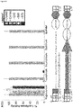
- Figures 7 , 8 , and 10 show exemplary expression cassettes and vectors useful in embodiments of the present invention. Exemplary vector components are described below.
- vectors comprise promoters that direct gene expression to particular cell type.
- promoters are platelet specific promoters. The present invention is not limited to particular platelet specific promoter.
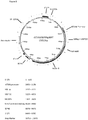
- truncated integrin allb gene (ITGA2B) promoters are used.
- Exemplary promoters include, but are not limited to, -1218, -889 and -673 ITGA2B promoters that encode"ETS" and "GATA" elements for high level of gene transcription within megakaryocytes and a Repressor region that inhibits gene transcription within other hematopoietic cell lineages (SEQ ID NOs: 21, 22, 23; and 25; Figures 8 and 10 ).
- the nucleotide sequence of the ITGA2B gene promoter was first characterized in 1988 by a group in France headed by Dr. Gerard Marguerie ( Prandini MH, Denarier E, Frachet P, Uzan G, Marguerie G. Isolation of the human platelet glycoprotein IIb gene and characterization of the 5' flanking region. Biochem Biophys Res Commun 1988, 156(1): 595-601 ).
- Figure 7 shows structural regions of the ITGA2B promoter.
- a fragment e.g., 1218, -889 and -673
- the -673 contains all of the essential regulatory elements to drive platelet-specific transgene expression.
- GATA-1 The transcription factor GATA-1 regulates the promoter activity of the platelet glycoprotein IIb gene. J Biol Chem 1993, 268(29): 21606-21612 ).
- the present invention is not limited to the ITGA2B promoters described in SEQ ID NOs: 21-23). Embodiments of the present invention contemplate fragments, portions, and combinations of the fragments described herein. In some embodiments, fragments are shorter than the full length ITGA2B promoter (e.g., 1, 2, 3, 4, 5, 6, 7, 8, 9, 10, 20, 30, 40, 50, 60, 75, 100, 150, 200 or more nucleotides shorter) and maintain desired activity (e.g., the ability to drive cell-specific expression to elicit a desired effect, e.g., reduction in sing or symptoms of a disease or condition).
- desired activity e.g., the ability to drive cell-specific expression to elicit a desired effect, e.g., reduction in sing or symptoms of a disease or condition.
- promoters comprise the ITGA2B fragments described in SEQ ID NOs: 21, 22, and 23, or sequences that are 1, 2, 3, 4, 5, 6, 7, 8, 9, 10, 20, 30, 40, 50, 60, 75, 100, 150, 200, 300, 400, 500, 600 or more nucleotides larger or smaller than SEQ ID NOs: 21, 22, and 23.
- fragments that are 1, 2, 3, 4, 5, 6, 7, 8, 9, 10, 20, 30, 40, 50, 60, 75, 100, 150, 200 or more nucleotides smaller than SEQ ID NO:21 are utilized.
- fragments that are 1, 2, 3, 4, 5, 6, 7, 8, 9, 10, 20, 30, 40, 50, 60, 75, 100, 150, 200, 300, 400, 500, 600 or more nucleotides larger than SEQ ID NO:23 are utilized.
- discontinuous fragments of SEQ ID NOs: 21, 22, or 23 that retain promoter activity are utilized.
- 1, 2, 3, 4, 5, 6, 7, 8, 9, 10, 20, 30, 40, 50, 60, 75, 100, 150, 200, 300, 400, 500, 600 nucleotides of SEQ ID NOs 21, 22, or 23 are utilized.
- promoter fragments comprise one or more elements useful for promoter activity. Examples include, but are not limited to, GATA elements (e.g., GATA54 or GATA454), sP1 elements, Ets35 elements, and the like (See e.g., Block et al., Blood 1994 84: 3385-3393 ; Prandini et al., Blood 1996 88: 2062-2070 ; Block et al., Blood 1996 88: 2071-2080 ; and Doubeikovski et al., J. Biol. Chem. 272: 24300-24307, 1997 ; each of which is herein incorporated by reference in its entirety).
- GATA elements e.g., GATA54 or GATA454
- sP1 elements e.g., Ets35 elements, and the like
- the 5' ends of promoters are modified to add restriction endonuclease sites to aid in cloning and constructing expression vectors.
- 1, 2, 3, or 4 nucleotides at the 5' end are modified from the wild type sequence or the fragments disclosed herein to add restriction endonuclease sites.
- the fragments comprise, consist essentially of, or consist of promoter sequence found in SEQ ID NOs: 21, 22, or 23.
- exogenous genes are generally clotting factors (e.g., Factor VIII and/or Factor IX).
- Human Factor VIII has the accession number NM_000132.3 and Human Factor IX has the accession number NM_000133.3.
- exogenous genes may be utilized in the treatment of other platelet related conditions.
- exogenous genes useful in the treatment of diseases other than platelet related disorders are utilized (e.g., in the treatment of cancer by using platelets to target release of anti-neplastic agents "i.e. IL-24" to shrink solid tumors and anti-thrombotic agents to be released at the site of blood clots such as cases of deep vein thrombosis).
- expression cassettes further comprise a targeting factor that targets expression into a particular sub-structure of a platelet.
- expression cassettes further comprise the minimal amino acid sequence of a signal sequence peptide that has been found to be able traffic not only VWF but also recombinant proteins fused to the peptide that has the proven ability to store proteins in cellular granule compartments specifically the natural storage sub-structure endothelial cells "Weibel-Palade bodies" and platelet ⁇ -granules, both of which can be secreted upon cellular activation.
- expression constructs comprise a nucleic acid encoding a Von Willebrand Factor propeptide signal peptide and D2 domain (SPD2) to promote trafficking of molecules directly into the ⁇ -granule compartment as shown in Rosenberg JB et al, Intracellular Trafficking of FVIII to von Willebrand Factor storage Granules, J. Clin. Invest. 101, 613-624 (1998 ); Haberichter SL, Jacobi P, Montgomery RR. Critical independent regions in the VWF propeptide and mature VWF that enable normal VWF storage.
- SPD2 Von Willebrand Factor propeptide signal peptide and D2 domain
- VWFpp Von Willlebrand Factor Propeptide
- FIG. 7 An example of an expression cassette comprising a 673 bp fragment of the ITGA2B gene promoter and a VWF/SPD2 gene is shown in Figure 7 (SEQ ID NO:24). Variants of these sequences that retain its desired activity are specifically contemplated for use in compositions and methods of embodiments of the present invention.
- constructs comprise an expression enhancer (e.g., Woodchuck Hepatitis Virus (WHP) Posttranscriptional Regulatory Element (WPRE)) between the promoter/signaling cassette and the exogenous gene of interest.
- WPRE Woodchuck Hepatitis Virus
- This element has been utilized by several gene transfer strategies because its structure inhibits degradation of the transcript within the cell and thus allows for more therapeutic protein to be synthesized compared to a gene transfer vector in the absence of WPRE.
- vectors are self-inactivating.
- vectors are retroviral vectors (e.g., lentiviral vectors). Table 2 provides a summary of exemplary suitable vectors.
- Retroviruses family Retroviridae are divided into three groups: the spumaviruses (e.g., human foamy virus); the lentiviruses (e.g., human immunodeficiency virus and sheep visna virus) and the oncoviruses (e.g., MLV, Rous sarcoma virus).
- the spumaviruses e.g., human foamy virus
- the lentiviruses e.g., human immunodeficiency virus and sheep visna virus
- the oncoviruses e.g., MLV, Rous sarcoma virus.
- Retroviruses are enveloped (e.g., surrounded by a host cell-derived lipid bilayer membrane) single-stranded RNA viruses that infect animal cells.
- a retrovirus infects a cell, its RNA genome is converted into a double-stranded linear DNA form (e.g., it is reverse transcribed).
- the DNA form of the virus is then integrated into the host cell genome as a provirus.
- the provirus serves as a template for the production of additional viral genomes and viral mRNAs. Mature viral particles containing two copies of genomic RNA bud from the surface of the infected cell.
- the viral particle comprises the genomic RNA, reverse transcriptase and other pol gene products inside the viral capsid (which contains the viral gag gene products), which is surrounded by a lipid bilayer membrane derived from the host cell containing the viral envelope glycoproteins (also referred to as membrane-associated proteins).
- retroviral vector which contains the sequences necessary for the efficient expression of the gene of interest (including promoter and/or enhancer elements which may be provided by the viral long terminal repeats [LTRs] or by an internal promoter/enhancer and relevant splicing signals), sequences required for the efficient packaging of the viral RNA into infectious virions (e.g., the packaging signal [Psi], the tRNA primer binding site [-PBS], the 3' regulatory sequences required for reverse transcription [+PBS] and the viral LTRs).
- promoter and/or enhancer elements which may be provided by the viral long terminal repeats [LTRs] or by an internal promoter/enhancer and relevant splicing signals
- sequences required for the efficient packaging of the viral RNA into infectious virions e.g., the packaging signal [Psi], the tRNA primer binding site [-PBS], the 3' regulatory sequences required for reverse transcription [+PBS] and the viral LTRs.
- the LTRs contain sequences required for the association of viral genomic RNA, reverse transcriptase and integrase functions, and sequences involved in directing the expression of the genomic RNA to be packaged in viral particles.
- many recombinant retroviral vectors lack functional copies of the genes that are essential for viral replication (these essential genes are either deleted or disabled); the resulting virus is said to be replication defective.
- the vector DNA is introduced into a packaging cell line.
- Packaging cell lines provide viral proteins required in trans for the packaging of the viral genomic RNA into viral particles having the desired host range (e.g., the viral-encoded gag, pol and env proteins). The host range is controlled, in part, by the type of envelope gene product expressed on the surface of the viral particle.
- Packaging cell lines may express ecotrophic, amphotropic or xenotropic envelope gene products.
- the packaging cell line may lack sequences encoding a viral envelope (env) protein. In this case the packaging cell line will package the viral genome into particles that lack a membrane-associated protein (e.g., an env protein).
- the packaging cell line containing the retroviral sequences is transfected with sequences encoding a membrane-associated protein (e.g., the G protein of vesicular stomatitis virus [VSV]).
- VSV vesicular stomatitis virus
- the transfected packaging cell will then produce viral particles that contain the membrane-associated protein expressed by the transfected packaging cell line; these viral particles, which contain viral genomic RNA derived from one virus encapsidated by the envelope proteins of another virus are said to be pseudotyped virus particles.
- Viral vectors including recombinant retroviral vectors, provide a more efficient means of transferring genes into cells as compared to other techniques such as calcium phosphate-DNA co-precipitation or DEAE-dextran-mediated transfection, electroporation or microinjection of nucleic acids. It is believed that the efficiency of viral transfer is due in part to the fact that the transfer of nucleic acid is a receptor-mediated process (i.e., the virus binds to a specific receptor protein on the surface of the cell to be infected).
- nucleic acids transferred by other means such as calcium phosphate-DNA co-precipitation are subject to rearrangement and degradation.
- MoMLV amphotropic Moloney murine leukemia virus
- the MoMLV system has several advantages: 1) this specific retrovirus can infect many different cell types, 2) established packaging cell lines are available for the production of recombinant MoMLV viral particles and 3) the transferred genes are permanently integrated into the target cell chromosome.
- the established MoMLV vector systems comprise a DNA vector containing a small portion of the retroviral sequence (the viral long terminal repeat or "LTR" and the packaging or "psi" signal) and a packaging cell line. The gene to be transferred is inserted into the DNA vector.
- the viral sequences present on the DNA vector provide the signals necessary for the insertion or packaging of the vector RNA into the viral particle and for the expression of the inserted gene.
- the packaging cell line provides the viral proteins required for particle assembly ( Markowitz et al., J. Virol., 62:1120 [1988 ]).
- the low titers associated with MoMLV-based vectors has been attributed, at least in part, to the instability of the virus-encoded envelope protein. Concentration of retrovirus stocks by physical means (e.g., ultracentrifugation and ultrafiltration) leads to a severe loss of infectious virus.
- retrovectors are derived from lentiviruses including, but not limited to, human immunodeficiency virus (HIV) or feline immunodeficiency virus (FIV). Lentivirus vectors have the advantage of being able to infect non replicating cells.
- HIV human immunodeficiency virus
- FMV feline immunodeficiency virus
- retroviral vectors which contain the G protein of VSV as the membrane associated protein.
- the VSV G protein interacts with a phospholipid component of the plasma membrane ( Mastromarino et al., J. Gen. Virol., 68:2359 [1977 ]). Because entry of VSV into a cell is not dependent upon the presence of specific protein receptors, VSV has an extremely broad host range.
- VSV G protein Pseudotyped retroviral vectors bearing the VSV G protein have an altered host range characteristic of VSV (i.e., they can infect almost all species of vertebrate, invertebrate and insect cells). Importantly, VSV G-pseudotyped retroviral vectors can be concentrated 2000-fold or more by ultracentrifugation without significant loss of infectivity ( Burns et al., Proc. Natl. Acad. Sci. USA, 90:8033 [1993 ]).
- the VSV G protein has also been used to pseudotype retroviral vectors based upon the human immunodeficiency virus (HIV) ( Naldini et al., Science 272:263 [1996 ]).
- HIV human immunodeficiency virus
- the VSV G protein may be used to generate a variety of pseudotyped retroviral vectors and is not limited to vectors based on MoMLV.
- the present invention is not limited to the use of the VSV G protein when a viral G protein is employed as the heterologous membrane-associated protein within a viral particle. Sequences encoding other G proteins derived from other members of the Rhabdoviridae family may be used; sequences encoding numerous rhabdoviral G proteins are available from the GenBank database.
- retroviruses can transfer or integrate a double-stranded linear form of the virus (the provirus) into the genome of the recipient cell only if the recipient cell is cycling (i.e., dividing) at the time of infection.
- Retroviruses that have been shown to infect dividing cells exclusively, or more efficiently, include MLV, spleen necrosis virus, Rous sarcoma virus human immunodeficiency virus, and other lentiviral vectors.
- the present invention is not limited to retroviral vectors.
- suitable vectors include, but are not limited to, the following vectors: 1) Bacterial -- pQE70, pQE60, pQE 9 (Qiagen), pBS, pD10, phagescript, psiX174, pbluescript SK, pBSKS, pNH8A, pNH16a, pNH18A, pNH46A (Stratagene); ptrc99a, pKK223 3, pKK233 3, pDR540, pRIT5 (Pharmacia); and 2) Eukaryotic -- pWLNEO, pSV2CAT, pOG44, PXT1, pSG (Stratagene) pSVK3, pBPV, pMSG, pSVL (Pharmacia).
- mammalian expression vectors comprise, along with an expression cassette as described herein, an origin of replication, any necessary ribosome binding sites, polyadenylation sites, splice donor and acceptor sites, transcriptional termination sequences, and 5' flanking non transcribed sequences.
- DNA sequences derived from the SV40 splice, and polyadenylation sites may be used to provide the non-transcribed genetic elements.
- the present invention provides systems and methods for genetic manipulation of stem cells (e.g., hematopoietic stem cells or cancer stem cells).
- stem cells e.g., hematopoietic stem cells or cancer stem cells.
- the compositions and methods described herein find use in the treatment of a variety of disorder related to platelet function (e.g., hemophilia and the disorders described in Table 3 below).
- hematopoietic stem cell gene therapy aimed at targeting therapeutic agents to the platelet surface, cytoplasm or granules finds use as a strategy to correct other disorders of hemostasis, thrombosis, immune response and cancer.
- therapeutic methods are ex vivo methods, in which autologous hematopoietic stem cells are harvested from an animal (e.g., human) in need of treatment, modified using one of the vector described herein, and re-introduced into the original donor.
- autologous methods reduce the risk of autoimmune or rejection responses that can occur with infusion of donor clotting factors and allow one to limit gene transduction to hematopoietic stem cells through ex vivo transduction.
- the method includes the steps of administering cytokines to mobilize peripheral blood stem cells into the peripheral blood; performing apheresis and magnetic bead selection for CD34+ cells; preconditioning using e.g., bulsulfan and/or other agents like fludarabine; and using a viral (e.g., lentiviral) gene transfer vector to modify stem cells before they are re-introduced into the patient.
- cytokines to mobilize peripheral blood stem cells into the peripheral blood
- preconditioning e.g., bulsulfan and/or other agents like fludarabine
- a viral e.g., lentiviral
- hematopoietic stem cells are mobilized using administration of cytokines or other mobilization agents (See e.g., Fu et al., Blood Rev. 2000 Dec;14(4):205-18 and United States Patent 7417026 , each of which is herein incorporated by reference in its entirety for a discussion of mobilization protocols), although other suitable protocols may be utilized.
- cytokines See e.g., Fu et al., Blood Rev. 2000 Dec;14(4):205-18 and United States Patent 7417026 , each of which is herein incorporated by reference in its entirety for a discussion of mobilization protocols
- mobilization cytokines include, but are not limited to, Interleukin-3 (IL-3), granulocyte colony stimulating factor (G-CSF), also known as Amgen's FDA approved drug Neupogen, stem cell factor (SCF), granulocyte macrophage colony-stimulating factor (GM-CSF), and sequential or co-administration of one or more of IL-3, GM-CSF, SCF, and GM-CSF.
- IL-3 Interleukin-3
- G-CSF granulocyte colony stimulating factor
- SCF stem cell factor
- GM-CSF granulocyte macrophage colony-stimulating factor
- Suitable dosage ranges for mobilization agents vary, but in general, the compounds are administered in the range of about 0.1 ⁇ g/kg-5 mg/kg of body weight; preferably the range is about 1 ⁇ g/kg-300 ⁇ g/kg of body weight; more preferably about 10 ⁇ g/kg-100 ⁇ g/kg of body weight.
- the dosage range is from about 0.7 ⁇ g-350 mg; preferably about 700 ⁇ g-21 mg; most preferably about 700 ⁇ g-7 mg.
- Dosages can be higher when the compounds are administered orally or transdermally as compared to, for example, i.v. administration.
- the compounds can be administered as a single bolus dose, a dose over time, as in i.v. or transdermal administration, or in multiple dosages.
- the amount of active compound to be administered can vary according to the discretion of the skilled artisan.
- the amount of active compound to be administered to the recipient is within the ranges described above for stem cell mobilization. However, the administration of such amounts will vary according to the standards set forth by clinicians in the field of stem cell enhancement therapy.
- CD34+ Peripheral Blood stem cells are isolated from the low molecular weight mononuclear cells by immunomagnetic beads using Miltenyi's automacs system (for large animal, dogs, 25-45 kg) and Miltenyi's Clinimacs system (for humans) recently approved for clinical use by the FDA.
- the CD34+PBC are the genetically modified using the vectors described herein and re-introduced into a subject in need by autologous stem cell transplant.
- a single treatment is utilized to provide long-term protection against episodes of bleeding.
- treatment is performed on a regular basis (e.g., weekly, monthly, yearly, once every 2, 3, 4, 5 or more years, and the like) in order to prevent episodes of bleeding.
- treatment is only administered when episodes of abnormal bleeding occur (e.g., following accidents, prior to or following surgery, etc,).
- maintenance therapy is administered in combination with extra therapy when episodes of abnormal bleeding occur.
- Human transformed cell lines were obtained from American Type Culture Collection (Rockville, MD) and propogated under conditions described for promegakaryocytic (HEL Megakaryocyte transformed cell line) ( Bray, P. F. et al. J Clin Invest 80, 1812-1817. (1987 ); Greenberg, S. M., et al., T-cell lymphoma (KT1) , ( Okamoto, T., et al. J Biol Chem 261, 4615-4619 (1986 )) B-cell lymphoma (Raji) , ( Choi, J. H. et al.
- pCMVLuc A BgIII and HindIII restriction digest of cytomegalovirus tissue non-specific gene promoter (878bp) from pRc/CMV (Invitrogen) is ligated into the pGL3-Basic Luciferase vector (Promega, Madison, WI). This construct served as the positive control for high level gene expression within all cell-types; thus, assigned an arbitrary level of 100% luciferase activity for each cell line ( Fig. 1 ).
- pGL3-BasicLuc Negative control construct for 0% luciferase activity ( Fig. 1 ) because lacks a gene promoter to drive luciferase gene transcription (Promega).
- pCMVnlac Cell lines were co-transfected with one of the pITGA2BLuc+ constructs and
- Cell lines (2x10 7 ) were co-transfected with either (20) ⁇ g) of the ITGA2B gene promoter construct (-1218, -889, -673) ( Fig. 1A ) or the positive (CMV) or negative (Basic) controls encoding firefly luciferase and pCMVnlac (20 ⁇ g) encoding ⁇ -galactosidase.49 Briefly, forty-eight hours after co-transfection cells were washed, harvested, and lysates were prepared and frozen to -80°C using the luciferase assay system (Promega). Luciferase activity was measured with a Turner Designs Model 20 Luminometer.
- ⁇ -galactosidase activity was performed to normalize transient transgene expression for each cell line with a sensitive ELISA enzymatic assay that measured colormetric change with the substrate for ⁇ -galactosidase, chlorophenol red ⁇ -D-galactophranoside (CPRG) ( Eustice DC, et al., Biotechniques 1991, 11(6): 739-740, 742-733 ).
- the percent of luciferase activity was determined by comparing the mean value of the Relative Light Units (RLU) of luciferase/CPRG Vmax value for each construct to reveal the transfection efficiency for each cell line.
- RLU Relative Light Units
- the RLU for pCMVLuc was assigned arbitrarily a value of 100% and all other results were calculated for each vector based upon that value as shown in Fig.1A .
- ITGA2B -(M)WPTS genetic transfer vectors are derived from a HIV type-1 lentiviral vector (D.Trono, University of Geneva, Switzerland).51 p-889ITGA2B-BDDFVIII-WPTS lentiviral vector ( Fig.1B ) encodes a -889 to +30 nucleotide fragment of the human ITGA2B promoter and human BDDFVIII molecule.16 p-673ITGA2B-VWFSPD2-BDDFVIII-WPTS lentiviral vector ( Fig.
- VWFpp Von Willebrand Factor propeptide
- SP amino acid VWF signal peptide
- cDNA encoding human BDDFVIII to allow megakaryocyte-specific transcription of a hybrid molecule that uses the SPD2 peptide to traffic human BDDFVIII to platelet ⁇ -granules.22 cDNA encoding SP was amplified by PCR with forward Primer
- Cytokine mobilized CD34+G-PBC gene transfer and autologous transplant studies using FVIII-Deficient dogs affected with hemophilia A (University of North Carolina, Chapel Hill, NC) ( Lozier JN, et al., Proc Natl Acad Sci U S A 2002, 99(20): 12991-12996 ) were conducted and approved by Institutional Animal Care and Use Committees of the University of North Carolina and The Medical College of Wisconsin which are both accredited facilities of the American Association for Accreditation of Laboratory Animal Care.
- CD34+ G-PBC were selected with a biotin-conjugated-1H6 Ab (1mg/ml) (Richard Nash, Fred Hutchinson Research Institute, Seattle, WA) and anti-biotin immuno-magnetic beads (1:5 dilution) on an Automacs magnetic cell separator (Miltenyi Biotec Inc., Auburn, CA).
- CD34+ G-PBC were transduced with -889ITGA2B-BDDFVIII-WPTS or -673ITGA2B-VWFSPD2-BDDFVIII-WPTS lentiviral vector.
- 4x10⁶ cells/well were seeded in a 6-well plate (Falcon-Becton Dickinson, Franklin Lakes, NJ) coated with 20 ⁇ g/cm2 RetroNectin (Takara Shuzo, Otsu, Shiga, Japan) and incubated with 1.0x10 4 ITGA28 -FVIII lentivirions/cell in X-Vivo 10 containing 10%FCS, rhIL-3, rcalL-6, rcaSCF, rhTPO and rhflk2/flt3 ligand.
- Blood was collected at preselected times into a vacutube containing 7.5% EDTA anticoagulant (Fang et al., supra). Blood cells were counted on a Vet ABC hematology analyzer (scil animal care company, Gurnee, IL). Platelets were isolated with Fico/LiteTM (Atlanta Biologicals, Norcross, GA), washed with PBS and used directly for immunofluorescent flow cytometry or FVIII:C activity analysis. Leukocytes were isolated with Ficoll-Paque Plus® (GE Healthcare) according to the manufactures specifications.
- Monoclonal 1°Abs (5-10 ⁇ g/ml), MBC 103.3 and 301.3 (R.R.Montgomery, BloodCenter of WI, Milwaukee, WI), recognize epitopes on human BDDFVIII.53 2°Abs used were Alexa Fluor® 488 F(ab')2 conjugated to a fragment of donkey anti-sheep IgG (H+L) (1:1,000 dilution) and Alexa Fluor® 568 F(ab')2 fragment of goat anti-mouse IgG (H+L) (1:500 dilution) were from Life Technologies (Grand Island, NY).
- Canine platelets were fixed with 3.7% (vol/vol) buffered formalin, permeabilized in 0.5% Triton X-100 (in 20 mmol/L Hepes, 300 mmol/L sucrose, 50 mmol/L NaCl, and 3mmol/L MgCl2, pH7.0), and blocked with 2.5% normal goat serum in HBSS.
- Platelets were incubated with a sheep polyclonal 1°Ab to canine fibrinogen and monoclonal 1°Ab (MBC 103.3 & 301.3) to human FVIII (5ug/ml) overnight at 4°C.53
- the Alexa Fluor® 488-conjugated F(ab')2 fragment of donkey anti-sheep IgG (H+L) was used as a 2°Ab (1:1,000 dilution) to detect fibrinogen and Alexa Fluor® 568-conjugated F(ab')2 fragment of goat anti-mouse IgG (H+L) conjugated 2°Ab (1:500 dilution) was used to detect the presence of FVIII for 30 min at 25°C. Platelets were mounted with Vectashield (Vector Labs, Burlingame, CA). Immunofluorescence was detected with a Zeiss LSM 510 Multiphoton Confocal Microscope (Carl Zeiss, Inc.
- Canine platelets were isolated from blood and treated with CytofixTM and PERM/WASHTM reagents (BD Biosciences) for intracellular detection of BDDFVIII. Platelets were incubated with a monoclonal 1°Ab (MBC 103.3 & 301.3) to human FVIII (5ug/ml) 30 minutes at 4°C and then incubated with Alexa Fluor® 568-conjugated F(ab')2 fragment of goat anti-mouse IgG (H+L) conjugated 2°Ab (1:500 dilution) for 30 minutes at 4°C. Platelets isolated from FVIII-Deficient dogs were used as negative controls. Nonspecific isotype control Ab served as negative controls. Cells were collected and analyzed on an Accuri® C6 Flow Cytometer (Accuri Cytometers, Inc., Ann Arbor MI) using the Accuri analysis software.
- MMC 103.3 & 301.3 monoclonal 1°Ab
- H+L Alexa Fluor® 568-conjugated
- Platelets were fixed in 1.25% glutaraldehyde (Fluka AG, Buchs, Switzerland), infused with 2.3M sucrose (Fluka), and frozen with a Reichert KF 80 freezing system (Leica, Vienna, Austria). Sections of ⁇ 80 nm were prepared with the Ultracut E ultramicrotome equipped with a FC 4E cryokit attachment and placed on collodion-coated nickel grids. Grids were incubated for 10 min on PBS with 1% BSA and then placed on (10 ⁇ g/ml) drops of the 1°Ab to FVIII(301.3) for 1h at 25°C.
- Sections were incubated for 1h with a goat anti-mouse 2°Ab adsorbed onto 10nm gold particles (1/100 dilution of AuroProbe EM G10). Controls included the use of an irrelevant IgG of the same species and at the same concentration.
- Grids were stained by uranyl acetate and osmium and then embedded in methylcellulose prior to observation with a Jeol JEM-1010 transmission electron microscope (Jeol, Croissy-sur-seine, France) at 80 KV.
- Platelets were isolated from circulating peripheral blood, washed, and activated with physiological agonists of platelet activation. To induce activation, platelets were resuspended in Tyrode's buffer (2.5 x 10⁶/ml) containing 1 mM CaCl2, 1 mM MgCl2, 25 ⁇ M each of adenosine diphosphate (ADP) (Sigma), epinephrine (Bio/Data Corporation, Horsham, PA) and canine thrombin receptor activating peptides: PAR1 (SFFLKN-NH2), PAR3 (TRFGAP-NH2) and PAR4 (SFPGQP-NH2) for 30 minutes at 37°C as previously described (Fang et al., supra).
- ADP adenosine diphosphate
- epinephrine Bio/Data Corporation, Horsham, PA
- canine thrombin receptor activating peptides PAR1 (SFFLKN-NH2), PAR3 (TRFGAP-NH2)
- DNA was isolated with a QIAamp® DNA Blood Mini Kit (Qiagen,, Maryland, USA) from canine leukocytes purified with Ficoll-PaqueTM Plus (Amersham Pharmacia Biotech AB, Uppsala, Sweden).
- p-889ITGA2-BDDFVIII-WPTS served as a positive control.
- PCR analysis was performed with Taq polymerase (Invitrogen, Carlsbad, CA) on a PTC200 instrument (MJ Research, Watertown, MA) with forward primer P1(5'-ACGCTATGTGGATACGCTG-3') and reverse primer P2(5'-AACACCACGGAATTGTCAG-3') (SEQ ID NO:12) to synthesize a 318 nucleotide primary product encoding the WPRE ( Figure 1B ,C ).
- a secondary PCR reaction was performed with nested forward primer P3(5'-TGGATACGCTGCTTTAATGC-3') (SEQ ID NO:13) and reverse primer P4(5'-AATTGTCAGTGCCCAACAG-3') (SEQ ID NO:14) encoding a 302 bp product of WPRE ( Fig.5A ).
- Percent lentiviral gene marking was measured by RT-qPCR using BIO-RAD CFX96 Real-Time System.52 Briefly, 12.5ul of TaqMan Universal PCR Master Mix (Life technologies), a 900nM concentration of each primer, and 200nM probe were combined in 20 ⁇ l of water. Then 5ul of canine genomic DNA was added and PCR utilized 2 min at 500C, 10 min at 950C, and then 40 cycles of 15 sec at 950C and 1 min at 600C. For each RT-qPCR, a no template control was included as negative control.
- lentiviral LTR primers and probe used were: Fwd:5'-AGCTTGCCTTGAGTGCTTCA-3' (SEQ ID NO:15); Rev:5'-TGACTAAAAGGGTCTGAGGGA-3'(SEQ ID NO:16); probe:6FAM-TGCCCGTCTGTTGTGTGACTCTG-MGBNFQ (SEQ ID NO:17).
- the canine ITGB3 gene was used as an endogenous control for gene copy number with Fwd:5'-ATGCATCCCACTTGCTGGTAT-3'(SEQ ID NO:18); Rev:5'-TGCCCATCGTTAGGTTGG-3'(SEQ ID NO:19); probe:6FAM-TGCCTGCCAGCCTTCCATCCAG-MGBNFG (SEQ ID NO:20). Copy number was based on TaqMan principle. Ten-fold serial dilution of the plasmid constructs of known concentration containing relevant sequences (Lentiviral vector LTR and canine ITGB3 ) were used to create standard curves for quantification of samples.
- LAM Linear Amplification-Mediated
- LAM-PCR was performed to localize the lentiviral vector insertion sites within genomic DNA isolated from peripheral blood leukocytes. Briefly, the junction between integrated proviral LTR and the host genome was selected by 2 rounds of linear PCR [95°C for 5min; (95°C for 1m, 60°C for 45s, 72°C for 90s) x 50; 72°C for 10m] with a vector-specific 5'-biotinylated primer [5'-/biotin/-GAACCCACTGCTTAAGCCTCA-3'(SEQ ID NO:26)] and purified using streptavidin-coated magnetic beads [Dynal M-280].
- Products were double stranded using Klenow polymerase and random hexanucleotide primers and digested with Tsp509I at 65°C for 2h.
- Directional double-stranded linker oligos were ligated onto the non-LTR end and the resulting products were amplified by nested PCR [95°C for 5min; (95°C for 1m, 60°C for 45s, 72°C for 90s) x 35; 72°C for 10m] using LTR-specific forward primers [F1:
- Sequence products from LAM-PCR that were verified to contain proviral LTR sequence were masked for known genomic repeats and proviral features.
- the resulting sequence was aligned to the dog genome (CanFam 2.0, May 2005 assembly) using the Blat (BLAST-like alignment tool) server at UCSC.
- Sequences mapping to a unique location in the genome at 95% similarity were selected and integration sites were determined as the base in the genomic alignment flanking the proviral LTR sequence. For each site, the closest RefSeq gene was determined and compared to a list of human cancer orthologs.
- Lysates of 1x10 8 platelets/ml were tested for FVIII:C using a Chromogenix Coatest® SP4 FVIII kit (DiaPharma, Franklin, OH).12 Duplicate samples of supernatant were placed in uncoated wells of a 96-well microtiter plate (25 ⁇ l/well) and assay components (phospholipid, Factor IXa, Factor X, and calcium chloride) were added, and incubated for 10 min at 37oC. The chromogenic Factor Xa substrate S-675 was added, and the plate was transferred to a Wallac Victor2 microplate reader preset at 37oC.
- the Factor Xa-dependent conversion of S-2675 is directly related of the amount of FVIII:C in each well.
- a standard curve was constructed by plotting known amounts of recombinant human FVIII (Kogenate; Bayer Healthcare Pharmaceuticals, Berkeley, CA) diluted in platelet lysate buffer using Vmax at 405nm. The Vmax of each reaction was converted to units of FVIII:C activity using the kinetic software program, SOFTmax, v.2.34 (Molecular Devices). The FVIII activity was measured by an endpoint reading at 405nm, a background reading at 490nm was subtracted from 405nm.
- the total maximum FVIII:C/dog was calculated by multiplying the mean FVIII:C U/ml/1x108 platelets x 92 ml blood/kg x dog weight (kg) x (2x10 8 platelets)/1 ml blood) using measured values recorded in Table 1 and Fig.6 .
- WBCT is a modification of the Lee-White clotting time using two siliconized glass tubes (Becton-Dickinson, Rutherford, NJ) at 28°C ( Nichols TC, et al., J Thromb Haemost 2012, 10(3): 474-476 ).
- One ml of whole blood was drawn and 0.5 ml blood was distributed into each tube.
- a timer was started. After one minute, one tube was tilted every 30 sec, the other left undisturbed. When a clot formed in the tilted tube, the second tube was then tilted every 30 sec until a clot formed. The time for formation of a fully gelled clot in the second tube was recorded as the WBCT.
- Blood was collected from a hemostatically normal (WBCT 7.5-12.5 min) and the three experimental dogs (F20,I42,M64) before and after G-PBC transplant if animals had not been treated with plasma for at least one month.
- Canine blood plasma (F20, 142 and M64) was screened for inhibitors with an activated partial thromboplastin time (aPTT) mixing assay that detects inhibitory antibodies to either coagulation factor VIII or IX as previously described ( Langdell RD, et al., J Lab Clin Med 1953, 41(4): 637-647 ; Sahud MA. Semin Thromb Hemost 2000, 26(2): 195-203 ; Matrai J, et al., Hepatology 2011, 53(5): 1696-1707 ). Briefly, test plasmas are incubated in a 1:1 mix with normal plasma for 2 h at 37°C and then the incubated mixture is analyzed using standard aPTT reagents.
- aPTT activated partial thromboplastin time
- BIU Bethesda Inhibitor
- a luciferase reporter assay revealed that fragments of the full-length human ITGA2B gene promoter permitted comparable platelet-specific gene transcription ( Fig.1A ).
- Three different ITGA2B promoter fragments (-1218, -889 and - 673 ) directed similar levels of luciferase activity within a pro-megakaryocytic cell line.
- ITGA2B promoter driven luciferase activity remained undetectable in the other blood cell lineages and an epithelial cell line.
- Each ITGA2B promoter encodes Ets and GATA factors permitting a high level of megakaryocyte gene transcription and a repressor region that inhibits expression within other lineages ( Prandini MH, et al., Blood 1996, 88(6): 2062-2070 ).20
- two lentiviral gene transfer vectors were tested for optimal hematopoietic stem cell transduction efficiency and the ability to improve hemostatic function with platelet-derived BDDFVIII in hemophilia A dogs to develop a strategy for human gene therapy.
- VWF is a normal ⁇ -granule constituent in human platelets (albeit absent in canine platelets) ( Nichols TC, et al., Blood 1993, 81(10): 2644-2651 ) that serves as a carrier protein of FVIII in human and canine plasma ( Kaufman RJ, et al. Molecular & Cellular Biology 1989, 9(3): 1233-1242 ).
- canine hematopoietic stem cells were mobilized from the bone marrow into the peripheral blood with canine cytokines (cG-CSF & cSCF) and G-PBC apheresis was performed without adverse incident identical to previous studies using GT dogs (Fang et al., supra).
- Mononuclear lymphocytes were isolated with Ficoll-Paque Plus from the apheresis product and then canine CD34 antigen positive (CD34+) cells were purified by immunomagnetic selection ( McSweeney PA, et al., Blood 1998, 91(6): 1977-1986 ).
- Table 1 summarizes the conditions for autologous transplant of three hemophilia A dogs transfused with approximately 3x10 6 FVIII-transduced CD34+G-PBC/kg of body weight where each target cell was transduced with approximately 1x10⁴ total viral particles/CD34+G-PBC without the use of ex vivo or in vivo selection for transduced cells (Columns 4,5) .
- a non-myeloablative pre-transplant conditioning regimen was employed to create a niche in the bone marrow for the newly transplanted cells to engraft (Table 1, Column 2) . The intensity of the conditioning regimen is determined by the level at which the dose becomes toxic to the organs.
- busulfan a drug preferentially toxic to hematopoietic stem cells
- EACA is an effective synthetic inhibitor of the plasmin-plasminogen system and controls subarachnoid hemorrhage, genitourinary bleeding from many causes and dental surgery in hemophiliacs ( Griffin JD, et al., Semin Thromb Hemost 1978, 5(1): 27-40 ). For comparison, the number of serious bleeding episodes that required treatment with cFVIII supplement has been recorded 1 year before and 2.5 years after G-PBC transplant for each dog (Table 1, Columns 10, 11) ( Niemeyer GP, et al., Experimental Hematology 2003, 31(12): 1357-1362 ).
- BDDFVIII was being synthesized and stored in platelets following G-PBC transplant.
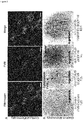
- Fig.2A are images of the results of microscopic analysis of platelets isolated from one dog (I42) that received an autologous transplant of lentiviral vector transduced G-PBC, which represents the outcome of analysis of all three dogs (F20, I42, M64).
- Fg fibrinogen
- Human BDDFVIII was also detected in a punctate pattern within platelets (Middle Panel).
- BDDFVIII staining co-localized frequently within Fg as evident by the appearance of a yellow staining when the left (Fg) and middle panel (BDDFVIII) were overlaid indicating that both proteins could be stored together within platelet ⁇ -granules (Right Panel) ( Wilcox et al., J Thromb Haemost 2003, 1(12): 2477-2489 ).
- Immuno-electron microscopy was performed to determine if exogenous BDDFVIII was being transported specifically to platelet ⁇ -granules. Immunogold analysis was performed on ultrathin sections of platelets with a 1°Ab to FVIII and a 2°Ab adsorbed on 10 nm gold particles ( Fig.2B ). The ⁇ -granules appeared normal in size and shape within platelets of FVIII-Deficient dogs as well as FVIII transplant recipients. BDDFVIII is absent in platelet ⁇ -granules from a FVIII-Deficient negative control (Left Panel).
- BDDFVIII was detected within ⁇ -granules and cytoplasm of platelets isolated from all three dogs (F20, 142, M64). This result is consistent with observations reported for ectopic expression of BDDFVIII within platelets of VWF(-/-) transgenic mice affected with von Willibrand disease ( Yarovoi H, et al., Blood 2005, 105(12): 4674-4676 ). -673 ITGA2B- VWFSPD2-BDDFVIII transduced platelets from M64 stored the greatest level of BDDFVIII within the ⁇ -granule (Right Panel). In addition, BDDFVIII was detected rarely within membrane systems in the platelet cytoplasm indicating that the VWFSPD2 indeed had an increased efficiency to traffic BDDFVIII directly into the ⁇ -granule compartment.
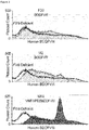
- a Chromogenix Coatest® SP4 FVIII assay was perform to determine if activated platelets could secrete a biologically active form of BDDFVIII (FVIII:C) as previously shown for activated human megakaryocytes in tissue culture ( Wilcox et al., Thromb Haemost 2003, 1(12): 2477-2489.12
- Fig.4 platelet lysates from a FVIII-Deficient dog show that the level of BDDFVIII:C background activity is virtually unchanged for untreated (Black, - Agonist) and activated platelets (White, + Agonist).
- FVIII:C activity was detected readily in the lysate of quiescent, untreated platelets from F20, I42 and M64.
- BDDFVIII:C levels were decreased in lysates of platelets stimulated by a mixture of physiological agonists of platelet activation: ADP, epinephrine, and canine PAR 1,3,4 in all three experimental dogs.
- dogs that received BDDFVIII-transduced G-PBC show an appreciable decrease in FVIII:C activity only after platelet activation indicating that platelets from experimental animals can be induced to secrete FVIII within the vasculature.
- the lentiviral vector WPRE element was detected by PCR of genomic DNA isolated from leukocytes collected from F20, 142, and M64 for at least 2.5 years after transplant ( Fig. 5A ).
- Real time quantitative PCR (RT-qPCR) analysis of genomic DNA isolated from peripheral blood leukocytes revealed that the transduction efficiency for each lentiviral vector was 1% (F20), 4% (I42) and 2% (M64) (Table 1, Column 8).
- the detection of lentiviral vector by genomic analysis in the absence of the appearance of insertional oncogenesis is consistent with the overall good health of all of the dogs with frequent evaluation of peripheral blood counts and peripheral blood smears documenting normal morphology and numbers of circulating hematopoietic cells.
- Linear Amplification-Mediated (LAM)-PCR was also performed to determine the integration pattern of lentiviral vector within the genome of the experimental dogs.
- Fig.5B shows that lentiviral vector was not present within the genome of a FVIII-Deficient control while multiple bands appear to be present in the genomic DNA of transplanted dogs (F20, I42 and M64).
- a distinct insertion site was detected specifically in chromosome 4 for F20 and chromosome 35 for M64.
- the results demonstrate that insertion of the lentiviral vector could be detected within I42 genomic DNA, although a site of insertion could not be localized to a precise region of the current canine genome map ( Sutter NB, Ostrander EA. Dog star rising: the canine genetic system.
- human hematopoietic cells could serve as a primary tissue source for the synthesis of a functional form of human BDDFVIII (FVIII:C) within tissue-cultured human megakaryocytes ( Shi Q, et al., Molecular Genetics and Metabolism 2003, 79(1): 25-33 .), in peripheral blood platelets isolated from mice xeno-transplanted with BDDFVIII-transduced human G-PBC,12 and in a murine model for hemophilia A that received a transplant of BDDFVIII-transduced bone marrow ( Shi Q, et al. Journal of Thrombosis and Haemostasis 2007, 5(2): 352-361 ).
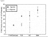
- FVIII:C activity ( ⁇ 5-15 mU/ml/108 platelets) can be detected by chromogenic analysis for at least 2.5 years after autologous G-PBC transplant in each dog with the highest levels appearing approximately one year after transplant and typically leveling off to ⁇ 5-10 mU/ml/10 8 platelets (F20, I42, M6). Samples from FVIII-Deficient dogs served as negative controls for each time point (black line).
- the term 1 U FVIII:C/ml defines 100% FVIII activity in the reference plasma from a normal (20 kg) dog; therefore, a normal (20kg) animal has ⁇ 800 total units of FVIII in its plasma volume at any given time.
- the results in Fig.6 show that multiple severe bleeding episodes occurred in each animal one year prior to G-PBC that required a transfusion with cFVIII supplements.
- each dog received daily supplements of cFVIII beginning on day one of the G-PBC transplant protocol.
- EACA was also administered to the transplanted dogs until blood was absent from their stool, which remarkably coincided with platelet FVIII:C levels reaching ⁇ 5mU/ml/10 8 platelets.
- F20 (Top Panel) displayed the lowest overall platelet FVIII:C levels of ⁇ 5mU/ml/10 8 platelets and also experienced severe intermittent bleeding episodes throughout the experimental follow-up of 2.5 years after transplant that required administration of additional supplements in the form of transfusions of normal canine plasma or cFVIII. This result indicates that 5mU/ml/108 platelet FVIII:C appears to be a threshold level of transgene expression that must be overcome in canine hemophilia A to achieve adequate correction of the bleeding phenotype.
- Transplant dog 142 (Middle Panel) maintained the highest steady state of FVIII:C of approximately 9mU/ml/10 8 platelets and did not experience severe bleeding requiring administration of cFVIII supplements ultimately demonstrating correction of the hemophilia A phenotype for at least 2.5 years after transplant.
- M64 (Bottom Panel) reached 5mU FVIII:C/ml/10 8 platelets earlier than the other transplant dogs with the synthesis of a hybrid SPD2FVIII molecule that obtained a mean FVIII:C activity level of 8mU/ml/10 8 platelets.
- the time required for whole blood to clot in a test tube was measured for each dog using traditional version of the Lee-White whole blood clotting time (WBCT) assay ( Nichols TC, et al., J Thromb Haemost 2012, 10(3): 474-476 ). Hemostatically normal dogs have a mean WBCT of 10.5 minutes ⁇ SD 1.4 minutes.
- canine blood plasma from (F20, I42 and M64) was screened for inhibitors with an activated partial thromboplastin time (aPTT) mixing assay which detects inhibitory antibodies to either coagulation factor VIII or IX.
- aPTT activated partial thromboplastin time
- Plasma from hemophilia A dogs with known Bethesda Inhibitor (BIU) titers that cross-react with and inhibit human FVIII was used as a positive control and plasma from dogs without inhibitors was assayed concurrently as a negative control for comparative analysis. The results indicate that F20, I42, and M64 did not develop inhibitors (Table 1: Column 12).
Abstract
Description
- This application claims priority to
U.S. Provisional Application No. 61/717,951 filed October 24, 2012 - The present disclosure relates to compositions and methods for targeting expression of exogenous genes to platelets. In particular, the present disclosure relates to treatment of hemophilia and other diseases and conditions by targeting expression of exogenous agents (e.g., clotting factors) to platelets.
- Hemophilia is a common bleeding disorder (occurring in approximately 1:10,000 males) in which causes severe internal bleeding that often leads to death because the patient's blood doesn't clot normally. Hemophilia usually is inherited with patients displaying severe uncontrollable bleeding events beginning at birth and reoccurring throughout the individual's life. Although there are several types of clotting factors that work together with platelets to help the blood coagulate, people with hemophilia usually have quantitative or qualitative defects in the proteins that encode coagulation factor VIII (hemophilia A) or factor IX (hemophilia B) that prevent normal hemostasis.
- Hematopoietic stem cells differentiate in the bone marrow to form megakaryocytes that mature and break-up into several thousand small fragments known as platelets, which in normal conditions circulate quietly (not interacting with the other blood cells or the vessel wall) in the blood stream for approximately 10 days with the main job to become activated, change shape and stick to damaged blood vessel to repair the injury. When blood vessels are injured, clotting factors help platelets stick together to plug cuts and close breaks on the vessels to stop bleeding.
- People with hemophilia A are missing or have low levels of clotting factor VIII. About 9 out of 10 people who have hemophilia have type A. People with hemophilia B are missing or have low levels of clotting factor IX. Both clotting factors are normally synthesized in the liver although there are reports that other cell types can be induced to synthesize fully functional forms of recombinant FVIII and FIX proteins.
- Hemophilia can be mild, moderate, or severe, depending on how much normal functional clotting factor is present in the blood. About 7 out of 10 people who have hemophilia A have the severe form of the disorder.
- Hemophilia usually occurs in males because Factors VIII and IX are located on the X chromosome (although with rare exceptions females who inherit a defective X chromosome each from an affected father and mother who is a carrier for the disease). About 1 in 10,000 individuals are born with hemophilia each year all over the world.
- The main treatment for hemophilia is protein replacement therapy. Concentrates of clotting factor VIII (for hemophilia A) or clotting factor IX (for hemophilia B) can be isolated from pools of donor blood or recombinant protein that has been prepared from tissue culture cell lines transformed with the normal genes encoding FVIII or FIX that are slowly dripped or injected into a vein at the onset of a serious bleeding event. These infusions help replace the clotting factor that's missing or low.
- Complications of replacement therapy include developing antibodies response to the normal therapeutic protein that is foreign to the patient's immune system (known as inhibitor formation), which ultimately leads to inactivation or destruction of the clotting factor and uncontrolled bleeding in about 30% of patients, developing viral infections from human clotting factors (from blood contaminated with HIV or Hepatitis from infected blood donors especially in third world countries), very expensive costs of the replacement protein which has a very short half-life (days) which requires frequent re-administration to subside a severe vascular injury and damage to joints, muscles, or other parts of the body resulting from delays in treatment.
- Thus, new treatments for hemophilia that overcome these complications are needed.
- The present disclosure relates to compositions and methods for targeting expression of exogenous genes to platelets. In particular, the present disclosure relates to treatment of hemophilia and other diseases and conditions by targeting expression of exogenous agents (e.g., clotting factors) to platelets. In some embodiments, the present disclosure relates to compositions and clinically relevant methods for hematopoietic stem cell gene therapy where targeting expression of exogenous genes within bone marrow megakaryocytes leads to expression and/or storage of recombinant therapeutic proteins within human platelets.
- For example, in some embodiments, the present invention provides a composition comprising an expression vector comprising a) an expression cassette comprising a fragment of the integrin allb gene (ITGA2B) promoter; and an exogenous gene of interest operably linked to the expression cassette. In some embodiments, the expression cassette comprises, consists essentially of, or consists of a nucleic acid sequence selected from, for example, SEQ ID NOs: 21, 22, or 23, or sequences larger or smaller than SEQ ID NOs:21, 22, 23, or 25 (e.g., 1, 2, 3, 4, 5, 6, 7, 8, 9, 10, 20, 30, 40, 50, 60, 75, 100, 150, 200, 300, 400, 500, 600 or more nucleotides larger or smaller than SEQ ID NOs: 21, 22, 23, and 25) or fragments thereof. In some embodiments, the expression vector further comprises targeting factor (e.g., a fragment of the human Von Willebrand Factor propeptide (VWFpp) operably linked to a D2 domain) (e.g., as described by SEQ ID NO:24 or
sequences - Further embodiments provide a hematopoietic stem cell comprising the expression vectors described herein (e.g., an ex vivo stem cell).
- In yet other embodiments, the present invention provides the use of such stem cells to treat diseases and conditions (e.g., hemophilia) in an animal (e.g., a human).
- The present invention also provides a method, comprising: contacting a hematopoietic stem cell with a vector as described herein to generate a modified stem cell under conditions such that a gene (e.g., Factor VIII gene) is expressed in the modified stem cell. In some embodiments, the method further comprises the step of transferring said modified stem cell into an animal (e.g., human). In some embodiments, the animal has been diagnosed with hemophilia and the transferring treats or prevents excessive bleeding in the animal. In some embodiments, the contacting occurs ex vivo. In some embodiments, the stem cells are mobilized from the animal (e.g., by administration of cytokines, mobilization into peripheral blood, contacting apheresis collection, immune magnetic bead isolation). In some embodiments, the animal expresses said Factor VIII in platelets. In some embodiments, the method is repeated (e.g., at regular intervals or when needed).
- Additional embodiments are described herein.
-
-
Figure 1 shows platelet-targeted lentiviral vector design. (A) ITGA2B gene promoter fragments direct megakaryocyte-specific expression in luciferase. (B) - 889ITGA2B-BDDFVIII-WPTS lentiviral vector diagram. (C) -673ITGA2B-VWFSPD2-BDDFVIII-WPTS lentiviral vector diagram. -
Figure 2 shows synthesis and trafficking of BDDFVIII into canine platelet α-granules
(A) confocal microscopy showing co-localization of BDDFVIII and Fg within platelets. (B) electron microscopy localized human BDDFVIII directly in α-granules. -
Figure 3 shows quantitative analysis of platelet FVIII. -
Figure 4 shows activated platelets induced to secrete FVIII:C. -
Figure 5 shows PCR analysis for detection and localization of lentiviral vector within canine genome. (A) long-term detection of BDDFVIII-lentiviral vector within leukocyte genomic DNA. (B) linear amplification-mediated (LAM)-PCR to localize lentiviral vector within
canine genome. -
Figure 6 shows correction of the canine Hemophilia A phenotype with platelet BDDFVIII. -
Figures 7 shows structural regions of the ITGA2B gene promoter and VWFspD2. Arrows show serious bleeding events before and after platelet targeted treatment. Dogs I42 and M64 show complete correction of hemostasis -
Figure 8 shows an exemplary lentiviral gene therapy vector of embodiments of the present disclosure. -
Figure 9 shows the sequence (SEQ ID NO:25) of the vector offigure 8 . -
Figure 10 shows a diagram and nucleotide sequence for integrin allb promoter fragments used in recombinant lentivirus gene transfer constructs. (A) Nucleotide sequence for -Sal I Bgl II -1218 to +30 of human allb-gene promoter Nco I used for Megakaryocyte-Specific Luciferase reporter studies (SEQ ID NO:21). (B) Nucleotide sequence for -Sal I Bgl II -889 to +30 of human allb-gene promoter Nco I used for Megakaryocyte-Specific Luciferase reporter studies (SEQ ID NO:22). (C) Nucleotide sequence for -Sal I Bgl II -673 to +30 of human allb-gene promoter Nco I used for Megakaryocyte-Specific Luciferase reporter studies (SEQ ID NO:23). Numbering is based on Prandini, et al (Biochem Biophys Res Commun 156(1) 595-601, 1988). - To facilitate an understanding of the present invention, a number of terms and phrases are defined below:
As used herein, the term "gene transfer system" refers to any means of delivering a composition comprising a nucleic acid sequence to a cell or tissue. For example, gene transfer systems include, but are not limited to, vectors (e.g., retroviral, adenoviral, adeno-associated viral, human artificial chromosomes, and other nucleic acid-based delivery systems), microinjection of naked nucleic acid, polymer-based delivery systems (e.g., liposome-based and metallic particle-based systems), biolistic injection, and the like. As used herein, the term "viral gene transfer system" refers to gene transfer systems comprising viral elements (e.g., intact viruses, modified viruses and viral components such as nucleic acids or proteins) to facilitate delivery of the sample to a desired cell or tissue. As used herein, the term "adenovirus gene transfer system" refers to gene transfer systems comprising intact or altered viruses belonging to the family Adenoviridae. - As used herein, the term "nucleic acid molecule" refers to any nucleic acid containing molecule, including but not limited to, DNA or RNA. The term encompasses sequences that include any of the known base analogs of DNA and RNA including, but not limited to, 4-acetylcytosine, 8-hydroxy-N6-methyladenosine, aziridinylcytosine, pseudoisocytosine, 5-(carboxyhydroxylmethyl) uracil, 5-fluorouracil, 5-bromouracil, 5-carboxymethylaminomethyl-2-thiouracil, 5-carboxymethylaminomethyluracil, dihydrouracil, inosine, N6-isopentenyladenine, 1-methyladenine, 1-methylpseudouracil, 1-methylguanine, 1-methylinosine, 2,2-dimethylguanine, 2-methyladenine, 2-methylguanine, 3-methylcytosine, 5-methylcytosine, N6-methyladenine, 7-methylguanine, 5-methylaminomethyluracil, 5-methoxyaminomethyl-2-thiouracil, beta-D-mannosylqueosine, 5'-methoxycarbonylmethyluracil, 5-methoxyuracil, 2-methylthio-N6-isopentenyladenine, uracil-5-oxyacetic acid methylester, uracil-5-oxyacetic acid, oxybutoxosine, pseudouracil, queosine, 2-thiocytosine, 5-methyl-2-thiouracil, 2-thiouracil, 4-thiouracil, 5-methyluracil, N-uracil-5-oxyacetic acid methylester, uracil-5-oxyacetic acid, pseudouracil, queosine, 2-thiocytosine, and 2,6-diaminopurine.
- The term "gene" refers to a nucleic acid (e.g., DNA) sequence that comprises coding sequences necessary for the production of a polypeptide, precursor, or RNA (e.g., rRNA, tRNA). The polypeptide can be encoded by a full length coding sequence or by any portion of the coding sequence so long as the desired activity or functional properties (e.g., enzymatic activity, ligand binding, signal transduction, immunogenicity, etc.) of the full-length or fragment are retained. The term also encompasses the coding region of a structural gene and the sequences located adjacent to the coding region on both the 5' and 3' ends for a distance of about 1 kb or more on either end such that the gene corresponds to the length of the full-length mRNA. Sequences located 5' of the coding region and present on the mRNA are referred to as 5' non-translated sequences. Sequences located 3' or downstream of the coding region and present on the mRNA are referred to as 3' non-translated sequences. The term "gene" encompasses both cDNA and genomic forms of a gene. A genomic form or clone of a gene contains the coding region interrupted with non-coding sequences termed "introns" or "intervening regions" or "intervening sequences." Introns are segments of a gene that are transcribed into nuclear RNA (hnRNA); introns may contain regulatory elements such as enhancers. Introns are removed or "spliced out" from the nuclear or primary transcript; introns therefore are absent in the messenger RNA (mRNA) transcript. The mRNA functions during translation to specify the sequence or order of amino acids in a nascent polypeptide.
- As used herein, the term "heterologous gene" refers to a gene that is not in its natural environment. For example, a heterologous gene includes a gene from one species introduced into another species. A heterologous gene also includes a gene native to an organism that has been altered in some way (e.g., mutated, added in multiple copies, linked to non-native regulatory sequences, etc). Heterologous genes are distinguished from endogenous genes in that the heterologous gene sequences are typically joined to DNA sequences that are not found naturally associated with the gene sequences in the chromosome or are associated with portions of the chromosome not found in nature (e.g., genes expressed in loci where the gene is not normally expressed).
- Promoter/enhancer," as used herein, denotes a segment of DNA which contains sequences capable of providing both promoter and enhancer functions (i.e., the functions provided by a promoter element and an enhancer element, see above for a discussion of these functions). For example, the long terminal repeats of retroviruses contain both promoter and enhancer functions. The enhancer/promoter may be "endogenous" or "exogenous" or "heterologous." An "endogenous" enhancer/promoter is one that is naturally linked with a given gene in the genome. An "exogenous" or "heterologous" enhancer/promoter is one that is placed in juxtaposition to a gene by means of genetic manipulation (i.e., molecular biological techniques such as cloning and recombination) such that transcription of that gene is directed by the linked enhancer/promoter. Regulatory elements may be tissue specific or cell specific. The term "tissue specific" as it applies to a regulatory element refers to a regulatory element that is capable of directing selective expression of a nucleotide sequence of interest to a specific type of tissue (e.g., mammary gland) in the relative absence of expression of the same nucleotide sequence(s) of interest in a different type of tissue (e.g., liver). Tissue specificity of a regulatory element may be evaluated by, for example, operably linking a reporter gene to a promoter sequence (which is not tissue-specific) and to the regulatory element to generate a reporter construct, introducing the reporter construct into the genome of an animal such that the reporter construct is integrated into every tissue of the resulting transgenic animal, and detecting the expression of the reporter gene (e.g., detecting mRNA, protein, or the activity of a protein encoded by the reporter gene) in different tissues of the transgenic animal. The detection of a greater level of expression of the reporter gene in one or more tissues relative to the level of expression of the reporter gene in other tissues shows that the regulatory element is "specific" for the tissues in which greater levels of expression are detected. Thus, the term "tissue-specific" (e.g., liver-specific) as used herein is a relative term that does not require absolute specificity of expression. In other words, the term "tissue-specific" does not require that one tissue have extremely high levels of expression and another tissue have no expression. It is sufficient that expression is greater in one tissue than another. By contrast, "strict" or "absolute" tissue-specific expression is meant to indicate expression in a single tissue type (e.g., liver) with no detectable expression in other tissues.
- As used herein the term "portion" or "fragment" when in reference to a nucleotide sequence (as in "a portion or fragment of a given nucleotide sequence") refers to fragments of that sequence. The fragments may range in size from four nucleotides to the entire nucleotide sequence minus one nucleotide (10 nucleotides, 20, 30, 40, 50, 100, 200, etc.). In some embodiments, fragments comprise a nucleotide sequence (e.g., promoter) that are 1, 2, 3, 4, 5, 6, 7, 8, 9, 10, 11, 12, 13, 14, 15, 16, 17, 18, 19, 20, 30, 40, 50, 100, 150, or 200 nucleotides less than the sequence or subsets thereof (e.g., 31, 32, 33, 34, 35, 35, 36, 37, 38, 39 nucleotides shorter and the like).
- As used herein, the term "oligonucleotide," refers to a short length of single-stranded polynucleotide chain. Oligonucleotides are typically less than 200 residues long (e.g., between 15 and 100), however, as used herein, the term is also intended to encompass longer polynucleotide chains. Oligonucleotides are often referred to by their length. For example a 24 residue oligonucleotide is referred to as a "24-mer". Oligonucleotides can form secondary and tertiary structures by self-hybridizing or by hybridizing to other polynucleotides. Such structures can include, but are not limited to, duplexes, hairpins, cruciforms, bends, and triplexes.
- As used herein, the terms "complementary" or "complementarity" are used in reference to polynucleotides (i.e., a sequence of nucleotides) related by the base-pairing rules. For example, the sequence "5'-A-G-T-3'," is complementary to the sequence "3'-T-C-A-5'." Complementarity may be "partial," in which only some of the nucleic acids' bases are matched according to the base pairing rules. Or, there may be "complete" or "total" complementarity between the nucleic acids. The degree of complementarity between nucleic acid strands has significant effects on the efficiency and strength of hybridization between nucleic acid strands. This is of particular importance in amplification reactions, as well as detection methods that depend upon binding between nucleic acids.
- The term "homology" refers to a degree of complementarity. There may be partial homology or complete homology (i.e., identity). A partially complementary sequence is a nucleic acid molecule that at least partially inhibits a completely complementary nucleic acid molecule from hybridizing to a target nucleic acid is "substantially homologous." The inhibition of hybridization of the completely complementary sequence to the target sequence may be examined using a hybridization assay (Southern or Northern blot, solution hybridization and the like) under conditions of low stringency. A substantially homologous sequence or probe will compete for and inhibit the binding (i.e., the hybridization) of a completely homologous nucleic acid molecule to a target under conditions of low stringency. This is not to say that conditions of low stringency are such that non-specific binding is permitted; low stringency conditions require that the binding of two sequences to one another be a specific (i.e., selective) interaction. The absence of non-specific binding may be tested by the use of a second target that is substantially non-complementary (e.g., less than about 30% identity); in the absence of non-specific binding the probe will not hybridize to the second non-complementary target.
- When used in reference to a double-stranded nucleic acid sequence such as a cDNA or genomic clone, the term "substantially homologous" refers to any probe that can hybridize to either or both strands of the double-stranded nucleic acid sequence under conditions of low stringency as described above.
- A gene may produce multiple RNA species that are generated by differential splicing of the primary RNA transcript. cDNAs that are splice variants of the same gene will contain regions of sequence identity or complete homology (representing the presence of the same exon or portion of the same exon on both cDNAs) and regions of complete non-identity (for example, representing the presence of exon "A" on
cDNA 1 wherein cDNA 2 contains exon "B" instead). Because the two cDNAs contain regions of sequence identity they will both hybridize to a probe derived from the entire gene or portions of the gene containing sequences found on both cDNAs; the two splice variants are therefore substantially homologous to such a probe and to each other. - When used in reference to a single-stranded nucleic acid sequence, the term "substantially homologous" refers to any probe that can hybridize (i.e., it is the complement of) the single-stranded nucleic acid sequence under conditions of low stringency as described above.
- As used herein, the term "hybridization" is used in reference to the pairing of complementary nucleic acids. Hybridization and the strength of hybridization (i.e., the strength of the association between the nucleic acids) is impacted by such factors as the degree of complementary between the nucleic acids, stringency of the conditions involved, the Tm of the formed hybrid, and the G:C ratio within the nucleic acids. A single molecule that contains pairing of complementary nucleic acids within its structure is said to be "self-hybridized."
- The term "isolated" when used in relation to a nucleic acid, as in "an isolated oligonucleotide" or "isolated polynucleotide" refers to a nucleic acid sequence that is identified and separated from at least one component or contaminant with which it is ordinarily associated in its natural source. Isolated nucleic acid is such present in a form or setting that is different from that in which it is found in nature. In contrast, non-isolated nucleic acids as nucleic acids such as DNA and RNA found in the state they exist in nature. For example, a given DNA sequence (e.g., a gene) is found on the host cell chromosome in proximity to neighboring genes; RNA sequences, such as a specific mRNA sequence encoding a specific protein, are found in the cell as a mixture with numerous other mRNAs that encode a multitude of proteins. However, isolated nucleic acid encoding a given protein includes, by way of example, such nucleic acid in cells ordinarily expressing the given protein where the nucleic acid is in a chromosomal location different from that of natural cells, or is otherwise flanked by a different nucleic acid sequence than that found in nature. The isolated nucleic acid, oligonucleotide, or polynucleotide may be present in single-stranded or double-stranded form. When an isolated nucleic acid, oligonucleotide or polynucleotide is to be utilized to express a protein, the oligonucleotide or polynucleotide will contain at a minimum the sense or coding strand (i.e., the oligonucleotide or polynucleotide may be single-stranded), but may contain both the sense and anti-sense strands (i.e., the oligonucleotide or polynucleotide may be double-stranded).
- As used herein, the term "purified" or "to purify" refers to the removal of components (e.g., contaminants) from a sample. For example, antibodies are purified by removal of contaminating non-immunoglobulin proteins; they are also purified by the removal of immunoglobulin that does not bind to the target molecule. The removal of non-immunoglobulin proteins and/or the removal of immunoglobulins that do not bind to the target molecule results in an increase in the percent of target-reactive immunoglobulins in the sample. In another example, recombinant polypeptides are expressed in bacterial host cells and the polypeptides are purified by the removal of host cell proteins; the percent of recombinant polypeptides is thereby increased in the sample.
- As used herein, the term "sample" is used in its broadest sense. In one sense, it is meant to include a specimen or culture obtained from any source, as well as biological and environmental samples. Biological samples may be obtained from animals (including humans) and encompass fluids, solids, tissues, and gases. Biological samples include blood products, such as plasma, serum and the like. Environmental samples include environmental material such as surface matter, soil, water, and industrial samples. Such examples are not however to be construed as limiting the sample types applicable to the present invention.
- As used herein, the term "subject" refers to organisms to be treated by the methods of the present invention. Such organisms preferably include, but are not limited to, mammals (e.g., murines, simians, equines, bovines, porcines, canines, felines, and the like), and most preferably includes humans. In the context of the invention, the term "subject" generally refers to an individual who will receive or who has received treatment (e.g., administration of a compound of the present invention and optionally one or more other agents) for a condition characterized by bacterial infection.
- The present disclosure relates to compositions and methods for targeting expression of exogenous genes to platelets. In particular, the present disclosure relates to treatment of hemophilia and other diseases and conditions by targeting expression of exogenous agents (e.g., clotting factors) to platelets.
- Embodiments of the present invention provide compositions and method for directing expression of a heterologous gene to a specific cell type using a cell-specific promoter. For example, in some embodiments, expression of heterologous or exogenous genes is targeted to a stem cell (e.g., cancer stem cell or hematopoietic stem cell) that in turn expresses the gene of interest in progenitor cells (e.g., platelets). The compositions and methods find use in the treatment of a variety of disease (e.g., platelet mediated diseases such as hemophilia). Certain embodiments of the present invention are illustrated based on treatment of hemophilia with exogenous or heterologous clotting factors, although the present invention is not limited to the treatment of hemophilia or platelet disorders.
- In some embodiments, the present disclosure relates to treatment of hemophilia and rare and common inherited bleeding disorders as well as other diseases states that involve platelets (e.g. thrombosis of veins and arteries, immune response, and cancer) and conditions by employing hematopoietic stem cell gene therapy using a fragment of a platelet specific gene promoter to drive expression of proteins only in the platelet lineage and in some circumstances fusion of a signal peptide to the therapeutic molecule to traffic recombinant proteins specifically to platelet α-granules to induce regulated release of the exogenous agents from activated platelets at the site of injury. In summary, this approach allows platelets to be utilized as a vehicle to deliver therapeutic agents to enable wound repair targeting expression of exogenous agents (e.g., normal replacement proteins to restore hemostasis by correcting inherited platelet defects, clotting factors for hemophilia, anti-thrombotic agents for deep vein thrombosis and artery occlusion and anti-neoplastic agents to shrink solid tumors and prevent angiogenesis in cancer) to platelets.
- There are several well-characterized inherited genetic defects that affect various aspects of platelet function (activation, adhesion, aggregation, signal transduction, granule storage), which usually manifest themselves clinically as a failure to control bleeding (Leslie, M. Science 328, 562-564 (2010)). Embodiments of the present invention provide autologous transplant of hematopoietic stem cells transduced with genes encoding normal integrin gene promoter driving synthesis of coagulation FVIII within platelets for correction of hemophilia A within humans. De novo synthesis of a biologically normal molecule within megakaryocytes has previous allowed trafficking of the entire protein to allow platelets to participate in wound repair. This is supported by the recent success of using hematopoietic stem cell gene transfer of integrin allb gene promoter driving expression of integrin allb to generate denovo synthesis of integrin αIIbβ3 receptor complex on platelets for improved platelet function and reduced bleeding times and blood loss for dogs deficient in integrin allb affected with canine Glanzmann Thrombasthenia (GT) (Fang, J. et al. Proc Natl Acad Sci U S A 108, 9583-9588 (2011)).
- Transduction of G-CSF mobilized peripheral blood stem cells (G-PBC) with an oncoretrovirus vector encoding integrin β3 generated de novo synthesis of viable integrin αIIbβ3 complexes on megakaryocytes derived from human GT patients (Wilcox, D. A et al., Blood. 95: 3645-52, 2000; Leslie, M. Science 328, 562-564 (2010)). It has also been shown that platelet function could be corrected within a murine model for GT by transplantation of bone marrow transduced with a lentivirus vector encoding β3 (Fang, J. et al., Blood 106, 2671-2679 (2005)) and that the use of hematopoietic stem cell gene transfer of integrin allb to generate αIIbβ3 on platelets can correct Canine Glanzmann Thrombasthenia (GT) (Fang, J. et al. Proc Natl Acad Sci U S A 108, 9583-9588 (2011)).
- Experiments conducted during the course of development of embodiments of the present invention demonstrate transferring genes into G-PBC show that oncoretrovirus transduced human megakaryocytes and platelets could synthesize and store human coagulation factor VIII and release FVIII upon activation in vitro with physiological agonists of platelet activation (Wilcox, D.A., et al., Journal of Thrombosis and Haemostasis. 1: 2477-89, 2003). It has also been shown that hemostasis could be improved within a murine model for hemophilia A (even in the presence of inhibitory antibodies to FVIII) by transplantation of bone marrow transduced with a lentivirus vector under the transcriptional control of the -889 fragment of the integrin allb gene promoter driving expression of human FVIII (Shi, Q. and Wilcox, D.A., et al., Journal of Thrombosis and Haemostasis 5: 352-361, 2007) and (Shi, Q., et al., Blood 112: 2713-21, 2008) and that the use of G-CSF mobilized PBC for hematopoietic stem cell lentivirus mediate gene transfer of integrin allb gene promoter fragment with and without VWFSPD2 fused to human FVIII induced expression of fully functional FVIII within platelets and platelet α granules, which corrected platelet function and resulted in improved hemostasis and the reduction and/or absence of any serious bleeding events; thus animals did not require injection with FVIII protein replacement therapy within a large animal "canine" model for hemophilia A for at least 2.5 years after transplant as well as inhibitory antibodies were not generated to the recombinant human FVIII stored within canine platelets
- Accordingly, in some embodiments the present invention provides compositions and methods for genetic therapies for targeting stem cells (e.g., hematopoietic stem cells). The compositions and methods described herein find use in the treatment of a variety of disease and conditions (e.g., platelet mediated disorders).
- Some embodiments of the present invention are illustrated with the treatment of hemophilia and other platelet diseases, although the present invention is not limited to the treatment of hemophilia. In some embodiments, compositions and methods comprise in vivo or ex vivo genetic therapies. For example, in some embodiments, hematopoietic stem cells are mobilized and targeted ex vivo with vectors that target expression of exogenous genes (e.g., clotting factors, platelet proteins pertinent to platelet function, anti-thrombotic agents for thrombotic disorders and anti-angiogenic and anti-neoplastic agents for oncogenic disorders) specifically to platelets following reintroduction of the modified hematopoietic stem cells.
- The present invention is not limited to a particular targeting vector or expression cassette. In some embodiments, expression cassettes comprise an exogenous gene of interest operable linked to a cell (e.g., platelet) specific promoter. In some embodiments, expression cassettes further comprise genes expression targeting or signal molecules, as well as expression enhancers.
Figures 7 ,8 , and10 show exemplary expression cassettes and vectors useful in embodiments of the present invention. Exemplary vector components are described below. - In some embodiments, vectors comprise promoters that direct gene expression to particular cell type. For example, in some embodiments, promoters are platelet specific promoters. The present invention is not limited to particular platelet specific promoter. In some embodiments, truncated integrin allb gene (ITGA2B) promoters are used. Exemplary promoters include, but are not limited to, -1218, -889 and -673 ITGA2B promoters that encode"ETS" and "GATA" elements for high level of gene transcription within megakaryocytes and a Repressor region that inhibits gene transcription within other hematopoietic cell lineages (SEQ ID NOs: 21, 22, 23; and 25;
Figures 8 and10 ). - The nucleotide sequence of the ITGA2B gene promoter was first characterized in 1988 by a group in France headed by Dr. Gerard Marguerie (Prandini MH, Denarier E, Frachet P, Uzan G, Marguerie G. Isolation of the human platelet glycoprotein IIb gene and characterization of the 5' flanking region. Biochem Biophys Res Commun 1988, 156(1): 595-601).
Figure 7 shows structural regions of the ITGA2B promoter. In some embodiments, a fragment (e.g., 1218, -889 and -673) of the promoter is utilized. In some embodiments, the -673 contains all of the essential regulatory elements to drive platelet-specific transgene expression. In 1993, researchers perform gene promoter studies and found that the transcription factor GATA-1 was important for high level gene expression in megakaryocytes with at least three and maybe a fourth region serves as GATA-1 binding site identified in ITGA2B (Martin F, Prandini MH, Thevenon D, Marguerie G, Uzan G. The transcription factor GATA-1 regulates the promoter activity of the platelet glycoprotein IIb gene. J Biol Chem 1993, 268(29): 21606-21612). Next it was discovered that there are three consensus sequences for another transcription factor Ets to bind to ITGA2B that are believed to act together to produce a high level of transgene expression (Lemarchandel V, Ghysdael J, Mignotte V, Rahuel C, Romeo PH. GATA and Ets cis-acting sequences mediate megakaryocyte-specific expression. Mol Cell Biol 1993, 13(1): 668-676). The Murine ITGA2B promoter was cloned and found to have very high nucleotide sequence homology with the human ITGA2B promoter, especially at the regions where transcription factors consensus binding sequences were identified (Denarier E, Martin F, Martineau S, Marguerie G. PCR cloning and sequence of the murine GPIIb gene promoter. Biochem Biophys Res Commun 1993, 195(3): 1360-1364). Further gene promoter analysis showed that a region of ITGA2B from -139 to -63 must be preserved to prevent the gene promoter from driving transgene transcription within other hematopoietic lineages, thus this region is labeled the Repressor (Prandini MH, Martin F, Thevenon D, Uzan G. The tissue-specific transcriptional regulation of the megakaryocytic glycoprotein IIb gene is controlled by interactions between a repressor and positive cis-acting elements. Blood 1996, 88(6): 2062-2070). Finally, a binding site for the Sp1 was identified in ITGA2B that is believed important to work with Ets in conjunction with the other elements when promoter forms a three dimensional structure that is essential for optimal platelet specific gene transcription (Block KL, Shou Y, Poncz M. An Ets/Sp1 interaction in the 5'-flanking region of the megakaryocyte- specific alpha IIb gene appears to stabilize Sp1 binding and is essential for expression of this TATA-less gene. Blood 1996, 88(6): 2071-2080). - The present invention is not limited to the ITGA2B promoters described in SEQ ID NOs: 21-23). Embodiments of the present invention contemplate fragments, portions, and combinations of the fragments described herein. In some embodiments, fragments are shorter than the full length ITGA2B promoter (e.g., 1, 2, 3, 4, 5, 6, 7, 8, 9, 10, 20, 30, 40, 50, 60, 75, 100, 150, 200 or more nucleotides shorter) and maintain desired activity (e.g., the ability to drive cell-specific expression to elicit a desired effect, e.g., reduction in sing or symptoms of a disease or condition).
- In some embodiments, promoters comprise the ITGA2B fragments described in SEQ ID NOs: 21, 22, and 23, or sequences that are 1, 2, 3, 4, 5, 6, 7, 8, 9, 10, 20, 30, 40, 50, 60, 75, 100, 150, 200, 300, 400, 500, 600 or more nucleotides larger or smaller than SEQ ID NOs: 21, 22, and 23. For example, in some embodiments, fragments that are 1, 2, 3, 4, 5, 6, 7, 8, 9, 10, 20, 30, 40, 50, 60, 75, 100, 150, 200 or more nucleotides smaller than SEQ ID NO:21 are utilized. In some embodiments, fragments that are 1, 2, 3, 4, 5, 6, 7, 8, 9, 10, 20, 30, 40, 50, 60, 75, 100, 150, 200, 300, 400, 500, 600 or more nucleotides larger than SEQ ID NO:23 are utilized.
- In some embodiments, discontinuous fragments of SEQ ID NOs: 21, 22, or 23 that retain promoter activity are utilized. For example in some embodiments, 1, 2, 3, 4, 5, 6, 7, 8, 9, 10, 20, 30, 40, 50, 60, 75, 100, 150, 200, 300, 400, 500, 600 nucleotides of SEQ ID NOs 21, 22, or 23 are utilized.
- In some embodiments, promoter fragments comprise one or more elements useful for promoter activity. Examples include, but are not limited to, GATA elements (e.g., GATA54 or GATA454), sP1 elements, Ets35 elements, and the like (See e.g., Block et al., Blood 1994 84: 3385-3393; Prandini et al., Blood 1996 88: 2062-2070; Block et al., Blood 1996 88: 2071-2080; and Doubeikovski et al., J. Biol. Chem. 272: 24300-24307, 1997; each of which is herein incorporated by reference in its entirety).
- In some embodiments, the 5' ends of promoters are modified to add restriction endonuclease sites to aid in cloning and constructing expression vectors. For example, in some embodiments, 1, 2, 3, or 4 nucleotides at the 5' end are modified from the wild type sequence or the fragments disclosed herein to add restriction endonuclease sites.
- In some embodiments, the fragments comprise, consist essentially of, or consist of promoter sequence found in SEQ ID NOs: 21, 22, or 23.
- The present invention is not limited to a particular exogenous gene. In embodiments that treat hemophilia, exogenous genes are generally clotting factors (e.g., Factor VIII and/or Factor IX). Human Factor VIII has the accession number NM_000132.3 and Human Factor IX has the accession number NM_000133.3.
- Other exogenous genes may be utilized in the treatment of other platelet related conditions. In some embodiments, exogenous genes useful in the treatment of diseases other than platelet related disorders are utilized (e.g., in the treatment of cancer by using platelets to target release of anti-neplastic agents "i.e. IL-24" to shrink solid tumors and anti-thrombotic agents to be released at the site of blood clots such as cases of deep vein thrombosis).
- In some embodiments, expression cassettes further comprise a targeting factor that targets expression into a particular sub-structure of a platelet. In some embodiments, expression cassettes further comprise the minimal amino acid sequence of a signal sequence peptide that has been found to be able traffic not only VWF but also recombinant proteins fused to the peptide that has the proven ability to store proteins in cellular granule compartments specifically the natural storage sub-structure endothelial cells "Weibel-Palade bodies" and platelet α-granules, both of which can be secreted upon cellular activation. For example, in some embodiments, expression constructs comprise a nucleic acid encoding a Von Willebrand Factor propeptide signal peptide and D2 domain (SPD2) to promote trafficking of molecules directly into the α-granule compartment as shown in Rosenberg JB et al, Intracellular Trafficking of FVIII to von Willebrand Factor storage Granules, J. Clin. Invest. 101, 613-624 (1998); Haberichter SL, Jacobi P, Montgomery RR. Critical independent regions in the VWF propeptide and mature VWF that enable normal VWF storage. Blood 2003, 101(4): 1384-1391 and Haberichter et al, The Von Willlebrand Factor Propeptide (VWFpp) Traffics an Unrelated Protein to Storage, Arterioscler Thromb Vas Biol. 22, 921 926 (2002).
- An example of an expression cassette comprising a 673 bp fragment of the ITGA2B gene promoter and a VWF/SPD2 gene is shown in
Figure 7 (SEQ ID NO:24). Variants of these sequences that retain its desired activity are specifically contemplated for use in compositions and methods of embodiments of the present invention. - In some embodiments, constructs comprise an expression enhancer (e.g., Woodchuck Hepatitis Virus (WHP) Posttranscriptional Regulatory Element (WPRE)) between the promoter/signaling cassette and the exogenous gene of interest. This element has been utilized by several gene transfer strategies because its structure inhibits degradation of the transcript within the cell and thus allows for more therapeutic protein to be synthesized compared to a gene transfer vector in the absence of WPRE.
- The present invention is not limited to a particular expression vector. In some embodiments, vectors are self-inactivating. In some embodiments, vectors are retroviral vectors (e.g., lentiviral vectors). Table 2 provides a summary of exemplary suitable vectors.
Table 2 Virus Advantages Disadvantages Adenovirus High titer High gene expression Can infect non-dividing cells Accepts very large cassettes (40 kb) Immunogenic Does not integrate into genome Adeno-associated virus Can infect non-dividing cells Relatively safe in humans Accepts small cassettes (4 kb) Low transduction efficiency in hematopoietic cells Alphavirus (Sindbis) Can infect non-dividing cells High titer High transduction efficiency High gene expression Toxic to cells Does not integrate into genome Lentivirus Stably incorporated into genome Can infect non-dividing cells New to field Safety uncertain in humans Retrovirus Stably incorporated into genome Relatively safe in humans High titer Accepts large cassettes (8 kb) Infects only dividing cells - Retroviruses (family Retroviridae) are divided into three groups: the spumaviruses (e.g., human foamy virus); the lentiviruses (e.g., human immunodeficiency virus and sheep visna virus) and the oncoviruses (e.g., MLV, Rous sarcoma virus).
- Retroviruses are enveloped (e.g., surrounded by a host cell-derived lipid bilayer membrane) single-stranded RNA viruses that infect animal cells. When a retrovirus infects a cell, its RNA genome is converted into a double-stranded linear DNA form (e.g., it is reverse transcribed). The DNA form of the virus is then integrated into the host cell genome as a provirus. The provirus serves as a template for the production of additional viral genomes and viral mRNAs. Mature viral particles containing two copies of genomic RNA bud from the surface of the infected cell. The viral particle comprises the genomic RNA, reverse transcriptase and other pol gene products inside the viral capsid (which contains the viral gag gene products), which is surrounded by a lipid bilayer membrane derived from the host cell containing the viral envelope glycoproteins (also referred to as membrane-associated proteins).
- The organization of the genomes of numerous retroviruses has allowed the adaptation of the retroviral genome to produce retroviral vectors. The production of a recombinant retroviral vector carrying a gene of interest is typically achieved in two stages. First, the gene of interest is inserted into a retroviral vector which contains the sequences necessary for the efficient expression of the gene of interest (including promoter and/or enhancer elements which may be provided by the viral long terminal repeats [LTRs] or by an internal promoter/enhancer and relevant splicing signals), sequences required for the efficient packaging of the viral RNA into infectious virions (e.g., the packaging signal [Psi], the tRNA primer binding site [-PBS], the 3' regulatory sequences required for reverse transcription [+PBS] and the viral LTRs). The LTRs contain sequences required for the association of viral genomic RNA, reverse transcriptase and integrase functions, and sequences involved in directing the expression of the genomic RNA to be packaged in viral particles. For safety reasons, many recombinant retroviral vectors lack functional copies of the genes that are essential for viral replication (these essential genes are either deleted or disabled); the resulting virus is said to be replication defective.
- Second, following the construction of the recombinant vector, the vector DNA is introduced into a packaging cell line. Packaging cell lines provide viral proteins required in trans for the packaging of the viral genomic RNA into viral particles having the desired host range (e.g., the viral-encoded gag, pol and env proteins). The host range is controlled, in part, by the type of envelope gene product expressed on the surface of the viral particle. Packaging cell lines may express ecotrophic, amphotropic or xenotropic envelope gene products. Alternatively, the packaging cell line may lack sequences encoding a viral envelope (env) protein. In this case the packaging cell line will package the viral genome into particles that lack a membrane-associated protein (e.g., an env protein). In order to produce viral particles containing a membrane associated protein that will permit entry of the virus into a cell, the packaging cell line containing the retroviral sequences is transfected with sequences encoding a membrane-associated protein (e.g., the G protein of vesicular stomatitis virus [VSV]). The transfected packaging cell will then produce viral particles that contain the membrane-associated protein expressed by the transfected packaging cell line; these viral particles, which contain viral genomic RNA derived from one virus encapsidated by the envelope proteins of another virus are said to be pseudotyped virus particles.
- Viral vectors, including recombinant retroviral vectors, provide a more efficient means of transferring genes into cells as compared to other techniques such as calcium phosphate-DNA co-precipitation or DEAE-dextran-mediated transfection, electroporation or microinjection of nucleic acids. It is believed that the efficiency of viral transfer is due in part to the fact that the transfer of nucleic acid is a receptor-mediated process (i.e., the virus binds to a specific receptor protein on the surface of the cell to be infected). In addition, the virally transferred nucleic acid once inside a cell integrates in controlled manner in contrast to the integration of nucleic acids which are not virally transferred; nucleic acids transferred by other means such as calcium phosphate-DNA co-precipitation are subject to rearrangement and degradation.
- Commonly used recombinant retroviral vectors are derived from the amphotropic Moloney murine leukemia virus (MoMLV) (Miller and Baltimore, Mol. Cell. Biol., 6:2895 [1986]). The MoMLV system has several advantages: 1) this specific retrovirus can infect many different cell types, 2) established packaging cell lines are available for the production of recombinant MoMLV viral particles and 3) the transferred genes are permanently integrated into the target cell chromosome. The established MoMLV vector systems comprise a DNA vector containing a small portion of the retroviral sequence (the viral long terminal repeat or "LTR" and the packaging or "psi" signal) and a packaging cell line. The gene to be transferred is inserted into the DNA vector. The viral sequences present on the DNA vector provide the signals necessary for the insertion or packaging of the vector RNA into the viral particle and for the expression of the inserted gene. The packaging cell line provides the viral proteins required for particle assembly (Markowitz et al., J. Virol., 62:1120 [1988]).
- Despite these advantages, existing retroviral vectors based upon MoMLV are limited by several intrinsic problems: 1) they do not infect non-dividing cells (Miller et al., Mol. Cell. Biol., 10:4239 [1992]), 2) they produce low titers of the recombinant virus (Miller and Rosman, BioTechn., 7: 980 [1989]; and Miller, Nature 357: 455 [1992]) and 3) they infect certain cell types (e.g., human lymphocytes) with low efficiency (Adams et al., Proc. Natl. Acad. Sci. USA 89:8981 [1992]). The low titers associated with MoMLV-based vectors has been attributed, at least in part, to the instability of the virus-encoded envelope protein. Concentration of retrovirus stocks by physical means (e.g., ultracentrifugation and ultrafiltration) leads to a severe loss of infectious virus.
- Other commonly used retrovectors are derived from lentiviruses including, but not limited to, human immunodeficiency virus (HIV) or feline immunodeficiency virus (FIV). Lentivirus vectors have the advantage of being able to infect non replicating cells.
- The low titer and inefficient infection of certain cell types by retro vectors has been overcome by the use of pseudotyped retroviral vectors which contain the G protein of VSV as the membrane associated protein. Unlike retroviral envelope proteins which bind to a specific cell surface protein receptor to gain entry into a cell, the VSV G protein interacts with a phospholipid component of the plasma membrane (Mastromarino et al., J. Gen. Virol., 68:2359 [1977]). Because entry of VSV into a cell is not dependent upon the presence of specific protein receptors, VSV has an extremely broad host range. Pseudotyped retroviral vectors bearing the VSV G protein have an altered host range characteristic of VSV (i.e., they can infect almost all species of vertebrate, invertebrate and insect cells). Importantly, VSV G-pseudotyped retroviral vectors can be concentrated 2000-fold or more by ultracentrifugation without significant loss of infectivity (Burns et al., Proc. Natl. Acad. Sci. USA, 90:8033 [1993]).
- The VSV G protein has also been used to pseudotype retroviral vectors based upon the human immunodeficiency virus (HIV) (Naldini et al., Science 272:263 [1996]). Thus, the VSV G protein may be used to generate a variety of pseudotyped retroviral vectors and is not limited to vectors based on MoMLV.
- The present invention is not limited to the use of the VSV G protein when a viral G protein is employed as the heterologous membrane-associated protein within a viral particle. Sequences encoding other G proteins derived from other members of the Rhabdoviridae family may be used; sequences encoding numerous rhabdoviral G proteins are available from the GenBank database.
- The majority of retroviruses can transfer or integrate a double-stranded linear form of the virus (the provirus) into the genome of the recipient cell only if the recipient cell is cycling (i.e., dividing) at the time of infection. Retroviruses that have been shown to infect dividing cells exclusively, or more efficiently, include MLV, spleen necrosis virus, Rous sarcoma virus human immunodeficiency virus, and other lentiviral vectors.
- It has been shown that the integration of MLV virus DNA depends upon the host cell's progression through mitosis and it has been postulated that the dependence upon mitosis reflects a requirement for the breakdown of the nuclear envelope in order for the viral integration complex to gain entry into the nucleus (Roe et al., EMBO J., 12:2099 [1993]). However, as integration does not occur in cells arrested in metaphase, the breakdown of the nuclear envelope alone may not be sufficient to permit viral integration; there may be additional requirements such as the state of condensation of the genomic DNA (Roe et al., supra).
- The present invention is not limited to retroviral vectors. Large numbers of suitable vectors are known to those of skill in the art, and are commercially available. Such vectors include, but are not limited to, the following vectors: 1) Bacterial -- pQE70, pQE60, pQE 9 (Qiagen), pBS, pD10, phagescript, psiX174, pbluescript SK, pBSKS, pNH8A, pNH16a, pNH18A, pNH46A (Stratagene); ptrc99a,
pKK223 3,pKK233 3, pDR540, pRIT5 (Pharmacia); and 2) Eukaryotic -- pWLNEO, pSV2CAT, pOG44, PXT1, pSG (Stratagene) pSVK3, pBPV, pMSG, pSVL (Pharmacia). Any other plasmid or vector may be used as long as they are replicable and viable in the host. In some embodiments of the present invention, mammalian expression vectors comprise, along with an expression cassette as described herein, an origin of replication, any necessary ribosome binding sites, polyadenylation sites, splice donor and acceptor sites, transcriptional termination sequences, and 5' flanking non transcribed sequences. In other embodiments, DNA sequences derived from the SV40 splice, and polyadenylation sites may be used to provide the non-transcribed genetic elements. - In some embodiments, the present invention provides systems and methods for genetic manipulation of stem cells (e.g., hematopoietic stem cells or cancer stem cells). In some embodiments, the compositions and methods described herein find use in the treatment of a variety of disorder related to platelet function (e.g., hemophilia and the disorders described in Table 3 below).
- There have been several bleeding disorders characterized by molecular genetic defects of platelet membrane, cytoplasmic and granular proteins that usually lead to prolonged bleeding events. While each disorder is rare, maybe occurring in 1:1,000,000 individuals (e.g. Glanzmann Thrombasthenia), taken collectively, an inherited platelet defect occur in 1:20,000 people worldwide as described in Wilcox, D.A. White II, G.C, Gene therapy for platelet disorders: studies with glanzmann's thrombasthenia. Journal of Thrombosis and Haemostasis. 1: 2300-2311, (2003) and Wilcox, D. A., White II, G.C: Gene therapy for platelet disorders. In: Platelets. Second Edition, A.D. Michelson (ed.), Academic Press, San Diego, Chapter 71: 1313-1325, (2007) and Third Edition, Chapter 64: In Press (2012). In addition to inherited platelet defects, hematopoietic stem cell gene therapy aimed at targeting therapeutic agents to the platelet surface, cytoplasm or granules finds use as a strategy to correct other disorders of hemostasis, thrombosis, immune response and cancer.
Table 3 Inherited Disorder Detect Function Disrupted G-Protein Disorder Gαq, Gαi1 Activation ADP Receptor Defect P2Y12 Activation Bernard-Soulier Syndrome Glycoprotein Ib-IX Adhesion Collagen Receptor Deficiency Glycoproteins Ia-IIa Adhesion Glanzmann Thrombasthenia Glycoproteins IIb-IIIa Aggregation Gray Platelet Syndrome NBEAL2 α-Granule Formation/Storage QuebecPlatelet Disorder Urokinase plasminogen activator α-Granule Storage Scott Syndrome Phosphatidylserine Translocation Coagulation May-Hegglin Anomaly Fechtner Syndrome Sebastian Platelet Syndrome Epstein Syndrome MYH9 Cytosketeton/Platelet Formation Wiskott-Aldrich Syndrome WAS Protein Cytoskeleton Chediak-Higashi Syndrome CHS protein Dense Body Formation/Storage Hermansky-Pudlak Syndrome HPS1, HPS3-7, AP-3 Dense Body Formation/Storage Thromboxane Deficiency Thromboxane A2 Signal Transduction - In some embodiments, therapeutic methods are ex vivo methods, in which autologous hematopoietic stem cells are harvested from an animal (e.g., human) in need of treatment, modified using one of the vector described herein, and re-introduced into the original donor. Such autologous methods reduce the risk of autoimmune or rejection responses that can occur with infusion of donor clotting factors and allow one to limit gene transduction to hematopoietic stem cells through ex vivo transduction.
- An exemplary method for ex vivo modification of hematopoietic stem cells is described in Aiuti et al. (Science 341:865 (2013; herein incorporated by reference in its entirety). For example, in some embodiments, the method includes the steps of administering cytokines to mobilize peripheral blood stem cells into the peripheral blood; performing apheresis and magnetic bead selection for CD34+ cells; preconditioning using e.g., bulsulfan and/or other agents like fludarabine; and using a viral (e.g., lentiviral) gene transfer vector to modify stem cells before they are re-introduced into the patient.
- In some embodiments, hematopoietic stem cells are mobilized using administration of cytokines or other mobilization agents (See e.g., Fu et al., Blood Rev. 2000 Dec;14(4):205-18 and United States Patent
7417026 , each of which is herein incorporated by reference in its entirety for a discussion of mobilization protocols), although other suitable protocols may be utilized. - For example, in some embodiments, mobilization cytokines include, but are not limited to, Interleukin-3 (IL-3), granulocyte colony stimulating factor (G-CSF), also known as Amgen's FDA approved drug Neupogen, stem cell factor (SCF), granulocyte macrophage colony-stimulating factor (GM-CSF), and sequential or co-administration of one or more of IL-3, GM-CSF, SCF, and GM-CSF.
- Suitable dosage ranges for mobilization agents vary, but in general, the compounds are administered in the range of about 0.1 µg/kg-5 mg/kg of body weight; preferably the range is about 1 µg/kg-300 µg/kg of body weight; more preferably about 10 µg/kg-100 µg/kg of body weight. For a typical 70-kg human subject, thus, the dosage range is from about 0.7 µg-350 mg; preferably about 700 µg-21 mg; most preferably about 700 µg-7 mg. Dosages can be higher when the compounds are administered orally or transdermally as compared to, for example, i.v. administration. The compounds can be administered as a single bolus dose, a dose over time, as in i.v. or transdermal administration, or in multiple dosages.
- The amount of active compound to be administered can vary according to the discretion of the skilled artisan. The amount of active compound to be administered to the recipient is within the ranges described above for stem cell mobilization. However, the administration of such amounts will vary according to the standards set forth by clinicians in the field of stem cell enhancement therapy.
- Following mobilization, CD34+ Peripheral Blood stem cells (PBC) are isolated from the low molecular weight mononuclear cells by immunomagnetic beads using Miltenyi's automacs system (for large animal, dogs, 25-45 kg) and Miltenyi's Clinimacs system (for humans) recently approved for clinical use by the FDA. The CD34+PBC are the genetically modified using the vectors described herein and re-introduced into a subject in need by autologous stem cell transplant. In some embodiments, a single treatment is utilized to provide long-term protection against episodes of bleeding. In some embodiments that treat hemophilia, treatment is performed on a regular basis (e.g., weekly, monthly, yearly, once every 2, 3, 4, 5 or more years, and the like) in order to prevent episodes of bleeding. In some embodiments, treatment is only administered when episodes of abnormal bleeding occur (e.g., following accidents, prior to or following surgery, etc,). In some embodiments, maintenance therapy is administered in combination with extra therapy when episodes of abnormal bleeding occur.
- The following examples are provided in order to demonstrate and further illustrate certain preferred embodiments and aspects of the present invention and are not to be construed as limiting the scope thereof.
- Human transformed cell lines were obtained from American Type Culture Collection (Rockville, MD) and propogated under conditions described for promegakaryocytic (HEL Megakaryocyte transformed cell line) (Bray, P. F. et al. ); Greenberg, S. M., et al., T-cell lymphoma (KT1), (Okamoto, T., et al. J Biol Chem 261, 4615-4619 (1986)) B-cell lymphoma (Raji), (Choi, J. H. et al. )) Erythroleukemia (K562), (Gauwerky, C. & Golde, D. W. Blood 56, 886-891 (1980)) and Epithelial (HeLa) (Goldstein, M. N., et al., Annals of the New York Academy of Sciences 89, 474-483 (1960)) cell lines.
- ITGA2B Gene Promoter Constructs: Genomic DNA was isolated from the human pro-megakaryocyte cell line, Dami,49 and human ITGA2B gene promoter fragments were amplified by PCR using either sense primer "-1218"(5'-TTACGCGTCGACAGATCTAAATGTGGCTGGTTACCCC-3')"-1198" (SEQ ID NO:1) (bold) of ITGA2B or "-889"(5'-TTACGCGTCGACAGATCTGTGCTCAATGCTGTGCC-3')"-872" (SEQ ID NO:2) (bold) of ITGA2B, or "-673"(5'-TTACGCGTCGACAGATCTCCTTGCCACCTAGACC-3')"-654" (SEQ ID NO:3) (bold) of ITGA2B and anti-sense primer (5'-GGCGTCTTCCATGGTCCTTCTTCCACAACC-3') (SEQ ID NO:4) encoding nucleotides +99 to +86 of luciferase pGL3-BASIC and nucleotides +30 to +15 (bold) of ITGA2B gene promoter. Correct identity of constructs was confirmed by
nucleotide sequence analysis. - pCMVLuc: A BgIII and HindIII restriction digest of cytomegalovirus tissue non-specific gene promoter (878bp) from pRc/CMV (Invitrogen) is ligated into the pGL3-Basic Luciferase vector (Promega, Madison, WI). This construct served as the positive control for high level gene expression within all cell-types; thus, assigned an arbitrary level of 100% luciferase activity for each cell line (
Fig. 1 ). - pGL3-BasicLuc: Negative control construct for 0% luciferase activity (
Fig. 1 ) because lacks a gene promoter to drive luciferase gene transcription (Promega). - pCMVnlac: Cell lines were co-transfected with one of the pITGA2BLuc+ constructs and
- pCMVnlac encoding the β-gaiactosidase marker gene to normalize transgene
expression (Wilcox et al., Proc Natl Acad Sci U S A 1999, 96(17): 9654-9659. - Cell lines (2x10⁷) were co-transfected with either (20) µg) of the ITGA2B gene promoter construct (-1218, -889, -673) (
Fig. 1A ) or the positive (CMV) or negative (Basic) controls encoding firefly luciferase and pCMVnlac (20µg) encoding β-galactosidase.49 Briefly, forty-eight hours after co-transfection cells were washed, harvested, and lysates were prepared and frozen to -80°C using the luciferase assay system (Promega). Luciferase activity was measured
with aTurner Designs Model 20 Luminometer. Detection of β-galactosidase activity was performed to normalize transient transgene expression for each cell line with a sensitive
ELISA enzymatic assay that measured colormetric change with the substrate for β-galactosidase, chlorophenol red β-D-galactophranoside (CPRG) (Eustice DC, et al., Biotechniques 1991, 11(6): 739-740, 742-733). The percent of luciferase activity was determined by comparing the mean value of the Relative Light Units (RLU) of luciferase/CPRG Vmax value for each construct to reveal the transfection efficiency for each cell line. The RLU for pCMVLuc was assigned arbitrarily a value of 100% and all other results were calculated for each vector based upon that
value as shown inFig.1A . - ITGA2B-(M)WPTS genetic transfer vectors are derived from a HIV type-1 lentiviral vector (D.Trono, University of Geneva, Switzerland).51 p-889ITGA2B-BDDFVIII-WPTS lentiviral vector (
Fig.1B ) encodes a -889 to +30 nucleotide fragment of the human ITGA2B promoter and human BDDFVIII molecule.16 p-673ITGA2B-VWFSPD2-BDDFVIII-WPTS lentiviral vector (Fig. 1C ) encodes a fragment of the human ITGA2B gene promoter from nucleotide -673 to +30 followed by a fragment of the human Von Willebrand Factor propeptide (VWFpp encoding 540 amino acid VWF signal peptide (SP;66bp) linked to the D2 domain(1,199bp) and cDNA encoding human BDDFVIII to allow megakaryocyte-specific transcription of a hybrid molecule that uses the SPD2 peptide to traffic human BDDFVIII to platelet α-granules.22 cDNA encoding SP was amplified by PCR with forward Primer - (P)1(5'GTTAATCGATATCTCCTTGCCACCTAGA3') (SEQ ID NO:5), and reverse
- P2(5'AATCTGGCAGGAATCATGGTCCTTCTTCCACAACCT3') (SEQ ID NO:6) and ligated to D2 amplified by PCR using forward P3(5'AGGTTGTGGAAGAAGGACCATGATTCCTGCCAGATTTGC3') (SEQ ID NO:7) and reverse P4(5'CGTCTCGGCCCTTTTGCTGCCATGAGACAG3') (SEQ ID NO:8). A nested PCR linked ITGA2B promoter and VWFSPD2 with P5(5'ATCGATATCTCCTTGCCACCT A3') (SEQ ID NO:9) and P4. p-889ITGA2-BDDFVIII-WPTS served as a template for PCR of cDNA encoding a fragment of BDDFVIII using forward
- P7(5'CGTCTCAGGGCCACCAGAAGATACTACCT3') (SEQ ID NO:10) and reverse
- P8(5'ACGCGTCTTCTCTACATACTAGTA3') (SEQ ID NO: 11) to synthesize cDNA that ligated directly to VWFD2. All PCR products were cloned into pCR-Blunt II-TOPO (Life Technologies, Grand Island, NY) using unique restriction sites -673ITGA2B-SPD2(Clal and BsmBI) ligated to 5'hBDDFVIII (BsmB1 and Mlul) with 3'BDDFVIII (Mlul and Spel). All fragments were cloned into pWPTS lentiviral vector and the correct identity confirmed by
nucleotide sequence analysis. Recombinant virions were generated from three-plasmid transient co-transfection followed by supernatant collection, 500-fold concentration by centrifugation, and storage at -80oC until utilized (Fang J, et al., Proc Natl Acad Sci U S A 2011, 108(23): 9583-9588). Virion titer was determined by RT-PCR (Lizee G, et al., Hum Gene Ther2003, 14(6): 497-507). Replication-competent virions were confirmed absent from stocks with marker rescue assays (Wilcox et al., Blood 2000, 95(12): 3645-3652). - Cytokine mobilized CD34+G-PBC gene transfer and autologous transplant studies using FVIII-Deficient dogs affected with hemophilia A (University of North Carolina, Chapel Hill, NC) (Lozier JN, et al., Proc Natl Acad Sci U S A 2002, 99(20): 12991-12996) were conducted and approved by Institutional Animal Care and Use Committees of the University of North Carolina and The Medical College of Wisconsin which are both accredited facilities of the American Association for Accreditation of Laboratory Animal Care.
- 19 Adult (1.25, 4.25, and 6.5 year old) FVIII-Deficient male dogs were injected daily with canine recombinant granulocyte colony stimulating factor (crG-CSF;10µg/kg/d) and stem cell factor (crSCF; 5µg/kg/d) (Amgen, Thousand Oaks, CA). G-PBC collection was performed on the third day using a COBE Spectra Blood Cell Separator. Mononuclear G-PBC were isolated with Fico-Paque Plus (GE Healthcare, Uppsala, Sweden). CD34+ G-PBC were selected with a biotin-conjugated-1H6 Ab (1mg/ml) (Richard Nash, Fred Hutchinson Research Institute, Seattle, WA) and anti-biotin immuno-magnetic beads (1:5 dilution) on an Automacs magnetic cell separator (Miltenyi Biotec Inc., Auburn, CA). CD34+ G-PBC were transduced with -889ITGA2B-BDDFVIII-WPTS or -673ITGA2B-VWFSPD2-BDDFVIII-WPTS lentiviral vector. Briefly, 4x10⁶ cells/well were seeded in a 6-well plate (Falcon-Becton Dickinson, Franklin Lakes, NJ) coated with 20µg/cm2 RetroNectin (Takara Shuzo, Otsu, Shiga, Japan) and incubated with 1.0x10⁴ ITGA28-FVIII lentivirions/cell in
X-Vivo 10 containing 10%FCS, rhIL-3, rcalL-6,
rcaSCF, rhTPO and rhflk2/flt3 ligand. Approximately 3x10⁶ FVIII-transduced CD34+G-PBC/kg and 2x10⁸ CD34(-)G-PBC were infused into the cephalic vein of each autologous transplant recipient pre-conditioned with a non-myeloablative dose of 5-10 mg/kg Busulfex®. Transient immune suppression administered for ≈90 days after transplant with 10 mg/kg/d cyclosporine (Gengraf®, Abbott Laboratories, North Chicago, IL) and 8 mg/kg/d MMF (Table 1) (Fang et al., Proc Natl Acad Sci U S A 2011, 108(23): 9583-9588). - Blood was collected at preselected times into a vacutube containing 7.5% EDTA anticoagulant (Fang et al., supra). Blood cells were counted on a Vet ABC hematology analyzer (scil animal care company, Gurnee, IL). Platelets were isolated with Fico/Lite™ (Atlanta Biologicals, Norcross, GA), washed with PBS and used directly for
immunofluorescent flow cytometry or FVIII:C activity analysis. Leukocytes were isolated with Ficoll-Paque Plus® (GE Healthcare) according to the manufactures specifications. - A murine monoclonal 1°Ab to canine CD34 "1H6" (1mg/ml) was from the
Fred Hutchinson Cancer Research Center (Seattle, WA).27 A sheep anti-rabbit fibrinogen polyclonal 1°Ab (5µg/ml) that recognizes canine fibrinogen was purchased from (Enzyme Research). Monoclonal 1°Abs (5-10 µg/ml), MBC 103.3 and 301.3 (R.R.Montgomery, BloodCenter of WI, Milwaukee, WI), recognize epitopes on human BDDFVIII.53 2°Abs used were Alexa Fluor® 488 F(ab')2 conjugated to a fragment of donkey anti-sheep IgG (H+L) (1:1,000 dilution) and Alexa Fluor® 568 F(ab')2 fragment of goat anti-mouse IgG (H+L) (1:500 dilution) were from Life Technologies (Grand Island, NY). - Canine platelets were fixed with 3.7% (vol/vol) buffered formalin, permeabilized in 0.5% Triton X-100 (in 20 mmol/L Hepes, 300 mmol/L sucrose, 50 mmol/L NaCl, and 3mmol/L MgCl2, pH7.0), and blocked with 2.5% normal goat serum in HBSS. Platelets were incubated with a sheep polyclonal 1°Ab to canine fibrinogen and monoclonal 1°Ab (MBC 103.3 & 301.3) to human FVIII (5ug/ml) overnight at 4°C.53 The Alexa Fluor® 488-conjugated F(ab')2 fragment of donkey anti-sheep IgG (H+L) was used as a 2°Ab (1:1,000 dilution) to detect fibrinogen and Alexa Fluor® 568-conjugated F(ab')2 fragment of goat anti-mouse IgG (H+L) conjugated 2°Ab (1:500 dilution) was used to detect the presence of FVIII for 30 min at 25°C. Platelets were mounted with Vectashield (Vector Labs, Burlingame, CA). Immunofluorescence was detected with a Zeiss LSM 510 Multiphoton Confocal Microscope (Carl Zeiss, Inc.
- Oberkochen, Germany) (Wilcox DA, et al., Journal of thrombosis and haemostasis : JTH 2003, 1(11): 2300-2311). Platelets isolated from FVIII-Deficient dogs were used as negative controls. Nonspecific isotype control Ab served as negative controls. Platelets were imaged by Z sections taken for each field and the entire Z series (12-25 images) combined into a stacked projection. The projections were merged using the Confocal Assistant software program (Bio-Rad). Computer-assigned colors were based on the intensities of bitmap overlaps, with Alexa488-fluorochrome represented by green pixels, Alexa568-fluorochrome represented by red pixels, and co-localization of the two fluorochrome-conjugated antibodies represented by yellow pixels.
- Canine platelets were isolated from blood and treated with Cytofix™ and PERM/WASH™ reagents (BD Biosciences) for intracellular detection of BDDFVIII. Platelets were incubated with a monoclonal 1°Ab (MBC 103.3 & 301.3) to human FVIII (5ug/ml) 30 minutes at 4°C and then incubated with Alexa Fluor® 568-conjugated F(ab')2 fragment of goat anti-mouse IgG (H+L) conjugated 2°Ab (1:500 dilution) for 30 minutes at 4°C. Platelets isolated from FVIII-Deficient dogs were used as negative controls. Nonspecific isotype control Ab served as negative controls. Cells were collected and analyzed on an Accuri® C6 Flow Cytometer (Accuri Cytometers, Inc., Ann Arbor MI) using the Accuri analysis software.
- Platelets were fixed in 1.25% glutaraldehyde (Fluka AG, Buchs, Switzerland), infused with 2.3M sucrose (Fluka), and frozen with a
Reichert KF 80 freezing system (Leica, Vienna, Austria). Sections of ≈80 nm were prepared with the Ultracut E ultramicrotome equipped with a FC 4E cryokit attachment and placed on collodion-coated nickel grids. Grids were incubated for 10 min on PBS with 1% BSA and then placed on (10µg/ml) drops of the 1°Ab to FVIII(301.3) for 1h at 25°C. Sections were incubated for 1h with a goat anti-mouse 2°Ab adsorbed onto 10nm gold particles (1/100 dilution of AuroProbe EM G10). Controls included the use of an irrelevant IgG of the same species and at the same concentration. - Grids were stained by uranyl acetate and osmium and then embedded in methylcellulose prior to observation with a Jeol JEM-1010 transmission electron microscope (Jeol, Croissy-sur-seine, France) at 80 KV.
- Platelets were isolated from circulating peripheral blood, washed, and activated with physiological agonists of platelet activation. To induce activation, platelets were resuspended in Tyrode's buffer (2.5 x 10⁶/ml) containing 1 mM CaCl2, 1 mM MgCl2, 25 µM each of adenosine diphosphate (ADP) (Sigma), epinephrine (Bio/Data Corporation, Horsham, PA) and canine thrombin receptor activating peptides: PAR1 (SFFLKN-NH2), PAR3 (TRFGAP-NH2) and PAR4 (SFPGQP-NH2) for 30 minutes at 37°C as previously described (Fang et al., supra). Separate aliquots were incubated in Tyrode's buffer without agonist as a negative control. The platelets were pelleted by centrifugation and supernatant was aspirated and discarded from agonist treated and negative control samples. The platelet pellet was frozen immediately to -80°C until tested for FVIII:C activity using the coatest assay.
- DNA was isolated with a QIAamp® DNA Blood Mini Kit (Qiagen,, Maryland, USA) from canine leukocytes purified with Ficoll-Paque™ Plus (Amersham Pharmacia Biotech AB,
Uppsala, Sweden). p-889ITGA2-BDDFVIII-WPTS served as a positive control. PCR analysis was performed with Taq polymerase (Invitrogen, Carlsbad, CA) on a PTC200 instrument (MJ Research, Watertown, MA) with forward primer P1(5'-ACGCTATGTGGATACGCTG-3') and reverse primer P2(5'-AACACCACGGAATTGTCAG-3') (SEQ ID NO:12) to synthesize a 318 nucleotide primary product encoding the WPRE (Figure 1B ,C). A secondary PCR reaction was performed with nested forward primer P3(5'-TGGATACGCTGCTTTAATGC-3') (SEQ ID NO:13) and reverse primer P4(5'-AATTGTCAGTGCCCAACAG-3') (SEQ ID NO:14) encoding a 302 bp product of WPRE (Fig.5A ). - Percent lentiviral gene marking was measured by RT-qPCR using BIO-RAD CFX96 Real-Time System.52 Briefly, 12.5ul of TaqMan Universal PCR Master Mix (Life technologies), a 900nM concentration of each primer, and 200nM probe were combined in 20µl of water. Then 5ul of canine genomic DNA was added and PCR utilized 2 min at 500C, 10 min at 950C, and then 40 cycles of 15 sec at 950C and 1 min at 600C. For each RT-qPCR, a no template control
was included as negative control. Each sample was analyzed in triplicate for gene copy number using Primer Express software (version 1.0; Applied Biosystems) and the mean value for transgene copy number/genome was converted to percent peripheral blood cells positive for lentiviral vector (also known as transduction efficiency) and reported in Table 1Column 8. The lentiviral LTR primers and probe used were: Fwd:5'-AGCTTGCCTTGAGTGCTTCA-3' (SEQ ID NO:15); Rev:5'-TGACTAAAAGGGTCTGAGGGA-3'(SEQ ID NO:16); probe:6FAM-TGCCCGTCTGTTGTGTGACTCTG-MGBNFQ (SEQ ID NO:17). The canine ITGB3 gene
was used as an endogenous control for gene copy number with Fwd:5'-ATGCATCCCACTTGCTGGTAT-3'(SEQ ID NO:18); Rev:5'-TGCCCATCGTTAGGTTGG-3'(SEQ ID NO:19); probe:6FAM-TGCCTGCCAGCCTTCCATCCAG-MGBNFG (SEQ ID NO:20). Copy number was based on TaqMan principle. Ten-fold serial dilution of the plasmid constructs of known concentration containing relevant sequences (Lentiviral vector LTR and canine ITGB3) were used to create standard curves for quantification of samples. - LAM-PCR was performed to localize the lentiviral vector insertion sites within genomic DNA isolated from peripheral blood leukocytes. Briefly, the junction between integrated proviral LTR and the host genome was selected by 2 rounds of linear PCR [95°C for 5min; (95°C for 1m, 60°C for 45s, 72°C for 90s) x 50; 72°C for 10m] with a vector-specific 5'-biotinylated primer
[5'-/biotin/-GAACCCACTGCTTAAGCCTCA-3'(SEQ ID NO:26)] and purified using streptavidin-coated magnetic beads [Dynal M-280]. Products were double stranded using Klenow polymerase and random hexanucleotide primers and digested with Tsp509I at 65°C for
2h. Directional double-stranded linker oligos were ligated onto the non-LTR end and the resulting products were amplified by nested PCR [95°C for 5min; (95°C for 1m, 60°C for
45s, 72°C for 90s) x 35; 72°C for 10m] using LTR-specific forward primers [F1: - 5'-/biotin/-AGCTTGCCTTGAGTGCTTCA-3'(SEQ ID NO:27);
- F2: 5'-AGTAGTGTGTGCCCGTCTGT-3'(SEQ ID NO:28)] and linker cassette specific reverse primers [R1: 5'-GACCCGGGAGATCTGAATTC-3'(SEQ ID NO:29);
- R2: 5'-AGTGGCACAGCAGTTAGG-3'(SEQ ID NO:30)]. Between rounds of nested PCR, products were purified using streptavidin-coated magnetic beads. Products were visualized on 2% TAE
- agarose gels. For sequencing, products were gel purified and cloned into pCR2.1-TOPO, transformed into E. coli Top10, selected on LB-Amp-Xgal plates, and amplified by colony PCR using M13F/R.
- Sequence products from LAM-PCR that were verified to contain proviral LTR sequence were masked for known genomic repeats and proviral features. The resulting sequence was aligned to the dog genome (CanFam 2.0, May 2005 assembly) using the Blat (BLAST-like alignment tool) server at UCSC. Sequences mapping to a unique location in the genome at 95% similarity were selected and integration sites were determined as the base in the genomic alignment flanking the proviral LTR sequence. For each site, the closest RefSeq gene was determined and compared to a list of human cancer orthologs.
- Lysates of 1x10⁸ platelets/ml were tested for FVIII:C using a Chromogenix Coatest® SP4 FVIII kit (DiaPharma, Franklin, OH).12 Duplicate samples of supernatant were placed in uncoated wells of a 96-well microtiter plate (25 µl/well) and assay components (phospholipid, Factor IXa, Factor X, and calcium chloride) were added, and incubated for 10 min at 37oC. The
chromogenic Factor Xa substrate S-675 was added, and the plate was transferred to a Wallac Victor2 microplate reader preset at 37oC. The Factor Xa-dependent conversion of S-2675 is directly related of the amount of FVIII:C in each well. A standard curve was constructed by plotting known amounts of recombinant human FVIII (Kogenate; Bayer Healthcare Pharmaceuticals, Berkeley, CA) diluted in platelet lysate buffer using Vmax at 405nm. The Vmax of each reaction was converted to units of FVIII:C activity using the kinetic software program, SOFTmax, v.2.34 (Molecular Devices). The FVIII activity was measured by an endpoint reading at 405nm, a background reading at 490nm was subtracted from 405nm. The total maximum FVIII:C/dog was calculated by multiplying the mean FVIII:C U/ml/1x108 platelets x 92 ml blood/kg x dog weight (kg) x (2x10⁸ platelets)/1 ml blood) using measured values recorded in Table 1 andFig.6 . - WBCT is a modification of the Lee-White clotting time using two siliconized glass tubes (Becton-Dickinson, Rutherford, NJ) at 28°C (Nichols TC, et al., J Thromb Haemost 2012, 10(3): 474-476). One ml of whole blood was drawn and 0.5 ml blood was distributed into each tube. A timer was started. After one minute, one tube was tilted every 30 sec, the other left undisturbed. When a clot formed in the tilted tube, the second tube was then tilted every 30 sec until a clot formed. The time for formation of a fully gelled clot in the second tube was recorded as the WBCT. Blood was collected from a hemostatically normal (WBCT 7.5-12.5 min) and the three experimental dogs (F20,I42,M64) before and after G-PBC transplant if animals had not been treated with plasma for at least one month.
- Canine blood plasma (F20, 142 and M64) was screened for inhibitors with an activated partial thromboplastin time (aPTT) mixing assay that detects inhibitory antibodies to either coagulation factor VIII or IX as previously described (Langdell RD, et al., J Lab Clin Med 1953, 41(4): 637-647; Sahud MA. Semin Thromb Hemost 2000, 26(2): 195-203; Matrai J, et al., Hepatology 2011, 53(5): 1696-1707). Briefly, test plasmas are incubated in a 1:1 mix with normal plasma for 2 h at 37°C and then the incubated mixture is analyzed using standard aPTT reagents. Plasma from hemophilia A dogs with known Bethesda Inhibitor (BIU) titers that cross-react with and inhibit human FVIII (positive control) and plasma from dogs without inhibitors (negative control) were assayed concurrently for comparison.
- A luciferase reporter assay revealed that fragments of the full-length human ITGA2B gene promoter permitted comparable platelet-specific gene transcription (
Fig.1A ). Three different ITGA2B promoter fragments (-1218, -889 and -673) directed similar levels of luciferase activity within a pro-megakaryocytic cell line. In contrast, ITGA2B promoter driven luciferase activity remained undetectable in the other blood cell lineages and an epithelial cell line. Each ITGA2B promoter encodes Ets and GATA factors permitting a high level of megakaryocyte gene transcription and a repressor region that inhibits expression within other lineages (Prandini MH, et al., Blood 1996, 88(6): 2062-2070).20 As a result, two lentiviral gene transfer vectors were tested for optimal hematopoietic stem cell transduction efficiency and the ability to improve hemostatic function with platelet-derived BDDFVIII in hemophilia A dogs to develop a strategy for human gene therapy. Two dogs received an infusion of G-PBC transduced with a lentiviral vector encoding a fragment beginning at -889 nucleotide of the human ITGA2B promoter shown capable of directing megakaryocyte-specific transcription of BDDFVIII (Fig.1B ).21 Although FVIII is absent from platelets under normal conditions, this approach proved successful for storing viable BDDFVIII in platelet progeny derived from tissue cultured human CD34+G-PBC,12 and lentiviral vector-transduced bone marrow transplanted into hemophilia A mice (Shi et al., Journal of Thrombosis and Haemostasis 2007, 5(2): 352-361). One dog received an infusion of G-PBC transduced with a novel lentiviral vector encoding the shortest fragment of the ITGA2B promoter (-673) designed to induce megakaryocyte-specific expresson of a hybrid molecule of BDDFVIII fused to the von Willebrand Factor (VWF) propeptide signal peptide and D2 domain (SPD2) to facilitate trafficking of BDDFVIII into the α-granule compartment (Fig.1C ) (Haberichter SL, et al., Arterioscler Thromb Vasc Biol 2002, 22(6): 921-926; Haberichter SL, et al., Blood 2003, 101(4): 1384-1391). VWF is a normal α-granule constituent in human platelets (albeit absent in canine platelets) (Nichols TC, et al., Blood 1993, 81(10): 2644-2651) that serves as a carrier protein of FVIII in human and canine plasma (Kaufman RJ, et al. Molecular & Cellular Biology 1989, 9(3): 1233-1242). - To design a clinically relevant protocol, canine hematopoietic stem cells were mobilized from the bone marrow into the peripheral blood with canine cytokines (cG-CSF & cSCF) and G-PBC apheresis was performed without adverse incident identical to previous studies using GT dogs (Fang et al., supra). Mononuclear lymphocytes were isolated with Ficoll-Paque Plus from the apheresis product and then canine CD34 antigen positive (CD34+) cells were purified by immunomagnetic selection (McSweeney PA, et al., Blood 1998, 91(6): 1977-1986). Table 1 summarizes the conditions for autologous transplant of three hemophilia A dogs transfused with approximately 3x10⁶ FVIII-transduced CD34+G-PBC/kg of body weight where each target cell was transduced with approximately 1x10⁴ total viral particles/CD34+G-PBC without the use of ex vivo or in vivo selection for transduced cells (Columns 4,5). A non-myeloablative pre-transplant conditioning regimen was employed to create a niche in the bone marrow for the newly transplanted cells to engraft (Table 1, Column 2). The intensity of the conditioning regimen is determined by the level at which the dose becomes toxic to the organs. Earlier studies performed with normal canine models have demonstrated that stable allogeneic mixed donor/host hematopoietic chimerism can be safely established by the administration of a sublethal dose of busulfan (a drug preferentially toxic to hematopoietic stem cells) for pre-transplant conditioning. A recent report also demonstrated successful use of busulfan at 10 mg/kg for hematopoietic stem cell gene transfer to correct canine leukocyte adhesion deficiency (Bauer TR, Jr., et al., Nat Med 2008, 14(1): 93-97), followed by transient immunosuppression with mycophenolate mofetil (MMF) and cyclosporine (CSP) after major histocompatibility complex identical marrow transplantation (Enssle J, et al., Hum Gene Ther 2010, 21(4): 397-403). However, this level of pre-transplant conditioning regimen proved inappropriate for animals with hemophilia A, since the first dog (F20) transplanted in the current study required daily supplements with canine (c)FVIII in the form of canine plasma products and recombinant cFVIII for three months after G-PBC transplant. Epsilon-aminocaproic acid (EACA) was also infused after G-PBC transplant until human BDDFVIII reached a significant level in platelets. EACA is an effective synthetic inhibitor of the plasmin-plasminogen system and controls subarachnoid hemorrhage, genitourinary bleeding from many causes and dental surgery in hemophiliacs (Griffin JD, et al., Semin Thromb Hemost 1978, 5(1): 27-40). For comparison, the number of serious bleeding episodes that required treatment with cFVIII supplement has been recorded 1 year before and 2.5 years after G-PBC transplant for each dog (Table 1,
Columns 10, 11) (Niemeyer GP, et al., Experimental Hematology 2003, 31(12): 1357-1362). As a result of the observation of frequent bleeding events with F20, a milder conditioning regimen consisting of a lower dose of busulfan was given to the next two transplant recipients (I42, 5 mg/kg; M64, 7 mg/kg). In conclusion, all three dogs received transient immune suppression (Fang et al., supra) and daily supplements of cFVIII and EACA for three months after G-PBC transplant as a standard transplant regimen until readily detectable human BDDFVIII levels were observed in platelets and hemocult tests indicated the absence of GI bleeding (Fang et al., supra). Ultimately, it was observed that I42 and M64 did not require further FVIII supplements as no severe bleeding episodes occurred for 2.5 years after transplant (Table 1, Column 11). - Immuno-confocal microscopy was performed to determine if BDDFVIII was being synthesized and stored in platelets following G-PBC transplant. Shown in
Fig.2A are images of the results of microscopic analysis of platelets isolated from one dog (I42) that received an autologous transplant of lentiviral vector transduced G-PBC, which represents the outcome of analysis of all three dogs (F20, I42, M64). There was a punctate staining pattern for a specific marker of platelet α-granules, fibrinogen (Fg) (Left Panel). Human BDDFVIII was also detected in a punctate pattern within platelets (Middle Panel). Note that BDDFVIII staining co-localized frequently within Fg as evident by the appearance of a yellow staining when the left (Fg) and middle panel (BDDFVIII) were overlaid indicating that both proteins could be stored together within platelet α-granules (Right Panel) (Wilcox et al., J Thromb Haemost 2003, 1(12): 2477-2489). - Immuno-electron microscopy was performed to determine if exogenous BDDFVIII was being transported specifically to platelet α-granules. Immunogold analysis was performed on ultrathin sections of platelets with a 1°Ab to FVIII and a 2°Ab adsorbed on 10 nm gold particles (
Fig.2B ). The α-granules appeared normal in size and shape within platelets of FVIII-Deficient dogs as well as FVIII transplant recipients. BDDFVIII is absent in platelet α-granules from a FVIII-Deficient negative control (Left Panel). In contrast, BDDFVIII was detected within α-granules and cytoplasm of platelets isolated from all three dogs (F20, 142, M64). This result is consistent with observations reported for ectopic expression of BDDFVIII within platelets of VWF(-/-) transgenic mice affected with von Willibrand disease (Yarovoi H, et al., Blood 2005, 105(12): 4674-4676). -673ITGA2B-VWFSPD2-BDDFVIII transduced platelets from M64 stored the greatest level of BDDFVIII within the α-granule (Right Panel). In addition, BDDFVIII was detected rarely within membrane systems in the platelet cytoplasm indicating that the VWFSPD2 indeed had an increased efficiency to traffic BDDFVIII directly into the α-granule compartment. - Immunofluorescent flow cytometric analysis of platelets confirmed that M64 stored the greatest level of FVIII per platelet because M64 platelets displayed the highest mean fluorescent intensity for detection of FVIII followed by I42 and F20 compared to FVIII-Deficient negative control platelets (
Fig.3 ). These results indicate that the VWFSPD2 targeting construct imparts an advantage for storing BDDFVIII within platelets. Subsequently, use of the smallest -673ITGA2B gene promoter allows the lentiviral vector to accommodate the largest therapeutic insert (in this case, the VWFSPD2-BDDFVIII); and therefore, may be more useful for gene transfer rather than the -1218 or -889 ITGA2B promoters. - A Chromogenix Coatest® SP4 FVIII assay was perform to determine if activated platelets could secrete a biologically active form of BDDFVIII (FVIII:C) as previously shown for activated human megakaryocytes in tissue culture (Wilcox et al., Thromb Haemost 2003, 1(12): 2477-2489.12 In
Fig.4 platelet lysates from a FVIII-Deficient dog show that the level of BDDFVIII:C background activity is virtually unchanged for untreated (Black, - Agonist) and activated platelets (White, + Agonist). In contrast, FVIII:C activity was detected readily in the lysate of quiescent, untreated platelets from F20, I42 and M64. Furthermore, BDDFVIII:C levels were decreased in lysates of platelets stimulated by a mixture of physiological agonists of platelet activation: ADP, epinephrine, andcanine PAR - The lentiviral vector WPRE element was detected by PCR of genomic DNA isolated from leukocytes collected from F20, 142, and M64 for at least 2.5 years after transplant (
Fig. 5A ). Real time quantitative PCR (RT-qPCR) analysis of genomic DNA isolated from peripheral blood leukocytes revealed that the transduction efficiency for each lentiviral vector was 1% (F20), 4% (I42) and 2% (M64) (Table 1, Column 8). The detection of lentiviral vector by genomic analysis in the absence of the appearance of insertional oncogenesis is consistent with the overall good health of all of the dogs with frequent evaluation of peripheral blood counts and peripheral blood smears documenting normal morphology and numbers of circulating hematopoietic cells. Linear Amplification-Mediated (LAM)-PCR was also performed to determine the integration pattern of lentiviral vector within the genome of the experimental dogs.Fig.5B shows that lentiviral vector was not present within the genome of a FVIII-Deficient control while multiple bands appear to be present in the genomic DNA of transplanted dogs (F20, I42 and M64). A distinct insertion site was detected specifically in chromosome 4 for F20 andchromosome 35 for M64. The results demonstrate that insertion of the lentiviral vector could be detected within I42 genomic DNA, although a site of insertion could not be localized to a precise region of the current canine genome map (Sutter NB, Ostrander EA. Dog star rising: the canine genetic system. Nat Rev Genet 2004, 5(12): 900-910). In summary, the results indicate that insertional mutagenesis had not occurred when this study was concluded (≈2.5 years after transplant). This is consistent with another report that found lentiviral vectors usually insert into benign areas of the genome in animals and humans (Biffi A, et al., Blood 2011, 117(20): 5332-5339). - It was observed previously that human hematopoietic cells could serve as a primary tissue source for the synthesis of a functional form of human BDDFVIII (FVIII:C) within tissue-cultured human megakaryocytes (Shi Q, et al., Molecular Genetics and Metabolism 2003, 79(1): 25-33.), in peripheral blood platelets isolated from mice xeno-transplanted with BDDFVIII-transduced human G-PBC,12 and in a murine model for hemophilia A that received a transplant of BDDFVIII-transduced bone marrow (Shi Q, et al. Journal of Thrombosis and Haemostasis 2007, 5(2): 352-361). The current study (
Fig.6 ) shows that FVIII:C activity (≈5-15 mU/ml/108 platelets) can be detected by chromogenic analysis for at least 2.5 years after autologous G-PBC transplant in each dog with the highest levels appearing approximately one year after transplant and typically leveling off to ≈ 5-10 mU/ml/108 platelets (F20, I42, M6). Samples from FVIII-Deficient dogs served as negative controls for each time point (black line). - To determine the total level FVIII:C activity present within each animal at any given time it is noted that there is ≈2x10⁸ platelets/1.0ml blood and there is ≈92 ml blood/kg in dogs. Using values recorded in Table 1 for weight and transduction efficiency and the mean FVIII:C level of each dog calculated from data points shown in
Fig.6 , it is estimated that there is approximately 0.230 U (F20), 1.325 U (I42) and 0.676 U (M64) FVIII:C/dog stored within all of the circulating platelets. To put these values in perspective, the term 1 U FVIII:C/ml defines 100% FVIII activity in the reference plasma from a normal (20 kg) dog; therefore, a normal (20kg) animal has ≈800 total units of FVIII in its plasma volume at any given time. The results inFig.6 show that multiple severe bleeding episodes occurred in each animal one year prior to G-PBC that required a transfusion with cFVIII supplements. Note, to prevent bleeding due to the gene therapy protocol, each dog received daily supplements of cFVIII beginning on day one of the G-PBC transplant protocol. EACA was also administered to the transplanted dogs until blood was absent from their stool, which remarkably coincided with platelet FVIII:C levels reaching ≈5mU/ml/108 platelets. F20 (Top Panel) displayed the lowest overall platelet FVIII:C levels of ≤5mU/ml/108 platelets and also experienced severe intermittent bleeding episodes throughout the experimental follow-up of 2.5 years after transplant that required administration of additional supplements in the form of transfusions of normal canine plasma or cFVIII. This result indicates that 5mU/ml/108 platelet FVIII:C appears to be a threshold level of transgene expression that must be overcome in canine hemophilia A to achieve adequate correction of the bleeding phenotype. Transplant dog 142 (Middle Panel) maintained the highest steady state of FVIII:C of approximately 9mU/ml/108 platelets and did not experience severe bleeding requiring administration of cFVIII supplements ultimately demonstrating correction of the hemophilia A phenotype for at least 2.5 years after transplant. M64 (Bottom Panel) reached 5mU FVIII:C/ml/108 platelets earlier than the other transplant dogs with the synthesis of a hybrid SPD2FVIII molecule that obtained a mean FVIII:C activity level of 8mU/ml/108 platelets. This result demonstrates that the use of either the -8891TGA2B gene promoter or the -673ITGA2B gene promoter coupled with the VWFSPD2 trafficking peptide can be used effectively to target BDDFVIII to platelets leading to correction of the canine hemophilia A phenotype. - The time required for whole blood to clot in a test tube was measured for each dog using traditional version of the Lee-White whole blood clotting time (WBCT) assay (Nichols TC, et al., J Thromb Haemost 2012, 10(3): 474-476). Hemostatically normal dogs have a mean WBCT of 10.5 minutes ± SD 1.4 minutes. The baseline WBCT for the FVIII-Deficient dogs was 44.5 (F20), 40.5 (I42) and >60 (M64) minutes before G-PBC transplant. After G-PBC transplant the average WBCT decreased to 39.5 (F20,n=5), 38.4 (I42,n=5) and 41.9 (M64,n=4) minutes. This result shows a very modest decrease in WBCT, which could be considered well within the normal variation of WBCT for FVIII-Deficient dogs. Interestingly, this result supports the inability to detect FVIII:C within the plasma of the experimental dogs (which is an essential component for success of the WBCT). Thus, this outcome indicates that measurement of WBCT ex vivo is not a suitable assay to predict the efficacy for platelet FVIII to improve hemostasis in vivo because (unlike plasma FVIII) the results indicate that platelet-derived FVIII must be secreted from activated platelets following stimulation with physiological platelet agonists at the site of vascular injury to improve hemostasis within FVIII-deficient dogs as shown in
Fig.4 andFig.6 . - To determine if the G-PBC transplant recipients developed a humoral antibody response to the newly expressed human BDDFVIII, canine blood plasma from (F20, I42 and M64) was screened for inhibitors with an activated partial thromboplastin time (aPTT) mixing assay which detects inhibitory antibodies to either coagulation factor VIII or IX. Plasma from hemophilia A dogs with known Bethesda Inhibitor (BIU) titers that cross-react with and inhibit human FVIII was used as a positive control and plasma from dogs without inhibitors was assayed concurrently as a negative control for comparative analysis. The results indicate that F20, I42, and M64 did not develop inhibitors (Table 1: Column 12). This result is consistent with our inability to detect the presence of FVIII:C in the plasma. This outcome is identical with the failure of hemophilia A mice to develop inhibitory antibodies to the human platelet BDDFVIII and our the inability to detect FVIII:C in the plasma following transplant of lentiviral vector-transduced murine bone marrow (Shi Q, et al., Journal of Thrombosis and Haemostasis 2007, 5(2): 352-361). This further supports targeting transgene synthesis of BDDFVIII to platelets as a treatment for humans with pre-existing antibodies to FVIII (Shi Q, et al., J Clin Invest 2006, 116(7): 1974-1982; Kuether et al., J Thromb Haemost 2012).
Table 1. Conditions for Autologous Transplant of ITGA28-8DDFVIII TransducEd CD34+ G-PBC into FVIII-Deficient Dogs Dog Pre-Tx Conditioning Busulfan (mg/kg) Lentivirus Vector Total Viral Particles (x10^4)/Cell Tx CD34(+) PBC/kg Infused Tx CD34(-) PBC/kg Infused Weight (kg) Post Tx Transduction Efficiency (%)* Days Post Tx Immune Suppression Pre Tx Serious Bleeding / yr Post Tx Serious Bleeding /yr Post Tx Inhibitor Detection Tx Years Foliow-up Years ♂ F20 10 -839 ITGA28-BDDFVIII 0.8 4.0 x 10^6 2.0 x 10^8 25.20 1.00 VMF31/CSP70 5.00 7.00 0/2 6.5 2.6 ♂ I42 5 -889 ITGA28-BDDFVIII 1.3 1.25 x 10^6 2.0 x 10^8 20.00 4.00 MMF45/CSP91 5.00 000 0/2 4.25 2.75 *Percent (%) peripheral blood cells positive for lentiviral vector by RT-PCR
*Mycophenolate Mofetil is MMF
*Cyclosporine is CSP - All publications, patents, patent applications and accession numbers mentioned in the above specification are herein incorporated by reference in their entirety. Although the invention has been described in connection with specific embodiments, it should be understood that the invention as claimed should not be unduly limited to such specific embodiments. Indeed, various modifications and variations of the described compositions and methods of the invention will be apparent to those of ordinary skill in the art and are intended to be within the scope of the following claims.
-
-
Clause 1. A composition comprising an expression vector comprising a) an expression cassette comprising a fragment of the integrin allb gene (ITGA2B) promoter; and an exogenous gene of interest operably linked to said expression cassette. - Clause 2. The composition of
clause 1, wherein said expression vector further comprises a targeting factor that targets expression of said gene of interest to a specific location with a cell. -
Clause 3. The composition of clause 2, wherein said targeting factor targets expression of said gene of interest within a specific hematopoietic cell lineage producing platelets. - Clause 4. The composition of
clause 3, wherein said targeting factor is a fragment of the human Von Willebrand Factor propeptide (VWFpp) operably linked to a D2 domain. -
Clause 5. The composition of clause 4, wherein said expression cassette has a nucleic acid sequence comprising the sequence of SEQ ID NO:24. - Clause 6. The composition of
clause 5, wherein said expression cassette has a nucleic acid sequence of SEQ ID NO:24. - Clause 7. The composition of
clause 1, wherein said expression cassette has a nucleic acid sequence comprising a sequence selected from the group consisting of SEQ ID NOs: 21, 22, 23, and 25. -
Clause 8. The composition of clause 7, wherein said expression cassette has a nucleic acid sequence selected from the group consisting of SEQ ID NOs: 21, 22, 23, and 25. - Clause 9. The composition of any one of
clauses 1 to 8, wherein said exogenous gene is a human FVIII gene. -
Clause 10. The composition of any one ofclauses 1 to 8, wherein said vector is a self-inactivating vector. - Clause 11. The composition of
clause 10, wherein said vector is a retroviral vector. - Clause 12. The composition of clause 11, wherein said retroviral vector is a lentiviral vector.
- Clause 13. A hematopoietic stem cell comprising the composition of any one of
clauses 1 to 12. - Clause 14. The stem of cell of clause 13, wherein said stem cell is ex vivo.
-
Clause 15. The use of the stem cell of clause 13 or 14 to treat hemophilia in an animal. - Clause 16. The use of
clause 15, wherein said animal is a human. -
Clause 17. A method, comprising: contacting a hematopoietic stem cell with a composition of any one ofclauses 1 to 8 to generate a modified stem cell under conditions such that said Factor VIII gene is expressed in said modified stem cell. -
Clause 18. The method ofclause 17, further comprising the step of transferring said modified stem cell into an animal. - Clause 19. The method of
clause 18, wherein said animal has been diagnosed with hemophilia. -
Clause 20. The method ofclause 18, wherein said transferring treats or prevents excessive bleeding in said animal. - Clause 21. The method of
clause 17, wherein said animal is a human. - Clause 22. The method of
clause 17, wherein said contacting occurs ex vivo. - Clause 23. The method of
clause 18, wherein said stem cells are mobilized from said animal. - Clause 24. The method of clause 23, wherein said mobilizing comprises administration of cytokines to said animal.
-
Clause 25. The method ofclause 18, wherein said animal expresses said Factor VIII in platelets. - Clause 26. The method of any one of
clauses 17 to 25, wherein said method is repeated. -
Clause 27. The method of clause 26, wherein said method is repeated at regular intervals.


Claims (15)
- A hematopoietic stem cell contacted with an expression vector comprising:a) an expression cassette comprising a fragment of the integrin allb gene (ITGA2B) promoter; and an exogenous gene of interest operably linked to said expression cassettefor use in treating or preventing a disease or condition in a subject in need thereof.
- The hematopoietic stem cell for use according to claim 1, wherein said disease or condition is selected form the group consisting of a platelet disorder, an immune response, cancer and a bleeding disorder, or
wherein said bleeding disorder is an inherited bleeding disorder or hemophilia. - The hematopoietic stem cell for use according to claim 2, wherein said bleeding disorder is selected from the group consisting of G-Protein disorder, ADP receptor defect, Bernard-Soulier syndrome, Platelet type Von Willebrand disease, collagen receptor deficiency, Glanzmann Thrombashenia, gray platelet syndrome, Quebec platelet syndrome, Scott syndrome, May-Hegglin anomaly, Flechtner syndrome, Sebastian platelet syndrome, Epstein syndrome, Wiskott-Aldrich syndrome, Chediak-Higashi syndrome, Hermansky-Pudlak syndrome, and thromboxane deficiency.
- The hematopoietic stem cell for use according to claim 2, wherein said platelet disorder is a thrombotic disorder.
- The use of claim 1, wherein said disease or condition is selected from the group consisting of hemophilia, Bernard-Soulier syndrome, and Glanzmann thrombasthenia.
- The hematopoietic stem cell for use according to claim 1, wherein said subject is a human.
- The hematopoietic stem cell for use according to claim 1, wherein said contacting occurs ex vivo.
- The hematopoietic stem cell for use according to claim 1, wherein said stem cells are from said subject, preferably wherein said stem cells are mobilized from said subject.
- The hematopoietic stem cell for use according to claim 8, wherein said stem cells are selected from the group consisting of peripheral blood stem cells and bone marrow stem cells
- The hematopoietic stem cell for use according to claim 1, wherein said subject expresses said exogenous gene of interest in their platelets.
- The hematopoietic stem cell for use according to claim 1, wherein said expression vector further comprises a targeting factor that targets expression of said gene of interest to a specific location with a cell, preferably wherein said targeting factor targets expression of said gene of interest within a specific hematopoietic cell lineage producing platelets, more preferably wherein said targeting factor is a fragment of the human Von Willebrand Factor propeptide (VWFpp) operably linked to a D2 domain.
- The hematopoietic stem cell for use according to claim 1, wherein said expression cassette has a nucleic acid sequence comprising the sequence of SEQ ID NO:24 or wherein said expression cassette has a nucleic acid sequence of SEQ ID NO:24.
- The hematopoietic stem cell for use according to claim 1, wherein said expression cassette has a nucleic acid sequence comprising a sequence selected from the group consisting of SEQ ID NOs: 21, 22, 23, and 25 or
wherein said expression cassette has a nucleic acid sequence selected from the group consisting of SEQ ID NOs: 21, 22, 23, and 25. - The hematopoietic stem cell for use according to claim 1, wherein said exogenous gene is selected from the group consisting of FVIII, FIX gene, Gαq, Gαi1, P2Y12, GPIBA, GPIBB, GP9, GPIA, GPIIA, GPIIb (ITGA2B) , GPIIIa (ITGB3), NBEAL2, Urokinase Plasminogen Activator, TMEM16F, MYH9, WAS Protein, HPS1, HPS3-7, AP-3, and Thromboxane A2.
- The hematopoietic stem cell for use according to claim 1, wherein said vector is a self-inactivating vector, or a retroviral vector, preferably wherein said retroviral vector is a lentiviral vector.
Applications Claiming Priority (3)
| Application Number | Priority Date | Filing Date | Title |
|---|---|---|---|
| US201261717951P | 2012-10-24 | 2012-10-24 | |
| EP13848463.9A EP2912186B1 (en) | 2012-10-24 | 2013-10-24 | Platelet targeted treatment |
| PCT/US2013/066651 WO2014066663A1 (en) | 2012-10-24 | 2013-10-24 | Platelet targeted treatment |
Related Parent Applications (1)
| Application Number | Title | Priority Date | Filing Date |
|---|---|---|---|
| EP13848463.9A Division EP2912186B1 (en) | 2012-10-24 | 2013-10-24 | Platelet targeted treatment |
Publications (1)
| Publication Number | Publication Date |
|---|---|
| EP3859004A1 true EP3859004A1 (en) | 2021-08-04 |
Family
ID=50545271
Family Applications (2)
| Application Number | Title | Priority Date | Filing Date |
|---|---|---|---|
| EP13848463.9A Active EP2912186B1 (en) | 2012-10-24 | 2013-10-24 | Platelet targeted treatment |
| EP21150184.6A Pending EP3859004A1 (en) | 2012-10-24 | 2013-10-24 | Platelet targeted treatment |
Family Applications Before (1)
| Application Number | Title | Priority Date | Filing Date |
|---|---|---|---|
| EP13848463.9A Active EP2912186B1 (en) | 2012-10-24 | 2013-10-24 | Platelet targeted treatment |
Country Status (13)
| Country | Link |
|---|---|
| US (3) | US9982034B2 (en) |
| EP (2) | EP2912186B1 (en) |
| JP (2) | JP2016501515A (en) |
| KR (1) | KR101819803B1 (en) |
| BR (1) | BR112015009229B1 (en) |
| CA (1) | CA2888982C (en) |
| DK (1) | DK2912186T3 (en) |
| ES (1) | ES2854754T3 (en) |
| HK (1) | HK1211986A1 (en) |
| IL (1) | IL238417B (en) |
| PL (1) | PL2912186T3 (en) |
| WO (1) | WO2014066663A1 (en) |
| ZA (1) | ZA201502945B (en) |
Families Citing this family (8)
| Publication number | Priority date | Publication date | Assignee | Title |
|---|---|---|---|---|
| EP3322800A4 (en) | 2015-07-13 | 2019-05-01 | University of Utah Research Foundation | Methods of making red blood cells and platelets in vitro and uses thereof |
| CA3115104A1 (en) * | 2018-10-05 | 2020-04-09 | University Of Utah Research Foundation | Methods of making platelets comprising modified receptors and uses thereof |
| US20200208110A1 (en) | 2018-11-30 | 2020-07-02 | Cellphire, Inc. | PLATELETS LOADED WITH mRNA |
| EP3886879A4 (en) | 2018-11-30 | 2022-12-07 | Cellphire Inc. | Platelets as delivery agents |
| WO2020186193A1 (en) * | 2019-03-13 | 2020-09-17 | Cellphire, Inc. | Canine blood platelet preparations |
| WO2020227149A1 (en) | 2019-05-03 | 2020-11-12 | Cellphire, Inc. | Materials and methods for producing blood products |
| CN114450066A (en) | 2019-08-16 | 2022-05-06 | 塞尔菲乐有限公司 | Thrombosomes as antiplatelet agent reversal agents |
| CA3170201A1 (en) | 2020-02-04 | 2021-08-12 | Cellphire, Inc. | Methods of treating congenital hemophilia with anti-fibrinolytic loaded platelets |
Citations (3)
| Publication number | Priority date | Publication date | Assignee | Title |
|---|---|---|---|---|
| US20040192599A1 (en) * | 2001-06-15 | 2004-09-30 | Schuh Andre C | Gene therapy for hemophilia a |
| US7417026B2 (en) | 2003-08-13 | 2008-08-26 | Children's Hospital Medical Center | Mobilization of hematopoietic cells |
| US20100183556A1 (en) * | 2008-02-29 | 2010-07-22 | Sang Yon Choi | Recombinant expression vector system for variants of coagulation factor VIII and von Willebrand factor |
Family Cites Families (25)
| Publication number | Priority date | Publication date | Assignee | Title |
|---|---|---|---|---|
| US2452892A (en) | 1947-05-07 | 1948-11-02 | Robert A Appleton | Igniter for rockets |
| US4596556A (en) | 1985-03-25 | 1986-06-24 | Bioject, Inc. | Hypodermic injection apparatus |
| EP0468520A3 (en) | 1990-07-27 | 1992-07-01 | Mitsui Toatsu Chemicals, Inc. | Immunostimulatory remedies containing palindromic dna sequences |
| IL101715A (en) | 1991-05-02 | 2005-06-19 | Amgen Inc | Recombinant dna-derived cholera toxin subunit analogs |
| IT1253009B (en) | 1991-12-31 | 1995-07-10 | Sclavo Ricerca S R L | DETOXIFIED IMMUNOGENIC MUTANTS OF COLERIC TOXIN AND TOXIN LT, THEIR PREPARATION AND USE FOR THE PREPARATION OF VACCINES |
| HU219808B (en) | 1992-06-25 | 2001-08-28 | Smithkline Beecham Biologicals S.A. | Vaccine composition containing adjuvant and process for preparation thereof |
| GB9326253D0 (en) | 1993-12-23 | 1994-02-23 | Smithkline Beecham Biolog | Vaccines |
| CA2194761C (en) | 1994-07-15 | 2006-12-19 | Arthur M. Krieg | Immunomodulatory oligonucleotides |
| UA56132C2 (en) | 1995-04-25 | 2003-05-15 | Смітклайн Бічем Байолоджікалс С.А. | Vaccine composition (variants), method for stabilizing qs21 providing resistance against hydrolysis (variants), method for manufacturing vaccine |
| US6230051B1 (en) | 1996-06-18 | 2001-05-08 | Alza Corporation | Device for enhancing transdermal agent delivery or sampling |
| US5980898A (en) | 1996-11-14 | 1999-11-09 | The United States Of America As Represented By The U.S. Army Medical Research & Material Command | Adjuvant for transcutaneous immunization |
| CN1133472C (en) | 1996-12-20 | 2004-01-07 | 阿尔萨公司 | Device and method for enhancing transdermal agent flux |
| US5993412A (en) | 1997-05-19 | 1999-11-30 | Bioject, Inc. | Injection apparatus |
| GB9711990D0 (en) | 1997-06-11 | 1997-08-06 | Smithkline Beecham Biolog | Vaccine |
| GB9718901D0 (en) | 1997-09-05 | 1997-11-12 | Smithkline Beecham Biolog | Vaccine |
| EP1009382B1 (en) | 1997-09-05 | 2003-06-18 | GlaxoSmithKline Biologicals S.A. | Oil in water emulsions containing saponins |
| CN1285753A (en) | 1997-12-02 | 2001-02-28 | 鲍德杰克特疫苗公司 | Transdermal delivery of particulate vaccine compositions |
| EP1048736A1 (en) * | 1999-04-27 | 2000-11-02 | Aventis Behring Gesellschaft mit beschränkter Haftung | DNA-construct for the tissue specific expression of a blood coagulation factor |
| ATE527347T1 (en) * | 2001-08-02 | 2011-10-15 | Inst Clayton De La Rech | METHODS AND COMPOSITIONS RELATED TO IMPROVED LENTIVIRUS VECTOR PRODUCTION SYSTEMS |
| EP1434602B1 (en) | 2001-10-06 | 2014-12-17 | Merial Limited | CpG plus oil in water emulsion as adjuvant system for truncated bovine herpesvirus-1 glycoprotein D |
| NZ554895A (en) * | 2004-11-03 | 2009-06-26 | Almac Diagnostics Ltd | Transcriptome microarray technology and methods of using the same |
| WO2010075303A1 (en) | 2008-12-23 | 2010-07-01 | The United States Of America, As Represented By The Secretary, Department Of Health And Human Services | Splicing factors with a puf protein rna-binding domain and a splicing effector domain and uses of same |
| US8147820B2 (en) | 2009-09-14 | 2012-04-03 | Board Of Supervisors Of Louisiana State University And Agricultural And Mechanical College | Attenuated vaccine against fish pathogen Francisella sp |
| NZ600278A (en) * | 2009-11-13 | 2014-04-30 | Grifols Therapeutics Inc | Von willebrand factor (vwf)-containing preparations, and methods, kits, and uses related thereto |
| WO2012119103A2 (en) * | 2011-03-02 | 2012-09-07 | Proteonomix, Inc. | Compositions and methods for mobilization of stem cells |
-
2013
- 2013-10-24 US US14/437,457 patent/US9982034B2/en active Active
- 2013-10-24 BR BR112015009229-2A patent/BR112015009229B1/en active IP Right Grant
- 2013-10-24 WO PCT/US2013/066651 patent/WO2014066663A1/en active Application Filing
- 2013-10-24 EP EP13848463.9A patent/EP2912186B1/en active Active
- 2013-10-24 PL PL13848463T patent/PL2912186T3/en unknown
- 2013-10-24 ES ES13848463T patent/ES2854754T3/en active Active
- 2013-10-24 DK DK13848463.9T patent/DK2912186T3/en active
- 2013-10-24 EP EP21150184.6A patent/EP3859004A1/en active Pending
- 2013-10-24 KR KR1020157012821A patent/KR101819803B1/en active IP Right Grant
- 2013-10-24 JP JP2015539804A patent/JP2016501515A/en active Pending
- 2013-10-24 CA CA2888982A patent/CA2888982C/en active Active
-
2015
- 2015-04-21 IL IL238417A patent/IL238417B/en active IP Right Grant
- 2015-04-29 ZA ZA2015/02945A patent/ZA201502945B/en unknown
- 2015-12-30 HK HK15112856.1A patent/HK1211986A1/en unknown
-
2017
- 2017-08-29 US US15/689,875 patent/US10294291B2/en active Active
-
2018
- 2018-07-19 JP JP2018135886A patent/JP6975105B2/en active Active
-
2019
- 2019-03-22 US US16/361,591 patent/US20190270791A1/en not_active Abandoned
Patent Citations (3)
| Publication number | Priority date | Publication date | Assignee | Title |
|---|---|---|---|---|
| US20040192599A1 (en) * | 2001-06-15 | 2004-09-30 | Schuh Andre C | Gene therapy for hemophilia a |
| US7417026B2 (en) | 2003-08-13 | 2008-08-26 | Children's Hospital Medical Center | Mobilization of hematopoietic cells |
| US20100183556A1 (en) * | 2008-02-29 | 2010-07-22 | Sang Yon Choi | Recombinant expression vector system for variants of coagulation factor VIII and von Willebrand factor |
Non-Patent Citations (63)
| Title |
|---|
| ADAMS ET AL., PROC. NATL. ACAD. SCI. USA, vol. 89, 1992, pages 8981 |
| AIUTI ET AL., SCIENCE, vol. 341, 2013, pages 865 |
| BAUER TR, JR. ET AL., NAT MED, vol. 14, no. 1, 2008, pages 93 - 97 |
| BIFFI A ET AL., BLOOD, vol. 117, no. 20, 2011, pages 5332 - 5339 |
| BLOCK ET AL., BLOOD, vol. 84, 1994, pages 3385 - 3393 |
| BLOCK KLSHOU YPONCZ M: "An Ets/Sp1 interaction in the 5'-flanking region of the megakaryocyte- specific alpha lib gene appears to stabilize Sp1 binding and is essential for expression of this TATA-less gene", BLOOD, vol. 88, no. 6, 1996, pages 2071 - 2080 |
| BRAY, P. F. ET AL., J CLIN INVEST, vol. 80, 1987, pages 1812 - 1817 |
| BURNS ET AL., PROC. NATL. ACAD. SCI. USA, vol. 90, 1993, pages 8033 |
| CHOI, J. H. ET AL., INTERNATIONAL IMMUNOPHARMACOLOGY, vol. 8, 2008, pages 852 - 858 |
| DENARIER EMARTIN FMARTINEAU SMARGUERIE G: "PCR cloning and sequence of the murine GPllb gene promoter", BIOCHEM BIOPHYS RES COMMUN, vol. 195, no. 3, 1993, pages 1360 - 1364, XP024767283, DOI: 10.1006/bbrc.1993.2193 |
| DOUBEIKOVSKI ET AL., J. BIOL. CHEM., vol. 272, 1997, pages 24300 - 24307 |
| ENSSLE J ET AL., HUM GENE THER, vol. 21, no. 4, 2010, pages 397 - 403 |
| EUSTICE DC ET AL., BIOTECHNIQUES, vol. 11, no. 6, 1991, pages 739 - 740,742-733 |
| FANG ET AL., PROC NATL ACAD SCI USA, vol. 108, no. 23, 2011, pages 9583 - 9588 |
| FANG, J. ET AL., PROC NATL ACAD SCI U S A, vol. 108, no. 23, 2011, pages 9583 - 9588 |
| FU ET AL., BLOOD REV., vol. 14, no. 4, December 2000 (2000-12-01), pages 205 - 18 |
| GAUWERKY, C.GOLDE, D. W., BLOOD, vol. 56, 1980, pages 886 - 891 |
| GOLDSTEIN, M. N. ET AL., ANNALS OF THE NEW YORK ACADEMY OF SCIENCES, vol. 89, 1960, pages 474 - 483 |
| GREENBERG, S. M. ET AL., T-CELL LYMPHOMA (KT1) |
| GRIFFIN JD ET AL., SEMIN THROMB HEMOST, vol. 5, no. 1, 1978, pages 27 - 40 |
| HABERICHTER ET AL.: "The Von Willlebrand Factor Propeptide (VWFpp) Traffics an Unrelated Protein to Storage", ARTERIOSCLER THROMB VAS BIOL., vol. 22, 2002, pages 921 926 |
| HABERICHTER SL ET AL., ARTERIOSCLER THROMB VASC BIOL, vol. 22, no. 6, 2002, pages 921 - 926 |
| HABERICHTER SL ET AL., BLOOD, vol. 101, no. 4, 2003, pages 1384 - 1391 |
| HABERICHTER SLJACOBI PMONTGOMERY RR: "Critical independent regions in the VWF propeptide and mature VWF that enable normal VWF storage", BLOOD, vol. 101, no. 4, 2003, pages 1384 - 1391 |
| KAUFMAN RJ ET AL., MOLECULAR & CELLULAR BIOLOGY, vol. 9, no. 3, 1989, pages 1233 - 1242 |
| LANGDELL RD ET AL., J LAB CLIN MED, vol. 41, no. 4, 1953, pages 637 - 647 |
| LEMARCHANDEL VGHYSDAEL JMIGNOTTE VRAHUEL CROMEO PH: "GATA and Ets cis-acting sequences mediate megakaryocyte-specific expression", MOL CELL BIOL, vol. 13, no. 1, 1993, pages 668 - 676 |
| LESLIE, M., SCIENCE, vol. 328, 2010, pages 562 - 564 |
| LIZEE G ET AL., HUM GENE THER, vol. 14, no. 6, 2003, pages 497 - 507 |
| LOZIER JN ET AL., PROC NATL ACAD SCI USA, vol. 99, no. 20, 2002, pages 12991 - 12996 |
| MARKOWITZ ET AL., J. VIROL., vol. 62, 1988, pages 1120 |
| MARTIN FPRANDINI MHTHEVENON DMARGUERIE GUZAN G: "The transcription factor GATA-1 regulates the promoter activity of the platelet glycoprotein lib gene", J BIOL CHEM, vol. 268, no. 29, 1993, pages 21606 - 21612 |
| MASTROMARINO ET AL., J. GEN. VIROL., vol. 68, no. 2359, 1977 |
| MATRAI J ET AL., HEPATOLOGY, vol. 53, no. 5, 2011, pages 1696 - 1707 |
| MCSWEENEY PA ET AL., BLOOD, vol. 91, no. 6, 1998, pages 1977 - 1986 |
| MILLER ET AL., MOL. CELL. BIOL., vol. 10, 1992, pages 4239 |
| MILLER, NATURE, vol. 357, 1992, pages 455 |
| MILLERROSMAN, BIOTECHN., vol. 7, 1989, pages 980 |
| NALDINI ET AL., SCIENCE, vol. 272, 1996, pages 263 |
| NAT REV GENET, vol. 5, no. 12, 2004, pages 900 - 910 |
| NICHOLS TC ET AL., BLOOD, vol. 81, no. 10, 1993, pages 2644 - 2651 |
| NICHOLS TC ET AL., J THROMB HAEMOST, vol. 10, no. 3, 2012, pages 474 - 476 |
| NIEMEYER GP ET AL., EXPERIMENTAL HEMATOLOGY, vol. 31, no. 12, 2003, pages 1357 - 1362 |
| OKAMOTO, T. ET AL., J BIOL CHEM, vol. 261, 1986, pages 4615 - 4619 |
| PRANDINI MH ET AL., BLOOD, vol. 88, no. 6, 1996, pages 2062 - 2070 |
| PRANDINI MHDENARIER EFRACHET PUZAN GMARGUERIE G: "Isolation of the human platelet glycoprotein lib gene and characterization of the 5' flanking region", BIOCHEM BIOPHYS RES COMMUN, vol. 156, no. 1, 1988, pages 595 - 601 |
| PRANDINI MHMARTIN FTHEVENON DUZAN G: "The tissue-specific transcriptional regulation of the megakaryocytic glycoprotein lib gene is controlled by interactions between a repressor and positive cis-acting elements", BLOOD, vol. 88, no. 6, 1996, pages 2062 - 2070 |
| Q. SHI ET AL: "Lentivirus-mediated platelet-derived factor VIII gene therapy in murine haemophilia A", JOURNAL OF THROMBOSIS AND HAEMOSTASIS, vol. 5, no. 2, 1 February 2007 (2007-02-01), GB, pages 352 - 361, XP055268805, ISSN: 1538-7933, DOI: 10.1111/j.1538-7836.2007.02346.x * |
| ROE ET AL., EMBO J., vol. 12, 1993, pages 2099 |
| ROSENBERG JB ET AL.: "Intracellular Trafficking of FVIII to von Willebrand Factor storage Granules", J. CLIN. INVEST., vol. 101, 1998, pages 613 - 624 |
| SAHUD MA, SEMIN THROMB HEMOST, vol. 26, no. 2, 2000, pages 195 - 203 |
| SHI Q ET AL., J CLIN INVEST, vol. 116, no. 7, 2006, pages 1974 - 1982 |
| SHI Q ET AL., MOLECULAR GENETICS AND METABOLISM, vol. 79, no. 1, 2003, pages 25 - 33 |
| SHI, Q. ET AL., BLOOD, vol. 112, 2008, pages 2713 - 21 |
| SHI, Q.WILCOX, D.A. ET AL., JOURNAL OF THROMBOSIS AND HAEMOSTASIS, vol. 5, no. 2, 2007, pages 352 - 361 |
| WILCOX DA ET AL., JOURNAL OF THROMBOSIS AND HAEMOSTASIS : JTH, vol. 1, no. 11, 2003, pages 2300 - 2311 |
| WILCOX ET AL., J THROMB HAEMOST, vol. 1, no. 12, 2003, pages 2477 - 2489 |
| WILCOX ET AL., PROC NATL ACAD SCI U S A, vol. 96, no. 17, 1999, pages 9654 - 9659 |
| WILCOX ET AL., THROMB HAEMOST, vol. 1, no. 12, 2003, pages 2477 - 2489 |
| WILCOX, D. A ET AL., BLOOD, vol. 95, no. 12, 2000, pages 3645 - 3652 |
| WILCOX, D. A.WHITE II, G.C: "Platelets", vol. 71, 2007, ACADEMIC PRESS, article "Gene therapy for platelet disorders", pages: 1313 - 1325 |
| WILCOX, D.A.WHITE II, G.C: "Gene therapy for platelet disorders: studies with glanzmann's thrombasthenia", JOURNAL OF THROMBOSIS AND HAEMOSTASIS, vol. 1, 2003, pages 2300 - 2311 |
| YAROVOI H ET AL., BLOOD, vol. 105, no. 12, 2005, pages 4674 - 4676 |
Also Published As
| Publication number | Publication date |
|---|---|
| IL238417A0 (en) | 2015-06-30 |
| EP2912186A4 (en) | 2016-06-08 |
| US9982034B2 (en) | 2018-05-29 |
| WO2014066663A1 (en) | 2014-05-01 |
| ES2854754T3 (en) | 2021-09-22 |
| CA2888982A1 (en) | 2014-05-01 |
| BR112015009229A2 (en) | 2017-11-14 |
| US20150344541A1 (en) | 2015-12-03 |
| KR20150069017A (en) | 2015-06-22 |
| HK1211986A1 (en) | 2016-06-03 |
| US20180009874A1 (en) | 2018-01-11 |
| EP2912186A1 (en) | 2015-09-02 |
| JP2018161152A (en) | 2018-10-18 |
| IL238417B (en) | 2019-07-31 |
| PL2912186T3 (en) | 2021-06-14 |
| DK2912186T3 (en) | 2021-02-22 |
| US20190270791A1 (en) | 2019-09-05 |
| CA2888982C (en) | 2020-07-21 |
| EP2912186B1 (en) | 2021-01-06 |
| BR112015009229B1 (en) | 2021-07-20 |
| ZA201502945B (en) | 2016-11-30 |
| JP6975105B2 (en) | 2021-12-01 |
| US10294291B2 (en) | 2019-05-21 |
| KR101819803B1 (en) | 2018-01-17 |
| JP2016501515A (en) | 2016-01-21 |
Similar Documents
| Publication | Publication Date | Title |
|---|---|---|
| US10294291B2 (en) | Platelet targeted treatment | |
| US20090148425A1 (en) | Therapeutic method for blood coagulation disorder | |
| AU2018202200B2 (en) | Improved gene therapy methods | |
| JP3941963B2 (en) | Retroviral delivery of full-length factor VIII | |
| Wang et al. | Intraosseous delivery of lentiviral vectors targeting factor VIII expression in platelets corrects murine hemophilia A | |
| Gangadharan et al. | High-level expression of porcine factor VIII from genetically modified bone marrow–derived stem cells | |
| JP2018519827A (en) | Retroviral vector containing human ubiquitin C promoter in reverse orientation | |
| Sitaskas et al. | Gene therapy for Fabry disease | |
| Sweeney et al. | Rapid and efficient stable gene transfer to mesenchymal stromal cells using a modified foamy virus vector | |
| Yamada et al. | Phenotype correction of Fanconi anemia group A hematopoietic stem cells using lentiviral vector | |
| Pan et al. | Prolonged adherence of human immunodeficiency virus-derived vector particles to hematopoietic target cells leads to secondary transduction in vitro and in vivo | |
| Coquin et al. | Syncytins enable novel possibilities to transduce human or mouse primary B cells and to achieve well-tolerated in vivo gene transfer | |
| KR100969271B1 (en) | Recombinant expression vector system for variants of coagulation factor VIII and von Willebrand factor | |
| Wilcox | Gene Therapy for Platelet Disorders | |
| Gutiérrez-Guerrero | Gene Editing as an Alternative to Retroviral Vectors for Wiskott-Aldrich syndrome Gene therapy | |
| KR100969268B1 (en) | Recombinant expression vector system for variants of coagulation factor VIII and von Willebrand factor | |
| Johnston | Optimization of a Lentiviral Vector for use in an HSC Gene Therapy Protocol for Hemophilia A | |
| Abdul-Razak | Correction of the classical scid mouse mutation by gene repair | |
| Ward | Lentiviral vectors for treatment of haemophilia | |
| Ide | Hematopoietic stem cell mediated gene therapy for hemophilia A using high-expression factor VIII constructs | |
| Avedillo Díez | A novel vector for the gene therapy of the Wiskott-Aldrich syndrome | |
| JP2016067306A (en) | Phylaxis method for human immunodeficiency virus |
Legal Events
| Date | Code | Title | Description |
|---|---|---|---|
| PUAI | Public reference made under article 153(3) epc to a published international application that has entered the european phase |
Free format text: ORIGINAL CODE: 0009012 |
|
| STAA | Information on the status of an ep patent application or granted ep patent |
Free format text: STATUS: THE APPLICATION HAS BEEN PUBLISHED |
|
| AC | Divisional application: reference to earlier application |
Ref document number: 2912186 Country of ref document: EP Kind code of ref document: P |
|
| AK | Designated contracting states |
Kind code of ref document: A1 Designated state(s): AL AT BE BG CH CY CZ DE DK EE ES FI FR GB GR HR HU IE IS IT LI LT LU LV MC MK MT NL NO PL PT RO RS SE SI SK SM TR |
|
| STAA | Information on the status of an ep patent application or granted ep patent |
Free format text: STATUS: REQUEST FOR EXAMINATION WAS MADE |
|
| 17P | Request for examination filed |
Effective date: 20220127 |
|
| RBV | Designated contracting states (corrected) |
Designated state(s): AL AT BE BG CH CY CZ DE DK EE ES FI FR GB GR HR HU IE IS IT LI LT LU LV MC MK MT NL NO PL PT RO RS SE SI SK SM TR |
|
| REG | Reference to a national code |
Ref country code: HK Ref legal event code: DE Ref document number: 40057844 Country of ref document: HK |