EP3851049A1 - Medical image processing device, method, and program - Google Patents
Medical image processing device, method, and program Download PDFInfo
- Publication number
- EP3851049A1 EP3851049A1 EP19860018.1A EP19860018A EP3851049A1 EP 3851049 A1 EP3851049 A1 EP 3851049A1 EP 19860018 A EP19860018 A EP 19860018A EP 3851049 A1 EP3851049 A1 EP 3851049A1
- Authority
- EP
- European Patent Office
- Prior art keywords
- neural network
- medical
- medical image
- feature
- image
- Prior art date
- Legal status (The legal status is an assumption and is not a legal conclusion. Google has not performed a legal analysis and makes no representation as to the accuracy of the status listed.)
- Pending
Links
Images



Classifications
-
- G—PHYSICS
- G16—INFORMATION AND COMMUNICATION TECHNOLOGY [ICT] SPECIALLY ADAPTED FOR SPECIFIC APPLICATION FIELDS
- G16H—HEALTHCARE INFORMATICS, i.e. INFORMATION AND COMMUNICATION TECHNOLOGY [ICT] SPECIALLY ADAPTED FOR THE HANDLING OR PROCESSING OF MEDICAL OR HEALTHCARE DATA
- G16H30/00—ICT specially adapted for the handling or processing of medical images
- G16H30/40—ICT specially adapted for the handling or processing of medical images for processing medical images, e.g. editing
-
- A—HUMAN NECESSITIES
- A61—MEDICAL OR VETERINARY SCIENCE; HYGIENE
- A61B—DIAGNOSIS; SURGERY; IDENTIFICATION
- A61B5/00—Measuring for diagnostic purposes; Identification of persons
- A61B5/0033—Features or image-related aspects of imaging apparatus, e.g. for MRI, optical tomography or impedance tomography apparatus; Arrangements of imaging apparatus in a room
- A61B5/004—Features or image-related aspects of imaging apparatus, e.g. for MRI, optical tomography or impedance tomography apparatus; Arrangements of imaging apparatus in a room adapted for image acquisition of a particular organ or body part
- A61B5/0042—Features or image-related aspects of imaging apparatus, e.g. for MRI, optical tomography or impedance tomography apparatus; Arrangements of imaging apparatus in a room adapted for image acquisition of a particular organ or body part for the brain
-
- A—HUMAN NECESSITIES
- A61—MEDICAL OR VETERINARY SCIENCE; HYGIENE
- A61B—DIAGNOSIS; SURGERY; IDENTIFICATION
- A61B6/00—Apparatus or devices for radiation diagnosis; Apparatus or devices for radiation diagnosis combined with radiation therapy equipment
- A61B6/02—Arrangements for diagnosis sequentially in different planes; Stereoscopic radiation diagnosis
- A61B6/03—Computed tomography [CT]
-
- G—PHYSICS
- G01—MEASURING; TESTING
- G01T—MEASUREMENT OF NUCLEAR OR X-RADIATION
- G01T1/00—Measuring X-radiation, gamma radiation, corpuscular radiation, or cosmic radiation
- G01T1/16—Measuring radiation intensity
- G01T1/161—Applications in the field of nuclear medicine, e.g. in vivo counting
-
- G—PHYSICS
- G06—COMPUTING OR CALCULATING; COUNTING
- G06N—COMPUTING ARRANGEMENTS BASED ON SPECIFIC COMPUTATIONAL MODELS
- G06N3/00—Computing arrangements based on biological models
- G06N3/02—Neural networks
- G06N3/04—Architecture, e.g. interconnection topology
- G06N3/045—Combinations of networks
-
- G—PHYSICS
- G06—COMPUTING OR CALCULATING; COUNTING
- G06N—COMPUTING ARRANGEMENTS BASED ON SPECIFIC COMPUTATIONAL MODELS
- G06N3/00—Computing arrangements based on biological models
- G06N3/02—Neural networks
- G06N3/04—Architecture, e.g. interconnection topology
- G06N3/0464—Convolutional networks [CNN, ConvNet]
-
- G—PHYSICS
- G06—COMPUTING OR CALCULATING; COUNTING
- G06N—COMPUTING ARRANGEMENTS BASED ON SPECIFIC COMPUTATIONAL MODELS
- G06N3/00—Computing arrangements based on biological models
- G06N3/02—Neural networks
- G06N3/08—Learning methods
-
- G—PHYSICS
- G06—COMPUTING OR CALCULATING; COUNTING
- G06N—COMPUTING ARRANGEMENTS BASED ON SPECIFIC COMPUTATIONAL MODELS
- G06N3/00—Computing arrangements based on biological models
- G06N3/02—Neural networks
- G06N3/08—Learning methods
- G06N3/09—Supervised learning
-
- G—PHYSICS
- G06—COMPUTING OR CALCULATING; COUNTING
- G06T—IMAGE DATA PROCESSING OR GENERATION, IN GENERAL
- G06T7/00—Image analysis
- G06T7/0002—Inspection of images, e.g. flaw detection
- G06T7/0012—Biomedical image inspection
-
- G—PHYSICS
- G16—INFORMATION AND COMMUNICATION TECHNOLOGY [ICT] SPECIALLY ADAPTED FOR SPECIFIC APPLICATION FIELDS
- G16H—HEALTHCARE INFORMATICS, i.e. INFORMATION AND COMMUNICATION TECHNOLOGY [ICT] SPECIALLY ADAPTED FOR THE HANDLING OR PROCESSING OF MEDICAL OR HEALTHCARE DATA
- G16H50/00—ICT specially adapted for medical diagnosis, medical simulation or medical data mining; ICT specially adapted for detecting, monitoring or modelling epidemics or pandemics
- G16H50/20—ICT specially adapted for medical diagnosis, medical simulation or medical data mining; ICT specially adapted for detecting, monitoring or modelling epidemics or pandemics for computer-aided diagnosis, e.g. based on medical expert systems
-
- G—PHYSICS
- G06—COMPUTING OR CALCULATING; COUNTING
- G06N—COMPUTING ARRANGEMENTS BASED ON SPECIFIC COMPUTATIONAL MODELS
- G06N3/00—Computing arrangements based on biological models
- G06N3/02—Neural networks
- G06N3/04—Architecture, e.g. interconnection topology
- G06N3/044—Recurrent networks, e.g. Hopfield networks
-
- G—PHYSICS
- G06—COMPUTING OR CALCULATING; COUNTING
- G06T—IMAGE DATA PROCESSING OR GENERATION, IN GENERAL
- G06T2207/00—Indexing scheme for image analysis or image enhancement
- G06T2207/10—Image acquisition modality
- G06T2207/10072—Tomographic images
- G06T2207/10081—Computed x-ray tomography [CT]
-
- G—PHYSICS
- G06—COMPUTING OR CALCULATING; COUNTING
- G06T—IMAGE DATA PROCESSING OR GENERATION, IN GENERAL
- G06T2207/00—Indexing scheme for image analysis or image enhancement
- G06T2207/20—Special algorithmic details
- G06T2207/20081—Training; Learning
-
- G—PHYSICS
- G06—COMPUTING OR CALCULATING; COUNTING
- G06T—IMAGE DATA PROCESSING OR GENERATION, IN GENERAL
- G06T2207/00—Indexing scheme for image analysis or image enhancement
- G06T2207/20—Special algorithmic details
- G06T2207/20084—Artificial neural networks [ANN]
-
- G—PHYSICS
- G06—COMPUTING OR CALCULATING; COUNTING
- G06T—IMAGE DATA PROCESSING OR GENERATION, IN GENERAL
- G06T2207/00—Indexing scheme for image analysis or image enhancement
- G06T2207/30—Subject of image; Context of image processing
- G06T2207/30004—Biomedical image processing
- G06T2207/30016—Brain
Definitions
- the present disclosure relates to a medical image processing apparatus, and a medical image processing method, and a medical image processing program which discriminate medical features included in a medical image such as a computed tomography (CT) image of a head.
- CT computed tomography
- an image diagnosis using high resolution medical images having higher quality can be performed.
- a target part is a brain
- MRI magnetic resonance imaging
- an appropriate treatment can be performed on the basis of the specified result.
- the disease region shows a higher CT value on the CT image as compared with the surrounding region. Therefore, it is possible to discriminate a disease region by interpreting the presence or absence of a region showing a higher CT value as compared with the surrounding region in the image diagnosis.
- JP1996-251404A JP-H08-251404A
- a discrimination device which comprises first and second neural networks having an input layer, an intermediate layer, and an output layer, and in which the first and second neural networks are combined such that the output from the input layer to the intermediate layer of the first neural network is input to the input layer of the second neural network.
- a discrimination result for region attributes of image data is output on the basis of the image data input to the first neural network.
- the present disclosure is made in view of such circumstances, and an object thereof is to make it possible to easily discriminate the medical features from the medical image.
- a medical image processing apparatus comprises a discrimination unit that discriminates a region of a first medical feature included in a medical image, in which the discrimination unit has a first neural network which discriminates the first medical feature in the medical image, and consists of a plurality of processing layers, and a second neural network which discriminates a second medical feature associated with the first medical feature, in the medical image, and consists of a plurality of processing layers, and a feature quantity output from the processing layer in the middle of the second neural network or an output result of the second neural network is input to the first neural network.
- discriminating includes both discriminating the presence or absence of the first or second medical feature in the medical image and discriminating a location of the first or second medical feature in the medical image.
- the feature quantity output from the processing layer in the middle of the second neural network or the output result of the second neural network may be input to the processing layer in the middle of the first neural network, or may be input to the first processing layer in the first neural network.
- the second neural network may have learned a task different from the first neural network.
- the task learned by the second neural network may be any one of a task to predict presence or absence of a disease, a task to predict a type of a disease, a task to predict clinical findings expressed in a patient, or a task to predict an awareness level of a patient.
- the first neural network may discriminate a local medical feature from the medical image, as the first medical feature
- the second neural network may discriminate a global medical feature from the medical image, as the second medical feature.
- the medical image may include a brain
- the local medical feature may be at least one of a bleeding region or an infarction region of the brain
- the global medical feature may be presence or absence of the at least one of the bleeding region or the infarction region.
- the medical image processing apparatus may further comprise a display controller that displays the medical image in which the local medical feature is specified, on a display unit.
- the medical image may be a CT image of a brain.
- a medical image processing method is a medical image processing method of discriminating a region of a first medical feature included in a medical image using a discrimination unit, in which the discrimination unit has a first neural network and a second neural network, and a feature quantity output from a processing layer in the middle of the second neural network or an output result of the second neural network is input to the first neural network, and the medical image processing method includes discriminating the first medical feature in the medical image using the first neural network; and discriminating a second medical feature associated with the first medical feature, in the medical image using the second neural network.
- a program for causing a computer to execute the medical image processing method according to the aspect of the present disclosure may be provided.
- a medical image processing apparatus comprises a memory that stores a command for causing a computer to execute a process of discriminating a region of a first medical feature included in a medical image using a discrimination unit, and a processor configured to execute the stored command, in which the processor functions as the discrimination unit that has a first neural network and a second neural network, in which a feature quantity output from a processing layer in the middle of the second neural network or an output result of the second neural network is input to the first neural network, and the processor executes a process of discriminating the first medical feature in the medical image using the first neural network, and discriminating a second medical feature associated with the first medical feature, in the medical image using the second neural network.
- Fig. 1 is a hardware configuration diagram illustrating an outline of a diagnosis support system to which a medical image processing apparatus according to an embodiment of the present disclosure is applied.
- a medical image processing apparatus 1 according to the embodiment, a three-dimensional image capturing device 2, and an image storage server 3 are connected via a network 4 in a communicable state.
- the three-dimensional image capturing device 2 is a device that images a part as a diagnosis target of a subject to generate a three-dimensional image representing the part, and specifically, is a CT device, an MRI device, a positron emission tomography (PET) device, or the like.
- the three-dimensional image generated by the three-dimensional image capturing device 2 is transmitted to the image storage server 3 to be saved.
- the diagnosis target part of the patient as the subject is a brain
- the three-dimensional image capturing device 2 is a CT device
- a CT image of a head including the brain of the subject is generated as a three-dimensional brain image B0.
- the image storage server 3 is a computer that saves and manages various kinds of data, and comprises a large-capacity external storage device and software for database management.
- the image storage server 3 performs communication with other devices via the network 4 in a wired or wireless manner, and transmits and receives various kinds of image data.
- the various kinds of data including the image data of the brain image B0 generated by the three-dimensional image capturing device 2 are acquired via the network, and are saved and managed in a recording medium such as a large-capacity external storage device.
- the image data storage format and the communication between the devices via the network 4 are based on the protocol such as Digital Imaging and Communication in Medicine (DICOM).
- DICOM Digital Imaging and Communication in Medicine
- the medical image processing apparatus 1 is obtained by installing a medical image processing program of the embodiment in one computer.
- the computer may be a workstation or a personal computer that a doctor performing a diagnosis operates directly, or a server computer connected to the workstation or personal computer via a network.
- the medical image processing program is distributed by being recorded in a recording medium such as a digital versatile disc (DVD) and a compact disc read only memory (CD-ROM), and is installed to a computer from the recording medium.
- the medical image display program may be stored in a storage device of a server computer connected to a network or a network storage in a state of being accessible from the outside, and be downloaded and installed in a computer that the doctor uses, in response to the request.
- the storage 13 consists of a hard disk drive or the like, and various kinds of information including the brain images of the subject and information required for the process which are acquired from the image storage server 3 via the network 4 are stored.
- the medical image processing program defines, as the process executed by the CPU 11, an image acquisition process of acquiring the brain image B0 of the subject, a discrimination process of discriminating the disease region included in the brain image B0, and a display control process of displaying the discriminated disease region on the display unit 14.
- the computer By the CPU 11 executing those processes according to the program, the computer functions as an image acquisition unit 21, a discrimination unit 22, and a display controller 23.
- the image acquisition unit 21 acquires the brain image B0 of the brain of the subject from the image storage server 3. In a case where the brain image B0 is already stored in the storage 13, the image acquisition unit 21 may acquire the brain image B0 from the storage 13.
- the discrimination unit 22 discriminates the disease region in the brain image B0. It is assumed that the disease region is a bleeding region in the embodiment. In the embodiment, it is assumed that the discrimination unit 22 is a convolutional neural network (hereinafter, referred to as CNN) which is one of multi-layer neural networks in which a plurality of processing layers are hierarchically connected to each other and deep learning is performed.
- CNN convolutional neural network
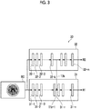
- Fig. 3 is a schematic diagram illustrating a configuration of the CNN in the embodiment.
- a CNN 30 has a first neural network 31 and a second neural network 32.
- the first neural network 31 has a plurality of processing layers 31-1, 31-,2, ..., 31-n (n is the number of processing layers).
- the second neural network 32 has a plurality of processing layers 32-1, 32-2, ..., 32-m (m is the number of processing layers).
- the first processing layer 31-1 is an input layer and the last processing layer 31-n is an output layer
- the first processing layer 32-1 is an input layer and the last processing layer 32-m is an output layer.
- the processing layers of the first neural network 31 and the second neural network 32 consist of convolutional layers or pooling layers.
- the convolutional layer performs a convolution process using various kernels on the input images, and outputs a feature map consisting of feature quantity data obtained by the convolution process.
- the convolutional layer applies the kernel to the entire feature map output from the brain image B0 or the processing layer at the previous stage while shifting the pixel of interest of the kernel. Further, the convolutional layer applies an activation function such as a sigmoid function to the convolved value to output the feature map.
- the pooling layer reduces the amount of data of the feature map by pooling the feature map output by the convolutional layer, and outputs the feature map of which the amount of data is reduced.
- the first neural network 31 has performed learning so as to output a discrimination result R1 of the bleeding region for each pixel included in the input brain image by using, as teacher data, multiple brain images including the bleeding region.
- the discrimination result R1 output by the first neural network 31 is a result obtained by discriminating whether each pixel of the brain image B0 is a bleeding region or a region other than the bleeding region.
- the second neural network 32 is different from the first neural network 31, and has learned a task to predict the presence or absence of a disease in the entire brain image B0.
- the presence or absence of bleeding corresponds to a second medical feature.
- the second neural network 32 learns to output a discrimination result R2 of whether a bleeding region is included in the input brain image by using, as teacher data, multiple brain images known to include the bleeding region.
- the feature map output from the processing layer at the previous stage is sequentially input to the processing layer at the next stage, the entire information on the brain image B0 is integrated, and the discrimination result R2 of whether the bleeding region is included in the brain image B0 is output from the final layer 32-m.
- the discrimination result R1 of the first neural network 31 includes information on the position of the bleeding region in the brain image B0, but the discrimination result R2 of the second neural network 32 does not include information on the position in the brain image B0 since the discrimination result R2 only indicates the presence or absence of the bleeding region.
- a feature map T2k output from the processing layer 32-k in the middle of the second neural network 32 is input to the processing layer 31-r in the middle of the first neural network 31.
- the feature map T2k includes a feature quantity in which the presence or absence of a bleeding region in the brain image B0 is known to some extent. Which processing layer in the first neural network 31 to input the feature map output from which processing layer in the second neural network 32 is designed in advance at the learning stage of the first and second neural networks 31 and 32.
- the feature map T2k is scaled to have the same size as a feature map T1r-1 output from the processing layer 31-r-1 at the previous stage of the processing layer 31-r, and is input to the processing layer 31-r together with the feature map T1r-1.
- the first neural network 31 integrates the feature map, which is output from the processing layer in the middle of the second neural network 32, in the processing layer subsequent the processing layer 31-r to output the discrimination result R1 of the local bleeding region in the brain image B0.
- the discrimination result R1 of the bleeding region is output by integrating the output of the second neural network 32, that is, information on the presence or absence of the bleeding in the brain image B0.
- Fig. 4 is a diagram illustrating the displayed brain image.
- a slice image of one tomographic plane of the brain image B0 is illustrated.
- a discriminated bleeding region A1 in the brain image B0 is highlighted and displayed.
- any form can be used such as enclosing the bleeding region A1 with a line, assigning a specific color to the bleeding region A1, or assigning an arrow to the bleeding region A1.
- Fig. 5 is a flowchart illustrating the process performed in the embodiment.
- the image acquisition unit 21 acquires the brain image B0 of the subject (Step ST1).
- the discrimination unit 22 discriminates the bleeding region in the brain image B0 (Step ST2)
- the display controller 23 displays the brain image B0 in which the bleeding region is discriminated on the display unit 14 (Step ST3), and the process is ended.
- the bleeding region in the brain image B0 is discriminated by the first neural network 31 of the discrimination unit 22, and the presence or absence of the bleeding region in the brain image B0 is discriminated by the second neural network 32 of the discrimination unit 22.
- the feature map T2k output by the processing layer 32-k in the middle of the second neural network 32 is input to the first neural network 31. Therefore, the feature map in the first neural network 31 and the feature map regarding the presence or absence of the bleeding region discriminated by the second neural network 32 are integrated so that the bleeding region in the brain image B0 is discriminated in the first neural network 32. Accordingly, according to the embodiment, it is possible to easily and accurately discriminate the bleeding region in the brain image B0.
- the feature map T2k output by the processing layer 32-k in the middle of the second neural network 32 is input to the first neural network 31, but the discrimination result R2 output by the output layer 32-m of the second neural network 32 may be input to the first neural network 31.
- the feature map T2k output by the processing layer 32-k in the middle of the second neural network 32 is input to the processing layer 31-r in the middle of the first neural network 31, but the invention is not limited thereto.
- the feature map T2k output by the processing layer 32-k in the middle of the second neural network 32 or the discrimination result R2 output by the output layer 32-m of the second neural network 32 may be input to the first processing layer 31-1, that is, the input layer of the first neural network 31.
- the second neural network 32 has learned a task to predict the presence or absence of a bleeding region.
- the task learned by the second neural network 32 is not limited thereto.
- a task to predict the type of a disease in the brain image BO for example, bleeding and infarction may be learned.
- the second neural network 32 learns to output a discrimination result of the type of included disease (bleeding, infarction, and no disease) in a case where the brain image is input, by using, as teacher data, brain images in which the included disease is known to be either bleeding or infarction.
- the bleeding type may be used as the type of a disease.
- the second neural network 32 may have learned to output a discrimination result of which of intracerebral hemorrhage, intraventricular hemorrhage, subarachnoid hemorrhage, and epidural hemorrhage the bleeding type is.
- the second neural network 32 may have learned to discriminate the presence or absence of brain compression findings, or may have learned to discriminate the type of cerebral infarction (acute, old, and the like).
- the task learned by the second neural network 32 may be a task to predict clinical findings expressed in the patient as the subject.
- examples of the clinical findings include a state of consciousness, a state of eye opening, and a state of awakening.
- the second neural network 32 learns to output a discrimination result of clinical findings in a case where the brain image is input, by using, as teacher data, the brain image in which the clinical findings are known.
- the CT image is used as the brain image B0, but the invention is not limited thereto, and other medical images such as the MRI image and the PET image may be used.
- the brain image B0 is used as the medical image, but the invention is not limited thereto.
- a medical feature included in the medical image such as the chest, abdomen, whole body, and four limbs of the human body, the present disclosure can be applied.
- the convolutional neural network is used as the first and second neural networks 31 and 32, but the invention is not limited thereto.
- a deep neural network DNN
- a recurrent neural network RNN
- the first neural network 31 and the second neural network 32 may not be the same neural network.
- the first neural network 31 may be the convolutional neural network
- the second neural network 32 may be the recurrent neural network.
- the following various processors can be used as the hardware structure of processing units executing various processes such as the image acquisition unit 21, the discrimination unit 22, and the display controller 23.
- the various processors include, for example, a programmable logic device (PLD) that is a processor of which the circuit configuration can be changed after manufacture, such as a field-programmable gate array (FPGA), and a dedicated electric circuit that is a processor having a dedicated circuit configuration designed to execute a specific process, such as an application specific integrated circuit (ASIC), in addition to the CPU that is a general-purpose processor which executes software (program) to function as various processing units as described above.
- PLD programmable logic device
- FPGA field-programmable gate array
- ASIC application specific integrated circuit
- One processing unit may be configured by one of the various processors or a combination of the same or different kinds of two or more processors (for example, a combination of a plurality of FPGAs or a combination of a CPU and an FPGA).
- a plurality of processing units may be configured by one processor.
- a plurality of processing units are configured by one processor
- one processor is configured by a combination of one or more CPUs and software as typified by a computer, such as a client and a server, and this processor functions as a plurality of processing units.
- IC integrated circuit
- SoC system on chip
- an electric circuit obtained by combining circuit elements, such as semiconductor elements, can be used as the hardware structure of the various processors.
Landscapes
- Engineering & Computer Science (AREA)
- Health & Medical Sciences (AREA)
- Physics & Mathematics (AREA)
- Life Sciences & Earth Sciences (AREA)
- General Health & Medical Sciences (AREA)
- Biomedical Technology (AREA)
- Theoretical Computer Science (AREA)
- Medical Informatics (AREA)
- Nuclear Medicine, Radiotherapy & Molecular Imaging (AREA)
- Molecular Biology (AREA)
- Radiology & Medical Imaging (AREA)
- Public Health (AREA)
- General Physics & Mathematics (AREA)
- Biophysics (AREA)
- Data Mining & Analysis (AREA)
- Pathology (AREA)
- Computational Linguistics (AREA)
- Evolutionary Computation (AREA)
- Artificial Intelligence (AREA)
- Computing Systems (AREA)
- General Engineering & Computer Science (AREA)
- Mathematical Physics (AREA)
- Software Systems (AREA)
- Veterinary Medicine (AREA)
- Epidemiology (AREA)
- Heart & Thoracic Surgery (AREA)
- Surgery (AREA)
- Animal Behavior & Ethology (AREA)
- Primary Health Care (AREA)
- Databases & Information Systems (AREA)
- Computer Vision & Pattern Recognition (AREA)
- Neurology (AREA)
- Quality & Reliability (AREA)
- High Energy & Nuclear Physics (AREA)
- Optics & Photonics (AREA)
- Spectroscopy & Molecular Physics (AREA)
- Image Analysis (AREA)
- Apparatus For Radiation Diagnosis (AREA)
- Measuring And Recording Apparatus For Diagnosis (AREA)
- Magnetic Resonance Imaging Apparatus (AREA)
Abstract
Description
- The present disclosure relates to a medical image processing apparatus, and a medical image processing method, and a medical image processing program which discriminate medical features included in a medical image such as a computed tomography (CT) image of a head.
- In recent years, with advances in medical equipment such as CT devices and magnetic resonance imaging (MRI) devices, an image diagnosis using high resolution medical images having higher quality can be performed. In particular, in a case where a target part is a brain, since it is possible to specify a disease region causing vascular disorders such as cerebral infarction and cerebral hemorrhage, by the image diagnosis using CT images and MRI images, an appropriate treatment can be performed on the basis of the specified result. In general, the disease region shows a higher CT value on the CT image as compared with the surrounding region. Therefore, it is possible to discriminate a disease region by interpreting the presence or absence of a region showing a higher CT value as compared with the surrounding region in the image diagnosis.
- In this manner, various methods for automatically discriminating the disease region from the medical image have been proposed. For example, in
JP1996-251404A JP-H08-251404A JP1996-251404A JP-H08-251404A - However, in some cases, depending on the types of the medical features, it is difficult to discriminate the medical feature from the medical image.
- The present disclosure is made in view of such circumstances, and an object thereof is to make it possible to easily discriminate the medical features from the medical image.
- A medical image processing apparatus according to an aspect of the present disclosure comprises a discrimination unit that discriminates a region of a first medical feature included in a medical image, in which the discrimination unit has a first neural network which discriminates the first medical feature in the medical image, and consists of a plurality of processing layers, and a second neural network which discriminates a second medical feature associated with the first medical feature, in the medical image, and consists of a plurality of processing layers, and a feature quantity output from the processing layer in the middle of the second neural network or an output result of the second neural network is input to the first neural network.
- The term "discriminating" includes both discriminating the presence or absence of the first or second medical feature in the medical image and discriminating a location of the first or second medical feature in the medical image.
- The feature quantity output from the processing layer in the middle of the second neural network or the output result of the second neural network may be input to the processing layer in the middle of the first neural network, or may be input to the first processing layer in the first neural network.
- In the medical image processing apparatus according to the aspect of the present disclosure, the second neural network may have learned a task different from the first neural network.
- In the medical image processing apparatus according to the aspect of the present disclosure, the task learned by the second neural network may be any one of a task to predict presence or absence of a disease, a task to predict a type of a disease, a task to predict clinical findings expressed in a patient, or a task to predict an awareness level of a patient.
- In the medical image processing apparatus according to the aspect of the present disclosure, the first neural network may discriminate a local medical feature from the medical image, as the first medical feature, and the second neural network may discriminate a global medical feature from the medical image, as the second medical feature.
- In the medical image processing apparatus according to the aspect of the present disclosure, the medical image may include a brain, the local medical feature may be at least one of a bleeding region or an infarction region of the brain, and the global medical feature may be presence or absence of the at least one of the bleeding region or the infarction region.
- The medical image processing apparatus according to the aspect of the present disclosure may further comprise a display controller that displays the medical image in which the local medical feature is specified, on a display unit.
- In the medical image processing apparatus according to the aspect of the present disclosure, the medical image may be a CT image of a brain.
- A medical image processing method according to another aspect of the present disclosure is a medical image processing method of discriminating a region of a first medical feature included in a medical image using a discrimination unit, in which the discrimination unit has a first neural network and a second neural network, and a feature quantity output from a processing layer in the middle of the second neural network or an output result of the second neural network is input to the first neural network, and the medical image processing method includes discriminating the first medical feature in the medical image using the first neural network; and discriminating a second medical feature associated with the first medical feature, in the medical image using the second neural network.
- A program for causing a computer to execute the medical image processing method according to the aspect of the present disclosure may be provided.
- A medical image processing apparatus according to another aspect of the present disclosure comprises a memory that stores a command for causing a computer to execute a process of discriminating a region of a first medical feature included in a medical image using a discrimination unit, and a processor configured to execute the stored command, in which the processor functions as the discrimination unit that has a first neural network and a second neural network, in which a feature quantity output from a processing layer in the middle of the second neural network or an output result of the second neural network is input to the first neural network, and the processor executes a process of discriminating the first medical feature in the medical image using the first neural network, and discriminating a second medical feature associated with the first medical feature, in the medical image using the second neural network.
- According to the present disclosure, it is possible to easily and accurately discriminate the first medical feature in the medical image.
-
-
Fig. 1 is a hardware configuration diagram illustrating an outline of a diagnosis support system to which a medical image processing apparatus according to an embodiment of the present disclosure is applied. -
Fig. 2 is a diagram illustrating a schematic configuration of the medical image processing apparatus according to the embodiment of the present disclosure. -
Fig. 3 is a schematic diagram illustrating a configuration of a convolutional neural network in the embodiment. -
Fig. 4 is a diagram illustrating a brain image in which a bleeding region is specified. -
Fig. 5 is a flowchart illustrating a process performed in the embodiment. - Hereinafter, embodiments of the present disclosure will be described in detail with reference to the drawings.
Fig. 1 is a hardware configuration diagram illustrating an outline of a diagnosis support system to which a medical image processing apparatus according to an embodiment of the present disclosure is applied. As illustrated inFig. 1 , in the diagnosis support system, a medicalimage processing apparatus 1 according to the embodiment, a three-dimensional image capturingdevice 2, and animage storage server 3 are connected via a network 4 in a communicable state. - The three-dimensional image capturing
device 2 is a device that images a part as a diagnosis target of a subject to generate a three-dimensional image representing the part, and specifically, is a CT device, an MRI device, a positron emission tomography (PET) device, or the like. The three-dimensional image generated by the three-dimensional image capturingdevice 2 is transmitted to theimage storage server 3 to be saved. In the embodiment, the diagnosis target part of the patient as the subject is a brain, the three-dimensional image capturingdevice 2 is a CT device, and a CT image of a head including the brain of the subject is generated as a three-dimensional brain image B0. - The
image storage server 3 is a computer that saves and manages various kinds of data, and comprises a large-capacity external storage device and software for database management. Theimage storage server 3 performs communication with other devices via the network 4 in a wired or wireless manner, and transmits and receives various kinds of image data. Specifically, the various kinds of data including the image data of the brain image B0 generated by the three-dimensionalimage capturing device 2 are acquired via the network, and are saved and managed in a recording medium such as a large-capacity external storage device. The image data storage format and the communication between the devices via the network 4 are based on the protocol such as Digital Imaging and Communication in Medicine (DICOM). - The medical
image processing apparatus 1 is obtained by installing a medical image processing program of the embodiment in one computer. The computer may be a workstation or a personal computer that a doctor performing a diagnosis operates directly, or a server computer connected to the workstation or personal computer via a network. The medical image processing program is distributed by being recorded in a recording medium such as a digital versatile disc (DVD) and a compact disc read only memory (CD-ROM), and is installed to a computer from the recording medium. Otherwise, the medical image display program may be stored in a storage device of a server computer connected to a network or a network storage in a state of being accessible from the outside, and be downloaded and installed in a computer that the doctor uses, in response to the request. -
Fig. 2 is a diagram illustrating a schematic configuration of the medical image processing apparatus realized by installing the medical image processing program in a computer. As illustrated inFig. 2 , the medicalimage processing apparatus 1 comprises a central processing unit (CPU) 11, a memory 12, and astorage 13 as a standard workstation configuration. In addition, adisplay unit 14 such as a liquid crystal display, and aninput unit 15 such as a keyboard and a mouse are connected to the medicalimage processing apparatus 1. - The
storage 13 consists of a hard disk drive or the like, and various kinds of information including the brain images of the subject and information required for the process which are acquired from theimage storage server 3 via the network 4 are stored. - In the memory 12, the medical image processing program is stored. The medical image processing program defines, as the process executed by the
CPU 11, an image acquisition process of acquiring the brain image B0 of the subject, a discrimination process of discriminating the disease region included in the brain image B0, and a display control process of displaying the discriminated disease region on thedisplay unit 14. - By the
CPU 11 executing those processes according to the program, the computer functions as animage acquisition unit 21, adiscrimination unit 22, and adisplay controller 23. - The
image acquisition unit 21 acquires the brain image B0 of the brain of the subject from theimage storage server 3. In a case where the brain image B0 is already stored in thestorage 13, theimage acquisition unit 21 may acquire the brain image B0 from thestorage 13. - The
discrimination unit 22 discriminates the disease region in the brain image B0. It is assumed that the disease region is a bleeding region in the embodiment. In the embodiment, it is assumed that thediscrimination unit 22 is a convolutional neural network (hereinafter, referred to as CNN) which is one of multi-layer neural networks in which a plurality of processing layers are hierarchically connected to each other and deep learning is performed. -
Fig. 3 is a schematic diagram illustrating a configuration of the CNN in the embodiment. As illustrated inFig. 3 , a CNN 30 has a firstneural network 31 and a secondneural network 32. The firstneural network 31 has a plurality of processing layers 31-1, 31-,2, ..., 31-n (n is the number of processing layers). Further, the secondneural network 32 has a plurality of processing layers 32-1, 32-2, ..., 32-m (m is the number of processing layers). In the firstneural network 31, the first processing layer 31-1 is an input layer and the last processing layer 31-n is an output layer, and in the secondneural network 32, the first processing layer 32-1 is an input layer and the last processing layer 32-m is an output layer. - The processing layers of the first
neural network 31 and the secondneural network 32 consist of convolutional layers or pooling layers. The convolutional layer performs a convolution process using various kernels on the input images, and outputs a feature map consisting of feature quantity data obtained by the convolution process. The kernel has an n×n pixel size (for example, n=3), and a weight is set in each element. Specifically, a weight such as a differential filter for emphasizing the edge of a two-dimensional image such as the brain image B0 or the feature map is set. The convolutional layer applies the kernel to the entire feature map output from the brain image B0 or the processing layer at the previous stage while shifting the pixel of interest of the kernel. Further, the convolutional layer applies an activation function such as a sigmoid function to the convolved value to output the feature map. - The pooling layer reduces the amount of data of the feature map by pooling the feature map output by the convolutional layer, and outputs the feature map of which the amount of data is reduced.
- In the embodiment, the first
neural network 31 has performed learning so as to output a discrimination result R1 of the bleeding region for each pixel included in the input brain image by using, as teacher data, multiple brain images including the bleeding region. The bleeding region corresponds to a first medical feature. Accordingly, in a case where the brain image B0 is input to the firstneural network 31, in the plurality of processing layers 31-i (i=1 to n), the feature map output from the processing layer at the previous stage is sequentially input to the processing layer at the next stage, and the discrimination result R1 of the bleeding region for each pixel in the brain image B0 is output from the output layer 31-n. The discrimination result R1 output by the firstneural network 31 is a result obtained by discriminating whether each pixel of the brain image B0 is a bleeding region or a region other than the bleeding region. - The second
neural network 32 is different from the firstneural network 31, and has learned a task to predict the presence or absence of a disease in the entire brain image B0. The presence or absence of bleeding corresponds to a second medical feature. The secondneural network 32 learns to output a discrimination result R2 of whether a bleeding region is included in the input brain image by using, as teacher data, multiple brain images known to include the bleeding region. Accordingly, in a case where the brain image B0 is input to the secondneural network 32, in the plurality of processing layers 32-j (j=1 to m), the feature map output from the processing layer at the previous stage is sequentially input to the processing layer at the next stage, the entire information on the brain image B0 is integrated, and the discrimination result R2 of whether the bleeding region is included in the brain image B0 is output from the final layer 32-m. - The discrimination result R1 of the first
neural network 31 includes information on the position of the bleeding region in the brain image B0, but the discrimination result R2 of the secondneural network 32 does not include information on the position in the brain image B0 since the discrimination result R2 only indicates the presence or absence of the bleeding region. - Here, in the embodiment, a feature map T2k output from the processing layer 32-k in the middle of the second
neural network 32 is input to the processing layer 31-r in the middle of the firstneural network 31. The feature map T2k includes a feature quantity in which the presence or absence of a bleeding region in the brain image B0 is known to some extent. Which processing layer in the firstneural network 31 to input the feature map output from which processing layer in the secondneural network 32 is designed in advance at the learning stage of the first and secondneural networks neural network 31 integrates the feature map, which is output from the processing layer in the middle of the secondneural network 32, in the processing layer subsequent the processing layer 31-r to output the discrimination result R1 of the local bleeding region in the brain image B0. - Here, depending on the brain image B0, it may be difficult to discriminate the bleeding region only by the local features. Such a bleeding region of the brain image B0 can be confirmed in a case where a skilled doctor interprets the brain image B0, but in the discrimination unit using the convolutional neural network or the like, it is difficult to accurately discriminate the bleeding region in some cases. In the embodiment, in the first
neural network 31, the discrimination result R1 of the bleeding region is output by integrating the output of the secondneural network 32, that is, information on the presence or absence of the bleeding in the brain image B0. - The
display controller 23 displays the brain image in which the bleeding region is discriminated on thedisplay unit 14.Fig. 4 is a diagram illustrating the displayed brain image. InFig. 4 , a slice image of one tomographic plane of the brain image B0 is illustrated. As illustrated inFig. 4 , a discriminated bleeding region A1 in the brain image B0 is highlighted and displayed. For the highlighting, any form can be used such as enclosing the bleeding region A1 with a line, assigning a specific color to the bleeding region A1, or assigning an arrow to the bleeding region A1. - Next, the process performed in the embodiment will be described.
Fig. 5 is a flowchart illustrating the process performed in the embodiment. First, theimage acquisition unit 21 acquires the brain image B0 of the subject (Step ST1). Next, thediscrimination unit 22 discriminates the bleeding region in the brain image B0 (Step ST2), thedisplay controller 23 displays the brain image B0 in which the bleeding region is discriminated on the display unit 14 (Step ST3), and the process is ended. - In this manner, in the embodiment, the bleeding region in the brain image B0 is discriminated by the first
neural network 31 of thediscrimination unit 22, and the presence or absence of the bleeding region in the brain image B0 is discriminated by the secondneural network 32 of thediscrimination unit 22. In this case, the feature map T2k output by the processing layer 32-k in the middle of the secondneural network 32 is input to the firstneural network 31. Therefore, the feature map in the firstneural network 31 and the feature map regarding the presence or absence of the bleeding region discriminated by the secondneural network 32 are integrated so that the bleeding region in the brain image B0 is discriminated in the firstneural network 32. Accordingly, according to the embodiment, it is possible to easily and accurately discriminate the bleeding region in the brain image B0. - In the above embodiment, the feature map T2k output by the processing layer 32-k in the middle of the second
neural network 32 is input to the firstneural network 31, but the discrimination result R2 output by the output layer 32-m of the secondneural network 32 may be input to the firstneural network 31. - Further, in the above embodiment, the feature map T2k output by the processing layer 32-k in the middle of the second
neural network 32 is input to the processing layer 31-r in the middle of the firstneural network 31, but the invention is not limited thereto. The feature map T2k output by the processing layer 32-k in the middle of the secondneural network 32 or the discrimination result R2 output by the output layer 32-m of the secondneural network 32 may be input to the first processing layer 31-1, that is, the input layer of the firstneural network 31. - Further, in the above embodiment, the second
neural network 32 has learned a task to predict the presence or absence of a bleeding region. However, the task learned by the secondneural network 32 is not limited thereto. For example, a task to predict the type of a disease in the brain image BO, for example, bleeding and infarction may be learned. In this case, the secondneural network 32 learns to output a discrimination result of the type of included disease (bleeding, infarction, and no disease) in a case where the brain image is input, by using, as teacher data, brain images in which the included disease is known to be either bleeding or infarction. - On the other hand, the bleeding type may be used as the type of a disease. For example, the second
neural network 32 may have learned to output a discrimination result of which of intracerebral hemorrhage, intraventricular hemorrhage, subarachnoid hemorrhage, and epidural hemorrhage the bleeding type is. In addition, the secondneural network 32 may have learned to discriminate the presence or absence of brain compression findings, or may have learned to discriminate the type of cerebral infarction (acute, old, and the like). - The task learned by the second
neural network 32 may be a task to predict clinical findings expressed in the patient as the subject. Here, examples of the clinical findings include a state of consciousness, a state of eye opening, and a state of awakening. In this case, the secondneural network 32 learns to output a discrimination result of clinical findings in a case where the brain image is input, by using, as teacher data, the brain image in which the clinical findings are known. - Further, in the above embodiment, the CT image is used as the brain image B0, but the invention is not limited thereto, and other medical images such as the MRI image and the PET image may be used.
- Further, in the above embodiment, the brain image B0 is used as the medical image, but the invention is not limited thereto. For example, even in a case of discriminating a medical feature included in the medical image, such as the chest, abdomen, whole body, and four limbs of the human body, the present disclosure can be applied.
- Further, in the above embodiment, the convolutional neural network is used as the first and second
neural networks neural network 31 and the secondneural network 32 may not be the same neural network. For example, the firstneural network 31 may be the convolutional neural network, and the secondneural network 32 may be the recurrent neural network. - In the above embodiment, for example, the following various processors can be used as the hardware structure of processing units executing various processes such as the
image acquisition unit 21, thediscrimination unit 22, and thedisplay controller 23. The various processors include, for example, a programmable logic device (PLD) that is a processor of which the circuit configuration can be changed after manufacture, such as a field-programmable gate array (FPGA), and a dedicated electric circuit that is a processor having a dedicated circuit configuration designed to execute a specific process, such as an application specific integrated circuit (ASIC), in addition to the CPU that is a general-purpose processor which executes software (program) to function as various processing units as described above. - One processing unit may be configured by one of the various processors or a combination of the same or different kinds of two or more processors (for example, a combination of a plurality of FPGAs or a combination of a CPU and an FPGA). In addition, a plurality of processing units may be configured by one processor.
- As an example where a plurality of processing units are configured by one processor, first, there is a form where one processor is configured by a combination of one or more CPUs and software as typified by a computer, such as a client and a server, and this processor functions as a plurality of processing units. Second, there is a form where a processor fulfilling the functions of the entire system including a plurality of processing units by one integrated circuit (IC) chip as typified by a system on chip (SoC) or the like is used. In this manner, various processing units are configured by using one or more of the above-described various processors as hardware structures.
- In addition, specifically, an electric circuit (circuitry) obtained by combining circuit elements, such as semiconductor elements, can be used as the hardware structure of the various processors.
-
- 1: medical image processing apparatus
- 2: three-dimensional image capturing device
- 3: image storage server
- 4: network
- 11: CPU
- 12: memory
- 13: storage
- 14: display
- 15: input unit
- 21: image acquisition unit
- 22: discrimination unit
- 23: display controller
- 30: CNN
- 31: first neural network
- 31-1, 31-2, ...: processing layer
- 32: second neural network
- 32-1, 32-2, ...: processing layer
- A1: bleeding region
- B0: brain image
- R1, R2: discrimination result
- T1r-1, T2k: feature map
Claims (9)
- A medical image processing apparatus comprising:a discrimination unit that discriminates a region of a first medical feature included in a medical image,wherein the discrimination unit hasa first neural network which discriminates the first medical feature in the medical image, and consists of a plurality of processing layers, anda second neural network which discriminates a second medical feature associated with the first medical feature, in the medical image, and consists of a plurality of processing layers, anda feature quantity output from the processing layer in the middle of the second neural network or an output result of the second neural network is input to the first neural network.
- The medical image processing apparatus according to claim 1,
wherein the second neural network has learned a task different from the first neural network. - The medical image processing apparatus according to claim 2,
wherein the task learned by the second neural network is any one of a task to predict presence or absence of a disease, a task to predict a type of a disease, a task to predict clinical findings expressed in a patient, or a task to predict an awareness level of a patient. - The medical image processing apparatus according to any one of claims 1 to 3,wherein the first neural network discriminates a local medical feature from the medical image, as the first medical feature, andthe second neural network discriminates a global medical feature from the medical image, as the second medical feature.
- The medical image processing apparatus according to claim 4,wherein the medical image includes a brain,the local medical feature is at least one of a bleeding region or an infarction region of the brain, andthe global medical feature is presence or absence of the at least one of the bleeding region or the infarction region.
- The medical image processing apparatus according to claim 4 or 5, further comprising:
a display controller that displays the medical image in which the local medical feature is specified, on a display unit. - The medical image processing apparatus according to any one of claims 1 to 6,
wherein the medical image is a CT image of a brain. - A medical image processing method of discriminating a region of a first medical feature included in a medical image using a discrimination unit, in which the discrimination unit has a first neural network and a second neural network, and a feature quantity output from a processing layer in the middle of the second neural network or an output result of the second neural network is input to the first neural network, the medical image processing method comprising:discriminating the first medical feature in the medical image using the first neural network; anddiscriminating a second medical feature associated with the first medical feature, in the medical image using the second neural network.
- A medical image processing program causing a computer to execute a process of discriminating a region of a first medical feature included in a medical image,wherein the medical image processing program causes the computer to function as a discrimination unit that has a first neural network and a second neural network, in which a feature quantity output from a processing layer in the middle of the second neural network or an output result of the second neural network is input to the first neural network, andthe medical image processing program causes the computer to executea procedure of discriminating the first medical feature in the medical image using the first neural network, anda procedure of discriminating a second medical feature associated with the first medical feature, in the medical image using the second neural network.
Applications Claiming Priority (2)
| Application Number | Priority Date | Filing Date | Title |
|---|---|---|---|
| JP2018172989 | 2018-09-14 | ||
| PCT/JP2019/026533 WO2020054188A1 (en) | 2018-09-14 | 2019-07-03 | Medical image processing device, method, and program |
Publications (2)
| Publication Number | Publication Date |
|---|---|
| EP3851049A1 true EP3851049A1 (en) | 2021-07-21 |
| EP3851049A4 EP3851049A4 (en) | 2021-11-10 |
Family
ID=69778538
Family Applications (1)
| Application Number | Title | Priority Date | Filing Date |
|---|---|---|---|
| EP19860018.1A Pending EP3851049A4 (en) | 2018-09-14 | 2019-07-03 | MEDICAL IMAGE PROCESSING DEVICE, METHOD AND PROGRAM |
Country Status (4)
| Country | Link |
|---|---|
| US (1) | US11915414B2 (en) |
| EP (1) | EP3851049A4 (en) |
| JP (1) | JP7339270B2 (en) |
| WO (1) | WO2020054188A1 (en) |
Families Citing this family (5)
| Publication number | Priority date | Publication date | Assignee | Title |
|---|---|---|---|---|
| US20210093278A1 (en) * | 2019-09-30 | 2021-04-01 | GE Precision Healthcare LLC | Computed tomography medical imaging intracranial hemorrhage model |
| WO2022054858A1 (en) * | 2020-09-11 | 2022-03-17 | 大原薬品工業株式会社 | Stroke diagnosis and therapy assistance system, stroke state information providing device, and stroke state information providing program |
| JP7602746B2 (en) * | 2020-11-04 | 2024-12-19 | 株式会社トプコン | Ophthalmic information processing device, ophthalmic device, operation method of ophthalmic information processing device, and program |
| AU2022252500A1 (en) * | 2021-04-02 | 2023-10-19 | Anode IP LLC | Systems and methods to process electronic medical images for diagnostic or interventional use |
| WO2024142395A1 (en) * | 2022-12-28 | 2024-07-04 | 日本電気株式会社 | Video processing device, video processing method, and recording medium |
Family Cites Families (12)
| Publication number | Priority date | Publication date | Assignee | Title |
|---|---|---|---|---|
| JPH08251404A (en) | 1995-03-13 | 1996-09-27 | Minolta Co Ltd | Method and device for discriminating attribute of image area |
| EP1815392A1 (en) * | 2004-11-23 | 2007-08-08 | Eastman Kodak Company | Automated radiograph classification using anatomy information |
| EP1780651A1 (en) * | 2005-10-25 | 2007-05-02 | Bracco Imaging, S.P.A. | Method and system for automatic processing and evaluation of images, particularly diagnostic images |
| JP2016031747A (en) | 2014-07-30 | 2016-03-07 | キヤノン株式会社 | Information processing apparatus and information processing method |
| WO2017106645A1 (en) * | 2015-12-18 | 2017-06-22 | The Regents Of The University Of California | Interpretation and quantification of emergency features on head computed tomography |
| US20170236027A1 (en) * | 2016-02-16 | 2017-08-17 | Brainchip Inc. | Intelligent biomorphic system for pattern recognition with autonomous visual feature extraction |
| US9886758B2 (en) * | 2016-03-31 | 2018-02-06 | International Business Machines Corporation | Annotation of skin image using learned feature representation |
| US9589374B1 (en) * | 2016-08-01 | 2017-03-07 | 12 Sigma Technologies | Computer-aided diagnosis system for medical images using deep convolutional neural networks |
| KR101908680B1 (en) * | 2016-08-30 | 2018-10-17 | 주식회사 루닛 | A method and apparatus for machine learning based on weakly supervised learning |
| KR102824640B1 (en) * | 2016-09-07 | 2025-06-25 | 삼성전자주식회사 | Recognition apparatus based on neural network and training method of neural network |
| US10482633B2 (en) | 2016-09-12 | 2019-11-19 | Zebra Medical Vision Ltd. | Systems and methods for automated detection of an indication of malignancy in a mammographic image |
| CN107292884B (en) * | 2017-08-07 | 2020-09-29 | 杭州深睿博联科技有限公司 | A method and apparatus for identifying edema and hematoma in MRI images |
-
2019
- 2019-07-03 JP JP2020546713A patent/JP7339270B2/en active Active
- 2019-07-03 EP EP19860018.1A patent/EP3851049A4/en active Pending
- 2019-07-03 WO PCT/JP2019/026533 patent/WO2020054188A1/en not_active Ceased
-
2021
- 2021-01-29 US US17/161,703 patent/US11915414B2/en active Active
Also Published As
| Publication number | Publication date |
|---|---|
| US11915414B2 (en) | 2024-02-27 |
| EP3851049A4 (en) | 2021-11-10 |
| WO2020054188A1 (en) | 2020-03-19 |
| JPWO2020054188A1 (en) | 2021-08-30 |
| JP7339270B2 (en) | 2023-09-05 |
| US20210150713A1 (en) | 2021-05-20 |
Similar Documents
| Publication | Publication Date | Title |
|---|---|---|
| US11087465B2 (en) | Medical image processing apparatus, medical image processing method, and medical image processing program | |
| US11915414B2 (en) | Medical image processing apparatus, method, and program | |
| US11139067B2 (en) | Medical image display device, method, and program | |
| US11244455B2 (en) | Apparatus, method, and program for training discriminator discriminating disease region, discriminator discriminating disease region, disease region discrimination apparatus, and disease region discrimination program | |
| JP7129869B2 (en) | Disease area extraction device, method and program | |
| US20200074633A1 (en) | Apparatus, method, and program for learning discriminator discriminating infarction region, discriminator for discriminating infarction region, and apparatus, method, and program for discriminating infarction region | |
| WO2019102917A1 (en) | Radiologist determination device, method, and program | |
| US11176413B2 (en) | Apparatus, method, and program for training discriminator discriminating disease region, discriminator discriminating disease region, disease region discrimination apparatus, and disease region discrimination program | |
| JP6843785B2 (en) | Diagnostic support system, diagnostic support method, and program | |
| US11551351B2 (en) | Priority judgement device, method, and program | |
| US11328414B2 (en) | Priority judgement device, method, and program | |
| US12272049B2 (en) | Learning device, method, and program, medical image processing apparatus, method, and program, and discriminator | |
| US20190019304A1 (en) | Medical image processing apparatus, method, and program | |
| JP6968195B2 (en) | Inspection information display device, method and program | |
| WO2019216125A1 (en) | Learning device, method, and program for classifier for classifying infarct region, classifier for classifying infarct region, and device, method and program for classifying infarct region | |
| JP2019017993A (en) | Medical image processing apparatus, method and program | |
| JP7262606B2 (en) | Region identification device, method and program | |
| US12374002B2 (en) | Image processing apparatus, method and program, learning apparatus, method and program, and derivation model | |
| JP7742014B2 (en) | Image providing device, image providing system, image providing method, and program | |
| US12412326B2 (en) | Image processing apparatus and non-transitory computer readable medium for visualizing magnetic resonance data | |
| JP2024149230A (en) | Image processing device and image processing program | |
| JP2025146147A (en) | Image processing device, method, and program, and learning device, method, and program | |
| JP2025117188A (en) | Image processing device, method and program |
Legal Events
| Date | Code | Title | Description |
|---|---|---|---|
| STAA | Information on the status of an ep patent application or granted ep patent |
Free format text: STATUS: THE INTERNATIONAL PUBLICATION HAS BEEN MADE |
|
| PUAI | Public reference made under article 153(3) epc to a published international application that has entered the european phase |
Free format text: ORIGINAL CODE: 0009012 |
|
| STAA | Information on the status of an ep patent application or granted ep patent |
Free format text: STATUS: REQUEST FOR EXAMINATION WAS MADE |
|
| 17P | Request for examination filed |
Effective date: 20210224 |
|
| AK | Designated contracting states |
Kind code of ref document: A1 Designated state(s): AL AT BE BG CH CY CZ DE DK EE ES FI FR GB GR HR HU IE IS IT LI LT LU LV MC MK MT NL NO PL PT RO RS SE SI SK SM TR |
|
| REG | Reference to a national code |
Ref country code: DE Ref legal event code: R079 Free format text: PREVIOUS MAIN CLASS: A61B0006030000 Ipc: G16H0030400000 |
|
| A4 | Supplementary search report drawn up and despatched |
Effective date: 20211008 |
|
| RIC1 | Information provided on ipc code assigned before grant |
Ipc: G01T 1/161 20060101ALI20211004BHEP Ipc: G06T 7/00 20170101ALI20211004BHEP Ipc: G06N 3/04 20060101ALI20211004BHEP Ipc: A61B 6/03 20060101ALI20211004BHEP Ipc: A61B 5/055 20060101ALI20211004BHEP Ipc: A61B 5/00 20060101ALI20211004BHEP Ipc: G16H 50/20 20180101ALI20211004BHEP Ipc: G16H 30/40 20180101AFI20211004BHEP |
|
| DAV | Request for validation of the european patent (deleted) | ||
| DAX | Request for extension of the european patent (deleted) | ||
| STAA | Information on the status of an ep patent application or granted ep patent |
Free format text: STATUS: EXAMINATION IS IN PROGRESS |
|
| 17Q | First examination report despatched |
Effective date: 20240820 |