EP3842461A1 - Chimeric antigen receptors that bind to prostate specific membrane antigen - Google Patents
Chimeric antigen receptors that bind to prostate specific membrane antigen Download PDFInfo
- Publication number
- EP3842461A1 EP3842461A1 EP19219238.3A EP19219238A EP3842461A1 EP 3842461 A1 EP3842461 A1 EP 3842461A1 EP 19219238 A EP19219238 A EP 19219238A EP 3842461 A1 EP3842461 A1 EP 3842461A1
- Authority
- EP
- European Patent Office
- Prior art keywords
- cells
- car
- seq
- chimeric antigen
- antigen receptor
- Prior art date
- Legal status (The legal status is an assumption and is not a legal conclusion. Google has not performed a legal analysis and makes no representation as to the accuracy of the status listed.)
- Withdrawn
Links
- 101000892862 Homo sapiens Glutamate carboxypeptidase 2 Proteins 0.000 title claims abstract description 75
- 108010019670 Chimeric Antigen Receptors Proteins 0.000 title claims abstract description 60
- 102100041003 Glutamate carboxypeptidase 2 Human genes 0.000 title claims description 71
- 210000001744 T-lymphocyte Anatomy 0.000 claims abstract description 103
- 239000000427 antigen Substances 0.000 claims abstract description 67
- 108091007433 antigens Proteins 0.000 claims abstract description 67
- 102000036639 antigens Human genes 0.000 claims abstract description 67
- 230000027455 binding Effects 0.000 claims abstract description 35
- 206010028980 Neoplasm Diseases 0.000 claims abstract description 33
- 239000012634 fragment Substances 0.000 claims abstract description 29
- 239000003814 drug Substances 0.000 claims abstract description 7
- 102000046689 human FOLH1 Human genes 0.000 claims abstract description 6
- 229940045988 antineoplastic drug protein kinase inhibitors Drugs 0.000 claims abstract 2
- 229960000074 biopharmaceutical Drugs 0.000 claims abstract 2
- 239000003909 protein kinase inhibitor Substances 0.000 claims abstract 2
- 150000007523 nucleic acids Chemical class 0.000 claims description 25
- 239000013598 vector Substances 0.000 claims description 24
- 206010060862 Prostate cancer Diseases 0.000 claims description 21
- 208000000236 Prostatic Neoplasms Diseases 0.000 claims description 21
- 108020004707 nucleic acids Proteins 0.000 claims description 19
- 102000039446 nucleic acids Human genes 0.000 claims description 19
- 210000002865 immune cell Anatomy 0.000 claims description 16
- 101000914514 Homo sapiens T-cell-specific surface glycoprotein CD28 Proteins 0.000 claims description 15
- 102100027213 T-cell-specific surface glycoprotein CD28 Human genes 0.000 claims description 15
- 125000003275 alpha amino acid group Chemical group 0.000 claims description 15
- 229920001481 poly(stearyl methacrylate) Polymers 0.000 claims description 13
- 238000000034 method Methods 0.000 claims description 11
- 238000000338 in vitro Methods 0.000 claims description 9
- 101000851370 Homo sapiens Tumor necrosis factor receptor superfamily member 9 Proteins 0.000 claims description 7
- 102100036856 Tumor necrosis factor receptor superfamily member 9 Human genes 0.000 claims description 7
- 230000002068 genetic effect Effects 0.000 claims description 7
- 108090000623 proteins and genes Proteins 0.000 claims description 7
- ZDZOTLJHXYCWBA-VCVYQWHSSA-N N-debenzoyl-N-(tert-butoxycarbonyl)-10-deacetyltaxol Chemical compound O([C@H]1[C@H]2[C@@](C([C@H](O)C3=C(C)[C@@H](OC(=O)[C@H](O)[C@@H](NC(=O)OC(C)(C)C)C=4C=CC=CC=4)C[C@]1(O)C3(C)C)=O)(C)[C@@H](O)C[C@H]1OC[C@]12OC(=O)C)C(=O)C1=CC=CC=C1 ZDZOTLJHXYCWBA-VCVYQWHSSA-N 0.000 claims description 6
- 229960003668 docetaxel Drugs 0.000 claims description 5
- 230000004068 intracellular signaling Effects 0.000 claims description 4
- 210000002307 prostate Anatomy 0.000 claims description 4
- 210000001519 tissue Anatomy 0.000 claims description 4
- 230000002463 transducing effect Effects 0.000 claims description 4
- 230000000259 anti-tumor effect Effects 0.000 claims description 3
- 102000004169 proteins and genes Human genes 0.000 claims description 3
- CMSMOCZEIVJLDB-UHFFFAOYSA-N Cyclophosphamide Chemical compound ClCCN(CCCl)P1(=O)NCCCO1 CMSMOCZEIVJLDB-UHFFFAOYSA-N 0.000 claims description 2
- GHASVSINZRGABV-UHFFFAOYSA-N Fluorouracil Chemical compound FC1=CNC(=O)NC1=O GHASVSINZRGABV-UHFFFAOYSA-N 0.000 claims description 2
- 238000002659 cell therapy Methods 0.000 claims description 2
- 229960004397 cyclophosphamide Drugs 0.000 claims description 2
- 231100000433 cytotoxic Toxicity 0.000 claims description 2
- 230000001472 cytotoxic effect Effects 0.000 claims description 2
- 229960002949 fluorouracil Drugs 0.000 claims description 2
- 239000000126 substance Substances 0.000 claims description 2
- 150000004579 taxol derivatives Chemical class 0.000 claims description 2
- 102100035360 Cerebellar degeneration-related antigen 1 Human genes 0.000 claims 4
- 239000013543 active substance Substances 0.000 claims 2
- 108010083359 Antigen Receptors Proteins 0.000 claims 1
- 102000006306 Antigen Receptors Human genes 0.000 claims 1
- 230000002009 allergenic effect Effects 0.000 claims 1
- 210000004369 blood Anatomy 0.000 claims 1
- 239000008280 blood Substances 0.000 claims 1
- 239000012503 blood component Substances 0.000 claims 1
- 239000003795 chemical substances by application Substances 0.000 claims 1
- 239000002532 enzyme inhibitor Substances 0.000 claims 1
- 238000001415 gene therapy Methods 0.000 claims 1
- 125000002924 primary amino group Chemical group [H]N([H])* 0.000 claims 1
- 229960000160 recombinant therapeutic protein Drugs 0.000 claims 1
- 210000001082 somatic cell Anatomy 0.000 claims 1
- 229960005486 vaccine Drugs 0.000 claims 1
- 238000010361 transduction Methods 0.000 abstract description 11
- 230000026683 transduction Effects 0.000 abstract description 10
- 229940079593 drug Drugs 0.000 abstract description 4
- 238000001890 transfection Methods 0.000 abstract description 4
- 238000001727 in vivo Methods 0.000 abstract description 3
- 238000004519 manufacturing process Methods 0.000 abstract description 2
- 238000002512 chemotherapy Methods 0.000 abstract 1
- 229940126586 small molecule drug Drugs 0.000 abstract 1
- 210000004027 cell Anatomy 0.000 description 89
- 108091007741 Chimeric antigen receptor T cells Proteins 0.000 description 56
- 150000001413 amino acids Chemical class 0.000 description 35
- 102100024952 Protein CBFA2T1 Human genes 0.000 description 31
- 241001529936 Murinae Species 0.000 description 27
- 210000004881 tumor cell Anatomy 0.000 description 23
- 230000004913 activation Effects 0.000 description 17
- 108091033380 Coding strand Proteins 0.000 description 14
- 230000001177 retroviral effect Effects 0.000 description 13
- 102100024216 Programmed cell death 1 ligand 1 Human genes 0.000 description 11
- 230000000638 stimulation Effects 0.000 description 11
- NFGXHKASABOEEW-UHFFFAOYSA-N 1-methylethyl 11-methoxy-3,7,11-trimethyl-2,4-dodecadienoate Chemical compound COC(C)(C)CCCC(C)CC=CC(C)=CC(=O)OC(C)C NFGXHKASABOEEW-UHFFFAOYSA-N 0.000 description 10
- 231100000135 cytotoxicity Toxicity 0.000 description 10
- 230000003013 cytotoxicity Effects 0.000 description 10
- 230000008685 targeting Effects 0.000 description 10
- 102000017420 CD3 protein, epsilon/gamma/delta subunit Human genes 0.000 description 9
- 239000002609 medium Substances 0.000 description 9
- 108010074708 B7-H1 Antigen Proteins 0.000 description 8
- 239000012636 effector Substances 0.000 description 8
- 102100040678 Programmed cell death protein 1 Human genes 0.000 description 7
- 238000002474 experimental method Methods 0.000 description 7
- 238000010186 staining Methods 0.000 description 7
- 102000017578 LAG3 Human genes 0.000 description 6
- 101710089372 Programmed cell death protein 1 Proteins 0.000 description 6
- 239000006146 Roswell Park Memorial Institute medium Substances 0.000 description 6
- 238000000684 flow cytometry Methods 0.000 description 6
- 230000006870 function Effects 0.000 description 6
- 230000003834 intracellular effect Effects 0.000 description 6
- RGLRXNKKBLIBQS-XNHQSDQCSA-N leuprolide acetate Chemical compound CC(O)=O.CCNC(=O)[C@@H]1CCCN1C(=O)[C@H](CCCNC(N)=N)NC(=O)[C@H](CC(C)C)NC(=O)[C@@H](CC(C)C)NC(=O)[C@@H](NC(=O)[C@H](CO)NC(=O)[C@H](CC=1C2=CC=CC=C2NC=1)NC(=O)[C@H](CC=1N=CNC=1)NC(=O)[C@H]1NC(=O)CC1)CC1=CC=C(O)C=C1 RGLRXNKKBLIBQS-XNHQSDQCSA-N 0.000 description 6
- 108010002350 Interleukin-2 Proteins 0.000 description 5
- 102000000588 Interleukin-2 Human genes 0.000 description 5
- 108091028043 Nucleic acid sequence Proteins 0.000 description 5
- 201000011510 cancer Diseases 0.000 description 5
- 230000001086 cytosolic effect Effects 0.000 description 5
- 238000005516 engineering process Methods 0.000 description 5
- 229960004338 leuprorelin Drugs 0.000 description 5
- 230000035772 mutation Effects 0.000 description 5
- 210000003819 peripheral blood mononuclear cell Anatomy 0.000 description 5
- 239000013612 plasmid Substances 0.000 description 5
- 210000000130 stem cell Anatomy 0.000 description 5
- 108010032595 Antibody Binding Sites Proteins 0.000 description 4
- IJGRMHOSHXDMSA-UHFFFAOYSA-N Atomic nitrogen Chemical compound N#N IJGRMHOSHXDMSA-UHFFFAOYSA-N 0.000 description 4
- 238000002965 ELISA Methods 0.000 description 4
- 102100025137 Early activation antigen CD69 Human genes 0.000 description 4
- 101000934374 Homo sapiens Early activation antigen CD69 Proteins 0.000 description 4
- 108010002586 Interleukin-7 Proteins 0.000 description 4
- 101150030213 Lag3 gene Proteins 0.000 description 4
- 108010000817 Leuprolide Proteins 0.000 description 4
- 101710163270 Nuclease Proteins 0.000 description 4
- BMQGVNUXMIRLCK-OAGWZNDDSA-N cabazitaxel Chemical compound O([C@H]1[C@@H]2[C@]3(OC(C)=O)CO[C@@H]3C[C@@H]([C@]2(C(=O)[C@H](OC)C2=C(C)[C@@H](OC(=O)[C@H](O)[C@@H](NC(=O)OC(C)(C)C)C=3C=CC=CC=3)C[C@]1(O)C2(C)C)C)OC)C(=O)C1=CC=CC=C1 BMQGVNUXMIRLCK-OAGWZNDDSA-N 0.000 description 4
- 230000010261 cell growth Effects 0.000 description 4
- 210000000822 natural killer cell Anatomy 0.000 description 4
- 102000005962 receptors Human genes 0.000 description 4
- 108020003175 receptors Proteins 0.000 description 4
- 230000011664 signaling Effects 0.000 description 4
- UCSJYZPVAKXKNQ-HZYVHMACSA-N streptomycin Chemical compound CN[C@H]1[C@H](O)[C@@H](O)[C@H](CO)O[C@H]1O[C@@H]1[C@](C=O)(O)[C@H](C)O[C@H]1O[C@@H]1[C@@H](NC(N)=N)[C@H](O)[C@@H](NC(N)=N)[C@H](O)[C@H]1O UCSJYZPVAKXKNQ-HZYVHMACSA-N 0.000 description 4
- RWRDJVNMSZYMDV-SIUYXFDKSA-L (223)RaCl2 Chemical compound Cl[223Ra]Cl RWRDJVNMSZYMDV-SIUYXFDKSA-L 0.000 description 3
- 108091079001 CRISPR RNA Proteins 0.000 description 3
- 238000010453 CRISPR/Cas method Methods 0.000 description 3
- 102000004127 Cytokines Human genes 0.000 description 3
- 108090000695 Cytokines Proteins 0.000 description 3
- 108090000369 Glutamate Carboxypeptidase II Proteins 0.000 description 3
- 101001018097 Homo sapiens L-selectin Proteins 0.000 description 3
- 101001117317 Homo sapiens Programmed cell death 1 ligand 1 Proteins 0.000 description 3
- 102100033467 L-selectin Human genes 0.000 description 3
- 229920002873 Polyethylenimine Polymers 0.000 description 3
- 238000010459 TALEN Methods 0.000 description 3
- 108010043645 Transcription Activator-Like Effector Nucleases Proteins 0.000 description 3
- UVIQSJCZCSLXRZ-UBUQANBQSA-N abiraterone acetate Chemical compound C([C@@H]1[C@]2(C)CC[C@@H]3[C@@]4(C)CC[C@@H](CC4=CC[C@H]31)OC(=O)C)C=C2C1=CC=CN=C1 UVIQSJCZCSLXRZ-UBUQANBQSA-N 0.000 description 3
- 108010052004 acetyl-2-naphthylalanyl-3-chlorophenylalanyl-1-oxohexadecyl-seryl-4-aminophenylalanyl(hydroorotyl)-4-aminophenylalanyl(carbamoyl)-leucyl-ILys-prolyl-alaninamide Proteins 0.000 description 3
- HJBWBFZLDZWPHF-UHFFFAOYSA-N apalutamide Chemical compound C1=C(F)C(C(=O)NC)=CC=C1N1C2(CCC2)C(=O)N(C=2C=C(C(C#N)=NC=2)C(F)(F)F)C1=S HJBWBFZLDZWPHF-UHFFFAOYSA-N 0.000 description 3
- 229960001573 cabazitaxel Drugs 0.000 description 3
- 230000032823 cell division Effects 0.000 description 3
- 230000000139 costimulatory effect Effects 0.000 description 3
- 210000004405 cytokine-induced killer cell Anatomy 0.000 description 3
- MEUCPCLKGZSHTA-XYAYPHGZSA-N degarelix Chemical compound C([C@H](C(=O)N[C@@H](CC(C)C)C(=O)N[C@@H](CCCCNC(C)C)C(=O)N1[C@@H](CCC1)C(=O)N[C@H](C)C(N)=O)NC(=O)[C@H](CC=1C=CC(NC(=O)[C@H]2NC(=O)NC(=O)C2)=CC=1)NC(=O)[C@H](CO)NC(=O)[C@@H](CC=1C=NC=CC=1)NC(=O)[C@@H](CC=1C=CC(Cl)=CC=1)NC(=O)[C@@H](CC=1C=C2C=CC=CC2=CC=1)NC(C)=O)C1=CC=C(NC(N)=O)C=C1 MEUCPCLKGZSHTA-XYAYPHGZSA-N 0.000 description 3
- 238000013461 design Methods 0.000 description 3
- 230000004069 differentiation Effects 0.000 description 3
- 230000000694 effects Effects 0.000 description 3
- WXCXUHSOUPDCQV-UHFFFAOYSA-N enzalutamide Chemical compound C1=C(F)C(C(=O)NC)=CC=C1N1C(C)(C)C(=O)N(C=2C=C(C(C#N)=CC=2)C(F)(F)F)C1=S WXCXUHSOUPDCQV-UHFFFAOYSA-N 0.000 description 3
- 238000010362 genome editing Methods 0.000 description 3
- 210000000987 immune system Anatomy 0.000 description 3
- 230000002401 inhibitory effect Effects 0.000 description 3
- 239000003446 ligand Substances 0.000 description 3
- 239000003550 marker Substances 0.000 description 3
- 210000000581 natural killer T-cell Anatomy 0.000 description 3
- XWXYUMMDTVBTOU-UHFFFAOYSA-N nilutamide Chemical compound O=C1C(C)(C)NC(=O)N1C1=CC=C([N+]([O-])=O)C(C(F)(F)F)=C1 XWXYUMMDTVBTOU-UHFFFAOYSA-N 0.000 description 3
- 210000003289 regulatory T cell Anatomy 0.000 description 3
- 125000006850 spacer group Chemical group 0.000 description 3
- 230000009870 specific binding Effects 0.000 description 3
- 239000013603 viral vector Substances 0.000 description 3
- 230000003612 virological effect Effects 0.000 description 3
- LKJPYSCBVHEWIU-KRWDZBQOSA-N (R)-bicalutamide Chemical compound C([C@@](O)(C)C(=O)NC=1C=C(C(C#N)=CC=1)C(F)(F)F)S(=O)(=O)C1=CC=C(F)C=C1 LKJPYSCBVHEWIU-KRWDZBQOSA-N 0.000 description 2
- JKMHFZQWWAIEOD-UHFFFAOYSA-N 2-[4-(2-hydroxyethyl)piperazin-1-yl]ethanesulfonic acid Chemical compound OCC[NH+]1CCN(CCS([O-])(=O)=O)CC1 JKMHFZQWWAIEOD-UHFFFAOYSA-N 0.000 description 2
- 208000025324 B-cell acute lymphoblastic leukemia Diseases 0.000 description 2
- 108091003079 Bovine Serum Albumin Proteins 0.000 description 2
- 102100027207 CD27 antigen Human genes 0.000 description 2
- 102000009410 Chemokine receptor Human genes 0.000 description 2
- 108050000299 Chemokine receptor Proteins 0.000 description 2
- 102000010834 Extracellular Matrix Proteins Human genes 0.000 description 2
- 108010037362 Extracellular Matrix Proteins Proteins 0.000 description 2
- 108010069236 Goserelin Proteins 0.000 description 2
- 102000001398 Granzyme Human genes 0.000 description 2
- 108060005986 Granzyme Proteins 0.000 description 2
- 108020005004 Guide RNA Proteins 0.000 description 2
- 239000007995 HEPES buffer Substances 0.000 description 2
- 101000914511 Homo sapiens CD27 antigen Proteins 0.000 description 2
- 101001137987 Homo sapiens Lymphocyte activation gene 3 protein Proteins 0.000 description 2
- 101001047681 Homo sapiens Tyrosine-protein kinase Lck Proteins 0.000 description 2
- 229940076838 Immune checkpoint inhibitor Drugs 0.000 description 2
- 241000699670 Mus sp. Species 0.000 description 2
- 229930182555 Penicillin Natural products 0.000 description 2
- JGSARLDLIJGVTE-MBNYWOFBSA-N Penicillin G Chemical compound N([C@H]1[C@H]2SC([C@@H](N2C1=O)C(O)=O)(C)C)C(=O)CC1=CC=CC=C1 JGSARLDLIJGVTE-MBNYWOFBSA-N 0.000 description 2
- 206010070834 Sensitisation Diseases 0.000 description 2
- 108091008874 T cell receptors Proteins 0.000 description 2
- 102000016266 T-Cell Antigen Receptors Human genes 0.000 description 2
- 102100022153 Tumor necrosis factor receptor superfamily member 4 Human genes 0.000 description 2
- 101710165473 Tumor necrosis factor receptor superfamily member 4 Proteins 0.000 description 2
- 102100024036 Tyrosine-protein kinase Lck Human genes 0.000 description 2
- 108010017070 Zinc Finger Nucleases Proteins 0.000 description 2
- 229960004103 abiraterone acetate Drugs 0.000 description 2
- 239000002253 acid Substances 0.000 description 2
- 230000003213 activating effect Effects 0.000 description 2
- 229950007511 apalutamide Drugs 0.000 description 2
- 238000003556 assay Methods 0.000 description 2
- 229960000997 bicalutamide Drugs 0.000 description 2
- 230000004071 biological effect Effects 0.000 description 2
- 210000000481 breast Anatomy 0.000 description 2
- 230000003833 cell viability Effects 0.000 description 2
- 230000000295 complement effect Effects 0.000 description 2
- 230000000875 corresponding effect Effects 0.000 description 2
- 229940127089 cytotoxic agent Drugs 0.000 description 2
- 239000002254 cytotoxic agent Substances 0.000 description 2
- 231100000599 cytotoxic agent Toxicity 0.000 description 2
- 230000034994 death Effects 0.000 description 2
- 231100000517 death Toxicity 0.000 description 2
- 229960002272 degarelix Drugs 0.000 description 2
- 238000001514 detection method Methods 0.000 description 2
- 201000010099 disease Diseases 0.000 description 2
- 208000037265 diseases, disorders, signs and symptoms Diseases 0.000 description 2
- 230000034431 double-strand break repair via homologous recombination Effects 0.000 description 2
- 229960004671 enzalutamide Drugs 0.000 description 2
- 210000002919 epithelial cell Anatomy 0.000 description 2
- 210000002744 extracellular matrix Anatomy 0.000 description 2
- 239000012894 fetal calf serum Substances 0.000 description 2
- 238000001943 fluorescence-activated cell sorting Methods 0.000 description 2
- 238000003384 imaging method Methods 0.000 description 2
- 230000008105 immune reaction Effects 0.000 description 2
- 239000012274 immune-checkpoint protein inhibitor Substances 0.000 description 2
- 230000005847 immunogenicity Effects 0.000 description 2
- 239000007788 liquid Substances 0.000 description 2
- 230000007246 mechanism Effects 0.000 description 2
- 210000004379 membrane Anatomy 0.000 description 2
- 239000012528 membrane Substances 0.000 description 2
- 206010061289 metastatic neoplasm Diseases 0.000 description 2
- 239000013642 negative control Substances 0.000 description 2
- 229960002653 nilutamide Drugs 0.000 description 2
- 229910052757 nitrogen Inorganic materials 0.000 description 2
- 230000006780 non-homologous end joining Effects 0.000 description 2
- 239000002245 particle Substances 0.000 description 2
- 229940049954 penicillin Drugs 0.000 description 2
- 238000002360 preparation method Methods 0.000 description 2
- 210000004986 primary T-cell Anatomy 0.000 description 2
- 238000001303 quality assessment method Methods 0.000 description 2
- 229940092814 radium (223ra) dichloride Drugs 0.000 description 2
- 230000000284 resting effect Effects 0.000 description 2
- 230000008313 sensitization Effects 0.000 description 2
- 229960000714 sipuleucel-t Drugs 0.000 description 2
- 229960005322 streptomycin Drugs 0.000 description 2
- 230000001225 therapeutic effect Effects 0.000 description 2
- 230000035899 viability Effects 0.000 description 2
- 108091032973 (ribonucleotides)n+m Proteins 0.000 description 1
- 241000238876 Acari Species 0.000 description 1
- QTBSBXVTEAMEQO-UHFFFAOYSA-M Acetate Chemical compound CC([O-])=O QTBSBXVTEAMEQO-UHFFFAOYSA-M 0.000 description 1
- 102100024222 B-lymphocyte antigen CD19 Human genes 0.000 description 1
- 238000011357 CAR T-cell therapy Methods 0.000 description 1
- 108010001017 CD71 antigen Proteins 0.000 description 1
- 108091033409 CRISPR Proteins 0.000 description 1
- 108700004991 Cas12a Proteins 0.000 description 1
- 208000005443 Circulating Neoplastic Cells Diseases 0.000 description 1
- 108091026890 Coding region Proteins 0.000 description 1
- 108700010070 Codon Usage Proteins 0.000 description 1
- KDXKERNSBIXSRK-RXMQYKEDSA-N D-lysine Chemical compound NCCCC[C@@H](N)C(O)=O KDXKERNSBIXSRK-RXMQYKEDSA-N 0.000 description 1
- 108020004414 DNA Proteins 0.000 description 1
- 102000004860 Dipeptidases Human genes 0.000 description 1
- 108090001081 Dipeptidases Proteins 0.000 description 1
- 239000006144 Dulbecco’s modified Eagle's medium Substances 0.000 description 1
- 229920001917 Ficoll Polymers 0.000 description 1
- 102000003958 Glutamate Carboxypeptidase II Human genes 0.000 description 1
- BLCLNMBMMGCOAS-URPVMXJPSA-N Goserelin Chemical compound C([C@@H](C(=O)N[C@H](COC(C)(C)C)C(=O)N[C@@H](CC(C)C)C(=O)N[C@@H](CCCN=C(N)N)C(=O)N1[C@@H](CCC1)C(=O)NNC(N)=O)NC(=O)[C@H](CO)NC(=O)[C@H](CC=1C2=CC=CC=C2NC=1)NC(=O)[C@H](CC=1NC=NC=1)NC(=O)[C@H]1NC(=O)CC1)C1=CC=C(O)C=C1 BLCLNMBMMGCOAS-URPVMXJPSA-N 0.000 description 1
- 208000002250 Hematologic Neoplasms Diseases 0.000 description 1
- 241000282412 Homo Species 0.000 description 1
- 101000980825 Homo sapiens B-lymphocyte antigen CD19 Proteins 0.000 description 1
- 102000004157 Hydrolases Human genes 0.000 description 1
- 108090000604 Hydrolases Proteins 0.000 description 1
- DGAQECJNVWCQMB-PUAWFVPOSA-M Ilexoside XXIX Chemical compound C[C@@H]1CC[C@@]2(CC[C@@]3(C(=CC[C@H]4[C@]3(CC[C@@H]5[C@@]4(CC[C@@H](C5(C)C)OS(=O)(=O)[O-])C)C)[C@@H]2[C@]1(C)O)C)C(=O)O[C@H]6[C@@H]([C@H]([C@@H]([C@H](O6)CO)O)O)O.[Na+] DGAQECJNVWCQMB-PUAWFVPOSA-M 0.000 description 1
- 102000008070 Interferon-gamma Human genes 0.000 description 1
- 108010074328 Interferon-gamma Proteins 0.000 description 1
- 108010092694 L-Selectin Proteins 0.000 description 1
- 102000016551 L-selectin Human genes 0.000 description 1
- 241000713666 Lentivirus Species 0.000 description 1
- 208000007433 Lymphatic Metastasis Diseases 0.000 description 1
- 102000012750 Membrane Glycoproteins Human genes 0.000 description 1
- 108010090054 Membrane Glycoproteins Proteins 0.000 description 1
- 206010027459 Metastases to lymph nodes Diseases 0.000 description 1
- 241000699666 Mus <mouse, genus> Species 0.000 description 1
- LKJPYSCBVHEWIU-UHFFFAOYSA-N N-[4-cyano-3-(trifluoromethyl)phenyl]-3-[(4-fluorophenyl)sulfonyl]-2-hydroxy-2-methylpropanamide Chemical compound C=1C=C(C#N)C(C(F)(F)F)=CC=1NC(=O)C(O)(C)CS(=O)(=O)C1=CC=C(F)C=C1 LKJPYSCBVHEWIU-UHFFFAOYSA-N 0.000 description 1
- 125000003047 N-acetyl group Chemical group 0.000 description 1
- 206010029113 Neovascularisation Diseases 0.000 description 1
- 102000004207 Neuropilin-1 Human genes 0.000 description 1
- 108090000772 Neuropilin-1 Proteins 0.000 description 1
- 102000035195 Peptidases Human genes 0.000 description 1
- 108091005804 Peptidases Proteins 0.000 description 1
- 102000002508 Peptide Elongation Factors Human genes 0.000 description 1
- 108010068204 Peptide Elongation Factors Proteins 0.000 description 1
- 102000007327 Protamines Human genes 0.000 description 1
- 108010007568 Protamines Proteins 0.000 description 1
- 239000004365 Protease Substances 0.000 description 1
- 108010029485 Protein Isoforms Proteins 0.000 description 1
- 102000001708 Protein Isoforms Human genes 0.000 description 1
- 239000012980 RPMI-1640 medium Substances 0.000 description 1
- -1 Sipuleucel-T Chemical compound 0.000 description 1
- 210000000662 T-lymphocyte subset Anatomy 0.000 description 1
- 108700019146 Transgenes Proteins 0.000 description 1
- 108091093126 WHP Posttrascriptional Response Element Proteins 0.000 description 1
- 241001492404 Woodchuck hepatitis virus Species 0.000 description 1
- 230000002378 acidificating effect Effects 0.000 description 1
- 208000009956 adenocarcinoma Diseases 0.000 description 1
- 230000001919 adrenal effect Effects 0.000 description 1
- 230000000735 allogeneic effect Effects 0.000 description 1
- 101150087698 alpha gene Proteins 0.000 description 1
- 238000004458 analytical method Methods 0.000 description 1
- 239000003098 androgen Substances 0.000 description 1
- 239000005557 antagonist Substances 0.000 description 1
- 230000001772 anti-angiogenic effect Effects 0.000 description 1
- 238000003782 apoptosis assay Methods 0.000 description 1
- 230000009286 beneficial effect Effects 0.000 description 1
- 230000008901 benefit Effects 0.000 description 1
- MVIOINXPSFUJEN-UHFFFAOYSA-N benzenesulfonic acid;hydrate Chemical compound O.OS(=O)(=O)C1=CC=CC=C1 MVIOINXPSFUJEN-UHFFFAOYSA-N 0.000 description 1
- 230000029918 bioluminescence Effects 0.000 description 1
- 238000005415 bioluminescence Methods 0.000 description 1
- 238000001574 biopsy Methods 0.000 description 1
- 210000004204 blood vessel Anatomy 0.000 description 1
- 230000037396 body weight Effects 0.000 description 1
- 210000001185 bone marrow Anatomy 0.000 description 1
- 210000004899 c-terminal region Anatomy 0.000 description 1
- 231100000504 carcinogenesis Toxicity 0.000 description 1
- 229940097647 casodex Drugs 0.000 description 1
- 230000020411 cell activation Effects 0.000 description 1
- 230000001413 cellular effect Effects 0.000 description 1
- 210000001638 cerebellum Anatomy 0.000 description 1
- 210000004720 cerebrum Anatomy 0.000 description 1
- 238000012512 characterization method Methods 0.000 description 1
- 210000001072 colon Anatomy 0.000 description 1
- 238000007398 colorimetric assay Methods 0.000 description 1
- 230000009137 competitive binding Effects 0.000 description 1
- 238000010276 construction Methods 0.000 description 1
- 230000002596 correlated effect Effects 0.000 description 1
- 230000009260 cross reactivity Effects 0.000 description 1
- 238000009109 curative therapy Methods 0.000 description 1
- 210000001151 cytotoxic T lymphocyte Anatomy 0.000 description 1
- 231100000050 cytotoxic potential Toxicity 0.000 description 1
- 238000002784 cytotoxicity assay Methods 0.000 description 1
- 231100000263 cytotoxicity test Toxicity 0.000 description 1
- 238000012217 deletion Methods 0.000 description 1
- 230000037430 deletion Effects 0.000 description 1
- 238000011161 development Methods 0.000 description 1
- 230000005782 double-strand break Effects 0.000 description 1
- 230000002183 duodenal effect Effects 0.000 description 1
- 210000003162 effector t lymphocyte Anatomy 0.000 description 1
- 238000004520 electroporation Methods 0.000 description 1
- 230000008030 elimination Effects 0.000 description 1
- 238000003379 elimination reaction Methods 0.000 description 1
- 210000003038 endothelium Anatomy 0.000 description 1
- 210000003989 endothelium vascular Anatomy 0.000 description 1
- 210000000981 epithelium Anatomy 0.000 description 1
- 230000008029 eradication Effects 0.000 description 1
- 210000003238 esophagus Anatomy 0.000 description 1
- 229960005420 etoposide Drugs 0.000 description 1
- VJJPUSNTGOMMGY-MRVIYFEKSA-N etoposide Chemical compound COC1=C(O)C(OC)=CC([C@@H]2C3=CC=4OCOC=4C=C3[C@@H](O[C@H]3[C@@H]([C@@H](O)[C@@H]4O[C@H](C)OC[C@H]4O3)O)[C@@H]3[C@@H]2C(OC3)=O)=C1 VJJPUSNTGOMMGY-MRVIYFEKSA-N 0.000 description 1
- 239000013604 expression vector Substances 0.000 description 1
- 210000001723 extracellular space Anatomy 0.000 description 1
- 230000002349 favourable effect Effects 0.000 description 1
- 210000002950 fibroblast Anatomy 0.000 description 1
- 229940002006 firmagon Drugs 0.000 description 1
- MKXKFYHWDHIYRV-UHFFFAOYSA-N flutamide Chemical compound CC(C)C(=O)NC1=CC=C([N+]([O-])=O)C(C(F)(F)F)=C1 MKXKFYHWDHIYRV-UHFFFAOYSA-N 0.000 description 1
- 229960002074 flutamide Drugs 0.000 description 1
- 229940014144 folate Drugs 0.000 description 1
- 235000019152 folic acid Nutrition 0.000 description 1
- 239000011724 folic acid Substances 0.000 description 1
- 230000004927 fusion Effects 0.000 description 1
- 210000004475 gamma-delta t lymphocyte Anatomy 0.000 description 1
- 238000010353 genetic engineering Methods 0.000 description 1
- 231100000024 genotoxic Toxicity 0.000 description 1
- 230000001738 genotoxic effect Effects 0.000 description 1
- PCHJSUWPFVWCPO-UHFFFAOYSA-N gold Chemical compound [Au] PCHJSUWPFVWCPO-UHFFFAOYSA-N 0.000 description 1
- 229960003690 goserelin acetate Drugs 0.000 description 1
- 210000002216 heart Anatomy 0.000 description 1
- 229940088597 hormone Drugs 0.000 description 1
- 239000005556 hormone Substances 0.000 description 1
- 238000009396 hybridization Methods 0.000 description 1
- 230000005746 immune checkpoint blockade Effects 0.000 description 1
- 238000009169 immunotherapy Methods 0.000 description 1
- 238000000126 in silico method Methods 0.000 description 1
- 238000011503 in vivo imaging Methods 0.000 description 1
- 238000001802 infusion Methods 0.000 description 1
- 230000000977 initiatory effect Effects 0.000 description 1
- 238000002347 injection Methods 0.000 description 1
- 239000007924 injection Substances 0.000 description 1
- 230000015788 innate immune response Effects 0.000 description 1
- 229960003130 interferon gamma Drugs 0.000 description 1
- 230000002601 intratumoral effect Effects 0.000 description 1
- 229940025735 jevtana Drugs 0.000 description 1
- 210000003734 kidney Anatomy 0.000 description 1
- 210000000265 leukocyte Anatomy 0.000 description 1
- 210000004185 liver Anatomy 0.000 description 1
- 210000004072 lung Anatomy 0.000 description 1
- 108010078259 luprolide acetate gel depot Proteins 0.000 description 1
- 230000001404 mediated effect Effects 0.000 description 1
- 210000005033 mesothelial cell Anatomy 0.000 description 1
- 230000002503 metabolic effect Effects 0.000 description 1
- 208000010658 metastatic prostate carcinoma Diseases 0.000 description 1
- 210000000110 microvilli Anatomy 0.000 description 1
- KKZJGLLVHKMTCM-UHFFFAOYSA-N mitoxantrone Chemical compound O=C1C2=C(O)C=CC(O)=C2C(=O)C2=C1C(NCCNCCO)=CC=C2NCCNCCO KKZJGLLVHKMTCM-UHFFFAOYSA-N 0.000 description 1
- 229960001156 mitoxantrone Drugs 0.000 description 1
- ZAHQPTJLOCWVPG-UHFFFAOYSA-N mitoxantrone dihydrochloride Chemical compound Cl.Cl.O=C1C2=C(O)C=CC(O)=C2C(=O)C2=C1C(NCCNCCO)=CC=C2NCCNCCO ZAHQPTJLOCWVPG-UHFFFAOYSA-N 0.000 description 1
- 229960004169 mitoxantrone hydrochloride Drugs 0.000 description 1
- 238000012986 modification Methods 0.000 description 1
- 230000004048 modification Effects 0.000 description 1
- 238000009126 molecular therapy Methods 0.000 description 1
- 238000012544 monitoring process Methods 0.000 description 1
- 230000000877 morphologic effect Effects 0.000 description 1
- 238000010172 mouse model Methods 0.000 description 1
- BLCLNMBMMGCOAS-UHFFFAOYSA-N n-[1-[[1-[[1-[[1-[[1-[[1-[[1-[2-[(carbamoylamino)carbamoyl]pyrrolidin-1-yl]-5-(diaminomethylideneamino)-1-oxopentan-2-yl]amino]-4-methyl-1-oxopentan-2-yl]amino]-3-[(2-methylpropan-2-yl)oxy]-1-oxopropan-2-yl]amino]-3-(4-hydroxyphenyl)-1-oxopropan-2-yl]amin Chemical compound C1CCC(C(=O)NNC(N)=O)N1C(=O)C(CCCN=C(N)N)NC(=O)C(CC(C)C)NC(=O)C(COC(C)(C)C)NC(=O)C(NC(=O)C(CO)NC(=O)C(CC=1C2=CC=CC=C2NC=1)NC(=O)C(CC=1NC=NC=1)NC(=O)C1NC(=O)CC1)CC1=CC=C(O)C=C1 BLCLNMBMMGCOAS-UHFFFAOYSA-N 0.000 description 1
- 210000005036 nerve Anatomy 0.000 description 1
- 229940099637 nilandron Drugs 0.000 description 1
- 230000009437 off-target effect Effects 0.000 description 1
- 210000001672 ovary Anatomy 0.000 description 1
- 210000002741 palatine tonsil Anatomy 0.000 description 1
- 210000000496 pancreas Anatomy 0.000 description 1
- 230000036961 partial effect Effects 0.000 description 1
- 210000003516 pericardium Anatomy 0.000 description 1
- 210000005259 peripheral blood Anatomy 0.000 description 1
- 239000011886 peripheral blood Substances 0.000 description 1
- 238000002823 phage display Methods 0.000 description 1
- 238000005191 phase separation Methods 0.000 description 1
- 230000001817 pituitary effect Effects 0.000 description 1
- 229920000232 polyglycine polymer Polymers 0.000 description 1
- 230000001124 posttranscriptional effect Effects 0.000 description 1
- 230000008569 process Effects 0.000 description 1
- 239000000047 product Substances 0.000 description 1
- 230000005522 programmed cell death Effects 0.000 description 1
- 210000005267 prostate cell Anatomy 0.000 description 1
- 229950008679 protamine sulfate Drugs 0.000 description 1
- 229940034080 provenge Drugs 0.000 description 1
- 238000011363 radioimmunotherapy Methods 0.000 description 1
- 230000002829 reductive effect Effects 0.000 description 1
- 230000001105 regulatory effect Effects 0.000 description 1
- 238000011160 research Methods 0.000 description 1
- 210000003079 salivary gland Anatomy 0.000 description 1
- 230000003248 secreting effect Effects 0.000 description 1
- 230000028327 secretion Effects 0.000 description 1
- 210000002955 secretory cell Anatomy 0.000 description 1
- 230000019491 signal transduction Effects 0.000 description 1
- 210000002027 skeletal muscle Anatomy 0.000 description 1
- 210000003491 skin Anatomy 0.000 description 1
- 229910052708 sodium Inorganic materials 0.000 description 1
- 239000011734 sodium Substances 0.000 description 1
- 210000004872 soft tissue Anatomy 0.000 description 1
- 239000000243 solution Substances 0.000 description 1
- 210000000952 spleen Anatomy 0.000 description 1
- 210000002784 stomach Anatomy 0.000 description 1
- 238000006467 substitution reaction Methods 0.000 description 1
- 239000006228 supernatant Substances 0.000 description 1
- 230000004083 survival effect Effects 0.000 description 1
- 229940063683 taxotere Drugs 0.000 description 1
- 210000001550 testis Anatomy 0.000 description 1
- 229940124597 therapeutic agent Drugs 0.000 description 1
- 238000002560 therapeutic procedure Methods 0.000 description 1
- 210000001541 thymus gland Anatomy 0.000 description 1
- 210000001685 thyroid gland Anatomy 0.000 description 1
- 238000012546 transfer Methods 0.000 description 1
- 230000001052 transient effect Effects 0.000 description 1
- 230000004614 tumor growth Effects 0.000 description 1
- 238000005199 ultracentrifugation Methods 0.000 description 1
- 241001430294 unidentified retrovirus Species 0.000 description 1
- 230000003827 upregulation Effects 0.000 description 1
- 210000004291 uterus Anatomy 0.000 description 1
- 238000001262 western blot Methods 0.000 description 1
- 229940066799 xofigo Drugs 0.000 description 1
- 229940085728 xtandi Drugs 0.000 description 1
- 229940033942 zoladex Drugs 0.000 description 1
- 229940051084 zytiga Drugs 0.000 description 1
Images


Classifications
-
- C—CHEMISTRY; METALLURGY
- C07—ORGANIC CHEMISTRY
- C07K—PEPTIDES
- C07K16/00—Immunoglobulins [IGs], e.g. monoclonal or polyclonal antibodies
- C07K16/18—Immunoglobulins [IGs], e.g. monoclonal or polyclonal antibodies against material from animals or humans
- C07K16/28—Immunoglobulins [IGs], e.g. monoclonal or polyclonal antibodies against material from animals or humans against receptors, cell surface antigens or cell surface determinants
- C07K16/30—Immunoglobulins [IGs], e.g. monoclonal or polyclonal antibodies against material from animals or humans against receptors, cell surface antigens or cell surface determinants from tumour cells
- C07K16/3069—Reproductive system, e.g. ovaria, uterus, testes, prostate
-
- A—HUMAN NECESSITIES
- A61—MEDICAL OR VETERINARY SCIENCE; HYGIENE
- A61K—PREPARATIONS FOR MEDICAL, DENTAL OR TOILETRY PURPOSES
- A61K39/00—Medicinal preparations containing antigens or antibodies
- A61K39/46—Cellular immunotherapy
- A61K39/461—Cellular immunotherapy characterised by the cell type used
- A61K39/4611—T-cells, e.g. tumor infiltrating lymphocytes [TIL], lymphokine-activated killer cells [LAK] or regulatory T cells [Treg]
-
- A—HUMAN NECESSITIES
- A61—MEDICAL OR VETERINARY SCIENCE; HYGIENE
- A61K—PREPARATIONS FOR MEDICAL, DENTAL OR TOILETRY PURPOSES
- A61K39/00—Medicinal preparations containing antigens or antibodies
- A61K39/46—Cellular immunotherapy
- A61K39/463—Cellular immunotherapy characterised by recombinant expression
- A61K39/4631—Chimeric Antigen Receptors [CAR]
-
- A—HUMAN NECESSITIES
- A61—MEDICAL OR VETERINARY SCIENCE; HYGIENE
- A61K—PREPARATIONS FOR MEDICAL, DENTAL OR TOILETRY PURPOSES
- A61K39/00—Medicinal preparations containing antigens or antibodies
- A61K39/46—Cellular immunotherapy
- A61K39/464—Cellular immunotherapy characterised by the antigen targeted or presented
- A61K39/4643—Vertebrate antigens
- A61K39/4644—Cancer antigens
- A61K39/464493—Prostate associated antigens e.g. Prostate stem cell antigen [PSCA]; Prostate carcinoma tumor antigen [PCTA]; Prostatic acid phosphatase [PAP]; Prostate-specific G-protein-coupled receptor [PSGR]
- A61K39/464495—Prostate specific membrane antigen [PSMA]
-
- A—HUMAN NECESSITIES
- A61—MEDICAL OR VETERINARY SCIENCE; HYGIENE
- A61P—SPECIFIC THERAPEUTIC ACTIVITY OF CHEMICAL COMPOUNDS OR MEDICINAL PREPARATIONS
- A61P35/00—Antineoplastic agents
-
- C—CHEMISTRY; METALLURGY
- C07—ORGANIC CHEMISTRY
- C07K—PEPTIDES
- C07K14/00—Peptides having more than 20 amino acids; Gastrins; Somatostatins; Melanotropins; Derivatives thereof
- C07K14/435—Peptides having more than 20 amino acids; Gastrins; Somatostatins; Melanotropins; Derivatives thereof from animals; from humans
- C07K14/705—Receptors; Cell surface antigens; Cell surface determinants
- C07K14/70503—Immunoglobulin superfamily
- C07K14/7051—T-cell receptor (TcR)-CD3 complex
-
- C—CHEMISTRY; METALLURGY
- C07—ORGANIC CHEMISTRY
- C07K—PEPTIDES
- C07K2317/00—Immunoglobulins specific features
- C07K2317/50—Immunoglobulins specific features characterized by immunoglobulin fragments
- C07K2317/56—Immunoglobulins specific features characterized by immunoglobulin fragments variable (Fv) region, i.e. VH and/or VL
- C07K2317/565—Complementarity determining region [CDR]
-
- C—CHEMISTRY; METALLURGY
- C07—ORGANIC CHEMISTRY
- C07K—PEPTIDES
- C07K2317/00—Immunoglobulins specific features
- C07K2317/60—Immunoglobulins specific features characterized by non-natural combinations of immunoglobulin fragments
- C07K2317/62—Immunoglobulins specific features characterized by non-natural combinations of immunoglobulin fragments comprising only variable region components
- C07K2317/622—Single chain antibody (scFv)
-
- C—CHEMISTRY; METALLURGY
- C07—ORGANIC CHEMISTRY
- C07K—PEPTIDES
- C07K2319/00—Fusion polypeptide
- C07K2319/01—Fusion polypeptide containing a localisation/targetting motif
- C07K2319/02—Fusion polypeptide containing a localisation/targetting motif containing a signal sequence
-
- C—CHEMISTRY; METALLURGY
- C07—ORGANIC CHEMISTRY
- C07K—PEPTIDES
- C07K2319/00—Fusion polypeptide
- C07K2319/01—Fusion polypeptide containing a localisation/targetting motif
- C07K2319/03—Fusion polypeptide containing a localisation/targetting motif containing a transmembrane segment
-
- C—CHEMISTRY; METALLURGY
- C07—ORGANIC CHEMISTRY
- C07K—PEPTIDES
- C07K2319/00—Fusion polypeptide
- C07K2319/33—Fusion polypeptide fusions for targeting to specific cell types, e.g. tissue specific targeting, targeting of a bacterial subspecies
Definitions
- the present invention relates to chimeric antigen receptors that bind to tumor antigens whereby the antigen is the prostate specific membrane antigen (PSMA).
- PSMA prostate specific membrane antigen
- the chimeric antigen receptors (in the following CAR) are brought into immune cells, in particular T cells, NK cells, iNKT cells and CIK cells, which then specifically react with tumor cells expressing PSMA which leads to the elimination of the tumor cells.
- the constructs of the present invention contain two major parts. On the one hand the antigen-binding region which specifically binds to the prostate specific membrane antigen (PSMA) and on the other hand co-stimulatory and activating domains derived from a receptor of an immune cell responsible for signal transduction and activation of the immune cell.
- Prostate cancer remains the second-most frequently diagnosed cancer among men worldwide with estimated 1.1 million new cases per year. Moreover, with expected 307,000 deaths, it represents the fifth leading cause of cancer deaths. Whereas primary tumors can successfully be treated, there is no curative treatment for advanced stages. Therefore, new therapeutic options are urgently needed.
- PSMA prostate specific membrane antigen
- This protein is also known as glutamate carboxypeptidase II (EC 3.4.17.21), N-acetyl-linked acidic dipeptidase I (NAALADase), or folate hydrolase.
- PSMA is a type II membrane glycoprotein consisting of 750 amino acids (aa) with a small intracellular domain of 19 aa, a transmembrane domain of 24 aa, and a large extracellular domain of 707 aa.
- the extracellular domain folds into three distinct domains: the protease domain (aa 57-116 and 352-590), the apical domain (aa 117-351), and the C-terminal domain (aa 591-750). It shows a high structural similarity and identity to the human transferrin receptor 1.
- PSMA is highly restricted to the surface of prostate cancer cells, is present on cancer cells during all tumor stages, and shows an enhanced expression in androgen-independent and metastatic disease. PSMA is not secreted into the extracellular space and undergoes constitutive internalization, which is enhanced by binding of PSMA-specific antibodies. These characteristics make it an ideal candidate for the targeted treatment of local and advanced prostate cancer.
- PSMA was also found to be expressed in the neovascular endothelium of virtually all solid tumor types without expression in normal vascular endothelium. It is therefore considered to be a unique antiangiogenic target.
- Monoclonal antibodies are highly specific and versatile tools for cell targeting. In the last decades, they have attracted high interest in medical research and have become the most rapidly expanding class of pharmaceuticals for treating a variety of human diseases including cancer.
- Antibody 7E11 was the first published PSMA-specific mAb and was found to bind to the N-terminus (MWNLLH) of the intracellular domain of PSMA.
- MWNLLH N-terminus
- the In-labeled form of 7E11 ProstaScint, Cytogen, Philadelphia, PA
- FDA U.S. Food and Drug Administration
- 7E11 is not capable to bind to viable cells.
- EP 1 883 698 discloses three different mAbs, 3/A12, 3/E7, 3/F11, which show a strong and specific binding to the extracellular moiety of PSMA on the surface of prostate cancer cells and prostate tissue specimens.
- mAb J591 a clinically validated antibody for radioimmunotherapy
- the single-chain variable fragment (scFv) A5 (as disclosed in WO 2006/125481 ) was generated by phage display technique from the mAb 3/A12.
- the most important fragments for the specificity of the anti-PSMA scFv are the V L and the V H parts, which are preferably linked with a polyglycin linker.
- the A5 scFv was used for the construction of chimeric antigen receptors (CAR's) to provide constructs for use in immune cells for targeting cancer cells expressing PSMA.
- CAR's chimeric antigen receptors
- Zhong et al. [Molecular Therapy (2010), pp. 413-420 ] disclose also chimeric antigen receptors whereby the PSMA binding fragment is also derived from the antibody J591.
- the sequence of J591 is well-known in the art.
- WO 2009/017823 discloses the VH and VL domains thereof.
- the antigen-binding fragments are derived from the murine sequences.
- the murine sequence of the variable domain of the heavy chain (VH) of A5 is shown as SEQ ID NO:1 and the murine sequence of the variable domain of the light chain (VL) of A5 is shown as SEQ ID NO:5.
- the CDR regions were identified as follows: CDR-H1 as SEQ ID NO:2 having the amino acid sequence of GFTFSDYYM; as CD-H2 (SEQ ID NO:3) IISDGGY and as CDR-H3 (SEQ ID NO:4) GFPLLRHGAMDY.
- CDRs CDR-L1 (SEQ ID NO:6) has the amino acid sequence KASQNVDTNVA
- CDR-L2 (SEQ ID NO:7) has the amino acid sequence SASYRYS
- CDR-L3 (SEQ ID NO:8) has the amino acid sequence QQYDSYPYT.
- CDR-H1 has been determined as SEQ ID NO:9: GFTFSDYY
- SEQ ID NO:10 ISDGGYYT as CDR-H2
- SEQ ID NO:11 corresponding to CDR-H3: TRGFPLLRHGAMDYWG.
- CDR-L1 SEQ ID NO:12: QNVDTN
- SEQ ID NO:13 showing CDR-L2: SAS
- SEQ ID NO:14 showing CDR-L3: QQYDSYPYT.
- Humanization of murine antibodies involves the transfer of beneficial properties (e.g. antigen-specific binding, avoidance of off-target effects by non-crossreactivity with other antigens) from one antibody to another to reduce immunogenicity. Humanization is usually necessary for human use as patients typically respond with an immune reaction against non-human antibodies that can lead to ineffectiveness of treatment and, in the worst-case scenario, to a life-threatening situation.
- a humanized construct can be derived from the sequence of the antigen-binding fragment shown in SEQ ID NO:1 and 5, respectively.
- the CDR regions structurally define the paratope, that is, the contact site of the antigen-binding fragment with the antigen.
- the remainder of the sequence codes for the framework regions, which form the scaffold of the paratope.
- the framework sequence is first compared with other antigen-binding sequences derived from humans.
- a human sequence acceptor framework
- acceptor framework which has the highest similarity with the framework sequence shown of the murine sequence.
- the CDR regions are grafted into the human acceptor framework to eliminate amino acid sequences which may cause undesired human anti-mouse-antibody (HAMA) immune reactions.
- Substitutions at potentially critical positions e.g. amino acids responsible for folding the paratope or the VH-VL interface
- exceptional modifications of amino acids may be made to avoid immunogenicity, to ensure the right folding of the paratope, and to maintain the antigen-specific binding.
- a sequence is selected among human immune sequences which have the highest homology with the corresponding murine sequence. Then the location of the CDRs is determined.
- the determination of the CDRs is well-known in the art and it should be noted that different methods for the determination are known whereby it is possible that the locations of the CDRs differ somewhat.
- the determination of the CDRs according to Kabat was used and also the determination according to the IMGT (International Immunogene Ticks).
- the humanization was performed according to the so-called "CDR-grafting".
- the functional CDRs are determined preferably according the IMGT method and those CDRs are transferred in a human framework region which has the highest sequence homology to the starting murine antibody.
- the differences regarding the single amino acids in the framework region of the humanized and murine antibodies were determined with regard to the biochemical properties like size, polarity or charge.
- similar amino acids were adapted and successively the different amino acids were changed in order to end up with a complete human framework.
- the humanized versions disclosed herein have maintained the CDRs either completely or to a very large extent the humanized variants have the same function of the murine antibody whereby, however, the affinity may differ somewhat from each other.
- sequences obtained by the humanization experiments are as SEQ ID NO:15 to 36.
- CDR-L1 QNVDTN
- CDR-L2 SAS
- CDR-L3 QQYDSYPYT.
- the preferred humanized CDRs were deduced from the murine sequences, but adapted to suitable human framework.
- the humanized antigen binding fragments like preferably scFv fragments or the like have at least three, preferably at least four, more preferably at least five and particularly preferred six CDRs as mentioned above.
- CARs chimeric antigen receptors
- HLA human leukocyte antigen
- CD19 targeting CAR T cells have been successfully used to treat B cell acute lymphoblastic leukemia (B-ALL), with >90% of patients going into complete remission in several clinical trials. Based on this success, more than 200 clinical trials have been initiated to treat mostly hematological malignancies. For solid tumors, however, the potency of CAR T cell therapy seems rather low to date.
- TME tumor microenvironment
- ECM extracellular matrix
- the tumor eradication requires an adequate survival and intratumoral activation of tumor antigen-specific immune cells, preferably T cells.
- T cells preferably T cells.
- T cells must be given appropriate activating signals at the time of antigen-priming and stimulation.
- the chimeric antigen receptors of the present invention combine therefore an antigen-binding fragment as part of the receptor on T cells.
- the antigen-binding fragment binds to the specific antigen (here PSMA) to which the T cells should bind.
- the receptors contain sequences from CD28 and 4-1BB, respectively, as co-stimulatory signaling domains.
- CD28 sequences or other co-stimulatory signaling domains
- CD3 ⁇ chain-based receptors increase antigen-induced secretion of interleukin-2 and in vitro T cell expansion.
- the signaling domain consists of the CD3 ⁇ domain and either the intracellular CD28 or the 4-1 BB domain.
- the design of a CAR can vary and meanwhile several generations of CARs are known.
- the main components of a CAR system are the CD3 ⁇ intracellular domain of the T cell receptor (TCR) complex, the transmembrane domain, the hinge region and the antigen-binding part.
- TCR T cell receptor
- the antigen-binding domain is linked to a hinge region, which is also called a spacer region, the transmembrane domain, and a cytoplasmic domain.
- Those parts are responsible for the position of the antigen-binding part, the attachment in T cell membrane, and intracellular signaling.
- the morphological characteristics of the hinge region such as their length and sequence, are important for an efficient targeting.
- the intracellular domain acts as a signal transducer.
- the cytoplasmic segment of the CD3 ⁇ plays the principal rule due to different functions in activated T cells and the resting ones. However, this cytoplasmic part cannot activate the resting T cells alone. Therefore, there is a need of at least a secondary signal for the full activation of T cells.
- 4-1BB or CD28 co-stimulatory domains were used.
- mutations are introduced into the human IgG1 Fc hinge region whereby side effects like preventing LcK activation or an unintended initiation of an innate immune response are avoided.
- One of those mutations avoids LcK binding and another mutation may inhibit the binding of Treg cells to the construct.
- Such mutations may improve the biological activity of the construct.
- T cells For the treatment of human patients, T cells have to be enriched from the individual patient's peripheral blood (autologous setting) or provided by a donor (allogeneic setting). This can be done for example by leukapheresis. The enriched T cells are then transfected or transduced ex vivo with a suitable vector comprising the genetic information for the CAR.
- the genetic information coding for the CAR is inserted into a suitable vector.
- suitable vectors are preferably lentiviral or retroviral vectors.
- a gold standard for transduction of primary T cells are presently considered lentiviral vectors which seem to be a valid alternative to simpler retroviral vectors.
- the information can be introduced into the T cells with the help of transposons or plasmids.
- An alternative to both viral and non-viral delivery are the recently described gene editing tools, designated as CRISPR/Cas or other designer nucleases, such as transcription activator-like effector nucleases (TALENs) or zinc finger nucleases (ZFNs). This technology platforms offer the possibility to target virtually any genomic site in a targeted manner.
- the editing complex comprises a Cas nuclease and a guide RNA, usually composed of a CRISPR RNA (crRNA) and a transacting crRNA.
- Cas9 or another Cas nuclease, such as e.g. Cpf1/Cas12a
- NHEJ non-homologous end joining
- CAR sequence an exogenous sequence
- genome editing is used to place the CAR coding sequence under control of an endogenous promoter.
- the expression from an endogenous promoter, such as the promoter of the TRAC locus, could ensure optimal expression levels of the CAR construct to fulfill its function.
- the nucleic acid sequence coding for the CARs of the present invention is preferably optimized for the human codon usage. Particularly preferred embodiments are SEQ ID NO:37 coding for the CAR28 construct ( Fig. 7A-D ) and SEQ ID NO:38 coding for the CAR41 construct ( Fig. 8A-D ).
- the RNA coding for the CAR construct is introduced into the target cells, such as T-cells.
- Nucleic acid coding for the CAR construct may be introduced into the target cells by physical procedures, such as electroporation, or by fusion of the cells' membranes with suitable vesicles.
- the CAR construct is preferably transiently expressed in the T-cells. The advantage is that there is then a population of transduced T-cells which is present in the treated patient only for a transient time.
- the CAR construct is introduced into natural killer cells (NK), invariant natural killer T-cells (iNKT), diverse natural killer cells (dNKT), cytokine-induced killer cells (CIK) or ⁇ - ⁇ T-cells.
- NK natural killer cells
- iNKT invariant natural killer T-cells
- dNKT diverse natural killer cells
- CIK cytokine-induced killer cells
- suitable allogenic cells may be used.
- another transgene that modulates the immune system such as a genes coding for cytokines, chemokine receptors and/or checkpoint inhibitors, may be introduced in the immune cells.
- genome editing is used to disrupt the expression of genes that modulate the immune system, such as genes coding for cytokines, chemokine receptors and/or checkpoint inhibitors.
- constructs according to the present invention and immune cells containing such constructs can be used for local therapy with a targeted tumor injection.
- this embodiment which is performed preferably with automated devices that apply the CAR T-cells to certain places in the body of the patient, where local tumor areas are located. With a biopsy needle a sample is then withdrawn whereby a small cavity is formed. In this cavity the transduced or transfected T-cells are introduced and then the needle is withdrawn.
- This embodiment is particularly advantageous when there are solid tumors which are extremely difficult to treat with regular methods.
- the chimeric antigen receptors disclosed herein can be used for the treatment of diseases which are related to the expression of PSMA.
- PSMA is expressed in tumor cells derived from prostatic cancer. There are several stages of prostatic cancer known, but it seems that PSMA is one of the markers best suited for the treatment of prostate cancer.
- prostatic cancer comprises all forms of prostate cancer cells either derived from a primary tumor or from a metastatic tumor or from circulating tumor cells.
- immune cells engineered with the chimeric antigen receptors of the present invention are used against the neovascularization of solid tumors expressing PSMA.
- immune cells engineered with the chimeric antigen receptors disclosed herein are used in combination with a therapeutic agent, in particular a cytotoxic agent.
- a therapeutic agent in particular a cytotoxic agent.
- Cytotoxic agents as used for the treatment of prostate cancer are known.
- such substances comprise taxol derivatives, 5-fluorouracil, cyclophosphamide, mitoxantrone, docetaxel, cabazitaxel and etoposide.
- the following drugs are approved for prostate cancer and preferably used: Abiraterone Acetate, Apalutamide, Bicalutamide, Cabazitaxel, Casodex (Bicalutamide), Degarelix, Docetaxel, Eligard (Leuprolide Acetate), Enzalutamide, Erleada (Apalutamide), Firmagon (Degarelix), Flutamide, Goserelin Acetate, Jevtana (Cabazitaxel), Leuprolide Acetate, Lupron Depot (Leuprolide Acetate), Mitoxantrone Hydrochloride, Nilandron (Nilutamide), Nilutamide, Provenge (Sipuleucel-T), Radium 223 Dichloride, Sipuleucel-T, Taxotere (Docetaxel), Xofigo (Radium 223 Dichloride), Xtandi (Enzalutamide), Zoladex (Gosereliin Acetate), Zytiga (Abira
- the Figures show the results of the experiments as follows: In the Figures and in the experiments the following abbreviations were used: Abbreviation Explanation 4-1BB tumor necrosis factor receptor superfamily member 9 BLI bioluminescence imaging bw body weight C4-2 PSMA positive prostate cancer cell line CAR Chimeric Antigen Receptor CAR28 anti-PSMA CAR with CD28 co-stimulatory domain CAR41 anti-PSMA CAR with 4-1 BB co-stimulatory domain CD28 cluster of differentiation 28 CD3 ⁇ CD3 zeta region CD45RA cluster of differentiation 45 isoform RA CD62L cluster of differentiation 62, L-selectin CR complete remission DOC, DTX docetaxel DU145 PSMA negative prostate cancer cell line EFS Short version of the elongation factor alpha gene promoter ELISA enzyme-linked immunosorbent assay i.v.
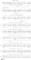
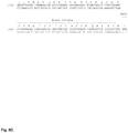
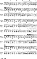
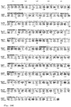
- Figure 1-6 Sequences of humanized ScFv derived from A5
- the humanized sequences have a homology of at least 80% to SEQ ID NO:1 or/and SEQ ID NO:5, respectively. In a more preferred embodiment the sequences have a homology of at least 90% and more preferred at least 95% to SEQ ID NO:1 and/or SEQ ID NO:5 and even more preferred the homology is at least 98% to SEQ ID NO:1 and/or SEQ ID NO:5. It should be noted that the CDR regions which are shown in Figure 13 are conserved to a very high level which means that the CDR regions which are determined by the Kabat method have not more than three, preferably not more than one and preferably no amino acid exchange. Fig.
- CARs are composed of an extracellular domain containing the antigen recognizing scFv, a hinge region, a transmembrane region, and one or more intracellular signaling domains that activate the T cell, including the CD3 ⁇ chain.
- co-stimulatory domains usually derived from CD28, 4-1BB, OX40, CD27 and/or ICOS are included.
- CARs targeting PSMA epitopes and some of these strategies have already entered clinical trials (e.g. NCT01140373, NCT01929239).
- A5-based CAR T cells were compared side-by-side with J591 and 3D8-based CAR T cells ( Figure 9 ).
- the antigen-specific activation profiles were compared in Jurkat cells transduced with expression vectors coding for A5, J591 and 3D8-based CARs. While the A5-based CAR and the J591-based CAR were able to mediate massive activation of the Jurkat cells upon antigen-specific sensitization ( Figure 9C ), 3D8-CAR bearing cells were only weakly activated.
- A5-based CAR T cells revealed a superior cytotoxicity profile as compared to J591-based CAR T cells on two PSMA positive tumor cell lines ( Figure 10A ), as evidenced by the fact that both tumor cell lines were eliminated with lower effector-to-target ratios.
- the manufactured A5-CAR T cell products expanded better ( Figure 11C ), showed less exhaustion ( Figure 11F ) and contained a higher percentage of undifferentiated T cells, such as a na ⁇ ve T cell and T stem cell memory cells ( Figure 11E ), as compared to J591-based CAR T cells.
- the A5-based anti-PSMA CAR T cells have unexpected properties that are superior to previously published PSMA-targeting CAR T cells, particularly in view of their superior in vitro cytotoxicity, as well as their T cell phenotype and their expansion and exhaustion profiles upon antigen-specific stimulation. Based on these results, the A5-based CAR T cells are promising tools for the development of novel immunotherapies for the treatment of local and advanced prostate cancer.
- the present invention relates therefore to chimeric antigen receptors for T cells which comprise an antigen-binding fragment which binds specifically to the PSMA antigen.
- the antigen-binding fragment comprises preferably a V H and a V L fragment which are connected with a suitable linker.
- the chimeric antigen receptor comprises preferably a spacer element, a transmembrane fragment and a CDR3 ⁇ cytoplasmic domain.
- the chimeric antigen receptor preferably comprises a fragment from the CD28 and/or a 4-1BB cytoplasmic domain.
- the chimeric antigen receptors of the present invention contain preferably at least three CDRs selected from the group consisting of CDR-H1, CDR-H2, CDR-H3, CDR-L1, CDR-L2 and CDR-L3.
- the CDRs are shown by grey arrows above the amino acid sequence and the relevant nucleic acid sequence coding.
- the chimeric antigen receptor comprises at least three (CDR-H1, CDR-H3, CDR-L3), preferably four (CDR-H1, CDR-H3, CDR-L2, CDR-L3), and more preferred at least five (CDR-H1, CDR-H3, CDR-L1, CDR-L2, CDR-L3) of the CDRs.
- the chimeric antigen receptors are preferably present in a humanized format.
- a humanized format can be obtained by inserting the at least three, preferably four, more preferred five or six CDRs into a suitable human antigen-binding scaffold having a high homology to the murine scaffold as shown in Fig. 1-6 .
- HEK293T cells were cultured at 37 °C in a humidified incubator with 5% CO 2 in DMEM (Gibco, Invitrogen, Düsseldorf, Germany) supplemented with 10% fetal calf serum (Biochrom, Berlin, Germany), penicillin (100 U/ml), streptomycin (100 mg/L) and 10 mM HEPES (Sigma-Aldrich).
- DMEM Gibco, Invitrogen, Düsseldorf, Germany
- penicillin 100 U/ml
- streptomycin 100 mg/L
- 10 mM HEPES Sigma-Aldrich
- Biological titers of the vector preparations were determined by transducing Jurkat T cells, followed by staining of the transduced cells with anti-human IgG to determine the fraction of CAR positive cells.
- PBMCs Peripheral blood mononuclear cells
- phase separation Ficoll, Sigma-Aldrich
- PBMCs were thawed and let to recover for 24 h in RPMI complete medium [RPMI 1640 medium (Gibco, Invitrogen, Düsseldorf, Germany) supplemented with 10% fetal calf serum (Biochrom, Berlin, Germany), penicillin (100 U/ml), streptomycin (100 mg/L) and 10 mM HEPES buffer (Sigma-Aldrich)].
- PBMCs were activated using anti-CD2/CD3/CD28 antibodies (Immunocult, StemCell Technologies) and cultured in RPMI complete medium supplemented with 100 U/ml of IL-2, 25 U/ml of IL-7 and 50 U/ml of IL-15 (all from Miltenyi Biotech) for 2 to 3 days before transduction with gamma-retroviral constructs encoding either of the PSMA targeting CAR T cells with a dose ranging from 50-300 transducing units per cell.
- anti-CD2/CD3/CD28 antibodies Immunocult, StemCell Technologies
- RPMI complete medium supplemented with 100 U/ml of IL-2, 25 U/ml of IL-7 and 50 U/ml of IL-15 (all from Miltenyi Biotech) for 2 to 3 days before transduction with gamma-retroviral constructs encoding either of the PSMA targeting CAR T cells with a dose ranging from 50-300 transducing units per
- Transduced cells were cultured in wells coated with poly-D-lysin (PDL, Sigma-Aldrich) containing RPMI complete medium supplemented with 5 ⁇ g/ml of protamine sulfate (Sigma-Aldrich) and 1000 U/ml of IL-2, 25 U/ml of IL-7 and 50 U/ml of IL-15. After one day, medium was changed and cells were further expanded for 8-9 days in RPMI complete medium supplemented with 100 U/ml of IL-2, 25 U/ml of IL-7 and 50 U/ml of IL-15 before being frozen in liquid nitrogen until further use.
- PDL poly-D-lysin
- RPMI complete medium supplemented with 5 ⁇ g/ml of protamine sulfate (Sigma-Aldrich) and 1000 U/ml of IL-2, 25 U/ml of IL-7 and 50 U/ml of IL-15.
- the biological activity of a preferred construct according to the present invention has been compared with a construct wherein the PSMA binding fragment is derived from the antibody J591. Furthermore, it was compared with a similar construct wherein the antigen binding construct was derived from another antibody (3D8).
- the constructs were introduced into Jurkat T-cell line and the CAR expression level was determined by flow cytometry after staining of cells with anti-human IgG. As shown in Figure 9B , all CAR constructs were properly expressed in Jurkat T-cells. UT (untransduced cells) served as a negative control for staining.
- the antigen-specific activation of transduced Jurkat T cells was measured by monitoring the expression of the activation marker CD69 upon antigen stimulation Figure 9C .
- the 3D8-based CAR mediated only weak activation of the transduced Jurkat cells upon exposure to antigen positive cells. This was independent of the expression of the inhibitory ligand PD-L1. None of the CAR constructs were activated when they were stimulated with antigen negative (DU145) cells.
- CAR T cells were generated from PBMCs following retroviral transduction as described in example 2.
- CAR T cells were co-cultured with irradiated PSMA positive (C4-2) tumor positive cells for 12 days at a 1:1 effector-to-target ratio in RPMI complete medium supplemented with 100 U/ml of IL-2, 25 U/ml of IL-7 and 50 U/ml of IL-15 (all from Miltenyi Biotech). Every 3 days, cells were harvested, counted and plated over fresh irradiated antigen positive (C4-2) tumor positive cells. To determine CAR expression levels and the fraction of CAR-positive cells by flow cytometry (FACS Canto II or Accuri, BD Biosciences), cells were stained with anti-human IgG-PE (Southern Biotech).
- the number of CAR T cells was counted at different time points using a NucleoCounter (NC-250, ChemoMetec). The number of cell divisions was calculated based on the absolute cell number.
- CAR T cells were determined by flow cytometric analysis to check the CAR T cell phenotype and the CAR T cell exhaustion pattern.
- CAR T cells were exposed to antigen positive (C4-2) tumor cells before cells were harvested and stained with anti-human CD62L-Bv421 (BD Biosciences), anti-human CD45RA-FITC (Biolegend), anti-human CD3-APC/H7 (BD Biosciences) and anti-human IgG-PE (CAR) (Southern Biotec).
- the T cell phenotype was determined based on the expression of CD62L and CD45RA. Cells were pre-gated on CD3+/CAR- for untransduced (UT) T cells or on CD3+/CAR+ for both types of CAR T cells (not shown).
- A5-CAR T cells were less differentiated as compared to J591CAR T cells as indicated by the presence of high proportion of undifferentiated cells na ⁇ ve T cell (Tn) and T stem cell memory (Tscm) but a low proportion of effector T cells (Teff).
- CAR T cells were exposed to PSMA-positive tumor cells (C4-2) before being harvested and stained with anti-human CD279-FITC (PD-1, BD Biosciences), anti-human CD223-eFluor710 (LAG-3, BD Biosciences), anti-human CD3-APC/H7 (BD Biosciences) and anti-human IgG-PE (Southern Biotec).
- the exhaustion profile was determined by flow cytometry (FACS Canto II) based on the expression of CD279 (PD-1) and CD223 (LAG-3).
- A5-CAR T cells maintained a significantly increased number of LAG-3 and PD-1 double negative cells upon antigen encounter, indicating a less exhausted T cell phenotype.
- the cytotoxic potential of PSMA targeting CAR T cells was determined by assessing their ability to kill PSMA positive tumor cells using a cell viability XTT assay.
- CAR T cells were co-cultured with either PSMA-positive (C4-2) or (LNCaP) tumor cells or antigen-negative tumor control cells (DU145) in 96 well plates for 48 h at different effector-to-target ratios in a final volume of 200 ⁇ l/well of RPMI complete medium without any cytokines.
- To determine cell viability as a function of metabolic activity 100 ⁇ l/well of medium was removed and replaced with 100 ⁇ l/well of XTT solution (Sigma-Aldrich), and cells incubated at 37°C.
- the genetic information coding for the chimeric antigen receptor is introduced with the help of a suitable vector into the target immune cells (e.g. T cell).
- a suitable vector can preferably be a lentivirus vector or a retroviral vector or a transposon or a plasmid.
- the genetic information can be introduced into the genome of the T cell in a targeted fashion with the help of designer nuclease technology, such as the CRISPR/Cas technology or the TALEN technology, as described herein.
- the present invention relates to an in vitro method of providing T cells comprising a chimeric antigen receptor as described herein.
- T cells are isolated from a donor preferably by leukapheresis methods. This results in a substantial enrichment of the T cells.
- the T cells are then genetically modified by transfection with a suitable vector or by transduction with a viral vector containing the genetic information for the chimeric antigen receptor, respectively.
- Such genetically modified T cells can then be isolated and amplified whereby those T cells which do not contain the desired genetic information can be separated from the modified T cells or at least reduced.
- the transfected T cells can then be applied to the patient to be treated.
Abstract
The present invention relates to a novel chimeric antigen receptor (CAR) comprising an antigen-binding fragment which binds specifically to PSMA antigen, and a method of manufacturing high-quality CAR T cell products by transfection and/or transduction of T cells therewith, which allows to effectively treat tumors in vivo alone or in combination with pharmaceutical drugs, such chemotherapies, biopharmaceutical drugs, such as antibodies, or small-molecule drugs, such as protein kinase inhibitors.
Description
- The present invention relates to chimeric antigen receptors that bind to tumor antigens whereby the antigen is the prostate specific membrane antigen (PSMA). The chimeric antigen receptors (in the following CAR) are brought into immune cells, in particular T cells, NK cells, iNKT cells and CIK cells, which then specifically react with tumor cells expressing PSMA which leads to the elimination of the tumor cells. The constructs of the present invention contain two major parts. On the one hand the antigen-binding region which specifically binds to the prostate specific membrane antigen (PSMA) and on the other hand co-stimulatory and activating domains derived from a receptor of an immune cell responsible for signal transduction and activation of the immune cell.
- Prostate cancer remains the second-most frequently diagnosed cancer among men worldwide with estimated 1.1 million new cases per year. Moreover, with expected 307,000 deaths, it represents the fifth leading cause of cancer deaths. Whereas primary tumors can successfully be treated, there is no curative treatment for advanced stages. Therefore, new therapeutic options are urgently needed.
- The prostate specific membrane antigen (PSMA) is the best characterized antigen in prostate cancer for antibody-based diagnostic and therapeutic intervention. This protein is also known as glutamate carboxypeptidase II (EC 3.4.17.21), N-acetyl-linked acidic dipeptidase I (NAALADase), or folate hydrolase. PSMA is a type II membrane glycoprotein consisting of 750 amino acids (aa) with a small intracellular domain of 19 aa, a transmembrane domain of 24 aa, and a large extracellular domain of 707 aa. The extracellular domain folds into three distinct domains: the protease domain (aa 57-116 and 352-590), the apical domain (aa 117-351), and the C-terminal domain (aa 591-750). It shows a high structural similarity and identity to the
human transferrin receptor 1. PSMA is highly restricted to the surface of prostate cancer cells, is present on cancer cells during all tumor stages, and shows an enhanced expression in androgen-independent and metastatic disease. PSMA is not secreted into the extracellular space and undergoes constitutive internalization, which is enhanced by binding of PSMA-specific antibodies. These characteristics make it an ideal candidate for the targeted treatment of local and advanced prostate cancer. Moreover, PSMA was also found to be expressed in the neovascular endothelium of virtually all solid tumor types without expression in normal vascular endothelium. It is therefore considered to be a unique antiangiogenic target. - Monoclonal antibodies (mAbs) are highly specific and versatile tools for cell targeting. In the last decades, they have attracted high interest in medical research and have become the most rapidly expanding class of pharmaceuticals for treating a variety of human diseases including cancer. Antibody 7E11 was the first published PSMA-specific mAb and was found to bind to the N-terminus (MWNLLH) of the intracellular domain of PSMA. The In-labeled form of 7E11 (ProstaScint, Cytogen, Philadelphia, PA) has received approval from the U.S. Food and Drug Administration (FDA) for the detection and imaging of metastatic prostate cancer in soft tissues. However, because the antibody binds an intracellular epitope, 7E11 is not capable to bind to viable cells. Positive signals in the in vivo imaging with ProstaScint had to be traced back to the detection of dead or dying cells within the tumor masses. Therefore, a new class of anti-PSMA mAbs was generated, which specifically bind to extracellular epitopes of PSMA expressed by living cells.
-
EP 1 883 698 - The single-chain variable fragment (scFv) A5 (as disclosed in
WO 2006/125481 ) was generated by phage display technique from the mAb 3/A12. The most important fragments for the specificity of the anti-PSMA scFv are the VL and the VH parts, which are preferably linked with a polyglycin linker. A5 bound to PSMA-expressing cells with a Kd of about 33 nM. - According to the present invention the A5 scFv was used for the construction of chimeric antigen receptors (CAR's) to provide constructs for use in immune cells for targeting cancer cells expressing PSMA.
- Ma et al. [The Prostate (2014) 74, pp 286-296] disclose a second generation CAR against PSMA comprising a CD28 costimulatory domain and CD3ζ signaling domains and an antigen binding part from a mouse anti-human PSMA monoclonal antibody 3D8 which is commercially available from Northwest Biopharmaceutics, Inc.
- Santoro et al. [Cancer Immunol. Res. (2015), pp 68-84] describe T-cells bearing a chimeric antigen receptor against prostate-specific membrane antigen, whereby the PSMA binding portion is derived from the antibody J591.
- Zhong et al. [Molecular Therapy (2010), pp. 413-420] disclose also chimeric antigen receptors whereby the PSMA binding fragment is also derived from the antibody J591. The sequence of J591 is well-known in the art.
WO 2009/017823 discloses the VH and VL domains thereof. - The antigen-binding fragments are derived from the murine sequences. The murine sequence of the variable domain of the heavy chain (VH) of A5 is shown as SEQ ID NO:1 and the murine sequence of the variable domain of the light chain (VL) of A5 is shown as SEQ ID NO:5. As highly relevant fragments thereof the CDR regions were identified as follows: CDR-H1 as SEQ ID NO:2 having the amino acid sequence of GFTFSDYYM; as CD-H2 (SEQ ID NO:3) IISDGGY and as CDR-H3 (SEQ ID NO:4) GFPLLRHGAMDY. In the light chain the CDRs CDR-L1 (SEQ ID NO:6) has the amino acid sequence KASQNVDTNVA, CDR-L2 (SEQ ID NO:7) has the amino acid sequence SASYRYS and CDR-L3 (SEQ ID NO:8) has the amino acid sequence QQYDSYPYT.
- While in the above paragraph the CDRs according to the Kabat numbering system are provided, the CDRs can alternatively also be determined according to the IMGT numbering. The following sequences of the CDRs according to IMGT have been determined: CDR-H1 has been determined as SEQ ID NO:9: GFTFSDYY; SEQ ID NO:10: ISDGGYYT as CDR-H2 and as SEQ ID NO:11 corresponding to CDR-H3: TRGFPLLRHGAMDYWG.
- For the light chain the IMGT numbering has been determined for CDR-L1: SEQ ID NO:12: QNVDTN, SEQ ID NO:13 showing CDR-L2: SAS and as SEQ ID NO:14 showing CDR-L3: QQYDSYPYT.
- Humanization of murine antibodies involves the transfer of beneficial properties (e.g. antigen-specific binding, avoidance of off-target effects by non-crossreactivity with other antigens) from one antibody to another to reduce immunogenicity. Humanization is usually necessary for human use as patients typically respond with an immune reaction against non-human antibodies that can lead to ineffectiveness of treatment and, in the worst-case scenario, to a life-threatening situation. A humanized construct can be derived from the sequence of the antigen-binding fragment shown in SEQ ID NO:1 and 5, respectively. The CDR regions structurally define the paratope, that is, the contact site of the antigen-binding fragment with the antigen. The remainder of the sequence codes for the framework regions, which form the scaffold of the paratope. For the humanization process (e.g. by in silico modeling), the framework sequence is first compared with other antigen-binding sequences derived from humans. Usually a human sequence (acceptor framework) is selected which has the highest similarity with the framework sequence shown of the murine sequence. The CDR regions are grafted into the human acceptor framework to eliminate amino acid sequences which may cause undesired human anti-mouse-antibody (HAMA) immune reactions. Substitutions at potentially critical positions (e.g. amino acids responsible for folding the paratope or the VH-VL interface) are analyzed for prospective back mutations. Even in the CDR sequences exceptional modifications of amino acids may be made to avoid immunogenicity, to ensure the right folding of the paratope, and to maintain the antigen-specific binding.
- In the course of the humanization of antibodies preferably a sequence is selected among human immune sequences which have the highest homology with the corresponding murine sequence. Then the location of the CDRs is determined. The determination of the CDRs is well-known in the art and it should be noted that different methods for the determination are known whereby it is possible that the locations of the CDRs differ somewhat. In the course of the present invention the determination of the CDRs according to Kabat was used and also the determination according to the IMGT (International Immunogene Ticks).
- In a preferred embodiment of the present invention the humanization was performed according to the so-called "CDR-grafting". The functional CDRs are determined preferably according the IMGT method and those CDRs are transferred in a human framework region which has the highest sequence homology to the starting murine antibody. Then the differences regarding the single amino acids in the framework region of the humanized and murine antibodies were determined with regard to the biochemical properties like size, polarity or charge. In the first step similar amino acids were adapted and successively the different amino acids were changed in order to end up with a complete human framework.
- Since the humanized versions disclosed herein have maintained the CDRs either completely or to a very large extent the humanized variants have the same function of the murine antibody whereby, however, the affinity may differ somewhat from each other.
- The sequences obtained by the humanization experiments are as SEQ ID NO:15 to 36.
- The results of the humanization experiments are shown in
Figure 1 to Figure 6 . It is always the nucleic acid sequence of the coding strand shown. Moreover, the amino acid sequence is provided where by the CDR regions are shown in grey. Those CDR sequences are preferred embodiments of the present invention. It turned out that in most cases for the heavy chains the CDRs were as follows: CDR-H1: GFTFSDYY, CDR-H2: ISDGGYYT and CDR-H3: TRGFPLLRHGAMDYWG. - For the light chains the following CDRs were preferably used: CDR-L1: QNVDTN, CDR-L2: SAS and CDR-L3: QQYDSYPYT. The preferred humanized CDRs were deduced from the murine sequences, but adapted to suitable human framework.
- In preferred embodiments the humanized antigen binding fragments like preferably scFv fragments or the like have at least three, preferably at least four, more preferably at least five and particularly preferred six CDRs as mentioned above.
- In recent years, adoptive immune cell therapy has been introduced as a novel concept to treat different cancers by redirecting the immune system to eliminate the tumor cells. One of the most successful concepts is based on the genetic engineering of T cells to express chimeric antigen receptors (CARs) that bind tumor antigens or tumor-associated antigens in a human leukocyte antigen (HLA)-independent manner. CD19 targeting CAR T cells have been successfully used to treat B cell acute lymphoblastic leukemia (B-ALL), with >90% of patients going into complete remission in several clinical trials. Based on this success, more than 200 clinical trials have been initiated to treat mostly hematological malignancies. For solid tumors, however, the potency of CAR T cell therapy seems rather low to date. The main reason for this failure seems to be the tumor microenvironment (TME), which is the cellular environment in which the tumor exists. It includes various kinds of immune cells, fibroblasts, the extracellular matrix (ECM) as well as the surrounding blood vessels. Many mechanisms that describe restriction of cytotoxic T cell activity in the TME have been described, including the activation of PD-1 based T cell immune checkpoint inhibition. Overcoming these restrictions, combined with T cell checkpoint antagonists, will help to improve anti-tumor activity in the TME.
- The tumor eradication, as used in the present invention, requires an adequate survival and intratumoral activation of tumor antigen-specific immune cells, preferably T cells. To meet these requirements T cells must be given appropriate activating signals at the time of antigen-priming and stimulation. The chimeric antigen receptors of the present invention combine therefore an antigen-binding fragment as part of the receptor on T cells. The antigen-binding fragment binds to the specific antigen (here PSMA) to which the T cells should bind. Moreover, the receptors contain sequences from CD28 and 4-1BB, respectively, as co-stimulatory signaling domains. It has been shown that the addition of CD28 sequences, or other co-stimulatory signaling domains, to CD3ζ chain-based receptors increase antigen-induced secretion of interleukin-2 and in vitro T cell expansion. In the present case the signaling domain consists of the CD3ζ domain and either the intracellular CD28 or the 4-1 BB domain.
- In general, the design of a CAR can vary and meanwhile several generations of CARs are known. The main components of a CAR system are the CD3ζ intracellular domain of the T cell receptor (TCR) complex, the transmembrane domain, the hinge region and the antigen-binding part. In the design of a CAR, the antigen-binding domain is linked to a hinge region, which is also called a spacer region, the transmembrane domain, and a cytoplasmic domain. Those parts are responsible for the position of the antigen-binding part, the attachment in T cell membrane, and intracellular signaling. Besides this structural rule in the CAR design the morphological characteristics of the hinge region, such as their length and sequence, are important for an efficient targeting. The intracellular domain acts as a signal transducer. The cytoplasmic segment of the CD3ζ plays the principal rule due to different functions in activated T cells and the resting ones. However, this cytoplasmic part cannot activate the resting T cells alone. Therefore, there is a need of at least a secondary signal for the full activation of T cells. In the present invention, preferably 4-1BB or CD28 co-stimulatory domains were used. Other co-stimulatory domains, such as co-stimulatory domains derived e.g. from CD27, ICOS and OX40, can be used alternatively.
- In preferred embodiments mutations are introduced into the human IgG1 Fc hinge region whereby side effects like preventing LcK activation or an unintended initiation of an innate immune response are avoided. One of those mutations avoids LcK binding and another mutation may inhibit the binding of Treg cells to the construct. Such mutations may improve the biological activity of the construct.
- For the treatment of human patients, T cells have to be enriched from the individual patient's peripheral blood (autologous setting) or provided by a donor (allogeneic setting). This can be done for example by leukapheresis. The enriched T cells are then transfected or transduced ex vivo with a suitable vector comprising the genetic information for the CAR.
- The genetic information coding for the CAR is inserted into a suitable vector. Such vectors are preferably lentiviral or retroviral vectors. A gold standard for transduction of primary T cells are presently considered lentiviral vectors which seem to be a valid alternative to simpler retroviral vectors. As a further alternative to lentiviral vectors the information can be introduced into the T cells with the help of transposons or plasmids. An alternative to both viral and non-viral delivery are the recently described gene editing tools, designated as CRISPR/Cas or other designer nucleases, such as transcription activator-like effector nucleases (TALENs) or zinc finger nucleases (ZFNs). This technology platforms offer the possibility to target virtually any genomic site in a targeted manner. In the case of CRISPR/Cas, the editing complex comprises a Cas nuclease and a guide RNA, usually composed of a CRISPR RNA (crRNA) and a transacting crRNA. Upon hybridization of the guide RNA to the target sequence, Cas9 (or another Cas nuclease, such as e.g. Cpf1/Cas12a) generates a double-strand break, which can be repaired by non-homologous end joining (NHEJ), an event that can result in a loss of function of the genomic locus. In the presence of a suitable donor DNA, by a mechanism of homology-directed repair (HDR), an exogenous sequence (CAR sequence) can be introduced into the targeted locus. This can be exploited to deliver CAR expression cassette in a desired genomic locus that does not interfere with endogenous gene function and therefore minimizing the genotoxic effects experienced with integrating viral vectors. In a further preferred embodiment, genome editing is used to place the CAR coding sequence under control of an endogenous promoter. The expression from an endogenous promoter, such as the promoter of the TRAC locus, could ensure optimal expression levels of the CAR construct to fulfill its function. The nucleic acid sequence coding for the CARs of the present invention is preferably optimized for the human codon usage. Particularly preferred embodiments are SEQ ID NO:37 coding for the CAR28 construct (
Fig. 7A-D ) and SEQ ID NO:38 coding for the CAR41 construct (Fig. 8A-D ). - In another preferred embodiment of the present invention the RNA coding for the CAR construct is introduced into the target cells, such as T-cells. Nucleic acid coding for the CAR construct may be introduced into the target cells by physical procedures, such as electroporation, or by fusion of the cells' membranes with suitable vesicles. In this embodiment, the CAR construct is preferably transiently expressed in the T-cells. The advantage is that there is then a population of transduced T-cells which is present in the treated patient only for a transient time.
- In other preferred embodiments the CAR construct is introduced into natural killer cells (NK), invariant natural killer T-cells (iNKT), diverse natural killer cells (dNKT), cytokine-induced killer cells (CIK) or γ-δ T-cells. Furthermore, suitable allogenic cells may be used.
- In a further preferred embodiment, in addition to the CAR construct described herein, another transgene that modulates the immune system, such a genes coding for cytokines, chemokine receptors and/or checkpoint inhibitors, may be introduced in the immune cells. In a further preferred embodiment, genome editing is used to disrupt the expression of genes that modulate the immune system, such as genes coding for cytokines, chemokine receptors and/or checkpoint inhibitors.
- In another embodiment the constructs according to the present invention and immune cells containing such constructs can be used for local therapy with a targeted tumor injection. In this embodiment, which is performed preferably with automated devices that apply the CAR T-cells to certain places in the body of the patient, where local tumor areas are located. With a biopsy needle a sample is then withdrawn whereby a small cavity is formed. In this cavity the transduced or transfected T-cells are introduced and then the needle is withdrawn. This embodiment is particularly advantageous when there are solid tumors which are extremely difficult to treat with regular methods.
- The chimeric antigen receptors disclosed herein can be used for the treatment of diseases which are related to the expression of PSMA. PSMA is expressed in tumor cells derived from prostatic cancer. There are several stages of prostatic cancer known, but it seems that PSMA is one of the markers best suited for the treatment of prostate cancer. The term "prostate cancer" comprises all forms of prostate cancer cells either derived from a primary tumor or from a metastatic tumor or from circulating tumor cells. In a particularly preferred embodiment immune cells engineered with the chimeric antigen receptors of the present invention are used against the neovascularization of solid tumors expressing PSMA.
- In a further embodiment of the present invention immune cells engineered with the chimeric antigen receptors disclosed herein are used in combination with a therapeutic agent, in particular a cytotoxic agent. Cytotoxic agents as used for the treatment of prostate cancer are known. Preferably such substances comprise taxol derivatives, 5-fluorouracil, cyclophosphamide, mitoxantrone, docetaxel, cabazitaxel and etoposide. The following drugs are approved for prostate cancer and preferably used: Abiraterone Acetate, Apalutamide, Bicalutamide, Cabazitaxel, Casodex (Bicalutamide), Degarelix, Docetaxel, Eligard (Leuprolide Acetate), Enzalutamide, Erleada (Apalutamide), Firmagon (Degarelix), Flutamide, Goserelin Acetate, Jevtana (Cabazitaxel), Leuprolide Acetate, Lupron Depot (Leuprolide Acetate), Mitoxantrone Hydrochloride, Nilandron (Nilutamide), Nilutamide, Provenge (Sipuleucel-T), Radium 223 Dichloride, Sipuleucel-T, Taxotere (Docetaxel), Xofigo (Radium 223 Dichloride), Xtandi (Enzalutamide), Zoladex (Gosereliin Acetate), Zytiga (Abiraterone Acetate). It is understood that the chimeric antigen receptors of the present invention can also be used in combination with a medicament used to treat hormone sensitive forms of prostate cancer like for example Leuprolide Acetate.
- The preferred embodiments of the present invention are further described and illustrated in the Figures and Examples of the present application. All aspects disclosed in the Figures or the examples, respectively, relate to the present invention unless expressly excluded. The single features of the present invention as disclosed in the experimental part can be combined unless there are technical reasons which speak against such combination. In particular, the Figures show the results of the experiments as follows:
In the Figures and in the experiments the following abbreviations were used:Abbreviation Explanation 4-1BB tumor necrosis factor receptor superfamily member 9 BLI bioluminescence imaging bw body weight C4-2 PSMA positive prostate cancer cell line CAR Chimeric Antigen Receptor CAR28 anti-PSMA CAR with CD28 co-stimulatory domain CAR41 anti-PSMA CAR with 4-1 BB co-stimulatory domain CD28 cluster of differentiation 28CD3ζ CD3 zeta region CD45RA cluster of differentiation 45 isoform RACD62L cluster of differentiation 62, L-selectin CR complete remission DOC, DTX docetaxel DU145 PSMA negative prostate cancer cell line EFS Short version of the elongation factor alpha gene promoter ELISA enzyme-linked immunosorbent assay i.v. intravenously i.p. intraperitoneally s.c. subcutaneously Gr.A Granzyme A Gr.B Granzyme B. IFN-g Interferon-gamma LcK lymphocyte-specific protein tyrosine kinase LNCaP PSMA positive prostate cancer cell line LTR long terminal repeat of retroviral vector PR partial remission R R region of the retroviral long terminal repeat scFv single chain variable fragment SD standard deviation Tcm T cell central memory Teff T cell effector Tem T cell effector memory Tn, scm T cell naïve or T stem cell memory U5 U5 region of the retroviral long terminal repeat UT untransduced T cell WPRE woodchuck hepatitis virus post-transcriptional regulatory element XTT sodium 3'-[1-(phenylaminocarbonyl)-3,4-tetrazolium]-bis(4-methoxy-6-nitro) benzene sulfonic acid hydrate ΔU3 deletion in the U3 region in the retroviral long terminal repeat - It should be mentioned that the sequences disclosed in the specification, the Figures and the sequence protocol and in particular the CDR-Sequences are preferred embodiments of the present invention.
-
- (
Fig. 1-3 ) Shown are the sequences of five humanized variants of the variable domain of the heavy chain (VH) of A5 whereby the locations of CDR-H1, CDR-H2 and CDR-H3 are shown according to the determination of IMGT as grey boxes. - (
Fig. 4-6 ) Shown are the sequences of six humanized variants of the variable domain of the light chain (VL) of A5, with CDR-L1, CDR-L2, and CDR-L3 marked with grey boxes. It is understood that the VH and VL domains can be combined with each other. It is for example possible to combine the VH sequence of variant 1 (hum A5-VH1) with the VL of variant 5 (hum A5-VL5). -
Figure 7
Figure 7A-D shows the complete sequence of a preferred embodiment of a construct comprising the scFv A5 antigen binding region. This antigen binding region is linked to the relevant parts of CD28 and CD3ζ. -
Figure 8
Figure 8A-D shows the whole construct of the antigen binding fragment scFv A5 which is coupled to the relevant parts of human 4-1BB and CD3ζ. -
Figure 9 : Comparison of CAR construct according to the invention with constructs wherein the PSMA antigen binding fragments were already described in the prior art- (A) Schematic of self-inactivating gamma-retroviral vectors to express 2nd generation anti-PSMA CARs. CAR expression is driven by an EFS promoter. The anti-PSMA CARs contain either the single chain variable fragment (scFv) 3D8 or J591 (both described in the prior art), or A5 (derived from murine monoclonal antibody 3/A12 against PSMA), which is fused to an Fc IgG1 derived hinge region, a transmembrane (tm) domain, a CD28 derived costimulatory domain, and an intracellular signaling domain derived from CD3ζ chain. The hinge region provides a physical spacer between the scFv and the tm domains for optimal target recognition. The CD28 costimulatory domain contains amino acid exchanges that prevent LCK binding and enhance anti-tumor activity in the presence of inhibitory regulatory T cells (Tregs). All generated retroviral constructs have the same scaffold but differ only in the scFv fragments allowing for a side-by-side comparison of the CAR constructs with respect to activity and cytotoxicity.
- (B) CAR expression in Jurkat cells were transduced with retroviral particles coding for the three different PSMA-CARs. Following viral transduction, Jurkat cells were expanded for 16-21 days before cells were harvested and stained with anti-human IgG antibody (CAR) to evaluate the transduction efficiencies and the CAR expression levels.
- (C) Antigen-specific-activation profile, CAR expressing Jurkat cells were stimulated for 24 h at an 1:1 effector-to-target ratio with either PSMA positive, PD-L1 negative C4-2 tumor cells (PSMA+/PDL1-), or PSMA positive, PD-L1 positive LNCaP tumor cells (PSMA+/PDL1+). As a negative control, CAR cells were co-cultured with PSMA negative DU145 tumor cells (PSMA-). Following 24 h stimulation, cells were harvested and activation profile was assessed by evaluating the percentage of cells that are positive for the activation marker CD69. Statistically significant differences are indicated by ****(P<0.001).
- (D) Characterization of employed prostate cancer cell lines. The extent of PSMA target antigen expression (left) and PD-L1 (CD274) expression (right) was assessed on C4-2, LNCaP and DU145 prostate cell lines. For flow cytometric analysis, cells were either stained with 3/
F11 1 antibody (anti-PSMA) or an anti-CD274 antibody. UT, untransduced T cells; PD-L1, Programmedcell death ligand 1; PSMA; prostate specific membrane antigen.
The antigen-specific activation profiles were compared in A5-based CAR T Jurkat cells side-by-side with J591 and 3D8-based CAR T Jurkat cells. As indicated in Panel C, the A5-based CAR and the J591-based CAR were able to mediate massive activation of the Jurkat cells upon antigen-specific sensitization, as measured by upregulation of the activation marker CD69 in about 70% of cells. Activation was not affected by the presence of the inhibitory ligand PD-L1. On the other hand, the 3D8-based CAR T cells were only weakly activated (up to 20% of CD69-positive cells). -
Figure 10 : Functional assessment of PSMA-CAR T cells.- (A) Cytotoxicity profile of CAR T cells on PSMA positive C4-2 tumor cells. T cells were activated for 2-3 days with anti-CD2/3/28 antibodies prior to retrovirus transduction. CAR T cells were generated by transducing activated T cells with the self-inactivating retroviral vector constructs to express 2nd generation anti-PSMA CARs (see
Figure 9A ). Following expansion for 6-9 days without antigen stimulation, CAR T cells were co-cultured for 48 hours at indicated effector to target (E:T) ratios, with either antigen positive C4-2 (PSMA+/PDL1+) or LNCaP (PSMA+/PDL1-) tumor cells or PSMA negative cells (DU145). Cytotoxicity was measured by an XTT ELISA based colorimetric assay (% cytotoxicity = 100% - % viability). Statistically significant differences are indicated by *(P<0.05) or **(P<0.01). UT, untransduced cells; PSMA, prostate specific membrane antigen.
- (A) Cytotoxicity profile of CAR T cells on PSMA positive C4-2 tumor cells. T cells were activated for 2-3 days with anti-CD2/3/28 antibodies prior to retrovirus transduction. CAR T cells were generated by transducing activated T cells with the self-inactivating retroviral vector constructs to express 2nd generation anti-PSMA CARs (see
-
Figure 11 : Expansion and exhaustion of PSMA targeting CAR T cells.- (A) CAR expression. Following transduction of activated T cells with self-inactivating retroviral vector constructs to express 2nd generation anti-PSMA CARs (see
Figures 9A ,10A ), CAR T cells were expanded for 6 days without antigen stimulation (-Ag). In parallel, CAR T cells were expanded and exposed to irradiated PSMA positive (C4-2) cells (+Ag). Expanded CAR T cells were harvested every 3 days and exposed to fresh irradiated antigen positive cells up to 4 times (4X) at effector-target ratio of 1:1. CAR and CD3 expression at indicated exposure times to irradiated PSMA-positive cells were visualized by flow cytometry after staining of cells with antibodies against human IgG (CAR) and CD3. UT, untransduced cells. - (B, C, D) CAR T cell expansion. Every 3 days during CAR T cell expansion, cells were harvested and stained with anti-human IgG antibody (CAR) and CD3 to evaluate fraction of CAR-positive cells (B), total cell number (C) and the number of cell divisions (D). Statistically significant differences are indicated by *(P<0.05). UT, untransduced cells.
As shown, multiple exposure to antigen-expressing cells leads to enrichment of CAR positive T cells in the cell population (>95%) for both A5-based CAR T cells as well as J591-based CAR T cells (Fig. 11A-B ). However, A5-based CAR T cells expand significantly better upon multiple antigen-specific stimulation as compared to J591-based CAR T cells. This is evident by a -∼160-fold increase in the number of total cells at the end of the expansion period (Fig. 11C ) as well as a significant increase in the number of cell divisions (Fig. 11D ). This data suggest the A5-based CAR T cells exhaust less than J591-based CAR T cells. - (E) Quantitative assessment of CAR T cell phenotype. CAR T cells were stimulated with PSMA-positive C4-2 tumor cells at a 1:1 effector-to-target (E:T) ratio before T cell phenotype profile was assessed based on the expression of CD62L and CD45RA. Shown are T the average percentages of the different T cell subsets from three experiments. Statistically significant differences are indicated by ***(P<0.001, n=3). Tn/Tscm, T cell naive or T stem cell memory; Tcm, T cell central memory; Tem, T cell effector memory; Teff, T cell effector; UT, untransduced cells.
- (F) Quantitative assessment of CAR T cell exhaustion. CAR T cells were stimulated with PSMA-positive C4-2 tumor cells at a 1:1 effector-to-target (E:T) ratio before T cell exhaustion profile was determined based on LAG-3 and PD-1 expression. Shown are the average percentages of PD-1 and LAG3 double-positive or double-negative cells. Statistically significant differences are indicated by ***(P<0.001, n=3). UT, untransduced cells.
This set of experiments addressed the CAR T cell phenotype as well as the exhaustion profiles of the CAR T cells upon antigen stimulation. As seen in panels 3E and 3F, both A5-based and J591-based CAR T cells, respectively, differentiate towards effector cells and exhaust to some extent after antigen exposure. However, the fraction of naive and memory-type CAR T cells is significantly higher for A5-CAR T cells as compared to J591-based CAR T cells (Fig. 11E ). Also, A5-CAR T cells exhaust less frequently upon antigen exposure than J591-based CAR T cells, as indicated by a significantly higher proportion of PD1-negative/LAG3-negative CAR T cells (Fig. 11F ). Overall, the CAR T cell phenotype and the exhaustion profile analysis upon antigen exposure indicates a favorable phenotype for A5-CAR T cells over J591-CAR T cells, which correlated with a better expansion of A5-CAR T cells as compared to J591-CAR T cells.
- (A) CAR expression. Following transduction of activated T cells with self-inactivating retroviral vector constructs to express 2nd generation anti-PSMA CARs (see
-
Figure 12 : The sequence (SEQ ID NO:1 and 5) of the murine scFv A5 is provided where by the CDR-H1, CDR-H2, CDR-H3 (SEQ ID NO:2-4), CDR-L1, CDR-L2 and CDR-L3 (SEQ ID NO:6-8) regions according to the Kabat method are shown. -
Figure 13 : Sequences
Figure 13 A-C shows the amino acid sequence ("Frame1") of the CAR construct A5CAR28 including the scFv A5 which is derived from mice. Moreover, the coding nucleic acid sequence and the complementary strand thereof are provided. The CDR H1-H3 and CDR L1-L3 nucleic acid and amino acid sequences are marked with grey arrows. -
Figure 14 : Sequences
Figure 14 A-C shows the amino acid sequence ("Frame1") of the CAR construct A5CAR41 including the scFv antigen-binding construct A5 which is derived from mice. Moreover, the coding nucleic acid sequence and the complementary strand thereof are provided. The CDR H1-H3 and CDR L1-L3 nucleic acid and amino acid sequences are marked with grey arrows. The amino acid sequence of the ScFv fragment of A5 is essential for the present invention insofar as it is the starting sequence for the humanization of the antigen binding fragment. - The humanized sequences have a homology of at least 80% to SEQ ID NO:1 or/and SEQ ID NO:5, respectively. In a more preferred embodiment the sequences have a homology of at least 90% and more preferred at least 95% to SEQ ID NO:1 and/or SEQ ID NO:5 and even more preferred the homology is at least 98% to SEQ ID NO:1 and/or SEQ ID NO:5. It should be noted that the CDR regions which are shown in
Figure 13 are conserved to a very high level which means that the CDR regions which are determined by the Kabat method have not more than three, preferably not more than one and preferably no amino acid exchange.Fig. 14A shows the sequence of the A5 CAR41 construct and the preferred homology values as lined out above apply also to this construct.SEQ ID NO: nucleic / amino acid (AA) description remarks 1 AA murine VH 2 AA murine CDR-H1 Kabat 3 AA murine CDR-H2 Kabat 4 AA murine CDR-H3 Kabat 5 AA murine VL 6 AA murine CDR-L1 Kabat 7 AA murine CDR-L2 Kabat 8 AA murine CDR-L3 Kabat 9 AA murine CDR-H1 IMGT 10 AA murine CDR-H2 IMGT 11 AA murine CDR-H3 IMGT 12 AA murine CDR-L1 IMGT 13 AA murine CDR-L2 IMGT 14 AA murine CDR-L3 IMGT 15 nucleic acid hum A5-VH1 coding strand 16 AA hum A5-VH1 grey: IMGT-CDR 17 nucleic acid hum A5-VH2 coding strand 18 AA hum A5-VH2 grey: IMGT-CDR 19 nucleic acid hum A5-VH3 coding strand 20 AA hum A5-VH3 grey: IMGT-CDR 21 nucleic acid hum A5-VH4 coding strand 22 AA hum A5-VH4 grey: IMGT-CDR 23 nucleic acid hum A5-VH5 coding strand 24 AA hum A5-VH5 grey: IMGT-CDR 25 nucleic acid hum A5-VL1 coding strand 26 AA hum A5-VL1 grey: IMGT-CDR 27 nucleic acid hum A5-VL2 coding strand 28 AA hum A5-VL2 grey: IMGT-CDR 29 nucleic acid acid hum A5-VL3 coding strand 30 AA hum A5-VL3 grey: IGMT-CDR 31 nucleic acid hum A5-VL4 coding strand 32 AA hum A5-VL4 grey: IGMT-CDR 33 nucleic acid hum A5-VL5 coding strand 34 AA hum A5-VL5 grey: IMGT-CDR 35 nucleic acid acid hum A5-VL6 coding strand 36 AA hum A5-VL6 grey: IGMT-CDR 37 coding strand CAR28 construct Fig. 7 A-D 38 coding strand CAR41 construct Fig. 8 A-D - The results of the experiments shown in the Figures can be interpreted as follows:
In general, CARs are composed of an extracellular domain containing the antigen recognizing scFv, a hinge region, a transmembrane region, and one or more intracellular signaling domains that activate the T cell, including the CD3ζ chain. In 2nd or 3rd generation CARs, co-stimulatory domains, usually derived from CD28, 4-1BB, OX40, CD27 and/or ICOS are included. For the treatment of prostate cancer, many attempts had utilized CARs targeting PSMA epitopes and some of these strategies have already entered clinical trials (e.g. NCT01140373, NCT01929239). However, the potency of these CARs seems to be rather low both in vitro and in vivo. In particular, early studies based on 1st generation CARs based on the anti-PSMA scFv 3D8 or J591 (known as Pzl), have shown low potency of the resulting CAR T cells, as indicated by the need of having to employ high effector to target (E:T) ratios up to 100:1 to eliminate the tumor cells in vitro. The in vitro potency improved when 2nd or 3rd generation CARs based on either D2B or J591 derived scFvs were used. However, the potency of these CAR T cells remained low in xenotransplantion tumor mouse models, as indicated by the fact that the these PSMA targeting CAR T cells were only able to suppress tumor growth but not to eliminate the tumors in vivo, although very high CAR T cell doses, sometimes up to 20x106 CAR T cells, or multiple infusions were applied (PMID 16204083, PMID: 18026115, PMID: 19773745, PMID: 25358763, PMID: 23242161, PMID: 4174378, PMID: 25279468). In view of these mixed results with rather inefficient PSMA-CAR T cells, it was intended to generate and validate novel, more efficient PSMA targeting CARs based on the scFv A5. To this end, a 2nd generation PSMA-CAR was designed which harbors a CD28 (CAR28) derived co-stimulatory domain. - A5-based CAR T cells were compared side-by-side with J591 and 3D8-based CAR T cells (
Figure 9 ). The antigen-specific activation profiles were compared in Jurkat cells transduced with expression vectors coding for A5, J591 and 3D8-based CARs. While the A5-based CAR and the J591-based CAR were able to mediate massive activation of the Jurkat cells upon antigen-specific sensitization (Figure 9C ), 3D8-CAR bearing cells were only weakly activated. - Furthermore, upon transduction of primary T cells, A5-based CAR T cells revealed a superior cytotoxicity profile as compared to J591-based CAR T cells on two PSMA positive tumor cell lines (
Figure 10A ), as evidenced by the fact that both tumor cell lines were eliminated with lower effector-to-target ratios. Also, in the presence of antigen bearing tumor cells, the manufactured A5-CAR T cell products expanded better (Figure 11C ), showed less exhaustion (Figure 11F ) and contained a higher percentage of undifferentiated T cells, such as a naïve T cell and T stem cell memory cells (Figure 11E ), as compared to J591-based CAR T cells. - In summary, it has been demonstrated that the A5-based anti-PSMA CAR T cells have unexpected properties that are superior to previously published PSMA-targeting CAR T cells, particularly in view of their superior in vitro cytotoxicity, as well as their T cell phenotype and their expansion and exhaustion profiles upon antigen-specific stimulation. Based on these results, the A5-based CAR T cells are promising tools for the development of novel immunotherapies for the treatment of local and advanced prostate cancer.
- The present invention relates therefore to chimeric antigen receptors for T cells which comprise an antigen-binding fragment which binds specifically to the PSMA antigen. The antigen-binding fragment comprises preferably a VH and a VL fragment which are connected with a suitable linker. Moreover, the chimeric antigen receptor comprises preferably a spacer element, a transmembrane fragment and a CDR3ζ cytoplasmic domain. Furthermore, the chimeric antigen receptor preferably comprises a fragment from the CD28 and/or a 4-1BB cytoplasmic domain.
- The chimeric antigen receptors of the present invention contain preferably at least three CDRs selected from the group consisting of CDR-H1, CDR-H2, CDR-H3, CDR-L1, CDR-L2 and CDR-L3. The CDRs are shown by grey arrows above the amino acid sequence and the relevant nucleic acid sequence coding. In a preferred embodiment the chimeric antigen receptor comprises at least three (CDR-H1, CDR-H3, CDR-L3), preferably four (CDR-H1, CDR-H3, CDR-L2, CDR-L3), and more preferred at least five (CDR-H1, CDR-H3, CDR-L1, CDR-L2, CDR-L3) of the CDRs.
- The chimeric antigen receptors are preferably present in a humanized format. Such a humanized format can be obtained by inserting the at least three, preferably four, more preferred five or six CDRs into a suitable human antigen-binding scaffold having a high homology to the murine scaffold as shown in
Fig. 1-6 . - HEK293T cells were cultured at 37 °C in a humidified incubator with 5% CO2 in DMEM (Gibco, Invitrogen, Karlsruhe, Germany) supplemented with 10% fetal calf serum (Biochrom, Berlin, Germany), penicillin (100 U/ml), streptomycin (100 mg/L) and 10 mM HEPES (Sigma-Aldrich). One day prior to transfection, cells were seeded in 10 cm dishes at cell density of 5x10⁶ cells/dish. 24 h later, cells were transfected using polyethylenimine (PEI: 0.1 mg PEI/ml, Polysicence Inc., USA). Per 10 cm dish, 3 µg of a plasmid encoding the VSV-G envelope, 6 µg of a gag/pol encoding plasmid, and 10 µg of a vector plasmid coding for the individual CAR constructs (
Fig. 9A ) were used. 48 h and 72 h post-transfection, supernatants containing viral vectors were collected and concentrated using ultracentrifugation (WX ultra series; Thermo Scientific: 25,000 rpm for 2 hours at 4°C). Concentrated vectors were suspended in 100 µl of cold PBS and kept at -80°C until used. - Biological titers of the vector preparations were determined by transducing Jurkat T cells, followed by staining of the transduced cells with anti-human IgG to determine the fraction of CAR positive cells.
- Peripheral blood mononuclear cells (PBMCs) were isolated using phase separation (Ficoll, Sigma-Aldrich) according to the manufacture's recommendation and then frozen in liquid nitrogen until used. For generation of CAR T cells, PBMCs were thawed and let to recover for 24 h in RPMI complete medium [RPMI 1640 medium (Gibco, Invitrogen, Karlsruhe, Germany) supplemented with 10% fetal calf serum (Biochrom, Berlin, Germany), penicillin (100 U/ml), streptomycin (100 mg/L) and 10 mM HEPES buffer (Sigma-Aldrich)]. Then, PBMCs were activated using anti-CD2/CD3/CD28 antibodies (Immunocult, StemCell Technologies) and cultured in RPMI complete medium supplemented with 100 U/ml of IL-2, 25 U/ml of IL-7 and 50 U/ml of IL-15 (all from Miltenyi Biotech) for 2 to 3 days before transduction with gamma-retroviral constructs encoding either of the PSMA targeting CAR T cells with a dose ranging from 50-300 transducing units per cell. Transduced cells were cultured in wells coated with poly-D-lysin (PDL, Sigma-Aldrich) containing RPMI complete medium supplemented with 5 µg/ml of protamine sulfate (Sigma-Aldrich) and 1000 U/ml of IL-2, 25 U/ml of IL-7 and 50 U/ml of IL-15. After one day, medium was changed and cells were further expanded for 8-9 days in RPMI complete medium supplemented with 100 U/ml of IL-2, 25 U/ml of IL-7 and 50 U/ml of IL-15 before being frozen in liquid nitrogen until further use.
- The biological activity of a preferred construct according to the present invention has been compared with a construct wherein the PSMA binding fragment is derived from the antibody J591. Furthermore, it was compared with a similar construct wherein the antigen binding construct was derived from another antibody (3D8).
- The constructs were introduced into Jurkat T-cell line and the CAR expression level was determined by flow cytometry after staining of cells with anti-human IgG. As shown in
Figure 9B , all CAR constructs were properly expressed in Jurkat T-cells. UT (untransduced cells) served as a negative control for staining. - The antigen-specific activation of transduced Jurkat T cells was measured by monitoring the expression of the activation marker CD69 upon antigen stimulation
Figure 9C . As opposed to A5 and J591-based CARs, respectively, the 3D8-based CAR mediated only weak activation of the transduced Jurkat cells upon exposure to antigen positive cells. This was independent of the expression of the inhibitory ligand PD-L1. None of the CAR constructs were activated when they were stimulated with antigen negative (DU145) cells. - CAR T cells were generated from PBMCs following retroviral transduction as described in example 2. At
day 6 post-transduction, CAR T cells were co-cultured with irradiated PSMA positive (C4-2) tumor positive cells for 12 days at a 1:1 effector-to-target ratio in RPMI complete medium supplemented with 100 U/ml of IL-2, 25 U/ml of IL-7 and 50 U/ml of IL-15 (all from Miltenyi Biotech). Every 3 days, cells were harvested, counted and plated over fresh irradiated antigen positive (C4-2) tumor positive cells. To determine CAR expression levels and the fraction of CAR-positive cells by flow cytometry (FACS Canto II or Accuri, BD Biosciences), cells were stained with anti-human IgG-PE (Southern Biotech). - The number of CAR T cells was counted at different time points using a NucleoCounter (NC-250, ChemoMetec). The number of cell divisions was calculated based on the absolute cell number.
- Furthermore, the quality of CAR T cells was determined by flow cytometric analysis to check the CAR T cell phenotype and the CAR T cell exhaustion pattern.
- For quality assessment, CAR T cells were exposed to antigen positive (C4-2) tumor cells before cells were harvested and stained with anti-human CD62L-Bv421 (BD Biosciences), anti-human CD45RA-FITC (Biolegend), anti-human CD3-APC/H7 (BD Biosciences) and anti-human IgG-PE (CAR) (Southern Biotec). The T cell phenotype was determined based on the expression of CD62L and CD45RA. Cells were pre-gated on CD3+/CAR- for untransduced (UT) T cells or on CD3+/CAR+ for both types of CAR T cells (not shown). It was observed that the A5-CAR T cells were less differentiated as compared to J591CAR T cells as indicated by the presence of high proportion of undifferentiated cells naïve T cell (Tn) and T stem cell memory (Tscm) but a low proportion of effector T cells (Teff).
- To monitor CAR T cell exhaustion upon antigen specific stimulation, CAR T cells were exposed to PSMA-positive tumor cells (C4-2) before being harvested and stained with anti-human CD279-FITC (PD-1, BD Biosciences), anti-human CD223-eFluor710 (LAG-3, BD Biosciences), anti-human CD3-APC/H7 (BD Biosciences) and anti-human IgG-PE (Southern Biotec). The exhaustion profile was determined by flow cytometry (FACS Canto II) based on the expression of CD279 (PD-1) and CD223 (LAG-3). Cells were pre-gated either on CD3+/CAR- for UT T cells or CD3+/CAR+ for both types of CARs (not shown). As compared to J591-CAR T cells, A5-CAR T cells maintained a significantly increased number of LAG-3 and PD-1 double negative cells upon antigen encounter, indicating a less exhausted T cell phenotype.
- Together, the data of examples 4 and 5 and 6 indicate that antigen exposure was a major determinant of CAR T cell phenotype, CAR T cell expansion and CAR T cell exhaustion. A5-CAR T cells outperformed J591-CAR T cells in all of these assays.
- The cytotoxic potential of PSMA targeting CAR T cells was determined by assessing their ability to kill PSMA positive tumor cells using a cell viability XTT assay. CAR T cells were co-cultured with either PSMA-positive (C4-2) or (LNCaP) tumor cells or antigen-negative tumor control cells (DU145) in 96 well plates for 48 h at different effector-to-target ratios in a final volume of 200 µl/well of RPMI complete medium without any cytokines. To determine cell viability as a function of metabolic activity, 100 µl/well of medium was removed and replaced with 100 µl/well of XTT solution (Sigma-Aldrich), and cells incubated at 37°C. Colorimetric changes were quantified using an ELISA reader (Infinite F50, Tecan) at 450 nm. Cytotoxicity is indicated as the percentage of dead cells, which equals 100% minus the percentage of viable cells. Viability was calculated according to the equation [ODE+T - ODE only]/[ODT only - ODmedium only] (E, effector cells = CAR T cells; T, target cells = tumor cells). A5-CAR T cells were able to eliminate the tumor positive cells at a significantly lower effector-to-target ratio as compared to the J591-CAR T cells. The results demonstrate that A5-CAR T cells outperformed J591-CAR T cells in this in vitro cytotoxicity assay.
- Usually the genetic information coding for the chimeric antigen receptor is introduced with the help of a suitable vector into the target immune cells (e.g. T cell). Such a vector can preferably be a lentivirus vector or a retroviral vector or a transposon or a plasmid. Alternatively, the genetic information can be introduced into the genome of the T cell in a targeted fashion with the help of designer nuclease technology, such as the CRISPR/Cas technology or the TALEN technology, as described herein.
- In a further aspect the present invention relates to an in vitro method of providing T cells comprising a chimeric antigen receptor as described herein. In the first step T cells are isolated from a donor preferably by leukapheresis methods. This results in a substantial enrichment of the T cells. The T cells are then genetically modified by transfection with a suitable vector or by transduction with a viral vector containing the genetic information for the chimeric antigen receptor, respectively. Such genetically modified T cells can then be isolated and amplified whereby those T cells which do not contain the desired genetic information can be separated from the modified T cells or at least reduced. The transfected T cells can then be applied to the patient to be treated.


Claims (15)
- Chimeric antigen receptor comprising an antigen-binding fragment which binds specifically to PSMA antigen, whereby the antigen receptor against PSMA is derived from the single-chain variable fragment A5 having SEQ ID NO:1 and SEQ ID NO:5, a transmembrane domain, in particular a CD3ζ cytoplasmatic domain and an intracellular signaling domain, namely a CD28 and/or a 4-1BB cytoplasmatic domain, characterized in that it contains at least three CDRs selected from the following amino acid sequences: GFTFSDYY, ISDGGYYT, TRGFPLLRHGAMDYWG, QNVDTN, SAS and QQYDSYPYT.
- Chimeric antigen receptor according to claim 1 characterized in that it contains at least one amino acid sequence selected from SEQ ID NO:16, SEQ ID NO:18, SEQ ID NO:20, SEQ ID NO:22, SEQ ID NO:24, SEQ ID NO:26, SEQ ID NO:28, SEQ ID NO:30, SEQ ID NO:32, SEQ ID NO:34, and SEQ ID NO:36.
- Chimeric antigen receptor according to claim 1 characterized in that the antigen-binding fragment is humanized.
- Chimeric antigen receptor according to claim 3 characterized in that it is derived from the single-chain variable fragment A5.
- Chimeric antigen receptor according to claim 1 characterized in that it comprises at least amino 4 CDR sequences.
- Chimeric antigen receptor according to claim 1 characterized in that it comprises at least 5 CDR sequences.
- Chimeric antigen receptor according to claim 1 characterized in that it comprises at least 6 CDR sequences.
- Nucleic acid coding for a chimeric antigen receptor according to any of claims 1 to 7 characterized in that it contains the nucleic acid coding for the chimeric antigen receptor.
- Vector coding for a chimeric antigen receptor according to any of claims 1-7 characterized in that it contains the nucleic acid coding for the chimeric antigen receptor according to claim 8.
- Vector according to claim 9 characterized in that it comprises SEQ ID NO:37 or SEQ ID NO:38 or a sequence which is identical therewith at a degree of at least 95%.
- In vitro method of providing immune cells, in particular T cells comprising a chimeric antigen receptor according to any of claims 1-7 comprising
isolating immune cells, in particular T cells from a donor by leukapheresis; transfecting/transducing the immune cells, in particular T cells with a vector according to claim 9 coding for a chimeric antigen receptor;
isolating and amplifying the transfected/transduced T cells. - Immune cell, in particular T cell, characterized in that it contains the genetic information coding for a chimeric antigen receptor according to any of claims 1-7.
- Chimeric antigen receptor according to any of claims 1-7 or a nucleic acid according to claim 8 or a vector according to claims 9 or 10 for use in the treatment of prostate cancer and/or prostate-derived tumors.
- Chimeric antigen receptor according to any of claims 1 to 7 and/or nucleic acid according to claim 8 and/or vector according to claim 9 or 10 for use in the treatment of solid tumor expressing PSMA wherein the agent having anti-tumor activity is introduced into the solid tumor.
- Chimeric antigen receptor for use according to any of claims 13-14 characterized in that it is combined with the administration of chemotherapeutically active agents whereby the chemotherapeutically active agents are selected from cytotoxic substances comprising taxol derivatives, 5-fluorouracil, cyclophosphamide, mitoxanthrione, docetaxel and/or capacitaxel and/or with the administration of a biopharmaceutical, comprising proteins, antibodies, vaccines, blood, blood components, allergenics, recombinant therapeutic proteins, gene therapies, somatic cells, tissues, and cell therapies, in particular protein kinase inhibitors, enzyme inhibitors, anti-genomic therapeutics.
Priority Applications (6)
| Application Number | Priority Date | Filing Date | Title |
|---|---|---|---|
| EP19219238.3A EP3842461A1 (en) | 2019-12-23 | 2019-12-23 | Chimeric antigen receptors that bind to prostate specific membrane antigen |
| PCT/EP2020/086003 WO2021130042A1 (en) | 2019-12-23 | 2020-12-14 | Chimeric antigen receptors that bind to prostate specific membrane antigen |
| EP20821244.9A EP4081553A1 (en) | 2019-12-23 | 2020-12-14 | Chimeric antigen receptors that bind to prostate specific membrane antigen |
| JP2022537320A JP2023508279A (en) | 2019-12-23 | 2020-12-14 | Chimeric antigen receptor that binds prostate-specific membrane antigen |
| CN202080090072.3A CN114867750A (en) | 2019-12-23 | 2020-12-14 | Chimeric antigen receptor binding to prostate specific membrane antigen |
| US17/788,723 US20230039030A1 (en) | 2019-12-23 | 2020-12-14 | Chimeric antigen receptors that bind to prostate specific membrane antigen |
Applications Claiming Priority (1)
| Application Number | Priority Date | Filing Date | Title |
|---|---|---|---|
| EP19219238.3A EP3842461A1 (en) | 2019-12-23 | 2019-12-23 | Chimeric antigen receptors that bind to prostate specific membrane antigen |
Publications (1)
| Publication Number | Publication Date |
|---|---|
| EP3842461A1 true EP3842461A1 (en) | 2021-06-30 |
Family
ID=69005529
Family Applications (2)
| Application Number | Title | Priority Date | Filing Date |
|---|---|---|---|
| EP19219238.3A Withdrawn EP3842461A1 (en) | 2019-12-23 | 2019-12-23 | Chimeric antigen receptors that bind to prostate specific membrane antigen |
| EP20821244.9A Pending EP4081553A1 (en) | 2019-12-23 | 2020-12-14 | Chimeric antigen receptors that bind to prostate specific membrane antigen |
Family Applications After (1)
| Application Number | Title | Priority Date | Filing Date |
|---|---|---|---|
| EP20821244.9A Pending EP4081553A1 (en) | 2019-12-23 | 2020-12-14 | Chimeric antigen receptors that bind to prostate specific membrane antigen |
Country Status (5)
| Country | Link |
|---|---|
| US (1) | US20230039030A1 (en) |
| EP (2) | EP3842461A1 (en) |
| JP (1) | JP2023508279A (en) |
| CN (1) | CN114867750A (en) |
| WO (1) | WO2021130042A1 (en) |
Cited By (1)
| Publication number | Priority date | Publication date | Assignee | Title |
|---|---|---|---|---|
| WO2024037531A1 (en) * | 2022-08-17 | 2024-02-22 | Beijing Meikang Geno-Immune Biotechnology Co., Ltd. | Anti-psma single-chain antibody, chimeric antigen receptor associated therewith and use thereof |
Citations (4)
| Publication number | Priority date | Publication date | Assignee | Title |
|---|---|---|---|---|
| WO2006125481A1 (en) | 2005-05-27 | 2006-11-30 | Universitätsklinikum Freiburg | Monoclonal antibodies and single chain antibody fragments against cell-surface prostate specific membrane antigen |
| WO2009017823A2 (en) | 2007-08-01 | 2009-02-05 | The Government Of The United States Of America, As Represented By The Secretary, Department Of Health Of Human Services, National Institutes Of Health | A fold-back diabody diphtheria toxin immunotoxin and methods of use |
| WO2010037836A2 (en) * | 2008-10-01 | 2010-04-08 | Micromet Ag | Cross-species-specific psmaxcd3 bispecific single chain antibody |
| WO2018111340A1 (en) * | 2016-12-16 | 2018-06-21 | Novartis Ag | Methods for determining potency and proliferative function of chimeric antigen receptor (car)-t cells |
Family Cites Families (3)
| Publication number | Priority date | Publication date | Assignee | Title |
|---|---|---|---|---|
| CA3056261A1 (en) * | 2017-04-07 | 2018-10-11 | Juno Therapeutics, Inc. | Engineered cells expressing prostate-specific membrane antigen (psma) or a modified form thereof and related methods |
| EA202092945A1 (en) * | 2018-06-01 | 2021-04-29 | Юниверсити Оф Саутерн Калифорния | DIFFERENT ANTIGEN BINDING DOMAINS, NEW PLATFORMS AND OTHER IMPROVEMENTS FOR CELL THERAPY |
| CN115427439A (en) * | 2019-10-30 | 2022-12-02 | 德克萨斯A&M大学系统 | Protease switches for dual target chimeric antigen receptor T cell therapy |
-
2019
- 2019-12-23 EP EP19219238.3A patent/EP3842461A1/en not_active Withdrawn
-
2020
- 2020-12-14 CN CN202080090072.3A patent/CN114867750A/en active Pending
- 2020-12-14 US US17/788,723 patent/US20230039030A1/en active Pending
- 2020-12-14 EP EP20821244.9A patent/EP4081553A1/en active Pending
- 2020-12-14 JP JP2022537320A patent/JP2023508279A/en active Pending
- 2020-12-14 WO PCT/EP2020/086003 patent/WO2021130042A1/en active Search and Examination
Patent Citations (5)
| Publication number | Priority date | Publication date | Assignee | Title |
|---|---|---|---|---|
| WO2006125481A1 (en) | 2005-05-27 | 2006-11-30 | Universitätsklinikum Freiburg | Monoclonal antibodies and single chain antibody fragments against cell-surface prostate specific membrane antigen |
| EP1883698A1 (en) | 2005-05-27 | 2008-02-06 | Universitätsklinikum Freiburg | Monoclonal antibodies and single chain antibody fragments against cell-surface prostate specific membrane antigen |
| WO2009017823A2 (en) | 2007-08-01 | 2009-02-05 | The Government Of The United States Of America, As Represented By The Secretary, Department Of Health Of Human Services, National Institutes Of Health | A fold-back diabody diphtheria toxin immunotoxin and methods of use |
| WO2010037836A2 (en) * | 2008-10-01 | 2010-04-08 | Micromet Ag | Cross-species-specific psmaxcd3 bispecific single chain antibody |
| WO2018111340A1 (en) * | 2016-12-16 | 2018-06-21 | Novartis Ag | Methods for determining potency and proliferative function of chimeric antigen receptor (car)-t cells |
Non-Patent Citations (10)
| Title |
|---|
| ALZUBI J ET AL: "A novel chimeric antigen receptor CAR-T cell approach eliminates prostate cancer in a mouse tumour model", HUMAN GENE THERAPY, vol. 29, no. 12, 1 December 2018 (2018-12-01), & CONFERENCE ON CHANGING THE FACE OF MODERN MEDICINE - STEM CELL AND GENE THERAPY; LAUSANNE, SWITZERLAND; OCTOBER 16 -19, 2018, pages A21, XP009520619, ISSN: 1557-7422 * |
| CARTELLIERI MARC ET AL: "Unicar: A Novel Modular Retargeting Platform Technology for CAR T Cells", BLOOD, THE AMERICAN SOCIETY OF HEMATOLOGY, US, vol. 126, no. 23, 30 November 2015 (2015-11-30), XP009502171, ISSN: 0006-4971 * |
| ELSAESSER-BEILE URSULA ET AL: "Anti-PSMA antibodies and recombinant fragments as agents for targeting prostate cancer", INTERNATIONAL JOURNAL OF MOLECULAR MEDICINE, SPANDIDOS PUBLICATIONS, GR, vol. 26, no. Suppl. 1, 1 January 2010 (2010-01-01), pages S50, XP009510752, ISSN: 1107-3756 * |
| MA ET AL., THE PROSTATE, vol. 74, 2014, pages 286 - 296 |
| MA QIANGZHONG ET AL: "Advanced generation anti-prostate specific membrane antigen designer T cells for prostate cancer immunotherapy", THE PROSTATE, JOHN WILEY & SONS, INC, US, vol. 74, no. 3, 1 February 2014 (2014-02-01), pages 286 - 296, XP002786351, ISSN: 1097-0045 * |
| MAHMOUD HASSANI ET AL: "Construction of a chimeric antigen receptor bearing a nanobody against prostate a specific membrane antigen in prostate cancer", JOURNAL OF CELLULAR BIOCHEMISTRY, vol. 120, no. 6, 22 January 2019 (2019-01-22), pages 10787 - 10795, XP055696932, ISSN: 0730-2312, DOI: 10.1002/jcb.28370 * |
| S. P. SANTORO ET AL: "T Cells Bearing a Chimeric Antigen Receptor against Prostate-Specific Membrane Antigen Mediate Vascular Disruption and Result in Tumor Regression", CANCER IMMUNOLOGY RESEARCH, vol. 3, no. 1, 1 January 2015 (2015-01-01), US, pages 68 - 84, XP055347210, ISSN: 2326-6066, DOI: 10.1158/2326-6066.CIR-14-0192 * |
| SANTORO ET AL., CANCER IMMUNOL. RES., 2015, pages 68 - 84 |
| XIAO-SONG ZHONG ET AL: "Chimeric Antigen Receptors Combining 4-1BB and CD28 Signaling Domains Augment PI3kinase/AKT/Bcl-XL Activation and CD8+ T Cell-mediated Tumor Eradication", MOLECULAR THERAPY, vol. 18, no. 2, 1 February 2010 (2010-02-01), pages 413 - 420, XP055023623, ISSN: 1525-0016, DOI: 10.1038/mt.2009.210 * |
| ZHONG ET AL., MOLECULAR THERAPY, 2010, pages 413 - 420 |
Cited By (1)
| Publication number | Priority date | Publication date | Assignee | Title |
|---|---|---|---|---|
| WO2024037531A1 (en) * | 2022-08-17 | 2024-02-22 | Beijing Meikang Geno-Immune Biotechnology Co., Ltd. | Anti-psma single-chain antibody, chimeric antigen receptor associated therewith and use thereof |
Also Published As
| Publication number | Publication date |
|---|---|
| US20230039030A1 (en) | 2023-02-09 |
| JP2023508279A (en) | 2023-03-02 |
| EP4081553A1 (en) | 2022-11-02 |
| CN114867750A (en) | 2022-08-05 |
| WO2021130042A1 (en) | 2021-07-01 |
Similar Documents
| Publication | Publication Date | Title |
|---|---|---|
| TWI728308B (en) | A chimeric antigen receptor (car) binding to bcma and use thereof | |
| US11266690B2 (en) | Chimeric antigen receptor (CAR) binding to BCMA, and uses thereof | |
| EP3599251B1 (en) | Construction of chimeric antigen receptor targeting cd20 antigen and activity identification of engineered t cells thereof | |
| EP4083067A1 (en) | Cd7-car-t cell and preparation and application thereof | |
| EP3114147B1 (en) | Chimeric antigen receptor | |
| US11453860B2 (en) | GPC3 and ASGPR1 double-targeted transgenic immune effector cell and use thereof | |
| JP2021072831A (en) | Universal chimeric antigen receptor expressing-immune cells for targeting of diverse multiple antigens and method of manufacturing the same and use of the same for treatment of cancer, infections, and autoimmune disorders | |
| US20170296623A1 (en) | INHIBITORY CHIMERIC ANTIGEN RECEPTOR (iCAR OR N-CAR) EXPRESSING NON-T CELL TRANSDUCTION DOMAIN | |
| US20190276514A1 (en) | Therapeutic and diagnostic cloned mhc-unrestricted receptor specific for the muc1 tumor associated antigen | |
| KR20190101979A (en) | Synthetic immune receptors and methods of use thereof | |
| US11090336B2 (en) | Tn-MUC1 chimeric antigen receptor (CAR) T cell therapy | |
| EP3825404A1 (en) | Anti-gpc3 single-chain antibody-containing car | |
| JP6971986B2 (en) | Mesenchymal stem cells to enhance the antitumor activity of immunotherapy | |
| US20210260118A1 (en) | Bcma car-t cells with enhanced activities | |
| CN109575143B (en) | Bispecific CD20-CD19-CAR and application thereof | |
| US20210371491A1 (en) | Chimeric antigen receptors that bind to prostate specific membrane antigen | |
| US20230039030A1 (en) | Chimeric antigen receptors that bind to prostate specific membrane antigen | |
| US20230071910A1 (en) | Methods for dosing and treatment of follicular lymphoma and marginal zone lymphoma in adoptive cell therapy | |
| JP7475088B2 (en) | Immunocompetent cells expressing cell surface molecules that specifically recognize human mesothelin, IL-7, and CCL19 | |
| Berinstein | The Development Of A Novel Chimeric Antigen Receptor Specific For Syndecan-1 |
Legal Events
| Date | Code | Title | Description |
|---|---|---|---|
| PUAI | Public reference made under article 153(3) epc to a published international application that has entered the european phase |
Free format text: ORIGINAL CODE: 0009012 |
|
| STAA | Information on the status of an ep patent application or granted ep patent |
Free format text: STATUS: THE APPLICATION HAS BEEN PUBLISHED |
|
| AK | Designated contracting states |
Kind code of ref document: A1 Designated state(s): AL AT BE BG CH CY CZ DE DK EE ES FI FR GB GR HR HU IE IS IT LI LT LU LV MC MK MT NL NO PL PT RO RS SE SI SK SM TR |
|
| STAA | Information on the status of an ep patent application or granted ep patent |
Free format text: STATUS: THE APPLICATION IS DEEMED TO BE WITHDRAWN |
|
| 18D | Application deemed to be withdrawn |
Effective date: 20220104 |