EP3836020B1 - Verfahren zur bestimmung des zellkern-zytoplasma-verhältnisses - Google Patents
Verfahren zur bestimmung des zellkern-zytoplasma-verhältnisses Download PDFInfo
- Publication number
- EP3836020B1 EP3836020B1 EP19215808.7A EP19215808A EP3836020B1 EP 3836020 B1 EP3836020 B1 EP 3836020B1 EP 19215808 A EP19215808 A EP 19215808A EP 3836020 B1 EP3836020 B1 EP 3836020B1
- Authority
- EP
- European Patent Office
- Prior art keywords
- nuclei
- image
- cytoplasmic
- snake
- acts
- Prior art date
- Legal status (The legal status is an assumption and is not a legal conclusion. Google has not performed a legal analysis and makes no representation as to the accuracy of the status listed.)
- Active
Links
Images





Classifications
-
- G—PHYSICS
- G06—COMPUTING OR CALCULATING; COUNTING
- G06V—IMAGE OR VIDEO RECOGNITION OR UNDERSTANDING
- G06V20/00—Scenes; Scene-specific elements
- G06V20/60—Type of objects
- G06V20/69—Microscopic objects, e.g. biological cells or cellular parts
- G06V20/695—Preprocessing, e.g. image segmentation
-
- G—PHYSICS
- G06—COMPUTING OR CALCULATING; COUNTING
- G06T—IMAGE DATA PROCESSING OR GENERATION, IN GENERAL
- G06T5/00—Image enhancement or restoration
- G06T5/20—Image enhancement or restoration using local operators
-
- G—PHYSICS
- G06—COMPUTING OR CALCULATING; COUNTING
- G06T—IMAGE DATA PROCESSING OR GENERATION, IN GENERAL
- G06T5/00—Image enhancement or restoration
- G06T5/40—Image enhancement or restoration using histogram techniques
-
- G—PHYSICS
- G06—COMPUTING OR CALCULATING; COUNTING
- G06T—IMAGE DATA PROCESSING OR GENERATION, IN GENERAL
- G06T5/00—Image enhancement or restoration
- G06T5/70—Denoising; Smoothing
-
- G—PHYSICS
- G06—COMPUTING OR CALCULATING; COUNTING
- G06T—IMAGE DATA PROCESSING OR GENERATION, IN GENERAL
- G06T5/00—Image enhancement or restoration
- G06T5/90—Dynamic range modification of images or parts thereof
- G06T5/92—Dynamic range modification of images or parts thereof based on global image properties
-
- G—PHYSICS
- G06—COMPUTING OR CALCULATING; COUNTING
- G06T—IMAGE DATA PROCESSING OR GENERATION, IN GENERAL
- G06T7/00—Image analysis
- G06T7/0002—Inspection of images, e.g. flaw detection
- G06T7/0012—Biomedical image inspection
-
- G—PHYSICS
- G06—COMPUTING OR CALCULATING; COUNTING
- G06T—IMAGE DATA PROCESSING OR GENERATION, IN GENERAL
- G06T7/00—Image analysis
- G06T7/10—Segmentation; Edge detection
- G06T7/11—Region-based segmentation
-
- G—PHYSICS
- G06—COMPUTING OR CALCULATING; COUNTING
- G06T—IMAGE DATA PROCESSING OR GENERATION, IN GENERAL
- G06T7/00—Image analysis
- G06T7/10—Segmentation; Edge detection
- G06T7/13—Edge detection
-
- G—PHYSICS
- G06—COMPUTING OR CALCULATING; COUNTING
- G06T—IMAGE DATA PROCESSING OR GENERATION, IN GENERAL
- G06T7/00—Image analysis
- G06T7/10—Segmentation; Edge detection
- G06T7/149—Segmentation; Edge detection involving deformable models, e.g. active contour models
-
- G—PHYSICS
- G06—COMPUTING OR CALCULATING; COUNTING
- G06T—IMAGE DATA PROCESSING OR GENERATION, IN GENERAL
- G06T7/00—Image analysis
- G06T7/60—Analysis of geometric attributes
-
- G—PHYSICS
- G06—COMPUTING OR CALCULATING; COUNTING
- G06T—IMAGE DATA PROCESSING OR GENERATION, IN GENERAL
- G06T7/00—Image analysis
- G06T7/70—Determining position or orientation of objects or cameras
-
- G—PHYSICS
- G06—COMPUTING OR CALCULATING; COUNTING
- G06V—IMAGE OR VIDEO RECOGNITION OR UNDERSTANDING
- G06V10/00—Arrangements for image or video recognition or understanding
- G06V10/40—Extraction of image or video features
- G06V10/44—Local feature extraction by analysis of parts of the pattern, e.g. by detecting edges, contours, loops, corners, strokes or intersections; Connectivity analysis, e.g. of connected components
- G06V10/443—Local feature extraction by analysis of parts of the pattern, e.g. by detecting edges, contours, loops, corners, strokes or intersections; Connectivity analysis, e.g. of connected components by matching or filtering
- G06V10/449—Biologically inspired filters, e.g. difference of Gaussians [DoG] or Gabor filters
-
- G—PHYSICS
- G06—COMPUTING OR CALCULATING; COUNTING
- G06V—IMAGE OR VIDEO RECOGNITION OR UNDERSTANDING
- G06V10/00—Arrangements for image or video recognition or understanding
- G06V10/70—Arrangements for image or video recognition or understanding using pattern recognition or machine learning
- G06V10/74—Image or video pattern matching; Proximity measures in feature spaces
- G06V10/75—Organisation of the matching processes, e.g. simultaneous or sequential comparisons of image or video features; Coarse-fine approaches, e.g. multi-scale approaches; using context analysis; Selection of dictionaries
- G06V10/755—Deformable models or variational models, e.g. snakes or active contours
-
- G—PHYSICS
- G06—COMPUTING OR CALCULATING; COUNTING
- G06T—IMAGE DATA PROCESSING OR GENERATION, IN GENERAL
- G06T2207/00—Indexing scheme for image analysis or image enhancement
- G06T2207/10—Image acquisition modality
- G06T2207/10056—Microscopic image
-
- G—PHYSICS
- G06—COMPUTING OR CALCULATING; COUNTING
- G06T—IMAGE DATA PROCESSING OR GENERATION, IN GENERAL
- G06T2207/00—Indexing scheme for image analysis or image enhancement
- G06T2207/20—Special algorithmic details
- G06T2207/20048—Transform domain processing
- G06T2207/20061—Hough transform
-
- G—PHYSICS
- G06—COMPUTING OR CALCULATING; COUNTING
- G06T—IMAGE DATA PROCESSING OR GENERATION, IN GENERAL
- G06T2207/00—Indexing scheme for image analysis or image enhancement
- G06T2207/20—Special algorithmic details
- G06T2207/20112—Image segmentation details
- G06T2207/20116—Active contour; Active surface; Snakes
-
- G—PHYSICS
- G06—COMPUTING OR CALCULATING; COUNTING
- G06T—IMAGE DATA PROCESSING OR GENERATION, IN GENERAL
- G06T2207/00—Indexing scheme for image analysis or image enhancement
- G06T2207/20—Special algorithmic details
- G06T2207/20112—Image segmentation details
- G06T2207/20152—Watershed segmentation
-
- G—PHYSICS
- G06—COMPUTING OR CALCULATING; COUNTING
- G06T—IMAGE DATA PROCESSING OR GENERATION, IN GENERAL
- G06T2207/00—Indexing scheme for image analysis or image enhancement
- G06T2207/30—Subject of image; Context of image processing
- G06T2207/30004—Biomedical image processing
-
- G—PHYSICS
- G06—COMPUTING OR CALCULATING; COUNTING
- G06T—IMAGE DATA PROCESSING OR GENERATION, IN GENERAL
- G06T2207/00—Indexing scheme for image analysis or image enhancement
- G06T2207/30—Subject of image; Context of image processing
- G06T2207/30204—Marker
Definitions
- the present invention relates to a cell image segmentation method to identify the regions of nuclei and cytoplasmic in the cytological image, and more particularly to a method for determining cellular Nuclear-to-Cytoplasmic (NC) ratio.
- NC Nuclear-to-Cytoplasmic
- segmentation methods can be deployed on computers for processing biomedical and cytological images, such as image thresholding, region growing, watershed transform, convergence index filter, clustering and active contour model.
- Image thresholding is the assumption that the contrast of interested region and background is differentiable.
- Region growing is one of region-based approaches that probe spatial relation between initial seed and adjacent pixels to group them together into larger regions.
- Watershed transform is another famous region-based image segmentation technique which considers contextual information in images. Based on the idea of the flooding phenomenon occurring on topographic surface, the watershed transformation uses the concept of morphological image processing to mimic that phenomenon and segment the desired objects.
- Convergence index filter is designed for the enhancement of object boundary with weak contrast and irregular noise in images.
- Clustering such as k-means is to divide the input data into several groups according to their similarities; however, the decision of cluster number is critical and should be considered carefully.
- Active contour model also known as snake, identifies the boundaries of objects by gradual movement of the contours under the influence of internal and external force through an optimization process to find desired features.
- snake identifies the boundaries of objects by gradual movement of the contours under the influence of internal and external force through an optimization process to find desired features.
- US 2019/163950 A1 discloses large scale cell image analysis method using active contours and dividing the cell region into a cytoplasmic region and a nucleus region, followed by a feature calculation process which calculates at least one feature at least according to the cell region, the nucleus region, and the cytoplasmic region, including a ratio of an area of the nucleus region and an area of the cell region.
- BYOUNG CHUL KO ET AL "Automatic white blood cell segmentation using stepwise merging rules and gradient vector flow snake",MICRON, PERGAMON, OXFORD, GB, vol. 42, no. 7, 31 March 2011 (2011-03-31), pages 695-705,ISSN: 0968-4328, DOI: 10.1016/J.MICRON.2011.03.009 discloses a method for segmenting nuclei and cytoplasm in cytological images using gradient vector flow snake.
- WO 2005/008569 A1 discloses a method of determining a sought object's contour in a digital microscope image using active contours.
- According to an aspect of the present disclosure is to provide a computer-aided cell segmentation method for determining cellular Nuclear-to-Cytoplasmic ratio.
- the method for determining cellular Nuclear-to-Cytoplasmic ratio comprises acts of obtaining a cytological image using non-invasive in vivo biopsy technique; performing a nuclei segmentation process to identify a position and a contour of each of identified nuclei in the cytological image; performing a cytoplasmic process with an improved active contour model to obtain a cytoplasmic region for each identified nucleus based; and determine a cellular Nuclear-to-Cytoplasmic ratio based on the obtained nucleus and cytoplasmic regions.
- the improved active contour model also called as a statistical pressure snake based on the balloon snake is configured for joining the contextual and locality information of image data into energy minimization process to make the snake more converge to the cytoplasm outer boundary with the initial contour on nuclei border.
- the proposed statistical pressure snake considering the contextual information in image domain not only adapts to different cell structure during each process of cytoplasmic boundary detection, but also becomes an optimal parameter setting snake.
- the present invention is able to provide a method for automatically computer-aided cell segmentation and NC ratio evaluation for many fields among biomedical and cytological images for generating high efficiency, consistent accuracy and objective image segmentation results.
- the embodiments disclosed herein may be implemented using general purpose or specialized computing devices, computer processors, or electronic circuitries including but not limited to application specific integrated circuits (ASIC), field programmable gate arrays (FPGA), and other programmable logic devices configured or programmed according to the teachings of the present disclosure.
- ASIC application specific integrated circuits
- FPGA field programmable gate arrays
- Computer instructions or software codes running in the general purpose or specialized computing devices, computer processors, or programmable logic devices can readily be prepared by practitioners skilled in the software or electronic art based on the teachings of the present disclosure.
- All or portions of the embodiments may be executed in one or more general purpose or specialized computing devices including server computers, personal computers, laptop computers, mobile computing devices such as 'smartphones' and 'tablet computer', one or more general purpose or specialized processors and electronic circuitries.
- general purpose or specialized computing devices including server computers, personal computers, laptop computers, mobile computing devices such as 'smartphones' and 'tablet computer', one or more general purpose or specialized processors and electronic circuitries.
- the embodiments include user interfaces and computer storage media having computer instructions or software codes stored therein which can be used to program computers or microprocessors to perform any of the processes of the present invention.
- the user interfaces may be using webpages, apps, chatbots, and/or other means of communication and interaction with the user.
- the storage media can include, but are not limited to, magneto-optical disks, ROMs, RAMs, flash memory devices, or any type of media or devices suitable for storing instructions, codes, and/or data.
- FIG. 1 is a flowchart illustrating the method for determining cellular Nuclear-to-Cytoplasmic ratio in accordance with an embodiment of the present invention
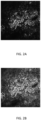
- FIG 2A is an exemplary diagram of an original acquired Third Harmonic Generation (THG) microscopy image. In this embodiment, as shown in FIG.
- THG Third Harmonic Generation
- the method for determining cellular Nuclear-to-Cytoplasmic ratio comprises acts of S100 obtaining a cytological image using non-invasive in vivo biopsy technique; S120 performing a nuclei segmentation process to identify a position and a contour of each of identified nuclei in the cytological image; S140 performing a cytoplasmic process with an improved active contour model to obtain a cytoplasmic region for each identified nucleus; and S160 determine a cellular Nuclear-to-Cytoplasmic ratio based on the obtained nuclei and cytoplasmic regions.
- the cytological image or biomedical image is obtained using non-invasive in vivo biopsy technique that acquire the cells or tissues to be analyzed and cell segmentation.
- HHGM Higher Harmonic Generation Microscopy
- THG Third Harmonic Generation
- THG intensity depends on the cubic of the incident light source, it can provide the characteristic of high depth resolution.
- the THG image may have some noise corruption, and the intensity of cells such as contrast, edges and contour in the THG image are often inconsistent, and thus an image preprocessing is required before performing cell segmentation.
- the image resolution of acquired THG image is 512 x 512 pixels and stored in 12-bit gray-level TIFF format with lossless compression.
- the cytological image of the present invention is not limited to THG image, it can be obtained from different non-invasive vivo biopsy techniques.
- FIG. 2B is an exemplary diagram of a preprocessed THG microscopy image
- FIG. 3 is a flowchart illustrating acts of preprocessing the cytological image in act S100 in accordance with an embodiment of the present invention.
- an act of S101 performing contrast limit adaptive histogram equalization method (CLAHE) to the cytological image which is configured for improving local contrast and details.
- the image preprocessing may further comprise an act of S102 performing Wiener filter to adaptively remove noise.
- FIG. 2A shows the preprocessed THG image.
- FIG. 4A is a flowchart illustrating acts of performing a nuclei segmentation process to identify a position and a contour of each of identified nuclei in the cytological image
- FIG 4B is a flowchart illustrating steps of nuclei initialization according to FIG. 4A
- the act of S 120 further comprises acts to be performed using gradient watershed transformation with marker-controlled strategy, blob detection, and consideration of shape descriptors to obtain accurate segmented nuclei.
- Nuclei segmentation is a crucial stage in the proposed cell segmentation algorithm since the following cytoplasm segmentation will desire to reference valid nuclei which can be thought of as initial condition of the optimization problem of whole cell segmentation to guide the algorithm to find the feasible solution with high performance.
- the nuclei segmentation process comprises acts of S 121 performing a nuclei initialization step to find an internal marker and an external marker to obtain a potential nuclei and a potential cell boundary corresponding to the potential nuclei; S 122 calculating a gradient map of the received cell image; S 123 performing a filtering step on the gradient map to generate a filtered gradient map according to the potential nuclei and the potential cell boundary; S124 performing a nuclei detection step by calculating watersheds of the filtered gradient map to obtain a segmented nuclei; S 125 performing a nuclei validation step by preserving a part of the segmented nuclei with relatively higher compactness and excluding another part of the segmented nuclei with relatively lower compactness to obtain a valid nucleus as an identified nucleus.
- the detailed description of the nuclei segmentation process can be found in the US patent No. US9122907 which is filed by a joint inventor to the present invention.
- the present disclosure may utilize a strict nuclei initialization that eliminates disqualified nuclei detection by shape information that is derived from Hough transform, labeling information and actual cell size to help designing a marker filter with more accurate nuclei position for subsequent nuclei segmentation.
- the nuclei initialization step comprises acts of S1211 performing a blob detection step to obtain a nuclei candidate; S1212 performing an outlier removal step to exclude an outlier of the nuclei candidate to obtain the internal marker; S1213 performing a strict nuclei initialization process, S1214 performing a distance transform step of the internal marker to obtain a distance map; and S1215 performing a cell boundary initialization step to obtain the external marker from the distance map.
- S1211, S1212, S1214 and S1215 are disclosed and described in US9122907 .
- the preprocessed image is spanned into the Gaussian scale-space for the purpose to extract structure of interest with the scale and spatial information simultaneously.
- the 2nd-order Hessian matrix presented by scale-space derivatives is used to estimate the existence of regional minimum and enhance those round valley feature.
- the potential nuclei by simply removing some outliers with local intensity constraint.
- the act of S 1213 is configured to re-inspect those potential nuclei candidates by providing an ellipse shape descriptor, and thus the present disclosure combines the features of both local intensity information and ellipse shape information simultaneously to provide a firm representation of nuclei.
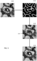
- FIG. 5 is exemplary diagrams illustrating the acts of performing a strict nuclei initialization process.
- the strict nuclei initialization process comprises acts of S200 obtaining sub-images from the cytological image; S210 identifying a contour with edge detection in the sub-image; S220 extracting prominent ellipses with identified contour from each of sub-images using an ellipse Hough transform; and S230 re-inspecting the nuclei candidates based on the center of the prominent ellipses.
- the sub-image is retrieved with smaller scale from the cytological image.
- the size of the sub-image is three times of the average ND size, which is about 30 x 30 pixels from the position of each potential nuclei.
- the scale of sub-image size is alterable for including all possible area of nuclei area without involving too much information.
- the delineation of the contour in the sub-image is an essential stage for shape description, thus we can adopt the edge detection method to concentrate on the shape's border.
- Edge is where the pixels with sharply change in intensity.
- the multi-directional Sobel operator and Laplacian operator which the 1 st -order derivative and 2 nd -order derivative are respectively used for edge identification. According to inventor's experiment, using Laplacian operator with 2 nd -order derivative image could provide more edge information than Sobel operator, but may also cause too many vague edge or unrealistic edge of nuclei. Therefore, in present disclosure, Sobel operator is preferable used for identifying a contour of the nuclei edge.
- the act of S220 adopted Hough transform as shape extractor in describing the edge contour of potential nuclei.
- the Hough transform is a feature extraction technique based on the concept of shape matching to detect the curves in image.
- cellular nuclei shapes vary from one to another in THG images.
- ellipse Hough transform due to the flexibility of varying from very flat ellipse to almost circular one.
- ellipse selection mechanism i.e., the act of S230
- the ellipse selection checks the distance between the center of potential nuclei Ci and the center of detected prominent ellipse Ce. If the distance between the center of potential nuclei Ci and the center of detected prominent ellipse Ce is smaller than the average radius of the nuclei, then re-inspect as a nucleus, and if not, the candidate nucleus is eliminated.
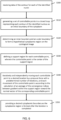
- FIG. 6 is a flowchart illustrating the act of performing a cytoplasmic process in accordance with an embodiment of the present invention.
- the act of S 140 performing a cytoplasmic process with an improved active contour model to obtain a cytoplasmic region for each identified nucleus based comprises acts of S300 receiving data of the contour for each of the identified nuclei; S310 generating a set of controllable points in a closed loop representing each contour of the identified nucleus as an initial boundary of the cytoplasm; S320 determining an inner boundary and an outer boundary to form a hypothetical cytoplasmic region on the cytological image, wherein within the hypothetical cytoplasmic region; S330 defining a support region for each controllable point, wherein the controllable point is the center of the support region; S340 iteratively and independently moving each controllable point to a desired location by a pressure force with a predetermined number of iterations, and each movement of the
- Active contour model also called snake
- snake is adopted as our base approach to achieve cytoplasm segmentation, which delineates boundary of cell in THG microscopy images. From the essential characteristic of snake that could be described as a closed curve, the traditional issue of detecting and linking boundary could be resolved. Additionally, the snake is an energy minimizing curve which transforms the image segmentation problem into an energy optimization process, and thus giving a systematic approach for cytoplasm segmentation.
- an improved active contour model i.e., a statistical pressure snake based on the balloon snake
- the improved active contour model is configured for joining the contextual and locality information of image data into energy minimization process to make the snake more converge to the cytoplasm outer boundary with the initial contour on nuclei border.
- the proposed statistical pressure snake considering the contextual information in image domain not only adapts to different cell structure during each process of cytoplasmic boundary detection, but also becomes an optimal parameter setting snake.
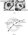
- FIG. 8 is an exemplary diagram of the support region of convergence control factor controlling the controllable points.
- a support region SR is defined for each controllable point, and the controllable point is the center of the support region.
- the measurement of the convergent extent provides a guideline on the movement of snake that the snake is able to inflate or deflate like a balloon, and we call the measurement a convergence control factor C(v(s)).
- n(v(s)) is the normal vector on each controllable point.
- C(v(s)) is a convergence control factor controlling the pressure force that is applied to the controllable point, which calculates the statistics of the directional convergence.
- the result of the calculation of the convergence control factor is normalized in a range of -1 to 1.
- SR is the abbreviation of support region which could be thought of as an interested locality around each controlled point of snake curve, as shown in Fig. 8
- N is the number of image pixels within the SR and p j represents image pixels
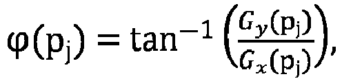
- ⁇ (p j ) is the orientation of the gradient vector G(p j ) on pixel p j , and is calculated from Gx(p j ) and Gy(p j ) which are first derivatives of image intensity at pixel pj in horizontal and vertical directions, respectively
- ⁇ (n(v(s))) is the orientation of normal vector calculated from x- and y- components of normal vector
- cos ⁇ p j v s is the convergence index indicating the directional similarity between the gradient vector G(p j ) at pixel pj and normal vector n ⁇ v s
- C(v(s)) would have positive value from the interior cytoplasmic boundary, which means large directional similarity, thus it inflates snake. While getting close to the exterior cytoplasmic boundary where C(v(s)) becomes negative which means low directional similarity, the snake would gradually deflate.
- the pressure force F pressure may further comprises a shifting parameter configured for ensuring each movement of the controllable point would pass through the local minimum of inner cytoplasmic boundary.
- the shifting parameter considers the maximum magnitude of the image force of entire snake during each minimization process.
- ⁇ is responsible for ensuring the initial snake would pass through the local minimum of inner cytoplasmic boundary.
- ⁇ provides additional fuel, and hence the snake could still have force to move outward.
- the image force of the cytoplasm boundary would dominate the snake movement because the convergence control factor counteracts the shifting parameter, thus the energy minimization would carry the rest of the way to find the desired cytoplasm boundary.
- FIG. 9A is an exemplary diagram illustrating separating adjacent cell without dividing line
- FIG. 9B is an exemplary diagram illustrating separating adjacent cell without dividing line.
- the corresponding cytoplasm boundary can be detected based on the local cell structure by using the proposed statistical pressure snake.
- the detection of cytoplasm is performed on each individual cell, and thus, as shown in FIG. 9A , the overlapping problem occurred when the basal cells touch with each other.
- the statistical pressure snake won't converge to the correct cytoplasm boundary and the overlapping problem appears.
- the Euclidean distance transform is adopted to separate the touch cells when implement the statistical pressure snake.
- the Euclidean distance transform generates a distance map from a binary image which labels each pixel with distance between that pixel and the nearest non-zero pixel in binary image using Euclidean distance metric.
- FIG. 10A is an exemplary diagram of a non-self-intersection discrete accumulator
- FIG. 10B is an exemplary diagram of a self-intersection discrete accumulator
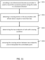
- FIG. 11 is a flowchart illustrating acts of detecting and removing self-intersection in accordance with an embodiment of the present invention.
- the magnitude of pressure force adaptively changes according to the convergent extent between snake movement and gradient vector. Nonetheless, the final direction of snake movement relies on both pressure force and image force. If some parts of the snake move faster than others under the influence of external force, those parts of curve may cross each other, leading to unstable behavior.
- the present disclosure further comprises acts of S331 providing a one-dimensional discrete accumulator is used to record the successive position along the snake in a clockwise direction; S332 detecting a crossing section in the accumulator cells whose value is equal or more than two; S333 determining the two endpoints of cells with crossing condition; and S334 removing the crossing section between two endpoints and re-interpolate the controllable point.
- the Nuclear-to-Cytoplasmic ratio is a common measurement used in medical diagnosis, which is defined as the volume ratio of nuclei to cytoplasm in cytology.
- the NC ratios of the skin epidermis for some skin diseases like Actinic Keratosis and Squamous Cell Carcinoma are commonly larger than that in the normal skin.
- NC ratio is defined in volume ratio
- the area ratio of nuclei to cytoplasm is reasonably approximated to NC ratio evaluation due to that they both use ratio analysis. Therefore, whether we use the estimation of the volume ratio or area ratio, their analyzed ratio will be close owing to the difference are obliterated by ratio analysis.
Landscapes
- Engineering & Computer Science (AREA)
- Theoretical Computer Science (AREA)
- Physics & Mathematics (AREA)
- General Physics & Mathematics (AREA)
- Computer Vision & Pattern Recognition (AREA)
- Health & Medical Sciences (AREA)
- General Health & Medical Sciences (AREA)
- Multimedia (AREA)
- Software Systems (AREA)
- Biomedical Technology (AREA)
- Life Sciences & Earth Sciences (AREA)
- Molecular Biology (AREA)
- Medical Informatics (AREA)
- Databases & Information Systems (AREA)
- Evolutionary Computation (AREA)
- Biodiversity & Conservation Biology (AREA)
- Computing Systems (AREA)
- Artificial Intelligence (AREA)
- Geometry (AREA)
- Nuclear Medicine, Radiotherapy & Molecular Imaging (AREA)
- Radiology & Medical Imaging (AREA)
- Quality & Reliability (AREA)
- Image Processing (AREA)
- Image Analysis (AREA)
- Investigating Or Analysing Biological Materials (AREA)
Claims (9)
- Ein Verfahren zur Bestimmung des zellulären Kern-zu-Zytoplasma-Verhältnisses, das folgende Schritte umfasst:Gewinnung eines zytologischen Bildes mittels nicht-invasiver in-vivo-Biopsietechnik;Durchführung eines Kernsegmentierungsprozesses, um eine Kontur jedes identifizierten Kerns im zytologischen Bild zu bestimmen;Durchführung eines zytoplasmatischen Prozesses mit einem aktiven Konturmodell, um einen zytoplasmatischen Bereich für jeden identifizierten Kern zu erhalten; undBerechnung der Flächen jedes identifizierten Kerns und des entsprechenden zytoplasmatischen Bereichs, um ein zelluläres Kern-zu-Zytoplasma-Verhältnis zu bestimmen, wobeider Schritt der Durchführung des zytoplasmatischen Prozesses mit dem aktiven Konturmodell folgende Schritte umfasst:Empfangen der Konturen für jeden der identifizierten Kerne;Erzeugen einer Menge von steuerbaren Punkten in einer geschlossenen Schleife, die jede Kontur des identifizierten Kerns des Zytoplasmas darstellen;iteratives und unabhängiges Bewegen jedes steuerbaren Punktes zu einem Ort durch eine Druckkraft mit einer vorbestimmten Anzahl von Iterationen, wobei die Druckkraft durch einen Steuerfaktor C(v(s)) gesteuert wird; und
Bereitstellen einer zytoplasmatischen Grenze als zytoplasmatische Region nach der Iteration; wobei die Druckkraft ein Vektor mit einem Modul ist, der eine Größe der Kraft und eine Richtung definiert, in die die Druckkraft ausgeübt wird, wobei der Steuerfaktor C(v(s)) folgende Beziehung aufweist:




- Verfahren nach Anspruch 1, wobei die Druckkraft eine Beziehung aufweist von

- Verfahren nach Anspruch 1, wobei die Druckkraft eine Beziehung aufweist von

- Verfahren nach Anspruch 1, das ferner die Bestimmung eines überlappenden zytoplasmatischen Bereichs unter Verwendung einer euklidischen Abstandstransformation bei der Implementierung des aktiven Konturmodells umfasst.
- Verfahren nach Anspruch 1, das ferner Maßnahmen zur Vermeidung von Selbstüberschneidungen umfasst, welche umfassen:Bereitstellung eines eindimensionalen diskreten Akkumulators zur sequenziellen Aufzeichnung der Positionen der Schlangenkurve im Uhrzeigersinn;Erkennung eines Kreuzungsabschnitts auf der Grundlage der aufgezeichneten Positionen des Akkumulators, dessen Wert gleich oder größer als zwei ist;Bestimmung zweier Endpunkte der Schlangenkurve, wenn der Kreuzungsabschnitt erkannt wird; undEntfernung des Kreuzungsabschnitts zwischen zwei Endpunkten und Neuinterpolation des steuerbaren Punktes.
- Verfahren nach Anspruch 1, wobei der Schritt der Gewinnung des zytologischen Bildes ferner folgende Schritte umfasst:Durchführen eines adaptiven Histogrammausgleichsverfahrens mit Kontrastgrenze, um den Kontrast und die Details in dem zytologischen Bild zu verbessern; undAnwendung eines Wiener-Filters zur adaptiven Entfernung von Rauschen.
- Verfahren nach Anspruch 6, wobei der Schritt der Kerninitialisierung ferner Schritte zur Durchführung eines strengen Kerninitialisierungsprozesses umfasst, um ungültige Kerne durch Forminformationen zu eliminieren, die von der Hough-Transformation abgeleitet sind.
- Verfahren nach Anspruch 7, wobei der strenge Kerninitialisierungsprozess folgende Schritte umfasst:Gewinnung eines Teilbildes aus dem zytologischen Bild;Identifizierung einer Kontur mit Kantenerkennung in dem Teilbild;Extrahieren markanter Ellipsen mit identifizierter Kontur aus jedem der Teilbilder unter Verwendung einer Ellipsen-Hough-Transformation; underneutes Untersuchen der Kerne auf Basis des Zentrums der markanten Ellipsen, wobei der Abstand zwischen dem Zentrum der potentiellen Kerne und dem Zentrum der detektierten markanten Ellipse kleiner ist als der durchschnittliche Radius der Kerne.
- Verfahren nach Anspruch 8, wobei die Kantendetektion den Sobel-Operator, den Laplace-Operator oder beide in der ersten bzw. zweiten Ableitung verwendet.
Priority Applications (2)
| Application Number | Priority Date | Filing Date | Title |
|---|---|---|---|
| EP19215808.7A EP3836020B1 (de) | 2019-12-12 | 2019-12-12 | Verfahren zur bestimmung des zellkern-zytoplasma-verhältnisses |
| US16/724,292 US11120249B2 (en) | 2019-12-12 | 2019-12-22 | Method for determining cellular nuclear-to-cytoplasmic ratio |
Applications Claiming Priority (1)
| Application Number | Priority Date | Filing Date | Title |
|---|---|---|---|
| EP19215808.7A EP3836020B1 (de) | 2019-12-12 | 2019-12-12 | Verfahren zur bestimmung des zellkern-zytoplasma-verhältnisses |
Publications (3)
| Publication Number | Publication Date |
|---|---|
| EP3836020A1 EP3836020A1 (de) | 2021-06-16 |
| EP3836020C0 EP3836020C0 (de) | 2024-08-28 |
| EP3836020B1 true EP3836020B1 (de) | 2024-08-28 |
Family
ID=68916257
Family Applications (1)
| Application Number | Title | Priority Date | Filing Date |
|---|---|---|---|
| EP19215808.7A Active EP3836020B1 (de) | 2019-12-12 | 2019-12-12 | Verfahren zur bestimmung des zellkern-zytoplasma-verhältnisses |
Country Status (2)
| Country | Link |
|---|---|
| US (1) | US11120249B2 (de) |
| EP (1) | EP3836020B1 (de) |
Families Citing this family (5)
| Publication number | Priority date | Publication date | Assignee | Title |
|---|---|---|---|---|
| CN116263951A (zh) * | 2021-12-14 | 2023-06-16 | 深圳市帝迈生物技术有限公司 | 白细胞的分割方法、分割装置、样本分析仪及存储介质 |
| CN114332145B (zh) * | 2021-12-29 | 2025-02-18 | 南京林业大学 | 一种木材细胞结构的量化方法及系统 |
| TWI900794B (zh) * | 2022-08-26 | 2025-10-11 | 普匯金融科技股份有限公司 | 電子計算裝置及其影像分析方法與電腦程式產品 |
| CN116681685B (zh) * | 2023-06-19 | 2025-07-29 | 浙江大学 | 基于双光子钙成像数据的细胞胞体自动识别与分割方法及装置 |
| CN120298371B (zh) * | 2025-04-07 | 2025-12-09 | 延边大学 | 基于孔径一致性分析的滤网质量检测方法 |
Family Cites Families (6)
| Publication number | Priority date | Publication date | Assignee | Title |
|---|---|---|---|---|
| SE0302114D0 (sv) * | 2003-07-21 | 2003-07-21 | Cellavision Ab | Sätt att urskilja en objektkontur |
| US9208394B2 (en) * | 2005-09-05 | 2015-12-08 | Alpvision S.A. | Authentication of an article of manufacture using an image of the microstructure of it surface |
| KR102090776B1 (ko) * | 2012-07-25 | 2020-03-18 | 테라노스, 인코포레이티드 | 생체시료의 이미지 분석 및 측정 |
| TWI496112B (zh) | 2013-09-13 | 2015-08-11 | Univ Nat Cheng Kung | 細胞影像分割方法以及核質比評估方法 |
| TWI594207B (zh) * | 2016-04-26 | 2017-08-01 | 財團法人金屬工業研究發展中心 | 細胞核影像輪廓擷取裝置及其方法 |
| US10402623B2 (en) * | 2017-11-30 | 2019-09-03 | Metal Industries Research & Development Centre | Large scale cell image analysis method and system |
-
2019
- 2019-12-12 EP EP19215808.7A patent/EP3836020B1/de active Active
- 2019-12-22 US US16/724,292 patent/US11120249B2/en active Active
Also Published As
| Publication number | Publication date |
|---|---|
| EP3836020A1 (de) | 2021-06-16 |
| US11120249B2 (en) | 2021-09-14 |
| EP3836020C0 (de) | 2024-08-28 |
| US20210182528A1 (en) | 2021-06-17 |
Similar Documents
| Publication | Publication Date | Title |
|---|---|---|
| EP3836020B1 (de) | Verfahren zur bestimmung des zellkern-zytoplasma-verhältnisses | |
| US12274542B2 (en) | Methods and systems for extracting blood vessel | |
| US11562491B2 (en) | Automatic pancreas CT segmentation method based on a saliency-aware densely connected dilated convolutional neural network | |
| Rampun et al. | Fully automated breast boundary and pectoral muscle segmentation in mammograms | |
| Kirbas et al. | Vessel extraction techniques and algorithms: a survey | |
| US7711165B2 (en) | System and method for coronary artery segmentation of cardiac CT volumes | |
| Elnakib et al. | Medical image segmentation: a brief survey | |
| CN107644420B (zh) | 基于中心线提取的血管图像分割方法、核磁共振成像系统 | |
| US10210612B2 (en) | Method and system for machine learning based estimation of anisotropic vessel orientation tensor | |
| CN115136189A (zh) | 基于图像处理的肿瘤的自动化检测 | |
| CN112308846B (zh) | 血管分割方法、装置及电子设备 | |
| US20130051676A1 (en) | Microcalcification Detection Classification in Radiographic Images | |
| CN107292312B (zh) | 肿瘤ct图像处理方法 | |
| Göçeri et al. | Fully automated liver segmentation from SPIR image series | |
| Janoos et al. | Robust 3D reconstruction and identification of dendritic spines from optical microscopy imaging | |
| WO2021136368A1 (zh) | 钼靶图像中胸大肌区域自动检测方法及装置 | |
| CN111784701B (zh) | 结合边界特征增强和多尺度信息的超声图像分割方法及系统 | |
| Zambal et al. | Shape and appearance models for automatic coronary artery tracking | |
| CN116912823A (zh) | 一种基于深度学习的肾脏病理切片细胞识别方法及系统 | |
| Jaffar et al. | Anisotropic diffusion based brain MRI segmentation and 3D reconstruction | |
| Nelson et al. | An effective approach for the nuclei segmentation from breast histopathological images using star-convex polygon | |
| Mahmoud et al. | Medical image segmentation techniques, a literature review, and some novel trends | |
| Roy et al. | Automated nuclei analysis from digital histopathology | |
| CN114897900B (zh) | 基于人工智能的乳腺核磁共振图像病灶定位方法 | |
| Hiraman | Liver Segmentation Using 3D CTScans |
Legal Events
| Date | Code | Title | Description |
|---|---|---|---|
| PUAI | Public reference made under article 153(3) epc to a published international application that has entered the european phase |
Free format text: ORIGINAL CODE: 0009012 |
|
| STAA | Information on the status of an ep patent application or granted ep patent |
Free format text: STATUS: REQUEST FOR EXAMINATION WAS MADE |
|
| 17P | Request for examination filed |
Effective date: 20191212 |
|
| AK | Designated contracting states |
Kind code of ref document: A1 Designated state(s): AL AT BE BG CH CY CZ DE DK EE ES FI FR GB GR HR HU IE IS IT LI LT LU LV MC MK MT NL NO PL PT RO RS SE SI SK SM TR |
|
| STAA | Information on the status of an ep patent application or granted ep patent |
Free format text: STATUS: EXAMINATION IS IN PROGRESS |
|
| 17Q | First examination report despatched |
Effective date: 20230228 |
|
| REG | Reference to a national code |
Ref country code: DE Ref legal event code: R079 Free format text: PREVIOUS MAIN CLASS: G06K0009460000 Ipc: G06V0010440000 Ref country code: DE Ref legal event code: R079 Ref document number: 602019057801 Country of ref document: DE Free format text: PREVIOUS MAIN CLASS: G06K0009460000 Ipc: G06V0010440000 |
|
| GRAP | Despatch of communication of intention to grant a patent |
Free format text: ORIGINAL CODE: EPIDOSNIGR1 |
|
| STAA | Information on the status of an ep patent application or granted ep patent |
Free format text: STATUS: GRANT OF PATENT IS INTENDED |
|
| RIC1 | Information provided on ipc code assigned before grant |
Ipc: G06V 20/69 20220101ALI20240318BHEP Ipc: G06V 10/75 20220101ALI20240318BHEP Ipc: G06V 10/44 20220101AFI20240318BHEP |
|
| INTG | Intention to grant announced |
Effective date: 20240409 |
|
| GRAS | Grant fee paid |
Free format text: ORIGINAL CODE: EPIDOSNIGR3 |
|
| GRAA | (expected) grant |
Free format text: ORIGINAL CODE: 0009210 |
|
| STAA | Information on the status of an ep patent application or granted ep patent |
Free format text: STATUS: THE PATENT HAS BEEN GRANTED |
|
| AK | Designated contracting states |
Kind code of ref document: B1 Designated state(s): AL AT BE BG CH CY CZ DE DK EE ES FI FR GB GR HR HU IE IS IT LI LT LU LV MC MK MT NL NO PL PT RO RS SE SI SK SM TR |
|
| REG | Reference to a national code |
Ref country code: GB Ref legal event code: FG4D |
|
| REG | Reference to a national code |
Ref country code: CH Ref legal event code: EP |
|
| REG | Reference to a national code |
Ref country code: DE Ref legal event code: R096 Ref document number: 602019057801 Country of ref document: DE |
|
| REG | Reference to a national code |
Ref country code: IE Ref legal event code: FG4D |
|
| U01 | Request for unitary effect filed |
Effective date: 20240923 |
|
| U07 | Unitary effect registered |
Designated state(s): AT BE BG DE DK EE FI FR IT LT LU LV MT NL PT RO SE SI Effective date: 20241016 |
|
| PG25 | Lapsed in a contracting state [announced via postgrant information from national office to epo] |
Ref country code: NO Free format text: LAPSE BECAUSE OF FAILURE TO SUBMIT A TRANSLATION OF THE DESCRIPTION OR TO PAY THE FEE WITHIN THE PRESCRIBED TIME-LIMIT Effective date: 20241128 |
|
| U20 | Renewal fee for the european patent with unitary effect paid |
Year of fee payment: 6 Effective date: 20241206 |
|
| PG25 | Lapsed in a contracting state [announced via postgrant information from national office to epo] |
Ref country code: PL Free format text: LAPSE BECAUSE OF FAILURE TO SUBMIT A TRANSLATION OF THE DESCRIPTION OR TO PAY THE FEE WITHIN THE PRESCRIBED TIME-LIMIT Effective date: 20240828 Ref country code: GR Free format text: LAPSE BECAUSE OF FAILURE TO SUBMIT A TRANSLATION OF THE DESCRIPTION OR TO PAY THE FEE WITHIN THE PRESCRIBED TIME-LIMIT Effective date: 20241129 |
|
| PG25 | Lapsed in a contracting state [announced via postgrant information from national office to epo] |
Ref country code: IS Free format text: LAPSE BECAUSE OF FAILURE TO SUBMIT A TRANSLATION OF THE DESCRIPTION OR TO PAY THE FEE WITHIN THE PRESCRIBED TIME-LIMIT Effective date: 20241228 |
|
| PG25 | Lapsed in a contracting state [announced via postgrant information from national office to epo] |
Ref country code: HR Free format text: LAPSE BECAUSE OF FAILURE TO SUBMIT A TRANSLATION OF THE DESCRIPTION OR TO PAY THE FEE WITHIN THE PRESCRIBED TIME-LIMIT Effective date: 20240828 |
|
| PG25 | Lapsed in a contracting state [announced via postgrant information from national office to epo] |
Ref country code: RS Free format text: LAPSE BECAUSE OF FAILURE TO SUBMIT A TRANSLATION OF THE DESCRIPTION OR TO PAY THE FEE WITHIN THE PRESCRIBED TIME-LIMIT Effective date: 20241128 Ref country code: ES Free format text: LAPSE BECAUSE OF FAILURE TO SUBMIT A TRANSLATION OF THE DESCRIPTION OR TO PAY THE FEE WITHIN THE PRESCRIBED TIME-LIMIT Effective date: 20240828 |
|
| PG25 | Lapsed in a contracting state [announced via postgrant information from national office to epo] |
Ref country code: RS Free format text: LAPSE BECAUSE OF FAILURE TO SUBMIT A TRANSLATION OF THE DESCRIPTION OR TO PAY THE FEE WITHIN THE PRESCRIBED TIME-LIMIT Effective date: 20241128 Ref country code: PL Free format text: LAPSE BECAUSE OF FAILURE TO SUBMIT A TRANSLATION OF THE DESCRIPTION OR TO PAY THE FEE WITHIN THE PRESCRIBED TIME-LIMIT Effective date: 20240828 Ref country code: NO Free format text: LAPSE BECAUSE OF FAILURE TO SUBMIT A TRANSLATION OF THE DESCRIPTION OR TO PAY THE FEE WITHIN THE PRESCRIBED TIME-LIMIT Effective date: 20241128 Ref country code: IS Free format text: LAPSE BECAUSE OF FAILURE TO SUBMIT A TRANSLATION OF THE DESCRIPTION OR TO PAY THE FEE WITHIN THE PRESCRIBED TIME-LIMIT Effective date: 20241228 Ref country code: HR Free format text: LAPSE BECAUSE OF FAILURE TO SUBMIT A TRANSLATION OF THE DESCRIPTION OR TO PAY THE FEE WITHIN THE PRESCRIBED TIME-LIMIT Effective date: 20240828 Ref country code: GR Free format text: LAPSE BECAUSE OF FAILURE TO SUBMIT A TRANSLATION OF THE DESCRIPTION OR TO PAY THE FEE WITHIN THE PRESCRIBED TIME-LIMIT Effective date: 20241129 Ref country code: ES Free format text: LAPSE BECAUSE OF FAILURE TO SUBMIT A TRANSLATION OF THE DESCRIPTION OR TO PAY THE FEE WITHIN THE PRESCRIBED TIME-LIMIT Effective date: 20240828 |
|
| PG25 | Lapsed in a contracting state [announced via postgrant information from national office to epo] |
Ref country code: SM Free format text: LAPSE BECAUSE OF FAILURE TO SUBMIT A TRANSLATION OF THE DESCRIPTION OR TO PAY THE FEE WITHIN THE PRESCRIBED TIME-LIMIT Effective date: 20240828 |
|
| PG25 | Lapsed in a contracting state [announced via postgrant information from national office to epo] |
Ref country code: CZ Free format text: LAPSE BECAUSE OF FAILURE TO SUBMIT A TRANSLATION OF THE DESCRIPTION OR TO PAY THE FEE WITHIN THE PRESCRIBED TIME-LIMIT Effective date: 20240828 |
|
| PG25 | Lapsed in a contracting state [announced via postgrant information from national office to epo] |
Ref country code: SK Free format text: LAPSE BECAUSE OF FAILURE TO SUBMIT A TRANSLATION OF THE DESCRIPTION OR TO PAY THE FEE WITHIN THE PRESCRIBED TIME-LIMIT Effective date: 20240828 |
|
| PLBE | No opposition filed within time limit |
Free format text: ORIGINAL CODE: 0009261 |
|
| STAA | Information on the status of an ep patent application or granted ep patent |
Free format text: STATUS: NO OPPOSITION FILED WITHIN TIME LIMIT |
|
| PG25 | Lapsed in a contracting state [announced via postgrant information from national office to epo] |
Ref country code: MC Free format text: LAPSE BECAUSE OF FAILURE TO SUBMIT A TRANSLATION OF THE DESCRIPTION OR TO PAY THE FEE WITHIN THE PRESCRIBED TIME-LIMIT Effective date: 20240828 |
|
| REG | Reference to a national code |
Ref country code: CH Ref legal event code: PL |
|
| 26N | No opposition filed |
Effective date: 20250530 |
|
| GBPC | Gb: european patent ceased through non-payment of renewal fee |
Effective date: 20241212 |
|
| PG25 | Lapsed in a contracting state [announced via postgrant information from national office to epo] |
Ref country code: GB Free format text: LAPSE BECAUSE OF NON-PAYMENT OF DUE FEES Effective date: 20241212 |
|
| PG25 | Lapsed in a contracting state [announced via postgrant information from national office to epo] |
Ref country code: CH Free format text: LAPSE BECAUSE OF NON-PAYMENT OF DUE FEES Effective date: 20241231 |
|
| PG25 | Lapsed in a contracting state [announced via postgrant information from national office to epo] |
Ref country code: IE Free format text: LAPSE BECAUSE OF NON-PAYMENT OF DUE FEES Effective date: 20241212 |
|
| U20 | Renewal fee for the european patent with unitary effect paid |
Year of fee payment: 7 Effective date: 20251222 |