EP3834729B1 - Auf elektrokardiogramm basierende beurteilung der diastolischen funktion - Google Patents
Auf elektrokardiogramm basierende beurteilung der diastolischen funktion Download PDFInfo
- Publication number
- EP3834729B1 EP3834729B1 EP20193719.0A EP20193719A EP3834729B1 EP 3834729 B1 EP3834729 B1 EP 3834729B1 EP 20193719 A EP20193719 A EP 20193719A EP 3834729 B1 EP3834729 B1 EP 3834729B1
- Authority
- EP
- European Patent Office
- Prior art keywords
- time
- frequency
- models
- machine
- diastolic
- Prior art date
- Legal status (The legal status is an assumption and is not a legal conclusion. Google has not performed a legal analysis and makes no representation as to the accuracy of the status listed.)
- Active
Links
Images


Classifications
-
- A—HUMAN NECESSITIES
- A61—MEDICAL OR VETERINARY SCIENCE; HYGIENE
- A61B—DIAGNOSIS; SURGERY; IDENTIFICATION
- A61B5/00—Measuring for diagnostic purposes; Identification of persons
- A61B5/02—Detecting, measuring or recording for evaluating the cardiovascular system, e.g. pulse, heart rate, blood pressure or blood flow
- A61B5/02028—Determining haemodynamic parameters not otherwise provided for, e.g. cardiac contractility or left ventricular ejection fraction
-
- A—HUMAN NECESSITIES
- A61—MEDICAL OR VETERINARY SCIENCE; HYGIENE
- A61B—DIAGNOSIS; SURGERY; IDENTIFICATION
- A61B5/00—Measuring for diagnostic purposes; Identification of persons
- A61B5/72—Signal processing specially adapted for physiological signals or for diagnostic purposes
- A61B5/7235—Details of waveform analysis
- A61B5/7264—Classification of physiological signals or data, e.g. using neural networks, statistical classifiers, expert systems or fuzzy systems
- A61B5/7267—Classification of physiological signals or data, e.g. using neural networks, statistical classifiers, expert systems or fuzzy systems involving training the classification device
-
- A—HUMAN NECESSITIES
- A61—MEDICAL OR VETERINARY SCIENCE; HYGIENE
- A61B—DIAGNOSIS; SURGERY; IDENTIFICATION
- A61B5/00—Measuring for diagnostic purposes; Identification of persons
- A61B5/02—Detecting, measuring or recording for evaluating the cardiovascular system, e.g. pulse, heart rate, blood pressure or blood flow
- A61B5/026—Measuring blood flow
- A61B5/0285—Measuring or recording phase velocity of blood waves
-
- A—HUMAN NECESSITIES
- A61—MEDICAL OR VETERINARY SCIENCE; HYGIENE
- A61B—DIAGNOSIS; SURGERY; IDENTIFICATION
- A61B5/00—Measuring for diagnostic purposes; Identification of persons
- A61B5/103—Measuring devices for testing the shape, pattern, colour, size or movement of the body or parts thereof, for diagnostic purposes
- A61B5/11—Measuring movement of the entire body or parts thereof, e.g. head or hand tremor or mobility of a limb
- A61B5/1102—Ballistocardiography
-
- A—HUMAN NECESSITIES
- A61—MEDICAL OR VETERINARY SCIENCE; HYGIENE
- A61B—DIAGNOSIS; SURGERY; IDENTIFICATION
- A61B5/00—Measuring for diagnostic purposes; Identification of persons
- A61B5/24—Detecting, measuring or recording bioelectric or biomagnetic signals of the body or parts thereof
- A61B5/316—Modalities, i.e. specific diagnostic methods
- A61B5/318—Heart-related electrical modalities, e.g. electrocardiography [ECG]
- A61B5/339—Displays specially adapted therefor
-
- A—HUMAN NECESSITIES
- A61—MEDICAL OR VETERINARY SCIENCE; HYGIENE
- A61B—DIAGNOSIS; SURGERY; IDENTIFICATION
- A61B5/00—Measuring for diagnostic purposes; Identification of persons
- A61B5/24—Detecting, measuring or recording bioelectric or biomagnetic signals of the body or parts thereof
- A61B5/316—Modalities, i.e. specific diagnostic methods
- A61B5/318—Heart-related electrical modalities, e.g. electrocardiography [ECG]
- A61B5/346—Analysis of electrocardiograms
- A61B5/347—Detecting the frequency distribution of signals
-
- A—HUMAN NECESSITIES
- A61—MEDICAL OR VETERINARY SCIENCE; HYGIENE
- A61B—DIAGNOSIS; SURGERY; IDENTIFICATION
- A61B5/00—Measuring for diagnostic purposes; Identification of persons
- A61B5/24—Detecting, measuring or recording bioelectric or biomagnetic signals of the body or parts thereof
- A61B5/316—Modalities, i.e. specific diagnostic methods
- A61B5/318—Heart-related electrical modalities, e.g. electrocardiography [ECG]
- A61B5/346—Analysis of electrocardiograms
- A61B5/349—Detecting specific parameters of the electrocardiograph cycle
-
- A—HUMAN NECESSITIES
- A61—MEDICAL OR VETERINARY SCIENCE; HYGIENE
- A61B—DIAGNOSIS; SURGERY; IDENTIFICATION
- A61B5/00—Measuring for diagnostic purposes; Identification of persons
- A61B5/72—Signal processing specially adapted for physiological signals or for diagnostic purposes
- A61B5/7271—Specific aspects of physiological measurement analysis
- A61B5/7278—Artificial waveform generation or derivation, e.g. synthesizing signals from measured signals
-
- G—PHYSICS
- G06—COMPUTING OR CALCULATING; COUNTING
- G06N—COMPUTING ARRANGEMENTS BASED ON SPECIFIC COMPUTATIONAL MODELS
- G06N3/00—Computing arrangements based on biological models
- G06N3/02—Neural networks
- G06N3/04—Architecture, e.g. interconnection topology
- G06N3/0464—Convolutional networks [CNN, ConvNet]
-
- G—PHYSICS
- G06—COMPUTING OR CALCULATING; COUNTING
- G06N—COMPUTING ARRANGEMENTS BASED ON SPECIFIC COMPUTATIONAL MODELS
- G06N3/00—Computing arrangements based on biological models
- G06N3/02—Neural networks
- G06N3/08—Learning methods
-
- G—PHYSICS
- G06—COMPUTING OR CALCULATING; COUNTING
- G06N—COMPUTING ARRANGEMENTS BASED ON SPECIFIC COMPUTATIONAL MODELS
- G06N3/00—Computing arrangements based on biological models
- G06N3/02—Neural networks
- G06N3/08—Learning methods
- G06N3/09—Supervised learning
-
- G—PHYSICS
- G16—INFORMATION AND COMMUNICATION TECHNOLOGY [ICT] SPECIALLY ADAPTED FOR SPECIFIC APPLICATION FIELDS
- G16H—HEALTHCARE INFORMATICS, i.e. INFORMATION AND COMMUNICATION TECHNOLOGY [ICT] SPECIALLY ADAPTED FOR THE HANDLING OR PROCESSING OF MEDICAL OR HEALTHCARE DATA
- G16H50/00—ICT specially adapted for medical diagnosis, medical simulation or medical data mining; ICT specially adapted for detecting, monitoring or modelling epidemics or pandemics
- G16H50/30—ICT specially adapted for medical diagnosis, medical simulation or medical data mining; ICT specially adapted for detecting, monitoring or modelling epidemics or pandemics for calculating health indices; for individual health risk assessment
Definitions
- the present disclosure relates to heart testing, and more particularly to systems, devices, and methods for determining quantitative and/or qualitative indicators of diastolic function.
- Heart testing for coronary heart disease, myocardial ischemia, and other abnormal heart conditions is routinely performed using an electrocardiogram (ECG), which represents electrical potentials reflecting the electrical activity of the heart measured via electrodes placed on the patient's skin.
- ECG electrocardiogram
- the heart's electrical system controls timing of the heartbeat by sending an electrical signal through the cells of the heart.
- the heart includes conducting cells for carrying the heart's electrical signal, and muscle cells that contract the chambers of the heart as triggered by the heart's electrical signal.
- the electrical signal starts in a group of cells at the top of the heart called the sinoatrial (SA) node. The signal then travels down through the heart, conducting cell to conducting cell, triggering first the two atria and then the two ventricles.
- SA sinoatrial
- each heartbeat occurs by the SA node sending out an electrical impulse.
- the impulse travels through the upper heart chambers, called “atria,” electrically depolarizing the atria and causing them to contract.
- the atrioventricular (AV) node of the heart located on the interatrial septum close to the tricuspid valve, sends an impulse into the lower chambers of the heart, called “ventricles,” via the His-Purkinje system, causing depolarization and contraction of the ventricles.
- the SA node sends another signal to the atria to contract, restarting the cycle.
- This pattern and variations therein indicative of disease are detectable in an ECG, and allow medically trained personnel to draw inferences about the heart's condition. However, not every developing abnormality is immediately visible in an ECG, and, consequently, many patients are misdiagnosed as healthy.
- a complementary test sometimes performed to evaluate heart condition is a transthoracic echocardiogram, which uses ultrasound to obtain images of the heart's valves and chambers and enables ascertaining metrics of heart movements to quantitatively assess pumping action. These metrics include, for instance, the mitral annular velocities and the transmitral flow velocities during early and late diastole. As the ventricle relaxes, the mitral annulus (a ring-like structure that separates the left atrium from the left ventricle) moves towards the base of the heart, signifying the volume expansion of the ventricle.
- the peak early diastolic mitral annular velocity, e', measured during early filling, is a metric of left ventricular diastolic function, and has been shown to be relatively independent of left ventricular filling pressure. In case of impaired relaxation (diastolic dysfunction), e' decreases.
- the peak late diastolic mitral annular velocity, a', which is measured after the early relaxation when the ventricular myocardium is passive, is a metric of atrial contraction, and may likewise serve to quantify diastolic function.
- the early/late ratio between e' and a' can be another useful quantitative indicator.
- Echocardiograms are currently the gold standard to diagnose diastolic dysfunction, but, at typical costs on the order of $200, they are expensive compared, e.g., with ECGs (which cost on the order of $50), and are therefore generally only used once a problem with heart function, such as a strong heart murmur or a symptom like chest pain or an irregular heartbeat, has been observed.
- the present invention provides a method and system for quantifying diastolic function as defined in the appended claims.
- diastolic indicators based on ECG measurements, which enhances the utility of ECGs and obviates, in many circumstances, the need for an additional costly echocardiogram.
- one or more machine-learned computational models trained on ECG measurements correlated with parameters obtained by echocardiography (hereinafter “echocardiogram parameters") (such as, e.g., e', a', E, A, and ratios) that serve as the ground truth, operate on ECG-derived features, optionally in conjunction with patient demographic parameters (e.g., age, sex, etc.), to compute quantitative estimates of the echocardiogram parameters and/or other indicators of diastolic function.
- the training of the one or more machine learning models can be supervised.
- the echocardiogram parameter estimates are provided as input to another layer of machine-learned computational models, or are otherwise processed, to compute one or more quantitative indices or scores (e.g., a left ventricular relaxation risk score, a lateral left ventricular relaxation index, a septal left ventricular relaxation index, or a composite left ventricular relaxation index) and/or to classify individuals' diastolic function categorically (e.g., distinguishing between normal, abnormal, or borderline function).
- quantitative indices or scores e.g., a left ventricular relaxation risk score, a lateral left ventricular relaxation index, a septal left ventricular relaxation index, or a composite left ventricular relaxation index
- the computational model(s) and outputs can be validated against a reference study population to determine associated statistical indicators such as prevalence probability, relative risk, likelihood ratios, or confidence intervals for predicted ranges of clinically measurable attributes, which aid risk stratification (e.g., the separation of a population into high-risk, low-risk, and rising-risk groups) and ultimately allow medical personnel to interpret the model outputs to render diagnoses for individual patients.
- risk stratification e.g., the separation of a population into high-risk, low-risk, and rising-risk groups
- risk stratification e.g., the separation of a population into high-risk, low-risk, and rising-risk groups
- the described approach provides a more transparent and more granular diagnostic tool than, e.g., a machine-learned model that directly outputs a categorical assessment of diastolic function.
- the ECG-derived features used as input to the computational model(s) include time-frequency features derived using discrete or continuous wavelet transforms (or other time-frequency transforms) of the ECGs.
- Conventional ECG parameters derived directly from the time-domain ECGs such as, e.g., Glasgow-derived parameters, may be used as additional, time-domain input features to the model.
- Supervised training of the computational model(s) may utilize training data that includes the ECG-derived time-frequency and time-domain features as input features, along with the ground-truth echocardiogram parameters as output labels.
- the time-frequency transform may itself be computed by a neural network model, whose output flows into neural networks implementing the computation model(s) for computing the echocardiogram parameter estimates and/or other diastolic indicators.
- a multi-level neural network system including a neural network for computing time-frequency transforms at the first level and one or more neural networks for computing echocardiogram parameter estimates and optionally additional diastolic indicators at one or more subsequent levels may be trained based on training data that includes the raw ECGs (along with patient demographic parameters) as input features, labeled by the ground-truth echocardiogram parameters.
- time-frequency ECG features (optionally in combination with time-domain ECG features and/or patient demographic parameters), whether provided explicitly as input to a computational model or computed within a level of a multi-level computational model, can increase the accuracy of the obtained echocardiogram parameter estimates, as compared with models whose ECG-derived input is limited to time-domain features.
- the processing functionality implements one or more computational models that operate on ECGs or ECG-derived features, such as Glasgow-derived parameters and/or parameters derived using wavelet or other time-frequency transforms (e.g., short-time Fourier transform) of the ECGs, along with patient demographic parameters, and output indicators of diastolic (dys-)function, including quantitative estimates of one or more echocardiogram parameters.
- ECGs or ECG-derived features such as Glasgow-derived parameters and/or parameters derived using wavelet or other time-frequency transforms (e.g., short-time Fourier transform) of the ECGs, along with patient demographic parameters, and output indicators of diastolic (dys-)function, including quantitative estimates of one or more echocardiogram parameters.
- time-frequency transform e.g., short-time Fourier transform
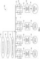
- FIG. 1 is a block diagram of an example system 100 for quantifying diastolic function, in accordance with various embodiments.
- the system 100 includes one or more electrodes 102 for acquiring ECG signals (e.g., 10 electrodes for a traditional 12-lead ECG), a processing facility 104 for processing the ECG signals, and an electrode interface 106 connecting the electrodes 102 to the processing facility 104.
- the electrode interface 106 includes circuitry that outputs electrical signals suitable as input to the processing facility 104, e.g., by digitally sampling analog input signals.
- the system 100 further includes a display device 108 for outputting the ECG test results (including, e.g., the ECGs themselves, time-frequency maps computed therefrom, and/or various quantitative and/or qualitative indicators of diastolic function computed as described herein), and optionally other input/output devices 109, such as a keyboard and mouse and/or a printer, for instance.
- the display device 108 may be a touchscreen doubling as a user-input device.
- the processing facility 104, electrode interface 106, display 108, and input/output devices 109 may be implemented as a single, stand-alone device implementing all computational functionality for ECG signal processing and presentation. Alternatively, they may be provided by a combination of multiple devices.
- an ECG test device with limited functionality for recording and/or processing ECG signals received from one or more electrodes 102 via an electrode interface 106 of the device may outsource certain computationally intense processing tasks to one or more other computers.
- Data exchange between the ECG test device and the other computer(s) may take place via a wired or wireless network.
- the ECG test device may be connected via the internet to a cloud-based signal-processing service.
- the functionality of the processing facility 104 may be distributed between multiple computational devices. Whether provided in a single device or distributed, the processing facility 104 may be implemented with a suitable combination of hardware and/or software, such as a suitably programmed general-purpose computer (including at least one central processing unit (CPU) or graphic processing unit (GPU) and associated memory); dedicated, special-purpose circuitry (such as, e.g., a digital signal processor (DSP), field-programmable gate array (FPGA), analog circuitry, or other); or a combination of both.
- DSP digital signal processor
- FPGA field-programmable gate array
- analog circuitry or other
- the term "hardware processor” is used in reference to both special-purpose circuitry and general-purpose processors as used in general-purpose computers and configured via software.
- the processing facility 104 may include various functionally distinct components, such as separate computer programs or functions called within a larger program flow, and/or special-purpose circuitry for certain computational tasks. These components may include an ECG-signal processing component 110 that generates ECGs for multiple leads from the (e.g., digitally sampled) ECG signals for display and analysis (e.g., by filtering, smoothing, scaling, etc., as well as by combining signals for various leads); a time-frequency transform component 112 that converts the ECG for each lead into a two-dimensional time-frequency map (signed or unsigned) and, optionally, normalizes the time-frequency map; a featurizer 114 that computes and extracts relevant parameters from the ECGs and/or the time-frequency maps for use as input features to one or more machine-learned models 116; the one or more machine-learned computational models 116, which determine echocardiogram parameter estimates and/or other diastolic indicators from these ECG-derived features in conjunction with patient demographic parameters; and/or a
- the ECG-signal-processing component 110 may be a conventional processing module as used in commercially available heart monitors and/or as is capable of straightforward implementation by one of ordinary skill in the art.
- the time-frequency transform component 112, featurizer 114, and computational models 116 implement algorithms and provide functionality explained further below, and can be readily implemented by one of ordinary skill in the art given the benefit of the present disclosure.
- some instances of the processing facility 104 also include a training engine 120 that implements one or more suitable machine-learning algorithms to build and/or train (that is, determine adjustable parameters of) the models 116 based on training data.
- suitable training algorithms for various types of models are well-known to those of ordinary skill in the art.
- a neural network model may be trained using backpropagation of errors, e.g., with stochastic gradient descent. Note that, once the machine-learned models 116 have been trained and their parameters are fixed, the training engine 120 is no longer used; accordingly, the training engine 120 may be omitted from a processing facility 104 configured for assessing diastolic function of patients in the inference phase.
- the depicted components reflect merely one among several different possibilities for organizing the overall computational functionality of the processing facility 104.
- the components may, of course, be further partitioned, combined, or altered to distribute the functionality differently.
- the various components may be implemented as hardware components, software components (e.g., executed by a general-purpose processor), or a combination of both.
- FIG. 2 is a flow chart of an example method 200 for quantifying diastolic function using echocardiogram parameter estimates computed from electrocardiograms, in accordance with various embodiments.
- the method 200 begins, in acts 102 and 104, with measuring one or more ECGs for a patient (act 102) and recording relevant patient demographic parameters (such as, e.g., age, sex, height, weight, health conditions such as hypertension, or other factors that might affect how the patient's diastolic functions is reflected in ECGs and parameters derived from ECG) (act 204).
- patient demographic parameters such as, e.g., age, sex, height, weight, health conditions such as hypertension, or other factors that might affect how the patient's diastolic functions is reflected in ECGs and parameters derived from ECG
- ECG in the singular is herein generally used in reference to an individual lead; and multiple ECGs measured for a patient may, accordingly, correspond to multiple respective leads.
- ECGs for twelve leads can be computed in a manner routinely used in the medical arts. It is to be understood, however, that the method described herein does not require the use of all twelve leads, but is generally applicable to any number of ECGs, such as any subset of the standard twelve leads.
- four of the ten electrodes are placed on the patient's left and right arms and legs; two electrodes (labeled V1 and V2) are placed between the fourth and fifth ribs on the left and right side of the sternum; a further, single electrode (labeled V3) is placed between V2 and V4 on the fourth intercostal space; one electrode (labeled V4) is placed between the fifth and sixth ribs at the mid-clavicular line (the imaginary reference line that extends down from the middle of the clavicle), and, in line therewith, another electrode (labeled V5) is positioned in the anterior axillary line (the imaginary reference line running southward from the point where the collarbone and arm meet), and the tenth electrode (labeled V6) is placed on the same horizontal line as these two, but oriented along the mid-axillary line (the imaginary reference point straight down from the patient's armpit).
- the method 200 further involves transforming the ECGs (immediately upon measurement, or at a later time after storage in memory) into two-dimensional time-frequency maps by a suitable mathematical transform, such as, for instance, wavelet transform or short time Fourier transform, in act 206.
- a suitable mathematical transform such as, for instance, wavelet transform or short time Fourier transform, in act 206.
- wavelet transform or short time Fourier transform
- CWT continuous wavelet transform
- the wavelet selected for processing may be, for example, a Mexican hat wavelet, Morlet wavelet, Meyer wavelet, Shannon wavelet, Spline wavelet, or other wavelet known to those of ordinary skill in the art.
- the CWT W ( a, b ) is also referred to as a scalogram.
- the time-frequency maps (such as, e.g., scalograms) generally include both positive and negative values, i.e., they are "signed.” In some embodiments, the absolute value of the signal value (or the square of the signal value) is taken at each time-frequency point, resulting in an "unsigned" time-frequency map.
- the ECGs and time-frequency maps are processed, in act 208, to extract parameters to be provided, along with the patient demographic parameters, as input features to one or more machine-learned computational models.
- Time-domain features extracted directly from the ECGs may include, for example (and without limitation), the extrema, durations, or area of any of the P, Q, R, S, or T waves, or signal amplitudes at one or more specified points in time associated with the P, Q, R, S, or T waves.
- the input to the models may also include conventional ECG-derived parameters, such as Glasgow parameters.
- Time-frequency features extracted from the time-frequency maps may include any or all of the time-frequency coefficients (e.g., wavelet coefficients) themselves or any parameters derived from the time-frequency maps, for example (and without limitation), extrema across frequency at one or more points in time associated with the P, Q, R, S, or T waves, extrema across time at one or more specified points in frequency, or integral measures associated with extrema in the time-frequency map.
- U.S. Patent No. 9,700,226, filed on September 20, 2016 which is incorporated herein by reference, describes time-frequency transforms of ECGs (in particular CWTs) and various parameters derived from the resulting time-frequency maps that may be used as input features.
- time-frequency features may be extracted from the signed or unsigned time-frequency map or a combination of both.
- both the time-domain and the time-frequency features may include parameters derived from individual heartbeats across synchronized ECGs for different leads and from beat to beat.
- a set of features corresponding to the same parameter measured across multiple leads captures the phase differences between leads, which can provide important comparisons useful as input to the computational models. Values of a single parameter measured over multiple heartbeats may be combined into a single aggregated feature, e.g., the average or median across the multiple heartbeats.
- one or more machine-learned computational models operate on the ECG-derived features (including the time-frequency features) and patient demographic parameters to compute one or more diastolic indicators, including one or more estimates of echocardiogram parameters (e.g., e', a', E, A, and ratios).
- Multiple computational models may be used to compute different respective parameter estimates and indicators.
- Multiple computational models are organized in a one-level structure or, alternatively, in a hierarchy of two or more levels.
- FIG. 4 (described below) provides an example of a two-level hierarchy according to an embodiment.
- individual indicators may be computed with a multi-level hierarchy of models, the number of models and/or levels generally being different for each indicator.
- different models may, for instance, be used to compute a first set of estimates, and an ensemble model may then be used at the second level to combine the first set of estimates into a final estimate.
- the estimates of the echocardiogram parameters are continuous-variable estimates that may be computed with regression models or may, alternatively, be piecewise approximated using classification models.
- the computational model(s) may include, e.g., decision trees or random forests, neural networks, regression analysis, Bayesian networks, or other suitable machine-learning models known to those of ordinary skill in the art. Regression models that were found, in some embodiments, to provide particularly good performance in estimating the echocardiogram parameters include random forest and least squares models.
- the echocardiogram parameter estimate(s) output by the computational model(s) may flow as inputs into further algorithms and/or additional machine-learned computational models for computing, in act 212, additional diastolic indicators, such as qualitative classifications of diastolic function (e.g., a three-level classification between normal, borderline, and abnormal diastolic function; or a 5-level classification between low-possibility, possible, borderline, probable, and highly probable LV relaxation abnormality), and/or quantitative indicators such as left ventricular (LV) relaxation risk score(s), LV relaxation indices (e.g. lateral, septal, and/or average/composite indices, etc.).
- LV left ventricular
- LV relaxation indices e.g. lateral, septal, and/or average/composite indices, etc.
- these models form, along with the computational models for computing the echocardiogram parameter estimates in act 210, a two-level hierarchical architecture (optionally with sub-levels at one or both levels), e.g., as illustrated by way of example in FIG. 5 (described below). Further, certain qualitative and/or quantitative diastolic indicators (such as classifications or LV relaxation indices) are computed along with the echocardiogram parameter estimates by the (first level of) computational models in act 210, and flow, along with the echocardiogram parameter estimates, into downstream computations.
- a two-level hierarchical architecture optionally with sub-levels at one or both levels
- echocardiogram parameter estimates may be scaled to indices spanning a fixed range (e.g., from 0 to 100).
- echocardiogram parameter estimates may be compared against specified thresholds to classify diastolic function between various degrees or likelihoods of abnormality. For example, one measure of left ventricular diastolic dysfunction is low e'.
- e' is defined as septal e' velocity ⁇ 7 cm/s or lateral e' velocity ⁇ 10 cm/s, where septal e' is the velocity of the septal mitral annular motion at early diastole and lateral e' is the velocity of the lateral mitral annular motion.
- septal and lateral e' are measured via transthoracic echocardiography, but they can be estimated, in accordance herewith, from ECGs.
- the estimated septal and lateral e' parameters may be compared against values of 7 or 10 (in cm/s), respectively, to classify diastolic function as normal or abnormal.
- the threshold values for comparison against the estimated e' parameters may be set lower or higher than those used with the parameters obtained by echocardiography to account for any error in the estimate and provide higher confidence for normal and abnormal classifications, with a region of uncertainty in between.
- the echocardiogram parameter estimates and/or other diastolic indicators computed in acts 210, 212 may be provided as output (e.g., displayed on screen, printed, sent via electronic notification, etc.) to a physician or other clinical personnel in act 214, optionally along with the ECGs and/or time-frequency maps from which these indicators have been computed.
- FIGS. 9 and 10 provide example output displays.
- Such metrics may include, e.g., statistical indicators such as prevalence probability, relative risk, likelihood ratios, or confidence intervals for predicted ranges of clinically measurable attributes.
- Prevalence probability is a measure of the probability that a person with a certain test result has a certain condition, determined using a reference study population, and can be calculated by dividing the number of persons with the same test results that have the condition in the reference study population by the total number of persons with the same test result in the reference study population.
- Relative risk is a ratio of the probability of an event occurring in a particular sub-group (Group A) of a population versus the probability of the event occurring in a reference sub-group (Group B) of the same population that is independent of the sub-group being studied (i.e., Group A and Group B are independent sub-groups within the same population), and can be calculated, accordingly, by dividing the probability of the event in Group A by the probability of the event in Group B.
- Likelihood ratios (LR) in medical testing are used to interpret diagnostic tests by indicating how likely a patient has a disease or condition. The higher the ratio, the more likely the patient has the disease or condition. Conversely, a low ratio means that the patient very likely does not have the disease or condition. Therefore, these ratios can help a physician rule in or rule out a disease.
- a likelihood ratio is calculated by dividing the probability that a person with the condition has a certain test result by the probability that a person without the condition has that test result.
- Confidence intervals demonstrate a range of values that a predicted measure (such as, e.g., a diastolic indicator) may actually fall between with a certain degree of confidence. Typically, if a dataset follows a normal distribution, then these intervals are calculated using statistical techniques based on formulas that are widely accepted for normally distributed datasets. The most common confidence intervals used in medicine are 95% and 70% confidence intervals since they are easily calculated using a mean and standard deviation derived from the overall dataset. Confidence intervals are used in risk-stratification by examining how much of the overall interval lies in the clinically accepted normal or abnormal range.
- Confidence intervals can also be used for rule-in or rule-out assessments if the 95% confidence interval lies completely to one side or the other of a clinically accepted threshold.
- a 70% confidence interval corresponds to the population mean +/- the population standard deviation.
- a 95% confidence interval corresponds to the population mean +/- twice the population standard deviation.
- FIG. 3 shows example distribution plots 300, 302 of the lateral LV relaxation index (along the independent axis) for "normal” and "abnormal” patient populations, respectively, based on calculated averages and standard deviations.
- the plots demonstrate that the lateral LV relaxation index, indeed, has risk-stratification value, especially for normal patients.
- the abnormal plot 302 overlaps significantly with the normal plot 300 (by about 30% of the normal plot), but the plots 300, 302 can nonetheless be used to calculate both percentile differences and likelihood ratios. While FIG. 3 shows distribution plots for, specifically, the lateral LV relaxation index, other LV relaxation indices exhibit similar behavior.
- FIG. 4 is a data flow diagram illustrating an example two-level computational model architecture 400 for computing echocardiogram parameter estimates, in accordance with various embodiments.
- the two-level architecture 400 takes a combination of conventional and/or other time-domain ECG features 402, time-frequency features 404 selected or derived from time-frequency transform(s) of the ECG(s), and patient demographic parameters 406 as input into multiple machine-learned computational models 408, 410, 412, 414 ("ML models") at the first level 416 of the hierarchy 400.
- ML models machine-learned computational models
- Different models may operate on different subsets of the input features 402, 404, 406.
- Outputs of the first-level models 408, 410, 412, 414 flow as inputs into respective ensemble models 418, 420, 422, 424 at the second level 426 of the hierarchy.
- the ensemble models 418, 420, 422, 424 each compute one of various diastolic indicators from the first-level outputs (optionally augmented by some or all of the first-level feature inputs 402, 404, 406).
- a number of computational models 408 may provide first-level estimates of lateral e', which are then further processed, in the associated ensemble model/algorithm 418, to compute a final estimate 430 of lateral e'. Combining the results of multiple computational models in this manner may achieve higher-accuracy estimates than one model alone.
- echocardiogram parameter estimates 432, 434 can be computed by two levels of computational and ensemble models (e.g., models 410, 422 to compute septal e' 432 and models 412, 422 to compute a' 434).
- the two-level architecture 400 includes models 414, 424 to compute other diastolic indicators, e.g., as shown, a composite LV relaxation index 436, directly from the inputs 402, 404, 406.
- the first-level computational models 408, 410, 412, 414 may include binary (2-class), multi-class (e.g., 3-class or 5-class), or regression (continuous-variable) models
- the second-level computational models may include ensemble models 418, 420, 424, 426.
- Each ensemble model 418, 420, 424, 426 may include one or more binary, multi-class, and/or regression models, optionally augmented by logic, equations, formulas, or algorithms to further refine the accuracy of the first-level models.
- FIG. 5 is a data flow diagram illustrating an example two-level computational model architecture 500 for computing various diastolic indicators using echocardiogram parameter estimates.
- machine-learned computational models 502, 504, 506, and optionally 508 operate on the ECG-derived features 402, 404 and patient demographic parameters 406 to generate echocardiogram parameter estimates 510, 512, 514 (e.g., as shown, lateral and septal e', as well as a') and optionally other diastolic indicators 516 at the output of the first level 518.
- echocardiogram parameter estimates 510, 512, 514 and/or other diastolic indicators 516 are then provided as input to a second level 520 of machine-learned computational models 522, 524, which compute various downstream categorical or quantitative diastolic indicators, e.g., as shown, a normal/borderline/abnormal classification 526 or risk score 528.
- Different selections and combinations of the first-level outputs may be relevant for different ones of the downstream diastolic indicators.
- the levels 518, 520 for computing first-level echocardiogram parameter estimates 510, 512, 514 and downstream second-level diastolic indicators 526, 528, respectively may each include multiple sub-levels.
- the architectures 400, 500 can be used in combination, such that each of the computational models 502, 504, 506, 508 for computing, e.g., echocardiogram parameter estimates is implemented by two sub-levels corresponding to models of the levels 416, 426 of the architecture 400.
- the model 502 for computing lateral e' may include a two-level structure of models 408, 418.
- the one or more models may be developed using machine-learning training processes, such as supervised learning.
- Training data to be used in the training process may be obtained from a (generally large) number of patients whose diastolic function spans a range from normal function to a high degree of abnormal (or dys-) function.
- ECGs For each patient, both one or more ECGs and one or more echocardiograms are acquired.
- the ECGs are processed to derive conventional/time-domain as well as time-frequency parameters to be used as input features to the model(s), and the echocardiograms are processed to derive one or more echocardiogram parameters of interest that will serve as the ground truth for training.
- the training dataset includes pairs of a set of input features and a set of output labels for each patient, the input features including the ECG-derived parameters as well as any relevant patient demographic parameters, and the output labels including the echocardiogram parameters for that patient.
- a model e.g., one of the models 408, 410, 412
- the input features are fed into the model, and the model-generated output is compared against the ground-truth echocardiogram parameter; the discrepancy between the measured (ground-truth) and estimated echocardiogram parameters is used as feedback to iteratively adjust the model.
- ground-truth classifications or scores may be determined for each patient, e.g., from the measured echocardiogram parameters. Training algorithms for building and training various types of models are well-known to those of ordinary skill in the art.
- Model development may also incorporate various feature-reduction techniques, such as hyper-parameter tuning, random forest feature importance, de-correlation, principal component analysis (PCA), clustering, minimum redundancy maximum relevance, mean-0 normalization, min-max normalization, etc.
- feature-reduction analysis is done during the model training process for each model individually to select the most useful features for that model and discard the others which have lower predictive qualities for the target value of the model (which is the parameter that the model is intended to predict).
- the training process may initially operate on hundreds of input features, whose relative contributions to accurately predict the target parameter are quantitatively assessed to reduce the input feature set to merely tens of features.
- Feature reduction performed during training of various models has shown that, in general, time-frequency features contribute significantly to the accuracy of the echocardiogram parameter estimates.
- the feature set retained at the completion of feature-reduction analysis includes at least one third time-frequency features, and less than 20% patient-demographic features, the remainder being traditional ECG features.
- a set of eighteen features selected as input to a model for estimating e' includes about 39% time-frequency features.
- the time-frequency features flowing into the computation of echocardiogram parameter estimates and/or other diastolic indicators are computed explicitly and provided to the machine-learned computational model(s) as input features.
- the computational models may be implemented as a multi-level neural network system including a neural network for computing a time-frequency transform at the first level and one or more neural networks for computing echocardiogram parameter estimates and optionally additional diastolic indicators at the subsequent second level (which, as explained above, may itself include multiple (sub-)levels).
- FIG. 6 is a data flow diagram illustrating an example two-level neural network architecture 600 for computing diastolic indicators from one or more raw ECGs 602, using an implicit time-frequency transform.
- a convolutional neural network (CNN) 604 may be configured to compute a time-frequency transform 606 for each ECG 602.
- the weights of the CNN 604 may be chosen to be the wavelets ⁇ evaluated across a range of different values of the scaling factor a .
- the second-level neural network(s) 608 may include multiple sub-levels, e.g., similar to the example architectures 400, 500. (Further, although not explicitly shown, the neural network(s) 608 may include multiple parallel branches of neural networks for determining multiple echocardiogram parameter estimates or other diastolic indicators.) However, in contrast to the computational model architectures 400, 500, which take time-domain and time-frequency features as input, the neural networks 604, 608 operate on the raw ECGs 602, and feature selection is performed implicitly as part of the application of the neural network(s) 608 at the second level.
- inputs to the second-level neural network(s) 608 may include the outputs 606 of the time-frequency transform implemented by the CNN 604 (all of these outputs constituting time-frequency features) as well as, optionally, the raw ECGs 602 and/or patient demographic parameters 406.
- the second-level neural network(s) 608 may be configured to implicitly derive time-domain features from the ECG(s) 602 and select or derive time-frequency features from the time-frequency transform outputs 606 in one or more initial network layers, with subsequent layers of the neural network(s) 608 operating on these features.
- the neural network(s) 608 for computing the diastolic parameters may be implemented by various types of neural networks known to those of ordinary skill in the art, which may be selected depending, e.g., on the particular diastolic indicator.
- Neural networks suitable for computing echocardiogram parameter estimates and/or other diastolic indicators include, for example and without limitation, multi-layer perceptrons (MLPs) and probabilistic neural networks (PNNs) based on dynamic decay adjustment (DDA).
- MLPs can learn non-linear function approximators for classification or regression, and may (in contrast to logistic regression) include one or more non-linear hidden layers between the input and output layers.
- PNNs may be created using an algorithm, known as "constructive training of PPNs," that generates rules based on numerical data, each rule defining a high-dimensional Gaussian function that is adjusted by two thresholds to avoid conflicts with rules of different classes.
- the neural networks 604, 608 of the two-level neural network architecture 600 may be trained in a supervised manner based on ECGs (and, optionally, patient demographic parameters) paired with ground-truth echocardiogram parameters (as measured by echocardiography).
- the weights of the CNN 604 are initialized in accordance with the desired time-frequency transform, e.g., to the selected wavelets of a CWT.
- the weights of the subsequent neural network(s) 608 may be initialized in multiple ways, e.g., with random weights, and are adjusted during training. Training may also include modifying the weights of the CNN 604.
- the combined, two-level neural network architecture 600 may be trained end-to-end from the initial weights, e.g., using back propagation (which is well-known in the art) all the way through the CNN 604. It may be beneficial, however, to instead train the networks 604, 608 in two stages: In the first stage, the weights of the CNN 604 may be held fixed to provide the time-frequency transform outputs 606 (e.g., wavelet coefficients) to be input to the second-level neural network(s) 608, which may be trained, e.g., using back propagation stopping at the output of the CNN 604. In this manner, the second-level neural network(s) 608 can benefit from the spectral information provided by the time-frequency transform(s). After training the second-level neural-networks 608, the restriction of fixing the weights of the CNN 604 at the first level can be relaxed, and the combined two-level system of neural networks 604, 608 can be trained to adjust the weights at both levels.
- back propagation which is well-known in the art
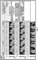
- FIGS. 7 and 8 are example user interfaces in accordance with various embodiments, e.g., as may be displayed on a computing device (e.g., desktop computer or tablet) utilized by a physician or other medical provider when evaluating a patient.
- the user interface in FIG. 7 shows an example report screen for an abnormal case
- the user interface in FIG. 8 shows an example report screen for a borderline case.
- the report screens display, on the left side, the ECG traces and associated time-frequency maps for multiple leads (e.g., as shown, leads I, II, and III).
- the displayed leads may be selectable (e.g., among the standard twelve leads) by the user (e.g., physician), using, for example, drop-down lists accessible via user-interface features next to the lead indicators.
- the unsigned version may be beneficial since it avoids presenting potentially distracting information that is not of immediate, intuitively discernible clinical significance because the sign is not relevant to an intuitive interpretation of the signal value of the time-frequency map as a measure of the electrical energy of the heart.
- various quantitative and qualitative metrics and indicators are displayed. These metrics may include conventional ECG information, such as Glasgow parameters (e.g., heart rate, various intervals measured between waveform features of the ECG, etc.).
- various diastolic indicators derived, in accordance with this disclosure, by machine-learned computational models from ECGs and time-frequency maps may be shown. For instance, a categorical indicator, e.g., displayed in the form of a segmented bar (optionally color-coded) with the applicable category being highlighted and the other categories being greyed out, may instantly inform the physician of the overall diastolic health of the patient, e.g., whether diastolic function is normal, borderline (as in FIG.
- a five-segment bar may alternatively be used for finer gradation.
- a quantitative risk score which generally correlates with the classification, may be indicated along a scale. For example, a high risk score of about 6 may correspond to an abnormality, whereas a lower risk score of about 3 may correspond to borderline diastolic function.
- a textual risk statement indicating the type and/or likelihood of abnormality may also be included.
- the report screen may also provide the computed echocardiogram parameter estimates, or indices derived therefrom, e.g., as shown, lateral, septal, and composite LV relaxation indices.
- a graphical representation of likelihood ratios for the various indices may be provided (e.g., by tick-marks along a scale, as shown).
- abnormal diastolic function may be reflected in significantly higher values of the LV relaxation indices.
- Other graphic, numerical, and/or textual representations are conceivable.
- the displayed information includes various measures and indicators that may be useful for risk stratification.
- medical personnel may also be provided with an interpretation guide that presents statistical measures taken from a reference study population.
- table 1 illustrates the kind of more detailed information that the interpretation guide may contain to explain risk classifications (e.g., into normal/borderline/abnormal), risk scores, and verbal risk assessments.
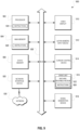
- FIG. 9 is a block diagram of a machine in the example form of a computer system 900 within which instructions for causing the machine to perform any one or more of the methodologies discussed herein may be executed.
- the machine operates as a standalone device or may be connected (e.g., networked) to other machines.
- the machine may operate in the capacity of a server or a client machine in server-client network environment, or as a peer machine in a peer-to-peer (or distributed) network environment. While only a single machine is illustrated, the term "machine" shall also be taken to include any collection of machines that individually or jointly execute a set (or multiple sets) of instructions to perform any one or more of the methodologies discussed herein.
- the example computer system 900 includes one or more processors 902 (e.g., a central processing unit (CPU), a graphics processing unit (GPU) or both), a main memory 904 and a static memory 906, which communicate with each other via a bus 908.
- the computer system 900 may further include a video display unit 910 (e.g., a liquid crystal display (LCD) or a cathode ray tube (CRT)).
- a video display unit 910 e.g., a liquid crystal display (LCD) or a cathode ray tube (CRT)
- the computer system 900 also includes an alphanumeric input device 912 (e.g., a keyboard), a user interface (UI) navigation device 914 (e.g., a mouse), a disk drive unit 916, a signal generation device 918 (e.g., a speaker), a network interface device 920 to communicate via a network 926, and a data interface device (such as, e.g., an electrode interface).
- a user interface (UI) navigation device 914 e.g., a mouse
- a disk drive unit 916 e.g., a disk drive unit 916
- a signal generation device 918 e.g., a speaker
- a network interface device 920 to communicate via a network 926
- a data interface device such as, e.g., an electrode interface
- the disk drive unit 916 includes a machine-readable medium 922 storing one or more sets of instructions and data structures (e.g., software) 924 embodying or utilized by any one or more of the methodologies or functions described herein.
- the instructions 924 may also reside, completely or at least partially, within the main memory 904 and/or within the processor 902 during execution thereof by the computer system 900, the main memory 904 and the processor 902 also constituting machine-readable media.
- machine-readable medium 922 is shown in an example embodiment to be a single medium, the term “machine-readable medium” may include a single medium or multiple media (e.g., a centralized or distributed database, and/or associated caches and servers) that store the one or more instructions or data structures.
- the term “machine-readable medium” shall also be taken to include any tangible medium that is capable of storing, encoding, or carrying instructions for execution by the machine and that cause the machine to perform any one or more of the methodologies of the present invention, or that is capable of storing, encoding or carrying data structures utilized by or associated with such instructions.
- the term “machine-readable medium” shall accordingly be taken to include, but not be limited to, solid-state memories, and optical and magnetic media.
- machine-readable media include non-volatile memory, including by way of example semiconductor memory devices, e.g., Erasable Programmable Read-Only Memory (EPROM), Electrically Erasable Programmable Read-Only Memory (EEPROM), and flash memory devices; magnetic disks such as internal hard disks and removable disks; magneto-optical disks; CD-ROM and DVD-ROM disks, or other data-storage devices.
- semiconductor memory devices e.g., Erasable Programmable Read-Only Memory (EPROM), Electrically Erasable Programmable Read-Only Memory (EEPROM), and flash memory devices
- magnetic disks such as internal hard disks and removable disks
- magneto-optical disks CD-ROM and DVD-ROM disks, or other data-storage devices.
- machine-readable medium shall be taken to include a non-tangible signal or transmission medium, including an electrical signal, a magnetic signal, an electromagnetic signal, an acoustic signal and an optical signal.
Landscapes
- Health & Medical Sciences (AREA)
- Life Sciences & Earth Sciences (AREA)
- Engineering & Computer Science (AREA)
- Physics & Mathematics (AREA)
- General Health & Medical Sciences (AREA)
- Biomedical Technology (AREA)
- Biophysics (AREA)
- Molecular Biology (AREA)
- Public Health (AREA)
- Medical Informatics (AREA)
- Cardiology (AREA)
- Pathology (AREA)
- Animal Behavior & Ethology (AREA)
- Surgery (AREA)
- Heart & Thoracic Surgery (AREA)
- Veterinary Medicine (AREA)
- Artificial Intelligence (AREA)
- Physiology (AREA)
- Theoretical Computer Science (AREA)
- Mathematical Physics (AREA)
- Evolutionary Computation (AREA)
- Data Mining & Analysis (AREA)
- Signal Processing (AREA)
- Computer Vision & Pattern Recognition (AREA)
- Psychiatry (AREA)
- General Engineering & Computer Science (AREA)
- Software Systems (AREA)
- Computing Systems (AREA)
- General Physics & Mathematics (AREA)
- Computational Linguistics (AREA)
- Hematology (AREA)
- Fuzzy Systems (AREA)
- Databases & Information Systems (AREA)
- Epidemiology (AREA)
- Primary Health Care (AREA)
- Dentistry (AREA)
- Oral & Maxillofacial Surgery (AREA)
- Measurement And Recording Of Electrical Phenomena And Electrical Characteristics Of The Living Body (AREA)
Claims (12)
- Ein computerimplementiertes Verfahren zum Quantifizieren der diastolischen Funktion, wobei das Verfahren Folgendes beinhaltet:Empfangen (202) eines oder mehrerer Elektrokardiogramme, die für einen Patienten gemessen wurden;Umwandeln (208) des einen oder der mehreren Elektrokardiogramme unter Verwendung von Zeit-Frequenz-Transformation in Zeit-Frequenz-Merkmale;Betreiben (210) eines oder mehrerer maschinell erlernter Berechnungsmodelle bei einer Eingabe, die die Zeit-Frequenz-Merkmale beinhaltet, um eine Schätzung oder Schätzungen eines oder mehrerer Echokardiogrammparameter zu berechnen, die für die diastolische Funktion indikativ sind, wobei das eine oder die mehreren maschinell erlernten Berechnungsmodelle unter Verwendung von Elektrokardiogrammdaten trainiert wurden, die mit Werten des einen oder der mehreren Echokardiogrammparameter korreliert sind, die durch Echokardiographie als Grundwahrheitsausgaben erhalten wurden;dadurch gekennzeichnet, dass es ferner Folgendes beinhaltet:Berechnen (212) eines oder mehrerer zusätzlicher Indikatoren der diastolischen Funktion durch Betreiben eines oder mehrerer zweiter maschinell erlernter Berechnungsmodelle bei einer Eingabe, die die Schätzung oder Schätzungen des einen oder der mehreren Echokardiogrammparameter beinhaltet;wobei das eine oder die mehreren zweiten maschinell erlernten Berechnungsmodelle ein oder mehrere Ensemble-Modelle beinhalten und der eine oder die mehreren zusätzlichen Indikatoren der diastolischen Funktion wenigstens einen von einem linksventrikulären Relaxationsrisikopunktwert, einem lateralen linksventrikulären Relaxationsindex, einem septalen linksventrikulären Relaxationsindex oder einem zusammengesetzten linksventrikulären Relaxationsindex beinhalten.
- Verfahren gemäß Anspruch 1, wobei das eine oder die mehreren maschinell erlernten Berechnungsmodelle aus dem Training mit Paaren von Eingabemerkmalssätzen und Grundwahrheitsausgaben für eine Vielzahl von Patienten resultieren, wobei die Eingabemerkmalssätze die Zeit-Frequenz-Merkmale beinhalten.
- Verfahren gemäß Anspruch 1 oder Anspruch 2, wobei ein erstes neuronales Netzwerk verwendet wird, um die Elektrokardiogramme in die Zeit-Frequenz-Merkmale umzuwandeln, wobei das eine oder die mehreren maschinell erlernten Berechnungsmodelle ein oder mehrere zweite neuronale Netzwerke beinhalten, und wobei die Zeit-Frequenz-Merkmale, die von dem ersten neuronalen Netzwerk ausgegeben werden, als Eingaben für das eine oder die mehreren zweiten neuronalen Netzwerke bereitgestellt werden.
- Verfahren gemäß Anspruch 3, wobei die Gewichte des ersten neuronalen Netzwerkes initialisiert werden, um eine Zeit-Frequenz-Transformation zu implementieren, und anschließend während des End-to-End-Trainings des kombinierten ersten und zweiten neuronalen Netzwerkes angepasst werden.
- Verfahren gemäß Anspruch 4, wobei das eine oder die mehreren zweiten neuronalen Netzwerke mit festen Werten der Gewichte des ersten neuronalen Netzwerkes vor dem End-to-End-Training des kombinierten ersten und zweiten neuronalen Netzwerkes trainiert werden.
- Verfahren gemäß einem der vorhergehenden Ansprüche, wobei das eine oder die mehreren Berechnungsmodelle ein oder mehrere Regressionsmodelle beinhalten.
- Verfahren gemäß Anspruch 6, wobei das eine oder die mehreren Regressionsmodelle wenigstens eines von einem Random-Forest-Modell oder einem Modell der kleinsten Quadrate beinhalten.
- Verfahren gemäß einem der vorhergehenden Ansprüche, wobei die aus den Zeit-Frequenz-Karten abgeleiteten Zeit-Frequenz-Merkmale Extrema über die Frequenz an einem oder mehreren Zeitpunkten beinhalten, die den P-, Q-, R-, S- oder T-Wellen zugeordnet sind.
- Verfahren gemäß einem der vorhergehenden Ansprüche, wobei die Eingabe in das eine oder die mehreren Berechnungsmodelle ferner wenigstens einen von einem oder mehreren demographischen Patientenparametern oder ein oder mehrere Zeitbereichsmerkmale beinhaltet, die direkt von dem einen oder den mehreren Elektrokardiogrammen abgeleitet werden.
- Verfahren gemäß Anspruch 9, wobei das eine oder die mehreren direkt aus den Elektrokardiogrammen abgeleiteten Zeitbereichsmerkmale Glasgow-abgeleitete EKG-Parameter beinhalten.
- Ein System zum Quantifizieren der diastolischen Funktion, wobei das System Folgendes beinhaltet:einen oder mehrere Hardware-Prozessoren; undSpeicher, der Anweisungen speichert, die, wenn sie von dem einen oder den mehreren Hardware-Prozessoren ausgeführt werden, das Verfahren gemäß einem der vorhergehenden Ansprüche durchführen.
- Ein computerlesbares Medium, das prozessorausführbare Anweisungen trägt, die, wenn sie von einem oder mehreren Computerprozessoren ausgeführt werden, den einen oder die mehreren Computerprozessoren veranlassen, das Verfahren gemäß einem der Ansprüche 1 bis 10 durchzuführen.
Priority Applications (1)
| Application Number | Priority Date | Filing Date | Title |
|---|---|---|---|
| EP23193015.7A EP4252651A3 (de) | 2019-08-30 | 2020-08-31 | Auf elektrokardiogramm basierende beurteilung der diastolischen funktion |
Applications Claiming Priority (2)
| Application Number | Priority Date | Filing Date | Title |
|---|---|---|---|
| US201962894598P | 2019-08-30 | 2019-08-30 | |
| US202063065837P | 2020-08-14 | 2020-08-14 |
Related Child Applications (2)
| Application Number | Title | Priority Date | Filing Date |
|---|---|---|---|
| EP23193015.7A Division EP4252651A3 (de) | 2019-08-30 | 2020-08-31 | Auf elektrokardiogramm basierende beurteilung der diastolischen funktion |
| EP23193015.7A Division-Into EP4252651A3 (de) | 2019-08-30 | 2020-08-31 | Auf elektrokardiogramm basierende beurteilung der diastolischen funktion |
Publications (3)
| Publication Number | Publication Date |
|---|---|
| EP3834729A1 EP3834729A1 (de) | 2021-06-16 |
| EP3834729C0 EP3834729C0 (de) | 2024-05-22 |
| EP3834729B1 true EP3834729B1 (de) | 2024-05-22 |
Family
ID=72292442
Family Applications (2)
| Application Number | Title | Priority Date | Filing Date |
|---|---|---|---|
| EP23193015.7A Pending EP4252651A3 (de) | 2019-08-30 | 2020-08-31 | Auf elektrokardiogramm basierende beurteilung der diastolischen funktion |
| EP20193719.0A Active EP3834729B1 (de) | 2019-08-30 | 2020-08-31 | Auf elektrokardiogramm basierende beurteilung der diastolischen funktion |
Family Applications Before (1)
| Application Number | Title | Priority Date | Filing Date |
|---|---|---|---|
| EP23193015.7A Pending EP4252651A3 (de) | 2019-08-30 | 2020-08-31 | Auf elektrokardiogramm basierende beurteilung der diastolischen funktion |
Country Status (2)
| Country | Link |
|---|---|
| US (3) | US11445918B2 (de) |
| EP (2) | EP4252651A3 (de) |
Families Citing this family (11)
| Publication number | Priority date | Publication date | Assignee | Title |
|---|---|---|---|---|
| US11445918B2 (en) | 2019-08-30 | 2022-09-20 | Heart Test Laboratories, Inc. | Electrocardiogram-based assessment of diastolic function |
| US11657921B2 (en) | 2019-09-18 | 2023-05-23 | Tempus Labs, Inc. | Artificial intelligence based cardiac event predictor systems and methods |
| US11762990B2 (en) * | 2020-04-07 | 2023-09-19 | Microsoft Technology Licensing, Llc | Unstructured text classification |
| US12003535B2 (en) | 2021-03-01 | 2024-06-04 | Microsoft Technology Licensing, Llc | Phishing URL detection using transformers |
| US20220296169A1 (en) * | 2021-03-17 | 2022-09-22 | Lakehead University | System and methods for collecting and processing data on one or more physiological parameters of monitored subject |
| AU2022281440A1 (en) | 2021-05-28 | 2023-12-14 | Geisinger Clinic | Artificial intelligence based cardiac event predictor systems and methods |
| WO2022266291A1 (en) | 2021-06-18 | 2022-12-22 | West Virginia University Board of Governors on behalf of West Virginia University | Synthetic echo from ecg |
| US12102485B2 (en) | 2022-04-05 | 2024-10-01 | Icahn School Of Medicine At Mount Sinai | Surfacing insights into left and right ventricular dysfunction through deep learning |
| WO2024050307A1 (en) * | 2022-08-30 | 2024-03-07 | Medtronic, Inc. | Electrocardiogram-based left ventricular dysfunction and ejection fraction monitoring |
| US12114984B2 (en) | 2022-11-22 | 2024-10-15 | Anumana, Inc. | System and apparatus for generating imaging information based on at least a signal |
| US12333413B1 (en) | 2024-03-01 | 2025-06-17 | Mayo Foundation For Medical Education And Research | Apparatus and method for training an artificial intelligence-supported diagnostic assessment tool |
Family Cites Families (8)
| Publication number | Priority date | Publication date | Assignee | Title |
|---|---|---|---|---|
| WO2014055980A1 (en) * | 2012-10-05 | 2014-04-10 | The Regents Of The University Of Michigan | Automated analysis of multi-lead electrocardiogram data to identify the exit sites of physiological conditions |
| US9824287B2 (en) * | 2015-09-29 | 2017-11-21 | Huami Inc. | Method, apparatus and system for biometric identification |
| KR102490960B1 (ko) * | 2015-09-30 | 2023-01-19 | 허트 테스트 레버러토리스, 인크. | 수량적 심장 테스팅 |
| US20170196473A1 (en) * | 2016-01-08 | 2017-07-13 | Heart Test Laboratories, Inc. | Quantitative heart monitoring and diagnostics |
| EP3681380A1 (de) * | 2017-09-11 | 2020-07-22 | Heart Test Laboratories, Inc. | Zeit-frequenz-analyse von elektrokardiogrammen |
| EP3731748B1 (de) | 2017-09-21 | 2021-06-02 | Koninklijke Philips N.V. | Detektion von vorhofflimmern mit kurzen einzel-elektroden-ekg-aufzeichnungen |
| EP4623811A3 (de) | 2017-10-06 | 2025-10-08 | Mayo Foundation for Medical Education and Research | Ekg-basiertes herzauswurffraktion-screening |
| US11445918B2 (en) | 2019-08-30 | 2022-09-20 | Heart Test Laboratories, Inc. | Electrocardiogram-based assessment of diastolic function |
-
2020
- 2020-08-27 US US16/948,021 patent/US11445918B2/en active Active
- 2020-08-31 EP EP23193015.7A patent/EP4252651A3/de active Pending
- 2020-08-31 EP EP20193719.0A patent/EP3834729B1/de active Active
-
2022
- 2022-08-08 US US17/818,209 patent/US12318172B2/en active Active
-
2025
- 2025-04-24 US US19/188,940 patent/US20250255492A1/en active Pending
Also Published As
| Publication number | Publication date |
|---|---|
| EP3834729C0 (de) | 2024-05-22 |
| US12318172B2 (en) | 2025-06-03 |
| EP4252651A2 (de) | 2023-10-04 |
| US20250255492A1 (en) | 2025-08-14 |
| US20210059540A1 (en) | 2021-03-04 |
| US20220378305A1 (en) | 2022-12-01 |
| EP3834729A1 (de) | 2021-06-16 |
| EP4252651A3 (de) | 2023-12-06 |
| US11445918B2 (en) | 2022-09-20 |
Similar Documents
| Publication | Publication Date | Title |
|---|---|---|
| EP3834729B1 (de) | Auf elektrokardiogramm basierende beurteilung der diastolischen funktion | |
| Daydulo et al. | Cardiac arrhythmia detection using deep learning approach and time frequency representation of ECG signals | |
| US20210353166A1 (en) | Analysis of cardiac data | |
| US11497430B2 (en) | Analysis of cardiac data | |
| US9295397B2 (en) | Method and apparatus for beat-space frequency domain prediction of cardiovascular death after acute coronary event | |
| Isler et al. | Comparison of the effects of cross-validation methods on determining performances of classifiers used in diagnosing congestive heart failure | |
| US20100217144A1 (en) | Diagnostic and predictive system and methodology using multiple parameter electrocardiography superscores | |
| US20250022596A1 (en) | Systems and methods for electrocardiogram deep learning interpretability | |
| US20240206821A1 (en) | Apparatus and a method for predicting a physiological indicator | |
| US12539087B2 (en) | ECG-based age and sex estimation | |
| CN118398208A (zh) | 基于机器学习的房颤风险评估方法 | |
| US20230389850A1 (en) | Noninvasive infarct size determination | |
| Sowmya et al. | Detecting anomalies in fetal electrocardiogram records using deep learning models | |
| Işler et al. | Heart rate normalization in the analysis of heart rate variability in congestive heart failure | |
| Shao et al. | Predicting cardiovascular and cerebrovascular events based on instantaneous high-order singular entropy and deep belief network | |
| Jacquemyn et al. | Phenotypic clustering of repaired Tetralogy of Fallot using unsupervised machine learning | |
| Jain et al. | Design of an integrated arrhythmia detection model using connectivity features and multivariate time series classification | |
| Li et al. | Clinical applications of machine learning in heart failure | |
| Nagavelli et al. | Research Article Machine Learning Technology-Based Heart Disease Detection Models | |
| Dahri et al. | Heart failure prediction: a comparative analysis of machine learning algorithms | |
| Chen | Application of heart rate variability (HRV) in congestive heart failure (CHF) detection and quantification | |
| AKMAL | TRACKING AND OBSERVING THE FETAL MOVEMENTS BY USING DEEP LEARNING METHODS | |
| ELSHAFEY | DETECTION OF MYOCARDIAL INFARCTION USING DEEP LEARNING TECHNIQUES | |
| Pk | Multi-Modal Anomaly Detection in Cardiac Arrhythmias: A Synchronized ECG-Audio Time Series Analysis Framework | |
| Tomala et al. | Artificial Intelligence in Contemporary Cardiology: Current Applications, Struggles, and Prospects |
Legal Events
| Date | Code | Title | Description |
|---|---|---|---|
| PUAI | Public reference made under article 153(3) epc to a published international application that has entered the european phase |
Free format text: ORIGINAL CODE: 0009012 |
|
| STAA | Information on the status of an ep patent application or granted ep patent |
Free format text: STATUS: THE APPLICATION HAS BEEN PUBLISHED |
|
| AK | Designated contracting states |
Kind code of ref document: A1 Designated state(s): AL AT BE BG CH CY CZ DE DK EE ES FI FR GB GR HR HU IE IS IT LI LT LU LV MC MK MT NL NO PL PT RO RS SE SI SK SM TR |
|
| STAA | Information on the status of an ep patent application or granted ep patent |
Free format text: STATUS: REQUEST FOR EXAMINATION WAS MADE |
|
| 17P | Request for examination filed |
Effective date: 20211213 |
|
| RBV | Designated contracting states (corrected) |
Designated state(s): AL AT BE BG CH CY CZ DE DK EE ES FI FR GB GR HR HU IE IS IT LI LT LU LV MC MK MT NL NO PL PT RO RS SE SI SK SM TR |
|
| RAP1 | Party data changed (applicant data changed or rights of an application transferred) |
Owner name: HEART TEST LABORATORIES, INC. |
|
| RIN1 | Information on inventor provided before grant (corrected) |
Inventor name: SENGUPTA, PARTHO Inventor name: KRUBSACK, DAVID Inventor name: PETERSON, AARON |
|
| GRAP | Despatch of communication of intention to grant a patent |
Free format text: ORIGINAL CODE: EPIDOSNIGR1 |
|
| STAA | Information on the status of an ep patent application or granted ep patent |
Free format text: STATUS: GRANT OF PATENT IS INTENDED |
|
| RIC1 | Information provided on ipc code assigned before grant |
Ipc: A61B 5/346 20210101AFI20230707BHEP |
|
| INTG | Intention to grant announced |
Effective date: 20230801 |
|
| GRAJ | Information related to disapproval of communication of intention to grant by the applicant or resumption of examination proceedings by the epo deleted |
Free format text: ORIGINAL CODE: EPIDOSDIGR1 |
|
| STAA | Information on the status of an ep patent application or granted ep patent |
Free format text: STATUS: REQUEST FOR EXAMINATION WAS MADE |
|
| GRAS | Grant fee paid |
Free format text: ORIGINAL CODE: EPIDOSNIGR3 |
|
| STAA | Information on the status of an ep patent application or granted ep patent |
Free format text: STATUS: GRANT OF PATENT IS INTENDED |
|
| GRAP | Despatch of communication of intention to grant a patent |
Free format text: ORIGINAL CODE: EPIDOSNIGR1 |
|
| INTC | Intention to grant announced (deleted) | ||
| INTG | Intention to grant announced |
Effective date: 20240105 |
|
| GRAA | (expected) grant |
Free format text: ORIGINAL CODE: 0009210 |
|
| STAA | Information on the status of an ep patent application or granted ep patent |
Free format text: STATUS: THE PATENT HAS BEEN GRANTED |
|
| AK | Designated contracting states |
Kind code of ref document: B1 Designated state(s): AL AT BE BG CH CY CZ DE DK EE ES FI FR GB GR HR HU IE IS IT LI LT LU LV MC MK MT NL NO PL PT RO RS SE SI SK SM TR |
|
| REG | Reference to a national code |
Ref country code: GB Ref legal event code: FG4D |
|
| REG | Reference to a national code |
Ref country code: CH Ref legal event code: EP |
|
| REG | Reference to a national code |
Ref country code: DE Ref legal event code: R096 Ref document number: 602020031207 Country of ref document: DE |
|
| REG | Reference to a national code |
Ref country code: IE Ref legal event code: FG4D |
|
| U01 | Request for unitary effect filed |
Effective date: 20240522 |
|
| U07 | Unitary effect registered |
Designated state(s): AT BE BG DE DK EE FI FR IT LT LU LV MT NL PT SE SI Effective date: 20240527 |
|
| U20 | Renewal fee for the european patent with unitary effect paid |
Year of fee payment: 5 Effective date: 20240715 |
|
| PG25 | Lapsed in a contracting state [announced via postgrant information from national office to epo] |
Ref country code: IS Free format text: LAPSE BECAUSE OF FAILURE TO SUBMIT A TRANSLATION OF THE DESCRIPTION OR TO PAY THE FEE WITHIN THE PRESCRIBED TIME-LIMIT Effective date: 20240922 |
|
| PG25 | Lapsed in a contracting state [announced via postgrant information from national office to epo] |
Ref country code: HR Free format text: LAPSE BECAUSE OF FAILURE TO SUBMIT A TRANSLATION OF THE DESCRIPTION OR TO PAY THE FEE WITHIN THE PRESCRIBED TIME-LIMIT Effective date: 20240522 |
|
| PG25 | Lapsed in a contracting state [announced via postgrant information from national office to epo] |
Ref country code: GR Free format text: LAPSE BECAUSE OF FAILURE TO SUBMIT A TRANSLATION OF THE DESCRIPTION OR TO PAY THE FEE WITHIN THE PRESCRIBED TIME-LIMIT Effective date: 20240823 |
|
| PG25 | Lapsed in a contracting state [announced via postgrant information from national office to epo] |
Ref country code: ES Free format text: LAPSE BECAUSE OF FAILURE TO SUBMIT A TRANSLATION OF THE DESCRIPTION OR TO PAY THE FEE WITHIN THE PRESCRIBED TIME-LIMIT Effective date: 20240522 |
|
| PG25 | Lapsed in a contracting state [announced via postgrant information from national office to epo] |
Ref country code: PL Free format text: LAPSE BECAUSE OF FAILURE TO SUBMIT A TRANSLATION OF THE DESCRIPTION OR TO PAY THE FEE WITHIN THE PRESCRIBED TIME-LIMIT Effective date: 20240522 |
|
| PG25 | Lapsed in a contracting state [announced via postgrant information from national office to epo] |
Ref country code: PL Free format text: LAPSE BECAUSE OF FAILURE TO SUBMIT A TRANSLATION OF THE DESCRIPTION OR TO PAY THE FEE WITHIN THE PRESCRIBED TIME-LIMIT Effective date: 20240522 Ref country code: NO Free format text: LAPSE BECAUSE OF FAILURE TO SUBMIT A TRANSLATION OF THE DESCRIPTION OR TO PAY THE FEE WITHIN THE PRESCRIBED TIME-LIMIT Effective date: 20240822 Ref country code: IS Free format text: LAPSE BECAUSE OF FAILURE TO SUBMIT A TRANSLATION OF THE DESCRIPTION OR TO PAY THE FEE WITHIN THE PRESCRIBED TIME-LIMIT Effective date: 20240922 Ref country code: HR Free format text: LAPSE BECAUSE OF FAILURE TO SUBMIT A TRANSLATION OF THE DESCRIPTION OR TO PAY THE FEE WITHIN THE PRESCRIBED TIME-LIMIT Effective date: 20240522 Ref country code: GR Free format text: LAPSE BECAUSE OF FAILURE TO SUBMIT A TRANSLATION OF THE DESCRIPTION OR TO PAY THE FEE WITHIN THE PRESCRIBED TIME-LIMIT Effective date: 20240823 Ref country code: ES Free format text: LAPSE BECAUSE OF FAILURE TO SUBMIT A TRANSLATION OF THE DESCRIPTION OR TO PAY THE FEE WITHIN THE PRESCRIBED TIME-LIMIT Effective date: 20240522 Ref country code: RS Free format text: LAPSE BECAUSE OF FAILURE TO SUBMIT A TRANSLATION OF THE DESCRIPTION OR TO PAY THE FEE WITHIN THE PRESCRIBED TIME-LIMIT Effective date: 20240822 |
|
| PG25 | Lapsed in a contracting state [announced via postgrant information from national office to epo] |
Ref country code: CZ Free format text: LAPSE BECAUSE OF FAILURE TO SUBMIT A TRANSLATION OF THE DESCRIPTION OR TO PAY THE FEE WITHIN THE PRESCRIBED TIME-LIMIT Effective date: 20240522 |
|
| PG25 | Lapsed in a contracting state [announced via postgrant information from national office to epo] |
Ref country code: RO Free format text: LAPSE BECAUSE OF FAILURE TO SUBMIT A TRANSLATION OF THE DESCRIPTION OR TO PAY THE FEE WITHIN THE PRESCRIBED TIME-LIMIT Effective date: 20240522 Ref country code: SK Free format text: LAPSE BECAUSE OF FAILURE TO SUBMIT A TRANSLATION OF THE DESCRIPTION OR TO PAY THE FEE WITHIN THE PRESCRIBED TIME-LIMIT Effective date: 20240522 |
|
| PG25 | Lapsed in a contracting state [announced via postgrant information from national office to epo] |
Ref country code: SM Free format text: LAPSE BECAUSE OF FAILURE TO SUBMIT A TRANSLATION OF THE DESCRIPTION OR TO PAY THE FEE WITHIN THE PRESCRIBED TIME-LIMIT Effective date: 20240522 |
|
| PG25 | Lapsed in a contracting state [announced via postgrant information from national office to epo] |
Ref country code: SM Free format text: LAPSE BECAUSE OF FAILURE TO SUBMIT A TRANSLATION OF THE DESCRIPTION OR TO PAY THE FEE WITHIN THE PRESCRIBED TIME-LIMIT Effective date: 20240522 Ref country code: SK Free format text: LAPSE BECAUSE OF FAILURE TO SUBMIT A TRANSLATION OF THE DESCRIPTION OR TO PAY THE FEE WITHIN THE PRESCRIBED TIME-LIMIT Effective date: 20240522 Ref country code: RO Free format text: LAPSE BECAUSE OF FAILURE TO SUBMIT A TRANSLATION OF THE DESCRIPTION OR TO PAY THE FEE WITHIN THE PRESCRIBED TIME-LIMIT Effective date: 20240522 Ref country code: CZ Free format text: LAPSE BECAUSE OF FAILURE TO SUBMIT A TRANSLATION OF THE DESCRIPTION OR TO PAY THE FEE WITHIN THE PRESCRIBED TIME-LIMIT Effective date: 20240522 |
|
| REG | Reference to a national code |
Ref country code: DE Ref legal event code: R097 Ref document number: 602020031207 Country of ref document: DE |
|
| PLBE | No opposition filed within time limit |
Free format text: ORIGINAL CODE: 0009261 |
|
| STAA | Information on the status of an ep patent application or granted ep patent |
Free format text: STATUS: NO OPPOSITION FILED WITHIN TIME LIMIT |
|
| REG | Reference to a national code |
Ref country code: CH Ref legal event code: PL |
|
| U1H | Name or address of the proprietor changed after the registration of the unitary effect |
Owner name: HEARTSCIENCES INC.; US |
|
| PG25 | Lapsed in a contracting state [announced via postgrant information from national office to epo] |
Ref country code: CH Free format text: LAPSE BECAUSE OF NON-PAYMENT OF DUE FEES Effective date: 20240831 Ref country code: MC Free format text: LAPSE BECAUSE OF FAILURE TO SUBMIT A TRANSLATION OF THE DESCRIPTION OR TO PAY THE FEE WITHIN THE PRESCRIBED TIME-LIMIT Effective date: 20240522 |
|
| 26N | No opposition filed |
Effective date: 20250225 |
|
| PG25 | Lapsed in a contracting state [announced via postgrant information from national office to epo] |
Ref country code: IE Free format text: LAPSE BECAUSE OF NON-PAYMENT OF DUE FEES Effective date: 20240831 |
|
| U20 | Renewal fee for the european patent with unitary effect paid |
Year of fee payment: 6 Effective date: 20250708 |
|
| PGFP | Annual fee paid to national office [announced via postgrant information from national office to epo] |
Ref country code: GB Payment date: 20250707 Year of fee payment: 6 |
|
| PG25 | Lapsed in a contracting state [announced via postgrant information from national office to epo] |
Ref country code: CY Free format text: LAPSE BECAUSE OF FAILURE TO SUBMIT A TRANSLATION OF THE DESCRIPTION OR TO PAY THE FEE WITHIN THE PRESCRIBED TIME-LIMIT; INVALID AB INITIO Effective date: 20200831 |