EP3832309B1 - Composition for diagnosis of bone metastasis of cancer and kit comprising same - Google Patents
Composition for diagnosis of bone metastasis of cancer and kit comprising same Download PDFInfo
- Publication number
- EP3832309B1 EP3832309B1 EP19844633.8A EP19844633A EP3832309B1 EP 3832309 B1 EP3832309 B1 EP 3832309B1 EP 19844633 A EP19844633 A EP 19844633A EP 3832309 B1 EP3832309 B1 EP 3832309B1
- Authority
- EP
- European Patent Office
- Prior art keywords
- cancer
- osteocalcin
- blood
- cells
- bone
- Prior art date
- Legal status (The legal status is an assumption and is not a legal conclusion. Google has not performed a legal analysis and makes no representation as to the accuracy of the status listed.)
- Active
Links
Images


Classifications
-
- G—PHYSICS
- G01—MEASURING; TESTING
- G01N—INVESTIGATING OR ANALYSING MATERIALS BY DETERMINING THEIR CHEMICAL OR PHYSICAL PROPERTIES
- G01N33/00—Investigating or analysing materials by specific methods not covered by groups G01N1/00 - G01N31/00
- G01N33/48—Biological material, e.g. blood, urine; Haemocytometers
- G01N33/50—Chemical analysis of biological material, e.g. blood, urine; Testing involving biospecific ligand binding methods; Immunological testing
- G01N33/53—Immunoassay; Biospecific binding assay; Materials therefor
- G01N33/575—Immunoassay; Biospecific binding assay; Materials therefor for cancer
- G01N33/5758—Immunoassay; Biospecific binding assay; Materials therefor for cancer involving compounds serving as markers for tumours, cancers or neoplasias, e.g. cellular determinants, receptors, heat shock/stress proteins, A-protein, oligosaccharides or metabolites
-
- G—PHYSICS
- G01—MEASURING; TESTING
- G01N—INVESTIGATING OR ANALYSING MATERIALS BY DETERMINING THEIR CHEMICAL OR PHYSICAL PROPERTIES
- G01N33/00—Investigating or analysing materials by specific methods not covered by groups G01N1/00 - G01N31/00
- G01N33/48—Biological material, e.g. blood, urine; Haemocytometers
- G01N33/50—Chemical analysis of biological material, e.g. blood, urine; Testing involving biospecific ligand binding methods; Immunological testing
- G01N33/53—Immunoassay; Biospecific binding assay; Materials therefor
- G01N33/575—Immunoassay; Biospecific binding assay; Materials therefor for cancer
- G01N33/57557—Immunoassay; Biospecific binding assay; Materials therefor for cancer of other specific parts of the body, e.g. brain
-
- G—PHYSICS
- G01—MEASURING; TESTING
- G01N—INVESTIGATING OR ANALYSING MATERIALS BY DETERMINING THEIR CHEMICAL OR PHYSICAL PROPERTIES
- G01N33/00—Investigating or analysing materials by specific methods not covered by groups G01N1/00 - G01N31/00
- G01N33/48—Biological material, e.g. blood, urine; Haemocytometers
- G01N33/50—Chemical analysis of biological material, e.g. blood, urine; Testing involving biospecific ligand binding methods; Immunological testing
- G01N33/53—Immunoassay; Biospecific binding assay; Materials therefor
- G01N33/575—Immunoassay; Biospecific binding assay; Materials therefor for cancer
- G01N33/57515—Immunoassay; Biospecific binding assay; Materials therefor for cancer of the breast
-
- G—PHYSICS
- G01—MEASURING; TESTING
- G01N—INVESTIGATING OR ANALYSING MATERIALS BY DETERMINING THEIR CHEMICAL OR PHYSICAL PROPERTIES
- G01N33/00—Investigating or analysing materials by specific methods not covered by groups G01N1/00 - G01N31/00
- G01N33/48—Biological material, e.g. blood, urine; Haemocytometers
- G01N33/50—Chemical analysis of biological material, e.g. blood, urine; Testing involving biospecific ligand binding methods; Immunological testing
- G01N33/68—Chemical analysis of biological material, e.g. blood, urine; Testing involving biospecific ligand binding methods; Immunological testing involving proteins, peptides or amino acids
- G01N33/6887—Chemical analysis of biological material, e.g. blood, urine; Testing involving biospecific ligand binding methods; Immunological testing involving proteins, peptides or amino acids from muscle, cartilage or connective tissue
-
- G—PHYSICS
- G01—MEASURING; TESTING
- G01N—INVESTIGATING OR ANALYSING MATERIALS BY DETERMINING THEIR CHEMICAL OR PHYSICAL PROPERTIES
- G01N2333/00—Assays involving biological materials from specific organisms or of a specific nature
- G01N2333/435—Assays involving biological materials from specific organisms or of a specific nature from animals; from humans
- G01N2333/475—Assays involving growth factors
- G01N2333/51—Bone morphogenetic factor; Osteogenins; Osteogenic factor; Bone-inducing factor
-
- G—PHYSICS
- G01—MEASURING; TESTING
- G01N—INVESTIGATING OR ANALYSING MATERIALS BY DETERMINING THEIR CHEMICAL OR PHYSICAL PROPERTIES
- G01N2800/00—Detection or diagnosis of diseases
- G01N2800/50—Determining the risk of developing a disease
-
- G—PHYSICS
- G01—MEASURING; TESTING
- G01N—INVESTIGATING OR ANALYSING MATERIALS BY DETERMINING THEIR CHEMICAL OR PHYSICAL PROPERTIES
- G01N2800/00—Detection or diagnosis of diseases
- G01N2800/56—Staging of a disease; Further complications associated with the disease
Definitions
- the present invention relates to the in vitro use of a composition for diagnosing bone metastasis of cancer and the in vitro use of a kit comprising the same.
- Metastatic bone cancer occurs while malignant tumors destroy bone tissue. Once metastasis bone cancer is diagnosed, the prognosis thereof becomes rapidly worse in many cases regardless of the type of cancer. The main reason for this is that diagnosis of metastatic bone cancer using current imaging tests is very late. In fact, in breast cancer that readily metastasizes to bone, "bone scanning" is performed every 6 to 12 months for prevention and early diagnosis of bone metastasis while hormone suppression therapy is usually performed for 5 years even after initial treatment. However, even when bone metastasis is diagnosed by bone scanning without symptoms such as pain, fracture or nerve compression, bone destruction has already progressed in most cases, and thus the response thereof to treatment is poor.
- metastatic bone cancer proceeds in the order of a resting stage, a microscopic bone metastasis stage and a clinical bone metastasis stage.
- cancer cells exist only in osteocytes, and no interaction occurs between cancer cells and osteocytes. However, in the microscopic bone metastasis stage, cancer cells interact with osteoclasts, and cell proliferation occurs. In the clinical bone metastasis stage, bone tissue is destroyed by cancer cells and osteoclasts, and symptoms such as pain and fracture are found.
- GAO Y ET AL "Predictive value of osteocalcin in bone metastatic differentiated thyroid carcinoma", CLINICAL BIOCHEMISTRY, ELSEVIER INC, US, CA, vol. 43, no. 3, 1 February 2010 (2010-02-01), pages 291 -295 , discloses the use of osteocalcin as a marker for bone metastasis of thyroid carcinoma.
- DI PALMA F ET AL "Physiological strains remodel extracellular matrix and cell-cell adhesion in osteoblastic cells cultured on alumina-coated titanium alloy", BIOMATERIALS, ELSEVIER, AMSTERDAM, NL, vol. 25, no. 13, 1 June 2004 ( 2004-06-01), pages 2565-2575 , discloses compositions and kits comprising antibodies for osteocalcin and primers for RT-PCR of N-cadherin which are suitable for the purpose of determining bone metastasis.
- compositions comprising agents that a differentially fluorescently labeled antibodies that allow for a multicolor immunofluorescent staining of osteocalcin and N-cadherin which are suitable for determining bone metastasis.
- An object of the present invention is to provide an in vitro use of a diagnostic composition and an in vitro use of a kit capable of diagnosing bone metastasis of cancer in an early stage.
- Another object of the present invention is to provide a method of providing information for diagnosis of bone metastasis of cancer.
- the present invention it is possible to diagnose bone metastasis of cancer by a simpler method than bone scanning, and to significantly increase the survival rate of a patient by dramatically advancing the time when bone metastasis of cancer is diagnosed.
- the term “diagnosis” means identifying the presence or characteristics of a pathological condition. With regard to the purpose of the present invention, the term “diagnosis” may mean identifying the presence of bone metastasis of cancer, or confirming whether or not bone metastasis of cancer is progressing or severe.
- diagnosis may encompass determining the susceptibility of a subject to a certain disease or disorder, determining whether a subject is affected with a certain or disorder, determining the prognosis of a subject affected with a certain or disorder, or therametrics (e.g., monitoring the state of a subject to provide information on therapeutic efficacy).
- the present invention is directed to the in vitro use of a composition comprising an agent for detecting cells released into blood expressing osteocalcin diagnosing bone metastasis of cancer.
- the present inventors have found that osteocalcin-positive cells are released into blood in the microscopic bone metastasis stage, which is an early stage of bone metastasis of cancer, to form circulating osteocalcin-positive cells, and bone metastasis of cancer may be diagnosed in an early stage by detecting the circulating osteocalcin-positive cells, thereby completing the present invention.
- the agent for detecting cells released into blood expressing osteocalcin is not limited as long as it may detect osteocalcin, but examples thereof include antibodies, aptamers, DNA, RNA, proteins, and polypeptides, which target osteocalcin, mRNA which is translated to osteocalcin, DNA which is transcribed into the mRNA, or DNA having a sequence complementary thereto.
- the composition comprises the agent for detecting cells released into blood expressing osteocalcin
- it may be used for diagnosis of bone metastasis of cancer.
- the composition may further comprise an agent for detecting cells released into blood expressing N-cadherin.
- the agent for detecting cells released into blood expressing N-cadherin is not limited as long as it may detect N-cadherin, but examples thereof include antibodies, aptamers, DNA, RNA, proteins, and polypeptides, which target N-cadherin, mRNA which is translated to N-cadherin, DNA which is transcribed into the mRNA, or DNA having a sequence complementary thereto.
- the term “antibody” refers to a protein molecule specific to an antigenic site. With regard to the purposes of the present invention, the term “antibody” refers to an antibody that binds specifically to the marker protein osteocalcin or N-cadherin, and may include all monoclonal antibodies, polyclonal antibodies and recombinant antibodies.
- the monoclonal antibody may be produced using a hybridoma method well known in the art or a phage antibody library technique, but is not limited thereto.
- the polyclonal antibody may be produced by a method well known in the art, which comprises injecting the protein antigen into an animal, collecting blood from the animal, and isolating serum containing the antibody.
- This polyclonal antibody may be produced from any animal species hosts such as goats, rabbits, sheep, monkeys, horses, pigs, cattle, or dogs, but is not limited thereto.
- antibodies of the present application may also include special antibodies such as chimeric antibodies, humanized antibodies, and human antibodies.
- the "peptide” has the advantage of high binding affinity for the target material, and does not denature even during thermal/chemical treatment.
- the peptide since the peptide has a small molecular size, it may be attached to another protein and used as a fusion protein.
- the peptide since the peptide may be used attached to a polymer protein chain, it may be used in a diagnostic kit and as a drug delivery material.
- the "aptamer” refers to a kind of polynucleotide consisting of a special kind of single-stranded nucleic acid (DNA, RNA or modified nucleic acid) capable of binding to a target molecule with high affinity and specificity while having a stable three-dimensional structure.
- the aptamer is composed of a polynucleotide which may bind specifically to an antigenic substance in the same manner as an antibody, and at the same time, is more stable than a protein, has a simple structure, and is easy to synthesize.
- the aptamer may be used instead of an antibody.
- the cancer may be at least one selected from among liver cancer, lung cancer, bladder cancer, stomach cancer, breast cancer, uterine cancer, colorectal cancer, colon cancer, blood cancer, ovarian cancer, prostate cancer, pancreatic cancer, spleen cancer, testicular cancer, thymus cancer, brain cancer, esophageal cancer, kidney cancer, biliary tract cancer, thyroid cancer, or skin cancer.
- the cancer may be at least one selected from among breast cancer, thyroid cancer or prostate cancer. More preferably, the cancer may be breast cancer or thyroid cancer, but is not limited thereto.
- the present invention is also directed to the in vitro use of a kit for diagnosing bone metastasis of cancer, the kit comprising the above composition.
- the kit for use of the present invention may comprise an antibody that specifically binds to a marker component, a secondary antibody conjugated with a label that develops color by reaction with a substrate, a chromogenic substrate solution for reaction with the label, and a washing solution, and an enzyme reaction stop solution, and may be made of a plurality of separate packaging or compartments including the reagent components used, but is not limited thereto.
- the kit for use of the present invention may comprise not only an agent capable of measuring the expression level of osteocalcin or N-cadherin in a patient sample, but also one or more compositions, solutions, or devices suitable for analysis of the expression level.
- the kit may comprise a substrate, an appropriate buffer solution, a secondary antibody conjugated with a detection label, and a chromogenic substrate, for immunological detection of an antibody, but is not limited thereto.
- the kit may be a kit comprising essential elements necessary for performing ELISA in order to implement various ELISA methods such as an ELISA kit, a sandwich ELISA, and the like.
- This ELISA kit comprises an antibody specific for the protein.
- the antibody may be a monoclonal antibody, a polyclonal antibody or a recombinant antibody, which has high specificity and affinity for osteocalcin or N-cadherin protein and has little cross-reactivity with other proteins.
- the ELISA kit may comprise an antibody specific for a control protein.
- the ELISA kit may comprise reagents capable of detecting bound antibodies, for example, labeled secondary antibodies, chromophores, enzymes, and other substances capable of binding to their substrates or antibodies, but is not limited thereto.
- the kit may be a kit for implementing Western blot assay, immunoprecipitation assay, complement fixation assay, flow cytometry, or protein chip assay, and may further comprise additional components suitable for each assay method. Through these assay methods, it is possible to diagnose bone metastasis of cancer by comparing the amount of antigen-antibody complex formed.
- the present invention is also directed to a method of providing information for diagnosis of bone metastasis of cancer, the method comprising a step of detecting cells released into blood expressing osteocalcin in a sample isolated from a subject to be diagnosed.
- the subject to be diagnosed is an animal that currently has cancer or has a history of cancer, and the animal may be a mammal including a human being.
- the sample is isolated from the subject to be diagnosed, and examples thereof include, but are not limited to, tissue, cells, whole blood, plasma, serum, blood, or cerebrospinal fluid.
- the sample may be blood, plasma, serum, or the like. More specifically, the sample may be peripheral blood mononuclear cells.
- the detection is performed to detect osteocalcin, and may be performed by treating the sample with a composition for diagnosing bone metastasis of cancer comprising an agent for detecting cells released into blood expressing osteocalcin.
- the method may further comprise detecting cells released into blood expressing N-cadherin in the sample.
- the detection may be performed by treating the sample with a composition for diagnosing bone metastasis of cancer comprising an agent for detecting cells released into blood expressing osteocalcin and an agent for detecting cells released into blood expressing N-cadherin.
- both osteocalcin and N-cadherin may be detected, but are not limited thereto.
- the scope of the agent for detecting cells released into blood expressing osteocalcin or the agent for detecting cells released into blood expressing N-cadherin is as described above.
- the method of the present invention is intended to detect cells released into blood expressing osteocalcin in a sample isolated from a subject to be diagnosed, or to detect cells released into blood expressing osteocalcin and N-cadherin, and the detection may be performed using, for example, an antibody, peptide, aptamer or protein specific for osteocalcin or N-cadherin, but is not limited thereto.
- the method for detection may be performed using an antibody specific for osteocalcin or N-cadherin by a method selected from the group consisting of enzyme-linked immunosorbent assay (ELISA), radioimmunoassay (RIA), sandwich assay, Western blotting, immunoprecipitation, immunohistochemical staining, flow cytometry, fluorescence activated cell sorting (FACS), enzyme substrate colorimetric assay, antigen-antibody aggregation, and protein chip assay, but is not limited thereto.
- ELISA enzyme-linked immunosorbent assay
- RIA radioimmunoassay
- sandwich assay Western blotting
- immunoprecipitation immunohistochemical staining
- flow cytometry flow cytometry
- FACS fluorescence activated cell sorting
- enzyme substrate colorimetric assay antigen-antibody aggregation
- protein chip assay but is not limited thereto.
- the method of the present invention may further comprise a step of determining that the cancer has metastasized to the bone, when cells released into blood expressing osteocalcin are detected.
- the method of the present invention further comprises the step of detecting cells released into blood expressing N-cadherin, if N-cadherin is also detected, bone metastasis of cancer may be diagnosed with higher accuracy.
- the method of the present invention may further comprise a step of treating the sample, isolated from the subject to be diagnosed, with vitamin K.
- the vitamin K may be vitamin K1 or vitamin K2.
- Treatment of the sample with vitamin K may ⁇ -carboxylate osteocalcin and increase and stabilize the expression of osteocalcin, so that osteocalcin may be more easily detected.
- the treatment concentration of vitamin K is not particularly limited, and may be, for example, 10 -5 M to 10 -7 M. Specifically, the treatment concentration of each of vitamin K1 and vitamin K2 may be 10 -5 M to 10 -7 M. At the above-described treatment concentration, the osteocalcin expression effect of circulating osteocalcin-positive cells may be maximized.
- the detection may be performed by treating the sample with an agent for detecting osteocalcin.
- the agent for detecting osteocalcin is as described above.
- bone metastasis of cancer may be diagnosed by measuring the number (%) of circulating osteocalcin-positive cells in the sample.
- bone metastasis of cancer may be diagnosed by comparing the number of circulating osteocalcin-positive cells in a patient having bone metastasis with the number of circulating osteocalcin-positive cells in a patient having no bone metastasis, but is not limited thereto.
- the method may further comprise a step of detecting N-cadherin in the sample.
- This step may be performed by treating the sample with an agent for detecting N-cadherin, and the scope of the agent is as described above.
- bone metastasis of cancer may be diagnosed with higher accuracy. Specifically, it is possible to detect circulating osteocalcin-positive cells in the sample, and then detect circulating N-cadherin-positive cells, but is not limited thereto.
- each sample was collected via liquid biopsy, and peripheral blood mononuclear cells were isolated from the sample.
- 4 cc of the blood sample collected from the patient to be diagnosed was placed in a heparin tube containing Ficoll (Pharmacia) and was centrifuged at a speed of 1800 rpm at a temperature of 4°C for 20 minutes to separate the blood into plasma, a mononuclear cell layer, Ficoll and an RBC layer.
- the mononuclear cell layer (second layer) at the interface between the separated plasma and Ficoll was isolated carefully and transferred into a fresh tube.
- the mononuclear cells were washed with PBS solution and then centrifuged at a speed of 2000 rpm at a temperature of 4°C for 5 minutes, and only cleanly washed mononuclear cells were collected.
- the stained mononuclear cells were analyzed by flow cytometry (BD FACSCanto II).
- the mononuclear cells stained with the above-listed antibodies were analyzed at the single-cell level, and first sorted into CD15- cell populations, and re-sorted into CD34- and osteocalcin+ cell populations. The number of the cells was quantified, and circulating osteocalcin-positive cells were analyzed. In addition, the number of N-cadherin+ cells in the corresponding population was analyzed, and finally, circulating CD15-/CD34-/osteocalcin+/N-cadherin+ cells specific to metastatic bone cancer were quantified using Equation 1 below.
- Circulating osteocalcin-positive cells % number of CD15 ⁇ /CD34 ⁇ /osteocalcin + / N ⁇ cadherin + cells / number of CD15 ⁇ cells ⁇ 100 %
- a mouse model For analysis of circulating osteocalcin-positive cells in a preclinical study, a mouse model was prepared, which facilitates analysis of the bone marrow environments and circulating osteocalcin-positive cells of actual patients with metastatic bone cancer. All animal experiments were conducted in accordance with strict regulations under approval of the Seoul National University Animal Experimental Ethics Committee (SNU-170607-6-2). An ideal animal model similar to metastatic bone cancer patients was prepared by injecting a human breast cancer cell line directly into the tibia of each immunodeficient mouse (Balb/c nude mouse).
- the human breast cancer cell line MDA-MB-231 was prepared in PBS (buffer injectable into the mouse body) in an amount of 1 ⁇ 10 5 cells/20 ⁇ l/mouse, and injected into the right tibia of each nude mouse.
- PBS buffer injectable into the mouse body
- formation and growth of tumors in the tibia was observed through in vivo imaging (Xenogen IVIS, PerkinElmer).
- Xenogen IVIS PerkinElmer
- the mouse was euthanized, the tumor-formed tibia was isolated, and a process of preparing a sample for histological analysis was performed.
- the isolated tissue was fixed by cold storage in 4% para-formaldehyde solution for 3 days, and then the muscle portion was removed from the bone which was then decalcified in 0.5M EDTA solution for about 2 weeks. (During the decalcification process, the tissue was cold-stored, and 0.5 M EDTA solution was replaced every 3 days). Then, the tissue was embedded in paraffin and sectioned, and the paraffin sections were placed on slides and stained with H&E. The results of in vivo imaging and tissue staining are shown in FIG. 2 .
- circulating osteocalcin-positive cells showed a good correlation with the tumor size measured by pathological analysis that can be considered as a gold standard ( FIG. 3(B) ).
- a mouse model As a mouse model different from the bone metastasis mouse model prepared by injecting cancer cells into the tibia, a mouse model was prepared by injecting breast cancer cells directly into the left ventricle of each mouse in order to conduct a preclinical study in an environment which is most similar to an actual bone metastatic breast cancer patient.
- Human breast cancer MDA-MB-231 cells were injected directly into the left ventricle of each immunodeficient mouse (Balb/c nude mice) to establish a mouse model in which the cancer cells metastasized to the tibia or femur after systemic circulation.
- the human breast cancer line MDA-MB-231 was prepared in PBS (buffer injectable into the mouse body) in an amount of 1 ⁇ 10 5 cells/100 ⁇ l/mouse, and injected directly into the left ventricle of each nude mouse.
- PBS buffer injectable into the mouse body
- formation and growth of the tumor that metastasized to the mouse bone was observed through in vivo imaging (Xenogen IVIS, PerkinElmer).
- ROI region of interest
- each mouse was euthanized, whole blood was sampled from the mouse, PBMCs were isolated therefrom, and circulating osteocalcin-positive cells therein were analyzed.
- the blood sampling and PBMC isolation process, and antibody staining for flow cytometry are as described in Example 3 above.
- the same microscopic bone metastatic breast cancer model as that in Example 4 was prepared by injecting cancer cells into the left ventricle.
- the mouse model was prepared using a breast cancer cell line derived from the corresponding mouse species in other mouse species, in order to demonstrate that the quantitative increase in circulating osteocalcin-positive cells is not a mouse species-specific response or a specific breast cancer cell line response.
- the Balb/c mouse-derived breast cancer cell line 4T1 was prepared in PBS (buffer injectable into the mouse body) in an amount of 1 ⁇ 10 4 cells/100 ⁇ l/mouse, and injected directly into the left ventricle of each Balb/c mouse.
- mice On 7 days after injection of the cancer cells, the mice were divided into a group with bone metastasis and a group without bone metastasis through BLI imaging, and the intensity of luminescence between the bone metastasis group and the control group was quantified through ROI analysis. Thereafter, the mice were sacrificed, whole blood was collected therefrom, and circulating osteocalcin-positive osteocalcin cells therein were analyzed in the same manner as described in the above Example. In addition, the correlation between the results of BLI analysis and the results of circulating osteocalcin-positive cell analysis was analyzed.
Landscapes
- Health & Medical Sciences (AREA)
- Life Sciences & Earth Sciences (AREA)
- Engineering & Computer Science (AREA)
- Immunology (AREA)
- Biomedical Technology (AREA)
- Urology & Nephrology (AREA)
- Hematology (AREA)
- Chemical & Material Sciences (AREA)
- Molecular Biology (AREA)
- Cell Biology (AREA)
- Physics & Mathematics (AREA)
- Microbiology (AREA)
- Food Science & Technology (AREA)
- Medicinal Chemistry (AREA)
- Biotechnology (AREA)
- Analytical Chemistry (AREA)
- Biochemistry (AREA)
- General Health & Medical Sciences (AREA)
- General Physics & Mathematics (AREA)
- Pathology (AREA)
- Proteomics, Peptides & Aminoacids (AREA)
- Measuring Or Testing Involving Enzymes Or Micro-Organisms (AREA)
- Investigating Or Analysing Biological Materials (AREA)
Description
- The present invention relates to the in vitro use of a composition for diagnosing bone metastasis of cancer and the in vitro use of a kit comprising the same.
- Metastatic bone cancer occurs while malignant tumors destroy bone tissue. Once metastasis bone cancer is diagnosed, the prognosis thereof becomes rapidly worse in many cases regardless of the type of cancer. The main reason for this is that diagnosis of metastatic bone cancer using current imaging tests is very late. In fact, in breast cancer that readily metastasizes to bone, "bone scanning" is performed every 6 to 12 months for prevention and early diagnosis of bone metastasis while hormone suppression therapy is usually performed for 5 years even after initial treatment. However, even when bone metastasis is diagnosed by bone scanning without symptoms such as pain, fracture or nerve compression, bone destruction has already progressed in most cases, and thus the response thereof to treatment is poor.
- In general, metastatic bone cancer proceeds in the order of a resting stage, a microscopic bone metastasis stage and a clinical bone metastasis stage.
- In the resting phase, cancer cells exist only in osteocytes, and no interaction occurs between cancer cells and osteocytes. However, in the microscopic bone metastasis stage, cancer cells interact with osteoclasts, and cell proliferation occurs. In the clinical bone metastasis stage, bone tissue is destroyed by cancer cells and osteoclasts, and symptoms such as pain and fracture are found.
- DAVIES S R ET AL: "OSTEOCALCIN AS A PROGNOSTIC INDICATOR FOR BONE METASTASIS IN DUCTAL BREAST CANCER", CALCIFIED TISSUE INTERNATIONAL, vol. 83, no. 1, 1 July 2008 (2008-07-01), pages 27- 27, discloses the use of osteocalcin as a marker for bone metastasis of breast cancer.
- GAO Y ET AL: "Predictive value of osteocalcin in bone metastatic differentiated thyroid carcinoma", CLINICAL BIOCHEMISTRY, ELSEVIER INC, US, CA, vol. 43, no. 3, 1 February 2010 (2010-02-01), pages 291 -295, discloses the use of osteocalcin as a marker for bone metastasis of thyroid carcinoma.
- DI PALMA F ET AL: "Physiological strains remodel extracellular matrix and cell-cell adhesion in osteoblastic cells cultured on alumina-coated titanium alloy", BIOMATERIALS, ELSEVIER, AMSTERDAM, NL, vol. 25, no. 13, 1 June 2004 ( 2004-06-01), pages 2565-2575, discloses compositions and kits comprising antibodies for osteocalcin and primers for RT-PCR of N-cadherin which are suitable for the purpose of determining bone metastasis.
- MUGURUMA YUKARI ET AL: "In Situ Visualization of Human MSC Engraftment in Bone Marrow: Integration into Murine Microenvironment and Interaction with Human HSC", BLOOD, AMERICAN SOCIETY OF HEMATOLOGY, US, vol. 104, no. 11, 16 November 2004 (2004-11-16), page 1283, discloses compositions comprising agents that a differentially fluorescently labeled antibodies that allow for a multicolor immunofluorescent staining of osteocalcin and N-cadherin which are suitable for determining bone metastasis.
- Diagnosis of bone metastasis of cancer using conventional imaging tests was possible only in this clinical bone metastasis stage, and in this case, there were limitations in that since the tumor has already progressed, effective treatment was difficult, and diagnosis was possible after the morphological change of bone tissue occurred. Accordingly, there is increasing need for a diagnostic composition, a kit, or a method for providing information for diagnosis, effective treatment through early diagnosis in the microscopic bone metastasis stage before bone tissue is destroyed.
- An object of the present invention is to provide an in vitro use of a diagnostic composition and an in vitro use of a kit capable of diagnosing bone metastasis of cancer in an early stage.
- Another object of the present invention is to provide a method of providing information for diagnosis of bone metastasis of cancer.
-
- 1. In vitro use of a composition comprising an agent for detecting cells released into blood expressing osteocalcin for diagnosing bone metastasis of cancer.
- 2. The use of 1, wherein the composition further comprises an agent for detecting cells released into blood expressing N-cadherin.
- 3. The use of 1, wherein the agent for detecting cells released into blood expressing osteocalcin is at least one selected from the group consisting of antibodies, aptamers, DNA, RNA, proteins, and polypeptides.
- 4. The use of 2, wherein the agent for detecting cells released into blood expressing osteocalcin and the agent for detecting blood-derived cells expressing N-cadherin are each independently at least one selected from the group consisting of antibodies, aptamers, DNA, RNA, proteins, and polypeptides.
- 5. The use of 1, wherein the cancer is at least one selected from among liver cancer, lung cancer, bladder cancer, stomach cancer, breast cancer, uterine cancer, colorectal cancer, colon cancer, blood cancer, ovarian cancer, prostate cancer, pancreatic cancer, spleen cancer, testicular cancer, thymus cancer, brain cancer, esophageal cancer, kidney cancer, biliary tract cancer, thyroid cancer, or skin cancer.
- 6. The use of 1, wherein the cancer is breast cancer or thyroid cancer.
- 7. In vitro use of a kit for diagnosing bone metastasis of cancer, the kit comprising the composition used according to any one of 1 to 6.
- 8. A method of providing information for diagnosis of bone metastasis of cancer, the method comprising a step of detecting cells released into blood expressing osteocalcin in a sample isolated from a subject to be diagnosed.
- 9. The method of 8, further comprising a step of detecting cells released into blood expressing N-cadherin in the sample.
- 10. The method of 8, wherein the cancer is at least one selected from among liver cancer, lung cancer, bladder cancer, stomach cancer, breast cancer, uterine cancer, colorectal cancer, colon cancer, blood cancer, ovarian cancer, prostate cancer, pancreatic cancer, spleen cancer, testicular cancer, thymus cancer, brain cancer, esophageal cancer, kidney cancer, biliary tract cancer, thyroid cancer, or skin cancer.
- 11. The method of 8, wherein the cancer is breast cancer or thyroid cancer.
- According to the present invention, it is possible to diagnose bone metastasis of cancer by a simpler method than bone scanning, and to significantly increase the survival rate of a patient by dramatically advancing the time when bone metastasis of cancer is diagnosed.
-
-
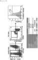
FIG. 1 is a view showing an example of a method of providing information for early diagnosis of bone metastasis of bone according to the present invention, and shows the results of quantifying circulating osteocalcin-positive (osteocalcin+) or osteocalcin-positive/N-cadherin-positive (osteocalcin+/N-cadherin+) cells in cancer patient blood by flow cytometry. -
FIG. 2 shows the preparation of a bone metastatic breast cancer mouse model and the results of bioluminescence imaging (BLI) and histopathological examination in the mouse model at 2 weeks, 3 weeks and 5 weeks. -
FIG. 3(A) is a graph showing that bone metastasis of cancer can be diagnosed fromweek 5 in BLI, but circulating osteocalcin-positive cells can be diagnosed fromweek 2 to 3, andFIG. 3(B) shows that the tumor size and the number (%) of circulating osteocalcin-positive cells are proportional to each other. -
FIG. 4 shows the results of injecting a breast cancer cell line into the left ventricle of a mouse to prepare a mouse model that is most similar to an actual bone metastatic breast cancer patient, performing bioluminescence imaging (BLI) on varying days, and analyzing calculating osteocalcin-positive cells by flow cytometry on varying days. -
FIG. 5 shows the results of preparing mouse models using different mouse species and breast cancer cells associated with the mouse species, dividing the mice into a bone metastasis group and a control group, and analyzing calculating osteocalcin-positive cells by flow cytometry. -
FIG. 6 shows the results of measuring the number of calculating osteocalcin-positive cells in metastatic breast cancer patients with or without bone metastasis. -
FIG. 7 shows the results of analyzing calculating osteocalcin-positive cells in a stable group without disease progression and a progressive group with disease progression depending on whether metastatic bone disease is active or inactive. -
FIG. 8 shows the results of determining an optimal cut-off value capable of predicting the progression of metastatic bone disease, dividing metastatic bone cancer patients into two group according to the cut-off value, and performing progression-free survival (PFS) analysis. -
FIG. 9(A) shows the results of analyzing calculating osteocalcin-positive cells in metastatic thyroid cancer patients divided into an inactive group and an active group according to whether bone metastasis is active or inactive at the time of cOC% analysis.FIG. 9(B) shows the results of analyzing calculating osteocalcin-positive cells in a stable group without disease progression and a progressive group with disease progression. - Hereinafter, the present invention will be described in detail.
- As used herein, the term "diagnosis" means identifying the presence or characteristics of a pathological condition. With regard to the purpose of the present invention, the term "diagnosis" may mean identifying the presence of bone metastasis of cancer, or confirming whether or not bone metastasis of cancer is progressing or severe.
- As used herein, the term "diagnosis" may encompass determining the susceptibility of a subject to a certain disease or disorder, determining whether a subject is affected with a certain or disorder, determining the prognosis of a subject affected with a certain or disorder, or therametrics (e.g., monitoring the state of a subject to provide information on therapeutic efficacy).
- The present invention is directed to the in vitro use of a composition comprising an agent for detecting cells released into blood expressing osteocalcin diagnosing bone metastasis of cancer.
- The present inventors have found that osteocalcin-positive cells are released into blood in the microscopic bone metastasis stage, which is an early stage of bone metastasis of cancer, to form circulating osteocalcin-positive cells, and bone metastasis of cancer may be diagnosed in an early stage by detecting the circulating osteocalcin-positive cells, thereby completing the present invention.
- The agent for detecting cells released into blood expressing osteocalcin is not limited as long as it may detect osteocalcin, but examples thereof include antibodies, aptamers, DNA, RNA, proteins, and polypeptides, which target osteocalcin, mRNA which is translated to osteocalcin, DNA which is transcribed into the mRNA, or DNA having a sequence complementary thereto.
- When the composition comprises the agent for detecting cells released into blood expressing osteocalcin, it may be used for diagnosis of bone metastasis of cancer. However, for more accurate diagnosis of bone metastasis of cancer, the composition may further comprise an agent for detecting cells released into blood expressing N-cadherin.
- The agent for detecting cells released into blood expressing N-cadherin is not limited as long as it may detect N-cadherin, but examples thereof include antibodies, aptamers, DNA, RNA, proteins, and polypeptides, which target N-cadherin, mRNA which is translated to N-cadherin, DNA which is transcribed into the mRNA, or DNA having a sequence complementary thereto.
- As used herein, the term "antibody" refers to a protein molecule specific to an antigenic site. With regard to the purposes of the present invention, the term "antibody" refers to an antibody that binds specifically to the marker protein osteocalcin or N-cadherin, and may include all monoclonal antibodies, polyclonal antibodies and recombinant antibodies.
- The monoclonal antibody may be produced using a hybridoma method well known in the art or a phage antibody library technique, but is not limited thereto.
- The polyclonal antibody may be produced by a method well known in the art, which comprises injecting the protein antigen into an animal, collecting blood from the animal, and isolating serum containing the antibody. This polyclonal antibody may be produced from any animal species hosts such as goats, rabbits, sheep, monkeys, horses, pigs, cattle, or dogs, but is not limited thereto.
- In addition, the antibodies of the present application may also include special antibodies such as chimeric antibodies, humanized antibodies, and human antibodies.
- The "peptide" has the advantage of high binding affinity for the target material, and does not denature even during thermal/chemical treatment. In addition, since the peptide has a small molecular size, it may be attached to another protein and used as a fusion protein. Specifically, since the peptide may be used attached to a polymer protein chain, it may be used in a diagnostic kit and as a drug delivery material.
- The "aptamer" refers to a kind of polynucleotide consisting of a special kind of single-stranded nucleic acid (DNA, RNA or modified nucleic acid) capable of binding to a target molecule with high affinity and specificity while having a stable three-dimensional structure. As described above, the aptamer is composed of a polynucleotide which may bind specifically to an antigenic substance in the same manner as an antibody, and at the same time, is more stable than a protein, has a simple structure, and is easy to synthesize. Thus, the aptamer may be used instead of an antibody.
- In addition, the cancer may be at least one selected from among liver cancer, lung cancer, bladder cancer, stomach cancer, breast cancer, uterine cancer, colorectal cancer, colon cancer, blood cancer, ovarian cancer, prostate cancer, pancreatic cancer, spleen cancer, testicular cancer, thymus cancer, brain cancer, esophageal cancer, kidney cancer, biliary tract cancer, thyroid cancer, or skin cancer. Preferably, the cancer may be at least one selected from among breast cancer, thyroid cancer or prostate cancer. More preferably, the cancer may be breast cancer or thyroid cancer, but is not limited thereto.
- The present invention is also directed to the in vitro use of a kit for diagnosing bone metastasis of cancer, the kit comprising the above composition.
- The kit for use of the present invention may comprise an antibody that specifically binds to a marker component, a secondary antibody conjugated with a label that develops color by reaction with a substrate, a chromogenic substrate solution for reaction with the label, and a washing solution, and an enzyme reaction stop solution, and may be made of a plurality of separate packaging or compartments including the reagent components used, but is not limited thereto.
- The kit for use of the present invention may comprise not only an agent capable of measuring the expression level of osteocalcin or N-cadherin in a patient sample, but also one or more compositions, solutions, or devices suitable for analysis of the expression level. For example, the kit may comprise a substrate, an appropriate buffer solution, a secondary antibody conjugated with a detection label, and a chromogenic substrate, for immunological detection of an antibody, but is not limited thereto.
- As a specific example, the kit may be a kit comprising essential elements necessary for performing ELISA in order to implement various ELISA methods such as an ELISA kit, a sandwich ELISA, and the like. This ELISA kit comprises an antibody specific for the protein. The antibody may be a monoclonal antibody, a polyclonal antibody or a recombinant antibody, which has high specificity and affinity for osteocalcin or N-cadherin protein and has little cross-reactivity with other proteins. In addition, the ELISA kit may comprise an antibody specific for a control protein. In addition, the ELISA kit may comprise reagents capable of detecting bound antibodies, for example, labeled secondary antibodies, chromophores, enzymes, and other substances capable of binding to their substrates or antibodies, but is not limited thereto.
- In addition, the kit may be a kit for implementing Western blot assay, immunoprecipitation assay, complement fixation assay, flow cytometry, or protein chip assay, and may further comprise additional components suitable for each assay method. Through these assay methods, it is possible to diagnose bone metastasis of cancer by comparing the amount of antigen-antibody complex formed.
- The present invention is also directed to a method of providing information for diagnosis of bone metastasis of cancer, the method comprising a step of detecting cells released into blood expressing osteocalcin in a sample isolated from a subject to be diagnosed.
- The subject to be diagnosed is an animal that currently has cancer or has a history of cancer, and the animal may be a mammal including a human being.
- The sample is isolated from the subject to be diagnosed, and examples thereof include, but are not limited to, tissue, cells, whole blood, plasma, serum, blood, or cerebrospinal fluid. Specifically, the sample may be blood, plasma, serum, or the like. More specifically, the sample may be peripheral blood mononuclear cells.
- The detection is performed to detect osteocalcin, and may be performed by treating the sample with a composition for diagnosing bone metastasis of cancer comprising an agent for detecting cells released into blood expressing osteocalcin.
- The method may further comprise detecting cells released into blood expressing N-cadherin in the sample. In this case, the detection may be performed by treating the sample with a composition for diagnosing bone metastasis of cancer comprising an agent for detecting cells released into blood expressing osteocalcin and an agent for detecting cells released into blood expressing N-cadherin.
- In terms of further increasing the accuracy of diagnosis, both osteocalcin and N-cadherin may be detected, but are not limited thereto.
- The scope of the agent for detecting cells released into blood expressing osteocalcin or the agent for detecting cells released into blood expressing N-cadherin is as described above.
- The scope of the cancer is as described above.
- The method of the present invention is intended to detect cells released into blood expressing osteocalcin in a sample isolated from a subject to be diagnosed, or to detect cells released into blood expressing osteocalcin and N-cadherin, and the detection may be performed using, for example, an antibody, peptide, aptamer or protein specific for osteocalcin or N-cadherin, but is not limited thereto.
- As a specific example, the method for detection may be performed using an antibody specific for osteocalcin or N-cadherin by a method selected from the group consisting of enzyme-linked immunosorbent assay (ELISA), radioimmunoassay (RIA), sandwich assay, Western blotting, immunoprecipitation, immunohistochemical staining, flow cytometry, fluorescence activated cell sorting (FACS), enzyme substrate colorimetric assay, antigen-antibody aggregation, and protein chip assay, but is not limited thereto.
- As an additional method, it is possible to a technique of detecting two fluorescent substances, which are at close distance, at one fluorescence wavelength using a fluorescence resonance energy transfer that creates new color development by transferring resonance energy. In addition, in order to increase the objectivity and reproducibility of tests other than flow cytometry, it is possible to use a technique comprising labeling cells with an antibody, filtering out cells having a size of 10 µm or less through a mesh filter, plating the cells on a slide, automatically scanning the slide, and then quantifying the number of positive cells by artificial intelligence.
- In addition, the method of the present invention may further comprise a step of determining that the cancer has metastasized to the bone, when cells released into blood expressing osteocalcin are detected.
- When cells released into blood expressing osteocalcin are detected in the sample from the subject to be diagnosed, it may be determined that the cancer has metastasized to the bone, and as the amount of osteocalcin detected increases, it may be determined that the bone metastasis stage of the cancer has further progressed.
- When the method of the present invention further comprises the step of detecting cells released into blood expressing N-cadherin, if N-cadherin is also detected, bone metastasis of cancer may be diagnosed with higher accuracy.
- If necessary, the method of the present invention may further comprise a step of treating the sample, isolated from the subject to be diagnosed, with vitamin K.
- The vitamin K may be vitamin K1 or vitamin K2.
- Treatment of the sample with vitamin K may γ-carboxylate osteocalcin and increase and stabilize the expression of osteocalcin, so that osteocalcin may be more easily detected.
- The treatment concentration of vitamin K is not particularly limited, and may be, for example, 10-5 M to 10-7 M. Specifically, the treatment concentration of each of vitamin K1 and vitamin K2 may be 10-5 M to 10-7 M. At the above-described treatment concentration, the osteocalcin expression effect of circulating osteocalcin-positive cells may be maximized.
- Further described but not claimed is a method of diagnosing bone metastasis of cancer by detecting osteocalcin in a sample isolated from a subject to be diagnosed.
- The scope of the sample is as described above.
- The detection may be performed by treating the sample with an agent for detecting osteocalcin.
- The agent for detecting osteocalcin is as described above.
- When osteocalcin is detected, it may be determined that the cancer has metastasized to the bone. For a more specific example, bone metastasis of cancer may be diagnosed by measuring the number (%) of circulating osteocalcin-positive cells in the sample. Specifically, bone metastasis of cancer may be diagnosed by comparing the number of circulating osteocalcin-positive cells in a patient having bone metastasis with the number of circulating osteocalcin-positive cells in a patient having no bone metastasis, but is not limited thereto.
- In addition, the method may further comprise a step of detecting N-cadherin in the sample.
- This step may be performed by treating the sample with an agent for detecting N-cadherin, and the scope of the agent is as described above.
- When N-cadherin is detected, bone metastasis of cancer may be diagnosed with higher accuracy. Specifically, it is possible to detect circulating osteocalcin-positive cells in the sample, and then detect circulating N-cadherin-positive cells, but is not limited thereto.
- Hereinafter, the present invention will be described in detail with reference to examples.
- For flow cytometry of circulating osteocalcin-positive cells in blood samples isolated from cancer patients, each sample was collected via liquid biopsy, and peripheral blood mononuclear cells were isolated from the sample. 4 cc of the blood sample collected from the patient to be diagnosed was placed in a heparin tube containing Ficoll (Pharmacia) and was centrifuged at a speed of 1800 rpm at a temperature of 4°C for 20 minutes to separate the blood into plasma, a mononuclear cell layer, Ficoll and an RBC layer. At this time, only the mononuclear cell layer (second layer) at the interface between the separated plasma and Ficoll was isolated carefully and transferred into a fresh tube. Next, the mononuclear cells were washed with PBS solution and then centrifuged at a speed of 2000 rpm at a temperature of 4°C for 5 minutes, and only cleanly washed mononuclear cells were collected.
- 106 washed mononuclear cells were placed in 500 µl of FBS-containing buffer and stained by incubation with anti-CD15-FITC (5 µl), anti-CD34-PE-Cy7 (5 µl), anti-osteocalcin-APC (3 µl) and anti-N-cadherin-PE (5 µl) antibodies at room temperature for 30 minutes. (Information on antibodies used: a-CD15 FITC (#555401,BD), a-CD34 PE-Cy7 (#34881, BD), a-osteocalcin APC (#SC-365797-AF647, Santa Cruz), and a-N-cadherin PE (#561554, BD) were purchased and used).
- After completion of staining, non-specifically bound and unbound antibodies were washed out by adding 4 ml of FBS-containing buffer, and antibody-labeled mononuclear cells were obtained by concentration at a speed of 1500 rpm at a temperature of 4°C for 5 minutes. The above-described cancer patient blood samples were obtained according to regulations under approval of the IRB of the Clinical Research Center of Seoul National University Hospital for use in clinical research in the present invention.
- The stained mononuclear cells were analyzed by flow cytometry (BD FACSCanto II). The mononuclear cells stained with the above-listed antibodies were analyzed at the single-cell level, and first sorted into CD15- cell populations, and re-sorted into CD34- and osteocalcin+ cell populations. The number of the cells was quantified, and circulating osteocalcin-positive cells were analyzed. In addition, the number of N-cadherin+ cells in the corresponding population was analyzed, and finally, circulating CD15-/CD34-/osteocalcin+/N-cadherin+ cells specific to metastatic bone cancer were quantified using
Equation 1 below. Analyses of circulating osteocalcin-positive cells in samples obtained from a total of 108 cancer patients (98 breast cancer cells and 10 thyroid cancer patients) were performed, and the results of representative analysis are shown inFIG. 1 .
- In flow cytometry of the cancer patient blood sample, a total of 1,000,000 cells were analyzed, and among them, 932,559 cells excluding dead cells were sorted. Subsequently, 881,302 CD15- cells were re-sorted, from which 1,252 osteocalcin+/CD34- cells were then sorted. Among 1,252 cells, 869 N-cadherin+ cells were sorted. The results are shown in
FIG. 1 . - For analysis of circulating osteocalcin-positive cells in a preclinical study, a mouse model was prepared, which facilitates analysis of the bone marrow environments and circulating osteocalcin-positive cells of actual patients with metastatic bone cancer. All animal experiments were conducted in accordance with strict regulations under approval of the Seoul National University Animal Experimental Ethics Committee (SNU-170607-6-2). An ideal animal model similar to metastatic bone cancer patients was prepared by injecting a human breast cancer cell line directly into the tibia of each immunodeficient mouse (Balb/c nude mouse). Specifically, the human breast cancer cell line MDA-MB-231 was prepared in PBS (buffer injectable into the mouse body) in an amount of 1×10⁵ cells/20 µl/mouse, and injected into the right tibia of each nude mouse. At 2, 3 and 5 weeks after injection of the cancer cells, formation and growth of tumors in the tibia was observed through in vivo imaging (Xenogen IVIS, PerkinElmer). In order to confirm substantial tumor formation at each week, the mouse was euthanized, the tumor-formed tibia was isolated, and a process of preparing a sample for histological analysis was performed. The isolated tissue was fixed by cold storage in 4% para-formaldehyde solution for 3 days, and then the muscle portion was removed from the bone which was then decalcified in 0.5M EDTA solution for about 2 weeks. (During the decalcification process, the tissue was cold-stored, and 0.5 M EDTA solution was replaced every 3 days). Then, the tissue was embedded in paraffin and sectioned, and the paraffin sections were placed on slides and stained with H&E. The results of in vivo imaging and tissue staining are shown in
FIG. 2 . - As a result of performing BLI imaging of the prepared bone metastatic breast cancer mouse model at each week, it could be confirmed that tumor formation and growth could be found even at 5 weeks. In addition, the results of histopathological examination were consistent with the results of BLI imaging. At 3 weeks, it was observed that a very small tumor was formed in the tibia, and at 5 weeks, it could be observed that a large tumor was formed in the tibia. These results suggest that a microscopic tumor is difficult to diagnose by imaging, and the tumor can be diagnosed when it has grown significantly for 5 weeks or more (
FIG. 2 ). - In order to analyze changes in circulating osteocalcin-positive cells depending on the tumor size in the metastatic bone cancer mouse model prepared in Example 2, blood sampling was performed. Specifically, the mouse was anesthetized and about 1 ml of whole blood was sampled from the mouse through cardiac puncture. In order to prevent blood clotting during blood collection, the inside of the syringe was coated with a heparin solution before blood sampling, and the sampled blood was added to a red blood cell lysis solution immediately after sampling and subjected to a red blood cell lysis process. The sample was left to stand at room temperature for 15 minutes, and then centrifuged at 1500 rpm for 5 minutes, and the supernatant was removed. This process was repeated until the red blood cells were completely removed, and cells from which the red blood cells have been completely removed were added to a buffer containing FBS. Thereafter, the cells were stained with anti-CD45-PE (#561087, BD) and the anti-osteocalcin antibody described in Example 1 above, and circulating CD45+/osteocalcin+ cells were analyzed by flow cytometry. The intensity of in vivo bioluminescence imaging (ph/s) and the number (%) of circulating osteocalcin-positive cells (cOC) as a function of the tumor size were compared. The results are shown in
FIG. 3 . - In the metastatic bone cancer mouse model prepared using breast cancer cells, it could be seen that circulating osteocalcin-positive cells (cOC) increased before diagnosis in the imaging technique, and that tumor cells could be detected by histopathological analysis when these cells started to form microclusters on the bone surface (see
FIG. 2 ). It could be confirmed that bone metastasis of cancer could be diagnosed at 5 weeks in conventional BLI imaging, but could be diagnosed at 2 to 3 weeks according to the present invention (FIG. 3(A) ). - In addition, in the metastatic bone cancer mouse model prepared using breast cancer cells, circulating osteocalcin-positive cells (cOC) showed a good correlation with the tumor size measured by pathological analysis that can be considered as a gold standard (
FIG. 3(B) ). - As a mouse model different from the bone metastasis mouse model prepared by injecting cancer cells into the tibia, a mouse model was prepared by injecting breast cancer cells directly into the left ventricle of each mouse in order to conduct a preclinical study in an environment which is most similar to an actual bone metastatic breast cancer patient. Human breast cancer MDA-MB-231 cells were injected directly into the left ventricle of each immunodeficient mouse (Balb/c nude mice) to establish a mouse model in which the cancer cells metastasized to the tibia or femur after systemic circulation.
- Specifically, the human breast cancer line MDA-MB-231 was prepared in PBS (buffer injectable into the mouse body) in an amount of 1×10⁵ cells/100 µl/mouse, and injected directly into the left ventricle of each nude mouse. On 3, 7, 14 and 22 days after injection of the cancer cells, formation and growth of the tumor that metastasized to the mouse bone was observed through in vivo imaging (Xenogen IVIS, PerkinElmer). In addition, a region of interest (ROI) was set and analyzed, and the growth of the tumor that metastasized to the mouse bone was numerically analyzed. On the above-described days, each mouse was euthanized, whole blood was sampled from the mouse, PBMCs were isolated therefrom, and circulating osteocalcin-positive cells therein were analyzed. The blood sampling and PBMC isolation process, and antibody staining for flow cytometry are as described in Example 3 above.
- The results of in vivo imaging and circulating osteocalcin-positive cell are shown in
FIG. 4 . - As a result of preparing the microscopic bone metastatic breast cancer mouse model and performing BLI imaging of the mouse model on varying days in the early stage of bone metastasis, it could be confirmed that bone metastasis could be diagnosed on 14 days after injection of the cancer cells and that significantly analysis results for the ROI value could be obtained only on 22 days after cell injection. However, the results of analysis of circulating osteocalcin-positive cells indicated that the circulating osteocalcin-positive cells already increased on
day 7 when bone metastasis could not be detected by BLI imaging. These results are consistent with the results obtained in the model prepared by injecting cancer cells directly into the tibia, and suggest that microscopic metastatic bone tumors are difficult to diagnose by BLI imaging, but early diagnosis thereof is possible by analysis of circulating osteocalcin-positive cells (FIG. 4 ). - To analyze circulating osteocalcin-positive cells depending on the presence or absence of bone metastasis, the same microscopic bone metastatic breast cancer model as that in Example 4 was prepared by injecting cancer cells into the left ventricle. The mouse model was prepared using a breast cancer cell line derived from the corresponding mouse species in other mouse species, in order to demonstrate that the quantitative increase in circulating osteocalcin-positive cells is not a mouse species-specific response or a specific breast cancer cell line response. Specifically, the Balb/c mouse-derived breast cancer cell line 4T1 was prepared in PBS (buffer injectable into the mouse body) in an amount of 1×10⁴ cells/100 µl/mouse, and injected directly into the left ventricle of each Balb/c mouse. On 7 days after injection of the cancer cells, the mice were divided into a group with bone metastasis and a group without bone metastasis through BLI imaging, and the intensity of luminescence between the bone metastasis group and the control group was quantified through ROI analysis. Thereafter, the mice were sacrificed, whole blood was collected therefrom, and circulating osteocalcin-positive osteocalcin cells therein were analyzed in the same manner as described in the above Example. In addition, the correlation between the results of BLI analysis and the results of circulating osteocalcin-positive cell analysis was analyzed.
- The results are shown in
FIG. 5 . - As a result of analyzing circulating osteocalcin-positive cells in the bone metastasis group and the control group, a significant quantitative increase in circulating osteocalcin-positive cells in the bone metastasis group compared to that in the control group could be confirmed. In addition, as a result of analyzing the correlation between the result of BLI imaging and the quantitative increase in circulating osteocalcin-positive cells, it was demonstrated that the quantitative increase in circulating osteocalcin-positive cells had a significant correlation with the presence of bone metastasis. This proves that circulating osteocalcin-positive cells may serve as a marker not only for early diagnosis of microscopic bone metastatic breast cancer, but also capable of detecting the presence of bone metastasis (
FIG. 5 ) . - In order to evaluate whether circulating osteocalcin-positive cells present in the blood of actual patients with metastatic bone cancer have clinical diagnostic value by analyzing the circulating osteocalcin-positive cells, the number (%) of circulating osteocalcin-positive cells was measured on 96 breast cancer patients with metastatic bone cancer. Patient's blood sampling, mononuclear cell isolation and cell staining processes were performed in the same manner as described in Example 1 above, and the analysis method was also performed in the same manner. The results are shown in
FIG. 6 . - In order to evaluate additional clinical value, various analysis methods were performed. At the time of cOC% analysis and after continuous follow-up, the patients were divided into a group with active metastatic bone disease and a group with inactive disease and analyzed. The results are shown in
FIG. 7 . In addition, based on the results of the follow-up analysis, an optimal cut-off value capable of predicting the progression of metastatic bone disease was determined, and based on the cut-off value, progression-free survival (PFS probability) analysis was performed. The results are shown inFIG. 8 . -
- 1. Among 96 metastatic breast cancer patients, 63 patients with bone metastasis had significant high cOC% compared to 33 patients without bone metastasis (
FIG. 6 ). - 2. When the patients with bone metastasis were divided into an inactive group of 26 patients and an active group of 37 patients according to whether the metastatic bone disease was active or inactive at the time of cOC% analysis, there was no difference in cOC% between the two groups. However, when the patients were divided into a stable group without progression of metastatic bone disease and a progressive group with progression of metastatic bone disease after 15-month follow-up, it could be confirmed that, in both the inactive group and the active group, the baseline cOC% of the progressive group was significantly higher than that of the stable group (
FIG. 7 ). - 3. The optimal cut-off value enabling cOC% to predict the progression of the metastatic bone disease by cOC% could be determined as 0.45% (
FIG. 8(A) ). When the patients with metastatic bone cancer were divided into two groups based on 0.45%, it could be confirmed that there was a significant difference in progression-free survival between the two groups (FIG. 8(B) ). - In order to evaluate whether circulating osteocalcin-positive cells present in the blood of actual patients with metastatic bone cancer have clinical diagnostic value by analyzing the circulating osteocalcin-positive cells, the number (%) of circulating osteocalcin-positive cells was measured on 14 thyroid cancer patients with metastatic bone cancer. Patient's blood sampling, mononuclear cell isolation and cell staining processes were performed in the same manner as described in Example 1 above. At the time of cOC% analysis and after continuous follow-up, the patients were divided into an active group with progression of metastatic bone disease and an inactive group without progression of metastatic bone disease and analyzed. The results are shown in
FIG. 9 . -
- 1. Among 14 metastatic thyroid cancer patients, 12 patients with bone metastasis were divided into an inactive group of 5 patients and an active group of 7 patients depending on whether the metastatic bone disease was active or inactive at the time of cOC% analysis, and as a result, the cOC% of the active group was significantly higher than that of the inactive group (
FIG. 9 (A) ). - 2. When the patients were divided into a stable group without progression of metastatic bone disease and a progressive group with progression of metastatic bone disease after 6-month follow-up, the baseline cOC% of the progressive group was higher than that of the stable group, but this difference was not statistically significant (
FIG. 9(B) ).
Claims (11)
- In vitro use of a composition comprising an agent for detecting cells released into blood expressing osteocalcin for diagnosing bone metastasis of cancer.
- The use of claim 1, wherein the composition further comprises an agent for detecting cells released into blood expressing N-cadherin.
- The use of claim 1, wherein the agent for detecting cells released into blood expressing osteocalcin is at least one selected from the group consisting of antibodies, aptamers, DNA, RNA, proteins, and polypeptides.
- The use of claim 2, wherein the agent for detecting cells released into blood expressing osteocalcin and the agent for detecting blood-derived cells expressing N-cadherin are each independently at least one selected from the group consisting of antibodies, aptamers, DNA, RNA, proteins, and polypeptides.
- The use of claim 1, wherein the cancer is at least one selected from among liver cancer, lung cancer, bladder cancer, stomach cancer, breast cancer, uterine cancer, colorectal cancer, colon cancer, blood cancer, ovarian cancer, prostate cancer, pancreatic cancer, spleen cancer, testicular cancer, thymus cancer, brain cancer, esophageal cancer, kidney cancer, biliary tract cancer, thyroid cancer, or skin cancer.
- The use of claim 1, wherein the cancer is breast cancer or thyroid cancer.
- In vitro use of a kit for diagnosing bone metastasis of cancer, the kit comprising the composition used according to any one of claims 1 to 6.
- A method of providing information for diagnosis of bone metastasis of cancer, the method comprising a step of detecting cells released into blood expressing osteocalcin in a sample isolated from a subject to be diagnosed.
- The method of claim 8, further comprising a step of detecting cells released into blood expressing N-cadherin in the sample.
- The method of claim 8, wherein the cancer is at least one selected from among liver cancer, lung cancer, bladder cancer, stomach cancer, breast cancer, uterine cancer, colorectal cancer, colon cancer, blood cancer, ovarian cancer, prostate cancer, pancreatic cancer, spleen cancer, testicular cancer, thymus cancer, brain cancer, esophageal cancer, kidney cancer, biliary tract cancer, thyroid cancer, or skin cancer.
- The method of claim 8, wherein the cancer is breast cancer or thyroid cancer.
Applications Claiming Priority (2)
| Application Number | Priority Date | Filing Date | Title |
|---|---|---|---|
| KR20180090914 | 2018-08-03 | ||
| PCT/KR2019/009139 WO2020027484A1 (en) | 2018-08-03 | 2019-07-24 | Composition for diagnosis of bone metastasis of cancer and kit comprising same |
Publications (4)
| Publication Number | Publication Date |
|---|---|
| EP3832309A1 EP3832309A1 (en) | 2021-06-09 |
| EP3832309A4 EP3832309A4 (en) | 2021-10-13 |
| EP3832309B1 true EP3832309B1 (en) | 2023-11-01 |
| EP3832309C0 EP3832309C0 (en) | 2023-11-01 |
Family
ID=69569452
Family Applications (1)
| Application Number | Title | Priority Date | Filing Date |
|---|---|---|---|
| EP19844633.8A Active EP3832309B1 (en) | 2018-08-03 | 2019-07-24 | Composition for diagnosis of bone metastasis of cancer and kit comprising same |
Country Status (4)
| Country | Link |
|---|---|
| EP (1) | EP3832309B1 (en) |
| KR (1) | KR102116691B1 (en) |
| CN (1) | CN112384806B (en) |
| ES (1) | ES2967857T3 (en) |
Families Citing this family (1)
| Publication number | Priority date | Publication date | Assignee | Title |
|---|---|---|---|---|
| KR102396511B1 (en) * | 2020-06-04 | 2022-05-11 | 서울대학교병원 | Composition for monitoring the treatment efficacy of cancer bone metastasis and kit comprising the same |
Family Cites Families (8)
| Publication number | Priority date | Publication date | Assignee | Title |
|---|---|---|---|---|
| JP2845347B2 (en) * | 1990-10-04 | 1999-01-13 | 帝人株式会社 | Propeptide of osteocalcin, method and kit for immunologically measuring proosteocalcin |
| US6004765A (en) * | 1992-02-27 | 1999-12-21 | Delmas; Pierre | Assessment of bone fragility and prediction of osteoporotic fracture risk using a quantitative determination of circulating under-carboxylated osteocalcin |
| US5772993A (en) | 1997-01-21 | 1998-06-30 | The University Of Virginia Patent Foundation | Osteocalcin promoter-based toxic gene therapy for the treatment of calcified tumors and tissues |
| EP1107006B1 (en) | 1998-08-21 | 2008-05-14 | Ogata, Etsuro | Method of diagnosing bone metastasis of malignant tumors |
| AU2001249064A1 (en) * | 2000-02-22 | 2001-09-03 | Mount Sinai School Of Medicine Of New York University | N-cadherin modulated migration, invasion, and metastasis |
| US8663635B2 (en) | 2006-03-21 | 2014-03-04 | The Regents Of The University Of California | N-cadherin: target for cancer diagnosis and therapy |
| WO2011116351A2 (en) * | 2010-03-19 | 2011-09-22 | University Of South Alabama | Methods and compositions for the treatment of cancer |
| KR101821455B1 (en) | 2015-08-27 | 2018-01-25 | 울산대학교 산학협력단 | Biomarker composition for diagnosing cancer metastasis comprising AWP1 |
-
2019
- 2019-07-24 CN CN201980045995.4A patent/CN112384806B/en active Active
- 2019-07-24 ES ES19844633T patent/ES2967857T3/en active Active
- 2019-07-24 EP EP19844633.8A patent/EP3832309B1/en active Active
- 2019-08-05 KR KR1020190095204A patent/KR102116691B1/en active Active
Also Published As
| Publication number | Publication date |
|---|---|
| KR20200015432A (en) | 2020-02-12 |
| CN112384806A (en) | 2021-02-19 |
| KR102116691B1 (en) | 2020-05-29 |
| EP3832309A4 (en) | 2021-10-13 |
| CN112384806B (en) | 2024-07-09 |
| ES2967857T3 (en) | 2024-05-06 |
| EP3832309A1 (en) | 2021-06-09 |
| EP3832309C0 (en) | 2023-11-01 |
Similar Documents
| Publication | Publication Date | Title |
|---|---|---|
| Hayes et al. | Monitoring expression of HER-2 on circulating epithelial cells in patients with advanced breast cancer | |
| Balic et al. | Circulating tumor cells: from bench to bedside | |
| JP6599334B2 (en) | Methods and assays for circulating tumor cells in the blood | |
| KR101976219B1 (en) | Biomarker for breast cancer | |
| EP2529223B1 (en) | Biomarkers for circulating tumor cells | |
| US20110091384A1 (en) | Biomarker for identification of melanoma tumor cells | |
| RU2641968C2 (en) | Compositions and methods for prostate cancer analysis | |
| JP2013543117A (en) | Means and methods for the diagnosis of cancer using antibodies that specifically bind to BRAFV600E | |
| KR20170141951A (en) | Biomarker composition for diagnosing colon cancer or prognosing metastasis of colon cancer comprising NCKAP1 | |
| KR102521638B1 (en) | A marker for diagnosis of nonalcoholic fatty liver disease | |
| EP3832309A1 (en) | Composition for diagnosis of bone metastasis of cancer and kit comprising same | |
| US9746472B2 (en) | Methods and kits for the detection of cancer infiltration of the central nervous system | |
| US20210263038A1 (en) | Composition for diagnosis of bone metastasis of cancer and kit comprising same | |
| KR102396511B1 (en) | Composition for monitoring the treatment efficacy of cancer bone metastasis and kit comprising the same | |
| JP7012363B2 (en) | Biomarkers for predicting the therapeutic effect of FSTL1 inhibitors in cancer patients | |
| KR102735773B1 (en) | A kit for diagnosing cancer comprising protein biomarker in blood | |
| KR101567053B1 (en) | Biomarkers for predicting liver damage caused by radiation exposure and a predicting method thereof | |
| WO2015077691A2 (en) | Biomarkers for multiple myeloma | |
| KR102363980B1 (en) | Biomarker for diagnosis or prognosis analysis of brain metastasis and diagnosis method using same | |
| KR101219979B1 (en) | A Method for Diagnosing the Risk of Lymph Node Metastasis in Papillary Thyroid Carcinoma | |
| KR102180623B1 (en) | Composition for diagnosing cancer bone metastasis and method for diagnosing cancer bone metastasis using the same | |
| JP2025513326A (en) | Method for predicting prognosis of breast cancer patients using c-Met-expressing CTCs | |
| Zeng et al. | CRISPR/Cas12a Integrated with a Microfluidic System Enhanced Analysis of Programmed Cell Death Ligand 1 Expression in Circulating Tumor Cells from Non-Small Cell Lung Cancer Patients | |
| CN117043604A (en) | Diagnostic markers for non-alcoholic fatty liver disease | |
| WO2025121385A1 (en) | Technique for predicting pathology, prognosis, and therapeutic effect of t-cell-related disease |
Legal Events
| Date | Code | Title | Description |
|---|---|---|---|
| STAA | Information on the status of an ep patent application or granted ep patent |
Free format text: STATUS: THE INTERNATIONAL PUBLICATION HAS BEEN MADE |
|
| PUAI | Public reference made under article 153(3) epc to a published international application that has entered the european phase |
Free format text: ORIGINAL CODE: 0009012 |
|
| STAA | Information on the status of an ep patent application or granted ep patent |
Free format text: STATUS: REQUEST FOR EXAMINATION WAS MADE |
|
| 17P | Request for examination filed |
Effective date: 20201221 |
|
| AK | Designated contracting states |
Kind code of ref document: A1 Designated state(s): AL AT BE BG CH CY CZ DE DK EE ES FI FR GB GR HR HU IE IS IT LI LT LU LV MC MK MT NL NO PL PT RO RS SE SI SK SM TR |
|
| A4 | Supplementary search report drawn up and despatched |
Effective date: 20210915 |
|
| RIC1 | Information provided on ipc code assigned before grant |
Ipc: G01N 33/574 20060101AFI20210909BHEP |
|
| DAV | Request for validation of the european patent (deleted) | ||
| DAX | Request for extension of the european patent (deleted) | ||
| STAA | Information on the status of an ep patent application or granted ep patent |
Free format text: STATUS: EXAMINATION IS IN PROGRESS |
|
| 17Q | First examination report despatched |
Effective date: 20221121 |
|
| GRAP | Despatch of communication of intention to grant a patent |
Free format text: ORIGINAL CODE: EPIDOSNIGR1 |
|
| STAA | Information on the status of an ep patent application or granted ep patent |
Free format text: STATUS: GRANT OF PATENT IS INTENDED |
|
| INTG | Intention to grant announced |
Effective date: 20230526 |
|
| GRAS | Grant fee paid |
Free format text: ORIGINAL CODE: EPIDOSNIGR3 |
|
| GRAA | (expected) grant |
Free format text: ORIGINAL CODE: 0009210 |
|
| STAA | Information on the status of an ep patent application or granted ep patent |
Free format text: STATUS: THE PATENT HAS BEEN GRANTED |
|
| AK | Designated contracting states |
Kind code of ref document: B1 Designated state(s): AL AT BE BG CH CY CZ DE DK EE ES FI FR GB GR HR HU IE IS IT LI LT LU LV MC MK MT NL NO PL PT RO RS SE SI SK SM TR |
|
| REG | Reference to a national code |
Ref country code: GB Ref legal event code: FG4D |
|
| REG | Reference to a national code |
Ref country code: CH Ref legal event code: EP |
|
| REG | Reference to a national code |
Ref country code: IE Ref legal event code: FG4D |
|
| REG | Reference to a national code |
Ref country code: DE Ref legal event code: R096 Ref document number: 602019040741 Country of ref document: DE |
|
| U01 | Request for unitary effect filed |
Effective date: 20231101 |
|
| U07 | Unitary effect registered |
Designated state(s): AT BE BG DE DK EE FI FR IT LT LU LV MT NL PT SE SI Effective date: 20231106 |
|
| U1N | Appointed representative for the unitary patent procedure changed after the registration of the unitary effect |
Representative=s name: KRAUS & WEISERT PATENTANWAELTE PARTGMBB; DE |
|
| PG25 | Lapsed in a contracting state [announced via postgrant information from national office to epo] |
Ref country code: GR Free format text: LAPSE BECAUSE OF FAILURE TO SUBMIT A TRANSLATION OF THE DESCRIPTION OR TO PAY THE FEE WITHIN THE PRESCRIBED TIME-LIMIT Effective date: 20240202 |
|
| PG25 | Lapsed in a contracting state [announced via postgrant information from national office to epo] |
Ref country code: IS Free format text: LAPSE BECAUSE OF FAILURE TO SUBMIT A TRANSLATION OF THE DESCRIPTION OR TO PAY THE FEE WITHIN THE PRESCRIBED TIME-LIMIT Effective date: 20240301 |
|
| PG25 | Lapsed in a contracting state [announced via postgrant information from national office to epo] |
Ref country code: IS Free format text: LAPSE BECAUSE OF FAILURE TO SUBMIT A TRANSLATION OF THE DESCRIPTION OR TO PAY THE FEE WITHIN THE PRESCRIBED TIME-LIMIT Effective date: 20240301 Ref country code: GR Free format text: LAPSE BECAUSE OF FAILURE TO SUBMIT A TRANSLATION OF THE DESCRIPTION OR TO PAY THE FEE WITHIN THE PRESCRIBED TIME-LIMIT Effective date: 20240202 |
|
| REG | Reference to a national code |
Ref country code: ES Ref legal event code: FG2A Ref document number: 2967857 Country of ref document: ES Kind code of ref document: T3 Effective date: 20240506 |
|
| PG25 | Lapsed in a contracting state [announced via postgrant information from national office to epo] |
Ref country code: RS Free format text: LAPSE BECAUSE OF FAILURE TO SUBMIT A TRANSLATION OF THE DESCRIPTION OR TO PAY THE FEE WITHIN THE PRESCRIBED TIME-LIMIT Effective date: 20231101 Ref country code: PL Free format text: LAPSE BECAUSE OF FAILURE TO SUBMIT A TRANSLATION OF THE DESCRIPTION OR TO PAY THE FEE WITHIN THE PRESCRIBED TIME-LIMIT Effective date: 20231101 Ref country code: NO Free format text: LAPSE BECAUSE OF FAILURE TO SUBMIT A TRANSLATION OF THE DESCRIPTION OR TO PAY THE FEE WITHIN THE PRESCRIBED TIME-LIMIT Effective date: 20240201 Ref country code: HR Free format text: LAPSE BECAUSE OF FAILURE TO SUBMIT A TRANSLATION OF THE DESCRIPTION OR TO PAY THE FEE WITHIN THE PRESCRIBED TIME-LIMIT Effective date: 20231101 |
|
| PG25 | Lapsed in a contracting state [announced via postgrant information from national office to epo] |
Ref country code: CZ Free format text: LAPSE BECAUSE OF FAILURE TO SUBMIT A TRANSLATION OF THE DESCRIPTION OR TO PAY THE FEE WITHIN THE PRESCRIBED TIME-LIMIT Effective date: 20231101 |
|
| PG25 | Lapsed in a contracting state [announced via postgrant information from national office to epo] |
Ref country code: SK Free format text: LAPSE BECAUSE OF FAILURE TO SUBMIT A TRANSLATION OF THE DESCRIPTION OR TO PAY THE FEE WITHIN THE PRESCRIBED TIME-LIMIT Effective date: 20231101 |
|
| PG25 | Lapsed in a contracting state [announced via postgrant information from national office to epo] |
Ref country code: SM Free format text: LAPSE BECAUSE OF FAILURE TO SUBMIT A TRANSLATION OF THE DESCRIPTION OR TO PAY THE FEE WITHIN THE PRESCRIBED TIME-LIMIT Effective date: 20231101 Ref country code: SK Free format text: LAPSE BECAUSE OF FAILURE TO SUBMIT A TRANSLATION OF THE DESCRIPTION OR TO PAY THE FEE WITHIN THE PRESCRIBED TIME-LIMIT Effective date: 20231101 Ref country code: CZ Free format text: LAPSE BECAUSE OF FAILURE TO SUBMIT A TRANSLATION OF THE DESCRIPTION OR TO PAY THE FEE WITHIN THE PRESCRIBED TIME-LIMIT Effective date: 20231101 |
|
| U20 | Renewal fee for the european patent with unitary effect paid |
Year of fee payment: 6 Effective date: 20240624 |
|
| REG | Reference to a national code |
Ref country code: DE Ref legal event code: R097 Ref document number: 602019040741 Country of ref document: DE |
|
| PLBE | No opposition filed within time limit |
Free format text: ORIGINAL CODE: 0009261 |
|
| STAA | Information on the status of an ep patent application or granted ep patent |
Free format text: STATUS: NO OPPOSITION FILED WITHIN TIME LIMIT |
|
| 26N | No opposition filed |
Effective date: 20240802 |
|
| PG25 | Lapsed in a contracting state [announced via postgrant information from national office to epo] |
Ref country code: MC Free format text: LAPSE BECAUSE OF FAILURE TO SUBMIT A TRANSLATION OF THE DESCRIPTION OR TO PAY THE FEE WITHIN THE PRESCRIBED TIME-LIMIT Effective date: 20231101 |
|
| PGFP | Annual fee paid to national office [announced via postgrant information from national office to epo] |
Ref country code: GB Payment date: 20250624 Year of fee payment: 7 |
|
| PG25 | Lapsed in a contracting state [announced via postgrant information from national office to epo] |
Ref country code: IE Free format text: LAPSE BECAUSE OF NON-PAYMENT OF DUE FEES Effective date: 20240724 |
|
| U20 | Renewal fee for the european patent with unitary effect paid |
Year of fee payment: 7 Effective date: 20250723 |
|
| PGFP | Annual fee paid to national office [announced via postgrant information from national office to epo] |
Ref country code: ES Payment date: 20250822 Year of fee payment: 7 |
|
| PGFP | Annual fee paid to national office [announced via postgrant information from national office to epo] |
Ref country code: CH Payment date: 20250801 Year of fee payment: 7 |
|
| PG25 | Lapsed in a contracting state [announced via postgrant information from national office to epo] |
Ref country code: RO Free format text: LAPSE BECAUSE OF FAILURE TO SUBMIT A TRANSLATION OF THE DESCRIPTION OR TO PAY THE FEE WITHIN THE PRESCRIBED TIME-LIMIT Effective date: 20231101 |
|
| PG25 | Lapsed in a contracting state [announced via postgrant information from national office to epo] |
Ref country code: CY Free format text: LAPSE BECAUSE OF FAILURE TO SUBMIT A TRANSLATION OF THE DESCRIPTION OR TO PAY THE FEE WITHIN THE PRESCRIBED TIME-LIMIT; INVALID AB INITIO Effective date: 20190724 |
|
| PG25 | Lapsed in a contracting state [announced via postgrant information from national office to epo] |
Ref country code: HU Free format text: LAPSE BECAUSE OF FAILURE TO SUBMIT A TRANSLATION OF THE DESCRIPTION OR TO PAY THE FEE WITHIN THE PRESCRIBED TIME-LIMIT; INVALID AB INITIO Effective date: 20190724 |