EP3826024A1 - Apparatus for diagnostic image acquisition determination - Google Patents
Apparatus for diagnostic image acquisition determination Download PDFInfo
- Publication number
- EP3826024A1 EP3826024A1 EP19210005.5A EP19210005A EP3826024A1 EP 3826024 A1 EP3826024 A1 EP 3826024A1 EP 19210005 A EP19210005 A EP 19210005A EP 3826024 A1 EP3826024 A1 EP 3826024A1
- Authority
- EP
- European Patent Office
- Prior art keywords
- data value
- patient
- threshold data
- diagnostic image
- determination
- Prior art date
- Legal status (The legal status is an assumption and is not a legal conclusion. Google has not performed a legal analysis and makes no representation as to the accuracy of the status listed.)
- Withdrawn
Links
Images


Classifications
-
- G—PHYSICS
- G16—INFORMATION AND COMMUNICATION TECHNOLOGY [ICT] SPECIALLY ADAPTED FOR SPECIFIC APPLICATION FIELDS
- G16H—HEALTHCARE INFORMATICS, i.e. INFORMATION AND COMMUNICATION TECHNOLOGY [ICT] SPECIALLY ADAPTED FOR THE HANDLING OR PROCESSING OF MEDICAL OR HEALTHCARE DATA
- G16H20/00—ICT specially adapted for therapies or health-improving plans, e.g. for handling prescriptions, for steering therapy or for monitoring patient compliance
- G16H20/10—ICT specially adapted for therapies or health-improving plans, e.g. for handling prescriptions, for steering therapy or for monitoring patient compliance relating to drugs or medications, e.g. for ensuring correct administration to patients
-
- G—PHYSICS
- G06—COMPUTING OR CALCULATING; COUNTING
- G06T—IMAGE DATA PROCESSING OR GENERATION, IN GENERAL
- G06T7/00—Image analysis
- G06T7/0002—Inspection of images, e.g. flaw detection
- G06T7/0012—Biomedical image inspection
-
- G—PHYSICS
- G16—INFORMATION AND COMMUNICATION TECHNOLOGY [ICT] SPECIALLY ADAPTED FOR SPECIFIC APPLICATION FIELDS
- G16H—HEALTHCARE INFORMATICS, i.e. INFORMATION AND COMMUNICATION TECHNOLOGY [ICT] SPECIALLY ADAPTED FOR THE HANDLING OR PROCESSING OF MEDICAL OR HEALTHCARE DATA
- G16H20/00—ICT specially adapted for therapies or health-improving plans, e.g. for handling prescriptions, for steering therapy or for monitoring patient compliance
- G16H20/10—ICT specially adapted for therapies or health-improving plans, e.g. for handling prescriptions, for steering therapy or for monitoring patient compliance relating to drugs or medications, e.g. for ensuring correct administration to patients
- G16H20/17—ICT specially adapted for therapies or health-improving plans, e.g. for handling prescriptions, for steering therapy or for monitoring patient compliance relating to drugs or medications, e.g. for ensuring correct administration to patients delivered via infusion or injection
-
- G—PHYSICS
- G16—INFORMATION AND COMMUNICATION TECHNOLOGY [ICT] SPECIALLY ADAPTED FOR SPECIFIC APPLICATION FIELDS
- G16H—HEALTHCARE INFORMATICS, i.e. INFORMATION AND COMMUNICATION TECHNOLOGY [ICT] SPECIALLY ADAPTED FOR THE HANDLING OR PROCESSING OF MEDICAL OR HEALTHCARE DATA
- G16H20/00—ICT specially adapted for therapies or health-improving plans, e.g. for handling prescriptions, for steering therapy or for monitoring patient compliance
- G16H20/40—ICT specially adapted for therapies or health-improving plans, e.g. for handling prescriptions, for steering therapy or for monitoring patient compliance relating to mechanical, radiation or invasive therapies, e.g. surgery, laser therapy, dialysis or acupuncture
-
- G—PHYSICS
- G16—INFORMATION AND COMMUNICATION TECHNOLOGY [ICT] SPECIALLY ADAPTED FOR SPECIFIC APPLICATION FIELDS
- G16H—HEALTHCARE INFORMATICS, i.e. INFORMATION AND COMMUNICATION TECHNOLOGY [ICT] SPECIALLY ADAPTED FOR THE HANDLING OR PROCESSING OF MEDICAL OR HEALTHCARE DATA
- G16H30/00—ICT specially adapted for the handling or processing of medical images
-
- G—PHYSICS
- G16—INFORMATION AND COMMUNICATION TECHNOLOGY [ICT] SPECIALLY ADAPTED FOR SPECIFIC APPLICATION FIELDS
- G16H—HEALTHCARE INFORMATICS, i.e. INFORMATION AND COMMUNICATION TECHNOLOGY [ICT] SPECIALLY ADAPTED FOR THE HANDLING OR PROCESSING OF MEDICAL OR HEALTHCARE DATA
- G16H40/00—ICT specially adapted for the management or administration of healthcare resources or facilities; ICT specially adapted for the management or operation of medical equipment or devices
- G16H40/60—ICT specially adapted for the management or administration of healthcare resources or facilities; ICT specially adapted for the management or operation of medical equipment or devices for the operation of medical equipment or devices
- G16H40/63—ICT specially adapted for the management or administration of healthcare resources or facilities; ICT specially adapted for the management or operation of medical equipment or devices for the operation of medical equipment or devices for local operation
-
- G—PHYSICS
- G16—INFORMATION AND COMMUNICATION TECHNOLOGY [ICT] SPECIALLY ADAPTED FOR SPECIFIC APPLICATION FIELDS
- G16H—HEALTHCARE INFORMATICS, i.e. INFORMATION AND COMMUNICATION TECHNOLOGY [ICT] SPECIALLY ADAPTED FOR THE HANDLING OR PROCESSING OF MEDICAL OR HEALTHCARE DATA
- G16H50/00—ICT specially adapted for medical diagnosis, medical simulation or medical data mining; ICT specially adapted for detecting, monitoring or modelling epidemics or pandemics
- G16H50/20—ICT specially adapted for medical diagnosis, medical simulation or medical data mining; ICT specially adapted for detecting, monitoring or modelling epidemics or pandemics for computer-aided diagnosis, e.g. based on medical expert systems
-
- G—PHYSICS
- G06—COMPUTING OR CALCULATING; COUNTING
- G06T—IMAGE DATA PROCESSING OR GENERATION, IN GENERAL
- G06T2207/00—Indexing scheme for image analysis or image enhancement
- G06T2207/30—Subject of image; Context of image processing
- G06T2207/30004—Biomedical image processing
- G06T2207/30096—Tumor; Lesion
-
- G—PHYSICS
- G06—COMPUTING OR CALCULATING; COUNTING
- G06T—IMAGE DATA PROCESSING OR GENERATION, IN GENERAL
- G06T2207/00—Indexing scheme for image analysis or image enhancement
- G06T2207/30—Subject of image; Context of image processing
- G06T2207/30204—Marker
Definitions
- the present invention relates to an apparatus for diagnostic image acquisition determination, a system for diagnostic image acquisition determination, an image acquisition system, a method for diagnostic image acquisition determination, as well as to a computer program element and a computer readable medium.
- NAT neo-adjuvant therapy
- all cancer patients receive a standard or standardized drug therapy regime involving a form of chemotherapy and/or radiation therapy and/or ablation treatment.
- imaging is the golden standard to assess the efficacy of a treatment (be it drugs, radiation, ablation) to see if the tumour responds to treatment and shrinks, remains unaltered or even grows further (regression, stable disease or progression).
- a treatment be it drugs, radiation, ablation
- imaging is typically applied mid-therapy, following a fixed therapy regimen.
- an apparatus for diagnostic image acquisition comprising:
- the input unit is configured to receive a data value relating to at least one biomarker in a measurement blood sample of a patient.
- the processing unit is configured to determine a time to acquire a diagnostic image of the patient, wherein the determination comprises utilization of the data value.
- the output unit is configured to output an indication of the time to acquire the diagnostic image of the patient.
- one or more biomarkers can be monitored for example over the time course of the NAT and the determination made when diagnostic image scanning could favorably be done to determine if indeed the patient is responding or not responding to therapy. It has been established, the change in value associated with one or more biomarkers can be correlated with the tumour response, and thus monitoring of the biomarker or biomarkers in the blood of the patient can be used to determine when an image scan of the patient is warranted.
- the apapratus also finds utility for example in the monitoring of a cancer patents after surgery, where it can be determined when imaging should be done as the patient may show relapse, based on liquid biopsye information in the form of one or more biomarkers.
- an optimum time to acquire diagnostic imagery of a patient can be determined from information from biomarkers in blood of the patient taken during the course of therapy. This can be used to trigger that image acquisition be carried out, or enable a medical practitioner better to make that decision.
- an image acquisition to control therapy response is called "diagnostic image”.
- the output unit can output an indication of a proposed optimal time to acquire the diagnostic image of the patient to assess the patient response as early as possible.
- blood samples of the patient can be taken at different time points in a therapy, and data values relating to the at least one biomarker in each of these data samples can be received by the input unit.
- a time history of the data value relating to the at least one biomarker can be determined, enabling a projection or prediction into the future and also enabling error analysis to provide statistical relevance to the accuracy of the present data value and of any predicted future data value, which can be used to determine the time to acquire a diagnostic image of the patient.
- the input unit can receive also general information available on the tumor (sub-)type and type of therapy (drug type, radiation dose and the like) relating to the patient. This information, along with the data value or values can be utilised by the processing unit to determine the time to acquire a diagnostic image of the patient.
- the input unit is configured to receive a baseline data value relating to the at least one biomarker in a baseline blood sample of the patient, and wherein the determination of the time to acquire the diagnostic image comprises utilization of the baseline data value.
- the baseline data value and the newly derived data value can be utilised to determine if a change in the biomarker composition indicates that the patient is responding well to therapy or not (yet) responding, enabling a determination to be made which time point would be optimal for the patient to undergo image scanning for confirmation of the related tumour size changes.
- the baseline data value in combination with the newly derived data value can also to be utilised to determine an expected change in size of the tumour, that could be indicative that the patient is responding well to therapy or not yet responding, again enabling a determination to be made of the time point that would be optimal for the patient to undergo image scanning for confirmation of the tumour size change.
- an optimum time to acquire an image of the patient can be determined.
- This time point could be immediately after the last blood draw, but from the change in biomarker values over time a prediction can be made about an optimum time point for imaging assessment in the future.
- the determination of the time to acquire the diagnostic image comprises a comparison of the data value with at least one threshold data value.
- the processing unit is configured to determine the at least one threshold data value, wherein the determination of the at least one threshold data value comprises utilization of the baseline data value.
- a threshold data value can be derived from the baseline data value, and if the newly determined data value falls below or rises above certain threshold data values, a prediction can be made that the tumour is expected to be shrunk by a size that can now be resolved by an imaging unit, or shrunk by a size that is an indicator of positive response to therapy, or that the tumour is growing. The prediction can then be used to recommend image scanning of the patient at a suitable time point, to confirm these findings.
- each threshold data value of the at least one threshold data value is calculated as a proportion of the baseline data value.
- the determination of the time to acquire the diagnostic image comprises a determination that the data value is equal to or less than at least one first threshold data value of the at least one threshold data value.
- the determination of the time to acquire the diagnostic image comprises a determination that the data value is equal to or greater than a second threshold data value of the at least one threshold data value.
- the determination of the at least one threshold data value comprises a determination of one or more threshold data values indicative of a dimension change or volume change of a tumour of the patient.
- threshold values can be determined relating to volume or dimension changes of a tumour that can correlate back to a change in data value with respect to a baseline data value - thereby providing a threshold data value.
- a threshold data value of the at least one data threshold data value is calculated as 0.34 of the baseline data value.
- a threshold data value of the at least one data threshold data value is calculated as 1.73 of the baseline data value; and/or a threshold data value of the at least one data threshold data value is calculated as 1.33 of the baseline data value.
- the input unit is configured to receive imaging resolution information relating to at least one image acquisition unit configured for the diagnostic image acquisition. Determination of one or more threshold data value of the at least one threshold data value can then comprise utilization of the imaging resolution information.
- a threshold data value can be determined on the basis of the baseline data value and the resolution of a scanner such as an MRI system or CT system, such that a change in data value with respect to the baseline data value would indicate that for a typically sized tumour the change in data value would indicate that a change in size of the tumour detectable by the scanner has occurred.
- a threshold data value of the at least one data threshold data value is calculated as 0.73 of the baseline data value
- a system for diagnostic image acquisition comprising:
- the analysis unit is configured to analyze a blood sample of a patient and determine a data value relating to at last one biomarker in the blood sample.
- an image acquisition system comprising:
- a method for diagnostic image acquisition comprising: c) receiving by an input unit a data value relating to at least one biomarker in a measurement blood sample of a patient:
- a computer program element controlling one or more of the apparatuses or systems as previously described which, if the computer program element is executed by a processing unit, is adapted to perform one or more of the methods as previously described.
- the computer program element can for example be a software program but can also be a FPGA, a PLD or any other appropriate digital means.
- the system for diagnostic image acquisition determination the system for diagnostic image acquisition determination, the image acquisition system, and the method for diagnostic image acquisition determination the following provides relevant information regarding utility of the new techniques described.
- Neo-adjuvant therapy is that in which a patient is given a therapy (drug, radiation or combination of both) typically done to shrink a tumor so that it would become operable (note that surgery is typically the most common form of curative treatment for cancer).
- NAT is a very prominent case, where the apparatus, systems and method described here find applicability.
- the apparatus, systems and methods described here also find utility with respect to the period when a patient typically is monitored after (presumably) successful surgery.
- a patient is then imaged at multiple time points. In colon cancer in the first 2 years, imaging every 6 months and subsequently every year is carried out by CT to determine if relapse of a tumour is detected. After 5 years, without relapse the patient is considered clean/cured.
- a blood sample or blood samples in the form of one or more liquid biopsies can be used to provide associated biomarker data to determine when a relapse seems to have occurred, which could then be confirmed by diagnostic imaging.
- Fig. 1 shows an example of an apparatus 10 for diagnostic image acquisition.
- the apparatus comprises an input unit 20, a processing unit 30, and an output unit 40.
- the input unit is configured to receive a data value relating to at least one biomarker in a measurement blood sample of a patient.
- the processing unit is configured to determine a time to acquire a diagnostic image of the patient. The determination comprises utilization of the data value.
- the output unit is configured to output an indication of the time to acquire the diagnostic image of the patient.
- the data value comprises one or more of: ct-DNA (circulating tumour DNA), CTCs (Circulating Tumour Cells), Exosomes, Platelets.
- the data value comprises a summation of one or more biomarkers.
- the data value comprises a mutant allele frequency.
- the data value comprises a summed mutant allele frequency.
- the data value comprises a summation over all mutant allele frequencies.
- the input unit is configured to receive a baseline data value relating to the at least one biomarker in a baseline blood sample of the patient.
- the determination of the time to acquire the diagnostic image can then comprise utilization of the baseline data value.
- the baseline data value comprises one or more of: ct-DNA (circulating tumour DNA), CTCs (Circulating Tumour Cells), Exosomes, Platelets.
- the baseline data value comprises a summation of one or more biomarkers.
- the baseline data value comprises a mutant allele frequency
- the baseline data value comprises a summed mutant allele frequency.
- the baseline data value comprises a sum over mutant allele frequency.
- the determination of the time to acquire the diagnostic image comprises a comparison of the data value with at least one threshold data value.
- the processing unit is configured to determine the at least one threshold data value.
- the determination of the at least one threshold data value can then comprise utilization of the baseline data value.
- each threshold data value of the at least one threshold data value is calculated as a proportion of the baseline data value.
- the determination of the time to acquire the diagnostic image comprises a determination that the data value is equal to or less than at least one first threshold data value of the at least one threshold data value.
- the determination of the time to acquire the diagnostic image comprises a determination that the data value is equal to or greater than a second threshold data value of the at least one threshold data value.
- the determination of the at least one threshold data value comprises a determination of one or more threshold data values indicative of a dimension change or volume change of a tumour of the patient.
- a threshold data value of the at least one threshold data value is determined as a value indicative of a 30% linear dimension reduction or 66% volume reduction.
- a threshold data value of the at least one data threshold data value is calculated as 0.34 of the baseline data value.
- a threshold data value of the at least one threshold data value is determined as a value indicative of a 20% linear dimension increase or 73% volume increase.
- a threshold data value of the at least one data threshold data value is calculated as 1.73 of the baseline data value; and/or a threshold data value of the at least one data threshold data value is calculated as 1.33 of the baseline data value.
- the input unit is configured to receive imaging resolution information relating to at least one image acquisition unit configured for the diagnostic image acquisition.
- the determination of one or more threshold data value of the at least one threshold data value can then comprise utilization of the imaging resolution information.
- the input unit is configured to receive information relating to a size of a tumour of the patient when, or close in time to when, the baseline blood was taken from the patient.
- the exact size of the tumour at a start point is known and a determination can be made on the data value in comparison with the baseline data value to determine a dimension change of the tumour and determine if the scanner could resolve this change in size or not.
- a threshold data value of the at least one data threshold data value is calculated as 0.73 of the baseline data value
- Fig. 2 shows an example of a system 100 for diagnostic image acquisition.
- the system 100 comprises an analysis unit 110, and an apparatus 10 as described above with respect to Fig. 1 .
- the analysis unit is configured to analyze a blood sample of a patient and determine a data value relating to at last one biomarker in the blood sample.
- the data value comprises one or more of: ct-DNA (circulating tumour DNA), CTCs (Circulating Tumour Cells), Exosomes, Platelets.
- the data value comprises a summation of one or more biomarkers.
- the data value comprises a mutant allele frequency.
- the data value comprises a summed mutant allele frequency.
- the baseline data value comprises a sum over mutant allele frequency.
- the data value comprises a summation over all mutant allele frequencies.
- Fig. 3 shows an example of an image acquisition system 200.
- the image acquisition system 200 comprises an image acquisition unit 210, and an apparatus 10 as described with respect to Fig. 1 , or a system 100 as described with respect to Fig. 2 .
- the image acquisition unit is a Magnetic Resonance Imaging (MRI) unit.
- MRI Magnetic Resonance Imaging
- the image acquisition unit is a Computer Tomography (CT) unit.
- CT Computer Tomography
- the image acquisition unit is a Positron Emission Tomography (PET) unit.
- PET Positron Emission Tomography
- the image acquisition unit is an X-ray radiography unit.
- Fig. 3 shows a method 300 for diagnostic image acquisition in it basic steps where essential steps are shown in solid lines and optional steps are shown in dashed lines.
- the method comprises:
- the data value comprises one or more of: ct-DNA (circulating tumour DNA), CTCs (Circulating Tumour Cells), Exosomes, Platelets.
- the data value comprises a summation of one or more biomarkers.
- the data value comprises a mutant allele frequency.
- the data value comprises a summed mutant allele frequency.
- the baseline data value comprises a sum over mutant allele frequency.
- the data value comprises a summation over all mutant allele frequencies.
- the method comprises step a) receiving 340 by the input unit a baseline data value relating to the at least one biomarker in a baseline blood sample of the patient, and wherein step e) comprises utilizing the baseline data value.
- the baseline data value comprises one or more of: ct-DNA (circulating tumour DNA), CTCs (Circulating Tumour Cells), Exosomes, Platelets.
- the baseline data value comprises a summation of one or more biomarkers.
- the baseline data value comprises a mutant allele frequency.
- the baseline data value comprises a summed mutant allele frequency.
- step e) comprises comparing the data value with at least one threshold data value.
- the method comprises step d) determining 350 by the processing unit at least one threshold data value, wherein the determining comprises utilizing the baseline data value.
- each threshold data value of the at least one threshold data value is calculated as a proportion of the baseline data value.
- step e) comprises determining that the data value is equal to or less than at least one first threshold data value of the at least one threshold data value; or comprises determining that the data value is equal to or greater than a second threshold data value of the at least one threshold data value.
- step d) comprises determining one or more threshold data values indicative of a dimension change or volume change of a tumour of the patient.
- a threshold data value of the at least one threshold data value is determined as a value indicative of a 30% linear dimension reduction or 66% volume reduction.
- a threshold data value of the at least one data threshold data value is calculated as 0.34 of the baseline data value.
- a threshold data value of the at least one threshold data value is determined as a value indicative of a 20% linear dimension increase or 73% volume increase.
- a threshold data value of the at least one data threshold data value is calculated as 1.73 of the baseline data value; and/or a threshold data value of the at least one data threshold data value is calculated as 1.33 of the baseline data value.
- the method comprises step b) receiving 360 by the input unit imaging resolution information relating to at least one image acquisition unit configured for the diagnostic image acquisition, and wherein in step d) determining one or more threshold data value of the at least one threshold data value comprises utilizing the imaging resolution information.
- the method comprises receiving by the input unit information relating to a size of a tumour of the patient when, or close in time to when, the baseline blood was taken from the patient, and wherein step d) comprises utilization of the size of the tumour.
- a threshold data value of the at least one data threshold data value is calculated as 0.73 of the baseline data value
- tumours release multiple biomarkers in the bloodstream: ct-DNA circulating tumour DNA (originating from dying tumour cells); CTCs circulating tumour cells (shed into the bloodstream from primary tumours and metastases); Exosomes (cell derived vesicles containing tumour mRNA, miRNA, protein and dsDNA); platelets (tumour RNA picked up in circulating platelets).
- biomarkers in the blood of the patient come in many forms such as: RBCs; Phagocyte; ct-DNA; normal cf-DNA; circulating tumour cells; healthy cells. Therefore, the inventors realised that using so-called liquid biopsy, determining how a cancer patient is responding to therapy and determining when the patient should undergo an image scan can be done quite simply and effectively from a simple blood draw and analysing the biomarkers in the blood. This is discussed in more detail below.
- circulating tumour DNA ct-DNA
- CTCs circulating tumour cells
- ct-DNA provides molecular information i.e. which mutations in a patient's DNA may be the cause of the cancer and hence which drug to give, ct-DNA provides information enabling a response of the tumour to therapy to be detected much sooner than by imaging.
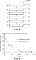
- Fig. 6 shows as example.
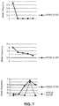
- mutant allelic frequencies also termed mutant allele frequencies, (MAF) (%) a patient's response to therapy can be tracked, whether this is to drug therapy, immune-therapy or radiation therapy. It can be determined whether the patient becomes resistant to therapy by the emergence of new mutations. Generally, one can classify, based on mutation response over time, whether a patient is responding to treatment or not. This is shown in Fig. 7 .
- a categorisation can be introduced. For example, if the allelic frequency drops to 0 this can be termed a complete response, if the allelic frequency does not drop to 0 this can be termed a partial response, and where the allelic frequency increases, this can be termed progressive disease.
- RECIST v1.1 criteria exist [http://www.eortc.be/Services/Doc/RECIST.pdf] that relate to if and when imaging will be capable of detecting a size difference in a tumour, which would be clinically meaningful.
- MRI magnetic resonance
- CT magnetic resonance
- US imaging modalities the RECIST v1.1 criteria are:
- the biomarker information can be used to determine when to carry out imaging to confirm whether a therapy is successful, because at this time point it is anticipated that the tumour has undergone a change in size that would be clinically meaningful.
- a patient who might be responding to therapy can undergo an image scan early on to determine whether that patient is a candidate for surgery, thus sparing the patient unnecessary (chemo)-therapy, and giving a cost benefit to the healthcare system.
- mutant allelic frequencies (MAF) in the sums at both time points, a more accurate time determination can be made as mutants may emerge or be suppressed, due to a failing or successful (cytostatic) treatment, respectively.
- the inventors realised that the resolution of image acquisition units such as MRI, CT could be taken into account in determining when to acquire an image scan of the patient based on how the biomarker information is changed over time.
- a typical diameter of a tumour will be 3 cm.
- an MRI voxel size of 1 mm at least a 2 mm change in dimension is required to detect reliably a change. That would correspond to a 17 % diameter change.
- CT spatial resolution is higher but lesion conspicuity is lower.
- the inventors have determined that a lesion size change of 10% should be reliably detectable.
- biomarker information can also be used for predicting measure of disease progression and determining when and image scan of the patient should be carried out.
- the biomarker information can in some examples can be utilised in the form of a sum over all allelic frequencies, but in examples it is anticipated that the biomarker information for some (one or more) molecular subtypes can be restricted to some (a more limited set) of mutant allelic frequencies associated with this molecular subtype of cancer and could better be used to predict when imaging could be undertaken.
- MAF i denotes the mutant allele frequency (in %) of genetic modification i, as a function of time.
- the mutant allele frequency can be used to determine that a patient is responding to therapy.
- imaging is the 'gold standard' for determining this, but could be replaced by the monitoring of MAF or biomarker values.
- the change in mutant allele frequency over time can be used to predict a mutant allele frequency into the future, thereby enabling a time at which to diagnostic image the patient to be determined.
- a computer program or computer program element is provided that is characterized by being configured to execute the method steps of the method according to one of the preceding embodiments, on an appropriate apparatus or system.
- the computer program element might therefore be stored on a computer unit, which might also be part of an embodiment.
- This computing unit may be configured to perform or induce performing of the steps of the method described above. Moreover, it may be configured to operate the components of the above described apparatus and/or system.
- the computing unit can be configured to operate automatically and/or to execute the orders of a user.
- a computer program may be loaded into a working memory of a data processor. The data processor may thus be equipped to carry out the method according to one of the preceding embodiments.
- This exemplary embodiment of the invention covers both, a computer program that right from the beginning uses the invention and computer program that by means of an update turns an existing program into a program that uses the invention.
- the computer program element might be able to provide all necessary steps to fulfill the procedure of an exemplary embodiment of the method as described above.
- a computer readable medium such as a CD-ROM, USB stick or the like
- the computer readable medium has a computer program element stored on it which computer program element is described by the preceding section.
- a computer program may be stored and/or distributed on a suitable medium, such as an optical storage medium or a solid state medium supplied together with or as part of other hardware, but may also be distributed in other forms, such as via the internet or other wired or wireless telecommunication systems.
- a suitable medium such as an optical storage medium or a solid state medium supplied together with or as part of other hardware, but may also be distributed in other forms, such as via the internet or other wired or wireless telecommunication systems.
- the computer program may also be presented over a network like the World Wide Web and can be downloaded into the working memory of a data processor from such a network.
- a medium for making a computer program element available for downloading is provided, which computer program element is arranged to perform a method according to one of the previously described embodiments of the invention.
Landscapes
- Engineering & Computer Science (AREA)
- Health & Medical Sciences (AREA)
- Medical Informatics (AREA)
- General Health & Medical Sciences (AREA)
- Public Health (AREA)
- Primary Health Care (AREA)
- Epidemiology (AREA)
- Biomedical Technology (AREA)
- Nuclear Medicine, Radiotherapy & Molecular Imaging (AREA)
- Radiology & Medical Imaging (AREA)
- Medicinal Chemistry (AREA)
- Chemical & Material Sciences (AREA)
- Bioinformatics & Cheminformatics (AREA)
- Computer Vision & Pattern Recognition (AREA)
- Theoretical Computer Science (AREA)
- General Physics & Mathematics (AREA)
- Physics & Mathematics (AREA)
- Quality & Reliability (AREA)
- Urology & Nephrology (AREA)
- Databases & Information Systems (AREA)
- Pathology (AREA)
- Surgery (AREA)
- Data Mining & Analysis (AREA)
- Business, Economics & Management (AREA)
- General Business, Economics & Management (AREA)
- Apparatus For Radiation Diagnosis (AREA)
- Investigating Or Analysing Biological Materials (AREA)
- Peptides Or Proteins (AREA)
- Medical Treatment And Welfare Office Work (AREA)
- Micro-Organisms Or Cultivation Processes Thereof (AREA)
Priority Applications (6)
| Application Number | Priority Date | Filing Date | Title |
|---|---|---|---|
| EP19210005.5A EP3826024A1 (en) | 2019-11-19 | 2019-11-19 | Apparatus for diagnostic image acquisition determination |
| JP2022529069A JP2023505023A (ja) | 2019-11-19 | 2020-11-17 | 診断画像取得決定のための装置 |
| US17/777,713 US12165319B2 (en) | 2019-11-19 | 2020-11-17 | Apparatus for diagnostic image acquisition determination |
| CN202080080188.9A CN114730623A (zh) | 2019-11-19 | 2020-11-17 | 用于诊断图像采集确定的设备 |
| EP20804296.0A EP4062414A1 (en) | 2019-11-19 | 2020-11-17 | Apparatus for diagnostic image acquisition determination |
| PCT/EP2020/082312 WO2021099280A1 (en) | 2019-11-19 | 2020-11-17 | Apparatus for diagnostic image acquisition determination |
Applications Claiming Priority (1)
| Application Number | Priority Date | Filing Date | Title |
|---|---|---|---|
| EP19210005.5A EP3826024A1 (en) | 2019-11-19 | 2019-11-19 | Apparatus for diagnostic image acquisition determination |
Publications (1)
| Publication Number | Publication Date |
|---|---|
| EP3826024A1 true EP3826024A1 (en) | 2021-05-26 |
Family
ID=68771338
Family Applications (2)
| Application Number | Title | Priority Date | Filing Date |
|---|---|---|---|
| EP19210005.5A Withdrawn EP3826024A1 (en) | 2019-11-19 | 2019-11-19 | Apparatus for diagnostic image acquisition determination |
| EP20804296.0A Withdrawn EP4062414A1 (en) | 2019-11-19 | 2020-11-17 | Apparatus for diagnostic image acquisition determination |
Family Applications After (1)
| Application Number | Title | Priority Date | Filing Date |
|---|---|---|---|
| EP20804296.0A Withdrawn EP4062414A1 (en) | 2019-11-19 | 2020-11-17 | Apparatus for diagnostic image acquisition determination |
Country Status (5)
| Country | Link |
|---|---|
| US (1) | US12165319B2 (https=) |
| EP (2) | EP3826024A1 (https=) |
| JP (1) | JP2023505023A (https=) |
| CN (1) | CN114730623A (https=) |
| WO (1) | WO2021099280A1 (https=) |
Cited By (1)
| Publication number | Priority date | Publication date | Assignee | Title |
|---|---|---|---|---|
| CN121306515A (zh) * | 2025-12-11 | 2026-01-09 | 陕西中医药大学附属医院 | 基于多模态影像的颅脑损伤区域精准识别方法 |
Families Citing this family (2)
| Publication number | Priority date | Publication date | Assignee | Title |
|---|---|---|---|---|
| US11838400B2 (en) * | 2019-11-19 | 2023-12-05 | International Business Machines Corporation | Image encoding for blockchain |
| CN115294191B (zh) * | 2022-10-08 | 2022-12-27 | 武汉楚精灵医疗科技有限公司 | 基于电子内镜的标志物尺寸测量方法、装置、设备及介质 |
Citations (2)
| Publication number | Priority date | Publication date | Assignee | Title |
|---|---|---|---|---|
| US20160032396A1 (en) * | 2013-03-15 | 2016-02-04 | The Board Of Trustees Of The Leland Stanford Junior University | Identification and Use of Circulating Nucleic Acid Tumor Markers |
| WO2018083467A1 (en) * | 2016-11-02 | 2018-05-11 | Ucl Business Plc | Method of detecting tumour recurrence |
Family Cites Families (10)
| Publication number | Priority date | Publication date | Assignee | Title |
|---|---|---|---|---|
| US7187790B2 (en) * | 2002-12-18 | 2007-03-06 | Ge Medical Systems Global Technology Company, Llc | Data processing and feedback method and system |
| US7490085B2 (en) * | 2002-12-18 | 2009-02-10 | Ge Medical Systems Global Technology Company, Llc | Computer-assisted data processing system and method incorporating automated learning |
| US20160032395A1 (en) * | 2013-03-14 | 2016-02-04 | Elai Davicioni | Cancer biomarkers and classifiers and uses thereof |
| AU2016322966B2 (en) * | 2015-09-17 | 2021-10-14 | S.D. Sight Diagnostics Ltd | Methods and apparatus for detecting an entity in a bodily sample |
| WO2017094805A1 (ja) * | 2015-11-30 | 2017-06-08 | 株式会社Dnaチップ研究所 | ctDNA量の測定による悪性新生物に対する治療の効果を評価する方法 |
| EP3241489A1 (en) * | 2016-05-04 | 2017-11-08 | Mensia Technologies | Predictive neuromarkers of alzheimer's disease |
| WO2018144044A1 (en) * | 2017-02-03 | 2018-08-09 | Q Bio, Inc. | lTERATIVE MEDICAL TESTING OF BIOLOGICAL SAMPLES |
| EP3542859A1 (en) * | 2018-03-20 | 2019-09-25 | Koninklijke Philips N.V. | Determining a medical imaging schedule |
| US10646156B1 (en) * | 2019-06-14 | 2020-05-12 | Cycle Clarity, LLC | Adaptive image processing in assisted reproductive imaging modalities |
| US11621077B2 (en) * | 2019-09-30 | 2023-04-04 | Kpn Innovations, Llc. | Methods and systems for using artificial intelligence to select a compatible element |
-
2019
- 2019-11-19 EP EP19210005.5A patent/EP3826024A1/en not_active Withdrawn
-
2020
- 2020-11-17 WO PCT/EP2020/082312 patent/WO2021099280A1/en not_active Ceased
- 2020-11-17 US US17/777,713 patent/US12165319B2/en active Active
- 2020-11-17 EP EP20804296.0A patent/EP4062414A1/en not_active Withdrawn
- 2020-11-17 JP JP2022529069A patent/JP2023505023A/ja active Pending
- 2020-11-17 CN CN202080080188.9A patent/CN114730623A/zh active Pending
Patent Citations (2)
| Publication number | Priority date | Publication date | Assignee | Title |
|---|---|---|---|---|
| US20160032396A1 (en) * | 2013-03-15 | 2016-02-04 | The Board Of Trustees Of The Leland Stanford Junior University | Identification and Use of Circulating Nucleic Acid Tumor Markers |
| WO2018083467A1 (en) * | 2016-11-02 | 2018-05-11 | Ucl Business Plc | Method of detecting tumour recurrence |
Non-Patent Citations (4)
| Title |
|---|
| DIEHL ET AL., NAT. MED, vol. 14, 2008, pages 985 |
| INTERNATIONAL SOCIETY FOR MAGNETIC RESONANCE IN MEDICINE, ISMRM, 2030 ADDISON STREET, 7TH FLOOR, BERKELEY, CA 94704 USA, no. E1024, 1 June 2018 (2018-06-01), XP040704877 * |
| MANFRED KAUFMANN ET AL: "Recommendations from an International Consensus Conference on the Current Status and Future of Neoadjuvant Systemic Therapy in Primary Breast Cancer", ANNALS OF SURGICAL ONCOLOGY, SPRINGER-VERLAG, NE, vol. 19, no. 5, 23 December 2011 (2011-12-23), pages 1508 - 1516, XP035046316, ISSN: 1534-4681, DOI: 10.1245/S10434-011-2108-2 * |
| WIKIPEDIA: "Response evaluation criteria in solid tumors", 11 February 2019 (2019-02-11), XP055675700, Retrieved from the Internet <URL:https://en.wikipedia.org/w/index.php?title=Response_evaluation_criteria_in_solid_tumors&oldid=882751552> [retrieved on 20200311] * |
Cited By (1)
| Publication number | Priority date | Publication date | Assignee | Title |
|---|---|---|---|---|
| CN121306515A (zh) * | 2025-12-11 | 2026-01-09 | 陕西中医药大学附属医院 | 基于多模态影像的颅脑损伤区域精准识别方法 |
Also Published As
| Publication number | Publication date |
|---|---|
| US12165319B2 (en) | 2024-12-10 |
| WO2021099280A1 (en) | 2021-05-27 |
| US20230028306A1 (en) | 2023-01-26 |
| JP2023505023A (ja) | 2023-02-08 |
| CN114730623A (zh) | 2022-07-08 |
| EP4062414A1 (en) | 2022-09-28 |
Similar Documents
| Publication | Publication Date | Title |
|---|---|---|
| Latifoltojar et al. | Whole body magnetic resonance imaging in newly diagnosed multiple myeloma: early changes in lesional signal fat fraction predict disease response | |
| Zamagni et al. | 18F-FDG PET/CT focal, but not osteolytic, lesions predict the progression of smoldering myeloma to active disease | |
| Body et al. | Sarcopenia and myosteatosis predict adverse outcomes after emergency laparotomy: a multi-center observational cohort study | |
| Dercle et al. | Limits of radiomic-based entropy as a surrogate of tumor heterogeneity: ROI-area, acquisition protocol and tissue site exert substantial influence | |
| Schwarzenberg et al. | Treatment response evaluation using 18F-FDOPA PET in patients with recurrent malignant glioma on bevacizumab therapy | |
| Haznedar et al. | Value of 18F-fluorodeoxyglucose uptake in positron emission tomography/computed tomography in predicting survival in multiple myeloma | |
| US12165319B2 (en) | Apparatus for diagnostic image acquisition determination | |
| EP3048977B1 (en) | Patient-specific analysis of positron emission tomography data | |
| Wennmann et al. | Deep learning for automatic bone marrow apparent diffusion coefficient measurements from whole-body magnetic resonance imaging in patients with multiple myeloma: a retrospective multicenter study | |
| Borggrefe et al. | Association of osteolytic lesions, bone mineral loss and trabecular sclerosis with prevalent vertebral fractures in patients with multiple myeloma | |
| Skamene et al. | Metabolic activity measured on PET/CT correlates with clinical outcomes in patients with limb and girdle sarcomas | |
| Alavi et al. | Quantification of adipose tissues by Dual-Energy X-Ray Absorptiometry and Computed Tomography in colorectal cancer patients | |
| Borggrefe et al. | Association of QCT bone mineral density and bone structure with vertebral fractures in patients with multiple myeloma | |
| Ikeda et al. | Soluble B-cell maturation antigen levels for disease monitoring in oligosecretory and nonsecretory relapsed multiple myeloma | |
| Pan et al. | Automated vertebral bone mineral density measurement with phantomless internal calibration in chest LDCT scans using deep learning | |
| Mets et al. | In vivo growth of 60 non-screening detected lung cancers: a computed tomography study | |
| Minarik et al. | Prospective comparison of conventional radiography, low-dose computed tomography and magnetic resonance imaging in monoclonal gammopathies | |
| EP4046160A1 (en) | Method and apparatus for assessing patient's responses to therapy | |
| Pfahler et al. | Tumor load in patients with multiple myeloma: β₂‐microglobulin levels versus low‐dose whole‐body CT | |
| Spira et al. | Can whole-body low-dose multidetector CT exclude the presence of myeloma bone disease in patients with monoclonal gammopathy of undetermined significance (MGUS)? | |
| Greuter et al. | The validation of a simulation model incorporating radiation risk for mammography breast cancer screening in women with a hereditary-increased breast cancer risk | |
| Otemuyiwa et al. | Dorsal muscle attenuation may predict failure to respond to interleukin-2 therapy in metastatic renal cell carcinoma | |
| Sebelego et al. | Factors influencing size-specific dose estimates of selected computed tomography protocols at two clinical practices in South Africa | |
| Lee et al. | Added values of DXA-derived visceral adipose tissue to discriminate cardiometabolic risks in pre-pubertal children | |
| US11120888B2 (en) | Systems and methods for predicting lung cancer immune therapy responsiveness using quantitative textural analysis |
Legal Events
| Date | Code | Title | Description |
|---|---|---|---|
| PUAI | Public reference made under article 153(3) epc to a published international application that has entered the european phase |
Free format text: ORIGINAL CODE: 0009012 |
|
| STAA | Information on the status of an ep patent application or granted ep patent |
Free format text: STATUS: THE APPLICATION HAS BEEN PUBLISHED |
|
| AK | Designated contracting states |
Kind code of ref document: A1 Designated state(s): AL AT BE BG CH CY CZ DE DK EE ES FI FR GB GR HR HU IE IS IT LI LT LU LV MC MK MT NL NO PL PT RO RS SE SI SK SM TR |
|
| STAA | Information on the status of an ep patent application or granted ep patent |
Free format text: STATUS: THE APPLICATION IS DEEMED TO BE WITHDRAWN |
|
| 18D | Application deemed to be withdrawn |
Effective date: 20211127 |