EP3822360A1 - Method for determining the sensitivity of a bacterial strain to a bacteriophage virus - Google Patents
Method for determining the sensitivity of a bacterial strain to a bacteriophage virus Download PDFInfo
- Publication number
- EP3822360A1 EP3822360A1 EP20207254.2A EP20207254A EP3822360A1 EP 3822360 A1 EP3822360 A1 EP 3822360A1 EP 20207254 A EP20207254 A EP 20207254A EP 3822360 A1 EP3822360 A1 EP 3822360A1
- Authority
- EP
- European Patent Office
- Prior art keywords
- bacteriophages
- sample
- bacteria
- image
- interest
- Prior art date
- Legal status (The legal status is an assumption and is not a legal conclusion. Google has not performed a legal analysis and makes no representation as to the accuracy of the status listed.)
- Pending
Links
Images


Classifications
-
- C—CHEMISTRY; METALLURGY
- C12—BIOCHEMISTRY; BEER; SPIRITS; WINE; VINEGAR; MICROBIOLOGY; ENZYMOLOGY; MUTATION OR GENETIC ENGINEERING
- C12Q—MEASURING OR TESTING PROCESSES INVOLVING ENZYMES, NUCLEIC ACIDS OR MICROORGANISMS; COMPOSITIONS OR TEST PAPERS THEREFOR; PROCESSES OF PREPARING SUCH COMPOSITIONS; CONDITION-RESPONSIVE CONTROL IN MICROBIOLOGICAL OR ENZYMOLOGICAL PROCESSES
- C12Q1/00—Measuring or testing processes involving enzymes, nucleic acids or microorganisms; Compositions therefor; Processes of preparing such compositions
- C12Q1/02—Measuring or testing processes involving enzymes, nucleic acids or microorganisms; Compositions therefor; Processes of preparing such compositions involving viable microorganisms
- C12Q1/04—Determining presence or kind of microorganism; Use of selective media for testing antibiotics or bacteriocides; Compositions containing a chemical indicator therefor
-
- C—CHEMISTRY; METALLURGY
- C12—BIOCHEMISTRY; BEER; SPIRITS; WINE; VINEGAR; MICROBIOLOGY; ENZYMOLOGY; MUTATION OR GENETIC ENGINEERING
- C12Q—MEASURING OR TESTING PROCESSES INVOLVING ENZYMES, NUCLEIC ACIDS OR MICROORGANISMS; COMPOSITIONS OR TEST PAPERS THEREFOR; PROCESSES OF PREPARING SUCH COMPOSITIONS; CONDITION-RESPONSIVE CONTROL IN MICROBIOLOGICAL OR ENZYMOLOGICAL PROCESSES
- C12Q1/00—Measuring or testing processes involving enzymes, nucleic acids or microorganisms; Compositions therefor; Processes of preparing such compositions
- C12Q1/02—Measuring or testing processes involving enzymes, nucleic acids or microorganisms; Compositions therefor; Processes of preparing such compositions involving viable microorganisms
- C12Q1/18—Testing for antimicrobial activity of a material
-
- C—CHEMISTRY; METALLURGY
- C12—BIOCHEMISTRY; BEER; SPIRITS; WINE; VINEGAR; MICROBIOLOGY; ENZYMOLOGY; MUTATION OR GENETIC ENGINEERING
- C12Q—MEASURING OR TESTING PROCESSES INVOLVING ENZYMES, NUCLEIC ACIDS OR MICROORGANISMS; COMPOSITIONS OR TEST PAPERS THEREFOR; PROCESSES OF PREPARING SUCH COMPOSITIONS; CONDITION-RESPONSIVE CONTROL IN MICROBIOLOGICAL OR ENZYMOLOGICAL PROCESSES
- C12Q1/00—Measuring or testing processes involving enzymes, nucleic acids or microorganisms; Compositions therefor; Processes of preparing such compositions
- C12Q1/70—Measuring or testing processes involving enzymes, nucleic acids or microorganisms; Compositions therefor; Processes of preparing such compositions involving virus or bacteriophage
-
- G—PHYSICS
- G01—MEASURING; TESTING
- G01N—INVESTIGATING OR ANALYSING MATERIALS BY DETERMINING THEIR CHEMICAL OR PHYSICAL PROPERTIES
- G01N21/00—Investigating or analysing materials by the use of optical means, i.e. using sub-millimetre waves, infrared, visible or ultraviolet light
- G01N21/17—Systems in which incident light is modified in accordance with the properties of the material investigated
- G01N21/47—Scattering, i.e. diffuse reflection
- G01N21/49—Scattering, i.e. diffuse reflection within a body or fluid
- G01N21/53—Scattering, i.e. diffuse reflection within a body or fluid within a flowing fluid, e.g. smoke
- G01N21/534—Scattering, i.e. diffuse reflection within a body or fluid within a flowing fluid, e.g. smoke by measuring transmission alone, i.e. determining opacity
-
- Y—GENERAL TAGGING OF NEW TECHNOLOGICAL DEVELOPMENTS; GENERAL TAGGING OF CROSS-SECTIONAL TECHNOLOGIES SPANNING OVER SEVERAL SECTIONS OF THE IPC; TECHNICAL SUBJECTS COVERED BY FORMER USPC CROSS-REFERENCE ART COLLECTIONS [XRACs] AND DIGESTS
- Y02—TECHNOLOGIES OR APPLICATIONS FOR MITIGATION OR ADAPTATION AGAINST CLIMATE CHANGE
- Y02A—TECHNOLOGIES FOR ADAPTATION TO CLIMATE CHANGE
- Y02A50/00—TECHNOLOGIES FOR ADAPTATION TO CLIMATE CHANGE in human health protection, e.g. against extreme weather
- Y02A50/30—Against vector-borne diseases, e.g. mosquito-borne, fly-borne, tick-borne or waterborne diseases whose impact is exacerbated by climate change
Abstract
Procédé de détermination de la sensibilité d'une souche bactérienne d'intérêt à l'égard d'une souche virale de bactériophages, le procédé comportant :a) préparation d'un échantillon (20), comportant une mise en contact de bactéries (2) appartenant à la souche bactérienne d'intérêt avec des bactériophages (3), chaque bactériophage appartenant à une même souche virale, les bactéries étant soit dans un milieu liquide, soit dans un milieu gélosé ;b) disposition de l'échantillon entre une source de lumière (10) et un capteur d'image (30), la source de lumière émettant une onde lumineuse (11) dans une bande spectrale d'émission comprise entre 500 nm et 600 nm ;c) illumination de l'échantillon à l'aide de la source de lumière et acquisition d'au moins une image de l'échantillon, par le capteur d'image, dans la bande spectrale d'émission, aucune optique de formation d'image n'étant disposé entre l'échantillon et le capteur d'image;d) à partir des images acquises, détermination d'une sensibilité de la souche bactérienne d'intérêt à l'égard de la souche virale.A method of determining the sensitivity of a bacterial strain of interest to a viral strain of bacteriophages, the method comprising: a) preparing a sample (20), comprising contacting bacteria (2 ) belonging to the bacterial strain of interest with bacteriophages (3), each bacteriophage belonging to the same viral strain, the bacteria being either in a liquid medium or in an agar medium; b) disposal of the sample between a source light (10) and an image sensor (30), the light source emitting a light wave (11) in an emission spectral band between 500 nm and 600 nm; c) illumination of the sample at the '' using the light source and acquiring at least one image of the sample, by the image sensor, in the emission spectral band, no imaging optics being arranged between the sample and the image sensor; d) from the acquired images, determining a sensitivity é of the bacterial strain of interest with regard to the viral strain.
Description
Le domaine technique de l'invention est la caractérisation de l'aptitude d'une souche bactérienne à être infectée, puis lysée, par un virus bactériophage.The technical field of the invention is the characterization of the ability of a bacterial strain to be infected, then lysed, by a bacteriophage virus.
La phagothérapie est l'utilisation de virus bactériophages, usuellement appelés phages, pour traiter les infections d'origine bactérienne. Il s'agit de mettre en œuvre des phages lytiques, capables d'infecter certaines bactéries, puis de les lyser. Il s'agit d'un traitement relativement ancien, découvert et pratiqué avant l'émergence des antibiotiques. Toutefois, dans la plupart des pays, ce traitement a été quelque peu négligé, au profit du recours aux antibiotiques. A l'heure actuelle, la phagothérapie est considérée comme approche thérapeutique prometteuse pour traiter certaines infections bactériennes, particulièrement adaptée lorsque les bactéries sont résistantes aux antibiotiques.Phage therapy is the use of bacteriophage viruses, usually called phages, to treat bacterial infections. This involves using lytic phages, capable of infecting certain bacteria, then lysing them. It is a relatively old treatment, discovered and practiced before the emergence of antibiotics. However, in most countries this treatment has been somewhat neglected, in favor of the use of antibiotics. At present, phage therapy is considered a promising therapeutic approach to treat certain bacterial infections, particularly suitable when the bacteria are resistant to antibiotics.
L'efficacité de la phagothérapie a été démontrée pour le traitement d'infections par des bactéries de type Pseudomonas aeruginosa, Escherichia coli, ou Staphilococcus Aureus, dans des applications de type greffe de peau pour des grands brûlés ou des pathologies liées au diabète.The effectiveness of phage therapy has been demonstrated for the treatment of infections by bacteria of the Pseudomonas aeruginosa, Escherichia coli, or Staphilococcus Aureus type, in skin graft-type applications for severe burns or pathologies linked to diabetes.
Les phages mis en œuvre dans des applications de phagothérapie sont des phages lytiques, de telle sorte que lorsqu'une bactérie a été infectée par un phage, cette dernière est lysée. La lyse résulte de la production d'endolysines. Au cours d'une infection, le métabolisme de la bactérie conduit à une réplication du nombre de phages, sous la forme de virions. Ces derniers sont libérés suite à la lyse de la bactérie. Une infection est donc suivie d'une augmentation de la quantité de phages aptes à infecter, puis lyser d'autres bactéries. Il en résulte une diminution, voire un arrêt de la prolifération bactérienne.The phages used in phage therapy applications are lytic phages, so that when a bacterium has been infected with a phage, the latter is lysed. Lysis results from the production of endolysins. During an infection, the metabolism of the bacteria leads to a replication of the number of phages, in the form of virions. These are released following lysis of the bacteria. An infection is therefore followed by an increase in the quantity of phages capable of infecting and then lysing other bacteria. This results in a reduction, or even a stop, of bacterial proliferation.
Afin de tester la sensibilité d'une bactérie à un phage, des essais, usuellement désignés par le terme "phagogramme", sont pratiqués en laboratoire. Il s'agit de tester la capacité d'une souche bactérienne à être infectée, puis lysée, par un phage.In order to test the sensitivity of a bacterium to a phage, tests, usually designated by the term “phagogram”, are carried out in the laboratory. This involves testing the ability of a bacterial strain to be infected, then lysed, by a phage.
Les documents
Les essais conduits en laboratoire nécessitent de tester différentes souches virales, avec différents ratios de quantités de phages / bactéries. Le nombre de souches virales peut être compris entre 5 et 100, tandis que 5 à 10 ratios de quantités de phages / bactéries sont généralement testés, et cela pour chaque souche virale. Le nombre d'essais à réaliser est donc élevé.The tests carried out in the laboratory require testing different viral strains, with different ratios of quantities of phages / bacteria. The number of viral strains can be between 5 and 100, while 5 to 10 ratios of quantities of phage / bacteria are generally tested, and this for each viral strain. The number of tests to be carried out is therefore high.
Les inventeurs proposent une méthode simple, permettant d'analyser la sensibilité d'une souche bactérienne à l'égard d'une souche virale. La méthode permet un suivi du développement de bactéries et une détection précoce de lyses bactériennes par des phages. Elle peut également adresser simultanément plusieurs combinaisons entre des quantités de phages, éventuellement de différentes souches virales, et des quantités de bactéries, éventuellement de différentes souches bactériennes. L'objectif est de réduire le temps passé pour détecter la sensibilité d'une souche bactérienne à l'égard d'une souche virale.The inventors propose a simple method making it possible to analyze the sensitivity of a bacterial strain with regard to a viral strain. The method allows monitoring of bacterial development and early detection of bacterial lyses by phages. It can also simultaneously address several combinations between quantities of phages, optionally of different viral strains, and quantities of bacteria, optionally of different bacterial strains. The objective is to reduce the time spent to detect the sensitivity of a bacterial strain towards a viral strain.
Un objet de l'invention est un procédé de détermination de la sensibilité d'une souche bactérienne à l'égard d'une souche virale de bactériophages, le procédé comportant :
- a) préparation d'un échantillon, comportant une mise en contact de bactéries, de la souche bactérienne, avec des bactériophages, chaque bactériophage appartenant à une même souche virale, les bactéries étant soit dans un milieu liquide, soit dans un milieu gélosé ;
- b) disposition de l'échantillon entre une source de lumière et un capteur d'image, la source de lumière émettant une onde lumineuse dans une bande spectrale d'émission de préférence comprise entre 500 nm et 600 nm ;
- c) illumination de l'échantillon à l'aide de la source de lumière et acquisition d'au moins une image de l'échantillon, par le capteur d'image, dans la bande spectrale d'émission, aucune optique de formation d'image n'étant de préférence disposé entre l'échantillon et le capteur d'image;
- d) à partir de chaque image acquise, détermination d'une sensibilité de la souche bactérienne à l'égard de la souche virale des bactériophages.
- a) preparation of a sample, comprising bringing bacteria into contact, of the bacterial strain, with bacteriophages, each bacteriophage belonging to the same viral strain, the bacteria being either in a liquid medium or in an agar medium;
- b) placing the sample between a light source and an image sensor, the light source emitting a light wave in an emission spectral band preferably between 500 nm and 600 nm;
- c) illumination of the sample using the light source and acquisition of at least one image of the sample, by the image sensor, in the emission spectral band, no formation optics image preferably not being disposed between the sample and the image sensor;
- d) from each image acquired, determination of a sensitivity of the bacterial strain with respect to the viral strain of bacteriophages.
La largeur de la bande spectrale d'émission est de préférence inférieure à 100 nm ou à 50 nm. La distance entre l'échantillon et le capteur d'image est de préférence inférieure à 5 cm ou inférieure à 1 cm.The width of the emission spectral band is preferably less than 100 nm or 50 nm. The distance between the sample and the image sensor is preferably less than 5 cm or less than 1 cm.
Selon un mode de réalisation :
- lors de l'étape a), les bactéries et les bactériophages sont mélangés dans une solution aqueuse ;
- l'étape c) comporte une acquisition d'au moins deux images, à des instants de mesure successifs ;
- l'étape d) comporte une détermination d'une intensité lumineuse détectée par tout ou partie du capteur d'image, sur chaque image acquise respectivement à chaque instant de mesure, de telle sorte que la souche bactérienne est considérée comme :
- peu ou pas sensible à la souche virale des bactériophages lorsque l'intensité lumineuse détectée diminue entre deux instants de mesure successifs;
- ou sensible à la souche virale des bactériophages lorsque l'intensité lumineuse détectée ne diminue pas ou augmente entre deux instants de mesure successifs.
- during step a), the bacteria and the bacteriophages are mixed in an aqueous solution;
- step c) comprises an acquisition of at least two images, at successive measurement instants;
- step d) comprises a determination of a light intensity detected by all or part of the image sensor, on each image acquired respectively at each measurement instant, so that the bacterial strain is considered to be:
- little or not sensitive to the viral strain of bacteriophages when the detected light intensity decreases between two successive measurement instants;
- or sensitive to the viral strain of bacteriophages when the detected light intensity does not decrease or increases between two successive measurement instants.
L'étape d) peut comporter une détermination d'une atténuation de la lumière, émise par la source de lumière, par l'échantillon, de telle sorte que la souche bactérienne est considérée comme:
- peu ou pas sensible à la souche virale des bactériophages lorsque l'atténuation augmente entre deux instants de mesure successifs;
- ou sensible à la souche virale des bactériophages lorsque l'atténuation n'augmente pas entre deux instants de mesure successifs.
- little or not sensitive to the viral strain of the bacteriophages when the attenuation increases between two successive measurement instants;
- or sensitive to the viral strain of bacteriophages when the attenuation does not increase between two successive measurement instants.
Selon un mode de réalisation :
- lors de l'étape a), les bactéries et les bactériophages sont mélangés dans une solution aqueuse ;
- l'étape c) comporte une acquisition d'au moins deux images, à des instants de mesure successifs ;
- l'étape d) comporte une détermination d'un descripteur de texturation de chaque image acquise, de telle sorte que la souche bactérienne est considérée comme :
- pas ou peu sensible à la souche virale des bactériophages lorsque l'évolution du descripteur de texturation traduit une augmentation d'une diffusion de la lumière par l'échantillon entre deux instants de mesure successifs ;
- ou sensible à la souche virale des bactériophages lorsque l'évolution du descripteur de texturation traduit une diminution ou une stagnation d'une diffusion de la lumière par l'échantillon entre deux instants de mesure successifs.
- during step a), the bacteria and the bacteriophages are mixed in an aqueous solution;
- step c) comprises an acquisition of at least two images, at successive measurement instants;
- step d) comprises determining a texturing descriptor for each image acquired, such that the bacterial strain is considered to be:
- not or not very sensitive to the viral strain of bacteriophages when the evolution of the texturing descriptor reflects an increase in light scattering by the sample between two successive measurement instants;
- or sensitive to the viral strain of bacteriophages when the evolution of the texturing descriptor reflects a decrease or a stagnation of light scattering by the sample between two successive measurement instants.
Le descripteur de texturation peut être déterminé selon une ou plusieurs régions d'intérêt de l'image.The texturing descriptor can be determined according to one or more regions of interest of the image.
Selon un mode de réalisation :
- lors de l'étape a), les bactéries et les bactériophages sont mélangés dans une gélose ;
- l'étape c) comporte une acquisition d'au moins une image, postérieurement à l'étape a) ;
- l'étape d), comporte une analyse de chaque image acquise, de façon à identifier des régions d'intérêt claires, chaque région d'intérêt claire correspondant à une infection de bactéries par des bactériophages, formant une plage de lyse, chaque région d'intérêt claire indiquant une sensibilité de la souche bactérienne à l'égard de la souche virale des bactériophages.
- during step a), the bacteria and the bacteriophages are mixed in an agar;
- step c) comprises an acquisition of at least one image, subsequent to step a);
- step d), comprises an analysis of each image acquired, so as to identify clear regions of interest, each clear region of interest corresponding to an infection of bacteria by bacteriophages, forming a lysis plaque, each region d 'clear interest indicating a sensitivity of the bacterial strain towards the viral strain of bacteriophages.
Le procédé peut alors comporter :
- un comptage du nombre de régions d'intérêt claires sur au moins une image acquise ;
- une estimation d'un titre infectieux des bactériophages dans l'échantillon à partir du nombre de régions d'intérêt claires comptées.
- counting the number of bright regions of interest on at least one acquired image;
- an estimate of an infectious titer of bacteriophages in the sample from the number of clear regions of interest counted.
Selon un mode de réalisation :
- lors de l'étape a), les bactéries sont disposées dans une gélose, et les bactériophages sont disposés dans une solution, l'étape a) comportant un dépôt d'au moins une goutte de la solution sur la gélose ;
- l'étape c) comporte une acquisition d'au moins une image, postérieurement à l'étape a) ;
- l'étape d) comporte une analyse de chaque image acquise, de façon à identifier des régions d'intérêt claires, chaque région d'intérêt claire correspondant à une infection de bactéries par des bactériophages, formant une plage de lyse, de telle sorte que l'apparition, de chaque région d'intérêt claire indique une sensibilité de la souche bactérienne à l'égard de la souche virale des bactériophages.
- during step a), the bacteria are placed in an agar, and the bacteriophages are placed in a solution, step a) comprising depositing at least one drop of the solution on the agar;
- step c) comprises an acquisition of at least one image, subsequent to step a);
- step d) comprises an analysis of each image acquired, so as to identify clear regions of interest, each clear region of interest corresponding to an infection of bacteria by bacteriophages, forming a lysis plaque, so that the appearance of each clear region of interest indicates a sensitivity of the bacterial strain to the viral strain of bacteriophages.
L'étape a) peut comporter un dépôt de plusieurs gouttes, les gouttes étant espacées les unes des autres, deux gouttes différentes comportant respectivement :
- des bactériophages de différentes souches virales ;
- et/ou différentes concentrations de bactériophages d'une même souche virale.
- bacteriophages of different viral strains;
- and / or different concentrations of bacteriophages of the same viral strain.
Selon un mode de réalisation :
- lors de l'étape a), les bactériophages sont disposés dans une gélose, et les bactéries sont disposées dans une solution, l'étape a) comportant un dépôt d'une goutte de la solution sur la gélose;
- l'étape c) comporte une acquisition d'au moins une image, postérieurement à l'étape a) ;
- l'étape d) comporte une analyse de chaque image acquise, de façon à identifier:
- des régions d'intérêt claires, chaque région d'intérêt claire correspondant à une infection de bactéries par des bactériophages, de telle sorte que chaque région d'intérêt claire indique une sensibilité de la souche bactérienne à l'égard de la souche virale des bactériophages;
- ou des régions d'intérêt sombres, chaque région d'intérêt sombre correspondant à un développement de bactéries en présence de bactériophages, de telle sorte que chaque région d'intérêt sombre indique une insensibilité ou une faible sensibilité de la souche bactérienne d'intérêt à l'égard de la souche virale des bactériophages.
- during step a), the bacteriophages are placed in an agar, and the bacteria are placed in a solution, step a) comprising depositing a drop of the solution on the agar;
- step c) comprises an acquisition of at least one image, subsequent to step a);
- step d) comprises an analysis of each image acquired, so as to identify:
- clear regions of interest, each clear region of interest corresponding to infection of bacteria by bacteriophages, such that each clear region of interest indicates a sensitivity of the bacterial strain to the viral strain of bacteriophages ;
- or dark regions of interest, each dark region of interest corresponding to a development of bacteria in the presence of bacteriophages, such that each dark region of interest indicates insensitivity or low sensitivity of the bacterial strain of interest to with regard to the viral strain of bacteriophages.
L'étape a) peut comporter un dépôt de plusieurs gouttes, les gouttes étant espacées les unes des autres, deux gouttes différentes comportant respectivement :
- des bactéries de différentes souches bactériennes
- et/ou différentes concentrations de bactéries d'une même souche bactérienne.
- bacteria from different bacterial strains
- and / or different concentrations of bacteria of the same bacterial strain.
Quel que soit le mode de réalisation, l'échantillon peut être divisé en différentes zones spatiales, séparées les unes des autres de telle sorte que:
- au moins deux zones spatiales différentes comportent respectivement une même souche bactérienne et une concentration différente de bactériophages d'une même souche virale;
- et/ou au moins deux zones spatiales différentes comportent respectivement une même souche bactérienne et des bactériophages de différentes souches virales ;
- et/ou au moins deux zones spatiales différentes comportent respectivement des bactériophages d'une même souche virale et différentes souches bactériennes.
- at least two different spatial zones respectively comprise the same bacterial strain and a different concentration of bacteriophages of the same viral strain;
- and / or at least two different spatial zones respectively comprise the same bacterial strain and bacteriophages of different viral strains;
- and / or at least two different spatial zones respectively comprise bacteriophages of the same viral strain and different bacterial strains.
Le procédé est alors tel que chaque zone spatiale est associée à une région d'intérêt de chaque image acquise, deux zones spatiales différentes étant associées à deux régions d'intérêt différentes, de façon que l'analyse d'une même image acquise permet d'obtenir une information relative à la sensibilité d'une souche bactérienne à l'égard d'une souche virale de bactériophages, et cela dans différentes zones spatiales.The method is then such that each spatial zone is associated with a region of interest of each acquired image, two different spatial zones being associated with two different regions of interest, so that the analysis of the same acquired image makes it possible to '' obtain information relating to the sensitivity of a bacterial strain to a viral strain of bacteriophages, and this in different spatial zones.
L'échantillon peut comporter plus de 10, voire plus de 100, zones spatiales différentes, séparées les unes des autres, chaque zone spatiale étant paramétrée trois paramètres correspondant respectivement à la souche bactérienne d'intérêt, à la souche virale des bactériophages et à la concentration des bactériophages, au moins un paramètre de deux zones spatiales différentes étant différent.The sample can include more than 10, or even more than 100, different spatial zones, separated from each other, each spatial zone being configured with three corresponding parameters. respectively to the bacterial strain of interest, to the viral strain of the bacteriophages and to the concentration of the bacteriophages, at least one parameter of two different spatial zones being different.
L'échantillon peut être réparti dans différentes chambres fluidiques, d'une carte fluidique, chaque chambre fluidique correspondant à une zone spatiale de l'échantillon.The sample can be distributed in different fluidic chambers, of a fluidic map, each fluidic chamber corresponding to a spatial zone of the sample.
L'échantillon peut être formé suite à un dépôt de gouttes à la surface d'une gélose, en différentes positions, les gouttes étant espacées les unes des autres, de telle sorte qu'une zone spatiale est définie par chaque position de gouttes. La gélose peut comporter les bactéries et les gouttes peuvent comporter des bactériophages. Alternativement, la gélose peut comporter des bactériophages et les gouttes peuvent comporter des bactéries.The sample can be formed by depositing drops on the surface of an agar, in different positions, the drops being spaced from each other, such that a spatial area is defined by each drop position. The agar may contain bacteria and the drops may contain bacteriophages. Alternatively, the agar can contain bacteriophages and the drops can contain bacteria.
Selon un mode de réalisation, le procédé comporte, préalablement à l'étape a), une étape d'enrichissement comportant :
- un mélange de bactériophages de même souche virale, ou de différentes souches virales, dans une solution aqueuse comportant au moins une souche de bactéries ;
- une incubation ;
- une filtration du mélange, de façon à retenir les bactéries et obtenir une solution enrichie en bactériophages ;
de telle sorte que lors de l'étape a), la mise en contact des bactériophages avec les bactéries est effectuée en utilisant la solution enrichie en bactériophages.
- a mixture of bacteriophages of the same viral strain, or of different viral strains, in an aqueous solution comprising at least one strain of bacteria;
- an incubation;
- filtration of the mixture, so as to retain the bacteria and obtain a solution enriched in bacteriophages;
so that during step a), the bringing into contact of the bacteriophages with the bacteria is carried out using the solution enriched in bacteriophages.
Selon un mode de réalisation, un système optique est interposé entre l'échantillon et le capteur d'image. Le système optique définit un plan image et un objet. Le système optique peut être tel que :
- le plan image est décalé par rapport à un plan de détection, défini par le capteur d'image, selon une distance de défocalisation image ;
- et/ou le plan objet est décalé, par rapport à un plan de l'échantillon, selon une distance de défocalisation objet.
- the image plane is offset with respect to a detection plane, defined by the image sensor, according to an image defocusing distance;
- and / or the object plane is offset, relative to a plane of the sample, by an object defocus distance.
La distance de défocalisation image et/ou la distance de défocalisation objet est de préférence inférieure à 5 mm, voire à 1 mm, voire inférieure à 500 µm. Le plan de l'échantillon est un plan s'étendant dans l'échantillon. Il s'agit de préférence de la surface de l'échantillon la plus proche du capteur d'image. Selon ce mode de réalisation, lors de l'étape c), l'image est acquise selon une configuration défocalisée.The image defocus distance and / or the object defocus distance is preferably less than 5 mm, or even 1 mm, or even less than 500 μm. The sample plane is a plane extending into the sample. This is preferably the surface of the sample closest to the image sensor. According to this embodiment, during step c), the image is acquired according to a defocused configuration.
Selon une variante, le système optique permet une conjugaison du capteur d'image avec l'échantillon. Le plan image est alors confondu avec le plan de détection formé par le capteur d'image. Le plan objet est alors confondu avec le plan de l'échantillon. Selon ce mode de réalisation, lors de l'étape c), l'image est acquise selon une configuration focaliséeAccording to one variant, the optical system allows conjugation of the image sensor with the sample. The image plane is then merged with the detection plane formed by the image sensor. The object plane is then merged with the sample plane. According to this embodiment, during step c), the image is acquired according to a focused configuration
L'invention sera mieux comprise à la lecture de l'exposé des exemples de réalisation présentés, dans la suite de la description, en lien avec les figures listées ci-dessous.The invention will be better understood on reading the description of the embodiments presented, in the remainder of the description, in connection with the figures listed below.
-
La
figure 1 représente un dispositif permettant une mise en œuvre d'un procédé selon l'invention.Thefigure 1 represents a device allowing an implementation of a method according to the invention. -
La
figure 2A schématise un premier mode de réalisation de l'invention.Thefigure 2A shows schematically a first embodiment of the invention. -
Les
figures 2B et 2C sont des images obtenues en mettant en œuvre le premier mode de réalisation de l'invention.Thefigures 2B and 2C are images obtained by implementing the first embodiment of the invention. -
La
figure 2D est une courbe obtenue expérimentalement en mettant en œuvre le premier mode de réalisation. Elle représente une évolution d'une densité optique en fonction du temps.The2D figure is a curve obtained experimentally by implementing the first embodiment. It represents an evolution of an optical density as a function of time. -
Les
figures 2E et2F illustrent des exemples de cartes fluidiques pouvant être mises en œuvre.Thefigures 2E and2 F illustrate examples of fluidic maps that can be implemented. -
Les
figures 3A et 3B sont des images obtenues en mettant en œuvre une variante du premier mode de réalisation.Thefigures 3A and 3B are images obtained by implementing a variant of the first embodiment. -
La
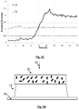
figure 3C montre une courbe obtenue expérimentalement en mettant en œuvre la variante du premier mode de réalisation. Elle représente une évolution d'un indicateur de texture en fonction du temps.Thefigure 3C shows a curve obtained experimentally by implementing the variant of the first embodiment. It represents an evolution of a texture indicator as a function of time. -
Les
figures 4A et4B schématisent un deuxième mode de réalisation de l'invention.Thefigures 4A and4B show schematically a second embodiment of the invention. -
Les
figures 4C, 4D et4E sont des images obtenues en mettant en œuvre le deuxième mode de réalisation. Elles montrent une formation de plages de lyse en fonction du temps.Thefigures 4C, 4D and4E are images obtained by implementing the second embodiment. They show the formation of lysis plaques as a function of time. -
Les
figures 5A et5B schématisent un troisième mode de réalisation de l'invention.Thefigures 5A and5B show schematically a third embodiment of the invention. -
Les
figures 5C et5D sont des images obtenues en mettant en œuvre le troisième mode de réalisation.Thefigures 5C and5D are images obtained by implementing the third embodiment. -
Les
figures 6A et6B schématisent un quatrième mode de réalisation de l'invention.Thefigures 6A and6B show schematically a fourth embodiment of the invention. -
La
figure 7 montre les principales étapes de mise en œuvre de l'invention.Thefigure 7 shows the main stages of implementation of the invention. -
La
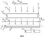
figure 8 montre un autre dispositif permettant une mise en œuvre de l'invention.Thefigure 8 shows another device allowing an implementation of the invention.
La
L'échantillon comporte des bactéries d'une ou plusieurs souches bactériennes, ainsi que des phages (ou bactériophages), d'une ou plusieurs souches virales. L'objectif de l'invention est de visualiser un effet optique, dans l'échantillon, d'une lyse induite par l'infection de bactéries par certains phages. De préférence, les bactéries ne sont pas marquées avec un marqueur colorimétrique ou fluorescent. L'invention est basée sur l'observation des interactions des bactéries avec l'onde lumineuse incidente. Plus précisément, la présence de bactéries dans l'échantillon induit une diffusion de l'onde lumineuse incidente. Plus le nombre de bactéries est élevé, plus l'onde lumineuse incidente est diffusée. Le capteur d'image comporte des pixels, généralement répartis selon un arrangement matriciel dans un plan de détection P. Le capteur d'image collecte onde d'exposition 14 résultant des interactions de l'onde lumineuse incidente 11 dans l'échantillon. L'onde lumineuse d'exposition 14, qui émerge de l'échantillon, est ainsi constituée:
d'une composante 12 de l'onde lumineuse incidente, non absorbée et non diffusée par l'échantillon. Cette composante est formée de photons usuellement désignés photons balistiques.d'une composante 13 résultant de la diffusion de l'onde lumineuse incidente par l'échantillon.
- a
component 12 of the incident light wave, not absorbed and not scattered by the sample. This component is formed from photons usually referred to as ballistic photons. - a
component 13 resulting from the scattering of the incident light wave by the sample.
Plus le nombre de bactéries est important, plus la composante de diffusion 13 est importante par rapport à la composante 12 non diffusée.The greater the number of bacteria, the greater the
De préférence, l'onde lumineuse incidente 11 s'étend selon une bande spectrale d'illumination Δλ s'étendant dans le domaine visible. Elle est de préférence comprise entre 450 nm et 650 nm, et encore de préférence entre 500 nm et 600 nm. C'est en effet entre 500 nm et 600 nm que la diffusion de l'onde lumineuse incidente 11 par les bactéries est maximale. De préférence, la largeur spectrale de la bande spectrale d'illumination est inférieure à 50 nm, voire inférieure à 40 nm. Par largeur spectrale, il est entendu la largeur à mi-hauteur, habituellement désignée par l'acronyme FWHM (Full Width at Half Maximum).Preferably, the
La source de lumière 10 est par exemple une diode électroluminescente ou une diode laser. Il peut également s'agir d'un écran à cristaux liquides, l'avantage étant de disposer d'une surface lumineuse étendue, de quelques cm2 à quelques dizaines de cm2, produisant une illumination uniforme. Un filtre passe-bande 16 peut être interposé entre la source de lumière 10 et l'échantillon 20, de façon à ajuster la bande spectrale de l'onde lumineuse atteignant l'échantillon.The
La distance D entre la source de lumière 10 et l'échantillon 20 est ajustée de telle sorte que la source de lumière soit considérée comme ponctuelle. Elle peut être comprise entre 5 cm et 20 ou 30 cm. Un diaphragme 17, définissant une ouverture de diamètre inférieur à 100 nm ou 200 µm, peut être interposé entre la source de lumière 10 et l'échantillon 20. De façon alternative, une fibre optique peut être interposée entre la source de lumière et l'échantillon, de façon à former une source de lumière ponctuelle.The distance D between the
Le capteur d'image 30 peut être un capteur de type CCD ou CMOS. De préférence, la surface de détection est supérieure à 10 mm2, voire supérieure à 1 cm2. Les inventeurs ont utilisé un capteur dont la surface de détection est de 14.9 mm x 22.3 mm soit de l'ordre de 3 cm2. Cela permet d'obtenir une image selon un champ d'observation élevée. Afin de maximiser le champ d'observation, la distance d entre l'échantillon 20 et le capteur d'image 30 est aussi faible que possible. Elle est de préférence inférieure à 1 cm. De préférence, l'échantillon 20 est placé au contact du capteur d'image, c'est-à-dire au contact d'une vitre de protection du capteur d'image, ou à quelques millimètres de cette dernière. On remarque l'absence d'optique de grossissement ou de formation d'image entre l'échantillon 20 et le capteur d'image 30. Cela n'empêche pas la présence de microlentilles de focalisation au niveau des pixels du capteur d'image 30. Ainsi, le capteur d'image 30 est disposé selon une configuration d'imagerie sans lentille. Cela permet de maximiser le champ d'observation du capteur d'image. Chaque image permet alors d'adresser un volume important d'échantillon. Le capteur d'image peut être un capteur monochrome ou un capteur couleur. Il permet une formation d'une image représentative de l'onde d'exposition 14.The
Le capteur d'image 30 est relié à une unité de traitement 40, recevant les images acquises par le capteur d'image. L'unité de traitement 40 est reliée à une mémoire 42 dans laquelle sont stockées des instructions pour la mise en œuvre de certaines étapes de traitement d'image décrites ci-après.The
L'épaisseur ε de l'échantillon 20 peut varier entre 100 µm et 1 cm ou 2 cm. La configuration de l'échantillon, et son épaisseur, peut varier selon les modes de réalisation décrits par la suite. D'une façon générale, l'échantillon comporte un support, dans lequel des bactéries, d'au moins une source bactérienne d'intérêt, sont mises en contact avec des phages, d'une souche virale. L'invention est ainsi mise à profit pour déterminer la souche virale et/ou la concentration des phages permettant une lyse des bactéries de la souche bactérienne d'intérêt.The thickness ε of
Un premier mode de réalisation est présenté en lien avec les
Selon ce mode de réalisation, l'échantillon est compartimenté selon des chambres fluidiques 221, 222, 22i isolées les unes des autres. L'indice i est un entier désignant le rang d'une carte fluidique, avec 1 ≤ i ≤ Ni où Ni correspond au nombre total de chambres fluidiques. La surface de chaque chambre fluidique, parallèlement au plan de détection P, peut par exemple être comprise entre 1 mm2 et 10 mm2. Les chambres fluidiques sont ménagées dans une carte fluidique 22. De préférence, chaque chambre fluidique est délimitée par une paroi opaque et/ou réfléchissante 231, 232, 23i. Cela évite qu'une lumière, diffusée à l'intérieur d'une chambre fluidique 22i, se propage dans une autre chambre fluidique, formant une lumière parasite dans cette dernière. Chaque chambre fluidique 22i comporte un échantillon élémentaire 20i pouvant être différent d'un autre échantillon élémentaire d'une autre chambre fluidique. Pour une souche bactérienne d'intérêt, chaque échantillon élémentaire 20i peut ainsi comporter des phages d'une même souche virale, selon une concentration prédéfinie.According to this embodiment, the sample is compartmentalized in
Ainsi, ce mode de réalisation permet de diviser l'échantillon en différentes zones spatiales, chaque zone spatiale correspondant à une chambre fluidique 22i, contenant un échantillon élémentaire 20i. Chaque échantillon élémentaire peut être paramétré par trois paramètres :
- la souche bactérienne ;
- la souche virale ;
- la concentration des phages de la souche virale.
- bacterial strain;
- the viral strain;
- the concentration of phages of the viral strain.
Entre deux échantillons élémentaires différents, correspondant à deux zones spatiales différentes, au moins un paramètre est différent, sauf dans le cas de répliquas.Between two different elementary samples, corresponding to two different spatial zones, at least one parameter is different, except in the case of replicas.
Le nombre de chambres fluidiques 22i est de préférence supérieur à 10, et encore de préférence supérieur à 100. Le recours à un capteur d'image de grande surface, tel que précédemment décrit, permet d'acquérir une image adressant simultanément un grand nombre de chambres fluidiques. Cela évite, ou limite, le recours à un mécanisme permettant une translation de l'échantillon 20 relativement au capteur d'image. On comprend que ce mode de réalisation permet d'effectuer un phagogramme. La carte fluidique 22 prend ainsi une forme de plaque à puits, permettant une analyse simultanée d'une multitude d'échantillons élémentaires différents les uns des autres.The number of
La carte fluidique 22 est par exemple réalisée, au moins en partie, dans un matériau plastique transparent, de préférence biocompatible, par exemple COC (Cyclic Olefin Copolymer) ou PMMA (Polymethylmetacrylate). Elle comporte des parois transversales 22t transparentes, qui confinent chaque chambre élémentaire 22i. Les parois transversales s'étendent de préférence perpendiculairement à un axe Z selon lequel se propage l'onde lumineuse 11 émise par la source de lumière 10.The
Chaque échantillon élémentaire comporte une quantité initiale de bactéries 2. En l'absence de phages, ou en l'absence d'effet notable des phages sur la souche bactérienne, les bactéries prolifèrent. Sur la
Plus la quantité de bactéries augmente, plus la diffusion de la lumière à travers une chambre fluidique augmente, ce qui réduit l'intensité de l'onde lumineuse d'exposition 14 se propageant vers le capteur d'image 30. Dans l'exemple représenté sur la
Les
- carte fluidique 22 : carte usinée dans une plaque d'aluminium, comportant huit chambres circulaires de diamètre 3 mm (soit une section de 7.1 mm2) ;
- volume de chaque chambre fluidique : 35 µL ;
- capteur d'image : capteur RGB CMOS Canon 1200D au format APS-C dimensions 14.9 mm x 22.3 mm - 18 millions de pixels;
- source de lumière 10 : Ecran LCD (Liquid Cristal Display) associé avec un filtre passe-bande (Thorlabs FB 560-10) centré sur une longueur d'onde de 560 nm et définissant une largeur à mi-hauteur de 10 nm ;
- température : 25 °C ;
- distance source de lumière - échantillon : 1 cm ;
- épaisseur de chaque chambre fluidique : 5 mm.
- fluidic card 22: card machined from an aluminum plate, comprising eight circular chambers with a diameter of 3 mm (ie a section of 7.1 mm 2 );
- volume of each fluidic chamber: 35 µL;
- image sensor: Canon 1200D RGB CMOS sensor in APS-C format dimensions 14.9 mm x 22.3 mm - 18 million pixels;
- light source 10: LCD screen (Liquid Crystal Display) associated with a band-pass filter (Thorlabs FB 560-10) centered on a wavelength of 560 nm and defining a width at half-height of 10 nm;
- temperature: 25 ° C;
- distance light source - sample: 1 cm;
- thickness of each fluidic chamber: 5 mm.
Chaque échantillon a été prélevé dans 9 mL de milieu nutritif Bouillon Tryptone Soja (usuellement désigné par l'acronyme TSB - Trypcase Soy Broth), dans lequel on avait introduit 250 µL d'une solution de culture de Pseudomonas putida ATCC12633, et 100 µL d'une suspension de phages Pseudomonas virus gh1. Une chambre de contrôle n'a pas fait l'objet d'ajout de phages. Une chambre de référence a été remplie avec le milieu nutritif liquide, sans bactériophages ni bactéries. Les suspensions de bactériophages avaient respectivement un titre infectieux égal à 6.109 ufp/mL, 6.108 ufp /mL et 6.107 ufp /mL. L'unité ufp est une unité connue de l'homme du métier signifiant unité formant plage, et usuellement désigné plaque forming unit. La détermination du titre de chaque suspension de phages a été effectuée selon une méthode de référence, en utilisant un milieu gélosé. Chaque chambre a fait l'objet d'un duplicat, sauf la chambre de contrôle et la chambre de référence.Each sample was taken from 9 mL of nutrient medium Tryptone Soy Broth (usually designated by the acronym TSB - Trypcase Soy Broth), into which 250 µL of a Pseudomonas putida ATCC12633 culture solution was introduced, and 100 µL of a suspension of Pseudomonas virus gh1 phages. A control chamber was not the subject of addition of phages. A reference chamber was filled with the liquid nutrient medium, without bacteriophages or bacteria. The suspensions of bacteriophages had respectively an infectious titre equal to 6.10 9 pfu / mL, 6.10 8 pfu / mL and 6.10 7 pfu / mL. The pfu unit is a unit known to those skilled in the art meaning plaque forming unit, and usually designated plaque forming unit. The determination of the titre of each phage suspension was carried out according to a reference method, using an agar medium. Each chamber was duplicated, except the control chamber and the reference chamber.
Le dispositif a été placé dans une enceinte thermostatée à 25°C. Des images ont été acquises toutes les 2 minutes durant une durée de 6 heures et 4 minutes. Les
Sur les
La quatrième colonne C4 comporte la chambre de référence C4-1 et la chambre de contrôle C4-2 ne comportant pas de phage.The fourth column C4 comprises the reference chamber C4-1 and the control chamber C4-2 not comprising phage.
Sur la chambre de contrôle C4-2, on constate un assombrissement de l'image. Cela est dû à la prolifération des bactéries en l'absence de phage.On the C4-2 control chamber, there is a darkening of the image. This is due to the proliferation of bacteria in the absence of phage.
Sur chaque colonne, on a effectué une moyenne de l'intensité (c'est-à-dire des niveaux de gris) des images acquises toutes les 2 minutes. On a calculé ensuite une densité optique équivalente DO(t), cette dernière étant obtenue par l'expression : ![]()
![]()
Où I(t) est l'intensité à un instant de mesure t, correspondant à une moyenne de niveaux de gris et I(t = 0) est l'intensité à l'instant initial t = 0. La constante 0.055 a été ajoutée afin que la densité optique à l'instant initial corresponde à celle mesurée par un spectrophotomètre.Where I ( t ) is the intensity at a measurement instant t, corresponding to an average of gray levels and I ( t = 0) is the intensity at the initial instant t = 0. The constant 0.055 has been added so that the optical density at the initial instant corresponds to that measured by a spectrophotometer.
La
A partir de ces courbes, on constate que sous l'action du phage, et pour l'ensemble des concentrations de phage considérées, la densité optique croît durant quelques heures après l'instant initial, mais se stabilise ensuite. Dans la chambre de contrôle, la densité optique augmente de façon continue. La comparaison des densités optiques respectivement mesurées dans la chambre de contrôle et dans les autres chambres montre l'inhibition du développement des bactéries par le phage. L'action du phage devient perceptible deux heures après l'instant initial.From these curves, it can be seen that under the action of the phage, and for all the phage concentrations considered, the optical density increases for a few hours after the initial instant, but then stabilizes. In the control chamber, the optical density increases continuously. The comparison of the optical densities respectively measured in the control chamber and in the other chambers shows the inhibition of the development of bacteria by the phage. The action of the phage becomes noticeable two hours after the initial moment.
On constate que l'invention permet un suivi en temps réel de l'action d'un phage sur des bactéries, et cela simultanément sur plusieurs échantillons élémentaires 20i. Il est possible de définir, pour chaque échantillon, un seuil de détection, dont le franchissement signifie qu'une prolifération bactérienne est suspectée dans l'échantillon. Un tel seuil peut être défini à partir de la densité optique moyenne mesurée sur l'échantillon de contrôle, ainsi que d'un indicateur de dispersion σ, par exemple l'écart type, à un instant de mesure ou en différents instants de mesure. Le seuil de détection peut être égal à µ + kσ, k étant un réel strictement positif, de préférence supérieur à 1 ou 2, et par exemple égal à 5. L'établissement d'un tel seuil de détection est aisément automatisable et permet de déterminer précocement la présence d'une prolifération bactérienne, par exemple entre 5h et 10h. D'autres seuils, basés sur une comparaison entre l'échantillon de contrôle et des échantillons comportant des phages peuvent être définis.It is noted that the invention allows real-time monitoring of the action of a phage on bacteria, and that simultaneously on several
La
Le milieu nutritif et/ou les phages peuvent être préalablement disposés dans chaque chambre fluidique 22i en étant par exemple à l'état lyophilisé. Dans l'exemple représenté sur la
De façon alternative, comme représenté sur la
Selon une autre possibilité, différentes chambres fluidiques comportent respectivement différentes souches bactériennes. Ces dernières peuvent être présentes à un état lyophilisé. Dans ce cas, le remplissage de chaque chambre fluidique peut être assuré par une entrée commune, à travers laquelle s'écoule une solution comportant des phages. L'objectif est alors d'identifier des souches virales pouvant être pertinentes pour certaines souches bactériennes. Selon cette possibilité, une étape préalable d'enrichissement de phages peut être prévue. Au cours de cette étape, les différentes souches bactériennes sont disposées dans une même enceinte, dans laquelle une solution de phages, comportant une seule souche virale ou plusieurs souches virales, est mélangée. Si, parmi les souches bactériennes présentes, une souche est sensible à un phage, ce dernier est répliqué, ce qui entraîne une augmentation de sa concentration. On obtient alors une amplification non spécifique de phages. Le mélange est ensuite filtré, de manière à retenir les bactéries, la taille de filtration pouvant être de l'ordre de 0.2 µm. Les souches bactériennes sont ensuite réparties dans différentes chambres fluidiques, de manière à aboutir à une seule souche bactérienne par puits. La solution filtrée est injectée dans chaque chambre fluidique, de manière à identifier la souche bactérienne sensible au phage.According to another possibility, different fluidic chambers respectively comprise different bacterial strains. The latter may be present in a lyophilized state. In this case, the filling of each fluidic chamber can be ensured by a common inlet, through which flows a solution comprising phages. The objective is then to identify viral strains that may be relevant for certain bacterial strains. According to this possibility, a preliminary phage enrichment step can be provided. During this step, the different bacterial strains are placed in the same enclosure, in which a phage solution, comprising a single viral strain or several viral strains, is mixed. If, among the bacterial strains present, a strain is sensitive to a phage, the latter is replicated, resulting in an increase in its concentration. A nonspecific amplification of phages is then obtained. The mixture is then filtered, so as to retain the bacteria, the filtration size possibly being of the order of 0.2 μm. The bacterial strains are then distributed in different fluidic chambers, so as to end up with a single bacterial strain per well. The filtered solution is injected into each fluidic chamber, so as to identify the bacterial strain sensitive to the phage.
Ce mode de réalisation peut être adapté pour tester l'effet de différentes souches virales sur une même souche bactérienne. Pour cela, différentes chambres fluidiques peuvent respectivement comporter des phages de différentes souches de phages. Au sein d'une chambre fluidique comportant une souche bactérienne sensible au phage, aucune prolifération bactérienne n'est observée, et le phage est amplifié de façon spécifique. Lorsque la souche bactérienne d'une chambre n'est pas sensible au phage, on observe une prolifération bactérienne.This embodiment can be adapted to test the effect of different viral strains on the same bacterial strain. For this, different fluidic chambers can respectively comprise phages of different phage strains. Within a fluidic chamber comprising a bacterial strain sensitive to the phage, no bacterial proliferation is observed, and the phage is amplified in a specific manner. When the bacterial strain in a chamber is not sensitive to phage, bacterial overgrowth is observed.
Dans ce mode de réalisation, on mesure une densité optique ou, de façon plus générale, une évolution d'une intensité lumineuse transmise par chaque échantillon élémentaire 20i et détectée par le capteur d'image. Afin d'obtenir une illumination uniforme de chaque échantillon élémentaire 20i, c'est-à-dire de chaque chambre fluidique élémentaire 22i, il est préférable que la source de lumière 10 soit une source étendue, par exemple un écran à cristaux liquides. Cela favorise la compacité du dispositif. De façon alternative, une source de lumière 10 ponctuelle peut être utilisée, sous réserve de l'éloigner suffisamment de l'échantillon.In this embodiment, an optical density or, more generally, an evolution of a light intensity transmitted by each
Les
La texturation de l'image résulte de la formation de figures d'interférences sur le capteur d'image, entre l'onde lumineuse 12 transmise par l'échantillon, et l'onde lumineuse 13 résultant de la diffusion de l'onde lumineuse incidente 11 par l'échantillon. Selon ce mode de réalisation, il est préférable que la source de lumière soit spatialement cohérente (ponctuelle), et suffisamment éloignée du détecteur pour que l'onde lumineuse incidente 11 atteigne l'échantillon sous la forme d'ondes planes, ou considérées comme telles.The texturing of the image results from the formation of interference figures on the image sensor, between the
Les
- carte fluidique 22 : lamelle de microscope sur laquelle une chambre fluidique Geneframe d'épaisseur 250 µm est déposée, la chambre fluidique étant refermée par une autre lamelle de microscope ;
- volume de chaque chambre fluidique : 25 µL ;
- capteur d'image : capteur monochrome 10.5 Mpixels de 1.67 µm de côté, formant une surface de détection de 29.5 mm2 (6.4 mm x 4.6 mm) ;
- source de lumière 10 : diode électroluminescente centrée sur la longueur d'onde de 650 nm avec une largeur à mi-hauteur de 40 nm ;
- température : 30 °C ;
- distance source de lumière - échantillon : 5 cm;
- épaisseur de chaque chambre fluidique : 250 µm.
- fluidic card 22: microscope slide on which a Geneframe 250 μm thick fluidic chamber is deposited, the fluidic chamber being closed by another microscope slide;
- volume of each fluidic chamber: 25 µL;
- image sensor: 10.5 Mpixel monochrome sensor with a side of 1.67 µm, forming a detection surface of 29.5 mm 2 (6.4 mm x 4.6 mm);
- light source 10: light emitting diode centered on the wavelength of 650 nm with a width at half height of 40 nm;
- temperature: 30 ° C;
- distance light source - sample: 5 cm;
- thickness of each fluidic chamber: 250 µm.
Dans cet exemple, la chambre fluidique n'est pas compartimentée. Il n'y a qu'un seul échantillon analysé. L'échantillon a été prélevé dans 9 mL de milieu nutritif Bouillon Tryptone Soja (usuellement désigné par l'acronyme TSB - Trypcase Soy Broth), dans lequel on avait introduit 250 µL d'une solution de culture de Pseudomonas putida ATCC12633, diluée d'un facteur 102. On a ajouté 100 µL d'une suspension de phages Pseudomonas virus gh1, connu pour infecter les bactéries Pseudomonas putida, dont la concentration était estimée à 7 105 ufp/mL. 30 µL d'échantillon ont été prélevés et introduits dans la carte fluidique 22.In this example, the fluidic chamber is not compartmentalized. There is only one sample analyzed. The sample was taken in 9 mL of nutrient medium Tryptone Soy Broth (usually designated by the acronym TSB - Trypcase Soy Broth), into which was introduced 250 μL of a culture solution of Pseudomonas putida ATCC12633, diluted with a factor of 10 2 . 100 μL of a suspension of Pseudomonas virus gh1 phages, known to infect Pseudomonas putida bacteria, the concentration of which was estimated at 7 10 5 pfu / ml, were added. 30 μL of sample were taken and introduced into the
Les
- pour un échantillon témoin, sans phage (courbe a)
- pour un échantillon avec des phages Pseudomonas virus gh1 - 7 105 ufp/mL (courbe b)
- for a control sample, without phage (curve a)
- for a sample with Pseudomonas virus gh1 phages - 7 10 5 pfu / mL (curve b)
Cette figure a été obtenue en effectuant une acquisition d'images par période de 10 minutes durant une durée de 18 heures. On observe que :
- pour l'échantillon témoin, le descripteur de texture augmente, sous l'effet de la prolifération bactérienne ;
- pour l'échantillon comportant les phages, le descripteur de texture stagne au cours du temps, traduisant une inhibition du développement des bactéries.
- for the control sample, the texture descriptor increases, under the effect of bacterial proliferation;
- for the sample comprising the phages, the texture descriptor stagnates over time, indicating an inhibition of the development of bacteria.
Il est possible de définir un seuil de détection, dont le franchissement signifie qu'une prolifération bactérienne est suspectée dans l'échantillon. Un tel seuil est déterminé à partir d'une valeur moyenne (ou médiane) µ et d'un indicateur de dispersion, par exemple l'écart type σ, du descripteur de texture, durant une courte durée temporelle après l'instant initial, par exemple 1h ou 2 h. Le seuil de détection peut être égal à µ + kσ, k étant un réel strictement positif, de préférence supérieur à 1 ou 2, et par exemple égal à 5. L'établissement d'un tel seuil de détection est aisément automatisable et permet de déterminer précocement la présence d'une prolifération bactérienne, par exemple entre 5h et 10h.It is possible to define a detection threshold, the crossing of which means that a bacterial proliferation is suspected in the sample. Such a threshold is determined from an average (or median) value µ and from a dispersion indicator, for example the standard deviation σ, of the texture descriptor, during a short period of time after the initial instant, by example 1h or 2h. The detection threshold may be equal to µ + kσ, k being a strictly positive real, preferably greater than 1 or 2, and for example equal to 5. The establishment of such a detection threshold is easily automated and makes it possible to early determine the presence of bacterial proliferation, for example between 5 a.m. and 10 a.m.
Les
Une telle gélose peut être constituée en mélangeant un milieu nutritif gélosé classique, porté en dessus de sa température de surfusion, par exemple entre 40°C et 60°C, avec une solution aqueuse comportant des bactéries et des phages. Ainsi, l'échantillon comporte une gélose molle, comprenant des bactéries et des phages. L'épaisseur de l'échantillon peut être comprise entre 2 mm à 5 mm. Le fait de disposer de bactéries et de phages selon une telle profondeur permet une meilleure mise en évidence de la lyse des bactéries. La fraction massique d'agar-agar est par exemple comprise entre 0.5% et 1.4%, ce qui est usuellement désigné par le terme "agar mou" (soft agar).Such an agar can be formed by mixing a conventional agar nutrient medium, brought above its supercooling temperature, for example between 40 ° C. and 60 ° C., with an aqueous solution comprising bacteria and phages. Thus, the sample comprises a soft agar, comprising bacteria and phages. The thickness of the sample can be between 2 mm to 5 mm. The fact of having bacteria and phages at such a depth allows a better demonstration of the lysis of the bacteria. The mass fraction of agar-agar is for example between 0.5% and 1.4%, which is usually designated by the term “soft agar”.
Selon une variante, une partie de l'épaisseur de l'échantillon est formée d'un milieu nutritif standard, sans bactérie ni phage. Il peut par exemple comprendre de l'agar-agar selon une fraction massique de 1.5 %. La fonction de ce milieu nutritif est de former un réservoir de nutriments pour les bactéries. Cependant, du fait que le procédé d'analyse d'un échantillon est rapide, de l'ordre de quelques heures ou de quelques dizaines d'heures, la présence d'un tel réservoir de nutriments n'est pas nécessaire. L'absence d'un tel réservoir est considérée comme avantageuse, car cela évite une diffusion, par ce dernier, de la lumière.According to one variant, part of the thickness of the sample is formed from a standard nutrient medium, without bacteria or phage. It can for example comprise agar-agar in a mass fraction of 1.5%. The function of this nutrient medium is to form a reservoir of nutrients for bacteria. However, because the method of analyzing a sample is rapid, of the order of a few hours or a few tens of hours, the presence of such a reservoir of nutrients is not necessary. The absence of such a reservoir is considered to be advantageous, since this prevents the latter from diffusing light.
Les
Les
- capteur d'image : capteur RGB CMOS Canon 1200D de dimensions 14.9 mm x 22.3 mm - 18 millions de pixels;
- source de lumière 10 : diode électroluminescente centrée sur la longueur d'onde de 560 nm;
- distance source de lumière - échantillon : environ 20 cm ;
- épaisseur de chaque chambre fluidique : 2 mm.
- image sensor: Canon 1200D RGB CMOS sensor measuring 14.9 mm x 22.3 mm - 18 million pixels;
- light source 10: light emitting diode centered on the wavelength of 560 nm;
- distance light source - sample: about 20 cm;
- thickness of each fluidic chamber: 2 mm.
L'instant initial t = 0 est considéré comme pouvant être confondu avec le dépôt de la préparation dans la boîte de Pétri. La répartition de la biomasse dans la boîte est considérée comme homogène. Au cours du temps, des plages de lyse se forment, ce qui correspond à des taches claires sur les images 4C, 4D et 4E. Sur la
Selon une autre approche, on utilise une carte fluidique compartimentée, telle que décrite en lien avec le premier mode de réalisation. La carte fluidique comporte des chambres fluidiques 22i séparées les unes des autres. Chaque chambre fluidique dispose d'un mélange de phages et d'un gélifiant à froid lyophilisé. Chaque chambre fluidique peut être alimentée par une solution comportant des bactéries. Il se forme alors une gélose, comportant un mélange de bactéries et de phages. Le gélifiant à froid est hydrosoluble. Il est configuré pour gélifier lorsqu'il est mis au contact d'une solution aqueuse, à température ambiante, formant alors un hydrogel. Il peut s'agir d'un polysaccharide hydrosoluble. Le gélifiant à froid peut être choisi parmi le carboxyméthylcellulose, la gomme de guar, la gomme arabique, la gomme gellane, la gomme de xanthane, ou un gélifiant obtenu à partir d'os d'animaux, par exemple porc, bœuf ou poulet.According to another approach, a compartmentalized fluidic card is used, as described in connection with the first embodiment. The fluidic card has
Une telle approche permet de diviser l'échantillon en différentes zones spatiales, chaque zone spatiale correspondant à une chambre fluidique.Such an approach makes it possible to divide the sample into different spatial zones, each spatial zone corresponding to a fluidic chamber.
De façon alternative, la carte fluidique est portée, lors de son remplissage, à une température de surfusion de la gélose comportant les bactéries. Ainsi, la gélose comportant les bactéries peut remplir chaque chambre fluidique, de façon à se mélanger, dans chaque chambre, aux phages préalablement disposés à l'état lyophilisé. Cela évite le recours à un gélifiant à froid, ce dernier pouvant être diffusant.Alternatively, the fluidic card is brought, during its filling, to a temperature of supercooling of the agar containing the bacteria. Thus, the agar containing the bacteria can fill each fluidic chamber, so as to mix, in each chamber, with the phages previously placed in the lyophilized state. This avoids the need for a cold gelling agent, the latter being able to diffuse.
Un troisième mode de réalisation est représenté sur les
Les
On a préparé un échantillon en mélangeant 120 µL d'un milieu de culture, comportant Pseudomonas putida ATCC12633 et 6mL d'agar-agar mou (7.5 g/L d'agar-agar). Cette préparation a été coulée dans une boîte de pétri, ce qui a permis d'obtenir une gélose d'épaisseur 2 mm environ. On a ensuite déposé, à la surface, des gouttes de 5 µL comportant :
- soit une suspension de phages Pseudomonas virus gh1 dont le titre était estimé à 3.6 106 ufp/mL ;
- soit un diluant pharmaceutique pour simuler la présence d'un phage non actif sur la souche bactérienne utilisée.
- or a suspension of Pseudomonas virus gh1 phages, the titre of which was estimated at 3.6 10 6 pfu / ml;
- or a pharmaceutical diluent to simulate the presence of a phage inactive on the bacterial strain used.
Les gouttes ont été séchées (15 minutes), puis l'échantillon a été déposé dans une enceinte thermostatée à 28 °C.. Les paramètres expérimentaux sont les suivants :
- capteur d'image : capteur RGB Canon CMOS 1200D de dimensions 14.9 mm x 22.3 mm - 18 millions de pixels;
- source de lumière 10 : diode électroluminescente centrée sur la longueur d'onde de 560 nm;
- distance source de lumière - échantillon : environ 20 cm ;
- épaisseur de la chambre fluidique : 2 mm.
- image sensor: Canon CMOS 1200D RGB sensor measuring 14.9 mm x 22.3 mm - 18 million pixels;
- light source 10: light emitting diode centered on the wavelength of 560 nm;
- distance light source - sample: about 20 cm;
- thickness of the fluidic chamber: 2 mm.
Une fine épaisseur (entre 0.1 mm et 1 mm) d'huile 25 peut être disposée sur l'échantillon, de façon à éviter un dessèchement. L'huile 25 peut être de l'huile de silicone, permettant une diffusion de l'oxygène jusqu'au milieu nutritif.A thin thickness (between 0.1 mm and 1 mm) of oil can be placed on the sample, so as to avoid drying out. The oil may be silicone oil, allowing diffusion of oxygen to the nutrient medium.
Les
Une tel mode de réalisation permet de diviser l'échantillon en différentes zones spatiales, chaque zone spatiale correspondant à une position au niveau de laquelle une goutte a été déposée. L'image acquise par le capteur d'image permet d'analyser simultanément plusieurs zones spatiales.Such an embodiment makes it possible to divide the sample into different spatial zones, each spatial zone corresponding to a position at which a drop has been deposited. The image acquired by the image sensor makes it possible to simultaneously analyze several spatial zones.
Un quatrième mode de réalisation est schématisé sur les
Les
Une tel mode de réalisation permet de diviser l'échantillon en différentes zones spatiales, chaque zone spatiale correspondant à une localisation au niveau de laquelle une goutte a été déposée. L'image acquise par le capteur d'image permet d'analyser simultanément plusieurs zones spatiales.Such an embodiment makes it possible to divide the sample into different spatial zones, each spatial zone corresponding to a location at which a drop has been deposited. The image acquired by the image sensor makes it possible to simultaneously analyze several spatial zones.
La
Etape 100 : mise en contact de souches bactériennes avec des phages. Cette étape peut être réalisée dans une seule chambre fluidique (cf. deuxième, troisième, ou quatrième modes de réalisation), ou dans différentes chambres fluidiques (cf. premier mode de réalisation). Au cours de cette étape, l'échantillon peut être divisé en différentes zones spatiales. Ces zones spatiales peuvent correspondre :
- à des chambres fluidiques différentes les unes des autres (cf. premier mode de réalisation);
- ou à des positions différentes au niveau desquelles des gouttes d'une solution aqueuse de phages sont déposées sur une gélose comportant des bactéries (troisième mode de réalisation) ;
- ou à des positions différentes au niveau desquelles des gouttes d'une solution aqueuse de bactéries sont déposées sur une gélose comportant des phages (quatrième mode de réalisation)
- to fluidic chambers which are different from each other (cf. first embodiment);
- or at different positions at which drops of an aqueous solution of phages are deposited on an agar containing bacteria (third embodiment);
- or at different positions at which drops of an aqueous solution of bacteria are deposited on an agar containing phages (fourth embodiment)
Etape 110 : illumination de l'échantillon à l'aide de la source de lumière ;Step 110: illumination of the sample using the light source;
Etape 120 : acquisition d'une image par le capteur d'image ;Step 120: acquisition of an image by the image sensor;
Etape 130 : analyse de l'image par l'unité de traitement, de façon à évaluer une sensibilité d'au moins une souche bactérienne à au moins une souche virale.Step 130: analysis of the image by the processing unit, so as to evaluate a sensitivity of at least one bacterial strain to at least one viral strain.
Les avantages du procédé tel que précédemment décrit sont :
- une détection précoce de la lyse de bactéries par des phages. En effet, en utilisant les procédés de l'art antérieur, on considère que des résultats exploitables sont obtenus en au moins une journée
- une possibilité de mise en oeuvre de façon à adresser simultanément différentes souches bactériennes en combinaison avec différentes souches de phages, ou différents ratios entre les quantités de souches de phage et la quantité de souches de bactéries ;
- simplicité de mise en œuvre, sans nécessiter de matériel onéreux.
- early detection of bacterial lysis by phages. Indeed, by using the methods of the prior art, it is considered that exploitable results are obtained in at least one day.
- a possibility of implementation so as to simultaneously address different bacterial strains in combination with different phage strains, or different ratios between the quantities of phage strains and the quantity of bacterial strains;
- simplicity of implementation, without requiring expensive equipment.
La
- le plan image est décalé par rapport au plan de détection d'une distance de défocalisation image;
- et/ou le plan objet est décalé par rapport au plan de l'échantillon d'une distance de défocalisation objet.
- the image plane is offset with respect to the detection plane by an image defocus distance;
- and / or the object plane is offset from the plane of the sample by an object defocus distance.
La distance de défocalisation image et/ou la distance de défocalisation objet est de préférence inférieure à 1 mm, voire inférieure à 500 µm.The image defocus distance and / or the object defocus distance is preferably less than 1 mm, or even less than 500 μm.
Selon une variante, le système optique permet une conjugaison du capteur d'image avec l'échantillon. Le plan image est alors confondu avec le plan de détection formé par le capteur d'image. Le plan objet est alors confondu avec le plan de l'échantillon. Selon ce mode de réalisation, l'image est acquise selon une configuration focalisée.According to one variant, the optical system allows conjugation of the image sensor with the sample. The image plane is then merged with the detection plane formed by the image sensor. The object plane is then merged with the sample plane. According to this embodiment, the image is acquired according to a focused configuration.
Sur la
Par rapport aux configurations exposées en lien avec la
Claims (16)
le procédé étant tel que :
the process being such that:
le procédé étant tel que chaque zone spatiale est associée à une région d'intérêt (ROIi) de chaque image acquise, deux zones spatiales différentes étant associées à deux régions d'intérêt différentes, de façon que l'analyse d'une même image acquise permet d'obtenir une information relative à la sensibilité d'une souche bactérienne à l'égard d'une souche virale de bactériophages, et cela dans différentes zones spatiales.
the method being such that each spatial zone is associated with a region of interest (ROI i ) of each acquired image, two different spatial zones being associated with two different regions of interest, so that the analysis of the same image acquired makes it possible to obtain information relating to the sensitivity of a bacterial strain with regard to a viral strain of bacteriophages, and this in different spatial zones.
de telle sorte que lors de l'étape a), la mise en contact des bactériophages avec les bactéries est effectuée en utilisant la solution enrichie en bactériophages.
so that during step a), the bringing into contact of the bacteriophages with the bacteria is carried out using the solution enriched in bacteriophages.
Applications Claiming Priority (1)
| Application Number | Priority Date | Filing Date | Title |
|---|---|---|---|
| FR1912804A FR3103198A1 (en) | 2019-11-15 | 2019-11-15 | Method for determining the sensitivity of a bacterial strain to a bacteriophage virus. |
Publications (1)
| Publication Number | Publication Date |
|---|---|
| EP3822360A1 true EP3822360A1 (en) | 2021-05-19 |
Family
ID=70295198
Family Applications (1)
| Application Number | Title | Priority Date | Filing Date |
|---|---|---|---|
| EP20207254.2A Pending EP3822360A1 (en) | 2019-11-15 | 2020-11-12 | Method for determining the sensitivity of a bacterial strain to a bacteriophage virus |
Country Status (3)
| Country | Link |
|---|---|
| US (1) | US20210147899A1 (en) |
| EP (1) | EP3822360A1 (en) |
| FR (1) | FR3103198A1 (en) |
Citations (5)
| Publication number | Priority date | Publication date | Assignee | Title |
|---|---|---|---|---|
| EP0745667A1 (en) | 1995-05-31 | 1996-12-04 | bioMerieux Vitek, Inc. | Improved sealant for sample holder |
| EP2261379A1 (en) * | 2008-04-09 | 2010-12-15 | Consejo Superior De Investigaciones Científicas | Method and system for detecting and/or quantifying bacteriophages, use of a microelectronic sensor device for detecting said bacteriophages and microelectronic sensor device for implementing said method |
| EP2747578A1 (en) | 2011-08-25 | 2014-07-02 | Proteon Pharmaceuticals S.A. | The method of obtaining a strain of bacteriophage, specific strains of bacteriophage and use thereof |
| WO2019018886A1 (en) * | 2017-07-23 | 2019-01-31 | Future Biosolutions Pty Ltd | Method for rapid detection and enumeration of viruses, bacteriophage and/or bacteria |
| WO2019188886A1 (en) | 2018-03-30 | 2019-10-03 | パイオニア株式会社 | Terminal device, information processing method, and storage medium |
Family Cites Families (1)
| Publication number | Priority date | Publication date | Assignee | Title |
|---|---|---|---|---|
| US9569664B2 (en) * | 2010-10-26 | 2017-02-14 | California Institute Of Technology | Methods for rapid distinction between debris and growing cells |
-
2019
- 2019-11-15 FR FR1912804A patent/FR3103198A1/en active Pending
-
2020
- 2020-11-12 EP EP20207254.2A patent/EP3822360A1/en active Pending
- 2020-11-13 US US17/097,687 patent/US20210147899A1/en active Pending
Patent Citations (5)
| Publication number | Priority date | Publication date | Assignee | Title |
|---|---|---|---|---|
| EP0745667A1 (en) | 1995-05-31 | 1996-12-04 | bioMerieux Vitek, Inc. | Improved sealant for sample holder |
| EP2261379A1 (en) * | 2008-04-09 | 2010-12-15 | Consejo Superior De Investigaciones Científicas | Method and system for detecting and/or quantifying bacteriophages, use of a microelectronic sensor device for detecting said bacteriophages and microelectronic sensor device for implementing said method |
| EP2747578A1 (en) | 2011-08-25 | 2014-07-02 | Proteon Pharmaceuticals S.A. | The method of obtaining a strain of bacteriophage, specific strains of bacteriophage and use thereof |
| WO2019018886A1 (en) * | 2017-07-23 | 2019-01-31 | Future Biosolutions Pty Ltd | Method for rapid detection and enumeration of viruses, bacteriophage and/or bacteria |
| WO2019188886A1 (en) | 2018-03-30 | 2019-10-03 | パイオニア株式会社 | Terminal device, information processing method, and storage medium |
Non-Patent Citations (7)
| Title |
|---|
| ESTRELLA LUIS ET AL., CHARACTERIZATION OF NOVEL STAPHYLOCOCCUS AUREUS LYTIC PHAGE AND DEFINING THEIR COMBINATORIAL VIRULENCE USING THE OMNILOG SYSTEM |
| HENRY M ET AL., DEVELOPMENT OF A HIGH THROUGHPUT ASSAY FOR INDIRECTLY MEASURING PHAGE GROWTH USING THE OMNILOG TM SYSTEM |
| LUIS A. ESTRELLA ET AL: "Characterization of novel Staphylococcus aureus lytic phage and defining their combinatorial virulence using the OmniLog system", BACTERIOPHAGE, vol. 6, no. 3, 2 July 2016 (2016-07-02), pages e1219440, XP055717278, DOI: 10.1080/21597081.2016.1219440 * |
| MATTHEW HENRY ET AL: "Development of a high throughput assay for indirectly measuring phage growth using the OmniLog TM system", BACTERIOPHAGE, vol. 2, no. 3, July 2012 (2012-07-01), pages 159 - 167, XP055717274, DOI: 10.4161/bact.21440 * |
| PRISCA PERLEMOINE ET AL: "Phage susceptibility testing with lensless imaging technique (Conference Presentation)", PHOTONIC DIAGNOSIS, MONITORING, PREVENTION, AND TREATMENT OF INFECTIONS AND INFLAMMATORY DISEASES 2020, 10 March 2020 (2020-03-10), pages 13, XP055715784, ISBN: 978-1-5106-3210-3, DOI: 10.1117/12.2543785 * |
| PRISCA PERLEMOINE: "Optical phage susceptibility testing: developing instrumentation for phage therapy", BACTO-GRE 2019, 17 April 2019 (2019-04-17), XP055715755 * |
| PRISCA PERLEMOINE: "Poster P-119 : Phagogramme Optique : L'imagerie sans lentille comme outil de diagnostic pour la phagothérapie", 39E RÉUNION INTERDISCIPLINAIRE DE CHIMIOTHÉRAPIE ANTI-INFECTIEUSE, 16 December 2019 (2019-12-16), pages 69, XP055715790 * |
Also Published As
| Publication number | Publication date |
|---|---|
| US20210147899A1 (en) | 2021-05-20 |
| FR3103198A1 (en) | 2021-05-21 |
Similar Documents
| Publication | Publication Date | Title |
|---|---|---|
| EP2488847B1 (en) | Method for optically detecting dissolved micrometric objects | |
| Arandian et al. | Optical Imaging Approaches to Monitor Static and Dynamic Cell‐on‐Chip Platforms: A Tutorial Review | |
| US10533207B2 (en) | Bioactivity testing structure for single cell tracking using gelling agents | |
| Siemons et al. | Robust adaptive optics for localization microscopy deep in complex tissue | |
| CN111133295A (en) | Method for determining antibiotic sensitivity by Raman imaging technology | |
| EP0333561B1 (en) | Device and method for the detection and counting of fluorescent particles on a solid support | |
| EP1774295A1 (en) | Device for reading plates bearing biological reaction support microdepositions | |
| EP2246691B1 (en) | Method and device for platelets counting | |
| JP2014505465A (en) | A cell counting method for life / death discrimination by differential light absorption | |
| WO1989008714A1 (en) | Qualititive or quantititive process for researching microorganisms, installation for application of said process | |
| FR3060746A1 (en) | METHOD FOR NUMBERING PARTICLES IN AN IMAGING SAMPLE WITHOUT LENS | |
| EP3115770A1 (en) | Method for estimating an amount of analyte in a liquid | |
| EP3766580A1 (en) | Microfluidic device for preparing and analysing a biological sample | |
| EP2678661B1 (en) | Method for observing a sample | |
| EP3161145B1 (en) | Method for detecting a presence or absence of biological particles | |
| EP3822360A1 (en) | Method for determining the sensitivity of a bacterial strain to a bacteriophage virus | |
| FR3066503A1 (en) | METHOD FOR ANALYZING MICROORGANISMS | |
| FR3075825A1 (en) | METHOD AND SYSTEM FOR IDENTIFYING GRAM TYPE OF A BACTERIUM | |
| EP3054284B1 (en) | Method for correcting a raman signal backscattered by a sample, and associated device | |
| EP4217713A1 (en) | Method for analysing a biological sample comprising determination of the spatial distribution of biomass along the optical axis | |
| FR2935803A1 (en) | DEVICE AND METHOD FOR OPTICALLY MEASURING THE TRANSMISSION AND DIFFUSION OF EYE MEDIA | |
| FR2719905A1 (en) | Measuring device for the control of bacteriological quality of water. | |
| EP3766581B1 (en) | Micro-fluidic system including an amplification reaction chamber | |
| WO2021198443A1 (en) | Method for characterising micro-organisms using transmission imaging | |
| BE1026469B1 (en) | MICROFLUIDIC DEVICE FOR SELECTING BACTERIOPHAGES CAPABLE OF INFECTING BACTERIA CONTAINED IN A SAMPLE |
Legal Events
| Date | Code | Title | Description |
|---|---|---|---|
| PUAI | Public reference made under article 153(3) epc to a published international application that has entered the european phase |
Free format text: ORIGINAL CODE: 0009012 |
|
| STAA | Information on the status of an ep patent application or granted ep patent |
Free format text: STATUS: REQUEST FOR EXAMINATION WAS MADE |
|
| 17P | Request for examination filed |
Effective date: 20201120 |
|
| AK | Designated contracting states |
Kind code of ref document: A1 Designated state(s): AL AT BE BG CH CY CZ DE DK EE ES FI FR GB GR HR HU IE IS IT LI LT LU LV MC MK MT NL NO PL PT RO RS SE SI SK SM TR |
|
| STAA | Information on the status of an ep patent application or granted ep patent |
Free format text: STATUS: EXAMINATION IS IN PROGRESS |
|
| 17Q | First examination report despatched |
Effective date: 20230922 |