EP3785740B1 - Gradient mineralized bone extracellular matrix material and preparation method therefor - Google Patents
Gradient mineralized bone extracellular matrix material and preparation method therefor Download PDFInfo
- Publication number
- EP3785740B1 EP3785740B1 EP19910842.4A EP19910842A EP3785740B1 EP 3785740 B1 EP3785740 B1 EP 3785740B1 EP 19910842 A EP19910842 A EP 19910842A EP 3785740 B1 EP3785740 B1 EP 3785740B1
- Authority
- EP
- European Patent Office
- Prior art keywords
- bone
- deionized water
- hours
- shaking
- embedding box
- Prior art date
- Legal status (The legal status is an assumption and is not a legal conclusion. Google has not performed a legal analysis and makes no representation as to the accuracy of the status listed.)
- Active
Links
- 210000000988 bone and bone Anatomy 0.000 title claims description 240
- 102000010834 Extracellular Matrix Proteins Human genes 0.000 title claims description 100
- 108010037362 Extracellular Matrix Proteins Proteins 0.000 title claims description 100
- 210000002744 extracellular matrix Anatomy 0.000 title claims description 100
- 239000011159 matrix material Substances 0.000 title claims description 26
- 238000002360 preparation method Methods 0.000 title claims description 23
- 239000000463 material Substances 0.000 claims description 148
- 230000033558 biomineral tissue development Effects 0.000 claims description 95
- 239000008367 deionised water Substances 0.000 claims description 95
- 229910021641 deionized water Inorganic materials 0.000 claims description 95
- XLYOFNOQVPJJNP-UHFFFAOYSA-N water Chemical compound O XLYOFNOQVPJJNP-UHFFFAOYSA-N 0.000 claims description 95
- 239000000243 solution Substances 0.000 claims description 42
- NLKNQRATVPKPDG-UHFFFAOYSA-M potassium iodide Chemical compound [K+].[I-] NLKNQRATVPKPDG-UHFFFAOYSA-M 0.000 claims description 39
- 210000002805 bone matrix Anatomy 0.000 claims description 31
- CSCPPACGZOOCGX-UHFFFAOYSA-N Acetone Chemical compound CC(C)=O CSCPPACGZOOCGX-UHFFFAOYSA-N 0.000 claims description 30
- HEMHJVSKTPXQMS-UHFFFAOYSA-M Sodium hydroxide Chemical compound [OH-].[Na+] HEMHJVSKTPXQMS-UHFFFAOYSA-M 0.000 claims description 27
- 229940124158 Protease/peptidase inhibitor Drugs 0.000 claims description 26
- 239000000137 peptide hydrolase inhibitor Substances 0.000 claims description 26
- WCUXLLCKKVVCTQ-UHFFFAOYSA-M Potassium chloride Chemical compound [Cl-].[K+] WCUXLLCKKVVCTQ-UHFFFAOYSA-M 0.000 claims description 22
- 230000001954 sterilising effect Effects 0.000 claims description 20
- IJGRMHOSHXDMSA-UHFFFAOYSA-N Atomic nitrogen Chemical compound N#N IJGRMHOSHXDMSA-UHFFFAOYSA-N 0.000 claims description 18
- 230000002328 demineralizing effect Effects 0.000 claims description 18
- 238000005115 demineralization Methods 0.000 claims description 17
- 239000007853 buffer solution Substances 0.000 claims description 15
- STCOOQWBFONSKY-UHFFFAOYSA-N tributyl phosphate Chemical compound CCCCOP(=O)(OCCCC)OCCCC STCOOQWBFONSKY-UHFFFAOYSA-N 0.000 claims description 15
- 210000001519 tissue Anatomy 0.000 claims description 13
- GPRLSGONYQIRFK-MNYXATJNSA-N triton Chemical compound [3H+] GPRLSGONYQIRFK-MNYXATJNSA-N 0.000 claims description 13
- 238000002604 ultrasonography Methods 0.000 claims description 13
- 239000008280 blood Substances 0.000 claims description 11
- 210000004369 blood Anatomy 0.000 claims description 11
- 238000005520 cutting process Methods 0.000 claims description 11
- 239000012535 impurity Substances 0.000 claims description 11
- 239000002504 physiological saline solution Substances 0.000 claims description 11
- 239000001103 potassium chloride Substances 0.000 claims description 11
- 235000011164 potassium chloride Nutrition 0.000 claims description 11
- 239000000872 buffer Substances 0.000 claims description 9
- 230000008014 freezing Effects 0.000 claims description 9
- 238000007710 freezing Methods 0.000 claims description 9
- 239000007788 liquid Substances 0.000 claims description 9
- 229910052757 nitrogen Inorganic materials 0.000 claims description 9
- 238000010257 thawing Methods 0.000 claims description 9
- 230000005847 immunogenicity Effects 0.000 claims description 6
- 238000012545 processing Methods 0.000 claims description 5
- 241000124008 Mammalia Species 0.000 claims description 2
- 230000007547 defect Effects 0.000 description 23
- 230000008439 repair process Effects 0.000 description 23
- 210000004027 cell Anatomy 0.000 description 17
- 102000008186 Collagen Human genes 0.000 description 15
- 108010035532 Collagen Proteins 0.000 description 15
- 229920001436 collagen Polymers 0.000 description 15
- 238000000034 method Methods 0.000 description 13
- 238000011069 regeneration method Methods 0.000 description 13
- 210000004204 blood vessel Anatomy 0.000 description 12
- 230000000694 effects Effects 0.000 description 12
- 210000002901 mesenchymal stem cell Anatomy 0.000 description 12
- 210000001991 scapula Anatomy 0.000 description 12
- 230000008929 regeneration Effects 0.000 description 11
- BHPQYMZQTOCNFJ-UHFFFAOYSA-N Calcium cation Chemical compound [Ca+2] BHPQYMZQTOCNFJ-UHFFFAOYSA-N 0.000 description 10
- 229910001424 calcium ion Inorganic materials 0.000 description 10
- FAPWRFPIFSIZLT-UHFFFAOYSA-M Sodium chloride Chemical compound [Na+].[Cl-] FAPWRFPIFSIZLT-UHFFFAOYSA-M 0.000 description 9
- 239000011780 sodium chloride Substances 0.000 description 9
- 238000005406 washing Methods 0.000 description 9
- ZGTMUACCHSMWAC-UHFFFAOYSA-L EDTA disodium salt (anhydrous) Chemical compound [Na+].[Na+].OC(=O)CN(CC([O-])=O)CCN(CC(O)=O)CC([O-])=O ZGTMUACCHSMWAC-UHFFFAOYSA-L 0.000 description 8
- 239000011575 calcium Substances 0.000 description 8
- 108010049931 Bone Morphogenetic Protein 2 Proteins 0.000 description 7
- 239000011521 glass Substances 0.000 description 7
- 230000012010 growth Effects 0.000 description 7
- 229910052500 inorganic mineral Inorganic materials 0.000 description 7
- 239000011707 mineral Substances 0.000 description 7
- 235000010755 mineral Nutrition 0.000 description 7
- 238000002054 transplantation Methods 0.000 description 7
- 102100024506 Bone morphogenetic protein 2 Human genes 0.000 description 6
- 230000033115 angiogenesis Effects 0.000 description 6
- 210000001185 bone marrow Anatomy 0.000 description 6
- 229910052791 calcium Inorganic materials 0.000 description 6
- 230000003247 decreasing effect Effects 0.000 description 6
- 238000001514 detection method Methods 0.000 description 6
- 238000011156 evaluation Methods 0.000 description 6
- 239000000835 fiber Substances 0.000 description 6
- 230000011164 ossification Effects 0.000 description 6
- 230000008569 process Effects 0.000 description 6
- 230000001737 promoting effect Effects 0.000 description 6
- 238000011160 research Methods 0.000 description 6
- 230000017423 tissue regeneration Effects 0.000 description 6
- OYPRJOBELJOOCE-UHFFFAOYSA-N Calcium Chemical compound [Ca] OYPRJOBELJOOCE-UHFFFAOYSA-N 0.000 description 5
- 230000021164 cell adhesion Effects 0.000 description 5
- 230000007423 decrease Effects 0.000 description 5
- 229910052698 phosphorus Inorganic materials 0.000 description 5
- 230000001105 regulatory effect Effects 0.000 description 5
- OAICVXFJPJFONN-UHFFFAOYSA-N Phosphorus Chemical compound [P] OAICVXFJPJFONN-UHFFFAOYSA-N 0.000 description 4
- 102000009524 Vascular Endothelial Growth Factor A Human genes 0.000 description 4
- 108010073929 Vascular Endothelial Growth Factor A Proteins 0.000 description 4
- 239000011574 phosphorus Substances 0.000 description 4
- 108090000623 proteins and genes Proteins 0.000 description 4
- 101710146526 Dual specificity mitogen-activated protein kinase kinase 1 Proteins 0.000 description 3
- 206010018852 Haematoma Diseases 0.000 description 3
- 238000004630 atomic force microscopy Methods 0.000 description 3
- 239000012620 biological material Substances 0.000 description 3
- 230000008859 change Effects 0.000 description 3
- 230000004069 differentiation Effects 0.000 description 3
- 239000000284 extract Substances 0.000 description 3
- 238000011532 immunohistochemical staining Methods 0.000 description 3
- 238000001727 in vivo Methods 0.000 description 3
- 239000000126 substance Substances 0.000 description 3
- 102100031480 Dual specificity mitogen-activated protein kinase kinase 1 Human genes 0.000 description 2
- 206010020649 Hyperkeratosis Diseases 0.000 description 2
- 241000283973 Oryctolagus cuniculus Species 0.000 description 2
- -1 PO4-3 Chemical compound 0.000 description 2
- 230000015572 biosynthetic process Effects 0.000 description 2
- 230000010478 bone regeneration Effects 0.000 description 2
- 229910052799 carbon Inorganic materials 0.000 description 2
- 230000001413 cellular effect Effects 0.000 description 2
- 238000012512 characterization method Methods 0.000 description 2
- 208000037265 diseases, disorders, signs and symptoms Diseases 0.000 description 2
- 238000009826 distribution Methods 0.000 description 2
- 238000011049 filling Methods 0.000 description 2
- 239000003102 growth factor Substances 0.000 description 2
- 238000007490 hematoxylin and eosin (H&E) staining Methods 0.000 description 2
- 230000002163 immunogen Effects 0.000 description 2
- 238000002513 implantation Methods 0.000 description 2
- 230000006698 induction Effects 0.000 description 2
- 150000002500 ions Chemical class 0.000 description 2
- 230000014759 maintenance of location Effects 0.000 description 2
- 238000004519 manufacturing process Methods 0.000 description 2
- 230000009818 osteogenic differentiation Effects 0.000 description 2
- 230000035755 proliferation Effects 0.000 description 2
- 102000004169 proteins and genes Human genes 0.000 description 2
- 238000011002 quantification Methods 0.000 description 2
- 239000011165 3D composite Substances 0.000 description 1
- 208000010392 Bone Fractures Diseases 0.000 description 1
- 241000283690 Bos taurus Species 0.000 description 1
- OKTJSMMVPCPJKN-UHFFFAOYSA-N Carbon Chemical compound [C] OKTJSMMVPCPJKN-UHFFFAOYSA-N 0.000 description 1
- 102000000503 Collagen Type II Human genes 0.000 description 1
- 108010041390 Collagen Type II Proteins 0.000 description 1
- 238000000116 DAPI staining Methods 0.000 description 1
- 206010017076 Fracture Diseases 0.000 description 1
- 206010061218 Inflammation Diseases 0.000 description 1
- 102000043136 MAP kinase family Human genes 0.000 description 1
- 108091054455 MAP kinase family Proteins 0.000 description 1
- 238000011529 RT qPCR Methods 0.000 description 1
- 239000002253 acid Substances 0.000 description 1
- 239000000853 adhesive Substances 0.000 description 1
- 230000001070 adhesive effect Effects 0.000 description 1
- 230000000735 allogeneic effect Effects 0.000 description 1
- 238000004458 analytical method Methods 0.000 description 1
- 230000002491 angiogenic effect Effects 0.000 description 1
- 230000009286 beneficial effect Effects 0.000 description 1
- 230000002146 bilateral effect Effects 0.000 description 1
- 238000004113 cell culture Methods 0.000 description 1
- 230000010261 cell growth Effects 0.000 description 1
- 239000003153 chemical reaction reagent Substances 0.000 description 1
- 238000004140 cleaning Methods 0.000 description 1
- 238000007796 conventional method Methods 0.000 description 1
- 238000012258 culturing Methods 0.000 description 1
- 230000006378 damage Effects 0.000 description 1
- 230000007850 degeneration Effects 0.000 description 1
- 230000001419 dependent effect Effects 0.000 description 1
- 238000010586 diagram Methods 0.000 description 1
- 201000010099 disease Diseases 0.000 description 1
- 208000035475 disorder Diseases 0.000 description 1
- 239000006185 dispersion Substances 0.000 description 1
- 238000004043 dyeing Methods 0.000 description 1
- 230000000431 effect on proliferation Effects 0.000 description 1
- 238000000635 electron micrograph Methods 0.000 description 1
- 238000002149 energy-dispersive X-ray emission spectroscopy Methods 0.000 description 1
- 238000002474 experimental method Methods 0.000 description 1
- 238000003125 immunofluorescent labeling Methods 0.000 description 1
- 239000007943 implant Substances 0.000 description 1
- 210000004969 inflammatory cell Anatomy 0.000 description 1
- 230000004054 inflammatory process Effects 0.000 description 1
- 230000028709 inflammatory response Effects 0.000 description 1
- 230000002427 irreversible effect Effects 0.000 description 1
- 230000003902 lesion Effects 0.000 description 1
- 239000003446 ligand Substances 0.000 description 1
- 230000007774 longterm Effects 0.000 description 1
- 230000035800 maturation Effects 0.000 description 1
- 230000007246 mechanism Effects 0.000 description 1
- 230000001404 mediated effect Effects 0.000 description 1
- 238000010603 microCT Methods 0.000 description 1
- 210000004088 microvessel Anatomy 0.000 description 1
- 210000003205 muscle Anatomy 0.000 description 1
- 231100000252 nontoxic Toxicity 0.000 description 1
- 230000003000 nontoxic effect Effects 0.000 description 1
- 210000000963 osteoblast Anatomy 0.000 description 1
- 230000002188 osteogenic effect Effects 0.000 description 1
- 230000002138 osteoinductive effect Effects 0.000 description 1
- 239000011148 porous material Substances 0.000 description 1
- 230000001172 regenerating effect Effects 0.000 description 1
- 238000001878 scanning electron micrograph Methods 0.000 description 1
- 238000004626 scanning electron microscopy Methods 0.000 description 1
- 238000007493 shaping process Methods 0.000 description 1
- 230000019491 signal transduction Effects 0.000 description 1
- 238000010186 staining Methods 0.000 description 1
- 230000000638 stimulation Effects 0.000 description 1
- 238000012360 testing method Methods 0.000 description 1
- 230000001225 therapeutic effect Effects 0.000 description 1
Images


Classifications
-
- A—HUMAN NECESSITIES
- A61—MEDICAL OR VETERINARY SCIENCE; HYGIENE
- A61L—METHODS OR APPARATUS FOR STERILISING MATERIALS OR OBJECTS IN GENERAL; DISINFECTION, STERILISATION OR DEODORISATION OF AIR; CHEMICAL ASPECTS OF BANDAGES, DRESSINGS, ABSORBENT PADS OR SURGICAL ARTICLES; MATERIALS FOR BANDAGES, DRESSINGS, ABSORBENT PADS OR SURGICAL ARTICLES
- A61L27/00—Materials for grafts or prostheses or for coating grafts or prostheses
- A61L27/36—Materials for grafts or prostheses or for coating grafts or prostheses containing ingredients of undetermined constitution or reaction products thereof, e.g. transplant tissue, natural bone, extracellular matrix
- A61L27/3604—Materials for grafts or prostheses or for coating grafts or prostheses containing ingredients of undetermined constitution or reaction products thereof, e.g. transplant tissue, natural bone, extracellular matrix characterised by the human or animal origin of the biological material, e.g. hair, fascia, fish scales, silk, shellac, pericardium, pleura, renal tissue, amniotic membrane, parenchymal tissue, fetal tissue, muscle tissue, fat tissue, enamel
- A61L27/3608—Bone, e.g. demineralised bone matrix [DBM], bone powder
-
- A—HUMAN NECESSITIES
- A61—MEDICAL OR VETERINARY SCIENCE; HYGIENE
- A61L—METHODS OR APPARATUS FOR STERILISING MATERIALS OR OBJECTS IN GENERAL; DISINFECTION, STERILISATION OR DEODORISATION OF AIR; CHEMICAL ASPECTS OF BANDAGES, DRESSINGS, ABSORBENT PADS OR SURGICAL ARTICLES; MATERIALS FOR BANDAGES, DRESSINGS, ABSORBENT PADS OR SURGICAL ARTICLES
- A61L27/00—Materials for grafts or prostheses or for coating grafts or prostheses
- A61L27/36—Materials for grafts or prostheses or for coating grafts or prostheses containing ingredients of undetermined constitution or reaction products thereof, e.g. transplant tissue, natural bone, extracellular matrix
- A61L27/3604—Materials for grafts or prostheses or for coating grafts or prostheses containing ingredients of undetermined constitution or reaction products thereof, e.g. transplant tissue, natural bone, extracellular matrix characterised by the human or animal origin of the biological material, e.g. hair, fascia, fish scales, silk, shellac, pericardium, pleura, renal tissue, amniotic membrane, parenchymal tissue, fetal tissue, muscle tissue, fat tissue, enamel
- A61L27/3633—Extracellular matrix [ECM]
-
- A—HUMAN NECESSITIES
- A61—MEDICAL OR VETERINARY SCIENCE; HYGIENE
- A61L—METHODS OR APPARATUS FOR STERILISING MATERIALS OR OBJECTS IN GENERAL; DISINFECTION, STERILISATION OR DEODORISATION OF AIR; CHEMICAL ASPECTS OF BANDAGES, DRESSINGS, ABSORBENT PADS OR SURGICAL ARTICLES; MATERIALS FOR BANDAGES, DRESSINGS, ABSORBENT PADS OR SURGICAL ARTICLES
- A61L27/00—Materials for grafts or prostheses or for coating grafts or prostheses
- A61L27/36—Materials for grafts or prostheses or for coating grafts or prostheses containing ingredients of undetermined constitution or reaction products thereof, e.g. transplant tissue, natural bone, extracellular matrix
- A61L27/3641—Materials for grafts or prostheses or for coating grafts or prostheses containing ingredients of undetermined constitution or reaction products thereof, e.g. transplant tissue, natural bone, extracellular matrix characterised by the site of application in the body
- A61L27/3645—Connective tissue
- A61L27/365—Bones
-
- A—HUMAN NECESSITIES
- A61—MEDICAL OR VETERINARY SCIENCE; HYGIENE
- A61L—METHODS OR APPARATUS FOR STERILISING MATERIALS OR OBJECTS IN GENERAL; DISINFECTION, STERILISATION OR DEODORISATION OF AIR; CHEMICAL ASPECTS OF BANDAGES, DRESSINGS, ABSORBENT PADS OR SURGICAL ARTICLES; MATERIALS FOR BANDAGES, DRESSINGS, ABSORBENT PADS OR SURGICAL ARTICLES
- A61L27/00—Materials for grafts or prostheses or for coating grafts or prostheses
- A61L27/36—Materials for grafts or prostheses or for coating grafts or prostheses containing ingredients of undetermined constitution or reaction products thereof, e.g. transplant tissue, natural bone, extracellular matrix
- A61L27/3683—Materials for grafts or prostheses or for coating grafts or prostheses containing ingredients of undetermined constitution or reaction products thereof, e.g. transplant tissue, natural bone, extracellular matrix subjected to a specific treatment prior to implantation, e.g. decellularising, demineralising, grinding, cellular disruption/non-collagenous protein removal, anti-calcification, crosslinking, supercritical fluid extraction, enzyme treatment
- A61L27/3687—Materials for grafts or prostheses or for coating grafts or prostheses containing ingredients of undetermined constitution or reaction products thereof, e.g. transplant tissue, natural bone, extracellular matrix subjected to a specific treatment prior to implantation, e.g. decellularising, demineralising, grinding, cellular disruption/non-collagenous protein removal, anti-calcification, crosslinking, supercritical fluid extraction, enzyme treatment characterised by the use of chemical agents in the treatment, e.g. specific enzymes, detergents, capping agents, crosslinkers, anticalcification agents
-
- A—HUMAN NECESSITIES
- A61—MEDICAL OR VETERINARY SCIENCE; HYGIENE
- A61L—METHODS OR APPARATUS FOR STERILISING MATERIALS OR OBJECTS IN GENERAL; DISINFECTION, STERILISATION OR DEODORISATION OF AIR; CHEMICAL ASPECTS OF BANDAGES, DRESSINGS, ABSORBENT PADS OR SURGICAL ARTICLES; MATERIALS FOR BANDAGES, DRESSINGS, ABSORBENT PADS OR SURGICAL ARTICLES
- A61L27/00—Materials for grafts or prostheses or for coating grafts or prostheses
- A61L27/36—Materials for grafts or prostheses or for coating grafts or prostheses containing ingredients of undetermined constitution or reaction products thereof, e.g. transplant tissue, natural bone, extracellular matrix
- A61L27/3683—Materials for grafts or prostheses or for coating grafts or prostheses containing ingredients of undetermined constitution or reaction products thereof, e.g. transplant tissue, natural bone, extracellular matrix subjected to a specific treatment prior to implantation, e.g. decellularising, demineralising, grinding, cellular disruption/non-collagenous protein removal, anti-calcification, crosslinking, supercritical fluid extraction, enzyme treatment
- A61L27/3691—Materials for grafts or prostheses or for coating grafts or prostheses containing ingredients of undetermined constitution or reaction products thereof, e.g. transplant tissue, natural bone, extracellular matrix subjected to a specific treatment prior to implantation, e.g. decellularising, demineralising, grinding, cellular disruption/non-collagenous protein removal, anti-calcification, crosslinking, supercritical fluid extraction, enzyme treatment characterised by physical conditions of the treatment, e.g. applying a compressive force to the composition, pressure cycles, ultrasonic/sonication or microwave treatment, lyophilisation
-
- A—HUMAN NECESSITIES
- A61—MEDICAL OR VETERINARY SCIENCE; HYGIENE
- A61L—METHODS OR APPARATUS FOR STERILISING MATERIALS OR OBJECTS IN GENERAL; DISINFECTION, STERILISATION OR DEODORISATION OF AIR; CHEMICAL ASPECTS OF BANDAGES, DRESSINGS, ABSORBENT PADS OR SURGICAL ARTICLES; MATERIALS FOR BANDAGES, DRESSINGS, ABSORBENT PADS OR SURGICAL ARTICLES
- A61L27/00—Materials for grafts or prostheses or for coating grafts or prostheses
- A61L27/50—Materials characterised by their function or physical properties, e.g. injectable or lubricating compositions, shape-memory materials, surface modified materials
- A61L27/54—Biologically active materials, e.g. therapeutic substances
-
- A—HUMAN NECESSITIES
- A61—MEDICAL OR VETERINARY SCIENCE; HYGIENE
- A61L—METHODS OR APPARATUS FOR STERILISING MATERIALS OR OBJECTS IN GENERAL; DISINFECTION, STERILISATION OR DEODORISATION OF AIR; CHEMICAL ASPECTS OF BANDAGES, DRESSINGS, ABSORBENT PADS OR SURGICAL ARTICLES; MATERIALS FOR BANDAGES, DRESSINGS, ABSORBENT PADS OR SURGICAL ARTICLES
- A61L2300/00—Biologically active materials used in bandages, wound dressings, absorbent pads or medical devices
- A61L2300/40—Biologically active materials used in bandages, wound dressings, absorbent pads or medical devices characterised by a specific therapeutic activity or mode of action
- A61L2300/412—Tissue-regenerating or healing or proliferative agents
-
- A—HUMAN NECESSITIES
- A61—MEDICAL OR VETERINARY SCIENCE; HYGIENE
- A61L—METHODS OR APPARATUS FOR STERILISING MATERIALS OR OBJECTS IN GENERAL; DISINFECTION, STERILISATION OR DEODORISATION OF AIR; CHEMICAL ASPECTS OF BANDAGES, DRESSINGS, ABSORBENT PADS OR SURGICAL ARTICLES; MATERIALS FOR BANDAGES, DRESSINGS, ABSORBENT PADS OR SURGICAL ARTICLES
- A61L2430/00—Materials or treatment for tissue regeneration
- A61L2430/02—Materials or treatment for tissue regeneration for reconstruction of bones; weight-bearing implants
-
- A—HUMAN NECESSITIES
- A61—MEDICAL OR VETERINARY SCIENCE; HYGIENE
- A61L—METHODS OR APPARATUS FOR STERILISING MATERIALS OR OBJECTS IN GENERAL; DISINFECTION, STERILISATION OR DEODORISATION OF AIR; CHEMICAL ASPECTS OF BANDAGES, DRESSINGS, ABSORBENT PADS OR SURGICAL ARTICLES; MATERIALS FOR BANDAGES, DRESSINGS, ABSORBENT PADS OR SURGICAL ARTICLES
- A61L2430/00—Materials or treatment for tissue regeneration
- A61L2430/40—Preparation and treatment of biological tissue for implantation, e.g. decellularisation, cross-linking
Definitions
- the present invention relates to a technical field of bone tissue repair and regeneration, and more particularly to a natural-tissue-derived gradient mineralized cancellous bone matrix material and a preparation method thereof.
- Bone regeneration is a long process, and most of the serious bone defects are difficult to repair by themselves.
- bone graft materials have been considered as an alternative treatment than can be widely used.
- natural bone tissue especially natural cancellous bone matrix (CBM)
- CBM cancellous bone matrix
- it also contains a lot of minerals (including Ca 2+ , PO4 -3 , Mg 2+ and other ions).
- minerals including Ca 2+ , PO4 -3 , Mg 2+ and other ions.
- mineral ions Ca 2+ , PO4 -3 , Mg 2+ , etc.
- the mineral content also affects material properties such as three-dimensional structure, porosity and microscopic biomechanical. Characterization of the above materials has a significant regulatory effect on bone repair and regeneration [6]. For example, studies have shown that the hardness and elastic modulus of the material can significantly promote osteoblast behavior induction and bone repair ability [7], and biological collagen mineralization is also believed by more and more professionals to significantly promote the osteogenesis and bone tissue regeneration process [8].
- Chinese patent publication No.106215238 deals with a bone tissue engineering scaffold and preparation method thereof.
- Chinese patent publication No. 105435307 relates to bone tissue restoration and technical field of regeneration thereof, de-cell associating decalcification bone material particularly relating to a kind of natural tissues source and preparation method thereof.
- US Patent No.6,090,998 relates to novel unitary bone implant having at least one rigid, mineralized bone segment, which may be machined to include threads, grooves, a driver head, a recess or a symmetric or asymmetric shape, and a flexible, demineralized segment, which may also be machined to any desired shape prior to demineralization, or after demineralization.
- US patent application No. 2010166879 deals with a cancellous bone graft substitute and a method of manufacturing the cancellous bone graft substitute. Particularly, it relates to a cancellous bone graft substitute and a method of manufacturing the cancellous bone graft substitute by which defatting, demineralizing, cleaning and sterilizing processes are performed within a short time using a supersonic cavitation without damaging a surface and an inside of a bone tissue so as to further rapidly, effectively supply an allogeneic or xenogeneic bone graft substitute.
- this present invention proposes a regenerative microenvironment material that can better simulate the natural bone repair process, and better promotes regeneration and repair of the damaged bone tissue in the "right time and right way".
- a specific precise gradient mineralized cancellous bone ECM material is prepared by adopting natural-tissue-derived bone materials as well as using an immunogenicity removal technique and a precise low-temperature rapid ultrasound softening method.
- Such material has rich biologically active components, good biomechanical properties, three-dimensional microstructure and a certain degree of mineral enrichment (Ca 2+ , PO4 -3 , etc.) in addition to its low immunogenicity, which has a good promotion effect on excellent regeneration of new bone tissue and vascularization [11-13].
- the present invention can also provide a brand new precise gradient mineralized cancellous material system based on natural bone matrix for the research of biomineralization materials.
- an object of the present invention is to provide a natural-tissue-derived gradient mineralized decellularized cancellous bone matrix (DCBM) material and a preparation method thereof.
- DCBM natural-tissue-derived gradient mineralized decellularized cancellous bone matrix
- the present invention provides a preparation method of a gradient mineralized cancellous bone matrix material as defined in claim 1.
- Preferred embodiments are defined in the dependent claims.
- the present invention provides a preparation method of a gradient mineralized cancellous bone matrix material, comprising: processing naturally-derived bone tissue with an immunogenicity removal treatment for decellularization, and processing an obtained decellularized bone with an ultrasound gradient demineralization treatment to obtain the gradient mineralized cancellous bone matrix material, comprising specific steps of:
- the concentration of the protease inhibitor is 20-40KIU.
- the volume ratio of the acetone to the deionized water is 13%-18%.
- the volume ratio of the tributyl phosphate to the deionized water is 2%-5%.
- the shaker speed is 30-180rpm.
- the concentration of the Triton ® X-100 is 0.5-3%
- the concentration of the SDS is 0.5-5%
- the concentration of the potassium chloride in deionized water is 0.3-1M.
- the concentration of the potassium iodide in the deionized water is 1-1.4M.
- the present invention also provides a specific gradient mineralized cancellous bone ECM scaffold material.
- mineralization degrees of the gradient mineralized cancellous bone ECM scaffold material are 90% and 60%.
- a source of the material is porcine scapula.
- the present invention adopts low temperature, precise and rapid ultrasound gradient demineralization treatment to prepare the natural-tissue-derived gradient mineralized cancellous bone matrix material with better regeneration and repair effect.
- Such material has low immunogenicity, rich biologically active components, good biomechanical properties, three-dimensional microstructure and a certain degree of mineral enrichment (Ca 2+ , PO4 -3 , etc.), which has a good promotion effect on excellent regeneration of new bone tissue and vascularization. It can be used to repair bone regeneration disorders such as bone defects and bone non-union caused by various clinical diseases.
- the present invention can also provide a brand new precise gradient mineralized cancellous material system based on natural bone matrix for the research of biomineralization materials.
- the present invention (bone ECM materials with the mineralization degrees of 90% and 60%) is significant in:
- the present invention provides a natural-tissue-derived gradient mineralized cancellous bone matrix material and a preparation method thereof.
- the gradient mineralized cancellous bone matrix materials obtained in the embodiments 7-9 are subjected to histological evaluation, calcium and phosphorus content detection, collagen surface morphology and content detection, and mechanical detection.
- the results are the same as those in the embodiment 1, which indicates that the gradient mineralized cancellous bone matrix materials with similar effects can be prepared by the above optimized reagents and adjusted processing time.
- the histological evaluation, cell culture experiments, and in-vivo repair experimental evaluation and test of the materials all indicate that the gradient mineralized cancellous bone matrix materials with mineralization degrees of 90% and 60% obtained in the embodiments 7-9 have good repair and regeneration effects.
- the materials can be used as a safe, reliable, effective and fast biomaterial for clinically promoting repair and regeneration of muscle defect and lesion.
- the natural-tissue-derived gradient mineralized cancellous bone matrix material and the preparation method thereof can better promote cell adhesion, induce differentiation and proliferation of engrafted cells, and promote angiogenesis as well as bone trabeculae formation, thereby achieving a better repair effect and a smaller inflammatory response.
- the material is a very promising material for bone defect transplantation.
Description
- The present invention relates to a technical field of bone tissue repair and regeneration, and more particularly to a natural-tissue-derived gradient mineralized cancellous bone matrix material and a preparation method thereof.
- Bone regeneration is a long process, and most of the serious bone defects are difficult to repair by themselves. Conventionally, bone graft materials have been considered as an alternative treatment than can be widely used. Among them, natural bone tissue, especially natural cancellous bone matrix (CBM), is rich in collagen, growth factors and other substances that promote cell growth and bone tissue repair. Furthermore, it also contains a lot of minerals (including Ca2+, PO4-3, Mg2+ and other ions). In recent years, more and more studies have shown that mineral ions (Ca2+, PO4-3, Mg2+, etc.) have extremely important regulating and promoting effects on the repair of new bone tissue, osteogenesis-related angiogenesis, collagen mineralization, etc [1-5]. In addition, the mineral content also affects material properties such as three-dimensional structure, porosity and microscopic biomechanical. Characterization of the above materials has a significant regulatory effect on bone repair and regeneration [6]. For example, studies have shown that the hardness and elastic modulus of the material can significantly promote osteoblast behavior induction and bone repair ability [7], and biological collagen mineralization is also believed by more and more scholars to significantly promote the osteogenesis and bone tissue regeneration process [8].
-
Chinese patent publication No.106215238 deals with a bone tissue engineering scaffold and preparation method thereof. -
Chinese patent publication No. 105435307 relates to bone tissue restoration and technical field of regeneration thereof, de-cell associating decalcification bone material particularly relating to a kind of natural tissues source and preparation method thereof. -
US Patent No.6,090,998 relates to novel unitary bone implant having at least one rigid, mineralized bone segment, which may be machined to include threads, grooves, a driver head, a recess or a symmetric or asymmetric shape, and a flexible, demineralized segment, which may also be machined to any desired shape prior to demineralization, or after demineralization. -
US patent application No. 2010166879 deals with a cancellous bone graft substitute and a method of manufacturing the cancellous bone graft substitute. Particularly, it relates to a cancellous bone graft substitute and a method of manufacturing the cancellous bone graft substitute by which defatting, demineralizing, cleaning and sterilizing processes are performed within a short time using a supersonic cavitation without damaging a surface and an inside of a bone tissue so as to further rapidly, effectively supply an allogeneic or xenogeneic bone graft substitute. - In recent years, biomaterials derived from natural bone matrix have been gradually prepared and initially used in clinic. However, due to lack of early research on the theoretical mechanism of bone repair, the conventional bone matrix materials have certain defects. For example, it was reported that cancellous bone ECM (Extracellular matrix) scaffold material was prepared by completely removing the cellular components of the natural bone matrix, thereby reducing immunogenicity and promoting bone repair [9]. However, bone repair needs to go through a hematoma organizing period, a callus formation period and a callus shaping period, and the application of biological materials is commonly before and after the hematoma organizing period, so the mature, dense and fully-mineralized bone ECM material cannot be well fused with new bone to promote regeneration. There are also reports in the literature that fully demineralized bone matrix materials were used for bone tissue repair and regeneration. Conventional treatment process uses strong acid and long-term EDTA-2Na soaked decalcification [10], which often causes irreversible damage to the natural bone ECM scaffold material and leads to the decline of biomechanical properties, the loss of minerals and growth active factors, and the change of three-dimensional microstructure. As a result, biological regeneration repair activity of the scaffold is seriously decreased.
- Therefore, based on the above background, this present invention proposes a regenerative microenvironment material that can better simulate the natural bone repair process, and better promotes regeneration and repair of the damaged bone tissue in the "right time and right way". And a specific precise gradient mineralized cancellous bone ECM material is prepared by adopting natural-tissue-derived bone materials as well as using an immunogenicity removal technique and a precise low-temperature rapid ultrasound softening method. Such material has rich biologically active components, good biomechanical properties, three-dimensional microstructure and a certain degree of mineral enrichment (Ca2+, PO4-3, etc.) in addition to its low immunogenicity, which has a good promotion effect on excellent regeneration of new bone tissue and vascularization [11-13]. Moreover, the present invention can also provide a brand new precise gradient mineralized cancellous material system based on natural bone matrix for the research of biomineralization materials.
- For overcoming conventional technique defects, an object of the present invention is to provide a natural-tissue-derived gradient mineralized decellularized cancellous bone matrix (DCBM) material and a preparation method thereof.
- The invention is set out in the appended set of claims. Thus, the present invention provides a preparation method of a gradient mineralized cancellous bone matrix material as defined in claim 1. Preferred embodiments are defined in the dependent claims.
- The present invention provides a preparation method of a gradient mineralized cancellous bone matrix material, comprising: processing naturally-derived bone tissue with an immunogenicity removal treatment for decellularization, and processing an obtained decellularized bone with an ultrasound gradient demineralization treatment to obtain the gradient mineralized cancellous bone matrix material, comprising specific steps of:
- Step 1: randomly selecting bone tissue from a mammal, removing the bone tissue with a drill, and cutting the bone tissue into cylindrical bone blocks with a scalpel;
- Step 2: then rinsing the bone blocks with sterile physiological saline for 2 hours before sterilizing by irradiation, wherein an irradiation dose is 5-40w;
- Step 3: rinsing the bone blocks with deionized water containing protease inhibitor to remove blood, fat tissue and other impurities, wherein a concentration of the protease inhibitor in the deionized water is 10-50KIU/ml, and the bone blocks are rinsed with the deionized water for 2-6 times and 3-20 minutes for each time;
- Step 4: separating the bone blocks into an embedding box, and putting the embedding box into a deionized water solution containing acetone and shaking for 1-4 hours, wherein a volume ratio of the acetone to deionized water in the deionized water solution is 10%-20%;
- Step 5: putting the embedding box into a deionized water solution containing tributyl phosphate and shaking for 1-4 hours, wherein a volume ratio of the tributyl phosphate to deionized water in the deionized water solution is 1%-5%;
- Step 6: putting the embedding box in a deionized water solution containing the protease inhibitor, shaking at 4°C for 24-48 hours with a shaker, and then freezing and thawing with liquid nitrogen for 2-6 cycles, wherein each cycle is from -80°C to 37°C, and a shaker speed is 50-300rpm;
- Step 7: putting the embedding box in a buffer solution containing Triton® X-100, and shaking with a constant temperature shaker for 24 hours, wherein a concentration of the Triton® X-100 is 0.5-5%;
- Step 8: shaking the embedding box in a buffer solution containing SDS with the constant temperature shaker for 36h, wherein a concentration of the SDS is 0.5-10%;
- Step 9: putting the embedding box in a PBS buffer solution having a concentration of potassium chloride of 0.1-1M, and shaking with the shaker at 4°C and 100rpm for 2-12 hours;
- Step 10: shaking the embedding box in a PBS buffer containing potassium iodide with the shaker at 4°C and 100rpm for 2-12 hours, wherein a concentration of the potassium iodide in deionized water is 1-1.5M;
- Step 11: performing the ultrasound gradient demineralization treatment in a NaOH buffer solution containing EDTA2Na at 4°C-10°C for 4, 8, 12 and 24 hours, so as to obtain bone ECM (Extracellular matrix) materials with mineralization degrees of 100%, 90%, 60% and 0%; and
- Step 12: sterilizing the obtained materials by irradiation;
- Preferably, in the deionized water containing the protease inhibitor, the concentration of the protease inhibitor is 20-40KIU.
- Preferably, in the deionized water solution containing the acetone, the volume ratio of the acetone to the deionized water is 13%-18%.
- Preferably, in the deionized water solution containing the tributyl phosphate, the volume ratio of the tributyl phosphate to the deionized water is 2%-5%.
- Preferably, the shaker speed is 30-180rpm.
- Preferably, the concentration of the Triton® X-100 is 0.5-3%
- Preferably, the concentration of the SDS is 0.5-5%
- Preferably, the concentration of the potassium chloride in deionized water is 0.3-1M.
- Preferably, the concentration of the potassium iodide in the deionized water is 1-1.4M.
- The present invention also provides a specific gradient mineralized cancellous bone ECM scaffold material.
- Preferably, mineralization degrees of the gradient mineralized cancellous bone ECM scaffold material are 90% and 60%.
- Preferably, a source of the material is porcine scapula.
- The present invention adopts low temperature, precise and rapid ultrasound gradient demineralization treatment to prepare the natural-tissue-derived gradient mineralized cancellous bone matrix material with better regeneration and repair effect. Such material has low immunogenicity, rich biologically active components, good biomechanical properties, three-dimensional microstructure and a certain degree of mineral enrichment (Ca2+, PO4-3, etc.), which has a good promotion effect on excellent regeneration of new bone tissue and vascularization. It can be used to repair bone regeneration disorders such as bone defects and bone non-union caused by various clinical diseases. Moreover, the present invention can also provide a brand new precise gradient mineralized cancellous material system based on natural bone matrix for the research of biomineralization materials.
- Compared with conventional non-demineralized or fully-demineralized natural bone matrix products, the present invention (bone ECM materials with the mineralization degrees of 90% and 60%) is significant in:
- 1) The specific low temperature and precise partial demineralization treatment expands a porosity of the bone matrix material and a collagen exposure degree on a surface thereof, which effectively releases growth factors and improves adhesion of the material to the cells, so as to up-regulate genes and proteins related to cell regeneration.
- 2) On the other hand, the certain degree of mineral enrichment (Ca2+, PO4-3, etc.) not only retains the biomechanical properties and three-dimensional microstructure of natural bone ECM scaffolds, but also plays an active role for osteogenesis, angiogenesis and collagen mineralization in the early stage of fracture (hematoma organizing stage), thereby increasing engraftment adhesion of cells and promoting differentiation induction of cells.
- 3) The natural-tissue-derived gradient mineralized cancellous (mineralization degrees of 90% and 60%) bone extracellular matrix materials have more potential than non-demineralized or fully-demineralized bone matrix materials in promoting mesenchymal stem cells differentiation and osteogenesis.
-
-
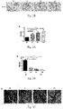
Fig. 1A and Fig. 1B are general appearance images of gradient mineralized decellularized cancellous bone matrix materials;Fig. 1C is a histogram illustrating that DNA content of the decellularized material is significantly reduced, and almost contains no cellular components and immunogenic substances;Fig. 1D is a histogram illustrating materials with mineralization degrees of 100%, 90%, 60% and 0% after gradient demineralization; Fig. IE is EDS analysis diagram indicating that contents of calcium and phosphorus are decreased significantly after the material is demineralized; and Fig. IF is Masson dyeing images indicating that exposure of immaturely mineralized collagen fiber is increased after the mineralization degree of the material is reduced, and all groups of cells are completely removed without obvious immunogenic substances after decellularization; -
Fig. 2A is scanning electron micrographs indicating that under 500 times magnification observation, surfaces of a demineralized bone ECM material are smoother and calcium nodule distribution is reduced, and under 5000 times magnification observation, exposure of mineralized collagen fiber is increased after the mineralization degree of the material is reduced; andFig. 2B is immunohistochemical staining images indicating that BMP-2 exposure in the demineralized material is gradually increased; -
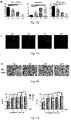
Fig. 3A and Fig. 3B are histograms indicating that porosity of the demineralized bone ECM material is increased, and stiffness of deformation resistance index is decreased;Fig. 3C is images indicating frontal force difference of the bone ECM materials with different mineralization degrees (scale bar of 1µm); andFig. 3D is histograms of micromechanics property changes of the bone ECM material, wherein as the mineralization degree is decreased, Young's modulus on material surfaces is decreased, deformation is increased, and adhesion force is decreased. -
Fig. 4A and Fig. 4B are DAPI staining images and 5000 times magnification electron micrographs indicating cell adhesion ability of the bone ECM materials under the mineralization degrees of 90% and 60%, wherein cell extension is sufficient, but cell adhesion ability of the bone ECM materials under the mineralization degrees of 100% and 0% is poor with insufficient cell extension; -
Fig. 5A is histograms indicating that the bone ECM materials with the mineralization degrees of 90% and 60% have no significant effect on proliferation of mesenchymal stem cells after the bone mesenchymal stem cells are cultured in bone ECM material extracts with different concentrations for 1-5 days; andFig. 5B is histograms indicating relative expression of Col-1α, ALP, BMP-2 genes in cells after qPCR detection of re-implantation mesenchymal stem cells of the bone ECM materials with different mineralization degrees, wherein the materials with the mineralization degrees of 90% and 60% have higher expression of osteoinductive proteins; -
Fig. 6A andFig. 6B are immunofluorescence staining detection images of relative expression levels of BMP-2 and MEK-1 genes in the mesenchymal stem cells after being implanted into the bone ECM materials with the different mineralization degrees for 2 and 4 weeks, wherein the materials with the mineralization degrees of 90% and 60% have more BMP-2 and MEK-1 expression, suggesting that promotion of BMP-2 expression by the material may be related to stimulation of calcium ions with an appropriate concentration; -
Fig. 7A is Masson three-color staining image of bone defect sites after the bone ECM material is implanted in a rabbit femoral bone defect model for 2 and 4 weeks, indicating that the materials with the mineralization degrees of 90% and 60% have better ability to promote trabecular bone and blood vessel growth; andFig. 7B is immunohistochemical staining images of type II collagen expression in thedefect site 2 weeks after implantation, suggesting that the bone ECM material may be involved in an intra-chondral osteogenesis process during promoting bone repair; -
Fig. 8A is images of angiogenesis in the bone defect site after the bone ECM material is implanted into the bone defect model for 2 weeks, indicating that the materials with the mineralization degrees of 90% and 60% have better angiogenic ability;Fig. 8B is images of VEGFA protein expression in the bone defect site after the bone ECM material is implanted into the bone defect model for 2 weeks, indicating that the materials with the mineralization degrees of 90% and 60% can better promote the VEGFA protein expression; andFig. 8C is quantification histograms of new vessel quantity, area and thickness after the bone ECM material is implanted in the bone defect site for 2 and 4 weeks, indicating that the materials with the mineralization degrees of 90% and 60% have a strong ability to promote blood vessel growth and maturation; -
Fig. 9A and Fig. 9B are three-dimensional composite images obtained by MicroCT scanning and reconstruction after the bone ECM material is implanted in the bone defect model for 2 and 4 weeks, wherein the materials with the mineralization degrees of 90% and 60% have batter repair ability the ones of 100% and 0%; andFig. 9C is histograms of new bone trabeculae quantity and thickness after the bone ECM material is implanted in the bone defect model for 2 and 4 weeks, wherein the materials with the mineralization degrees of 90% and 60% have better ability to promote bone trabecular growth; -
Fig. 10A and Fig. 10B are H&E staining images and repair area quantification histograms after the bone ECM material is implanted in the bone defect model for 2 and 4 weeks, indicating that the bone ECM materials with different mineralization degrees can all promote bone defect repair, while the materials with the mineralization degrees of 90% and 60% have better repair ability than the ones of 100% and 0%. - The present invention provides a natural-tissue-derived gradient mineralized cancellous bone matrix material and a preparation method thereof.
- The natural-tissue-derived gradient mineralized cancellous bone matrix material and the preparation method thereof provided by the present invention will be further illustrated with embodiments. However, the embodiments are not intended to limit the protection scope of the present invention as defined by the appended claims .
-
- 1) selecting fresh porcine scapula and washing for 4 times with sterile saline, removing cancellous bone with a 6mm drill, and cutting the cancellous bone into cylindrical bone blocks about 2mm high with a scalpel;
- 2) then rinsing the bone blocks with sterile physiological saline for 2 hours before sending to an irradiation center for sterilizing by irradiation, wherein an irradiation dose is 25w;
- 3) rinsing the bone blocks with deionized water containing 20KIU/ml protease inhibitor for 3 times and 10 minutes for each time, to remove blood, fat tissue and other impurities;
- 4) preparing high-temperature sterilized 1L glass bottles containing 500ml deionized water and preparing 20 embedding boxes on a sterile operating table with sterile gloves; separating 3 sterilized bone blocks into each embedding box, and putting the embedding box into 10ml deionized water solution containing 15% acetone and shaking at 10°C for 2 hours;
- 5) putting the embedding box into 5ml deionized water solution containing 2% tributyl phosphate and shaking at 10°C for 4 hours;
- 6) putting the embedding box in a deionized water solution containing the protease inhibitor, shaking at 4°C and 50rpm for 24 hours with a shaker, and then freezing and thawing with liquid nitrogen for 3 cycles (-80°C/37°C);
- 7) putting the embedding box in
5ml 2% Triton® X-100, and shaking with a constant temperature shaker at 10°C and 100rpm for 24 hours; - 8) shaking the embedding box in deionized water containing 5% SDS with the constant temperature shaker at 10°C and 100rpm for 36h;
- 9) putting the embedding box in a PBS buffer solution containing 0.5M potassium chloride, and shaking with the shaker at 4°C and 100rpm for 6 hours;
- 10) shaking the embedding box in a PBS buffer containing 1M potassium iodide with the shaker at 4°C and 100rpm for 6 hours, to obtain decellularized bone ECM materials (as shown in
Fig. 1A ); - 11) preparing a decalcification solution (deionized water 1750 ml + EDTA-2Na 450 g + NaOH 35 g); taking out the bone blocks and decalcifying in an ultrasound decalcifier at 250 kHz and 4°C for 4, 8, 12 and 24h;
- 12) after each of the steps 4)-11), rinsing with the deionized water for 6 hours before a next step; and
- 13) taking out the bone blocks to obtain bone ECM materials with mineralization degrees of 100%, 90%, 60% and 0% (as shown in
Fig. 1B ), and sterilizing the obtained materials by 25w irradiation. -
- 1) selecting fresh porcine scapula and washing for 4 times with sterile saline, removing cancellous bone with a 6mm drill, and cutting the cancellous bone into cylindrical bone blocks about 2mm high with a scalpel;
- 2) then rinsing the bone blocks with sterile physiological saline for 2 hours before sending to an irradiation center for sterilizing by irradiation, wherein an irradiation dose is 25w;
- 3) rinsing the bone blocks with deionized water containing 10KIU/ml protease inhibitor for 3 times and 20 minutes for each time, to remove blood, fat tissue and other impurities;
- 4) preparing high-temperature sterilized 1L glass bottles containing 500ml deionized water and preparing 20 embedding boxes on a sterile operating table with sterile gloves; separating 3 sterilized bone blocks into each embedding box, and putting the embedding box into 10ml deionized water solution containing 15% acetone and shaking at 10°C for 4 hours;
- 5) putting the embedding box into 5ml deionized water solution containing 2% tributyl phosphate and shaking at 10°C for 4 hours;
- 6) putting the embedding box in a deionized water solution containing the protease inhibitor, shaking at 4°C and 50rpm for 24 hours with a shaker, and then freezing and thawing with liquid nitrogen for 3 cycles (-80°C/37°C);
- 7) putting the embedding box in
5ml 2% Triton® X-100, and shaking with a constant temperature shaker at 10°C and 100rpm for 24 hours; - 8) shaking the embedding box in deionized water containing 5% SDS with the constant temperature shaker at 10°C and 100rpm for 36h;
- 9) putting the embedding box in a PBS buffer solution containing 0.5M potassium chloride, and shaking with the shaker at 4°C and 100rpm for 6 hours;
- 10) shaking the embedding box in a PBS buffer containing 1M potassium iodide with the shaker at 4°C and 100rpm for 6 hours, to obtain decellularized bone ECM materials (as shown in
Fig. 1A ); - 11) preparing a decalcification solution (deionized water 1750 ml + EDTA-2Na 450 g + NaOH 35 g); taking out the bone blocks and decalcifying in an ultrasound decalcifier at 250 kHz and 4°C for 4, 8, 12 and 24h;
- 12) after each of the steps 4)-11), rinsing with the deionized water for 6 hours before a next step;
- 13) taking out the bone blocks to obtain bone ECM materials with mineralization degrees of 100%, 90%, 60% and 0% (as shown in
Fig. 1B ), and sterilizing the obtained materials by 25w irradiation; - 14) detecting DNA contents in the decellularized materials, which are very low (as shown in
Fig. 1C ); - 15) detecting mineralization contents in the bone ECM materials with the mineralization degrees of 100%, 90%, 60% and 0% (taking calcium ion contents as an example), wherein the bone ECM material group with the mineralization degree of 90% (4h demineralization) is 4.58 ± 0.01mmol/mg, the bone ECM material group with the mineralization degree of 60% (8h demineralization) is 3.26 ± 0.38mmol/mg, the bone ECM material with the mineralization degree of 0% (12h demineralization) contains almost no calcium ion; (the bone ECM material with the mineralization degree of 100% (non-demineralized material) has a calcium ion content of 4.99 ± 0.22mmol/mg) (as shown in Fig. ID);
- 16) detecting porosities of the bone ECM materials with the mineralization degrees of 100%, 90%, 60% and 0%, wherein the porosity increases with the demineralization time (as shown in
Fig. 3A ); and - 17) detecting stiffnesses of the bone ECM materials with the mineralization degrees of 100%, 90%, 60% and 0%, wherein the stiffness is an indicator of resistance to stress and deformation; as the mineralization degree decreases, the stiffness of corresponding material also decreases in sequence; the stiffnesses of the bone ECM materials with the mineralization degrees of 100%, 90%, 60% and 0% (4 h, 8h and 12h demineralization) are 5.71 ± 0.46N/mm, 3.68 ± 0.18N/mm, and 2.53 ± 1.62N/mm (the stiffness of the bone ECM material with the mineralization degree of 100% (non-demineralized material) is 21.55 ± 1.62N/mm) (as shown in
Fig. 3B ). -
- 1) selecting fresh porcine scapula and washing for 4 times with sterile saline, removing cancellous bone with a 6mm drill, and cutting the cancellous bone into cylindrical bone blocks about 2mm high with a scalpel;
- 2) then rinsing the bone blocks with sterile physiological saline for 2 hours before sending to an irradiation center for sterilizing by irradiation, wherein an irradiation dose is 25w;
- 3) rinsing the bone blocks with deionized water containing 50KIU/ml protease inhibitor for 2 times and 10 minutes for each time, to remove blood, fat tissue and other impurities;
- 4) preparing high-temperature sterilized 1L glass bottles containing 500ml deionized water and preparing 20 embedding boxes on a sterile operating table with sterile gloves; separating 3 sterilized bone blocks into each embedding box, and putting the embedding box into 10ml deionized water solution containing 10% acetone and shaking at 10°C for 1 hours;
- 5) putting the embedding box into 5ml deionized water solution containing 5% tributyl phosphate and shaking at 10°C for 3 hours;
- 6) putting the embedding box in a deionized water solution containing the protease inhibitor, shaking at 4°C and 50rpm for 36 hours with a shaker, and then freezing and thawing with liquid nitrogen for 2 cycles (-80°C/37°C);
- 7) putting the embedding box in
5ml 2% Triton® X-100, and shaking with a constant temperature shaker at 10°C and 100rpm for 24 hours; - 8) shaking the embedding box in deionized water containing 5% SDS with the constant temperature shaker at 10°C and 100rpm for 36h;
- 9) putting the embedding box in a PBS buffer solution containing 0.5M potassium chloride, and shaking with the shaker at 4°C and 100rpm for 6 hours;
- 10) shaking the embedding box in a PBS buffer containing 1M potassium iodide with the shaker at 4°C and 100rpm for 6 hours, to obtain decellularized bone ECM materials (as shown in
Fig. 1A ); - 11) preparing a decalcification solution (deionized water 1750 ml + EDTA-2Na 450 g + NaOH 35 g); taking out the bone blocks and decalcifying in an ultrasound decalcifier at 250 kHz and 4°C for 4, 8, 12 and 24h;
- 12) after each of the steps 4)-11), rinsing with the deionized water for 6 hours before a next step;
- 13) taking out the bone blocks to obtain bone ECM materials with mineralization degrees of 100%, 90%, 60% and 0% (as shown in
Fig. 1B ), and sterilizing the obtained materials by 25w irradiation; - 14) observing with a scanning electron microscopy (SEM) to obtain ultrastructural characteristics of the bone ECM materials with the mineralization degrees of 100%, 90%, 60% and 0%, wherein compared with the other groups, the bone ECM materials with the mineralization degrees of 100% and 90% (0 and 4h demineralization) have rougher surfaces and smaller pores; in addition, structures and arrangement of collagen fibers are also different among the four groups; in the bone ECM material group with the mineralization degree of 100% (non-demineralized material), most of the collagen fibers are covered by the surface, while on the surfaces of the bone ECM materials with the mineralization degrees of 90% and 60% (4h and 8h demineralization), collagen fibers are exposed with good arrangement, so as to produce more adhesive retention sites for the cells; however, for the bone ECM material with the mineralization degree of 0% (12h demineralization), the structure of the collagen fibrils is more disordered and the density is worse, which is not conducive to cell retention (as shown in
Fig. 2A ); - 15) observing with an atomic force microscopy (AFM), which also shows that more collagen is exposed on the surfaces of the bone ECM materials with the mineralization degrees of 100%, 90%, 60% and 0% (as shown in
Figs. 3C-3D ). - 16) observing with immunohistochemical staining, which shows that BMP-2 expression on the surfaces of the bone ECM materials with the mineralization degrees of 90% and 60% are increased (as shown in
Fig. 2B ). - 17) detecting ratio of C, P, Ca (carbon, phosphorus, calcium) in specific areas of the bone ECM materials with the mineralization degrees of 100%, 90%, 60% and 0% by an EDS method (as shown in Figs. IE-IF); taking C as a reference to measuring Ca concentration according to a selective electrode method, wherein Ca density becomes more dispersed as the mineralization degree decreases; in addition, change in phosphorus content is consistent with change in calcium content, and dispersion degree increases as the mineralization degree decreases; AFM is used to evaluate ultra-microscopic mechanical properties of the mineralization degrees of 100%, 90%, 60% and 0%, wherein with different mineralization degrees of natural bone ECM sources, the bone ECM materials with the mineralization degrees of 90% and 60% have more fibrils exposed on the surfaces, thus providing many RGD ligands for cell adhesion.
-
- 1) selecting fresh porcine scapula and washing for 4 times with sterile saline, removing cancellous bone with a 6mm drill, and cutting the cancellous bone into cylindrical bone blocks about 2mm high with a scalpel;
- 2) then rinsing the bone blocks with sterile physiological saline for 2 hours before sending to an irradiation center for sterilizing by irradiation, wherein an irradiation dose is 25w;
- 3) rinsing the bone blocks with deionized water containing 20KIU/ml protease inhibitor for 3 times and 10 minutes for each time, to remove blood, fat tissue and other impurities;
- 4) preparing high-temperature sterilized 1L glass bottles containing 500ml deionized water and preparing 20 embedding boxes on a sterile operating table with sterile gloves; separating 3 sterilized bone blocks into each embedding box, and putting the embedding box into 10ml deionized water solution containing 15% acetone and shaking at 10°C for 2 hours;
- 5) putting the embedding box into 5ml deionized water solution containing 2% tributyl phosphate and shaking at 10°C for 4 hours;
- 6) putting the embedding box in a deionized water solution containing the protease inhibitor, shaking at 4°C and 50rpm for 24 hours with a shaker, and then freezing and thawing with liquid nitrogen for 3 cycles (-80°C/37°C);
- 7) putting the embedding box in
5ml 2% Triton® X-100, and shaking with a constant temperature shaker at 10°C and 100rpm for 24 hours; - 8) shaking the embedding box in deionized water containing 5% SDS with the constant temperature shaker at 10°C and 100rpm for 36h;
- 9) putting the embedding box in a PBS buffer solution containing 0.5M potassium chloride, and shaking with the shaker at 4°C and 100rpm for 6 hours;
- 10) shaking the embedding box in a PBS buffer containing 1M potassium iodide with the shaker at 4°C and 100rpm for 6 hours, to obtain decellularized bone ECM materials (as shown in
Fig. 1A ); - 11) preparing a decalcification solution (deionized water 1750 ml + EDTA-2Na 450 g + NaOH 35 g); taking out the bone blocks and decalcifying in an ultrasound decalcifier at 250 kHz and 4°C for 4, 8, 12 and 24h;
- 12) after each of the steps 4)-11), rinsing with the deionized water for 6 hours before a next step;
- 13) taking out the bone blocks to obtain bone ECM materials with mineralization degrees of 100%, 90%, 60% and 0% (as shown in
Fig. 1B ), and sterilizing the obtained materials by 25w irradiation; - 14) culturing bone mesenchymal stem cells for 1-5 days with an extract of the bone ECM material with a specific mineralization degree (the extract is derived from the bone ECM materials with the mineralization degrees of 90% and 60%), wherein the cells grows well, indicating that the material is safe and non-toxic (as shown in
Fig. 5A ); - 15) observing after the bone marrow mesenchymal stem cells are transplanted in the bone ECM materials with the mineralization degrees of 100%, 90%, 60% and 0% for 3 days, wherein under confocal microscope observation, the cells are adhered to a scaffold (as shown in
Fig. 4A ); under 1000 times magnification scanning electron microscope observation, the bone ECM materials with the mineralization degrees of 90% and 60% have more bone marrow mesenchymal stem cells than the bone ECM material with the mineralization degrees of 100%, indicating that the material of the present invention can effectively promote cell adhesion engraftment and proliferation; the bone ECM material with the mineralization degrees of 0% has fewer cells, as shown inFig. 4B ; and - 16) comparing relative expression of osteogenic genes in the bone marrow mesenchymal stem cells engrafted on the bone ECM materials with the mineralization degrees of 100%, 90%, 60% and 0% (as shown in
Fig. 5B ), wherein ALP is one of the most important bone formation indicators of the bone marrow mesenchymal stem cells in an early stage, which shows that the ALP of the cells cultured in the bone ECM material with the mineralization degree of 90% for 1 week is up-regulated by 17 times compared with the non-demineralized group, and Col-1α1 has same trend; At 2nd and 4th weeks of culture, BMP-2 expression of the cells in the bone ECM materials with the mineralization degrees of 90% and 60%, especially the one of 90%, is increased (as shown inFig. 6A ); in addition, a MAPK signaling pathway plays a role in a Ca2+-mediated osteogenic differentiation process; compared with the bone ECM materials with the mineralization degrees of 100% and 0%, MEK-1 expression of the cells in the bone ECM materials with the mineralization degrees of 90% and 60% is up-regulated (as shown inFig. 6B ); in summary, the bone ECM materials with the mineralization degrees of 90% and 60%, especially the one of 90%, show better promotion effect on osteogenic differentiation of the bone marrow mesenchymal stem cells than the bone ECM materials with the mineralization degrees of 100% and 0%. -
- 1) selecting fresh porcine scapula and washing for 4 times with sterile saline, removing cancellous bone with a 6mm drill, and cutting the cancellous bone into cylindrical bone blocks about 2mm high with a scalpel;
- 2) then rinsing the bone blocks with sterile physiological saline for 2 hours before sending to an irradiation center for sterilizing by irradiation, wherein an irradiation dose is 25w;
- 3) rinsing the bone blocks with deionized water containing 20KIU/ml protease inhibitor for 3 times and 10 minutes for each time, to remove blood, fat tissue and other impurities;
- 4) preparing high-temperature sterilized 1L glass bottles containing 500ml deionized water and preparing 20 embedding boxes on a sterile operating table with sterile gloves; separating 3 sterilized bone blocks into each embedding box, and putting the embedding box into 10ml deionized water solution containing 20% acetone and shaking at 10°C for 4 hours;
- 5) putting the embedding box into 5ml deionized water solution containing 1% tributyl phosphate and shaking at 10°C for 1 hours;
- 6) putting the embedding box in a deionized water solution containing the protease inhibitor, shaking at 4°C and 300rpm for 48 hours with a shaker, and then freezing and thawing with liquid nitrogen for 6 cycles (-80°C/37°C);
- 7) putting the embedding box in 5ml 0.5% Triton® X-100, and shaking with a constant temperature shaker at 10°C and 100rpm for 24 hours;
- 8) shaking the embedding box in deionized water containing 5% SDS with the constant temperature shaker at 10°C and 100rpm for 36h;
- 9) putting the embedding box in a PBS buffer solution containing 0.5M potassium chloride, and shaking with the shaker at 4°C and 100rpm for 2 hours;
- 10) shaking the embedding box in a PBS buffer containing 1.2M potassium iodide with the shaker at 4°C and 100rpm for 12 hours, to obtain decellularized bone ECM materials (as shown in
Fig. 1A ); - 11) preparing a decalcification solution (deionized water 1750 ml + EDTA-2Na 450 g + NaOH 35 g); taking out the bone blocks and decalcifying in an ultrasound decalcifier at 250 kHz and 4°C for 4, 8, 12 and 24h;
- 12) after each of the steps 4)-11), rinsing with the deionized water for 6 hours before a next step;
- 13) taking out the bone blocks to obtain bone ECM materials with mineralization degrees of 100%, 90%, 60% and 0% (as shown in
Fig. 1B ), and sterilizing the obtained materials by 25w irradiation; - 14) establishing a rabbit femoral epicondyle bilateral defect model, implanting the bone ECM materials with the mineralization degrees of 100%, 90%, 60% and 0%, and evaluating therapeutic effects;
- 15) performing Micro-CT analysis (as shown in
Figs. 9A-9B ), which shows that after 4 weeks of transplantation, wherein in the bone ECM materials with the mineralization degrees of 90% and 60%, bone defect site are almost filled with new bone trabeculae, while filling effect of the non-demineralized and fully-demineralized material groups is low; the filling effects of the bone ECM materials with the mineralization degrees of 100%, 90%, 60% and 0% are 1.23 ± 0.14/mm, 2.16 ± 0.03/mm, 1.57 ± 0.21/mm and 0.94 ± 0.22/mm, respectively; bone trabecular thicknesses are 0.16 ± 0.03µm, 0.24 ± 0.04µm, 0.18 ± 0.01µm and 0.14 ± 0.02µm, respectively; growth promotion effects on body new trabecular bone of the bone ECM materials with the mineralization degrees of 90% and 60% are better than the other groups (as shown inFig. 9C ); - 16) observing by H&E staining (as shown in
Figs. 10A-10B ), which shown no obvious inflammation or inflammatory cells, proving that the material is safety; and - 17) observing after 4 weeks of transplantation, wherein the bone ECM materials with the mineralization degrees of 90% and 60% are partially degraded, and new bone has grown into them; collagen fiber mineralization in new bone tissue is mostly surface collagen of immature mineralized materials (as shown in
Figs. 7A-7B ). -
- 1) selecting fresh porcine scapula and washing for 4 times with sterile saline, removing cancellous bone with a 6mm drill, and cutting the cancellous bone into cylindrical bone blocks about 2mm high with a scalpel;
- 2) then rinsing the bone blocks with sterile physiological saline for 2 hours before sending to an irradiation center for sterilizing by irradiation, wherein an irradiation dose is 25w;
- 3) rinsing the bone blocks with deionized water containing 20KIU/ml protease inhibitor for 3 times and 10 minutes for each time, to remove blood, fat tissue and other impurities;
- 4) preparing high-temperature sterilized 1L glass bottles containing 500ml deionized water and preparing 20 embedding boxes on a sterile operating table with sterile gloves; separating 3 sterilized bone blocks into each embedding box, and putting the embedding box into 10ml deionized water solution containing 15% acetone and shaking at 10°C for 2 hours;
- 5) putting the embedding box into 5ml deionized water solution containing 5% tributyl phosphate and shaking at 10°C for 4 hours;
- 6) putting the embedding box in a deionized water solution containing the protease inhibitor, shaking at 4°C and 50rpm for 24 hours with a shaker, and then freezing and thawing with liquid nitrogen for 3 cycles (-80°C/37°C);
- 7) putting the embedding box in
5ml 2% Triton® X-100, and shaking with a constant temperature shaker at 10°C and 100rpm for 24 hours; - 8) shaking the embedding box in deionized water containing 10% SDS with the constant temperature shaker at 10°C and 100rpm for 36h;
- 9) putting the embedding box in a PBS buffer solution containing 0.1M potassium chloride, and shaking with the shaker at 4°C and 100rpm for 12 hours;
- 10) shaking the embedding box in a PBS buffer containing 1M potassium iodide with the shaker at 4°C and 100rpm for 6 hours, to obtain decellularized bone ECM materials (as shown in
Fig. 1A ); - 11) preparing a decalcification solution (deionized water 1750 ml + EDTA-2Na 450 g + NaOH 35 g); taking out the bone blocks and decalcifying in an ultrasound decalcifier at 250 kHz and 4°C for 4, 8, 12 and 24h;
- 12) after each of the steps 4)-11), rinsing with the deionized water for 6 hours before a next step;
- 13) taking out the bone blocks to obtain bone ECM materials with mineralization degrees of 100%, 90%, 60% and 0% (as shown in
Fig. 1B ), and sterilizing the obtained materials by 25w irradiation; and - 14) observing angiogenesis, wherein new blood vessels are distributed in middle of the trabecular bone; after 2 weeks of transplantation, immature new blood vessels are mostly formed; quantities of new blood vessels of the bone ECM materials with the mineralization degrees of 100%, 90%, 60% and 0% are 5.5 ± 1.3/500µm2, 8.0 ± 1.6/500µm2, 8.0 ± 1.8/500µm2, and 5.0 ± 1.6/500µm2, respectively; blood vessel areas are 23.92 ± 7.25µm2, 38.95 ± 8.12µm2, 45.54 ± 8.70µm2, and 18.86 ± 9.43µm2, respectively; new blood vessel thicknesses of the bone ECM materials with the mineralization degrees of 90% and 60% are 4.86 ± 0.15µm and 5.07 ± 0.20µm, respectively, which are higher than that of the bone ECM material group with the mineralization degree of 100% (4.29 ± 0.38µm) and the bone ECM material group with the mineralization degree of 0% (4.41 ± 0.26µm); after 4 weeks of transplantation, mature new blood vessels are mainly formed; quantities of new blood vessels of the bone ECM materials with the mineralization degrees of 100%, 90%, 60% and 0% are 3.5 ± 0.6/500µm2, 5.8 ± 1.0/500µm2 , 5.0 ± 0.8/500µm2, and 2.8 ± 1.5/500µm2 , respectively; the blood vessel areas are 41.26 ± 5.69µm2, 69.92 ± 11.26µm2, 60.76 ± 8.66µm2 , and 24.87 ± 8.18µm2, respectively; thicknesses are increased in each group compared with those after 2 weeks of transplantation, wherein the bone ECM material group with the mineralization degree of 90% (12.18 ± 0.54µm) and the bone ECM material group with the mineralization degree of 60% (12.18 ± 0.32µm) are still higher than that of the bone ECM material group with the mineralization degree of 100% (11.83 ± 0.49µm) and the bone ECM material group with the mineralization degree of 0% (9.68 ± 1.83µm); the bone ECM material groups with the mineralization degrees of 90% and 60% have promoted growth of new blood vessels, microvessel degeneration, and stable growth of large blood vessels compared with the other two groups, and distribution of vascular endothelial growth factor A (VEGFA) in defect tissue is up-regulated, so as to provided better repair effect (as shown in
Figs. 8A-8E ). -
- 1) selecting fresh bovine scapula and washing for 4 times with sterile saline, removing cancellous bone with a 6mm drill, and cutting the cancellous bone into cylindrical bone blocks about 2mm high with a scalpel;
- 2) then rinsing the bone blocks with sterile physiological saline for 2 hours before sending to an irradiation center for sterilizing by irradiation, wherein an irradiation dose is 25w;
- 3) rinsing the bone blocks with deionized water containing 50KIU/ml protease inhibitor for 2 times and 5 minutes for each time, to remove blood, fat tissue and other impurities; and
- 4) performing subsequent operations with reference to the method of the embodiment 1, to obtain the gradient mineralized cancellous bone matrix material.
-
- 1) selecting fresh porcine rib and washing for 4 times with sterile saline, removing cancellous bone with a 6mm drill, and cutting the cancellous bone into cylindrical bone blocks about 2mm high with a scalpel;
- 2) then rinsing the bone blocks with sterile physiological saline for 2 hours before sending to an irradiation center for sterilizing by irradiation, wherein an irradiation dose is 25w;
- 3) rinsing the bone blocks with deionized water containing 10KIU/ml protease inhibitor for 5 times and 5 minutes for each time, to remove blood, fat tissue and other impurities; and
- 4) performing subsequent operations with reference to the method of the embodiment 1, to obtain the gradient mineralized cancellous bone matrix material.
-
- 1) selecting fresh porcine scapula and washing for 4 times with sterile saline, removing cancellous bone with a 6mm drill, and cutting the cancellous bone into cylindrical bone blocks about 2mm high with a scalpel;
- 2) then rinsing the bone blocks with sterile physiological saline for 2 hours before sending to an irradiation center for sterilizing by irradiation, wherein an irradiation dose is 25w;
- 3) rinsing the bone blocks with deionized water containing 20KIU/ml protease inhibitor for 3 times and 10 minutes for each time, to remove blood, fat tissue and other impurities;
- 4) preparing high-temperature sterilized 1L glass bottles containing 500ml deionized water and preparing 20 embedding boxes on a sterile operating table with sterile gloves; separating 3 sterilized bone blocks into each embedding box, and putting the embedding box into 10ml deionized water solution containing 10% acetone and shaking at 10°C for 1 hour;
- 5) putting the embedding box into 5ml deionized water solution containing 2% tributyl phosphate and shaking at 10°C for 4 hours;
- 6) putting the embedding box in a deionized water solution containing the protease inhibitor, shaking at 4°C for 24 hours with a shaker, and then freezing and thawing with liquid nitrogen for 3 cycles (-80°C/37°C);
- 7) putting the embedding box in
5ml 2% Triton® X-100, and shaking with a constant temperature shaker at 10°C and 100rpm for 24 hours; - 8) shaking the embedding box in deionized water containing 5% SDS with the constant temperature shaker at 10°C and 100rpm for 36h;
- 9) putting the embedding box in a PBS buffer solution containing 0.5M potassium chloride, and shaking with the shaker at 4°C and 100rpm for 6 hours;
- 10) shaking the embedding box in a PBS buffer containing 1M potassium iodide with the shaker at 4°C and 100rpm for 6 hours, to obtain decellularized bone ECM materials (as shown in
Fig. 1A ); - 11) preparing a decalcification solution (deionized water 1750 ml + EDTA-2Na 450 g + NaOH 10 g); taking out the bone blocks and decalcifying in an ultrasound decalcifier at 350 kHz and 4°C for 4, 8, 12 and 24h; and
- 12) performing subsequent operations with reference to the method of the embodiment 1, to obtain the gradient mineralized cancellous bone matrix material.
- The gradient mineralized cancellous bone matrix materials obtained in the embodiments 7-9 are subjected to histological evaluation, calcium and phosphorus content detection, collagen surface morphology and content detection, and mechanical detection. The results are the same as those in the embodiment 1, which indicates that the gradient mineralized cancellous bone matrix materials with similar effects can be prepared by the above optimized reagents and adjusted processing time. In addition, the histological evaluation, cell culture experiments, and in-vivo repair experimental evaluation and test of the materials all indicate that the gradient mineralized cancellous bone matrix materials with mineralization degrees of 90% and 60% obtained in the embodiments 7-9 have good repair and regeneration effects. The materials can be used as a safe, reliable, effective and fast biomaterial for clinically promoting repair and regeneration of muscle defect and lesion.
- It can be known from the above embodiments that according to the present invention, the natural-tissue-derived gradient mineralized cancellous bone matrix material and the preparation method thereof can better promote cell adhesion, induce differentiation and proliferation of engrafted cells, and promote angiogenesis as well as bone trabeculae formation, thereby achieving a better repair effect and a smaller inflammatory response. The material is a very promising material for bone defect transplantation.
- The above is only the preferred embodiments of the present invention as defined by the appended claims.
-
- [1] Y. Wang, S. Von Euw, F.M. Fernandes, S. Cassaignon, M. Selmane, G. Laurent, G. Pehau-Arnaudet, C. Coelho, L. Bonhomme-Coury, M.M. Giraud-Guille, F. Babonneau, T. Azais, N. Nassif, Water-mediated structuring of bone apatite, Nat Mater 12(12) (2013) 1144-53.
- [2] Y. Zheng, C. Xiong, S. Zhang, X. Li, L. Zhang, Bone-like apatite coating on functionalized poly(etheretherketone) surface via tailored silanization layers technique, Mater Sci Eng C Mater Biol Appl 55 (2015) 512-23.
- [3] Sunarso, R. Toita, K. Tsuru, K. Ishikawa, Immobilization of calcium and phosphate ions improves the osteoconductivity of titanium implants, Mater Sci Eng C Mater Biol Appl 68 (2016) 291-298.
- [4] Melita M. Dvorak, Ashia Siddiqua, Donald T. Ward, D. Howard Carter, Sarah L. Dallas, Edward F. Nemeth, D. Riccardi, Physiological changes in extracellular calcium concentration directly control osteoblast function in the absence of calciotropic hormones, Proc Natl Acad Sci U S A. 101(14) (2004) 5140-5145.
- [5] Y. Zhang, J. Xu, Y.C. Ruan, M.K. Yu, M. O'Laughlin, H. Wise, D. Chen, L. Tian, D. Shi, J. Wang, S. Chen, J.Q. Feng, D.H. Chow, X. Xie, L. Zheng, L. Huang, S. Huang, K. Leung, N. Lu, L. Zhao, H. Li, D. Zhao, X. Guo, K. Chan, F. Witte, H.C. Chan, Y. Zheng, L. Qin, Implant-derived magnesium induces local neuronal production of CGRP to improve bone-fracture healing in rats, Nat Med 22(10) (2016) 1160-1169.
- [6] A. Szczes, L. Holysz, E. Chibowski, Synthesis of hydroxyapatite for biomedical applications, Adv Colloid Interface Sci 249 (2017) 321-330.
- [7] Q. Hu, M. Liu, G. Chen, Z. Xu, Y. Lv, Demineralized Bone Scaffolds with Tunable Matrix Stiffness for Efficient Bone Integration, ACS Appl Mater Interfaces 10(33) (2018) 27669-27680.
- [8] Y. Wang, T. Azais, M. Robin, A. Vallee, C. Catania, P. Legriel, G. Pehau-Arnaudet, F. Babonneau, M.M. Giraud-Guille, N. Nassif, The predominant role of collagen in the nucleation, growth, structure and orientation of bone apatite, Nat Mater 11(8) (2012) 724-33.
- [9] W.L. Grayson, S. Bhumiratana, C. Cannizzaro, P.H. Chao, D.P. Lennon, A.I. Caplan, G. Vunjak-Novakovic, Effects of initial seeding density and fluid perfusion rate on formation of tissue-engineered bone, Tissue Eng Part A 14(11) (2008) 1809-20.
- [10] F.M. Savi, G.I. Brierly, J. Baldwin, C. Theodoropoulos, M.A. Woodruff, Comparison of Different Decalcification Methods Using Rat Mandibles as a Model, J Histochem Cytochem 65(12) (2017) 705-722.
- [11] S.H. Yu, H.L. Chan, L.Y. Chong, Y.H. Jheng, P.C. Chang, Evaluation of the osteogenic potential of growth factor-rich demineralized bone matrix in vivo, J Periodontol 86(1) (2015) 36-43.
- [12] R.U. Rodriguez, N. Kemper, E. Breathwaite, S.M. Dutta, E.L. Hsu, W.K. Hsu, M.P. Francis, Demineralized bone matrix fibers formable as general and custom 3D printed mold-based implants for promoting bone regeneration, Biofabrication 8(3) (2016) 035007.
- [13] E. Huber, A.M. Pobloth, N. Bormann, N. Kolarczik, K. Schmidt-Bleek, H. Schell, P. Schwabe, G.N. Duda, B. Wildemann, (∗) Demineralized Bone Matrix as a Carrier for Bone Morphogenetic Protein-2: Burst Release Combined with Long-Term Binding and Osteoinductive Activity Evaluated In Vitro and In Vivo, Tissue Eng Part A 23(23-24) (2017) 1321-1330.
Claims (9)
- A preparation method of a gradient mineralized cancellous bone matrix material, comprising: processing naturally-derived bone tissue with an immunogenicity removal treatment for decellularization, and processing an obtained decellularized bone with an ultrasound gradient demineralization treatment to obtain the gradient mineralized cancellous bone matrix material and comprising specific steps of:Step 1: randomly selecting bone tissue from a mammal, removing the bone tissue with a drill, and cutting the bone tissue into cylindrical bone blocks with a scalpel;Step 2: then rinsing the bone blocks with sterile physiological saline for 2 hours before sterilizing by irradiation, wherein an irradiation dose is 5-40w;Step 3: rinsing the bone blocks with deionized water containing protease inhibitor to remove blood, fat tissue and other impurities, wherein a concentration of the protease inhibitor in the deionized water is 10-50KIU/ml, and the bone blocks are rinsed with the deionized water for 2-6 times and 3-20 minutes for each time;Step 4: separating the bone blocks into an embedding box, and putting the embedding box into a deionized water solution containing acetone and shaking for 1-4 hours, wherein a volume ratio of the acetone to deionized water in the deionized water solution is 10%-20%;Step 5: putting the embedding box into a deionized water solution containing tributyl phosphate and shaking for 1-4 hours, wherein a volume ratio of the tributyl phosphate to deionized water in the deionized water solution is 1%-5%;Step 6: putting the embedding box in a deionized water solution containing the protease inhibitor, shaking at 4°C for 24-48 hours with a shaker, and then freezing and thawing with liquid nitrogen for 2-6 cycles, wherein each cycle is from -80°C to 37°C, and a shaker speed is 50-300rpm;Step 7: putting the embedding box in a buffer solution containing Triton® X-100, and shaking with a constant temperature shaker for 24 hours, wherein a concentration of the Triton® X-100 is 0.5-5%;Step 8: shaking the embedding box in a buffer solution containing SDS with the constant temperature shaker for 36h, wherein a concentration of the SDS is 0.5- 10%;Step 9: putting the embedding box in a PBS buffer solution having a concentration of potassium chloride of 0.1-1M, and shaking with the shaker at 4°C and 100rpm for 2-12 hours;Step 10: shaking the embedding box in a PBS buffer containing potassium iodide with the shaker at 4°C and 100rpm for 2-12 hours, wherein a concentration of the potassium iodide in deionized water is 1-1.5M;Step 11: performing the ultrasound gradient demineralization treatment in a NaOH buffer solution containing EDTA2Na at 4°C-10°C for 4, 8, 12 and 24 hours, so as to obtain bone ECM (Extracellular matrix) materials with mineralization degrees of 100%, 90%, 60% and 0%; andStep 12: sterilizing the obtained materials by irradiation; wherein after each of the Steps 4-11, the deionized water is used to rinse for 6 hours before a next step.
- The preparation method, as recited in claim 1, wherein in the deionized water containing the protease inhibitor, the concentration of the protease inhibitor is 20-40KIU.
- The preparation method, as recited in claim 1, wherein in the deionized water solution containing the acetone, the volume ratio of the acetone to the deionized water is 13%-18%.
- The preparation method, as recited in claim 1, wherein in the deionized water solution containing the tributyl phosphate, the volume ratio of the tributyl phosphate to the deionized water is 2%-5%.
- The preparation method, as recited in claim 1, wherein the shaker speed is 30-180rpm.
- The preparation method, as recited in claim 1, wherein the concentration of the Triton® X-100 is 0.5-3%.
- The preparation method, as recited in claim 1, wherein the concentration of the SDS is 0.5-5%.
- The preparation method, as recited in claim 1, wherein the concentration of the potassium chloride in deionized water is 0.3-1M.
- The preparation method, as recited in claim 1, wherein the concentration of the potassium iodide in the deionized water is 1-1.4M.
Applications Claiming Priority (2)
| Application Number | Priority Date | Filing Date | Title |
|---|---|---|---|
| CN201910044252.7A CN110227182B (en) | 2019-01-17 | 2019-01-17 | Preparation method of gradient mineralized bone extracellular matrix material |
| PCT/CN2019/077754 WO2020147181A1 (en) | 2019-01-17 | 2019-03-12 | Gradient mineralized bone extracellular matrix material and preparation method therefor |
Publications (3)
| Publication Number | Publication Date |
|---|---|
| EP3785740A1 EP3785740A1 (en) | 2021-03-03 |
| EP3785740A4 EP3785740A4 (en) | 2021-08-18 |
| EP3785740B1 true EP3785740B1 (en) | 2022-10-19 |
Family
ID=67860100
Family Applications (1)
| Application Number | Title | Priority Date | Filing Date |
|---|---|---|---|
| EP19910842.4A Active EP3785740B1 (en) | 2019-01-17 | 2019-03-12 | Gradient mineralized bone extracellular matrix material and preparation method therefor |
Country Status (5)
| Country | Link |
|---|---|
| US (1) | US11684696B2 (en) |
| EP (1) | EP3785740B1 (en) |
| JP (1) | JP7118161B2 (en) |
| CN (1) | CN110227182B (en) |
| WO (1) | WO2020147181A1 (en) |
Families Citing this family (6)
| Publication number | Priority date | Publication date | Assignee | Title |
|---|---|---|---|---|
| AU2020405636A1 (en) * | 2019-12-19 | 2022-04-28 | Spiderwort Inc. | Biomaterials for bone tissue engineering |
| CN111110919A (en) * | 2019-12-30 | 2020-05-08 | 广东泓志生物科技有限公司 | Preparation method of omentum majus acellular matrix material and construction method of cartilage tissue |
| CN111905146B (en) * | 2020-08-01 | 2021-11-16 | 北京欧亚铂瑞科技有限公司 | Acellular bone matrix hydrogel retaining natural hydroxyapatite and preparation method thereof |
| CN112618797B (en) * | 2020-12-15 | 2022-08-05 | 浙江大学医学院附属邵逸夫医院 | Preparation method of specific demineralized extracellular matrix scaffold for cross-linking antibiotics |
| CN115227868B (en) * | 2022-07-20 | 2023-07-07 | 中南大学湘雅医院 | Bone defect repair material and magnesium pretreatment decellularized tissue engineering bone scaffold |
| CN116328039A (en) * | 2023-02-24 | 2023-06-27 | 浙江狄赛生物科技有限公司 | Natural bone repair material with specific mineralization degree and capable of regulating inflammatory metabolism, and preparation method and application thereof |
Family Cites Families (20)
| Publication number | Priority date | Publication date | Assignee | Title |
|---|---|---|---|---|
| US6090998A (en) * | 1997-10-27 | 2000-07-18 | University Of Florida | Segmentally demineralized bone implant |
| US6652592B1 (en) * | 1997-10-27 | 2003-11-25 | Regeneration Technologies, Inc. | Segmentally demineralized bone implant |
| WO1999051170A1 (en) * | 1998-04-02 | 1999-10-14 | Crosscart, Inc. | Bone xenografts |
| CN1507925A (en) * | 2002-12-20 | 2004-06-30 | 上海组织工程研究与开发中心 | Use of decellularized, decalcitied bone as tissue engineered material |
| DE102006041690A1 (en) * | 2006-09-06 | 2008-03-27 | Life Research & Development Services Gmbh | Preparing treated demineralized or partly-demineralized hard tissue matrix, useful e.g. as a component for diet supplements, comprises mechanically purifying the hard tissue using water and demineralizing the tissue in an solution |
| US8574825B2 (en) * | 2007-06-01 | 2013-11-05 | Bacterin International, Inc. | Process for demineralization of bone matrix with preservation of natural growth factors |
| CN101496913B (en) * | 2008-01-31 | 2012-09-26 | 中国人民解放军总医院 | Cartilage cell epimatrix three-dimensional porous sponge stent for tissue engineering and preparation method thereof |
| KR100978562B1 (en) * | 2008-12-31 | 2010-08-27 | 주식회사 코리아본뱅크 | Cancellous bone graft substitute and its process |
| CN101979105A (en) * | 2010-11-11 | 2011-02-23 | 昆明医学院第一附属医院 | Tissue engineering scaffold material for repairing cartilage defects and preparation method thereof |
| US9290738B2 (en) * | 2012-06-13 | 2016-03-22 | Miromatrix Medical Inc. | Methods of decellularizing bone |
| CN102871774B (en) | 2012-10-15 | 2015-03-25 | 四川大学华西医院 | Ligament repair scaffold and preparation method and application thereof |
| CN103536965B (en) | 2013-09-24 | 2014-11-05 | 北京大学口腔医学院 | Preparation method for three-dimensional mineralized collagen bracket with strict grade structure as well as product and application of three-dimensional mineralized collagen bracket |
| CN103877616B (en) * | 2014-03-18 | 2016-01-20 | 北京大学第三医院 | A kind of cartilage tissue engineered recovery support and preparation method thereof |
| CN104436318B (en) * | 2014-11-21 | 2016-08-17 | 李柏霖 | A kind of manufacture method of the guide tissue regeneration film of absorbable anti-microbial type bone structure |
| US20160206781A1 (en) * | 2014-12-29 | 2016-07-21 | Zurab Kakabadze | Methods and compositions for the production of composites for bone implantation |
| CN105435307A (en) * | 2015-11-30 | 2016-03-30 | 广西医科大学 | Natural-tissue-derived decellularized and decalcified bone material and preparation method thereof |
| CN105664255B (en) * | 2016-01-20 | 2019-11-05 | 浙江大学医学院附属邵逸夫医院 | A kind of synchondrosis bone in natural tissues source takes off the preparation method of cell material |
| CN106215238A (en) * | 2016-07-27 | 2016-12-14 | 重庆大学 | A kind of three-dimensional bone tissue engineering scaffold based on decalcification process and preparation method thereof |
| CN108324990A (en) * | 2018-02-11 | 2018-07-27 | 温州优墨生物科技有限公司 | A kind of method bone material cell free method and prepare Acellular bone powder |
| CN108465125A (en) * | 2018-04-04 | 2018-08-31 | 浙江大学 | The tendon composite muscle in natural tissues source takes off the preparation method of cell material |
-
2019
- 2019-01-17 CN CN201910044252.7A patent/CN110227182B/en active Active
- 2019-03-12 WO PCT/CN2019/077754 patent/WO2020147181A1/en active Application Filing
- 2019-03-12 EP EP19910842.4A patent/EP3785740B1/en active Active
- 2019-03-12 US US17/040,999 patent/US11684696B2/en active Active
- 2019-03-12 JP JP2020543063A patent/JP7118161B2/en active Active
Also Published As
| Publication number | Publication date |
|---|---|
| CN110227182A (en) | 2019-09-13 |
| US11684696B2 (en) | 2023-06-27 |
| WO2020147181A1 (en) | 2020-07-23 |
| EP3785740A1 (en) | 2021-03-03 |
| JP2021523749A (en) | 2021-09-09 |
| US20210121605A1 (en) | 2021-04-29 |
| JP7118161B2 (en) | 2022-08-15 |
| EP3785740A4 (en) | 2021-08-18 |
| CN110227182B (en) | 2020-12-15 |
Similar Documents
| Publication | Publication Date | Title |
|---|---|---|
| EP3785740B1 (en) | Gradient mineralized bone extracellular matrix material and preparation method therefor | |
| Chang et al. | Influence of pore size of porous titanium fabricated by vacuum diffusion bonding of titanium meshes on cell penetration and bone ingrowth | |
| Annaz et al. | Porosity variation in hydroxyapatite and osteoblast morphology: a scanning electron microscopy study | |
| Okamoto et al. | Influence of the porosity of hydroxyapatite ceramics on in vitro and in vivo bone formation by cultured rat bone marrow stromal cells | |
| JP5408578B2 (en) | Composition for autologous or allogeneic transplantation using dental pulp stem cells and use thereof | |
| CN1973910B (en) | Tissue engineering bone | |
| Wang et al. | Biomimetic design strategy of complex porous structure based on 3D printing Ti-6Al-4V scaffolds for enhanced osseointegration | |
| Fenelon et al. | Assessment of fresh and preserved amniotic membrane for guided bone regeneration in mice | |
| Zheng et al. | A bioactive polymethylmethacrylate bone cement for prosthesis fixation in osteoporotic hip replacement surgery | |
| Zhou et al. | Repair of segmental defects with nano-hydroxyapatite/collagen/PLA composite combined with mesenchymal stem cells | |
| Liu et al. | Vascularized bone tissue formation induced by fiber-reinforced scaffolds cultured with osteoblasts and endothelial cells | |
| Ma et al. | Small extracellular vesicles with nanomorphology memory promote osteogenesis | |
| CN112587729A (en) | Bone repair material | |
| Zhu et al. | Three-dimensionally printed porous biomimetic composite for sustained release of recombinant human bone morphogenetic protein 9 to promote osteointegration | |
| Yuan et al. | Functionalized 3D-printed porous titanium scaffold induces in situ vascularized bone regeneration by orchestrating bone microenvironment | |
| Che et al. | Bifunctionalized hydrogels promote angiogenesis and osseointegration at the interface of three-dimensionally printed porous titanium scaffolds | |
| Yang et al. | Full-thickness osteochondral defect repair using a biodegradable bilayered scaffold of porous zinc and chondroitin sulfate hydrogel | |
| Zhu et al. | Nano-Hydroxyapatite scaffold based on recombinant human bone morphogenetic protein 2 and its application in bone defect repair | |
| Zhao et al. | Preparation of porous PLGA/Ti biphasic scaffold and osteochondral defect repair | |
| WO2012112723A2 (en) | Thin shell graft for cartilage resurfacing | |
| US20240091407A1 (en) | Cartilage tissue engineering complex and use thereof | |
| Yoshikawa et al. | Osteogenesis with cryopreserved marrow mesenchymal cells | |
| Feng et al. | Bone regeneration combining platelet rich plasma with engineered bone tissue | |
| Tan et al. | Research on the osteogenesis and biosafety of ECM–Loaded 3D–Printed Gel/SA/58sBG scaffolds | |
| Zhanghua et al. | Repair of sheep metatarsus defects by using tissue-engineering technique |
Legal Events
| Date | Code | Title | Description |
|---|---|---|---|
| STAA | Information on the status of an ep patent application or granted ep patent |
Free format text: STATUS: THE INTERNATIONAL PUBLICATION HAS BEEN MADE |
|
| STAA | Information on the status of an ep patent application or granted ep patent |
Free format text: STATUS: THE INTERNATIONAL PUBLICATION HAS BEEN MADE |
|
| PUAI | Public reference made under article 153(3) epc to a published international application that has entered the european phase |
Free format text: ORIGINAL CODE: 0009012 |
|
| STAA | Information on the status of an ep patent application or granted ep patent |
Free format text: STATUS: REQUEST FOR EXAMINATION WAS MADE |
|
| 17P | Request for examination filed |
Effective date: 20201123 |
|
| AK | Designated contracting states |
Kind code of ref document: A1 Designated state(s): AL AT BE BG CH CY CZ DE DK EE ES FI FR GB GR HR HU IE IS IT LI LT LU LV MC MK MT NL NO PL PT RO RS SE SI SK SM TR |
|
| AX | Request for extension of the european patent |
Extension state: BA ME |
|
| A4 | Supplementary search report drawn up and despatched |
Effective date: 20210716 |
|
| RIC1 | Information provided on ipc code assigned before grant |
Ipc: A61L 27/36 20060101AFI20210712BHEP |
|
| DAV | Request for validation of the european patent (deleted) | ||
| DAX | Request for extension of the european patent (deleted) | ||
| GRAP | Despatch of communication of intention to grant a patent |
Free format text: ORIGINAL CODE: EPIDOSNIGR1 |
|
| STAA | Information on the status of an ep patent application or granted ep patent |
Free format text: STATUS: GRANT OF PATENT IS INTENDED |
|
| INTG | Intention to grant announced |
Effective date: 20220503 |
|
| GRAS | Grant fee paid |
Free format text: ORIGINAL CODE: EPIDOSNIGR3 |
|
| GRAA | (expected) grant |
Free format text: ORIGINAL CODE: 0009210 |
|
| STAA | Information on the status of an ep patent application or granted ep patent |
Free format text: STATUS: THE PATENT HAS BEEN GRANTED |
|
| AK | Designated contracting states |
Kind code of ref document: B1 Designated state(s): AL AT BE BG CH CY CZ DE DK EE ES FI FR GB GR HR HU IE IS IT LI LT LU LV MC MK MT NL NO PL PT RO RS SE SI SK SM TR |
|
| REG | Reference to a national code |
Ref country code: GB Ref legal event code: FG4D |
|
| REG | Reference to a national code |
Ref country code: CH Ref legal event code: EP |
|
| REG | Reference to a national code |
Ref country code: IE Ref legal event code: FG4D |
|
| REG | Reference to a national code |
Ref country code: AT Ref legal event code: REF Ref document number: 1525172 Country of ref document: AT Kind code of ref document: T Effective date: 20221115 |
|
| REG | Reference to a national code |
Ref country code: DE Ref legal event code: R096 Ref document number: 602019020945 Country of ref document: DE |
|
| REG | Reference to a national code |
Ref country code: LT Ref legal event code: MG9D |
|
| REG | Reference to a national code |
Ref country code: NL Ref legal event code: MP Effective date: 20221019 |
|
| REG | Reference to a national code |
Ref country code: AT Ref legal event code: MK05 Ref document number: 1525172 Country of ref document: AT Kind code of ref document: T Effective date: 20221019 |
|
| PG25 | Lapsed in a contracting state [announced via postgrant information from national office to epo] |
Ref country code: NL Free format text: LAPSE BECAUSE OF FAILURE TO SUBMIT A TRANSLATION OF THE DESCRIPTION OR TO PAY THE FEE WITHIN THE PRESCRIBED TIME-LIMIT Effective date: 20221019 |
|
| PG25 | Lapsed in a contracting state [announced via postgrant information from national office to epo] |
Ref country code: SE Free format text: LAPSE BECAUSE OF FAILURE TO SUBMIT A TRANSLATION OF THE DESCRIPTION OR TO PAY THE FEE WITHIN THE PRESCRIBED TIME-LIMIT Effective date: 20221019 Ref country code: PT Free format text: LAPSE BECAUSE OF FAILURE TO SUBMIT A TRANSLATION OF THE DESCRIPTION OR TO PAY THE FEE WITHIN THE PRESCRIBED TIME-LIMIT Effective date: 20230220 Ref country code: NO Free format text: LAPSE BECAUSE OF FAILURE TO SUBMIT A TRANSLATION OF THE DESCRIPTION OR TO PAY THE FEE WITHIN THE PRESCRIBED TIME-LIMIT Effective date: 20230119 Ref country code: LT Free format text: LAPSE BECAUSE OF FAILURE TO SUBMIT A TRANSLATION OF THE DESCRIPTION OR TO PAY THE FEE WITHIN THE PRESCRIBED TIME-LIMIT Effective date: 20221019 Ref country code: FI Free format text: LAPSE BECAUSE OF FAILURE TO SUBMIT A TRANSLATION OF THE DESCRIPTION OR TO PAY THE FEE WITHIN THE PRESCRIBED TIME-LIMIT Effective date: 20221019 Ref country code: ES Free format text: LAPSE BECAUSE OF FAILURE TO SUBMIT A TRANSLATION OF THE DESCRIPTION OR TO PAY THE FEE WITHIN THE PRESCRIBED TIME-LIMIT Effective date: 20221019 Ref country code: AT Free format text: LAPSE BECAUSE OF FAILURE TO SUBMIT A TRANSLATION OF THE DESCRIPTION OR TO PAY THE FEE WITHIN THE PRESCRIBED TIME-LIMIT Effective date: 20221019 |
|
| PGFP | Annual fee paid to national office [announced via postgrant information from national office to epo] |
Ref country code: FR Payment date: 20230331 Year of fee payment: 5 |
|
| PG25 | Lapsed in a contracting state [announced via postgrant information from national office to epo] |
Ref country code: RS Free format text: LAPSE BECAUSE OF FAILURE TO SUBMIT A TRANSLATION OF THE DESCRIPTION OR TO PAY THE FEE WITHIN THE PRESCRIBED TIME-LIMIT Effective date: 20221019 Ref country code: PL Free format text: LAPSE BECAUSE OF FAILURE TO SUBMIT A TRANSLATION OF THE DESCRIPTION OR TO PAY THE FEE WITHIN THE PRESCRIBED TIME-LIMIT Effective date: 20221019 Ref country code: LV Free format text: LAPSE BECAUSE OF FAILURE TO SUBMIT A TRANSLATION OF THE DESCRIPTION OR TO PAY THE FEE WITHIN THE PRESCRIBED TIME-LIMIT Effective date: 20221019 Ref country code: IS Free format text: LAPSE BECAUSE OF FAILURE TO SUBMIT A TRANSLATION OF THE DESCRIPTION OR TO PAY THE FEE WITHIN THE PRESCRIBED TIME-LIMIT Effective date: 20230219 Ref country code: HR Free format text: LAPSE BECAUSE OF FAILURE TO SUBMIT A TRANSLATION OF THE DESCRIPTION OR TO PAY THE FEE WITHIN THE PRESCRIBED TIME-LIMIT Effective date: 20221019 Ref country code: GR Free format text: LAPSE BECAUSE OF FAILURE TO SUBMIT A TRANSLATION OF THE DESCRIPTION OR TO PAY THE FEE WITHIN THE PRESCRIBED TIME-LIMIT Effective date: 20230120 |
|
| REG | Reference to a national code |
Ref country code: DE Ref legal event code: R097 Ref document number: 602019020945 Country of ref document: DE |
|
| PG25 | Lapsed in a contracting state [announced via postgrant information from national office to epo] |
Ref country code: SM Free format text: LAPSE BECAUSE OF FAILURE TO SUBMIT A TRANSLATION OF THE DESCRIPTION OR TO PAY THE FEE WITHIN THE PRESCRIBED TIME-LIMIT Effective date: 20221019 Ref country code: RO Free format text: LAPSE BECAUSE OF FAILURE TO SUBMIT A TRANSLATION OF THE DESCRIPTION OR TO PAY THE FEE WITHIN THE PRESCRIBED TIME-LIMIT Effective date: 20221019 Ref country code: EE Free format text: LAPSE BECAUSE OF FAILURE TO SUBMIT A TRANSLATION OF THE DESCRIPTION OR TO PAY THE FEE WITHIN THE PRESCRIBED TIME-LIMIT Effective date: 20221019 Ref country code: DK Free format text: LAPSE BECAUSE OF FAILURE TO SUBMIT A TRANSLATION OF THE DESCRIPTION OR TO PAY THE FEE WITHIN THE PRESCRIBED TIME-LIMIT Effective date: 20221019 Ref country code: CZ Free format text: LAPSE BECAUSE OF FAILURE TO SUBMIT A TRANSLATION OF THE DESCRIPTION OR TO PAY THE FEE WITHIN THE PRESCRIBED TIME-LIMIT Effective date: 20221019 |
|
| PGFP | Annual fee paid to national office [announced via postgrant information from national office to epo] |
Ref country code: DE Payment date: 20230412 Year of fee payment: 5 |
|
| PLBE | No opposition filed within time limit |
Free format text: ORIGINAL CODE: 0009261 |
|
| STAA | Information on the status of an ep patent application or granted ep patent |
Free format text: STATUS: NO OPPOSITION FILED WITHIN TIME LIMIT |
|
| PG25 | Lapsed in a contracting state [announced via postgrant information from national office to epo] |
Ref country code: SK Free format text: LAPSE BECAUSE OF FAILURE TO SUBMIT A TRANSLATION OF THE DESCRIPTION OR TO PAY THE FEE WITHIN THE PRESCRIBED TIME-LIMIT Effective date: 20221019 Ref country code: AL Free format text: LAPSE BECAUSE OF FAILURE TO SUBMIT A TRANSLATION OF THE DESCRIPTION OR TO PAY THE FEE WITHIN THE PRESCRIBED TIME-LIMIT Effective date: 20221019 |
|
| 26N | No opposition filed |
Effective date: 20230720 |
|
| PG25 | Lapsed in a contracting state [announced via postgrant information from national office to epo] |
Ref country code: MC Free format text: LAPSE BECAUSE OF FAILURE TO SUBMIT A TRANSLATION OF THE DESCRIPTION OR TO PAY THE FEE WITHIN THE PRESCRIBED TIME-LIMIT Effective date: 20221019 |
|
| PGFP | Annual fee paid to national office [announced via postgrant information from national office to epo] |
Ref country code: GB Payment date: 20230424 Year of fee payment: 5 |
|
| REG | Reference to a national code |
Ref country code: CH Ref legal event code: PL |
|
| PG25 | Lapsed in a contracting state [announced via postgrant information from national office to epo] |
Ref country code: SI Free format text: LAPSE BECAUSE OF FAILURE TO SUBMIT A TRANSLATION OF THE DESCRIPTION OR TO PAY THE FEE WITHIN THE PRESCRIBED TIME-LIMIT Effective date: 20221019 |
|
| REG | Reference to a national code |
Ref country code: BE Ref legal event code: MM Effective date: 20230331 |
|
| PG25 | Lapsed in a contracting state [announced via postgrant information from national office to epo] |
Ref country code: LU Free format text: LAPSE BECAUSE OF NON-PAYMENT OF DUE FEES Effective date: 20230312 |
|
| REG | Reference to a national code |
Ref country code: IE Ref legal event code: MM4A |
|
| PG25 | Lapsed in a contracting state [announced via postgrant information from national office to epo] |
Ref country code: LI Free format text: LAPSE BECAUSE OF NON-PAYMENT OF DUE FEES Effective date: 20230331 Ref country code: IE Free format text: LAPSE BECAUSE OF NON-PAYMENT OF DUE FEES Effective date: 20230312 Ref country code: CH Free format text: LAPSE BECAUSE OF NON-PAYMENT OF DUE FEES Effective date: 20230331 |
|
| PG25 | Lapsed in a contracting state [announced via postgrant information from national office to epo] |
Ref country code: BE Free format text: LAPSE BECAUSE OF NON-PAYMENT OF DUE FEES Effective date: 20230331 |