EP3769282B1 - Systeme und verfahren für lernen mit mehrfachen instanzen zur klassifikation und lokalisierung in biomedizinischen bildgebung - Google Patents
Systeme und verfahren für lernen mit mehrfachen instanzen zur klassifikation und lokalisierung in biomedizinischen bildgebung Download PDFInfo
- Publication number
- EP3769282B1 EP3769282B1 EP19771248.2A EP19771248A EP3769282B1 EP 3769282 B1 EP3769282 B1 EP 3769282B1 EP 19771248 A EP19771248 A EP 19771248A EP 3769282 B1 EP3769282 B1 EP 3769282B1
- Authority
- EP
- European Patent Office
- Prior art keywords
- tiles
- model
- image
- condition
- tile
- Prior art date
- Legal status (The legal status is an assumption and is not a legal conclusion. Google has not performed a legal analysis and makes no representation as to the accuracy of the status listed.)
- Active
Links
Images


Classifications
-
- G—PHYSICS
- G16—INFORMATION AND COMMUNICATION TECHNOLOGY [ICT] SPECIALLY ADAPTED FOR SPECIFIC APPLICATION FIELDS
- G16H—HEALTHCARE INFORMATICS, i.e. INFORMATION AND COMMUNICATION TECHNOLOGY [ICT] SPECIALLY ADAPTED FOR THE HANDLING OR PROCESSING OF MEDICAL OR HEALTHCARE DATA
- G16H50/00—ICT specially adapted for medical diagnosis, medical simulation or medical data mining; ICT specially adapted for detecting, monitoring or modelling epidemics or pandemics
- G16H50/70—ICT specially adapted for medical diagnosis, medical simulation or medical data mining; ICT specially adapted for detecting, monitoring or modelling epidemics or pandemics for mining of medical data, e.g. analysing previous cases of other patients
-
- G—PHYSICS
- G06—COMPUTING OR CALCULATING; COUNTING
- G06F—ELECTRIC DIGITAL DATA PROCESSING
- G06F18/00—Pattern recognition
- G06F18/20—Analysing
- G06F18/21—Design or setup of recognition systems or techniques; Extraction of features in feature space; Blind source separation
- G06F18/211—Selection of the most significant subset of features
- G06F18/2113—Selection of the most significant subset of features by ranking or filtering the set of features, e.g. using a measure of variance or of feature cross-correlation
-
- G—PHYSICS
- G06—COMPUTING OR CALCULATING; COUNTING
- G06F—ELECTRIC DIGITAL DATA PROCESSING
- G06F18/00—Pattern recognition
- G06F18/20—Analysing
- G06F18/21—Design or setup of recognition systems or techniques; Extraction of features in feature space; Blind source separation
- G06F18/217—Validation; Performance evaluation; Active pattern learning techniques
-
- G—PHYSICS
- G06—COMPUTING OR CALCULATING; COUNTING
- G06F—ELECTRIC DIGITAL DATA PROCESSING
- G06F18/00—Pattern recognition
- G06F18/20—Analysing
- G06F18/23—Clustering techniques
- G06F18/232—Non-hierarchical techniques
- G06F18/2323—Non-hierarchical techniques based on graph theory, e.g. minimum spanning trees [MST] or graph cuts
-
- G—PHYSICS
- G06—COMPUTING OR CALCULATING; COUNTING
- G06F—ELECTRIC DIGITAL DATA PROCESSING
- G06F18/00—Pattern recognition
- G06F18/20—Analysing
- G06F18/24—Classification techniques
- G06F18/241—Classification techniques relating to the classification model, e.g. parametric or non-parametric approaches
- G06F18/2415—Classification techniques relating to the classification model, e.g. parametric or non-parametric approaches based on parametric or probabilistic models, e.g. based on likelihood ratio or false acceptance rate versus a false rejection rate
-
- G—PHYSICS
- G06—COMPUTING OR CALCULATING; COUNTING
- G06F—ELECTRIC DIGITAL DATA PROCESSING
- G06F18/00—Pattern recognition
- G06F18/20—Analysing
- G06F18/24—Classification techniques
- G06F18/243—Classification techniques relating to the number of classes
- G06F18/2431—Multiple classes
-
- G—PHYSICS
- G06—COMPUTING OR CALCULATING; COUNTING
- G06N—COMPUTING ARRANGEMENTS BASED ON SPECIFIC COMPUTATIONAL MODELS
- G06N20/00—Machine learning
-
- G—PHYSICS
- G06—COMPUTING OR CALCULATING; COUNTING
- G06T—IMAGE DATA PROCESSING OR GENERATION, IN GENERAL
- G06T7/00—Image analysis
- G06T7/0002—Inspection of images, e.g. flaw detection
- G06T7/0012—Biomedical image inspection
-
- G—PHYSICS
- G06—COMPUTING OR CALCULATING; COUNTING
- G06V—IMAGE OR VIDEO RECOGNITION OR UNDERSTANDING
- G06V10/00—Arrangements for image or video recognition or understanding
- G06V10/70—Arrangements for image or video recognition or understanding using pattern recognition or machine learning
- G06V10/762—Arrangements for image or video recognition or understanding using pattern recognition or machine learning using clustering, e.g. of similar faces in social networks
- G06V10/7635—Arrangements for image or video recognition or understanding using pattern recognition or machine learning using clustering, e.g. of similar faces in social networks based on graphs, e.g. graph cuts or spectral clustering
-
- G—PHYSICS
- G06—COMPUTING OR CALCULATING; COUNTING
- G06V—IMAGE OR VIDEO RECOGNITION OR UNDERSTANDING
- G06V10/00—Arrangements for image or video recognition or understanding
- G06V10/70—Arrangements for image or video recognition or understanding using pattern recognition or machine learning
- G06V10/764—Arrangements for image or video recognition or understanding using pattern recognition or machine learning using classification, e.g. of video objects
-
- G—PHYSICS
- G06—COMPUTING OR CALCULATING; COUNTING
- G06V—IMAGE OR VIDEO RECOGNITION OR UNDERSTANDING
- G06V10/00—Arrangements for image or video recognition or understanding
- G06V10/98—Detection or correction of errors, e.g. by rescanning the pattern or by human intervention; Evaluation of the quality of the acquired patterns
-
- G—PHYSICS
- G16—INFORMATION AND COMMUNICATION TECHNOLOGY [ICT] SPECIALLY ADAPTED FOR SPECIFIC APPLICATION FIELDS
- G16H—HEALTHCARE INFORMATICS, i.e. INFORMATION AND COMMUNICATION TECHNOLOGY [ICT] SPECIALLY ADAPTED FOR THE HANDLING OR PROCESSING OF MEDICAL OR HEALTHCARE DATA
- G16H30/00—ICT specially adapted for the handling or processing of medical images
- G16H30/40—ICT specially adapted for the handling or processing of medical images for processing medical images, e.g. editing
-
- G—PHYSICS
- G16—INFORMATION AND COMMUNICATION TECHNOLOGY [ICT] SPECIALLY ADAPTED FOR SPECIFIC APPLICATION FIELDS
- G16H—HEALTHCARE INFORMATICS, i.e. INFORMATION AND COMMUNICATION TECHNOLOGY [ICT] SPECIALLY ADAPTED FOR THE HANDLING OR PROCESSING OF MEDICAL OR HEALTHCARE DATA
- G16H50/00—ICT specially adapted for medical diagnosis, medical simulation or medical data mining; ICT specially adapted for detecting, monitoring or modelling epidemics or pandemics
- G16H50/20—ICT specially adapted for medical diagnosis, medical simulation or medical data mining; ICT specially adapted for detecting, monitoring or modelling epidemics or pandemics for computer-aided diagnosis, e.g. based on medical expert systems
-
- G—PHYSICS
- G16—INFORMATION AND COMMUNICATION TECHNOLOGY [ICT] SPECIALLY ADAPTED FOR SPECIFIC APPLICATION FIELDS
- G16H—HEALTHCARE INFORMATICS, i.e. INFORMATION AND COMMUNICATION TECHNOLOGY [ICT] SPECIALLY ADAPTED FOR THE HANDLING OR PROCESSING OF MEDICAL OR HEALTHCARE DATA
- G16H50/00—ICT specially adapted for medical diagnosis, medical simulation or medical data mining; ICT specially adapted for detecting, monitoring or modelling epidemics or pandemics
- G16H50/30—ICT specially adapted for medical diagnosis, medical simulation or medical data mining; ICT specially adapted for detecting, monitoring or modelling epidemics or pandemics for calculating health indices; for individual health risk assessment
-
- G—PHYSICS
- G06—COMPUTING OR CALCULATING; COUNTING
- G06T—IMAGE DATA PROCESSING OR GENERATION, IN GENERAL
- G06T2207/00—Indexing scheme for image analysis or image enhancement
- G06T2207/10—Image acquisition modality
- G06T2207/10056—Microscopic image
-
- G—PHYSICS
- G06—COMPUTING OR CALCULATING; COUNTING
- G06T—IMAGE DATA PROCESSING OR GENERATION, IN GENERAL
- G06T2207/00—Indexing scheme for image analysis or image enhancement
- G06T2207/20—Special algorithmic details
- G06T2207/20021—Dividing image into blocks, subimages or windows
-
- G—PHYSICS
- G06—COMPUTING OR CALCULATING; COUNTING
- G06T—IMAGE DATA PROCESSING OR GENERATION, IN GENERAL
- G06T2207/00—Indexing scheme for image analysis or image enhancement
- G06T2207/20—Special algorithmic details
- G06T2207/20076—Probabilistic image processing
-
- G—PHYSICS
- G06—COMPUTING OR CALCULATING; COUNTING
- G06T—IMAGE DATA PROCESSING OR GENERATION, IN GENERAL
- G06T2207/00—Indexing scheme for image analysis or image enhancement
- G06T2207/20—Special algorithmic details
- G06T2207/20081—Training; Learning
-
- G—PHYSICS
- G06—COMPUTING OR CALCULATING; COUNTING
- G06T—IMAGE DATA PROCESSING OR GENERATION, IN GENERAL
- G06T2207/00—Indexing scheme for image analysis or image enhancement
- G06T2207/20—Special algorithmic details
- G06T2207/20084—Artificial neural networks [ANN]
-
- G—PHYSICS
- G06—COMPUTING OR CALCULATING; COUNTING
- G06T—IMAGE DATA PROCESSING OR GENERATION, IN GENERAL
- G06T2207/00—Indexing scheme for image analysis or image enhancement
- G06T2207/30—Subject of image; Context of image processing
- G06T2207/30004—Biomedical image processing
- G06T2207/30024—Cell structures in vitro; Tissue sections in vitro
-
- G—PHYSICS
- G06—COMPUTING OR CALCULATING; COUNTING
- G06T—IMAGE DATA PROCESSING OR GENERATION, IN GENERAL
- G06T2207/00—Indexing scheme for image analysis or image enhancement
- G06T2207/30—Subject of image; Context of image processing
- G06T2207/30004—Biomedical image processing
- G06T2207/30096—Tumor; Lesion
-
- G—PHYSICS
- G06—COMPUTING OR CALCULATING; COUNTING
- G06V—IMAGE OR VIDEO RECOGNITION OR UNDERSTANDING
- G06V2201/00—Indexing scheme relating to image or video recognition or understanding
- G06V2201/03—Recognition of patterns in medical or anatomical images
Definitions
- Computer vision algorithms may be used to recognize and detect various features on digital images. Detection of features on a biomedical image may consume a significant amount of computing resources and time, due to the potentially enormous resolution and size of biomedical images.
- the article " Patch-Based Convolutional Neural Network for Whole Slide Tissue Image Classification" by Le Hou et al. discloses a multiple instance learning applied to histopathology slices where a cancer score is determined for each tile by a CNN, a subset of tiles is selected based on their score and the inference model parameters are modified based on the comparison of the subset tile scores with a threshold.
- At least one aspect of this disclosure is directed to a method of training models for classifying biomedical images.
- An image classifier executing on one or more processors may generate a plurality of tiles from each biomedical image of a plurality of biomedical images.
- the plurality of biomedical images may include a first biomedical image and a second biomedical image.
- the first biomedical image may have a first label indicating a presence of a first condition and the second biomedical image may have a second label indicating a lack of presence of the first condition or a presence of a second condition.
- the image classifier may establish an inference system to determine, for each tile of the plurality of tiles in each biomedical image of the plurality of biomedical images, a score indicating a likelihood that the tile includes a feature indicative of the presence of the first condition. For the first biomedical image, the image classifier may select a first subset of tiles from the plurality of tiles having the highest scores. The image classifier may compare the scores of the tiles in the first subset to a first threshold value corresponding to the presence of the first condition. The image classifier may modify the inference system responsive to determining that the scores of at least one tile of the first subset of tiles is below the first threshold value.
- the image classifier may select a second subset of tiles from the plurality of tiles having the highest scores.
- the image classifier may compare the scores of the tiles in the second subset to a second threshold value corresponding to the lack of the presence of the first condition or the presence of the second condition.
- the image classifier may modify the inference system responsive to determining that the scores of at least one tile of the second subset of tiles is above the second threshold value.
- the image classifier may determine, for the at least one tile of the first subset, a first error metric between the score of the at least one tile to a first value corresponding to the presence of the first condition.
- modifying the inference system may include modifying the inference system based on the first error metric of the at least one tile of the first subset.
- the image classifier may determine, for the at least one tile of the second subset, a second error metric between the score of the at least one tile to a second value corresponding to the lack of the presence of the first condition.
- modifying the inference system may include modifying the inference system based on the second error metric of the at least one tile of the second subset.
- the image classifier may maintain the inference system responsive to determining that scores of none of a plurality of tiles for a third biomedical image of the plurality of biomedical images is below the first threshold.
- the third biomedical image may have the first label indicating the presence of the first condition.
- the image classifier may maintain the inference system responsive to determining that scores of none of a plurality of tiles for a fourth biomedical image of the plurality of biomedical images is below the second threshold.
- the fourth biomedical image may have the first label indicating the lack of the presence of the first condition.
- selecting the first subset of tiles may include selecting a predefined first number of tiles from the plurality of tiles for the first biomedical image having the highest scores.
- selecting the second subset of tiles may include selecting a predefined second number of tiles from the plurality of tiles for the second biomedical image having the highest scores.
- establishing the inference system may include initializing the inference system comprising a convolutional neural network.
- the convolutional neural network may have one or more parameters. Each parameter of the one or more parameters may be set to a random value.
- the image classifier may apply a third subset of tiles from a plurality of tiles for a third biomedical image of the plurality of biomedical images to an aggregation system to train the aggregation system based on a comparison on a label of the third biomedical image with a classification result from applying the aggregation system to third subset.
- At least one aspect is directed to a method of training models for classifying biomedical images.
- An image classifier executing on one or more processors may identify a subset of tiles from a plurality of tiles of a biomedical image of a plurality of biomedical images, the biomedical image having a label indicating a presence of a condition.
- the image classifier may establish an aggregation system to determine classifications of biomedical images to indicate whether the corresponding biomedical image contains a feature indicative of the presence of the condition.
- the image classifier may determine a classification result for the biomedical image by applying the aggregation system to the subset of tiles identified from the biomedical image.
- the classification result may indicate one of the biomedical image as containing at least one feature corresponding to the presence of the condition or the biomedical image as lacking any features corresponding to the lack the of the condition.
- the image classifier may compare the classification result determined for the biomedical image with the label indicating the presence of the condition on the biomedical image.
- the image classifier may modify the aggregation system responsive to determining that the classification result from the aggregation system does not match the label for the biomedical image.
- the image classifier may determine an error metric between the classification result and the label, responsive to determining that the classification result does not match the label for the biomedical image.
- modifying the aggregation system may include modifying at least one parameter of the aggregation system based on the error metric.
- establishing the aggregation system may include initializing the aggregation system comprising a recurrent neural network.
- the recurrent neural network may have one or more parameters. Each parameter of the one or more parameters may be set to a random value.
- the image classifier may maintain the aggregation system responsive to determining that a second classification result from the aggregation system for a second subset of tiles from a second biomedical image matches a second label for the second biomedical image.
- applying the aggregation system to the subset of tiles may include applying the subset of tiles in one of a sequential order or random order from the plurality of tiles for the biomedical image.
- identifying the subset of tiles may include identifying the subset of tiles from the plurality of tiles for the biomedical image selected by an inference system based on scores. Each score for a corresponding tile of the subset may indicate a likelihood that the corresponding tile includes a feature indicative of the presence of the condition.
- At least one aspect of this disclosure is directed to a system for classifying biomedical images.
- the system may include a plurality of biomedical images maintainable on a database.
- the system may include an inference system maintainable on one or more processors.
- the inference system may select subsets of tiles from the plurality of biomedical images including features indicative of a presence of a first condition.
- the system may include an aggregation system maintainable on the one or more processors.
- the aggregation system may determine whether biomedical images are classified as one of including the presence of the first condition or a lack of the first condition or a presence of a second condition.
- the system may include a feature classifier executable on the one or more processors.
- the feature classifier may generate a plurality of tiles from at least one biomedical image of the plurality of biomedical images. Each tile may correspond to a portion of the biomedical image.
- the feature classifier may select a subset of tiles from the plurality of tiles for the biomedical image by applying the inference system to the plurality of tiles. The subset of tiles may have highest scores. Each score may indicate a likelihood that the corresponding tile includes a feature indicative of the presence of the first condition.
- the feature classifier may determine a classification result for the biomedical image by applying the aggregation system to the selected subset of tiles. The classification result may indicate whether the biomedical includes the presence of the first condition or the lack of the first condition or the presence of the second condition.
- the feature classifier may generate the plurality of tiles by using one of a plurality of defined magnification factors onto the biomedical image.
- the feature classifier may determine, for each tile of the plurality of tiles of the biomedical image, by applying the inference system to the tile, a score indicating the likelihood that the tile includes features indicative of the presence of the first condition.
- the feature classifier may select a predefined number of tiles from the plurality of tiles having the highest scores to form the subset of tiles.
- the feature classifier may input the selected subset of tiles in sequential order or in random order in to the aggregation system to determine the classification result for the biomedical image.
- the system may include a model trainer executable on the one or more processors.
- the model trainer may generate a plurality of tiles from each biomedical image of the plurality of biomedical images.
- the plurality of biomedical images may include a first biomedical image having a first label indicating the presence of the first condition and a second biomedical image having a second label indicating a lack of the presence of the first condition or the presence of the second condition.
- the model trainer may select a first subset of tiles from the plurality of tiles of the first biomedical image having the highest scores among the plurality of tiles from the first biomedical image.
- the model trainer may select a second subset of tiles from the plurality of tiles of the second biomedical image having the highest scores among the plurality of tiles from the second biomedical image.
- the model trainer may modify the inference system based on a first comparison between the scores of the first subset of tiles and a first value corresponding to the presence of the first condition and a second comparison between the scores of the second subset of tiles and a second value corresponding to the lack of the presence of the first condition or the presence of the second condition.
- the system may include a model trainer executable on the one or more processors.
- the model trainer may determine a first error metric based on the first comparison between the scores of the first subset of tiles and a first value corresponding to the presence of the first condition.
- the model trainer may determine a second error metric based on the second comparison between the scores of the second subset of tiles and a second value corresponding to the lack of the presence of the first condition or the presence of the second condition.
- the model trainer may modify at least one parameter of the inference system based on the first error metric and the second error metric.
- the system may include a model trainer executable on the one or more processors.
- the model trainer may identify a subset of tiles from the plurality of tiles of a second biomedical image of the plurality of biomedical images, the second biomedical image having a label indicating the presence of a first condition.
- the model trainer may determine a second classification result for the second biomedical image by applying the aggregation system to the subset of tiles identified from the second biomedical image.
- the classification result may indicate one of the second biomedical image as containing at least one feature corresponding to the presence of the first condition or the second biomedical image as lacking any features corresponding to the lack the presence of the first condition or the presence of the second condition.
- the model trainer may modify the aggregation system based on a comparison between the second classification result and the label for the second biomedical image.
- the system may include a model trainer executable on the one or more processors.
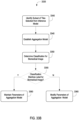
- the model trainer may determine, subsequent to modifying the inference system, that one or more parameters of the inference system have converged relative the one or more parameters prior to the modification of the inference system.
- the model trainer may initiate training of the aggregation mode, responsive to the determination that the one or more parameters of the inference has converged.
- Section A describes Terabyte-Scale Deep Multiple Instance Learning for Classification and Localization in Pathology.
- Section B describes systems and methods of using two-dimensional slicing in training an encoder-decoder model for reconstructing biomedical images and applying the encoder-decoder model to reconstruct biomedical images.
- Section C describes systems and methods of classifying biomedical images and training models for classifying biomedical images using multiple-instance learning.
- Section D describes a network environment and computing environment which may be useful for practicing various computing related embodiments described herein.
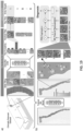
- FIG. 1 depicted are whole slide images (WSI) at various magnification factors.
- Prostate cancer diagnosis is a difficult task. The diagnosis can be based on very small lesions. In the slide above, only about 6 small tumor glands are present. The right most image shows an example tumor gland. Its relation to the entire slide is put in evidence to reiterate the complexity of the task. The figure depicts the difficulty of the task, where only a few tumor glands concentrated in a small region of the slide determine the diagnosis.
- MIL Multiple Instance Learning
- pathology digital slide classification is formalized as a weakly supervised learning task under the MIL framework.
- MIL is used to enhance the classification accuracy and provide localization of the most characteristic regions in each image.
- WSI Whole Slide Images
- MIL Multiple Instance Learning
- each WSI is a collection of small tiles. Each tile has a certain probability of being of class positive. Only if all tiles in a WSI are negative, the probability of being positive is lower than 0.5, the WSI is negative. According to MIL, learning can be achieved from the top-1 most positive tile in each WSI via a simple cross-entropy loss function and gradient descent optimization.
- a dataset including 12,160 needle biopsies slides scanned at 20x magnification, of which 2,424 are positive and 9,736 are negative is used.
- the diagnosis was retrieved from the original pathology reports in the Laboratory Information System (LIS) of a medical institution.
- Exploratory experiments were run on a subset of the full dataset including 1,759 slides split among a training set of 1,300 slides and a validation set of 459 slides. Both splits had a balanced number of positive and negative cases.
- the large-scale experiments were run on the entire dataset on a 70%-15%-15% random split for training, validation and testing respectively. During training, tiles are augmented on the fly with random horizontal flips and 90°rotations.
- FIG. 2 depicted are bar graphs of splitting of a biopsy dataset.
- the full dataset was divided into 70-15-15% splits for training, validation, and test for all experiments except the ones investigating dataset size importance.
- training sets of increasing size were generated along with a common validation set.
- the dataset was randomly split in training (70%), validation (15%) and testing (15%). No augmentation was performed during training.
- dataset size importance experiments, explained further in the Experiments section, a set of slides from the above mentioned training set were drawn to create training sets of different sizes.
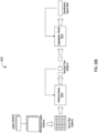
- the complete pipeline of the method comprises the following steps: (i) tiling of each slide in the dataset; for each epoch, which consists of an entire pass through the training data, (ii) a complete inference pass through all the data; (iii) intra-slide ranking of instances; (iv) model learning based on the top-1 ranked instance for each slide.
- FIG. 3 depicted is a schema of performing multiple instance learning for classification of tumorous features on whole slide images.
- the slide or bag consists of multiple instances. Given the current model, all the instances in the bag are used for inference. They are then ranked according to the probability of being of class positive (tumor probability). The top ranked instance is used for model learning via the standard cross-entropy loss. Unless otherwise noted a gradient step is taken every 100 randomly sampled slides and the models used in experiments is an AlexNet and VGG11 pretrained on ImageNet allowing all layers to be optimized.
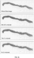
- Slide Tiling The instances are generated by tiling the slide on a grid. All the background tiles are efficiently discarded by an algorithm, reducing drastically the amount of computation per slide, since quite a big portion of it is not covered by tissue. Furthermore, tiling can be performed at different magnification levels and with various levels of overlap between adjacent tiles. In this work three magnification levels (5x, 10x and 20x) were investigated, with no overlap for 10x and 20x magnification and with 50% overlap for 5x magnification. On average each slide contains about 100 non overlapping tissue tiles at 5x magnification and 1,000 at 20x magnification. More detailed information on the composition of the bags is given in FIGs. 6A-C .
- An example of tiling can be seen in FIG. 5 .
- Model Training The model is a function f ⁇ with current parameters ⁇ that maps input tiles b i,j to class probabilities for "negative" and "positive” classes.
- the highest ranking tile in bag B s i is then b i,k .
- l ⁇ w i y i log y ⁇ i ⁇ w 0 1 ⁇ y i log 1 ⁇ y ⁇ i
- weights w0 and w1 for negative and positive classes respectively, can be used to give more importance to the underrepresented examples.
- the final loss is the weighted average of the losses over a minibatch. Minimization of the loss is achieved via stochastic gradient descent using the Adam optimizer and learning rate 0.0001. Mini-batches of size 512 for AlexNet, 256 for ResNets and 128 for VGGs were used.
- Model Testing At test time all the instances of each slide are fed through the network. Given a threshold (usually 0.5), if at least one instance is positive then the entire slide is called positive; if all the instances are negative then the slide is negative. Accuracy, confusion matrix and ROC curve are calculated to analyze performance.
- AlexNet was able to reduce the loss 4/5 of the time
- VGG11 which has an architecture very similar to AlexNet but contains 11 convolutional layers instead of 5, run successfully 2/5 of the time.
- adding batch normalization to VGG11 completely erases the performance seen in the standard VGG11.
- ResNet18 similarly to VGG11BN also gets stuck on a suboptimal minimum. Different optimizers and learning rates were also tested with similar results.
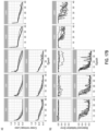
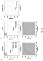
- FIG. 4(a) shows the training loss for the AlexNet model trained at different magnifications; to note how after 400 steps convergence has not been reached yet.
- FIG. 4(b) shows the overall misclassification error, the false negative rate and false positive rate for the validation set. As expected, the model originally assigns a positive label to every slide. As training proceeds, the false positive rate decreases while the false negative rate tends to increase.
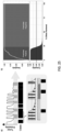
- AlexNet and a VGG11 models pretrained on ImageNet on the full dataset were trained: 8,512 slides for training and 1,824 for validation. Each experiment was run 4 times to inspect the robustness to random initializations and optimization. Given the computational cost of fully inspecting every 20x tile in such a large dataset, the training was tested on the validation set only every 50 steps. The jobs were stopped after 160 hours completing almost 200 steps training steps. Traces of the training procedure are shown in FIG. 4(c) and 13 (depicting confusion matrices for the best AlexNet and VGG11 models on the test set). Both AlexNet and VGG11 were able, at least in a subset of the runs, to reduce the loss during training. It is also clear that the models were still learning and that with more training the error could have decreased more.
- VGG11 achieved the best performance on the test set with a balanced error rate of 13% and an AUC of 0.946 as seen in FIG. 4(d) .
- Needle biopsy diagnosis is an unbalanced classification task.
- the full dataset consists of 19.9% positive examples and 80.1% negative ones.
- training was performed on the full dataset an AlexNet and a Resnet18 networks, both pretrained on ImageNet, with weights for the positive class w 1 equal to 0.5, 0.7, 0.9, 0.95 and 0.99.
- Test Dataset Performance For each architecture, the best model on the validation dataset was chosen for final testing. Performance was similar with the one on the validation data indicating good generalization. The best models were Resnet34 and VGG11-BN which achieved 0.976 and 0.977 AUC respectively. The ROC curves are shown in FIG. 16(a) .
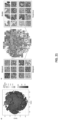
- Feature Embedding Visualization Understanding what features the model uses to classify a tile is an important bottle-neck of current clinical applications of deep learning.
- One can gain insight by visualizing a projection of the feature space in two dimensions using dimensionality reduction techniques such as PCA. 50 tiles were sampled from each test slide, in addition to its top-ranked tile, and extracted the final feature embedding before the classification layer. Shown in FIG. 17A are the results of the ResNet34 model. From the 2D projection, a clear decision boundary between positively and negatively classified tiles can be seen. Interestingly, most of the points are clustered at the top left region where tiles are rarely top-ranked in a slide. By observing examples in this region of the PCA space, it can be determined that they are tiles containing stroma.
- Tiles containing glands extend along the second principal component axis, where there is a clear separation between benign and malignant glands. Other top-ranked tiles in negative slides contain edges and inked regions. The model trained only with the weak MIL assumption was still able to extract features that embed visually.
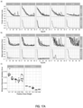
- VGG11-BN and ResNet34 models were trained with tiles generated at 5x and 10x magnifications. Lowering the magnification led consistently to higher error rates across both models. Training curves and validation errors are shown in FIG. 17E .
- Ensemble models were also generated by averaging or taking the maximum response across different combinations of the three models trained at different magnifications. On the test set these naive multi-scale models outperformed the single-scale models, as can be seen in the ROC curves in FIG. 16(b) . In particular, max-pooling the response of all the three models resulted in the best results with an AUC of 0.979, a balanced error of 5.8% and a false negative rate of 4.8%.
- the performance of the pipelines can be optimized to be able to run training in a fraction of the time. Investigation can be done on how to add supervision from a small pool of pixel-wise annotated slides to increase accuracy and achieve faster convergence. In addition, this MIL pipeline can be tested on other types of cancer to further validate the widespread applicability of the method described herein.
- FIG. 5 shown is an example of a slide tiled on a grid with no overlap at different magnifications.
- the slide is the bag and the tiles constitute the instances of the bag. In this work instances at different magnifications are not part of the same bag.
- the slide is the bag and the tiles constitute the instances of the bag. In this work instances at different magnifications are not part of the same bag.
- FIG. 6A illustrates some statistics on the composition of the bags for the exploratory dataset.
- FIG. 6B illustrates some statistics on the composition of the bags for the exploratory dataset tiled with 50% overlap.
- FIG. 6C illustrates some statistics on the composition of the bags for the full dataset consisting of 12,160 slides.
- FIG. 7A shown are setups for exploratory experiments. Standard MIL setup at 20x magnification with no overlap; adam optimizer with starting learning rate of 0.0001 for 100 steps. The training loss is plotted for different architectures. To note how AlexNet and VGG11 are able to reduce the loss, while VGG11BN and ResNet18 are stuck in a suboptimal minimum.
- FIG. 7B MIL training of an AlexNet at different magnifications.
- FIG. 8A shown is a selection of true positives from the best models on the validation set.
- FIG. 8B shown are three examples of false positive slides on the validation set. These are all the cases that were mistakenly classified by the best models at each magnification tested. Inside the red rectangles are the tissue areas with a prostate cancer mimicker. a) The slide contains portions of seminal vesicle tissue. b) The slide presents areas of adenosis and general gland atrophy, c) The slide present areas of inflammation.
- the false negatives are in general cases where the tumor regions are particularly small.
- FIG. 9 shown is a table of a performance comparison of the classic MIL approach and the naive multi- scale version. A significant performance boost is observed by combining the prediction from multiple models.
- FIG. 10A shown are ROC curves for the naive multi-scale approach. The dotted lines are the ROC curves for each model alone. The performance of the three models together is improved as shown by the higher AUCs and overall error rates.
- FIG. 10B shown is performance of MIL trained with overlap.
- the results on the naive multi-scale approach are encouraging to try to learn feature at different scales within the same model.
- Three architectures were tested: (i) The "single” model uses as input a 6-channel image where the first three channels are for a 20x image and the second three channels are for a 5x image, both centered around the same pixel. (ii) The "double-sum” model has two parallel feature extractor, one for the 20x image and one for the 5x image. The features are then added element-wise and fed to a classifier. (iii) The "double-cat” model is very similar to the "double-sum” model but the features coming from the two streams are concatenated instead of added.
- Model architectures for the learned multi-scale MIL experiments receive as input a tile at 5x and 20x magnification.
- the tiles can be stacked into a "single" stream, or they can each go through parallel feature extractors.
- the features can then either be summed element-wise or concatenated before being fed to the final classifier.
- the tiling for these experiments is done at 20x magnification without overlap, as before, but now two tiles are extracted at each time, one at 5x and one at 20x. The 5x tiles have 75% overlap.
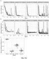
- FIG. 12A shown are performance of the trained multi-scale experiments in comparison with the performance of the 20x magnification experiment from previous sections (dotted line). a) Training loss. b) Classification error on the validation set. The pipeline is slower than the non-multi-scale approach and fewer training steps could be completed. The performance of the "double" models is comparable to the 20x magnification model, while the "single” model seems to performs significantly worse.
- FIG. 12B shown are results from the large-scale training experiments on AlexNet (left column) and VGG11 (right column). Training loss and validation balanced error are plotted in the first and second rows respectively. The experiments were run 4 times each (gray traces) and the average curve is shown in red. While the AlexNet curve all show diminishing loss, in the VGG case, two of the four curves were stuck in a suboptimal minimum. The arrows point to the models chosen for the final testing on the test. Referring now to FIG. 13 , shown are the confusion matrices for the best AlexNet and VGG11 models on the test set.
- the CAMELYON16 challenge for breast cancer metastasis detection contains one of the largest labeled datasets in the field, with a total of 400 whole-slide images (WSIs). But this amount of cases is extremely small compared to the millions of instances present in the popular ImageNet dataset.
- One widely adopted solution to the scarcity of labeled examples in pathology is to take advantage of the size of each example. Pathology slides scanned at 20x magnification produce image files of several gigapixels. About 470 WSIs scanned at 20x contain roughly the same number of pixels as the entire ImageNet dataset. By breaking the WSIs into small tiles, it is possible to obtain thousands of instances per slide, enough to train high-capacity models from a few hundred slides.
- the slides in this work are representative of slides generated in a true pathology laboratory, which include common artifacts, such as air bubbles, microtomy knife slicing irregularities, fixation problems, cautery, folds, and cracks, as well as digitization artifacts, such as striping and blurred regions.
- common artifacts such as air bubbles, microtomy knife slicing irregularities, fixation problems, cautery, folds, and cracks
- digitization artifacts such as striping and blurred regions.
- Prostate cancer beyond its medical relevance as the leading source of new cancer cases and the second most frequent cause of death among men after lung cancers, can be diagnostically challenging, even for trained pathologists.
- Multiple studies have shown that prostate cancer diagnosis has a high inter- and intra-observer variability. Diagnosis is frequently based on the presence of very small lesions that comprise less than 1% of the entire tissue surface area (e.g., FIG. 18 ). Referring to FIG. 18 , shown is a hematoxylin and eosin stained whole slide image for prostate cancer biopsy.
- the diagnosis can be based on very small foci of cancer that account for less than 1% of the tissue surface.
- the slide above only about 6 small tumor glands are present.
- the right-most image shows an example of a malignant gland. Its relation to the entire slide is put in perspective to reiterate the difficulty of the task.
- BCC the most common skin cancer, with approximately 4.3 million individuals diagnosed annually in the US - rarely causes metastases or death.
- pathologists can readily identify and diagnose the lesion; however, given its high frequency, the volume of cases that a pathologist must report is increasing.
- a decision support system should streamline the work of the pathologist and lead to faster diagnosis.
- a clinical support system could allow for prioritization of slides with a higher probability of metastasis to be presented to the pathologist for confirmation.
- This assistive model would lower false negative rates and enable automation of subsequent downstream clinical tasks, such as quantification of metastatic tumor volume for clinical staging purposes. Detection of breast cancer metastasis in lymph nodes is also important because it allows directly comparison of the proposed methods to the state-of-the-art WSI classification that was established based on the CAMELYON16 challenge.
- MIL has seen relatively little application in medical image analysis and computational pathology, in part due to the lack of large WSI datasets.
- This disclosure takes advantage of the large datasets and propose a deep MIL framework where only the whole-slide diagnosis is needed to train a decision-support system capable of classifying digital slides on a large scale with a performance in line with clinical practice.
- the present disclosure is different because MIL supervision is used to learn a semantically rich tile vector representation. Such representation is then used in a recurrent neural network (RNN) to integrate the information across the slide and emit the final classification result (e.g., FIG. 19 ).
- RNN recurrent neural network
- all previous works used small datasets, which precludes a proper estimation of the clinical relevance of the learned models.
- the model is trained on tens of thousands of slides, a scale at which clinically relevant performance can be achieved.
- H&E hematoxylin and eosin
- the breast cancer metastases dataset of axillary lymph nodes consisted of 9,894 slides, 2,521 of which contained macro-metastases, micrometastases, or isolated tumor cells (ITCs). Included in this dataset were slides generated from intraoperative consultations (e.g. frozen section slides), in which the quality of staining varied from the standardized H&E staining protocols used on slides from formalin-fixed, paraffin-embedded tissue.
- the dataset also included patients treated with neoadjuvant chemotherapy, which may be diagnostically challenging in routine pathology practice (i.e. small volume of metastatic tumor, therapy-related change in tumor morphology) and are known to lead to high false negative rates.
- Table 1 Datasets description. This study is based on a total of 44,732 slides from 15,187 patients across three different tissue types: prostate, skin and axillary lymph nodes.
- the prostate dataset was divided into in-house slides and consultation slides to test for staining bias.
- the class imbalance varied from 1:4 for prostate to 1:3 for breast.
- a total of 17,661 slides were submitted to MSK from more than 800 outside institutions in 45 countries for a second opinion.
- the last column shows a comparison in terms of pixel count with ImageNet, the state-of-the-art in computer vision, containing over 14 million images.
- the MIL assumption in the context of WSI classification states that for negative slides, all its tiles are of negative class; for positive slides, there must exist one or more positive tiles, sometimes also referred to as discriminant tiles.
- the MIL assumption can be applied to deep learning as follows: given a model that predicts the probability of being class positive for a small tile, a full inference pass through the dataset is performed. Within each slide, the tiles are ranked according to their probability of being positive. The top most probable tiles for each slide are then used for training the model ( FIG. 19 ). The top-ranking tiles from positive slides should have a probability of being positive close to 1. Conversely, top-ranking tiles from negative slides should have a probability of being positive close to 0.
- the model can be trained on the top-ranking tiles using a standard cross-entropy loss by assigning the slide level target to its respective tile.
- the MIL assumption determines that if one positive tile is found, the slide is predicted positive.
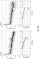
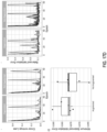
- the prostate dataset (excluding the test portion) was split in a common validation set with 2,000 slides and training sets of different sizes (100, 200, 500, 1,000, 2,000, 4,000, 6,000 and 8,000), with each training dataset being a superset of all previous datasets.
- the results indicate that while the validation error is starting to saturate for ResNet34, and further improvement can be expected from even larger datasets than the one collected for this study.
- Training was performed with datasets of increasing size. The experiment underlies the fact that a large number of slides is necessary for generalization of learning under the MIL assumption. ResNet architectures result in lower errors conditioned on the dataset size. Although the number of slides needed to achieve satisfactory results may vary by tissue type, it is observed that, in general, at least 10,000 slides are necessary for good performance.
- Performance on the test set was measured for ResNet34 architectures trained at different magnifications for each dataset (see FIG. 26 ). It was noticed that the error modes on the test set across magnification conditions were complementary: in prostate, for example, the 20x model performed better in terms of false negatives, while the 5x model performed better on false positives. This observation led to generating ensemble models by averaging or max-pooling the response across models trained at different magnifications. These naive multi-scale models outperformed the single-scale models for the prostate dataset in terms of accuracy and AUC, but not for the other datasets. The AUC for the models trained at 20x was 0.986, 0.986 and 0.965 on the test sets of the prostate, BCC and axillary lymph node datasets, respectively.
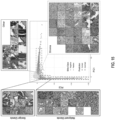
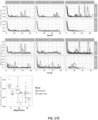
- FIG. 21 shown are the results of the ResNet34 model trained on prostate at 20x.
- Tiles corresponding to points in the 2D t-SNE space were randomly sampled from different regions. Abnormal glands are clustered together on the bottom and left sides of the plot.
- a region of tiles with tumor probability around 0:5 contains glands with features suspicious for prostatic carcinoma. Normal glands are clustered on the top left region of the plot.
- the model trained with MIL supervision was able to extract features that embed visually and semantically related tiles close to each other.
- a large region of different stroma tiles at the center of the plot was observed, extending towards the top right corner.
- the top left corner is where benign-looking glands are represented.
- the bottom portion contains background and edge tiles.
- the discriminative tiles with high tumor probability are clustered in two regions at the bottom and left of the plot. A closer look reveals the presence of malignant glands.
- the max-pooling operation that leads to the slide prediction under the MIL assumption is not robust. A single spurious misclassification can change the slide prediction, possibly resulting in a large number of false positives.
- One way to mitigate this type of mistake is to learn a slide aggregation model on top of the MIL classification results. For example, one approach learned a logistic regression based on the number of tiles per class as predicted by an ensemble of tile classifiers. Similarly, another approach extracted geometrical features from the tumor probability heat-map generated by a tile-level classifier, and trained a random forest model winning the CAMELYON16 challenge. In addition to the counts of tiles in each class, numerous other features were extracted from the heat-map generated by the MIL-based tile classifier.
- a random forest model is then trained on the set of engineered features. An in-depth description is found in the Methods section. This approach was analyzed on the prostate dataset, and also the model was utilized on the CAMELYON16 experiments that will be discussed later.
- the random forest trained on the validation split at 20x magnification produced a 0.98 AUC on the test set, no better than MIL alone (see FIG. 28 ). Although this procedure decreased drastically the false positive rate, and at 20x achieved a better balanced error than the basic max-pooling aggregation, this comes with an unacceptable increase of the false negative rate.
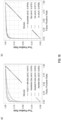
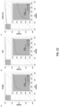
- FIG. 22 shown are line graphs of MIL-RNN model performance for different classification tasks. Performance of the models trained at 20x magnification on the respective test datasets was measured in terms of AUC for each tumor type.
- Statistical significance was assessed using DeLong's test for two correlated receiver operating characteristic (ROC) curves.
- ROC receiver operating characteristic
- the MIL-RNN models resulted in 0.991, 0.989 and 0.965 AUCs for prostate, BCC and breast metastases datasets, respectively.
- the MIL-RNN method was significantly better than max-pooling aggregation.
- the multi-scale approach was tested on the prostate data, but its performance was not better than the one achieved by the single-scale model trained at 20x.
- the MIL-RNN model trained at 20x magnification was run with a step size of 20 pixels across a region of interest, generating a tumor probability heat-map. On every slide, the blue square represents the enlarged area.
- Prostate TP: difficult diagnosis due to tumor found next to atrophy and inflammation; FN: very low tumor volume; and FP: model identified atypical small acinar proliferation (ASAP) showing a small focus of glands with atypical epithelial cells.
- BCC TP: low tumor volume case; FN: low tumor volume case; and FP: the tongue of the epithelium abutting from the base of the epidermis shows an architecture similar to BCC.
- Axillary lymph nodes TP: ITCs with neoadjuvant chemotherapy modifications; FN: slightly blurred cluster of ITCs missed due to very low volume; and FP: displaced epithelium/benign papillary inclusion.
- a dermatopathologist reviewed the cases. On the test set, four false negatives were corrected to true negatives, and four false positives were corrected to true positives. Given these corrections, the AUC improved from 0.988 to 0.994.
- the 12 cases determined to be false negatives were characterized by low tumor volume.
- the 15 false positives included squamous cell carcinomas and miscellaneous benign neoplastic and non-neoplastic skin lesions.
- Prostate BCC Axillary LNs FN FP FN FP FN FP Benign/Negative 3 56 3 2 17 1 Atypical/Other/Suspicious 3 16 1 11 4 31 Carcinoma/Positive 6 0 12 4 23 2 True Error Rate 6/345 72/1,439 12/255 13/1,320 23/403 32/1,070
- FIG. 27 depicted are generalization of performance results.
- the generalization performance of the proposed prostate and breast models were evaluated on different external test sets.
- Performance in terms of AUC decreased by 3% and 6% for the Philips scanner and external slides respectively.
- the pathologist is presented with the model's recommendations through an interface that would flag positive slides for rapid review in a screening scenario, or to disregard all benign slides in a diagnostic scenario.
- HPC high performance computing
- Pathology reports are recorded in the laboratory information system (LIS) of the pathology department.
- LIS laboratory information system
- the ground-truth labels i.e. the slide-level diagnoses
- the LIS database This is made possible by the structured nature of the reporting done for these subspecialties.
- Basal cell carcinomas are not reported in structured form.
- A.M. trained dermatopathologist checked the free text diagnoses and assigned final binary labels to each case manually.
- Classification of a whole digital slide e.g. WSI
- a tile-level classifier can be formalized under the classic MIL paradigm when only the slide-level class is known and the classes of each tile in the slide are unknown.
- For positive bags there must exist at least one instance that is classified as positive by some classifier.
- negative bags instead, all instances must be classified as negative. Given a bag, all instances are exhaustively classified and ranked according to their probability of being positive.
- the complete pipeline for the MIL classification comprises the following steps: (i) tiling of each slide in the dataset; for each epoch, which consists of an entire pass through the training data, (ii) a complete inference pass through all the data; (iii) intra-slide ranking of instances; (iv) model learning based on the top-ranked instance for each slide.
- the instances were generated by tiling each slide on a grid.
- FIG .31 depicted is an example of a slide tiled on a grid with no overlap at different magnifications.
- a slide represents a bag, and the tiles constitute the instances in that bag.
- instances at different magnifications are not part of the same bag.
- Otsu's method is used to threshold the slide thumbnail image to efficiently discard all background tiles, thus, drastically reducing the amount of computation per slide. Tiling can be performed at different magnification levels and with various levels of overlap between adjacent tiles. Three magnification levels (5x, 10x and 20x) were investigated.
- the model is a function f ⁇ with current parameters ⁇ that maps input tiles b i,j to class probabilities for "negative" and "positive” classes.
- weights w 0 and w 1 for negative and positive classes, respectively, can be used to give more importance to the underrepresented examples.
- the final loss is the weighted average of the losses over a minibatch. Minimization of the loss is achieved via stochastic gradient descent (SGD) using the Adam optimizer and learning rate 0.0001. Mini-batches of size 512 for AlexNet, 256 for resnets and 128 for VGGs and DenseNet201 were used. All models were initialized with ImageNet pre-trained weights. Early stopping was used to avoid over-fitting.
- SGD stochastic gradient descent
- a multi-scale ensemble can be created by pooling the predictions of each model with an operator. Average and max-pooling was used to obtain naive multi-scale models.
- a heat-map of tumor probability can be obtained over the slide.
- Several features can then be extracted from the heat-map to train a slide aggregation model. For example, one approach used the count of tiles in each class to train a logistic regression model. Here, that approach was extended by adding several global and local features and train a random forest to emit a slide diagnosis.
- Model f mapping a tile to class probability consists of two parts: a feature extractor f F that transforms the pixel space to representation space and a linear classifier f C that projects the representation variables into the class probabilities.
- the output of f F for the ResNet34 architecture is a 512-dimensional vector representation. Given a slide and model f , a list of the S most interesting tiles within the slide in terms of positive class probability can be obtained.
- the state vector is initialized with a zero vector.
- step i 1, 2,..., S of the recurrent forward pass
- the S most interesting tiles from a slide was obtained by averaging the prediction of the three models on tiles extracted at the same center pixel but at different magnifications.
- the inputs to the RNN at each step i are e 20 x,i , e 10 x , i , e 5 x,i , and the state vector h i -1 .
- Equation 3 ReLU W 20 xe 20 x , i + W 10 xe 10 x , i + W 5 xe 5 x , i + Whhi ⁇ 1 + b All RNN models were trained with cross-entropy loss and SGD with a batch size of 256.
- the CAMELYON16 dataset consists of 400 total patients for whom a single WSI is provided as a tag image file format (TIFF).
- Annotations are given in extensible markup language (XML) format, one per each positive slide.
- XML extensible markup language
- regions defined by vertex coordinates, may be present. Since these slides were scanned at a higher resolution than the slides scanned at MSK, a tiling method was developed to extract tiles containing tissue from both inside and outside the annotated regions at MSK's 20x equivalent magnification (0.5 ⁇ m/pixel) to enable direct comparison with the datasets.
- the method generates a grid of possible tiles, excludes background via Otsu thresholding and determines whether a tile is inside an annotation region by solving a point in polygon problem.
- FIG. 27 shown is t-SNE visualization of the representation space for the BCC and axillary lymph node models.
- 2D t-SNE projection of the 512-dimensional representation space were generated for 100 randomly sampled tiles per slide.
- FIG. 28 shown is performance of the MIL-RF model at multiple scales on the prostate dataset.
- the MIL model was run on each slide of the test dataset with a stride of 40 pixels. From the resulting tumor probability heat-map, handengineered features were extracted for classification with the random forest (RF) model.
- the best MIL-RF model (ensemble model, AUC of 0.987) did not outperform the MIL-only model (20x model, AUC of 0.986, see Figure 5 ).
- ROC curves of the generalization experiments summarized in Figure 7 shown are ROC curves of the generalization experiments summarized in Figure 7 .
- b-c Comparison of the proposed MIL approach to state-of-the-art fully supervised learning for breast metastasis detection in lymph nodes.
- FIG. 30 shown is decision support with the BCC and breast metastases models.
- slides are ordered by their probability of being positive for cancer as predicted by the respective MIL-RNN model.
- the sensitivity is computed at the case level.
- FIG. 31 shown is example of a slide tiled on a grid with no overlap at different magnifications.
- a slide represents a bag, and the tiles constitute the instances in that bag. In this work, instances at different magnifications are not part of the same bag.
- Stratified prediction performance of the prostate cancer MIL-RNN model Relevant categories for positive slides are Gleason grades and tumor sizes and for negative slides they are the presence of atrophy or hyperplasia.
- the dataset was divided into in-house and external consultation cases.
- the in-house data was sub-divided into training, validation and test sets.
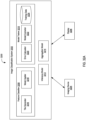
- the system 3200 may include an image classification system 3202 (sometimes referred herein as an image classifier), at least one imaging device 2304, and at least one display 3206.
- the image classification system 3202 may include at least one feature classifier 3208, at least one model trainer 3210, at least one inference model 3212 (sometimes referred herein as an inference system), and at least one aggregation model 3214 (sometimes referred herein as an aggregation system), among others.
- the feature classifier 3208 may include at least one tile generator 3216 and at least one model applier 3218.
- the model trainer 3210 may include at least one error calculator 3220, at least one model corrector 3222, and at least one training database 3224.
- the inference model 3212 and the aggregation model 3214 each may have a training mode and a runtime mode. Under the training mode, the image classification system 3202 may invoke both the feature classifier 3208 and the model trainer 3210.
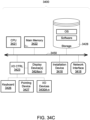
- Each of the components of system 3200 may be implemented using hardware (e.g., processing circuitry and memory) or a combination of hardware and software as detailed here in Section D in conjunction with FIGs. 34A-D .
- the tile generator 3216 of the feature classifier 3208 may identify one or more biomedical images 3232A-N (hereinafter referred generally as biomedical images 3232).
- the biomedical images 3232 may be of a micro-anatomical sample or specimen.
- the biomedical images 3232 may be, for example, a histological section with a hematoxylin and eosin (H&E) stain, hemosiderin stain, a Sudan stain, a Schiff stain, a Congo red stain, a Gram stain, a Ziehl-Neelsen stain, a Auramine-rhodamine stain, a trichrome stain, a Silver stain, and Wright's Stain, among others.
- the tile generator 3216 may receive the biomedical images 3232 from the imaging device 3204.
- the receipt of the biomedical images 3232 from the imaging device 3204 may be under the runtime mode for the inference model 3212 or the aggregation model 3214.
- the imaging device 3204 may be a microscope communicatively coupled with the image classification system 3202.
- the imaging device 3204 may scan the micro-anatomical sample or specimen, and may generate the biomedical image 3232 from the scan.
- the tile generator 3216 may access the training database 3224 to retrieve the biomedical images 3232.
- the retrieval of the biomedical images 3232 from the training database 3224 may be under training mode for the inference model 3212.
- Each biomedical image 3232 retrieved from the training database 3324 may have a label 3234A-N (hereinafter referred generally as label 3234 and sometimes referred herein as an annotation).
- the training database 3324 may maintain a set of biomedical images 3232 with the label 3234 for training the inference model 3212 and the aggregation model 3214.
- the label 3234 may indicate a presence or a lack of a condition on the biomedical image 3232.
- the condition may be a pathological condition, such as a tumor, injury, scarring, dead cells, or other defect.
- the label 3234 may indicate the presence or the lack of multiple conditions on the biomedical image 3232.
- one label 3234 may indicate the presence of benign tumorous growth, while another label 3234 may indicate the presence of malignant tumor formation.
- MIL multiple instance learning
- the label 3234 may not identify a specific location of the presence or the lack of the condition on the biomedical image 3232. Rather, the label 3234 may indicate that the condition is present somewhere on the biomedical image 3232.
- the tile generator 3216 may generate a set of tiles 3236A-N (hereinafter referred as tiles 3236) from the biomedical image 3232. Each tile 3236 may correspond to a portion of the biomedical image 3232.
- the tile generator 3215 may partition or divide the biomedical image 3232 into the set of tiles 3236.
- the tile generator 3216 may apply to one or more magnification factors to generate the set of tiles 3236. The magnification factors applied to the biomedical image 3232 may range from 3x to 100x.
- the tile generator 3216 may generate the set of tiles 3236 from the biomedical image 3232 without overlap.
- the tile generator 3216 may generate the set of tiles 3236 with an overlap of a set ratio.
- the ratio may range from 10% to 90% overlap between pairs of adjacent tiles 3236.
- the set ratio for the overlap may depend on the magnification factor applied to the biomedical image 3232. For example, an overlap of 50% may be used at 10x magnification factor and an overlap of 67% maybe used at 5x magnification factor.
- the tile generator 3216 may identify or detect one or more regions of the biomedical image 3232 corresponding to negative space.
- the identification of the negative space may be in accordance with a feature detection algorithm.
- the negative space region of the biomedical image 3232 may lack any portion of the micro-anatomical sample or specimen.
- the negative space may correspond to the region of the biomedical image 3232 that is null or white.
- the tile generator 3216 may apply the one or more magnification factor to the biomedical image 3232, prior to the detection of the negative space. With the identification of the negative space, the tile generator 3216 may remove the corresponding region from the biomedical image 3232.
- the tile generator 3216 may generate the set of tiles 3236 from the remaining one or more regions of the biomedical image 3232. In some embodiments, the tile generator 3216 may detect or identify a subset of the tiles 3236 generated from the biomedical image 3232 corresponding to the negative space (e.g., having at least 97% white space) using the feature detection algorithm. The tile generator 3216 may remove the identified subset of the set of tiles 3236 corresponding to the negative space.
- the model applier 3218 may establish the inference model 3212. Under training mode for the image classification system, the model applier 3218 may initialize the inference model 3212. Under runtime mode, the model applier 3218 may identify the previously established inference model 3212.
- the inference model 3212 may determine a score for each tile 3236 generated from the biomedical image 3232.
- the score may indicate a likelihood that the tile 3236 includes at least one feature indicative of the presence of the condition.
- the score may be a numerical value, such as a probability, a percentage, or within a defined range of numbers (e.g., -1 to 1, 0 to 1, -10 to 10, or 0 to 100), to indicate the likelihood.
- the feature may be a visual characteristic, property, or object within the portion of the biomedical image 3232 corresponding to the slide 3236.
- the inference model 3212 may have one or more parameters to determine the score for each tile 3236.
- the inference model 3212 may include a set of transform layers (e.g., convolutional layer, pooling layer, rectified layer, and normalization layer).
- the inference model 3212 may have any number of transform layers.
- Each transform layer may include at least one of the one or more parameters to convert the set of tiles 3236 to a set of feature maps and to determine the score for each tile 3236.
- Each transform layer may be of a predefined size to generate the feature maps of a predefined size.
- the inference model 3212 may be a convolutional neural network (CNN) and a deep convolutional network (DCN), among others, with the set of transform layers.
- CNN convolutional neural network
- DCN deep convolutional network
- the inference model 3212 may be the convolutional neural network detailed herein in Sections A and B.
- the inference model 3212 may be a feedforward network without internal state memory, and may lack temporal or sequentially dependent behavior.
- the model applier 3218 may set the parameters of the inference model 3212.
- the one or more parameters of the inference model 3212 may be set to random values. The random values may be generated using a pseudo-random number generator.
- one or more parameters of the inference model 3212 may be set to a predefined value. The predefined value may be maintained on the training database 3224.
- the model applier 3218 may set a number of the set of transform layers of the inference model 3212.
- the model applier 3218 may set a size of the set of transform layers of the inference model 3212.
- the model applier 3218 may set connections between transform layers in the inference model 3212 in initializing.
- the model applier 3218 may apply the inference model 3212 to the set of tiles 3236 for each biomedical image 3232. In applying the inference model 3212, the model applier 3218may apply the entire set of tiles 3236 as an input into the inference model 3212. In some embodiments, the model applier 3218may identify an output generated from one transform layer in the inference model 3212. The model applier 3218may feed the output generated from one transform layer as an input of the subsequent transform layer in the inference model 3212. The output from the first transform layer and onward may include a feature map. The input of the first transform layer may be the set of tiles 3236 generated from the biomedical image 3232. The input of the second transform layer and onward in the inference model 3212 may include the feature map generated from the previous transform layer.
- the model applier 3218 may repeat the feeding of the output of one transform layer into the input of the subsequent transform layer in the inference model 3212 until the last transform layer.
- the model applier 3218 may determine the score for each tile 3236.
- the model applier 3218 may determine the score for each condition for each tile 3236.
- one tile 3236 may be associated with a score indicating likelihood of presence of prostate cancer and another score indicating likelihood of bruising to the organ tissue on the tile 3236.
- the model applier 3218 may identify the output of the last transform layer in the inference model 3212. The output may include the scores for all of the tiles 3236.
- the model applier 3218 may select a subset from the set of tiles 3236 to form a subset 3238A-N (hereinafter generally referred to as subset 3238 or selected tiles 3238).
- the model applier 3218 may select the tiles 3236 with the highest scores to form the subset 3238.
- the selected tiles 3238 may represent the tiles 3236 with the highest likelihood of including a feature correlated with or corresponding to the presence of the condition.
- the number of tiles 3238 selected from the original set of tiles 3236 may be in accordance to a predefined number, and may range from 1 to 50.
- the model applier 3218 may select the subset 3238 from the set of tiles 3236 for each condition. For example, the model applier 3218may select one subset 3238 from the tiles 3236 for the condition of breast cancer based on the scores for breast cancer. In conjunction, the model applier 3218may select another subset 3238 from the tiles 3236 for lesion to breast tissue based on the corresponding scores. Under the runtime mode, with the selection from the tiles 3236, the model applier 3218may apply the aggregation model 3214 onto the selected tiles 3238, and feed the selected tiles 3238 into the input of the aggregation model 3214.
- the error calculator 3220 of the model trainer 3210 may compare the scores for the selected tiles 3238 to a threshold value for the condition indicated by the label 3234 of the biomedical image 3232.
- the threshold value for the label 3234 may correspond to the occurrence of the condition specified by the label 3234, and may indicate a score at which to modify one or more parameters of the inference model 3212. For example, the threshold score may be set at 75% for the presence of the condition and 50% for the lack of the presence.
- the scores may be the same or may differ for the presence or the lack of the condition defined by the label 3234.
- the threshold value may differ depending on the condition specified by the label 3234.
- the label 3234 may specify the threshold value to be compared against.
- an equality (e.g., less than or greater than) for the comparison performed by the error calculator 3220 may depend on the label 3234 indicating the presence or the lack of the condition. For example, when the label 3234 specifies the presence of the condition on the corresponding biomedical image 3232, the error calculator 3220 may determine whether the scores of the selected tiles 3238 are less than the threshold value for the condition. Conversely, when the label 3234 specifies the lack of the condition on the corresponding biomedical image 3232, the error calculator 3220 may determine whether the scores of the selected tiles 3238 are greater than or equal to the threshold value for the condition.
- the error calculator 3220 may calculate or determine an error measure between the score of each selected tile 3238 and a baseline value for the condition indicated by the label 3234 of the biomedical image 3232.
- the error measure may indicate one or more deviations from the score and an anticipated score as represented by the baseline value, and may be used to modify the parameters of the inference model 3212.
- the baseline value for the condition indicated by the label 3234 may indicate a score at which the inference model 3212 is expected to output.
- the baseline value for the presence of the condition may differ from the baseline value for the lack of the present of the value. For example, the baseline value for the presence of the condition may range between 0.9 and 1, while the baseline value for the lack of the condition may range between 0 and 0.2.
- the error measure calculated by the error calculator 3220 may be in accordance with a loss function, such as mean square error (MSE), root mean square error (rMSE), an entropy loss (e.g., cross-entropy or relative entropy), a quadratic loss, and mean integrated square error, among others.
- the model corrector 3222 may determine whether to modify the inference model 3212 based on the comparison of the scores of the selected tiles 3238 with the threshold value for the condition.
- the label 3234 may indicate the presence of the condition on the corresponding biomedical image 3232. In such a scenario, when at least one of the scores of the selected tiles 3238 is less than the threshold value, the model corrector 3222 may determine to modify the inference model 3212. On the other hand, when all the scores of the selected tiles 3238 are greater than or equal to the threshold value, the model corrector 3222 may determine to not modify the inference model 3212. Conversely, the label 3234 may indicate the lack of the condition on the corresponding biomedical image 3232.
- the model corrector 3222 may determine to modify the inference model 3212.
- the model corrector 3222 may determine to not modify the inference model 3212.
- the threshold value when the label 3234 indicates lack of the condition may be the same or may differ from the threshold value when the label 3234 indicates the presence of the condition.
- the model corrector 3222 may maintain the inference model 3212. For example, the model corrector 3222 may maintain the parameters of the inference model 3212.
- the model corrector 3222 of the model trainer 3210 may update or otherwise modify the inference model 3212.
- the modification of the inference model 3212 may be responsive to the determination to modify.
- the model corrector 3222 may set, adjust, or otherwise change the one or more parameters of the inference model 3212 based on the condition indicated by the label 3234 for the biomedical image 3232 from which the tiles 3238 are selected.
- the model corrector 3222 may change the parameters of the inference model 3212 to increase the scores for the tiles 3236.
- the model corrector 3222 may change the parameters of the inference model 3212 to decrease the score for the tiles 3236.
- the model corrector 3222 may modify the inference model 3212 using the error measures calculated for the scores of the subset 3238.
- the modification of the inference model 3212 using the calculated error measures may be responsive to the determination to modify or independent of the determination of the modify.
- the model corrector 3222 may set, adjust, or otherwise change the one or more parameters of the inference model 3212 based on the error measures.
- the model corrector 3222 may change the parameters of the inference model 3212 based on the whether error measures are positive or negative.
- the model corrector 3222 may change the size of one or more of the transform layers in the inference model 3212 using the error measure.
- the model corrector 3222 may change the number of transform layers in the inference model 3212 using the error measure. In modifying the parameters, the model corrector 3222 may perform regularization on the set of transform layers in the inference model 3212.
- the regularization may include, for example, dropout, drop connect, stochastic pooling, or max pooling, among others.
- the model corrector 3222 may determine whether the one or more parameters of the inference model 3212 have converged. The determination of whether the inference model 3212 has converged may be responsive to the modification of the inference model 3212.
- the model corrector 3222 may identify the one or more parameters of the inference model 3212 prior to the modification.
- the model corrector 3222 may identify the one or more parameters of the inference model 3212 with the modification. With these identifications, the model corrector 3222 may compare the parameters prior to the modification with the parameters subsequent to the modification. Based on the comparison, the model corrector 3222 may calculate or determine a difference between the parameters prior to the modification and the parameters with the modification.
- the model corrector 3222 may compare the determined difference to a convergence threshold value.
- the convergence threshold value may indicate the point in which to terminate further training of the inference model 3212. If the difference of the parameters is less than or equal to the convergence threshold value, the model corrector 3222 may determine that the inference model 3212 has reached convergence. Furthermore, the model corrector 3222 may terminate the training mode for the inference model 3212 and may switch to runtime mode. In contrast, if the difference of the parameters is greater than the convergence threshold value, the model corrector 3222 may determine that the inference model 3212 has not reached convergence. Additionally, the model corrector 3222 may continue the training mode for the inference model 3212.
- model trainer 3210 for training the inference model 3212 may be implemented in accordance with the following pseudocode:
- the model applier 3218 may establish the aggregation model 3214. Under training mode, the aggregation model 3214, the model applier 3218 may initialize the aggregation model 3214. In some embodiments, the model applier 3218 may initialize the aggregation model 3214, responsive to determining that the inference model 3212 has reached convergence under the training mode. Under runtime mode, the model applier 3218 may identify the previously established aggregation model 3214. The aggregation model 3214 may determine a classification result for each biomedical image 3232 based on the selected tiles 3238 from the inference model 3212. The classification result may indicate whether the biomedical image 3232 contains at least one feature corresponding to the presence of the condition or the lack of the condition. The classification result may be, for example, a binary value (e.g., 0 and 1 or true and false) or one of an enumerate value or indicator (e.g., "high,” "medium,” or “low”), among others.
- a binary value e.
- the aggregation model 3214 may have one or more parameters to determine the classification result for the biomedical image 3232.
- the aggregation model 3214 may include a set of transform layers (e.g., input layer, context layer, state layer, and hidden layer).
- the aggregation model 3214 may have any number of transform layers.
- Each transform layer may include at least one of the one or more parameters to determine the classification result for the biomedical image 3232.
- Each transform layer may include at least one of the one or more parameters to convert the set of tiles 3238 to a set of feature maps and to determine the classification result for the entire biomedical image 3232.
- Each transform layer may be of a predefined size to generate the feature maps of a predefined size.
- the aggregation model 3214 may be a recurrent neural network (RNN), an echo state network (ESN), a long/short term memory (LSTM) network, a deep residual network (DRN), and gated recurrent units (GRU), among others, with the set of transform layers.
- RNN recurrent neural network
- ESN echo state network
- LSTM long/short term memory
- DNN deep residual network
- GRU gated recurrent units
- the aggregation model 3214 may be the recurrent neural network detailed herein in Section B.
- the aggregation model 3214 may have internal state memory, and may exhibit temporally or sequentially dynamic behavior. In this manner, information may be integrated across the selected tiles 3238 from the inference model 3212 to determine the classification result for the overall biomedical image 3232.
- the model applier 3218 may set the parameters of the aggregation model 3214. In some embodiments, the model applier 3218 may commence training of the aggregation model 3214, responsive to determining that the inference model 3212 has reached convergence. In some embodiments, the one or more parameters of the aggregation model 3214 may be set to random values. The random values may be generated using a pseudo-random number generator. In some embodiments, one or more parameters of the aggregation model 3214 may be set to a predefined value. The predefined value may be maintained on the training database 3224. In some embodiments, the model applier 3218 may set a number of the set of transform layers of the aggregation model 3214. In some embodiments, the model applier 3218 may set a size of the set of transform layers of the aggregation model 3214. In some embodiments, the model applier 3218 may set connections between transform layers in the aggregation model 3214 in initializing.
- the model applier 3218 may apply the aggregation model 3214 to the subset of tiles 3238 from the inference model 3212. In some embodiments, the model applier 3218 may identify the subset 3238 outputted by the inference model 3212. In some embodiments, the model applier 3218 may identify the subsets 3238 outputted by the inference model 3212 using the application of different magnification factors on the biomedical image 3232. For example, the input to the aggregation model 3214 may include selected tiles 3238 from the inference model 3212 applied to the biomedical image 3232 at the magnification factors of 5x, 10x, and 20x.
- the model applier 3218 may generate an aggregate subset using a combination of the selected tiles 3238.
- the combination may be, for example, an average, a weighted average, or a predefined function, among others.
- the combination may be among tiles 3238 with similar center points (e.g., within 10%) or overlapping coordinates on the biomedical image 3232.
- the model applier 3218 may feed the aggregate subset to the aggregation model 3214.
- the model applier 3218 may feed the selected tiles 3238 into the aggregation model 3214.
- the model applier 3218 may apply the aggregation model 3214 to the subset of tiles 3238 in sequential order.
- the sequential order may arrange the tiles 3238 from the lowest coordinates to the highest coordinates relative to a starting point (e.g., top left point defined as (0,0)) on the biomedical image 3232.
- the model applier 3218 may apply the aggregation model 3214 to the subset of tiles 3238 in random order.
- the random order may be generated by the model applier 3218 using a pseudo-random number generator.
- the model applier 3218 may feed the output generated from one transform layer as an input of the subsequent transform layer in the aggregation model 3214.
- the output from the first transform layer and onward may include a feature map.
- the input of the first transform layer may be the set of tiles 3238 from the inference model 3212.
- the input of the second transform layer and onward in the aggregation model 3214 may include the feature map generated from the previous transform layer.
- the model applier 3218 may repeat the feeding of the output of one transform layer into the input of the subsequent transform layer in the aggregation model 3214 until the last transform layer.
- the model applier 3218 may identify the classification result for the condition from the last transform layer of the aggregation model 3214. The identification of the classification result may be repeated for multiple conditions (e.g., prostate tumor, breast lesion, and bruised tissue).
- the model applier 3218 may provide the classification result generated by the aggregation model 3214. In some embodiments, the model applier 3218 may display the classification result onto the display 3206.
- the display 3206 may include any monitor, such as a liquid crystal display (LCD), an organic light-emitting diode (OLED) monitor, and a cathode ray tube (CRT), among others.
- the display 3206 may be communicatively coupled with the image classification system 3202, and may render the classification result.
- the model applier 3218 may display the biomedical image 3232 for which the classification result was generated on the display 3206.
- the model applier 3218 may indicate a location in the biomedical image 3232 corresponding to the selected tiles 3238 on the display 3206.
- the error calculator 3220 may compare the classification result determined by the aggregation model 3214 with the label 3234 for the biomedical image 3232.
- the label 3234 may indicate the presence or the lack of the condition on the biomedical image 3232.
- the classification result may indicate the presence or the lack of the condition on the biomedical image 3232.
- the error calculator 3220 may determine whether the classification result from the aggregation model 3214 matches the indication of the label 3234. Whether to modify the aggregation model 3214 may be based on the determination. When the classification result does not match the indication, the error calculator 3220 may determine to modify the aggregation model 3214.
- the error calculator 3220 may set, adjust, or otherwise change the one or more parameters of the aggregation model 314 based on the mismatch between the classification result and the indication. Conversely, when the classification matches the indication, the error calculator 3220 may determine not to modify and maintain the aggregation model 3214. For example, the error calculator 3220 may maintain the parameters of the aggregation model 3214.
- the error calculator 3220 may calculate or determine an error measure between the classification result from the aggregation model 3214 and the indication of the label 3234.
- the determination of the error measure may be performed responsive to determination of the mismatch between the classification result and the indication on the label 3234.
- the error measure may indicate one or more deviations from the anticipated classification result as indicated by the label 3234 for the biomedical image 3232, and may be used to modify the aggregation model 3214.
- the determination of the error measure may be over multiple classification results from the aggregation model 3214 compared against the indications of the corresponding labels 3234.
- the multiple classification results may be from the same biomedical image 3232 with the tiles 3236 generated using different magnification factors.
- the error measure calculated by the error calculator 3220 may be in accordance with a loss function, such as mean square error (MSE), root mean square error (rMSE), an entropy loss (e.g., cross-entropy or relative entropy), a quadratic loss, and mean integrated square error, among others.
- MSE mean square error
- rMSE root mean square error
- entropy loss e.g., cross-entropy or relative entropy
- the model corrector 3222 may update or modify the aggregation model 3214.
- the modification of the aggregation model 3214 may be responsive to the determination to modify.
- the model corrector 3222 may modify the one or more parameters of the aggregation model 3214 using the determined error measures between the classification results and the corresponding indications on the labels 3234.
- the modification of the aggregation model 3214 using the calculated error measures may be responsive to the determination to modify or independent of the determination of the modify.
- the model corrector 3222 may set, adjust, or otherwise change the one or more parameters of the aggregation model 3214 based on the error measures.
- the model corrector 3222 may change the parameters of the aggregation model 3214 based on the whether error measures are positive or negative.
- the model corrector 3222 may change the size of one or more of the transform layers in the aggregation model 3214 using the error measure. In some embodiments, the model corrector 3222 may change the number of transform layers in the aggregation model 3214 using the error measure. In modifying the parameters, the model corrector 3222 may perform regularization on the set of transform layers in the inference model 3212. The regularization may include, for example, dropout, drop connect, stochastic pooling, or max pooling, among others. In some embodiments, the model corrector 3222 may modify the aggregation model 3214 using the error measures in accordance with an iterative optimization algorithm, such as a gradient descent or stochastic gradient descent.
- an iterative optimization algorithm such as a gradient descent or stochastic gradient descent.
- model corrector 3222 may determine whether the one or more parameters of the aggregation model 3214 have converged. The determination of whether the aggregation model 3214 has converged may be responsive to the modification of the aggregation model 3214.
- the model corrector 3222 may identify the one or more parameters of the aggregation model 3214 prior to the modification.
- the model corrector 3222 may identify the one or more parameters of the aggregation model 3214 with the modification. With these identifications, the model corrector 3222 may compare the parameters prior to the modification with the parameters subsequent to the modification. Based on the comparison, the model corrector 3222 may calculate or determine a difference between the parameters prior to the modification and the parameters with the modification.
- the model corrector 3222 may compare the determined difference to a convergence threshold value.
- the convergence threshold value may indicate the point in which to terminate further training of the aggregation model 3214. If the difference of the parameters is less than or equal to the convergence threshold value, the model corrector 3222 may determine that the aggregation model 3214 has reached convergence. Furthermore, the model corrector 3222 may terminate the training mode for the aggregation model 3214. In contrast, if the difference of the parameters is greater than the convergence threshold value, the model corrector 3222 may determine that the aggregation model 3214 has not reached convergence. Additionally, the model corrector 3222 may continue the training mode for the aggregation model 3214.
- the inference model 3212 and the aggregation model 3214 may identify pathological features on biomedical images 3232 with higher accuracy. Furthermore, by eliminating the manual inspection of biomedical images for diagnosis, the recognition of such features on biomedical images 3232 may be performed faster.
- the image classifier may generate tiles from a biomedical image (3305).
- the image classifier may establish an inference model (3310).
- the image classifier may select a subset of tiles based on scores (3315).
- the image classifier may determine whether the score satisfies a threshold for a label of the biomedical image (3320). If the score satisfies the threshold for the label, the image classifier may maintain parameters of the inference model (3325). Otherwise, if the score does not satisfy the threshold for the label, the image classifier may modify the parameters of the inference model (3330).
- the image classifier may generate tiles (e.g., the set of tiles 3236) from a biomedical image (e.g., the biomedical image 3232) (3305).
- the biomedical image may be retrieved from an imaging device or from a training dataset, and may be of a histological section with a hematoxylin and eosin (H&E) stain.
- the biomedical image from the training dataset may be associate with a label.
- the label may indicate a presence or a lack of a pathological condition non the biomedical image.
- the image classifier may generate the tiles at various magnification factor applied to the biomedical image.
- the image classifier may also generate the tiles to overlap with one another at a set ratio.
- the image classifier may establish an inference model (e.g., the inference model 3212) (3310).
- the inference model may have one or more parameters in a set of transform layers for calculating or determining a score for each tile.
- the inference model may be a convolutional neural network.
- the score may indicate a likelihood that a feature on the tile correlates with the presence of the condition.
- the parameters of the inference model may be initially set to random values.
- the image classifier may select a subset of tiles (e.g., selected tile 3238)) based on scores (3315). The selection of the subset of tiles from the initial set may be based on the scores determined by the inference model.
- the image classifier may select a predefined number of tiles with the highest scores.
- the tiles of the subset may represent the corresponding portion on the biomedical image most likely to contain the pathological condition.
- the image classifier may determine whether the score satisfies a threshold for a label of the biomedical image (3320).
- the threshold for the label may indicate a score at which to modify the parameters of the inference model. Whether the score satisfies the threshold may depend on the indication of the label for the biomedical image.
- the image classifier may determine whether the scores of the selected tiles are less than the threshold for the condition. If the scores are greater than the threshold, the image classifier may determine that the score satisfies the threshold. Conversely, the if the scores are less than or equal to the threshold, the image classifier may determine that the score does not satisfy the threshold.
- the image classifier may determine whether the scores of the selected tiles are greater than the threshold for the condition. If the scores are greater than the threshold, the image classifier may determine that the score does not satisfy the threshold. Conversely, the if the scores are less than or equal to the threshold, the image classifier may determine that the score satisfies the threshold
- the image classifier may maintain parameters of the inference model (3325). Otherwise, if the score does not satisfy the threshold for the label, the image classifier may modify the parameters of the inference model (3330).
- the image classifier may also determine an error measure between the scores of the selected tiles and a baseline value for the condition. The baseline value may indicate a score at which the inference model is expected to output. Using the error measure, the image classifier may change the parameters of the inference model.
- an image classifier may identify a subset of tiles selected from an inference model (3340).
- the image classifier may establish an aggregation model (3345).
- the image classifier may determine a classification for the biomedical image (3350).
- the image classifier may determine whether the classification matches a label for the biomedical image (3355). If the classification matches the label, the image classifier may maintain parameters of the aggregation model (3360). On the other hand, if the classification does not match the label, the image classifier may modify the parameters of the aggregation model (3365).
- an image classifier may identify a subset of tiles (e.g., selected tiles 3238) selected from an inference model (e.g., the inference model 3212) (3340).
- the image classifier may retrieve the subset of tiles from the output of the inference model.
- the image classifier may also aggregate the subset of tiles from the inference model for the same biomedical image at different magnification factors.
- the image classifier may combine the tiles from the inference model with similar center coordinates (e.g., within 10% difference).
- the image classifier may establish an aggregation model (e.g., the aggregation model 3214) (3345).
- the aggregation model may have one or more parameters in a set of transform layers to determine a classification result for the biomedical image using the selected subset of tiles.
- the aggregation model may be a recurrent neural network.
- the classification result may indicate the presence or the lack of the pathological condition on the biomedical image.
- the image classifier may determine a classification for the biomedical image (e.g., the biomedical image 3232) (3350).
- the image classifier may apply the aggregation model onto the selected subset of tiles.
- the image classifier may feed the selected subset of tiles in sequential order or random order into the aggregation model. By applying the aggregation model, the image classifier may generate the classification result for the biomedical image.
- the image classifier may determine whether the classification matches a label (e.g., the label 3234) for the biomedical image (3355).
- the image classifier may identify the indication from the label for the biomedical image.
- the label may indicate the presence or the lack of the pathological on the biomedical image. If the classification matches the label, the image classifier may maintain parameters of the aggregation model (3360). On the other hand, if the classification does not match the label, the image classifier may modify the parameters of the aggregation model (3365).
- the image classifier may calculate an error measure over the classification results of multiple subsets of tiles for the same biomedical image at different magnification factors. The error measure may include cross-entropy loss. Using the error measure, the image classifier may change the parameters of the aggregation model.
- an image classifier may generate tiles from a biomedical image (3375).
- the image classifier may select a subset of tiles by applying an inference model (3380).
- the image classifier may determine a classification result by applying an aggregation model (3385).
- an image classifier may generate tiles (e.g., the set of tiles 3236) from a biomedical image (e.g., the biomedical image 3232) (3375).
- the biomedical image may be retrieved from an imaging device or from a training dataset, and may be of a histological section with a hematoxylin and eosin (H&E) stain.
- the biomedical image from the training dataset may be associate with a label.
- the label may indicate a presence or a lack of a pathological condition non the biomedical image.
- the image classifier may generate the tiles at various magnification factor applied to the biomedical image.
- the image classifier may also generate the tiles to overlap with one another at a set ratio.
- the image classifier may select a subset of tiles (e.g., the selected tiles 3238) by applying an inference model (e.g., the inference model 3212) (3380).
- the inference model may have one or more parameters in a set of transform layers for calculating or determining a score for each tile.
- the inference model may be a convolutional neural network.
- the score may indicate a likelihood that a feature on the tile correlates with the presence of the condition.
- the selection of the subset of tiles from the initial set may be based on the scores determined by the inference model.
- the image classifier may select a predefined number of tiles with the highest scores.
- the tiles of the subset may represent the corresponding portion on the biomedical image most likely to contain the pathological condition.
- the image classifier may determine a classification result by applying an aggregation model (e.g., the aggregation model 3214) (3385).
- the aggregation model may have one or more parameters in a set of transform layers to determine a classification result for the biomedical image using the selected subset of tiles from the inference model.
- the aggregation model may be a recurrent neural network.
- the classification result may indicate the presence or the lack of the pathological condition on the biomedical image.
- the image classifier may apply the aggregation model onto the selected subset of tiles.
- the image classifier may feed the selected subset of tiles in sequential order or random order into the aggregation model. By applying the aggregation model, the image classifier may generate the classification result for the biomedical image.
- FIG. 34A an embodiment of a network environment is depicted.
- the illustrated exploring network environment includes one or more clients 3402a-1602n (also generally referred to as local machine(s) 3402, client(s) 3402, client node(s) 3402, client machine(s) 3402, client computer(s) 3402, client device(s) 3402, endpoint(s) 3402, or endpoint node(s) 3402) in communication with one or more servers 3406a-1506n (also generally referred to as server(s) 3406, node 3406, or remote machine(s) 3406) via one or more networks 3404.
- a client 3402 has the capacity to function as both a client node seeking access to resources provided by a server and as a server providing access to hosted resources for other clients 3402a-3402n.
- FIG. 34A shows a network 3404 between the clients 3402 and the servers 3406
- the clients 3402 and the servers 3406 may be on the same network 3404.
- a network 3404' (not shown) may be a private network and a network 3404 may be a public network.
- a network 3404 may be a private network and a network 3404' a public network.
- networks 3404 and 3404' may both be private networks.
- the network 3404 may be connected via wired or wireless links.
- Wired links may include Digital Subscriber Line (DSL), coaxial cable lines, or optical fiber lines.
- the wireless links may include BLUETOOTH, Wi-Fi, NFC, RFID Worldwide Interoperability for Microwave Access (WiMAX), an infrared channel or satellite band.
- the wireless links may also include any cellular network standards used to communicate among mobile devices, including standards that qualify as 1G, 2G, 3G, or 4G.
- the network standards may qualify as one or more generation of mobile telecommunication standards by fulfilling a specification or standards such as the specifications maintained by International Telecommunication Union.
- the 3G standards may correspond to the International Mobile Telecommunications-2000 (IMT-2000) specification, and the 4G standards may correspond to the International Mobile Telecommunications Advanced (IMT-Advanced) specification.
- cellular network standards include AMPS, GSM, GPRS, UMTS, LTE, LTE Advanced, Mobile WiMAX, and WiMAX-Advanced.
- Cellular network standards may use various channel access methods e.g. FDMA, TDMA, CDMA, or SDMA.
- different types of data may be transmitted via different links and standards.
- the same types of data may be transmitted via different links and standards.
- the network 3404 may be any type and/or form of network.
- the geographical scope of the network 3404 may vary widely and the network 3404 can be a body area network (BAN), a personal area network (PAN), a local-area network (LAN), e.g. Intranet, a metropolitan area network (MAN), a wide area network (WAN), or the Internet.
- the topology of the network 3404 may be of any form and may include, e.g., any of the following: point-to-point, bus, star, ring, mesh, or tree.
- the network 3404 may be an overlay network, which is virtual and sits on top of one or more layers of other networks 3404'.
- the network 3404 may be of any such network topology as known to those ordinarily skilled in the art capable of supporting the operations described herein.
- the network 3404 may utilize different techniques and layers or stacks of protocols, including, e.g., the Ethernet protocol, the internet protocol suite (TCP/IP), the ATM (Asynchronous Transfer Mode) technique, the SONET (Synchronous Optical Networking) protocol, or the SDH (Synchronous Digital Hierarchy) protocol.
- the TCP/IP internet protocol suite may include application layer, transport layer, internet layer (including, e.g., IPv6), or the link layer.
- the network 3404 may be a type of a broadcast network, a telecommunications network, a data communication network, or a computer network.
- the system may include multiple, logically-grouped servers 3406.
- the logical group of servers may be referred to as a server farm 3407 or a machine farm 3407.
- the servers 3406 may be geographically dispersed.
- a machine farm 3407 may be administered as a single entity.
- the machine farm 3407 includes a plurality of machine farms 38.
- the servers 3406 within each machine farm 3407 can be heterogeneous - one or more of the servers 3406 or machines 3406 can operate according to one type of operating system platform (e.g., WINDOWS NT, manufactured by Microsoft Corp. of Redmond, Washington), while one or more of the other servers 3406 can operate on according to another type of operating system platform (e.g., Unix, Linux, or Mac OS X).
- operating system platform e.g., Unix, Linux, or Mac OS X
- servers 3406 in the machine farm 3407 may be stored in high-density rack systems, along with associated storage systems, and located in an enterprise data center. In this embodiment, consolidating the servers 3406 in this way may improve system manageability, data security, the physical security of the system, and system performance by locating servers 3406 and high performance storage systems on localized high performance networks. Centralizing the servers 3406 and storage systems and coupling them with advanced system management tools allows more efficient use of server resources.
- the servers 3406 of each machine farm 3407 do not need to be physically proximate to another server 3406 in the same machine farm 3407.
- the group of servers 3406 logically grouped as a machine farm 3407 may be interconnected using a wide-area network (WAN) connection or a metropolitan-area network (MAN) connection.
- WAN wide-area network
- MAN metropolitan-area network
- a machine farm 3407 may include servers 3406 physically located in different continents or different regions of a continent, country, state, city, campus, or room. Data transmission speeds between servers 3406 in the machine farm 3407 can be increased if the servers 3406 are connected using a local-area network (LAN) connection or some form of direct connection.
- LAN local-area network
- a heterogeneous machine farm 3407 may include one or more servers 3406 operating according to a type of operating system, while one or more other servers 3406 execute one or more types of hypervisors rather than operating systems.
- hypervisors may be used to emulate virtual hardware, partition physical hardware, virtualized physical hardware, and execute virtual machines that provide access to computing environments, allowing multiple operating systems to run concurrently on a host computer.
- Native hypervisors may run directly on the host computer.
- Hypervisors may include VMware ESX/ESXi, manufactured by VMWare, Inc., of Palo Alto, California; the Xen hypervisor, an open source product whose development is overseen by Citrix Systems, Inc.; the HYPER-V hypervisors provided by Microsoft or others.
- Hosted hypervisors may run within an operating system on a second software level. Examples of hosted hypervisors may include VMware Workstation and VIRTUALBOX.
- Management of the machine farm 3407 may be de-centralized.
- one or more servers 3406 may comprise components, subsystems and modules to support one or more management services for the machine farm 3407.
- one or more servers 3406 provide functionality for management of dynamic data, including techniques for handling failover, data replication, and increasing the robustness of the machine farm 3407.
- Each server 3406 may communicate with a persistent store and, in some embodiments, with a dynamic store.
- Server 3406 may be a file server, application server, web server, proxy server, appliance, network appliance, gateway, gateway server, virtualization server, deployment server, SSL VPN server, or firewall.
- the server 3406 may be referred to as a remote machine or a node.
- a plurality of nodes may be in the path between any two communicating servers.
- a cloud computing environment may provide client 3402 with one or more resources provided by a network environment.
- the cloud computing environment may include one or more clients 3402a-3402n, in communication with the cloud 3408 over one or more networks 3404.
- Clients 3402 may include, e.g., thick clients, thin clients, and zero clients.
- a thick client may provide at least some functionality even when disconnected from the cloud 3408 or servers 3406.
- a thin client or a zero client may depend on the connection to the cloud 3408 or server 3406 to provide functionality.
- a zero client may depend on the cloud 3408 or other networks 3404 or servers 3406 to retrieve operating system data for the client device.
- the cloud 3408 may include back end platforms, e.g., servers 3406, storage, server farms or data centers.
- the cloud 3408 may be public, private, or hybrid.
- Public clouds may include public servers 3406 that are maintained by third parties to the clients 3402 or the owners of the clients.
- the servers 3406 may be located off-site in remote geographical locations as disclosed above or otherwise.
- Public clouds may be connected to the servers 3406 over a public network.
- Private clouds may include private servers 3406 that are physically maintained by clients 3402 or owners of clients.
- Private clouds may be connected to the servers 3406 over a private network 3404.
- Hybrid clouds 3408 may include both the private and public networks 3404 and servers 3406.
- the cloud 3408 may also include a cloud based delivery, e.g. Software as a Service (SaaS) 3410, Platform as a Service (PaaS) 3412, and Infrastructure as a Service (IaaS) 3414.
- SaaS Software as a Service
- PaaS Platform as a Service
- IaaS Infrastructure as a Service
- IaaS may refer to a user renting the use of infrastructure resources that are needed during a specified time period.
- IaaS providers may offer storage, networking, servers or virtualization resources from large pools, allowing the users to quickly scale up by accessing more resources as needed.
- PaaS providers may offer functionality provided by IaaS, including, e.g., storage, networking, servers or virtualization, as well as additional resources such as, e.g., the operating system, middleware, or runtime resources.
- PaaS providers may offer the resources that PaaS provides, including storage, networking, servers, virtualization, operating system, middleware, or runtime resources. In some embodiments, SaaS providers may offer additional resources including, e.g., data and application resources.
- Clients 3402 may access IaaS resources with one or more IaaS standards, including, e.g., Amazon Elastic Compute Cloud (EC2), Open Cloud Computing Interface (OCCI), Cloud Infrastructure Management Interface (CIMI), or OpenStack standards.
- IaaS standards may allow clients access to resources over HTTP, and may use Representational State Transfer (REST) protocol or Simple Object Access Protocol (SOAP).
- REST Representational State Transfer
- SOAP Simple Object Access Protocol
- Clients 3402 may access PaaS resources with different PaaS interfaces.
- Some PaaS interfaces use HTTP packages, standard Java APIs, JavaMail API, Java Data Objects (JDO), Java Persistence API (JPA), Python APIs, web integration APIs for different programming languages including, e.g., Rack for Ruby, WSGI for Python, or PSGI for Perl, or other APIs that may be built on REST, HTTP, XML, or other protocols.
- Clients 3402 may access SaaS resources through the use of web-based user interfaces, provided by a web browser.
- Clients 3402 may also access SaaS resources through smartphone or tablet applications, including.
- Clients 3402 may also access SaaS resources through the client operating system.
- access to IaaS, PaaS, or SaaS resources may be authenticated.
- a server or authentication server may authenticate a user via security certificates, HTTPS, or API keys.
- API keys may include various encryption standards such as, e.g., Advanced Encryption Standard (AES).
- Data resources may be sent over Transport Layer Security (TLS) or Secure Sockets Layer (SSL).
- TLS Transport Layer Security
- SSL Secure Sockets Layer
- the client 3402 and server 3406 may be deployed as and/or executed on any type and form of computing device, e.g. a computer, network device or appliance capable of communicating on any type and form of network and performing the operations described herein.
- FIGS. 34C and 34D depict block diagrams of a computing device 3400 useful for practicing an embodiment of the client 3402 or a server 3406.
- each computing device 3400 includes a central processing unit 3421, and a main memory unit 3422. As shown in FIG.
- a computing device 3400 may include a storage device 3428, an installation device 3416, a network interface 3418, an I/O controller 3423, display devices 3424a-3424n, a keyboard 3426 and a pointing device 3427, e.g. a mouse.
- the storage device 3428 may include, without limitation, an operating system, and/or software 3420.
- each computing device 3400 may also include additional optional elements, e.g. a memory port 3403, a bridge 3470, one or more input/output devices 3430a-3430n (generally referred to using reference numeral 3430), and a cache memory 3440 in communication with the central processing unit 3421.
- the central processing unit 3421 is any logic circuitry that responds to and processes instructions fetched from the main memory unit 3422.
- the central processing unit 3421 is provided by a microprocessor unit.
- the computing device 3400 may be based on any of these processors, or any other processor capable of operating as described herein.
- the central processing unit 3421 may utilize instruction level parallelism, thread level parallelism, different levels of cache, and multi-core processors.
- a multi-core processor may include two or more processing units on a single computing component.
- Main memory unit 3422 may include one or more memory chips capable of storing data and allowing any storage location to be directly accessed by the microprocessor 3421. Main memory unit 3422 may be volatile and faster than storage 3428 memory. Main memory units 3422 may be Dynamic random access memory (DRAM) or any variants, including static random access memory (SRAM), Burst SRAM or SynchBurst SRAM (BSRAM), Fast Page Mode DRAM (FPM DRAM), Enhanced DRAM (EDRAM), Extended Data Output RAM (EDO RAM), Extended Data Output DRAM (EDO DRAM), Burst Extended Data Output DRAM (BEDO DRAM), Single Data Rate Synchronous DRAM (SDR SDRAM), Double Data Rate SDRAM (DDR SDRAM), Direct Rambus DRAM (DRDRAM), or Extreme Data Rate DRAM (XDR DRAM).
- DRAM Dynamic random access memory
- SRAM static random access memory
- BSRAM Burst SRAM or SynchBurst SRAM
- FPM DRAM Fast Page Mode DRAM
- EDRAM
- the main memory 3422 or the storage 3428 may be non-volatile; e.g., non-volatile read access memory (NVRAM), flash memory non-volatile static RAM (nvSRAM), Ferroelectric RAM (FeRAM), Magnetoresistive RAM (MRAM), Phase-change memory (PRAM), conductive-bridging RAM (CBRAM), Silicon-Oxide-Nitride-Oxide-Silicon (SONOS), Resistive RAM (RRAM), Racetrack, Nano-RAM (NRAM), or Millipede memory.
- NVRAM non-volatile read access memory
- nvSRAM flash memory non-volatile static RAM
- FeRAM Ferroelectric RAM
- MRAM Magnetoresistive RAM
- PRAM Phase-change memory
- CBRAM conductive-bridging RAM
- SONOS Silicon-Oxide-Nitride-Oxide-Silicon
- RRAM Racetrack
- Nano-RAM NRAM
- Millipede memory Millipede memory
- FIG. 34C depicts an embodiment of a computing device 3400 in which the processor communicates directly with main memory 3422 via a memory port 3403.
- the main memory 3422 may be DRDRAM.
- FIG. 34D depicts an embodiment in which the main processor 3421 communicates directly with cache memory 3440 via a secondary bus, sometimes referred to as a backside bus.
- the main processor 3421 communicates with cache memory 3440 using the system bus 3450.
- Cache memory 3440 typically has a faster response time than main memory 3422 and is typically provided by SRAM, BSRAM, or EDRAM.
- the processor 3421 communicates with various I/O devices 3430 via a local system bus 3450.
- Various buses may be used to connect the central processing unit 3421 to any of the I/O devices 3430, including a PCI bus, a PCI-X bus, or a PCI-Express bus, or a NuBus.
- the processor 3421 may use an Advanced Graphics Port (AGP) to communicate with the display 3424 or the I/O controller 3423 for the display 3424.
- AGP Advanced Graphics Port
- FIG. 34D depicts an embodiment of a computer 3400 in which the main processor 3421 communicates directly with I/O device 3430b or other processors 3421' via HYPERTRANSPORT, RAPIDIO, or INFINIBAND communications technology.
- FIG. 34D also depicts an embodiment in which local busses and direct communication are mixed: the processor 3421 communicates with I/O device 3430a using a local interconnect bus while communicating with I/O device 3430b directly.
- I/O devices 3430a-3430n may be present in the computing device 3400.
- Input devices may include keyboards, mice, trackpads, trackballs, touchpads, touch mice, multi-touch touchpads and touch mice, microphones, multi-array microphones, drawing tablets, cameras, single-lens reflex camera (SLR), digital SLR (DSLR), CMOS sensors, accelerometers, infrared optical sensors, pressure sensors, magnetometer sensors, angular rate sensors, depth sensors, proximity sensors, ambient light sensors, gyroscopic sensors, or other sensors.
- Output devices may include video displays, graphical displays, speakers, headphones, inkjet printers, laser printers, and 3D printers.
- Devices 3430a-3430n may include a combination of multiple input or output devices, including. Some devices 3430a-3430n allow gesture recognition inputs through combining some of the inputs and outputs. Some devices 3430a-3430n provides for facial recognition which may be utilized as an input for different purposes including authentication and other commands. Some devices 3430a-3430n provides for voice recognition and inputs. Additional devices 3430a-3430n have both input and output capabilities, including, e.g., haptic feedback devices, touchscreen displays, or multi-touch displays.
- Touchscreen, multi-touch displays, touchpads, touch mice, or other touch sensing devices may use different technologies to sense touch, including, e.g., capacitive, surface capacitive, projected capacitive touch (PCT), in-cell capacitive, resistive, infrared, waveguide, dispersive signal touch (DST), in-cell optical, surface acoustic wave (SAW), bending wave touch (BWT), or force-based sensing technologies.
- Some multi-touch devices may allow two or more contact points with the surface, allowing advanced functionality including, e.g., pinch, spread, rotate, scroll, or other gestures.
- Some touchscreen devices including, such as on a table-top or on a wall, and may also interact with other electronic devices.
- I/O devices 3430a-3430n, display devices 3424a-3424n or group of devices may be augment reality devices.
- the I/O devices may be controlled by an I/O controller 3423 as shown in FIG. 34C .
- the I/O controller may control one or more I/O devices, such as, e.g., a keyboard 3426 and a pointing device 3427, e.g., a mouse or optical pen.
- an I/O device may also provide storage and/or an installation medium 3416 for the computing device 3400.
- the computing device 3400 may provide USB connections (not shown) to receive handheld USB storage devices.
- an I/O device 3430 may be a bridge between the system bus 3450 and an external communication bus, e.g. a USB bus, a SCSI bus, a FireWire bus, an Ethernet bus, a Gigabit Ethernet bus, a Fibre Channel bus, or a Thunderbolt bus.
- an external communication bus e.g. a USB bus, a SCSI bus, a FireWire bus, an Ethernet bus, a Gigabit Ethernet bus, a Fibre Channel bus, or a Thunderbolt bus.
- display devices 3424a-3424n may be connected to I/O controller 3423.
- Display devices may include, e.g., liquid crystal displays (LCD), thin film transistor LCD (TFT-LCD), blue phase LCD, electronic papers (e-ink) displays, flexile displays, light emitting diode displays (LED), digital light processing (DLP) displays, liquid crystal on silicon (LCOS) displays, organic light-emitting diode (OLED) displays, active-matrix organic light-emitting diode (AMOLED) displays, liquid crystal laser displays, time-multiplexed optical shutter (TMOS) displays, or 3D displays. Examples of 3D displays may use, e.g.
- Display devices 3424a-3424n may also be a head-mounted display (HMD).
- display devices 3424a-3424n or the corresponding I/O controllers 3423 may be controlled through or have hardware support for OPENGL or DIRECTX API or other graphics libraries.
- the computing device 3400 may include or connect to multiple display devices 3424a-3424n, which each may be of the same or different type and/or form.
- any of the I/O devices 3430a-3430n and/or the I/O controller 3423 may include any type and/or form of suitable hardware, software, or combination of hardware and software to support, enable or provide for the connection and use of multiple display devices 3424a-3424n by the computing device 3400.
- the computing device 3400 may include any type and/or form of video adapter, video card, driver, and/or library to interface, communicate, connect or otherwise use the display devices 3424a-3424n.
- a video adapter may include multiple connectors to interface to multiple display devices 3424a-3424n.
- the computing device 3400 may include multiple video adapters, with each video adapter connected to one or more of the display devices 3424a-3424n.
- any portion of the operating system of the computing device 3400 may be configured for using multiple displays 3424a-3424n.
- one or more of the display devices 3424a-3424n may be provided by one or more other computing devices 3400a or 3400b connected to the computing device 3400, via the network 3404.
- software may be designed and constructed to use another computer's display device as a second display device 3424a for the computing device 3400.
- the computing device 3400 may comprise a storage device 3428 (e.g. one or more hard disk drives or redundant arrays of independent disks) for storing an operating system or other related software, and for storing application software programs such as any program related to the software 3420.
- storage device 3428 include, e.g., hard disk drive (HDD); optical drive; solid-state drive (SSD); USB flash drive; or any other device suitable for storing data.
- Some storage devices may include multiple volatile and non-volatile memories, including, e.g., solid state hybrid drives that combine hard disks with solid state cache.
- Some storage device 3428 may be non-volatile, mutable, or read-only.
- Some storage device 3428 may be internal and connect to the computing device 3400 via a bus 3450. Some storage device 3428 may be external and connect to the computing device 3400 via an I/O device 3430 that provides an external bus. Some storage device 3428 may connect to the computing device 3400 via the network interface 3418 over a network 3404. Some client devices 3400 may not require a non-volatile storage device 3428 and may be thin clients or zero clients 3402. Some storage device 3428 may also be used as an installation device 3416, and may be suitable for installing software and programs.
- Client device 3400 may also install software or application from an application distribution platform.
- An application distribution platform may facilitate installation of software on a client device 3402.
- An application distribution platform may include a repository of applications on a server 3406 or a cloud 3408, which the clients 3402a-3402n may access over a network 3404.
- An application distribution platform may include application developed and provided by various developers. A user of a client device 3402 may select, purchase and/or download an application via the application distribution platform.
- the computing device 3400 may include a network interface 3418 to interface to the network 3404 through a variety of connections including, but not limited to, standard telephone lines LAN or WAN links (e.g., 802.11, T1, T3, Gigabit Ethernet, Infiniband), broadband connections (e.g., ISDN, Frame Relay, ATM, Gigabit Ethernet, Ethernet-over-SONET, ADSL, VDSL, BPON, GPON, fiber optical including FiOS), wireless connections, or some combination of any or all of the above.
- standard telephone lines LAN or WAN links e.g., 802.11, T1, T3, Gigabit Ethernet, Infiniband
- broadband connections e.g., ISDN, Frame Relay, ATM, Gigabit Ethernet, Ethernet-over-SONET, ADSL, VDSL, BPON, GPON, fiber optical including FiOS
- wireless connections or some combination of any or all of the above.
- Connections can be established using a variety of communication protocols (e.g., TCP/IP, Ethernet, ARCNET, SONET, SDH, Fiber Distributed Data Interface (FDDI), IEEE 802.11a/b/g/n/ac CDMA, GSM, WiMax and direct asynchronous connections).
- the computing device 3400 communicates with other computing devices 3400' via any type and/or form of gateway or tunneling protocol e.g. Secure Socket Layer (SSL) or Transport Layer Security (TLS).
- SSL Secure Socket Layer
- TLS Transport Layer Security
- the network interface 3418 may comprise a built-in network adapter, network interface card, PCMCIA network card, EXPRESS CARD network card, card bus network adapter, wireless network adapter, USB network adapter, modem or any other device suitable for interfacing the computing device 3400 to any type of network capable of communication and performing the operations described herein.
- a computing device 3400 of the sort depicted in FIGS. 34B and 34C may operate under the control of an operating system, which controls scheduling of tasks and access to system resources.
- the computing device 3400 can be running any operating system such as any of the versions of the MICROSOFT WINDOWS operating systems, the different releases of the Unix and Linux operating systems, any version of the MAC OS for Macintosh computers, any embedded operating system, any real-time operating system, any open source operating system, any proprietary operating system, any operating systems for mobile computing devices, or any other operating system capable of running on the computing device and performing the operations described herein.
- Typical operating systems include, but are not limited to: WINDOWS 2000, WINDOWS Server 2012, WINDOWS CE, WINDOWS Phone, WINDOWS XP, WINDOWS VISTA, and WINDOWS 7, WINDOWS RT, and WINDOWS 8 all of which are manufactured by Microsoft Corporation of Redmond, Washington; MAC OS and iOS, manufactured by Apple, Inc. of Cupertino, California; and Linux, a freely-available operating system, e.g. Linux Mint distribution ("distro") or Ubuntu, distributed by Canonical Ltd. of London, United Kingdom; or Unix or other Unix-like derivative operating systems; and Android, designed by Google, of Mountain View, California, among others.
- Some operating systems including, e.g., the CHROME OS by Google, may be used on zero clients or thin clients, including, e.g., CHROMEBOOKS.
- the computer system 3400 can be any workstation, telephone, desktop computer, laptop or notebook computer, netbook, tablet, server, handheld computer, mobile telephone, smartphone or other portable telecommunications device, media playing device, a gaming system, mobile computing device, or any other type and/or form of computing, telecommunications or media device that is capable of communication.
- the computer system 3400 has sufficient processor power and memory capacity to perform the operations described herein.
- the computing device 3400 may have different processors, operating systems, and input devices consistent with the device.
- the computing device 3400 is a gaming system. In some embodiments, the computing device 3400 is a digital audio player. Some digital audio players may have other functionality, including, e.g., a gaming system or any functionality made available by an application from a digital application distribution platform. In some embodiments, the computing device 3400 is a portable media player or digital audio player supporting file formats including. In some embodiments, the computing device 3400 is a tablet. In other embodiments, the computing device 3400 is an eBook reader. In some embodiments, the communications device 3402 includes a combination of devices, e.g. a smartphone combined with a digital audio player or portable media player. For example, one of these embodiments is a smartphone.
- the communications device 3402 is a laptop or desktop computer equipped with a web browser and a microphone and speaker system, e.g. a telephony headset.
- the communications devices 3402 are web-enabled and can receive and initiate phone calls.
- a laptop or desktop computer is also equipped with a webcam or other video capture device that enables video chat and video call.
- the communication device 3402 is a wearable mobile computing device.
- the status of one or more machines 3402, 3406 in the network 3404 is monitored, generally as part of network management.
- the status of a machine may include an identification of load information (e.g., the number of processes on the machine, CPU and memory utilization), of port information (e.g., the number of available communication ports and the port addresses), or of session status (e.g., the duration and type of processes, and whether a process is active or idle).
- this information may be identified by a plurality of metrics, and the plurality of metrics can be applied at least in part towards decisions in load distribution, network traffic management, and network failure recovery as well as any aspects of operations of the present solution described herein.
- modules emphasizes the structural independence of the aspects of the image classifier, and illustrates one grouping of operations and responsibilities of the image classifier. Other groupings that execute similar overall operations are understood within the scope of the present application. Modules may be implemented in hardware and/or as computer instructions on a non-transient computer readable storage medium, and modules may be distributed across various hardware or computer based components.
- Example and non-limiting module implementation elements include sensors providing any value determined herein, sensors providing any value that is a precursor to a value determined herein, datalink and/or network hardware including communication chips, oscillating crystals, communication links, cables, twisted pair wiring, coaxial wiring, shielded wiring, transmitters, receivers, and/or transceivers, logic circuits, hard-wired logic circuits, reconfigurable logic circuits in a particular non-transient state configured according to the module specification, any actuator including at least an electrical, hydraulic, or pneumatic actuator, a solenoid, an op-amp, analog control elements (springs, filters, integrators, adders, dividers, gain elements), and/or digital control elements.
- datalink and/or network hardware including communication chips, oscillating crystals, communication links, cables, twisted pair wiring, coaxial wiring, shielded wiring, transmitters, receivers, and/or transceivers, logic circuits, hard-wired logic circuits, reconfigurable logic circuits in a particular non-transient
- the term “coupled” means the joining of two members directly or indirectly to one another. Such joining may be stationary or moveable in nature. Such joining may be achieved with the two members or the two members and any additional intermediate members being integrally formed as a single unitary body with one another or with the two members or the two members and any additional intermediate members being attached to one another. Such joining may be permanent in nature or may be removable or releasable in nature.
- the technology described herein may be embodied as a method, of which at least one example has been provided.
- the acts performed as part of the method may be ordered in any suitable way unless otherwise specifically noted. Accordingly, embodiments may be constructed in which acts are performed in an order different than illustrated, which may include performing some acts simultaneously, even though shown as sequential acts in illustrative embodiments.
- the phrase "at least one,” in reference to a list of one or more elements, should be understood to mean at least one element selected from any one or more of the elements in the list of elements, but not necessarily including at least one of each and every element specifically listed within the list of elements and not excluding any combinations of elements in the list of elements.
- This definition also allows that elements may optionally be present other than the elements specifically identified within the list of elements to which the phrase "at least one" refers, whether related or unrelated to those elements specifically identified.
- At least one of A and B can refer, in one embodiment, to at least one, optionally including more than one, A, with no B present (and optionally including elements other than B); in another embodiment, to at least one, optionally including more than one, B, with no A present (and optionally including elements other than A); in yet another embodiment, to at least one, optionally including more than one, A, and at least one, optionally including more than one, B (and optionally including other elements); etc.
Landscapes
- Engineering & Computer Science (AREA)
- Theoretical Computer Science (AREA)
- Data Mining & Analysis (AREA)
- Health & Medical Sciences (AREA)
- Physics & Mathematics (AREA)
- Medical Informatics (AREA)
- General Physics & Mathematics (AREA)
- Computer Vision & Pattern Recognition (AREA)
- General Health & Medical Sciences (AREA)
- Evolutionary Computation (AREA)
- Artificial Intelligence (AREA)
- Public Health (AREA)
- Databases & Information Systems (AREA)
- Software Systems (AREA)
- General Engineering & Computer Science (AREA)
- Epidemiology (AREA)
- Primary Health Care (AREA)
- Computing Systems (AREA)
- Evolutionary Biology (AREA)
- Bioinformatics & Computational Biology (AREA)
- Life Sciences & Earth Sciences (AREA)
- Bioinformatics & Cheminformatics (AREA)
- Multimedia (AREA)
- Biomedical Technology (AREA)
- Pathology (AREA)
- Radiology & Medical Imaging (AREA)
- Nuclear Medicine, Radiotherapy & Molecular Imaging (AREA)
- Quality & Reliability (AREA)
- Spectroscopy & Molecular Physics (AREA)
- Mathematical Physics (AREA)
- Probability & Statistics with Applications (AREA)
- Discrete Mathematics (AREA)
- Image Analysis (AREA)
- Medical Treatment And Welfare Office Work (AREA)
- Image Processing (AREA)
Claims (11)
- Verfahren zum Trainieren von Modellen zur Klassifizierung von Bildern, umfassend:Identifizieren einer Vielzahl von Kacheln aus einem Bild durch einen Bildklassifizierer, der auf einem oder mehreren Prozessoren ausgeführt wird, wobei das Bild mit einem Etikett verknüpft ist, das entweder ein Vorhandensein oder ein Nichtvorhandensein eines Zustands innerhalb des Bildes anzeigt;Anwenden eines Inferenzmodells mit einer Vielzahl von Parametern auf die Vielzahl von Kacheln durch den Bildklassifizierer, um einen Wert für jede Kachel der Vielzahl von Kacheln aus dem Bild zu bestimmen, wobei der Wert eine Wahrscheinlichkeit entweder des Vorhandenseins oder des Nichtvorhandenseins des Zustands innerhalb der Kachel anzeigt;Auswählen einer vordefinierten Anzahl von Kacheln aus der Vielzahl von Kacheln durch den Bildklassifizierer, wobei die vordefinierte Anzahl von Kacheln die höchsten entsprechenden Werte unter den für die Vielzahl von Kacheln bestimmten Werten aufweist;Vergleichen, durch den Bildklassifizierer, für das Bild, das mit dem Etikett verknüpft ist, das entweder das Vorhandensein oder das Nichtvorhandensein des Zustands anzeigt, des Werts, der für jede Kachel der Untermenge von Kacheln bestimmt wurde, mit einem Schwellenwert; undModifizieren, durch den Bildklassifizierer, mindestens eines Parameters des Inferenzmodells, basierend auf dem Vergleichen des Werts jeder Kachel mit dem Schwellenwert.
- Verfahren nach Anspruch 1, wobei das Vergleichen des Werts weiter das Bestimmen einer Fehlermetrik für jede Kachel der Untermenge von Kacheln zwischen dem Wert für die Kachel und einem Basiswert für das Vorhandensein oder das Nichtvorhandensein des Zustands, wie durch das Etikett identifiziert, umfasst; und
wobei das Modifizieren des mindestens einen Parameters weiter das Modifizieren des mindestens einen Parameters basierend auf der für jede Kachel der Untermenge von Kacheln bestimmten Fehlermetrik umfasst. - Verfahren nach Anspruch 1, wobei das Vergleichen des Werts weiter das Vergleichen des Werts mit dem Schwellenwert und einem Ungleichheitsoperanden umfasst, wobei der Ungleichheitsoperand ein größer als für das Vorhandensein des Zustands und ein kleiner als oder gleich für das Nichtvorhandensein des Zustands aufweist.
- Verfahren nach Anspruch 1, wobei das Anwenden des Inferenzmodells weiter das Anwenden des Inferenzmodells auf die Vielzahl von Kacheln umfasst, wobei dem Inferenzmodell ein interner Zustandsspeicher fehlt, um die Vielzahl von Kacheln ohne jegliche sequentielle Abhängigkeit zwischen der Verarbeitung der Kacheln zu verarbeiten.
- Verfahren nach Anspruch 1, weiter umfassend das Bereitstellen der Untermenge von Kacheln, die aus der Vielzahl von Kacheln ausgewählt wurden, durch den Bildklassifizierer, um ein Aggregationsmodell anzuwenden, um ein Klassifizierungsergebnis für das Bild zu bestimmen, wobei das Klassifizierungsergebnis entweder anzeigt, dass das Bild mindestens ein Merkmal aufweist, das dem Vorhandensein des Zustands entspricht, oder dass dem Bild ein Merkmal fehlt, das dem Nichtvorhandensein des Zustands entspricht.
- Verfahren zum Trainieren von Modellen zur Klassifizierung von Bildern, umfassend:Identifizieren einer Vielzahl von Kacheln aus einem Bild durch einen Bildklassifizierer, der einen oder mehrere Prozessoren ausführt, wobei das Bild mit einem Etikett verknüpft ist, das entweder ein Vorhandensein oder ein Nichtvorhandensein eines Zustands innerhalb des Bildes anzeigt;Anwenden eines Inferenzmodells mit einer Vielzahl von Parametern auf die Vielzahl von Kacheln durch den Bildklassifizierer, um einen Wert für jede Kachel der Vielzahl von Kacheln aus dem Bild zu bestimmen, wobei der Wert eine Wahrscheinlichkeit entweder des Vorhandenseins oder des Nichtvorhandenseins des Zustands innerhalb der Kachel anzeigt;Auswählen einer Untermenge von Kacheln aus der Vielzahl von Kacheln durch den Bildklassifizierer basierend auf dem für jede Kachel bestimmten Wert;Vergleichen, durch den Bildklassifizierer, für das Bild, das mit dem Etikett verknüpft ist, das entweder das Vorhandensein oder das Nichtvorhandensein des Zustands anzeigt, des für jede Kachel der Anzahl von Kacheln bestimmten Werts mit einem Schwellenwert, der einem aus einer Vielzahl von Schwellenwerten entspricht, wobei die Vielzahl von Schwellenwerten einen ersten Schwellenwert für das Vorhandensein des Zustands und einen zweiten Schwellenwert für das Nichtvorhandensein des Zustands aufweist; undModifizieren mindestens eines Parameters des Inferenzmodells durch den Bildklassifizierer basierend auf einem Vergleichen des Werts jeder Kachel mit dem Schwellenwert.
- Verfahren nach Anspruch 6, wobei das Vergleichen des Werts weiter das Bestimmen einer Fehlermetrik für jede Kachel der Untermenge von Kacheln zwischen dem Wert für die Kachel und einem Basiswert für das Vorhandensein oder das Nichtvorhandensein des Zustands, wie durch das Etikett identifiziert, umfasst; und
wobei das Modifizieren des mindestens einen Parameters weiter das Modifizieren des mindestens einen Parameters basierend auf der für jede Kachel der Untermenge von Kacheln bestimmten Fehlermetrik umfasst. - Verfahren nach Anspruch 6, wobei das Vergleichen des Werts weiter das Vergleichen des Werts mit dem Schwellenwert und dem Ungleichheitsoperanden umfasst, wobei der Ungleichheitsoperand ein größer als für das Vorhandensein des Zustands und ein kleiner als oder gleich für das Nichtvorhandensein des Zustands aufweist.
- Verfahren nach Anspruch 6, wobei das Anwenden des Inferenzmodells weiter das Anwenden des Inferenzmodells auf die Vielzahl von Kacheln umfasst, wobei dem Inferenzmodell ein interner Zustandsspeicher fehlt, um die Vielzahl von Kacheln ohne jegliche sequentielle Abhängigkeit zwischen der Verarbeitung der Kacheln zu verarbeiten.
- Verfahren nach Anspruch 6, weiter umfassend das Bereitstellen der Untermenge von Kacheln, die aus der Vielzahl von Kacheln ausgewählt wurden, durch den Bildklassifizierer, um ein Aggregationsmodell anzuwenden, um ein Klassifizierungsergebnis für das Bild zu bestimmen, wobei das Klassifizierungsergebnis entweder anzeigt, dass das Bild mindestens ein Merkmal aufweist, das dem Vorhandensein des Zustands entspricht, oder dass dem Bild ein Merkmal fehlt, das dem Nichtvorhandensein des Zustands entspricht.
- System zum Klassifizieren von Bildern, umfassend: einen oder mehrere mit einem Speicher gekoppelte Prozessoren, die konfiguriert sind, um das Verfahren nach einem der vorstehenden Ansprüche durchzuführen.
Priority Applications (1)
| Application Number | Priority Date | Filing Date | Title |
|---|---|---|---|
| EP23192041.4A EP4361947A3 (de) | 2018-03-23 | 2019-03-22 | Systeme und verfahren für lernen mit mehrfachen instanzen zur klassifikation und lokalisierung in biomedizinischen bildgebung |
Applications Claiming Priority (3)
| Application Number | Priority Date | Filing Date | Title |
|---|---|---|---|
| US201862647002P | 2018-03-23 | 2018-03-23 | |
| US201862670432P | 2018-05-11 | 2018-05-11 | |
| PCT/US2019/023748 WO2019183596A1 (en) | 2018-03-23 | 2019-03-22 | Systems and methods for multiple instance learning for classification and localization in biomedical imagining |
Related Child Applications (1)
| Application Number | Title | Priority Date | Filing Date |
|---|---|---|---|
| EP23192041.4A Division EP4361947A3 (de) | 2018-03-23 | 2019-03-22 | Systeme und verfahren für lernen mit mehrfachen instanzen zur klassifikation und lokalisierung in biomedizinischen bildgebung |
Publications (3)
| Publication Number | Publication Date |
|---|---|
| EP3769282A1 EP3769282A1 (de) | 2021-01-27 |
| EP3769282A4 EP3769282A4 (de) | 2022-04-06 |
| EP3769282B1 true EP3769282B1 (de) | 2023-08-23 |
Family
ID=67985406
Family Applications (2)
| Application Number | Title | Priority Date | Filing Date |
|---|---|---|---|
| EP23192041.4A Pending EP4361947A3 (de) | 2018-03-23 | 2019-03-22 | Systeme und verfahren für lernen mit mehrfachen instanzen zur klassifikation und lokalisierung in biomedizinischen bildgebung |
| EP19771248.2A Active EP3769282B1 (de) | 2018-03-23 | 2019-03-22 | Systeme und verfahren für lernen mit mehrfachen instanzen zur klassifikation und lokalisierung in biomedizinischen bildgebung |
Family Applications Before (1)
| Application Number | Title | Priority Date | Filing Date |
|---|---|---|---|
| EP23192041.4A Pending EP4361947A3 (de) | 2018-03-23 | 2019-03-22 | Systeme und verfahren für lernen mit mehrfachen instanzen zur klassifikation und lokalisierung in biomedizinischen bildgebung |
Country Status (3)
| Country | Link |
|---|---|
| US (6) | US10445879B1 (de) |
| EP (2) | EP4361947A3 (de) |
| WO (1) | WO2019183596A1 (de) |
Families Citing this family (76)
| Publication number | Priority date | Publication date | Assignee | Title |
|---|---|---|---|---|
| CN115919266A (zh) | 2016-03-08 | 2023-04-07 | 恩斯派克特拉健康公司 | 皮肤疾病的非侵入式检测 |
| EP3614915A4 (de) | 2017-04-28 | 2021-01-20 | Enspectra Health, Inc. | Systeme und verfahren zur bildgebung und messung von sarkomeren |
| US11636288B2 (en) * | 2017-11-06 | 2023-04-25 | University Health Network | Platform, device and process for annotation and classification of tissue specimens using convolutional neural network |
| WO2019183596A1 (en) * | 2018-03-23 | 2019-09-26 | Memorial Sloan Kettering Cancer Center | Systems and methods for multiple instance learning for classification and localization in biomedical imagining |
| CN108596882B (zh) * | 2018-04-10 | 2019-04-02 | 中山大学肿瘤防治中心 | 病理图片的识别方法及装置 |
| JP2019215728A (ja) * | 2018-06-13 | 2019-12-19 | キヤノン株式会社 | 情報処理装置、情報処理方法及びプログラム |
| CN112543934A (zh) * | 2018-06-22 | 2021-03-23 | H-Labs股份有限公司 | 一种确定异常程度的方法、相应的计算机可读介质和分布式癌症分析系统 |
| US10740895B2 (en) * | 2018-06-25 | 2020-08-11 | International Business Machines Corporation | Generator-to-classifier framework for object classification |
| JP6757378B2 (ja) * | 2018-08-28 | 2020-09-16 | 株式会社モルフォ | 画像識別装置、画像識別方法及び画像識別プログラム |
| WO2020102442A1 (en) | 2018-11-13 | 2020-05-22 | Enspectra Health, Inc. | Methods and systems for generating depth profiles |
| US11151706B2 (en) * | 2019-01-16 | 2021-10-19 | Applied Material Israel, Ltd. | Method of classifying defects in a semiconductor specimen and system thereof |
| EP3938948A1 (de) * | 2019-03-12 | 2022-01-19 | F. Hoffmann-La Roche AG | Multiinstanzlerner für die prognostische identifizierung von gewebemustern |
| EP3977481A4 (de) * | 2019-05-24 | 2023-01-25 | The Regents of the University of California | Systeme und verfahren zur automatischen bildanalyse |
| US11158048B2 (en) * | 2019-06-28 | 2021-10-26 | Shandong University Of Science And Technology | CT lymph node detection system based on spatial-temporal recurrent attention mechanism |
| EP3757872A1 (de) * | 2019-06-28 | 2020-12-30 | Visiopharm A/S | Abtastende/vorabtastende qualitätskontrolle von objektträgern |
| US11195060B2 (en) * | 2019-07-05 | 2021-12-07 | Art Eye-D Associates Llc | Visualization of subimage classifications |
| CN112288672B (zh) * | 2019-07-10 | 2023-12-08 | 富泰华工业(深圳)有限公司 | 肿瘤检测方法及肿瘤检测装置 |
| US11907810B2 (en) | 2019-07-18 | 2024-02-20 | Qualcomm Incorporated | Concurrent optimization of machine learning model performance |
| US20210110243A1 (en) * | 2019-10-10 | 2021-04-15 | Hewlett Packard Enterprise Development Lp | Deep learning accelerator system interface |
| JP7583041B2 (ja) * | 2019-11-22 | 2024-11-13 | エフ. ホフマン-ラ ロシュ アーゲー | 組織画像分類用のマルチインスタンス学習器 |
| CA3158687A1 (en) | 2019-12-19 | 2021-06-24 | Alcon Inc. | Deep learning for optical coherence tomography segmentation |
| US11823378B2 (en) | 2019-12-20 | 2023-11-21 | PAIGE.AI, Inc. | Systems and methods for processing electronic images to detect contamination in specimen preparations |
| US11908130B2 (en) * | 2020-01-03 | 2024-02-20 | Protein Metrics Llc | Apparatuses and methods for digital pathology |
| US11687778B2 (en) | 2020-01-06 | 2023-06-27 | The Research Foundation For The State University Of New York | Fakecatcher: detection of synthetic portrait videos using biological signals |
| CN111261299B (zh) * | 2020-01-14 | 2022-02-22 | 之江实验室 | 基于多源迁移学习的多中心协同癌症预后预测系统 |
| AU2021213131B2 (en) | 2020-01-28 | 2023-02-02 | PAIGE.AI, Inc. | Systems and methods for processing electronic images for biomarker localization |
| CN111368909B (zh) * | 2020-03-03 | 2021-05-11 | 温州大学 | 一种基于卷积神经网络深度特征的车标识别方法 |
| CN115210755A (zh) | 2020-03-06 | 2022-10-18 | 基因泰克公司 | 解决训练数据中遗漏注释的类别不同损失函数 |
| EP4118663A1 (de) | 2020-03-13 | 2023-01-18 | PAIGE.AI, Inc. | Systeme und verfahren zur automatischen verarbeitung elektronischer bilder über regionen hinweg |
| US11636390B2 (en) * | 2020-03-19 | 2023-04-25 | International Business Machines Corporation | Generating quantitatively assessed synthetic training data |
| CN111476309B (zh) * | 2020-04-13 | 2023-05-23 | 抖音视界有限公司 | 图像处理方法、模型训练方法、装置、设备及可读介质 |
| US20230289955A1 (en) * | 2020-06-02 | 2023-09-14 | Memorial Sloan Kettering Cancer Center | End-to-end part learning for image analysis |
| US12154268B2 (en) | 2020-06-18 | 2024-11-26 | Steven Frank | Digital tissue segmentation |
| JP2023532292A (ja) * | 2020-06-24 | 2023-07-27 | エフ ホフマン-ラ ロッシュ アクチェン ゲゼルシャフト | 機械学習ベースの医療データチェッカ |
| CN111783613B (zh) * | 2020-06-28 | 2021-10-08 | 北京百度网讯科技有限公司 | 异常检测方法、模型的训练方法、装置、设备及存储介质 |
| CN111985536B (zh) * | 2020-07-17 | 2024-02-09 | 万达信息股份有限公司 | 一种基于弱监督学习的胃镜病理图像分类方法 |
| CN112070727B (zh) * | 2020-08-21 | 2022-04-19 | 电子科技大学 | 一种基于机器学习的金属表面缺陷检测方法 |
| EP3975110A1 (de) * | 2020-09-25 | 2022-03-30 | Panakeia Technologies Limited | Verfahren zur verarbeitung eines gewebebildes und system zur verarbeitung eines gewebebildes |
| CA3194826A1 (en) * | 2020-10-05 | 2022-04-14 | Joseph N. STEMBER | Reinforcement learning to perform localization, segmentation, and classification on biomedical images |
| CN112598620B (zh) * | 2020-11-25 | 2022-11-15 | 哈尔滨工程大学 | 尿沉渣中透明管型、病理管型以及粘液丝的识别方法 |
| KR102283673B1 (ko) * | 2020-11-30 | 2021-08-03 | 주식회사 코어라인소프트 | 병변 추적 검사에 기반하여 진단 보조 정보의 임계치를 조정하는 의료 영상 판독 지원 장치 및 방법 |
| CN112652032B (zh) * | 2021-01-14 | 2023-05-30 | 深圳科亚医疗科技有限公司 | 器官的建模方法、图像分类装置和存储介质 |
| WO2022164133A1 (ko) * | 2021-01-26 | 2022-08-04 | 주식회사 뷰노 | 의료 영상의 병변 평가 방법 |
| CN112784849B (zh) * | 2021-02-05 | 2023-10-10 | 苏州大学 | 一种基于选择多尺度注意力的腺体分割方法 |
| CN113034448B (zh) * | 2021-03-11 | 2022-06-21 | 电子科技大学 | 一种基于多示例学习的病理图像细胞识别方法 |
| EP4288975A1 (de) * | 2021-03-18 | 2023-12-13 | Leica Biosystems Imaging, Inc. | Vorrichtung und verfahren zum trainieren von maschinenlernmodellen unter verwendung von kommentierten bilddaten zur pathologiebildgebung |
| US12288603B2 (en) * | 2021-04-09 | 2025-04-29 | Endocanna Health, Inc. | Machine-learning based efficacy predictions based on genetic and biometric information |
| EP4075325A1 (de) | 2021-04-15 | 2022-10-19 | Ardigen S.A. | Verfahren und system zur klassifizierung von histopathologischen bildern auf der grundlage des lernens mit mehreren instanzen |
| WO2022232172A1 (en) * | 2021-04-26 | 2022-11-03 | Icahn School Of Medicine At Mount Sinai | Deep learning approach identified pathological abnormalities predictive of graft loss in kidney transplant |
| WO2023287776A1 (en) * | 2021-07-12 | 2023-01-19 | Lightlab Imaging, Inc. | A deep learning based approach for oct image quality assurance |
| SE546738C2 (en) * | 2021-09-28 | 2025-02-11 | Stratipath Ab | Method and system for cancer progression risk determination for a breast cancer patient |
| TWI793865B (zh) | 2021-11-18 | 2023-02-21 | 倍利科技股份有限公司 | Ai自動輔助標記之系統及方法 |
| CN114066867B (zh) * | 2021-11-23 | 2024-10-18 | 沈阳建筑大学 | 一种基于深度学习的裂纹扩展痕迹缺失区域的分割方法 |
| US20230274165A1 (en) * | 2022-02-28 | 2023-08-31 | International Business Machines Corporation | Inference model on restrained gpu memory |
| US20250182505A1 (en) * | 2022-03-08 | 2025-06-05 | Basf Corporation | Ai-integrated systems, methods, and computer-readable media for characterizing microspheric material |
| WO2023200990A1 (en) * | 2022-04-15 | 2023-10-19 | Memorial Sloan-Kettering Cancer Center | Automated methods for determining fibroglandular density on mammograms |
| US20230359927A1 (en) * | 2022-05-09 | 2023-11-09 | GE Precision Healthcare LLC | Dynamic user-interface comparison between machine learning output and training data |
| CN114896436B (zh) * | 2022-06-14 | 2024-04-30 | 厦门大学 | 一种基于表征互信息的网络结构搜索方法 |
| KR102586484B1 (ko) * | 2022-09-05 | 2023-10-12 | 한국과학기술원 | 분류 과정에서 발생하는 불확실성을 고려한 조직 이미지 분류 장치 및 조직 이미지 분류 방법 |
| US12198030B1 (en) | 2024-04-16 | 2025-01-14 | Citibank, N.A. | Validating vector constraints of outputs generated by machine learning models |
| US12223063B2 (en) | 2023-01-19 | 2025-02-11 | Citibank, N.A. | End-to-end measurement, grading and evaluation of pretrained artificial intelligence models via a graphical user interface (GUI) systems and methods |
| US12299140B2 (en) | 2023-01-19 | 2025-05-13 | Citibank, N.A. | Dynamic multi-model monitoring and validation for artificial intelligence models |
| US12197859B1 (en) | 2024-04-11 | 2025-01-14 | Citibank, N.A. | Identifying and analyzing actions from vector representations of alphanumeric characters using a large language model |
| WO2024211491A2 (en) * | 2023-04-04 | 2024-10-10 | Pixcell Inc. | Systems, methods, and devices for microscopic analysis |
| CN116342582B (zh) * | 2023-05-11 | 2023-08-04 | 湖南工商大学 | 基于可变形注意力机制的医学图像分类方法与医疗设备 |
| CN117095232A (zh) * | 2023-09-04 | 2023-11-21 | 上海科技大学 | 基于深度学习的医学图像分级分类方法、装置、存储介质以及终端 |
| CN117115573B (zh) * | 2023-10-25 | 2024-01-05 | 华侨大学 | 一种有毒生物图像分类识别方法、装置、设备及存储介质 |
| WO2025160489A1 (en) * | 2024-01-25 | 2025-07-31 | 10X Genomics, Inc. | Systems and methods for image segmentation using multiple stain indicators |
| WO2025166017A1 (en) * | 2024-01-31 | 2025-08-07 | The Children's Medical Center Corporation | Interpretable sequential multiple-instance learning for medical imaging |
| KR102775866B1 (ko) * | 2024-03-04 | 2025-02-28 | 성균관대학교산학협력단 | 데이터 분류 장치, 데이터 분류 장치의 불균형 데이터의 분류를 위한 신경망을 학습시키는 방법 |
| US12346820B1 (en) | 2024-04-11 | 2025-07-01 | Citibank, N. A. | Identifying and remediating gaps in artificial intelligence use cases using a generative artificial intelligence model |
| US12524508B2 (en) | 2024-04-11 | 2026-01-13 | Citibank, N.A. | Prompt refinement to improve accuracy of outputs from machine learning models |
| US12443894B1 (en) | 2024-04-16 | 2025-10-14 | Citibank, N.A. | Validating artificial intelligence model outputs using hash signatures and chunk-level access controls |
| US12154019B1 (en) * | 2024-06-07 | 2024-11-26 | Citibank, N.A. | System and method for constructing a layered artificial intelligence model |
| US12493772B1 (en) | 2024-06-07 | 2025-12-09 | Citibank, N.A. | Layered multi-prompt engineering for pre-trained large language models |
| US12135949B1 (en) | 2024-06-07 | 2024-11-05 | Citibank, N.A. | Layered measurement, grading and evaluation of pretrained artificial intelligence models |
Family Cites Families (18)
| Publication number | Priority date | Publication date | Assignee | Title |
|---|---|---|---|---|
| WO2013049153A2 (en) | 2011-09-27 | 2013-04-04 | Board Of Regents, University Of Texas System | Systems and methods for automated screening and prognosis of cancer from whole-slide biopsy images |
| WO2014194160A1 (en) * | 2013-05-30 | 2014-12-04 | H. Lee Moffitt Cancer Center And Research Institute, Inc. | Automated percentage of breast density measurements for full field digital mammography |
| CA2932892C (en) * | 2014-01-28 | 2020-09-22 | Ventana Medical Systems, Inc. | Adaptive classification for whole slide tissue segmentation |
| US9805248B2 (en) * | 2014-08-29 | 2017-10-31 | Definiens Ag | Applying pixelwise descriptors to a target image that are generated by segmenting objects in other images |
| US10943345B2 (en) * | 2015-11-17 | 2021-03-09 | The Board Of Trustees Of The Leland Stanford Junior University | Profiling of pathology images for clinical applications |
| US10049450B2 (en) * | 2015-12-03 | 2018-08-14 | Case Western Reserve University | High-throughput adaptive sampling for whole-slide histopathology image analysis |
| WO2018075873A1 (en) * | 2016-10-20 | 2018-04-26 | Cedars-Sinai Medical Center | Methods for single-cell prostate tissue classification and prediction of cancer progression |
| US10037601B1 (en) * | 2017-02-02 | 2018-07-31 | International Business Machines Corporation | Systems and methods for automatic detection of architectural distortion in two dimensional mammographic images |
| US10108845B2 (en) * | 2017-03-02 | 2018-10-23 | Hong Kong Applied Science and Technology Research Institute Company Limited | Anomaly detection for medical samples under multiple settings |
| US20190034497A1 (en) * | 2017-07-27 | 2019-01-31 | Nec Laboratories America, Inc. | Data2Data: Deep Learning for Time Series Representation and Retrieval |
| US10650286B2 (en) * | 2017-09-07 | 2020-05-12 | International Business Machines Corporation | Classifying medical images using deep convolution neural network (CNN) architecture |
| US20190277854A1 (en) * | 2018-03-06 | 2019-09-12 | Jessika Baral | System and method for cancer detection |
| WO2019183596A1 (en) * | 2018-03-23 | 2019-09-26 | Memorial Sloan Kettering Cancer Center | Systems and methods for multiple instance learning for classification and localization in biomedical imagining |
| US11461599B2 (en) * | 2018-05-07 | 2022-10-04 | Kennesaw State University Research And Service Foundation, Inc. | Classification of images based on convolution neural networks |
| US10957041B2 (en) * | 2018-05-14 | 2021-03-23 | Tempus Labs, Inc. | Determining biomarkers from histopathology slide images |
| WO2020181098A1 (en) * | 2019-03-05 | 2020-09-10 | Memorial Sloan Kettering Cancer Center | Systems and methods for image classification using visual dictionaries |
| US11501434B2 (en) * | 2019-10-02 | 2022-11-15 | Memorial Sloan Kettering Cancer Center | Deep multi-magnification networks for multi-class image segmentation |
| US20210265015A1 (en) * | 2020-02-20 | 2021-08-26 | Illumina, Inc. | Hardware Execution and Acceleration of Artificial Intelligence-Based Base Caller |
-
2019
- 2019-03-22 WO PCT/US2019/023748 patent/WO2019183596A1/en not_active Ceased
- 2019-03-22 US US16/362,470 patent/US10445879B1/en active Active
- 2019-03-22 EP EP23192041.4A patent/EP4361947A3/de active Pending
- 2019-03-22 EP EP19771248.2A patent/EP3769282B1/de active Active
- 2019-10-11 US US16/599,992 patent/US10810736B2/en active Active
-
2020
- 2020-10-19 US US17/074,293 patent/US11538155B2/en active Active
-
2022
- 2022-11-10 US US17/985,114 patent/US11810677B2/en active Active
-
2023
- 2023-09-28 US US18/476,613 patent/US12347569B2/en active Active
-
2025
- 2025-05-23 US US19/216,944 patent/US20250285769A1/en active Pending
Non-Patent Citations (1)
| Title |
|---|
| CAMPANELLA GABRIELE ET AL: "Terabyte-scale Deep Multiple Instance Learning for Classification and Localization in Pathology", ARXIV.ORG, 27 September 2018 (2018-09-27), Ithaca, XP093022458, Retrieved from the Internet <URL:https://arxiv.org/abs/1805.06983> [retrieved on 20230209] * |
Also Published As
| Publication number | Publication date |
|---|---|
| US20190295252A1 (en) | 2019-09-26 |
| US20200043164A1 (en) | 2020-02-06 |
| US20250285769A1 (en) | 2025-09-11 |
| US11810677B2 (en) | 2023-11-07 |
| US11538155B2 (en) | 2022-12-27 |
| US20230082710A1 (en) | 2023-03-16 |
| US20240021324A1 (en) | 2024-01-18 |
| EP3769282A1 (de) | 2021-01-27 |
| US10445879B1 (en) | 2019-10-15 |
| EP4361947A2 (de) | 2024-05-01 |
| US10810736B2 (en) | 2020-10-20 |
| EP3769282A4 (de) | 2022-04-06 |
| US12347569B2 (en) | 2025-07-01 |
| WO2019183596A1 (en) | 2019-09-26 |
| EP4361947A3 (de) | 2024-06-19 |
| US20210049763A1 (en) | 2021-02-18 |
Similar Documents
| Publication | Publication Date | Title |
|---|---|---|
| US12347569B2 (en) | Systems and methods for multiple instance learning for classification and localization in biomedical imaging | |
| Campanella et al. | Clinical-grade computational pathology using weakly supervised deep learning on whole slide images | |
| US20250363639A1 (en) | Deep multi-magnification networks for multi-class image segmentation | |
| Ing et al. | Semantic segmentation for prostate cancer grading by convolutional neural networks | |
| CN115210772B (zh) | 用于处理通用疾病检测的电子图像的系统和方法 | |
| JP2021509713A (ja) | 腫瘍を識別するための畳み込みニューラルネットワークを用いた組織像の処理 | |
| CN116157846A (zh) | 用于分析来自转移部位的病理学数据的机器学习模型 | |
| Berman et al. | PathML: a unified framework for whole-slide image analysis with deep learning | |
| Guo et al. | A novel breast cancer image classification model based on multiscale texture feature analysis and dynamic learning | |
| JP7781072B2 (ja) | 電子画像を処理して組織マップ可視化を生成するシステム及び方法 | |
| WO2023114577A1 (en) | Systems and methods to process electronic images to identify abnormal morphologies | |
| Ahmadvand | A deep learning approach for classification of pancreatic adenocarcinoma whole-slide pathology images | |
| Shawki et al. | The temple university hospital digital pathology corpus | |
| Ritter et al. | Emerging tools for computer-Aided diagnosis and prognostication | |
| Korir | Utilizing Convolution Neural Networks for enhanced lung cancer classification through CT scan analysis | |
| Das et al. | Artificial Intelligence for Skin Cancer Imaging: Performance, Translational Gaps, and a Three-Tier Clinical Readiness Framework | |
| Campanella | Diagnostic Decision Support Systems for Computational Pathology in Cancer Care | |
| Levy | Emerging Diagnostic and Investigative Technologies: Validation of Deep Learning Technologies for DNA Methylation and Histopathology | |
| Ren | Computer Aided Analysis of Prostate Histopathology Images |
Legal Events
| Date | Code | Title | Description |
|---|---|---|---|
| STAA | Information on the status of an ep patent application or granted ep patent |
Free format text: STATUS: THE INTERNATIONAL PUBLICATION HAS BEEN MADE |
|
| PUAI | Public reference made under article 153(3) epc to a published international application that has entered the european phase |
Free format text: ORIGINAL CODE: 0009012 |
|
| STAA | Information on the status of an ep patent application or granted ep patent |
Free format text: STATUS: REQUEST FOR EXAMINATION WAS MADE |
|
| 17P | Request for examination filed |
Effective date: 20201021 |
|
| AK | Designated contracting states |
Kind code of ref document: A1 Designated state(s): AL AT BE BG CH CY CZ DE DK EE ES FI FR GB GR HR HU IE IS IT LI LT LU LV MC MK MT NL NO PL PT RO RS SE SI SK SM TR |
|
| AX | Request for extension of the european patent |
Extension state: BA ME |
|
| DAV | Request for validation of the european patent (deleted) | ||
| DAX | Request for extension of the european patent (deleted) | ||
| A4 | Supplementary search report drawn up and despatched |
Effective date: 20220307 |
|
| RIC1 | Information provided on ipc code assigned before grant |
Ipc: G06K 9/00 20060101ALI20220301BHEP Ipc: G06T 7/00 20170101AFI20220301BHEP |
|
| RIC1 | Information provided on ipc code assigned before grant |
Ipc: G06T 7/00 19950101AFI20230112BHEP |
|
| GRAP | Despatch of communication of intention to grant a patent |
Free format text: ORIGINAL CODE: EPIDOSNIGR1 |
|
| STAA | Information on the status of an ep patent application or granted ep patent |
Free format text: STATUS: GRANT OF PATENT IS INTENDED |
|
| INTG | Intention to grant announced |
Effective date: 20230313 |
|
| P01 | Opt-out of the competence of the unified patent court (upc) registered |
Effective date: 20230518 |
|
| GRAS | Grant fee paid |
Free format text: ORIGINAL CODE: EPIDOSNIGR3 |
|
| GRAA | (expected) grant |
Free format text: ORIGINAL CODE: 0009210 |
|
| STAA | Information on the status of an ep patent application or granted ep patent |
Free format text: STATUS: THE PATENT HAS BEEN GRANTED |
|
| AK | Designated contracting states |
Kind code of ref document: B1 Designated state(s): AL AT BE BG CH CY CZ DE DK EE ES FI FR GB GR HR HU IE IS IT LI LT LU LV MC MK MT NL NO PL PT RO RS SE SI SK SM TR |
|
| REG | Reference to a national code |
Ref country code: GB Ref legal event code: FG4D |
|
| REG | Reference to a national code |
Ref country code: CH Ref legal event code: EP |
|
| REG | Reference to a national code |
Ref country code: IE Ref legal event code: FG4D |
|
| REG | Reference to a national code |
Ref country code: DE Ref legal event code: R096 Ref document number: 602019035670 Country of ref document: DE |
|
| REG | Reference to a national code |
Ref country code: LT Ref legal event code: MG9D |
|
| REG | Reference to a national code |
Ref country code: NL Ref legal event code: FP |
|
| REG | Reference to a national code |
Ref country code: AT Ref legal event code: MK05 Ref document number: 1603491 Country of ref document: AT Kind code of ref document: T Effective date: 20230823 |
|
| PG25 | Lapsed in a contracting state [announced via postgrant information from national office to epo] |
Ref country code: GR Free format text: LAPSE BECAUSE OF FAILURE TO SUBMIT A TRANSLATION OF THE DESCRIPTION OR TO PAY THE FEE WITHIN THE PRESCRIBED TIME-LIMIT Effective date: 20231124 |
|
| PG25 | Lapsed in a contracting state [announced via postgrant information from national office to epo] |
Ref country code: IS Free format text: LAPSE BECAUSE OF FAILURE TO SUBMIT A TRANSLATION OF THE DESCRIPTION OR TO PAY THE FEE WITHIN THE PRESCRIBED TIME-LIMIT Effective date: 20231223 |
|
| PG25 | Lapsed in a contracting state [announced via postgrant information from national office to epo] |
Ref country code: SE Free format text: LAPSE BECAUSE OF FAILURE TO SUBMIT A TRANSLATION OF THE DESCRIPTION OR TO PAY THE FEE WITHIN THE PRESCRIBED TIME-LIMIT Effective date: 20230823 Ref country code: RS Free format text: LAPSE BECAUSE OF FAILURE TO SUBMIT A TRANSLATION OF THE DESCRIPTION OR TO PAY THE FEE WITHIN THE PRESCRIBED TIME-LIMIT Effective date: 20230823 Ref country code: PT Free format text: LAPSE BECAUSE OF FAILURE TO SUBMIT A TRANSLATION OF THE DESCRIPTION OR TO PAY THE FEE WITHIN THE PRESCRIBED TIME-LIMIT Effective date: 20231226 Ref country code: NO Free format text: LAPSE BECAUSE OF FAILURE TO SUBMIT A TRANSLATION OF THE DESCRIPTION OR TO PAY THE FEE WITHIN THE PRESCRIBED TIME-LIMIT Effective date: 20231123 Ref country code: LV Free format text: LAPSE BECAUSE OF FAILURE TO SUBMIT A TRANSLATION OF THE DESCRIPTION OR TO PAY THE FEE WITHIN THE PRESCRIBED TIME-LIMIT Effective date: 20230823 Ref country code: LT Free format text: LAPSE BECAUSE OF FAILURE TO SUBMIT A TRANSLATION OF THE DESCRIPTION OR TO PAY THE FEE WITHIN THE PRESCRIBED TIME-LIMIT Effective date: 20230823 Ref country code: IS Free format text: LAPSE BECAUSE OF FAILURE TO SUBMIT A TRANSLATION OF THE DESCRIPTION OR TO PAY THE FEE WITHIN THE PRESCRIBED TIME-LIMIT Effective date: 20231223 Ref country code: HR Free format text: LAPSE BECAUSE OF FAILURE TO SUBMIT A TRANSLATION OF THE DESCRIPTION OR TO PAY THE FEE WITHIN THE PRESCRIBED TIME-LIMIT Effective date: 20230823 Ref country code: GR Free format text: LAPSE BECAUSE OF FAILURE TO SUBMIT A TRANSLATION OF THE DESCRIPTION OR TO PAY THE FEE WITHIN THE PRESCRIBED TIME-LIMIT Effective date: 20231124 Ref country code: FI Free format text: LAPSE BECAUSE OF FAILURE TO SUBMIT A TRANSLATION OF THE DESCRIPTION OR TO PAY THE FEE WITHIN THE PRESCRIBED TIME-LIMIT Effective date: 20230823 Ref country code: AT Free format text: LAPSE BECAUSE OF FAILURE TO SUBMIT A TRANSLATION OF THE DESCRIPTION OR TO PAY THE FEE WITHIN THE PRESCRIBED TIME-LIMIT Effective date: 20230823 |
|
| PG25 | Lapsed in a contracting state [announced via postgrant information from national office to epo] |
Ref country code: PL Free format text: LAPSE BECAUSE OF FAILURE TO SUBMIT A TRANSLATION OF THE DESCRIPTION OR TO PAY THE FEE WITHIN THE PRESCRIBED TIME-LIMIT Effective date: 20230823 |
|
| PG25 | Lapsed in a contracting state [announced via postgrant information from national office to epo] |
Ref country code: ES Free format text: LAPSE BECAUSE OF FAILURE TO SUBMIT A TRANSLATION OF THE DESCRIPTION OR TO PAY THE FEE WITHIN THE PRESCRIBED TIME-LIMIT Effective date: 20230823 |
|
| PG25 | Lapsed in a contracting state [announced via postgrant information from national office to epo] |
Ref country code: SM Free format text: LAPSE BECAUSE OF FAILURE TO SUBMIT A TRANSLATION OF THE DESCRIPTION OR TO PAY THE FEE WITHIN THE PRESCRIBED TIME-LIMIT Effective date: 20230823 Ref country code: RO Free format text: LAPSE BECAUSE OF FAILURE TO SUBMIT A TRANSLATION OF THE DESCRIPTION OR TO PAY THE FEE WITHIN THE PRESCRIBED TIME-LIMIT Effective date: 20230823 Ref country code: ES Free format text: LAPSE BECAUSE OF FAILURE TO SUBMIT A TRANSLATION OF THE DESCRIPTION OR TO PAY THE FEE WITHIN THE PRESCRIBED TIME-LIMIT Effective date: 20230823 Ref country code: EE Free format text: LAPSE BECAUSE OF FAILURE TO SUBMIT A TRANSLATION OF THE DESCRIPTION OR TO PAY THE FEE WITHIN THE PRESCRIBED TIME-LIMIT Effective date: 20230823 Ref country code: DK Free format text: LAPSE BECAUSE OF FAILURE TO SUBMIT A TRANSLATION OF THE DESCRIPTION OR TO PAY THE FEE WITHIN THE PRESCRIBED TIME-LIMIT Effective date: 20230823 Ref country code: CZ Free format text: LAPSE BECAUSE OF FAILURE TO SUBMIT A TRANSLATION OF THE DESCRIPTION OR TO PAY THE FEE WITHIN THE PRESCRIBED TIME-LIMIT Effective date: 20230823 Ref country code: SK Free format text: LAPSE BECAUSE OF FAILURE TO SUBMIT A TRANSLATION OF THE DESCRIPTION OR TO PAY THE FEE WITHIN THE PRESCRIBED TIME-LIMIT Effective date: 20230823 |
|
| REG | Reference to a national code |
Ref country code: DE Ref legal event code: R097 Ref document number: 602019035670 Country of ref document: DE |
|
| PG25 | Lapsed in a contracting state [announced via postgrant information from national office to epo] |
Ref country code: IT Free format text: LAPSE BECAUSE OF FAILURE TO SUBMIT A TRANSLATION OF THE DESCRIPTION OR TO PAY THE FEE WITHIN THE PRESCRIBED TIME-LIMIT Effective date: 20230823 |
|
| PLBE | No opposition filed within time limit |
Free format text: ORIGINAL CODE: 0009261 |
|
| STAA | Information on the status of an ep patent application or granted ep patent |
Free format text: STATUS: NO OPPOSITION FILED WITHIN TIME LIMIT |
|
| 26N | No opposition filed |
Effective date: 20240524 |
|
| PG25 | Lapsed in a contracting state [announced via postgrant information from national office to epo] |
Ref country code: SI Free format text: LAPSE BECAUSE OF FAILURE TO SUBMIT A TRANSLATION OF THE DESCRIPTION OR TO PAY THE FEE WITHIN THE PRESCRIBED TIME-LIMIT Effective date: 20230823 |
|
| REG | Reference to a national code |
Ref country code: CH Ref legal event code: PL |
|
| PG25 | Lapsed in a contracting state [announced via postgrant information from national office to epo] |
Ref country code: BG Free format text: LAPSE BECAUSE OF FAILURE TO SUBMIT A TRANSLATION OF THE DESCRIPTION OR TO PAY THE FEE WITHIN THE PRESCRIBED TIME-LIMIT Effective date: 20230823 |
|
| PG25 | Lapsed in a contracting state [announced via postgrant information from national office to epo] |
Ref country code: LU Free format text: LAPSE BECAUSE OF NON-PAYMENT OF DUE FEES Effective date: 20240322 |
|
| PG25 | Lapsed in a contracting state [announced via postgrant information from national office to epo] |
Ref country code: MC Free format text: LAPSE BECAUSE OF FAILURE TO SUBMIT A TRANSLATION OF THE DESCRIPTION OR TO PAY THE FEE WITHIN THE PRESCRIBED TIME-LIMIT Effective date: 20230823 |
|
| PG25 | Lapsed in a contracting state [announced via postgrant information from national office to epo] |
Ref country code: MC Free format text: LAPSE BECAUSE OF FAILURE TO SUBMIT A TRANSLATION OF THE DESCRIPTION OR TO PAY THE FEE WITHIN THE PRESCRIBED TIME-LIMIT Effective date: 20230823 Ref country code: LU Free format text: LAPSE BECAUSE OF NON-PAYMENT OF DUE FEES Effective date: 20240322 Ref country code: BG Free format text: LAPSE BECAUSE OF FAILURE TO SUBMIT A TRANSLATION OF THE DESCRIPTION OR TO PAY THE FEE WITHIN THE PRESCRIBED TIME-LIMIT Effective date: 20230823 |
|
| REG | Reference to a national code |
Ref country code: BE Ref legal event code: MM Effective date: 20240331 |
|
| PG25 | Lapsed in a contracting state [announced via postgrant information from national office to epo] |
Ref country code: BE Free format text: LAPSE BECAUSE OF NON-PAYMENT OF DUE FEES Effective date: 20240331 |
|
| PG25 | Lapsed in a contracting state [announced via postgrant information from national office to epo] |
Ref country code: IE Free format text: LAPSE BECAUSE OF NON-PAYMENT OF DUE FEES Effective date: 20240322 |
|
| PG25 | Lapsed in a contracting state [announced via postgrant information from national office to epo] |
Ref country code: IE Free format text: LAPSE BECAUSE OF NON-PAYMENT OF DUE FEES Effective date: 20240322 Ref country code: BE Free format text: LAPSE BECAUSE OF NON-PAYMENT OF DUE FEES Effective date: 20240331 Ref country code: CH Free format text: LAPSE BECAUSE OF NON-PAYMENT OF DUE FEES Effective date: 20240331 |
|
| PGFP | Annual fee paid to national office [announced via postgrant information from national office to epo] |
Ref country code: DE Payment date: 20250319 Year of fee payment: 7 |
|
| PGFP | Annual fee paid to national office [announced via postgrant information from national office to epo] |
Ref country code: NL Payment date: 20250319 Year of fee payment: 7 |
|
| PGFP | Annual fee paid to national office [announced via postgrant information from national office to epo] |
Ref country code: FR Payment date: 20250326 Year of fee payment: 7 |
|
| PGFP | Annual fee paid to national office [announced via postgrant information from national office to epo] |
Ref country code: GB Payment date: 20250324 Year of fee payment: 7 |
|
| PG25 | Lapsed in a contracting state [announced via postgrant information from national office to epo] |
Ref country code: CY Free format text: LAPSE BECAUSE OF FAILURE TO SUBMIT A TRANSLATION OF THE DESCRIPTION OR TO PAY THE FEE WITHIN THE PRESCRIBED TIME-LIMIT; INVALID AB INITIO Effective date: 20190322 |
|
| PG25 | Lapsed in a contracting state [announced via postgrant information from national office to epo] |
Ref country code: HU Free format text: LAPSE BECAUSE OF FAILURE TO SUBMIT A TRANSLATION OF THE DESCRIPTION OR TO PAY THE FEE WITHIN THE PRESCRIBED TIME-LIMIT; INVALID AB INITIO Effective date: 20190322 |
|
| PG25 | Lapsed in a contracting state [announced via postgrant information from national office to epo] |
Ref country code: TR Free format text: LAPSE BECAUSE OF FAILURE TO SUBMIT A TRANSLATION OF THE DESCRIPTION OR TO PAY THE FEE WITHIN THE PRESCRIBED TIME-LIMIT Effective date: 20230823 |