EP3763306A1 - Self-expanding intravascular medical device - Google Patents
Self-expanding intravascular medical device Download PDFInfo
- Publication number
- EP3763306A1 EP3763306A1 EP20185031.0A EP20185031A EP3763306A1 EP 3763306 A1 EP3763306 A1 EP 3763306A1 EP 20185031 A EP20185031 A EP 20185031A EP 3763306 A1 EP3763306 A1 EP 3763306A1
- Authority
- EP
- European Patent Office
- Prior art keywords
- self
- component
- expanding
- outer cage
- wave
- Prior art date
- Legal status (The legal status is an assumption and is not a legal conclusion. Google has not performed a legal analysis and makes no representation as to the accuracy of the status listed.)
- Granted
Links
Images






Classifications
-
- A—HUMAN NECESSITIES
- A61—MEDICAL OR VETERINARY SCIENCE; HYGIENE
- A61B—DIAGNOSIS; SURGERY; IDENTIFICATION
- A61B17/00—Surgical instruments, devices or methods
- A61B17/22—Implements for squeezing-off ulcers or the like on inner organs of the body; Implements for scraping-out cavities of body organs, e.g. bones; for invasive removal or destruction of calculus using mechanical vibrations; for removing obstructions in blood vessels, not otherwise provided for
-
- A—HUMAN NECESSITIES
- A61—MEDICAL OR VETERINARY SCIENCE; HYGIENE
- A61B—DIAGNOSIS; SURGERY; IDENTIFICATION
- A61B17/00—Surgical instruments, devices or methods
- A61B17/22—Implements for squeezing-off ulcers or the like on inner organs of the body; Implements for scraping-out cavities of body organs, e.g. bones; for invasive removal or destruction of calculus using mechanical vibrations; for removing obstructions in blood vessels, not otherwise provided for
- A61B17/22031—Gripping instruments, e.g. forceps, for removing or smashing calculi
- A61B17/22032—Gripping instruments, e.g. forceps, for removing or smashing calculi having inflatable gripping elements
-
- A—HUMAN NECESSITIES
- A61—MEDICAL OR VETERINARY SCIENCE; HYGIENE
- A61B—DIAGNOSIS; SURGERY; IDENTIFICATION
- A61B17/00—Surgical instruments, devices or methods
- A61B17/22—Implements for squeezing-off ulcers or the like on inner organs of the body; Implements for scraping-out cavities of body organs, e.g. bones; for invasive removal or destruction of calculus using mechanical vibrations; for removing obstructions in blood vessels, not otherwise provided for
- A61B17/221—Gripping devices in the form of loops or baskets for gripping calculi or similar types of obstructions
-
- A—HUMAN NECESSITIES
- A61—MEDICAL OR VETERINARY SCIENCE; HYGIENE
- A61B—DIAGNOSIS; SURGERY; IDENTIFICATION
- A61B17/00—Surgical instruments, devices or methods
- A61B2017/00526—Methods of manufacturing
-
- A—HUMAN NECESSITIES
- A61—MEDICAL OR VETERINARY SCIENCE; HYGIENE
- A61B—DIAGNOSIS; SURGERY; IDENTIFICATION
- A61B17/00—Surgical instruments, devices or methods
- A61B2017/00831—Material properties
- A61B2017/00867—Material properties shape memory effect
-
- A—HUMAN NECESSITIES
- A61—MEDICAL OR VETERINARY SCIENCE; HYGIENE
- A61B—DIAGNOSIS; SURGERY; IDENTIFICATION
- A61B17/00—Surgical instruments, devices or methods
- A61B17/22—Implements for squeezing-off ulcers or the like on inner organs of the body; Implements for scraping-out cavities of body organs, e.g. bones; for invasive removal or destruction of calculus using mechanical vibrations; for removing obstructions in blood vessels, not otherwise provided for
- A61B2017/22001—Angioplasty, e.g. PCTA
-
- A—HUMAN NECESSITIES
- A61—MEDICAL OR VETERINARY SCIENCE; HYGIENE
- A61B—DIAGNOSIS; SURGERY; IDENTIFICATION
- A61B17/00—Surgical instruments, devices or methods
- A61B17/22—Implements for squeezing-off ulcers or the like on inner organs of the body; Implements for scraping-out cavities of body organs, e.g. bones; for invasive removal or destruction of calculus using mechanical vibrations; for removing obstructions in blood vessels, not otherwise provided for
- A61B17/22031—Gripping instruments, e.g. forceps, for removing or smashing calculi
- A61B2017/22034—Gripping instruments, e.g. forceps, for removing or smashing calculi for gripping the obstruction or the tissue part from inside
-
- A—HUMAN NECESSITIES
- A61—MEDICAL OR VETERINARY SCIENCE; HYGIENE
- A61B—DIAGNOSIS; SURGERY; IDENTIFICATION
- A61B17/00—Surgical instruments, devices or methods
- A61B17/22—Implements for squeezing-off ulcers or the like on inner organs of the body; Implements for scraping-out cavities of body organs, e.g. bones; for invasive removal or destruction of calculus using mechanical vibrations; for removing obstructions in blood vessels, not otherwise provided for
- A61B2017/22038—Implements for squeezing-off ulcers or the like on inner organs of the body; Implements for scraping-out cavities of body organs, e.g. bones; for invasive removal or destruction of calculus using mechanical vibrations; for removing obstructions in blood vessels, not otherwise provided for with a guide wire
-
- A—HUMAN NECESSITIES
- A61—MEDICAL OR VETERINARY SCIENCE; HYGIENE
- A61B—DIAGNOSIS; SURGERY; IDENTIFICATION
- A61B17/00—Surgical instruments, devices or methods
- A61B17/22—Implements for squeezing-off ulcers or the like on inner organs of the body; Implements for scraping-out cavities of body organs, e.g. bones; for invasive removal or destruction of calculus using mechanical vibrations; for removing obstructions in blood vessels, not otherwise provided for
- A61B2017/22082—Implements for squeezing-off ulcers or the like on inner organs of the body; Implements for scraping-out cavities of body organs, e.g. bones; for invasive removal or destruction of calculus using mechanical vibrations; for removing obstructions in blood vessels, not otherwise provided for after introduction of a substance
-
- A—HUMAN NECESSITIES
- A61—MEDICAL OR VETERINARY SCIENCE; HYGIENE
- A61B—DIAGNOSIS; SURGERY; IDENTIFICATION
- A61B17/00—Surgical instruments, devices or methods
- A61B17/22—Implements for squeezing-off ulcers or the like on inner organs of the body; Implements for scraping-out cavities of body organs, e.g. bones; for invasive removal or destruction of calculus using mechanical vibrations; for removing obstructions in blood vessels, not otherwise provided for
- A61B2017/22094—Implements for squeezing-off ulcers or the like on inner organs of the body; Implements for scraping-out cavities of body organs, e.g. bones; for invasive removal or destruction of calculus using mechanical vibrations; for removing obstructions in blood vessels, not otherwise provided for for crossing total occlusions, i.e. piercing
-
- A—HUMAN NECESSITIES
- A61—MEDICAL OR VETERINARY SCIENCE; HYGIENE
- A61B—DIAGNOSIS; SURGERY; IDENTIFICATION
- A61B17/00—Surgical instruments, devices or methods
- A61B17/22—Implements for squeezing-off ulcers or the like on inner organs of the body; Implements for scraping-out cavities of body organs, e.g. bones; for invasive removal or destruction of calculus using mechanical vibrations; for removing obstructions in blood vessels, not otherwise provided for
- A61B17/221—Gripping devices in the form of loops or baskets for gripping calculi or similar types of obstructions
- A61B2017/2212—Gripping devices in the form of loops or baskets for gripping calculi or similar types of obstructions having a closed distal end, e.g. a loop
-
- A—HUMAN NECESSITIES
- A61—MEDICAL OR VETERINARY SCIENCE; HYGIENE
- A61B—DIAGNOSIS; SURGERY; IDENTIFICATION
- A61B90/00—Instruments, implements or accessories specially adapted for surgery or diagnosis and not covered by any of the groups A61B1/00 - A61B50/00, e.g. for luxation treatment or for protecting wound edges
- A61B90/39—Markers, e.g. radio-opaque or breast lesions markers
- A61B2090/3966—Radiopaque markers visible in an X-ray image
Definitions
- the present invention relates to a self-expanding intravascular medical device.
- the present invention is directed to a self-expanding intravascular medical device such as a mechanical thrombectomy device that promotes embedding in the clot, full retrieval of the clot into a catheter, minimizes fragmentation of the clot and other improvements to the overall successful performance of the medical device.
- Acute ischemic stroke is caused by a thrombotic or embolic occlusion (e.g., blockage) in a cerebral artery which blocks the flow of blood to the brain.
- the occlusion is typically caused by a blood clot liberated from another part of the body which travels through the vessel and eventually become lodged in the cerebral artery of the brain.
- a drug e.g., a tissue plasminogen activator (tPA)
- tPA tissue plasminogen activator
- the success of restoring blood flow using such a drug alone requires that the medication be given in a relatively short period of time and is suitable only for relatively small clot sizes.
- drug treatment is not a viable option in all ischemic stroke cases.
- a mechanical device is generally deployed intravascularly during a thrombectomy procedure to restore blood flow through the vessel and thereafter physically remove the thrombus, clot, occlusion or blockage into a receiving catheter in a less invasive manner than open surgery.
- Such mechanical devices may be used alone or in combination with a clot dissolving/busting drug.
- the mechanical device used to capture the clot is a self-expanding mesh scaffolding, skeleton, cage or tube (e.g., a stent retriever or stentriever) attached to a wire that is deployed through a catheter to a desired target location.
- Thrombectomy treatment or procedure is typically performed on patients within a relative short period of time following a stroke (e.g., less than an approximately 48-hour period after the occurrence of a stroke) and is best suited for large vessel occlusions typically with a diameter greater than approximately 1.0 mm.
- Non-invasive imaging for example, MR and non- contrast CT is typically used to determine early ischemic changes in patients to determine if thrombectomy treatment is suitable for that particular patient.
- a physician or interventionalist endovascularly introduces a guidewire through the vasculature, typically in an artery located in the groin or arm, or by direct access through the carotid artery.
- the guidewire is advanced through the vasculature to the target location of the clot, blockage or occlusion.
- a microcatheter with an outer diameter typically less than approximately 1.0 mm, tracks over the guidewire passing through a lumen defined axially through the microcatheter.
- the guidewire and microcatheter are used to cross the clot or occlusion using standard intervention techniques.
- a stent or mechanical thrombectomy device While in a compressed state, a stent or mechanical thrombectomy device may be guided through the lumen of the microcatheter to the target site. Upon deployment from the microcatheter the stentriever or mechanical thrombectomy device automatically expands to its original enlarged state.
- Stentrievers or mechanical thrombectomy devices are typically made of a biocompatible material such as stainless steel, nickel-titanium or tantalum.
- an improved thrombectomy device with enhanced thrombectomy performance based on one or more factors such as: (i) optimization of embedding of the stent in the clot for clot dislodgement and retention (ii) minimization of clot compression; (iii) minimization of clot fragmentation; (iii) restoration of blood flow; (iv) stabilization of the clot within the stent during retrieval; and (v) reduction of shear force imposed on the clot during retrieval into a catheter.
- factors such as: (i) optimization of embedding of the stent in the clot for clot dislodgement and retention (ii) minimization of clot compression; (iii) minimization of clot fragmentation; (iii) restoration of blood flow; (iv) stabilization of the clot within the stent during retrieval; and (v) reduction of shear force imposed on the clot during retrieval into a catheter.
- An aspect of the present invention is directed to an improved mechanical thrombectomy device having enhanced performance based on one or more of the factors discussed above.
- a self-expanding intravascular medical device including a multi-component assembly with a self-expanding outer cage component comprising a plurality of struts; and a single cell wave shape component disposed within the self-expanding outer cage component forming a channel therein.
- the proximal end of the single cell wave shape component is connected to the self-expanding outer cage component at a proximal joint.
- Still another aspect of the present invention is directed to a self-expanding intravascular medical device having a multi-component assembly including a self-expanding outer cage component comprising a plurality of struts; and an inner component disposed within the self-expanding outer cage component forming a cylindrical channel therein, wherein a proximal end of the inner component is connected to the self-expanding outer cage component at a proximal joint.
- Each of the self-expanding outer cage component and a circular cross-section of the cylindrical channel formed by the inner component differ in diameter and are formable from a single cut tube or cut pattern, wherein the single cut tube or cut pattern has a primary diameter equal to an expandable diameter of primary struts forming the self-expanding outer cage component while the single cut tube or cut pattern is heat settable to a secondary diameter smaller than the primary diameter to form the circular cross-section of the cylindrical channel formed by the inner component.
- a self-expanding intravascular medical device including a multi-component assembly comprising (i) a self-expanding outer cage component comprising a plurality of struts; (ii) a wave shape component disposed within the self-expanding outer cage component; and (iii) an expanded distal closed-end mesh section.
- the expanded distal closed-end mesh section is attached directly to the wave shape component. There is no direct physical connection between the expanded distal closed-end mesh section and the self-expanding outer cage component.
- While still another aspect of the present invention is directed to a self-expanding intravascular medical device including a self-expanding outer cage component comprising a plurality of struts.
- a self-expanding outer cage component comprising a plurality of struts.
- Each of the plural struts having a wedge shape radial cross-section having a curved outer surface forming part of an outer diameter of the expandable outer cage and a curved inner surface forming part of an inner diameter of the self-expanding outer cage; wherein a portion of the curved outer surface is removed.
- While yet another aspect of the present invention relates to a self-expanding intravascular medical device made from an everted concave sheet or plate cut into a desired pattern comprising a plurality of struts.
- Each of the plural struts of the everted cut tube has a wedge shape radial cross-section that is widest at a concave inner radial surface and tapers smaller until reaching a concave outer radial surface.
- Still another aspect of the present invention is directed to a method for manufacturing a self-expanding outer cage component of a self-expanding intravascular medical device.
- a desired pattern is cut in a concave sheet or plate.
- the cut concave sheet or plate is then inverted.
- the everted cut concave sheet or plate is shape set around a mandrel of a predetermined diameter drawing opposing free edges of the everted cut concave sheet or plate towards one another to form a radial stent comprising a plurality of struts.
- Each of the plural struts has a wedge shape radial cross-section that is widest at a concave inner radial surface and tapers smaller until reaching a concave outer radial surface.
- distal or proximal are used in the following description with respect to a position or direction relative to the treating physician or medical interventionalist.
- distal or disally are a position distant from or in a direction away from the physician or interventionalist.
- Proximal or “proximally” or “proximate” are a position near or in a direction toward the physician or medical interventionalist.
- occlusion or “clot” or “blockage” are used interchangeably.
- cell is the space, region or area that is defined, formed or surrounded by a plurality of adjacent struts (e.g., 4 struts); whereas a “segment” represents a section comprising a number of cells connected by a connector strut.
- the self-expanding framework, cage, skeleton or scaffolding designs of the present inventive mechanical thrombectomy device (stentriever) disclosed herein are made from a material capable of recovering its shape automatically once released from a highly strained delivery configuration.
- a biocompatible superelastic material such as Nitinol or an alloy of similar properties is particularly suitable.
- the material may be in many forms such as wire, strip, plate, sheet or tube.
- a particularly suitable manufacturing process is to laser cut a Nitinol tube and then heat-set and electropolish the resultant structure to create an expandable framework, cage, skeleton or scaffolding comprising of struts and connecting elements.
- This framework may be any of a diverse range of shapes as disclosed herein and may be rendered visible under fluoroscopy through the addition of alloying elements (such as Platinum for example) or through a variety of other coatings or marker bands.
- the diameter of the access or intermediate catheter is typically smaller than the diameter of the targeted clot, occlusion or blockage so there is a resistance to clot retraction therethrough and a risk that shear forces may cause the clot to fragment during retrieval. This is an issue with all mechanical thrombectomy devices and shear forces on the clot, occlusion or blockage increase as the diameter of the intermediate catheter decreases.
- Some clots, occlusions or blockages pose a greater challenge to full retrieval into the intermediate or access catheter such as friable clots which are prone to fragmentation, and organized fibrin rich clots which are less compressible than spontaneous clots.
- the mechanical thrombectomy device has a dual structure assembly configuration.
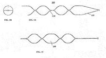
- An expandable outer cage 100 of the mechanical thrombectomy device as illustrated in the exemplary plan view depicted in Figure 1A , has a 2-cell (or 2-segment) pattern but may contain a range of 2 - 6 cells, wherein the number of cells present in the outer cage is measured in a circumferential direction.
- the cut pattern forming the outer cage 100 in Figure 1A has an open strut configuration comprising a minimal number of Nitinol struts to maximize cell area and preferably includes U-shaped crown/saddle points 110 to optimize clot engagement on deployment and improve retention performance and retrieval into the catheter.
- This open strut configuration minimizes clot compression on deployment thereby minimizing friction between the clot and interior surface of vessel wall.
- the relatively large reception spaces 105 e.g., large cell size or area
- the crowns 110 also aid in retrieving the clot into the tip of the intermediate or guide catheter.
- Distal radiopaque coils or markers may be attached to the outer cage to improve visibility of the device during imaging (e.g., fluoroscopy).
- the 2-cell configuration of the outer cage has a reduced maximum outer diameter when radially compressed thereby enhancing the wrapping efficiency allowing the device to be loaded and delivered through relatively small size microcatheters with an inner diameter less than or equal to approximately 0.021 inches, preferably less than or equal to approximately 0.017".
- the 2-cell outer cage configuration ensures a maximum of 4 struts aligned circumferentially side by side in comparison to the typical 4-cell mechanical thrombectomy device having 8 struts aligned circumferentially side by side.
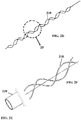
- the wave-shape component 310 may be laser cut from a tube of raw material 330 ( Figure 2E ) to form the wave-shape component ( Figure 2F ). Alternatively, the wave-shape component may be cut from a smaller diameter tube and expanded to the desired outer diameter.
- a different configuration of the single cell wave-shape component 310' is shown in isometric and side views provided in Figures 2G and 2H , respectively, wherein the cell pattern twists or rotates about a central axis (A) extending longitudinally through the component.
- the wave-shape component 310 opens a channel through the clot facilitating immediate restoration of partial flow of blood through the clot.
- This channel is evident when viewed end-on and is typically circular in shape when freely expanded, as seen in Figure 2B .
- the wave profile of the wave-shape component as depicted in the side view in Figure 2C , promotes engagement of the clot on dislodgement and during retention into the catheter.
- the wave-shape component also stabilizes relatively small and friable clots which migrate fully or partially inside the outer cage 100 during retrieval.
- the wave-shape component stabilizes the clot minimizing movement to the distal end of the thrombectomy device, where fragments may be lost or captured in the fragment protection zone.
- the wave-shape component is also deflectable to the side when clots are being pulled into the intermediate guide catheter.
- the wave-shape component enhances performance without inhibiting the ability of the device to retrieve clots into local aspiration or intermediate catheters.
- one or more struts may be removed.
- the dotted or dashed line represents a single strut that has been removed from the outer cage.
- the laser cut pattern of the outer cage tubing is no longer symmetrical, i.e., asymmetrical or lacking symmetry. Removal of one or more struts enlarges the cell area and potential reception space for the clot to embed in the outer cage.
- FIG. 5 depicts in an unrolled state (laser cut pattern) another exemplary embodiment of the outer cage 500 with two struts removed (505, 505') creating two enlarged cells or reception spaces in which the clot may embed. It is contemplated and within the intended scope of the present invention to remove any number of one or more struts from the outer cage.
- Figures 3A-3C depict an isometric view, an end view, and side view, respectively, of the dual structure assembly of the 2-cell outer cage and single cell wave-shape component, shown in Figures 1A-1C and 2A-2D , respectively.
- the wave-shape component 310 disposed inside the outer cage 305 is connected to the shaft 315 at the proximal joint 320.
- the two components (305, 310) are connected to one another, but may otherwise be free or floating relative to one another.
- a distal fragment protection member may be included in the present inventive mechanical thrombectomy device to provide a distal fragment protection zone.
- a distal mesh section 335 comprising a plurality of struts heat set in shape to have a tapered distal end and attached to the distal end of the outer cage 305 provides distal fragment protection.
- Disposed within the outer cage 305 is the wave-shape component 310 whose distal end 325 is floating (e.g., unattached, untethered, unsecured, unconnected) relative to both the outer cage and the distal mesh section.
- Figure 3E is an isometric view of the outer cage and distal mesh section of Figure 3D without the wave-shape component.
- distal fragment protection is provided by distal mesh section 335 connected to the distal end of the wave-shape component 310 while the distal end of the outer cage 305 is floating, free or unconnected relative to the distal mesh section 335.
- distal mesh section 335 is connected via a wire or extension of the proximal shaft 315.
- distal fragment protection may be provided by a distal clump, ball or sphere 335' of intertwined strands (e.g., biocompatible fibers or wire such as Nitinol wire), as depicted in Figure 3H .
- the intertwined strands forming the clump 335' need not necessarily be in the shape of a ball or sphere, but may take on any amorphous or geometric shape.
- the distal clump of intertwined strands 335' is connected to the distal end of the wave-shape component 310 while the distal end of the outer cage 305 is floating, free or unconnected relative to the distal clump 335'.
- the wave pattern of the wave-shape component 310 may be varied in a number of different ways.
- the tube may be laser cut into cells of different sizes that are then heat set into a variable wave shape whose amplitude (height) varies along the length of the component.
- the amplitude (height) "B" is greater than that of the amplitude (height) "A” in the wave shape.
- crowns at the connection points between cells are positioned at the peak and trough of each wave.
- FIG. 6C Another possible configuration of the wave shape is shown in Figure 6C , wherein the tube is cut into cells substantially uniform in size and then heat set to produce a varying wave amplitude (height) along the length of the wave-shape component.
- crowns at the connection points between cells in the wave-shape component structure in Figure 6C are not always positioned at the peak or trough of each wave. More connection points and hence more crowns are present in the configuration of Figure 6C promoting engagement with the clot.
- Variation in amplitude and/or length of the single cell of the wave-shape component provides additional protection against fragmentation of the clot and, when assembled within the outer cage, optimizes clot stabilization performance.
- the present invention contemplates the outer cage and inner component being formed or manufactured from a single cut tube or cut pattern, rather than two separately cut tubes or cut patterns.
- a single cut tube or cut pattern used to form both the primary and secondary struts in accordance with the present invention reduces the overall cost of manufacture while maintaining the benefits associated with the dual structure assembly (e.g., outer cage and inner component).
- the single cut tube or cut pattern is deformed and heat set into the inner component and the outer cage, respectively, with the same benefits and advantages discussed above with respect to the two separately cut tubes or cut patterns then assembled together.
- Figure 7A depicts part of the single cut tube or cut pattern while in an unrolled (flattened) state.
- This novel configuration of the dual structure assembly manufactured from a single cut tube or cut pattern is achieved by primary struts 700 forming the outer cage component being expanded and heat set to a primary diameter approximately 3.0mm - approximately 6.0 mm (preferably, approximately 5.0 mm) while the secondary struts 705 comprising the inner component are expanded and heat-set to form a circular channel having a smaller secondary diameter approximately 1.0mm - approximately 3.0 mm (preferably, approximately 1.25 mm), during respective expansion steps.
- Figure 7B is an isometric view of the dual structure assembly formed from a single cut tube or cut pattern while in a rolled expanded state, wherein the assembly includes formed primary struts 700 defining an outer cage and inwardly radially deformed secondary struts 705 comprising the inner component defining a cylindrical channel.
- a radial cross-sectional view of the dual structure assembly of Figure 7B along lines VIIC-VIIC is illustrated in Figure 7C .
- the single cut tube or cut pattern may have a diameter equal to the preferred expanded diameter of the primary struts 700 forming the outer cage (e.g., approximately 5.0 mm) and a heat-set process applied to reduce in diameter (e.g., approximately 1.25mm) the single cut tube or cut pattern to form the secondary struts 705 of the inner component.
- a single raw material cut tube or cut pattern e.g., Nitinol
- the dual structure assembly mechanical thrombectomy device it is further desirable for the dual structure assembly mechanical thrombectomy device to collapse/reduce/wrap-down sufficiently small in diameter to be receivable within a smaller size (e.g., approximately 0.017") microcatheter.
- mechanical thrombectomy devices that collapse to smaller diameter sizes face several challenges.
- a smaller diameter Nitinol cut tube raw material is preferred.
- the present inventive mechanical thrombectomy device has a multi-component assembly 800 (as seen in Figures 8A & 8B , depicting exploded and assembled views, respectively) including: an outer cage 805, a wave-shape component 810 (similar to that shown in Figures 2A-2D ) and an expanded distal tapered-end mesh section 815.
- outer cage 805 is preferably a 2-segment design or configuration (i.e., 2-segment outer cage 805 comprising a first segment 890 connected to a second segment 890' by a connector strut 895); however other configurations are contemplated and within the intended scope of the present invention.
- Outer cage 805 may be manufactured from a raw material (e.g., Nitinol) cut tube having a diameter of approximately 0.36mm to approximately 2.0 mm to provide appropriate strut geometry.
- the wave-shape component 810 may be manufactured from a smaller diameter (approximately 0.15 mm) raw material (e.g., Nitinol) cut tube to facilitate efficient wrapping of the device during loading into a microcatheter, and assembly of the proximal joint with a profile compatible with 0.017" microcatheters.
- maximization of strut width of the outer cage 805 in the multi-component configuration of Figures 8A & 8B is realized by having the wave-shape component 810 form a wave pattern (similar to that shown in Figures 2A-2D ) expand or extend axially into the distal section which contains a distal fragment protection member (e.g., expanded distal tapered-end mesh section 815).
- the connector strut 895 connecting the second segment 890' of the outer cage 805 to the expanded distal tapered-end mesh section 815 has been eliminated, replaced by the wave-shape component 810. Elimination of the connector strut also optimizes efficiency of wrap-down of the outer cage to be receivable within smaller size microcatheters (e.g., 0.017" microcatheters).
- the strut 895 connecting the second segment 890' of the outer cage 805 to the distal mesh section 815 is able to be eliminated in the present inventive configuration because the expanded distal tapered-end mesh section 815 is attached directly to the wave-shape component 810 providing a distal fragment protection zone and also serving as a distal anchor for intermediate catheter support during the procedure, rather than the outer cage 805. Functionality provided by the outer cage and wave-shape component have been discussed in detail above.
- the expanded distal tapered-end mesh section 815 maintains the clot in place during retrieval so that it does not fragment.
- the laser cut tube forming the expanded distal tapered-end mesh section has a reduced diameter to aid in securing it on its closed distal end 820.
- a laser beam used to cut a desired tube pattern remains stationary while a raw material (e.g., Nitinol) cut tube forming the stent is rotated.
- Figure 9A represents a standard or conventional strut cross-section geometry when laser cut from a raw material tube (e.g., Nitinol tube). Rotation is guided or dictated by the desired laser cut flat pattern.
- the radial cross-section of the conventional wedge shape strut is widest along its outer surface (e.g., outer diameter) and tapers smaller until reaching its inner surface (e.g., inner diameter). That is, the conventional wedge shape strut has a smallest arc length (i.e., measure of the distance along the curved line making up the inner arc) denoted as "IA" along the inner surface (inner arc forming the inner diameter of the tube) and a largest arc length (i.e., measure of the distance along the curved line making up the outer arc) denoted as "OA" along the outer surface of the cross-section (outer arc forming the outer diameter of the tube).
- IA smallest arc length
- OA largest arc length
- the arc length increases from the inner surface (inner diameter of the tube) to the outer surface (outer diameter of the tube). Accordingly, the arc length is longest (greatest contact surface area) along the outer surface (OA) of the radial cross-section of the conventional strut and the arc length is shortest (smallest contact surface area) along the inner surface (IA) of the cross-section of the conventional strut.
- Such conventional design results in an outward radial force of the stent being applied over a larger surface area, hence reducing outward radial pressure per strut surface area and clot embedding.
- Enhanced or improved embedding of the clot in the mechanical thrombectomy device may be realized in accordance with the present invention by modifying the strut geometry of the outer cage, specifically reducing the contact surface area of the outer surface (OA).
- the desired pattern of the stent may be laser cut on a concave Nitinol sheet or plate, while rotation of the concave sheet is controlled in an X-Y plane.
- Figure 9B depicts the present inventive radial cross-section geometry of a laser cut single strut of an everted concave Nitinol sheet. To achieve this geometry, a desired pattern is laser cut in the concave Nitinol sheet or plate.
- FIG. 10 illustrates a radial cross-sectional view of the configured stent after the everted laser cut concave Nitinol sheet or plate has been wrapped about a mandrel, shape set to form a tube, and its opposing longitudinal edges 1000, 1000' drawn toward one another to define a longitudinal slit or open seam 1110 allowing overlap when the stent is wrapped-down/reduced in diameter.
- the opposing longitudinal edges can be joined together, e.g., laser welded, soldered or similar mechanical joining process.
- the present inventive laser cut concave Nitinol sheet forms a wedge shape strut that is widest along its inner surface (inner diameter) and tapers smaller until reaching its outer surface (outer diameter), as seen in Figure 9B .
- the present inventive wedge shape strut has a largest arc length (i.e., measure of the distance along the curved line making up the outer arc) denoted as (IA') along the inner surface (inner arc forming the inner diameter of the tube) and a smallest arc length (i.e., measure of the distance along the curved line making up the outer arc) denoted as (OA') along the outer surface of the cross-section (outer arc forming the outer diameter of the tube).
- the arc length decreases from the inner surface (IA') to the outer surface (OA').
- the arc length is shortest (smallest contact surface area) along the outer surface (OA') of the cross-section of the present inventive strut and the arc length is longest (largest contact surface area) along the inner surface (IA') of the cross-section of the present inventive strut.
- Such novel strut design results in maintaining an outward radial force of the stent being applied over a smaller or reduced contact surface area of the outer surface (OA') in contact with the clot than the contact surface area of the outer surface (OA) of the conventional strut depicted in Figure 9A , hence increasing outward radial pressure and clot embedding.
- Figure 11A is a partial longitudinal view of the present inventive laser cut Nitinol concave sheet of a stent with eyelets, rolled about the mandrel depicting the laser welded seam in the body segments aligned with the connection struts along the center-line of the device.
- Figure 11B is a similar view of the present inventive laser cut Nitinol concave sheet of a stent without eyelets.
- the seam may be welded or soldered to form a cylindrical device from the inverted laser cut concave sheet. If eyelets are included in the body segments/cells of the stent ( Figure 11A ), the eyelet may be closed by laser welding the seam together.
- a notch, groove or channel is cut into or otherwise removed (e.g., grinding) from the outer diameter surface of the Nitinol tubing strut thereby reducing the contact surface area along its outer surface (outer diameter) with that of the clot.
- the notch, groove or channel may be a predefined radius, V-shaped, U-shaped or any other desired shape.
- the thickness of the strut can be increased to maintain the overall cross-sectional area of the strut.
- the two thinner struts formed along the outer surface or profile (outer diameter) on either side of the notch, groove or channel each has a smaller contact surface area (in comparison to the outer surface area without the notch, groove or channel) promoting the clot to more easily to embed.
- a chamfer edge or secondary off-axis cut can be made in each corner of the outer surface (outer diameter) of the Nitinol tubing strut, as shown in Figures 13A-13C , once again with the shared goal of reducing the outer surface contact area of the strut.
- the removal of the corners reduces the overall contact surface area arc length along the outer surface (outer diameter) from a conventional prior art width L 0 (before making the secondary cuts) to the present inventive reduced arc length Li (after making the secondary cuts).
- Figures 13A & 13B respectively, having a reduced contact surface area along the outer surface (outer diameter) of the strut.
- Each of the present inventive geometry strut configurations may be used independently or in any combination thereof to improve clot removal performance by improving embedding of the clot in the thrombectomy device. Also, features associated with some aspects of the present inventive mechanical thrombectomy device are applicable for use in a wide range of different size catheters, even those having a relatively small diameter.
- the present inventive device has been illustrated and described for use in a mechanical thrombectomy procedure but is applicable for use in other intravascular medical procedures in which a self-expanding mesh, cage, scaffolding or skeleton is utilized.
Landscapes
- Health & Medical Sciences (AREA)
- Surgery (AREA)
- Life Sciences & Earth Sciences (AREA)
- Heart & Thoracic Surgery (AREA)
- Molecular Biology (AREA)
- Vascular Medicine (AREA)
- Engineering & Computer Science (AREA)
- Biomedical Technology (AREA)
- Orthopedic Medicine & Surgery (AREA)
- Medical Informatics (AREA)
- Nuclear Medicine, Radiotherapy & Molecular Imaging (AREA)
- Animal Behavior & Ethology (AREA)
- General Health & Medical Sciences (AREA)
- Public Health (AREA)
- Veterinary Medicine (AREA)
- Surgical Instruments (AREA)
- Media Introduction/Drainage Providing Device (AREA)
Abstract
Description
- The present invention relates to a self-expanding intravascular medical device. In particular, the present invention is directed to a self-expanding intravascular medical device such as a mechanical thrombectomy device that promotes embedding in the clot, full retrieval of the clot into a catheter, minimizes fragmentation of the clot and other improvements to the overall successful performance of the medical device.
- Acute ischemic stroke is caused by a thrombotic or embolic occlusion (e.g., blockage) in a cerebral artery which blocks the flow of blood to the brain. The occlusion is typically caused by a blood clot liberated from another part of the body which travels through the vessel and eventually become lodged in the cerebral artery of the brain.
- One procedure used to restore blood flow in such conditions is administration of a drug (e.g., a tissue plasminogen activator (tPA)) that helps to dissolve or breakdown the clot. However, the success of restoring blood flow using such a drug alone requires that the medication be given in a relatively short period of time and is suitable only for relatively small clot sizes. Thus, drug treatment is not a viable option in all ischemic stroke cases.
- In such other circumstances, a mechanical device is generally deployed intravascularly during a thrombectomy procedure to restore blood flow through the vessel and thereafter physically remove the thrombus, clot, occlusion or blockage into a receiving catheter in a less invasive manner than open surgery. Such mechanical devices may be used alone or in combination with a clot dissolving/busting drug. Typically, the mechanical device used to capture the clot is a self-expanding mesh scaffolding, skeleton, cage or tube (e.g., a stent retriever or stentriever) attached to a wire that is deployed through a catheter to a desired target location.
- Thrombectomy treatment or procedure is typically performed on patients within a relative short period of time following a stroke (e.g., less than an approximately 48-hour period after the occurrence of a stroke) and is best suited for large vessel occlusions typically with a diameter greater than approximately 1.0 mm. Non-invasive imaging, for example, MR and non- contrast CT is typically used to determine early ischemic changes in patients to determine if thrombectomy treatment is suitable for that particular patient.
- During the thrombectomy procedure or treatment a physician or interventionalist endovascularly introduces a guidewire through the vasculature, typically in an artery located in the groin or arm, or by direct access through the carotid artery. The guidewire is advanced through the vasculature to the target location of the clot, blockage or occlusion. Once the guidewire is properly positioned, a microcatheter with an outer diameter typically less than approximately 1.0 mm, tracks over the guidewire passing through a lumen defined axially through the microcatheter. The guidewire and microcatheter are used to cross the clot or occlusion using standard intervention techniques. While in a compressed state, a stent or mechanical thrombectomy device may be guided through the lumen of the microcatheter to the target site. Upon deployment from the microcatheter the stentriever or mechanical thrombectomy device automatically expands to its original enlarged state. Stentrievers or mechanical thrombectomy devices are typically made of a biocompatible material such as stainless steel, nickel-titanium or tantalum.
- It is desirable to develop an improved thrombectomy device with enhanced thrombectomy performance based on one or more factors such as: (i) optimization of embedding of the stent in the clot for clot dislodgement and retention (ii) minimization of clot compression; (iii) minimization of clot fragmentation; (iii) restoration of blood flow; (iv) stabilization of the clot within the stent during retrieval; and (v) reduction of shear force imposed on the clot during retrieval into a catheter. In addition to designing a mechanical thrombectomy device that takes one or more of such factors into consideration, it is also advantageous to design the improved mechanical thrombectomy device to be suitable for use with smaller size catheters.
- An aspect of the present invention is directed to an improved mechanical thrombectomy device having enhanced performance based on one or more of the factors discussed above.
- Another aspect of the present invention relates to a self-expanding intravascular medical device including a multi-component assembly with a self-expanding outer cage component comprising a plurality of struts; and a single cell wave shape component disposed within the self-expanding outer cage component forming a channel therein. The proximal end of the single cell wave shape component is connected to the self-expanding outer cage component at a proximal joint.
- Still another aspect of the present invention is directed to a self-expanding intravascular medical device having a multi-component assembly including a self-expanding outer cage component comprising a plurality of struts; and an inner component disposed within the self-expanding outer cage component forming a cylindrical channel therein, wherein a proximal end of the inner component is connected to the self-expanding outer cage component at a proximal joint. Each of the self-expanding outer cage component and a circular cross-section of the cylindrical channel formed by the inner component differ in diameter and are formable from a single cut tube or cut pattern, wherein the single cut tube or cut pattern has a primary diameter equal to an expandable diameter of primary struts forming the self-expanding outer cage component while the single cut tube or cut pattern is heat settable to a secondary diameter smaller than the primary diameter to form the circular cross-section of the cylindrical channel formed by the inner component.
- Yet another aspect of the present invention relates to a self-expanding intravascular medical device including a multi-component assembly comprising (i) a self-expanding outer cage component comprising a plurality of struts; (ii) a wave shape component disposed within the self-expanding outer cage component; and (iii) an expanded distal closed-end mesh section. The expanded distal closed-end mesh section is attached directly to the wave shape component. There is no direct physical connection between the expanded distal closed-end mesh section and the self-expanding outer cage component.
- While still another aspect of the present invention is directed to a self-expanding intravascular medical device including a self-expanding outer cage component comprising a plurality of struts. Each of the plural struts having a wedge shape radial cross-section having a curved outer surface forming part of an outer diameter of the expandable outer cage and a curved inner surface forming part of an inner diameter of the self-expanding outer cage; wherein a portion of the curved outer surface is removed.
- While yet another aspect of the present invention relates to a self-expanding intravascular medical device made from an everted concave sheet or plate cut into a desired pattern comprising a plurality of struts. Each of the plural struts of the everted cut tube has a wedge shape radial cross-section that is widest at a concave inner radial surface and tapers smaller until reaching a concave outer radial surface.
- Still another aspect of the present invention is directed to a method for manufacturing a self-expanding outer cage component of a self-expanding intravascular medical device. A desired pattern is cut in a concave sheet or plate. The cut concave sheet or plate is then inverted. Thereafter, the everted cut concave sheet or plate is shape set around a mandrel of a predetermined diameter drawing opposing free edges of the everted cut concave sheet or plate towards one another to form a radial stent comprising a plurality of struts. Each of the plural struts has a wedge shape radial cross-section that is widest at a concave inner radial surface and tapers smaller until reaching a concave outer radial surface.
- The foregoing and other features of the present invention will be more readily apparent from the following detailed description and drawings of illustrative of the invention wherein like reference numbers refer to similar elements throughout the several views and in which:
-
Figure 1A is a plan view of a 2-cell outer cage of a mechanical thrombectomy device; -
Figure 1B is an end view of the 2-cell outer cage ofFigure 1A ; -
Figure 1C is a side view of the 2-cell outer cage ofFigure 1A ; -
Figure 2A is a plan view of a single cell wave-shape component of a mechanical thrombectomy device in accordance with the present invention; -
Figure 2B is an end view of the single cell wave-shape component ofFigure 2A ; -
Figure 2C is a side view of the single cell wave-shape component ofFigure 2A ; -
Figure 2D is an isometric view of the single cell wave-shape component ofFigure 2A ; -
Figure 2E is an isometric view representational of either the raw material tubing from which the single cell wave-shape component is laser cut or alternatively, the tubular channel formed by the single cell wave-shape component; -
Figure 2F is an enlarged representation of the dashedcircular area 2F in the isometric view of the laser cut single cell wave-shape component ofFigure 2D ; -
Figure 2G is an isometric view of an alternative single cell wave-shape component wherein the cell pattern twists or rotates about a central axis extending longitudinally through the component; -
Figure 2H is a side view of the single cell wave-shape component ofFigure 2G ; -
Figure 3A is an isometric view of a dual structure assembly mechanical thrombectomy device in accordance with the present invention including the 2-cell outer cage ofFigures 1A-1C and the single cell wave-shape component ofFigures 2A-2D disposed therein, wherein the distal end of the wave-shape component is attached to the outer cage; -
Figure 3B is an end view of the dual structure assembly mechanical thrombectomy device ofFigure 3A ; -
Figure 3C is a side view of the dual structure assembly mechanical thrombectomy device ofFigure 3A ; -
Figure 3D is an isometric view of an alternative dual structure assembly mechanical thrombectomy device in accordance with the present invention including the 2-cell outer cage ofFigures 1A-1C and the single cell wave-shape component ofFigures 2A-2D disposed therein, wherein a distal mesh connected to the distal end of the outer cage provides distal fragment protection and the wave-shape component is floating or free relative to the outer cage; -
Figure 3E is an isometric view of the outer cage inFigure 3D without the wave-shape component; -
Figure 3F is an isometric view of fragment protection provided by a distal mesh connected to the distal end of the wave-shape component while the distal end of the outer cage remains floating relative to the distal mesh; -
Figure 3G is an isometric view of distal fragment protection provided by a distal mesh, wherein the wave-shape component has been eliminated, the distal mesh is connected via a wire or extension of the proximal shaft; -
Figure 3H is an isometric view of distal fragment protection provided by a clump or ball of fibers or wire, wherein the ball is connected to the distal end of the wave-shape component while the distal end of the outer cage remains floating relative to the distal mesh; -
Figure 4 is an isometric view of an alternative 2-cell outer cage configuration of a mechanical thrombectomy device in accordance with the present invention, wherein one of the primary struts forming the outer cage has been removed (as denoted by the dashed line); -
Figure 5 is a plan view while in an unrolled, flat state of another exemplary 2-cell outer cage mechanical thrombectomy device, wherein two struts have been removed; -
Figure 6A is a side view of a wave-shape of the single cell wave-shape component of the mechanical thrombectomy device in accordance with the present invention, wherein the wave-shape varies in height along the length of the component; -
Figure 6B is an isometric view of one configuration of the single cell wave-shape component of the mechanical thrombectomy device in accordance with the present invention, wherein both the amplitude (height) and corresponding cell size varies along the length of the component; -
Figure 6C is an isometric view of another alternative configuration of the single cell wave-shape component of the mechanical thrombectomy device in accordance with the present invention; wherein each cell size is substantially the same, but the substantially uniform cut cells are heat set to a desired shape to produce a varying wave amplitude (height) along the length of the component; -
Figure 7A is a plan view while in an unrolled, flat state of the dual structure assembly mechanical thrombectomy device in accordance with the present invention; wherein the outer cage and inner component are manufactured from a single cut tube or cut pattern; -
Figure 7B is an isometric view while in the expanded state of the dual structure assembly mechanical thrombectomy device ofFigure 7A ; -
Figure 7C is a radial cross-sectional view of the dual structure assembly mechanical thrombectomy device ofFigure 7B along lines VIIC-VIIC; -
Figure 8A is an exploded view of an exemplary dual structure assembly of the mechanical thrombectomy device in accordance with the present invention having a reduced diameter when compressed or loaded into a catheter; -
Figure 8B is an assembled view of the exemplary dual structure assembly of the mechanical thrombectomy device ofFigure 8A ; -
Figure 8C is a flat, unrolled view of a radial section of the proximal first cell/segment outer cage ofFigure 8A with the connector strut between the first and distal second cell/segment of the outer cage identified, this connector strut may be eliminated in the distal second section to maximize adjacent strut widths; -
Figure 9A is a prior art strut cross-section geometry of a single wedge shape strut comprising a stent laser cut from a Nitinol tube; -
Figure 9B is a strut cross-section geometry of a single wedge shape strut of a stent in accordance with the present invention laser cut from a Nitinol concave sheet; -
Figure 10 is a radial cross-sectional view depicting the longitudinal slit or open seam of the everted laser cut concave sheet or plate deformed about a mandrel and heat set, wherein each strut has a cross-sectional geometry shown inFigure 9B ; -
Figure 11A is a partial longitudinal view of the present inventive device laser cut from a Nitinol concave sheet rolled about the mandrel depicting the seam in the body segments along the connection struts with eyelets; -
Figure 11B is a partial longitudinal view of the present inventive device laser cut from a Nitinol concave sheet rolled about the mandrel depicting the seam in the body segments along the connection struts without eyelets; -
Figure 12A a perspective view of a strut laser cut from a Nitinol tube, wherein a notch, groove or channel is cut into or removed from the outer diameter surface of the strut thereby reducing the outer surface contact area of the strut; -
Figure 12B is an end view of the strut ofFigure 12A ; -
Figure 13A is a perspective view of another configuration of a strut laser cut from a Nitinol tube having chamfered cuts to the corners of the outer surface (outer diameter) of the strut thereby reducing the outer surface contact area of the strut; -
Figure 13B is an end view of the strut ofFigure 13A ; -
Figure 13C is an end view of the strut ofFigure 13A illustrating by dashed lines the corners that have been removed; and -
Figure 14 is an illustration of an exemplary strut of the mechanical thrombectomy device to define the measurements of strut length, width and thickness. - The terms "distal" or "proximal" are used in the following description with respect to a position or direction relative to the treating physician or medical interventionalist. "Distal" or "distally" are a position distant from or in a direction away from the physician or interventionalist. "Proximal" or "proximally" or "proximate" are a position near or in a direction toward the physician or medical interventionalist. The terms "occlusion", "clot" or "blockage" are used interchangeably. Furthermore, the term "cell" is the space, region or area that is defined, formed or surrounded by a plurality of adjacent struts (e.g., 4 struts); whereas a "segment" represents a section comprising a number of cells connected by a connector strut.
- The self-expanding framework, cage, skeleton or scaffolding designs of the present inventive mechanical thrombectomy device (stentriever) disclosed herein are made from a material capable of recovering its shape automatically once released from a highly strained delivery configuration. A biocompatible superelastic material such as Nitinol or an alloy of similar properties is particularly suitable. The material may be in many forms such as wire, strip, plate, sheet or tube. A particularly suitable manufacturing process is to laser cut a Nitinol tube and then heat-set and electropolish the resultant structure to create an expandable framework, cage, skeleton or scaffolding comprising of struts and connecting elements. This framework may be any of a diverse range of shapes as disclosed herein and may be rendered visible under fluoroscopy through the addition of alloying elements (such as Platinum for example) or through a variety of other coatings or marker bands.
- Current self-expanding mechanical stent devices, in particular, mechanical thrombectomy devices (e.g., stentrievers), for use during intravascular medical procedures, such as the medical treatment of acute ischemic stroke, face several challenges or problems when trying to engage and dislodge a target clot, occlusion or blockage and thereafter fully retrieve the clot into an intermediate catheter, guide catheter or sheath under aspiration. The features of the present inventive mechanical thrombectomy device discussed in detail herein overcome such challenges associated with conventional mechanical thrombectomy devices.
- One problem addressed by the present invention is that the diameter of the access or intermediate catheter is typically smaller than the diameter of the targeted clot, occlusion or blockage so there is a resistance to clot retraction therethrough and a risk that shear forces may cause the clot to fragment during retrieval. This is an issue with all mechanical thrombectomy devices and shear forces on the clot, occlusion or blockage increase as the diameter of the intermediate catheter decreases. Some clots, occlusions or blockages pose a greater challenge to full retrieval into the intermediate or access catheter such as friable clots which are prone to fragmentation, and organized fibrin rich clots which are less compressible than spontaneous clots.
- It has been discovered through experimental testing that the cut pattern of a mechanical thrombectomy device influences clot embedding and the ability of the device to dislodge and retrieve a clot fully into an intermediate catheter, guide catheter or sheath. In one embodiment of the present invention the mechanical thrombectomy device has a dual structure assembly configuration. An expandable
outer cage 100 of the mechanical thrombectomy device, as illustrated in the exemplary plan view depicted inFigure 1A , has a 2-cell (or 2-segment) pattern but may contain a range of 2 - 6 cells, wherein the number of cells present in the outer cage is measured in a circumferential direction. The cut pattern forming theouter cage 100 inFigure 1A has an open strut configuration comprising a minimal number of Nitinol struts to maximize cell area and preferably includes U-shaped crown/saddle points 110 to optimize clot engagement on deployment and improve retention performance and retrieval into the catheter. This open strut configuration minimizes clot compression on deployment thereby minimizing friction between the clot and interior surface of vessel wall. While the relatively large reception spaces 105 (e.g., large cell size or area) allow the clot, occlusion or blockage to embed deeply therein aiding in retraction into the access or intermediate catheter while minimizing clot shear. Thecrowns 110 also aid in retrieving the clot into the tip of the intermediate or guide catheter. Distal radiopaque coils or markers may be attached to the outer cage to improve visibility of the device during imaging (e.g., fluoroscopy). - The 2-cell configuration of the outer cage has a reduced maximum outer diameter when radially compressed thereby enhancing the wrapping efficiency allowing the device to be loaded and delivered through relatively small size microcatheters with an inner diameter less than or equal to approximately 0.021 inches, preferably less than or equal to approximately 0.017". When loaded into a microcatheter, the 2-cell outer cage configuration ensures a maximum of 4 struts aligned circumferentially side by side in comparison to the typical 4-cell mechanical thrombectomy device having 8 struts aligned circumferentially side by side.
- Disposed within the 2-cell
outer cage 100 of the dual structure mechanical thrombectomy device is a single cell wave-shape component 310, the single cell configuration being clearly illustrated in the plan view ofFigure 2A and isometric view ofFigure 2D . The wave-shape component 310 may be laser cut from a tube of raw material 330 (Figure 2E ) to form the wave-shape component (Figure 2F ). Alternatively, the wave-shape component may be cut from a smaller diameter tube and expanded to the desired outer diameter. A different configuration of the single cell wave-shape component 310' is shown in isometric and side views provided inFigures 2G and 2H , respectively, wherein the cell pattern twists or rotates about a central axis (A) extending longitudinally through the component. - Upon deployment, the wave-
shape component 310 opens a channel through the clot facilitating immediate restoration of partial flow of blood through the clot. This channel is evident when viewed end-on and is typically circular in shape when freely expanded, as seen inFigure 2B . The wave profile of the wave-shape component, as depicted in the side view inFigure 2C , promotes engagement of the clot on dislodgement and during retention into the catheter. The wave-shape component also stabilizes relatively small and friable clots which migrate fully or partially inside theouter cage 100 during retrieval. The wave-shape component stabilizes the clot minimizing movement to the distal end of the thrombectomy device, where fragments may be lost or captured in the fragment protection zone. In addition, the wave-shape component is also deflectable to the side when clots are being pulled into the intermediate guide catheter. As a result, the wave-shape component enhances performance without inhibiting the ability of the device to retrieve clots into local aspiration or intermediate catheters. - To maximize the cell area or clot reception spaces in the outer cage of the present inventive mechanical thrombectomy device and thereby improve embedding of the clot therein, one or more struts may be removed. In the exemplary illustration of the
outer cage 400 in an expanded state depicted in the isometric view ofFigure 4 , the dotted or dashed line represents a single strut that has been removed from the outer cage. With the removal of one or more struts, the laser cut pattern of the outer cage tubing is no longer symmetrical, i.e., asymmetrical or lacking symmetry. Removal of one or more struts enlarges the cell area and potential reception space for the clot to embed in the outer cage. Such enlarging of spaces allows the clot to embed more deeply and, as a result, minimizes the potential for dislodgement hence increasing the probability of full or entire clot retrieval into the intermediate catheter. However, the laser cut pattern retains sufficient contact with the clot to prevent loss of the clot into a side branch or during retrieval around a challenging bend in the vessel.Figure 5 depicts in an unrolled state (laser cut pattern) another exemplary embodiment of theouter cage 500 with two struts removed (505, 505') creating two enlarged cells or reception spaces in which the clot may embed. It is contemplated and within the intended scope of the present invention to remove any number of one or more struts from the outer cage. -
Figures 3A-3C depict an isometric view, an end view, and side view, respectively, of the dual structure assembly of the 2-cell outer cage and single cell wave-shape component, shown inFigures 1A-1C and2A-2D , respectively. The wave-shape component 310 disposed inside theouter cage 305 is connected to theshaft 315 at the proximal joint 320. At an oppositedistal end 325, the two components (305, 310) are connected to one another, but may otherwise be free or floating relative to one another. - A distal fragment protection member may be included in the present inventive mechanical thrombectomy device to provide a distal fragment protection zone. Referring to
Figure 3D , adistal mesh section 335 comprising a plurality of struts heat set in shape to have a tapered distal end and attached to the distal end of theouter cage 305 provides distal fragment protection. Disposed within theouter cage 305 is the wave-shape component 310 whosedistal end 325 is floating (e.g., unattached, untethered, unsecured, unconnected) relative to both the outer cage and the distal mesh section.Figure 3E is an isometric view of the outer cage and distal mesh section ofFigure 3D without the wave-shape component. In still another variation illustrated inFigure 3F , distal fragment protection is provided bydistal mesh section 335 connected to the distal end of the wave-shape component 310 while the distal end of theouter cage 305 is floating, free or unconnected relative to thedistal mesh section 335. Whereas, inFigure 3G , since the wave-shape component has been eliminated,distal mesh section 335 is connected via a wire or extension of theproximal shaft 315. Rather than a distal mesh section being an expanded skeleton having a plurality of struts, distal fragment protection may be provided by a distal clump, ball or sphere 335' of intertwined strands (e.g., biocompatible fibers or wire such as Nitinol wire), as depicted inFigure 3H . The intertwined strands forming the clump 335' need not necessarily be in the shape of a ball or sphere, but may take on any amorphous or geometric shape. The distal clump of intertwined strands 335' is connected to the distal end of the wave-shape component 310 while the distal end of theouter cage 305 is floating, free or unconnected relative to the distal clump 335'. - The wave pattern of the wave-
shape component 310 may be varied in a number of different ways. In a first configuration shown in the isometric view ofFigure 6B , the tube may be laser cut into cells of different sizes that are then heat set into a variable wave shape whose amplitude (height) varies along the length of the component. Referring to the side view inFigure 6A , the amplitude (height) "B" is greater than that of the amplitude (height) "A" in the wave shape. In this configuration in which the wave shape varies in both amplitude (height) and corresponding cell size or length, crowns at the connection points between cells are positioned at the peak and trough of each wave. Another possible configuration of the wave shape is shown inFigure 6C , wherein the tube is cut into cells substantially uniform in size and then heat set to produce a varying wave amplitude (height) along the length of the wave-shape component. Unlike the embodiment inFigure 6B , crowns at the connection points between cells in the wave-shape component structure inFigure 6C are not always positioned at the peak or trough of each wave. More connection points and hence more crowns are present in the configuration ofFigure 6C promoting engagement with the clot. Variation in amplitude and/or length of the single cell of the wave-shape component provides additional protection against fragmentation of the clot and, when assembled within the outer cage, optimizes clot stabilization performance. - The
outer cage 305 and wave-shape component 310 differing in diameter, as depicted inFigures 3A-3C , are each manufactured from two separately cut tubes or cut patterns of different diameters thereafter assembled together to form the dual structure assembly. Alternatively, the present invention contemplates the outer cage and inner component being formed or manufactured from a single cut tube or cut pattern, rather than two separately cut tubes or cut patterns. A single cut tube or cut pattern used to form both the primary and secondary struts in accordance with the present invention reduces the overall cost of manufacture while maintaining the benefits associated with the dual structure assembly (e.g., outer cage and inner component). Specifically, the single cut tube or cut pattern is deformed and heat set into the inner component and the outer cage, respectively, with the same benefits and advantages discussed above with respect to the two separately cut tubes or cut patterns then assembled together.Figure 7A depicts part of the single cut tube or cut pattern while in an unrolled (flattened) state. This novel configuration of the dual structure assembly manufactured from a single cut tube or cut pattern is achieved byprimary struts 700 forming the outer cage component being expanded and heat set to a primary diameter approximately 3.0mm - approximately 6.0 mm (preferably, approximately 5.0 mm) while thesecondary struts 705 comprising the inner component are expanded and heat-set to form a circular channel having a smaller secondary diameter approximately 1.0mm - approximately 3.0 mm (preferably, approximately 1.25 mm), during respective expansion steps.Figure 7B is an isometric view of the dual structure assembly formed from a single cut tube or cut pattern while in a rolled expanded state, wherein the assembly includes formedprimary struts 700 defining an outer cage and inwardly radially deformedsecondary struts 705 comprising the inner component defining a cylindrical channel. A radial cross-sectional view of the dual structure assembly ofFigure 7B along lines VIIC-VIIC is illustrated inFigure 7C . - Alternatively, the single cut tube or cut pattern may have a diameter equal to the preferred expanded diameter of the
primary struts 700 forming the outer cage (e.g., approximately 5.0 mm) and a heat-set process applied to reduce in diameter (e.g., approximately 1.25mm) the single cut tube or cut pattern to form thesecondary struts 705 of the inner component. Thus, no expansion nor reduction in diameter of the single cut tube or cut pattern is necessary to form the primary struts of the outer cage. Accordingly, a single raw material cut tube or cut pattern (e.g., Nitinol) can be varied to any desired preferential primary and secondary diameter, respectively, to form the outer cage and inner component of the dual structure assembly mechanical thrombectomy device. - It is further desirable for the dual structure assembly mechanical thrombectomy device to collapse/reduce/wrap-down sufficiently small in diameter to be receivable within a smaller size (e.g., approximately 0.017") microcatheter. However, mechanical thrombectomy devices that collapse to smaller diameter sizes face several challenges. In order to allow construction of the proximal bond of the dual structure mechanical thrombectomy device and ensure an appropriate device crossing profile (i.e., cross-sectional area) (preferably less than approximately 0.0165" for compatibility with 0.017" microcatheters), a smaller diameter Nitinol cut tube raw material is preferred. An inherent problem to be solved or overcome in accordance with the present invention when utilizing smaller diameter cut tube raw material is accommodation of sufficient strut width and thickness for the outer cage to exert sufficient radial force when engaging the clot in the vessel to restore blood flow and facilitate clot removal. Wherein the measurements of strut length, width and thickness are defined by the illustration in
Figure 14 . - It is therefore desirable to maximize strut width to exert sufficient radial force while simultaneously configuring the outer cage to efficiently wrap down reducing in diameter to be receivable within a smaller size microcatheter (e.g., 0.017" microcatheter). To achieve these goals, the present inventive mechanical thrombectomy device has a multi-component assembly 800 (as seen in
Figures 8A & 8B , depicting exploded and assembled views, respectively) including: anouter cage 805, a wave-shape component 810 (similar to that shown inFigures 2A-2D ) and an expanded distal tapered-end mesh section 815. In the illustrative example shown inFigure 8A ,outer cage 805 is preferably a 2-segment design or configuration (i.e., 2-segmentouter cage 805 comprising a first segment 890 connected to a second segment 890' by a connector strut 895); however other configurations are contemplated and within the intended scope of the present invention.Outer cage 805 may be manufactured from a raw material (e.g., Nitinol) cut tube having a diameter of approximately 0.36mm to approximately 2.0 mm to provide appropriate strut geometry. The wave-shape component 810 may be manufactured from a smaller diameter (approximately 0.15 mm) raw material (e.g., Nitinol) cut tube to facilitate efficient wrapping of the device during loading into a microcatheter, and assembly of the proximal joint with a profile compatible with 0.017" microcatheters. In particular, maximization of strut width of theouter cage 805 in the multi-component configuration ofFigures 8A & 8B is realized by having the wave-shape component 810 form a wave pattern (similar to that shown inFigures 2A-2D ) expand or extend axially into the distal section which contains a distal fragment protection member (e.g., expanded distal tapered-end mesh section 815). In addition, the connector strut 895 connecting the second segment 890' of theouter cage 805 to the expanded distal tapered-end mesh section 815 (as depicted inFigure 8C ) has been eliminated, replaced by the wave-shape component 810. Elimination of the connector strut also optimizes efficiency of wrap-down of the outer cage to be receivable within smaller size microcatheters (e.g., 0.017" microcatheters). The strut 895 connecting the second segment 890' of theouter cage 805 to thedistal mesh section 815 is able to be eliminated in the present inventive configuration because the expanded distal tapered-end mesh section 815 is attached directly to the wave-shape component 810 providing a distal fragment protection zone and also serving as a distal anchor for intermediate catheter support during the procedure, rather than theouter cage 805. Functionality provided by the outer cage and wave-shape component have been discussed in detail above. The expanded distal tapered-end mesh section 815 maintains the clot in place during retrieval so that it does not fragment. Preferably, the laser cut tube forming the expanded distal tapered-end mesh section has a reduced diameter to aid in securing it on its closeddistal end 820. - During manufacture of conventional mechanical thrombectomy devices, a laser beam used to cut a desired tube pattern remains stationary while a raw material (e.g., Nitinol) cut tube forming the stent is rotated.
Figure 9A represents a standard or conventional strut cross-section geometry when laser cut from a raw material tube (e.g., Nitinol tube). Rotation is guided or dictated by the desired laser cut flat pattern. - As is clearly evident from
Figure 9A , the radial cross-section of the conventional wedge shape strut is widest along its outer surface (e.g., outer diameter) and tapers smaller until reaching its inner surface (e.g., inner diameter). That is, the conventional wedge shape strut has a smallest arc length (i.e., measure of the distance along the curved line making up the inner arc) denoted as "IA" along the inner surface (inner arc forming the inner diameter of the tube) and a largest arc length (i.e., measure of the distance along the curved line making up the outer arc) denoted as "OA" along the outer surface of the cross-section (outer arc forming the outer diameter of the tube). In other words, the arc length increases from the inner surface (inner diameter of the tube) to the outer surface (outer diameter of the tube). Accordingly, the arc length is longest (greatest contact surface area) along the outer surface (OA) of the radial cross-section of the conventional strut and the arc length is shortest (smallest contact surface area) along the inner surface (IA) of the cross-section of the conventional strut. Such conventional design results in an outward radial force of the stent being applied over a larger surface area, hence reducing outward radial pressure per strut surface area and clot embedding. Enhanced or improved embedding of the clot in the mechanical thrombectomy device may be realized in accordance with the present invention by modifying the strut geometry of the outer cage, specifically reducing the contact surface area of the outer surface (OA). - Rather than being cut from the Nitinol tube, as was the conventional strut cross-section depicted in
Figure 9A , in accordance with the present invention, the desired pattern of the stent may be laser cut on a concave Nitinol sheet or plate, while rotation of the concave sheet is controlled in an X-Y plane.Figure 9B depicts the present inventive radial cross-section geometry of a laser cut single strut of an everted concave Nitinol sheet. To achieve this geometry, a desired pattern is laser cut in the concave Nitinol sheet or plate. Thereafter, the laser cut concave Nitinol sheet or plate is everted (turned inside out), formed around a mandrel of the desired diameter drawing the free edges towards one another to form a radial stent, and heat set to maintain this shape.Figure 10 illustrates a radial cross-sectional view of the configured stent after the everted laser cut concave Nitinol sheet or plate has been wrapped about a mandrel, shape set to form a tube, and its opposinglongitudinal edges 1000, 1000' drawn toward one another to define a longitudinal slit oropen seam 1110 allowing overlap when the stent is wrapped-down/reduced in diameter. Alternatively, the opposing longitudinal edges can be joined together, e.g., laser welded, soldered or similar mechanical joining process. - Contrary to that of the conventional cross-section wedge shape strut laser cut from a Nitinol tube (
Figure 9A ), the present inventive laser cut concave Nitinol sheet forms a wedge shape strut that is widest along its inner surface (inner diameter) and tapers smaller until reaching its outer surface (outer diameter), as seen inFigure 9B . That is, the present inventive wedge shape strut has a largest arc length (i.e., measure of the distance along the curved line making up the outer arc) denoted as (IA') along the inner surface (inner arc forming the inner diameter of the tube) and a smallest arc length (i.e., measure of the distance along the curved line making up the outer arc) denoted as (OA') along the outer surface of the cross-section (outer arc forming the outer diameter of the tube). Hence, the arc length decreases from the inner surface (IA') to the outer surface (OA'). Accordingly, the arc length is shortest (smallest contact surface area) along the outer surface (OA') of the cross-section of the present inventive strut and the arc length is longest (largest contact surface area) along the inner surface (IA') of the cross-section of the present inventive strut. Such novel strut design results in maintaining an outward radial force of the stent being applied over a smaller or reduced contact surface area of the outer surface (OA') in contact with the clot than the contact surface area of the outer surface (OA) of the conventional strut depicted inFigure 9A , hence increasing outward radial pressure and clot embedding. -
Figure 11A is a partial longitudinal view of the present inventive laser cut Nitinol concave sheet of a stent with eyelets, rolled about the mandrel depicting the laser welded seam in the body segments aligned with the connection struts along the center-line of the device. WhileFigure 11B is a similar view of the present inventive laser cut Nitinol concave sheet of a stent without eyelets. The seam may be welded or soldered to form a cylindrical device from the inverted laser cut concave sheet. If eyelets are included in the body segments/cells of the stent (Figure 11A ), the eyelet may be closed by laser welding the seam together. - Another novel way to realize the aforementioned goal of reducing the strut contact surface area along the outer surface (outer diameter) of the stent is by removing a portion of the outer surface (outer diameter) from the cut tubing. In
Figures 12A &12B , a notch, groove or channel is cut into or otherwise removed (e.g., grinding) from the outer diameter surface of the Nitinol tubing strut thereby reducing the contact surface area along its outer surface (outer diameter) with that of the clot. The notch, groove or channel may be a predefined radius, V-shaped, U-shaped or any other desired shape. Despite removing a portion of the outer surface (outer diameter) of the strut, if desired, the thickness of the strut can be increased to maintain the overall cross-sectional area of the strut. The two thinner struts formed along the outer surface or profile (outer diameter) on either side of the notch, groove or channel each has a smaller contact surface area (in comparison to the outer surface area without the notch, groove or channel) promoting the clot to more easily to embed. Alternatively, or in combination therewith, a chamfer edge or secondary off-axis cut can be made in each corner of the outer surface (outer diameter) of the Nitinol tubing strut, as shown inFigures 13A-13C , once again with the shared goal of reducing the outer surface contact area of the strut. Referring to the end view of the Nitinol tubing formed strut inFigure 13C the removal of the corners (denoted by dashed lines) reduces the overall contact surface area arc length along the outer surface (outer diameter) from a conventional prior art width L0 (before making the secondary cuts) to the present inventive reduced arc length Li (after making the secondary cuts). What results is depicted in the end and perspective views shown inFigures 13A &13B , respectively, having a reduced contact surface area along the outer surface (outer diameter) of the strut. - Each of the present inventive geometry strut configurations may be used independently or in any combination thereof to improve clot removal performance by improving embedding of the clot in the thrombectomy device. Also, features associated with some aspects of the present inventive mechanical thrombectomy device are applicable for use in a wide range of different size catheters, even those having a relatively small diameter.
- The present inventive device has been illustrated and described for use in a mechanical thrombectomy procedure but is applicable for use in other intravascular medical procedures in which a self-expanding mesh, cage, scaffolding or skeleton is utilized.
- Thus, while there have been shown, described, and pointed out fundamental novel features of the invention as applied to a preferred embodiment thereof, it will be understood that various omissions, substitutions, and changes in the form and details of the devices illustrated, and in their operation, may be made by those skilled in the art without departing from the spirit and scope of the invention. For example, it is expressly intended that all combinations of those elements and/or steps that perform substantially the same function, in substantially the same way, to achieve the same results be within the scope of the invention. Substitutions of elements from one described embodiment to another are also fully intended and contemplated. It is also to be understood that the drawings are not necessarily drawn to scale, but that they are merely conceptual in nature. It is the intention, therefore, to be limited only as indicated by the scope of the claims appended hereto.
- Every issued patent, pending patent application, publication, journal article, book or any other reference cited herein is each incorporated by reference in their entirety.
-
- 1. A self-expanding intravascular medical device, comprising:
a multi-component assembly comprising:- a self-expanding outer cage component comprising a plurality of struts; and
- an inner component disposed within the self-expanding outer cage component forming a cylindrical channel therein, wherein a proximal end of the inner component is connected to the self-expanding outer cage component at a proximal joint;
- wherein each of the self-expanding outer cage component and a circular cross-section of the cylindrical channel formed by the inner component differ in diameter and are formable from a single cut tube or cut pattern; wherein the single cut tube or cut pattern has a primary diameter equal to an expandable diameter of primary struts forming the self-expanding outer cage component while the single cut tube or cut pattern is heat settable to a secondary diameter smaller than the primary diameter to form the circular cross-section of the cylindrical channel formed by the inner component.
- 2. A self-expanding intravascular medical device, comprising:
a multi-component assembly comprising:- a self-expanding outer cage component comprising a plurality of struts;
- a wave-shape component disposed within the self-expanding outer cage component;
- an expanded distal tapered-end mesh section;
- wherein the expanded distal tapered-end mesh section is attached directly to the wave-shape component; and there is no direct physical connection between the expanded distal tapered-end mesh section and the self-expanding outer cage component.
- 3. The self-expanding intravascular medical device in accordance with aspect 2, wherein the self-expanding outer cage component has a primary diameter and the wave-shape component forms a wave shape of circular cross-section having a secondary diameter smaller than that of the primary diameter.
- 4. The self-expanding intravascular medical device in accordance with aspect 3, wherein the primary diameter is between approximately 0.36 mm and approximately 2.0 mm, while the secondary diameter is approximately 0.15 mm.
- 5. A self-expanding intravascular medical device comprising:
a self-expanding outer cage component comprising a plurality of struts; each of the plural struts having a wedge shape radial cross-section having a curved outer surface forming part of an outer diameter of the expandable outer cage and a curved inner surface forming part of an inner diameter of the self-expanding outer cage; wherein a portion of the curved outer surface is removed. - 6. The self-expanding intravascular medical device in accordance with aspect 5, wherein the removed portion of the curved outer surface is a channel defined longitudinally therein corresponding to an axial direction of the self-expanding outer cage component.
- 7. The self-expanding intravascular medical device in accordance with aspect 5, wherein the removed portion of the curved outer surface is at least one angled corner chamfer cut.
- 8. A self-expanding intravascular medical device, comprising:
an everted concave sheet or plate cut into a desired pattern comprising a plurality of struts; wherein each of the plural struts of the everted cut tube has a wedge shape radial cross-section that is widest at a concave inner radial surface and tapers smaller until reaching a concave outer radial surface. - 9. A method for manufacturing a self-expanding outer cage component of a self-expanding intravascular medical device, said method comprising the steps of:
- cutting a desired pattern in a concave sheet or plate;
- everting the cut concave sheet or plate;
- shape setting the everted cut concave sheet or plate around a mandrel of a predetermined diameter drawing opposing free edges of the everted cut concave sheet or plate towards one another to form a radial stent comprising a plurality of struts, wherein each of the plural struts has a wedge shape radial cross-section that is widest at a concave inner radial surface and tapers smaller until reaching a concave outer radial surface.
- 10. The method in accordance with aspect 9, wherein the wedge shape radial cross-section of each of the plural struts has a longest arc length representing a first measured length of a curved line along an inner diameter at the concave inner radial surface of the wedge shape radial cross-section and a shortest arc length representing a second measured length of another curved line along an outer diameter at the concave outer radial surface of the wedge shape radial cross-section.
- 11. The method in accordance with aspect 9, wherein arc length decreases from the concave inner radial surface of the wedge shape radial cross-section along an inner diameter to the concave outer radial surface of the wedge shape radial cross-section along an outer diameter.
- 12. The method in accordance with aspect 9, wherein arc length has: (i) a smallest contact surface area along the concave outer radial surface of the wedge shape radial cross-section along an outer diameter; and (ii) a largest contact surface area along the concave inner radial surface of the wedge shape radial cross-section along an inner diameter.
Claims (10)
- A self-expanding intravascular medical device, comprising:
a multi-component assembly comprising:a self-expanding outer cage component comprising a plurality of struts; anda single cell wave-shape component disposed within the self-expanding outer cage component forming a channel therein, wherein a proximal end of the single cell wave-shape component is connected to the self-expanding outer cage component at a proximal joint. - The self-expanding intravascular medical device according to claim 1, wherein the self-expanding outer cage component is formed by a plurality of asymmetrically arranged struts.
- The self-expanding intravascular medical device according to claim 1, wherein the single cell wave-shape component is a mesh forming a plurality of connected cells, the plurality of connected cells varying in size and forming a wave shape varying in amplitude.
- The self-expanding intravascular medical device according to claim 1, wherein the single cell wave-shape component is a mesh forming a plurality of connected cells uniform in size and forming a wave shape varying in amplitude.
- The self-expanding intravascular medical device according to claim 1, wherein each of the self-expanding outer cage component and single cell wave-shape component differ in diameter and are formable from a single cut tube or cut pattern; wherein the single cut tube or cut pattern is expandable to a primary diameter to form primary struts of the self-expanding outer cage component, while the single cut tube or cut pattern is expandable to a secondary diameter smaller than the primary diameter to form secondary struts of the single cell wave-shape component.
- The self-expanding intravascular medical device in accordance with claim 1, wherein the self-expanding outer cage component has a distal fragment protection member disposed on a distal end of the self-expanding outer cage component.
- The self-expanding intravascular medical device in accordance with claim 6, wherein the distal fragment protection member is a mesh comprising a plurality of interconnected struts or a clump of intertwined strands.
- The self-expanding intravascular medical device in accordance with claim 6, wherein the distal end of the self-expandable outer cage is attached to the distal fragment protection member, while the distal end of the single cell wave-shape component is not connected to the distal fragment protection member.
- The self-expanding intravascular medical device in accordance with claim 6, wherein the distal end of the single cell wave-shape component is attached to the distal fragment protection member, while the distal end of the self-expandable outer cage is not connected to the distal fragment protection member.
- The self-expanding intravascular medical device in accordance with claim 6, wherein the single cell wave-shape component is a mesh forming a plurality of connected cells in a pattern twisted about a central axis extending longitudinally through the single cell wave-shape component.
Applications Claiming Priority (1)
| Application Number | Priority Date | Filing Date | Title |
|---|---|---|---|
| US16/508,029 US11266427B2 (en) | 2019-07-10 | 2019-07-10 | Self-expanding intravascular medical device |
Publications (3)
| Publication Number | Publication Date |
|---|---|
| EP3763306A1 true EP3763306A1 (en) | 2021-01-13 |
| EP3763306B1 EP3763306B1 (en) | 2024-01-10 |
| EP3763306C0 EP3763306C0 (en) | 2024-01-10 |
Family
ID=71575050
Family Applications (1)
| Application Number | Title | Priority Date | Filing Date |
|---|---|---|---|
| EP20185031.0A Active EP3763306B1 (en) | 2019-07-10 | 2020-07-09 | Self-expanding intravascular medical device |
Country Status (6)
| Country | Link |
|---|---|
| US (3) | US11266427B2 (en) |
| EP (1) | EP3763306B1 (en) |
| JP (1) | JP7547102B2 (en) |
| KR (1) | KR102875169B1 (en) |
| CN (1) | CN112206034A (en) |
| ES (1) | ES2970152T3 (en) |
Cited By (1)
| Publication number | Priority date | Publication date | Assignee | Title |
|---|---|---|---|---|
| CN116407210A (en) * | 2021-12-31 | 2023-07-11 | 苏州天鸿盛捷医疗器械有限公司 | A device for treating vascular stenosis |
Families Citing this family (26)
| Publication number | Priority date | Publication date | Assignee | Title |
|---|---|---|---|---|
| EP2629684B1 (en) | 2010-10-22 | 2018-07-25 | Neuravi Limited | Clot engagement and removal system |
| US9301769B2 (en) | 2011-03-09 | 2016-04-05 | Neuravi Limited | Clot retrieval device for removing clot from a blood vessel |
| US12076037B2 (en) | 2011-03-09 | 2024-09-03 | Neuravi Limited | Systems and methods to restore perfusion to a vessel |
| PL2967610T3 (en) | 2013-03-14 | 2019-07-31 | Neuravi Limited | A clot retrieval device for removing occlusive clot from a blood vessel |
| SI2967611T1 (en) | 2013-03-14 | 2019-04-30 | Neuravi Limited | Devices for removal of acute blockages from blood vessels |
| US10617435B2 (en) | 2014-11-26 | 2020-04-14 | Neuravi Limited | Clot retrieval device for removing clot from a blood vessel |
| US11253278B2 (en) | 2014-11-26 | 2022-02-22 | Neuravi Limited | Clot retrieval system for removing occlusive clot from a blood vessel |
| EP3682821B1 (en) | 2014-11-26 | 2022-05-11 | Neuravi Limited | A clot retrieval device for removing an occlusive clot from a blood vessel |
| KR102494176B1 (en) | 2016-09-06 | 2023-02-02 | 뉴라비 리미티드 | Thrombus recovery device for removing occluded thrombi from blood vessels |
| US11406416B2 (en) | 2018-10-02 | 2022-08-09 | Neuravi Limited | Joint assembly for vasculature obstruction capture device |
| US12285182B2 (en) | 2018-10-10 | 2025-04-29 | Innova Vascular, Inc. | Devices and methods for removing an embolus |
| US11712231B2 (en) | 2019-10-29 | 2023-08-01 | Neuravi Limited | Proximal locking assembly design for dual stent mechanical thrombectomy device |
| US11517340B2 (en) | 2019-12-03 | 2022-12-06 | Neuravi Limited | Stentriever devices for removing an occlusive clot from a vessel and methods thereof |
| US11871946B2 (en) | 2020-04-17 | 2024-01-16 | Neuravi Limited | Clot retrieval device for removing clot from a blood vessel |
| US11730501B2 (en) | 2020-04-17 | 2023-08-22 | Neuravi Limited | Floating clot retrieval device for removing clots from a blood vessel |
| US11717308B2 (en) | 2020-04-17 | 2023-08-08 | Neuravi Limited | Clot retrieval device for removing heterogeneous clots from a blood vessel |
| US11737771B2 (en) | 2020-06-18 | 2023-08-29 | Neuravi Limited | Dual channel thrombectomy device |
| US11937836B2 (en) | 2020-06-22 | 2024-03-26 | Neuravi Limited | Clot retrieval system with expandable clot engaging framework |
| US11439418B2 (en) | 2020-06-23 | 2022-09-13 | Neuravi Limited | Clot retrieval device for removing clot from a blood vessel |
| US11864781B2 (en) | 2020-09-23 | 2024-01-09 | Neuravi Limited | Rotating frame thrombectomy device |
| US11937837B2 (en) | 2020-12-29 | 2024-03-26 | Neuravi Limited | Fibrin rich / soft clot mechanical thrombectomy device |
| US12029442B2 (en) | 2021-01-14 | 2024-07-09 | Neuravi Limited | Systems and methods for a dual elongated member clot retrieval apparatus |
| US12064130B2 (en) * | 2021-03-18 | 2024-08-20 | Neuravi Limited | Vascular obstruction retrieval device having sliding cages pinch mechanism |
| US11974764B2 (en) | 2021-06-04 | 2024-05-07 | Neuravi Limited | Self-orienting rotating stentriever pinching cells |
| CN115869040B (en) * | 2021-09-28 | 2026-01-23 | 先健科技(深圳)有限公司 | Thrombolysis device |
| CN119924940B (en) * | 2025-02-28 | 2026-05-05 | 归创通桥医疗科技股份有限公司 | Thrombectomy stents and thrombus removal systems |
Citations (4)
| Publication number | Priority date | Publication date | Assignee | Title |
|---|---|---|---|---|
| EP1225949A1 (en) * | 1999-09-17 | 2002-07-31 | Bacchus Vascular Inc. | Mechanical pump for removal of fragmented matter and methods of manufacture and use |
| US20130035628A1 (en) * | 2011-08-05 | 2013-02-07 | Garrison Michi E | Methods and systems for treatment of acute ischemic stroke |
| WO2017089424A1 (en) * | 2015-11-25 | 2017-06-01 | Neuravi Limited | A clot retrieval device for removing occlusive clot from a blood vessel |
| WO2019079296A1 (en) * | 2017-10-16 | 2019-04-25 | Wallaby Medical, Inc. | Devices and methods for treating blocked blood vessels |
Family Cites Families (131)
| Publication number | Priority date | Publication date | Assignee | Title |
|---|---|---|---|---|
| US6511492B1 (en) | 1998-05-01 | 2003-01-28 | Microvention, Inc. | Embolectomy catheters and methods for treating stroke and other small vessel thromboembolic disorders |
| KR100457630B1 (en) * | 2001-04-04 | 2004-11-18 | (주) 태웅메디칼 | Flexible self-expandable stent and methods for making the stent for lumen |
| US8252040B2 (en) | 2001-07-20 | 2012-08-28 | Microvention, Inc. | Aneurysm treatment device and method of use |
| US8715312B2 (en) | 2001-07-20 | 2014-05-06 | Microvention, Inc. | Aneurysm treatment device and method of use |
| US6551342B1 (en) | 2001-08-24 | 2003-04-22 | Endovascular Technologies, Inc. | Embolic filter |
| US20040199201A1 (en) | 2003-04-02 | 2004-10-07 | Scimed Life Systems, Inc. | Embolectomy devices |
| US7371228B2 (en) | 2003-09-19 | 2008-05-13 | Medtronic Vascular, Inc. | Delivery of therapeutics to treat aneurysms |
| US9308382B2 (en) | 2004-06-10 | 2016-04-12 | Medtronic Urinary Solutions, Inc. | Implantable pulse generator systems and methods for providing functional and/or therapeutic stimulation of muscles and/or nerves and/or central nervous system tissue |
| US9655633B2 (en) | 2004-09-10 | 2017-05-23 | Penumbra, Inc. | System and method for treating ischemic stroke |
| US20060089637A1 (en) | 2004-10-14 | 2006-04-27 | Werneth Randell L | Ablation catheter |
| US8562672B2 (en) | 2004-11-19 | 2013-10-22 | Medtronic, Inc. | Apparatus for treatment of cardiac valves and method of its manufacture |
| US9636115B2 (en) | 2005-06-14 | 2017-05-02 | Stryker Corporation | Vaso-occlusive delivery device with kink resistant, flexible distal end |
| EP1895927A4 (en) | 2005-06-20 | 2011-03-09 | Medtronic Ablation Frontiers | Ablation catheter |
| US8066036B2 (en) | 2005-11-17 | 2011-11-29 | Microvention, Inc. | Three-dimensional complex coil |
| US9757260B2 (en) | 2006-03-30 | 2017-09-12 | Medtronic Vascular, Inc. | Prosthesis with guide lumen |
| US9615832B2 (en) | 2006-04-07 | 2017-04-11 | Penumbra, Inc. | Aneurysm occlusion system and method |
| CA2655026C (en) | 2006-06-15 | 2016-08-02 | Microvention, Inc. | Embolization device constructed from expansible polymer |
| US9198687B2 (en) | 2007-10-17 | 2015-12-01 | Covidien Lp | Acute stroke revascularization/recanalization systems processes and products thereby |
| US8066757B2 (en) * | 2007-10-17 | 2011-11-29 | Mindframe, Inc. | Blood flow restoration and thrombus management methods |
| US10123803B2 (en) | 2007-10-17 | 2018-11-13 | Covidien Lp | Methods of managing neurovascular obstructions |
| WO2009086208A2 (en) | 2007-12-21 | 2009-07-09 | Microvention, Inc. | Hydrogel filaments for biomedical uses |
| WO2009094270A1 (en) * | 2008-01-24 | 2009-07-30 | Boston Scientific Scimed, Inc. | Stent for delivering a therapeutic agent from a side surface of a stent strut |
| US8974518B2 (en) | 2008-03-25 | 2015-03-10 | Medtronic Vascular, Inc. | Eversible branch stent-graft and deployment method |
| WO2009135075A1 (en) | 2008-04-30 | 2009-11-05 | Medtronic, Inc. | Techniques for placing medical leads for electrical stimulation of nerve tissue |
| US8070694B2 (en) | 2008-07-14 | 2011-12-06 | Medtronic Vascular, Inc. | Fiber based medical devices and aspiration catheters |
| US8333796B2 (en) | 2008-07-15 | 2012-12-18 | Penumbra, Inc. | Embolic coil implant system and implantation method |
| US8721714B2 (en) | 2008-09-17 | 2014-05-13 | Medtronic Corevalve Llc | Delivery system for deployment of medical devices |
| JP5608731B2 (en) | 2009-04-15 | 2014-10-15 | マイクロベンション インコーポレイテッド | Implant delivery system |
| WO2011038017A1 (en) | 2009-09-22 | 2011-03-31 | Penumbra, Inc. | Manual actuation system for deployment of implant |
| CA2795740C (en) | 2010-04-14 | 2018-03-13 | Microvention, Inc. | Implant delivery device |
| US8764811B2 (en) | 2010-04-20 | 2014-07-01 | Medtronic Vascular, Inc. | Controlled tip release stent graft delivery system and method |
| US8876878B2 (en) | 2010-07-23 | 2014-11-04 | Medtronic, Inc. | Attachment mechanism for stent release |
| US8616040B2 (en) | 2010-09-17 | 2013-12-31 | Medtronic Vascular, Inc. | Method of forming a drug-eluting medical device |
| WO2012088162A1 (en) | 2010-12-20 | 2012-06-28 | Microvention, Inc. | Polymer stents and methods of manufacture |
| US9301769B2 (en) | 2011-03-09 | 2016-04-05 | Neuravi Limited | Clot retrieval device for removing clot from a blood vessel |
| US9486604B2 (en) | 2011-05-12 | 2016-11-08 | Medtronic, Inc. | Packaging and preparation tray for a delivery system |
| WO2012158668A1 (en) | 2011-05-17 | 2012-11-22 | Stryker Corporation | Method of fabricating an implantable medical device that includes one or more thin film polymer support layers |
| WO2012166467A1 (en) | 2011-05-27 | 2012-12-06 | Stryker Corporation | Assembly for percutaneously inserting an implantable medical device, steering the device to a target location and deploying the device |
| US9750565B2 (en) | 2011-09-30 | 2017-09-05 | Medtronic Advanced Energy Llc | Electrosurgical balloons |
| CA2867181C (en) | 2012-03-16 | 2020-08-11 | Microvention, Inc. | Stent and stent delivery device |
| US9833625B2 (en) | 2012-03-26 | 2017-12-05 | Medtronic, Inc. | Implantable medical device delivery with inner and outer sheaths |
| US9717421B2 (en) | 2012-03-26 | 2017-08-01 | Medtronic, Inc. | Implantable medical device delivery catheter with tether |
| US9242290B2 (en) | 2012-04-03 | 2016-01-26 | Medtronic Vascular, Inc. | Method and apparatus for creating formed elements used to make wound stents |
| US9549832B2 (en) | 2012-04-26 | 2017-01-24 | Medtronic Vascular, Inc. | Apparatus and methods for filling a drug eluting medical device via capillary action |
| US9700399B2 (en) | 2012-04-26 | 2017-07-11 | Medtronic Vascular, Inc. | Stopper to prevent graft material slippage in a closed web stent-graft |
| EP2844163A4 (en) | 2012-04-30 | 2015-09-23 | Bio2 Medical Inc | Multi-lumen central access vena cava filter apparatus for clot management and method of using same |
| US9445828B2 (en) * | 2012-07-05 | 2016-09-20 | Cognition Medical Corp. | Methods, devices, and systems for postconditioning with clot removal |
| US9149190B2 (en) | 2012-07-17 | 2015-10-06 | Stryker Corporation | Notification system of deviation from predefined conditions |
| US9770251B2 (en) | 2012-08-13 | 2017-09-26 | Microvention, Inc. | Shaped removal device |
| WO2014047650A1 (en) * | 2012-09-24 | 2014-03-27 | Inceptus Medical LLC | Device and method for treating vascular occlusion |
| US9504476B2 (en) | 2012-10-01 | 2016-11-29 | Microvention, Inc. | Catheter markers |
| BR112015008245B1 (en) | 2012-10-15 | 2022-09-27 | Microvention, Inc | POLYMERIC TREATMENT COMPOSITIONS |
| US9456834B2 (en) * | 2012-10-31 | 2016-10-04 | Covidien Lp | Thrombectomy device with distal protection |
| US9314248B2 (en) * | 2012-11-06 | 2016-04-19 | Covidien Lp | Multi-pivot thrombectomy device |
| US9539022B2 (en) | 2012-11-28 | 2017-01-10 | Microvention, Inc. | Matter conveyance system |
| WO2014089392A1 (en) | 2012-12-07 | 2014-06-12 | Medtronic, Inc. | Minimally invasive implantable neurostimulation system |
| US9539382B2 (en) | 2013-03-12 | 2017-01-10 | Medtronic, Inc. | Stepped catheters with flow restrictors and infusion systems using the same |
| US9539011B2 (en) | 2013-03-14 | 2017-01-10 | Stryker Corporation | Vaso-occlusive device delivery system |
| EP2967572B1 (en) | 2013-03-14 | 2019-04-24 | Stryker Corporation | Vaso-occlusive device delivery system |
| PL2967610T3 (en) * | 2013-03-14 | 2019-07-31 | Neuravi Limited | A clot retrieval device for removing occlusive clot from a blood vessel |
| EP3009084B1 (en) | 2013-03-14 | 2017-09-13 | Stryker Corporation | Vaso-occlusive device delivery system |
| US9833252B2 (en) | 2013-03-15 | 2017-12-05 | Microvention, Inc. | Multi-component obstruction removal system and method |
| JP6636908B2 (en) | 2013-03-15 | 2020-01-29 | テルモ株式会社 | Embolism prevention device |
| US9398966B2 (en) | 2013-03-15 | 2016-07-26 | Medtronic Vascular, Inc. | Welded stent and stent delivery system |
| WO2014174437A1 (en) | 2013-04-22 | 2014-10-30 | Sandvik Intellectual Property Ab | Method for drug loading hydroxyapatite coated implant surfaces |
| US9445928B2 (en) | 2013-05-30 | 2016-09-20 | Medtronic Vascular, Inc. | Delivery system having a single handed deployment handle for a retractable outer sheath |
| US9675782B2 (en) | 2013-10-10 | 2017-06-13 | Medtronic Vascular, Inc. | Catheter pull wire actuation mechanism |
| US10383644B2 (en) * | 2013-10-17 | 2019-08-20 | Covidien Lp | Mechanical thrombectomy with proximal occlusion |
| US9955978B2 (en) | 2013-10-25 | 2018-05-01 | Medtronic Vascular, Inc. | Tissue compression device with multi-chamber bladder |
| US9833604B2 (en) | 2013-12-20 | 2017-12-05 | Microvention, Inc. | Delivery adapter |
| CA2934555C (en) | 2013-12-20 | 2020-07-07 | Microvention, Inc. | Device delivery system |
| EP3128962B1 (en) | 2014-04-08 | 2018-05-23 | Stryker Corporation | Implant delivery system |
| WO2015167997A1 (en) | 2014-04-30 | 2015-11-05 | Stryker Corporation | Implant delivery system and method of use |
| US9060777B1 (en) | 2014-05-28 | 2015-06-23 | Tw Medical Technologies, Llc | Vaso-occlusive devices and methods of use |
| US9668898B2 (en) | 2014-07-24 | 2017-06-06 | Medtronic Vascular, Inc. | Stent delivery system having dynamic deployment and methods of manufacturing same |
| US9770577B2 (en) | 2014-09-15 | 2017-09-26 | Medtronic Xomed, Inc. | Pressure relief for a catheter balloon device |
| US9579484B2 (en) | 2014-09-19 | 2017-02-28 | Medtronic Vascular, Inc. | Sterile molded dispenser |
| US11253278B2 (en) | 2014-11-26 | 2022-02-22 | Neuravi Limited | Clot retrieval system for removing occlusive clot from a blood vessel |
| US10617435B2 (en) * | 2014-11-26 | 2020-04-14 | Neuravi Limited | Clot retrieval device for removing clot from a blood vessel |
| EP3682821B1 (en) * | 2014-11-26 | 2022-05-11 | Neuravi Limited | A clot retrieval device for removing an occlusive clot from a blood vessel |
| US9692557B2 (en) | 2015-02-04 | 2017-06-27 | Stryker European Holdings I, Llc | Apparatus and methods for administering treatment within a bodily duct of a patient |
| US10307168B2 (en) | 2015-08-07 | 2019-06-04 | Terumo Corporation | Complex coil and manufacturing techniques |
| US10154905B2 (en) | 2015-08-07 | 2018-12-18 | Medtronic Vascular, Inc. | System and method for deflecting a delivery catheter |
| EP3334354B1 (en) | 2015-08-11 | 2021-03-03 | Terumo Corporation | System for implant delivery |
| EP3349689B1 (en) | 2015-09-18 | 2023-12-27 | Microvention, Inc. | Implant retention, detachment, and delivery system |
| JP6816126B2 (en) | 2015-09-18 | 2021-01-20 | マイクロベンション インコーポレイテッドMicrovention, Inc. | Releasable delivery system |
| JP6816127B2 (en) | 2015-09-18 | 2021-01-20 | テルモ株式会社 | Vascular prosthesis |
| CN108156803B (en) | 2015-09-18 | 2021-05-04 | 泰尔茂株式会社 | Pushable Implant Delivery System |
| CN108024821B (en) | 2015-09-21 | 2020-10-30 | 斯瑞克公司 | embolectomy device |
| ES2788738T3 (en) | 2015-09-21 | 2020-10-22 | Stryker Corp | Embolectomy devices |
| US10172632B2 (en) | 2015-09-22 | 2019-01-08 | Medtronic Vascular, Inc. | Occlusion bypassing apparatus with a re-entry needle and a stabilization tube |
| WO2017062383A1 (en) | 2015-10-07 | 2017-04-13 | Stryker Corporation | Multiple barrel clot removal devices |
| US10327791B2 (en) | 2015-10-07 | 2019-06-25 | Medtronic Vascular, Inc. | Occlusion bypassing apparatus with a re-entry needle and a distal stabilization balloon |
| US10786302B2 (en) | 2015-10-09 | 2020-09-29 | Medtronic, Inc. | Method for closure and ablation of atrial appendage |
| US10271873B2 (en) | 2015-10-26 | 2019-04-30 | Medtronic Vascular, Inc. | Sheathless guide catheter assembly |
| US20170147765A1 (en) | 2015-11-19 | 2017-05-25 | Penumbra, Inc. | Systems and methods for treatment of stroke |
| US10631946B2 (en) | 2015-11-30 | 2020-04-28 | Penumbra, Inc. | System for endoscopic intracranial procedures |
| WO2017100472A1 (en) | 2015-12-09 | 2017-06-15 | Medtronic Vascular Inc. | Catheter with a lumen shaped as an identification symbol |
| US10159568B2 (en) | 2015-12-14 | 2018-12-25 | Medtronic, Inc. | Delivery system having retractable wires as a coupling mechanism and a deployment mechanism for a self-expanding prosthesis |
| US10500046B2 (en) | 2015-12-14 | 2019-12-10 | Medtronic, Inc. | Delivery system having retractable wires as a coupling mechanism and a deployment mechanism for a self-expanding prosthesis |
| US10743883B2 (en) | 2015-12-30 | 2020-08-18 | Stryker European Holdings I, Llc | Embolic devices and methods of manufacturing same |
| US20170189033A1 (en) | 2016-01-06 | 2017-07-06 | Microvention, Inc. | Occlusive Embolic Coil |
| US10070950B2 (en) | 2016-02-09 | 2018-09-11 | Medtronic Vascular, Inc. | Endoluminal prosthetic assemblies, and associated systems and methods for percutaneous repair of a vascular tissue defect |
| AU2017217879B9 (en) | 2016-02-10 | 2021-08-19 | Microvention, Inc. | Intravascular treatment site access |
| AU2017218115B2 (en) | 2016-02-10 | 2020-03-05 | Microvention, Inc. | Devices for vascular occlusion |
| US10188500B2 (en) | 2016-02-12 | 2019-01-29 | Medtronic Vascular, Inc. | Stent graft with external scaffolding and method |
| EP4112111A1 (en) | 2016-03-31 | 2023-01-04 | Medtronic Vascular Inc. | Expandable introducer sheath |
| US20170281331A1 (en) | 2016-03-31 | 2017-10-05 | Medtronic Vascular, Inc. | Endoluminal prosthetic devices having fluid-absorbable compositions for repair of a vascular tissue defect |
| US10695542B2 (en) | 2016-04-04 | 2020-06-30 | Medtronic Vascular, Inc. | Drug coated balloon |
| US10252024B2 (en) | 2016-04-05 | 2019-04-09 | Stryker Corporation | Medical devices and methods of manufacturing same |
| US10441407B2 (en) | 2016-04-12 | 2019-10-15 | Medtronic Vascular, Inc. | Gutter filling stent-graft and method |
| US9987122B2 (en) | 2016-04-13 | 2018-06-05 | Medtronic Vascular, Inc. | Iliac branch device and method |
| US10010403B2 (en) | 2016-04-18 | 2018-07-03 | Medtronic Vascular, Inc. | Stent-graft prosthesis and method of manufacture |
| US20170304097A1 (en) | 2016-04-21 | 2017-10-26 | Medtronic Vascular, Inc. | Stent-graft delivery system having an inner shaft component with a loading pad or covering on a distal segment thereof for stent retention |
| JP6924256B2 (en) | 2016-04-25 | 2021-08-25 | ストライカー コーポレイションStryker Corporation | Pre-installed reversing tractor thrombectomy device and method |
| ES2859656T3 (en) | 2016-04-25 | 2021-10-04 | Stryker Corp | Anti-jam and macerant thrombectomy appliances |
| US10517711B2 (en) | 2016-04-25 | 2019-12-31 | Medtronic Vascular, Inc. | Dissection prosthesis system and method |
| JP6934935B2 (en) | 2016-04-25 | 2021-09-15 | ストライカー コーポレイションStryker Corporation | Inverted mechanical thrombectomy and intravascular use |
| US10940294B2 (en) | 2016-04-25 | 2021-03-09 | Medtronic Vascular, Inc. | Balloon catheter including a drug delivery sheath |
| US11147952B2 (en) | 2016-04-28 | 2021-10-19 | Medtronic Vascular, Inc. | Drug coated inflatable balloon having a thermal dependent release layer |
| US10406011B2 (en) | 2016-04-28 | 2019-09-10 | Medtronic Vascular, Inc. | Implantable medical device delivery system |
| US10191615B2 (en) | 2016-04-28 | 2019-01-29 | Medtronic Navigation, Inc. | Method and apparatus for image-based navigation |
| US10292844B2 (en) | 2016-05-17 | 2019-05-21 | Medtronic Vascular, Inc. | Method for compressing a stented prosthesis |
| CN113648518B (en) | 2016-06-01 | 2023-10-20 | 微仙美国有限公司 | Improved reinforced balloon catheter |
| JP6980703B2 (en) | 2016-06-03 | 2021-12-15 | ストライカー コーポレイションStryker Corporation | Inverting thrombectomy device |
| KR102494176B1 (en) * | 2016-09-06 | 2023-02-02 | 뉴라비 리미티드 | Thrombus recovery device for removing occluded thrombi from blood vessels |
| US9993257B2 (en) | 2016-09-07 | 2018-06-12 | NeuroVarc Technologies Inc. | Clot retrieval device for ischemic stroke treatment |
| AU2018204059A1 (en) * | 2017-06-28 | 2019-01-17 | Neuravi Limited | Clot retrieval system for removing occlusive clot from a blood vessel |
| CN108158630B (en) * | 2018-02-26 | 2024-06-07 | 上海加奇生物科技苏州有限公司 | Intravascular thrombus catcher |
| CN109303587A (en) * | 2018-08-24 | 2019-02-05 | 苏州瑞帆医疗科技有限公司 | A kind of encephalic takes pin device and its manufacturing method |
| CN109512486B (en) * | 2018-11-12 | 2021-05-28 | 中国人民解放军总医院 | Segmented Bolt Retrieval Bracket |
-
2019
- 2019-07-10 US US16/508,029 patent/US11266427B2/en active Active
-
2020
- 2020-07-07 KR KR1020200083430A patent/KR102875169B1/en active Active
- 2020-07-09 JP JP2020118290A patent/JP7547102B2/en active Active
- 2020-07-09 ES ES20185031T patent/ES2970152T3/en active Active
- 2020-07-09 EP EP20185031.0A patent/EP3763306B1/en active Active
- 2020-07-10 CN CN202010662285.0A patent/CN112206034A/en active Pending
-
2022
- 2022-01-27 US US17/586,444 patent/US11883046B2/en active Active
-
2023
- 2023-12-18 US US18/543,100 patent/US12465383B2/en active Active
Patent Citations (4)
| Publication number | Priority date | Publication date | Assignee | Title |
|---|---|---|---|---|
| EP1225949A1 (en) * | 1999-09-17 | 2002-07-31 | Bacchus Vascular Inc. | Mechanical pump for removal of fragmented matter and methods of manufacture and use |
| US20130035628A1 (en) * | 2011-08-05 | 2013-02-07 | Garrison Michi E | Methods and systems for treatment of acute ischemic stroke |
| WO2017089424A1 (en) * | 2015-11-25 | 2017-06-01 | Neuravi Limited | A clot retrieval device for removing occlusive clot from a blood vessel |
| WO2019079296A1 (en) * | 2017-10-16 | 2019-04-25 | Wallaby Medical, Inc. | Devices and methods for treating blocked blood vessels |
Cited By (1)
| Publication number | Priority date | Publication date | Assignee | Title |
|---|---|---|---|---|
| CN116407210A (en) * | 2021-12-31 | 2023-07-11 | 苏州天鸿盛捷医疗器械有限公司 | A device for treating vascular stenosis |
Also Published As
| Publication number | Publication date |
|---|---|
| KR102875169B1 (en) | 2025-10-23 |
| JP7547102B2 (en) | 2024-09-09 |
| US11266427B2 (en) | 2022-03-08 |
| ES2970152T3 (en) | 2024-05-27 |
| US20220142660A1 (en) | 2022-05-12 |
| CN112206034A (en) | 2021-01-12 |
| EP3763306B1 (en) | 2024-01-10 |
| US11883046B2 (en) | 2024-01-30 |
| US12465383B2 (en) | 2025-11-11 |
| JP2021013742A (en) | 2021-02-12 |
| KR20210008309A (en) | 2021-01-21 |
| US20240115279A1 (en) | 2024-04-11 |
| US20210007757A1 (en) | 2021-01-14 |
| EP3763306C0 (en) | 2024-01-10 |
Similar Documents
| Publication | Publication Date | Title |
|---|---|---|
| US11883046B2 (en) | Self-expanding intravascular medical device | |
| US20220354516A1 (en) | Visibility of Mechanical Thrombectomy Device During Diagnostic Imaging | |
| EP3858291B1 (en) | Dual layer icad device | |
| JP7374933B2 (en) | Embolus removal device with multiple embolus removal structures | |
| JP6231175B2 (en) | Devices and methods for treating vascular and body conduits | |
| JP6042372B2 (en) | Device for fluid recovery and component embedding in the human body | |
| EP4124305B1 (en) | Collapsible super-bore catheter | |
| JP2018527106A (en) | Embolization removal device | |
| US11291463B2 (en) | Methods and apparatus for restoring flow | |
| US20210353317A1 (en) | Dual layer icad device | |
| EP4124306A1 (en) | Dual layer icad device |
Legal Events
| Date | Code | Title | Description |
|---|---|---|---|
| PUAI | Public reference made under article 153(3) epc to a published international application that has entered the european phase |
Free format text: ORIGINAL CODE: 0009012 |
|
| STAA | Information on the status of an ep patent application or granted ep patent |
Free format text: STATUS: THE APPLICATION HAS BEEN PUBLISHED |
|
| AK | Designated contracting states |
Kind code of ref document: A1 Designated state(s): AL AT BE BG CH CY CZ DE DK EE ES FI FR GB GR HR HU IE IS IT LI LT LU LV MC MK MT NL NO PL PT RO RS SE SI SK SM TR |
|
| AX | Request for extension of the european patent |
Extension state: BA ME |
|
| STAA | Information on the status of an ep patent application or granted ep patent |
Free format text: STATUS: REQUEST FOR EXAMINATION WAS MADE |
|
| 17P | Request for examination filed |
Effective date: 20210713 |
|
| RBV | Designated contracting states (corrected) |
Designated state(s): AL AT BE BG CH CY CZ DE DK EE ES FI FR GB GR HR HU IE IS IT LI LT LU LV MC MK MT NL NO PL PT RO RS SE SI SK SM TR |
|
| STAA | Information on the status of an ep patent application or granted ep patent |
Free format text: STATUS: EXAMINATION IS IN PROGRESS |
|
| 17Q | First examination report despatched |
Effective date: 20221121 |
|
| GRAP | Despatch of communication of intention to grant a patent |
Free format text: ORIGINAL CODE: EPIDOSNIGR1 |
|
| STAA | Information on the status of an ep patent application or granted ep patent |
Free format text: STATUS: GRANT OF PATENT IS INTENDED |
|
| RIC1 | Information provided on ipc code assigned before grant |
Ipc: A61B 90/00 20160101ALN20230823BHEP Ipc: A61B 17/221 20060101AFI20230823BHEP |
|
| RIC1 | Information provided on ipc code assigned before grant |
Ipc: A61B 90/00 20160101ALN20230828BHEP Ipc: A61B 17/221 20060101AFI20230828BHEP |
|
| INTG | Intention to grant announced |
Effective date: 20230912 |
|
| RIN1 | Information on inventor provided before grant (corrected) |
Inventor name: VALE, DAVID Inventor name: O'MALLEY, THOMAS Inventor name: DUFFY, AIDAN Inventor name: CASEY, BRENDAN |
|
| GRAS | Grant fee paid |
Free format text: ORIGINAL CODE: EPIDOSNIGR3 |
|
| GRAA | (expected) grant |
Free format text: ORIGINAL CODE: 0009210 |
|
| STAA | Information on the status of an ep patent application or granted ep patent |
Free format text: STATUS: THE PATENT HAS BEEN GRANTED |
|
| AK | Designated contracting states |
Kind code of ref document: B1 Designated state(s): AL AT BE BG CH CY CZ DE DK EE ES FI FR GB GR HR HU IE IS IT LI LT LU LV MC MK MT NL NO PL PT RO RS SE SI SK SM TR |
|
| REG | Reference to a national code |
Ref country code: GB Ref legal event code: FG4D |
|
| REG | Reference to a national code |
Ref country code: CH Ref legal event code: EP |
|
| REG | Reference to a national code |
Ref country code: DE Ref legal event code: R096 Ref document number: 602020024111 Country of ref document: DE |
|
| REG | Reference to a national code |
Ref country code: IE Ref legal event code: FG4D |
|
| U01 | Request for unitary effect filed |
Effective date: 20240131 |
|
| U07 | Unitary effect registered |
Designated state(s): AT BE BG DE DK EE FI FR IT LT LU LV MT NL PT SE SI Effective date: 20240208 |
|
| REG | Reference to a national code |
Ref country code: ES Ref legal event code: FG2A Ref document number: 2970152 Country of ref document: ES Kind code of ref document: T3 Effective date: 20240527 |
|
| PG25 | Lapsed in a contracting state [announced via postgrant information from national office to epo] |
Ref country code: IS Free format text: LAPSE BECAUSE OF FAILURE TO SUBMIT A TRANSLATION OF THE DESCRIPTION OR TO PAY THE FEE WITHIN THE PRESCRIBED TIME-LIMIT Effective date: 20240510 |
|
| U20 | Renewal fee for the european patent with unitary effect paid |
Year of fee payment: 5 Effective date: 20240605 |
|
| PG25 | Lapsed in a contracting state [announced via postgrant information from national office to epo] |
Ref country code: GR Free format text: LAPSE BECAUSE OF FAILURE TO SUBMIT A TRANSLATION OF THE DESCRIPTION OR TO PAY THE FEE WITHIN THE PRESCRIBED TIME-LIMIT Effective date: 20240411 |
|
| PG25 | Lapsed in a contracting state [announced via postgrant information from national office to epo] |
Ref country code: HR Free format text: LAPSE BECAUSE OF FAILURE TO SUBMIT A TRANSLATION OF THE DESCRIPTION OR TO PAY THE FEE WITHIN THE PRESCRIBED TIME-LIMIT Effective date: 20240110 Ref country code: RS Free format text: LAPSE BECAUSE OF FAILURE TO SUBMIT A TRANSLATION OF THE DESCRIPTION OR TO PAY THE FEE WITHIN THE PRESCRIBED TIME-LIMIT Effective date: 20240410 |
|
| PG25 | Lapsed in a contracting state [announced via postgrant information from national office to epo] |
Ref country code: RS Free format text: LAPSE BECAUSE OF FAILURE TO SUBMIT A TRANSLATION OF THE DESCRIPTION OR TO PAY THE FEE WITHIN THE PRESCRIBED TIME-LIMIT Effective date: 20240410 Ref country code: NO Free format text: LAPSE BECAUSE OF FAILURE TO SUBMIT A TRANSLATION OF THE DESCRIPTION OR TO PAY THE FEE WITHIN THE PRESCRIBED TIME-LIMIT Effective date: 20240410 Ref country code: IS Free format text: LAPSE BECAUSE OF FAILURE TO SUBMIT A TRANSLATION OF THE DESCRIPTION OR TO PAY THE FEE WITHIN THE PRESCRIBED TIME-LIMIT Effective date: 20240510 Ref country code: HR Free format text: LAPSE BECAUSE OF FAILURE TO SUBMIT A TRANSLATION OF THE DESCRIPTION OR TO PAY THE FEE WITHIN THE PRESCRIBED TIME-LIMIT Effective date: 20240110 Ref country code: GR Free format text: LAPSE BECAUSE OF FAILURE TO SUBMIT A TRANSLATION OF THE DESCRIPTION OR TO PAY THE FEE WITHIN THE PRESCRIBED TIME-LIMIT Effective date: 20240411 |
|
| PG25 | Lapsed in a contracting state [announced via postgrant information from national office to epo] |
Ref country code: PL Free format text: LAPSE BECAUSE OF FAILURE TO SUBMIT A TRANSLATION OF THE DESCRIPTION OR TO PAY THE FEE WITHIN THE PRESCRIBED TIME-LIMIT Effective date: 20240110 |
|
| PG25 | Lapsed in a contracting state [announced via postgrant information from national office to epo] |
Ref country code: PL Free format text: LAPSE BECAUSE OF FAILURE TO SUBMIT A TRANSLATION OF THE DESCRIPTION OR TO PAY THE FEE WITHIN THE PRESCRIBED TIME-LIMIT Effective date: 20240110 |
|
| REG | Reference to a national code |
Ref country code: DE Ref legal event code: R097 Ref document number: 602020024111 Country of ref document: DE |
|
| PG25 | Lapsed in a contracting state [announced via postgrant information from national office to epo] |
Ref country code: SM Free format text: LAPSE BECAUSE OF FAILURE TO SUBMIT A TRANSLATION OF THE DESCRIPTION OR TO PAY THE FEE WITHIN THE PRESCRIBED TIME-LIMIT Effective date: 20240110 |
|
| PG25 | Lapsed in a contracting state [announced via postgrant information from national office to epo] |
Ref country code: CZ Free format text: LAPSE BECAUSE OF FAILURE TO SUBMIT A TRANSLATION OF THE DESCRIPTION OR TO PAY THE FEE WITHIN THE PRESCRIBED TIME-LIMIT Effective date: 20240110 |
|
| PG25 | Lapsed in a contracting state [announced via postgrant information from national office to epo] |
Ref country code: SK Free format text: LAPSE BECAUSE OF FAILURE TO SUBMIT A TRANSLATION OF THE DESCRIPTION OR TO PAY THE FEE WITHIN THE PRESCRIBED TIME-LIMIT Effective date: 20240110 |
|
| PG25 | Lapsed in a contracting state [announced via postgrant information from national office to epo] |
Ref country code: SM Free format text: LAPSE BECAUSE OF FAILURE TO SUBMIT A TRANSLATION OF THE DESCRIPTION OR TO PAY THE FEE WITHIN THE PRESCRIBED TIME-LIMIT Effective date: 20240110 Ref country code: SK Free format text: LAPSE BECAUSE OF FAILURE TO SUBMIT A TRANSLATION OF THE DESCRIPTION OR TO PAY THE FEE WITHIN THE PRESCRIBED TIME-LIMIT Effective date: 20240110 Ref country code: RO Free format text: LAPSE BECAUSE OF FAILURE TO SUBMIT A TRANSLATION OF THE DESCRIPTION OR TO PAY THE FEE WITHIN THE PRESCRIBED TIME-LIMIT Effective date: 20240110 Ref country code: CZ Free format text: LAPSE BECAUSE OF FAILURE TO SUBMIT A TRANSLATION OF THE DESCRIPTION OR TO PAY THE FEE WITHIN THE PRESCRIBED TIME-LIMIT Effective date: 20240110 |
|
| PLBE | No opposition filed within time limit |
Free format text: ORIGINAL CODE: 0009261 |
|
| STAA | Information on the status of an ep patent application or granted ep patent |
Free format text: STATUS: NO OPPOSITION FILED WITHIN TIME LIMIT |
|
| 26N | No opposition filed |
Effective date: 20241011 |
|
| PG25 | Lapsed in a contracting state [announced via postgrant information from national office to epo] |
Ref country code: MC Free format text: LAPSE BECAUSE OF FAILURE TO SUBMIT A TRANSLATION OF THE DESCRIPTION OR TO PAY THE FEE WITHIN THE PRESCRIBED TIME-LIMIT Effective date: 20240110 |
|
| REG | Reference to a national code |
Ref country code: CH Ref legal event code: PL |
|
| GBPC | Gb: european patent ceased through non-payment of renewal fee |
Effective date: 20240709 |
|
| PG25 | Lapsed in a contracting state [announced via postgrant information from national office to epo] |
Ref country code: CH Free format text: LAPSE BECAUSE OF NON-PAYMENT OF DUE FEES Effective date: 20240731 |
|
| PG25 | Lapsed in a contracting state [announced via postgrant information from national office to epo] |
Ref country code: GB Free format text: LAPSE BECAUSE OF NON-PAYMENT OF DUE FEES Effective date: 20240709 |
|
| U20 | Renewal fee for the european patent with unitary effect paid |
Year of fee payment: 6 Effective date: 20250606 |
|
| PGFP | Annual fee paid to national office [announced via postgrant information from national office to epo] |
Ref country code: IE Payment date: 20250610 Year of fee payment: 6 |
|
| PGFP | Annual fee paid to national office [announced via postgrant information from national office to epo] |
Ref country code: ES Payment date: 20250804 Year of fee payment: 6 |
|
| PG25 | Lapsed in a contracting state [announced via postgrant information from national office to epo] |
Ref country code: CY Free format text: LAPSE BECAUSE OF FAILURE TO SUBMIT A TRANSLATION OF THE DESCRIPTION OR TO PAY THE FEE WITHIN THE PRESCRIBED TIME-LIMIT; INVALID AB INITIO Effective date: 20200709 |
|
| PG25 | Lapsed in a contracting state [announced via postgrant information from national office to epo] |
Ref country code: HU Free format text: LAPSE BECAUSE OF FAILURE TO SUBMIT A TRANSLATION OF THE DESCRIPTION OR TO PAY THE FEE WITHIN THE PRESCRIBED TIME-LIMIT; INVALID AB INITIO Effective date: 20200709 |