EP3755197B1 - Verfahren zur automatischen bestimmung der anzeigequalität der bilder, die bei einer kapselendoskopie übertragen werden - Google Patents
Verfahren zur automatischen bestimmung der anzeigequalität der bilder, die bei einer kapselendoskopie übertragen werden Download PDFInfo
- Publication number
- EP3755197B1 EP3755197B1 EP19709072.3A EP19709072A EP3755197B1 EP 3755197 B1 EP3755197 B1 EP 3755197B1 EP 19709072 A EP19709072 A EP 19709072A EP 3755197 B1 EP3755197 B1 EP 3755197B1
- Authority
- EP
- European Patent Office
- Prior art keywords
- images
- visualization
- image
- numerical
- classifier
- Prior art date
- Legal status (The legal status is an assumption and is not a legal conclusion. Google has not performed a legal analysis and makes no representation as to the accuracy of the status listed.)
- Active
Links
Images




Classifications
-
- A—HUMAN NECESSITIES
- A61—MEDICAL OR VETERINARY SCIENCE; HYGIENE
- A61B—DIAGNOSIS; SURGERY; IDENTIFICATION
- A61B1/00—Instruments for performing medical examinations of the interior of cavities or tubes of the body by visual or photographical inspection, e.g. endoscopes; Illuminating arrangements therefor
- A61B1/04—Instruments for performing medical examinations of the interior of cavities or tubes of the body by visual or photographical inspection, e.g. endoscopes; Illuminating arrangements therefor combined with photographic or television appliances
- A61B1/041—Capsule endoscopes for imaging
-
- A—HUMAN NECESSITIES
- A61—MEDICAL OR VETERINARY SCIENCE; HYGIENE
- A61B—DIAGNOSIS; SURGERY; IDENTIFICATION
- A61B1/00—Instruments for performing medical examinations of the interior of cavities or tubes of the body by visual or photographical inspection, e.g. endoscopes; Illuminating arrangements therefor
- A61B1/00002—Operational features of endoscopes
- A61B1/00004—Operational features of endoscopes characterised by electronic signal processing
- A61B1/00009—Operational features of endoscopes characterised by electronic signal processing of image signals during a use of endoscope
- A61B1/000094—Operational features of endoscopes characterised by electronic signal processing of image signals during a use of endoscope extracting biological structures
-
- A—HUMAN NECESSITIES
- A61—MEDICAL OR VETERINARY SCIENCE; HYGIENE
- A61B—DIAGNOSIS; SURGERY; IDENTIFICATION
- A61B1/00—Instruments for performing medical examinations of the interior of cavities or tubes of the body by visual or photographical inspection, e.g. endoscopes; Illuminating arrangements therefor
- A61B1/00002—Operational features of endoscopes
- A61B1/00004—Operational features of endoscopes characterised by electronic signal processing
- A61B1/00009—Operational features of endoscopes characterised by electronic signal processing of image signals during a use of endoscope
- A61B1/000095—Operational features of endoscopes characterised by electronic signal processing of image signals during a use of endoscope for image enhancement
-
- A—HUMAN NECESSITIES
- A61—MEDICAL OR VETERINARY SCIENCE; HYGIENE
- A61B—DIAGNOSIS; SURGERY; IDENTIFICATION
- A61B1/00—Instruments for performing medical examinations of the interior of cavities or tubes of the body by visual or photographical inspection, e.g. endoscopes; Illuminating arrangements therefor
- A61B1/00002—Operational features of endoscopes
- A61B1/00004—Operational features of endoscopes characterised by electronic signal processing
- A61B1/00009—Operational features of endoscopes characterised by electronic signal processing of image signals during a use of endoscope
- A61B1/000096—Operational features of endoscopes characterised by electronic signal processing of image signals during a use of endoscope using artificial intelligence
-
- A—HUMAN NECESSITIES
- A61—MEDICAL OR VETERINARY SCIENCE; HYGIENE
- A61B—DIAGNOSIS; SURGERY; IDENTIFICATION
- A61B1/00—Instruments for performing medical examinations of the interior of cavities or tubes of the body by visual or photographical inspection, e.g. endoscopes; Illuminating arrangements therefor
- A61B1/00002—Operational features of endoscopes
- A61B1/0002—Operational features of endoscopes provided with data storages
-
- G—PHYSICS
- G06—COMPUTING OR CALCULATING; COUNTING
- G06N—COMPUTING ARRANGEMENTS BASED ON SPECIFIC COMPUTATIONAL MODELS
- G06N20/00—Machine learning
- G06N20/20—Ensemble learning
Definitions
- the present invention relates to a device and a method for determining the quality of visualization of images (still images or videos) of endoscopic videocapsule (VCE), of one or more segments of the digestive tract (small intestine, colon, esophagus or stomach).
- VCE endoscopic videocapsule
- ECV is a first-line method in the exploration of the small intestine, in particular because of its very good safety and tolerance profile (outpatient examination), the most often complete nature of the examination (in more than 90% of cases), and its good diagnostic profitability (around 40%, all indications combined) (1).
- the PilICam SB3® capsule is the 3rd generation of hail capsule from the manufacturer Medtronic. It is the most widely used, but significant competition has emerged, with three other hail ECV devices currently marketed in France: Mirocam® (Intromedic), Endocapsule® (Olympus) and Capsocam® SV2 (Capsovision).
- Mirocam® Intromedic
- Endocapsule® Olympus
- Capsocam® SV2 Capsovision
- the technological improvements proposed during the last two decades include in particular a better image resolution, an increased number of images captured (and possibly adapted to the speed of movement of the capsule in the intestine), a widening of the field of vision , greater energy autonomy, computer aids for the detection and characterization of pathological images.
- the third generation SB capsule has a rate of images captured from 2 to 6 per second, higher than that of the second generation SB.
- visualization quality is understood here to mean the ability (for a reader trained in interpreting the examination) to correctly see the intestinal mucous surface, in particular with regard to its quantity (ie non-mucosal surface. covered with colored liquids or solid residues or bubbles or any other obstacle) and with regard to its quality (i.e. mucosa examined with correctly rendered colors and adequate brightness - neither too dark nor too overexposed ).
- An aim of the invention is to provide a method for evaluating the quality of visualization of one or more segments of the digestive tract, in particular of the intestinal mucosa or of the colon during examination by VCE.
- the invention is based on an automatic analysis by a computer, image by image, more efficient and better reproducible than a human evaluation depending on the operator.
- the “ground truth” step can be replaced by a step of accessing a database already made up of a set of images divided into images of so-called “adequate” visualization quality and images of so-called visualization quality. "Inadequate”, in relation to one or more medical criteria.
- the invention relates to a control method applied to a video produced by videocapsule, of at least one segment of the digestive tract of a person, to automatically determine the quality of viewing of the images of the video, using the digital image classifier produced by the method according to the invention, applied to the images of the video, to automatically determine, during an automatic control examination, the images with “adequate” visualization, the images with “inadequate” visualization, and the frame rate with "adequate” visualization in the video according to the decision of the digital classifier.
- the present invention relates to a method for producing a digital image classifier, in order to automatically determine the quality of visualization of endoscopic videocapsule images of one or more segments of the digestive tract.
- the medical criteria can be chosen in particular from the following list: the percentage of mucosa visualized, the brightness of the image, the presence of bubbles, the presence of bile / chyme, the presence of undigested fluids and debris (food, drugs, feces, blood or others)
- the segments of the digestive tract can be the small intestine, colon, esophagus and / or stomach.
- the automatic learning sub-step can be repeated to obtain several digital classifiers.
- training images for example 300
- test images for example 300
- This process can be generalized to the esophagus, stomach, small intestine and / or colon, individually or in combination (especially small intestine and colon), with the use of endoscopic videocapsules suitable for one or more segments of the digestive tract, as appropriate.
- panenteric capsules that cover the entire length of the digestive tract.
- Visualization-quality electronic analyzes can be performed online on the Internet; videos can be uploaded to servers, analyzed online, and reports then sent back to the doctor.
- the sub-step of automatic realization of the digital classifier is carried out with the 3 digital parameters.
- the digital parameter or parameters are chosen so that the method has a minimum sensitivity (number of images said to have “adequate” viewing quality and selected by the learning step / total number of images said to have “adequate” quality). of 90% and a specificity (number of images with “inadequate” visualization quality and selected by the learning step / total number of “inadequate” visualization images according to ground truth) of minimum 70% desired.
- the distribution function is made up of a succession of automatic thresholds, and for each threshold “stage”, the digital parameter which allows the best distribution of the images and the associated automatic threshold is determined. , this digital parameter being chosen (which may be different) at each “stage” in the succession of automatic thresholds in order to optimize the distribution of the images.
- the automatic thresholds are determined to automatically distribute, at the end of the training sub-step, the training images into two sub-groups, a first sub-group of training images having to include the most 'images with “adequate” visualization and a second subgroup of training images to include the most images said with “inadequate” visualization.
- the construction of the classifier is characterized by an automatic and iterative and dichotomous calculation of thresholds associated with the digital parameters extracted from the training images, to digitally distribute the images into images classified numerically: in “adequate” quality or in "inadequate” quality.
- the thresholds are determined automatically in the learning step in order to minimize the error of digital classification (or distribution) of the images classified numerically (in “adequate” quality or in “inadequate” quality), the ground truth of the clinicians being used here. of reference in order to be able to calculate the error associated with a choice of threshold.
- the thresholds thus learned automatically are called final thresholds and are then used in the test step to numerically classify so-called “test” images and determine the sensitivity and selectivity [always with respect to the images whose reference distribution (in quality "Adequate” and “inadequate”) is that of the clinicians] of the method.
- Decision trees are classification or distribution tools belonging to the category of so-called recursive partitioning methods. All the observations are grouped together at the root of the tree and then each division or cut separates each node into two child nodes that are more homogeneous than the parent node within the meaning of a criterion to be specified.
- the R / V, GLCM, luminosity parameters calculated on each training image constitute an observation batch (explanatory variables) characterized by ground truth (adequate / inadequate visualization).
- the homogeneity criterion making it possible to select the best division among those admissible is in the present case an entropy criterion making it possible to give a measure of the gain in classification or distribution between two levels of the tree (Gini index, Shannon entropy).
- a set of binary decision trees built on subsets resulting from the learning images forms a forest.
- the step of deciding the quality of display of the test images can be a system of voting on all the decisions of the binary decision trees, each test image having been tested in all the binary decision trees.
- Boosting is a field of machine learning (branch of artificial intelligence). It is a principle which brings together many algorithms which are based on sets of binary classifiers: boosting optimizes their performance.
- the validated bowel preparation protocols will improve the quality of care, avoiding the need to repeat several videocapsules for the same patient due to inadequate bowel preparation and visualization.
- the secondary objective was to determine the time necessary for the analysis of an image according to the method of the invention.
- the videos were anonymized.
- the video sequences corresponding strictly to the small intestine (from the first to the last image of the small intestine) were extracted and transformed into a universal format (mpeg) using the RAPID® playback software (Medtronic).
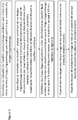
- a first step of electronic analysis of the images used a base made up of 500 images drawn at random from among the 600 selected with as reference the "ground truth" established by expert readers, in order to train the statistical classifier. considered.
- the chosen classifier was developed from a method known as decision tree forests (also called random forests) (11). This type of decision support tool is widely used in the field of data analysis by learning.
- decision trees describe how to classify or distribute a set of complex data (such as a population or, here, a series of images) according to a set of discriminant variables (here red / green ratio, co-occurrence matrix, luminosity) and as a function of an objective set in terms of number of classes (in this case, here images of “good” or “bad” visualization quality) based on ground truth (here the average scores of the experts).
- discriminant variables here red / green ratio, co-occurrence matrix, luminosity
- ground truth here the average scores of the experts.
- decision tree forests perform training on multiple decision trees trained on subsets of data. slightly different created on the basis of the 500 images considered.
- a second step made it possible to test the performance of the first training.
- test base consisted of 100 images drawn at random from the 600 images selected, with the ground truth established by expert readers as a reference. These images were classified by the previously learned classifier. It was then possible to deduce the performance of the classifier.
- a “cross-validation” method was used to evaluate the diagnostic performances of the various criteria tested (red / green ratio, bubbles. , brightness, and their combinations).
- This cross-validation method proven in the field of electronic image analysis, was based on the following principle: a first training was carried out on 500 images drawn randomly among the 600, then validation is carried out on the remaining 100 images; the operation was repeated ten times. The diagnostic performance (sensitivity, specificity) of the method was then characterized by the average ROC curve obtained over the 10 random draws (12).
- the primary endpoint was the sensitivity and specificity of the combination of the three criteria tested (R / V ratio, GLCM, brightness).
- the secondary endpoints were: the sensitivity and specificity of each of the criteria taken individually.
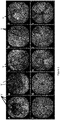
- the average scores (out of 2 points) assigned for each of the criteria by the 3 reader-experts are reported in Tables 2 to 6.
- the scores were below the threshold of 1.4 out of 2 in 72% of the images for the criterion "percentage of mucosa visualized ”(table 2), 65% for the“ debris, residues, liquids ”criterion (table 3), 67% for the“ bubble ”criterion (table 4), 61% for the“ bile / chyme ”criterion ( table 5) and 47% for the “brightness” criterion (table 6).
- the overall score was greater than or equal to 7 out of 10, and therefore made it possible to distribute the image as having a "good” quality, for 40.5% of the images for reader 1, 37% of the images for reader 2, and 39% for reader 3, with an average of 37% for the 3 readers (Table 7).
- the inter-observer correlation kappa coefficients are shown in Table 8 concerning the overall score, determining the cleanliness of an image (score ⁇ 7).
- Reader 1's kappa coefficient with Reader 2 and Reader 3 being 0.83 and 0.81 respectively. It was 0.87 between reader 2 and reader 3.
- the 600 images were analyzed according to algorithms already validated (the ratio of red / green pixels, a contrast assessment method derived from the co-occurrence matrix, the contrast of the image being similar to its brightness) and compared to the reading of the three experts, having determined that the threshold beyond which an image was of good viewing quality had an overall score greater than or equal to 7 out of 10.
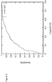
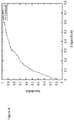
- the sensitivity was 80% and the specificity 83% ( Figure 2 ).
- the time required for the electronic analysis of an image according to the three criteria tested was 2.3 milliseconds.
- a composite electronic analysis method has been proposed according to the invention, making it possible to determine whether a fixed image of VCE from the small intestine has good visualization quality.
- This method includes three parameters: the ratio of red / green pixels (representing mucosal visualization), a grayscale co-occurrence matrix contrast (representing the abundance of bubbles) and a luminosity index.
- the crude combination of these three criteria makes it possible to obtain good diagnostic performance: sensitivity at 90% and specificity at 88%.
- the “Ground truth” is based on a strong agreement between the analysis of the three experts, the kappa coefficients ranging from 0.81 to 0.87 concerning the overall score.
- a multicriteria electronic evaluation method is proposed, integrating the analysis of a ratio of red / green pixels, the abundance of bubbles and the brightness of the image in order to determine the quality of visualization of VCE images of the intestine. 3rd generation hail.
- This composite score is reliable, easy to use, reproducible and could be integrated into reading software in order to facilitate the determination of the quality of visualization of the images in VCE and thus to conclude on the level of reliability of each examination. It could also make it possible to compare the different preparation methods in a reproducible manner in order to validate the procedure to be followed and the recommendations.
Landscapes
- Health & Medical Sciences (AREA)
- Life Sciences & Earth Sciences (AREA)
- Engineering & Computer Science (AREA)
- Surgery (AREA)
- Medical Informatics (AREA)
- Physics & Mathematics (AREA)
- Public Health (AREA)
- Radiology & Medical Imaging (AREA)
- Pathology (AREA)
- Optics & Photonics (AREA)
- Biomedical Technology (AREA)
- Heart & Thoracic Surgery (AREA)
- Nuclear Medicine, Radiotherapy & Molecular Imaging (AREA)
- Molecular Biology (AREA)
- Animal Behavior & Ethology (AREA)
- General Health & Medical Sciences (AREA)
- Biophysics (AREA)
- Veterinary Medicine (AREA)
- Signal Processing (AREA)
- Evolutionary Computation (AREA)
- Artificial Intelligence (AREA)
- Software Systems (AREA)
- Theoretical Computer Science (AREA)
- General Physics & Mathematics (AREA)
- Computing Systems (AREA)
- General Engineering & Computer Science (AREA)
- Data Mining & Analysis (AREA)
- Mathematical Physics (AREA)
- Computer Vision & Pattern Recognition (AREA)
- Image Analysis (AREA)
- Measuring And Recording Apparatus For Diagnosis (AREA)
- Apparatus For Radiation Diagnosis (AREA)
- Image Processing (AREA)
Claims (8)
- Vorrichtung zur Realisierung eines digitalen Bildklassifizierers zur Bestimmung der Anzeigequalität von Videokapsel-Endoskopiebildern eines Segments des Verdauungstrakts, die Folgendes umfasst:- eine Videokapsel, die eine Videoaufnahme von Segmenten des Verdauungstrakts ermöglicht;- Mittel zum Speichern der Bilder in Verbindung mit der Videokapsel;- eine Datenbank mit Bildern, die aus der Videokapsel extrahiert und einer Ground-Truth-Klassifikation unterzogen werden: in Bilder mit sogenannter "adäquater" Visualisierung und in Bilder mit sogenannter "inadäquater" Visualisierung; entsprechend eines Scorewerts, der durch eine visuelle Analyse der Bilder auf der Grundlage eines oder mehrerer medizinischer Kriterien bestimmt wurde,
wobei das medizinische Kriterium aus der folgenden Liste ausgewählt wird: der Prozentsatz der visualisierten Schleimhaut, die Helligkeit des Bildes, das Vorhandensein von Blasen, das Vorhandensein von Galle/Schleim, das Vorhandensein von Flüssigkeiten und unverdauten Resten,- Verarbeitungs- und Berechnungsmitteln, die mit den Speichermitteln verbunden sind und die Datenbank beinhalten und die konfiguriert sind, um:• mindestens zwei digitale Parameter zu berechnen, die sich jeweils auf mindestens eines der medizinischen Kriterien des Scorewerts beziehen, und von den Bildern in der Datenbank extrahiert sind,• ein automatisches statistisches Lernverfahren nach dem sogenannten "Random Forest"-Verfahren durchzuführen, das Folgendes aufweist:durch digitale Analyse dieser Lernbilder zur Konstruktion von N binären Entscheidungsbäumen, einem binären Entscheidungsbaum mit Zufallsziehung,a) mehrere Teilschritte für die Zufallsauswahl mit Zurücklegen einer gleichen Anzahl von Lernbildern, die eine Teilmenge von Bildern aus der Datenbank sind;b) einen Teilschritt für die automatischen Erstellung des Klassifizierers, mit einer Abfolge von automatischen Schwellenwerten, die auf die berechneten digitalen Parameter angewendet werden,
wobei jeder binäre Entscheidungsbaum unter Verwendung von mindestens den beiden digitalen Parametern konstruiert wird,
wobei die automatischen Schwellenwerte automatisch an jedem Knoten jedes binären Entscheidungsbaums mit dem digitalen Parameter berechnet werden, der die Einteilung der Lernbilder in eine erste Untergruppe und in eine zweite Untergruppe ermöglicht, die der Verteilung der Bilder mit sogenannter "adäquater" Visualisierung und sogenannter "inadäquater" Visualisierung, die mit einer "Ground-Truth"-Klassifizierung erstellt wurde, am nächsten kommt,
wobei die erhaltene Menge von binären Entscheidungsbäumen den digitalen Klassifizierer bildet,
wobei der digitale Klassifizierer dazu bestimmt ist, am Ende des Teilschritts b) die Lernbilder automatisch in eine erste Untergruppe von Lernbildern einzuteilen, die die meisten Bilder mit einer "adäquaten" Visualisierung beinhaltet und in eine zweite Untergruppe von Lernbildern, die die meisten Bilder mit "inadäquater" Visualisierung beinhaltet,
In dem der oder die digitale(n) Paramater aus der folgenden Liste ausgewählt sind:- ein globaler farbmetrischer Parameter der Bilder,- ein oder mehrere Parameter, die die Häufigkeit der Blasen in den Bildern widerspiegeln,- ein Parameter, der die Helligkeit des Bildes, das heißt den Grauwertkontrast der Bilder widerspiegelt. - Vorrichtung nach Anspruch 1, bei der die Verarbeitungs- und Berechnungsmittel so konfiguriert sind, um einen digitalen Schritt zur Entscheidung der Visualisierungsqualität der Testbilder durchzuführen, bei dem es sich um ein Abstimmungssystem auf allen digitalen Entscheidungen der binären Entscheidungsbäume handelt, wobei jedes Testbild in allen binären Entscheidungsbäumen getestet wurde und die Testbilder die verbleibenden Bilder der Datenbank abzüglich der Lernbilder im Klassifizierer oder in den Klassifizierern ist.
- Vorrichtung nach einem der Ansprüche 1 bis 2, bei der die Verarbeitungs- und Berechnungsmittel so konfiguriert sind, dass sie die Teilschritte a) und b) x-mal wiederholen, um einen endgültigen Klassifizierer zu erhalten, der aus der Menge der digitalen Klassifizierer besteht, die sich aus dem Lernschritt ergeben und daher x*N binäre Entscheidungsbäume aufweisen, wobei N größer oder gleich 100 und x größer oder gleich 10 ist.
- Vorrichtung nach einem der vorhergehenden Ansprüche 1 bis 3, bei der- der globale Farbparameter des Bildes das Rot/Grün-Verhältnis des Bildes ist, wenn das Segment des Verdauungstrakts der Dünndarm ist; oder das Rot/(Grün+Blau)-Verhältnis, wenn das Segment des Verdauungstrakts der Dickdarm ist;- der Parameter, der die Häufigkeit der Blasen widerspiegelt,:• ein Texturparameter aus der Grauwertematrixx (GLCM) des verarbeiteten Bildes oder• eine von den Blasen belegte Fläche ist- der Parameter, der die Helligkeit der Bilder wiedergibt, der Graustufenkontrast des Bildes ist.
- Vorrichtung nach einem der vorhergehenden Ansprüche, bei der der Teilschritt für die automatische Erstellung des digitalen Klassifizierers mit den 3 folgenden digitalen Parametern ausgeführt wird:- der globale Farbparameter des Bildes, der das Rot/Grün-Verhältnis des Bildes ist, wenn das Segment des Verdauungstrakts der Dünndarm ist; oder das Rot/(Grün+Blau)-Verhältnis, wenn das Segment des Verdauungstrakts der Dickdarm ist;- der Parameter, der die Häufigkeit der Blasen widerspiegelt, der:• ein Texturparameter aus der Grauwertematrixx (GLCM) des verarbeiteten Bildes oder• eine von den Blasen belegte Fläche ist- der Parameter, der die Helligkeit der Bilder wiedergibt, der der Graustufenkontrast des Bildes ist.
- Kontrollverfahren für ein mit einer Videokapsel in mindestens einem Segment des Verdauungstraktes einer Person aufgenommenes Video, um automatisch die Visualisierungsqualität der Bilder des Videos zu bestimmen,
unter Verwendung des digitalen Bildklassifizierers der Vorrichtung nach einem der Ansprüche 1 bis 5, angewendet auf die Bilder des Videos,
um bei einer automatischen Kontrolluntersuchung die Bilder mit "adäquater" Visualisierung, die Bilder mit "inadäquater" Visualisierung und den Anteil von Bildern mit "adäquater" Visualisierung im Video entsprechend der Entscheidung des digitalen Klassifizierers zu bestimmen. - Kontrollverfahren nach Anspruch 6 für verschiedene Personen, dadurch gekennzeichnet, dass das Verfahren Folgendes aufweist:- einen Schritt zur Vorbereitung des Darms auf die Kontrolluntersuchung, der für jede Person unterschiedlich ist;- einen Schritt zur Kontrolle der automatischen Untersuchung,- einen Schritt zum Vergleich der Effizienz der verschiedenen Darmvorbereitungen auf die Untersuchung entsprechend dem Anteil an Bildern mit "adäquater" Visualisierung, der für jede unterschiedliche Darmvorbereitung durch das Kontrollverfahren bestimmt wird.
- Kontrollvorrichtung zur automatischen Bestimmung der Qualität der Visualisierung eines Videos eines oder mehrerer Segmente des Verdauungstrakts einer Person, das mittels Videokapsel-Endoskopie aufgenommen wird und Folgendes umfasst:- eine Videokapsel, die eine Videoaufnahme von mindestens einem Segment eines Verdauungstrakts ermöglicht;- Mittel zum Speichern der Bilder in Verbindung mit der Videokapsel;- Verarbeitungs- und Berechnungsmittel, die mit den Speichermitteln verbunden sind und den durch die nach den Ansprüchen 1 bis 5 erstellten digitalen Klassifizierer beinhalten,und konfiguriert sind, um:• mindestens zwei digitale Parameter zu berechnen, die sich jeweils auf eines der folgenden medizinischen Kriterien auf den Bildern beziehen: Prozentsatz der visualisierten Schleimhaut, Helligkeit des Bildes, Vorhandensein von Blasen, Vorhandensein von Galle/Schleim, Vorhandensein von Flüssigkeiten und unverdauten Resten,• die Bilder des Videos digital zu testen und digital die Bilder mit "adäquater" Visualisierung, die Bilder mit "inadäquater" Visualisierung und den Anteil von Bildern mit "adäquater" Visualisierung im Video entsprechend der Entscheidung des digitalen Klassifizierers zu bestimmen.
Applications Claiming Priority (2)
| Application Number | Priority Date | Filing Date | Title |
|---|---|---|---|
| EP18305275.2A EP3539455A1 (de) | 2018-03-14 | 2018-03-14 | Verfahren zur automatischen bestimmung der anzeigequalität der bilder, die bei einer kapselendoskopie übertragen werden |
| PCT/EP2019/056295 WO2019175248A1 (fr) | 2018-03-14 | 2019-03-13 | Dispositif et procédé de réalisation d'un classifieur numérique d'images, afin de déterminer la qualité de visualisation des images de vidéocapsule endoscopique d'un segment du tube digestif |
Publications (2)
| Publication Number | Publication Date |
|---|---|
| EP3755197A1 EP3755197A1 (de) | 2020-12-30 |
| EP3755197B1 true EP3755197B1 (de) | 2021-07-21 |
Family
ID=61768203
Family Applications (2)
| Application Number | Title | Priority Date | Filing Date |
|---|---|---|---|
| EP18305275.2A Withdrawn EP3539455A1 (de) | 2018-03-14 | 2018-03-14 | Verfahren zur automatischen bestimmung der anzeigequalität der bilder, die bei einer kapselendoskopie übertragen werden |
| EP19709072.3A Active EP3755197B1 (de) | 2018-03-14 | 2019-03-13 | Verfahren zur automatischen bestimmung der anzeigequalität der bilder, die bei einer kapselendoskopie übertragen werden |
Family Applications Before (1)
| Application Number | Title | Priority Date | Filing Date |
|---|---|---|---|
| EP18305275.2A Withdrawn EP3539455A1 (de) | 2018-03-14 | 2018-03-14 | Verfahren zur automatischen bestimmung der anzeigequalität der bilder, die bei einer kapselendoskopie übertragen werden |
Country Status (3)
| Country | Link |
|---|---|
| US (1) | US10980404B2 (de) |
| EP (2) | EP3539455A1 (de) |
| WO (1) | WO2019175248A1 (de) |
Families Citing this family (13)
| Publication number | Priority date | Publication date | Assignee | Title |
|---|---|---|---|---|
| KR102063492B1 (ko) * | 2018-11-30 | 2020-01-08 | 아주대학교산학협력단 | 기계학습용 의료 영상 데이터의 학습 노이즈 필터링 방법 및 그 시스템 |
| KR102430946B1 (ko) * | 2019-11-08 | 2022-08-10 | 주식회사 인트로메딕 | 소장 정결도를 진단하는 시스템 및 방법 |
| JP7792354B2 (ja) | 2020-05-19 | 2025-12-25 | スピロ・ロボティクス・インコーポレーテッド | 気道管理の処置、組立体、及びシステムのためのロボット支援ナビゲーション及び制御 |
| JP7787161B2 (ja) * | 2020-09-08 | 2025-12-16 | ギブン イメージング リミテッド | 手順の妥当性を推定すること |
| EP3977909A1 (de) * | 2020-09-30 | 2022-04-06 | ENSEA - Ecole Nationale Supérieure de l'Electronique et de ses Applications | Vorrichtung und verfahren zur herstellung eines digitalen video-klassifikators |
| US11776118B2 (en) * | 2020-12-02 | 2023-10-03 | International Business Machines Corporation | Recognition of partially digested medications |
| CN112669311A (zh) * | 2021-01-08 | 2021-04-16 | 西北大学 | 一种陶瓷显微图像气泡的提取方法 |
| US20220309345A1 (en) * | 2021-03-25 | 2022-09-29 | Disney Enterprises, Inc. | Machine Learning Model Based Embedding for Adaptable Content Evaluation |
| CN113052843B (zh) * | 2021-05-31 | 2021-09-28 | 萱闱(北京)生物科技有限公司 | 辅助内镜检查的方法、装置、系统、存储介质和计算设备 |
| EP4412690A4 (de) * | 2021-10-08 | 2025-07-16 | Spiro Robotics Inc | Navigation und steuerung für atemwegsverwaltungsverfahren, -anordnungen und -systeme |
| CN114259197B (zh) * | 2022-03-03 | 2022-05-10 | 深圳市资福医疗技术有限公司 | 一种胶囊内窥镜质控方法及系统 |
| CN114565538B (zh) * | 2022-03-10 | 2024-03-01 | 山东大学齐鲁医院 | 一种内镜图像处理方法、系统、存储介质及设备 |
| JP2025514665A (ja) | 2022-04-10 | 2025-05-09 | スピロ・ロボティクス・インコーポレーテッド | 使い捨てコントロール、再使用可能なデバイス、およびそれらの使用方法 |
Family Cites Families (12)
| Publication number | Priority date | Publication date | Assignee | Title |
|---|---|---|---|---|
| US6882990B1 (en) * | 1999-05-01 | 2005-04-19 | Biowulf Technologies, Llc | Methods of identifying biological patterns using multiple data sets |
| US6636623B2 (en) * | 2001-08-10 | 2003-10-21 | Visiongate, Inc. | Optical projection imaging system and method for automatically detecting cells with molecular marker compartmentalization associated with malignancy and disease |
| EP1780651A1 (de) * | 2005-10-25 | 2007-05-02 | Bracco Imaging, S.P.A. | Verfahren und System zur automatischen Verarbeitung und Bewertung von Bildern, im Besonderen diagnostischer Bilder |
| JP4472631B2 (ja) * | 2005-12-28 | 2010-06-02 | オリンパスメディカルシステムズ株式会社 | 画像処理装置および当該画像処理装置における画像処理方法 |
| JP5312807B2 (ja) * | 2008-01-08 | 2013-10-09 | オリンパス株式会社 | 画像処理装置および画像処理プログラム |
| US8331699B2 (en) * | 2009-03-16 | 2012-12-11 | Siemens Medical Solutions Usa, Inc. | Hierarchical classifier for data classification |
| CN102934127B (zh) * | 2010-04-28 | 2016-11-16 | 基文影像公司 | 用于显示体内图像部分的系统和方法 |
| US8860715B2 (en) * | 2010-09-22 | 2014-10-14 | Siemens Corporation | Method and system for evaluation using probabilistic boosting trees |
| US8744172B2 (en) * | 2011-06-15 | 2014-06-03 | Siemens Aktiengesellschaft | Image processing using random forest classifiers |
| US8923631B2 (en) * | 2011-08-17 | 2014-12-30 | Volcano Corporation | Method and device for classifying vascular objects using classification trees evaluated on a graphics processing unit |
| KR101353952B1 (ko) * | 2012-10-05 | 2014-01-23 | 계명대학교 산학협력단 | 시공간적 비오에프와 랜덤 포레스트를 이용한 산불 연기 감지 방법 |
| CN105142493A (zh) * | 2013-08-30 | 2015-12-09 | 奥林巴斯株式会社 | 图像管理装置 |
-
2018
- 2018-03-14 EP EP18305275.2A patent/EP3539455A1/de not_active Withdrawn
-
2019
- 2019-03-13 US US16/979,252 patent/US10980404B2/en active Active
- 2019-03-13 WO PCT/EP2019/056295 patent/WO2019175248A1/fr not_active Ceased
- 2019-03-13 EP EP19709072.3A patent/EP3755197B1/de active Active
Also Published As
| Publication number | Publication date |
|---|---|
| US10980404B2 (en) | 2021-04-20 |
| EP3539455A1 (de) | 2019-09-18 |
| US20200397238A1 (en) | 2020-12-24 |
| WO2019175248A1 (fr) | 2019-09-19 |
| EP3755197A1 (de) | 2020-12-30 |
Similar Documents
| Publication | Publication Date | Title |
|---|---|---|
| EP3539455A1 (de) | Verfahren zur automatischen bestimmung der anzeigequalität der bilder, die bei einer kapselendoskopie übertragen werden | |
| Gao et al. | Reconstruction of high-resolution 6× 6-mm OCT angiograms using deep learning | |
| CN112927243B (zh) | 一种基于卷积神经网络的微出血病灶分割方法 | |
| Agurto et al. | Automatic detection of diabetic retinopathy and age-related macular degeneration in digital fundus images | |
| JP7752529B2 (ja) | 生体内画像ストリームの精査用情報を生成及び表示するためのシステム並びに方法 | |
| Wan et al. | Retinal image enhancement using cycle-constraint adversarial network | |
| CN109815965B (zh) | 一种图像过滤方法、装置及存储介质 | |
| CA3095089A1 (fr) | Methode de detection de cellules presentant au moins une anomalie dans un echantilllon cytologique | |
| Asad et al. | Ant colony-based system for retinal blood vessels segmentation | |
| KR20190087681A (ko) | 자궁경부암에 대한 피검체의 발병 여부를 판정하는 방법 | |
| Li et al. | Automatic quantification of cone photoreceptors in adaptive optics scanning light ophthalmoscope images using multi-task learning | |
| Adorno III et al. | Advancing eosinophilic esophagitis diagnosis and phenotype assessment with deep learning computer vision | |
| García et al. | Suspicious Skin Lesion Detection in Wide-Field Body Images using Deep Learning Outlier Detection | |
| Reddy et al. | Diagnosing and categorizing of pulmonary diseases using Deep learning conventional Neural network | |
| De Carvalho et al. | Automated colonoscopy withdrawal phase duration estimation using cecum detection and surgical tasks classification | |
| FR3113155A1 (fr) | Procédé d’identification d’un implant dentaire visible sur une image d’entrée au moyen d’au moins un réseau de neurones à convolution. | |
| Firmbach et al. | Quantitative Analysis of Distinct Colon Crypt Branching Modes Using Interpretable Machine Learning | |
| Basreddy et al. | Preprocessing, feature extraction, and classification methodologies on diabetic retinopathy using fundus images | |
| CN115294434A (zh) | 一种基于分子谱像特征采集与融合的多模态学习卷积神经网络模型的实施方法 | |
| Akbar et al. | Microscope‐Assisted Hypertensive Retinopathy Diagnosis Using Deep Learning Models | |
| CN115861718A (zh) | 胃活检图像分类方法、装置、设备、介质和程序产品 | |
| Malekian et al. | Computer aided measurement of sub-epithelial collagen band in colon biopsies for collagenous colitis diagnosis | |
| CN115393326B (zh) | 基于局部细粒度轮廓特征的电子喉镜医学图像检测方法 | |
| Gupta et al. | Transformer-Dilated ResUNet: An Efficient and Accurate Framework for Polyp Segmentation | |
| EP3815037A1 (de) | System und verfahren zur erzeugung eines mehrparametrigen grafischen indikators aus einem bild eines histologischen schnittes |
Legal Events
| Date | Code | Title | Description |
|---|---|---|---|
| STAA | Information on the status of an ep patent application or granted ep patent |
Free format text: STATUS: UNKNOWN |
|
| STAA | Information on the status of an ep patent application or granted ep patent |
Free format text: STATUS: THE INTERNATIONAL PUBLICATION HAS BEEN MADE |
|
| PUAI | Public reference made under article 153(3) epc to a published international application that has entered the european phase |
Free format text: ORIGINAL CODE: 0009012 |
|
| STAA | Information on the status of an ep patent application or granted ep patent |
Free format text: STATUS: REQUEST FOR EXAMINATION WAS MADE |
|
| 17P | Request for examination filed |
Effective date: 20200923 |
|
| AK | Designated contracting states |
Kind code of ref document: A1 Designated state(s): AL AT BE BG CH CY CZ DE DK EE ES FI FR GB GR HR HU IE IS IT LI LT LU LV MC MK MT NL NO PL PT RO RS SE SI SK SM TR |
|
| AX | Request for extension of the european patent |
Extension state: BA ME |
|
| GRAP | Despatch of communication of intention to grant a patent |
Free format text: ORIGINAL CODE: EPIDOSNIGR1 |
|
| STAA | Information on the status of an ep patent application or granted ep patent |
Free format text: STATUS: GRANT OF PATENT IS INTENDED |
|
| INTG | Intention to grant announced |
Effective date: 20210219 |
|
| GRAS | Grant fee paid |
Free format text: ORIGINAL CODE: EPIDOSNIGR3 |
|
| GRAA | (expected) grant |
Free format text: ORIGINAL CODE: 0009210 |
|
| STAA | Information on the status of an ep patent application or granted ep patent |
Free format text: STATUS: THE PATENT HAS BEEN GRANTED |
|
| DAV | Request for validation of the european patent (deleted) | ||
| DAX | Request for extension of the european patent (deleted) | ||
| AK | Designated contracting states |
Kind code of ref document: B1 Designated state(s): AL AT BE BG CH CY CZ DE DK EE ES FI FR GB GR HR HU IE IS IT LI LT LU LV MC MK MT NL NO PL PT RO RS SE SI SK SM TR |
|
| REG | Reference to a national code |
Ref country code: GB Ref legal event code: FG4D Free format text: NOT ENGLISH |
|
| REG | Reference to a national code |
Ref country code: CH Ref legal event code: EP |
|
| REG | Reference to a national code |
Ref country code: DE Ref legal event code: R096 Ref document number: 602019006302 Country of ref document: DE |
|
| REG | Reference to a national code |
Ref country code: AT Ref legal event code: REF Ref document number: 1411819 Country of ref document: AT Kind code of ref document: T Effective date: 20210815 |
|
| REG | Reference to a national code |
Ref country code: IE Ref legal event code: FG4D Free format text: LANGUAGE OF EP DOCUMENT: FRENCH |
|
| REG | Reference to a national code |
Ref country code: LT Ref legal event code: MG9D |
|
| REG | Reference to a national code |
Ref country code: NL Ref legal event code: MP Effective date: 20210721 |
|
| REG | Reference to a national code |
Ref country code: AT Ref legal event code: MK05 Ref document number: 1411819 Country of ref document: AT Kind code of ref document: T Effective date: 20210721 |
|
| PG25 | Lapsed in a contracting state [announced via postgrant information from national office to epo] |
Ref country code: ES Free format text: LAPSE BECAUSE OF FAILURE TO SUBMIT A TRANSLATION OF THE DESCRIPTION OR TO PAY THE FEE WITHIN THE PRESCRIBED TIME-LIMIT Effective date: 20210721 Ref country code: SE Free format text: LAPSE BECAUSE OF FAILURE TO SUBMIT A TRANSLATION OF THE DESCRIPTION OR TO PAY THE FEE WITHIN THE PRESCRIBED TIME-LIMIT Effective date: 20210721 Ref country code: RS Free format text: LAPSE BECAUSE OF FAILURE TO SUBMIT A TRANSLATION OF THE DESCRIPTION OR TO PAY THE FEE WITHIN THE PRESCRIBED TIME-LIMIT Effective date: 20210721 Ref country code: NO Free format text: LAPSE BECAUSE OF FAILURE TO SUBMIT A TRANSLATION OF THE DESCRIPTION OR TO PAY THE FEE WITHIN THE PRESCRIBED TIME-LIMIT Effective date: 20211021 Ref country code: PT Free format text: LAPSE BECAUSE OF FAILURE TO SUBMIT A TRANSLATION OF THE DESCRIPTION OR TO PAY THE FEE WITHIN THE PRESCRIBED TIME-LIMIT Effective date: 20211122 Ref country code: NL Free format text: LAPSE BECAUSE OF FAILURE TO SUBMIT A TRANSLATION OF THE DESCRIPTION OR TO PAY THE FEE WITHIN THE PRESCRIBED TIME-LIMIT Effective date: 20210721 Ref country code: FI Free format text: LAPSE BECAUSE OF FAILURE TO SUBMIT A TRANSLATION OF THE DESCRIPTION OR TO PAY THE FEE WITHIN THE PRESCRIBED TIME-LIMIT Effective date: 20210721 Ref country code: HR Free format text: LAPSE BECAUSE OF FAILURE TO SUBMIT A TRANSLATION OF THE DESCRIPTION OR TO PAY THE FEE WITHIN THE PRESCRIBED TIME-LIMIT Effective date: 20210721 Ref country code: LT Free format text: LAPSE BECAUSE OF FAILURE TO SUBMIT A TRANSLATION OF THE DESCRIPTION OR TO PAY THE FEE WITHIN THE PRESCRIBED TIME-LIMIT Effective date: 20210721 Ref country code: BG Free format text: LAPSE BECAUSE OF FAILURE TO SUBMIT A TRANSLATION OF THE DESCRIPTION OR TO PAY THE FEE WITHIN THE PRESCRIBED TIME-LIMIT Effective date: 20211021 Ref country code: AT Free format text: LAPSE BECAUSE OF FAILURE TO SUBMIT A TRANSLATION OF THE DESCRIPTION OR TO PAY THE FEE WITHIN THE PRESCRIBED TIME-LIMIT Effective date: 20210721 |
|
| PG25 | Lapsed in a contracting state [announced via postgrant information from national office to epo] |
Ref country code: PL Free format text: LAPSE BECAUSE OF FAILURE TO SUBMIT A TRANSLATION OF THE DESCRIPTION OR TO PAY THE FEE WITHIN THE PRESCRIBED TIME-LIMIT Effective date: 20210721 Ref country code: LV Free format text: LAPSE BECAUSE OF FAILURE TO SUBMIT A TRANSLATION OF THE DESCRIPTION OR TO PAY THE FEE WITHIN THE PRESCRIBED TIME-LIMIT Effective date: 20210721 Ref country code: GR Free format text: LAPSE BECAUSE OF FAILURE TO SUBMIT A TRANSLATION OF THE DESCRIPTION OR TO PAY THE FEE WITHIN THE PRESCRIBED TIME-LIMIT Effective date: 20211022 |
|
| REG | Reference to a national code |
Ref country code: DE Ref legal event code: R097 Ref document number: 602019006302 Country of ref document: DE |
|
| PG25 | Lapsed in a contracting state [announced via postgrant information from national office to epo] |
Ref country code: DK Free format text: LAPSE BECAUSE OF FAILURE TO SUBMIT A TRANSLATION OF THE DESCRIPTION OR TO PAY THE FEE WITHIN THE PRESCRIBED TIME-LIMIT Effective date: 20210721 |
|
| PLBE | No opposition filed within time limit |
Free format text: ORIGINAL CODE: 0009261 |
|
| STAA | Information on the status of an ep patent application or granted ep patent |
Free format text: STATUS: NO OPPOSITION FILED WITHIN TIME LIMIT |
|
| PG25 | Lapsed in a contracting state [announced via postgrant information from national office to epo] |
Ref country code: SM Free format text: LAPSE BECAUSE OF FAILURE TO SUBMIT A TRANSLATION OF THE DESCRIPTION OR TO PAY THE FEE WITHIN THE PRESCRIBED TIME-LIMIT Effective date: 20210721 Ref country code: SK Free format text: LAPSE BECAUSE OF FAILURE TO SUBMIT A TRANSLATION OF THE DESCRIPTION OR TO PAY THE FEE WITHIN THE PRESCRIBED TIME-LIMIT Effective date: 20210721 Ref country code: RO Free format text: LAPSE BECAUSE OF FAILURE TO SUBMIT A TRANSLATION OF THE DESCRIPTION OR TO PAY THE FEE WITHIN THE PRESCRIBED TIME-LIMIT Effective date: 20210721 Ref country code: EE Free format text: LAPSE BECAUSE OF FAILURE TO SUBMIT A TRANSLATION OF THE DESCRIPTION OR TO PAY THE FEE WITHIN THE PRESCRIBED TIME-LIMIT Effective date: 20210721 Ref country code: CZ Free format text: LAPSE BECAUSE OF FAILURE TO SUBMIT A TRANSLATION OF THE DESCRIPTION OR TO PAY THE FEE WITHIN THE PRESCRIBED TIME-LIMIT Effective date: 20210721 Ref country code: AL Free format text: LAPSE BECAUSE OF FAILURE TO SUBMIT A TRANSLATION OF THE DESCRIPTION OR TO PAY THE FEE WITHIN THE PRESCRIBED TIME-LIMIT Effective date: 20210721 |
|
| 26N | No opposition filed |
Effective date: 20220422 |
|
| PG25 | Lapsed in a contracting state [announced via postgrant information from national office to epo] |
Ref country code: IT Free format text: LAPSE BECAUSE OF FAILURE TO SUBMIT A TRANSLATION OF THE DESCRIPTION OR TO PAY THE FEE WITHIN THE PRESCRIBED TIME-LIMIT Effective date: 20210721 |
|
| PG25 | Lapsed in a contracting state [announced via postgrant information from national office to epo] |
Ref country code: MC Free format text: LAPSE BECAUSE OF FAILURE TO SUBMIT A TRANSLATION OF THE DESCRIPTION OR TO PAY THE FEE WITHIN THE PRESCRIBED TIME-LIMIT Effective date: 20210721 |
|
| REG | Reference to a national code |
Ref country code: CH Ref legal event code: PL |
|
| REG | Reference to a national code |
Ref country code: BE Ref legal event code: MM Effective date: 20220331 |
|
| PG25 | Lapsed in a contracting state [announced via postgrant information from national office to epo] |
Ref country code: LU Free format text: LAPSE BECAUSE OF NON-PAYMENT OF DUE FEES Effective date: 20220313 Ref country code: LI Free format text: LAPSE BECAUSE OF NON-PAYMENT OF DUE FEES Effective date: 20220331 Ref country code: IE Free format text: LAPSE BECAUSE OF NON-PAYMENT OF DUE FEES Effective date: 20220313 Ref country code: CH Free format text: LAPSE BECAUSE OF NON-PAYMENT OF DUE FEES Effective date: 20220331 |
|
| PG25 | Lapsed in a contracting state [announced via postgrant information from national office to epo] |
Ref country code: BE Free format text: LAPSE BECAUSE OF NON-PAYMENT OF DUE FEES Effective date: 20220331 |
|
| P01 | Opt-out of the competence of the unified patent court (upc) registered |
Effective date: 20230705 |
|
| PG25 | Lapsed in a contracting state [announced via postgrant information from national office to epo] |
Ref country code: MK Free format text: LAPSE BECAUSE OF FAILURE TO SUBMIT A TRANSLATION OF THE DESCRIPTION OR TO PAY THE FEE WITHIN THE PRESCRIBED TIME-LIMIT Effective date: 20210721 Ref country code: CY Free format text: LAPSE BECAUSE OF FAILURE TO SUBMIT A TRANSLATION OF THE DESCRIPTION OR TO PAY THE FEE WITHIN THE PRESCRIBED TIME-LIMIT Effective date: 20210721 |
|
| PG25 | Lapsed in a contracting state [announced via postgrant information from national office to epo] |
Ref country code: HU Free format text: LAPSE BECAUSE OF FAILURE TO SUBMIT A TRANSLATION OF THE DESCRIPTION OR TO PAY THE FEE WITHIN THE PRESCRIBED TIME-LIMIT; INVALID AB INITIO Effective date: 20190313 |
|
| PG25 | Lapsed in a contracting state [announced via postgrant information from national office to epo] |
Ref country code: MT Free format text: LAPSE BECAUSE OF FAILURE TO SUBMIT A TRANSLATION OF THE DESCRIPTION OR TO PAY THE FEE WITHIN THE PRESCRIBED TIME-LIMIT Effective date: 20210721 |
|
| PGFP | Annual fee paid to national office [announced via postgrant information from national office to epo] |
Ref country code: DE Payment date: 20250218 Year of fee payment: 7 |
|
| PGFP | Annual fee paid to national office [announced via postgrant information from national office to epo] |
Ref country code: FR Payment date: 20250330 Year of fee payment: 7 |
|
| PGFP | Annual fee paid to national office [announced via postgrant information from national office to epo] |
Ref country code: GB Payment date: 20250221 Year of fee payment: 7 |
|
| PG25 | Lapsed in a contracting state [announced via postgrant information from national office to epo] |
Ref country code: TR Free format text: LAPSE BECAUSE OF FAILURE TO SUBMIT A TRANSLATION OF THE DESCRIPTION OR TO PAY THE FEE WITHIN THE PRESCRIBED TIME-LIMIT Effective date: 20210721 |