EP3746791B1 - Verwendung von bmmf rep protein als biomarker für kolonkarzinom - Google Patents
Verwendung von bmmf rep protein als biomarker für kolonkarzinom Download PDFInfo
- Publication number
- EP3746791B1 EP3746791B1 EP19705289.7A EP19705289A EP3746791B1 EP 3746791 B1 EP3746791 B1 EP 3746791B1 EP 19705289 A EP19705289 A EP 19705289A EP 3746791 B1 EP3746791 B1 EP 3746791B1
- Authority
- EP
- European Patent Office
- Prior art keywords
- rep
- protein
- antibody
- seq
- tissue
- Prior art date
- Legal status (The legal status is an assumption and is not a legal conclusion. Google has not performed a legal analysis and makes no representation as to the accuracy of the status listed.)
- Active
Links
Images





Classifications
-
- G—PHYSICS
- G01—MEASURING; TESTING
- G01N—INVESTIGATING OR ANALYSING MATERIALS BY DETERMINING THEIR CHEMICAL OR PHYSICAL PROPERTIES
- G01N33/00—Investigating or analysing materials by specific methods not covered by groups G01N1/00 - G01N31/00
- G01N33/48—Biological material, e.g. blood, urine; Haemocytometers
- G01N33/50—Chemical analysis of biological material, e.g. blood, urine; Testing involving biospecific ligand binding methods; Immunological testing
- G01N33/53—Immunoassay; Biospecific binding assay; Materials therefor
- G01N33/575—Immunoassay; Biospecific binding assay; Materials therefor for cancer
- G01N33/57535—Immunoassay; Biospecific binding assay; Materials therefor for cancer of the large intestine, e.g. colon, rectum or anus
-
- G—PHYSICS
- G01—MEASURING; TESTING
- G01N—INVESTIGATING OR ANALYSING MATERIALS BY DETERMINING THEIR CHEMICAL OR PHYSICAL PROPERTIES
- G01N33/00—Investigating or analysing materials by specific methods not covered by groups G01N1/00 - G01N31/00
- G01N33/48—Biological material, e.g. blood, urine; Haemocytometers
- G01N33/50—Chemical analysis of biological material, e.g. blood, urine; Testing involving biospecific ligand binding methods; Immunological testing
- G01N33/53—Immunoassay; Biospecific binding assay; Materials therefor
- G01N33/577—Immunoassay; Biospecific binding assay; Materials therefor involving monoclonal antibodies binding reaction mechanisms characterised by the use of monoclonal antibodies
-
- G—PHYSICS
- G01—MEASURING; TESTING
- G01N—INVESTIGATING OR ANALYSING MATERIALS BY DETERMINING THEIR CHEMICAL OR PHYSICAL PROPERTIES
- G01N2333/00—Assays involving biological materials from specific organisms or of a specific nature
- G01N2333/435—Assays involving biological materials from specific organisms or of a specific nature from animals; from humans
- G01N2333/705—Assays involving receptors, cell surface antigens or cell surface determinants
- G01N2333/70503—Immunoglobulin superfamily, e.g. VCAMs, PECAM, LFA-3
-
- G—PHYSICS
- G01—MEASURING; TESTING
- G01N—INVESTIGATING OR ANALYSING MATERIALS BY DETERMINING THEIR CHEMICAL OR PHYSICAL PROPERTIES
- G01N2333/00—Assays involving biological materials from specific organisms or of a specific nature
- G01N2333/90—Enzymes; Proenzymes
- G01N2333/914—Hydrolases (3)
Definitions
- the invention relates to the use of a DNA-replication-associated (Rep) protein as a biomarker for colorectal cancer (CRC).
- Rep DNA-replication-associated protein
- CRC Colorectal cancer
- CRC is a cancer that evolved as a consequence of uncontrolled cell growth in the colon or rectum. These malignancies may develop as a consequence of pre-existing benign adenomas where genetic alterations promote the transition from normal to cancerous growth. Epigenetic events have been recognized as an important mechanism for this transition.
- a blood-based protein biomarker panel for the detection of colorectal cancer as described by Fung et al., 2015 has not yet brought the desired results as regards predictability.
- a biomarker for CRC which may be used both for prognostic and diagnostic purposes.
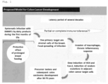
- Fig. 1 the inventors have created a model for colon cancer development that is shown in Fig. 1 .
- BMMF Bovine Meat and Milk Factor
- This accumulation may be triggered also by the uptake of specific molecules that may represent receptors for BMMFs. These molecules are also taken up by consumption of cow products and are metabolized into receptors on the surface of the host cells.
- ROS reactive oxygen species
- Cox-2 cyclooxygenase-2
- Ki67 proliferation marker
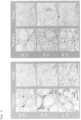
- IHC staining show that especially the basal epithelial cells derived from epithelial stem cells located close to the endpoint of the crypts and migrating towards the distal luminal part of the crypts feature an intrinsically high proliferation enabling stochastic manifestation of mutations as a basic requirement for tumorgenesis and development of colon cancer ( Fig. 2 ).
- BMMFs represent a specific and local trigger for induction of chronic inflammation within the lamina intestinal leading to an increase of ROS which induces proliferation and mutation in surrounding epithelial cells eventually leading to the formation of polyps as precursors for colon cancer.
- a selection of 11 pairs of donor tissue samples (each tumor tissue and peripheral tissue) and 4 samples from different donors of colon polyp tissue were subjected to IHC staining with mouse monoclonal anti-Rep antibodies.
- 9 out of 11 tissues showed strong specific antibody staining with at least two anti-Rep antibodies.
- the staining with an anti-Rep antibody (e.g. antibody 10-3) shows specific detection of protein targets in tissue samples 4798, 4799, 4802, 4806, 4809 and 4813 in the tumor as well as in the peripheral tissue.
- the staining intensity is higher for peripheral tissue, where intensely stained cytoplasmic aggregates of smaller size concentrate within distinct islands of cells within the lamina intestinal between the characteristic Crypts of Lieberkuhn of the colon tissue (all pictures showing traversal cuts throughout the Crypts of Lieberkuhn with the (inner) lumen of the crypts having contact to the colonic fluids in the center of the ring-like arrangement of epithelial cells of the crypts; Fig. 2 ). Staining of tumor tissues also shows intense staining of smaller sized aggregates mainly within the cytoplasmic regions of the stroma cells within the tumor tissue.
- the inventors also tested the antibody level of colon cancer patients by contacting the Rep protein with a serum specimen suspected of containing anti-Rep protein antibodies under conditions that permit the Rep protein to bind to any such antibody present in the specimen.
- the inventors recognized that the serum reactivity is reduced in colon cancer patients in comparison to healthy controls.
- the fourth group comprised 3 isolates being representatives of Gemycirularviridae.
- Putative Rep genes were identified as part of the BMMF's DNA sequences obtained by in silica comparisons to available sequences. Amplification using abutting primers in the rep gene led to the isolation of full and partial circular DNA genomes from bovine sera (Funk et al., 2014). This was extended to samples from commercially available milk products for the presence of specific circular single-stranded DNA genomes.
- Cow milk isolates were CMI1.252, CMI2.214 and CMI3.168 which are designated as "CMI1 genome”, “CMI2 genome” and “CMI3 genome”, respectively.
- the sequences of the isolates have been deposited in the EMBL Databank under accession numbers LK931487 (CMI1.252), LK931488 (CMI2.214) and LK931489 (CMI3.168) and have been aligned and described in WO 2016/005054 A2 .
- the present inventors have found that both CMI genomes and MSBI genomes show a significant production of transcribed RNA and the encoded Rep protein is expressed in peripheral tissue around the colon cancer islands and polyps.
- the present inventors have found that the encoded Rep proteins (MSBI1 Rep, MSBI2 Rep, CMI1 Rep, CMI2 Rep, CMI3 Rep) represent a biomarker for colon cancer.
- DNA-replication-associated protein (RepB) the Rep protein has DNA binding activity and can be essential for initiation of replication of episomal or viral DNA molecules.
- Rep proteins show a marked potential of self-oligomerization and aggregation, which was described within prokaryotic systems in vivo and in vitro (Giraldo, Moreno-Diaz de la Espina et al. 2011, Torreira, Moreno-Del Alamo et al. 2015).
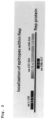
- the anti-Rep antibodies bind to epitopes of Rep protein that are exemplified in Fig. 3 .
- Particular preferred antibodies bind to epitopes within an amino acid sequence selected from the group consisting of amino acids from 1 to 136, from 137 to 229 and from 230 to 324 of SEQ ID NO:1.
- the antibody binds to an epitope comprised by SEQ ID NO:2 or SEQ ID NO:3.
- Rep proteins may represent biomarkers for an enhanced risk to develop colon cancer and are useful as a marker for determining the overall survival prognosis of CRC patients.
- colon cancer and “colorectal cancer” (CRC) are used interchangeably.
- CRC is a cancer that evolved as a consequence of uncontrolled cell growth in the colon or rectum. These malignancies may develop as a consequence of pre-existing benign adenomas where genetic alterations promote the transition from normal to cancerous growth.
- colon cancer or CRC mean pre-stages, early stages or late stages of the disease and metastases derived therefrom.
- the present invention may also encompass the systematic testing of healthy colonic tissue (tissue from individuals without cancer diagnosis or a specific hint for the disease) to assess the disease risk in the future. This means that the present invention is also suitable to determine the predisposition for developing CRC.
- Rep protein refers to a DNA-replication-associated protein (RepB).
- the Rep protein comprises DNA binding activity and could be essential for initiation of replication of episomal/viral DNA molecules.
- Rep protein refers to a Rep protein from the group of the Small Sphinx Genome (Whitley et al., 2014).
- the Rep protein is a MSBI1 genome-encoded Rep protein (MSBI1 Rep), a MSBI2 genome-encoded Rep protein (MSBI2 Rep), a CMI1 genome-encoded Rep protein (CMI1 Rep), a CMI2 genome-encoded Rep protein (CMI2 Rep) or CMI3 genome-encoded Rep protein (CMI3 Rep).
- the MSBI1 Rep protein is encoded by MSBI1.176 deposited in the EMBL databank under the acc. no. LK931491 and has the amino acid sequence as depicted in SEQ ID NO:1 or the Rep protein is MSBI2 encoded by MSBI2.176 deposited in the EMBL databank under the acc. no. LK931492 and has the amino acid sequence as depicted in SEQ ID NO:8 (Whitley, Gunst et al. 2014).
- the CMI1 Rep protein is encoded by CMI1.252 deposited in the EMBL databank under the acc. no. LK931487 and has the amino acid sequence as depicted in SEQ ID NO:10.
- the CMI2 Rep protein is encoded by CMI2.214 deposited in the EMBL databank under the acc. no. LK931488 and has the amino acid sequence as depicted in SEQ ID NO:11.
- the CMI3 Rep protein is encoded by CMI3.168 deposited in the EMBL databank under the acc. no. LK931489 and has the amino acid sequence as depicted in SEQ ID NO:12.
- the Rep protein comprises a N-terminal region conserved among BMMF1 genomes consisting essentially of amino acids from 1 to 229 of SEQ ID NO:1 and a C-terminal variable region specific for MSBI1.176 consisting essentially from amino acids 230 to 324 of SEQ ID NO:1.
- the N-terminal conserved region comprises a putative, first DNA binding domain consisting essentially of amino acids from 1 to 136 of SEQ ID NO: 1 and a second putative DNA binding domain consisting essentially of amino acids from 137 to 229 of SEQ ID NO:1.
- the C-terminal domain shows little sequence homology with any known protein and consists of amino acids 230 to 324.
- Rep protein also encompasses fragments and variants of the protein with SEQ ID NO:1 or SEQ ID NO:8 which are capable of binding an anti-Rep antibody specific for Rep protein having the amino acid sequence of SEQ ID NO:1 or SEQ ID NO:8.
- a fragment is an immunogenic fragment of the protein having the amino acid sequence of SEQ ID NO:1 or SEQ ID NO:8 which encompasses at least one epitope for an anti-Rep protein antibody against the Rep protein of SEQ ID NO:1 or SEQ ID NO:8 and, preferably, comprises at least 7, 8, 9, 10, 15, 20, 25 or 50 contiguous amino acids.
- the fragment comprises or consists essentially of a domain of the Rep protein, for example, the N-terminal conserved region, the C-terminal variable region, the first or second DNA binding domain.
- a variant of the protein with SEQ ID NO:1 or SEQ ID NO:8 comprises one or more amino acid deletions, substitutions or additions compared to SEQ ID NO:1 and has a homology of at least 90%, 91%, 92%, 93%, 94%, 95%, 96%, 97%, 98% or 99% to the amino acid sequence of SEQ ID NO:1 or SEQ ID NO:8, wherein the variant is capable of binding an anti-Rep antibody specific for a Rep protein having the amino acid sequence of SEQ ID NO:1 or SEQ ID NO:8.
- Rep protein includes fusion proteins with a heterologous amino acid sequence, with a leader sequence or with a Tag-sequence and the like.
- protein tags are genetically grafted onto the Rep protein described above, for example the Rep protein selected from the group consisting of MSBI1, MSBI2, CMI1, CMI2 or CMI3.
- At least one protein tag is attached to a polypeptide consisting of an amino acid sequence as depicted in any one of SEQ ID NOs:1-3,8-12,14.
- Such protein tags may be removable by chemical agents or by enzymatic means.
- protein tags are affinity or chromatography tags for purification.
- the Rep protein may be fused to a Tag-sequence, for example, selected from the group consisting of His 6 -Tag (SEQ ID NO:4), T7-Tag (SEQ ID NO:5), FLAG-Tag (SEQ ID NO:6)and Strep-II-Tag (SEQ ID NO:7).
- a His-Tag SEQ ID No:4
- a T7-Tag SEQ ID NO:5
- FLAG-Tag SEQ ID NO:6
- Strepll-Tag SEQ ID NO:7
- fluorescence tags such as green fluorescence protein (GFP) or its variants may be attached to a Rep-protein used in the present invention invention.
- MSBI1 genome-encoded Rep protein (MSBI1 Rep) is codon-optimized for the production in human cell lines (e.g. HEK-293, HEK293TT, HEK293T, HEK293FT, HaCaT, HeLa, SiHa, CaSki, HDMEC, L1236, L428, BJAB, MCF7, Colo678, any primary cell lines) as well as bovine (e.g. MAC-T) or murine cell lines (e.g. GT1-7). This is described in detail in PCT/EP2017/075774 .
- human cell lines e.g. HEK-293, HEK293TT, HEK293T, HEK293FT, HaCaT, HeLa, SiHa, CaSki, HDMEC, L1236, L428, BJAB, MCF7, Colo678, any primary cell lines
- bovine e.g. MAC-T
- murine cell lines e.g.
- the Rep protein used in the invention can be prepared by classical chemical synthesis.
- the synthesis can be carried out in homogeneous solution or in solid phase.
- the polypeptides used in this invention can also be prepared by means of recombinant DNA techniques.
- Subject refers to a mammalian individual or patient, including murines, cattle, for example bovines, simians and humans. Preferably, the subject is a human patient.
- Anti-Rep antibody refers to an antibody binding at a detectable level to Rep protein which affinity is more strongly to the Rep protein of the invention than to a non-Rep protein.
- the antigen affinity for Rep protein is at least 2 fold larger than background binding.
- the anti-Rep antibody is specific for the MSBI1 Rep having the amino acid sequence of SEQ ID NO:1 or MSBI2 Rep.
- the antibody is cross-specific for MSBI1 Rep, MSBI2 Rep, CMI1 Rep, CMI2 Rep and/or CMI3 Rep.
- the anti-Rep antibody is cross-specific for at least two, preferably all, off MSBI1 Rep, MSBI2 Rep, CMI1 Rep, CMI2 Rep and/or CMI3 Rep.
- the inventors also tested the antibody level of colon cancer patients by contacting the Rep protein with a specimen suspected of containing anti-Rep protein antibodies under conditions that permit the Rep protein to bind to any such antibody present in the specimen. Such conditions will typically be physiologic temperature, pH and ionic strength using an excess of Rep protein.
- the incubation of the Rep protein with the specimen is followed by detection of immune complexes comprised of the antigen.
- the Rep protein is coupled to a signal generating compound, e.g. detectable label, or an additional binding agent, e.g. secondary antihuman antibody, coupled to a signal generating compound is used for detecting the immune complex.
- Anti-Rep antibodies can be detected and quantified in assays based on Rep protein as protein antigen, which serves as target for the mammalian, e.g. human, antibodies suspected in the specimen.
- the Rep protein is purified and the specimen can be, for example, serum or plasma.
- the methods include immobilization of Rep protein on a matrix followed by incubation of the immobilized Rep protein with the specimen.
- the Rep-bound antibodies of the formed immunological complex between Rep protein and antibodies of the specimen are quantified by a detection binding agent coupled to a signal generating compound, e.g. secondary HRP-(horseradish-peroxidase)-coupled detection antibody allowing for HRP-substrate based quantification.
- This signal generating compound or label is in itself detectable or may be reacted with an additional compound to generate a detectable product.
- Protocols may, for example, use solid supports, or immunoprecipitation.
- Most assays involve the use of binding agents coupled to signal generating compounds, for example labelled antibody or labelled Rep protein; the labels may be, for example, enzymatic, fluorescent, chemiluminescent, radioactive, or dye molecules.
- Assays which amplify the signals from the immune complex are also known; examples of which are assays which utilize biotin and avidin or streptavidin, and enzyme-labeled and mediated immunoassays, such as ELISA assays.
- the immunoassay may be in a heterogeneous or in a homogeneous format, and of a standard or competitive type. Both standard and competitive formats are known in the art.
- the reaction between the Rep protein and the anti-Rep antibody forms a network that precipitates from the solution or suspension and forms a visible layer or film of precipitate. If no anti-Rep antibody is present in the specimen, no visible precipitate is formed.
- the inventors used methods wherein an increased amount of Rep protein in a sample correlates with a diagnosis or predisposition of colon cancer.
- the Rep protein in the sample is detected by anti-Rep antibodies.
- sample refers to a biological sample encompassing cancerous colon tissue, peripheral tissue surrounding the cancerous colon tissue and (benign) colon polyps.
- the samples encompass tissue samples such as tissue cultures or biopsy specimen.
- Such methods comprise the steps of detecting Rep protein in a sample from a subject by anti-Rep antibodies.
- Rep protein is detected in tissue samples by immunohistochemical methods or immunofluoresence microscopy.
- anti-Rep antibodies are used for the detection or capturing of the Rep protein in the sample.
- antibody preferably, relates to antibodies which consist essentially of pooled polyclonal antibodies with different epitopic specificities, as well as distinct monoclonal antibody preparations.
- antibody (Ab) or “monoclonal antibody” (Mab) is meant to include intact immunoglobulin molecules as well as antibody fragments (such as, for example, Fab and F(ab')2 fragments) which are capable of specifically binding to Rep protein.
- Fab and F(ab')2 fragments lack the Fc fragment of intact antibody, clear more rapidly from the circulation, and may have less non-specific tissue binding than an intact antibody. Thus, these fragments are preferred, as well as the products of a FAB or other immunoglobulin expression library.
- antibodies useful for the purposes of the present invention include chimeric, single chain, multifunctional (e.g. bispecific) and humanized antibodies or human antibodies.
- the antibody or antigen binding fragment thereof is coupled to a signal generating compound, e.g., carries a detectable label.
- the antibody or antigen binding fragment thereof can be directly or indirectly detectably labeled, for example, with a radioisotope, a fluorescent compound, a bioluminescent compound, a chemiluminescent compound, a metal chelator or an enzyme.
- a radioisotope e.g., a fluorescent compound, a bioluminescent compound, a chemiluminescent compound, a metal chelator or an enzyme.
- Anti-Rep antibodies are, preferably, raised (generated) against a Rep protein having the amino acid sequence of SEQ ID NO:1 or SEQ ID NO:8 or a fragment thereof by methods well known to those skilled in the art.
- anti-Rep antibodies are used in the methods of the invention which are capable of binding to several or all kinds of Rep proteins from the group of the Small Sphinx Genome (anti-Small-Sphinx-like Rep antibody or anti-SSLRep antibody).
- anti-SSLRep antibody binds to an epitope within the conserved N-terminal region of the Rep protein from amino acids 1 to 229 of SEQ ID NO:1.
- anti-Rep antibodies of the anti-SSLRep type are used which bind to an epitope within SEQ ID NO:2 (amino acids 32-49 of SEQ ID NO:1) or SEQ ID NO:3 (amino acids 197-216 of SEQ ID NO:1).
- Anti-Rep antibodies of the anti-SSLRep type may be produced by immunization, for example of mice or guinea pig, by peptides consisting essentially of the amino acid sequences as depicted in SEQ ID NOs:2 or 3; or by other immunogenic fragments, preferably comprising at least 8-15 amino acids, derived from the conserved N-terminal Rep protein region from amino acids 1 to 229 of SEQ ID NO:1.
- anti-Rep antibodies specific for MSBI1 Rep protein are used. Such antibodies may be produced, for example, by immunization of a mammal such as mice or guinea pig with a full-length Rep protein having the amino acid sequence of SEQ ID NO:1.
- the methods of the invention use anti-Rep antibodies which are capable of detecting Rep protein up to ranges from picogramm to femtogramm.
- Antibody Group Rep-Protein Localisation Specificity Antibody DSMZ deposit Group A cytoplasm + nuclear membrane (+nucleus) MSBI1 + small-sphinx-like AB01 523-1-1 (Ab 1-5) DSM ACC3327 All BMMF1 Reps Group B speckles in cytoplasm MSBI1 + small-sphinx-like AB02 304-4-1 (Ab 5-2) DSM ACC3328 Group C cytoplasm + nuclear membrane (+ nucleus) MSBI1 specific MSBI1 381-6-2 (Ab 3-6) DSM ACC3329 MSBI1 572-13-19 (Ab 10-3) MSBI1 617-1-3 (Ab 11-5) Group D speckles in cytoplasm MSBI1 specific D1: MSBI1 961-2-2 DSM ACC3331 D2: MSBI1 761-5-1 (Ab 13) DSM ACC3330
- Anti-Rep antibodies of group A have an epitope within the amino acid sequence depicted in SEQ ID NO:3 (aa 198-217 of SEQ ID NO:1) and are capable of detecting MSBI1 Rep and Rep proteins comprising this conserved epitope of the Small Sphinx Genome group (e.g. MSBI2, CMI1, CMI4).
- MSBI2 Rep and Rep proteins comprising this conserved epitope of the Small Sphinx Genome group (e.g. MSBI2, CMI1, CMI4).
- MSBI2 Small Sphinx Genome group
- CMI1 Small Sphinx Genome group
- An example of such a group A antibody is antibody AB01 523-1-1 (also called antibody 1-5; DSM ACC3327) which was employed in the examples as group A antibody.
- Anti-Rep antibodies of group B have an epitope within the amino acid sequence depicted in SEQ ID NO:2 (aa 33-50 of SEQ ID NO:1) and are capable of detecting MSBI1 Rep and Rep proteins comprising this conserved epitope of the Small Sphinx Genome group (e.g. MSBI2, CMI1, CMI4). In immunofluoresence assays such anti-Rep antibodies detect specifically speckles (cytoplasmatic aggregations) of the Rep protein (often in the periphery of the nuclear membrane).
- An example of such a group B antibody is the antibody designated as AB02 304-4-1 (also called antibody 5-2; DSM ACC3328) which was employed in the examples as group B antibody.
- Anti-Rep antibodies of group C detect specifically a structural epitope of MSBI1 (SEQ ID NO:1). In immunofluoresence assays such anti-Rep antibodies detect a specific Rep localisation pattern, wherein the main localisation is homogeneously distributed over the cytoplasm and nuclear membrane; and additional weak and homogeneously distributed localisation is seen in the nucleus.
- An example of such a group C antibody is antibody MSBI1 381-6-2 (also called antibody 3-6; DSM ACC3329) which was employed in the Example as group C antibody with an epitope in the sequence of aa 137-324.
- an antibody of a group C antibody is antibody MBSI1 572-13-19 (also called antibody 10-3) detecting an epitope in the C-terminal domain of MSBI 1 Rep (aa 230-324).
- Another example of an antibody of a group C antibody is antibody MBSI1 617-1-3 (also called antibody 11-5) detecting an epitope in the N-terminal domain of MSBI 1 Rep (aa 1-136).
- Anti-Rep antibodies of group D detect specifically a structural epitope of MSBI1 (SEQ ID NO:1), where antibody MSBI1 961-2-2 designated as "D1" (DSM ACC3331) detects an epitope depicted in SEQ ID NO:9 (aa 281-287) in the C-terminal domain of MSBI1.
- Antibody MSBI1 761-5-1 also called antibody 13; DSM ACC3328 designated as "D2” detects a 3D structural epitope of MSBI1 which is exclusively accessible under in vivo conditions and is not accessible in Western Blots.
- anti-Rep antibodies detect specifically speckles (cytoplasmatic aggregations) of the Rep protein (often in the periphery of the nuclear membrane.
- Example 1 Detection of BMMF protein targets in colon tissue
- Anti-Rep antibodies 10-3 (1:500 dilution), 3-6 (1:500), 11-5 (1:100) and 1-5 (1:100) were used for detection of BMMF-Rep related protein targets in colon tissue based on immunohistochemistry (IHC).
- IHC immunohistochemistry
- CD68-specific staining of monocytes, circulating macrophages, and tissue macrophages was performed based on an rabbit anti-CD68 antibody (Cell Signaling Technology Europe BV, Catalogue #76437, 1:800). Therefore paraffin embedded tissue sections of colon tissue samples were incubated with the respective antibody on a Leica BOND automated IHC staining system (EDTA epitope retrieval buffer). Colorimetric staining was performed with the Leica DAB staining kit (Leica biosystems, rabbit anti-mouse secondary antibody ab125904 (1:500) from Abcam). The stained tissue sections were digitalized on a Zeiss optical light microscope.
- 9 out of 11 tissues showed strong specific antibody staining with at least two anti-Rep antibodies.
- the staining with antibody 10-3 shows specific detection of protein targets in tissue samples 4798, 4799, 4802, 4806, 4809 and 4813 in the tumor as well as in the peripheral tissue.
- the staining intensity is higher for peripheral tissue, where intensely stained cytoplasmic aggregates of smaller size concentrate within distinct islands of cells within the lamina intestinal between the characteristic Crypts of Lieberkuhn of the colon tissue (all pictures showing traversal cuts throughout the Crypts of Lieberkuhn with the (inner) lumen of the crypts having contact to the colonic fluids in the center of the ring-like arrangement of epithelial cells of the crypts). Staining of tumor tissues also shows intense staining of smaller sized aggregates mainly within the cytoplasmic regions of the stroma cells within the tumor tissue.
- part of the patient tissue material was lysed in 8M urea denaturing lysis buffer with a tissue homogenizer and subjected to SDS-PAGE.
- the areas of interest were cut, tryptically digested and analyzed by mass spectroscopy for the presence of BMMF1 specific peptide targets.
- a set of 4 pairs (tumor and peripheral tissue) with strong IHC signal levels was chosen together with one pair with no IHC signal. In line with the IHC results, only the 4 strongly IHC-positive peripheral tissues allowed for Mass-Spec detection of one BMMF1-group-specific peptide sequence.
- DNA isolations from the five samples with strongest IHC signal detection were subjected to rolling circle amplification and PCR with BMMF-group1 specific primers and DNA sequencing to analyze the presence of BMMF DNA in the same tissue material. Indeed, based on a set of the 5 strongest IHC sample pairs (tumor and peripheral tissue), within each donor MSBI1-specific DNA was isolated from at least one tissue type (tumor or peripheral tissue).
- TMAs CRC tissue multi arrays
- TMA samples were provided by the tissue bank of the National Center for Tumor Diseases (NCT, Heidelberg, Germany) in accordance with the regulations of the tissue bank and the approval of the ethics committee of Heidelberg University by permission of Hermann Brenner, Michael Hoffmeister and Jenny Chang-Claude (Tissue Bank of the National Center for Tumor Diseases (NCT) Heidelberg, Germany and Institute of Pathology, Heidelberg University Hospital, Germany).
- TMAs were stained fully automatically on a BOND MAX machine (Leica Biosystems) with EDTA epitope retrieval and the given antibody incubations (Table 2). Detection was performed by using Bond Polymer Refine Detection Kit (DS9800 Leica DAB Kit) including DAB chromogen and hematoxylin counterstain. Slides were scanned with a digital slide scanner (Hamamatsu) and analyzed based on Hamamatsu NDP viewer software. Table 2: Experimental parameters used for IHC staining Antibody Source Host Dilution Final concentration in ⁇ g/ml Incubation time Primary Rep mAb #3-6 T. Bund, DKFZ mouse 1:500 3.9 30 min at room temperature CD68 Cell signaling #76437 rabbit 1:1000 Secondary rabbit anti-mouse Abcam #125904 rabbit 1:500 20 min at room temperature
- the positivity score was set as 0 for negative tissue spots, 1 indicated ⁇ 20% positive cells, 2 indicated 20-60%, 3 indicated more than 60% of positive cells.
- Table 3 An overview is shown in Table 3. Out of 259 patients, 12 patients were excluded because of bad tissue quality and one because of missing clinical data. In total, data from 246 patients was processed for further analysis. Table 3: Parameters used for quantification of TMA staining (IRS: Immunoreactive Score).
- I Positivity proportion of positive cells, POS
- POS Proportion of positive cells
- BMMF antigen within the lamina intestinal crypts is important for the induction of chronic local inflammation, induction of diffusing ROS/NOS which finally induces random mutations in dividing stem and daughter cells of neighboring crypts which might turn into early polyps/tumor precursor cells by manifestation of key mutations.
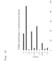
- a correlation of peritumoral BMMF levels and intensity of CD68-positive macrophages should be observable.
- Example 3 Detection of BMMF Rep antigens by immunoblotting of tissue lysates of CRC patients
- Peritumoral or tumoral tissue material of the same patient e.g. patient 4800 P (peritumoral) or 4800 T (tumoral)
- tissue material form early polyps of four individual donors was lysed in 8 M urea, 100 mM NaH2PO4, 10 mM Tris, 5 mM DTT, pH 8.0 with the use of protease inhibitors (P8340 Sigma-Aldrich) in a tissue homogenizer at 4 °C with silica beads before SDS-PAGE and immunodetection with anti-BMMF Rep antibodies, in particular anti-Rep Ab 5-2 (1:250 dilution).
- Target bands at around 42 kDa were clearly visible with strongest intensities for the peritumoral CRC patient sample 4800 and even stronger bands for tissue lysates of early polyps of the four individual patients ( Figure 16 ).
- the suspension was then centrifuged for 5 min at 12.000 g at room temperature to pellet the Chelex resin. 1 ⁇ l of the supernatant was used for rolling circle amplification (RCA) with phi29 DNA Polymerase (New England Biolabs M0269S, 10 U) and the use of Exo-Resistant Random primers (Thermo Fisher scientific SO181, 25 ⁇ M), dNTPs (0,75 mM each), 50 mM Tris-HCl, 10 mM MgCl2, 10 mM (NH4)2 S04, 4 mM DTT, 0,4 mg/ml BSA, pH 7.5.
- RCA rolling circle amplification
- phi29 DNA Polymerase New England Biolabs M0269S, 10 U
- Exo-Resistant Random primers Thermo Fisher scientific SO181, 25 ⁇ M
- dNTPs (0,75 mM each)
- 50 mM Tris-HCl 10 mM MgCl2, 10
- PCR targets were subjected to an additional cycle of PCR amplification after gel extraction of the PCR targets, whenever the target bands showed inadequate band intensity.
- PCR around 5 ng of DNA was used for cloning into a pCR2.1 TA cloning vector (TA-cloning kit, Invitrogen K203040) according to the manufacturer's protocol followed by sequencing of the PCR insert.
- SEQ ID NO SEQUENCE 1 Amino acid sequence of Rep protein encoded by MSBI1.176 2 Amino acid sequence of Rep peptide fragment EARETGKGINANDPLTVH 3 Amino acid sequence of Rep peptide fragment KQINEHTDITASYEQHKKGRT 4 His-Tag (with two neutral stuffer amino acids) GAHHHHHH 5 T7-Tag MASMTGGQQMG 6 FLAG-Tag DYKDDDDK 7 Strep-II-Tag WSHPQFEK 8 Amino acid sequence of Rep protein encoded by MSBI2.176 9 MSBI.1 specific epitope NRLSDRF 10 Amino acid sequence of Rep protein encoded by CMI1.252 11 Amino acid sequence of Rep protein encoded by CMI2.214 12 Amino acid sequence of Rep protein encoded by CMI3.168 13 DNA sequence MSBI1 Rep codon-optimized 14 Protein sequence MSBI1 Rep codon-optimized 15 DNA sequence MSBI1 Rep wild-type 16 Primer gag gac gaa tta ata tta
Landscapes
- Health & Medical Sciences (AREA)
- Life Sciences & Earth Sciences (AREA)
- Immunology (AREA)
- Engineering & Computer Science (AREA)
- Chemical & Material Sciences (AREA)
- Molecular Biology (AREA)
- Biomedical Technology (AREA)
- Hematology (AREA)
- Urology & Nephrology (AREA)
- Biotechnology (AREA)
- Biochemistry (AREA)
- Cell Biology (AREA)
- Food Science & Technology (AREA)
- Medicinal Chemistry (AREA)
- Physics & Mathematics (AREA)
- Analytical Chemistry (AREA)
- Microbiology (AREA)
- General Health & Medical Sciences (AREA)
- General Physics & Mathematics (AREA)
- Pathology (AREA)
- Chemical Kinetics & Catalysis (AREA)
- Peptides Or Proteins (AREA)
- Measuring Or Testing Involving Enzymes Or Micro-Organisms (AREA)
Claims (7)
- In-vitro Verwendung des Rep-Proteins der Rindfleisch- und Milchfaktorgruppe 1 (BMMF1) als ein Biomarker für kolorektalen Krebs.
- Verwendung nach Anspruch 1, wobei das Rep-Protein ein MSBI1-genomkodiertes Rep-Protein (MSBI1 Rep), ein MSBI2-genomkodiertes Rep-Protein (MSBI2 Rep), ein CMI1-genomkodiertes Rep-Protein (CMI1 Rep), ein CMI2-genomkodiertes Rep-Protein (CMI2 Rep) oder ein CMI3-genomkodiertes Rep-Protein (CMI3 Rep) ist.
- In-vitro Verfahren zur Bereitstellung einer Diagnose oder Veranlagung für kolorektalen krebs (CRC) bei einem Patienten, umfassend den Schritt:
Nachweisen des Rep-Proteins in einer Probe eines Patienten mittels Anti-Rep-Antikörpern, die an ein Epitop, das von SEQ ID NO: 2 oder SEQ ID NO: 3 umfasst ist, binden. - Verfahren nach Anspruch 3, wobei der für das Rep-Protein spezifische Antikörper an ein Epitop, das sich in einer Aminosäuresequenz befindet, die aus der Gruppe bestehend aus den Aminosäuren 1 bis 136, 137 bis 229 und 230 bis 324 der SEQ ID NO: 1 ausgewählt ist, bindet.
- Verfahren nach Anspruch 3 oder 4, wobei die Probe aus einem Patienten aus der Gruppe bestehend aus krebsartigem Darmgewebe, peripherem Gewebe, das das krebsartige Darmgewebe umgibt, (gutartigen) Darmpolypen ausgewählt ist.
- Verfahren nach einem der Ansprüche 3 bis 5, wobei in der Probe zusätzlich CD68-positive Zellen mittels eines Anti-CD68-Antikörpers nachgewiesen werden.
- In-vitro Verfahren zur Bereitstellung eines prognostischen Faktors für die Überlebensdauer eines CRC-Patienten, umfassend die Schritte:Nachweisen des Rep-Proteins in einer Probe aus peritumoralen Regionen der CRC-Patienten mittels Anti-Rep-Antikörpern, die an ein Epitop, das von SEQ ID NO: 2 und SEQ ID NO: 3 umfasst ist, binden,Bestimmen des Prozentsatzes angefärbter Zellen und der Intensität für jeden Antikörper, was einen immunhistologischen Faktor, der als ein allgemeiner Indikator für die spezifische Überlebensdauer der CRC-Patienten dient, aufstellt.
Applications Claiming Priority (2)
| Application Number | Priority Date | Filing Date | Title |
|---|---|---|---|
| EP18154190.5A EP3517960A1 (de) | 2018-01-30 | 2018-01-30 | Verwendung von bmmf rep protein als biomarker für kolonkarzinom |
| PCT/EP2019/051868 WO2019149633A1 (en) | 2018-01-30 | 2019-01-25 | Use of bmmf1 rep protein as biomarker for colorectal cancer |
Publications (3)
| Publication Number | Publication Date |
|---|---|
| EP3746791A1 EP3746791A1 (de) | 2020-12-09 |
| EP3746791B1 true EP3746791B1 (de) | 2024-03-06 |
| EP3746791C0 EP3746791C0 (de) | 2024-03-06 |
Family
ID=61198670
Family Applications (2)
| Application Number | Title | Priority Date | Filing Date |
|---|---|---|---|
| EP18154190.5A Ceased EP3517960A1 (de) | 2018-01-30 | 2018-01-30 | Verwendung von bmmf rep protein als biomarker für kolonkarzinom |
| EP19705289.7A Active EP3746791B1 (de) | 2018-01-30 | 2019-01-25 | Verwendung von bmmf rep protein als biomarker für kolonkarzinom |
Family Applications Before (1)
| Application Number | Title | Priority Date | Filing Date |
|---|---|---|---|
| EP18154190.5A Ceased EP3517960A1 (de) | 2018-01-30 | 2018-01-30 | Verwendung von bmmf rep protein als biomarker für kolonkarzinom |
Country Status (7)
| Country | Link |
|---|---|
| US (1) | US12282020B2 (de) |
| EP (2) | EP3517960A1 (de) |
| JP (1) | JP6987278B2 (de) |
| AU (1) | AU2019214823B2 (de) |
| CA (1) | CA3088235C (de) |
| ES (1) | ES2976963T3 (de) |
| WO (1) | WO2019149633A1 (de) |
Families Citing this family (5)
| Publication number | Priority date | Publication date | Assignee | Title |
|---|---|---|---|---|
| EP3517960A1 (de) * | 2018-01-30 | 2019-07-31 | Deutsches Krebsforschungszentrum | Verwendung von bmmf rep protein als biomarker für kolonkarzinom |
| EP3699595A1 (de) * | 2019-02-22 | 2020-08-26 | Deutsches Krebsforschungszentrum, Stiftung des öffentlichen Rechts | Verwendung von bmmf1-rep-protein als ein biomarker für brustkrebs |
| EP3699594A1 (de) * | 2019-02-22 | 2020-08-26 | Deutsches Krebsforschungszentrum, Stiftung des öffentlichen Rechts | Verwendung von bmmf1-rep-protein als ein biomarker für prostatakrebs |
| CA3252723A1 (en) * | 2022-05-11 | 2023-11-16 | Deutsches Krebsforschungszentrum Stiftung Des Offentlichen Rechts | USE OF A REP PROTEIN BMMF1 AS A DIAGNOSTIC MARKER FOR LUNG CANCER |
| AU2023269517A1 (en) * | 2022-05-11 | 2024-12-19 | Deutsches Krebsforschungszentrum Stiftung des öffentlichen Rechts | Use of bmmf1 rep protein as diagnostic marker for pancreatic cancer and diabetes mellitus type 2 |
Family Cites Families (5)
| Publication number | Priority date | Publication date | Assignee | Title |
|---|---|---|---|---|
| EP2868751A1 (de) | 2013-10-30 | 2015-05-06 | Deutsches Krebsforschungszentrum | HCBI-Sequenzen als früher Marker für die zukünftige Entwicklung von Krebs und Erkrankungen des zentralen Nervensystems, und als Ziel zur Behandlung und Prävention von Krebs |
| EP2966176A1 (de) * | 2014-07-10 | 2016-01-13 | Deutsches Krebsforschungszentrum | HCBIO-, MSBI-, MSSI- und CMI-Sequenzen als frühe Marker für die zukünftige Entwicklung von Krebs und Erkrankungen des ZNS und als Ziel zur Behandlung und Prävention dieser Erkrankungen |
| DE102014213512A1 (de) | 2014-07-11 | 2016-01-14 | Voith Patent Gmbh | Maschine zur Herstellung einer Faserstoffbahn |
| EP3306316A1 (de) * | 2016-10-10 | 2018-04-11 | Deutsches Krebsforschungszentrum Stiftung des Öffentlichen Rechts | Rep-protein als proteinantigen zur verwendung in diagnostischen tests |
| EP3517960A1 (de) * | 2018-01-30 | 2019-07-31 | Deutsches Krebsforschungszentrum | Verwendung von bmmf rep protein als biomarker für kolonkarzinom |
-
2018
- 2018-01-30 EP EP18154190.5A patent/EP3517960A1/de not_active Ceased
-
2019
- 2019-01-25 CA CA3088235A patent/CA3088235C/en active Active
- 2019-01-25 ES ES19705289T patent/ES2976963T3/es active Active
- 2019-01-25 WO PCT/EP2019/051868 patent/WO2019149633A1/en not_active Ceased
- 2019-01-25 JP JP2020562839A patent/JP6987278B2/ja active Active
- 2019-01-25 AU AU2019214823A patent/AU2019214823B2/en active Active
- 2019-01-25 EP EP19705289.7A patent/EP3746791B1/de active Active
-
2020
- 2020-07-27 US US16/939,154 patent/US12282020B2/en active Active
Non-Patent Citations (2)
| Title |
|---|
| KURT DE SWAAF: "Das gefährliche Erbe der Auerochsen", 4 May 2019 (2019-05-04), Retrieved from the Internet <URL:https://www.nzz.ch/wissenschaft/bovine-milk-and-meat-factors-eine neue-art-von-krankheitserreger-ld.1478322?reduced=true> [retrieved on 20210122] * |
| THEODOR DINGERMANN: "Viren, Bakterien und Parasieten - Krebs ist auch eine Infektionskrankheit", 26 April 2020 (2020-04-26), Retrieved from the Internet <URL:https://www.pharmazeutische-zeitung.de/krebs-ist-auch-eine-infektionskrankheit-1170110/> [retrieved on 20201020] * |
Also Published As
| Publication number | Publication date |
|---|---|
| EP3746791A1 (de) | 2020-12-09 |
| CA3088235A1 (en) | 2019-08-08 |
| JP2021513658A (ja) | 2021-05-27 |
| ES2976963T3 (es) | 2024-08-13 |
| US20210003575A1 (en) | 2021-01-07 |
| EP3517960A1 (de) | 2019-07-31 |
| JP6987278B2 (ja) | 2021-12-22 |
| EP3746791C0 (de) | 2024-03-06 |
| WO2019149633A1 (en) | 2019-08-08 |
| AU2019214823B2 (en) | 2022-03-17 |
| US12282020B2 (en) | 2025-04-22 |
| CA3088235C (en) | 2022-09-06 |
| AU2019214823A1 (en) | 2020-07-30 |
Similar Documents
| Publication | Publication Date | Title |
|---|---|---|
| US12282020B2 (en) | Use of BMMFI Rep protein as biomarker for colorectal cancer | |
| US20250271436A1 (en) | Use of bmmf1 rep protein as a biomarker for prostate cancer | |
| JP7544930B2 (ja) | 乳がんのバイオマーカとしてのbmmf1 repタンパク質の使用 | |
| HK40042803A (en) | Use of bmmf1 rep protein as biomarker for colorectal cancer | |
| HK40042803B (en) | Use of bmmf1 rep protein as biomarker for colorectal cancer | |
| AU2026200113A1 (en) | Use of BMMF1 Rep Protein as a Biomarker for Breast Cancer | |
| EP4522991B1 (de) | Verwendung von bmmf1 rep-protein als diagnostischer marker für lungenkrebs | |
| US20250067744A1 (en) | Use of bmmf1 rep protein as diagnostic marker for pancreatic cancer and diabetes mellitus type 2 |
Legal Events
| Date | Code | Title | Description |
|---|---|---|---|
| STAA | Information on the status of an ep patent application or granted ep patent |
Free format text: STATUS: UNKNOWN |
|
| STAA | Information on the status of an ep patent application or granted ep patent |
Free format text: STATUS: THE INTERNATIONAL PUBLICATION HAS BEEN MADE |
|
| PUAI | Public reference made under article 153(3) epc to a published international application that has entered the european phase |
Free format text: ORIGINAL CODE: 0009012 |
|
| STAA | Information on the status of an ep patent application or granted ep patent |
Free format text: STATUS: REQUEST FOR EXAMINATION WAS MADE |
|
| 17P | Request for examination filed |
Effective date: 20200826 |
|
| AK | Designated contracting states |
Kind code of ref document: A1 Designated state(s): AL AT BE BG CH CY CZ DE DK EE ES FI FR GB GR HR HU IE IS IT LI LT LU LV MC MK MT NL NO PL PT RO RS SE SI SK SM TR |
|
| AX | Request for extension of the european patent |
Extension state: BA ME |
|
| DAV | Request for validation of the european patent (deleted) | ||
| DAX | Request for extension of the european patent (deleted) | ||
| REG | Reference to a national code |
Ref country code: HK Ref legal event code: DE Ref document number: 40042803 Country of ref document: HK |
|
| GRAP | Despatch of communication of intention to grant a patent |
Free format text: ORIGINAL CODE: EPIDOSNIGR1 |
|
| STAA | Information on the status of an ep patent application or granted ep patent |
Free format text: STATUS: GRANT OF PATENT IS INTENDED |
|
| INTG | Intention to grant announced |
Effective date: 20230331 |
|
| RAP3 | Party data changed (applicant data changed or rights of an application transferred) |
Owner name: DEUTSCHES KREBSFORSCHUNGSZENTRUM STIFTUNG DES OEFFENTLICHEN RECHTS |
|
| GRAJ | Information related to disapproval of communication of intention to grant by the applicant or resumption of examination proceedings by the epo deleted |
Free format text: ORIGINAL CODE: EPIDOSDIGR1 |
|
| STAA | Information on the status of an ep patent application or granted ep patent |
Free format text: STATUS: REQUEST FOR EXAMINATION WAS MADE |
|
| GRAP | Despatch of communication of intention to grant a patent |
Free format text: ORIGINAL CODE: EPIDOSNIGR1 |
|
| STAA | Information on the status of an ep patent application or granted ep patent |
Free format text: STATUS: GRANT OF PATENT IS INTENDED |
|
| INTC | Intention to grant announced (deleted) | ||
| INTG | Intention to grant announced |
Effective date: 20230824 |
|
| GRAS | Grant fee paid |
Free format text: ORIGINAL CODE: EPIDOSNIGR3 |
|
| GRAA | (expected) grant |
Free format text: ORIGINAL CODE: 0009210 |
|
| STAA | Information on the status of an ep patent application or granted ep patent |
Free format text: STATUS: THE PATENT HAS BEEN GRANTED |
|
| AK | Designated contracting states |
Kind code of ref document: B1 Designated state(s): AL AT BE BG CH CY CZ DE DK EE ES FI FR GB GR HR HU IE IS IT LI LT LU LV MC MK MT NL NO PL PT RO RS SE SI SK SM TR |
|
| REG | Reference to a national code |
Ref country code: CH Ref legal event code: EP |
|
| REG | Reference to a national code |
Ref country code: DE Ref legal event code: R096 Ref document number: 602019047715 Country of ref document: DE |
|
| REG | Reference to a national code |
Ref country code: IE Ref legal event code: FG4D |
|
| U01 | Request for unitary effect filed |
Effective date: 20240403 |
|
| U07 | Unitary effect registered |
Designated state(s): AT BE BG DE DK EE FI FR IT LT LU LV MT NL PT SE SI Effective date: 20240411 |
|
| PG25 | Lapsed in a contracting state [announced via postgrant information from national office to epo] |
Ref country code: GR Free format text: LAPSE BECAUSE OF FAILURE TO SUBMIT A TRANSLATION OF THE DESCRIPTION OR TO PAY THE FEE WITHIN THE PRESCRIBED TIME-LIMIT Effective date: 20240607 |
|
| PG25 | Lapsed in a contracting state [announced via postgrant information from national office to epo] |
Ref country code: HR Free format text: LAPSE BECAUSE OF FAILURE TO SUBMIT A TRANSLATION OF THE DESCRIPTION OR TO PAY THE FEE WITHIN THE PRESCRIBED TIME-LIMIT Effective date: 20240306 Ref country code: RS Free format text: LAPSE BECAUSE OF FAILURE TO SUBMIT A TRANSLATION OF THE DESCRIPTION OR TO PAY THE FEE WITHIN THE PRESCRIBED TIME-LIMIT Effective date: 20240606 |
|
| PG25 | Lapsed in a contracting state [announced via postgrant information from national office to epo] |
Ref country code: RS Free format text: LAPSE BECAUSE OF FAILURE TO SUBMIT A TRANSLATION OF THE DESCRIPTION OR TO PAY THE FEE WITHIN THE PRESCRIBED TIME-LIMIT Effective date: 20240606 Ref country code: NO Free format text: LAPSE BECAUSE OF FAILURE TO SUBMIT A TRANSLATION OF THE DESCRIPTION OR TO PAY THE FEE WITHIN THE PRESCRIBED TIME-LIMIT Effective date: 20240606 Ref country code: HR Free format text: LAPSE BECAUSE OF FAILURE TO SUBMIT A TRANSLATION OF THE DESCRIPTION OR TO PAY THE FEE WITHIN THE PRESCRIBED TIME-LIMIT Effective date: 20240306 Ref country code: GR Free format text: LAPSE BECAUSE OF FAILURE TO SUBMIT A TRANSLATION OF THE DESCRIPTION OR TO PAY THE FEE WITHIN THE PRESCRIBED TIME-LIMIT Effective date: 20240607 |
|
| REG | Reference to a national code |
Ref country code: ES Ref legal event code: FG2A Ref document number: 2976963 Country of ref document: ES Kind code of ref document: T3 Effective date: 20240813 |
|
| PG25 | Lapsed in a contracting state [announced via postgrant information from national office to epo] |
Ref country code: IS Free format text: LAPSE BECAUSE OF FAILURE TO SUBMIT A TRANSLATION OF THE DESCRIPTION OR TO PAY THE FEE WITHIN THE PRESCRIBED TIME-LIMIT Effective date: 20240706 |
|
| PG25 | Lapsed in a contracting state [announced via postgrant information from national office to epo] |
Ref country code: SM Free format text: LAPSE BECAUSE OF FAILURE TO SUBMIT A TRANSLATION OF THE DESCRIPTION OR TO PAY THE FEE WITHIN THE PRESCRIBED TIME-LIMIT Effective date: 20240306 |
|
| PG25 | Lapsed in a contracting state [announced via postgrant information from national office to epo] |
Ref country code: CZ Free format text: LAPSE BECAUSE OF FAILURE TO SUBMIT A TRANSLATION OF THE DESCRIPTION OR TO PAY THE FEE WITHIN THE PRESCRIBED TIME-LIMIT Effective date: 20240306 |
|
| PG25 | Lapsed in a contracting state [announced via postgrant information from national office to epo] |
Ref country code: PL Free format text: LAPSE BECAUSE OF FAILURE TO SUBMIT A TRANSLATION OF THE DESCRIPTION OR TO PAY THE FEE WITHIN THE PRESCRIBED TIME-LIMIT Effective date: 20240306 |
|
| PG25 | Lapsed in a contracting state [announced via postgrant information from national office to epo] |
Ref country code: SK Free format text: LAPSE BECAUSE OF FAILURE TO SUBMIT A TRANSLATION OF THE DESCRIPTION OR TO PAY THE FEE WITHIN THE PRESCRIBED TIME-LIMIT Effective date: 20240306 |
|
| PG25 | Lapsed in a contracting state [announced via postgrant information from national office to epo] |
Ref country code: SM Free format text: LAPSE BECAUSE OF FAILURE TO SUBMIT A TRANSLATION OF THE DESCRIPTION OR TO PAY THE FEE WITHIN THE PRESCRIBED TIME-LIMIT Effective date: 20240306 Ref country code: SK Free format text: LAPSE BECAUSE OF FAILURE TO SUBMIT A TRANSLATION OF THE DESCRIPTION OR TO PAY THE FEE WITHIN THE PRESCRIBED TIME-LIMIT Effective date: 20240306 Ref country code: RO Free format text: LAPSE BECAUSE OF FAILURE TO SUBMIT A TRANSLATION OF THE DESCRIPTION OR TO PAY THE FEE WITHIN THE PRESCRIBED TIME-LIMIT Effective date: 20240306 Ref country code: PL Free format text: LAPSE BECAUSE OF FAILURE TO SUBMIT A TRANSLATION OF THE DESCRIPTION OR TO PAY THE FEE WITHIN THE PRESCRIBED TIME-LIMIT Effective date: 20240306 Ref country code: IS Free format text: LAPSE BECAUSE OF FAILURE TO SUBMIT A TRANSLATION OF THE DESCRIPTION OR TO PAY THE FEE WITHIN THE PRESCRIBED TIME-LIMIT Effective date: 20240706 Ref country code: CZ Free format text: LAPSE BECAUSE OF FAILURE TO SUBMIT A TRANSLATION OF THE DESCRIPTION OR TO PAY THE FEE WITHIN THE PRESCRIBED TIME-LIMIT Effective date: 20240306 |
|
| REG | Reference to a national code |
Ref country code: DE Ref legal event code: R097 Ref document number: 602019047715 Country of ref document: DE |
|
| PLBE | No opposition filed within time limit |
Free format text: ORIGINAL CODE: 0009261 |
|
| STAA | Information on the status of an ep patent application or granted ep patent |
Free format text: STATUS: NO OPPOSITION FILED WITHIN TIME LIMIT |
|
| 26N | No opposition filed |
Effective date: 20241209 |
|
| U20 | Renewal fee for the european patent with unitary effect paid |
Year of fee payment: 7 Effective date: 20250131 |
|
| PG25 | Lapsed in a contracting state [announced via postgrant information from national office to epo] |
Ref country code: MC Free format text: LAPSE BECAUSE OF FAILURE TO SUBMIT A TRANSLATION OF THE DESCRIPTION OR TO PAY THE FEE WITHIN THE PRESCRIBED TIME-LIMIT Effective date: 20240306 |
|
| REG | Reference to a national code |
Ref country code: CH Ref legal event code: U11 Free format text: ST27 STATUS EVENT CODE: U-0-0-U10-U11 (AS PROVIDED BY THE NATIONAL OFFICE) Effective date: 20260201 |
|
| U20 | Renewal fee for the european patent with unitary effect paid |
Year of fee payment: 8 Effective date: 20260129 |
|
| PGFP | Annual fee paid to national office [announced via postgrant information from national office to epo] |
Ref country code: GB Payment date: 20260130 Year of fee payment: 8 |
|
| PGFP | Annual fee paid to national office [announced via postgrant information from national office to epo] |
Ref country code: ES Payment date: 20260210 Year of fee payment: 8 |
|
| PGFP | Annual fee paid to national office [announced via postgrant information from national office to epo] |
Ref country code: IE Payment date: 20260129 Year of fee payment: 8 |
|
| PGFP | Annual fee paid to national office [announced via postgrant information from national office to epo] |
Ref country code: CH Payment date: 20260201 Year of fee payment: 8 |