EP3740499B1 - Anti-tumor therapeutic agents based on b7h receptor ligands - Google Patents
Anti-tumor therapeutic agents based on b7h receptor ligands Download PDFInfo
- Publication number
- EP3740499B1 EP3740499B1 EP19704678.2A EP19704678A EP3740499B1 EP 3740499 B1 EP3740499 B1 EP 3740499B1 EP 19704678 A EP19704678 A EP 19704678A EP 3740499 B1 EP3740499 B1 EP 3740499B1
- Authority
- EP
- European Patent Office
- Prior art keywords
- receptor
- icos
- ligand
- nano
- micro
- Prior art date
- Legal status (The legal status is an assumption and is not a legal conclusion. Google has not performed a legal analysis and makes no representation as to the accuracy of the status listed.)
- Active
Links
Images



Classifications
-
- C—CHEMISTRY; METALLURGY
- C07—ORGANIC CHEMISTRY
- C07K—PEPTIDES
- C07K14/00—Peptides having more than 20 amino acids; Gastrins; Somatostatins; Melanotropins; Derivatives thereof
- C07K14/435—Peptides having more than 20 amino acids; Gastrins; Somatostatins; Melanotropins; Derivatives thereof from animals; from humans
- C07K14/705—Receptors; Cell surface antigens; Cell surface determinants
- C07K14/70503—Immunoglobulin superfamily
- C07K14/70521—CD28, CD152
-
- A—HUMAN NECESSITIES
- A61—MEDICAL OR VETERINARY SCIENCE; HYGIENE
- A61K—PREPARATIONS FOR MEDICAL, DENTAL OR TOILETRY PURPOSES
- A61K38/00—Medicinal preparations containing peptides
- A61K38/16—Peptides having more than 20 amino acids; Gastrins; Somatostatins; Melanotropins; Derivatives thereof
- A61K38/17—Peptides having more than 20 amino acids; Gastrins; Somatostatins; Melanotropins; Derivatives thereof from animals; from humans
- A61K38/177—Receptors; Cell surface antigens; Cell surface determinants
- A61K38/1774—Immunoglobulin superfamily (e.g. CD2, CD4, CD8, ICAM molecules, B7 molecules, Fc-receptors, MHC-molecules)
-
- A—HUMAN NECESSITIES
- A61—MEDICAL OR VETERINARY SCIENCE; HYGIENE
- A61K—PREPARATIONS FOR MEDICAL, DENTAL OR TOILETRY PURPOSES
- A61K47/00—Medicinal preparations characterised by the non-active ingredients used, e.g. carriers or inert additives; Targeting or modifying agents chemically bound to the active ingredient
- A61K47/50—Medicinal preparations characterised by the non-active ingredients used, e.g. carriers or inert additives; Targeting or modifying agents chemically bound to the active ingredient the non-active ingredient being chemically bound to the active ingredient, e.g. polymer-drug conjugates
- A61K47/69—Medicinal preparations characterised by the non-active ingredients used, e.g. carriers or inert additives; Targeting or modifying agents chemically bound to the active ingredient the non-active ingredient being chemically bound to the active ingredient, e.g. polymer-drug conjugates the conjugate being characterised by physical or galenical forms, e.g. emulsion, particle, inclusion complex, stent or kit
- A61K47/6921—Medicinal preparations characterised by the non-active ingredients used, e.g. carriers or inert additives; Targeting or modifying agents chemically bound to the active ingredient the non-active ingredient being chemically bound to the active ingredient, e.g. polymer-drug conjugates the conjugate being characterised by physical or galenical forms, e.g. emulsion, particle, inclusion complex, stent or kit the form being a particulate, a powder, an adsorbate, a bead or a sphere
- A61K47/6927—Medicinal preparations characterised by the non-active ingredients used, e.g. carriers or inert additives; Targeting or modifying agents chemically bound to the active ingredient the non-active ingredient being chemically bound to the active ingredient, e.g. polymer-drug conjugates the conjugate being characterised by physical or galenical forms, e.g. emulsion, particle, inclusion complex, stent or kit the form being a particulate, a powder, an adsorbate, a bead or a sphere the form being a solid microparticle having no hollow or gas-filled cores
- A61K47/6929—Medicinal preparations characterised by the non-active ingredients used, e.g. carriers or inert additives; Targeting or modifying agents chemically bound to the active ingredient the non-active ingredient being chemically bound to the active ingredient, e.g. polymer-drug conjugates the conjugate being characterised by physical or galenical forms, e.g. emulsion, particle, inclusion complex, stent or kit the form being a particulate, a powder, an adsorbate, a bead or a sphere the form being a solid microparticle having no hollow or gas-filled cores the form being a nanoparticle, e.g. an immuno-nanoparticle
- A61K47/6931—Medicinal preparations characterised by the non-active ingredients used, e.g. carriers or inert additives; Targeting or modifying agents chemically bound to the active ingredient the non-active ingredient being chemically bound to the active ingredient, e.g. polymer-drug conjugates the conjugate being characterised by physical or galenical forms, e.g. emulsion, particle, inclusion complex, stent or kit the form being a particulate, a powder, an adsorbate, a bead or a sphere the form being a solid microparticle having no hollow or gas-filled cores the form being a nanoparticle, e.g. an immuno-nanoparticle the material constituting the nanoparticle being a polymer
- A61K47/6939—Medicinal preparations characterised by the non-active ingredients used, e.g. carriers or inert additives; Targeting or modifying agents chemically bound to the active ingredient the non-active ingredient being chemically bound to the active ingredient, e.g. polymer-drug conjugates the conjugate being characterised by physical or galenical forms, e.g. emulsion, particle, inclusion complex, stent or kit the form being a particulate, a powder, an adsorbate, a bead or a sphere the form being a solid microparticle having no hollow or gas-filled cores the form being a nanoparticle, e.g. an immuno-nanoparticle the material constituting the nanoparticle being a polymer the polymer being a polysaccharide, e.g. starch, chitosan, chitin, cellulose or pectin
-
- A—HUMAN NECESSITIES
- A61—MEDICAL OR VETERINARY SCIENCE; HYGIENE
- A61K—PREPARATIONS FOR MEDICAL, DENTAL OR TOILETRY PURPOSES
- A61K47/00—Medicinal preparations characterised by the non-active ingredients used, e.g. carriers or inert additives; Targeting or modifying agents chemically bound to the active ingredient
- A61K47/50—Medicinal preparations characterised by the non-active ingredients used, e.g. carriers or inert additives; Targeting or modifying agents chemically bound to the active ingredient the non-active ingredient being chemically bound to the active ingredient, e.g. polymer-drug conjugates
- A61K47/69—Medicinal preparations characterised by the non-active ingredients used, e.g. carriers or inert additives; Targeting or modifying agents chemically bound to the active ingredient the non-active ingredient being chemically bound to the active ingredient, e.g. polymer-drug conjugates the conjugate being characterised by physical or galenical forms, e.g. emulsion, particle, inclusion complex, stent or kit
- A61K47/6949—Medicinal preparations characterised by the non-active ingredients used, e.g. carriers or inert additives; Targeting or modifying agents chemically bound to the active ingredient the non-active ingredient being chemically bound to the active ingredient, e.g. polymer-drug conjugates the conjugate being characterised by physical or galenical forms, e.g. emulsion, particle, inclusion complex, stent or kit inclusion complexes, e.g. clathrates, cavitates or fullerenes
- A61K47/6951—Medicinal preparations characterised by the non-active ingredients used, e.g. carriers or inert additives; Targeting or modifying agents chemically bound to the active ingredient the non-active ingredient being chemically bound to the active ingredient, e.g. polymer-drug conjugates the conjugate being characterised by physical or galenical forms, e.g. emulsion, particle, inclusion complex, stent or kit inclusion complexes, e.g. clathrates, cavitates or fullerenes using cyclodextrin
-
- A—HUMAN NECESSITIES
- A61—MEDICAL OR VETERINARY SCIENCE; HYGIENE
- A61K—PREPARATIONS FOR MEDICAL, DENTAL OR TOILETRY PURPOSES
- A61K9/00—Medicinal preparations characterised by special physical form
- A61K9/0012—Galenical forms characterised by the site of application
- A61K9/0019—Injectable compositions; Intramuscular, intravenous, arterial, subcutaneous administration; Compositions to be administered through the skin in an invasive manner
-
- A—HUMAN NECESSITIES
- A61—MEDICAL OR VETERINARY SCIENCE; HYGIENE
- A61K—PREPARATIONS FOR MEDICAL, DENTAL OR TOILETRY PURPOSES
- A61K9/00—Medicinal preparations characterised by special physical form
- A61K9/48—Preparations in capsules, e.g. of gelatin, of chocolate
- A61K9/50—Microcapsules having a gas, liquid or semi-solid filling; Solid microparticles or pellets surrounded by a distinct coating layer, e.g. coated microspheres, coated drug crystals
- A61K9/51—Nanocapsules; Nanoparticles
- A61K9/5107—Excipients; Inactive ingredients
- A61K9/513—Organic macromolecular compounds; Dendrimers
- A61K9/5146—Organic macromolecular compounds; Dendrimers obtained otherwise than by reactions only involving carbon-to-carbon unsaturated bonds, e.g. polyethylene glycol, polyamines, polyanhydrides
- A61K9/5153—Polyesters, e.g. poly(lactide-co-glycolide)
-
- A—HUMAN NECESSITIES
- A61—MEDICAL OR VETERINARY SCIENCE; HYGIENE
- A61K—PREPARATIONS FOR MEDICAL, DENTAL OR TOILETRY PURPOSES
- A61K9/00—Medicinal preparations characterised by special physical form
- A61K9/48—Preparations in capsules, e.g. of gelatin, of chocolate
- A61K9/50—Microcapsules having a gas, liquid or semi-solid filling; Solid microparticles or pellets surrounded by a distinct coating layer, e.g. coated microspheres, coated drug crystals
- A61K9/51—Nanocapsules; Nanoparticles
- A61K9/5107—Excipients; Inactive ingredients
- A61K9/513—Organic macromolecular compounds; Dendrimers
- A61K9/5161—Polysaccharides, e.g. alginate, chitosan, cellulose derivatives; Cyclodextrin
-
- C—CHEMISTRY; METALLURGY
- C07—ORGANIC CHEMISTRY
- C07K—PEPTIDES
- C07K14/00—Peptides having more than 20 amino acids; Gastrins; Somatostatins; Melanotropins; Derivatives thereof
- C07K14/435—Peptides having more than 20 amino acids; Gastrins; Somatostatins; Melanotropins; Derivatives thereof from animals; from humans
- C07K14/705—Receptors; Cell surface antigens; Cell surface determinants
- C07K14/70503—Immunoglobulin superfamily
- C07K14/70532—B7 molecules, e.g. CD80, CD86
-
- C—CHEMISTRY; METALLURGY
- C07—ORGANIC CHEMISTRY
- C07K—PEPTIDES
- C07K2319/00—Fusion polypeptide
- C07K2319/30—Non-immunoglobulin-derived peptide or protein having an immunoglobulin constant or Fc region, or a fragment thereof, attached thereto
-
- G—PHYSICS
- G01—MEASURING; TESTING
- G01N—INVESTIGATING OR ANALYSING MATERIALS BY DETERMINING THEIR CHEMICAL OR PHYSICAL PROPERTIES
- G01N2500/00—Screening for compounds of potential therapeutic value
- G01N2500/04—Screening involving studying the effect of compounds C directly on molecule A (e.g. C are potential ligands for a receptor A, or potential substrates for an enzyme A)
Definitions
- the disclosure concerns novel anti-tumor therapeutic agents comprising at least one ligand of the receptor B7h.
- ICOS is a T cell costimulatory molecule described by the present inventors as H4 1,2 . Later on, Hutloff cloned ICOS as a molecule belonging to the CD28 family and it was shown that ICOS and H4 are the same molecule 3,4 .
- a distinctive feature of ICOS is its selective expression in activated T cells, but it has been recently detected also in dendritic cells (DC) 5 .
- the ICOS receptor is ICOSL (or B7h), expressed by several cell types, such as DC, macrophages, B cells, endothelial cells (EC), epithelial cells, and fibroblasts 6 .
- the ICOS/B7h interaction regulates T cell activation in lymphoid organs and controls T cell function at inflammation sites. It supports differentiation of Treg, TH17, and follicular T helper cells and development of germinal centers and its deficiency causes common variable immunodeficiency 7-11 .
- B7h Inducible T-Cell Costimulator Ligand (ICOSL or ICOS-L), B7-Related Protein 1 (B7RP-1 or B7RP1) B7 Homolog 2 (B7-H2), B7-Like Protein Gl50, GL50, KIAA0653, LICOS, CD275.
- B7h:ICOS interaction triggers bidirectional signals modulating the response of the B7h-expressing cells. In mouse DC, this B7h-mediated "reverse signalling" increases IL-6 secretion 12 .
- ICOS-Fc bivalent soluble form of ICOS composed by the Fc portion of IgG1 and two molecules of the extracellular portions of ICOS. Since ICOS-Fc is bivalent, it crosslinks B7h and exerts an agonist effect triggering B7h signaling in the B7h-expressing cells. In human cells, the following effects of B7h triggering by ICOS-Fc were shown. i) In EC and tumor cell lines, it inhibits adhesiveness and migration in vitro 13,14 . ii) In tumor cell lines, it inhibits development of experimental lung metastases in vivo 14 .
- iii) In DC, it modulates cytokine secretion by increasing secretion of IL-23 (supporting TH17 differentiation involved in the anti-tumor response), promotes antigen cross-presentation, and inhibits adhesiveness and migration in vitro 15 .
- B7h triggering induces dephosphorylation of ERK and p38 in EC, dephosphorylation of FAK in tumor cells, and down-modulation of ⁇ -Pix in DC and tumor cells 13-
- WO 2016/189428 discloses the use of ICOS in the treatment of osteopenia and osteoporosis.
- the object of this disclosure is to provide novel anti-tumor therapeutic agents comprising at least one ligand of the receptor B7h, wherein the ligand of receptor B7h is able to bind specifically to receptor B7h and to trigger the receptor B7h activity.
- the instant disclosure concerns novel anti-tumor therapeutic agents comprising at least one ligand of receptor B7h having the ability of triggering the receptor B7h activity.
- the present invention provides for a ligand of receptor B7h for use in the treatment of a subject suffering from a tumor, wherein the ligand of receptor B7h is loaded into or onto a biocompatible micro- or nano-carrier, and the ligand of B7h receptor is able of binding the receptor B7h and triggering the receptor B7h activity, and optionally inhibiting osteopontin activity, wherein the ligand of receptor B7h is selected from:
- the ligand of B7h receptor comprises an amino acid sequence as set forth in SEQ ID No.: 3.
- the ligand of receptor B7h loaded into or onto a biocompatible micro- or nano-carrier is to be administered by injection, infusion.
- the expression "ligand of receptor B7h” comprises the human wild type ligand ICOS having an amino acid sequence as set forth in SEQ ID No.: 1, as well as portions of the same (for example having an amino acid sequence as set forth in SEQ ID No.: 2) provided that such portions have the ability of binding receptor B7h, triggering its activity and optionally inhibiting osteopontin activity.
- ligands of receptor B7h useful within the present invention comprise monoclonal antibodies able to bind specifically receptor B7h (i.e. with high affinity), provided that such monoclonal antibodies have agonistic activity towards B7h receptor, i.e. they are able to trigger the receptor B7h activity, and optionally have antagonistic activity towards osteopontin, i.e. they are able to inhibit the osteopontin activity.
- homologue of human ICOS or portions thereof means proteins having at least 80%, preferably at least 90%, more preferably at least 95%, still more preferably 98%, identity with any one of the SEQ ID NO.: 1 and 2 or portions thereof, provided that such homologues have the ability of binding receptor B7h and triggering its activity, and optionally inhibiting the osteopontin activity.
- bivalent o multivalent ICOS-Fc constructs by bivalent o multivalent ICOS-Fc constructs, anti-B7h receptor antibodies, organic/inorganic, natural/synthetic scaffolds comprising at least two ICOS molecules attached thereto and ICOS multimers obtained by chemical or genetic crosslinking of multiple ICOS molecules.
- Multimerization may be achieved by metal ion- or small molecule-based assembly, protein (peptide) interaction-based assembly, covalent protein assembly, genetic fusion of functional proteins to protein building blocks. Multimerization may be achieved by use of dendrimers, dendrons, dendronized polymers, hyperbranched polymers, and polymer brushes.
- the scaffolds may include organic polymers (such as those derived from sucrose, squalene or solanesol), and organic or inorganic solid nano/microparticles exposing multiple ICOS molecules on their surface.
- the present invention provides for the ligand of receptor B7h being fused or conjugated to a stabilizing molecule.
- stabilizing molecules that can be conjugated with the receptor B7h ligand, such molecules are widely known in the art and do not necessitate a detailed description herein.
- stabilizing molecules one can cite human Fc antibody domain, polyethylene glycols (PEGs) or derivatives thereof, poly-L-lysine citramide (via a lysine or an ethylcarbamate spacer), styrenemaleic acid anhydride and poly-hydroxypropylmetacrylamide.
- PEG derivatives able to link amino groups present on the receptor B7h ligands are i.a. epoxide PEG, aldehyde PEG, nitrophenyl carbonate PEG and succinimidyl ester PEG; PEG derivatives able to link thiol groups present on the B7h receptor ligands are i.a. orthopyridyl disulfide PEGs; PEG derivatives able to link hydroxyl groups present on the B7h receptor ligands are i.a. PEG-COOH activated with N-hydroxysuccinimide or hydroxybenzotriazole.
- Other PEG derivatives are represented by PEG-polyacetal with pH-dependent hydrolysis, and PEG-dextrin (polymer-masking-unmasking-protein therapy, PUMPT).
- the receptor B7h ligand may also be hyperglycosylated or conjugated to mannose residues.
- Hyperglycosylation may be performed by either in situ chemical reactions or site-directed mutagenesis resulting in either N-linked or O-linked protein glycosylation.
- N-linked glycosylation the saccharide chain is attached to asparagine of tripeptide sequence Asn-X-Ser/Thr, where X represents an amino acid other than proline.
- Polysialic acid (PSA) is often used for hyperglycosylation.
- PSA Polysialic acid
- Large-molecular-weight PSAs are suitable for the delivery of low-molecular-weight drugs and peptides, while lower molecular weight PSAs could be used for large proteins as well as particulate drug-delivery systems.
- Mannose residues exploits binding of the conjugate to mannose receptors, which are reported to be expressed on Kupffer cells, macrophages, alveolar, monocyte-derived dendritic cells and subsets of vascular and lymphatic endothelial cells.
- Mannosylated proteins can be recognized by mannose-specific lectins, namely, mannose receptors and MBPs.
- the carrier is selected from particles, capsules, vesicles, bubbles, each of them having dimensions in the order of micron or nanometers.
- Other suitable carriers are represented by nanoemulsions, nanosuspensions, nanohydrogels, micelles, dendrimers, quantum dots, liposomes or carbon derivatives (e.g. carbon nanotubes).
- the micro-, nano-particles can be made of polymers, metals (e.g. gold), silica or a synthetic polymer core with a lipoid shell.
- the metal particles may also be magnetic. All these carriers are widely known in the pharmaceutical field for the delivery of drugs and do not necessitate a detailed description herein.
- the antitumor activity of the receptor B7h ligand/carrier system is not related to a specific type of carrier, but the association of receptor B7h ligand to a nano/microcarrier create a micro-, nano-platform formulation wherein the two components work together.
- the loading of the B7h receptor ligand into or onto a micro/nanocarrier is crucial for its therapeutic effectiveness and for clinical translation.
- the carrier and the ligand of receptor B7h equally contribute to the antitumor activity and their synergistic effect is mandatory for an effective anticancer therapy. Additionally, the B7h receptor ligand/carrier system is important to magnify the antitumor activity as described below.
- the incorporation of the receptor B7h ligand within a carrier create a reservoir of the active molecule and minimize its systemic exposure, allowing accumulation and release to tumor tissues and avoiding non specific delivery. Indeed, the incorporation of the receptor B7h ligand in micro/nanocarrier allows the development of site-specific targeted systems for delivering to cancer cells, exploiting passive and active targeting strategies.
- the carrier is represented by micro- or nano-particles made of cyclodextrin polymer, poly(lactide-co-glycolic acid) (PLGA), polycaprolactone, (PCL), polylactic acid (PLA), poly (glycolide), chitosan, alginate, starch, alginate, collagen, albumin, silica, metal.
- the carrier is represented by micro- or nano-particles made of cyclodextrin polymer or poly(lactide-co-glycolic acid).
- the present invention concerns a pharmaceutical composition
- a pharmaceutical composition comprising at least one ligand of receptor B7h loaded into or onto a biocompatible micro- or nano-carrier and a pharmaceutical acceptable vehicle for use in the treatment of a tumor, wherein the at least one ligand of receptor B7h contained in the pharmaceutical composition is selected from:
- the at least one ligand of receptor B7h contained in the pharmaceutical composition comprises an amino acid sequence as set forth in SEQ ID No.: 3.
- the at least one ligand of receptor B7h contained in the pharmaceutical composition is fused or conjugated to a stabilizing molecule.
- the present disclosure also concerns use of receptor B7h as target for the screening of pharmaceutical active agents useful in the treatment of a tumor, wherein the pharmaceutically active agents bind to receptor B7h, trigger receptor B7h activity and inhibit osteopontin activity.
- the tumor growth actually involves several distinct processes, such as neoplastic cell proliferation and apoptosis, tissue invasion, adhesion and migration, angiogenesis, intra- and extra-vasation. Therefore, anti-metastatic drugs acting on neoplastic cell adhesion, migration, intra- and extra-vasation (so called "migrastatic” agents) are not effective on the tumor growth, and the potential clinical relevance of "migrastatic” agents is now heavily questioned 17,18 . In fact, the previous experiments of the inventors did not detect any effect of ICOS-Fc on the growth of established primary tumors either in vivo 13,14 or in vitro.
- the present inventors then tested the anti-tumor activity of ICOS-Fc encapsuled in biocompatible nanoparticles.
- ICOS-Fc encapsulated in cyclodextrin polymer nanosponges display a potent anti-tumor activity decreasing the growth of established tumor masses and decreasing its vascularization.
- the effect was not dependent on the nanoparticle type and the tumor type since a similar anti-tumor effect was detected using human ICOS-Fc loaded in poly(lactide-co-glycolic acid) (PLGA) nanoparticles and in a xenogeneic model of glioblastoma.
- PLGA poly(lactide-co-glycolic acid)
- B7h binds also osteopontin (OPN), that can act either as a protein of the extracellular matrix and a soluble cytokine.
- OPN osteopontin
- OPN is secreted by several cell types including macrophages, DC, osteoblasts, and T cells. It mediates several functions such as bone remodeling, macrophage response, cell migration and adhesion, inflammation, and support of TH1 and TH17 cell differentiation.
- OPN produced by tumor cells and the tumor microenvironment plays a key role in promoting tumor growth, migration, metastatic dissemination, and neoangiogenesis.
- OPN interacts with several receptors, such as integrins and CD44, and it is cleaved by thrombin in a N-terminal (OPN-N) and a C-terminal (OPN-C) portion, which bind to different receptors and exert different functions.
- a RGD domain binding to several integrins is located in OPN-N nearby to two other binding sites for the ⁇ 4 ⁇ 1 integrin, which are exposed upon thrombin cleavage; a CD44 binding site is located in OPN-C 19 .
- the present inventors found unexpectedly that B7h expression is required for the OPN-induced migratory response of tumor cell lines and EC and this effect is inhibited by ICOS-Fc.
- the present inventors verified that the nature of the carrier has not any effect on the ability of the B7h receptor ligand of exerting its anti-tumor activity.
- the micro-, nano-particles can be made of cyclodextrin (CD) polymer nanosponges (CDNS), poly(lactide-co-glycolic acid) (PLGA), polycaprolactones (PCL), polylactic acid (PLA), poly(glycolide), chitosan, alginate, starch, dextran, collagen, albumin, silica, metal (e.g. gold).
- CDNS cyclodextrin
- CDNS cyclodextrin
- CDNS cyclodextrin
- PLGA poly(lactide-co-glycolic acid)
- PCL polycaprolactones
- PVA polylactic acid
- PAG poly(glycolide)
- chitosan alginate, starch, dextran, collagen, albumin, silica, metal (e.g. gold).
- Liposomes may also be surface modified with PEG in order to interfere with recognition and uptake by reticulo endo
- CD are cyclic ⁇ -1,4-glucans comprising from six to >100 glucose units. They are natural products resulting from intramolecular transglycosylation reactions of starch degraded by CD glucanotransferase.
- the enzymatic product is generally a mixture of ⁇ , ⁇ , ⁇ -CD comprising six, seven, and eight glucose units respectively. They play an important role in supramolecular chemistry due to their ability for molecular encapsulation with a wide range of guest molecules and are of high interest in the pharmaceutical field, biomedical science and biotechnology. They present a torus-shaped ring structure with an interior hydrophobic cavity and a hydrophilic exterior site.
- NS can form nanoporous insoluble nanoparticles (NP) due to the presence of CD cavities and the nanochannels of the cross-linked network with a crystalline or amorphous structure, spherical shaped, and with swelling properties 20-24 .
- Cyclodextrin-nanosponges can form inclusion complexes able to host a wide range of hydrophobic molecules.
- the hydrophobic CD cavity is occupied by water molecules bounded by "weak forces" (energetically unfavoured).
- one or two hydrophobic guest molecule(s) may be entrapped by one, two, or even three CD.
- CDNS can form porous insoluble NP, either crystalline or amorphous, with a spherical shape.
- the polarity and dimension of the polymer mesh can be easily tuned by varying the type and degree of crosslinking.
- CDNS can incorporate different types of lipophilic or hydrophilic molecules, and they can be functionalized for site-specific targeting by conjugating various ligands on their surface. They are safe and biodegradable, display negligible toxicity on cell cultures, and are well-tolerated upon injection in mice. Release of the entrapped molecules can be modulated by adjusting the NS structure to achieve to improve the aqueous solubility of poorly water-soluble molecules, protect degradable substances, and obtain sustained release.
- Poly(lactic-co-glycolic acid) (PLGA) NPs are the best characterized NPs that increase potency and bioavailability of several drugs and their use has been approved by the Food and Drug Administration (FDA) for several therapeutic applications.
- FDA Food and Drug Administration
- the modulation of the polymer lactide-glycolide ratio, molecular weight and crystal profile allows to protract the degradation rate and subsequent release of the entrapped molecules from several days up to one year.
- ICOS-Fc significantly inhibits the growth of established primary tumors only when it is encapsulated in biocompatible nanoparticles, whereas the free ICOS-Fc has no effect ( Fig.1 ).
- This has been detected in both a strongly immunogenic peripheral tumor (melanoma) and a weakly immunogenic CNS tumor (glioblastoma), using either CDNS or PLGA nanoparticles as the drug carriers, and using either a mouse tumor model (melanoma) ( Fig.1 ) or a human/mouse tumor xenograft model (glioblastoma) ( Fig. 9 ).
- ICOS-Fc was effective not only in immunocompetent mice (melanoma) ( Fig.1 ), but also in athymic mice (glioblastoma) ( Fig. 9 ) and ICOS-KO mice (melanoma) ( Fig.4 ). These data show that the ICOS-Fc effect was not ascribable to blocking the interaction between B7h and the endogenous ICOS expressed on T cells (or other cell types). This blocking, using an anti-ICOS mAb, has been shown to inhibit the development of Treg cells suppressing the anti-tumor immune response.
- the present inventors have reasons to believe that the loading of ICOS-Fc in the carrier (nanoparticles) for exerting its anti-tumor activity may be ascribed to the carrier ability to carry ICOS-Fc into the tumor mass possibly through the Enhanced Permeability and Retention (EPR) effect.
- EPR Enhanced Permeability and Retention
- the same effect may be exerted by various types of carriers acting either through EPR or other mechanisms to increase drug delivery into the tumor, such as carriers conjugated to biomolecules increasing targeting into the tumor.
- the same effect of ICOS-Fc may be exerted by other molecules capable to trigger B7h, such as monoclonal antibodies.
- the antitumor activity of B7h triggering may be partly ascribed to the ICOS-Fc effect on tumor angiogenesis, as suggested by the antiangiogenic activity of ICOS-Fc in vitro ( Fig.8 ) and in vivo ( Fig.2 ).
- This finding is novel since previous data did not detect any antiangiogenic effect exerted by ICOS-Fc 14 .
- the effect of the human ICOS-Fc in the glioblastoma xenograft model shows that ICOS-Fc exerts also a direct effect on the tumor cells, since the human ICOS-Fc does not interact with the mouse B7h expressed by the host cells.
- a key point is that it has been shown for the first time that B7h also binds to OPN ( Fig.6 ), which is a key molecule involved in the tumor growth, migration and metastatization, and the B7h/OPN interaction is involved in these OPN activities ( Fig. 7 ).
- the B7h/OPN interaction involves a different binding site than the ICOS/B7h interaction ( Fig.6 ), and ICOS-Fc displays a strong dominant negative effect on several tumor-promoting activities mediated by OPN ( Fig.8 ).
- ICOS-Fc is an antineoplastic drug that can be used either in monotherapy or in combination therapies with other anti-neoplastic therapies.
- ICOS-Fc acts by triggering B7h and inhibiting the OPN activity, which are substantial additional effects to the effect of inhibiting the endogenous ICOS/B7h interaction.
- mice carrying palpable subcutaneous B16-F10 tumors were i.v. treated with either the mouse ICOS-Fc, or ICOS-Fc/CDNS or the empty CDNS (100 ⁇ g each) or the same volume of PBS as control every 4 days and the tumor growth was monitored every 4 days.
- Results showed that ICOS-Fc loaded in CDNS (CDNS/ICOS-Fc) substantially inhibited the growth of melanoma cells in the mice, whereas free ICOS-Fc has no effect ( Fig. 1 ).
- infiltrating lymphocytes were obtained from the tumors and the mRNA levels of IL-17A and ROR ⁇ t (marking TH17 cells), IL-10 and Foxp3 (marking Treg cells) were evaluated by Real Time PCR, since ICOS has a key role in Th17 and Treg cell differentiation. Results showed that treatment with CDNS/ICOS-Fc significantly decreased expression of Foxp3 and IL-10 compared with the levels detected in control mice, whereas free ICOS-Fc and empty CDNS had no effect. In contrast, no significant differences were detected in the expression of RoR ⁇ t and IL17A ( Fig. 3 ).
- the inventors used two approaches to test the hypothesis that OPN binds to B7h using recombinant B7h-Fc and histidine-tagged OPN or B7h (B7h-his) ( Fig.6 ).
- OPN (or B7h-his) was adsorbed on ELISA plates and then incubated with titrated amounts of B7h-Fc (or OPN) for 1 h. After washing, binding was evaluated with anti-IgG1 mAb (or polyclonal anti-OPN Ab). Results showed a concentration dependent binding of B7h to OPN.
- B7h-Fc was used as a bait protein captured on Sepharose-protein A, and was incubated with OPN for 1 h. After washing, proteins were eluted from the resin and analyzed by Western blot using anti-OPN polyclonal antibodies. Results showed an association between OPN and B7h.
- OPN induces migration of tumor cell lines expressing high levels of B7h (B7h high ) but not of those expressing low levels (B7h low ), whereas no difference is found when migration is induced by fetal bovine serum (FBS) ( Fig.7A-B ).
- FBS fetal bovine serum
- Fig.7A-B fetal bovine serum
- B7h low tumor cells the migratory response to OPN is restored by B7h transfection reinforcing B7h expression whereas, in B7h high HUVEC, it is inhibited by shRNA-mediated silencing of B7h (Fig. 6C-D).
- the effect is specific since modulation of B7h expression does not affect migration induced by FBS in tumor cells and by VEGF in HUVEC.
- ICOS-Fc exerts dominant inhibition of OPN-induced migration and tubulogenesis
- mice carrying palpable subcutaneous B16-F10 tumors were i.p. treated with either PLGA/ICOS-Fc or the empty PLGA (100 ⁇ g each) or the same volume of PBS as control every 4 days and the tumor growth was monitored every 4 days. Results showed that treatment with PLGA/ICOS-Fc effectively inhibited the growth of B16-F10 tumors compared to both control treatments ( Fig.9B ).
- Athymic mice bearing human glioblastoma in the brain were treated with PLGA-loaded with human ICOS-Fc or empty PLGA one week after implantation of human neuroblastoma cells (MD13) into the brain.
- Treatment with PLGA/ICOS-Fc significantly prolonged the median survival of tumor-bearing mice ( Fig. 10 ).
- the treatment with PLGA/ICOS-Fc reduced the tumor formation compared to empty PLGA treated-mice.
- the effect of PLGA/ICOS-Fc in the tumor growth in vivo does not depend on the presence of the endogenous ICOS + CD4 + T cells, because we evaluated the effect of PLGA/ICOS-Fc in T cell-deficient mice.
- the extracellular portion of the human or mouse ICOS was cloned into a modified eukaryotic expression vector derived from pCDNA3.1/Hygro(+) plasmid (cod. V870-20, Invitrogen) and reported as p-Minibody (pMB-SV5) by Di Niro R. et al. 25 , PubMed ID: 17678525.
- This vector differs from the original one by: the Kozak sequence (5'CCACCATGG 3' - SEQ ID No.: 11) which is required for the initiation of the translation in eukaryotic cells; the secretory leader sequence (5' GCTGGAGCCTGATCCTCCTGTTCCTCGTCGCTGTGGCTACA 3' - SEQ ID No.: 12) which was introduced to allow the release of the protein in the culture supernatants; the mini intron sequence(5'GGTAAGGGGCTCACAGTAGCAGGCTTGAGGTCTGGACATATATA TGGGTGACAATGACATCCTTTGCCTTTCTCTCCACAGGTG 3' - SEQ ID No.: 13) to increase the level of the protein expression.
- a tag sequence to target the produced-protein was introduced and it is derived from Simian Virus-5 (SV5 tag) (5' GGCAAACCAATCCCAAACCCACTGCTGGGCCTGGATAGTACT 3'- SEQ ID No.: 14) and it is useful for monoclonal antibody recognition of the protein.
- This vector allowed to clone the fragments of interest in frame with the coding sequence of the human or mouse constant fragment of the immunoglobulin IgG1 (Fc) domain, having nucleotide sequences set forth in SEQ ID No.: 5 and 9, respectively.
- the nucleotide sequence encoding the extracellular portion of the human ICOS was amplified with specific primers: ICOS forward BsshII primer (5' TTGGCGCGCATGCCGAAATCAATGGTTCTGCC 3' - SEQ ID No.: 15, Sigma-Genosys, The Woodlands, TX, USA) and ICOS reverse NheI primer (5' CTAGCTAGCAAGTTGTGATTCATAAATATGC 3' - SEQ ID No.: 16, Sigma-Genosys).
- the amplified fragments were digested with BssHII (cod.
- the nucleotide sequence encoding the extracellular portion of the mouse ICOS was amplified with specific primers: ICOS mouse forward BsshII primer (5' TTGGCGCGCATGCCGAAATCAATGGCTCG 3' - SEQ ID No.: 17, Sigma-Genosys) and ICOS mouse reverse NheI primer (5' CTAGCTAGCTAGCCAGAGCTTCAGCTGGC 3' - SEQ ID No.: 18, Sigma-Genosys).
- the amplified fragments were digested with BssHII (cod.
- the vector used in this cloning was the pMB-SV5 with the coding sequence of the mouse Fc domain (the mouse Fc domain has the sequence set forth in SEQ ID No.: 9).
- the double digested fragments were cloned into the previously described pMB-SV5 plasmid.
- the nucleotide sequence was determined by sequencing.
- the nucleotide sequence of the expression vector coding for msICOS-msFc is set forth in SEQ ID No.: 10.
- the plasmid DNA was transformed into One Shot ® TOP10 Chemically Competent Escherichia Coli bacterial cells ( E. coli; cod.C4040-03, Life Technologies, Carlsbad, CA, USA). The resulting colonies were screened using specific primers: P-Hygro sense (5' CTGCTTACTGGCTTATCG 3' - SEQ ID No.: 19, Sigma-Genosys) and P-Hygro antisense (5' CAGATGGCTGGCAACTAG 3' - SEQ ID No.: 20, Sigma-Genosys) and the construct was confirmed by sequencing. Finally, the plasmid DNA was transfected using FreeStyle TM MAX Reagent (cod.
- CDNS ⁇ -cyclodextrins
- DMF dimethylformamide
- carbonyldiimidazole code 115533, Sigma-Aldrich, St. Luis, MO, USA
- ⁇ -CDNS is crosslinked with pyromelliticdianhydride (code 412287, Sigma-Aldrich, St. Luis, MO, USA) to form a carboxylic acid terminated nanoporous material ( ⁇ NS-Pyro) that form solid particles with a rather spherical morphology and a very high solubilizing power over poorly soluble substances.
- pyromelliticdianhydride code 412287, Sigma-Aldrich, St. Luis, MO, USA
- DMSO dimethyl sulfoxide
- the molar ⁇ NS-Pyro were prepared by crosslinking -CDNS and pyromelliticdianhydride (code 412287, Sigma-Aldrich, St. Luis, MO, USA) in a 1:8 molar ratio in the presence of ammonia.
- the dried preparations were dispersed in a 0.9% NaCl (code S7653, Sigma-Aldrich, St. Luis, MO, USA) solution at a concentration of 10 mg/ml using an Ultraturrax instrument (IKA, Germany) for 3 min. All reagents were of analytical grade.
- NP neuropeptide derived neuropeptide
- the structures of NP were investigated by Raman spectroscopy and imaging methods. Our data by transmission electron microscopy showed that the formed NP have almost spherical morphology with size ranging from 50 to 100 nm with an average of 81 ⁇ 9 nm. The instant results demonstrated that the NS-Pyro NP had not inflammatory effects and did not have any adverse effect on melanoma cells line. The inventors then encapsulated into these ⁇ NS-Pyro NP ICOS-Fc. A weighed amount of freeze-dried ⁇ NS-Pyro has been dispersed by stirring in an aqueous solution at pH 6.0 containing NaCl(code S7653, Sigma-Aldrich, St.
- PLGA nanoparticles were prepared by a modified double solvent evaporation method 26 . Briefly 60 mg of PLGA 65:35 crystals (cod. P2066; Sigma-Aldrich, Saint Luis, MO, USA) were dissolved in 1ml of dichloromethane (DCM) (cod. 270997; Sigma-Aldrich, St. Luis, MO, USA) at room temperature. 50 ⁇ l of PBS were added to PLGA and the solution was sonicated for 1 min. 5 volumes of 1% PVA (cod. P8136; Sigma-Aldrich, St. Luis, MO, USA) acqueous solution were carefully added to the resulting emulsion in order to maintain phase separation.
- DCM dichloromethane
- Nanoparticles containing ICOS-Fc were produced as above by adding ICOS-Fc (1mg was lyophilized and then resuspended in 50 ⁇ l of PBS - phosphate buffer saline), to the PLGA solution dissolved in DCM during the first step of the preparation. The release of ICOS-Fc was evaluated by leaving the nanoparticles in PBS at 37°C, and the proteins released were quantified by the BCA assay (ThermoScientific).
- mice Female 5-7-wk-old C57BL/6 mice (either wild type cod. 000664-C57BL/6J or ICOS -/- cod. 004859-B6.129P2-Icos tm1Mak /J; Charles River Laboratories, Wilmington, MA, USA) were injected subcutaneously (s.c.) with B16-F10 cells (10 5 cells/mouse; cod. CRL-6475; ATTC, Manassas, VA, USA).
- mice were treated every 4 days with an intravenous (i.v.) injection of either the mouse ICOS-Fc, ICOS-Fc loaded in CDNS (ICOS-Fc/CDNS) or the empty CDNS (100 ⁇ g each) or the same volume of PBS as control.
- the tumor size was measured every 4 days with caliper and mice were sacrificed after 3 weeks.
- the same treatment protocol was applied by treating mice with either ICOS-Fc loaded in PLGA (ICOS-Fc/PLGA) or the empty PLGA NP(100 ⁇ g each) or the same volume of PBS as control.
- DMEM/F12 cod. 31331-028, Thermo Fisher, Waltham, USA
- B27 MACS ® NeuroBrew ® -21 previously MACS Supplement B27 PLUS cod. 130-093-566 Miltenyi Biotec, Bergisch Gladbach, Germany
- heparin 2.5 ⁇ g/ml, cod. H3149, Sigma-Aldrich, St. Loius, MO, USA
- bFGF basic fibroblast growth factor
- mice 100-18B, Peprotech, London, UK) and epidermal growth factor (EGF; 20 ng/ml, cod. AF-100-15 Peprotech, London. UK) were added to the sphere cultures twice a week 27 .
- the mice were bred under pathogen-free conditions in the animal facility of the Department of Health Sciences and were treated in accordance with the University Ethical Committee. The study was approved by the Bioethics Committee for Animal Experimentation of the University of Piemonte Orientale and Ministero della Salute (Prot. No. 477/2016-PR).
- the infiltrating cells were obtained from the tumors, and the mRNA levels of IL-17A and ROR ⁇ t (marking TH17 cells), IL-10 and Foxp3 (marking Treg cells) were evaluated via Real Time PCR.
- the total RNA was then isolated using TRIzol reagent (cod. 15596026, Thermo Fisher, Waltham, USA).
- RNA was retrotranscribed using the QuantiTect Reverse Transcription Kit (cod. 205311, Qiagen, Hilden, Germany). Their expressions were evaluated with a gene expression assay (cod. 4453320, Assay-on Demand, Applied Biosystems, Forest City, CA, USA).
- the GUSB gene was used to normalize the cDNA amounts.
- Real Time PCR was performed using the CFX96 System (Bio-Rad Laboratories) in duplicate for each sample in a 10 ⁇ l final volume containing 1 ⁇ l diluted cDNA, 5 ⁇ l TaqMan universal PCR master mix (cod. 4369016 Applied Biosystem, Foster City, CA), and 0.5 ⁇ l TaqMan Gene Expression Assays (Applied Biosystem).
- the following assays were used: GUSB, Mm01197698_m1; IL-17A, Mm00439618_m1; ROR ⁇ t, Mm01261022_m1; IL-10, Mm01288386_m1; Foxp3, Mm00475162_m1.
- the thermocycler parameters were 95°C for 10 min, followed by 40 cycles of 95°C for 15 sec and 60°C for 1 min. The results were analyzed with a Delta-Delta CT method.
- tumor samples were embedded in OCT compound (cod. 05-9801 Killik, Bioptica Milano SpA), snap-frozen, and stored at -80 °C until use.
- Tumor tissues were cut with a cryostat (thickness 5-6 ⁇ m) and treated with 4% paraformaldehyde (cod. P6148, Sigma-Aldrich) diluted in PBS for 5 minutes at room temperature to fix the sample on the glass slides. The samples were then blocked with 5% Normal Goat Serum (NGS cod.DY005 R&D System, Minneapolis, USA) in PBS for one hour, in order to block aspecific sites to which could bind the primary antibody.
- NGS cod.DY005 R&D System R&D System, Minneapolis, USA
- slides were incubated with the primary antibody rabbit anti-CD31 (cod. ab28364 dilution 1:50 Abcam, Cambridge, UK) room temperature for 2 hours.
- the secondary antibody used was an anti-rabbit Ig Alexa fluor 488-conjugated (cod. A-11008, Thermo Fisher), diluited 1:400.
- the sections were stained with 0.5 mg/ml of the fluorescent dye 4,6-diamidino-2-phenylindole-dihydrochloride (DAPI, cod. D8417, Sigma-Aldrich) for 5 minutes, to color the cell nuclei, and then mounted using Prolong anti-fade mounting medium (Slow Fade AntiFADE Kit, cod.
- DAPI 4,6-diamidino-2-phenylindole-dihydrochloride
- melanoma cells (A2058, cod. CRL-11147, ATCC, Manassas, VA, USA; M14, RRID:CVCL_1395; JR8, RRID:CVCL_5780; PCF-2) were plated onto the apical side of 50 ⁇ g/ml matrigel-coated filters (cod. 8.2 mm diameter and 0.3 ⁇ m or 0.5 ⁇ m pore size; Neuro Probe, Inc.; BIOMAP snc, Milan, Italy; Matrigel Matrix Basement Membrane Cod. L003975 Rif.
- the cells on the bottom of the filter were stained with crystal violet (cod. 61135, Sigma-Aldrich, St. Louis, MO, USA) and all were counted (fourfold filter) with an inverted microscope (magnification 40X). Data are shown as percentages of the treated cells migration vs the control migration measured for untreated cells.
- HUVECs were grown to confluence in 24-well plates (cod. ET3024, Euroclone, Milan, Italy) in complete medium M200 (cod. M200500, Gibco, Gaithersburg, MD, USA) and then treated or not with OPN (10 ⁇ g/ml, cod. 1433-OP-050, R&D System, Minneapolis, USA), or ICOS-Fc (5 ⁇ g/ml), for 30 min, washed with fresh medium twice, and incubated for 1 h with melanoma cells (5x10 4 cell/well; A2058, cod.
- HUVECs 2.5x10 4 /well were cultured in M200 (cod. M200500, Gibco, Gaithersburg, MD, USA ) serum-free medium and seeded onto 48-well plates (cod. ET3048, Euroclone, Milan, Italy) previously coated with 150 ⁇ l of growth factor-reduced matrigel (Matrigel Matrix Basement Membrane Cod. L003975 Rif. Cat 354230, SACCO s.r.l. , COMO) in the presence of OPN (10 ⁇ g/ml, cod. 1433-OP-050, R&D System, Minneapolis, USA), or control medium with VEGF- ⁇ (10 ng/ml, cod.
- M200 cod. M200500, Gibco, Gaithersburg, MD, USA
- serum-free medium seeded onto 48-well plates (cod. ET3048, Euroclone, Milan, Italy) previously coated with 150 ⁇ l of growth factor-reduced matrigel (Matrigel Matrix Basement
- rhB7h2-Fc (cod. 165-B7-100, R&D system, Minneapolis, MN, USA) and rhOPN (cod. 1433-OP-050/CF, R&D system, Minneapolis, MN, USA) were join together in PBS at RT on the wheel for 1h, then B7h was precipitated using Sepharose-protein G (cod. 17-0618-01, GE Healthcare, Piscataway, NJ, USA), sample buffer with 20% of ⁇ -mercaptoethanol (cod. M-3148, Sigma-Aldrich, Saint Louis, MO, USA) was used to dissociate the proteins and Western blot was performed.
- Anti-OPN polyclonal antibodies (cod. MAB14331-SP, R&D system, Minneapolis, MN, USA) was used to detect OPN on the membrane.
- HUVEC cells 1.5*10 ⁇ 5 cells were seeded on 6 wells plate (cod. ET3006, Euroclone, Milan, Italy) in complete medium IMDM (cod. cod. BE12-915F01, Lonza, Basel, Switzerland) for 24h.
- IMDM cod. cod. BE12-915F01, Lonza, Basel, Switzerland
- lipofectamine tm RNAiMAX transfection reagent cod.
- oligo1 ICOSLGHSS177318 (5'-CAGCAGCCUUCGAGCUGAUACUCAG-3' - SEQ ID No.: 21 and 5'-CUGAGUAUCAGCUCGAAGGCUGCUG-3'- SEQ ID No.: 22) and oligo2: ICOSLGHSS118565 (5'-GGCCCAACGUGUACUGGAUCAAUAA-3' - SEQ ID No.: 23, and 5'-UUAUUGAUCCAGUACACGUUGGGCC-3'- SEQ ID No.: 24) Life technologies, Carlsbad, CA, USA) mapping in two different exons.
- A2058 cells (10 ⁇ 6 cells; cod. CRL-11147, ATCC, Manassas, VA, USA)were seeded in 10 cm 2 dishes (cod. ET2100, Euroclone, Milan, Italy)in complete medium RPMI-1640 (cod.BE12-702F/12, Lonza, Basel, Switzerland.
- 10 ⁇ g of DNA and 10 ⁇ l of lipofectamine 3000 (cod.L3000001, Life technologies, Carlsbad, California, USA) were used. After 24 or 48 hours, the cells silenced or transfected were used for the migration experiments.
- rhOPN 80nM of rhOPN (#1433-OP-050/CF, R&D system, Minneapolis, MN, USA) was adsorbed on Nunc MaxiSorp TM flat-bottom ELISA plate (cod. M9410-1CS, Sigma-Aldrich, St. Louis, MO, USA) and then incubated with titrated amounts of B7h-Fc (cod. 165-B7-100, R&D system, Minneapolis, MN, USA) for 1 h with or without 80nM of rhICOS-Fc. After washing with PBS + 0,025% Tryton (cod. T8787, Sigma-Aldrich, St.
- anti-human-IgG1 mAb HRP conjugated (cod. P0214, Dako, Santa Clara, CA, USA) was added for 1h then TMB substrate (cod. T4444, Sigma-Aldrich, St. Louis, MO, USA) was used and the reaction was stopped after 2 min with H 2 SO 4 2N (cod. 339741, Sigma-Aldrich, St. Louis, MO, USA) reading the absorbance at 450nm using Victor-X1 plate reader (Perkin Elmer, Waltham, MA, USA).
- B16-F10 cells were seeded in 96 well plates at 1x10 3 cells/well in RPMI-1640 complete medium. After 24 h the medium was removed and the cells were incubated for 48 h in the medium containing titrated amounts (0.5-5 ⁇ g/ml) of CDNS or PLGA NP. After 72 h of incubation, viable cells were evaluated by adding 2,3-bis[2-methoxy-4-nitro-5sulfophenyl]-2H-tetrazolium-5carboxanilide (MTT, Sigma-Aldrich) inner salt reagent (0.5 mg/ml) for 4 h at 37 °C.
- MTT 2,3-bis[2-methoxy-4-nitro-5sulfophenyl]-2H-tetrazolium-5carboxanilide
Landscapes
- Health & Medical Sciences (AREA)
- Life Sciences & Earth Sciences (AREA)
- Chemical & Material Sciences (AREA)
- Engineering & Computer Science (AREA)
- Medicinal Chemistry (AREA)
- General Health & Medical Sciences (AREA)
- Immunology (AREA)
- Bioinformatics & Cheminformatics (AREA)
- Pharmacology & Pharmacy (AREA)
- Epidemiology (AREA)
- Animal Behavior & Ethology (AREA)
- Public Health (AREA)
- Veterinary Medicine (AREA)
- Nanotechnology (AREA)
- Zoology (AREA)
- Proteomics, Peptides & Aminoacids (AREA)
- Cell Biology (AREA)
- Gastroenterology & Hepatology (AREA)
- Organic Chemistry (AREA)
- Biophysics (AREA)
- Genetics & Genomics (AREA)
- Molecular Biology (AREA)
- Toxicology (AREA)
- Biochemistry (AREA)
- Physics & Mathematics (AREA)
- Biomedical Technology (AREA)
- Optics & Photonics (AREA)
- Dermatology (AREA)
- Medicines That Contain Protein Lipid Enzymes And Other Medicines (AREA)
- Medicinal Preparation (AREA)
- Peptides Or Proteins (AREA)
- Pharmaceuticals Containing Other Organic And Inorganic Compounds (AREA)
Description
- The disclosure concerns novel anti-tumor therapeutic agents comprising at least one ligand of the receptor B7h.
- ICOS is a T cell costimulatory molecule described by the present inventors as H41,2. Later on, Hutloff cloned ICOS as a molecule belonging to the CD28 family and it was shown that ICOS and H4 are the same molecule3,4. A distinctive feature of ICOS is its selective expression in activated T cells, but it has been recently detected also in dendritic cells (DC)5. The ICOS receptor is ICOSL (or B7h), expressed by several cell types, such as DC, macrophages, B cells, endothelial cells (EC), epithelial cells, and fibroblasts6. The ICOS/B7h interaction regulates T cell activation in lymphoid organs and controls T cell function at inflammation sites. It supports differentiation of Treg, TH17, and follicular T helper cells and development of germinal centers and its deficiency causes common variable immunodeficiency7-11.
- Other names for B7h are Inducible T-Cell Costimulator Ligand (ICOSL or ICOS-L), B7-Related Protein 1 (B7RP-1 or B7RP1) B7 Homolog 2 (B7-H2), B7-Like Protein Gl50, GL50, KIAA0653, LICOS, CD275. The B7h:ICOS interaction triggers bidirectional signals modulating the response of the B7h-expressing cells. In mouse DC, this B7h-mediated "reverse signalling" increases IL-6 secretion12. To assess the effects of B7h triggering in vitro and in vivo, the present inventors produced a bivalent soluble form of ICOS composed by the Fc portion of IgG1 and two molecules of the extracellular portions of ICOS (ICOS-Fc). Since ICOS-Fc is bivalent, it crosslinks B7h and exerts an agonist effect triggering B7h signaling in the B7h-expressing cells. In human cells, the following effects of B7h triggering by ICOS-Fc were shown. i) In EC and tumor cell lines, it inhibits adhesiveness and migration in vitro 13,14 . ii) In tumor cell lines, it inhibits development of experimental lung metastases in vivo 14 . iii) In DC, it modulates cytokine secretion by increasing secretion of IL-23 (supporting TH17 differentiation involved in the anti-tumor response), promotes antigen cross-presentation, and inhibits adhesiveness and migration in vitro 15 . iv) In osteoclasts (OC), it inhibits differentiation from monocytes and bone resorption ability in vitro, and development of osteoporosis in mice in vivo 16 . v) B7h triggering induces dephosphorylation of ERK and p38 in EC, dephosphorylation of FAK in tumor cells, and down-modulation of β-Pix in DC and tumor cells13-
-
WO 2016/189428 discloses the use of ICOS in the treatment of osteopenia and osteoporosis. - Dianzani et al. THE JOURNAL OF IMMUNOLOGY (2019), 185(7):3970-3979 disclose that the B7h-ICOS interaction may modulate spreading of cancer metastasis.
- The object of this disclosure is to provide novel anti-tumor therapeutic agents comprising at least one ligand of the receptor B7h, wherein the ligand of receptor B7h is able to bind specifically to receptor B7h and to trigger the receptor B7h activity.
- According to the invention, the above object is achieved thanks to the subject matter recalled specifically in the ensuing claims, which are understood as forming an integral part of this disclosure.
- The present invention is defined in the appended claims.
- The invention will now be described in detail, purely by way of illustrative and non-limiting example, with reference to the attached figures, wherein:
-
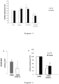
Figure 1 : Effect of different forms of ICOS-Fc on the growth of B16-F10 tumors in C57BL/6 mice. Mice with palpable subcutaneous B16 tumors were treated every 4 days with PBS, CDNS (nanoparticles) alone, ICOS-Fc or CDNS loaded with ICOS-Fc. The tumor growth was evaluated 16 days after the first treatment. Each treatment involved 5 mice/experiment. **p<0.05 vs each other condition. -
Figure 2 : Effect of CDNS/ICOS-Fc on tumor angiogenesis in vivo. Immunofluorescence staining for anti-CD31 of tumors tissue sections from mice treated with PBS, CDNS alone or CDNS loaded with ICOS-Fc. The slides were stained with either Ab rabbit α-mouse CD31 plus a secondary antibody α-rabbit conjugated with Alexa Fluor® 488. Representative images of 3 independent experiments are shown. The bar graph shows the cumulative results of these experiments as tumor microvessel density (TDM). **p<0.05 vs each other condition. -
Figure 3 : Effect of CDNS/ICOS-Fc on IL-10 and Foxp3 ex-vivo. Infiltrating cells from tumors were harvested and used for the real time PCR analysis; the data are normalized for the expression in the control mice (control expression PBS group set at 100%; *p<0.05). -
Figure 4 : Effect of different forms of ICOS-Fc on the growth of B16-F10 tumors in ICOS-deficient C57BL/6 mice. Mice with palpable subcutaneous B16 tumors were treated every 4 days with CDNS alone or CDNS loaded with ICOS-Fc. (A) Tumor growth was evaluated 8 days after the first treatment; each treatment involved 8 mice. **p<0.05. (B) Expression of IL-10 and Foxp3 in tumor infiltrating lymphocytes analyzed by real time PCR analysis; the data are normalized for the expression in the CDNS control. # p<0.05 from the respective value in mice treated with CDNS. -
Figure 5 Effect of different forms of ICOS-Fc on the viability of B16-F10 cells in vitro. Cell viability of B16-F10 cells was assessed by MTT upon culture in the presence and absence of titrated amounts (0.5-5 µg/ml) of free ICOS-Fc, or empty CDNS or CDNS/ICOS-Fc. Results are shown as inhibition % of the viability detected in cells cultured in the absence of those reagents. -
Figure 6 . Results of two approaches detecting the interaction of OPN with B7h. (Left) It shows the interaction of titrated amounts of soluble B7h-Fc (grey line) with a fixed amount of osteopontin (OPN) coated on the ELISA plate. The black line shows the same experiment in the presence of 5 µg/ml of soluble ICOS-Fc to evaluate the competition between ICOS and OPN for B7h binding. The dashed line shows the lack of binding of titrated amounts of soluble ICOS-Fc to the OPN-coated plates. (Right) The Western blot shows a pull-down assay in which B7h-Fc was used as a sepharose-bound bait protein incubated (first lane) or not (second lane) with OPN. The third lane was the OPN positive control. The membrane was blotted with an anti-OPN polyclonal antibody. -
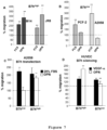
Figure 7 . Role of B7h in OPN function. (A-B) Cell migration induced in tumor cell lines expressing high (A) and low (B) levels of B7h by OPN or FCS; ** significantly different from untreated cells. (C) The migratory response to OPN is restored in B7h-trasfected (B7hhigh) A2058 cell; ** significantly different from untransfected cells (B7hlow). (D) The migratory response to OPN is suppressed in B7h-silenced (B7hlow) HUVEC; ** significantly different from unsilenced cells (B7hhigh). The dotted horizontal lines correspond to basal migration of untreated cells, set al 100%. -
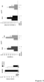
Figure 8 . Effect of ICOS-Fc in OPN-induced tubulogenesis and tumor cell migration and adhesion. (A) Effect of ICOS-Fc on HUVEC tubulogenesis induced by either OPN or VEGF; NT: basal tube formation without OPN and VEGF. ** significantly different from the corresponding cells treated with ICOS-Fc. (B-C) Effect of ICOS-Fc on migration (B) and adhesion (C) to HUVEC of two human melanoma cell lines expressing high levels of B7h (i.e. M14 and JR8). ** p<0.05 versus untreated cells; §§ p<0.05 vs OPN-treated cells. -
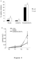
Figure 9 : Effect of PLGA/ICOS-Fc on the survival of B16-F10 cells in vitro and the growth of B16-F10 tumors in C57BL/6 mice. (A) Cell viability of B16-F10 cells was assessed by MTT upon culture in the presence and absence of titrated amounts (0.5-5 µg/ml) of free ICOS-Fc, or empty PLGA NP or PLGA/ICOS-Fc NP. Results are shown as inhibition % of the viability detected in cells cultured in the absence of those reagents. (B) Mice with palpable subcutaneous B16 tumors were treated every 4 days with PBS, PLGA (nanoparticles) alone, PLGA/ICOS-Fc. The tumor growth was evaluated 16 days after the first treatment. Each treatment involved 5 mice/experiment. *or** p<0.05 vs each other condition. -
Figure 10 . Effect of PLGA/ICOS-Fc in the survival of athymic mice injected with human glioblastoma cells into the brain. Kaplan-Meier analyses of MD13 tumor-bearing athymic mice treated intraperitoneally by with PLGA alone (blank) (100µl) or PLGA loaded with human ICOS-Fc (100µl) after 7 days from implantation and every 7 days. PLGA loading with ICOS-Fc treatment significantly prolonged survival of tumor-bearing mice (blank vs ICOS-Fc in immunocompromised mice, n=6 for all group, P = 0.00591, log-rank test). - In the following description, numerous specific details are given to provide a thorough understanding of embodiments. The embodiments can be practiced without one or more of the specific details, or with other methods, components, materials, etc. In other instances, well-known structures, materials, or operations are not shown or described in detail to avoid obscuring aspects of the embodiments.
- Reference throughout this specification to "one embodiment" or "an embodiment" means that a particular feature, structure, or characteristic described in connection with the embodiment is included in at least one embodiment. Thus, the appearances of the phrases "in one embodiment" or "in an embodiment" in various places throughout this specification are not necessarily all referring to the same embodiment. Furthermore, the particular features, structures, or characteristics may be combined in any suitable manner in one or more embodiments.
- The headings provided herein are for convenience only and do not interpret the scope or meaning of the embodiments.
- The instant disclosure concerns novel anti-tumor therapeutic agents comprising at least one ligand of receptor B7h having the ability of triggering the receptor B7h activity.
- According to one embodiment, the present invention provides for a ligand of receptor B7h for use in the treatment of a subject suffering from a tumor, wherein the ligand of receptor B7h is loaded into or onto a biocompatible micro- or nano-carrier, and the ligand of B7h receptor is able of binding the receptor B7h and triggering the receptor B7h activity, and optionally inhibiting osteopontin activity, wherein the ligand of receptor B7h is selected from:
- a) a human ICOS protein having an amino acid sequence as set forth in SEQ ID No.: 1 or portions thereof;
- b) a human ICOS extracellular domain having an amino acid sequence as set forth in SEQ ID No.: 2 or portions thereof; and
- c) a homologue of any one of proteins a) and b) having at least 80%, preferably at least 90%, more preferably at least 95%, still more preferably at least 98%, sequence identity to the amino acid sequences as set forth in SEQ ID No.: 1, 2 or portions thereof.
- In a still further preferred embodiment, the ligand of B7h receptor comprises an amino acid sequence as set forth in SEQ ID No.: 3.
- In one embodiment of the present invention, the ligand of receptor B7h loaded into or onto a biocompatible micro- or nano-carrier is to be administered by injection, infusion.
- As used herein, the expression "ligand of receptor B7h" comprises the human wild type ligand ICOS having an amino acid sequence as set forth in SEQ ID No.: 1, as well as portions of the same (for example having an amino acid sequence as set forth in SEQ ID No.: 2) provided that such portions have the ability of binding receptor B7h, triggering its activity and optionally inhibiting osteopontin activity.
- Other ligands of receptor B7h useful within the present invention comprise monoclonal antibodies able to bind specifically receptor B7h (i.e. with high affinity), provided that such monoclonal antibodies have agonistic activity towards B7h receptor, i.e. they are able to trigger the receptor B7h activity, and optionally have antagonistic activity towards osteopontin, i.e. they are able to inhibit the osteopontin activity.
- The expression "homologue of human ICOS or portions thereof" means proteins having at least 80%, preferably at least 90%, more preferably at least 95%, still more preferably 98%, identity with any one of the SEQ ID NO.: 1 and 2 or portions thereof, provided that such homologues have the ability of binding receptor B7h and triggering its activity, and optionally inhibiting the osteopontin activity.
- It is common general knowledge that the ability of triggering a receptor activity implies the ability to cross-link the receptor of interest expressed on the cell surface as indicated e.g. in Seed B. "Making agonists of antagonists", Chem Biol. 1994 Nov;1(3):125-9, and A, Schlessinger J. "Signal transduction by receptors with tyrosine kinase activity", Cell. 1990 . From such common general knowledge it derives that ligand of receptor B7h needs to be in at least a dimeric form for exerting the anti-tumor effect. Examples of ligands in at least a dimeric form able to trigger receptor B7h activity are represented i.a. by bivalent o multivalent ICOS-Fc constructs, anti-B7h receptor antibodies, organic/inorganic, natural/synthetic scaffolds comprising at least two ICOS molecules attached thereto and ICOS multimers obtained by chemical or genetic crosslinking of multiple ICOS molecules. Multimerization may be achieved by metal ion- or small molecule-based assembly, protein (peptide) interaction-based assembly, covalent protein assembly, genetic fusion of functional proteins to protein building blocks. Multimerization may be achieved by use of dendrimers, dendrons, dendronized polymers, hyperbranched polymers, and polymer brushes. The scaffolds may include organic polymers (such as those derived from sucrose, squalene or solanesol), and organic or inorganic solid nano/microparticles exposing multiple ICOS molecules on their surface.
- In one embodiment, the present invention provides for the ligand of receptor B7h being fused or conjugated to a stabilizing molecule.
- With respect to the stabilizing molecules that can be conjugated with the receptor B7h ligand, such molecules are widely known in the art and do not necessitate a detailed description herein. As examples of stabilizing molecules, one can cite human Fc antibody domain, polyethylene glycols (PEGs) or derivatives thereof, poly-L-lysine citramide (via a lysine or an ethylcarbamate spacer), styrenemaleic acid anhydride and poly-hydroxypropylmetacrylamide.
- PEG derivatives able to link amino groups present on the receptor B7h ligands are i.a. epoxide PEG, aldehyde PEG, nitrophenyl carbonate PEG and succinimidyl ester PEG; PEG derivatives able to link thiol groups present on the B7h receptor ligands are i.a. orthopyridyl disulfide PEGs; PEG derivatives able to link hydroxyl groups present on the B7h receptor ligands are i.a. PEG-COOH activated with N-hydroxysuccinimide or hydroxybenzotriazole. Other PEG derivatives are represented by PEG-polyacetal with pH-dependent hydrolysis, and PEG-dextrin (polymer-masking-unmasking-protein therapy, PUMPT).
- In order to increase the delivery of the B7h receptor ligand to the site of treatment, namely the tumor, the receptor B7h ligand may also be hyperglycosylated or conjugated to mannose residues.
- Hyperglycosylation may be performed by either in situ chemical reactions or site-directed mutagenesis resulting in either N-linked or O-linked protein glycosylation. In N-linked glycosylation, the saccharide chain is attached to asparagine of tripeptide sequence Asn-X-Ser/Thr, where X represents an amino acid other than proline. Polysialic acid (PSA) is often used for hyperglycosylation. Large-molecular-weight PSAs are suitable for the delivery of low-molecular-weight drugs and peptides, while lower molecular weight PSAs could be used for large proteins as well as particulate drug-delivery systems.
- The conjugation to mannose residues exploits binding of the conjugate to mannose receptors, which are reported to be expressed on Kupffer cells, macrophages, alveolar, monocyte-derived dendritic cells and subsets of vascular and lymphatic endothelial cells. Mannosylated proteins can be recognized by mannose-specific lectins, namely, mannose receptors and MBPs.
- With respect to the biocompatible carrier (which may have different degrees of biodegradability), the carrier is selected from particles, capsules, vesicles, bubbles, each of them having dimensions in the order of micron or nanometers. Other suitable carriers are represented by nanoemulsions, nanosuspensions, nanohydrogels, micelles, dendrimers, quantum dots, liposomes or carbon derivatives (e.g. carbon nanotubes). The micro-, nano-particles can be made of polymers, metals (e.g. gold), silica or a synthetic polymer core with a lipoid shell. The metal particles may also be magnetic. All these carriers are widely known in the pharmaceutical field for the delivery of drugs and do not necessitate a detailed description herein. The antitumor activity of the receptor B7h ligand/carrier system is not related to a specific type of carrier, but the association of receptor B7h ligand to a nano/microcarrier create a micro-, nano-platform formulation wherein the two components work together. The loading of the B7h receptor ligand into or onto a micro/nanocarrier is crucial for its therapeutic effectiveness and for clinical translation. The carrier and the ligand of receptor B7h equally contribute to the antitumor activity and their synergistic effect is mandatory for an effective anticancer therapy. Additionally, the B7h receptor ligand/carrier system is important to magnify the antitumor activity as described below. The incorporation of the receptor B7h ligand within a carrier create a reservoir of the active molecule and minimize its systemic exposure, allowing accumulation and release to tumor tissues and avoiding non specific delivery. Indeed, the incorporation of the receptor B7h ligand in micro/nanocarrier allows the development of site-specific targeted systems for delivering to cancer cells, exploiting passive and active targeting strategies.
- In a preferred embodiment, the carrier is represented by micro- or nano-particles made of cyclodextrin polymer, poly(lactide-co-glycolic acid) (PLGA), polycaprolactone, (PCL), polylactic acid (PLA), poly (glycolide), chitosan, alginate, starch, alginate, collagen, albumin, silica, metal. Preferably the carrier is represented by micro- or nano-particles made of cyclodextrin polymer or poly(lactide-co-glycolic acid).
- In a further embodiment, the present invention concerns a pharmaceutical composition comprising at least one ligand of receptor B7h loaded into or onto a biocompatible micro- or nano-carrier and a pharmaceutical acceptable vehicle for use in the treatment of a tumor, wherein the at least one ligand of receptor B7h contained in the pharmaceutical composition is selected from:
- a) a human ICOS protein having an amino acid sequence as set forth in SEQ ID No.: 1 or portions thereof;
- b) a human ICOS extracellular domain having an amino acid sequence as set forth in SEQ ID No.: 2 or portions thereof; and
- c) a homologue of any one of proteins a) and b) having at least 80%, preferably at least 90%, more preferably at least 95%, still more preferably at least 98%, sequence identity to the amino acid sequences set forth in SEQ ID No.: 1, 2 or portions thereof.
- In a still preferred embodiment, the at least one ligand of receptor B7h contained in the pharmaceutical composition comprises an amino acid sequence as set forth in SEQ ID No.: 3.
- In a preferred embodiment, the at least one ligand of receptor B7h contained in the pharmaceutical composition is fused or conjugated to a stabilizing molecule.
- The present disclosure also concerns use of receptor B7h as target for the screening of pharmaceutical active agents useful in the treatment of a tumor, wherein the pharmaceutically active agents bind to receptor B7h, trigger receptor B7h activity and inhibit osteopontin activity.
- Previous work in vitro showed that B7h triggering by ICOS-Fc inhibits migration and adhesion of DC, EC and several tumor cell lines. Moreover, in vivo studies showed that ICOS-Fc inhibits the metastatization into the lungs of B7h-positive tumor cells injected in the tail vein. By injecting human B7h-positive neoplastic cells in the tail vein of mice and treating these mice with both mouse and mouse ICOS-Fc (which are species-specific), the present inventors showed that the ICOS-Fc-mediated inhibition of in vivo metastatization involves effects on both the tumor cells and the host cells. In vivo treatment with ICOS-Fc inhibited development of experimental lung metastases upon injection of B16 melanoma cells in the tail vein of syngeneic mice. Moreover, using the xenograft model of human CF-PAC1 cell line injected in the tail vein of NOD-SCID-IL2Rγnull (NSG) mice, the present inventors found that optimal inhibition of metastatization requires contemporary treatment with both the human and the mouse ICOS-Fc. These data indicate that ICOS-Fc exerts its inhibitory effect on metastatic dissemination by acting on both the human tumor cells and the mouse environment14.
- The tumor growth actually involves several distinct processes, such as neoplastic cell proliferation and apoptosis, tissue invasion, adhesion and migration, angiogenesis, intra- and extra-vasation. Therefore, anti-metastatic drugs acting on neoplastic cell adhesion, migration, intra- and extra-vasation (so called "migrastatic" agents) are not effective on the tumor growth, and the potential clinical relevance of "migrastatic" agents is now heavily questioned17,18. In fact, the previous experiments of the inventors did not detect any effect of ICOS-Fc on the growth of established primary tumors either in vivo 13,14 or in vitro. These negative results have been obtained in vivo testing several doses and delivery routes of ICOS-Fc (intravenous, intraperitoneal, and peritumoral injection) and several experimental models of tumor expressing B7h (transplantable B16 melanoma and PC3 prostate carcinoma and Balb/neuT spontaneous mammary carcinoma). In vitro experiments, indeed, did not detect any effect of ICOS-Fc on the proliferation and apoptosis of several tumor cells lines expressing B7h14. Moreover, ICOS-Fc did not show any effect on vascular endothelial cell proliferation and angiogenesis assays in vitro14. In conclusion, the only anti-tumor effect of ICOS-Fc detected by the previous studies was the anti-metastatic one, whereas no effects were detected on the tumor growth in vivo, and tumor cell proliferation and apoptosis, and angiogenesis in vitro.
- The present inventors then tested the anti-tumor activity of ICOS-Fc encapsuled in biocompatible nanoparticles.
- Using the B16 melanoma model of transplantable tumor, the inventors found unexpectedly that ICOS-Fc encapsulated in cyclodextrin polymer nanosponges (CDNS) display a potent anti-tumor activity decreasing the growth of established tumor masses and decreasing its vascularization. These effects were detectable also in ICOS-deficient mice, which shows that they were not ascribable only to an antagonistic effect of ICOS-Fc on the interaction between B7h and the endogenous ICOS. The effect was not dependent on the nanoparticle type and the tumor type since a similar anti-tumor effect was detected using human ICOS-Fc loaded in poly(lactide-co-glycolic acid) (PLGA) nanoparticles and in a xenogeneic model of glioblastoma.
- Another aspect of the B7h involvement in the antitumor response is that the present inventors found that B7h binds also osteopontin (OPN), that can act either as a protein of the extracellular matrix and a soluble cytokine. OPN is secreted by several cell types including macrophages, DC, osteoblasts, and T cells. It mediates several functions such as bone remodeling, macrophage response, cell migration and adhesion, inflammation, and support of TH1 and TH17 cell differentiation. In the tumor biology, OPN produced by tumor cells and the tumor microenvironment plays a key role in promoting tumor growth, migration, metastatic dissemination, and neoangiogenesis. OPN interacts with several receptors, such as integrins and CD44, and it is cleaved by thrombin in a N-terminal (OPN-N) and a C-terminal (OPN-C) portion, which bind to different receptors and exert different functions. A RGD domain binding to several integrins is located in OPN-N nearby to two other binding sites for the α4β1 integrin, which are exposed upon thrombin cleavage; a CD44 binding site is located in OPN-C19. The present inventors found unexpectedly that B7h expression is required for the OPN-induced migratory response of tumor cell lines and EC and this effect is inhibited by ICOS-Fc. Then, it was found that OPN directly binds to B7h using a binding site of B7h different from that bound by ICOS. These data depict a scenario in which B7h triggering by ICOS inhibits cell migration induced by several chemoattrancts whereas its triggering by OPN induces cell migration, with a dominant effect exerted by ICOS. Therefore, without being bound to any specific theory, the present inventors have reasons to believe that ICOS-Fc exerts its anti-tumor effects also by inhibiting the OPN effects promoting tumor growth, migration, metastatic dissemination, and neoangiogenesis.
- With respect to the carrier for delivery of the B7h receptor ligand for its use in the treatment of a tumor, the present inventors verified that the nature of the carrier has not any effect on the ability of the B7h receptor ligand of exerting its anti-tumor activity.
- The micro-, nano-particles can be made of cyclodextrin (CD) polymer nanosponges (CDNS), poly(lactide-co-glycolic acid) (PLGA), polycaprolactones (PCL), polylactic acid (PLA), poly(glycolide), chitosan, alginate, starch, dextran, collagen, albumin, silica, metal (e.g. gold). Liposomes may also be surface modified with PEG in order to interfere with recognition and uptake by reticulo endothelial system and to extend circulation time. Moreover, in situ thermosensitive hydrogels undergo sol-gel phase transition in response to changes in temperature.
- CD are cyclic α-1,4-glucans comprising from six to >100 glucose units. They are natural products resulting from intramolecular transglycosylation reactions of starch degraded by CD glucanotransferase. The enzymatic product is generally a mixture of α, β, γ-CD comprising six, seven, and eight glucose units respectively. They play an important role in supramolecular chemistry due to their ability for molecular encapsulation with a wide range of guest molecules and are of high interest in the pharmaceutical field, biomedical science and biotechnology. They present a torus-shaped ring structure with an interior hydrophobic cavity and a hydrophilic exterior site. NS can form nanoporous insoluble nanoparticles (NP) due to the presence of CD cavities and the nanochannels of the cross-linked network with a crystalline or amorphous structure, spherical shaped, and with swelling properties20-24.
- Cyclodextrin-nanosponges (CDNS) can form inclusion complexes able to host a wide range of hydrophobic molecules. In aqueous solution, the hydrophobic CD cavity is occupied by water molecules bounded by "weak forces" (energetically unfavoured). Owing to the size of the internal cavity, one or two hydrophobic guest molecule(s) may be entrapped by one, two, or even three CD. CDNS can form porous insoluble NP, either crystalline or amorphous, with a spherical shape. The polarity and dimension of the polymer mesh can be easily tuned by varying the type and degree of crosslinking. CDNS can incorporate different types of lipophilic or hydrophilic molecules, and they can be functionalized for site-specific targeting by conjugating various ligands on their surface. They are safe and biodegradable, display negligible toxicity on cell cultures, and are well-tolerated upon injection in mice. Release of the entrapped molecules can be modulated by adjusting the NS structure to achieve to improve the aqueous solubility of poorly water-soluble molecules, protect degradable substances, and obtain sustained release.
- Poly(lactic-co-glycolic acid) (PLGA) NPs are the best characterized NPs that increase potency and bioavailability of several drugs and their use has been approved by the Food and Drug Administration (FDA) for several therapeutic applications. The modulation of the polymer lactide-glycolide ratio, molecular weight and crystal profile allows to protract the degradation rate and subsequent release of the entrapped molecules from several days up to one year.
- In the instant description, the present inventor show that ICOS-Fc significantly inhibits the growth of established primary tumors only when it is encapsulated in biocompatible nanoparticles, whereas the free ICOS-Fc has no effect (
Fig.1 ). This has been detected in both a strongly immunogenic peripheral tumor (melanoma) and a weakly immunogenic CNS tumor (glioblastoma), using either CDNS or PLGA nanoparticles as the drug carriers, and using either a mouse tumor model (melanoma) (Fig.1 ) or a human/mouse tumor xenograft model (glioblastoma) (Fig. 9 ). Moreover, ICOS-Fc was effective not only in immunocompetent mice (melanoma) (Fig.1 ), but also in athymic mice (glioblastoma) (Fig. 9 ) and ICOS-KO mice (melanoma) (Fig.4 ). These data show that the ICOS-Fc effect was not ascribable to blocking the interaction between B7h and the endogenous ICOS expressed on T cells (or other cell types). This blocking, using an anti-ICOS mAb, has been shown to inhibit the development of Treg cells suppressing the anti-tumor immune response. - Without wishing to be bound to any theory, the present inventors have reasons to believe that the loading of ICOS-Fc in the carrier (nanoparticles) for exerting its anti-tumor activity may be ascribed to the carrier ability to carry ICOS-Fc into the tumor mass possibly through the Enhanced Permeability and Retention (EPR) effect. The same effect may be exerted by various types of carriers acting either through EPR or other mechanisms to increase drug delivery into the tumor, such as carriers conjugated to biomolecules increasing targeting into the tumor. Moreover, the same effect of ICOS-Fc may be exerted by other molecules capable to trigger B7h, such as monoclonal antibodies.
- The antitumor activity of B7h triggering may be partly ascribed to the ICOS-Fc effect on tumor angiogenesis, as suggested by the antiangiogenic activity of ICOS-Fc in vitro (
Fig.8 ) and in vivo (Fig.2 ). This finding is novel since previous data did not detect any antiangiogenic effect exerted by ICOS-Fc14. Moreover, the effect of the human ICOS-Fc in the glioblastoma xenograft model shows that ICOS-Fc exerts also a direct effect on the tumor cells, since the human ICOS-Fc does not interact with the mouse B7h expressed by the host cells. In support of a direct effect of ICOS-Fc encapsulated in NP on the tumor cell viability, the inventors show that high doses of ICOS-Fc encapsulateed in CDNS exert a cytotoxic effect on B16-F10 cells in vitro, whereas empty CDNS and free ICOS-Fc had no effect (Figure 5 ). One possibility is that the difference may be due to the NP ability to increase the ICOS-Fc cell internalization, which would increase the ICOS-Fc interaction with the intracellular receptor B7h. - A key point is that it has been shown for the first time that B7h also binds to OPN (
Fig.6 ), which is a key molecule involved in the tumor growth, migration and metastatization, and the B7h/OPN interaction is involved in these OPN activities (Fig. 7 ). The B7h/OPN interaction involves a different binding site than the ICOS/B7h interaction (Fig.6 ), and ICOS-Fc displays a strong dominant negative effect on several tumor-promoting activities mediated by OPN (Fig.8 ). These data confirm that the ICOS-Fc inhibitory activity of the tumor growth is not limited to blocking the interaction between B7h and the endogenous ICOS expressed on T cells. - In view of the experiments performed by the inventors, ICOS-Fc is an antineoplastic drug that can be used either in monotherapy or in combination therapies with other anti-neoplastic therapies. ICOS-Fc acts by triggering B7h and inhibiting the OPN activity, which are substantial additional effects to the effect of inhibiting the endogenous ICOS/B7h interaction.
- C57BL/6 mice carrying palpable subcutaneous B16-F10 tumors were i.v. treated with either the mouse ICOS-Fc, or ICOS-Fc/CDNS or the empty CDNS (100 µg each) or the same volume of PBS as control every 4 days and the tumor growth was monitored every 4 days. Results showed that ICOS-Fc loaded in CDNS (CDNS/ICOS-Fc) substantially inhibited the growth of melanoma cells in the mice, whereas free ICOS-Fc has no effect (
Fig. 1 ). - To assess the effects of CDNS/ICOS-Fc on tumor angiogenesis, the expression of CD31 in the tumors obtained with B16-F10 cells was evaluated. Results showed that the treatment with CDNS/ICOS-Fc reduced blood vessel formation compared to control mice (PBS) or empty CDNS treated-mice (
Fig. 2 ). - To assess if the treatment modulates the immune response, infiltrating lymphocytes were obtained from the tumors and the mRNA levels of IL-17A and RORγt (marking TH17 cells), IL-10 and Foxp3 (marking Treg cells) were evaluated by Real Time PCR, since ICOS has a key role in Th17 and Treg cell differentiation. Results showed that treatment with CDNS/ICOS-Fc significantly decreased expression of Foxp3 and IL-10 compared with the levels detected in control mice, whereas free ICOS-Fc and empty CDNS had no effect. In contrast, no significant differences were detected in the expression of RoRγt and IL17A (
Fig. 3 ). - To assess at which extent the ICOS-Fc anti-tumor effect depends on inhibition of the interaction between B7h and the endogenous ICOS, the effect of CDSN/ICOS-Fc on the B16 tumor growth in ICOS-deficient mice was evaluated. Results showed that ICOS-Fc effectively inhibited the growth of B16 tumors in ICOS-deficient mice (
Fig.4A ). The effect on the tumor growth was accompanied by decreased expression of IL-10 and FoxP3 as detected by real time PCR analysis of mRNA extracted from the tissue (Fig.4B ). These data show that the ICOS-Fc effect depends of triggering of B7h and not to antagonism of the B7h binding to the endogenous ICOS. - Cell toxicity of the CDNS preparations and free ICOS-Fc was assessed by performing an MTT assay on B16-F10 cells incubated with or without titrated amounts (0.5, 1, 2, 5 µg/ml) of CDNS/ICOS-Fc, or empty CDNS, or free ICOS-Fc. The results detected cell toxicity at the highest doses (30% inhibition at 2 and 5 µg/ml) which was exerted only by CDNS/ICOS-Fc and not by empty CDNS or free ICOS-Fc (
Fig. 5 ). - The inventors used two approaches to test the hypothesis that OPN binds to B7h using recombinant B7h-Fc and histidine-tagged OPN or B7h (B7h-his) (
Fig.6 ). A) ELISA. OPN (or B7h-his) was adsorbed on ELISA plates and then incubated with titrated amounts of B7h-Fc (or OPN) for 1 h. After washing, binding was evaluated with anti-IgG1 mAb (or polyclonal anti-OPN Ab). Results showed a concentration dependent binding of B7h to OPN. Moreover, we showed that the OPN/B7h binding is not inhibited by ICOS-Fc, which indicates that OPN and ICOS-Fc binds different sites of B7h. ICOS-Fc did not show any binding to OPN. B) Pull-down. B7h-Fc was used as a bait protein captured on Sepharose-protein A, and was incubated with OPN for 1 h. After washing, proteins were eluted from the resin and analyzed by Western blot using anti-OPN polyclonal antibodies. Results showed an association between OPN and B7h. - OPN induces migration of tumor cell lines expressing high levels of B7h (B7hhigh) but not of those expressing low levels (B7hlow), whereas no difference is found when migration is induced by fetal bovine serum (FBS) (
Fig.7A-B ). In B7hlow tumor cells, the migratory response to OPN is restored by B7h transfection reinforcing B7h expression whereas, in B7hhigh HUVEC, it is inhibited by shRNA-mediated silencing of B7h (Fig. 6C-D). The effect is specific since modulation of B7h expression does not affect migration induced by FBS in tumor cells and by VEGF in HUVEC. - In HUVEC, treatment with ICOS-Fc inhibits tubulogenesis induced by OPN but not that induced by VEGF (
Fig.8A ). - In tumor cell lines expressing high levels of B7h, treatment with ICOS-Fc inhibits cell migration and adhesion to HUVEC induced by OPN (
Fig.8B ). - Effect of PLGA/ICOS-Fc in the tumor growth in vivo. Cell toxicity of the PLGA preparations was assessed by performing an MTT assay on B16-F10 cells incubated with titrated amounts (0.5, 1, 2, 5 µg/ml) of empty PLGA or PLGA/ICOS-Fc NP. Some toxicity was exerted by PLGA/ICOS-Fc NP only at the highest doses, but not by empty PLGA NP or free ICOS-Fc (
Fig.9A ). - C57BL/6 mice carrying palpable subcutaneous B16-F10 tumors were i.p. treated with either PLGA/ICOS-Fc or the empty PLGA (100 µg each) or the same volume of PBS as control every 4 days and the tumor growth was monitored every 4 days. Results showed that treatment with PLGA/ICOS-Fc effectively inhibited the growth of B16-F10 tumors compared to both control treatments (
Fig.9B ). - Athymic mice bearing human glioblastoma in the brain were treated with PLGA-loaded with human ICOS-Fc or empty PLGA one week after implantation of human neuroblastoma cells (MD13) into the brain. Treatment with PLGA/ICOS-Fc significantly prolonged the median survival of tumor-bearing mice (
Fig. 10 ). The treatment with PLGA/ICOS-Fc reduced the tumor formation compared to empty PLGA treated-mice. The effect of PLGA/ICOS-Fc in the tumor growth in vivo does not depend on the presence of the endogenous ICOS+CD4+ T cells, because we evaluated the effect of PLGA/ICOS-Fc in T cell-deficient mice. - The extracellular portion of the human or mouse ICOS was cloned into a modified eukaryotic expression vector derived from pCDNA3.1/Hygro(+) plasmid (cod. V870-20, Invitrogen) and reported as p-Minibody (pMB-SV5) by Di Niro R. et al.25, PubMed ID: 17678525. This vector differs from the original one by: the Kozak sequence (5'CCACCATGG 3' - SEQ ID No.: 11) which is required for the initiation of the translation in eukaryotic cells; the secretory leader sequence (5' GCTGGAGCCTGATCCTCCTGTTCCTCGTCGCTGTGGCTACA 3' - SEQ ID No.: 12) which was introduced to allow the release of the protein in the culture supernatants; the mini intron sequence(5'GGTAAGGGGCTCACAGTAGCAGGCTTGAGGTCTGGACATATATA TGGGTGACAATGACATCCTTTGCCTTTCTCTCCACAGGTG 3' - SEQ ID No.: 13) to increase the level of the protein expression. A tag sequence to target the produced-protein was introduced and it is derived from Simian Virus-5 (SV5 tag) (5' GGCAAACCAATCCCAAACCCACTGCTGGGCCTGGATAGTACT 3'- SEQ ID No.: 14) and it is useful for monoclonal antibody recognition of the protein. This vector allowed to clone the fragments of interest in frame with the coding sequence of the human or mouse constant fragment of the immunoglobulin IgG1 (Fc) domain, having nucleotide sequences set forth in SEQ ID No.: 5 and 9, respectively.
- To generate the human ICOS-Fc construct (SEQ ID No.: 3), the nucleotide sequence encoding the extracellular portion of the human ICOS (SEQ ID No.: 4) was amplified with specific primers: ICOS forward BsshII primer (5' TTGGCGCGCATGCCGAAATCAATGGTTCTGCC 3' - SEQ ID No.: 15, Sigma-Genosys, The Woodlands, TX, USA) and ICOS reverse NheI primer (5' CTAGCTAGCAAGTTGTGATTCATAAATATGC 3' - SEQ ID No.: 16, Sigma-Genosys). The amplified fragments were digested with BssHII (cod. R0199S, New England Biolabs inc, Ipswich, MA, USA) and NheI (cod. R0131S, New England Biolabs inc) enzymes. The double digested fragments were cloned into the previously described pMB-SV5 plasmid with the coding sequence of the human Fc domain (the human Fc domain has the sequence set forth in SEQ ID No.:5. The nucleotide sequence was determined by sequencing. The nucleotide sequence of the expression vector coding for huICOS-huFc is set forth in SEQ ID No.: 6.
- To generate the mouse ICOS-Fc construct (SEQ ID No.: 7), the nucleotide sequence encoding the extracellular portion of the mouse ICOS (SEQ ID No.: 8) was amplified with specific primers: ICOS mouse forward BsshII primer (5' TTGGCGCGCATGCCGAAATCAATGGCTCG 3' - SEQ ID No.: 17, Sigma-Genosys) and ICOS mouse reverse NheI primer (5' CTAGCTAGCTAGCCAGAGCTTCAGCTGGC 3' - SEQ ID No.: 18, Sigma-Genosys). The amplified fragments were digested with BssHII (cod. R0199S, New England Biolabs inc, Ipswich, MA, USA) and NheI (cod. R0131S, New England Biolabs inc) enzymes. The vector used in this cloning was the pMB-SV5 with the coding sequence of the mouse Fc domain (the mouse Fc domain has the sequence set forth in SEQ ID No.: 9). The double digested fragments were cloned into the previously described pMB-SV5 plasmid. The nucleotide sequence was determined by sequencing. The nucleotide sequence of the expression vector coding for msICOS-msFc is set forth in SEQ ID No.: 10.
- The plasmid DNA was transformed into One Shot® TOP10 Chemically Competent Escherichia Coli bacterial cells (E. coli; cod.C4040-03, Life Technologies, Carlsbad, CA, USA). The resulting colonies were screened using specific primers: P-Hygro sense (5' CTGCTTACTGGCTTATCG 3' - SEQ ID No.: 19, Sigma-Genosys) and P-Hygro antisense (5' CAGATGGCTGGCAACTAG 3' - SEQ ID No.: 20, Sigma-Genosys) and the construct was confirmed by sequencing. Finally, the plasmid DNA was transfected using FreeStyle™ MAX Reagent (cod. 16447100, Life technologies) into Chinese Hamster Ovarian-suspension cell line (CHO-s) (cod. R8/00-07, Invitrogen). The stable clones were obtained thanks to the presence of Hygromycin resistence in the vector; to this end the clones were grown under selection with Hygromycin-B (cod. 10687-010, Invitrogen) at the concentration of 0.2 mg/ml that allow full selection of transfected cells. The cells were grown in serum free IMDM medium (cod. BE12-915F01, Lonza, Basel, Switzerland) and the serum free culture supernatants were purified using
Protein G Sepharose ™ 4 Fast Flow columns (cod. 17-0618-01, GE Healthcare, Piscataway Township, NJ, USA). - A carbonate NS, containing β-cyclodextrins (CDNS; code C4767, Sigma-Aldrich, St. Luis, MO, USA) as building blocks, cross-linked with a carbonate bridge, was prepared as previously reported20,21 . Briefly, an amount of anhydrous CD was dissolved in anhydrous dimethylformamide (DMF; code 227056, Sigma-Aldrich, St. Luis, MO, USA) and allowed to react with carbonyldiimidazole (code 115533, Sigma-Aldrich, St. Luis, MO, USA) at 90°C for at least 5 h. Once the reaction was over, a large excess of water was added to destroy the excess of carbonyldiimidazole (code 115533, Sigma-Aldrich, St. Luis, MO, USA), the solid was recovered by filtration and purified with water. Then, the solid was ground in a mortar and Sohxlet-extracted with ethanol (code 51976, Sigma-Aldrich) to remove residual reaction by-products. The reaction was carried out using a molar excess of crosslinker(e.g. 1:4 cyclodextrin:crosslinker). Following purification, dried NS were stored at 25°C.
- β-CDNS is crosslinked with pyromelliticdianhydride (code 412287, Sigma-Aldrich, St. Luis, MO, USA) to form a carboxylic acid terminated nanoporous material (βNS-Pyro) that form solid particles with a rather spherical morphology and a very high solubilizing power over poorly soluble substances. To obtain βPyro-NS, pyromelliticdianhydride (code 412287, Sigma-Aldrich, St. Luis, MO, USA) was added to anhydrous cyclodextrin. Synthesis was carried out in anhydrous dimethyl sulfoxide (DMSO, code D1435, Sigma-Aldrich, St. Luis, MO, USA) at room temperature for 24 h. The molar βNS-Pyro were prepared by crosslinking -CDNS and pyromelliticdianhydride (code 412287, Sigma-Aldrich, St. Luis, MO, USA) in a 1:8 molar ratio in the presence of ammonia. For biological experiments, the dried preparations were dispersed in a 0.9% NaCl (code S7653, Sigma-Aldrich, St. Luis, MO, USA) solution at a concentration of 10 mg/ml using an Ultraturrax instrument (IKA, Germany) for 3 min. All reagents were of analytical grade.
- The structures of NP were investigated by Raman spectroscopy and imaging methods. Our data by transmission electron microscopy showed that the formed NP have almost spherical morphology with size ranging from 50 to 100 nm with an average of 81 ± 9 nm. The instant results demonstrated that the NS-Pyro NP had not inflammatory effects and did not have any adverse effect on melanoma cells line. The inventors then encapsulated into these βNS-Pyro NP ICOS-Fc. A weighed amount of freeze-dried βNS-Pyro has been dispersed by stirring in an aqueous solution at pH 6.0 containing NaCl(code S7653, Sigma-Aldrich, St. Luis, MO, USA) and PEG 400 (
polyethylene glycol 400, code 202398, Sigma Aldrich, St. Luis, MO, USA) 3% w/v to obtain an isotonic aqueous nanosuspension containing ICOS-Fc. - PLGA nanoparticles were prepared by a modified double solvent evaporation method26. Briefly 60 mg of PLGA 65:35 crystals (cod. P2066; Sigma-Aldrich, Saint Luis, MO, USA) were dissolved in 1ml of dichloromethane (DCM) (cod. 270997; Sigma-Aldrich, St. Luis, MO, USA) at room temperature. 50 µl of PBS were added to PLGA and the solution was sonicated for 1 min. 5 volumes of 1% PVA (cod. P8136; Sigma-Aldrich, St. Luis, MO, USA) acqueous solution were carefully added to the resulting emulsion in order to maintain phase separation. A further 2 min sonication was performed to obtain the final emulsion that was evaporated overnight under fume hood, to remove DCM. The resulting nanoparticles were washed 7X in distilled water by centrifugation at 7,000rpm for 10 min and resuspended in NaCl 0.9%, and stored at 4°C. Nanoparticles containing ICOS-Fc were produced as above by adding ICOS-Fc (1mg was lyophilized and then resuspended in 50 µl of PBS - phosphate buffer saline), to the PLGA solution dissolved in DCM during the first step of the preparation. The release of ICOS-Fc was evaluated by leaving the nanoparticles in PBS at 37°C, and the proteins released were quantified by the BCA assay (ThermoScientific).
- Female 5-7-wk-old C57BL/6 mice (either wild type cod. 000664-C57BL/6J or ICOS-/- cod. 004859-B6.129P2-Icostm1Mak/J; Charles River Laboratories, Wilmington, MA, USA) were injected subcutaneously (s.c.) with B16-F10 cells (105 cells/mouse; cod. CRL-6475; ATTC, Manassas, VA, USA). After 10 days, when tumors were palpable, mice were treated every 4 days with an intravenous (i.v.) injection of either the mouse ICOS-Fc, ICOS-Fc loaded in CDNS (ICOS-Fc/CDNS) or the empty CDNS (100 µg each) or the same volume of PBS as control. The tumor size was measured every 4 days with caliper and mice were sacrificed after 3 weeks. In other experiments, the same treatment protocol was applied by treating mice with either ICOS-Fc loaded in PLGA (ICOS-Fc/PLGA) or the empty PLGA NP(100 µg each) or the same volume of PBS as control.
- In other experiments, athymic mice (Balb/c nu/nu; strain code 194, Charles-River) were injected stereotactically into the right striatum with 1 x 10⁴ human dissociated glioblastoma tumor sphere cells (mesenchymal phenotype) obtained from cell cultures derived from a freshly resected glioma tumor27, corresponding to stem-like tumor cells, expressing B7h (n = 6 per treatment group). After 7 days of tumor challenge, the mice were injected intraperitoneally with 100 µl of PLGA nanoparticles containing ICOS-Fc or empty PLGA. Mice were euthanized when neuropathological signs developed in brain tumor-bearing animals. To obtain the tumor sphere cells, freshly resected glioma tumor samples were dissociated, and the established cells were cultured in defined medium containing DMEM/F12 (cod. 31331-028, Thermo Fisher, Waltham, USA) supplemented with B27 MACS® NeuroBrew®-21 (formerly MACS Supplement B27 PLUS cod. 130-093-566 Miltenyi Biotec, Bergisch Gladbach, Germany) and heparin (2.5 µg/ml, cod. H3149, Sigma-Aldrich, St. Loius, MO, USA). To enhance proliferation and maintain the stemness, basic fibroblast growth factor (bFGF; 20 ng/ml, cod. 100-18B, Peprotech, London, UK) and epidermal growth factor (EGF; 20 ng/ml, cod. AF-100-15 Peprotech, London. UK) were added to the sphere cultures twice a week27. The mice were bred under pathogen-free conditions in the animal facility of the Department of Health Sciences and were treated in accordance with the University Ethical Committee. The study was approved by the Bioethics Committee for Animal Experimentation of the University of Piemonte Orientale and Ministero della Salute (Prot. No. 477/2016-PR).
- The infiltrating cells were obtained from the tumors, and the mRNA levels of IL-17A and RORγt (marking TH17 cells), IL-10 and Foxp3 (marking Treg cells) were evaluated via Real Time PCR. The total RNA was then isolated using TRIzol reagent (cod. 15596026, Thermo Fisher, Waltham, USA). RNA was retrotranscribed using the QuantiTect Reverse Transcription Kit (cod. 205311, Qiagen, Hilden, Germany). Their expressions were evaluated with a gene expression assay (cod. 4453320, Assay-on Demand, Applied Biosystems, Forest City, CA, USA). The GUSB gene was used to normalize the cDNA amounts. Real Time PCR was performed using the CFX96 System (Bio-Rad Laboratories) in duplicate for each sample in a 10 µl final volume containing 1 µl diluted cDNA, 5 µl TaqMan universal PCR master mix (cod. 4369016 Applied Biosystem, Foster City, CA), and 0.5 µl TaqMan Gene Expression Assays (Applied Biosystem). The following assays were used: GUSB, Mm01197698_m1; IL-17A, Mm00439618_m1; RORγt, Mm01261022_m1; IL-10, Mm01288386_m1; Foxp3, Mm00475162_m1. The thermocycler parameters were 95°C for 10 min, followed by 40 cycles of 95°C for 15 sec and 60°C for 1 min. The results were analyzed with a Delta-Delta CT method.
- Immediately after dissection, tumor samples were embedded in OCT compound (cod. 05-9801 Killik, Bioptica Milano SpA), snap-frozen, and stored at -80 °C until use. Tumor tissues were cut with a cryostat (thickness 5-6 µm) and treated with 4% paraformaldehyde (cod. P6148, Sigma-Aldrich) diluted in PBS for 5 minutes at room temperature to fix the sample on the glass slides. The samples were then blocked with 5% Normal Goat Serum (NGS cod.DY005 R&D System, Minneapolis, USA) in PBS for one hour, in order to block aspecific sites to which could bind the primary antibody. To detect CD31 expression, slides were incubated with the primary antibody rabbit anti-CD31 (cod. ab28364 dilution 1:50 Abcam, Cambridge, UK) room temperature for 2 hours. The secondary antibody used was an anti-rabbit Ig Alexa fluor 488-conjugated (cod. A-11008, Thermo Fisher), diluited 1:400. Then the sections were stained with 0.5 mg/ml of the
fluorescent dye 4,6-diamidino-2-phenylindole-dihydrochloride (DAPI, cod. D8417, Sigma-Aldrich) for 5 minutes, to color the cell nuclei, and then mounted using Prolong anti-fade mounting medium (Slow Fade AntiFADE Kit, cod. S2828, Molecular Probes Invitrogen). The sections were then observed by a fluorescence microscope (Leica, Italy), and analyzed with the Image Pro Plus Software for micro-imaging 5.0 (Media Cybernetics, version 5.0, Bethesda, MD, USA). - In the Boyden chamber (BD Biosciences, San Diego, CA, USA) migration assay, melanoma cells (A2058, cod. CRL-11147, ATCC, Manassas, VA, USA; M14, RRID:CVCL_1395; JR8, RRID:CVCL_5780; PCF-2) were plated onto the apical side of 50 µg/ml matrigel-coated filters (cod. 8.2 mm diameter and 0.3 µm or 0.5 µm pore size; Neuro Probe, Inc.; BIOMAP snc, Milan, Italy; Matrigel Matrix Basement Membrane Cod. L003975 Rif. Cat 354230, SACCO s.r.l., COMO, Italy) in RPMI-1640 (cod.BE12-702F/12, Lonza, Basel, Switzerland) serum-free medium, with or without OPN (10 µg/ml cod. 1433-OP-050, R&D System, Minneapolis, USA) or ICOS-Fc (5 µg/ml). Medium containing 20% FCS (cod. 10270106, Gibco, Gaithersburg, MD, USA) were placed in the basolateral chamber as a positive chemoattractant stimuli. The chamber was incubated at 37°C under 5% CO2. After 20 h, the cells on the apical side were wiped off with Q-tips. The cells on the bottom of the filter were stained with crystal violet (cod. 61135, Sigma-Aldrich, St. Louis, MO, USA) and all were counted (fourfold filter) with an inverted microscope (magnification 40X). Data are shown as percentages of the treated cells migration vs the control migration measured for untreated cells.
- HUVECs were grown to confluence in 24-well plates (cod. ET3024, Euroclone, Milan, Italy) in complete medium M200 (cod. M200500, Gibco, Gaithersburg, MD, USA) and then treated or not with OPN (10 µg/ml, cod. 1433-OP-050, R&D System, Minneapolis, USA), or ICOS-Fc (5 µg/ml), for 30 min, washed with fresh medium twice, and incubated for 1 h with melanoma cells (5x10⁴ cell/well; A2058, cod. CRL-11147, ATCC, Manassas, VA, USA; M14, RRID:CVCL_1395; JR8, RRID:CVCL_5780; PCF-2)). After incubation in the adhesion assay, non-adherent cells were removed by washing three times with M200 medium. The centre of each well was analysed by fluorescence image analysis. Adherent cells were counted by the Image Pro Plus Software for micro-imaging (Media Cybernetics, Bethesda, MD, version 5.0). Data are shown as percentages of the treated cells adhesion vs the control adhesion measured for untreated cells.
- In the tube-formation assay28, HUVECs (2.5x10⁴/well) were cultured in M200 (cod. M200500, Gibco, Gaithersburg, MD, USA ) serum-free medium and seeded onto 48-well plates (cod. ET3048, Euroclone, Milan, Italy) previously coated with 150 µl of growth factor-reduced matrigel (Matrigel Matrix Basement Membrane Cod. L003975 Rif. Cat 354230, SACCO s.r.l., COMO) in the presence of OPN (10 µg/ml, cod. 1433-OP-050, R&D System, Minneapolis, USA), or control medium with VEGF-α (10 ng/ml, cod. 293-VE-010; R&D System, Minneapolis USA). The morphology of the capillary-like structures formed by the HUVECs was analyzed after 6 h of culture using an inverted microscope (Leica Microsystem, Milano, Italy; magnification 10X) and was photographed with a digital camera (Leica Microsystem, Milano, Italy). Tube formation was analyzed and the number of tubes (with branching at both ends) was counted with an imaging system (Image-Pro Plus software for micro-imaging, Media Cybernetics, version5.0, Bethesda, MD, USA). Tube formation was evaluated by counting the total number of tubes in three wells (n=5).
- 10ug of rhB7h2-Fc (cod. 165-B7-100, R&D system, Minneapolis, MN, USA) and rhOPN (cod. 1433-OP-050/CF, R&D system, Minneapolis, MN, USA) were join together in PBS at RT on the wheel for 1h, then B7h was precipitated using Sepharose-protein G (cod. 17-0618-01, GE Healthcare, Piscataway, NJ, USA), sample buffer with 20% of β-mercaptoethanol (cod. M-3148, Sigma-Aldrich, Saint Louis, MO, USA) was used to dissociate the proteins and Western blot was performed. Anti-OPN polyclonal antibodies (cod. MAB14331-SP, R&D system, Minneapolis, MN, USA) was used to detect OPN on the membrane.
- For B7h silencing experiments, HUVEC cells (1.5*10^5 cells) were seeded on 6 wells plate (cod. ET3006, Euroclone, Milan, Italy) in complete medium IMDM (cod. cod. BE12-915F01, Lonza, Basel, Switzerland) for 24h. To silence the cells, lipofectaminetm RNAiMAX transfection reagent (cod. 13778030, Life technologies, Carlsbad, CA, USA) was used with two different siRNA direct B7h (oligo1: ICOSLGHSS177318 (5'-CAGCAGCCUUCGAGCUGAUACUCAG-3' - SEQ ID No.: 21 and 5'-CUGAGUAUCAGCUCGAAGGCUGCUG-3'- SEQ ID No.: 22) and oligo2: ICOSLGHSS118565 (5'-GGCCCAACGUGUACUGGAUCAAUAA-3' - SEQ ID No.: 23, and 5'-UUAUUGAUCCAGUACACGUUGGGCC-3'- SEQ ID No.: 24) Life technologies, Carlsbad, CA, USA) mapping in two different exons. For B7h transfection experiments, A2058 cells (10^6 cells; cod. CRL-11147, ATCC, Manassas, VA, USA)were seeded in 10 cm2 dishes (cod. ET2100, Euroclone, Milan, Italy)in complete medium RPMI-1640 (cod.BE12-702F/12, Lonza, Basel, Switzerland. To transfect the cells, 10 µg of DNA and 10µl of lipofectamine 3000 (cod.L3000001, Life technologies, Carlsbad, California, USA) were used. After 24 or 48 hours, the cells silenced or transfected were used for the migration experiments.
- 80nM of rhOPN (#1433-OP-050/CF, R&D system, Minneapolis, MN, USA) was adsorbed on Nunc MaxiSorp™ flat-bottom ELISA plate (cod. M9410-1CS, Sigma-Aldrich, St. Louis, MO, USA) and then incubated with titrated amounts of B7h-Fc (cod. 165-B7-100, R&D system, Minneapolis, MN, USA) for 1 h with or without 80nM of rhICOS-Fc. After washing with PBS + 0,025% Tryton (cod. T8787, Sigma-Aldrich, St. Louis, MO, USA ), anti-human-IgG1 mAb HRP conjugated (cod. P0214, Dako, Santa Clara, CA, USA) was added for 1h then TMB substrate (cod. T4444, Sigma-Aldrich, St. Louis, MO, USA) was used and the reaction was stopped after 2 min with H2SO4 2N (cod. 339741, Sigma-Aldrich, St. Louis, MO, USA) reading the absorbance at 450nm using Victor-X1 plate reader (Perkin Elmer, Waltham, MA, USA).
- B16-F10 cells were seeded in 96 well plates at 1x10³ cells/well in RPMI-1640 complete medium. After 24 h the medium was removed and the cells were incubated for 48 h in the medium containing titrated amounts (0.5-5 µg/ml) of CDNS or PLGA NP. After 72 h of incubation, viable cells were evaluated by adding 2,3-bis[2-methoxy-4-nitro-5sulfophenyl]-2H-tetrazolium-5carboxanilide (MTT, Sigma-Aldrich) inner salt reagent (0.5 mg/ml) for 4 h at 37 °C. Then, the MTT solution was discarded and formazan crystals were solubilized using 100 µl of DMSO (Sigma-Aldrich). Absorbance was measured at 570 nm in a microplate spectrophotometer (Perkin Elmer, Waltham, Massachusetts, USA). Cell viability was calculated with the following formula: cell viability = Absorbance of sample / absorbance of control x 100 (n=5).
- Statistical analyses were performed using Mann-Whitney Test using GraphPad Instat Software (GraphPad Software, San Diego, CA, USA). Data are expressed as mean±SEM and statistical significance was set at p < 0.05 (Mann-Whitney test).
-
- 1. Redoglia V, Dianzani U, Rojo JM, Portolés P, Bragardo M, Wolff H, Buonfiglio D, Bonissoni S, Janeway CA. Characterization of H4: a mouse T lymphocyte activation molecule functionally associated with the CD3/T cell receptor. Eur J Immunol 1996;26:2781-9 .
- 2. Buonfiglio D, Bragardo M, Bonissoni S, Redoglia V, Cauda R, Zupo S, Burgio VL, Wolff H, Franssila K, Gaidano G, Carbone A, Janeway CA Jr, Dianzani U. Characterization of a novel human surface molecule selectively expressed by mature thymocytes, activated T cells and subsets of T cell lymphomas. Eur J Immunol 1999;29:2863-74 .
- 3. Hutloff, A, AM Dittrich, KC Beier, B Eljaschewitsch, R Kraft, I Anagnostopoulos, and R. Kroczek. ICOS is an inducible T cell co-stimulator structurally and functionally related to CD28. Nature 1999;397:263-266 .
- 4. Buonfiglio D, Bragardo M, Redoglia V, Vaschetto R, Bottarel F, Bonissoni S, R. Bensi T, Mezzatesta C, Janeway CA, Dianzani U. The T cell activation molecule H4 and the CD28-like molecule ICOS are identical. Eur J Immunol 2000;30:3463-7 .
- 5. Hedl M, Lahiri A, Ning K, Cho JH, Abraham C. Pattern recognition receptor signaling in human dendritic cells is enhanced by ICOS ligand and modulated by the Crohn's disease ICOSLG risk allele. Immunity 2014;40:734-465.
- 6. Sharpe AH, Freeman GJ. The B7-CD28 superfamily. Nat Rev Immunol 2003; 2:116-26.
- 7. Nurieva RI. Regulation of immune and autoimmune responses by ICOS-B7h interaction. Clin Immunol 2005;115:19-25.
- 8. Bauquet AT, Jin H, Paterson AM, Mitsdoerffer M, Ho IC, Sharpe AH, Kuchroo VK. The costimulatory molecule ICOS regulates the expression of c-Maf and IL-21 in the development of follicular T helper cells and TH-17 cells. Nat Immunol 2009;10:167-75 .
- 9. Yong PF, Salzer U, Grimbacher B. The role of costimulation in antibody deficiencies: ICOS and common variable immunodeficiency. Immunol Rev 2009; 229:101-13 .
- 10. Mesturini R, Nicola S, Chiocchetti A, Bernardone IS, Castelli L, Bensi T, Ferretti M, Comi C, Dong C, Rojo JM, Yagi J, Dianzani U. ICOS cooperates with CD28, IL-2, and IFN-gamma and modulates activation of human naïve CD4+ T cells. Eur J Immunol 2006;36:2601-12 .
- 11. Mesturini R, Gigliotti CL, Orilieri E, Cappellano G, Soluri MF, Boggio E, Woldetsadik A, Dianzani C, Sblattero D, Chiocchetti A, Yagi J, Rojo JM, Dianzani U. Differential induction of IL-17, IL-10, and IL-9 in human T helper cells by B7h and B7.1. Cytokine 2013;64:322-30 .
- 12.Tang, G., Q. Qin, P. Zhang, G. Wang, M. Liu, Q. Ding, Y. Qin, and Q. Shen. Reverse signaling using an inducible costimulator to enhance immunogenic function of dendritic cells. Cell Mol Life Sci 2008;66:3067-3080 .
- 13. Dianzani C, Minelli R, Mesturini R, Chiocchetti A, Barrera G, Boscolo S, Sarasso C, Gigliotti CL, Sblattero D, Yagi J, Rojo JM, Fantozzi R, Dianzani U. B7h triggering inhibits umbilical vascular endothelial cell adhesiveness to tumor cell lines and polymorphonuclear cells. J Immunol 2010;185: 3970-3979 .
- 14. Dianzani C, Minelli R, Gigliotti CL, Occhipinti S, Giovarelli M, Conti L, Boggio E, Shivakumar Y, Baldanzi G, Malacarne V, Orilieri E, Cappellano G,Fantozzi R, Sblattero D, Yagi J, Rojo JM, Chiocchetti A, Dianzani U. B7h triggering inhibits the migration of tumor cell lines. J Immunol 2014;192:4921-31 .
- 15. Occhipinti S, Dianzani C, Chiocchetti A, Boggio E, Clemente N, Gigliotti CL, Soluri MF, Minelli R, Fantozzi R, Yagi J, Rojo JM, Sblattero D, Giovarelli M, Dianzani U. Triggering of B7h by the inducible costimulator modulates maturation and migration of monocyte-derived dendritic cells. J Immunol 2013;190: 1125-1134 .
- 16. Gigliotti CL, Boggio E, Clemente N, Shivakumar Y, Toth E, Sblattero D, D'Amelio P, Isaia GC, Dianzani C, Yagi J, Rojo JM, Chiocchetti A, Boldorini R, Bosetti M, Dianzani U. ICOS-Ligand Triggering Impairs Osteoclast Differentiation and Function In Vitro and In Vivo. J Immunol 2016; 97:3905-3916 .
- 17. Gandalovičová A, Rosel D, Fernandes M, Veselý P, Heneberg P, ermák V, Petruželka L, Kumar S, Sanz-Moreno V,Č Brábek J. Migrastatics-Anti-metastatic and Anti-invasion Drugs: Promises and Challenges. Trends Cancer 2017; 3:391-406.
- 18. Steeg PS. Targeting metastasis. Nat Rev Cancer 2016;16:201-18 .
- 19. Morimoto J, Kon S, Matsui Y, Uede T. Osteopontin; as a target molecule for the treatment of inflammatory diseases. Curr Drug Targets 2010;11:494-505 .
- 20. Swaminathan S, Pastero L, Serpe L, Trotta F, Vavia P, Aquilano D, Trotta M, Zara G, Cavalli R. Cyclodextrin-based nanosponges encapsulating camptothecin: physicochemical characterization, stability and cytotoxicity. Eur J Pharm Biopharm. 2010;74:193-201.
- 21. Trotta F, Zanetti M, Cavalli R. Cyclodextrin-based nanosponges as drug carriers. J. Org. Chem. 2012;8:2091-2099.
- 22. Szejtl J. Introduction and general overview of cyclodextrin chemistry. Chem. Rev. 1998;98:1743-1754.
- 23. Swaminathan S, Cavalli R, Trotta F. Cyclodextrin-based nanosponges: a versatile platform for cancer nanotherapeutics development. Interdiscip Rev Nanomed Nanobiotechnol. 2016;8:579-601.
- 24. Trotta F, Dianzani C, Caldera F, Mognetti B, Cavalli R. The application of nanosponges to cancer drug delivery. Expert Opin Drug Deliv. 2014;11:931-941.
- 25. Di Niro R, Ziller F, Florian F, Crovella S, Stebel M, Bestagno M, Burrone O, Bradbury AR, Secco P, Marzari R, Sblattero D. Construction of miniantibodies for the in vivo study of human autoimmune diseases in animal models. BMC Biotechnol. 2007 Aug 1;7:46.
- 26. Zhou X, Liu B, Yu X, Zha X, Zhang X, Wang X, Jin Y, Wu Y, Chen Y, Shan Y, Chen Y, Liu J, Kong W, Shen J. Controlled release of PEI/DNA complexes from PLGA microspheres as a potent delivery system to enhance immune response to HIV vaccine DNA prime/MVA boost regime. Eur J Pharm Biopharm 2008;68:589-95.
- 27. Kim SH, Ezhilarasan R, Phillips E, Gallego-Perez D, Sparks A, Taylor D, Ladner K, Furuta T, Sabit H, Chhipa R, Cho JH, Mohyeldin A, Beck S, Kurozumi K, Kuroiwa T, Iwata R, Asai A, Kim J, Sulman EP, Cheng SY, Lee LJ, Nakada M, Guttridge D, DasGupta B, Goidts V, Bhat KP, Nakano I. Serine/Threonine Kinase MLK4 Determines Mesenchymal Identity in Glioma Stem Cells in an NF-kappaB-dependent Manner. Cancer Cell, 2016. 29(2):201-13.
- 28. DeCicco-Skinner KL, Henry GH, Cataisson C, Tabib T, Gwilliam JC, Watson NJ, Bullwinkle EM, Falkenburg L, O'Neill RC, Morin A, Wiest JS. Endothelial cell tube formation assay for the in vitro study of angiogenesis. J Vis Exp. 2014 Sep 1; (91):e51312.
Claims (13)
- Ligand of receptor B7h for use in the treatment of a subject suffering from a tumor, wherein the ligand of B7h receptor is loaded into or onto a biocompatible micro- or nano-carrier, and the ligand of receptor B7h is able of binding the receptor B7h and triggering the receptor B7h activity, wherein the ligand is selected from:a) a human ICOS protein having an amino acid sequence as set forth in SEQ ID No.: 1 or portions thereof;b) a human ICOS extracellular domain having an amino acid sequence as set forth in SEQ ID No.: 2 or portions thereof; andc) a homologue of any one of proteins a) and b) having at least 80%, preferably at least 90%, more preferably at least 95%, still more preferably at least 98%, sequence identity to the amino acid sequences as set forth in SEQ ID No.: 1, 2 or portions thereof.
- Ligand of receptor B7h for use according to claim 1, wherein the ligand is hyperglycosylated or conjugated to mannose residues.
- Ligand of receptor B7h for use according to any one of claims 1 to 2, wherein the ligand loaded into or onto a biocompatible micro- or nano-carrier is to be administered by injection, infusion.
- Ligand of receptor B7h for use according to any one of claims 1 to 3, wherein the ligand is fused or conjugated to a stabilizing molecule.
- Ligand of receptor B7h for use according to claim 4, wherein the stabilizing molecule is selected from: a human Fc antibody domain, polyethylene glycols and derivatives thereof, poly-L-lysine citramide, styrenemaleic acid anhydride, and polyhydroxypropylmetacrylamide.
- Ligand of receptor B7h for use according to any one of claims 1 to 5, wherein the ligand comprises an amino acid sequence as set forth in SEQ ID No.: 3.
- Ligand of receptor B7h for use according to any one of claims 1 to 8, wherein the biocompatible micro- or nano-carrier is selected from micro- or nano-particles, micro- or nano-capsules, micro- or nano-vesicles, micro- or nano-bubbles, nanoemulsions, nanosuspensions, nanohydrogels, micelles, dendrimers, quantum dots, liposomes, and carbon nanotubes.
- Ligand of receptor B7h for use according to claim 7, wherein the micro- or nano-particles are made of cyclodextrin polymer, poly(lactide-co-glycolic acid), polycaprolactone, (PCL), polylactic acid (PLA), poly (glycolide), chitosan, alginate, starch, collagen, albumin, silica, metal.
- Pharmaceutical composition comprising at least one ligand of receptor B7h loaded into or onto a biocompatible micro- or nano-carrier and a pharmaceutical acceptable vehicle for use in the treatment of a tumor, wherein the at least one ligand of receptor B7h is selected from:a) a human ICOS protein having an amino acid sequence as set forth in SEQ ID No.: 1 or portions thereof;b) a human ICOS extracellular domain having an amino acid sequence as set forth in SEQ ID No.: 2 or portions thereof; andc) a homologue of any one of proteins a) and b) having at least 80%, preferably at least 90%, more preferably at least 95%, still more preferably at least 98%, sequence identity to the amino acid sequences as set forth in SEQ ID No.: 1, 2 or portions thereof.
- Pharmaceutical composition according to claim 9, wherein the ligand is fused or conjugated to a stabilizing molecule.
- Pharmaceutical composition according to any one of claims 9 to 10, wherein the ligand comprises an amino acid sequence as set forth in SEQ ID No.: 3.
- Pharmaceutical composition according to any one of claims 9 to 11, wherein the biocompatible micro- or nano-carrier is selected from micro- or nano-particles, micro- or nano-capsules, micro- or nano-vesicles, micro- or nano-bubbles, nanoemulsions, nanosuspensions, nanohydrogels, micelles, dendrimers, quantum dots, liposomes, and carbon derivatives.
- Use of receptor B7h as target for the screening of pharmaceutical active agents useful in the treatment of a tumor, wherein the pharmaceutically active agent binds to receptor B7h, triggers receptor B7h activity and inhibits osteopontin activity.
Applications Claiming Priority (2)
| Application Number | Priority Date | Filing Date | Title |
|---|---|---|---|
| IT201800001315A IT201800001315A1 (en) | 2018-01-18 | 2018-01-18 | NEW THERAPEUTIC ANTI-TUMOR AGENTS |
| PCT/IB2019/050154 WO2019142070A1 (en) | 2018-01-18 | 2019-01-09 | Anti-tumor therapeutic agents based on b7h receptor ligands |
Publications (3)
| Publication Number | Publication Date |
|---|---|
| EP3740499A1 EP3740499A1 (en) | 2020-11-25 |
| EP3740499B1 true EP3740499B1 (en) | 2025-05-14 |
| EP3740499C0 EP3740499C0 (en) | 2025-05-14 |
Family
ID=61873847
Family Applications (1)
| Application Number | Title | Priority Date | Filing Date |
|---|---|---|---|
| EP19704678.2A Active EP3740499B1 (en) | 2018-01-18 | 2019-01-09 | Anti-tumor therapeutic agents based on b7h receptor ligands |
Country Status (8)
| Country | Link |
|---|---|
| US (1) | US12263204B2 (en) |
| EP (1) | EP3740499B1 (en) |
| JP (1) | JP2021513505A (en) |
| CN (1) | CN111902423A (en) |
| CA (1) | CA3088835A1 (en) |
| ES (1) | ES3032109T3 (en) |
| IT (1) | IT201800001315A1 (en) |
| WO (1) | WO2019142070A1 (en) |
Families Citing this family (2)
| Publication number | Priority date | Publication date | Assignee | Title |
|---|---|---|---|---|
| CN120795059A (en) | 2018-08-27 | 2025-10-17 | 瑞泽恩制药公司 | Use of raman spectroscopy in downstream purification |
| CN113230415B (en) * | 2021-05-18 | 2022-04-12 | 南华大学 | Fucose and cyclodextrin-modified polypeptide targeting atherosclerosis-related macrophage nanocarrier system and its preparation method and application |
Family Cites Families (6)
| Publication number | Priority date | Publication date | Assignee | Title |
|---|---|---|---|---|
| US7235631B2 (en) * | 2002-02-07 | 2007-06-26 | Mayo Foundation For Medical Education And Research | ICOS mutants |
| WO2006003999A1 (en) * | 2004-07-05 | 2006-01-12 | Juridical Foundation The Chemo-Sero-Therapeutic Research Institute | Human antihuman b7rp-1 antibody and antibody fragment thereof |
| WO2011007747A1 (en) * | 2009-07-16 | 2011-01-20 | 大塚化学株式会社 | Sugar chain-added ailim extracellular domain and method for producing same |
| EP2925350B1 (en) * | 2012-12-03 | 2019-01-30 | Bristol-Myers Squibb Company | Enhancing anti-cancer activity of immunomodulatory fc fusion proteins |
| ITUB20151014A1 (en) * | 2015-05-27 | 2016-11-27 | Univ Degli Studi Del Piemonte Orientale Amedeo Avogadro | B7H RECEPTOR LIGANDS IN THE TREATMENT OF OSTEOPENIA AND OSTEOPOROSIS |
| CA2998208A1 (en) * | 2015-10-22 | 2017-04-27 | Jounce Therapeutics, Inc. | Gene signatures for determining icos expression |
-
2018
- 2018-01-18 IT IT201800001315A patent/IT201800001315A1/en unknown
-
2019
- 2019-01-09 EP EP19704678.2A patent/EP3740499B1/en active Active
- 2019-01-09 JP JP2020537603A patent/JP2021513505A/en active Pending
- 2019-01-09 CN CN201980009152.9A patent/CN111902423A/en active Pending
- 2019-01-09 US US16/963,185 patent/US12263204B2/en active Active
- 2019-01-09 ES ES19704678T patent/ES3032109T3/en active Active
- 2019-01-09 CA CA3088835A patent/CA3088835A1/en active Pending
- 2019-01-09 WO PCT/IB2019/050154 patent/WO2019142070A1/en not_active Ceased
Also Published As
| Publication number | Publication date |
|---|---|
| US12263204B2 (en) | 2025-04-01 |
| IT201800001315A1 (en) | 2019-07-18 |
| CN111902423A (en) | 2020-11-06 |
| US20210128684A1 (en) | 2021-05-06 |
| WO2019142070A1 (en) | 2019-07-25 |
| EP3740499C0 (en) | 2025-05-14 |
| JP2021513505A (en) | 2021-05-27 |
| ES3032109T3 (en) | 2025-07-15 |
| EP3740499A1 (en) | 2020-11-25 |
| CA3088835A1 (en) | 2019-07-25 |
Similar Documents
| Publication | Publication Date | Title |
|---|---|---|
| Zhou et al. | Octa-functional PLGA nanoparticles for targeted and efficient siRNA delivery to tumors | |
| KR100883471B1 (en) | Conjugate between siRNA and hydrophilic polymer for intracellular delivery of siRNA and its preparation method | |
| Sun et al. | Simultaneous delivery of siRNA and paclitaxel via a “two-in-one” micelleplex promotes synergistic tumor suppression | |
| US9567430B2 (en) | Enzymatic synthesis of poly(amine-co-esters) and methods of use thereof for gene delivery | |
| KR101255338B1 (en) | Polynucleotide delivering complex for a targeting cell | |
| AU2019216781B2 (en) | Nanoparticle-hydrogel composite for nucleic acid molecule delivery | |
| Liu et al. | Engineered magnetic polymer nanoparticles can ameliorate breast cancer treatment inducing pyroptosis–starvation along with chemotherapy | |
| TWI632922B (en) | Polymer microcell medicinal composition | |
| Kozlu et al. | An aquaporin 4 antisense oligonucleotide loaded, brain targeted nanoparticulate system design | |
| Cheng et al. | Delivery of a granzyme B inhibitor gene using carbamate-mannose modified PEI protects against cytotoxic lymphocyte killing | |
| Neri et al. | Tuning the Loading and Release Properties of MicroRNA-Silencing Porous Silicon Nanoparticles by Using Chemically Diverse Peptide Nucleic Acid Payloads | |
| US12263204B2 (en) | Anti-tumor therapeutic agents based on B7h receptor ligands | |
| CN116964192A (en) | Engineered migrants and their preparation methods and uses | |
| Laomeephol et al. | Potential roles of hyaluronic acid in in vivo CAR T cell reprogramming for cancer immunotherapy | |
| JP2019531330A (en) | Poly (L-lysineisophthalamide) (PLP) polymer with hydrophobic pendant chains | |
| EP2175841A1 (en) | Osteopontin nanoparticles system for drug delivery | |
| Golan et al. | Inhibition of gene expression and cancer cell migration by CD44v3/6-targeted polyion complexes | |
| Ju et al. | Injectable in situ forming hydrogel gene depot to improve the therapeutic effect of STAT3 shRNA | |
| KR102464966B1 (en) | INJECTABLE γ-PGA-BASED HYDROGEL AND METHOD FOR PREPARING THE SAME | |
| KR20190108918A (en) | Nanoparticle delivery system for inducing chondrogenic differentiation of stem cells through RNA interference and method for chondrogenic differentiation using the same | |
| Li | Designing polyprodrug and protein nanocarriers with stealth effect for drug delivery | |
| JP2019038787A (en) | Temperature-responsive micelles and their applications | |
| Sawdon | Design and synthesis of multifunctional polymeric micelles for gene-directed enzyme prodrug therapy | |
| Ненчева | Cationic polymers and their therapeutic potential | |
| Law | Peptide Mediated siRNA Delivery: Physicochemical and in vitro Characterizations |
Legal Events
| Date | Code | Title | Description |
|---|---|---|---|
| STAA | Information on the status of an ep patent application or granted ep patent |
Free format text: STATUS: UNKNOWN |
|
| STAA | Information on the status of an ep patent application or granted ep patent |
Free format text: STATUS: THE INTERNATIONAL PUBLICATION HAS BEEN MADE |
|
| PUAI | Public reference made under article 153(3) epc to a published international application that has entered the european phase |
Free format text: ORIGINAL CODE: 0009012 |
|
| STAA | Information on the status of an ep patent application or granted ep patent |
Free format text: STATUS: REQUEST FOR EXAMINATION WAS MADE |
|
| 17P | Request for examination filed |
Effective date: 20200806 |
|
| AK | Designated contracting states |
Kind code of ref document: A1 Designated state(s): AL AT BE BG CH CY CZ DE DK EE ES FI FR GB GR HR HU IE IS IT LI LT LU LV MC MK MT NL NO PL PT RO RS SE SI SK SM TR |
|
| AX | Request for extension of the european patent |
Extension state: BA ME |
|
| DAV | Request for validation of the european patent (deleted) | ||
| DAX | Request for extension of the european patent (deleted) | ||
| STAA | Information on the status of an ep patent application or granted ep patent |
Free format text: STATUS: EXAMINATION IS IN PROGRESS |
|
| RAP3 | Party data changed (applicant data changed or rights of an application transferred) |
Owner name: NOVAICOS IMMUNOTHERAPEUTICS S.R.L. Owner name: UNIVERSITA DEGLI STUDI DEL PIEMONTE ORIENTALE"AMEDEO AVOGADRO" |
|
| RAP3 | Party data changed (applicant data changed or rights of an application transferred) |
Owner name: NOVAICOS IMMUNOTHERAPEUTICS S.R.L. Owner name: UNIVERSITA DEGLI STUDI DEL PIEMONTE ORIENTALE"AMEDEO AVOGADRO" |
|
| GRAP | Despatch of communication of intention to grant a patent |
Free format text: ORIGINAL CODE: EPIDOSNIGR1 |
|
| STAA | Information on the status of an ep patent application or granted ep patent |
Free format text: STATUS: GRANT OF PATENT IS INTENDED |
|
| RIC1 | Information provided on ipc code assigned before grant |
Ipc: A61K 47/69 20170101ALI20250211BHEP Ipc: A61K 9/51 20060101ALI20250211BHEP Ipc: A61K 9/00 20060101ALI20250211BHEP Ipc: A61K 38/17 20060101ALI20250211BHEP Ipc: C07K 14/705 20060101AFI20250211BHEP |
|
| RAP1 | Party data changed (applicant data changed or rights of an application transferred) |
Owner name: NOVAICOS IMMUNOTHERAPEUTICS S.R.L. |
|
| GRAS | Grant fee paid |
Free format text: ORIGINAL CODE: EPIDOSNIGR3 |
|
| GRAA | (expected) grant |
Free format text: ORIGINAL CODE: 0009210 |
|
| STAA | Information on the status of an ep patent application or granted ep patent |
Free format text: STATUS: THE PATENT HAS BEEN GRANTED |
|
| INTG | Intention to grant announced |
Effective date: 20250317 |
|
| AK | Designated contracting states |
Kind code of ref document: B1 Designated state(s): AL AT BE BG CH CY CZ DE DK EE ES FI FR GB GR HR HU IE IS IT LI LT LU LV MC MK MT NL NO PL PT RO RS SE SI SK SM TR |
|
| REG | Reference to a national code |
Ref country code: GB Ref legal event code: FG4D |
|
| REG | Reference to a national code |
Ref country code: CH Ref legal event code: EP |
|
| REG | Reference to a national code |
Ref country code: IE Ref legal event code: FG4D |
|
| REG | Reference to a national code |
Ref country code: DE Ref legal event code: R096 Ref document number: 602019069927 Country of ref document: DE |
|
| U01 | Request for unitary effect filed |
Effective date: 20250527 |
|
| U07 | Unitary effect registered |
Designated state(s): AT BE BG DE DK EE FI FR IT LT LU LV MT NL PT RO SE SI Effective date: 20250605 |
|
| REG | Reference to a national code |
Ref country code: ES Ref legal event code: FG2A Ref document number: 3032109 Country of ref document: ES Kind code of ref document: T3 Effective date: 20250715 |
|
| PG25 | Lapsed in a contracting state [announced via postgrant information from national office to epo] |
Ref country code: NO Free format text: LAPSE BECAUSE OF FAILURE TO SUBMIT A TRANSLATION OF THE DESCRIPTION OR TO PAY THE FEE WITHIN THE PRESCRIBED TIME-LIMIT Effective date: 20250814 Ref country code: GR Free format text: LAPSE BECAUSE OF FAILURE TO SUBMIT A TRANSLATION OF THE DESCRIPTION OR TO PAY THE FEE WITHIN THE PRESCRIBED TIME-LIMIT Effective date: 20250815 |
|
| PG25 | Lapsed in a contracting state [announced via postgrant information from national office to epo] |
Ref country code: PL Free format text: LAPSE BECAUSE OF FAILURE TO SUBMIT A TRANSLATION OF THE DESCRIPTION OR TO PAY THE FEE WITHIN THE PRESCRIBED TIME-LIMIT Effective date: 20250514 |
|
| PG25 | Lapsed in a contracting state [announced via postgrant information from national office to epo] |
Ref country code: HR Free format text: LAPSE BECAUSE OF FAILURE TO SUBMIT A TRANSLATION OF THE DESCRIPTION OR TO PAY THE FEE WITHIN THE PRESCRIBED TIME-LIMIT Effective date: 20250514 |
|
| PG25 | Lapsed in a contracting state [announced via postgrant information from national office to epo] |
Ref country code: RS Free format text: LAPSE BECAUSE OF FAILURE TO SUBMIT A TRANSLATION OF THE DESCRIPTION OR TO PAY THE FEE WITHIN THE PRESCRIBED TIME-LIMIT Effective date: 20250814 |
|
| PG25 | Lapsed in a contracting state [announced via postgrant information from national office to epo] |
Ref country code: IS Free format text: LAPSE BECAUSE OF FAILURE TO SUBMIT A TRANSLATION OF THE DESCRIPTION OR TO PAY THE FEE WITHIN THE PRESCRIBED TIME-LIMIT Effective date: 20250914 |
|
| PG25 | Lapsed in a contracting state [announced via postgrant information from national office to epo] |
Ref country code: SM Free format text: LAPSE BECAUSE OF FAILURE TO SUBMIT A TRANSLATION OF THE DESCRIPTION OR TO PAY THE FEE WITHIN THE PRESCRIBED TIME-LIMIT Effective date: 20250514 |
|
| PG25 | Lapsed in a contracting state [announced via postgrant information from national office to epo] |
Ref country code: CZ Free format text: LAPSE BECAUSE OF FAILURE TO SUBMIT A TRANSLATION OF THE DESCRIPTION OR TO PAY THE FEE WITHIN THE PRESCRIBED TIME-LIMIT Effective date: 20250514 |
|
| PG25 | Lapsed in a contracting state [announced via postgrant information from national office to epo] |
Ref country code: SK Free format text: LAPSE BECAUSE OF FAILURE TO SUBMIT A TRANSLATION OF THE DESCRIPTION OR TO PAY THE FEE WITHIN THE PRESCRIBED TIME-LIMIT Effective date: 20250514 |
|
| U20 | Renewal fee for the european patent with unitary effect paid |
Year of fee payment: 8 Effective date: 20260102 |
|
| PLBE | No opposition filed within time limit |
Free format text: ORIGINAL CODE: 0009261 |
|
| STAA | Information on the status of an ep patent application or granted ep patent |
Free format text: STATUS: NO OPPOSITION FILED WITHIN TIME LIMIT |
|
| REG | Reference to a national code |
Ref country code: CH Ref legal event code: L10 Free format text: ST27 STATUS EVENT CODE: U-0-0-L10-L00 (AS PROVIDED BY THE NATIONAL OFFICE) Effective date: 20260325 |
|
| PGFP | Annual fee paid to national office [announced via postgrant information from national office to epo] |
Ref country code: GB Payment date: 20260327 Year of fee payment: 8 |
|
| PGFP | Annual fee paid to national office [announced via postgrant information from national office to epo] |
Ref country code: ES Payment date: 20260210 Year of fee payment: 8 |
|
| 26N | No opposition filed |
Effective date: 20260217 |