EP3715369A1 - Multistep final filtration - Google Patents
Multistep final filtration Download PDFInfo
- Publication number
- EP3715369A1 EP3715369A1 EP20156152.9A EP20156152A EP3715369A1 EP 3715369 A1 EP3715369 A1 EP 3715369A1 EP 20156152 A EP20156152 A EP 20156152A EP 3715369 A1 EP3715369 A1 EP 3715369A1
- Authority
- EP
- European Patent Office
- Prior art keywords
- filter
- immunoglobulin
- pore size
- solution
- combination
- Prior art date
- Legal status (The legal status is an assumption and is not a legal conclusion. Google has not performed a legal analysis and makes no representation as to the accuracy of the status listed.)
- Pending
Links
Images




Classifications
-
- C—CHEMISTRY; METALLURGY
- C07—ORGANIC CHEMISTRY
- C07K—PEPTIDES
- C07K16/00—Immunoglobulins [IG], e.g. monoclonal or polyclonal antibodies
- C07K16/06—Immunoglobulins [IG], e.g. monoclonal or polyclonal antibodies from serum
- C07K16/065—Purification, fragmentation
-
- C—CHEMISTRY; METALLURGY
- C07—ORGANIC CHEMISTRY
- C07K—PEPTIDES
- C07K1/00—General methods for the preparation of peptides, i.e. processes for the organic chemical preparation of peptides or proteins of any length
- C07K1/14—Extraction; Separation; Purification
- C07K1/16—Extraction; Separation; Purification by chromatography
-
- C—CHEMISTRY; METALLURGY
- C07—ORGANIC CHEMISTRY
- C07K—PEPTIDES
- C07K1/00—General methods for the preparation of peptides, i.e. processes for the organic chemical preparation of peptides or proteins of any length
- C07K1/14—Extraction; Separation; Purification
- C07K1/34—Extraction; Separation; Purification by filtration, ultrafiltration or reverse osmosis
-
- C—CHEMISTRY; METALLURGY
- C07—ORGANIC CHEMISTRY
- C07K—PEPTIDES
- C07K1/00—General methods for the preparation of peptides, i.e. processes for the organic chemical preparation of peptides or proteins of any length
- C07K1/14—Extraction; Separation; Purification
- C07K1/36—Extraction; Separation; Purification by a combination of two or more processes of different types
-
- C—CHEMISTRY; METALLURGY
- C07—ORGANIC CHEMISTRY
- C07K—PEPTIDES
- C07K16/00—Immunoglobulins [IG], e.g. monoclonal or polyclonal antibodies
- C07K16/18—Immunoglobulins [IG], e.g. monoclonal or polyclonal antibodies against material from animals or humans
- C07K16/28—Immunoglobulins [IG], e.g. monoclonal or polyclonal antibodies against material from animals or humans against receptors, cell surface antigens or cell surface determinants
- C07K16/2866—Immunoglobulins [IG], e.g. monoclonal or polyclonal antibodies against material from animals or humans against receptors, cell surface antigens or cell surface determinants against receptors for cytokines, lymphokines, interferons
-
- C—CHEMISTRY; METALLURGY
- C07—ORGANIC CHEMISTRY
- C07K—PEPTIDES
- C07K16/00—Immunoglobulins [IG], e.g. monoclonal or polyclonal antibodies
- C07K16/18—Immunoglobulins [IG], e.g. monoclonal or polyclonal antibodies against material from animals or humans
- C07K16/32—Immunoglobulins [IG], e.g. monoclonal or polyclonal antibodies against material from animals or humans against translation products of oncogenes
Definitions
- a method for the final filtration of concentrated polypeptide solutions comprising the combination of two immediately consecutive filtration steps with a first filtration step with a pre-filtration with a filter with a pore size of 3.0 ⁇ m and a main-filtration with a filter with a pore size of 0.8 ⁇ m and a second filtration with a pre-filtration with a filter with a pore size of 0.45 ⁇ m and with a main-filtration with a filter with a pore size of 0.22 ⁇ m.
- Protein solutions with a concentration of more than 100 g/l are prone to difficulties during the final filtration step, e.g. by having only low transmembrane fluxes or blocking of the employed filter by aggregates or particles formed during the formulation or concentration process or due to added excipients resulting in an increased viscosity of the concentrated solution.
- a two-stage filter constructed using a membrane with a smooth interior underlaid with a thin, flexible porous membrane supported by a rigid screen support with a ridged expander tube is reported in EP 0 204 836 .
- a combination of at least two membrane filter units of different membrane material and different filter pore size and filter pore geometries is reported in DE 3 818 860 .
- One aspect as reported herein is a method for the preparation of an immunoglobulin solution comprising the following steps
- Another aspect as reported herein is the use of a filter combination as reported herein of a combination of a first and second filter, whereby the first filter comprises a pre-filter with a pore size of 3.0 ⁇ m and a main-filter with a pore size of 0.8 ⁇ m and the second filter comprises a pre-filter with a pore size of 0.45 ⁇ m and a main-filter with a pore size of 0.22 ⁇ m, for the final filtration of an immunoglobulin solution prior to active pharmaceutical ingredient preparation.
- Another aspect as reported herein is a method for producing an immunoglobulin comprising the following steps
- a further aspect as reported herein is a kit comprising a first filter comprising a pre-filter with a pore size of 3.0 ⁇ m and a main-filter with a pore size of 0.8 ⁇ m and the second filter comprising a pre-filter with a pore size of 0.45 ⁇ m and a main-filter with a pore size of 0.22 ⁇ m.
- the first filter has an area that is at most twice the area of the second filter. In another embodiment the first and second filter have about the same total filter area.
- the immunoglobulin solution comprises a sugar, and/or an amino acid, and/or a surfactant, and/or a salt. In a further embodiment the immunoglobulin solution has a concentration of from 100 g/l to 300 g/l. In still another embodiment the immunoglobulin solution has a volume of from 3 liter to 100 liter. In a further embodiment the filtrating is with an applied pressure of from 0.1 bar to 4.0 bar. In one embodiment the immunoglobulin solution has a concentration of 160 g/l or more and the filtrating is with an applied pressure of 1.45 bar or more.
- the immunoglobulin solution comprises a sugar and a surfactant and has a concentration of 125 mg/ml or more and the filtrating is with an applied pressure of 0.75 bar or less. In a further embodiment of 0.7 bar or less.
- the immunoglobulin is an anti-IL13 receptor alpha antibody or an anti-HER2 antibody.
- the purifying is with a protein A affinity chromatography step and at least one step selected from cation exchange chromatography, anion exchange chromatography, and hydrophobic interaction chromatography.
- a combination of two filters or filter units each comprising a pre-filter and a main-filter and each with a specifically selected pore size can be used to filter highly concentrated and viscous, as well as formulated immunoglobulin solutions, i.e. comprising a sugar and a surfactant, during the final packaging step.
- a first filter comprising a pre-filter an a main-filter with a pore size of 3.0 ⁇ m and 0.8 ⁇ m, respectively, and a second filter comprising a pre-filter and a main-filter with a pore size of 0.45 ⁇ m and 0.22 ⁇ m, respectively is highly advantageous.
- the immunoglobulin solution comprises the immunoglobulin and an excipient.
- the excipient comprises one or more substances selected from sugars, such as glucose, galactose, maltose, sucrose, trehalose and raffinose, amino acids, such as arginine, lysine, histidine, ornithine, isoleucine, leucine, alanine, glutamic acid, aspartic acid, glycine, and methionine, salts, such as sodium chloride, potassium chloride, sodium citrate, potassium citrate, sodium phosphate, potassium phosphate, and surfactants, such as polysorbates, and poly (oxyethylene-polyoxypropylene) polymers.
- the filtrating as reported herein is used as the final filtration step in the production of a therapeutic antibody. It can be carried out after the required excipients, stabilizer and/or anti-oxidants have been added to the highly concentrated antibody solution.
- the ratio of amount of antibody in kg to total area of the filter is of from 1000 g/m 2 to 10,000 g/m 2 . In another embodiment the ratio is of from 1000 g/m 2 to 6000 g/m 2 . In still another embodiment the ratio is from 4000 g/m 2 to 6000 g/m 2 .
- polypeptide is a polymer consisting of amino acids joined by peptide bonds, whether produced naturally or synthetically. Polypeptides of less than about 20 amino acid residues may be referred to as "peptides", whereas molecules consisting of two or more polypeptides or comprising one polypeptide of more than 100 amino acid residues may be referred to as "proteins".
- a polypeptide may also comprise non-amino acid components, such as carbohydrate groups, metal ions, or carboxylic acid esters. The non-amino acid components may be added by the cell, in which the polypeptide is expressed, and may vary with the type of cell. Polypeptides are defined herein in terms of their amino acid backbone structure or the nucleic acid encoding the same. Additions such as carbohydrate groups are generally not specified, but may be present nonetheless.
- immunoglobulin refers to a protein consisting of one or more polypeptide(s) substantially encoded by immunoglobulin genes.
- the recognized immunoglobulin genes include the different constant region genes as well as the myriad immunoglobulin variable region genes. Immunoglobulins may exist in a variety of formats, including, for example, Fv, Fab, and F(ab) 2 as well as single chains (scFv) or diabodies.
- complete immunoglobulin denotes an immunoglobulin which comprises two so called light immunoglobulin chain polypeptides (light chain) and two so called heavy immunoglobulin chain polypeptides (heavy chain).
- Each of the heavy and light immunoglobulin chain polypeptides of a complete immunoglobulin contains a variable domain (variable region) (generally the amino terminal portion of the polypeptide chain) comprising binding regions that are able to interact with an antigen.
- variable domain variable region
- Each of the heavy and light immunoglobulin chain polypeptides of a complete immunoglobulin also comprises a constant region (generally the carboxyl terminal portion).
- the constant region of the heavy chain mediates the binding of the antibody i) to cells bearing a Fc gamma receptor (FcyR), such as phagocytic cells, or ii) to cells bearing the neonatal Fc receptor (FcRn) also known as Brambell receptor. It also mediates the binding to some factors including factors of the classical complement system such as component (C1q).
- FcyR Fc gamma receptor
- FcRn neonatal Fc receptor
- C1q component
- the variable domain of an immunoglobulin's light or heavy chain in turn comprises different segments, i.e. four framework regions (FR) and three hypervariable regions (CDR).
- immunoglobulin fragment denotes a polypeptide comprising at least one domain of the variable domain of a heavy chain, the C H 1 domain, the hinge-region, the C H 2 domain, the C H 3 domain, the C H 4 domain of a heavy chain, the variable domain of a light chain and/or the C L domain of a light chain. Also comprised are derivatives and variants thereof. For example, a variable domain, in which one or more amino acids or amino acid regions are deleted, may be present.
- immunoglobulin conjugate denotes a polypeptide comprising at least one domain of an immunoglobulin heavy or light chain conjugated via a peptide bond to a further polypeptide.
- the further polypeptide is a non-immunoglobulin peptide, such as a hormone, or growth receptor, or antifusogenic peptide, or complement factor, or the like.
- the term "filter” denotes both a microporous or macroporous filter.
- the filter comprises a filter membrane which itself is composed of a polymeric material such as, e.g. polyethylene, polypropylene, ethylene vinyl acetate copolymers, polytetrafluoroethylene, polycarbonate, poly vinyl chloride, polyamides (nylon, e.g. ZetaporeTM, N 66 PosidyneTM), polyesters, cellulose acetate, regenerated cellulose, cellulose composites, polysulphones, polyethersulfones, polyarylsulphones, polyphenylsulphones, polyacrylonitrile, polyvinylidene fluoride, non-woven and woven fabrics (e.g.
- the filter membrane of the first and second filter is made of cellulose acetate.
- the final purification step is a so called “polishing step" for the removal of trace impurities and contaminants like aggregated immunoglobulins, residual HCP (host cell protein), DNA (host cell nucleic acid), viruses, or endotoxins.
- polishing step often an anion exchange material in a flow-through mode is used.
- affinity chromatography with microbial proteins e.g. protein A or protein G affinity chromatography
- ion exchange chromatography e.g. cation exchange (carboxymethyl resins), anion exchange (amino ethyl resins) and mixed-mode exchange
- thiophilic adsorption e.g. with beta-mercaptoethanol and other SH ligands
- hydrophobic interaction or aromatic adsorption chromatography e.g. with phenyl-sepharose, aza-arenophilic resins, or m-aminophenylboronic acid
- metal chelate affinity chromatography e.g.
- Ni(II)- and Cu(II)-affinity material size exclusion chromatography
- electrophoretical methods such as gel electrophoresis, capillary electrophoresis
- a first aspect as reported herein is a method for the preparation of an immunoglobulin solution comprising
- the protein concentration is of from 100 g/l to 300 g/l. In another embodiment the protein concentration is of from 100 g/l up to 200 g/l. In a further embodiment the protein concentration is of from 120 g/l to 165 g/l. In another embodiment the immunoglobulin solution has a volume of from 3 liter to 100 liter. This solution volume is equivalent to a total mass of the immunoglobulin of from 300 g to 50,000 g. In one embodiment the volume is of from 3.1 liter to 80 liter. At a protein concentration of from 120 g/l to 165 g/l this solution volume is equivalent to a total mass of the immunoglobulin of from 370 g to 13,200 g. In one embodiment the immunoglobulin is an anti-IL13 receptor alpha antibody. In another embodiment the immunoglobulin is an anti-HER2 antibody.
- Another aspect as reported herein is a method for producing an immunoglobulin comprises the following steps
- the cell is a prokaryotic cell or a eukaryotic cell. In one embodiment in which the cell is a prokaryotic cell the cell is selected from E.coli cells, or bacillus cells. In one embodiment in which the cell is a eukaryotic cell the cell is selected from mammalian cells, in a special embodiment from CHO cells, BHK cells, HEK cells, Per.C6® cells and hybridoma cells. In one embodiment the cell is a mammalian cell selected from CHO-K1 and CHO DG44. In one embodiment the cultivating is at a temperature of from 20 °C to 40 °C, and for a period of from 4 to 28 days. In one embodiment the purifying is with a protein A affinity chromatography step and at least one step selected from cation exchange chromatography, anion exchange chromatography, and hydrophobic interaction chromatography.
- a combination of a first filter unit comprising a pre-filter an a main-filter with a pore size of 3.0 ⁇ m and 0.8 ⁇ m, respectively, and a second filter unit comprising a pre-filter and a main-filter with a pore size of 0.45 ⁇ m and 0.22 ⁇ m, respectively, is advantageous for processing (filtrating) highly concentrated immunoglobulin solution by allowing the filtration of a complete batch of a concentrated immunoglobulin solution without the need to replace the filter.
- each of the two filters employed in the units as well as the filter combination has approximately the same filter area, i.e. within two times the area of the smallest filter.
- the method is operated in one embodiment with an applied pressure of 1.45 bar or more, in another of 1.5 bar or more. If the solution is a concentrated immunoglobulin solution with a concentration of 125 g/l or more, i.e. 130 g/l or 135 g/l, to which at least a sugar and a surfactant have been added then the method is operated in an embodiment with an applied pressure of 0.75 bar or less, in another embodiment of 0.7 bar or less.
- kits comprising a first filter unit comprising a pre-filter and a main-filter with a pore size of 3.0 ⁇ m and 0.8 ⁇ m, respectively, and a second filter unit comprising a pre-filter and a main-filter with a pore size of 0.45 ⁇ m and 0.22 ⁇ m, respectively.
- a filter comprising a first filter unit comprising a pre-filter and a main-filter with a pore size of 3.0 ⁇ m and 0.8 ⁇ m, respectively, and a second filter unit comprising a pre-filter and a main-filter with a pore size of 0.45 ⁇ m and 0.22 ⁇ m, respectively for the filtration of a concentrated immunoglobulin solution with a protein concentration of at least 100 g/l.
- An exemplary antibody is an immunoglobulin against the IL13 receptor ⁇ 1 protein (anti-IL13R ⁇ 1 antibody) e.g. as reported in SEQ ID NO: 01 to 12 of WO 2006/072564 (incorporated herein by reference).
- Another exemplary immunoglobulin is an anti-HER2 antibody reported in WO 92/022653 , WO 99/057134 , WO 97/04801 , US 5,677,171 and US 5,821,337 (incorporated herein by reference).
- the protein A present in the sample is bound by the chicken antibody and retained in the wells of the plate. After the incubation the sample solution is removed and the wells are washed. For detection are added subsequently a chicken derived polyclonal anti-protein A-IgG-biotin conjugate and a Streptavidin peroxidase conjugate. After a further washing step substrate solution is added resulting in the formation of a colored reaction product. The intensity of the color is proportional to the protein A content of the sample. After a defined time the reaction is stopped and the absorbance is measured.
- Host cell protein(HCP)ELISA The walls of the wells of a micro titer plate are coated with a mixture of serum albumin and Streptavidin.
- a goat derived polyclonal antibody against HCP is bound to the walls of the wells of the micro titer plate. After a washing step different wells of the micro titer plate are incubated with a HCP calibration sequence of different concentrations and sample solution. After the incubation not bound sample material is removed by washing with buffer solution. For the detection the wells are incubated with an antibody peroxidase conjugate to detect bound host cell protein. The fixed peroxidase activity is detected by incubation with ABTS and detection at 405 nm.
- a highly concentrated immunoglobulin solution cannot be filtered with a single sterile filter with a pore size of 0.45 ⁇ m (pre-filter) and 0.22 ⁇ m (main-filter) without blocking of the pores of the filter with a loading of more than 2,460 g protein per square meter of filter area.
- a highly concentrated immunoglobulin solution can be filtered with a combination of two filters with a pore size of 3.0 ⁇ m (pre-filter) and 0.8 ⁇ m (main-filter) and of 0.45 ⁇ m (pre-filter) and 0.22 ⁇ m (main-filter) without blocking of the pores of the filter independent from the loading of protein per square meter of total filter area.
- a conditioned protein A eluate can be filtered with a combination of two filters but the flow has to be reduced if the filter area does not match between the two filters.
- a conditioned protein A eluate can be filtered with a combination of two filters without a reduction of the flow if the filter area does match between the two filters.
- the filter unit with a pore size of 3.0 ⁇ m and 0.8 ⁇ m has a filter area of 0.2 square meters and the filter unit with a pore size of 0.45 ⁇ m and 0.22 ⁇ m has a filter area of 0.2 square meters.
- Table 10 Solutions employed in the combined filter filtration. solution No. 16 17 18 19 20 protein mass [g] 495 634 825 861 956 volume [1] 3.5 4.14 5.5 5.6 6.3 loading [g/m 2 ] 1,237.5 1,585.0 2,062.5 2,152.5 2,390
- Solutions comprising either an anti-IL13R ⁇ antibody or an anti-HER2 antibody were filtered with a filter combination employing different filter area and filter pore size as well as different excipients and applied pressure.
- Table 13 Results obtained with an anti-HER2 antibody solution with an antibody concentration of 222 mg/ml and an applied pressure of 2.0 bar.
Landscapes
- Chemical & Material Sciences (AREA)
- Health & Medical Sciences (AREA)
- Organic Chemistry (AREA)
- Immunology (AREA)
- Proteomics, Peptides & Aminoacids (AREA)
- Biophysics (AREA)
- General Health & Medical Sciences (AREA)
- Genetics & Genomics (AREA)
- Medicinal Chemistry (AREA)
- Molecular Biology (AREA)
- Biochemistry (AREA)
- Life Sciences & Earth Sciences (AREA)
- Oncology (AREA)
- Analytical Chemistry (AREA)
- Engineering & Computer Science (AREA)
- Water Supply & Treatment (AREA)
- Peptides Or Proteins (AREA)
- Preparation Of Compounds By Using Micro-Organisms (AREA)
- Medicines Containing Antibodies Or Antigens For Use As Internal Diagnostic Agents (AREA)
- Transition And Organic Metals Composition Catalysts For Addition Polymerization (AREA)
- Fats And Perfumes (AREA)
- Separation Using Semi-Permeable Membranes (AREA)
Abstract
Description
- Herein is reported a method for the final filtration of concentrated polypeptide solutions comprising the combination of two immediately consecutive filtration steps with a first filtration step with a pre-filtration with a filter with a pore size of 3.0 µm and a main-filtration with a filter with a pore size of 0.8 µm and a second filtration with a pre-filtration with a filter with a pore size of 0.45 µm and with a main-filtration with a filter with a pore size of 0.22 µm.
- Protein solutions with a concentration of more than 100 g/l are prone to difficulties during the final filtration step, e.g. by having only low transmembrane fluxes or blocking of the employed filter by aggregates or particles formed during the formulation or concentration process or due to added excipients resulting in an increased viscosity of the concentrated solution.
- The combination of high viscosity and increased particle or aggregate content results often in the blocking of the pores of an employed 0.22 µm final filtration filter. As a consequence either the filter has to be replaced during the filtration step, i.e. before the batch is completely processed, or an increased filter surface has to be used.
- Further it has been observed that a combination of a filter with a pore size of 0.45 µm and a filter with a pore size of 0.22 µm has no advantages, e.g. provided as Sartobran P 0.45/0.22 µm filter. Filter with an increased pore size probable to circumvent the before described problems are employed as depth-filters or pre-filters but not a final filters.
- In
DE 4 204 444US 4,488,961 . InUS 5,643,566 a combination of a pre-filtration with a filter with a pore size of 0.45 µm and a sterile-filtration with a filter of a pore size of 0.22 µm is reported. A two-stage filter constructed using a membrane with a smooth interior underlaid with a thin, flexible porous membrane supported by a rigid screen support with a ridged expander tube is reported inEP 0 204 836DE 3 818 860 - Aldington et al. (J. Chrom. B 848 (2007) 64-78) report a scale-up of monoclonal antibody purification processes. In
CS 247484 - It has been found that a combination of two filters each comprising a pre-filter and a main-filter and each with a specifically selected pore size can be used to filter highly concentrated immunoglobulin solutions during the final packaging step without the risk of pore blocking and the need to replace the filter during the filtration process.
- One aspect as reported herein is a method for the preparation of an immunoglobulin solution comprising the following steps
- a) providing an immunoglobulin solution with a protein concentration of at least 100 g/l,
- b) filtering the immunoglobulin solution through a combination of a first and second filter, whereby the first filter comprises a pre-filter with a pore size of 3.0 µm and a main-filter with a pore size of 0.8 µm and the second filter comprises a pre-filter with a pore size of 0.45 µm and a main-filter with a pore size of 0.22 µm, and thereby preparing an immunoglobulin solution.
- Another aspect as reported herein is the use of a filter combination as reported herein of a combination of a first and second filter, whereby the first filter comprises a pre-filter with a pore size of 3.0 µm and a main-filter with a pore size of 0.8 µm and the second filter comprises a pre-filter with a pore size of 0.45 µm and a main-filter with a pore size of 0.22 µm, for the final filtration of an immunoglobulin solution prior to active pharmaceutical ingredient preparation.
- Another aspect as reported herein is a method for producing an immunoglobulin comprising the following steps
- a) providing a cell comprising a nucleic acid encoding the immunoglobulin,
- b) cultivating the cell,
- c) recovering the immunoglobulin from the cell or the cultivation medium,
- d) purifying the immunoglobulin with one or more chromatography steps and providing an immunoglobulin solution, and
- e) filtrating the immunoglobulin solution of step d) through a combination of a first and second filter, whereby the first filter comprises a pre-filter with a pore size of 3.0 µm and a main-filter with a pore size of 0.8 µm and the second filter comprises a pre-filter with a pore size of 0.45 µm and a main-filter with a pore size of 0.22 µm, and thereby producing an immunoglobulin.
- A further aspect as reported herein is a kit comprising a first filter comprising a pre-filter with a pore size of 3.0 µm and a main-filter with a pore size of 0.8 µm and the second filter comprising a pre-filter with a pore size of 0.45 µm and a main-filter with a pore size of 0.22 µm.
- In one embodiment the first filter has an area that is at most twice the area of the second filter. In another embodiment the first and second filter have about the same total filter area. In an embodiment the immunoglobulin solution comprises a sugar, and/or an amino acid, and/or a surfactant, and/or a salt. In a further embodiment the immunoglobulin solution has a concentration of from 100 g/l to 300 g/l. In still another embodiment the immunoglobulin solution has a volume of from 3 liter to 100 liter. In a further embodiment the filtrating is with an applied pressure of from 0.1 bar to 4.0 bar. In one embodiment the immunoglobulin solution has a concentration of 160 g/l or more and the filtrating is with an applied pressure of 1.45 bar or more. In a further embodiment of 1.50 bar or more. In another embodiment the immunoglobulin solution comprises a sugar and a surfactant and has a concentration of 125 mg/ml or more and the filtrating is with an applied pressure of 0.75 bar or less. In a further embodiment of 0.7 bar or less.
- In one embodiment the immunoglobulin is an anti-IL13 receptor alpha antibody or an anti-HER2 antibody. In a further embodiment the purifying is with a protein A affinity chromatography step and at least one step selected from cation exchange chromatography, anion exchange chromatography, and hydrophobic interaction chromatography.
- It has been found that a combination of two filters or filter units each comprising a pre-filter and a main-filter and each with a specifically selected pore size can be used to filter highly concentrated and viscous, as well as formulated immunoglobulin solutions, i.e. comprising a sugar and a surfactant, during the final packaging step. Especially the combination of a first filter comprising a pre-filter an a main-filter with a pore size of 3.0 µm and 0.8 µm, respectively, and a second filter comprising a pre-filter and a main-filter with a pore size of 0.45 µm and 0.22 µm, respectively, is highly advantageous. With a single filter unit of this combination it has been possible to filtrate highly concentrated solutions containing in total e.g. 1 kg of an anti-IL-13Ra1 antibody or 6 kg of an anti-HER2 antibody and to package this amounts with only minor substance losses. In one embodiment a ratio of filer surface area to solution volume has been determined.
- In one embodiment the immunoglobulin solution comprises the immunoglobulin and an excipient. In another embodiment the excipient comprises one or more substances selected from sugars, such as glucose, galactose, maltose, sucrose, trehalose and raffinose, amino acids, such as arginine, lysine, histidine, ornithine, isoleucine, leucine, alanine, glutamic acid, aspartic acid, glycine, and methionine, salts, such as sodium chloride, potassium chloride, sodium citrate, potassium citrate, sodium phosphate, potassium phosphate, and surfactants, such as polysorbates, and poly (oxyethylene-polyoxypropylene) polymers.
- The filtrating as reported herein is used as the final filtration step in the production of a therapeutic antibody. It can be carried out after the required excipients, stabilizer and/or anti-oxidants have been added to the highly concentrated antibody solution. In one embodiment the ratio of amount of antibody in kg to total area of the filter is of from 1000 g/m2 to 10,000 g/m2. In another embodiment the ratio is of from 1000 g/m2 to 6000 g/m2. In still another embodiment the ratio is from 4000 g/m2 to 6000 g/m2.
- A "polypeptide" is a polymer consisting of amino acids joined by peptide bonds, whether produced naturally or synthetically. Polypeptides of less than about 20 amino acid residues may be referred to as "peptides", whereas molecules consisting of two or more polypeptides or comprising one polypeptide of more than 100 amino acid residues may be referred to as "proteins". A polypeptide may also comprise non-amino acid components, such as carbohydrate groups, metal ions, or carboxylic acid esters. The non-amino acid components may be added by the cell, in which the polypeptide is expressed, and may vary with the type of cell. Polypeptides are defined herein in terms of their amino acid backbone structure or the nucleic acid encoding the same. Additions such as carbohydrate groups are generally not specified, but may be present nonetheless.
- The term "immunoglobulin" refers to a protein consisting of one or more polypeptide(s) substantially encoded by immunoglobulin genes. The recognized immunoglobulin genes include the different constant region genes as well as the myriad immunoglobulin variable region genes. Immunoglobulins may exist in a variety of formats, including, for example, Fv, Fab, and F(ab)2 as well as single chains (scFv) or diabodies.
- The term "complete immunoglobulin" denotes an immunoglobulin which comprises two so called light immunoglobulin chain polypeptides (light chain) and two so called heavy immunoglobulin chain polypeptides (heavy chain). Each of the heavy and light immunoglobulin chain polypeptides of a complete immunoglobulin contains a variable domain (variable region) (generally the amino terminal portion of the polypeptide chain) comprising binding regions that are able to interact with an antigen. Each of the heavy and light immunoglobulin chain polypeptides of a complete immunoglobulin also comprises a constant region (generally the carboxyl terminal portion). The constant region of the heavy chain mediates the binding of the antibody i) to cells bearing a Fc gamma receptor (FcyR), such as phagocytic cells, or ii) to cells bearing the neonatal Fc receptor (FcRn) also known as Brambell receptor. It also mediates the binding to some factors including factors of the classical complement system such as component (C1q). The variable domain of an immunoglobulin's light or heavy chain in turn comprises different segments, i.e. four framework regions (FR) and three hypervariable regions (CDR).
- The term "immunoglobulin fragment" denotes a polypeptide comprising at least one domain of the variable domain of a heavy chain, the
C H1 domain, the hinge-region, theC H2 domain, theC H3 domain, theC H4 domain of a heavy chain, the variable domain of a light chain and/or the CL domain of a light chain. Also comprised are derivatives and variants thereof. For example, a variable domain, in which one or more amino acids or amino acid regions are deleted, may be present. - The term "immunoglobulin conjugate" denotes a polypeptide comprising at least one domain of an immunoglobulin heavy or light chain conjugated via a peptide bond to a further polypeptide. The further polypeptide is a non-immunoglobulin peptide, such as a hormone, or growth receptor, or antifusogenic peptide, or complement factor, or the like.
- The term "filter" denotes both a microporous or macroporous filter. The filter comprises a filter membrane which itself is composed of a polymeric material such as, e.g. polyethylene, polypropylene, ethylene vinyl acetate copolymers, polytetrafluoroethylene, polycarbonate, poly vinyl chloride, polyamides (nylon, e.g. Zetapore™, N66 Posidyne™), polyesters, cellulose acetate, regenerated cellulose, cellulose composites, polysulphones, polyethersulfones, polyarylsulphones, polyphenylsulphones, polyacrylonitrile, polyvinylidene fluoride, non-woven and woven fabrics (e.g. Tyvek®), fibrous material, or of inorganic material such as zeolithe, SiO2, Al2O3, TiO2, or hydroxyapatite. In one embodiment the filter membrane of the first and second filter is made of cellulose acetate.
- For the purification of recombinantly produced immunoglobulins often a combination of different column chromatography steps is employed. Generally a protein A affinity chromatography is followed by one or two additional separation steps. The final purification step is a so called "polishing step" for the removal of trace impurities and contaminants like aggregated immunoglobulins, residual HCP (host cell protein), DNA (host cell nucleic acid), viruses, or endotoxins. For this polishing step often an anion exchange material in a flow-through mode is used.
- Different methods are well established and widespread used for protein recovery and purification, such as affinity chromatography with microbial proteins (e.g. protein A or protein G affinity chromatography), ion exchange chromatography (e.g. cation exchange (carboxymethyl resins), anion exchange (amino ethyl resins) and mixed-mode exchange), thiophilic adsorption (e.g. with beta-mercaptoethanol and other SH ligands), hydrophobic interaction or aromatic adsorption chromatography (e.g. with phenyl-sepharose, aza-arenophilic resins, or m-aminophenylboronic acid), metal chelate affinity chromatography (e.g. with Ni(II)- and Cu(II)-affinity material), size exclusion chromatography, and electrophoretical methods (such as gel electrophoresis, capillary electrophoresis) (Vijayalakshmi, M. A., Appl. Biochem. Biotech. 75 (1998) 93-102).
- A first aspect as reported herein is a method for the preparation of an immunoglobulin solution comprising
- providing an immunoglobulin solution with a protein concentration of at least 100 g/l,
- filtering the immunoglobulin solution through a combination of a first and second filter unit, whereby the first filter unit comprises a pre-filter an a main-filter with a pore size of 3.0 µm and 0.8 µm, respectively, and the second filter unit comprises a pre-filter and a main-filter with a pore size of 0.45 µm and 0.22 µm, respectively, by applying the solution to the filter combination and by applying pressure and thereby preparing an immunoglobulin solution.
- In one embodiment the protein concentration is of from 100 g/l to 300 g/l. In another embodiment the protein concentration is of from 100 g/l up to 200 g/l. In a further embodiment the protein concentration is of from 120 g/l to 165 g/l. In another embodiment the immunoglobulin solution has a volume of from 3 liter to 100 liter. This solution volume is equivalent to a total mass of the immunoglobulin of from 300 g to 50,000 g. In one embodiment the volume is of from 3.1 liter to 80 liter. At a protein concentration of from 120 g/l to 165 g/l this solution volume is equivalent to a total mass of the immunoglobulin of from 370 g to 13,200 g. In one embodiment the immunoglobulin is an anti-IL13 receptor alpha antibody. In another embodiment the immunoglobulin is an anti-HER2 antibody.
- Another aspect as reported herein is a method for producing an immunoglobulin comprises the following steps
- cultivating a cell comprising a nucleic acid encoding the immunoglobulin,
- recovering the immunoglobulin from the cell or the cultivation medium,
- purifying the immunoglobulin with one or more chromatography steps, and providing a purified immunoglobulin solution, and
- filtrating the purified immunoglobulin solution through a combination of filters as reported herein, i.e. a combination of a first and second filter unit, whereby the first filter unit comprises a pre-filter with a pore size of 3.0 µm and a main-filter with a pore size of 0.8 µm, respectively, and the second filter unit comprises a pre-filter with a pore size of 0.45 µm and a main-filter with a pore size of 0.22 µm, respectively, by applying the solution to the filter combination and by applying pressure.
- In one embodiment the cell is a prokaryotic cell or a eukaryotic cell. In one embodiment in which the cell is a prokaryotic cell the cell is selected from E.coli cells, or bacillus cells. In one embodiment in which the cell is a eukaryotic cell the cell is selected from mammalian cells, in a special embodiment from CHO cells, BHK cells, HEK cells, Per.C6® cells and hybridoma cells. In one embodiment the cell is a mammalian cell selected from CHO-K1 and CHO DG44. In one embodiment the cultivating is at a temperature of from 20 °C to 40 °C, and for a period of from 4 to 28 days. In one embodiment the purifying is with a protein A affinity chromatography step and at least one step selected from cation exchange chromatography, anion exchange chromatography, and hydrophobic interaction chromatography.
- It has been found that a combination of a first filter unit comprising a pre-filter an a main-filter with a pore size of 3.0 µm and 0.8 µm, respectively, and a second filter unit comprising a pre-filter and a main-filter with a pore size of 0.45 µm and 0.22 µm, respectively, is advantageous for processing (filtrating) highly concentrated immunoglobulin solution by allowing the filtration of a complete batch of a concentrated immunoglobulin solution without the need to replace the filter.
- It has further been found that in the filter combination it is advantageous that each of the two filters employed in the units as well as the filter combination has approximately the same filter area, i.e. within two times the area of the smallest filter.
- It has further been found that depending on the components of the solution beside the immunoglobulin different pressure and concentration ranges provide for advantageous processes.
- If the solution is a concentrated immunoglobulin solution with a concentration of 160 g/l or more, i.e. 165 g/l or 170 g/l, to which no sugar or surfactant has been added then the method is operated in one embodiment with an applied pressure of 1.45 bar or more, in another of 1.5 bar or more. If the solution is a concentrated immunoglobulin solution with a concentration of 125 g/l or more, i.e. 130 g/l or 135 g/l, to which at least a sugar and a surfactant have been added then the method is operated in an embodiment with an applied pressure of 0.75 bar or less, in another embodiment of 0.7 bar or less.
- Another aspect as reported herein is a kit comprising a first filter unit comprising a pre-filter and a main-filter with a pore size of 3.0 µm and 0.8 µm, respectively, and a second filter unit comprising a pre-filter and a main-filter with a pore size of 0.45 µm and 0.22 µm, respectively. Another aspect as reported herein is the use of a filter comprising a first filter unit comprising a pre-filter and a main-filter with a pore size of 3.0 µm and 0.8 µm, respectively, and a second filter unit comprising a pre-filter and a main-filter with a pore size of 0.45 µm and 0.22 µm, respectively for the filtration of a concentrated immunoglobulin solution with a protein concentration of at least 100 g/l.
- The following examples and figures are provided to aid the understanding of the present invention, the true scope of which is set forth in the appended claims. It is understood that modifications can be made in the procedures set forth without departing from the spirit of the invention.
-
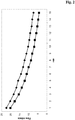
- Figure 1:
- Time course of permeate flow obtained with an anti-HER2 antibody solution with an antibody concentration of 222 mg/ml and an applied pressure of 2.0 bar (diamonds = 1.2 µm pore size filter containing combination; squares = 3.0 µm pore size filter containing combination).
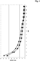
- Figure 2:
- Time course of permeate flow obtained with an anti-HER2 antibody solution with an antibody concentration of 125 mg/ml supplemented with about 200 mM trehalose and about 0.05 % (w/v)
Tween 20 and an applied pressure of 2.0 bar (diamonds = 1.2 µm pore size filter containing combination; squares = 3.0 µm pore size filter containing combination). - Figure 3:
- Time course of permeate flow obtained with an anti-HER2 antibody solution with an antibody concentration of 162 mg/ml and an applied pressure of 1.8 bar (diamonds = 1.2 µm pore size filter containing combination; squares = 3.0 µm pore size filter containing combination).
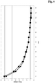
- Figure 4:
- Time course of permeate flow obtained with an anti-IL13Rα antibody solution with an antibody concentration of 141 mg/ml supplemented with about 200 mM trehalose and about 0.2 % (w/v) Poloxamer and an applied pressure of 1.6 bar (diamonds = 1.2 µm pore size filter containing combination; squares = 3.0 µm pore size filter containing combination).
- Figure 5:
- Time course of permeate flow obtained with an anti-HER2 antibody solution with an antibody concentration of 162 mg/ml and an applied pressure of 1.1 bar (diamonds = 1.2 µm pore size filter containing combination; squares = 3.0 µm pore size filter containing combination).
- Figure 6:
- Time course of permeate flow obtained with an anti-IL13Rα antibody solution with an antibody concentration of 141 mg/ml supplemented with trehalose and Poloxamer and an applied pressure of 0.8 bar (diamonds = 1.2 µm pore size filter containing combination; squares = 3.0 µm pore size filter containing combination).
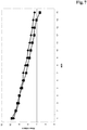
- Figure 7:
- Time course of permeate flow obtained with an anti-HER2 antibody solution with an antibody concentration of 125 mg/ml supplemented with trehalose and
Tween 20 and an applied pressure of 0.8 bar (diamonds = 1.2 µm pore size filter containing combination; squares = 3.0 µm pore size filter containing combination). - Figure 8:
- Time course of permeate flow obtained with an anti-HER2 antibody solution with an antibody concentration of 125 mg/ml supplemented with trehalose and
Tween 20 and an applied pressure of 0.3 bar (diamonds = 1.2 µm pore size filter containing combination; squares = 3.0 µm pore size filter containing combination). - An exemplary antibody is an immunoglobulin against the IL13 receptor α1 protein (anti-IL13Rα1 antibody) e.g. as reported in SEQ ID NO: 01 to 12 of
WO 2006/072564 (incorporated herein by reference). - Another exemplary immunoglobulin is an anti-HER2 antibody reported in
WO 92/022653 WO 99/057134 WO 97/04801 US 5,677,171 andUS 5,821,337 (incorporated herein by reference). - Herein among others a Sartobran P 0.45 µm + 0.2 µm filter cartridge and a Sartoclean CA 3.0 µm + 0.8 µm filter cartridge have been exemplarily employed. Both filter cartridges are available from Sartorius AG, Gottingen, Germany.
-
resin: TSK 3000 (Tosohaas) column: 300 x 7.8 mm flow rate: 0.5 ml/min buffer: 200 mM potassium phosphate containing 250 mM potassium chloride, adjusted to pH 7.0 wavelength: 280 nm DNA-threshold-system: see e.g. Merrick, H., and Hawlitschek, G., Biotech Forum Europe 9 (1992) 398-403 Protein A ELISA: The wells of a micro titer plate are coated with a polyclonal anti-protein A-IgG derived from chicken. After binding non-reacted antibody is removed by washing with sample buffer. For protein A binding a defined sample volume is added to the wells. The protein A present in the sample is bound by the chicken antibody and retained in the wells of the plate. After the incubation the sample solution is removed and the wells are washed. For detection are added subsequently a chicken derived polyclonal anti-protein A-IgG-biotin conjugate and a Streptavidin peroxidase conjugate. After a further washing step substrate solution is added resulting in the formation of a colored reaction product. The intensity of the color is proportional to the protein A content of the sample. After a defined time the reaction is stopped and the absorbance is measured. Host cell protein(HCP)ELISA: The walls of the wells of a micro titer plate are coated with a mixture of serum albumin and Streptavidin. A goat derived polyclonal antibody against HCP is bound to the walls of the wells of the micro titer plate. After a washing step different wells of the micro titer plate are incubated with a HCP calibration sequence of different concentrations and sample solution. After the incubation not bound sample material is removed by washing with buffer solution. For the detection the wells are incubated with an antibody peroxidase conjugate to detect bound host cell protein. The fixed peroxidase activity is detected by incubation with ABTS and detection at 405 nm. - In this example it is shown that a highly concentrated immunoglobulin solution cannot be filtered with a single sterile filter with a pore size of 0.45 µm (pre-filter) and 0.22 µm (main-filter) without blocking of the pores of the filter with a loading of more than 2,460 g protein per square meter of filter area.
- In this example a single filter with a pore size of 0.45 µm and 0.22 µm and a total filter area of 0.2 square meters has been employed.
Table 1: Solutions employed in the single filter filtration. solution No. 1 2 3 4 5 protein mass [g] 473 491 496 501 542 volume [1] 3.940 4.200 4.134 4.139 4.516 loading [g/m2] 2,365 2,455 2,480 2,505 2,710 - The concentrated immunoglobulin solutions were filtered through the single filter with the parameters as shown in Table 2.
Table 2: Process parameters. solution No. 1 2 3 4 5 volume flow [1/h] 1.97 2.1 Drop to 0 due to pore blocking Drop to 0 due to pore blocking Drop to 0 due to pore blocking mass flow [g/h] 237 246 Drop to 0 due to pore blocking Drop to 0 due to pore blocking Drop to 0 due to pore blocking - For solutions No. 3 to 5 the pores of the single filter were blocked prior to the complete filtration of the batch volume. To complete the filtration the blocked filter had to be changed resulting in additional time required and loss of product.
Table 3: Results of the filtration. solution No. 1 2 3 4 5 protein mass passing the filter [g/m2] 2,365 2,455 960 968 1,440 volume passing the filter [1] 3.940 4.200 1.600 1.600 2.400 pore blocking of the filter NO NO YES YES YES - In this example it is shown that a highly concentrated immunoglobulin solution can be filtered with a combination of two filters with a pore size of 3.0 µm (pre-filter) and 0.8 µm (main-filter) and of 0.45 µm (pre-filter) and 0.22 µm (main-filter) without blocking of the pores of the filter independent from the loading of protein per square meter of total filter area.
- In this example a combined filter with a first filter unit with a pore size of 3.0 µm and 0.8 µm, respectively, and a second filter unit with a pore size of 0.45 µm and 0.22 µm, respectively, and a filter area each of 0.6 square meters has been employed.
Table 4: Solutions employed in the combined filter filtration. solution No. 6 7 8 9 10 protein mass [g] 5,217 5,191 5,356 6,151 5,580 volume [1] 42.070 42.201 43.542 48.055 44.998 loading [g/m2] 4,347.5 4,325.8 4,463.3 5,125.8 4,650.0 - The concentrated immunoglobulin solutions were filtered through the combination of the two filters with the parameters as shown in Table 5.
Table 5: Process parameters. solution No. 6 7 8 9 10 volume flow [1/h] 38.95 42.20 43.54 33.02 45.00 mass flow [g/h] 4830 5191 5356 4226 5580 - For none of the solutions No. 6 to 10 the pores of the combined filters were blocked prior to the complete filtration of the batch volume.
Table 6: Results of the filtration. solution No. 6 7 8 9 10 protein mass passing the filter [g/m2] 4,347.5 4,325.8 4,463.3 5,125.8 4,650.0 volume passing the filter [1] 42.070 42.201 43.542 48.055 44.998 pore blocking of the filter NO NO NO NO NO - In this example it is shown that a conditioned protein A eluate can be filtered with a combination of two filters but the flow has to be reduced if the filter area does not match between the two filters.
- In this example a filter unit with a pore size of 3.0 µm (pre-filter) and 0.8 µm (main-filter) with a filter area of 1.8 square meters and a filter unit with a pore size of 0.45 µm (pre-filter) and 0.22 µm (main-filter) with a filter area of 0.6 square meters has been employed.
Table 7: Solutions employed in the combined filter filtration. solution No. 11 12 13 14 15 protein mass [g] 1,169.0 1,299.6 1,154.4 1,220.4 1,284.7 volume [1] 71.4 76.0 74.0 67.8 70.2 loading [g/m2] 487.1 541.5 481.0 508.5 535.3 - The concentrated immunoglobulin solutions were filtered through the combined filter with the parameters as shown in Table 8.
Table 8: Process parameters. solution No. 11 12 13 14 15 volume flow [1/h] Drop to 0 due to pore blocking 22 13 12 98 mass flow [g/h] Drop to 0 due to pore blocking 376 203 216 1793 - For solution No. 11 the pores of the combined filter were blocked prior to the complete filtration of the batch volume. To complete the filtration the blocked filter had to be changed resulting in additional time required and loss of product.
Table 9: Results of the filtration. solution No. 1 2 3 4 5 protein mass passing the filter [g/m2] 347.9 541.5 481.0 508.5 535.3 volume passing the filter [1] 51.0 76.0 74.0 67.8 70.2 pore blocking of the filter YES NO NO NO NO - In order to prevent filter blocking as in the experiment with solution No. 11 the flow through the membrane had to be reduced in experiments with solutions No. 12 to 14. In experiment with solution No. 15 the protein A eluate has been decanted resulting in a loss of protein.
- In this example it is shown that a conditioned protein A eluate can be filtered with a combination of two filters without a reduction of the flow if the filter area does match between the two filters.
- In this example the filter unit with a pore size of 3.0 µm and 0.8 µm has a filter area of 0.2 square meters and the filter unit with a pore size of 0.45 µm and 0.22 µm has a filter area of 0.2 square meters.
Table 10: Solutions employed in the combined filter filtration. solution No. 16 17 18 19 20 protein mass [g] 495 634 825 861 956 volume [1] 3.5 4.14 5.5 5.6 6.3 loading [g/m2] 1,237.5 1,585.0 2,062.5 2,152.5 2,390 - For none of the solutions No. 16 to 20 the pores of the combined filters were blocked prior to the complete filtration of the batch volume.
Table 11: Results of the filtration. solution No. 16 17 18 19 20 Protein mass passing the filter [g/m2] 1,237.5 1,585.0 2,062.5 2,152.5 2,390 Volume passing the filter [1] 3.5 4.14 5.5 5.6 6.3 Pore blocking of the filter NO NO NO NO NO - Solutions comprising either an anti-IL13Rα antibody or an anti-HER2 antibody were filtered with a filter combination employing different filter area and filter pore size as well as different excipients and applied pressure.
- The used filter combinations are listed in Table 12. In the following the denotation 'A1', 'A2', 'B1', and 'B2' will be used therefore.
Table 12: Filter combinations combination filter 1 pore size / diameter filter 2 pore size / diameter filter 3 pore size / diameter filter 4 pore size / diameter A1 1.2 µm / 26 mm 0.8 µm / 26 mm 0.45 µm / 26 mm 0.2 µm / 26 mm A2 1.2 µm / 47 mm 0.8 µm / 26 mm 0.45 µm / 26 mm 0.2 µm / 26 mm B1 3.0 µm / 26 mm 0.8 µm / 26 mm 0.45 µm / 26 mm 0.2 µm / 26 mm B2 3.0 µm / 47 mm 0.8 µm / 26 mm 0.45 µm / 26 mm 0.2 µm / 26 mm - In the following Tables 13 to 20 and in corresponding
Figures 1 to 8 the results obtained with different filter combinations, different antibody solutions and different filtering conditions are presented.Table 13: Results obtained with an anti-HER2 antibody solution with an antibody concentration of 222 mg/ml and an applied pressure of 2.0 bar. combination filtration duration [min] flow [ml/min] combination filtration duration [min] flow [ml/min] A1 1 3.7 B1 1 3.4 2 3.5 2 3.2 3 3.2 3 3.1 4 3.0 4 3.0 5 2.9 5 2.8 6 2.6 6 2.9 7 2.5 7 2.8 8 2.3 8 2.7 9 2.0 9 2.7 10 2.0 10 2.7 11 1.7 11 2.7 12 1.6 12 2.5 13 1.5 13 2.6 14 1.3 14 2.5 15 1.2 15 2.5 Table 14: Results obtained with an anti-HER2 antibody solution with an antibody concentration of 125 mg/ml supplemented with about 200 mM trehalose and about 0.05 % (w/v) Tween 20 and an applied pressure of 2.0 bar.combination filtration duration [min] flow [ml/min] combination filtration duration [min] flow [ml/min] A1 1 22.4 B1 1 20.1 2 20.2 2 17.7 3 18.3 3 15.5 4 16.8 4 13.8 5 15.9 5 12.2 6 14.3 6 11.1 7 13.1 7 10.0 8 12.3 8 8.7 9 11.3 9 8.1 10 10.3 10 7.0 11 9.7 11 6.6 12 9.2 12 5.8 13 8.4 13 5.2 14 8.1 14 4.8 15 7.4 15 4.3 Table 15: Results obtained with an anti-HER2 antibody solution with an antibody concentration of 162 mg/ml and an applied pressure of 1.8 bar. combination filtration duration [min] flow [ml/min] combination filtration duration [min] flow [ml/min] A2 1 7.6 B2 1 8.1 2 6.5 2 6.9 3 6.1 3 6.4 4 5.7 4 6.2 5 5.4 5 5.9 6 5.1 6 5.6 7 5.2 7 5.5 8 5.0 8 5.3 9 4.9 9 5.2 10 4.7 10 5.1 11 4.8 11 5.0 12 4.8 12 4.8 13 4.6 13 4.9 14 4.7 14 4.6 15 4.6 15 4.6 Table 16: Results obtained with an anti-IL13Rα antibody solution with an antibody concentration of 141 mg/ml supplemented with about 200 mM trehalose and about 0.2 % (w/v) Poloxamer and an applied pressure of 1.6 bar. combination filtration duration [min] flow [ml/min] combination filtration duration [min] flow [ml/min] A2 1 15.6 B2 1 13.2 2 9.4 2 8.1 3 7.0 3 5.5 4 5.5 4 4.1 5 4.6 5 3.3 6 3.8 6 2.6 7 3.3 7 2.3 8 2.9 8 1.9 9 2.5 9 1.6 10 2.2 10 1.5 11 1.5 11 1.2 12 0.5 12 1.2 13 0.3 13 1.0 14 0.3 14 0.9 15 0.3 15 0.8 Table 17: Results obtained with an anti-HER2 antibody solution with an antibody concentration of 162 mg/ml and an applied pressure of 1.1 bar. combination filtration duration [min] flow [ml/min] combination filtration duration [min] flow [ml/min] A1 1 4.4 B1 1 4.3 2 4.0 2 4.0 3 3.6 3 3.5 4 3.5 4 3.0 5 3.3 5 3.0 6 3.2 6 3.0 7 3.2 7 2.9 8 3.1 8 2.8 9 3.1 9 2.8 10 2.9 10 2.7 11 3.0 11 2.6 12 2.9 12 2.8 13 2.8 13 2.5 14 2.8 14 2.6 15 2.8 15 2.5 Table 18: Results obtained with an anti-IL13Rα antibody solution with an antibody concentration of 141 mg/ml supplemented with trehalose and Poloxamer and an applied pressure of 0.8 bar. combination filtration duration [min] flow [ml/min] combination filtration duration [min] flow [ml/min] A2 1 7.6 B2 1 8.1 2 5.0 2 5.5 3 3.7 3 4.2 4 2.9 4 3.1 5 2.5 5 2.6 6 2.1 6 2.2 7 1.8 7 1.8 8 1.5 8 1.5 9 1.4 9 1.4 10 1.2 10 1.2 11 1.1 11 1.1 12 1.0 12 1.0 13 0.9 13 0.8 14 0.8 14 0.8 15 0.8 15 0.8 Table 19: Results obtained with an anti-HER2 antibody solution with an antibody concentration of 125 mg/ml supplemented with trehalose and Tween 20 and an applied pressure of 0.8 bar.combination filtration duration [min] flow [ml/min] combination filtration duration [min] flow [ml/min] A1 1 9.3 B1 1 9.7 2 8.7 2 8.8 3 8.1 3 8.4 4 7.9 4 8.0 5 7.7 5 7.4 6 7.2 6 7.0 7 7.1 7 6.4 8 6.6 8 6.1 9 6.2 9 5.7 10 6.0 10 5.4 11 5.6 11 5.0 12 5.3 12 4.6 13 5.0 13 4.5 14 4.8 14 4.1 15 4.5 15 3.3 Table 20: Results obtained with an anti-HER2 antibody solution with an antibody concentration of 125 mg/ml supplemented with trehalose and Tween 20 and an applied pressure of 0.3 bar.combination filtration duration [min] flow [ml/min] combination filtration duration [min] flow [ml/min] A1 1 3.9 B1 1 3.7 2 3.2 2 4.8 3 3.0 3 4.6 4 2.7 4 3.8 5 2.6 5 4.0 6 2.3 6 3.8 7 2.1 7 3.8 8 2.0 8 3.7 9 1.8 9 3.6 10 1.5 10 3.6 11 1.4 11 3.5 12 1.3 12 3.5 13 1.2 13 3.3 14 1.1 14 3.3 15 1.1 15 3.2
Claims (10)
- Method for producing an immunoglobulin comprising the following stepsa) purifying the immunoglobulin with one or more chromatography steps, and providing an immunoglobulin solution,b) concentrating the immunoglobulin solution to a concentration of 100 g/l or more with a method selected from diafiltration or tangential-flow filtration, andc) applying the immunoglobulin solution of the previous step to a combination of a first and second filter unit, whereby the first filter unit comprises a pre-filter with a pore size of 3.0 µm and a main-filter with a pore size of 0.8 µm and the second filter unit comprises a pre-filter with a pore size of 0.45 µm and a main-filter with a pore size of 0.22 µm with a pressure of from 0.1 to 4.0 bar, and thereby producing an immunoglobulin.
- The method according to claim 1, further comprising the following step after step a)
a-b) adding a sugar, an amino acid and/or a detergent to the solution, - Method according to any one of the preceding claims, characterized in that the filter in the first and second filter unit have about the same filter area.
- Method according to any one of the preceding claims, characterized in that the immunoglobulin solution has a concentration of from 100 g/l to 300 g/l.
- Method according to any one of the preceding claims, characterized in that the immunoglobulin solution has a volume of from 3 liter to 100 liter.
- Method according to any one of the preceding claims, characterized in that the immunoglobulin is an anti-IL13 receptor alpha antibody or an anti-HER2 antibody.
- Method according to any one of claims 2 to 6, characterized in that the purifying is with a protein A affinity chromatography step and at least one step selected from cation exchange chromatography, anion exchange chromatography, and hydrophobic interaction chromatography.
- Method according to any one of the preceding claims, characterized in that the immunoglobulin solution has a concentration of 160 g/l or more and the applying to the combination of filters is by applying a pressure of 1.45 bar or more.
- Method according to any one of claims 1 to 7, characterized in that the immunoglobulin solution comprises a sugar and a surfactant and has a concentration of 125 mg/ml or more and the applying to the combination of the filter is by applying a pressure of 0.75 bar or less.
- Kit comprising a first filter with a pore size of from 3.0 µm to 0.8 µm and a second filter with a pore size of from 0.45 µm to 0.22 µm.
Applications Claiming Priority (4)
| Application Number | Priority Date | Filing Date | Title |
|---|---|---|---|
| EP09012460 | 2009-10-01 | ||
| EP16188558.7A EP3133083B1 (en) | 2009-10-01 | 2010-09-29 | Multistep final filtration |
| EP10757795.9A EP2483305B1 (en) | 2009-10-01 | 2010-09-29 | Multistep final filtration of immunoglobulin |
| PCT/EP2010/064487 WO2011039274A1 (en) | 2009-10-01 | 2010-09-29 | Multistep final filtration |
Related Parent Applications (3)
| Application Number | Title | Priority Date | Filing Date |
|---|---|---|---|
| EP16188558.7A Division EP3133083B1 (en) | 2009-10-01 | 2010-09-29 | Multistep final filtration |
| PCT/EP2010/064487 Previously-Filed-Application WO2011039274A1 (en) | 2009-10-01 | 2010-09-29 | Multistep final filtration |
| EP10757795.9A Division EP2483305B1 (en) | 2009-10-01 | 2010-09-29 | Multistep final filtration of immunoglobulin |
Publications (1)
| Publication Number | Publication Date |
|---|---|
| EP3715369A1 true EP3715369A1 (en) | 2020-09-30 |
Family
ID=41466840
Family Applications (3)
| Application Number | Title | Priority Date | Filing Date |
|---|---|---|---|
| EP16188558.7A Active EP3133083B1 (en) | 2009-10-01 | 2010-09-29 | Multistep final filtration |
| EP10757795.9A Active EP2483305B1 (en) | 2009-10-01 | 2010-09-29 | Multistep final filtration of immunoglobulin |
| EP20156152.9A Pending EP3715369A1 (en) | 2009-10-01 | 2010-09-29 | Multistep final filtration |
Family Applications Before (2)
| Application Number | Title | Priority Date | Filing Date |
|---|---|---|---|
| EP16188558.7A Active EP3133083B1 (en) | 2009-10-01 | 2010-09-29 | Multistep final filtration |
| EP10757795.9A Active EP2483305B1 (en) | 2009-10-01 | 2010-09-29 | Multistep final filtration of immunoglobulin |
Country Status (21)
| Country | Link |
|---|---|
| US (4) | US20120231506A1 (en) |
| EP (3) | EP3133083B1 (en) |
| JP (1) | JP5458183B2 (en) |
| KR (3) | KR101508044B1 (en) |
| CN (3) | CN102574913A (en) |
| AU (1) | AU2010302662B2 (en) |
| BR (1) | BR112012004054B1 (en) |
| CA (1) | CA2773674C (en) |
| CY (1) | CY1118577T1 (en) |
| DK (1) | DK2483305T3 (en) |
| ES (2) | ES2787403T3 (en) |
| HK (2) | HK1200178A1 (en) |
| HR (2) | HRP20161693T1 (en) |
| HU (1) | HUE030186T2 (en) |
| IL (2) | IL217626A (en) |
| LT (1) | LT2483305T (en) |
| MX (1) | MX342442B (en) |
| PL (2) | PL2483305T3 (en) |
| PT (1) | PT2483305T (en) |
| SI (2) | SI2483305T1 (en) |
| WO (1) | WO2011039274A1 (en) |
Families Citing this family (2)
| Publication number | Priority date | Publication date | Assignee | Title |
|---|---|---|---|---|
| WO2019183334A1 (en) | 2018-03-21 | 2019-09-26 | Waters Technologies Corporation | Non-antibody high-affinity-based sample preparation, sorbents, devices and methods |
| CN111007242A (en) * | 2019-12-20 | 2020-04-14 | 苏州和迈精密仪器有限公司 | Fluorescence immunoassay method and device based on multilayer high molecular porous membrane and application |
Citations (4)
| Publication number | Priority date | Publication date | Assignee | Title |
|---|---|---|---|---|
| US4592848A (en) * | 1984-12-11 | 1986-06-03 | Pabst Richard E | Flow through filter with backflush clearing capability |
| CS247484B1 (en) * | 1985-04-12 | 1987-01-15 | Eva Hamsikova | A method of preparing an immunoglobulin against human lymphocytes |
| US20080247949A1 (en) * | 2004-11-22 | 2008-10-09 | Oxygenix Co., Ltd. | Biological Tissue Examination Agent Comprising Hemoglobin Labeled With Oxygen Isotope And Method For Producing The Same |
| WO2009010269A1 (en) * | 2007-07-17 | 2009-01-22 | F.Hoffmann-La Roche Ag | Variable tangential flow filtration |
Family Cites Families (26)
| Publication number | Priority date | Publication date | Assignee | Title |
|---|---|---|---|---|
| US5643566A (en) | 1982-09-23 | 1997-07-01 | Cetus Corporation | Formulation processes for lipophilic proteins |
| US4488961A (en) | 1982-09-29 | 1984-12-18 | E. I. Du Pont De Nemours And Company | One-way filter unit |
| WO1989006692A1 (en) | 1988-01-12 | 1989-07-27 | Genentech, Inc. | Method of treating tumor cells by inhibiting growth factor receptor function |
| DE3818860A1 (en) | 1988-06-03 | 1989-12-07 | Seitz Filter Werke | FILTER ELEMENT |
| ATE255131T1 (en) | 1991-06-14 | 2003-12-15 | Genentech Inc | HUMANIZED HEREGULIN ANTIBODIES |
| DE4204444C1 (en) | 1992-02-14 | 1993-05-19 | Krankenhausentsorgungs Gmbh, 1000 Berlin, De | |
| CA2226575C (en) | 1995-07-27 | 2011-10-18 | Genentech, Inc. | Stabile isotonic lyophilized protein formulation |
| ES2337091T3 (en) | 1996-11-27 | 2010-04-20 | Genentech, Inc. | PURIFICATION FOR POLYPEPTIDE AFFINITY IN A PROTEIN MATRIX. |
| BRPI9910332B8 (en) | 1998-05-06 | 2021-05-25 | Genentech Inc | method for purifying a polypeptide from a composition |
| US6984494B2 (en) * | 2000-08-15 | 2006-01-10 | Genentech, Inc. | Analytical method |
| CA2473144C (en) | 2002-02-05 | 2013-05-28 | Genentech, Inc. | Protein purification |
| EP1551875A4 (en) * | 2002-06-21 | 2006-06-28 | Biogen Idec Inc | Buffered formulations for concentrating antibodies and methods of use thereof |
| US20060185025A1 (en) * | 2002-10-04 | 2006-08-17 | Kirin Beer Kabushiki Kaisha | Human artificial chromosome (hac) vector |
| JPWO2004087761A1 (en) | 2003-03-31 | 2006-07-27 | 麒麟麦酒株式会社 | Purification of human monoclonal antibodies and human polyclonal antibodies |
| GB0316560D0 (en) * | 2003-07-15 | 2003-08-20 | Chiron Srl | Vesicle filtration |
| AU2005247950B2 (en) * | 2004-05-27 | 2012-02-02 | Receptor Logic, Inc. | Antibodies as T cell receptor mimics, methods of production and uses thereof |
| AU2005277190B2 (en) * | 2004-08-20 | 2011-03-03 | American National Red Cross | Sequential protein isolation and purification schemes by affinity chromatography |
| US20060051347A1 (en) * | 2004-09-09 | 2006-03-09 | Winter Charles M | Process for concentration of antibodies and therapeutic products thereof |
| TW200902555A (en) | 2005-01-03 | 2009-01-16 | Hoffmann La Roche | Antibodies against IL-13 receptor alpha 1 and uses thereof |
| LT2620450T (en) * | 2005-03-08 | 2019-02-11 | Pfizer Products Inc. | ANTI-CTLA-4 ANTIBODY COMPOSITIONS |
| WO2007018316A1 (en) | 2005-08-09 | 2007-02-15 | Takeda Pharmaceutical Company Limited | Prophylactic/therapeutic agent for cancer |
| US9109015B2 (en) * | 2006-11-01 | 2015-08-18 | Biogen Ma Inc | Method of isolating biomacromolecules using low pH and divalent cations |
| CL2008002054A1 (en) | 2007-07-17 | 2009-05-29 | Hoffmann La Roche | Method for the regeneration of a cation exchange chromatography column after elusion of monopeglated erythropoietin and method to obtain a monopeglated erythropoietin, incorporating the regeneration method of the cation exchange column. |
| WO2009058769A1 (en) | 2007-10-30 | 2009-05-07 | Schering Corporation | Purification of antibodies containing hydrophobic variants |
| CA2739352C (en) | 2008-10-29 | 2021-07-13 | Wyeth Llc | Methods for purification of single domain antigen binding molecules |
| ES2706190T3 (en) * | 2012-10-30 | 2019-03-27 | Hoffmann La Roche | Purification of polypeptides using two-phase tangential flow ultrafiltration |
-
2010
- 2010-09-29 PT PT107577959T patent/PT2483305T/en unknown
- 2010-09-29 BR BR112012004054-5A patent/BR112012004054B1/en active IP Right Grant
- 2010-09-29 CA CA2773674A patent/CA2773674C/en active Active
- 2010-09-29 KR KR1020127008385A patent/KR101508044B1/en active Active
- 2010-09-29 DK DK10757795.9T patent/DK2483305T3/en active
- 2010-09-29 EP EP16188558.7A patent/EP3133083B1/en active Active
- 2010-09-29 EP EP10757795.9A patent/EP2483305B1/en active Active
- 2010-09-29 EP EP20156152.9A patent/EP3715369A1/en active Pending
- 2010-09-29 PL PL10757795T patent/PL2483305T3/en unknown
- 2010-09-29 KR KR1020167023445A patent/KR101718301B1/en active Active
- 2010-09-29 PL PL16188558T patent/PL3133083T3/en unknown
- 2010-09-29 MX MX2012003727A patent/MX342442B/en active IP Right Grant
- 2010-09-29 ES ES16188558T patent/ES2787403T3/en active Active
- 2010-09-29 WO PCT/EP2010/064487 patent/WO2011039274A1/en not_active Ceased
- 2010-09-29 LT LTEP10757795.9T patent/LT2483305T/en unknown
- 2010-09-29 HU HUE10757795A patent/HUE030186T2/en unknown
- 2010-09-29 SI SI201031343A patent/SI2483305T1/en unknown
- 2010-09-29 HR HRP20161693TT patent/HRP20161693T1/en unknown
- 2010-09-29 CN CN2010800444816A patent/CN102574913A/en active Pending
- 2010-09-29 AU AU2010302662A patent/AU2010302662B2/en active Active
- 2010-09-29 SI SI201032002T patent/SI3133083T1/en unknown
- 2010-09-29 JP JP2012531416A patent/JP5458183B2/en active Active
- 2010-09-29 ES ES10757795.9T patent/ES2604103T3/en active Active
- 2010-09-29 CN CN201410246761.5A patent/CN103980346A/en active Pending
- 2010-09-29 KR KR1020147016901A patent/KR101653471B1/en active Active
- 2010-09-29 CN CN201510004012.6A patent/CN104610447A/en active Pending
- 2010-09-29 US US13/394,766 patent/US20120231506A1/en not_active Abandoned
-
2012
- 2012-01-19 IL IL217626A patent/IL217626A/en active IP Right Grant
-
2015
- 2015-01-21 HK HK15100670.0A patent/HK1200178A1/en unknown
- 2015-10-28 HK HK15110663.8A patent/HK1210183A1/en unknown
-
2016
- 2016-11-25 CY CY20161101229T patent/CY1118577T1/en unknown
-
2017
- 2017-02-27 US US15/444,018 patent/US20180009878A1/en not_active Abandoned
- 2017-11-14 IL IL255658A patent/IL255658A/en active IP Right Grant
-
2019
- 2019-10-22 US US16/660,635 patent/US11891430B2/en active Active
-
2020
- 2020-04-14 HR HRP20200588TT patent/HRP20200588T1/en unknown
-
2023
- 2023-12-20 US US18/389,809 patent/US20240228589A9/en active Pending
Patent Citations (4)
| Publication number | Priority date | Publication date | Assignee | Title |
|---|---|---|---|---|
| US4592848A (en) * | 1984-12-11 | 1986-06-03 | Pabst Richard E | Flow through filter with backflush clearing capability |
| CS247484B1 (en) * | 1985-04-12 | 1987-01-15 | Eva Hamsikova | A method of preparing an immunoglobulin against human lymphocytes |
| US20080247949A1 (en) * | 2004-11-22 | 2008-10-09 | Oxygenix Co., Ltd. | Biological Tissue Examination Agent Comprising Hemoglobin Labeled With Oxygen Isotope And Method For Producing The Same |
| WO2009010269A1 (en) * | 2007-07-17 | 2009-01-22 | F.Hoffmann-La Roche Ag | Variable tangential flow filtration |
Non-Patent Citations (2)
| Title |
|---|
| ALDINGTON ET AL: "Scale-up of monoclonal antibody purification processes", JOURNAL OF CHROMATOGRAPHY B: BIOMEDICAL SCIENCES & APPLICATIONS, ELSEVIER, AMSTERDAM, NL, vol. 848, no. 1, 12 March 2007 (2007-03-12), pages 64 - 78, XP005922828, ISSN: 1570-0232 * |
| HONIG ET AL: "Impact of design and selection of prefilters on operating cost", FILTRATION AND SEPARATION, ELSEVIER ADVANCED TECHNOLOGY, OXFORD, GB, vol. 34, no. 1, 1 January 1997 (1997-01-01), pages 73 - 78, XP005635274, ISSN: 0015-1882 * |
Also Published As
Similar Documents
| Publication | Publication Date | Title |
|---|---|---|
| US20240228589A9 (en) | Multistep final filtration | |
| EP2152745B1 (en) | Immunoglobulin purification | |
| EP2462157B1 (en) | Methods for purifying a target protein from one or more impurities in a sample | |
| US12600748B2 (en) | Affinity chromatography of immunoglobulins by using pre-capture flocculation | |
| CN118994410A (en) | Continuous manufacturing process for manufacturing biological agents by integrating pharmaceutical substance and pharmaceutical product processes | |
| JP2010051927A (en) | Method of separating biogenic substance using ultra-filtration membrane, module and device | |
| JPWO2020004583A1 (en) | Peptide separation method, polypeptide production method and polypeptide purification device | |
| CA3203540A1 (en) | Systems and methods for process scale isolation of immunoglobulin g | |
| CN113646066B (en) | Protein purification methods | |
| RU2794431C1 (en) | Improving immunoglobulin affinity chromatography by the application of pre-capture floculation | |
| EA049616B1 (en) | CONTINUOUS METHOD FOR MANUFACTURING BIOLOGICAL PRODUCTS BY INTEGRATION OF THE PROCESSES OF OBTAINING A DRUG SUBSTANCE AND A DRUG |
Legal Events
| Date | Code | Title | Description |
|---|---|---|---|
| PUAI | Public reference made under article 153(3) epc to a published international application that has entered the european phase |
Free format text: ORIGINAL CODE: 0009012 |
|
| STAA | Information on the status of an ep patent application or granted ep patent |
Free format text: STATUS: THE APPLICATION HAS BEEN PUBLISHED |
|
| AC | Divisional application: reference to earlier application |
Ref document number: 2483305 Country of ref document: EP Kind code of ref document: P Ref document number: 3133083 Country of ref document: EP Kind code of ref document: P |
|
| AK | Designated contracting states |
Kind code of ref document: A1 Designated state(s): AL AT BE BG CH CY CZ DE DK EE ES FI FR GB GR HR HU IE IS IT LI LT LU LV MC MK MT NL NO PL PT RO SE SI SK SM TR |
|
| STAA | Information on the status of an ep patent application or granted ep patent |
Free format text: STATUS: REQUEST FOR EXAMINATION WAS MADE |
|
| 17P | Request for examination filed |
Effective date: 20210330 |
|
| RBV | Designated contracting states (corrected) |
Designated state(s): AL AT BE BG CH CY CZ DE DK EE ES FI FR GB GR HR HU IE IS IT LI LT LU LV MC MK MT NL NO PL PT RO SE SI SK SM TR |
|
| STAA | Information on the status of an ep patent application or granted ep patent |
Free format text: STATUS: EXAMINATION IS IN PROGRESS |
|
| 17Q | First examination report despatched |
Effective date: 20241205 |