EP3714894A1 - Suicide gene therapeutic agent for brain tumors using pluripotent stem cell - Google Patents
Suicide gene therapeutic agent for brain tumors using pluripotent stem cell Download PDFInfo
- Publication number
- EP3714894A1 EP3714894A1 EP18878961.4A EP18878961A EP3714894A1 EP 3714894 A1 EP3714894 A1 EP 3714894A1 EP 18878961 A EP18878961 A EP 18878961A EP 3714894 A1 EP3714894 A1 EP 3714894A1
- Authority
- EP
- European Patent Office
- Prior art keywords
- gene
- stem cells
- cells
- neural stem
- uprt
- Prior art date
- Legal status (The legal status is an assumption and is not a legal conclusion. Google has not performed a legal analysis and makes no representation as to the accuracy of the status listed.)
- Granted
Links
Images








Classifications
-
- A—HUMAN NECESSITIES
- A61—MEDICAL OR VETERINARY SCIENCE; HYGIENE
- A61K—PREPARATIONS FOR MEDICAL, DENTAL OR TOILETRY PURPOSES
- A61K35/00—Medicinal preparations containing materials or reaction products thereof with undetermined constitution
- A61K35/12—Materials from mammals; Compositions comprising non-specified tissues or cells; Compositions comprising non-embryonic stem cells; Genetically modified cells
- A61K35/30—Nerves; Brain; Eyes; Corneal cells; Cerebrospinal fluid; Neuronal stem cells; Neuronal precursor cells; Glial cells; Oligodendrocytes; Schwann cells; Astroglia; Astrocytes; Choroid plexus; Spinal cord tissue
-
- A—HUMAN NECESSITIES
- A61—MEDICAL OR VETERINARY SCIENCE; HYGIENE
- A61K—PREPARATIONS FOR MEDICAL, DENTAL OR TOILETRY PURPOSES
- A61K31/00—Medicinal preparations containing organic active ingredients
- A61K31/33—Heterocyclic compounds
- A61K31/395—Heterocyclic compounds having nitrogen as a ring hetero atom, e.g. guanethidine or rifamycins
- A61K31/495—Heterocyclic compounds having nitrogen as a ring hetero atom, e.g. guanethidine or rifamycins having six-membered rings with two or more nitrogen atoms as the only ring heteroatoms, e.g. piperazine or tetrazines
- A61K31/505—Pyrimidines; Hydrogenated pyrimidines, e.g. trimethoprim
- A61K31/513—Pyrimidines; Hydrogenated pyrimidines, e.g. trimethoprim having oxo groups directly attached to the heterocyclic ring, e.g. cytosine
-
- A—HUMAN NECESSITIES
- A61—MEDICAL OR VETERINARY SCIENCE; HYGIENE
- A61K—PREPARATIONS FOR MEDICAL, DENTAL OR TOILETRY PURPOSES
- A61K35/00—Medicinal preparations containing materials or reaction products thereof with undetermined constitution
- A61K35/12—Materials from mammals; Compositions comprising non-specified tissues or cells; Compositions comprising non-embryonic stem cells; Genetically modified cells
- A61K35/48—Reproductive organs
- A61K35/54—Ovaries; Ova; Ovules; Embryos; Foetal cells; Germ cells
- A61K35/545—Embryonic stem cells; Pluripotent stem cells; Induced pluripotent stem cells; Uncharacterised stem cells
-
- A—HUMAN NECESSITIES
- A61—MEDICAL OR VETERINARY SCIENCE; HYGIENE
- A61P—SPECIFIC THERAPEUTIC ACTIVITY OF CHEMICAL COMPOUNDS OR MEDICINAL PREPARATIONS
- A61P35/00—Antineoplastic agents
-
- C—CHEMISTRY; METALLURGY
- C12—BIOCHEMISTRY; BEER; SPIRITS; WINE; VINEGAR; MICROBIOLOGY; ENZYMOLOGY; MUTATION OR GENETIC ENGINEERING
- C12N—MICROORGANISMS OR ENZYMES; COMPOSITIONS THEREOF; PROPAGATING, PRESERVING, OR MAINTAINING MICROORGANISMS; MUTATION OR GENETIC ENGINEERING; CULTURE MEDIA
- C12N15/00—Mutation or genetic engineering; DNA or RNA concerning genetic engineering, vectors, e.g. plasmids, or their isolation, preparation or purification; Use of hosts therefor
- C12N15/09—Recombinant DNA-technology
- C12N15/63—Introduction of foreign genetic material using vectors; Vectors; Use of hosts therefor; Regulation of expression
- C12N15/635—Externally inducible repressor mediated regulation of gene expression, e.g. tetR inducible by tetracyline
-
- C—CHEMISTRY; METALLURGY
- C12—BIOCHEMISTRY; BEER; SPIRITS; WINE; VINEGAR; MICROBIOLOGY; ENZYMOLOGY; MUTATION OR GENETIC ENGINEERING
- C12N—MICROORGANISMS OR ENZYMES; COMPOSITIONS THEREOF; PROPAGATING, PRESERVING, OR MAINTAINING MICROORGANISMS; MUTATION OR GENETIC ENGINEERING; CULTURE MEDIA
- C12N5/00—Undifferentiated human, animal or plant cells, e.g. cell lines; Tissues; Cultivation or maintenance thereof; Culture media therefor
- C12N5/06—Animal cells or tissues; Human cells or tissues
- C12N5/0602—Vertebrate cells
- C12N5/0618—Cells of the nervous system
- C12N5/0623—Stem cells
-
- C—CHEMISTRY; METALLURGY
- C12—BIOCHEMISTRY; BEER; SPIRITS; WINE; VINEGAR; MICROBIOLOGY; ENZYMOLOGY; MUTATION OR GENETIC ENGINEERING
- C12N—MICROORGANISMS OR ENZYMES; COMPOSITIONS THEREOF; PROPAGATING, PRESERVING, OR MAINTAINING MICROORGANISMS; MUTATION OR GENETIC ENGINEERING; CULTURE MEDIA
- C12N9/00—Enzymes; Proenzymes; Compositions thereof; Processes for preparing, activating, inhibiting, separating or purifying enzymes
- C12N9/10—Transferases (2.)
- C12N9/1048—Glycosyltransferases (2.4)
- C12N9/1077—Pentosyltransferases (2.4.2)
-
- C—CHEMISTRY; METALLURGY
- C12—BIOCHEMISTRY; BEER; SPIRITS; WINE; VINEGAR; MICROBIOLOGY; ENZYMOLOGY; MUTATION OR GENETIC ENGINEERING
- C12N—MICROORGANISMS OR ENZYMES; COMPOSITIONS THEREOF; PROPAGATING, PRESERVING, OR MAINTAINING MICROORGANISMS; MUTATION OR GENETIC ENGINEERING; CULTURE MEDIA
- C12N9/00—Enzymes; Proenzymes; Compositions thereof; Processes for preparing, activating, inhibiting, separating or purifying enzymes
- C12N9/14—Hydrolases (3)
- C12N9/78—Hydrolases (3) acting on carbon to nitrogen bonds other than peptide bonds (3.5)
-
- C—CHEMISTRY; METALLURGY
- C12—BIOCHEMISTRY; BEER; SPIRITS; WINE; VINEGAR; MICROBIOLOGY; ENZYMOLOGY; MUTATION OR GENETIC ENGINEERING
- C12Y—ENZYMES
- C12Y204/00—Glycosyltransferases (2.4)
- C12Y204/02—Pentosyltransferases (2.4.2)
- C12Y204/02009—Uracil phosphoribosyltransferase (2.4.2.9)
-
- C—CHEMISTRY; METALLURGY
- C12—BIOCHEMISTRY; BEER; SPIRITS; WINE; VINEGAR; MICROBIOLOGY; ENZYMOLOGY; MUTATION OR GENETIC ENGINEERING
- C12Y—ENZYMES
- C12Y305/00—Hydrolases acting on carbon-nitrogen bonds, other than peptide bonds (3.5)
- C12Y305/04—Hydrolases acting on carbon-nitrogen bonds, other than peptide bonds (3.5) in cyclic amidines (3.5.4)
- C12Y305/04001—Cytosine deaminase (3.5.4.1)
-
- C—CHEMISTRY; METALLURGY
- C12—BIOCHEMISTRY; BEER; SPIRITS; WINE; VINEGAR; MICROBIOLOGY; ENZYMOLOGY; MUTATION OR GENETIC ENGINEERING
- C12N—MICROORGANISMS OR ENZYMES; COMPOSITIONS THEREOF; PROPAGATING, PRESERVING, OR MAINTAINING MICROORGANISMS; MUTATION OR GENETIC ENGINEERING; CULTURE MEDIA
- C12N2506/00—Differentiation of animal cells from one lineage to another; Differentiation of pluripotent cells
- C12N2506/45—Differentiation of animal cells from one lineage to another; Differentiation of pluripotent cells from artificially induced pluripotent stem cells
Definitions
- the present invention relates to a cell preparation for treating brain tumors and a method for preparing neural stem cells used for the cell preparation.
- NSCs neural stem cells
- Patent Literature 1 Japanese Patent Laid-Open No. 2006-345726
- neural stem cells or mesenchymal stem cells As mentioned above, it is difficult, including ethical issues, to obtain an adequate amount of neural stem cells which can be administered to humans clinically. It is easier to obtain mesenchymal stem cells than neural stem cells. As long as mesenchymal stem cells are collected from the living body, the amount thereof collected is however limited.
- the herpes simplex virus thymidine kinase gene (HSVtk) is generally used as a suicide gene (for example, this gene is used in Examples in Patent Literature 1), and besides this, the cytosine deaminase (CD) gene and the uracil phosphoribosyltransferase (UPRT) gene are known. It is known that even though the CD gene and the UPRT gene are used alone, the CD gene and the UPRT gene have antitumor effects, and the antitumor effect improves synergistically by combining the two (a gene into which these two genes are combined may be called a "CD-UPRT gene" hereinafter).
- CD-UPRT gene a gene into which these two genes are combined
- An object of the present invention is to solve the difficulty of obtaining neural stem cells and mesenchymal stem cells, which is problematic, in such a context and to provide new means for treating brain tumors using the CD-UPRT gene, which has a high antitumor effect.
- the present inventor has earnestly examined repeatedly to solve the above-mentioned problem and consequently found that inducing the differentiation of neural stem cells from iPS cells enables obtaining neural stem cells in an amount enough to be used for a suicide gene therapy. It has been found that CD-UPRT gene-expressing neural stem cells which are excellent in safety, quality control, and stable supply properties can be obtained by inserting the CD-UPRT gene into the region of a housekeeping gene of iPS cells by genome editing. Furthermore, it has been found that these CD-UPRT gene-expressing neural stem cells exhibit an antitumor effect not only on brain tumor cells but also on brain tumor stem cells.
- the present invention has been completed based on the above knowledge. That is, the present invention provides the following [1] to [17].
- the present invention provides new means for treating brain tumors using neural stem cells derived from pluripotent stem cells such as iPS cells and ES cells.
- a cell preparation for treating brain tumors of the present invention is a cell preparation for treating brain tumors used in combination with a prodrug that is converted to 5-fluorouracil by cytosine deaminase (CD), wherein the cell preparation comprises neural stem cells derived from pluripotent stem cells having a cytosine deaminase gene and a uracil phosphoribosyltransferase gene (CD-UPRT gene).
- CD-UPRT gene cytosine deaminase
- pluripotent stem cells in the present invention can be interpreted as a meaning which those skilled in the art usually use, and include, for example, iPS cells and ES cells.
- the "brain tumors” in the present invention means tumors which are developed in intracranial tissue. Specific examples thereof include gliomas, medulloblastomas, neuroblastomas, meningiomas, pituitary adenomas, neurinomas, primary central nervous system lymphomas, sarcomas and spinal cord tumors. In the present invention, although all of these brain tumors can be objects to be treated, it is preferable that gliomas be objects to be treated.
- the "treatment” in the present invention means not only killing tumor cells but also reducing tumor cells or inhibiting the proliferation of tumor cells.
- a CD gene and a UPRT gene are suicide genes having 5-fluorocytosine (5-FC) and 5-fluorouracil (5-FU) as prodrugs, respectively.
- Vectors which express these suicide genes are sold on the market, and pluripotent stem cells and neural stem cells which express these suicide genes can be prepared using such vectors. It is known that even though the CD gene and the UPRT gene are used alone, the CD gene and the UPRT gene have antitumor effect, and around 100 times higher antitumor effect is obtained by introducing these two genes into cells than when the CD gene is introduced alone.
- the prodrug in the present invention is converted to 5-FU by CD
- the prodrug may be any prodrug.
- examples of such a prodrug include 5-FC, the prodrug is not limited to this.
- neural stem cells in the present invention means stem cells having the capability to supply cells which differentiates into neurons and glia cells.
- the cell preparation of the present invention contains these neural stem cells. As long as the cell preparation does not exert a great adverse influence on the effect of treating brain tumors, the cell preparation may contain cells other than neural stem cells.
- neural stem cells are prepared from iPS cells in accordance with a method described in the below-mentioned Mol Brain 9: 85, 2016, not only neural stem cells but also neural precursor cells are produced. For this reason, the cell preparation of the present invention may contain both neural stem cells and neural precursor cells.
- Neural stem cells used in the present invention is neural stem cells derived from pluripotent stem cell such as iPS cells, and neural stem cells collected from the brain or the like is not used as they are.
- Neural stem cells derived from pluripotent stem cells having a CD-UPRT gene can be prepared by a method including performing a step (A1) and then performing a step (A2), or a method including performing a step (B1) and then performing a step (B2) described below.
- the CD-UPRT gene is introduced into pluripotent stem cells.
- ES cells can also be used as the pluripotent stem cells, it is preferable to use iPS cells.
- iPS cells to be used can be differentiated into neural stem cells, the origin thereof, a reprogramming factor to be introduced, a method for introducing a reprogramming factor and the like are not particularly limited.
- the cell preparation of the present invention is used mainly for treating human brain tumors, it is however preferable to use iPS cells derived from a human in such a case. In this case, iPS cells derived from a patient to whom the cell preparation is administered may be used, and iPS cells derived from a human other than the patient may be used.
- iPS cells to be used may be prepared in accordance with a well-known method, iPS cells can also be obtained from research institutions such as the Center for iPS Cell Research and Application (CiRA), Kyoto University.
- CiRA Center for iPS Cell Research and Application
- the CD-UPRT gene may be introduced into the pluripotent stem cells using a viral vector
- the CD-UPRT gene is preferably introduced into the pluripotent stem cells using genome editing.
- the suicide gene is inserted into the genome of the pluripotent stem cells using a viral vector such as a lentiviral vector
- the suicide gene is inserted into chromosomes at random, and the gene variations of the insertion sites, the activation of peripheral genes and the inactivation of the suicide gene by positional effect are therefore concerned. It is considered that using not a viral vector but genome editing enables avoiding such a problem.
- the genome can be edited using ZFN, TALEN, CRISPR/Cas9 or the like, and it is preferable to edit the genome using CRISPR/Cas9 among these. It is preferable to insert the CD-UPRT gene into the region of a housekeeping gene of the pluripotent stem cells by genome editing. As long as the housekeeping gene is expressed in a certain amount in many cells, the housekeeping gene is not particularly limited. Specific examples thereof include a glyceraldehyde-3-phosphate dehydrogenase (GAPDH) gene, a cyclophilin gene, a ⁇ -actin gene and an ⁇ -tubulin gene, and the GAPDH gene is preferable among these.
- GPDH glyceraldehyde-3-phosphate dehydrogenase
- the CD-UPRT gene may be inserted into AAVS1 region of the pluripotent stem cells.
- Adeno-associated viruses AAVs
- the AAVS1 region is considered to be a genome region into which foreign genes are inserted highly safely.
- the CD-UPRT gene is preferably a gene structure, the expression of which can be regulated artificially.
- a gene structure can be produced using a tetracycline-induced system ( PLoS One 8:e59890, 2013 ), which is a well-known expression-inducing system.
- This tetracycline induction system comprises a region which expresses a reverse tetracycline-controlled transactivator (rtTA), a gene of interest and a region including a TRE promoter disposed upstream thereof.
- Using this system enables inducing the expression of the suicide gene by the addition of tetracycline.
- the expression in this system can also be induced with tetracycline as mentioned above, but is usually induced with doxycycline, which is a derivative of tetracycline.
- the gene structure in which the expression of the CD-UPRT gene can be regulated artificially can also be produced using various induced promoters such as drug-induced promoters besides this tetracycline-induced system.
- the viral vector examples include adenovirus vectors, adeno-associated viral vectors, retroviral vectors and lentiviral vectors.
- a vector which incorporates a gene into the genome is preferable, and a lentiviral vector is most preferable.
- the sequences of a reporter gene, an IRES or the like may be included in the vector besides the CD-UPRT gene.
- pluripotent stem cells are differentiated into neural stem cells.
- Differentiation from pluripotent stem cells to neural stem cells may be performed in accordance with either of well-known methods of a method for preparation through embryoid bodies and a method for preparation without forming embryoid bodies.
- the differentiation from the iPS cells can be performed, for example, in accordance with the method described in Stem Cell Reports. 2017 Nov 14;9(5):1675-1691 .
- embryoid bodies can be formed from iPS cells using well-known embryoid body-forming medium, and the embryoid body medium contains a TGF ⁇ family inhibitor (for example, SB431542) and a BMP inhibitor (for example, LDN-193189) .
- the neurosphere medium contains an epidermal growth factor and a fibroblast growth factor-2, a leukemia inhibitory factor, a B-27 supplement and the like. Culturing the embryoid bodies in the neurosphere medium forms cell masses called neurospheres and containing neural stem cells. The formed neurospheres are collected and disassembled into single cells. The single cells are cultured in neurosphere medium to form neurospheres again. Such operation is repeated several times, and neural stem cells can be obtained by collecting neurospheres.
- neural stem cells derived from pluripotent stem cells having the CD-UPRT gene are prepared by sequentially performing the steps (A1) and (A2), there is the advantage of neural stem cells for treatment being able to be supplied steadily by proliferating pluripotent stem cells.
- step (B1) pluripotent stem cells are differentiated into neural stem cells.
- the differentiation from pluripotent stem cells to neural stem cells in the step (B1) can be performed in the same way as the differentiation in the step (A2).
- a CD-UPRT gene is introduced into neural stem cells.
- the introduction of a CD-UPRT gene into neural stem cells in a step (B2) can be performed in the same way as the introduction in the step (A1). Note that since a problem with cytotoxicity may occur in the step (A1), it is preferable that genome editing be used, and the CD-UPRT gene be a gene structure, the expression of which can be regulated artificially; however, since a problem with cytotoxicity very rarely occurs in the step (B2), these do not usually need to be performed.
- neural stem cells which are a “product,” are not specified by structure or properties but specified by a preparation method herein. This is because since cells are parts of the living body, the structure and properties thereof are very complicated, and markedly excessive economic expenditure and time are taken to work to specify them.
- the cell preparation of the present invention may contain other pharmaceutically acceptable components besides neural stem cells.
- other components include carriers, excipients, disintegrators, buffers, emulsifiers, suspending agents, soothing agents, stabilizers, preservatives, antiseptics and physiological saline solutions.
- Dimethyl sulfoxide, serum albumin or the like may be contained to protect cells at the time of freeze storage, and an antibiotic or the like may be contained to prevent the contamination and proliferation of bacteria.
- the number of neural stem cells contained in the cell preparation of the present invention can be optionally determined in view of the sex, age or weight of a subject, the state of an affected part, the state of cells to be used or the like so as to obtain a desired effect (for example, the disappearance of tumors or the reduction of tumor size) in the treatment of brain tumors.
- the cell preparation of the present invention may be administered a plurality of times (for example, 2 to 10 times) at intervals (for example, twice per day, once per day, twice per week, once per week, once per two week).
- the dose can be optionally determined in view of the sex, age or weight of a subject, the state of an affected part, the state of cells to be used or the like, the administration is preferably 1 ⁇ 10 6 to 1 ⁇ 10 10 cells per individual (human) 1 to 10 times.
- the administration site or administration method of the cell preparation of the present invention is not particularly limited. Examples of the administration method include tumor local administration, intracarotidarterial administration and intravenous administration.
- the administration site or administration method of a prodrug used in combination with the cell preparation of the present invention is not particularly limited, either.
- Examples of the administration method also include intraperitoneal administration besides tumor local administration, intracarotidarterial administration and intravenous administration mentioned above.
- the prodrug may be administered before or after the administration of the cell preparation of the present invention or at the same time as the administration of the cell preparation, but is usually administered in divided doses after the administration of the cell preparation.
- the dose of the prodrug can be optionally determined in view of the type of prodrug to be used, the sex, age or weight of a subject or the state of an affected part.
- 5-FC is preferably administered at 50 to 200 mg/kg per once, 4 times per day for 2 to 3 weeks (28 times to 42 times) per individual (human).
- doxycycline When the tetracycline induction system is used to prevent cytotoxicity, doxycycline is also administered at the time of treatment to express a suicide gene.
- a method for administer doxycycline include oral administration, tumor local administration, intracarotidarterial administration, intravenous administration and intraperitoneal administration.
- Doxycycline may be administered before or after the administration of the cell preparation of the present invention or at the same time as the administration of the cell preparation, but is usually administered after the administration of the cell preparation.
- the used human iPS cell (1210B2) was obtained from the Center for iPS Cell Research and Application (CiRA), Kyoto University. Then, 1210B2 was established by a method for introducing a reprogramming factor using an episomal vector for human peripheral blood mononuclear cells (O kita K, Yamakawa T, Matsumura Y, Sato Y, Amano N, Watanabe A, Goshima N, Yamanaka S. An efficient nonviral method to generate integration-free human-induced pluripotent stem cells from cord blood and peripheral blood cells. Stem Cells. 2013 Mar;31(3):458-66 .).
- iPS cells were inoculated on a plastic culture dish coated with iMatrix-511 (produced by Nippi, Incorporated) and subjected to maintenance culture using Stem Fit AK03 or AK03N medium (produced by AJINOMOTO CO., INC.) by a known feeder-free method ( Nakagawa M, Taniguchi Y, Senda S, Takizawa N, Ichisaka T, Asano K, Morizane A, Doi D, Takahashi J, Nishizawa M, Yoshida Y, Toyoda T, Osafune K, Sekiguchi K, Yamanaka S. A novel efficient feeder-free culture system for the derivation of human induced pluripotent stem cells. Sci Rep. 2014 Jan 8;4:3594 .).
- a 10 ⁇ M ROCK inhibitor Y276352 (produced by Wako Pure Chemical Industries, Ltd.) was added to medium of iPS cells, the iPS cells were incubated for 1 to 3 hours, then washed with PBS and disassembled into single cells using TrypLE Select (produced by Life Technologies Corporation).
- the human iPS cells disassembled into the single cells were suspended in EB (embryoid body) -forming medium to which a 10 ⁇ M ROCK inhibitor Y276352 was added (10 ⁇ M SB431542 and 100 nM LDN-193189 were added to Stem Fit medium to which C liquid was not added), inoculated at a concentration of 9.0 ⁇ 10 3 cells/75 ⁇ l per 1 well of a low adhesive 96 hole-culture plate (Prime Surface 96V, manufactured by Sumitomo Bakelite Co., Ltd.) and cultured.
- EB epioid body
- EB-forming media 75 ⁇ l of EB-forming media was added 1 day after, a half amount of EB-forming medium was replaced everyday on and after, and the human iPS cells were cultured for 13 to 14 days to obtain EBs.
- the EBs were collected from each well and cultured in NS (neurosphere) medium (to D-MEM/Ham's F-12 medium (containing HEPES) (produced by Wako Pure Chemical Industries, Ltd.) were added a 20 ng/ml recombination human epidermal growth factor (produced by PeproTech, Inc.), a 20 ng/ml recombination human fibroblast growth factor-2 (produced by PeproTech, Inc.), a 10 3 units/ml recombination human leukemia inhibitory factor (produced by NACALAI TESQUE, INC.), a 2% B-27 supplement (produced by Thermo Fisher Scientific K.K.) and 1 unit/ml heparin sodium (produced by AY PHARMACEUTICAL
- the medium was replaced once per 3 or 4 days during culture.
- Cell masses were collected by a centrifuge, disassembled into single cells using TrypLE Select and suspended in NS medium.
- the single cells were inoculated into a low adhesive flask (manufactured by Corning Incorporated) at a concentration of 1 ⁇ 10 5 cells/ml and cultured for 10 days with the medium replaced every 3 to 4 days to obtain primary NS/PCs.
- the primary NS/PCs were collected by the centrifuge, disassembled into single cells using TrypLE Select and suspended in NS medium.
- the single cells were inoculated into a low adhesive flask at a concentration of 1 ⁇ 10 5 cells/ml and cultured for 7 to 10 days with the medium replaced every 3 to 4 days to obtain secondary, tertiary, quaternary and quinary NS/PCs.
- the yCD-UPRT cDNA was subcloned to the lentiviral vector plasmids CSIV-EF-RfA-IRES2-hKO1 to obtain CSIV-EF-yCD-UPRT-IRES2-hKO1.
- CSIV-EF-yCD-UPRT-IRES2-hKO1 was used to produce a lentiviral vector which expresses yCD-UPRT and hKO1 (Humanized Kusabira-Orange fluorescence protein gene) under the EF-1 ⁇ promoter.
- the lentiviral vectors were produced by a known method ( Miyoshi H, Blomer U, Takahashi M, Gage FH, Verma IM. Development of a self-inactivating lentivirus vector.
- quaternary NS/PCs differentiation-induced from the human iPS cells were collected by a centrifuge, disassembled into single cells using TrypLE Select, suspended in NS medium, inoculated into a low adhesive flask at a concentration of 1 ⁇ 10 5 cells/ml, infected with a CSIV-EF-yCD-UPRT-IRES2-hKO1 lentiviral vector at an MOI (multiplicity of infection) of 1 to 2 and cultured for 7 days with the medium replaced every 3 to 4 days.
- MOI multiplicity of infection
- the obtained NS/PCs were infected with the CSIV-EF-yCD-UPRT-IRES2-hKO1 lentiviral vector at an MOI of 1 to 2 in the same way again to prepare therapeutic stem cells (TSCs (1)), that is, NS/PCs expressing yCD-UPRT. It was confirmed that the cell death of the NS/PCs expressing yCD-UPRT was induced by adding 1 ⁇ g/ml 5-FC (fluorocytosine) to the medium.
- TSCs (1) therapeutic stem cells
- a construct for homologous recombination (HR-GAPDH-2A-yCD-UPRT-2A-Bsd) in which GAPDH (glyceraldehyde-3-phosphate dehydrogenase), yCD-UPRT and Bsd (blasticidin resistance gene) were inserted, being connected by 2A peptide sequences, and an expression vector construct (U6-GAPDH-gRNA4-Cas9) of a gRNA which targets the vicinity to the stop codon of GAPDH and a Cas9 were produced to incorporate yCD-UPRT into a GAPDH gene region using the CRISPR/Cas9 genome editing technique.
- constructs were introduced into a human iPS cell line (1210B2) by electroporation, the culture was performed in the presence of blasticidin S, and the cloning was performed.
- the homologous recombination iPS cell line was confirmed by nucleotide sequence confirmation by the genomic PCR and the genomic sequencing of clones.
- the human glioma cell line U87 was infected with the ffLuc (fusion gene of Venus fluorescence protein and Luc2 firefly luciferase) expression lentiviral vector (CSII-EF-ffLuc) ( Takahashi Y, Tsuji O, Kumagai G, Hara CM, Okano HJ, Miyawaki A, Toyama Y, Okano H, Nakamura M. Comparative study of methods for administering neural stem/progenitor cells to treat spinal cord injury in mice. Cell Transplant. 2011;20(5):727-39 .). The clones were sorted with a cell sorter to obtain the U87 cell line, which expresses ffLuc highly and steadily.
- TSCs Suicide gene cell therapy for malignant brain tumor using therapeutic stem cells, TSCs (1) expressing yCD-UPRT (mixture transplantation model)
- Change in the tumor of the same individual over time was observed with an IVIS, that is, an in vivo imaging system, every week.
- IVIS an in vivo imaging system
- 200 ⁇ l of 30 mg/ml VivoGlo (TM) Luciferin was intraperitoneally administered to the individual subjected to isoflurane inhalation anesthesia, and the tumor was photographed for 10 minutes when the peak was reached.
- mice of each of the treated groups 1 and 2 and the control group 1 were decapitated in the third week after the tumor transplantation, and the actual tumor volumes were measured.
- the mice were decapitated by perfusing the mice with 4% paraformaldehyde for fixation through the heart.
- the brain tissues taken out were replaced with 10% and 20% sucrose, and the brain tissues were sliced to a thickness of 20 ⁇ m with a microtome.
- An 8-well dish the wells of which each contain 1 ml of an antifreeze solution was charged with the sections sequentially and stored at -20°C.
- the tumor volumes of the brain sections were measured in more detail by staining with HE or observation through a fluorescence microscope since U87 expresses Venus fluorescence protein. It was evaluated by fluorescently staining the brain tissues of the groups with an anti-KO antibody in the third week in the same way whether the transplanted TSCs remained. Finally, the survival curves of the groups were compared.
- TSCs Suicide gene cell therapy for malignant brain tumor using therapeutic stem cells, TSCs (1) expressing yCD-UPRT (transplantation model after tumor formation)
- 1 ⁇ 10 4 cells/2 ⁇ l U87-ffLuc was transplanted to the right striate body (2 mm away from the anterior fontanel to the right and 3 mm deep from the brain surface) of each T cell-deficient mouse (female BALB/c nude mouse, 20 g, 6w) subjected to general anesthesia, and 1 ⁇ 10 6 cells/2 ⁇ l TSCs expressing yCD-UPRT (yCD-UPRT is introduced into NS/PCs with the lentiviral vector) were stereotactically transplanted to the same site 5 days later.
- a method for evaluating tumors, the analysis of survival curves and the like were all performed in the same way as in the examination of the therapeutic effect of the above-mentioned mixture transplantation model.
- NS neurosphere
- D-MEM/Ham's F-12 medium containing HEPES
- Wako Pure Chemical Industries, Ltd. were added a 20 ng/ml recombination human epidermal growth factor (produced by PeproTech, Inc.), a 20 ng/ml recombination human fibroblast growth factor-2 (produced by PeproTech, Inc.), a 103 units/ml recombination human leukemia inhibitory factor (produced by NACALAI TESQUE, INC.), a 2% B-27 supplement (produced by Thermo Fisher Scientific K.K.) and 1 unit/ml heparin sodium (produced by AY PHARMACEUTICALS CO., LTD.)).
- the tumor was labeled with Venus in green, and TSCs were labeled with hKO1 in red.
- Time-lapse photography was performed through a confocal microscope for 5 days from the same day.
- 200 ⁇ l of 10 mg/ml 5-FC was administered to the above-mentioned medium.
- 200 ⁇ l of 1 M PBS was administered to the medium.
- 250 ⁇ M temozolomide was administered.
- the medium was replaced once every 2 days, and 5-FC, PBS and temozolomide were administered in the same way.
- TSCs (2) expressing yCD-UPRT transplantation model after tumor formation
- 1 ⁇ 10 4 cells/2 ⁇ l U87-ffLuc was transplanted to the right striate body (2 mm away from the anterior fontanel to the right and 3 mm deep from the brain surface) of each T cell-deficient mouse (female BALB/c nude mouse, 20 g, 6w) subjected to general anesthesia, and 1 ⁇ 10 6 cells/2 ⁇ l TSCs expressing yCD-UPRT (yCD-UPRT is introduced into iPS cells with CRISPR/Cas9) were stereotactically transplanted to the same site 5 days later.
- a method for evaluating tumors, the analysis of survival curves and the like were all performed in the same way as the above.
- TSCs ⁇ Introduction of tetracycline (Tet)-induced yCD-UPRT gene into iPS cells using lentiviral vector - therapeutic stem cells, TSCs (3)>
- the yCD-UPRT cDNA was subcloned into the lentiviral vector plasmid CSIV-RfA-TRE-EF-KT to obtain CSIV-yCD-UPRT-TRE-EF-KT.
- CSIV-yCD-UPRT-TRE-EF-KT was used for preparing a lentiviral vector expressing hKO1 and rtTA (reverse Tet-controlled transactivator) under the EF-1 ⁇ promoter and expressing yCD-UPRT under the tetracycline (Tet)-induced promoter.
- the lentiviral vector was prepared by a known method.
- iPS cells were infected with the CSIV-yCD-UPRT-TRE-EF-KT lentiviral vector at an MOI of around 15 and cultured, the clones were then sorted with a cell sorter to obtain an iPS cell line expressing hKO1 steadily and highly. It was confirmed that the cell death of the Tet-induced iPS cells expressing yCD-UPRT was induced by adding 1 ⁇ g/ml doxycycline (Dox) and 1 ⁇ g/ml 5-FC to the medium.
- Dox doxycycline
- Tet-induced iPS cells expressing yCD-UPRT was obtained by differentiation induction from the Tet-induced iPS cells expressing yCD-UPRT to NS/PCs was performed to obtain secondary and tertiary Tet-induced NS/PCs expressing yCD-UPRT. It was confirmed that the cell death of the Tet-induced NS/PCs expressing yCD-UPRT was also induced by adding 1 ⁇ g/ml Dox and 1 ⁇ g/ml 5-FC to the medium.
- TSCs (3) expressing yCD-UPRT mixture transplantation model
- TSCs therapeutic stem cells
- yCD-UPRT mixture transplantation model
- Figure 1 shows the summary of a suicide gene cell therapy when a mixture of therapeutic stem cells and tumor cells are transplanted (mixture transplantation model).
- Yeast cytosine deaminase (yCD) which was a suicide gene to be introduced and 5-fluorocytosine (5-FC) which was a prodrug were used in combination.
- CD suppresses DNA and RNA synthesis and induces cell death by converting 5-FC to 5-fluorouracil (5-FU)
- uracil phosphoribosyltransferase (UPRT) converts 5-FU to 5-FUMP, which is a thymidylate synthetase inhibitor, by combining yCD and the UPRT gene. Therefore, 100 times or more stronger bystander effect is obtained than that of yCD alone. A local bystander effect is dependent on the cell cycle, and only tumor cells are killed selectively. Therefore, a high therapeutic index is expected.
- iPS Induced Pluripotent Stem
- TSC (+) / 5FC (+) 5 mg/day, n 9
- the survival analysis was performed while the luminescence imaging analysis (IVIS) of tumors was performed chronologically. Some mice were euthanized in the third week after the transplantation, and the histological analysis of the brains was performed.
- IVIS luminescence imaging analysis
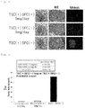
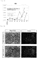
- Tumors tended to be smaller in both treated groups than in both control groups ( Figure 2 ).
- the tumor volumes were quantitatively analyzed using a fluorescence microscope.
- the tumor volume of the treated group 1 was 0.76 ⁇ 0.84 mm 3
- the tumor volume of the treated group 1 tended to be still smaller than that of the treated group 2.
- the tumor of one of the three mice was not confirmed in the third week, and the result was that complete disappearance of the tumor was expected.
- FIG 8 shows the summary of a suicide gene cell therapy when therapeutic stem cells are transplanted after tumor formation (transplantation model after tumor formation).
- Induced Pluripotent Stem (iPS) cells were differentiation-induced into NS/PCs, and the yCD-UPRT gene was then introduced with the lentiviral vector in the same way as in the mixture transplantation model to establish therapeutic stem cells (TSCs) expressing the suicide gene yCD-UPRT.
- TSCs therapeutic stem cells
- the survival analysis was performed while the luminescence imaging analysis (IVIS) of tumors was performed chronologically. Some mice were euthanized in the third week after the tumor transplantation, and the histological analysis of the brains was performed.
- IVIS luminescence imaging analysis
- a tumor tended to be smaller in the treated group than in both control groups ( Figure 9 ).
- KO Kusabira-Orange
- TSCs prepared by differentiation-inducing iPS cells into NS/PCs, and then introducing the yCD-UPRT gene into the NS/PCs with the lentiviral vector
- the mouse was decapitated under non-perfusion, and brain sections having a thickness of 200 ⁇ m were prepared using a vibratome on the next day.
- Slice culture was started on sterile porous (0.4 ⁇ m) insert membranes.
- the tumor was labeled with Venus in green, and TSCs were labeled with hKO1 in red. Photography was performed through a confocal microscope for 5 days from the same day.
- 200 ⁇ l of 10 mg/ml 5-FC was administered to the medium.
- 200 ⁇ l of 1 M PBS was administered to the medium.
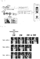
- 250 ⁇ M temozolomide was administered. It could be observed that, in the treated group, the transplanted TSCs migrated, diffused in a tumor, then satisfactorily reacted to 5-FC and died, and surrounding tumors then died ( Figure 15 , Figure 16 and Figure 17 ).
- TSCs therapeutic stem cells
- CRISPR/Cas9 transplantation model after tumor formation
- Figure 18 shows the summary of a suicide gene cell therapy using iPS cells into which the suicide gene is incorporated by genome editing. It is desirable to introduce the yCD-UPRT gene into human iPS cells for steady supply of therapeutic neural stem cells. Since the gene is inserted into chromosomes at random in gene introduction with the lentiviral vector, the gene variations of the insertion sites, the activation of peripheral genes and the inactivation of yCD-UPRT by positional effect are concerned. iPS cells in which yCD-UPRT was inserted into the GAPDH gene region were prepared using CRISPR/Cas9 genome editing techniques to solve this problem. NS/PCs were induced from these iPS cells to obtain therapeutic stem cells (TSCs) expressing yCD-UPRT.
- TSCs therapeutic stem cells
- the survival analysis was performed while the luminescence imaging analysis (IVIS) of tumors was performed chronologically. Some mice were euthanized in the third week after the tumor transplantation, and the histological analysis of the brains was performed.
- IVIS luminescence imaging analysis
- a tumor tended to be smaller in the treated group than in both control groups ( Figure 19 ).
- the tumor volumes were quantitatively analyzed using a fluorescence microscope.
- an individual in which the tumor disappeared completely was also observed (the take of the tumor cells was confirmed with IVIS before 5-FC administration on Day 7).
- Figure 24 shows the summary of a suicide gene cell therapy for malignant brain tumors using Tet-induced therapeutic stem cells, TSCs expressing yCD-UPRT.
- yCD-UPRT which can be induced with tetracycline was introduced into the iPS cells with the lentiviral vector so that the gene was expressed only when doxycycline (Dox) was added. Consequently, the iPS cells could be induced into NS/PCs without problems through EBs with Dox not added, yCD-UPRT was then expressed by adding Dox, and NS/PCs which react to 5-FC satisfactorily were obtained thereby.
- Dox doxycycline
- control group 1 Group 2
- TSC (+)/ 5-FC (+)/ Dox (-), n 9
- a group to which only U87-ffLuc was transplanted was also generated as another control group (Group 3)
- Feed containing 200 mg/kg of Dox was orally ingested.
- a method for evaluating tumors, the analysis of survival curves and the like were all performed in the same way as the above.
- a tumor tended to be smaller in the treated group than in both control groups ( Figure 25 ). Since the fluorescence protein Venus gene was introduced into the glioma cells (U87), the tumor volumes were quantitatively analyzed using a fluorescence microscope. The tumor volume of the treated group tended to be small as compared with the control group ( Figure 26 ).
- a 5 ⁇ 10 5 cells/2 ⁇ l human glioma stem cell line hG008-ffLuc was transplanted to the right striate body (2 mm away from the anterior fontanel to the right and 3 mm deep from the brain surface) of each T cell-deficient mouse (female BALB/c nude mouse, 20 g, 6w) subjected to general anesthesia, and 1 ⁇ 10 6 cells/2 ⁇ l TSCs expressing yCD-UPRT (yCD-UPRT is introduced into iPS cells with CRISPR/Cas9) were stereotactically transplanted to the same site 45 days later.
- a method for evaluating tumors, the analysis of survival curves and the like were all performed in the same way as in the examination of the therapeutic effect of the above-mentioned mixture transplantation model.
- the signal of the treated group decreased remarkably after 5-FC administration as compared with the two control groups ( Figure 29 ).
- hG008 is fluorescently labeled with Venus fluorescence protein and visualized. Since the tumor model is a tumor model which infiltrates diffusely, the measurement of volume is difficult. However, the tumors of the treated group decrease clearly as compared with the control group 1 ( Figure 31 ). Tumors which infiltrate contralaterally are little observed in the treated group ( Figure 31 ).
- the present invention can be used in industrial fields such as medicine manufacture.
Landscapes
- Health & Medical Sciences (AREA)
- Life Sciences & Earth Sciences (AREA)
- Chemical & Material Sciences (AREA)
- Engineering & Computer Science (AREA)
- Organic Chemistry (AREA)
- Genetics & Genomics (AREA)
- Zoology (AREA)
- General Health & Medical Sciences (AREA)
- Biomedical Technology (AREA)
- Wood Science & Technology (AREA)
- Bioinformatics & Cheminformatics (AREA)
- Biotechnology (AREA)
- Medicinal Chemistry (AREA)
- General Engineering & Computer Science (AREA)
- Biochemistry (AREA)
- Cell Biology (AREA)
- Veterinary Medicine (AREA)
- Public Health (AREA)
- Animal Behavior & Ethology (AREA)
- Developmental Biology & Embryology (AREA)
- Pharmacology & Pharmacy (AREA)
- Microbiology (AREA)
- Molecular Biology (AREA)
- Epidemiology (AREA)
- Neurosurgery (AREA)
- Neurology (AREA)
- Virology (AREA)
- General Chemical & Material Sciences (AREA)
- Nuclear Medicine, Radiotherapy & Molecular Imaging (AREA)
- Chemical Kinetics & Catalysis (AREA)
- Immunology (AREA)
- Reproductive Health (AREA)
- Ophthalmology & Optometry (AREA)
- Plant Pathology (AREA)
- Physics & Mathematics (AREA)
- Biophysics (AREA)
- Gynecology & Obstetrics (AREA)
- Medicines Containing Material From Animals Or Micro-Organisms (AREA)
- Micro-Organisms Or Cultivation Processes Thereof (AREA)
- Pharmaceuticals Containing Other Organic And Inorganic Compounds (AREA)
Abstract
Description
- The present invention relates to a cell preparation for treating brain tumors and a method for preparing neural stem cells used for the cell preparation.
- Malignant gliomas are very difficultly treated in the present therapies due to treatment resistance by brain tumor stem cells (BTSCs), which infiltrate the brain substance widely. Since suicide gene therapies using viral vectors exerted bystander effects, the effectiveness was expected. Since the diffusion among infiltrating tumor cells was insufficient, the results of the clinical tests were however limited. It has been revealed recently that neural stem cells (NSCs) have the properties of migrating and accumulating in brain tumor stem cells, and attempts to apply neural stem cells to suicide gene treatment as cell vehicles attract attention, and a technique as to suicide gene treatment using mesenchymal stem cells, which has properties similar to neural stem cells has been developed (Patent Literature 1) .
- Patent Literature 1:
Japanese Patent Laid-Open No.2006-345726 - Although great expectations have been placed on suicide gene treatment using neural stem cells or mesenchymal stem cells as mentioned above, it is difficult, including ethical issues, to obtain an adequate amount of neural stem cells which can be administered to humans clinically. It is easier to obtain mesenchymal stem cells than neural stem cells. As long as mesenchymal stem cells are collected from the living body, the amount thereof collected is however limited.
- The herpes simplex virus thymidine kinase gene (HSVtk) is generally used as a suicide gene (for example, this gene is used in Examples in Patent Literature 1), and besides this, the cytosine deaminase (CD) gene and the uracil phosphoribosyltransferase (UPRT) gene are known. It is known that even though the CD gene and the UPRT gene are used alone, the CD gene and the UPRT gene have antitumor effects, and the antitumor effect improves synergistically by combining the two (a gene into which these two genes are combined may be called a "CD-UPRT gene" hereinafter).
- An object of the present invention is to solve the difficulty of obtaining neural stem cells and mesenchymal stem cells, which is problematic, in such a context and to provide new means for treating brain tumors using the CD-UPRT gene, which has a high antitumor effect.
- The present inventor has earnestly examined repeatedly to solve the above-mentioned problem and consequently found that inducing the differentiation of neural stem cells from iPS cells enables obtaining neural stem cells in an amount enough to be used for a suicide gene therapy. It has been found that CD-UPRT gene-expressing neural stem cells which are excellent in safety, quality control, and stable supply properties can be obtained by inserting the CD-UPRT gene into the region of a housekeeping gene of iPS cells by genome editing. Furthermore, it has been found that these CD-UPRT gene-expressing neural stem cells exhibit an antitumor effect not only on brain tumor cells but also on brain tumor stem cells.
- The present invention has been completed based on the above knowledge.
That is, the present invention provides the following [1] to [17]. - [1] A cell preparation for treating brain tumors used in combination with a prodrug that is converted to 5-fluorouracil by cytosine deaminase, wherein the cell preparation comprises neural stem cells derived from pluripotent stem cells having a cytosine deaminase gene and a uracil phosphoribosyltransferase gene.
- [2] The cell preparation for treating brain tumors according to [1], wherein the prodrug that is converted to 5-fluorouracil by cytosine deaminase is 5-fluorocytosine.
- [3] The cell preparation for treating brain tumors according to [1] or [2], wherein the neural stem cells derived from pluripotent stem cells are neural stem cells obtained by performing the following step (1) and then performing the following step (2):
- (1) introducing a cytosine deaminase gene and a uracil phosphoribosyltransferase gene into pluripotent stem cells,
- (2) differentiating the pluripotent stem cells into neural stem cells.
- [4] The cell preparation for treating brain tumors according to [3], wherein the cytosine deaminase gene and the uracil phosphoribosyltransferase gene are inserted into the genome of the pluripotent stem cells by genome editing.
- [5] The cell preparation for treating brain tumors according to [4], wherein the cytosine deaminase gene and the uracil phosphoribosyltransferase gene are inserted into a region of a housekeeping gene or AAVS1 region of the pluripotent stem cells.
- [6] The cell preparation for treating brain tumors according to [5], wherein the housekeeping gene is a glyceraldehyde-3-phosphate dehydrogenase gene.
- [7] The cell preparation for treating brain tumors according to [3], wherein the cytosine deaminase gene and the uracil phosphoribosyltransferase gene are a gene structure, expression of which can be regulated artificially.
- [8] The cell preparation for treating brain tumors according to [7], wherein the gene structure is a gene structure expressed by addition of doxycycline.
- [9] The cell preparation for treating brain tumors according to [1] or [2], wherein the neural stem cells derived from pluripotent stem cells are neural stem cells obtained by performing the following step (1) and then performing the following step (2):
- (1) differentiating pluripotent stem cells into neural stem cells,
- (2) introducing a cytosine deaminase gene and a uracil phosphoribosyltransferase gene into the neural stem cells.
- [10] The cell preparation for treating brain tumors according to any one of [1] to [9], wherein the pluripotent stem cells are iPS cells.
- [11] A method for preparing neural stem cells having a cytosine deaminase gene and a uracil phosphoribosyltransferase gene, the method being a method comprising performing the following step (A1) and then performing the following step (A2), or a method comprising performing the following step (B1) and then performing the following step (B2):
- (A1) introducing a cytosine deaminase gene and a uracil phosphoribosyltransferase gene into pluripotent stem cells,
- (A2) differentiating the pluripotent stem cells into neural stem cells,
- (B1) differentiating pluripotent stem cells into neural stem cells,
- (B2) introducing a cytosine deaminase gene and a uracil phosphoribosyltransferase gene into the neural stem cells.
- [12] The method for preparing neural stem cells according to [11], wherein the cytosine deaminase gene and the uracil phosphoribosyltransferase gene are inserted into the genome of the pluripotent stem cells by genome editing in the step (A1).
- [13] The method for preparing neural stem cells according to [12], wherein the cytosine deaminase gene and the uracil phosphoribosyltransferase gene are inserted into a region of a housekeeping gene or AAVS1 region of the pluripotent stem cells in the step (A1).
- [14] The method for preparing neural stem cells according to [13], wherein the housekeeping gene is a glyceraldehyde-3-phosphate dehydrogenase gene.
- [15] The method for preparing neural stem cells according to [11], wherein the cytosine deaminase gene and the uracil phosphoribosyltransferase gene are a gene structure, expression of which can be regulated artificially, in the step (A1).
- [16] The method for preparing neural stem cells according to [15], wherein the gene structure is a gene structure expressed by addition of doxycycline.
- [17] The method for preparing neural stem cells according to any one of [11] to [16], wherein the pluripotent stem cells are iPS cells.
- The present specification includes contents described in the description and/or the drawings of Japanese Patent Application No.
2017-223202 - The present invention provides new means for treating brain tumors using neural stem cells derived from pluripotent stem cells such as iPS cells and ES cells.
-
- [
Figure 1] Figure 1 shows the summary of a suicide gene cell therapy for malignant brain tumors using therapeutic stem cells (TSCs) expressing yCD-UPRT (mixture transplantation model). - [
Figure 2] Figure 2 shows the results of the luminescence imaging analysis (IVIS) of tumors. - [
Figure 3] Figure 3 shows the measurement results of the ROI values of the luminescence imaging. - [
Figure 4] Figure 4 shows photographs of brains extracted from mice (left), HE-stained images of mouse brain sections (center) and Venus fluorescence images of mouse brain sections (right). - [
Figure 5] Figure 5 shows the results of the analysis of tumor volumes. - [
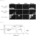
Figure 6] Figure 6 shows the results of the survival analysis of the transplanted TSCs. The upper row and the middle row show treated groups, and the lower row shows a control group. From the left, images stained with Venus (U87 tumor cells are stained), images stained with KO (TSCs are stained), images stained with DAPI (nuclei are stained), and a merge (fused image of Venus, KO and DAPI). - [
Figure 7] Figure 7 shows the results of the survival analysis of brain tumor models. - [
Figure 8] Figure 8 shows the summary of a suicide gene cell therapy for malignant brain tumors using therapeutic stem cells (TSCs) expressing yCD-UPRT (transplantation model after tumor formation). - [
Figure 9] Figure 9 shows the results of the luminescence imaging analysis (IVIS) of tumors. - [
Figure 10] Figure 10 shows the measurement results of the ROI values of the luminescence imaging. - [
Figure 11] Figure 11 shows photographs of brains extracted from mice (left), HE-stained images of mouse brain sections (center) and Venus fluorescence images of mouse brain sections (right). - [
Figure 12] Figure 12 shows the results of the analysis of tumor volumes. - [
Figure 13] Figure 13 shows the results of the survival analysis of transplanted TSCs. The upper row shows a treated group, and the lower row shows a control group. From the left, images stained with Venus (U87 tumor cells are stained), images stained with KO (TSCs are stained), images stained with DAPI (nuclei are stained), and a merge (fused image of Venus, KO and DAPI). - [
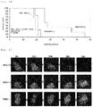
Figure 14] Figure 14 shows the results of the survival analysis of brain tumor models. - [
Figure 15] Figure 15 shows the results of time-lapse imaging of TSCs expressing yCD-UPRT. The upper row shows a treated group, the middle row shows acontrol group 1, and the lower row shows acontrol group 2. - [
Figure 16] Figure 16 shows KO-stained images of TSCs expressing yCD-UPRT. The upper row shows a treated group and the lower row shows acontrol group 1. - [
Figure 17] Figure 17 shows HE-stained images of TSCs expressing yCD-UPRT. The upper row shows a treated group and the lower row shows acontrol group 1. - [
Figure 18] Figure 18 shows the summary of a suicide gene cell therapy for malignant brain tumors using therapeutic stem cells (TSCs) expressing yCD-UPRT with CRISPR/Cas9 (transplantation model after tumor formation). - [
Figure 19] Figure 19 shows the results of the luminescence imaging analysis (IVIS) of tumors. - [
Figure 20] Figure 20 shows the measurement results of the ROI values of the luminescence imaging. - [
Figure 21] Figure 21 shows photographs of brains extracted from mice (left), HE-stained images of mouse brain sections (center) and Venus fluorescence images of mouse brain sections (right). - [
Figure 22] Figure 22 shows the results of the analysis of tumor volumes. - [
Figure 23] Figure 23 shows the results of the survival analysis of brain tumor models. - [
Figure 24] Figure 24 shows the summary of a suicide gene cell therapy for malignant brain tumors using Tet-induced therapeutic stem cells, that is, TSCs, expressing yCD-UPRT (mixture transplantation model). - [
Figure 25] Figure 25 shows the results of the luminescence imaging analysis (IVIS) of tumors. - [
Figure 26] Figure 26 shows photographs of brains extracted from mice (left), HE-stained images of mouse brain sections (center) and Venus fluorescence images of mouse brain sections (right). - [
Figure 27] Figure 27 shows the results of the survival analysis of brain tumor models. - [
Figure 28] Figure 28 shows the summary of a suicide gene cell therapy for malignant brain tumor stem cells using therapeutic stem cells (TSCs) expressing yCD-UPRT with CRISPR/Cas9 (transplantation model after tumor formation). - [
Figure 29] Figure 29 shows the results of the luminescence imaging analysis (IVIS) of tumors. - [
Figure 30] Figure 30 shows the measurement results of the ROI values of the luminescence imaging. - [
Figure 31] Figure 31 shows the results of residual tumor analysis. - [
Figure 32] Figure 32 shows the results of the survival analysis of brain tumor models. - The present invention will be described in detail hereinafter.
- A cell preparation for treating brain tumors of the present invention is a cell preparation for treating brain tumors used in combination with a prodrug that is converted to 5-fluorouracil by cytosine deaminase (CD), wherein the cell preparation comprises neural stem cells derived from pluripotent stem cells having a cytosine deaminase gene and a uracil phosphoribosyltransferase gene (CD-UPRT gene).
- The "pluripotent stem cells" in the present invention can be interpreted as a meaning which those skilled in the art usually use, and include, for example, iPS cells and ES cells.
- The "brain tumors" in the present invention means tumors which are developed in intracranial tissue. Specific examples thereof include gliomas, medulloblastomas, neuroblastomas, meningiomas, pituitary adenomas, neurinomas, primary central nervous system lymphomas, sarcomas and spinal cord tumors. In the present invention, although all of these brain tumors can be objects to be treated, it is preferable that gliomas be objects to be treated.
- The "treatment" in the present invention means not only killing tumor cells but also reducing tumor cells or inhibiting the proliferation of tumor cells.
- A CD gene and a UPRT gene are suicide genes having 5-fluorocytosine (5-FC) and 5-fluorouracil (5-FU) as prodrugs, respectively. Vectors which express these suicide genes are sold on the market, and pluripotent stem cells and neural stem cells which express these suicide genes can be prepared using such vectors. It is known that even though the CD gene and the UPRT gene are used alone, the CD gene and the UPRT gene have antitumor effect, and around 100 times higher antitumor effect is obtained by introducing these two genes into cells than when the CD gene is introduced alone.
- As long as the prodrug in the present invention is converted to 5-FU by CD, the prodrug may be any prodrug. Although examples of such a prodrug include 5-FC, the prodrug is not limited to this.
- The "neural stem cells" in the present invention means stem cells having the capability to supply cells which differentiates into neurons and glia cells. The cell preparation of the present invention contains these neural stem cells. As long as the cell preparation does not exert a great adverse influence on the effect of treating brain tumors, the cell preparation may contain cells other than neural stem cells. When neural stem cells are prepared from iPS cells in accordance with a method described in the below-mentioned Mol Brain 9: 85, 2016, not only neural stem cells but also neural precursor cells are produced. For this reason, the cell preparation of the present invention may contain both neural stem cells and neural precursor cells. Neural stem cells used in the present invention is neural stem cells derived from pluripotent stem cell such as iPS cells, and neural stem cells collected from the brain or the like is not used as they are.
- Neural stem cells derived from pluripotent stem cells having a CD-UPRT gene can be prepared by a method including performing a step (A1) and then performing a step (A2), or a method including performing a step (B1) and then performing a step (B2) described below.
- In the step (A1), the CD-UPRT gene is introduced into pluripotent stem cells.
- Although ES cells can also be used as the pluripotent stem cells, it is preferable to use iPS cells. As long as iPS cells to be used can be differentiated into neural stem cells, the origin thereof, a reprogramming factor to be introduced, a method for introducing a reprogramming factor and the like are not particularly limited. Since the cell preparation of the present invention is used mainly for treating human brain tumors, it is however preferable to use iPS cells derived from a human in such a case. In this case, iPS cells derived from a patient to whom the cell preparation is administered may be used, and iPS cells derived from a human other than the patient may be used. As mentioned above, although a method for introducing a reprogramming factor is not limited, either, it is preferable to use integration-free iPS cells. Although iPS cells to be used may be prepared in accordance with a well-known method, iPS cells can also be obtained from research institutions such as the Center for iPS Cell Research and Application (CiRA), Kyoto University.
- Although the CD-UPRT gene may be introduced into the pluripotent stem cells using a viral vector, the CD-UPRT gene is preferably introduced into the pluripotent stem cells using genome editing. When the suicide gene is inserted into the genome of the pluripotent stem cells using a viral vector such as a lentiviral vector, the suicide gene is inserted into chromosomes at random, and the gene variations of the insertion sites, the activation of peripheral genes and the inactivation of the suicide gene by positional effect are therefore concerned. It is considered that using not a viral vector but genome editing enables avoiding such a problem.
- The genome can be edited using ZFN, TALEN, CRISPR/Cas9 or the like, and it is preferable to edit the genome using CRISPR/Cas9 among these. It is preferable to insert the CD-UPRT gene into the region of a housekeeping gene of the pluripotent stem cells by genome editing. As long as the housekeeping gene is expressed in a certain amount in many cells, the housekeeping gene is not particularly limited. Specific examples thereof include a glyceraldehyde-3-phosphate dehydrogenase (GAPDH) gene, a cyclophilin gene, a β-actin gene and an α-tubulin gene, and the GAPDH gene is preferable among these. The CD-UPRT gene may be inserted into AAVS1 region of the pluripotent stem cells. Adeno-associated viruses (AAVs) do not have pathogenicity to humans, and the AAVS1 region is considered to be a genome region into which foreign genes are inserted highly safely.
- When the CD-UPRT gene is introduced into the pluripotent stem cells using a viral vector, as mentioned above, harmful influence due to random insertion into chromosomes can be considered. Therefore, the CD-UPRT gene is preferably a gene structure, the expression of which can be regulated artificially. Such a gene structure can be produced using a tetracycline-induced system (PLoS One 8:e59890, 2013), which is a well-known expression-inducing system. This tetracycline induction system comprises a region which expresses a reverse tetracycline-controlled transactivator (rtTA), a gene of interest and a region including a TRE promoter disposed upstream thereof. The rtTA bonds to the TRE promoter by bonding to tetracycline and induces the expression of the gene of interest disposed downstream. Using this system enables inducing the expression of the suicide gene by the addition of tetracycline. Although the expression in this system can also be induced with tetracycline as mentioned above, but is usually induced with doxycycline, which is a derivative of tetracycline. The gene structure in which the expression of the CD-UPRT gene can be regulated artificially can also be produced using various induced promoters such as drug-induced promoters besides this tetracycline-induced system.
- Examples of the viral vector include adenovirus vectors, adeno-associated viral vectors, retroviral vectors and lentiviral vectors. Among these, a vector which incorporates a gene into the genome is preferable, and a lentiviral vector is most preferable. The sequences of a reporter gene, an IRES or the like may be included in the vector besides the CD-UPRT gene.
- In a step (A2), pluripotent stem cells are differentiated into neural stem cells.
- Differentiation from pluripotent stem cells to neural stem cells may be performed in accordance with either of well-known methods of a method for preparation through embryoid bodies and a method for preparation without forming embryoid bodies. When differentiation from iPS cells is performed, the differentiation from the iPS cells can be performed, for example, in accordance with the method described in Stem Cell Reports. 2017 . According to the above-mentioned method, embryoid bodies can be formed from iPS cells using well-known embryoid body-forming medium, and the embryoid body medium contains a TGFβ family inhibitor (for example, SB431542) and a BMP inhibitor (for example, LDN-193189) .
- Differentiation from the embryoid bodies to neural stem cells can be performed using well-known neurosphere medium. For example, the neurosphere medium contains an epidermal growth factor and a fibroblast growth factor-2, a leukemia inhibitory factor, a B-27 supplement and the like. Culturing the embryoid bodies in the neurosphere medium forms cell masses called neurospheres and containing neural stem cells. The formed neurospheres are collected and disassembled into single cells. The single cells are cultured in neurosphere medium to form neurospheres again. Such operation is repeated several times, and neural stem cells can be obtained by collecting neurospheres.
- When neural stem cells derived from pluripotent stem cells having the CD-UPRT gene are prepared by sequentially performing the steps (A1) and (A2), there is the advantage of neural stem cells for treatment being able to be supplied steadily by proliferating pluripotent stem cells.
- In the step (B1), pluripotent stem cells are differentiated into neural stem cells.
- The differentiation from pluripotent stem cells to neural stem cells in the step (B1) can be performed in the same way as the differentiation in the step (A2).
- In the step (B2), a CD-UPRT gene is introduced into neural stem cells.
- The introduction of a CD-UPRT gene into neural stem cells in a step (B2) can be performed in the same way as the introduction in the step (A1). Note that since a problem with cytotoxicity may occur in the step (A1), it is preferable that genome editing be used, and the CD-UPRT gene be a gene structure, the expression of which can be regulated artificially; however, since a problem with cytotoxicity very rarely occurs in the step (B2), these do not usually need to be performed.
- As mentioned above, "neural stem cells," which are a "product," are not specified by structure or properties but specified by a preparation method herein. This is because since cells are parts of the living body, the structure and properties thereof are very complicated, and markedly excessive economic expenditure and time are taken to work to specify them.
- The cell preparation of the present invention may contain other pharmaceutically acceptable components besides neural stem cells. Examples of such other components include carriers, excipients, disintegrators, buffers, emulsifiers, suspending agents, soothing agents, stabilizers, preservatives, antiseptics and physiological saline solutions. Dimethyl sulfoxide, serum albumin or the like may be contained to protect cells at the time of freeze storage, and an antibiotic or the like may be contained to prevent the contamination and proliferation of bacteria.
- The number of neural stem cells contained in the cell preparation of the present invention can be optionally determined in view of the sex, age or weight of a subject, the state of an affected part, the state of cells to be used or the like so as to obtain a desired effect (for example, the disappearance of tumors or the reduction of tumor size) in the treatment of brain tumors.
- The cell preparation of the present invention may be administered a plurality of times (for example, 2 to 10 times) at intervals (for example, twice per day, once per day, twice per week, once per week, once per two week). Although the dose can be optionally determined in view of the sex, age or weight of a subject, the state of an affected part, the state of cells to be used or the like, the administration is preferably 1 × 10⁶ to 1 × 10¹⁰ cells per individual (human) 1 to 10 times.
- The administration site or administration method of the cell preparation of the present invention is not particularly limited. Examples of the administration method include tumor local administration, intracarotidarterial administration and intravenous administration.
- The administration site or administration method of a prodrug used in combination with the cell preparation of the present invention is not particularly limited, either. Examples of the administration method also include intraperitoneal administration besides tumor local administration, intracarotidarterial administration and intravenous administration mentioned above.
- The prodrug may be administered before or after the administration of the cell preparation of the present invention or at the same time as the administration of the cell preparation, but is usually administered in divided doses after the administration of the cell preparation. The dose of the prodrug can be optionally determined in view of the type of prodrug to be used, the sex, age or weight of a subject or the state of an affected part. When 5-FC is administered, 5-FC is preferably administered at 50 to 200 mg/kg per once, 4 times per day for 2 to 3 weeks (28 times to 42 times) per individual (human).
- When the tetracycline induction system is used to prevent cytotoxicity, doxycycline is also administered at the time of treatment to express a suicide gene. Examples of a method for administer doxycycline include oral administration, tumor local administration, intracarotidarterial administration, intravenous administration and intraperitoneal administration. Doxycycline may be administered before or after the administration of the cell preparation of the present invention or at the same time as the administration of the cell preparation, but is usually administered after the administration of the cell preparation.
- Next, although the present invention will be described in more detail by giving Examples, the present invention is not limited to these Examples.
- The used human iPS cell (1210B2) was obtained from the Center for iPS Cell Research and Application (CiRA), Kyoto University. Then, 1210B2 was established by a method for introducing a reprogramming factor using an episomal vector for human peripheral blood mononuclear cells (Okita K, Yamakawa T, Matsumura Y, Sato Y, Amano N, Watanabe A, Goshima N, Yamanaka S. An efficient nonviral method to generate integration-free human-induced pluripotent stem cells from cord blood and peripheral blood cells. Stem Cells. 2013 Mar;31(3):458-66.). Human iPS cells were inoculated on a plastic culture dish coated with iMatrix-511 (produced by Nippi, Incorporated) and subjected to maintenance culture using Stem Fit AK03 or AK03N medium (produced by AJINOMOTO CO., INC.) by a known feeder-free method (Nakagawa M, Taniguchi Y, Senda S, Takizawa N, Ichisaka T, Asano K, Morizane A, Doi D, Takahashi J, Nishizawa M, Yoshida Y, Toyoda T, Osafune K, Sekiguchi K, Yamanaka S. A novel efficient feeder-free culture system for the derivation of human induced pluripotent stem cells. Sci Rep. 2014 .).
- Differentiation induction from human iPS cells to NS/PCs was performed by a known method (Sugai K, Fukuzawa R, Shofuda T, Fukusumi H, Kawabata S, Nishiyama Y, Higuchi Y, Kawai K, Isoda M, Kanematsu D, Hashimoto-Tamaoki T, Kohyama J, Iwanami A, Suemizu H, Ikeda E, Matsumoto M, Kanemura Y, Nakamura M, Okano H. Pathological classification of human iPSC-derived neural stem/progenitor cells towards safety assessment of transplantation therapy for CNS diseases. Mol Brain. 2016 Sep 19;9(1):85.). First, a 10 µM ROCK inhibitor Y276352 (produced by Wako Pure Chemical Industries, Ltd.) was added to medium of iPS cells, the iPS cells were incubated for 1 to 3 hours, then washed with PBS and disassembled into single cells using TrypLE Select (produced by Life Technologies Corporation). The human iPS cells disassembled into the single cells were suspended in EB (embryoid body) -forming medium to which a 10 µM ROCK inhibitor Y276352 was added (10 µM SB431542 and 100 nM LDN-193189 were added to Stem Fit medium to which C liquid was not added), inoculated at a concentration of 9.0 × 10³ cells/75 µl per 1 well of a low adhesive 96 hole-culture plate (Prime Surface 96V, manufactured by Sumitomo Bakelite Co., Ltd.) and cultured. Then, 75 µl of EB-forming media was added 1 day after, a half amount of EB-forming medium was replaced everyday on and after, and the human iPS cells were cultured for 13 to 14 days to obtain EBs. The EBs were collected from each well and cultured in NS (neurosphere) medium (to D-MEM/Ham's F-12 medium (containing HEPES) (produced by Wako Pure Chemical Industries, Ltd.) were added a 20 ng/ml recombination human epidermal growth factor (produced by PeproTech, Inc.), a 20 ng/ml recombination human fibroblast growth factor-2 (produced by PeproTech, Inc.), a 103 units/ml recombination human leukemia inhibitory factor (produced by NACALAI TESQUE, INC.), a 2% B-27 supplement (produced by Thermo Fisher Scientific K.K.) and 1 unit/ml heparin sodium (produced by AY PHARMACEUTICALS CO., LTD.)) for 7 days. The medium was replaced once per 3 or 4 days during culture. Cell masses were collected by a centrifuge, disassembled into single cells using TrypLE Select and suspended in NS medium. The single cells were inoculated into a low adhesive flask (manufactured by Corning Incorporated) at a concentration of 1 × 10⁵ cells/ml and cultured for 10 days with the medium replaced every 3 to 4 days to obtain primary NS/PCs. Similarly, the primary NS/PCs were collected by the centrifuge, disassembled into single cells using TrypLE Select and suspended in NS medium. The single cells were inoculated into a low adhesive flask at a concentration of 1 × 10⁵ cells/ml and cultured for 7 to 10 days with the medium replaced every 3 to 4 days to obtain secondary, tertiary, quaternary and quinary NS/PCs.
- The cDNA of the yCD(yest Cytosine Deaminase)-UPRT(Uracil PhosphoRibosyl Transferase) wherein the cDNA was subcloned from the pSELECT-zeo-Fcy::fur plasmid (produced by InvivoGen Corporation) to pENTR/D-TOPO (produced by Thermo Fisher Scientific K.K.) by PCR and the nucleotide sequence of the cDNA was confirmed, was used. The yCD-UPRT cDNA was subcloned to the lentiviral vector plasmids CSIV-EF-RfA-IRES2-hKO1 to obtain CSIV-EF-yCD-UPRT-IRES2-hKO1. CSIV-EF-yCD-UPRT-IRES2-hKO1 was used to produce a lentiviral vector which expresses yCD-UPRT and hKO1 (Humanized Kusabira-Orange fluorescence protein gene) under the EF-1α promoter. The lentiviral vectors were produced by a known method (Miyoshi H, Blomer U, Takahashi M, Gage FH, Verma IM. Development of a self-inactivating lentivirus vector. J Virol. 1998 Oct;72(10):8150-7.; Kurita R, Suda N, Sudo K, Miharada K, Hiroyama T, Miyoshi H, Tani K, Nakamura Y. Establishment of immortalized human erythroid progenitor cell lines able to produce enucleated red blood cells. PLoS One. 2013;8(3):e59890.; Hashizume O, Ohnishi S, Mito T, Shimizu A, Ishikawa K, Nakada K, Soda M, Mano H, Togayachi S, Miyoshi H, Okita K, Hayashi J. Epigenetic regulation of the nuclear-coded GCAT and SHMT2 genes confers human age-associated mitochondrial respiration defects. Sci Rep. 2015 May 22;5:10434.)
- Secondary to quaternary NS/PCs differentiation-induced from the human iPS cells were collected by a centrifuge, disassembled into single cells using TrypLE Select, suspended in NS medium, inoculated into a low adhesive flask at a concentration of 1 × 10⁵ cells/ml, infected with a CSIV-EF-yCD-UPRT-IRES2-hKO1 lentiviral vector at an MOI (multiplicity of infection) of 1 to 2 and cultured for 7 days with the medium replaced every 3 to 4 days. The obtained NS/PCs were infected with the CSIV-EF-yCD-UPRT-IRES2-hKO1 lentiviral vector at an MOI of 1 to 2 in the same way again to prepare therapeutic stem cells (TSCs (1)), that is, NS/PCs expressing yCD-UPRT. It was confirmed that the cell death of the NS/PCs expressing yCD-UPRT was induced by adding 1 µg/ml 5-FC (fluorocytosine) to the medium.
- A construct for homologous recombination (HR-GAPDH-2A-yCD-UPRT-2A-Bsd) in which GAPDH (glyceraldehyde-3-phosphate dehydrogenase), yCD-UPRT and Bsd (blasticidin resistance gene) were inserted, being connected by 2A peptide sequences, and an expression vector construct (U6-GAPDH-gRNA4-Cas9) of a gRNA which targets the vicinity to the stop codon of GAPDH and a Cas9 were produced to incorporate yCD-UPRT into a GAPDH gene region using the CRISPR/Cas9 genome editing technique. These constructs were introduced into a human iPS cell line (1210B2) by electroporation, the culture was performed in the presence of blasticidin S, and the cloning was performed. The homologous recombination iPS cell line was confirmed by nucleotide sequence confirmation by the genomic PCR and the genomic sequencing of clones.
- Differentiation induction from iPS cells in which yCD-UPRT was incorporated into the GAPDH gene region to NS/PCs was performed to obtain secondary to quinary NS/PCs (TSCs (2)). Culture was performed with 2 µg/ml blasticidin S always added to the medium. It was confirmed that the cell death of the NS/PCs expressing yCD-UPRT was induced by adding 1 µg/ml 5-FC to the medium.
- The human glioma cell line U87 was infected with the ffLuc (fusion gene of Venus fluorescence protein and Luc2 firefly luciferase) expression lentiviral vector (CSII-EF-ffLuc) (Takahashi Y, Tsuji O, Kumagai G, Hara CM, Okano HJ, Miyawaki A, Toyama Y, Okano H, Nakamura M. Comparative study of methods for administering neural stem/progenitor cells to treat spinal cord injury in mice. Cell Transplant. 2011;20(5):727-39.). The clones were sorted with a cell sorter to obtain the U87 cell line, which expresses ffLuc highly and steadily.
- First, 1 × 10⁵ cells/2 µl U87-ffLuc and 5 × 10⁵ cells/2 µl TSCs (1) expressing yCD-UPRT (yCD-UPRT was introduced into NS/PCs with the lentiviral vector) were mixed and stereotactically transplanted to the right striate body (2 mm away from the anterior fontanel to the right and 3 mm deep from the brain surface) of each T cell-deficient mouse (female BALB/c nude mouse, 20 g, 6w) subjected to general anesthesia. Then, for 21 days from the next day, 500 µl of 5-FC (10 mg/ml) was intraperitoneally administered once per day for 2 weeks (treated group 1: TSC (+)/ 5-FC (+) 5 mg/day, n = 9). As another group, 100 µl of 5-FC (10 mg/ml) was intraperitoneally administered once per day for 2 weeks (treated group 2: TSC (+)/ 5-
FC 1 mg/day, n = 9). Next, 500 µl of 1 M PBS was administered to a control group for 2 weeks in the same way (control group 1: TSC (+)/ 5-FC (-), n = 9). A group to which only U87-ffLuc was transplanted was also generated as another control group, and 500 µl of 5-FC (10 mg/ml) was intraperitoneally administered to this once per day for 2 weeks (control group 2: TSC (-)/ 5-FC (+) 5 mg/day, n = 3). Change in the tumor of the same individual over time was observed with an IVIS, that is, an in vivo imaging system, every week. At the time of photographing with IVIS, 200 µl of 30 mg/ml VivoGlo (TM) Luciferin was intraperitoneally administered to the individual subjected to isoflurane inhalation anesthesia, and the tumor was photographed for 10 minutes when the peak was reached. - Three mice of each of the treated
groups control group 1 were decapitated in the third week after the tumor transplantation, and the actual tumor volumes were measured. The mice were decapitated by perfusing the mice with 4% paraformaldehyde for fixation through the heart. The brain tissues taken out were replaced with 10% and 20% sucrose, and the brain tissues were sliced to a thickness of 20 µm with a microtome. An 8-well dish the wells of which each contain 1 ml of an antifreeze solution was charged with the sections sequentially and stored at -20°C. - The tumor volumes of the brain sections were measured in more detail by staining with HE or observation through a fluorescence microscope since U87 expresses Venus fluorescence protein. It was evaluated by fluorescently staining the brain tissues of the groups with an anti-KO antibody in the third week in the same way whether the transplanted TSCs remained. Finally, the survival curves of the groups were compared.
- First, 1 × 10⁴ cells/2 µl U87-ffLuc was transplanted to the right striate body (2 mm away from the anterior fontanel to the right and 3 mm deep from the brain surface) of each T cell-deficient mouse (female BALB/c nude mouse, 20 g, 6w) subjected to general anesthesia, and 1 × 10⁶ cells/2 µl TSCs expressing yCD-UPRT (yCD-UPRT is introduced into NS/PCs with the lentiviral vector) were stereotactically transplanted to the
same site 5 days later. The take of tumor cells was confirmed 2 days later with an IVIS, that is, an in vivo imaging system, and 500 µl of 5-FC (10 mg/ml) was then intraperitoneally administered to a treated group once per day for 2 weeks (TSC (+)/ 5-FC (+) 5 mg/day, n = 9). Next, 500 µl of 1 M PBS was administered to acontrol group 1 for 2 weeks in the same way (control group 1: TSC (+)/ 5-FC (-), n = 9). A group to which only U87-ffLuc was transplanted was also generated as another control group, and 500 µl of 5-FC (10 mg/ml) was intraperitoneally administered to this once per day for 2 weeks (control group 2: TSC (-)/ 5-FC (+) 5 mg/day, n = 3). - A method for evaluating tumors, the analysis of survival curves and the like were all performed in the same way as in the examination of the therapeutic effect of the above-mentioned mixture transplantation model.
- First, 1 × 10⁵ U87-ffLuc cells were stereotactically transplanted to the right striate body (2 mm away from the anterior fontanel to the right and 3 mm deep from the brain surface) of a T cell-deficient mouse (female BALB/c nude mouse, 20 g, 6w) subjected to general anesthesia (Day 0). TSCs expressing yCD-UPRT (yCD-UPRT is introduced into NS/PCs with the lentiviral vector) were stereotactically transplanted to the same site on
Day 7. The mouse was decapitated under non-perfusion, and brain sections having a thickness of 200 µm were prepared using a vibratome on the next day (Day 8). Slice culture was started on sterile porous (0.4 µm) insert membranes (Merck KGaA). Each glass bottom dish was filled with 1800 µl of NS (neurosphere) medium (to D-MEM/Ham's F-12 medium (containing HEPES) (produced by Wako Pure Chemical Industries, Ltd.) were added a 20 ng/ml recombination human epidermal growth factor (produced by PeproTech, Inc.), a 20 ng/ml recombination human fibroblast growth factor-2 (produced by PeproTech, Inc.), a 103 units/ml recombination human leukemia inhibitory factor (produced by NACALAI TESQUE, INC.), a 2% B-27 supplement (produced by Thermo Fisher Scientific K.K.) and 1 unit/ml heparin sodium (produced by AY PHARMACEUTICALS CO., LTD.)). The tumor was labeled with Venus in green, and TSCs were labeled with hKO1 in red. Time-lapse photography was performed through a confocal microscope for 5 days from the same day. In a treatment dish, 200 µl of 10 mg/ml 5-FC was administered to the above-mentioned medium. In acontrol dish control dish 2, 250 µM temozolomide was administered. The medium was replaced once every 2 days, and 5-FC, PBS and temozolomide were administered in the same way. - First, 1 × 10⁴ cells/2 µl U87-ffLuc was transplanted to the right striate body (2 mm away from the anterior fontanel to the right and 3 mm deep from the brain surface) of each T cell-deficient mouse (female BALB/c nude mouse, 20 g, 6w) subjected to general anesthesia, and 1 × 10⁶ cells/2 µl TSCs expressing yCD-UPRT (yCD-UPRT is introduced into iPS cells with CRISPR/Cas9) were stereotactically transplanted to the
same site 5 days later. The take of tumor cells was confirmed 2 days later with an IVIS, that is, an in vivo imaging system, and further 2 days later, 500 µl of 5-FC (10 mg/ml) was intraperitoneally administered to a treated group once per day for 2 weeks (TSC (+)/ 5-FC (+), n = 8). Then, 500 µl of 1 M PBS was administered to acontrol group 1 for 2 weeks in the same way (control group 1: TSC/5-FC (-), n = 7). A group to which only U87-ffLuc was transplanted was also generated as another control group, and 500 µl of 5-FC (10 mg/ml) was intraperitoneally administered to this once per day for 2 weeks (control group 2: TSC (-)/ 5-FC (+), n = 3). - A method for evaluating tumors, the analysis of survival curves and the like were all performed in the same way as the above.
- The yCD-UPRT cDNA was subcloned into the lentiviral vector plasmid CSIV-RfA-TRE-EF-KT to obtain CSIV-yCD-UPRT-TRE-EF-KT. CSIV-yCD-UPRT-TRE-EF-KT was used for preparing a lentiviral vector expressing hKO1 and rtTA (reverse Tet-controlled transactivator) under the EF-1α promoter and expressing yCD-UPRT under the tetracycline (Tet)-induced promoter. The lentiviral vector was prepared by a known method.
- Human iPS cells were infected with the CSIV-yCD-UPRT-TRE-EF-KT lentiviral vector at an MOI of around 15 and cultured, the clones were then sorted with a cell sorter to obtain an iPS cell line expressing hKO1 steadily and highly. It was confirmed that the cell death of the Tet-induced iPS cells expressing yCD-UPRT was induced by adding 1 µg/ml doxycycline (Dox) and 1 µg/ml 5-FC to the medium.
- Differentiation induction from the Tet-induced iPS cells expressing yCD-UPRT to NS/PCs was performed to obtain secondary and tertiary Tet-induced NS/PCs expressing yCD-UPRT. It was confirmed that the cell death of the Tet-induced NS/PCs expressing yCD-UPRT was also induced by adding 1 µg/ml Dox and 1 µg/ml 5-FC to the medium.
- First, 1 × 10⁵ cells/2 µl U87-ffLuc and 5 × 10⁵ cells/2 µl TSCs (3) (Tet-induced yCD-UPRT was introduced into iPS cells with the lentiviral vector) were mixed and stereotactically transplanted to the right striate body (2 mm away from the anterior fontanel to the right and 3 mm deep from the brain surface) of each T cell-deficient mouse (female BALB/c nude mouse, 20 g, 6w) subjected to general anesthesia. Then, 500 µl of 5-FC (10 mg/ml) was intraperitoneally administered once per day for 2 weeks from the next day (treated group 1: TSC (+)/ 5-FC (+)/ Dox (+), n = 7). Next, 500 µl of 1 M PBS was administered to a
control group 1 for 2 weeks in the same way (control group 1: TSC (+)/ 5-FC (+)/ Dox (-), n = 9). A group to which only U87-ffLuc was transplanted was also generated as another control group, and 500 µl of 5-FC (10 mg/ml) was intraperitoneally administered to this once per day for 2 weeks (control group 2: TSC (-)/ 5-FC (+)/ Dox (+), n = 4). Feed containing 200 mg/kg of Dox was orally ingested. A method for evaluating tumors, the analysis of survival curves and the like were all performed in the same way as the above. -
Figure 1 shows the summary of a suicide gene cell therapy when a mixture of therapeutic stem cells and tumor cells are transplanted (mixture transplantation model). Yeast cytosine deaminase (yCD) which was a suicide gene to be introduced and 5-fluorocytosine (5-FC) which was a prodrug were used in combination. Although CD suppresses DNA and RNA synthesis and induces cell death by converting 5-FC to 5-fluorouracil (5-FU), uracil phosphoribosyltransferase (UPRT) converts 5-FU to 5-FUMP, which is a thymidylate synthetase inhibitor, by combining yCD and the UPRT gene. Therefore, 100 times or more stronger bystander effect is obtained than that of yCD alone. A local bystander effect is dependent on the cell cycle, and only tumor cells are killed selectively. Therefore, a high therapeutic index is expected. - Induced Pluripotent Stem (iPS) cells were differentiation-induced into NS/PCs, and the yCD-UPRT gene was introduced with the lentiviral vector to establish therapeutic stem cells (TSCs) expressing the suicide gene yCD-UPRT.
- A mixture of 1 × 10⁵ human glioma cells (U87) and 5 × 10⁵ TSCs was transplanted to the T cell-deficient mouse brain (right striate body). In a treated group 1 (Group 1), 250 mg/kg of 5-fluorocytosine (5-FC) was administered once per day for 21 days from the day following the transplantation (TSC (+) / 5FC (+) 5 mg/day, n = 9). In a treated group 2 (Group 2), the dose was reduced, and 50 mg/kg was administered in the same way (TSC (+)/
5FC 1 mg/day, n = 9). PBS was administered to a control group 1 (Group 3) (TSC (+)/ 5FC (-), n = 9). In a control group 2 (Group 4), only 1 × 10⁵ U87 cells were transplanted in the same way, and 250 mg/kg of 5-FC was administered for the same period (TSC (-)/ 5FC (+) 5 mg/day, n = 3). - The survival analysis was performed while the luminescence imaging analysis (IVIS) of tumors was performed chronologically. Some mice were euthanized in the third week after the transplantation, and the histological analysis of the brains was performed.
- Tumors tended to be smaller in both treated groups than in both control groups (
Figure 2 ). When the ROI values of the luminescence imaging were measured, the ROI values of the treated group were significantly low, and the ROI values were remarkably low especially from the second week onward (Figure 3 ) (TSC (+) / 5FC (+) 5 mg/day vs TSC (+) / 5FC (-), the second week, P = 0.0046, t-test) . - Since the fluorescence protein Venus gene was introduced into the glioma cells (U87), the tumor volumes were quantitatively analyzed using a fluorescence microscope. The tumor volume of the treated
group 1 was 0.76 ± 0.84 mm3, and the tumor volume of the treatedgroup 2 was 2.15 ± 1.86 mm3. Both were significantly small as compared with 51.59 ± 7.62 mm3 of the control group 1 (Figure 4 and Figure 5 ) (TSC (+)/ 5FC (+) 5 mg/day vs TSC (+)/ 5FC (-), P = 0.00033, t-test). The tumor volume of the treatedgroup 1 tended to be still smaller than that of the treatedgroup 2. Moreover, in the treatedgroup 1, the tumor of one of the three mice was not confirmed in the third week, and the result was that complete disappearance of the tumor was expected. - The extinction of the above-mentioned transplanted TSCs in the mouse brains was confirmed as to the safety of the present therapy (the risk of the tumorigenesis of iPS cells). Since the Kusabira-Orange (KO) gene was introduced into the TSCs, the TSCs were analyzed by fluorescence staining using a KO antibody. Consequently, although the transplanted TSCs remained in the brains in the control group, clear survival of the transplanted TSCs was not observed in both treated groups (n = 3 in each group) (
Figure 6 ). It was therefore confirmed that the transplanted TSCs died within three weeks by the yCD-UPRT/5FC system. - In both treated groups, significant extension of the survival periods was observed as compared with both control groups (
Figure 7 ). The average survival period of the treatedgroup 1 was 42 days, the average survival period of the treatedgroup 2 was 34 days, the average survival period of thecontrol group 1 was 25 days, and the average survival period of thecontrol group 2 was 21 days (TSC (+)/ 5FC (+) 5 mg/day vs TSC (+)/ 5FC (-), logrank, p = 0.001). The survival period of the treatedgroup 1 was significantly still longer than that of the treatedgroup 2. (2) Suicide gene cell therapy for malignant brain tumors using therapeutic stem cells (TSCs) expressing yCD-UPRT (transplantation model after tumor formation). -
Figure 8 shows the summary of a suicide gene cell therapy when therapeutic stem cells are transplanted after tumor formation (transplantation model after tumor formation). Induced Pluripotent Stem (iPS) cells were differentiation-induced into NS/PCs, and the yCD-UPRT gene was then introduced with the lentiviral vector in the same way as in the mixture transplantation model to establish therapeutic stem cells (TSCs) expressing the suicide gene yCD-UPRT. - First, 1 × 10⁴ human glioma cells (U87) were transplanted to the T cell-deficient mouse brain (right striate body), and 1 × 10⁶ TSCs were stereotactically transplanted to the
same site 5 days later. The take of tumor cells was confirmed 4 days later with an IVIS, that is, an in vivo imaging system, 5-FC (250 mg/kg) was then intraperitoneally administered to a treated group (Group 1) once per day for 2 weeks (TSC (+)/ 5FC (+) 5 mg/day, n = 9). PBS was administered to a control group 1 (Group 2) for 2 weeks in the same way (TSC (+)/ 5FC (-), n = 9). Only U87 was transplanted in a control group 2 (Group 3), and 5-FC (250 mg/kg) was administered once per day for 2 weeks (TSC (-)/ 5FC (+) 5 mg/day, n = 3). - The survival analysis was performed while the luminescence imaging analysis (IVIS) of tumors was performed chronologically. Some mice were euthanized in the third week after the tumor transplantation, and the histological analysis of the brains was performed.
- A tumor tended to be smaller in the treated group than in both control groups (
Figure 9 ). When the ROI values of the luminescence imaging were measured, the ROI values of the treated group were significantly low, and the ROI values were remarkably low especially from the second week onward (Figure 10 ) (TSC (+)/ 5FC (+) vs TSC (+)/ 5FC (-), P = 0.032, t-test). - Since the fluorescence protein Venus gene was introduced into the glioma cells (U87), the tumor volumes were quantitatively analyzed using a fluorescence microscope. The tumor volume of the treated group was 0.82 ± 1.23 mm3, and was significantly small as compared with 43.73 ± 24.0 mm3 of the control group 1 (
Figures 11 and12 ) (TSC (+)/ 5FC (+) vs TSC (+) / 5FC (-), P = 0.036, t-test). - The extinction of the above-mentioned transplanted TSCs in the mouse brains was confirmed as to the safety of the present therapy (the risk of the tumorigenesis of iPS cells). Since the Kusabira-Orange (KO) gene was introduced into the TSCs, the TSCs were analyzed by fluorescence staining using a KO antibody. Consequently, although the transplanted TSCs remained in the brains in the
control group 1, clear survival of the transplanted TSCs was not observed in the treated group (n = 3 in each group) (Figure 13 ). It was therefore confirmed that the transplanted TSCs died within three weeks by the yCD-UPRT/5FC system. - In the treated group, significant extension of the survival periods was observed as compared with both control groups (
Figure 14 ). The average survival period of the treated group was 36 days, the average survival period of thecontrol group 1 was 20 days, and the average survival period of thecontrol group 2 was 21 days (TSC (+)/ 5FC (+) vs TSC (+)/ 5FC (-), logrank, p = 0.0013). - As mentioned above, when yCD-UPRT was introduced into NS/PCs derived from human iPS cells using a lentiviral vector which can be applied clinically, and the NS/PCs were transplanted to a human brain tumor model mouse, a remarkable therapeutic effect (bystander effect) was observed by administering 5-FC, and the efficacy of the present therapy could be proved.
- First, 1 × 10⁵ ffLuc-U87 cells were stereotactically transplanted to a T cell-deficient mouse (right striate body) (Day 0). TSCs (TSCs prepared by differentiation-inducing iPS cells into NS/PCs, and then introducing the yCD-UPRT gene into the NS/PCs with the lentiviral vector) were stereotactically transplanted to the same site on
Day 7. The mouse was decapitated under non-perfusion, and brain sections having a thickness of 200 µm were prepared using a vibratome on the next day. Slice culture was started on sterile porous (0.4 µm) insert membranes. The tumor was labeled with Venus in green, and TSCs were labeled with hKO1 in red. Photography was performed through a confocal microscope for 5 days from the same day. As a treated group, 200 µl of 10 mg/ml 5-FC was administered to the medium. As acontrol group control group 2, 250 µM temozolomide was administered. It could be observed that, in the treated group, the transplanted TSCs migrated, diffused in a tumor, then satisfactorily reacted to 5-FC and died, and surrounding tumors then died (Figure 15 ,Figure 16 and Figure 17 ). It could be confirmed that, in thecontrol group 1, TSCs continued surviving, migrated and entered the tumor (Figure 15 ,Figure 16 and Figure 17 ). As thecontrol group 2, although temozolomide had the effect of suppressing temporary tumor increase, it was not observed that temozolomide had the effect of reducing the tumor (Figure 15 ). -
Figure 18 shows the summary of a suicide gene cell therapy using iPS cells into which the suicide gene is incorporated by genome editing. It is desirable to introduce the yCD-UPRT gene into human iPS cells for steady supply of therapeutic neural stem cells. Since the gene is inserted into chromosomes at random in gene introduction with the lentiviral vector, the gene variations of the insertion sites, the activation of peripheral genes and the inactivation of yCD-UPRT by positional effect are concerned. iPS cells in which yCD-UPRT was inserted into the GAPDH gene region were prepared using CRISPR/Cas9 genome editing techniques to solve this problem. NS/PCs were induced from these iPS cells to obtain therapeutic stem cells (TSCs) expressing yCD-UPRT. - First, 1 × 10⁴ human glioma cells (U87) were transplanted to the T cell-deficient mouse brain (right striate body), and 1 × 10⁶ TSCs were stereotactically transplanted to the
same site 5 days later. The take of tumor cells was confirmed 4 days later with an IVIS, that is, an in vivo imaging system, 5-FC (250 mg/kg) was then administered to a treated group (Group 1) once per day (TSC (+)/ 5FC (+), n = 6). PBS was administered to a control group 1 (Group 2) for 2 weeks in the same way (TSC/ 5FC (-), n = 4). Only U87 was transplanted in a control group 2 (Group 3), and 5-FC (250 mg/kg) was intraperitoneally administered once per day for 2 weeks (TSC (-)/ 5FC (+), n = 3) . - The survival analysis was performed while the luminescence imaging analysis (IVIS) of tumors was performed chronologically. Some mice were euthanized in the third week after the tumor transplantation, and the histological analysis of the brains was performed.
- A tumor tended to be smaller in the treated group than in both control groups (
Figure 19 ). When the ROI values of the luminescence imaging were measured, the ROI values of the treated group tended to be low, and the ROI values were remarkably low especially from the second week onward (Figure 20 ) (TSC (+)/ 5FC (+) vs TSC (+)/ 5FC (-), P = 0.067, t-test). - Since the fluorescence protein Venus gene was introduced into the glioma cells (U87), the tumor volumes were quantitatively analyzed using a fluorescence microscope. The tumor volume of the treated group was 6.02 ± 7.08 mm3, and was significantly small as compared with 74.12 ± 31.6 mm3 of the control group (
Figure 21 andFigure 22 ) (TSC (+)/ 5FC (+) vs TSC (+)/ 5FC (-), P = 0.022, t-test). In the treated group, an individual in which the tumor disappeared completely was also observed (the take of the tumor cells was confirmed with IVIS before 5-FC administration on Day 7). - In the treated group, significant extension of the survival period was observed as compared with both control groups (
Figure 23 ). The average survival period of the treated group was 35 days (one mouse in the group was finished for analysis), the average survival period of thecontrol group 1 was 20 days, and the average survival period of thecontrol group 2 was 21 days (TSC (+)/ 5FC (+) vs TSC (+)/ 5FC (-), logrank, p = 0.0042). In the treated group, a case in which the tumor disappeared completely, and a mouse continued surviving was also observed. -
Figure 24 shows the summary of a suicide gene cell therapy for malignant brain tumors using Tet-induced therapeutic stem cells, TSCs expressing yCD-UPRT. To introduce the yCD-UPRT gene into not NS/PCs but iPS cells with the lentiviral vector, yCD-UPRT which can be induced with tetracycline was introduced into the iPS cells with the lentiviral vector so that the gene was expressed only when doxycycline (Dox) was added. Consequently, the iPS cells could be induced into NS/PCs without problems through EBs with Dox not added, yCD-UPRT was then expressed by adding Dox, and NS/PCs which react to 5-FC satisfactorily were obtained thereby. - First, 1 × 10⁵ cells/2 µl U87-ffLuc and 5 × 10⁵ cells/2 µl TSCs were mixed and stereotactically transplanted to the right striate body (2 mm away from the anterior fontanel to the right and 3 mm deep from the brain surface) of each T cell-deficient mouse (female BALB/c nude mouse, 20 g, 6w) subjected to general anesthesia. Then, 500 µl of 5-FC (10 mg/ml) was intraperitoneally administered once per day for 2 weeks from the next day (treated group (Group 1): TSC (+)/ 5-FC (+)/ Dox (+), n = 7). Next, 500 µl of 1 M PBS was administered to a control group 1 (Group 2) for 2 weeks in the same way (control group 1: TSC (+)/ 5-FC (+)/ Dox (-), n = 9). A group to which only U87-ffLuc was transplanted was also generated as another control group (Group 3), and 500 µl of 5-FC (10 mg/ml) was intraperitoneally administered to this once per day for 2 weeks (control group 2: TSC (-)/ 5-FC (+)/ Dox (+), n = 4). Feed containing 200 mg/kg of Dox was orally ingested. A method for evaluating tumors, the analysis of survival curves and the like were all performed in the same way as the above.
- Survival analysis was performed, while the luminescence imaging analysis (IVIS) of the tumors was performed chronologically. Some mice were euthanized in the third week after the tumor transplantation, and the histological analysis of the brains was performed. <Chronological analysis of tumors by luminescence imaging analysis (IVIS) and tumor volume>
- A tumor tended to be smaller in the treated group than in both control groups (
Figure 25 ). Since the fluorescence protein Venus gene was introduced into the glioma cells (U87), the tumor volumes were quantitatively analyzed using a fluorescence microscope. The tumor volume of the treated group tended to be small as compared with the control group (Figure 26 ). - In the treated group, significant extension of the survival period was observed as compared with both control groups (
Figure 27 ). The average survival period of the treated group was 25.5 days, the average survival period of thecontrol group 1 was 20 days, and the average survival period of thecontrol group 2 was 19 days (TSC (+)/ 5-FC (+)/ Dox (+) vs TSC (+)/ 5-FC (+)/ Dox (-), logrank, p = 0.004). - First, a 5 × 10⁵ cells/2 µl human glioma stem cell line hG008-ffLuc was transplanted to the right striate body (2 mm away from the anterior fontanel to the right and 3 mm deep from the brain surface) of each T cell-deficient mouse (female BALB/c nude mouse, 20 g, 6w) subjected to general anesthesia, and 1 × 10⁶ cells/2 µl TSCs expressing yCD-UPRT (yCD-UPRT is introduced into iPS cells with CRISPR/Cas9) were stereotactically transplanted to the
same site 45 days later. The signal of the tumor was confirmed 7 days later with an IVIS, that is, an in vivo imaging system, and 500 µl of 5-FC (10 mg/ml) was then administered to a treated group once per day for 2 weeks (TSC (+)/ 5-FC (+) 5 mg/day, n = 8). Next, 500 µl of 1 M PBS was administered to acontrol group 1 for 2 weeks in the same way (control group 1: TSC (+) / 5-FC (-), n = 7). A group to which only hG008-ffLuc was transplanted was also generated as another control group, and 500 µl of 5-FC (10 mg/ml) was intraperitoneally administered to this once per day for 2 weeks (control group 2: TSC (-)/ 5-FC (+) 5 mg/day, n = 3) . - A method for evaluating tumors, the analysis of survival curves and the like were all performed in the same way as in the examination of the therapeutic effect of the above-mentioned mixture transplantation model.
- In an IVIS, that is, an in vivo imaging system, the signal of the treated group decreased remarkably after 5-FC administration as compared with the two control groups (
Figure 29 ). The ROI of the signal of the IVIS of the treated group was significantly low, with p = 0.0019 at the time of 7W after 5-FC administration, which significantly demonstrated a significant difference between the treated group and the control group 1 (Figure 30 ) . - hG008 is fluorescently labeled with Venus fluorescence protein and visualized. Since the tumor model is a tumor model which infiltrates diffusely, the measurement of volume is difficult. However, the tumors of the treated group decrease clearly as compared with the control group 1 (
Figure 31 ). Tumors which infiltrate contralaterally are little observed in the treated group (Figure 31 ). - The survival curve of the treated group was extended significantly, with p = 0.0026 in the log-rank test, which demonstrated a significant difference between the treated group and the
control group 1. - All of the publications, patents and patent applications cited herein are incorporated herein as reference as they are.
- Since a cell preparation for treating brain tumors of the present invention can be used as a pharmaceutical, the present invention can be used in industrial fields such as medicine manufacture.
Claims (17)
- A cell preparation for treating brain tumors used in combination with a prodrug that is converted to 5-fluorouracil by cytosine deaminase, wherein the cell preparation comprises neural stem cells derived from pluripotent stem cells having a cytosine deaminase gene and a uracil phosphoribosyltransferase gene.
- The cell preparation for treating brain tumors according to claim 1, wherein the prodrug that is converted to 5-fluorouracil by cytosine deaminase is 5-fluorocytosine.
- The cell preparation for treating brain tumors according to claim 1 or 2, wherein the neural stem cells derived from pluripotent stem cells are neural stem cells obtained by performing the following step (1) and then performing the following step (2):(1) introducing a cytosine deaminase gene and a uracil phosphoribosyltransferase gene into pluripotent stem cells,(2) differentiating the pluripotent stem cells into neural stem cells.
- The cell preparation for treating brain tumors according to claim 3, wherein the cytosine deaminase gene and the uracil phosphoribosyltransferase gene are inserted into the genome of the pluripotent stem cells by genome editing.
- The cell preparation for treating brain tumors according to claim 4, wherein the cytosine deaminase gene and the uracil phosphoribosyltransferase gene are inserted into a region of a housekeeping gene or AAVS1 region of the pluripotent stem cells.
- The cell preparation for treating brain tumors according to claim 5, wherein the housekeeping gene is a glyceraldehyde-3-phosphate dehydrogenase gene.
- The cell preparation for treating brain tumors according to claim 3, wherein the cytosine deaminase gene and the uracil phosphoribosyltransferase gene are a gene structure, expression of which can be regulated artificially.
- The cell preparation for treating brain tumors according to claim 7, wherein the gene structure is a gene structure expressed by addition of doxycycline.
- The cell preparation for treating brain tumors according to claim 1 or 2, wherein the neural stem cells derived from pluripotent stem cells are neural stem cells obtained by performing the following step (1) and then performing the following step (2):(1) differentiating pluripotent stem cells into neural stem cells,(2) introducing a cytosine deaminase gene and a uracil phosphoribosyltransferase gene into the neural stem cells.
- The cell preparation for treating brain tumors according to any one of claims 1 to 9, wherein the pluripotent stem cells are iPS cells.
- A method for preparing neural stem cells having a cytosine deaminase gene and a uracil
phosphoribosyltransferase gene, the method being a method comprising performing the following step (A1) and then performing the following step (A2), or a method comprising performing the following step (B1) and then performing the following step (B2):(A1) introducing a cytosine deaminase gene and a uracil phosphoribosyltransferase gene into pluripotent stem cells,(A2) differentiating the pluripotent stem cells into neural stem cells,(B1) differentiating pluripotent stem cells into neural stem cells,(B2) introducing a cytosine deaminase gene and a uracil phosphoribosyltransferase gene into the neural stem cells. - The method for preparing neural stem cells according to claim 11, wherein the cytosine deaminase gene and the uracil phosphoribosyltransferase gene are inserted into the genome of the pluripotent stem cells by genome editing in the step (A1).
- The method for preparing neural stem cells according to claim 12, wherein the cytosine deaminase gene and the uracil phosphoribosyltransferase gene are inserted into a region of a housekeeping gene or AAVS1 region of the pluripotent stem cells in the step (A1).
- The method for preparing neural stem cells according to claim 13, wherein the housekeeping gene is a glyceraldehyde-3-phosphate dehydrogenase gene.
- The method for preparing neural stem cells according to claim 11, wherein the cytosine deaminase gene and the uracil phosphoribosyltransferase gene are a gene structure, expression of which can be regulated artificially, in the step (A1).
- The method for preparing neural stem cells according to claim 15, wherein the gene structure is a gene structure expressed by addition of doxycycline.
- The method for preparing neural stem cells according to any one of claims 11 to 16, wherein the pluripotent stem cells are iPS cells.
Applications Claiming Priority (2)
| Application Number | Priority Date | Filing Date | Title |
|---|---|---|---|
| JP2017223202 | 2017-11-20 | ||
| PCT/JP2018/042629 WO2019098361A1 (en) | 2017-11-20 | 2018-11-19 | Suicide gene therapeutic agent for brain tumors using pluripotent stem cell |
Publications (3)
| Publication Number | Publication Date |
|---|---|
| EP3714894A1 true EP3714894A1 (en) | 2020-09-30 |
| EP3714894A4 EP3714894A4 (en) | 2021-07-14 |
| EP3714894B1 EP3714894B1 (en) | 2023-11-01 |
Family
ID=66539796
Family Applications (1)
| Application Number | Title | Priority Date | Filing Date |
|---|---|---|---|
| EP18878961.4A Active EP3714894B1 (en) | 2017-11-20 | 2018-11-19 | Suicide gene therapeutic agent for brain tumors using pluripotent stem cell |
Country Status (5)
| Country | Link |
|---|---|
| US (1) | US12090172B2 (en) |
| EP (1) | EP3714894B1 (en) |
| JP (2) | JP7150233B2 (en) |
| CN (1) | CN111356463A (en) |
| WO (1) | WO2019098361A1 (en) |
Families Citing this family (3)
| Publication number | Priority date | Publication date | Assignee | Title |
|---|---|---|---|---|
| WO2020101570A1 (en) * | 2018-11-14 | 2020-05-22 | Advanced Cell Therapeutics Pte Ltd | Stem cell infusion |
| US20240226175A1 (en) * | 2020-03-31 | 2024-07-11 | Keio University | Therapeutic agent using genome-edited pluripotent stem cell |
| CN112553286A (en) * | 2020-11-05 | 2021-03-26 | 北京大学深圳医院 | Method for evaluating curative effect of suicide gene/prodrug system and drug screening method |
Family Cites Families (5)
| Publication number | Priority date | Publication date | Assignee | Title |
|---|---|---|---|---|
| JP2006345726A (en) | 2005-06-14 | 2006-12-28 | Hamamatsu Univ School Of Medicine | Expression vector for brain tumor treatment |
| CN103484462B (en) * | 2013-09-02 | 2016-04-27 | 广东药学院 | The recombinant adenoviral vector of Survivin promoter regulation CD gene builds and application |
| US12642852B1 (en) | 2015-11-04 | 2026-06-02 | Fate Therapeutics, Inc. | Genomic engineering of pluripotent cells |
| JP6684163B2 (en) | 2016-06-17 | 2020-04-22 | 日本サーモスタット株式会社 | Thermoactuator casing structure |
| EP3622961A4 (en) | 2017-05-09 | 2021-01-13 | Keio University | CELL FORMULATION FOR THE TREATMENT OF BRAIN TUMORS |
-
2018
- 2018-11-19 WO PCT/JP2018/042629 patent/WO2019098361A1/en not_active Ceased
- 2018-11-19 JP JP2019554440A patent/JP7150233B2/en active Active
- 2018-11-19 EP EP18878961.4A patent/EP3714894B1/en active Active
- 2018-11-19 US US16/765,257 patent/US12090172B2/en active Active
- 2018-11-19 CN CN201880074333.5A patent/CN111356463A/en active Pending
-
2022
- 2022-05-20 JP JP2022083217A patent/JP2022103424A/en active Pending
Also Published As
| Publication number | Publication date |
|---|---|
| CN111356463A (en) | 2020-06-30 |
| WO2019098361A1 (en) | 2019-05-23 |
| JP7150233B2 (en) | 2022-10-11 |
| EP3714894B1 (en) | 2023-11-01 |
| US20210369785A1 (en) | 2021-12-02 |
| JPWO2019098361A1 (en) | 2020-11-19 |
| EP3714894A4 (en) | 2021-07-14 |
| JP2022103424A (en) | 2022-07-07 |
| US12090172B2 (en) | 2024-09-17 |
Similar Documents
| Publication | Publication Date | Title |
|---|---|---|
| US11497771B2 (en) | Plasmid combination and application thereof in preparing modified immune cells | |
| US20220401491A1 (en) | Mesenchymal stem cell expressing trail and cd, and use thereof | |
| US11053476B2 (en) | Generation of cancer stem cells and use thereof | |
| JP2016513974A (en) | Compositions and methods for reprogramming hematopoietic stem cell lineage | |
| WO2001048151A1 (en) | Cells capable of differentiating into heart muscle cells | |
| JP7807434B2 (en) | Cellular compositions and therapeutic methods | |
| US10961509B2 (en) | Engineered endothelial cells expressing an ETS transcription factor | |
| CN114423860A (en) | Cellular compositions comprising viral vectors and methods of treatment | |
| JP2022103424A (en) | Suicide gene therapeutic agent for brain tumors using pluripotent stem cell | |
| US10947503B2 (en) | Method for modifying T cell population | |
| EP3754025A1 (en) | Mesenchymal stem cells expressing brain-derived neurotrophic factor and use thereof | |
| EP3622961A1 (en) | Cell formulation for treating brain tumor | |
| Norol et al. | GFP-transduced CD34+ and Lin− CD34− hematopoietic stem cells did not adopt a cardiac phenotype in a nonhuman primate model of myocardial infarct | |
| WO2019027298A9 (en) | Pharmaceutical composition comprising mesenchymal stem cell expressing trail and cd as effective ingredient for prevention or treatment of cancer | |
| WO2001048150A1 (en) | Cells capable of differentiating into heart muscle cells | |
| WO2007008309A2 (en) | Stem and progenitor cell expansion by evi, evi-like genes and setbp1 | |
| CN121574935A (en) | Preparation method and application of multi-type CAR-immune cell hybrid products containing CAR-macrophages and T cells | |
| CN118697878A (en) | Application of CD36 as a molecular marker and therapeutic target for inhibiting lung cancer meningeal metastasis | |
| Meregalli et al. | Duchenne Muscular Dystrophy: isolation of CD133-Expressing myogenic progenitors from blood and muscle of DMD patients | |
| Karolak | Fas and Fas Ligand in the Development of the Hematopoietic System: A Grant Proposal | |
| Seymour et al. | European Society of Gene and Cell Therapy British Society for Gene Therapy Collaborative Congress 2011 October 27–31, 2011 The Brighton Centre, Brighton United Kingdom |
Legal Events
| Date | Code | Title | Description |
|---|---|---|---|
| STAA | Information on the status of an ep patent application or granted ep patent |
Free format text: STATUS: THE INTERNATIONAL PUBLICATION HAS BEEN MADE |
|
| PUAI | Public reference made under article 153(3) epc to a published international application that has entered the european phase |
Free format text: ORIGINAL CODE: 0009012 |
|
| STAA | Information on the status of an ep patent application or granted ep patent |
Free format text: STATUS: REQUEST FOR EXAMINATION WAS MADE |
|
| 17P | Request for examination filed |
Effective date: 20200602 |
|
| AK | Designated contracting states |
Kind code of ref document: A1 Designated state(s): AL AT BE BG CH CY CZ DE DK EE ES FI FR GB GR HR HU IE IS IT LI LT LU LV MC MK MT NL NO PL PT RO RS SE SI SK SM TR |
|
| AX | Request for extension of the european patent |
Extension state: BA ME |
|
| DAV | Request for validation of the european patent (deleted) | ||
| DAX | Request for extension of the european patent (deleted) | ||
| A4 | Supplementary search report drawn up and despatched |
Effective date: 20210611 |
|
| RIC1 | Information provided on ipc code assigned before grant |
Ipc: A61K 35/545 20150101AFI20210607BHEP Ipc: A61K 31/513 20060101ALI20210607BHEP Ipc: A61P 35/00 20060101ALI20210607BHEP |
|
| STAA | Information on the status of an ep patent application or granted ep patent |
Free format text: STATUS: EXAMINATION IS IN PROGRESS |
|
| 17Q | First examination report despatched |
Effective date: 20220428 |
|
| GRAP | Despatch of communication of intention to grant a patent |
Free format text: ORIGINAL CODE: EPIDOSNIGR1 |
|
| STAA | Information on the status of an ep patent application or granted ep patent |
Free format text: STATUS: GRANT OF PATENT IS INTENDED |
|
| INTG | Intention to grant announced |
Effective date: 20230519 |
|
| GRAS | Grant fee paid |
Free format text: ORIGINAL CODE: EPIDOSNIGR3 |
|
| GRAA | (expected) grant |
Free format text: ORIGINAL CODE: 0009210 |
|
| STAA | Information on the status of an ep patent application or granted ep patent |
Free format text: STATUS: THE PATENT HAS BEEN GRANTED |
|
| P01 | Opt-out of the competence of the unified patent court (upc) registered |
Effective date: 20230911 |
|
| AK | Designated contracting states |
Kind code of ref document: B1 Designated state(s): AL AT BE BG CH CY CZ DE DK EE ES FI FR GB GR HR HU IE IS IT LI LT LU LV MC MK MT NL NO PL PT RO RS SE SI SK SM TR |
|
| REG | Reference to a national code |
Ref country code: GB Ref legal event code: FG4D |
|
| REG | Reference to a national code |
Ref country code: CH Ref legal event code: EP |
|
| REG | Reference to a national code |
Ref country code: IE Ref legal event code: FG4D |
|
| REG | Reference to a national code |
Ref country code: DE Ref legal event code: R096 Ref document number: 602018060603 Country of ref document: DE |
|
| REG | Reference to a national code |
Ref country code: NL Ref legal event code: FP |
|
| REG | Reference to a national code |
Ref country code: SE Ref legal event code: TRGR |
|
| REG | Reference to a national code |
Ref country code: LT Ref legal event code: MG9D |
|
| PG25 | Lapsed in a contracting state [announced via postgrant information from national office to epo] |
Ref country code: GR Free format text: LAPSE BECAUSE OF FAILURE TO SUBMIT A TRANSLATION OF THE DESCRIPTION OR TO PAY THE FEE WITHIN THE PRESCRIBED TIME-LIMIT Effective date: 20240202 |
|
| PG25 | Lapsed in a contracting state [announced via postgrant information from national office to epo] |
Ref country code: IS Free format text: LAPSE BECAUSE OF FAILURE TO SUBMIT A TRANSLATION OF THE DESCRIPTION OR TO PAY THE FEE WITHIN THE PRESCRIBED TIME-LIMIT Effective date: 20240301 |
|
| PG25 | Lapsed in a contracting state [announced via postgrant information from national office to epo] |
Ref country code: LT Free format text: LAPSE BECAUSE OF FAILURE TO SUBMIT A TRANSLATION OF THE DESCRIPTION OR TO PAY THE FEE WITHIN THE PRESCRIBED TIME-LIMIT Effective date: 20231101 |
|
| REG | Reference to a national code |
Ref country code: AT Ref legal event code: MK05 Ref document number: 1626451 Country of ref document: AT Kind code of ref document: T Effective date: 20231101 |
|
| PG25 | Lapsed in a contracting state [announced via postgrant information from national office to epo] |
Ref country code: AT Free format text: LAPSE BECAUSE OF FAILURE TO SUBMIT A TRANSLATION OF THE DESCRIPTION OR TO PAY THE FEE WITHIN THE PRESCRIBED TIME-LIMIT Effective date: 20231101 |
|
| PG25 | Lapsed in a contracting state [announced via postgrant information from national office to epo] |
Ref country code: ES Free format text: LAPSE BECAUSE OF FAILURE TO SUBMIT A TRANSLATION OF THE DESCRIPTION OR TO PAY THE FEE WITHIN THE PRESCRIBED TIME-LIMIT Effective date: 20231101 |
|
| PG25 | Lapsed in a contracting state [announced via postgrant information from national office to epo] |
Ref country code: LT Free format text: LAPSE BECAUSE OF FAILURE TO SUBMIT A TRANSLATION OF THE DESCRIPTION OR TO PAY THE FEE WITHIN THE PRESCRIBED TIME-LIMIT Effective date: 20231101 Ref country code: IS Free format text: LAPSE BECAUSE OF FAILURE TO SUBMIT A TRANSLATION OF THE DESCRIPTION OR TO PAY THE FEE WITHIN THE PRESCRIBED TIME-LIMIT Effective date: 20240301 Ref country code: GR Free format text: LAPSE BECAUSE OF FAILURE TO SUBMIT A TRANSLATION OF THE DESCRIPTION OR TO PAY THE FEE WITHIN THE PRESCRIBED TIME-LIMIT Effective date: 20240202 Ref country code: ES Free format text: LAPSE BECAUSE OF FAILURE TO SUBMIT A TRANSLATION OF THE DESCRIPTION OR TO PAY THE FEE WITHIN THE PRESCRIBED TIME-LIMIT Effective date: 20231101 Ref country code: BG Free format text: LAPSE BECAUSE OF FAILURE TO SUBMIT A TRANSLATION OF THE DESCRIPTION OR TO PAY THE FEE WITHIN THE PRESCRIBED TIME-LIMIT Effective date: 20240201 Ref country code: AT Free format text: LAPSE BECAUSE OF FAILURE TO SUBMIT A TRANSLATION OF THE DESCRIPTION OR TO PAY THE FEE WITHIN THE PRESCRIBED TIME-LIMIT Effective date: 20231101 Ref country code: PT Free format text: LAPSE BECAUSE OF FAILURE TO SUBMIT A TRANSLATION OF THE DESCRIPTION OR TO PAY THE FEE WITHIN THE PRESCRIBED TIME-LIMIT Effective date: 20240301 |
|
| PG25 | Lapsed in a contracting state [announced via postgrant information from national office to epo] |
Ref country code: RS Free format text: LAPSE BECAUSE OF FAILURE TO SUBMIT A TRANSLATION OF THE DESCRIPTION OR TO PAY THE FEE WITHIN THE PRESCRIBED TIME-LIMIT Effective date: 20231101 Ref country code: PL Free format text: LAPSE BECAUSE OF FAILURE TO SUBMIT A TRANSLATION OF THE DESCRIPTION OR TO PAY THE FEE WITHIN THE PRESCRIBED TIME-LIMIT Effective date: 20231101 Ref country code: NO Free format text: LAPSE BECAUSE OF FAILURE TO SUBMIT A TRANSLATION OF THE DESCRIPTION OR TO PAY THE FEE WITHIN THE PRESCRIBED TIME-LIMIT Effective date: 20240201 Ref country code: LV Free format text: LAPSE BECAUSE OF FAILURE TO SUBMIT A TRANSLATION OF THE DESCRIPTION OR TO PAY THE FEE WITHIN THE PRESCRIBED TIME-LIMIT Effective date: 20231101 Ref country code: HR Free format text: LAPSE BECAUSE OF FAILURE TO SUBMIT A TRANSLATION OF THE DESCRIPTION OR TO PAY THE FEE WITHIN THE PRESCRIBED TIME-LIMIT Effective date: 20231101 |
|
| PG25 | Lapsed in a contracting state [announced via postgrant information from national office to epo] |
Ref country code: DK Free format text: LAPSE BECAUSE OF FAILURE TO SUBMIT A TRANSLATION OF THE DESCRIPTION OR TO PAY THE FEE WITHIN THE PRESCRIBED TIME-LIMIT Effective date: 20231101 |
|
| PG25 | Lapsed in a contracting state [announced via postgrant information from national office to epo] |
Ref country code: LU Free format text: LAPSE BECAUSE OF NON-PAYMENT OF DUE FEES Effective date: 20231119 |
|
| PG25 | Lapsed in a contracting state [announced via postgrant information from national office to epo] |
Ref country code: CZ Free format text: LAPSE BECAUSE OF FAILURE TO SUBMIT A TRANSLATION OF THE DESCRIPTION OR TO PAY THE FEE WITHIN THE PRESCRIBED TIME-LIMIT Effective date: 20231101 |
|
| PG25 | Lapsed in a contracting state [announced via postgrant information from national office to epo] |
Ref country code: SK Free format text: LAPSE BECAUSE OF FAILURE TO SUBMIT A TRANSLATION OF THE DESCRIPTION OR TO PAY THE FEE WITHIN THE PRESCRIBED TIME-LIMIT Effective date: 20231101 |
|
| PG25 | Lapsed in a contracting state [announced via postgrant information from national office to epo] |
Ref country code: SM Free format text: LAPSE BECAUSE OF FAILURE TO SUBMIT A TRANSLATION OF THE DESCRIPTION OR TO PAY THE FEE WITHIN THE PRESCRIBED TIME-LIMIT Effective date: 20231101 Ref country code: SK Free format text: LAPSE BECAUSE OF FAILURE TO SUBMIT A TRANSLATION OF THE DESCRIPTION OR TO PAY THE FEE WITHIN THE PRESCRIBED TIME-LIMIT Effective date: 20231101 Ref country code: LU Free format text: LAPSE BECAUSE OF NON-PAYMENT OF DUE FEES Effective date: 20231119 Ref country code: IT Free format text: LAPSE BECAUSE OF FAILURE TO SUBMIT A TRANSLATION OF THE DESCRIPTION OR TO PAY THE FEE WITHIN THE PRESCRIBED TIME-LIMIT Effective date: 20231101 Ref country code: EE Free format text: LAPSE BECAUSE OF FAILURE TO SUBMIT A TRANSLATION OF THE DESCRIPTION OR TO PAY THE FEE WITHIN THE PRESCRIBED TIME-LIMIT Effective date: 20231101 Ref country code: DK Free format text: LAPSE BECAUSE OF FAILURE TO SUBMIT A TRANSLATION OF THE DESCRIPTION OR TO PAY THE FEE WITHIN THE PRESCRIBED TIME-LIMIT Effective date: 20231101 Ref country code: CZ Free format text: LAPSE BECAUSE OF FAILURE TO SUBMIT A TRANSLATION OF THE DESCRIPTION OR TO PAY THE FEE WITHIN THE PRESCRIBED TIME-LIMIT Effective date: 20231101 |
|
| REG | Reference to a national code |
Ref country code: BE Ref legal event code: MM Effective date: 20231130 |
|
| REG | Reference to a national code |
Ref country code: DE Ref legal event code: R097 Ref document number: 602018060603 Country of ref document: DE |
|
| PG25 | Lapsed in a contracting state [announced via postgrant information from national office to epo] |
Ref country code: MC Free format text: LAPSE BECAUSE OF FAILURE TO SUBMIT A TRANSLATION OF THE DESCRIPTION OR TO PAY THE FEE WITHIN THE PRESCRIBED TIME-LIMIT Effective date: 20231101 |
|
| PG25 | Lapsed in a contracting state [announced via postgrant information from national office to epo] |
Ref country code: MC Free format text: LAPSE BECAUSE OF FAILURE TO SUBMIT A TRANSLATION OF THE DESCRIPTION OR TO PAY THE FEE WITHIN THE PRESCRIBED TIME-LIMIT Effective date: 20231101 |
|
| PLBE | No opposition filed within time limit |
Free format text: ORIGINAL CODE: 0009261 |
|
| STAA | Information on the status of an ep patent application or granted ep patent |
Free format text: STATUS: NO OPPOSITION FILED WITHIN TIME LIMIT |
|
| REG | Reference to a national code |
Ref country code: IE Ref legal event code: MM4A |
|
| 26N | No opposition filed |
Effective date: 20240802 |
|
| PG25 | Lapsed in a contracting state [announced via postgrant information from national office to epo] |
Ref country code: IE Free format text: LAPSE BECAUSE OF NON-PAYMENT OF DUE FEES Effective date: 20231119 |
|
| PG25 | Lapsed in a contracting state [announced via postgrant information from national office to epo] |
Ref country code: BE Free format text: LAPSE BECAUSE OF NON-PAYMENT OF DUE FEES Effective date: 20231130 |
|
| PG25 | Lapsed in a contracting state [announced via postgrant information from national office to epo] |
Ref country code: SI Free format text: LAPSE BECAUSE OF FAILURE TO SUBMIT A TRANSLATION OF THE DESCRIPTION OR TO PAY THE FEE WITHIN THE PRESCRIBED TIME-LIMIT Effective date: 20231101 |
|
| PG25 | Lapsed in a contracting state [announced via postgrant information from national office to epo] |
Ref country code: SI Free format text: LAPSE BECAUSE OF FAILURE TO SUBMIT A TRANSLATION OF THE DESCRIPTION OR TO PAY THE FEE WITHIN THE PRESCRIBED TIME-LIMIT Effective date: 20231101 Ref country code: IE Free format text: LAPSE BECAUSE OF NON-PAYMENT OF DUE FEES Effective date: 20231119 Ref country code: BE Free format text: LAPSE BECAUSE OF NON-PAYMENT OF DUE FEES Effective date: 20231130 |
|
| PG25 | Lapsed in a contracting state [announced via postgrant information from national office to epo] |
Ref country code: RO Free format text: LAPSE BECAUSE OF FAILURE TO SUBMIT A TRANSLATION OF THE DESCRIPTION OR TO PAY THE FEE WITHIN THE PRESCRIBED TIME-LIMIT Effective date: 20231101 |
|
| PG25 | Lapsed in a contracting state [announced via postgrant information from national office to epo] |
Ref country code: FI Free format text: LAPSE BECAUSE OF FAILURE TO SUBMIT A TRANSLATION OF THE DESCRIPTION OR TO PAY THE FEE WITHIN THE PRESCRIBED TIME-LIMIT Effective date: 20231101 |
|
| PG25 | Lapsed in a contracting state [announced via postgrant information from national office to epo] |
Ref country code: CY Free format text: LAPSE BECAUSE OF FAILURE TO SUBMIT A TRANSLATION OF THE DESCRIPTION OR TO PAY THE FEE WITHIN THE PRESCRIBED TIME-LIMIT; INVALID AB INITIO Effective date: 20181119 |
|
| PG25 | Lapsed in a contracting state [announced via postgrant information from national office to epo] |
Ref country code: HU Free format text: LAPSE BECAUSE OF FAILURE TO SUBMIT A TRANSLATION OF THE DESCRIPTION OR TO PAY THE FEE WITHIN THE PRESCRIBED TIME-LIMIT; INVALID AB INITIO Effective date: 20181119 |
|
| REG | Reference to a national code |
Ref country code: CH Ref legal event code: U11 Free format text: ST27 STATUS EVENT CODE: U-0-0-U10-U11 (AS PROVIDED BY THE NATIONAL OFFICE) Effective date: 20251201 |
|
| PG25 | Lapsed in a contracting state [announced via postgrant information from national office to epo] |
Ref country code: TR Free format text: LAPSE BECAUSE OF FAILURE TO SUBMIT A TRANSLATION OF THE DESCRIPTION OR TO PAY THE FEE WITHIN THE PRESCRIBED TIME-LIMIT Effective date: 20231101 |
|
| PGFP | Annual fee paid to national office [announced via postgrant information from national office to epo] |
Ref country code: NL Payment date: 20251119 Year of fee payment: 8 |
|
| PGFP | Annual fee paid to national office [announced via postgrant information from national office to epo] |
Ref country code: DE Payment date: 20251119 Year of fee payment: 8 |
|
| PGFP | Annual fee paid to national office [announced via postgrant information from national office to epo] |
Ref country code: GB Payment date: 20251121 Year of fee payment: 8 |
|
| PGFP | Annual fee paid to national office [announced via postgrant information from national office to epo] |
Ref country code: FR Payment date: 20251126 Year of fee payment: 8 |
|
| PGFP | Annual fee paid to national office [announced via postgrant information from national office to epo] |
Ref country code: SE Payment date: 20251119 Year of fee payment: 8 Ref country code: CH Payment date: 20251201 Year of fee payment: 8 |