EP3714819A1 - Anti-perforation device - Google Patents
Anti-perforation device Download PDFInfo
- Publication number
- EP3714819A1 EP3714819A1 EP20166758.1A EP20166758A EP3714819A1 EP 3714819 A1 EP3714819 A1 EP 3714819A1 EP 20166758 A EP20166758 A EP 20166758A EP 3714819 A1 EP3714819 A1 EP 3714819A1
- Authority
- EP
- European Patent Office
- Prior art keywords
- needle
- sensor
- surgical
- advancing mechanism
- distal tip
- Prior art date
- Legal status (The legal status is an assumption and is not a legal conclusion. Google has not performed a legal analysis and makes no representation as to the accuracy of the status listed.)
- Granted
Links
Images



Classifications
-
- A—HUMAN NECESSITIES
- A61—MEDICAL OR VETERINARY SCIENCE; HYGIENE
- A61B—DIAGNOSIS; SURGERY; IDENTIFICATION
- A61B34/00—Computer-aided surgery; Manipulators or robots specially adapted for use in surgery
- A61B34/20—Surgical navigation systems; Devices for tracking or guiding surgical instruments, e.g. for frameless stereotaxis
-
- A—HUMAN NECESSITIES
- A61—MEDICAL OR VETERINARY SCIENCE; HYGIENE
- A61B—DIAGNOSIS; SURGERY; IDENTIFICATION
- A61B17/00—Surgical instruments, devices or methods
- A61B17/34—Trocars; Puncturing needles
- A61B17/3403—Needle locating or guiding means
-
- A—HUMAN NECESSITIES
- A61—MEDICAL OR VETERINARY SCIENCE; HYGIENE
- A61B—DIAGNOSIS; SURGERY; IDENTIFICATION
- A61B90/00—Instruments, implements or accessories specially adapted for surgery or diagnosis and not covered by any of the groups A61B1/00 - A61B50/00, e.g. for luxation treatment or for protecting wound edges
- A61B90/06—Measuring instruments not otherwise provided for
-
- A—HUMAN NECESSITIES
- A61—MEDICAL OR VETERINARY SCIENCE; HYGIENE
- A61B—DIAGNOSIS; SURGERY; IDENTIFICATION
- A61B90/00—Instruments, implements or accessories specially adapted for surgery or diagnosis and not covered by any of the groups A61B1/00 - A61B50/00, e.g. for luxation treatment or for protecting wound edges
- A61B90/03—Automatic limiting or abutting means, e.g. for safety
-
- A—HUMAN NECESSITIES
- A61—MEDICAL OR VETERINARY SCIENCE; HYGIENE
- A61M—DEVICES FOR INTRODUCING MEDIA INTO, OR ONTO, THE BODY; DEVICES FOR TRANSDUCING BODY MEDIA OR FOR TAKING MEDIA FROM THE BODY; DEVICES FOR PRODUCING OR ENDING SLEEP OR STUPOR
- A61M25/00—Catheters; Hollow probes
- A61M25/01—Introducing, guiding, advancing, emplacing or holding catheters
- A61M25/06—Body-piercing guide needles or the like
- A61M25/065—Guide needles
-
- A—HUMAN NECESSITIES
- A61—MEDICAL OR VETERINARY SCIENCE; HYGIENE
- A61B—DIAGNOSIS; SURGERY; IDENTIFICATION
- A61B17/00—Surgical instruments, devices or methods
- A61B17/34—Trocars; Puncturing needles
- A61B17/3494—Trocars; Puncturing needles with safety means for protection against accidental cutting or pricking, e.g. limiting insertion depth, pressure sensors
-
- A—HUMAN NECESSITIES
- A61—MEDICAL OR VETERINARY SCIENCE; HYGIENE
- A61B—DIAGNOSIS; SURGERY; IDENTIFICATION
- A61B17/00—Surgical instruments, devices or methods
- A61B2017/00017—Electrical control of surgical instruments
-
- A—HUMAN NECESSITIES
- A61—MEDICAL OR VETERINARY SCIENCE; HYGIENE
- A61B—DIAGNOSIS; SURGERY; IDENTIFICATION
- A61B17/00—Surgical instruments, devices or methods
- A61B2017/00017—Electrical control of surgical instruments
- A61B2017/00022—Sensing or detecting at the treatment site
- A61B2017/00026—Conductivity or impedance, e.g. of tissue
-
- A—HUMAN NECESSITIES
- A61—MEDICAL OR VETERINARY SCIENCE; HYGIENE
- A61B—DIAGNOSIS; SURGERY; IDENTIFICATION
- A61B17/00—Surgical instruments, devices or methods
- A61B2017/00017—Electrical control of surgical instruments
- A61B2017/00022—Sensing or detecting at the treatment site
- A61B2017/00039—Electric or electromagnetic phenomena other than conductivity, e.g. capacity, inductivity, Hall effect
-
- A—HUMAN NECESSITIES
- A61—MEDICAL OR VETERINARY SCIENCE; HYGIENE
- A61B—DIAGNOSIS; SURGERY; IDENTIFICATION
- A61B17/00—Surgical instruments, devices or methods
- A61B2017/00017—Electrical control of surgical instruments
- A61B2017/00022—Sensing or detecting at the treatment site
- A61B2017/00057—Light
-
- A—HUMAN NECESSITIES
- A61—MEDICAL OR VETERINARY SCIENCE; HYGIENE
- A61B—DIAGNOSIS; SURGERY; IDENTIFICATION
- A61B17/00—Surgical instruments, devices or methods
- A61B2017/00017—Electrical control of surgical instruments
- A61B2017/00022—Sensing or detecting at the treatment site
- A61B2017/00106—Sensing or detecting at the treatment site ultrasonic
-
- A—HUMAN NECESSITIES
- A61—MEDICAL OR VETERINARY SCIENCE; HYGIENE
- A61B—DIAGNOSIS; SURGERY; IDENTIFICATION
- A61B17/00—Surgical instruments, devices or methods
- A61B17/34—Trocars; Puncturing needles
- A61B17/3403—Needle locating or guiding means
- A61B2017/3405—Needle locating or guiding means using mechanical guide means
- A61B2017/3409—Needle locating or guiding means using mechanical guide means including needle or instrument drives
-
- A—HUMAN NECESSITIES
- A61—MEDICAL OR VETERINARY SCIENCE; HYGIENE
- A61B—DIAGNOSIS; SURGERY; IDENTIFICATION
- A61B17/00—Surgical instruments, devices or methods
- A61B17/34—Trocars; Puncturing needles
- A61B17/3403—Needle locating or guiding means
- A61B2017/3413—Needle locating or guiding means guided by ultrasound
-
- A—HUMAN NECESSITIES
- A61—MEDICAL OR VETERINARY SCIENCE; HYGIENE
- A61B—DIAGNOSIS; SURGERY; IDENTIFICATION
- A61B17/00—Surgical instruments, devices or methods
- A61B17/34—Trocars; Puncturing needles
- A61B17/3417—Details of tips or shafts, e.g. grooves, expandable, bendable; Multiple coaxial sliding cannulas, e.g. for dilating
- A61B17/3421—Cannulas
- A61B2017/345—Cannulas for introduction into a natural body opening
-
- A—HUMAN NECESSITIES
- A61—MEDICAL OR VETERINARY SCIENCE; HYGIENE
- A61B—DIAGNOSIS; SURGERY; IDENTIFICATION
- A61B34/00—Computer-aided surgery; Manipulators or robots specially adapted for use in surgery
- A61B34/20—Surgical navigation systems; Devices for tracking or guiding surgical instruments, e.g. for frameless stereotaxis
- A61B2034/2046—Tracking techniques
- A61B2034/2063—Acoustic tracking systems, e.g. using ultrasound
-
- A—HUMAN NECESSITIES
- A61—MEDICAL OR VETERINARY SCIENCE; HYGIENE
- A61B—DIAGNOSIS; SURGERY; IDENTIFICATION
- A61B90/00—Instruments, implements or accessories specially adapted for surgery or diagnosis and not covered by any of the groups A61B1/00 - A61B50/00, e.g. for luxation treatment or for protecting wound edges
- A61B90/06—Measuring instruments not otherwise provided for
- A61B2090/062—Measuring instruments not otherwise provided for penetration depth
-
- A—HUMAN NECESSITIES
- A61—MEDICAL OR VETERINARY SCIENCE; HYGIENE
- A61B—DIAGNOSIS; SURGERY; IDENTIFICATION
- A61B90/00—Instruments, implements or accessories specially adapted for surgery or diagnosis and not covered by any of the groups A61B1/00 - A61B50/00, e.g. for luxation treatment or for protecting wound edges
- A61B90/08—Accessories or related features not otherwise provided for
- A61B2090/0801—Prevention of accidental cutting or pricking
- A61B2090/08021—Prevention of accidental cutting or pricking of the patient or his organs
-
- A—HUMAN NECESSITIES
- A61—MEDICAL OR VETERINARY SCIENCE; HYGIENE
- A61M—DEVICES FOR INTRODUCING MEDIA INTO, OR ONTO, THE BODY; DEVICES FOR TRANSDUCING BODY MEDIA OR FOR TAKING MEDIA FROM THE BODY; DEVICES FOR PRODUCING OR ENDING SLEEP OR STUPOR
- A61M25/00—Catheters; Hollow probes
- A61M25/01—Introducing, guiding, advancing, emplacing or holding catheters
- A61M25/06—Body-piercing guide needles or the like
- A61M25/0662—Guide tubes
- A61M2025/0687—Guide tubes having means for atraumatic insertion in the body or protection of the tip of the sheath during insertion, e.g. special designs of dilators, needles or sheaths
Definitions
- the present teachings relate to a surgical device with a control unit for controlling a depth of penetration of the surgical device and specifically a sensor, control unit, or both that controls a penetration depth of the surgical device or prevents movement of the surgical device into a feature of interest based upon a measurement of the feature of interest.
- a site of interest is located proximate to or directly abuts a sensitive area.
- a bowel structure may be located proximate to a wall of interest.
- the sensitive area e.g., bowel
- the sensitive area may be blindly hidden behind the feature of interest, and if the device penetrates through the wall of interest the surgical device may contact a sensitive area. Contacting the sensitive area could create trauma to the sensitive area or spread the contents in the sensitive area within the surgical site. Examples of surgical devices and puncturing tools may be found in International Patent Application Publication Nos. WO2014/066042 and WO2015/138708 ; European Patent Application No.
- a surgical device with a sensor that senses a thickness of a feature of interest and then prevents the surgical device from penetrating further than a measured thickness of the feature of interest or prevents the device for activating.
- a surgical device that prevents a needle of the surgical device from being deployed if the needle is set to deploy a distance that is greater than a thickness of a feature of interest.
- a sensor that determined a distance between a tip of a needle and an organ and prevents the tip from reaching the tip of the needle.
- What is needed is one or more sensors, control units, or both that control a depth of a surgical device or prevents the surgical device from being used.
- a sensor and signal source that communicate a distance between a first lumen and a second lumen. What is needed is a device and method to prevent perforation of a blind lumen or a feature behind a blind lumen.
- a surgical needle comprising: (a) a sensor; (b) a distal tip with the sensor being located at the distal tip; (c) a needle advancing mechanism that is adjustable to change an insertion depth; and (d) a control unit in communication with the needle advancing mechanism; wherein the sensor provides a signal to the control unit regarding a thickness of a feature of interest and the control unit controls the insertion depth based upon the signal from the sensor so that the insertion depth into the feature of interest is varied or the control unit prevents the needle advancing mechanism from activating.
- the present teachings provide a surgical system comprising: (a) a surgical needle comprising: (i) a distal tip and (ii) a needle advancing mechanism that adjustably controls an insertion depth; (b) a tissue marker; (c) a sensor located on the tissue marker or the distal tip of the surgical needle; and (d) a signal source that is located on the tissue marker when the sensor is located on the distal tip of the surgical needle and is located on the distal tip of the surgical needle when the sensor is located on the tissue marker; wherein the needle advancing mechanism controls the insertion depth based upon the relationship between the tissue marker and the distal tip of the surgical needle so that the insertion depth is less than a distance between the tissue marker and the distal tip of the surgical needle or the needle advancing mechanism is prevented from advancing.
- the present teachings provide: a method comprising: (a) inserting all or a portion of a surgical needle into a lumen; (b) activating a needle advancing mechanism; (c) monitoring a thickness of the lumen with a sensor; (d) receiving feedback based upon monitoring with the sensor; and (e) controlling an insertion depth of the needle advancing mechanism or preventing movement of the needle advancing mechanism based upon the feedback from the sensor.
- the present teachings provide a surgical device with a sensor that senses a thickness of a feature of interest and then prevents the surgical device from penetrating further than a measured thickness of the feature of interest or prevents the device for activating.
- the present teachings provide a surgical device that prevents a needle of the surgical device from being deployed if the needle is set to deploy a distance that is greater than a thickness of a feature of interest.
- the present teachings provide a sensor that determined a distance between a tip of a needle and an organ and prevents the tip from reaching the tip of the needle.
- the present teachings provide one or more sensors, control units, or both that control a depth of surgical device or prevents the surgical device from being used.
- the present teachings provide a sensor and signal source that communicate a distance between a first lumen and a second lumen. The present teachings prevent perforation of a blind lumen or a feature behind a blind lumen.
- the present teachings relate to surgical system.
- the surgical system functions to operate on a lumen, a region proximate to a lumen, a blind lumen, or a combination thereof.
- the surgical system may include one or more cannulas, one or more needles, or both.
- the surgical system may puncture or penetrate tissue so that a region behind the tissue may be exposed.
- the surgical system may biopsy tissue or a feature of interest.
- the surgical system may include a surgical device, an anti-perforation device, or both.
- the present teachings relate to a surgical device.
- the surgical device may be a non-electrical device (i.e., may only provide mechanical functions such as mechanical cutting or gripping).
- the surgical device may be an electrosurgical device.
- the surgical device may cut, puncture, open, or a combination thereof.
- the surgical device may be inserted into a lumen.
- the surgical device may be inserted into a natural lumen.
- the surgical device may be inserted into a vagina, vaginal cavity, rectum, or a combination thereof.
- the surgical device may be an endoscope.
- the surgical device may be inserted through an endoscope or be an active part of an endoscope.
- the surgical device may be or include a cannulation device, a surgical needle, a needle, a marker, or a combination thereof.
- the surgical device may extend out of an endoscope and then be retracted back into the endoscope.
- the surgical device may puncture tissue to take a sample or create an opening.
- the surgical device may include be or include one or more anti-perforation devices, one or more cannulation devices, or both.
- the cannulation device may function to assist in deploying a cannula, a needle, a puncturing device, or a combination thereof.
- the cannulation device may extend into a lumen to take a biopsy, puncture tissue, or both.
- the cannulation device may include one or more cannulas, surgical needles, needle advancing mechanism, needle actuators, or a combination thereof.
- the cannulation device may house one or more cannulas, needles, or both and assist in inserting the cannula, needle, or both into tissue, into a feature of interest, or both.
- the cannulation device may advance a needle, advance a cannula, or both.
- the cannulation device may partially enter a lumen.
- the full cannulation device may enter a lumen.
- the cannulation device may include one or more anti-perforation devices, one or more stops, one or more sensors, one or more signal sensors, or a combination thereof.
- the cannulation device may function to insert a needle, surgical needle, cannula, needle, or a combination thereof into tissue, a feature of interest, or both.
- the cannulation device may insert a cannula into tissue, through tissue, through a feature of interest, into an anatomical feature, through an anatomical feature, or a combination thereof.
- the one or more cannulas may function to create an open space or maintain an open space.
- the one or more cannulas may create a pathway through tissue, an anatomical feature, or both that one or more surgical devices may extend through.
- the one or more cannulas may hold open an opening in tissue, an anatomical feature, or both.
- the one or more cannulas may remove tissue, puncture tissue, or both.
- the one or more cannulas may be removable from the tissue or anatomical feature.
- the one or more cannulas may temporarily be located within tissue or an anatomical feature.
- the cannula may have a blunt tip.
- the cannula may not pierce tissue, an anatomical feature, or both.
- a needle may be used in place of a cannula or in addition to a cannula.
- a needle may be located within and a cannula and extend out of a cannula.
- the needle may be located within a surgical needle, a cannula, or both.
- the needle may function to pierce tissue, an anatomical feature, or both.
- the needle may have a sharp tip.
- the needle may have an angled tip.
- the needle may penetrate tissue, an anatomical feature, or both.
- the needle may be reciprocated one or more times.
- the needle may penetrate tissue, an anatomical feature, or both a plurality of times so that a biopsy or samples may be taken.
- the needle may create an opening that a cannula may be inserted through.
- the needle may puncture or penetrate tissue, an anatomical feature or both and the cannula may follow into the opening created by the needle so that the cannula may be retained when the needle is removed.
- the needle, cannula, or both may be part of a surgical needle.
- the surgical needle may be a device that assists in inserting or placing the needle, cannula, or both into tissue, an anatomical feature, or both.
- the surgical needle may have a portion that extends into a lumen and a portion that is located outside of a lumen so that a user may control the surgical needle.
- the surgical needle may pierce tissue with a needle to create a hole, to insert a cannula, or both.
- the surgical needle may be a type of a cannulation device.
- the surgical needle may include or deploy a Veress needle, a trocar needle, or both.
- the surgical needle may include one or more needle advancing mechanism, one or more needle actuators, or both.
- the surgical needle may house all or a portion of the cannula, needle, or both.
- the surgical needle may have a portion that extends into a lumen.
- the surgical needle may have a portion that extends into a lumen to deploy a needle, cannula, or both.
- the surgical needle may have a portion that is hand held.
- the surgical needle may include a needle advancing mechanism, needle, cannula, or a combination thereof.
- the surgical needle has a distal tip.
- the distal tip of the surgical needle may be a portion of the surgical needle that is located farthest into a lumen, that is deployed through tissue, deployed through an anatomical feature, or a combination thereof.
- the distal tip of the surgical needle houses a needle, a cannula, or both.
- the distal tip may be moved proximate to or into contact with tissue so that the needle, cannula, or both may be moved through the tissue.
- the distal tip of the surgical needle may include an opening that the needle, cannula, surgical instruments, or a combination thereof pass through.
- the sensor, signal source, or both may be located on or at the distal end of the surgical needle.
- the surgical need may be a device that deploys surgical devices, needles, cannulas, or a combination thereof for performing a surgical procedure.
- the cannula, needle, or both may have a distal tip.
- the distal tip of the cannula and needle may be coplanar.
- the distal tip of the needle may extend beyond the distal tip of the cannula.
- the distal tip of the cannula may be extend beyond a distal tip of the cannula.
- the distal tip of the needle may extend beyond the distal tip of the cannula so that during insertion of the needle the needle penetrates tissue and then the cannula is slid over the needle to create an opening.
- the needle advancing mechanism may advance the needle, the cannula, or both so that a distal tip extends through tissue, an anatomical feature, or both but stops short of a protected region.
- the needle advancing mechanism functions to extend a needle, a cannula, or both through tissue, a feature of interest, or both.
- the needle advancing mechanism may be located within or in communication with a surgical needle.
- the needle advancing mechanism may deploy needles, cannulas, or both out of a distal tip of the surgical needle.
- the needle advancing mechanism functions to control an insertion depth of a needle or cannula based upon a relationship between a tissue marker and a distal end of a needle or cannula.
- the needle advancing mechanism may insert a needle or cannula a distance that is less than a distance between the tissue marker and the distal end of the cannula or the needle.
- the needle advancing mechanism may be prevented from actuating is the distance is greater than a distance between the tissue marker and sensor.
- the needle advancing mechanism may extend a distal tip of the needle, cannula, or both into an open space.
- the needle advancing mechanism may extend a distal tip of the needle, cannula, or both to a location short of an adjacent structure, second lumen, a protected region, or a combination thereof.
- the needle advancing mechanism may extend a needle, cannula, or both through a wall of a vagina or uterus and into a pouch of Douglas but the distal end of the needle, cannula, or both will stop before contacting a rectum.
- the needle advancing mechanism may deploy a needle, cannula, or both upon pressing a button.
- the needle advancing mechanism may advance a needle a predetermined distance. A distance the needle advancing mechanism moves a needle may be adjusted, varied, changed, or a combination thereof.
- the needle advancing mechanism may have a distal tip that is placed into contact with tissue or a feature of interest and then a needle or cannula advanced through the tissue or feature of interest in contact with the needle advancing mechanism.
- the needle advancing mechanism may house all or a portion of a needle, cannula, or both.
- the needle advancing mechanism may include one or more needle actuators, control units, sensors, stops, or a combination thereof.
- the one or more needle actuators may function to move one or more needles, one or more cannulas a distance.
- the one or more needle actuators may move a needle, a cannula, or both a predetermined distance.
- the one or more needle actuators may puncture tissue, a feature of interest, or both.
- the one or more needle actuators axially move the needle, the cannula, or both.
- the one or more needle actuators may move a needle, a cannula, or both at an angle relative to the tissue, the feature of interest, or both.
- the one or more needle actuators may move the needle, the cannula, or both with a sufficient amount of force to puncture the tissue, the feature of interest, or both.
- the one or more needle actuators may be variable.
- the one or more needle actuators may move the needle, the cannula, or both a distance of about 1mm or more, about 2 mm or more, about 3 mm or more, about 4 mm or more, about 5 mm or more, about 1 cm or more, or about 1.5 cm or more.
- the one or more needle actuators may move the needle, the cannula, or both a distance of about 10 cm or less, about 7 cm or less, about 5 cm or less, or about 3 cm or less.
- the one or more needle actuators may include one or more bias members, one or more pneumatic systems, one or more hydraulic systems, or a combination thereof.
- the one or more bias members may be a spring, compressible member, rubber member, elastomeric member, a tensioning member, solenoid, pneumatic system, or a combination thereof.
- the one or more needle actuators may be preloaded prior to being inserted into a lumen.
- the one or more needle actuators may be adjusted while they were in a lumen.
- the one or more needle actuators may be set, reset, adjusted, or a combination thereof within a lumen, from a location external of the lumen, or both.
- the one or more needle actuators may include or be connected to one or more anti-perforation devices.
- the one or more anti-perforation device function to protect a protected tissue, protected area, a protected area, or a combination thereof (hereinafter referred to as protected tissue).
- the one or more anti-perforation device may prevent actuation of the one or more needle actuators.
- the one or more anti-perforation devices may automatically control an actuation depth of a needle, cannula, or both.
- the one or more anti-perforation devices may prevent actuation of a needle, a cannula, or both if a monitored thickness is less than a set distance.
- the one or more anti-perforation devices may change a set distance based upon a monitored distance, based upon feedback from monitoring, or both.
- the one or more anti-perforation devices may control a distance the needle, cannula, or both move.
- the one or more anti-perforation devices may provide feedback, communicate with a control unit, move a stop, or a combination thereof.
- the one or more anti-perforation devices may lock a needle or cannula, make a sound, create a haptic signal, create a visual signal, or a combination thereof.
- the one or more anti-perforation devices may include one or more sensors, one or more control units, one or more tissue markers, or a combination thereof.
- the one or more control units function to prevent a needle, a cannula, or both from contacting protected tissue.
- the one or more control units may move a stop, prevent actuation of a needle, prevent actuation of a cannula, adjust a distance a needle is moved, adjust a distance a cannula is moved, or a combination thereof.
- the one or more control units may communicate with one or more sensors, one or more signal sources, one or more detecting devices, or a combination thereof.
- the one or more control units may be in communication with the needle advancing mechanisms.
- the one or more control units may be in communication with the sensors, signal source, detecting device, stop, one or more needle advancing devices, or a combination thereof.
- the one or more control units may be in wired communication or wireless communication with the sensors, signal sources, detecting devices, tops, needle advancing devices, or a combination thereof.
- the one or more control units may control actuation of the needle advancing device.
- the control unit may determine a depth or be provided a depth and the control unit may determine a distance of actuation and then actuate the needle advancing device, change the distance, or prevent actuation.
- the control units may monitor one or more distances.
- the control unit may monitor a distance between two lumens.
- the control unit may monitor a thickness of an open space.
- the control unit may be in communication with a sensor and a signal source and determine a distance therebetween.
- the control unit may communicate with only a sensor to determine a thickness.
- the control unit may monitor a distance between the sensor and the signal source so that a needle does not extend into the colon, stops short of the colon, stops in an open space such a pouch of Douglas; or a combination thereof.
- the control unit may calculate a distance the needle, cannula, or both can extend.
- the control unit may set a stop once a distance is calculated.
- the one or more stops may function to limit movement of a needle, cannula, or both to a predetermined distance, a calculated distance, a measured distance, or a combination thereof (e.g., a distance).
- the one or more stops may pre-compress a needle actuator, adjust a needle actuator, or both.
- the one or more stops may release a compression of a needle actuator.
- the one or more stops may be in contact a needle or needle actuator once a needle moves the distance.
- the one or more stops may move forward or backward to control a distance or depth the needle advances or penetrated into a feature of interest.
- the stop may be located inside of the needle advancing mechanism.
- the stop may be connected to or part of the needle actuator.
- the stop may be located external to the needle advancing mechanism but in communication with the needle or cannula.
- the needle advancing mechanism, needle actuator, or both may move the needle, the cannula, or both and the stop may determine the distance the needle penetrates into the feature of interest.
- the stop may be axially movable.
- the stop may be connected to the needle advancing mechanism and may be inserted into a lumen with the needle advancing mechanism.
- the stop may be electrically controlled.
- the stop may electrically move.
- the stop may be moved from a location external of the lumen.
- the stop may be moved by the control unit.
- the stop may be located in a same or a different lumen as a tissue marker, sensor, signal source, or a combination thereof.
- the one or more tissue markers may function to mark protected tissue.
- the one or more tissue markers may connect to or be located near a protected tissue.
- the one or more tissue markers may mark a first lumen, a second lumen, or both.
- the one or more tissue markers may be inserted into a second lumen.
- the one or more tissue markers may locate a protected tissue so that the protected tissue is not contacted or damaged when the surgical device is in operation.
- the one or more tissue markers may be inserted into a lumen and be part of a surgical device.
- the one or more tissue markers may include a sensor, a signal source, or both.
- the one or more tissue markers may be part of a surgical device.
- the one or more tissue markers may be located on a distal tip of a surgical needle, cannula, or both.
- the one or more tissue markers may be located in a different lumen or a different surgical device as the needle.
- the tissue markers may connect to tissue, be located in tissue, or both.
- the one or more tissue markers may indicate protected tissue so that a distance relative to the tissue markers can be determined.
- the tissue markers may be expandable within protected tissue or a lumen.
- the tissue markers may connect to tissue or a lumen.
- the tissue marker may be removably inserted into a lumen.
- the tissue marker is connected to a surgical device or a finger of a surgeon and the tissue marker is located within a lumen.
- the tissue marker may be inserted into a rectum by a surgical device or a surgeon's finger and then removed when the procedure is finished.
- the tissue markers may include one or more sensors.
- the one or more sensors may function to measure a distance between a protected tissue and a surgical device.
- the one or more sensors may work without any other sensors.
- the sensors may sense a distance between two locations.
- the sensors may determine a distance between one tissue and a second tissue or an open space between two tissues (e.g., a tissue of interest and a protected tissue).
- the sensor may be a soft tissue thickness sensor, an ultrasonic sensor, a light reflective sensor, a conductive material thickness sensor, a capacitance sensor, or a combination thereof.
- the sensor may be connected to or located proximate to a distal tip of a surgical device. Preferably, the sensor is located at or on a distal tip of a needle, a cannula, or both.
- the sensor may determine a depth that a needle or cannula can safely advance.
- the sensor may determine a distance or length of an open space.
- the sensor may determine a distance a protected tissue is away from a distal tip.
- the sensor may directly or indirectly connected to a surgical device. Preferably, the sensor is directly connected to a distal tip of the surgical device.
- the sensor may work without a signal source.
- the sensor may work in conjunction with a signal source.
- the sensor and the signal source may be mechanically separate, electrically separate, or both.
- the signal source may function mark protected tissue.
- the signal source may communicate with the sensor.
- the signal source may transmit a signal to a sensor that a sensor monitors to determine a distance.
- the signal source may be located within a different lumen than the sensor.
- the signal source may transmit a signal through tissue, an open space, an organ, or a combination thereof that is picked up by a sensor.
- the signal source and sensor may determine a distance between a distal tip and protected tissue.
- the signal source may be ultrasound, acoustic, doppler ultrasound, vibrations, or a combination thereof.
- the signal source and sensor may be the only two devices needed to determine a distance between two locations. If the signal source is located on the tissue marker than the sensor is located on the cannula, the needle, or both. If the signal source is located on the needle, the cannula, or both then the sensor is located on the tissue marker.
- a detecting device may monitor a distance between the sensor and signal source.
- the detecting device may function to detect a signal between the sensor and the signal source, to calculate a distance based upon the signal transmitted from the signal source and received by the sensor, or both.
- the detecting device may be part of a control unit, sensor, or both.
- the detecting device may be part of the sensor.
- the detecting device may calculate a distance.
- the detecting device may control the stop, an insertion depth, or both. All or a portion of the detecting device may extend into the lumen or be located outside of the lumen.
- the lumen may be any opening that a surgical device may extend into.
- the lumen is a natural lumen.
- the lumen may be a vaginal canal, a rectum, urethra, mouth, ear, nose, eye socket, or the like.
- the lumen may be located proximate to a second lumen, an open space, or both.
- the second lumen may be any of the lumen discussed herein.
- the lumen, second lumen, or both may receive the sensor, the signal source, a portion of a surgical device, a distal tip of a needle, a distal tip of a cannula, a needle advancing mechanism, an anti-perforation device, or a combination thereof.
- a sensor is applied. More preferably, if two lumen are being operated on or a location between two lumen is being operated on a sensor is placed in one lumen and a signal source is placed in a second lumen.
- a first lumen may be a vaginal canal and may receive a portion of a surgical device and a sensor and a second lumen may be the rectum and may receive all or a portion of a tissue marker, signal source, or both.
- An open space proximate to or located between the first lumen, the second lumen, or both may be monitored.
- the open space may be any space where a needle or lumen may be inserted that may not damage or contaminate a lumen.
- the open space may receive the needle or cannula when the needle advancing mechanism advances the needle or cannula.
- the open space may provide access to a feature of interest located proximate to the lumen.
- the open space may be a pouch of Douglas.
- the open space may be located within an abdomen, sinus cavity, peritoneal cavity, or a combination thereof.
- the open space may be filled with a fluid, air, open, or a combination thereof.
- the open space may have a distance.
- the open space may be accessed during surgery and may be monitored by a method taught herein.
- the present device and system taught herein may be used in a method to protect one or more features of interest.
- the device and system may include one or more anti-perforation devices.
- the method may include a step of protecting protected tissue.
- a surgical device, sensor, surgical needle, anti-perforation device, needle advancing mechanism, or a combination thereof may be inserted into a first lumen, a second lumen, or both.
- a sensor may be inserted into a first lumen (e.g., a vaginal canal).
- a signal source may be inserted into a second lumen (e.g., a rectum).
- the sensor may measure a thickness or a lumen, a distance between a distal tip and a protected tissue, an open space, or a combination thereof.
- the sensor may measure without any other devices.
- the sensor may communicate with a signal source.
- the sensor may receive a signal.
- the signal source may transmit one or more signals to the sensor.
- the sensor, a detecting device, or both may determine a distance.
- a needle depth may be adjusted based upon feedback from the detecting device, control unit, or both.
- the needle plunge or insertion depth may be varied by moving a stop.
- the insertion depth or plunge depth may be varied by adjusting one or more parameters of the needle advancing mechanism, the needle actuator, or both.
- the tissue marker may be placed.
- the surgical device may be inserted.
- the needle actuator may be activated.
- the needle, cannula, or both may be inserted a depth.
- the needle, cannula, or both may be prevented from actuating, advancing, being inserted, or both.
- Feedback from the sensor, detecting device, control unit, or a combination thereof may be monitored.
- a distance may be calculated.
- the distance may be provided as feedback.
- the sensor, signal source, or both may be removed from the lumen, the second lumen, or both. Adjusting an insertion depth of a needle, cannula, or both. Providing feedback between the control unit and the needle advancing mechanism, the stop, the needle actuator, or both.
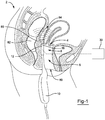
- FIG. 1 is plan view of a surgical system 2 within a lumen 80 of a patient.
- the surgical system 2 includes an anti-perforation device 6 and a surgical needle 10 having a distal tip 16.
- the surgical needle 10 as shown is also a surgical device 4 and cannulation device 8.
- the distal tip 16 of the surgical needle 10 includes a sensor 12 that measures a thickness of tissue 86 (e.g., as shown is the peritoneal wall).
- the sensor 12 measures the thickness of the tissue so that when a needle is deployed, the needle does not contact a second lumen 82, which as shown is a colon.
- the lumen 80 and the second lumen 82 are separated by an open space 84.
- the sensor 12 is connected to a control unit 30 that assists in measuring the thickness of the tissue, the distance between the lumen 80 and the second lumen 82, controlling the surgical needle, or a combination thereof.
- Figure 2A illustrates a distal tip 16 of a surgical device having a needle advancing mechanism 20 with a needle actuator 22.
- the needle advancing mechanism 20 as shown, is in the retracted position 90.
- the needle advancing mechanism 20 includes a stop 32 that limits movement of the needle (no shown) when the needle actuator 22 is activated.
- the stop 32 sets an actuation distance (X) of the needle.
- Figure 2B illustrates the needle 18, in the deployed position 92, extending from the distal tip 16 of the needle advancing mechanism 20.
- the needle 18 extends the distance (X) because the stop 32 restricts movement of the needle 18.
- Figure 3 illustrates a cannulation device 8 with a needle 18 extending from the lumen 80 through the tissue 86 and into the open space 84, without contacting the second lumen 82.
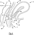
- FIG. 4 illustrates a surgical system 2 with a surgical device 4 and an anti-perforation device 6.
- the surgical device 6 is both a surgical needle 10 having a cannulation device 8, a sensor 12, or a detecting device 52 (the detecting device may also be located outside of the lumen and/or surgical device).
- the anti-perforation device 6 also includes a tissue marker 40, which as shown includes a signal source 44 (but may include a sensor 42).
- the surgical needle 10 extends into a lumen 80 and contacts a first side of tissue 86 and the tissue marker 40 is inserted into a second lumen 82.
- a signal is sent from the signal source 44 in the tissue marker 40 to the sensor 12 in the distal tip 16 of the surgical needle 10 to determine a distance between the lumen 80 and the second lumen 82 and if the open space 84 is sufficiently large that the needle (not shown) can be deployed without contacting the second lumen 82. If the distance is not large enough a control unit (not shown) may prevent deployment of the needle (not shown).
- Figure 5A illustrates a needle 18 partially puncturing the tissue 86 and extending towards the open space 84 and the tissue marker 70.
- Figure 5B illustrates the needle 18 extending beyond the distal tip 16 to a fully deployed position and stopping in the open space 84 between the lumen 80 and the second lumen 82 due to a measured distance between the distal tip 16 and the tissue marker 40.
- Figure 6 illustrates a second surgical device 4' inserted into a lumen 40 after a cannula 14 has been inserted into tissue 86 creating a space through the tissue 86 from the lumen 80 into an open space 84 but short of the second lumen 82.
- the cannula 14 provides temporary access to the open space 84 to perform a surgical procedure.
- any numerical values recited herein include all values from the lower value to the upper value in increments of one unit provided that there is a separation of at least 2 units between any lower value and any higher value.
- the amount of a component or a value of a process variable such as, for example, temperature, pressure, time and the like is, for example, from 1 to 90, preferably from 20 to 80, more preferably from 30 to 70, it is intended that values such as 15 to 85, 22 to 68, 43 to 51, 30 to 32 etc. are expressly enumerated in this specification.
- one unit is considered to be 0.0001, 0.001, 0.01 or 0.1 as appropriate.
Landscapes
- Health & Medical Sciences (AREA)
- Life Sciences & Earth Sciences (AREA)
- Surgery (AREA)
- Engineering & Computer Science (AREA)
- Veterinary Medicine (AREA)
- Public Health (AREA)
- General Health & Medical Sciences (AREA)
- Biomedical Technology (AREA)
- Heart & Thoracic Surgery (AREA)
- Animal Behavior & Ethology (AREA)
- Molecular Biology (AREA)
- Medical Informatics (AREA)
- Nuclear Medicine, Radiotherapy & Molecular Imaging (AREA)
- Pathology (AREA)
- Oral & Maxillofacial Surgery (AREA)
- Biophysics (AREA)
- Pulmonology (AREA)
- Anesthesiology (AREA)
- Hematology (AREA)
- Robotics (AREA)
- Surgical Instruments (AREA)
- Ultra Sonic Daignosis Equipment (AREA)
- Endoscopes (AREA)
Abstract
Description
- The present teachings relate to a surgical device with a control unit for controlling a depth of penetration of the surgical device and specifically a sensor, control unit, or both that controls a penetration depth of the surgical device or prevents movement of the surgical device into a feature of interest based upon a measurement of the feature of interest.
- During some surgical procedures a site of interest is located proximate to or directly abuts a sensitive area. For example, a bowel structure may be located proximate to a wall of interest. The sensitive area (e.g., bowel) may be blindly hidden behind the feature of interest, and if the device penetrates through the wall of interest the surgical device may contact a sensitive area. Contacting the sensitive area could create trauma to the sensitive area or spread the contents in the sensitive area within the surgical site. Examples of surgical devices and puncturing tools may be found in International Patent Application Publication Nos.
WO2014/066042 andWO2015/138708 ; European Patent Application No.EP3162294 ; and the devices discussed in the following websites: https://en.wikipedia.org/wiki/Veress_needle; https://en.wikipedia.org/wiki/Epidural_needle; https://en.wikipedia.org/wiki/Epidural_administration and all of which are incorporated by reference herein in their entirety for all purposes. Thus, attempts have been made to protect the sensitive areas and in many cases a more invasive surgical procedure may be used to avoid possible trauma to a sensitive area. - It would be attractive to have a surgical device with a sensor that senses a thickness of a feature of interest and then prevents the surgical device from penetrating further than a measured thickness of the feature of interest or prevents the device for activating. What is needed is a surgical device that prevents a needle of the surgical device from being deployed if the needle is set to deploy a distance that is greater than a thickness of a feature of interest. It would be attractive to have a sensor that determined a distance between a tip of a needle and an organ and prevents the tip from reaching the tip of the needle. What is needed is one or more sensors, control units, or both that control a depth of a surgical device or prevents the surgical device from being used. It would be attractive to have a sensor and signal source that communicate a distance between a first lumen and a second lumen. What is needed is a device and method to prevent perforation of a blind lumen or a feature behind a blind lumen.
- The disclosure meets one or more of the needs by providing: A surgical needle comprising: (a) a sensor; (b) a distal tip with the sensor being located at the distal tip; (c) a needle advancing mechanism that is adjustable to change an insertion depth; and (d) a control unit in communication with the needle advancing mechanism; wherein the sensor provides a signal to the control unit regarding a thickness of a feature of interest and the control unit controls the insertion depth based upon the signal from the sensor so that the insertion depth into the feature of interest is varied or the control unit prevents the needle advancing mechanism from activating.
- The present teachings provide a surgical system comprising: (a) a surgical needle comprising: (i) a distal tip and (ii) a needle advancing mechanism that adjustably controls an insertion depth; (b) a tissue marker; (c) a sensor located on the tissue marker or the distal tip of the surgical needle; and (d) a signal source that is located on the tissue marker when the sensor is located on the distal tip of the surgical needle and is located on the distal tip of the surgical needle when the sensor is located on the tissue marker; wherein the needle advancing mechanism controls the insertion depth based upon the relationship between the tissue marker and the distal tip of the surgical needle so that the insertion depth is less than a distance between the tissue marker and the distal tip of the surgical needle or the needle advancing mechanism is prevented from advancing.
- The present teachings provide: a method comprising: (a) inserting all or a portion of a surgical needle into a lumen; (b) activating a needle advancing mechanism; (c) monitoring a thickness of the lumen with a sensor; (d) receiving feedback based upon monitoring with the sensor; and (e) controlling an insertion depth of the needle advancing mechanism or preventing movement of the needle advancing mechanism based upon the feedback from the sensor.
- The present teachings provide a surgical device with a sensor that senses a thickness of a feature of interest and then prevents the surgical device from penetrating further than a measured thickness of the feature of interest or prevents the device for activating. The present teachings provide a surgical device that prevents a needle of the surgical device from being deployed if the needle is set to deploy a distance that is greater than a thickness of a feature of interest. The present teachings provide a sensor that determined a distance between a tip of a needle and an organ and prevents the tip from reaching the tip of the needle. The present teachings provide one or more sensors, control units, or both that control a depth of surgical device or prevents the surgical device from being used. The present teachings provide a sensor and signal source that communicate a distance between a first lumen and a second lumen. The present teachings prevent perforation of a blind lumen or a feature behind a blind lumen.
-
-
FIG. 1 is a plan view of a surgical system located within a patient; -
FIG. 2A is a needle advancing mechanism with the needle in a retracted position and a stop position set; -
FIG. 2B is a needle advancing mechanism with the needle in a deployed position; -
FIG. 3 is a plan view of a surgical system with the needle advanced into an open space; -
FIG. 4 is a plan view of a surgical system with a sensor located in a first lumen and a signal source 44 in a second lumen; -
FIG. 5A illustrates the needle partially advancing into tissue; and -
FIG 5B illustrates the needle advanced through the tissue and into an open space. - The explanations and illustrations presented herein are intended to acquaint others skilled in the art with the teachings, its principles, and its practical application. Those skilled in the art may adapt and apply the teachings in its numerous forms, as may be best suited to the requirements of a particular use. Accordingly, the specific embodiments of the present teachings as set forth are not intended as being exhaustive or limiting of the teachings. The scope of the teachings should, therefore, be determined not with reference to the above description, but should instead be determined with reference to the appended claims, along with the full scope of equivalents to which such claims are entitled. The disclosures of all articles and references, including patent applications and publications, are incorporated by reference for all purposes. Other combinations are also possible as will be gleaned from the following claims, which are also hereby incorporated by reference into this written description.
- The present teachings relate to surgical system. The surgical system functions to operate on a lumen, a region proximate to a lumen, a blind lumen, or a combination thereof. The surgical system may include one or more cannulas, one or more needles, or both. The surgical system may puncture or penetrate tissue so that a region behind the tissue may be exposed. The surgical system may biopsy tissue or a feature of interest. The surgical system may include a surgical device, an anti-perforation device, or both.
- The present teachings relate to a surgical device. The surgical device may be a non-electrical device (i.e., may only provide mechanical functions such as mechanical cutting or gripping). The surgical device may be an electrosurgical device. The surgical device may cut, puncture, open, or a combination thereof. The surgical device may be inserted into a lumen. Preferably, the surgical device may be inserted into a natural lumen. The surgical device may be inserted into a vagina, vaginal cavity, rectum, or a combination thereof. The surgical device may be an endoscope. The surgical device may be inserted through an endoscope or be an active part of an endoscope. The surgical device may be or include a cannulation device, a surgical needle, a needle, a marker, or a combination thereof. The surgical device may extend out of an endoscope and then be retracted back into the endoscope. The surgical device may puncture tissue to take a sample or create an opening. The surgical device may include be or include one or more anti-perforation devices, one or more cannulation devices, or both.
- The cannulation device may function to assist in deploying a cannula, a needle, a puncturing device, or a combination thereof. The cannulation device may extend into a lumen to take a biopsy, puncture tissue, or both. The cannulation device may include one or more cannulas, surgical needles, needle advancing mechanism, needle actuators, or a combination thereof. The cannulation device may house one or more cannulas, needles, or both and assist in inserting the cannula, needle, or both into tissue, into a feature of interest, or both. The cannulation device may advance a needle, advance a cannula, or both. The cannulation device may partially enter a lumen. The full cannulation device may enter a lumen. The cannulation device may include one or more anti-perforation devices, one or more stops, one or more sensors, one or more signal sensors, or a combination thereof. The cannulation device may function to insert a needle, surgical needle, cannula, needle, or a combination thereof into tissue, a feature of interest, or both. The cannulation device may insert a cannula into tissue, through tissue, through a feature of interest, into an anatomical feature, through an anatomical feature, or a combination thereof.
- The one or more cannulas may function to create an open space or maintain an open space. The one or more cannulas may create a pathway through tissue, an anatomical feature, or both that one or more surgical devices may extend through. The one or more cannulas may hold open an opening in tissue, an anatomical feature, or both. The one or more cannulas may remove tissue, puncture tissue, or both. The one or more cannulas may be removable from the tissue or anatomical feature. The one or more cannulas may temporarily be located within tissue or an anatomical feature. The cannula may have a blunt tip. The cannula may not pierce tissue, an anatomical feature, or both. A needle may be used in place of a cannula or in addition to a cannula. A needle may be located within and a cannula and extend out of a cannula.
- The needle may be located within a surgical needle, a cannula, or both. The needle may function to pierce tissue, an anatomical feature, or both. The needle may have a sharp tip. The needle may have an angled tip. The needle may penetrate tissue, an anatomical feature, or both. The needle may be reciprocated one or more times. The needle may penetrate tissue, an anatomical feature, or both a plurality of times so that a biopsy or samples may be taken. The needle may create an opening that a cannula may be inserted through. The needle may puncture or penetrate tissue, an anatomical feature or both and the cannula may follow into the opening created by the needle so that the cannula may be retained when the needle is removed. The needle, cannula, or both may be part of a surgical needle.
- The surgical needle may be a device that assists in inserting or placing the needle, cannula, or both into tissue, an anatomical feature, or both. The surgical needle may have a portion that extends into a lumen and a portion that is located outside of a lumen so that a user may control the surgical needle. The surgical needle may pierce tissue with a needle to create a hole, to insert a cannula, or both. The surgical needle may be a type of a cannulation device. The surgical needle may include or deploy a Veress needle, a trocar needle, or both. The surgical needle may include one or more needle advancing mechanism, one or more needle actuators, or both. The surgical needle may house all or a portion of the cannula, needle, or both. The surgical needle may have a portion that extends into a lumen. The surgical needle may have a portion that extends into a lumen to deploy a needle, cannula, or both. The surgical needle may have a portion that is hand held. The surgical needle may include a needle advancing mechanism, needle, cannula, or a combination thereof. The surgical needle has a distal tip.
- The distal tip of the surgical needle may be a portion of the surgical needle that is located farthest into a lumen, that is deployed through tissue, deployed through an anatomical feature, or a combination thereof. Preferably, the distal tip of the surgical needle houses a needle, a cannula, or both. The distal tip may be moved proximate to or into contact with tissue so that the needle, cannula, or both may be moved through the tissue. The distal tip of the surgical needle may include an opening that the needle, cannula, surgical instruments, or a combination thereof pass through. The sensor, signal source, or both may be located on or at the distal end of the surgical needle. The surgical need may be a device that deploys surgical devices, needles, cannulas, or a combination thereof for performing a surgical procedure. The cannula, needle, or both may have a distal tip. The distal tip of the cannula and needle may be coplanar. The distal tip of the needle may extend beyond the distal tip of the cannula. The distal tip of the cannula may be extend beyond a distal tip of the cannula. The distal tip of the needle may extend beyond the distal tip of the cannula so that during insertion of the needle the needle penetrates tissue and then the cannula is slid over the needle to create an opening. The needle advancing mechanism may advance the needle, the cannula, or both so that a distal tip extends through tissue, an anatomical feature, or both but stops short of a protected region.
- The needle advancing mechanism functions to extend a needle, a cannula, or both through tissue, a feature of interest, or both. The needle advancing mechanism may be located within or in communication with a surgical needle. The needle advancing mechanism may deploy needles, cannulas, or both out of a distal tip of the surgical needle. The needle advancing mechanism functions to control an insertion depth of a needle or cannula based upon a relationship between a tissue marker and a distal end of a needle or cannula. The needle advancing mechanism may insert a needle or cannula a distance that is less than a distance between the tissue marker and the distal end of the cannula or the needle. The needle advancing mechanism may be prevented from actuating is the distance is greater than a distance between the tissue marker and sensor. The needle advancing mechanism may extend a distal tip of the needle, cannula, or both into an open space. The needle advancing mechanism may extend a distal tip of the needle, cannula, or both to a location short of an adjacent structure, second lumen, a protected region, or a combination thereof. The needle advancing mechanism may extend a needle, cannula, or both through a wall of a vagina or uterus and into a pouch of Douglas but the distal end of the needle, cannula, or both will stop before contacting a rectum. The needle advancing mechanism may deploy a needle, cannula, or both upon pressing a button. The needle advancing mechanism may advance a needle a predetermined distance. A distance the needle advancing mechanism moves a needle may be adjusted, varied, changed, or a combination thereof. The needle advancing mechanism may have a distal tip that is placed into contact with tissue or a feature of interest and then a needle or cannula advanced through the tissue or feature of interest in contact with the needle advancing mechanism. The needle advancing mechanism may house all or a portion of a needle, cannula, or both. The needle advancing mechanism may include one or more needle actuators, control units, sensors, stops, or a combination thereof.
- The one or more needle actuators may function to move one or more needles, one or more cannulas a distance. The one or more needle actuators may move a needle, a cannula, or both a predetermined distance. The one or more needle actuators may puncture tissue, a feature of interest, or both. The one or more needle actuators axially move the needle, the cannula, or both. The one or more needle actuators may move a needle, a cannula, or both at an angle relative to the tissue, the feature of interest, or both. The one or more needle actuators may move the needle, the cannula, or both with a sufficient amount of force to puncture the tissue, the feature of interest, or both. The one or more needle actuators may be variable. The one or more needle actuators may move the needle, the cannula, or both a distance of about 1mm or more, about 2 mm or more, about 3 mm or more, about 4 mm or more, about 5 mm or more, about 1 cm or more, or about 1.5 cm or more. The one or more needle actuators may move the needle, the cannula, or both a distance of about 10 cm or less, about 7 cm or less, about 5 cm or less, or about 3 cm or less. The one or more needle actuators may include one or more bias members, one or more pneumatic systems, one or more hydraulic systems, or a combination thereof. The one or more bias members may be a spring, compressible member, rubber member, elastomeric member, a tensioning member, solenoid, pneumatic system, or a combination thereof. The one or more needle actuators may be preloaded prior to being inserted into a lumen. The one or more needle actuators may be adjusted while they were in a lumen. The one or more needle actuators may be set, reset, adjusted, or a combination thereof within a lumen, from a location external of the lumen, or both. The one or more needle actuators may include or be connected to one or more anti-perforation devices.
- The one or more anti-perforation device function to protect a protected tissue, protected area, a protected area, or a combination thereof (hereinafter referred to as protected tissue). The one or more anti-perforation device may prevent actuation of the one or more needle actuators. The one or more anti-perforation devices may automatically control an actuation depth of a needle, cannula, or both. The one or more anti-perforation devices may prevent actuation of a needle, a cannula, or both if a monitored thickness is less than a set distance. The one or more anti-perforation devices may change a set distance based upon a monitored distance, based upon feedback from monitoring, or both. The one or more anti-perforation devices may control a distance the needle, cannula, or both move. The one or more anti-perforation devices may provide feedback, communicate with a control unit, move a stop, or a combination thereof. The one or more anti-perforation devices may lock a needle or cannula, make a sound, create a haptic signal, create a visual signal, or a combination thereof. The one or more anti-perforation devices may include one or more sensors, one or more control units, one or more tissue markers, or a combination thereof.
- The one or more control units function to prevent a needle, a cannula, or both from contacting protected tissue. The one or more control units may move a stop, prevent actuation of a needle, prevent actuation of a cannula, adjust a distance a needle is moved, adjust a distance a cannula is moved, or a combination thereof. The one or more control units may communicate with one or more sensors, one or more signal sources, one or more detecting devices, or a combination thereof. The one or more control units may be in communication with the needle advancing mechanisms. The one or more control units may be in communication with the sensors, signal source, detecting device, stop, one or more needle advancing devices, or a combination thereof. The one or more control units may be in wired communication or wireless communication with the sensors, signal sources, detecting devices, tops, needle advancing devices, or a combination thereof. The one or more control units may control actuation of the needle advancing device. For example, the control unit may determine a depth or be provided a depth and the control unit may determine a distance of actuation and then actuate the needle advancing device, change the distance, or prevent actuation. The control units may monitor one or more distances. The control unit may monitor a distance between two lumens. The control unit may monitor a thickness of an open space. The control unit may be in communication with a sensor and a signal source and determine a distance therebetween. The control unit may communicate with only a sensor to determine a thickness. For example, if a sensor is inserted in a vaginal canal and a signal source is inserted into a rectum, the control unit may monitor a distance between the sensor and the signal source so that a needle does not extend into the colon, stops short of the colon, stops in an open space such a pouch of Douglas; or a combination thereof. The control unit may calculate a distance the needle, cannula, or both can extend. The control unit may set a stop once a distance is calculated.
- The one or more stops may function to limit movement of a needle, cannula, or both to a predetermined distance, a calculated distance, a measured distance, or a combination thereof (e.g., a distance). The one or more stops may pre-compress a needle actuator, adjust a needle actuator, or both. The one or more stops may release a compression of a needle actuator. The one or more stops may be in contact a needle or needle actuator once a needle moves the distance. The one or more stops may move forward or backward to control a distance or depth the needle advances or penetrated into a feature of interest. The stop may be located inside of the needle advancing mechanism. The stop may be connected to or part of the needle actuator. The stop may be located external to the needle advancing mechanism but in communication with the needle or cannula. The needle advancing mechanism, needle actuator, or both may move the needle, the cannula, or both and the stop may determine the distance the needle penetrates into the feature of interest. The stop may be axially movable. The stop may be connected to the needle advancing mechanism and may be inserted into a lumen with the needle advancing mechanism. The stop may be electrically controlled. The stop may electrically move. The stop may be moved from a location external of the lumen. The stop may be moved by the control unit. The stop may be located in a same or a different lumen as a tissue marker, sensor, signal source, or a combination thereof.
- The one or more tissue markers may function to mark protected tissue. The one or more tissue markers may connect to or be located near a protected tissue. The one or more tissue markers may mark a first lumen, a second lumen, or both. The one or more tissue markers may be inserted into a second lumen. The one or more tissue markers may locate a protected tissue so that the protected tissue is not contacted or damaged when the surgical device is in operation. The one or more tissue markers may be inserted into a lumen and be part of a surgical device. The one or more tissue markers may include a sensor, a signal source, or both. The one or more tissue markers may be part of a surgical device. The one or more tissue markers may be located on a distal tip of a surgical needle, cannula, or both. The one or more tissue markers, preferably, may be located in a different lumen or a different surgical device as the needle. The tissue markers may connect to tissue, be located in tissue, or both. The one or more tissue markers may indicate protected tissue so that a distance relative to the tissue markers can be determined. The tissue markers may be expandable within protected tissue or a lumen. The tissue markers may connect to tissue or a lumen. The tissue marker may be removably inserted into a lumen. Preferably, the tissue marker is connected to a surgical device or a finger of a surgeon and the tissue marker is located within a lumen. For example, the tissue marker may be inserted into a rectum by a surgical device or a surgeon's finger and then removed when the procedure is finished. The tissue markers may include one or more sensors.
- The one or more sensors may function to measure a distance between a protected tissue and a surgical device. The one or more sensors may work without any other sensors. The sensors may sense a distance between two locations. The sensors may determine a distance between one tissue and a second tissue or an open space between two tissues (e.g., a tissue of interest and a protected tissue). The sensor may be a soft tissue thickness sensor, an ultrasonic sensor, a light reflective sensor, a conductive material thickness sensor, a capacitance sensor, or a combination thereof. The sensor may be connected to or located proximate to a distal tip of a surgical device. Preferably, the sensor is located at or on a distal tip of a needle, a cannula, or both. The sensor may determine a depth that a needle or cannula can safely advance. The sensor may determine a distance or length of an open space. The sensor may determine a distance a protected tissue is away from a distal tip. The sensor may directly or indirectly connected to a surgical device. Preferably, the sensor is directly connected to a distal tip of the surgical device. The sensor may work without a signal source. The sensor may work in conjunction with a signal source. The sensor and the signal source may be mechanically separate, electrically separate, or both.
- The signal source may function mark protected tissue. The signal source may communicate with the sensor. The signal source may transmit a signal to a sensor that a sensor monitors to determine a distance. The signal source may be located within a different lumen than the sensor. The signal source may transmit a signal through tissue, an open space, an organ, or a combination thereof that is picked up by a sensor. The signal source and sensor may determine a distance between a distal tip and protected tissue. The signal source may be ultrasound, acoustic, doppler ultrasound, vibrations, or a combination thereof. The signal source and sensor may be the only two devices needed to determine a distance between two locations. If the signal source is located on the tissue marker than the sensor is located on the cannula, the needle, or both. If the signal source is located on the needle, the cannula, or both then the sensor is located on the tissue marker. A detecting device may monitor a distance between the sensor and signal source.
- The detecting device may function to detect a signal between the sensor and the signal source, to calculate a distance based upon the signal transmitted from the signal source and received by the sensor, or both. The detecting device may be part of a control unit, sensor, or both. The detecting device may be part of the sensor. The detecting device may calculate a distance. The detecting device may control the stop, an insertion depth, or both. All or a portion of the detecting device may extend into the lumen or be located outside of the lumen.
- The lumen may be any opening that a surgical device may extend into. Preferably, the lumen is a natural lumen. The lumen may be a vaginal canal, a rectum, urethra, mouth, ear, nose, eye socket, or the like. The lumen may be located proximate to a second lumen, an open space, or both.
- The second lumen may be any of the lumen discussed herein. The lumen, second lumen, or both may receive the sensor, the signal source, a portion of a surgical device, a distal tip of a needle, a distal tip of a cannula, a needle advancing mechanism, an anti-perforation device, or a combination thereof. Preferably, if only one lumen is being operated on, only a sensor is applied. More preferably, if two lumen are being operated on or a location between two lumen is being operated on a sensor is placed in one lumen and a signal source is placed in a second lumen. A first lumen may be a vaginal canal and may receive a portion of a surgical device and a sensor and a second lumen may be the rectum and may receive all or a portion of a tissue marker, signal source, or both. An open space proximate to or located between the first lumen, the second lumen, or both may be monitored.
- The open space may be any space where a needle or lumen may be inserted that may not damage or contaminate a lumen. The open space may receive the needle or cannula when the needle advancing mechanism advances the needle or cannula. The open space may provide access to a feature of interest located proximate to the lumen. The open space may be a pouch of Douglas. The open space may be located within an abdomen, sinus cavity, peritoneal cavity, or a combination thereof. The open space may be filled with a fluid, air, open, or a combination thereof. The open space may have a distance. The open space may be accessed during surgery and may be monitored by a method taught herein.
- The present device and system taught herein may be used in a method to protect one or more features of interest. The device and system may include one or more anti-perforation devices. The method may include a step of protecting protected tissue. A surgical device, sensor, surgical needle, anti-perforation device, needle advancing mechanism, or a combination thereof may be inserted into a first lumen, a second lumen, or both. A sensor may be inserted into a first lumen (e.g., a vaginal canal). A signal source may be inserted into a second lumen (e.g., a rectum). The sensor may measure a thickness or a lumen, a distance between a distal tip and a protected tissue, an open space, or a combination thereof. The sensor may measure without any other devices. The sensor may communicate with a signal source. The sensor may receive a signal. The signal source may transmit one or more signals to the sensor. The sensor, a detecting device, or both may determine a distance. A needle depth may be adjusted based upon feedback from the detecting device, control unit, or both. The needle plunge or insertion depth may be varied by moving a stop. The insertion depth or plunge depth may be varied by adjusting one or more parameters of the needle advancing mechanism, the needle actuator, or both. The tissue marker may be placed. The surgical device may be inserted. The needle actuator may be activated. The needle, cannula, or both may be inserted a depth. The needle, cannula, or both may be prevented from actuating, advancing, being inserted, or both. Feedback from the sensor, detecting device, control unit, or a combination thereof may be monitored. A distance may be calculated. The distance may be provided as feedback. The sensor, signal source, or both may be removed from the lumen, the second lumen, or both. Adjusting an insertion depth of a needle, cannula, or both. Providing feedback between the control unit and the needle advancing mechanism, the stop, the needle actuator, or both.
-
Figure 1 is plan view of asurgical system 2 within alumen 80 of a patient. Thesurgical system 2 includes ananti-perforation device 6 and asurgical needle 10 having adistal tip 16. Thesurgical needle 10 as shown is also asurgical device 4 andcannulation device 8. Thedistal tip 16 of thesurgical needle 10 includes asensor 12 that measures a thickness of tissue 86 (e.g., as shown is the peritoneal wall). Thesensor 12 measures the thickness of the tissue so that when a needle is deployed, the needle does not contact asecond lumen 82, which as shown is a colon. Thelumen 80 and thesecond lumen 82 are separated by anopen space 84. Thesensor 12 is connected to acontrol unit 30 that assists in measuring the thickness of the tissue, the distance between thelumen 80 and thesecond lumen 82, controlling the surgical needle, or a combination thereof. -
Figure 2A illustrates adistal tip 16 of a surgical device having aneedle advancing mechanism 20 with a needle actuator 22. Theneedle advancing mechanism 20, as shown, is in the retractedposition 90. Theneedle advancing mechanism 20 includes astop 32 that limits movement of the needle (no shown) when the needle actuator 22 is activated. Thestop 32 sets an actuation distance (X) of the needle. -
Figure 2B illustrates theneedle 18, in the deployedposition 92, extending from thedistal tip 16 of theneedle advancing mechanism 20. Theneedle 18 extends the distance (X) because thestop 32 restricts movement of theneedle 18. -
Figure 3 illustrates acannulation device 8 with aneedle 18 extending from thelumen 80 through thetissue 86 and into theopen space 84, without contacting thesecond lumen 82. -
Figure 4 illustrates asurgical system 2 with asurgical device 4 and ananti-perforation device 6. Thesurgical device 6 is both asurgical needle 10 having acannulation device 8, asensor 12, or a detecting device 52 (the detecting device may also be located outside of the lumen and/or surgical device). Theanti-perforation device 6 also includes atissue marker 40, which as shown includes a signal source 44 (but may include a sensor 42). Thesurgical needle 10 extends into alumen 80 and contacts a first side oftissue 86 and thetissue marker 40 is inserted into asecond lumen 82. A signal is sent from the signal source 44 in thetissue marker 40 to thesensor 12 in thedistal tip 16 of thesurgical needle 10 to determine a distance between thelumen 80 and thesecond lumen 82 and if theopen space 84 is sufficiently large that the needle (not shown) can be deployed without contacting thesecond lumen 82. If the distance is not large enough a control unit (not shown) may prevent deployment of the needle (not shown). -
Figure 5A illustrates aneedle 18 partially puncturing thetissue 86 and extending towards theopen space 84 and the tissue marker 70. -
Figure 5B illustrates theneedle 18 extending beyond thedistal tip 16 to a fully deployed position and stopping in theopen space 84 between thelumen 80 and thesecond lumen 82 due to a measured distance between thedistal tip 16 and thetissue marker 40. -
Figure 6 illustrates a second surgical device 4' inserted into alumen 40 after acannula 14 has been inserted intotissue 86 creating a space through thetissue 86 from thelumen 80 into anopen space 84 but short of thesecond lumen 82. Thecannula 14 provides temporary access to theopen space 84 to perform a surgical procedure. - Any numerical values recited herein include all values from the lower value to the upper value in increments of one unit provided that there is a separation of at least 2 units between any lower value and any higher value. As an example, if it is stated that the amount of a component or a value of a process variable such as, for example, temperature, pressure, time and the like is, for example, from 1 to 90, preferably from 20 to 80, more preferably from 30 to 70, it is intended that values such as 15 to 85, 22 to 68, 43 to 51, 30 to 32 etc. are expressly enumerated in this specification. For values which are less than one, one unit is considered to be 0.0001, 0.001, 0.01 or 0.1 as appropriate. These are only examples of what is specifically intended and all possible combinations of numerical values between the lowest value and the highest value enumerated are to be considered to be expressly stated in this application in a similar manner.
- Unless otherwise stated, all ranges include both endpoints and all numbers between the endpoints. Unless otherwise stated, a teaching with the term "about" or "approximately" in combination with a numerical amount encompasses a teaching of the recited amount, as well as approximations of that recited amount. By way of example, a teaching of "about 100" encompasses a teaching of within a range of 100 +/- 15.
- The disclosures of all articles and references, including patent applications and publications, are incorporated by reference for all purposes. The term "consisting essentially of" to describe a combination shall include the elements, ingredients, components or steps identified, and such other elements ingredients, components or steps that do not materially affect the basic and novel characteristics of the combination. The use of the terms "comprising" or "including" to describe combinations of elements, ingredients, components or steps herein also contemplates embodiments that consist essentially of the elements, ingredients, components or steps. By use of the term "may" herein, it is intended that any described attributes that "may" be included are optional.
- Plural elements, ingredients, components or steps can be provided by a single integrated element, ingredient, component or step. Alternatively, a single integrated element, ingredient, component or step might be divided into separate plural elements, ingredients, components or steps. The disclosure of "a" or "one" to describe an element, ingredient, component or step is not intended to foreclose additional elements, ingredients, components or steps.
- It is understood that the above description is intended to be illustrative and not restrictive. Many embodiments as well as many applications besides the examples provided will be apparent to those of skill in the art upon reading the above description. The scope of the teachings should, therefore, be determined not with reference to the above description, but should instead be determined with reference to the appended claims, along with the full scope of equivalents to which such claims are entitled. The disclosures of all articles and references, including patent applications and publications, are incorporated by reference for all purposes. The omission in the following claims of any aspect of subject matter that is disclosed herein is not a disclaimer of such subject matter, nor should it be regarded that the inventors did not consider such subject matter to be part of the disclosed inventive subject matter.
- Further examples of the present disclosure are described in the following paragraphs.
- Example 1: A surgical needle comprising:
- a. a sensor;
- b. a distal tip with the sensor being located at the distal tip;
- c. a needle advancing mechanism that is adjustable to change an insertion depth; and
- d. a control unit in communication with the needle advancing mechanism;
- Example 2: The surgical needle of Example 1, wherein the sensor is a soft tissue thickness sensor.
- Example 3: The surgical needle of Example 1, wherein the sensor is an ultrasonic sensor, a light reflective sensor, a conductive material thickness measurement sensor, a capacitance sensor, or a combination thereof.
- Example 4: The surgical needle of Example 1, wherein the needle advancing mechanism includes and advances a needle out of the distal tip.
- Example 5: The surgical needle of Example 1, wherein the needle advancing mechanism includes a solenoid, a spring, a pneumatic system, or a combination thereof to move the needle out of the distal tip.
- Example 6: The surgical needle of Example 1, wherein the surgical needle includes a stop that is movable to control the insertion depth of a needle penetrates beyond the distal tip into the feature of interest.
- Example 7: The surgical needle of Example 6, wherein the needle advancing mechanism moves the needle into contact with the stop and a position of the stop determines the depth the needle penetrates into the feature of interest.
- Example 8: The surgical needle of Example 6, wherein the needle advancing mechanism is connected to the stop and the needle advancing mechanism controls a location of the stop to determine the depth the needle penetrates into a feature of interest.
- Example 9: The surgical needle of Example 1, wherein the feature of interest is located adjacent to a blind space, another feature, or both and the needle penetrates into the blind space and the depth the needle penetrates is restricted before the needle contacts the blind space, the another feature, or both.
- Example 10: A surgical system comprising:
- a. a surgical needle comprising:
- i. a distal tip and
- ii. a needle advancing mechanism that adjustably controls an insertion depth;
- b. a tissue marker;
- c. a sensor located on the tissue marker or the distal tip of the surgical needle; and
- d. a signal source that is located on the tissue marker when the sensor is located on the distal tip of the surgical needle and is located on the distal tip of the surgical needle when the sensor is located on the tissue marker;
- a. a surgical needle comprising:
- Example 11: The surgical system of Example 10, wherein the needle advancing mechanism is prevented from actuating if the insertion depth is greater than the distance between the tissue marker and the sensor. The surgical system of
claim 10, wherein the needle advancing mechanism is prevented from actuating if the insertion depth is greater than the distance between the tissue marker and the sensor. - Example 12: The surgical system of Example 10, wherein the tissue marker and the sensor are mechanically separate, electrically separate, or both.
- Example 13: The surgical system of Example 10, wherein the distance is determined by the sensor monitoring a signal created by the signal source.
- Example 14: A method comprising:
- a. inserting all or a portion of a surgical needle into a lumen;
- b. activating a needle advancing mechanism;
- c. monitoring a thickness of the lumen with a sensor;
- d. receiving feedback based upon monitoring with the sensor; and
- e. controlling an insertion depth of the needle advancing mechanism or preventing movement of the needle advancing mechanism based upon the feedback from the sensor.
- Example 15: The method of Example 14, wherein the feedback is a haptic signal, a visual signal, an auditory signal, a needle being locked out, or a combination thereof.
- Example 16: The method of Example 14, wherein the feedback controls an insertion depth of a needle by the needle advancing mechanism so that when the needle advances only the monitored thickness or less.
- Example 17: The method of Example 14, wherein the needle advancing mechanism advances a needle.
- Example 18: The method of Example 14, wherein a signal source is located opposite the sensor.
- Example 19: The method of Example 14, wherein a signal source is inserted into a second lumen.
- Example 20: The method of Example 19, wherein the lumen is a vaginal canal and the second lumen is the rectum.
- 2 Surgical system
- 4 Surgical device
- 6 Anti-perforation device
- 8 Cannulation device
- 10 Surgical needle
- 12 Sensor
- 14 Cannula
- 16 Distal tip (of cannula)
- 18 Needle
- 20 Needle advancing mechanism
- 22 Needle actuator (solenoid, spring, pneumatic system)
- 30 Control unit
- 32 stop
- 40 Tissue Marker
- 42 sensor
- 44 Signal source
- 50 Detecting device
- 80 Lumen (blind lumen)
- 82 Second Lumen
- 84 Open space
- 86 tissue
- 90 Retracted position
- 92 Deployed position
Claims (13)
- A surgical needle comprising:a. a sensor;b. a distal tip with the sensor being located at the distal tip;c. a needle advancing mechanism that is adjustable to change an insertion depth; andd. a control unit in communication with the needle advancing mechanism;wherein the sensor provides a signal to the control unit regarding a thickness of a feature of interest and the control unit controls the insertion depth based upon the signal from the sensor so that the insertion depth into the feature of interest is varied or the control unit prevents the needle advancing mechanism from activating.
- The surgical needle of claim 1, wherein the sensor is a soft tissue thickness sensor.
- The surgical needle of claim 1 or 2, wherein the sensor is an ultrasonic sensor, a light reflective sensor, a conductive material thickness measurement sensor, a capacitance sensor, or a combination thereof.
- The surgical needle of any of claims 1 to 3, wherein the needle advancing mechanism includes and advances a needle out of the distal tip.
- The surgical needle of any of claims 1 to 4, wherein the needle advancing mechanism includes a solenoid, a spring, a pneumatic system, or a combination thereof to move the needle out of the distal tip.
- The surgical needle of any of claims 1 to 7, wherein the surgical needle includes a stop that is movable to control the insertion depth of a needle penetrates beyond the distal tip into the feature of interest.
- The surgical needle of claim 6, wherein the needle advancing mechanism moves the needle into contact with the stop and a position of the stop determines the depth the needle penetrates into the feature of interest.
- The surgical needle of claim 6 or 7, wherein the needle advancing mechanism is connected to the stop and the needle advancing mechanism controls a location of the stop to determine the depth the needle penetrates into a feature of interest.
- The surgical needle of any of claims 1 to 8, wherein the feature of interest is located adjacent to a blind space, another feature, or both and the needle penetrates into the blind space and the depth the needle penetrates is restricted before the needle contacts the blind space, the another feature, or both.
- A surgical system comprising:a. a surgical needle comprising:i. a distal tip andii. a needle advancing mechanism that adjustably controls an insertion depth;b. a tissue marker;c. a sensor located on the tissue marker or the distal tip of the surgical needle; andd. a signal source that is located on the tissue marker when the sensor is located on the distal tip of the surgical needle and is located on the distal tip of the surgical needle when the sensor is located on the tissue marker;wherein the needle advancing mechanism controls the insertion depth based upon the relationship between the tissue marker and the distal tip of the surgical needle so that the insertion depth is less than a distance between the tissue marker and the distal tip of the surgical needle or the needle advancing mechanism is prevented from advancing.
- The surgical system of claim 10, wherein the needle advancing mechanism is prevented from actuating if the insertion depth is greater than the distance between the tissue marker and the sensor.
- The surgical system of claim 10 or 11, wherein the tissue marker and the sensor are mechanically separate, electrically separate, or both.
- The surgical system of any of claims 10 to 12, wherein the distance is determined by the sensor monitoring a signal created by the signal source.
Applications Claiming Priority (1)
| Application Number | Priority Date | Filing Date | Title |
|---|---|---|---|
| US16/369,442 US11406454B2 (en) | 2019-03-29 | 2019-03-29 | Anti-perforation device |
Publications (2)
| Publication Number | Publication Date |
|---|---|
| EP3714819A1 true EP3714819A1 (en) | 2020-09-30 |
| EP3714819B1 EP3714819B1 (en) | 2025-02-12 |
Family
ID=70058247
Family Applications (1)
| Application Number | Title | Priority Date | Filing Date |
|---|---|---|---|
| EP20166758.1A Active EP3714819B1 (en) | 2019-03-29 | 2020-03-30 | Anti-perforation device |
Country Status (4)
| Country | Link |
|---|---|
| US (2) | US11406454B2 (en) |
| EP (1) | EP3714819B1 (en) |
| JP (1) | JP7483458B2 (en) |
| CN (1) | CN111743637B (en) |
Families Citing this family (3)
| Publication number | Priority date | Publication date | Assignee | Title |
|---|---|---|---|---|
| US11406454B2 (en) | 2019-03-29 | 2022-08-09 | Gyrus Acmi, Inc. | Anti-perforation device |
| US20230086838A1 (en) * | 2021-09-17 | 2023-03-23 | Corral Technologies, LLC | Virtual livestock management |
| CN116530925B (en) * | 2023-05-06 | 2025-08-22 | 中国人民解放军空军军医大学 | An intelligent laparoscopic limiter for preventing uterine perforation |
Citations (9)
| Publication number | Priority date | Publication date | Assignee | Title |
|---|---|---|---|---|
| US4674503A (en) * | 1981-03-05 | 1987-06-23 | Peyman Gholam A | Controlled depth penetrant apparatus and method |
| ES2216124T3 (en) * | 1996-01-08 | 2004-10-16 | Biosense Inc. | APPARATUS FOR MIOCARDIAL REVASCULARIZATION. |
| EP2478856A1 (en) * | 2011-01-24 | 2012-07-25 | Samsung Medison Co., Ltd. | Treatment device |
| WO2014066042A2 (en) | 2012-10-22 | 2014-05-01 | Ethicon Endo-Surgery, Inc. | Surgeon feedback sensing and display methods |
| US20140309522A1 (en) * | 2013-01-26 | 2014-10-16 | Larry W. Fullerton | Microwave antenna apparatus, systems, and methods for localizing markers or tissue structures within a body |
| WO2015138708A1 (en) | 2014-03-12 | 2015-09-17 | Proximed, Llc | Surgical guidance systems, devices, and methods |
| EP3162294A1 (en) | 2014-06-30 | 2017-05-03 | Olympus Corporation | Processing tool for puncturing |
| US20170252060A1 (en) * | 2016-03-06 | 2017-09-07 | Andrew N. Ellingson | Guide Wire Device and Method |
| US9844641B2 (en) * | 2014-07-16 | 2017-12-19 | Fractyl Laboratories, Inc. | Systems, devices and methods for performing medical procedures in the intestine |
Family Cites Families (38)
| Publication number | Priority date | Publication date | Assignee | Title |
|---|---|---|---|---|
| US4546773A (en) | 1981-01-23 | 1985-10-15 | Accutome, Inc. | Apparatus to measure conical thickness |
| US6405072B1 (en) | 1991-01-28 | 2002-06-11 | Sherwood Services Ag | Apparatus and method for determining a location of an anatomical target with reference to a medical apparatus |
| JPH05337127A (en) * | 1992-06-04 | 1993-12-21 | Olympus Optical Co Ltd | Tracheal puncturing apparatus |
| US5772597A (en) | 1992-09-14 | 1998-06-30 | Sextant Medical Corporation | Surgical tool end effector |
| US5486188A (en) | 1993-11-15 | 1996-01-23 | Smith; Alan D. | Keratoscopic surgical instrument for making radial and arcuate corneal incisions |
| JP4265698B2 (en) | 1997-02-14 | 2009-05-20 | バイオセンス・ウェブスター・インコーポレイテッド | X-ray guided surgical positioning system using extended mapping space |
| WO2001008578A1 (en) | 1999-07-30 | 2001-02-08 | Vivant Medical, Inc. | Device and method for safe location and marking of a cavity and sentinel lymph nodes |
| JP3522171B2 (en) * | 1999-10-14 | 2004-04-26 | 茂 川島 | Automatic syringe |
| FR2816200A1 (en) | 2000-11-06 | 2002-05-10 | Praxim | DETERMINING THE POSITION OF A KNEE PROSTHESIS |
| US6835193B2 (en) * | 2001-07-10 | 2004-12-28 | Myocardial Therapeutics, Inc. | Methods for controlled depth injections into interior body cavities |
| US20030026758A1 (en) | 2001-07-27 | 2003-02-06 | Baker Gregg S. | Method and device for monitoring real-time position of an area targeted by a radiosurgery system |
| US6685623B2 (en) | 2001-11-20 | 2004-02-03 | Surx, Inc. | Incontinence treatment with urethral guide |
| CA2521738C (en) | 2003-04-07 | 2010-09-14 | Bausch & Lomb Incorporated | Bar-link drive system for a microkeratome |
| US20070135803A1 (en) * | 2005-09-14 | 2007-06-14 | Amir Belson | Methods and apparatus for performing transluminal and other procedures |
| US20080097501A1 (en) | 2006-06-22 | 2008-04-24 | Tyco Healthcare Group Lp | Ultrasonic probe deflection sensor |
| US8840603B2 (en) | 2007-01-10 | 2014-09-23 | Ethicon Endo-Surgery, Inc. | Surgical instrument with wireless communication between control unit and sensor transponders |
| US7730628B2 (en) * | 2008-09-05 | 2010-06-08 | Bayer Schering Pharma Ag | Depth stop devices and systems |
| US8437833B2 (en) | 2008-10-07 | 2013-05-07 | Bard Access Systems, Inc. | Percutaneous magnetic gastrostomy |
| US8690776B2 (en) | 2009-02-17 | 2014-04-08 | Inneroptic Technology, Inc. | Systems, methods, apparatuses, and computer-readable media for image guided surgery |
| EP3847959B1 (en) | 2009-06-26 | 2024-09-11 | Cianna Medical, Inc. | System for localizing markers or tissue structures within a body |
| US10828786B2 (en) | 2009-08-17 | 2020-11-10 | Mazor Robotics Ltd. | Device for improving the accuracy of manual operations |
| NO332094B1 (en) | 2009-12-23 | 2012-06-18 | Nofence As | Method and system for fencing animals without the use of a physical fence |
| US8690887B2 (en) | 2010-02-08 | 2014-04-08 | Jeffrey B. Kleiner | Method and system for identification of a center of a vertebral disc space and corresponding central axis of a spinal column |
| US8510012B2 (en) | 2010-12-22 | 2013-08-13 | Bendix Commercial Vehicle Systems Llc | Anti-tailgating system and method |
| US9332990B2 (en) | 2010-12-30 | 2016-05-10 | Wake Forest University Health Sciences | Ureter to ileal conduit anastomosis using magnetic compression and related delivery devices and methods |
| US11304686B2 (en) * | 2011-06-17 | 2022-04-19 | Koninklijke Philips N.V. | System and method for guided injection during endoscopic surgery |
| US20120326875A1 (en) | 2011-06-27 | 2012-12-27 | Deborah Coppola | Invisible fence for a child |
| US9724124B2 (en) * | 2011-08-08 | 2017-08-08 | Boston Scientific Scimed Inc. | Devices and methods for controlling depth of insertion |
| US8670816B2 (en) | 2012-01-30 | 2014-03-11 | Inneroptic Technology, Inc. | Multiple medical device guidance |
| US9814448B2 (en) * | 2012-05-21 | 2017-11-14 | Precision Biopsy, Inc. | Three-dimensional optical imaging and therapy of prostate cancer |
| US10653423B2 (en) | 2012-06-29 | 2020-05-19 | University Of Washington | Catheters for emergency endovascular surgery and associated devices, systems, and methods |
| US20150238276A1 (en) | 2012-09-30 | 2015-08-27 | M.S.T. Medical Surgery Technologies Ltd. | Device and method for assisting laparoscopic surgery - directing and maneuvering articulating tool |
| ES2401860B1 (en) | 2012-12-13 | 2014-03-04 | Javier FERRER TORREGROSA | Guidance and support device for minimally incised surgery of the foot or hand |
| US9320539B2 (en) | 2013-09-30 | 2016-04-26 | Coloplast A/S | Surgical system for and a method of dissection of tissue away from an organ or body lumen |
| US20150157787A1 (en) * | 2013-12-05 | 2015-06-11 | W. L. Gore & Associates, Inc. | Needle guide and related systems and methods |
| WO2016160348A1 (en) * | 2015-03-31 | 2016-10-06 | SPIRATION, INC., d/b/a OLYMPUS RESPIRATORY AMERICA | Catheter access device |
| AU2017257701B2 (en) * | 2016-04-26 | 2019-08-15 | Boston Scientific Limited | Percutaneous access device with adjustable depth stop |
| US11406454B2 (en) | 2019-03-29 | 2022-08-09 | Gyrus Acmi, Inc. | Anti-perforation device |
-
2019
- 2019-03-29 US US16/369,442 patent/US11406454B2/en active Active
-
2020
- 2020-03-27 CN CN202010230057.6A patent/CN111743637B/en active Active
- 2020-03-27 JP JP2020058276A patent/JP7483458B2/en active Active
- 2020-03-30 EP EP20166758.1A patent/EP3714819B1/en active Active
-
2022
- 2022-07-05 US US17/857,791 patent/US11925421B2/en active Active
Patent Citations (9)
| Publication number | Priority date | Publication date | Assignee | Title |
|---|---|---|---|---|
| US4674503A (en) * | 1981-03-05 | 1987-06-23 | Peyman Gholam A | Controlled depth penetrant apparatus and method |
| ES2216124T3 (en) * | 1996-01-08 | 2004-10-16 | Biosense Inc. | APPARATUS FOR MIOCARDIAL REVASCULARIZATION. |
| EP2478856A1 (en) * | 2011-01-24 | 2012-07-25 | Samsung Medison Co., Ltd. | Treatment device |
| WO2014066042A2 (en) | 2012-10-22 | 2014-05-01 | Ethicon Endo-Surgery, Inc. | Surgeon feedback sensing and display methods |
| US20140309522A1 (en) * | 2013-01-26 | 2014-10-16 | Larry W. Fullerton | Microwave antenna apparatus, systems, and methods for localizing markers or tissue structures within a body |
| WO2015138708A1 (en) | 2014-03-12 | 2015-09-17 | Proximed, Llc | Surgical guidance systems, devices, and methods |
| EP3162294A1 (en) | 2014-06-30 | 2017-05-03 | Olympus Corporation | Processing tool for puncturing |
| US9844641B2 (en) * | 2014-07-16 | 2017-12-19 | Fractyl Laboratories, Inc. | Systems, devices and methods for performing medical procedures in the intestine |
| US20170252060A1 (en) * | 2016-03-06 | 2017-09-07 | Andrew N. Ellingson | Guide Wire Device and Method |
Also Published As
| Publication number | Publication date |
|---|---|
| CN111743637A (en) | 2020-10-09 |
| US20200305981A1 (en) | 2020-10-01 |
| EP3714819B1 (en) | 2025-02-12 |
| JP7483458B2 (en) | 2024-05-15 |
| JP2020163149A (en) | 2020-10-08 |
| US11925421B2 (en) | 2024-03-12 |
| US20220401159A1 (en) | 2022-12-22 |
| CN111743637B (en) | 2024-08-09 |
| US11406454B2 (en) | 2022-08-09 |
Similar Documents
| Publication | Publication Date | Title |
|---|---|---|
| US11925421B2 (en) | Anti-perforation device | |
| JP2554849B2 (en) | Electrosurgical tubular trocar | |
| US5797906A (en) | Retrograde tissue splitter and method | |
| JP6378746B2 (en) | Subcutaneous delivery device | |
| US5183463A (en) | Apparatus for locating a breast mass | |
| JP5345297B2 (en) | Flexible endoscope safety needle | |
| US5445645A (en) | Apparatus for locating a breast mass | |
| US4966583A (en) | Apparatus for locating a breast mass | |
| US8235940B2 (en) | Trocar entry incorporating an airbag | |
| US6234177B1 (en) | Apparatus and method for deploying an expandable biopsy marker | |
| EP0511676B1 (en) | Safety trocar | |
| US5662674A (en) | Apparatus for locating a breast mass | |
| EP2138106B1 (en) | Multi lumen access port | |
| EP2311394A1 (en) | Cannulated arthroscopic knife | |
| JPH06511175A (en) | electrosurgical controller for trocar | |
| JPH05506176A (en) | surgical trocar | |
| JP7798853B2 (en) | Portal Saver Assembly | |
| WO2013022957A1 (en) | Devices and methods for controlling depth of insertion | |
| CA2696576C (en) | Device to open and close a bodily wall | |
| CN119770011A (en) | Device and method for measuring portal vein pressure | |
| EP2709685B1 (en) | A fluid removal and delivery apparatus | |
| CN120036949A (en) | Artificial intelligent spinal film protection film and signal monitoring device in spinal surgery |
Legal Events
| Date | Code | Title | Description |
|---|---|---|---|
| PUAI | Public reference made under article 153(3) epc to a published international application that has entered the european phase |
Free format text: ORIGINAL CODE: 0009012 |
|
| STAA | Information on the status of an ep patent application or granted ep patent |
Free format text: STATUS: THE APPLICATION HAS BEEN PUBLISHED |
|
| AK | Designated contracting states |
Kind code of ref document: A1 Designated state(s): AL AT BE BG CH CY CZ DE DK EE ES FI FR GB GR HR HU IE IS IT LI LT LU LV MC MK MT NL NO PL PT RO RS SE SI SK SM TR |
|
| AX | Request for extension of the european patent |
Extension state: BA ME |
|
| STAA | Information on the status of an ep patent application or granted ep patent |
Free format text: STATUS: REQUEST FOR EXAMINATION WAS MADE |
|
| 17P | Request for examination filed |
Effective date: 20210330 |
|
| RBV | Designated contracting states (corrected) |
Designated state(s): AL AT BE BG CH CY CZ DE DK EE ES FI FR GB GR HR HU IE IS IT LI LT LU LV MC MK MT NL NO PL PT RO RS SE SI SK SM TR |
|
| STAA | Information on the status of an ep patent application or granted ep patent |
Free format text: STATUS: EXAMINATION IS IN PROGRESS |
|
| 17Q | First examination report despatched |
Effective date: 20210721 |
|
| P01 | Opt-out of the competence of the unified patent court (upc) registered |
Effective date: 20230517 |
|
| GRAP | Despatch of communication of intention to grant a patent |
Free format text: ORIGINAL CODE: EPIDOSNIGR1 |
|
| STAA | Information on the status of an ep patent application or granted ep patent |
Free format text: STATUS: GRANT OF PATENT IS INTENDED |
|
| INTG | Intention to grant announced |
Effective date: 20240903 |
|
| GRAS | Grant fee paid |
Free format text: ORIGINAL CODE: EPIDOSNIGR3 |
|
| GRAA | (expected) grant |
Free format text: ORIGINAL CODE: 0009210 |
|
| STAA | Information on the status of an ep patent application or granted ep patent |
Free format text: STATUS: THE PATENT HAS BEEN GRANTED |
|
| AK | Designated contracting states |
Kind code of ref document: B1 Designated state(s): AL AT BE BG CH CY CZ DE DK EE ES FI FR GB GR HR HU IE IS IT LI LT LU LV MC MK MT NL NO PL PT RO RS SE SI SK SM TR |
|
| RAP3 | Party data changed (applicant data changed or rights of an application transferred) |
Owner name: GYRUS ACMI, INC. D.B.A.OLYMPUS SURGICAL TECHNOLOGIES AMERICA |
|
| REG | Reference to a national code |
Ref country code: GB Ref legal event code: FG4D |
|
| REG | Reference to a national code |
Ref country code: CH Ref legal event code: EP |
|
| REG | Reference to a national code |
Ref country code: DE Ref legal event code: R096 Ref document number: 602020045794 Country of ref document: DE |
|
| REG | Reference to a national code |
Ref country code: IE Ref legal event code: FG4D |
|
| REG | Reference to a national code |
Ref country code: NL Ref legal event code: MP Effective date: 20250212 |
|
| PG25 | Lapsed in a contracting state [announced via postgrant information from national office to epo] |
Ref country code: RS Free format text: LAPSE BECAUSE OF FAILURE TO SUBMIT A TRANSLATION OF THE DESCRIPTION OR TO PAY THE FEE WITHIN THE PRESCRIBED TIME-LIMIT Effective date: 20250512 |
|
| PG25 | Lapsed in a contracting state [announced via postgrant information from national office to epo] |
Ref country code: FI Free format text: LAPSE BECAUSE OF FAILURE TO SUBMIT A TRANSLATION OF THE DESCRIPTION OR TO PAY THE FEE WITHIN THE PRESCRIBED TIME-LIMIT Effective date: 20250212 |
|
| PG25 | Lapsed in a contracting state [announced via postgrant information from national office to epo] |
Ref country code: PL Free format text: LAPSE BECAUSE OF FAILURE TO SUBMIT A TRANSLATION OF THE DESCRIPTION OR TO PAY THE FEE WITHIN THE PRESCRIBED TIME-LIMIT Effective date: 20250212 |
|
| PG25 | Lapsed in a contracting state [announced via postgrant information from national office to epo] |
Ref country code: ES Free format text: LAPSE BECAUSE OF FAILURE TO SUBMIT A TRANSLATION OF THE DESCRIPTION OR TO PAY THE FEE WITHIN THE PRESCRIBED TIME-LIMIT Effective date: 20250212 |
|
| REG | Reference to a national code |
Ref country code: LT Ref legal event code: MG9D |
|
| PG25 | Lapsed in a contracting state [announced via postgrant information from national office to epo] |
Ref country code: IS Free format text: LAPSE BECAUSE OF FAILURE TO SUBMIT A TRANSLATION OF THE DESCRIPTION OR TO PAY THE FEE WITHIN THE PRESCRIBED TIME-LIMIT Effective date: 20250612 Ref country code: NO Free format text: LAPSE BECAUSE OF FAILURE TO SUBMIT A TRANSLATION OF THE DESCRIPTION OR TO PAY THE FEE WITHIN THE PRESCRIBED TIME-LIMIT Effective date: 20250512 |
|
| PG25 | Lapsed in a contracting state [announced via postgrant information from national office to epo] |
Ref country code: NL Free format text: LAPSE BECAUSE OF FAILURE TO SUBMIT A TRANSLATION OF THE DESCRIPTION OR TO PAY THE FEE WITHIN THE PRESCRIBED TIME-LIMIT Effective date: 20250212 |
|
| PG25 | Lapsed in a contracting state [announced via postgrant information from national office to epo] |
Ref country code: HR Free format text: LAPSE BECAUSE OF FAILURE TO SUBMIT A TRANSLATION OF THE DESCRIPTION OR TO PAY THE FEE WITHIN THE PRESCRIBED TIME-LIMIT Effective date: 20250212 |
|
| PG25 | Lapsed in a contracting state [announced via postgrant information from national office to epo] |
Ref country code: LV Free format text: LAPSE BECAUSE OF FAILURE TO SUBMIT A TRANSLATION OF THE DESCRIPTION OR TO PAY THE FEE WITHIN THE PRESCRIBED TIME-LIMIT Effective date: 20250212 Ref country code: PT Free format text: LAPSE BECAUSE OF FAILURE TO SUBMIT A TRANSLATION OF THE DESCRIPTION OR TO PAY THE FEE WITHIN THE PRESCRIBED TIME-LIMIT Effective date: 20250612 |
|
| PG25 | Lapsed in a contracting state [announced via postgrant information from national office to epo] |
Ref country code: BG Free format text: LAPSE BECAUSE OF FAILURE TO SUBMIT A TRANSLATION OF THE DESCRIPTION OR TO PAY THE FEE WITHIN THE PRESCRIBED TIME-LIMIT Effective date: 20250212 Ref country code: GR Free format text: LAPSE BECAUSE OF FAILURE TO SUBMIT A TRANSLATION OF THE DESCRIPTION OR TO PAY THE FEE WITHIN THE PRESCRIBED TIME-LIMIT Effective date: 20250513 |
|
| REG | Reference to a national code |
Ref country code: AT Ref legal event code: MK05 Ref document number: 1765331 Country of ref document: AT Kind code of ref document: T Effective date: 20250212 |
|
| PG25 | Lapsed in a contracting state [announced via postgrant information from national office to epo] |
Ref country code: SE Free format text: LAPSE BECAUSE OF FAILURE TO SUBMIT A TRANSLATION OF THE DESCRIPTION OR TO PAY THE FEE WITHIN THE PRESCRIBED TIME-LIMIT Effective date: 20250212 |
|
| PG25 | Lapsed in a contracting state [announced via postgrant information from national office to epo] |
Ref country code: SM Free format text: LAPSE BECAUSE OF FAILURE TO SUBMIT A TRANSLATION OF THE DESCRIPTION OR TO PAY THE FEE WITHIN THE PRESCRIBED TIME-LIMIT Effective date: 20250212 |
|
| PG25 | Lapsed in a contracting state [announced via postgrant information from national office to epo] |
Ref country code: DK Free format text: LAPSE BECAUSE OF FAILURE TO SUBMIT A TRANSLATION OF THE DESCRIPTION OR TO PAY THE FEE WITHIN THE PRESCRIBED TIME-LIMIT Effective date: 20250212 |
|
| PG25 | Lapsed in a contracting state [announced via postgrant information from national office to epo] |
Ref country code: IT Free format text: LAPSE BECAUSE OF FAILURE TO SUBMIT A TRANSLATION OF THE DESCRIPTION OR TO PAY THE FEE WITHIN THE PRESCRIBED TIME-LIMIT Effective date: 20250212 |
|
| PG25 | Lapsed in a contracting state [announced via postgrant information from national office to epo] |
Ref country code: AT Free format text: LAPSE BECAUSE OF FAILURE TO SUBMIT A TRANSLATION OF THE DESCRIPTION OR TO PAY THE FEE WITHIN THE PRESCRIBED TIME-LIMIT Effective date: 20250212 |
|
| PG25 | Lapsed in a contracting state [announced via postgrant information from national office to epo] |
Ref country code: EE Free format text: LAPSE BECAUSE OF FAILURE TO SUBMIT A TRANSLATION OF THE DESCRIPTION OR TO PAY THE FEE WITHIN THE PRESCRIBED TIME-LIMIT Effective date: 20250212 Ref country code: CZ Free format text: LAPSE BECAUSE OF FAILURE TO SUBMIT A TRANSLATION OF THE DESCRIPTION OR TO PAY THE FEE WITHIN THE PRESCRIBED TIME-LIMIT Effective date: 20250212 |
|
| REG | Reference to a national code |
Ref country code: CH Ref legal event code: H13 Free format text: ST27 STATUS EVENT CODE: U-0-0-H10-H13 (AS PROVIDED BY THE NATIONAL OFFICE) Effective date: 20251023 |
|
| PG25 | Lapsed in a contracting state [announced via postgrant information from national office to epo] |
Ref country code: RO Free format text: LAPSE BECAUSE OF FAILURE TO SUBMIT A TRANSLATION OF THE DESCRIPTION OR TO PAY THE FEE WITHIN THE PRESCRIBED TIME-LIMIT Effective date: 20250212 |
|
| PG25 | Lapsed in a contracting state [announced via postgrant information from national office to epo] |
Ref country code: SK Free format text: LAPSE BECAUSE OF FAILURE TO SUBMIT A TRANSLATION OF THE DESCRIPTION OR TO PAY THE FEE WITHIN THE PRESCRIBED TIME-LIMIT Effective date: 20250212 |
|
| REG | Reference to a national code |
Ref country code: DE Ref legal event code: R097 Ref document number: 602020045794 Country of ref document: DE |
|
| PG25 | Lapsed in a contracting state [announced via postgrant information from national office to epo] |
Ref country code: LU Free format text: LAPSE BECAUSE OF NON-PAYMENT OF DUE FEES Effective date: 20250330 |
|
| REG | Reference to a national code |
Ref country code: BE Ref legal event code: MM Effective date: 20250331 |
|
| PLBE | No opposition filed within time limit |
Free format text: ORIGINAL CODE: 0009261 |
|
| STAA | Information on the status of an ep patent application or granted ep patent |
Free format text: STATUS: NO OPPOSITION FILED WITHIN TIME LIMIT |
|
| PG25 | Lapsed in a contracting state [announced via postgrant information from national office to epo] |
Ref country code: MC Free format text: LAPSE BECAUSE OF FAILURE TO SUBMIT A TRANSLATION OF THE DESCRIPTION OR TO PAY THE FEE WITHIN THE PRESCRIBED TIME-LIMIT Effective date: 20250212 |
|
| PG25 | Lapsed in a contracting state [announced via postgrant information from national office to epo] |
Ref country code: FR Free format text: LAPSE BECAUSE OF NON-PAYMENT OF DUE FEES Effective date: 20250412 |
|
| PG25 | Lapsed in a contracting state [announced via postgrant information from national office to epo] |
Ref country code: BE Free format text: LAPSE BECAUSE OF NON-PAYMENT OF DUE FEES Effective date: 20250331 |
|
| PG25 | Lapsed in a contracting state [announced via postgrant information from national office to epo] |
Ref country code: CH Free format text: LAPSE BECAUSE OF NON-PAYMENT OF DUE FEES Effective date: 20250331 |
|
| PG25 | Lapsed in a contracting state [announced via postgrant information from national office to epo] |
Ref country code: IE Free format text: LAPSE BECAUSE OF NON-PAYMENT OF DUE FEES Effective date: 20250330 |
|
| 26N | No opposition filed |
Effective date: 20251113 |
|
| GBPC | Gb: european patent ceased through non-payment of renewal fee |
Effective date: 20250512 |
|
| PG25 | Lapsed in a contracting state [announced via postgrant information from national office to epo] |
Ref country code: GB Free format text: LAPSE BECAUSE OF NON-PAYMENT OF DUE FEES Effective date: 20250512 |
|
| PGFP | Annual fee paid to national office [announced via postgrant information from national office to epo] |
Ref country code: DE Payment date: 20260327 Year of fee payment: 7 |