EP3710820B1 - Devices for sample analysis using epitachophoresis - Google Patents
Devices for sample analysis using epitachophoresis Download PDFInfo
- Publication number
- EP3710820B1 EP3710820B1 EP18804263.4A EP18804263A EP3710820B1 EP 3710820 B1 EP3710820 B1 EP 3710820B1 EP 18804263 A EP18804263 A EP 18804263A EP 3710820 B1 EP3710820 B1 EP 3710820B1
- Authority
- EP
- European Patent Office
- Prior art keywords
- sample
- electrolyte
- epitachophoresis
- devices
- electrode
- Prior art date
- Legal status (The legal status is an assumption and is not a legal conclusion. Google has not performed a legal analysis and makes no representation as to the accuracy of the status listed.)
- Active
Links
Images






Classifications
-
- B—PERFORMING OPERATIONS; TRANSPORTING
- B01—PHYSICAL OR CHEMICAL PROCESSES OR APPARATUS IN GENERAL
- B01L—CHEMICAL OR PHYSICAL LABORATORY APPARATUS FOR GENERAL USE
- B01L3/00—Containers or dishes for laboratory use, e.g. laboratory glassware; Droppers
- B01L3/50—Containers for the purpose of retaining a material to be analysed, e.g. test tubes
- B01L3/502—Containers for the purpose of retaining a material to be analysed, e.g. test tubes with fluid transport, e.g. in multi-compartment structures
-
- G—PHYSICS
- G01—MEASURING; TESTING
- G01N—INVESTIGATING OR ANALYSING MATERIALS BY DETERMINING THEIR CHEMICAL OR PHYSICAL PROPERTIES
- G01N27/00—Investigating or analysing materials by the use of electric, electrochemical, or magnetic means
- G01N27/26—Investigating or analysing materials by the use of electric, electrochemical, or magnetic means by investigating electrochemical variables; by using electrolysis or electrophoresis
- G01N27/416—Systems
- G01N27/447—Systems using electrophoresis
- G01N27/44704—Details; Accessories
- G01N27/44713—Particularly adapted electric power supply
-
- G—PHYSICS
- G01—MEASURING; TESTING
- G01N—INVESTIGATING OR ANALYSING MATERIALS BY DETERMINING THEIR CHEMICAL OR PHYSICAL PROPERTIES
- G01N27/00—Investigating or analysing materials by the use of electric, electrochemical, or magnetic means
- G01N27/26—Investigating or analysing materials by the use of electric, electrochemical, or magnetic means by investigating electrochemical variables; by using electrolysis or electrophoresis
- G01N27/416—Systems
- G01N27/447—Systems using electrophoresis
- G01N27/44756—Apparatus specially adapted therefor
- G01N27/44795—Isoelectric focusing
-
- B—PERFORMING OPERATIONS; TRANSPORTING
- B01—PHYSICAL OR CHEMICAL PROCESSES OR APPARATUS IN GENERAL
- B01L—CHEMICAL OR PHYSICAL LABORATORY APPARATUS FOR GENERAL USE
- B01L2300/00—Additional constructional details
- B01L2300/06—Auxiliary integrated devices, integrated components
- B01L2300/0627—Sensor or part of a sensor is integrated
- B01L2300/0645—Electrodes
-
- B—PERFORMING OPERATIONS; TRANSPORTING
- B01—PHYSICAL OR CHEMICAL PROCESSES OR APPARATUS IN GENERAL
- B01L—CHEMICAL OR PHYSICAL LABORATORY APPARATUS FOR GENERAL USE
- B01L2300/00—Additional constructional details
- B01L2300/08—Geometry, shape and general structure
- B01L2300/0803—Disc shape
-
- B—PERFORMING OPERATIONS; TRANSPORTING
- B01—PHYSICAL OR CHEMICAL PROCESSES OR APPARATUS IN GENERAL
- B01L—CHEMICAL OR PHYSICAL LABORATORY APPARATUS FOR GENERAL USE
- B01L2400/00—Moving or stopping fluids
- B01L2400/04—Moving fluids with specific forces or mechanical means
- B01L2400/0403—Moving fluids with specific forces or mechanical means specific forces
- B01L2400/0415—Moving fluids with specific forces or mechanical means specific forces electrical forces, e.g. electrokinetic
Definitions
- the present disclosure generally relates to the field of electrophoresis, and more particularly to sample analysis by selective separation, detection, extraction, and/or (pre-) concentration of samples such as, for example, biological samples, through devices and methods for epitachophoresis.
- Electrophoresis approaches have been long used in the separation and analysis of samples for a variety of purposes, such as for identifying a particular substance or for determining the size and type of molecules in a solution.
- a variety of molecular biology applications have employed electrophoresis to separate proteins or nucleic acids, determine molecular weight, and/or prepare samples for further analysis.
- electrophoresis generally involves the movement of an electrically-charged substance (e.g., molecules or ions) under the influence of an electric field. This movement can facilitate the separation of a sample from other samples or substances. Once separated, the sample may readily be analyzed using an optical or other approach.
- an electrically-charged substance e.g., molecules or ions
- ITP isotachophoresis
- LE high effective mobility leading electrolyte
- TE low effective mobility trailing electrolyte
- ITP is effected through use of devices and methods that feature capillary or microfluidic channel designs.
- Such devices and methods are capable of handling only small volumes ( ⁇ l scale) of sample for analysis, which can make the analysis of biological samples, such as the extraction of nucleic acids from blood and/or plasma, difficult.
- further development of devices and methods for analyzing samples that may comprise a large volume would likely be beneficial.
- epitachophoresis methods which provide for more rapid analysis of samples would be beneficial.
- US2011/247935 discloses electrophoretic separation being performed in a circular device.
- a circular electrode is disposed at an outer edge of the device and another electrode is disposed at the center of the circle.
- the present invention provides a device as defined by claim 1. Additional details are provided by the dependent claims.
- electric field is used to mean the effect produced by the existence of an electric charge, such as an electron, ion, or proton, in the volume of space or medium that surrounds it.
- an electric charge such as an electron, ion, or proton
- Each of a distribution of charges contributes to the whole field at a point on the basis of superposition.
- a charge placed in the volume of space or in the surrounding medium has a force exerted on it.
- Electric fields can be created by differences in voltage: the higher the voltage, the stronger will be the resultant field.
- magnetic fields can be created when electric current flows: the greater the current, the stronger the magnetic field.
- An electric field can exist even when there is no current flowing. Electric fields can be measured in Volts per meter (V/m).
- the electric field strength may be about 10 V to about 10 kV with electric powers ranging from about 1 mW to about 100 W.
- the maximum electric power applied for the fastest analysis may depend on the electric resistivity of the sample and electrolyte solutions and the cooling capabilities of the materials that may be used for construction of the devices described herein.
- ITP generally refers to the separation of charged particles by using an electric field to create boundaries or interfaces between materials (e.g., between the charged particles and other materials in a solution).
- ITP generally uses multiple electrolytes, where the electrophoretic mobilities of sample ions are less than that of a leading electrolyte (LE) and greater than that of a trailing electrolyte (TE) that are placed in a device for ITP.
- the leading electrolyte (LE) generally contains a relatively high mobility ion
- TE trailing electrolyte
- the TE and LE ions are chosen to have effective mobilities respectively lower and higher than target analyte ions of interest.
- analyte ions is higher than that of the TE and lower than that of the LE.
- These target analytes have the same sign of charge as the LE and TE ions (i.e., a co-ion).
- An applied electric field causes LE ions to move away from TE ions and TE ions to trail behind.
- a moving interface forms between the adjacent and contiguous TE and LE zones. This creates a region of electric field gradient (typically from the low electric field of the LE to the high electric field of the TE).
- Analyte ions in the TE overtake TE ions but cannot overtake LE ions and accumulate ("focus" or form a "focused zone") at the interface between TE and LE.
- target ions in the LE are overtaken by the LE ions; and also accumulate at interface.
- ITP is fairly generally applicable, can be accomplished with samples initially dissolved in either or both the TE and LE electrolytes, and may not require very low electrical conductivity background electrolytes.
- epitachophoresis generally refers to methods of electrophoretic separation that are performed using a circular or spheroid and/or concentric device and/or circular and/or concentric electrode arrangement, such as by use of the circular/concentric and/or polygonal devices as described herein. Due to a circular/concentric or another polygonal arrangement that is used during epitachophoresis; unlike conventional isotachophoresis devices, the cross section area changes during migration of ions and zones, and the velocity of the zone movement is not constant in time due to the changing cross sectional area. Thus, an epitachophoretic arrangement does not strictly follow conventional isotachophoretic principles, wherein the zones migrate with constant velocities.
- epitachophoresis can be used to efficiently separate and focus charged particles by using an electric field to create boundaries or interfaces between materials that may have different electrophoretic mobilities (e.g., between the charged particles and other materials in a solution).
- LE and TE as described for use with ITP, can be used for epitachophoresis as well.
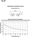
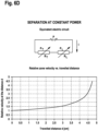
- a description of the movement of the zones under constant current, constant voltage, and constant power for embodiments wherein circular or spheroid device architectures, e.g., devices comprising one or more circular electrodes, may be used, are presented in the Examples section infra.
- epitachophoresis may be effected using constant current, constant voltage, and/or constant power.
- epitachophoresis may be effected using varying current, varying voltage, and/or varying power.
- epitachophoresis may be effected within the context of devices and/or an arrangement of electrodes whose shape may be described in general as circular or spheroid, such that the basic principles of epitachophoresis may be accomplished as described herein.
- epitachophoresis may be effected within the context of devices and/or an arrangement of electrodes whose shape may be described in general as polygons, such that the basic principles of epitachophoresis may be accomplished as described herein.
- epitachophoresis may be effected by any non-linear, contiguous arrangement of electrodes, such as electrodes arranged in the shape of a circle and/or electrodes arranged in the shape of a polygon.
- in vitro diagnostic application IVD application
- in vitro diagnostic method IVD method
- said sample may comprise blood and/or plasma from a subject.
- said sample may comprise nucleic acids and/or target nucleic acids from a subject, optionally further wherein said nucleic acids originated from blood and/or plasma from a subject.
- an epitachophoresis device may be used as an in vitro diagnostic device.
- a target analyte that has been concentrated/enriched through epitachophoresis may be used in a downstream in vitro diagnostic assay.
- an in vitro diagnostic assay may comprise nucleic acid sequencing, e.g., DNA sequencing.
- an in vitro diagnostic method may be, but is not limited to being, any one or more of the following: staining, immunohistochemical staining, flow cytometry, FACS, fluorescence-activated droplet sorting, image analysis, hybridization, DASH, molecular beacons, primer extension, microarrays, CISH, FISH, fiber FISH, quantitative FISH, flow FISH, comparative genomic hybridization, blotting, Western blotting, Southern blotting, Eastern blotting, Far-Western blotting, Soiled blotting, Northwestern blotting, and Northern blotting, enzymatic assays, ELISA, ligand binding assays, immunoprecipitation, ChIP, ChIP-seq, ChIP-ChiP, radioimmunoassays, fluorescence polarization, FRET, surface plasmon resonance, filter binding assays, affinity chromatography, immunocytochemistry, gene expression profiling, DNA profiling with PCR, DNA microarrays,
- leading electrolyte and “leading ion” generally refer to ions having a higher effective electrophoretic mobility as compared to that of the sample ion of interest and/or the trailing electrolyte as used during ITP and/or epitachophoresis.
- leading electrolytes for use with cationic epitachophoresis may include, but are not limited to including, chloride, sulphate and/or formate, buffered to desired pH with a suitable base, such as, for example, histidine, TRIS, creatinine, and the like.
- leading electrolytes for use with anionic epitachophoresis may include, but are not limited to including, potassium, ammonium and/or sodium with acetate or formate.
- an increase of the concentration of the leading electrolyte may result in a proportional increase of the sample zone and a corresponding increase in electric current (power) for a given applied voltage.
- Typical concentrations generally may be in the 10-20 mM range; however, higher concentrations may also be used.
- trailing electrolyte As used herein, the terms “trailing electrolyte”, “trailing ion”, “terminating electrolyte”, and “terminating ion” generally refer to ions having a lower effect electrophoretic mobility as compared to that of the sample ion of interest and/or the leading electrolyte as used during ITP and/or epitachophoresis.
- trailing electrolytes for use with cationic epitachophoresis may include, but are not limited to including, MES, MOPS, acetate, glutamate and other anions of weak acids and low mobility anions.
- trailing electrolytes for use with anionic epitachophoresis may include, but are not limited to including, reaction hydroxonium ion at the moving boundary as formed by any weak acid during epitachophoresis.
- the term "focused zone(s)" generally refers to a volume of solution that comprises a component that has been concentrated ("focused") as a result of performing epitachophoresis.
- a focused zone may be collected or removed from a device, and said focused zone may comprise an enriched and/or concentrated amount of a desired sample, e.g., a target analyte, e.g., a target nucleic acid.
- a target analyte e.g., a target nucleic acid.
- the target analyte generally becomes focussed in the center of the device, e.g., a circular or spheroid or other polygonal shaped device.
- nucleic acid and “nucleic acid molecule” may be used interchangeably throughout the disclosure.
- the term generally refers to polymers of nucleotides (e.g., ribonucleotides, deoxyribonucleotides, nucleotide analogs etc.) and comprising deoxyribonucleic acids (DNA), ribonucleic acids (RNA), DNA-RNA hybrids, oligonucleotides, polynucleotides, aptamers, peptide nucleic acids (PNAs), PNA-DNA conjugates, PNA-RNA conjugates, etc., that comprise nucleotides covalently linked together, either in a linear or branched fashion.
- nucleotides e.g., ribonucleotides, deoxyribonucleotides, nucleotide analogs etc.
- DNA deoxyribonucleic acids
- RNA ribonucleic acids
- DNA-RNA hybrids DNA-RNA hybrids
- a nucleic acid is typically single-stranded or double-stranded and will generally contain phosphodiester bonds, although in some cases, nucleic acid analogs are included that may have alternate backbones, including, for example, phosphoramide (Beaucage et al. (1993) Tetrahedron 49(10):1925); phosphorothioate ( Mag et al. (1991) Nucleic Acids Res. 19:1437 ; and U.S. Pat. No. 5,644,048 ), phosphorodithioate ( Briu et al. (1989) J. Am. Chem. Soc.
- nucleotide analogs also may include non-naturally occurring heterocyclic bases, such as those described in, e.g., Seela et al. (1999) Helv. Chim. Acta 82:1640. Certain bases used in nucleotide analogs act as melting temperature (Tm) modifiers.
- Tm melting temperature
- some of these include 7-deazapurines (e.g., 7-deazaguanine, 7-deazaadenine, etc.), pyrazolo[3,4-d]pyrimidines, propynyl-dN (e.g., propynyl-dU, propynyl-dC, etc.), and the like, see, e.g., U.S. Pat. No. 5,990,303 .
- heterocyclic bases include, e.g., hypoxanthine, inosine, xanthine; 8-aza derivatives of 2-aminopurine, 2,6-diaminopurine, 2-amino-6-chloropurine, hypoxanthine, inosine and xanthine; 7-deaza-8-aza derivatives of adenine, guanine, 2-aminopurine, 2,6-diaminopurine, 2-amino-6-chloropurine, hypoxanthine, inosine and xanthine; 6-azacytidine; 5-fluorocytidine; 5-chlorocytidine; 5-iodocytidine; 5-bromocytidine; 5-methylcytidine; 5-propynylcytidine; 5-bromovinyluracil; 5-fluorouracil; 5-chlorouracil; 5-iodouracil; 5-bromouracil; 5-trifluoromethyl
- nucleic acid and nucleic acid molecule also may generally refer to oligonucleotides, oligos, polynucleotides, genomic DNA, mitochondrial DNA (mtDNA), complementary DNA (cDNA), bacterial DNA, viral DNA, viral RNA, RNA, message RNA (mRNA), transfer RNA (tRNA), ribosomal RNA (rRNA), siRNA, catalytic RNA, clones, plasmids, M13, PI, cosmid, bacteria artificial chromosome (BAC), yeast artificial chromosome (YAC), amplified nucleic acid, amplicon, PCR product and other types of amplified nucleic acid, RNA/DNA hybrids and PNAs, all of which can be in either single- or double-stranded form, and unless otherwise limited, would encompass known analogs of natural nucleotides that can function in a similar manner as naturally occurring nucleotides and combinations and/or mixtures thereof.
- mtDNA mitochondrial DNA
- cDNA
- nucleotides refers to both naturally-occurring and modified/nonnaturally-occurring nucleotides, including nucleoside tri, di, and monophosphates as well as monophosphate monomers present within polynucleic acid or oligonucleotide.
- a nucleotide may also be a ribo; 2'-deoxy; 2',3'-deoxy as well as a vast array of other nucleotide mimics that are well-known in the art.
- Mimics include chain-terminating nucleotides, such as 3'-0-methyl, halogenated base or sugar substitutions; alternative sugar structures including nonsugar, alkyl ring structures; alternative bases including inosine; deaza-modified; chi, and psi, linker-modified; mass label-modified; phosphodiester modifications or replacements including phosphorothioate, methylphosphonate, boranophosphate, amide, ester, ether; and a basic or complete internucleotide replacements, including cleavage linkages such a photocleavable nitrophenyl moieties.
- nucleotides such as 3'-0-methyl, halogenated base or sugar substitutions
- alternative sugar structures including nonsugar, alkyl ring structures
- alternative bases including inosine
- deaza-modified chi
- psi linker-modified
- mass label-modified mass label-modified
- phosphodiester modifications or replacements including
- nucleoside refers to a nucleic acid component that comprises a base or basic group (comprising at least one homocyclic ring, at least one heterocyclic ring, at least one aryl group, and/or the like) covalently linked to a sugar moiety (a ribose sugar or a deoxyribose sugar), a derivative of a sugar moiety, or a functional equivalent of a sugar moiety (e.g. a carbocyclic ring).
- a nucleoside when a nucleoside includes a sugar moiety, the base is typically linked to a l'-position of that sugar moiety.
- a base can be a naturally occurring base or a non-naturally occurring base.
- Exemplary nucleosides include ribonucleosides, deoxyribonucleosides, dideoxyribonucleosides and carbocyclic nucleosides.
- a “purine nucleotide” refers to a nucleotide that comprises a purine base
- a "pyrimidine nucleotide” refers to a nucleotide that comprises a pyrimidine base
- modified nucleotide refers to rare or minor nucleic acid bases, nucleotides and modifications, derivations, or analogs of conventional bases or nucleotides and includes synthetic nucleotides having modified base moieties and/or modified sugar moieties (see, Protocols for Oligonucleotide Conjugates, Methods in Molecular Biology, Vol. 26 (Suhier Agrawal, Ed., Humana Press, Totowa, N.J., (1994 )); and Oligonucleotides and Analogues, A Practical Approach (Fritz Eckstein, Ed., IRL Press, Oxford University Press, Oxford ).
- Oligonucleotide refers to linear oligomers of natural or modified nucleosidic monomers linked by phosphodiester bonds or analogs thereof. Oligonucleotides include deoxyribonucleosides, ribonucleosides, anomeric forms thereof, PNAs, and the like, capable of specifically binding to a target nucleic acid. Usually monomers are linked by phosphodiester bonds or analogs thereof to form oligonucleotides ranging in size from a few monomeric units, e.g., 3-4, to several tens of monomeric units, e.g., 40-60.
- oligonucleotide is represented by a sequence of letters, such as "ATGCCTG,” it will be understood that the nucleotides are in 5'-3' order from left to right and that "A” denotes deoxyadenosine, “C” denotes deoxycytidine, “G” denotes deoxyguanosine, “T” denotes deoxythymidine, and “U” denotes the ribonucleoside, uridine, unless otherwise noted.
- oligonucleotides comprise the four natural deoxynucleotides; however, they may also comprise ribonucleosides or non-natural nucleotide analogs.
- oligonucleotide or polynucleotide substrate requirements for activity e.g., single stranded DNA, RNA/DNA duplex, or the like
- selection of appropriate composition for the oligonucleotide or polynucleotide substrates is well within the knowledge of one of ordinary skill.
- oligonucleotide primer refers to a polynucleotide sequence that hybridizes to a sequence on a target nucleic acid template and may facilitate the detection or amplification of a target nucleic acid.
- an oligonucleotide primer serves as a point of initiation of nucleic acid synthesis.
- an oligonucleotide primer may be used to create a structure that is capable of being cleaved by a cleavage agent.
- Primers can be of a variety of lengths and are often less than 50 nucleotides in length, for example 12-25 nucleotides, in length. The length and sequences of primers for use in PCR can be designed based on principles known to those of skill in the art.
- oligonucleotide probe refers to a polynucleotide sequence capable of hybridizing or annealing to a target nucleic acid of interest and allows for the specific detection of the target nucleic acid.
- Nucleic acids are "extended” or “elongated” when additional nucleotides are incorporated into the nucleic acids, for example by a nucleotide incorporating biocatalyst, at the 3' end of a nucleic acid.
- hybridization and “annealing” and the like are used interchangeably and refer to the base-pairing interaction of one polynucleotide with another polynucleotide (typically an antiparallel polynucleotide) that results in formation of a duplex or other higher-ordered structure, typically termed a hybridization complex.
- the primary interaction between the antiparallel polynucleotide molecules is typically base specific, e.g., A/T and G/C, by Watson/Crick and/or Hoogsteen-type hydrogen bonding. It is not a requirement that two polynucleotides have 100% complementarity over their full length to achieve hybridization.
- a hybridization complex can form from intermolecular interactions, or alternatively, can form from intramolecular interactions.
- nucleic acid is identical to, or hybridizes selectively to, another nucleic acid molecule.
- Selectivity of hybridization exists when hybridization occurs that is more selective than total lack of specificity.
- selective hybridization will occur when there is at least about 55% identity over a stretch of at least 14-25 nucleotides, preferably at least 65%, more preferably at least 75%, and most preferably at least 90%.
- one nucleic acid hybridizes specifically to the other nucleic acid. See M. Kanehisa, Nucleic Acids Res. 12:203 (1984 ).
- a primer that is "perfectly complementary” has a sequence fully complementary across the entire length of the primer and has no mismatches.
- the primer is typically perfectly complementary to a portion (subsequence) of a target sequence and/or target nucleic acid.
- a “mismatch” refers to a site at which the nucleotide in the primer and the nucleotide in the target nucleic acid with which it is aligned are not complementary.
- substantially complementary when used in reference to a primer means that a primer is not perfectly complementary to its target sequence; instead, the primer is only sufficiently complementary to hybridize selectively to its respective strand at the desired primer-binding site.
- target nucleic acid as used herein is intended to mean any nucleic acid whose presence is to be detected, measured, amplified, and/or subject to further assays and analyses.
- a target nucleic acid may comprise any single and/or double-stranded nucleic acid.
- Target nucleic acids can exist as isolated nucleic acid fragments or be a part of a larger nucleic acid fragment.
- Target nucleic acids can be derived or isolated from essentially any source, such as cultured microorganisms, uncultured microorganisms, complex biological mixtures, biological samples, tissues, sera, ancient or preserved tissues or samples, environmental isolates or the like.
- target nucleic acids include or are derived from cDNA, RNA, genomic DNA, cloned genomic DNA, genomic DNA libraries, enzymatically fragmented DNA or RNA, chemically fragmented DNA or RNA, physically fragmented DNA or RNA, or the like.
- a target nucleic acid may comprise a whole genome.
- a target nucleic acid may comprise the entire nucleic acid content of a sample and/or biological sample.
- a target nucleic acid may comprise circulating or cell-free DNA's, e.g., circulating tumor DNA ("ctDNA”) present in individuals with cancer or circulating fetal or circulating maternal DNA (“cfDNA”) fragments present in plasma or serum of pregnant women.
- Target nucleic acids can come in a variety of different forms including, for example, simple or complex mixtures, or in substantially purified forms.
- a target nucleic acid can be part of a sample that contains other components or can be the sole or major component of the sample.
- a target nucleic acid can have either a known or unknown sequence.
- amplification reaction refers to any in vitro means for amplifying the copies of a target sequence of nucleic acid.
- amplification and “amplifying” the like refer generally to any process that results in an increase in the copy number of a molecule or set of related molecules.
- Components of an amplification reaction may include, but are not limited to, e.g., primers, a polynucleotide template, nucleic acid polymerase, nucleotides, dNTPs and the like.
- amplifying typically refers to an "exponential" increase in target nucleic acid.
- amplifying as used herein can also refer to linear increases in the numbers of a select target sequence of nucleic acid. Amplification typically starts from a small amount of a target nucleic acid (e.g.
- Amplification of target nucleic acid encompasses a variety of chemical and enzymatic processes.
- the generation of multiple DNA copies from one or a few copies of a target nucleic acid may be effected by a polymerase chain reaction (PCR), a hot start PCR, a strand displacement amplification (SDA) reaction, a transcription mediated amplification (TMA) reaction, a nucleic acid sequence-based amplification (NASBA) reaction, or a ligase chain reaction (LCR).
- PCR polymerase chain reaction
- SDA strand displacement amplification
- TMA transcription mediated amplification
- NASBA nucleic acid sequence-based amplification
- LCR ligase chain reaction
- the generation of multiple cDNA molecules from a limited amount of viral RNA in a sample using RT-PCR is a form of amplification.
- the generation of multiple RNA molecules from a single DNA molecule during the process of transcription is also a form of amplification.
- Amplification may optionally followed by additional steps, for example, but not limited to, labeling, sequencing, purification, isolation, hybridization, size resolution, expression, detecting and/or cloning.
- target microbe as used herein is intended to mean any unicellular or multicellular microbe, found in blood, plasma, other body fluids, samples such as biological samples, and/or tissues, e.g., one associated with an infectious condition or disease. Examples thereof include bacteria, archaea, eukaryotes, viruses, yeasts, fungi, protozoan, amoeba, and/or parasites, Further examples of diseases caused by microbes, and the microbes that may cause such diseases, can be found in Table 1 infra. As such, the term “microbe” generally refers to the microbe that may cause a disease, whether the disease is referred to or the disease-causing microbe is referred to.
- biomarker refers to a biological molecule found in blood, plasma, other body fluids, and/or tissues that is a sign of a normal or abnormal process, or of a condition or disease (such as cancer).
- a biomarker may be used to see how well the body responds to a treatment for a disease or condition.
- a biomarker refers to a biological substance that is indicative of the presence of cancer in the body.
- a biomarker may be a molecule secreted by a tumor or a specific response of the body to the presence of cancer. Genetic, epigenetic, proteomic, glycomic, and imaging biomarkers can be used for cancer diagnosis, prognosis, and epidemiology.
- biomarkers can be assayed in non-invasively collected biofluids like blood or serum.
- gene and protein based biomarkers have already been used in patient care including but, not limited to, AFP (Liver Cancer), BCR-ABL (Chronic Myeloid Leukemia), BRCA1 / BRCA2 (Breast/Ovarian Cancer), BRAF V600E (Melanoma/Colorectal Cancer), CA-125 (Ovarian Cancer), CA19.9 (Pancreatic Cancer), CEA (Colorectal Cancer), EGFR (Non-small-cell lung carcinoma), HER-2 (Breast Cancer), KIT(Gastrointestinal stromal tumor), PSA (Prostate Specific Antigen) (Prostate Cancer), S100 (Melanoma), and many others.
- Biomarkers may be useful as diagnostics (to identify early stage cancers) and/or prognostics (to forecast how aggressive a cancer is and/or predict how a subject will respond to a particular treatment and/or how likely a cancer is to recur).
- Biomarkers of interest include, but are not limited to, such oncology biomarkers as AKAP4, ALK, APC, AR, BRAF, BRCA1, BRCA2, CCND1, CCND2, CCND3, CD274, CDK4, CDK6, CFB, CFH, CFI, DKK1, DPYD, EDNRB, EGFR, ERBB2, EPSTI1, ESR1, FCRL5, FGFR1, FGFR2, FGFR3, FLT3, FN14, HER2, HER4, HERC5, IDH1, IDH2, IDO1, KIF5B, KIT, KRAS, LGR5, LIV1, LY6E, LYPD3, MACC1, MET, MRD, MSI, MSLN, MUC16, MYC
- biomarkers of interest may include Her2, bRaf, an ERBB2 amplification, a Pl3KCA mutation, a FGFR2 amplification, a p53 mutation, a BRCA mutation, a CCND1 amplification, a MAP2K4 mutation, an ATR mutation, or any other biomarker the expression of which is correlated to a specific cancer; at least one of AFP, ALK, BCR-ABL, BRCA1/BRCA2, BRAF, V600E, Ca-125, CA19.9, EGFR, Her-2, KIT, PSA, S100, KRAS, ER/Pr, UGT1A1, CD30,CD20, F1P1L1-PDGRF ⁇ , PDGFR, TMPT, and TMPRSS2; or at least one biomarker selected from ABCB5, AFP-L3, Alpha-fetoprotein, Alpha-methyl acyl-CoA racemase, BRCA1, BRCA2, CA 15-3, CA 242, Ca 27
- the biomarker of interest may be an immune checkpoint inhibitor such as, but not limited to, CTLA-4, PDL1, PDL2, PD1, B7-H3, B7-H4, BTLA, HVEM, KIR, TIM3, GAL9, GITR, LAG3, VISTA, KIR, 2B4, TRPO2, CD160, CGEN-15049, CHK 1, CHK2, A2aR, TL1A, and B-7 family ligands or a combination thereof or is a ligand of a checkpoint protein selected from the group consisting of CTLA-4, PDL1, PDL2, PD1, B7-H3, B7-H4, BTLA, HVEM, TIM3, GAL9, LAG3, VISTA, KIR, 2B4, CD160, CGEN-15049, CHK1, CHK2, A2aR, B
- biomarkers may include, but are not limited to including, any one or more biomarkers associated with acute lymphoblastic leukemia (etv6, am11, cyclophilin b), B cell lymphoma (Ig-idiotype), glioma (E-cadherin, .alpha.-catenin, .beta.-catenin, .gamma.-catenin, p120 ctn), bladder cancer (p21ras), biliary cancer (p21ras), breast cancer (MUC family, HER2/neu, c-erbB-2), cervical carcinoma (p53, p21ras), colon carcinoma (p21ras, HER2/neu, c-erbB-2, MUC family), colorectal cancer (Colorectal associated antigen (CRC)-C017-1A/GA733, APC), choriocarcinoma (CEA), epithelial cell cancer (cyclophilin b), gastric cancer (HER2/
- sample includes a specimen or culture (e.g., microbiological cultures) that includes nucleic acids and/or a target nucleic acid.
- sample is also meant to include both biological and environmental samples.
- a sample may include a specimen of synthetic origin.
- a sample may include one or more microbes from any source from which one or more microbes may be derived.
- a “biological sample” may include, but is not limited, to whole blood, serum, plasma, umbilical cord blood, chorionic villi, amniotic fluid, cerebrospinal fluid, spinal fluid, lavage fluid (e.g., bronchioalveolar, gastric, peritoneal, ductal, ear, arthroscopic), biopsy sample, urine, feces, sputum, saliva, nasal mucous, prostate fluid, semen, lymphatic fluid, bile, tears, sweat, breast milk, breast fluid, embryonic cells and fetal cells.
- the biological sample may be blood, and may be plasma.
- blood encompasses whole blood or any fractions of blood, such as serum and plasma as conventionally defined.
- Blood plasma refers to the fraction of whole blood resulting from centrifugation of blood treated with anticoagulants.
- Blood serum refers to the watery portion of fluid remaining after a blood sample has coagulated.
- Environmental samples include environmental material such as surface matter, soil, water and industrial samples, as well as samples obtained from food and dairy processing instruments, apparatus, equipment, utensils, disposable and non-disposable items.
- a “representative" sample may comprise the different subpopulations of a sample, e.g., a sample comprising cancer cells comprised within a tumor.
- a “representative” sample alternatively may comprise the different subpopulations within a normal and/or control sample, e.g., normal or control cells, or may comprise a mixed sample of test and normal/control sample, e.g., tumor cells and normal cells, respectively.
- these representative samples may be used in a plurality of assay methods, without compromising the ability to use the specimen in traditional diagnostic assays.
- representative samples may be produced by analyzing a sample, e.g., a sample comprising tumor cells, using the devices and methods described herein.
- a representative sample may be analyzed by the devices and methods described herein.
- representative samples produced by analyzing a sample using the devices and methods described herein may be used in several different assay formats simultaneously in order to detect the presence of even minor sub-populations of sample within a sample, e.g., a tumor or lymph node.
- target analyte as used herein is intended to mean any analyte whose presence is to be detected, measured, separated, concentrated, and/or subject to further assays and analyses.
- said analyte may be, but is not limited to, any ion, molecular, nucleic acid, biomarker, cell or population of cells, e.g., desired cells, and the like, whose detection, measurement, separation, concentration, and/or use in further assays is desired.
- a target analyte may be derived from any of the samples described herein.
- analysis generally refers to a process or step involving physical, chemical, biochemical, or biological analysis that includes characterization, testing, measurement, optimization, separation, synthesis, addition, filtration, dissolution, or mixing.
- chemical refers to a substance, compound, mixture, solution, emulsion, dispersion, molecule, ion, dimer, macromolecule such as a polymer or protein, biomolecule, precipitate, crystal, chemical moiety or group, particle, nanoparticle, reagent, reaction product, solvent, or fluid any one of which may exist in the solid, liquid, or gaseous state, and which is typically the subject of an analysis.
- protein generally refers to a set of amino acids linked together usually in a specific sequence.
- a protein can be either naturally-occurring or man-made.
- protein includes amino acid sequences that have been modified to contain moieties or groups such as sugars, polymers, metalloorganic groups, fluorescent or light-emitting groups, moieties or groups that enhance or participate in a process such as intramolecular or intermolecular electron transfer, moieties or groups that facilitate or induce a protein into assuming a particular conformation or series of conformations, moieties or groups that hinder or inhibit a protein from assuming a particular conformation or series of conformations, moieties or groups that induce, enhance, or inhibit protein folding, or other moieties or groups that are incorporated into the amino acid sequence and that are intended to modify the sequence's chemical, biochemical, or biological properties.
- a protein includes, but is not limited to, enzymes, structural elements, antibodies, hormones, electron carriers, and other macromolecules that are involved in processes such as cellular processes or activities. Proteins typically have up to four structural levels that include primary, secondary, tertiary, and quaternary structures.
- a given component such as a layer, region, liquid or substrate is referred to herein as being disposed or formed “on”, “in” or “at” another component, that given component can be directly on the other component or, alternatively, intervening components (e.g., one or more buffer layers, interlayers, electrodes or contacts) can also be present.
- intervening components e.g., one or more buffer layers, interlayers, electrodes or contacts
- the terms “disposed on” and “formed on” are used interchangeably to describe how a given component is positioned or situated in relation to another component. Hence, the terms “disposed on” and “formed on” are not intended to introduce any limitations relating particular methods of material transport, deposition, or fabrication.
- communicate is used herein to indicate a structural, functional, mechanical, electrical, optical, thermal, or fluidic relation, or any combination thereof, between two or more components or elements.
- communicate is not intended to exclude the possibility that additional components may be present between, and/or operatively associated or engaged with, the first and the second component.
- a "subject” refers to a mammalian subject (such as a human, rodent, non-human primate, canine, bovine, ovine, equine, feline, etc.) to be treated and/or one from whom a biological sample is obtained.
- a mammalian subject such as a human, rodent, non-human primate, canine, bovine, ovine, equine, feline, etc.
- tumor refers to a mass or a neoplasm, which itself is defined as an abnormal new growth of cells that usually grow more rapidly than normal cells and will continue to grow if not treated sometimes resulting in damage to adjacent structures. Tumor sizes can vary widely.
- a tumor may be solid or fluid-filled.
- a tumor can refer to benign (not cancerous, generally harmless), pre-malignant (pre-cancerous), or malignant (cancerous) growths.
- pre-cancerous pre-malignant growths.
- the dividing line between cancerous, pre-cancerous, and cancerous growths is not always clear (sometimes determining which is which may be arbitrary, especially if the tumor is in the middle of the spectrum), but there are general properties of each type of growth. Benign tumors are non-malignant/non-cancerous tumors.
- a benign tumor is usually localized, and does not spread (metastasize) to other parts of the body. Most benign tumors respond well to treatment. However, if left untreated, some benign tumors can grow large and lead to serious disease because of their size. In this way, benign tumors can mimic malignant tumors and, thus, are sometimes treated.
- a premalignant or precancerous tumor is not yet malignant, but it is primed to become so.
- Malignant tumors are cancerous growths. They are often resistant to treatment, may spread to other parts of the body, and sometimes recur after removal. "Cancer" is another term for a malignant growth (a malignant tumor or neoplasm).
- Tumor virulence may be determined, at least in part, by differential gene expression.
- cancerous cells cells comprising a pre-malignant and/or malignant tumor
- the mechanisms that allow a cell to activate or silence genes are damaged.
- tumorous cells that express genes specific to the production of spermatozoids, which should be silent are extremely virulent (a high-risk cancer that exhibits increased proliferative abilities and a facility to hide from the body's immune system).
- the present disclosure generally describes novel devices for sample analysis, for effecting epitachophoresis.
- Said devices comprise use of concentric or polygonal disk (e.g., circular) designs to effect epitachophoresis, in contrast to the capillary or microfluidic channel designs which may often be used for conventional isotachophoresis.
- the devices of the present disclosure confer numerous advantageous properties and features, as discussed herein.
- the architecture of devices for epitachophoresis enable the analysis of large sample volumes, e.g., 15 mL or more of a sample and/or biological sample, whereas conventional capillary or microfluidic techniques are generally only equipped to handle microliter scale volumes.
- the present devices allow for whole genome and/or whole nucleic acid content extraction from a sample and/or biological sample, whereas such an extraction would be difficult when using conventional capillary or microfluidic based devices and methods, in particular ITP-based capillary or microfluidic devices and methods.
- the highly efficient extraction of target nucleic acids attained through use of the devices and methods described herein is helpful for downstream in vitro diagnostic methods, in which the amount of target nucleic acid, e.g., DNA and/or RNA, directly correlates with the sensitivity that may be achieved in said down-stream IVD assay.
- spin columns or magnetic glass particles that bind nucleic acids on their surface conventionally may be used in order to effect extraction of nucleic acids.
- the devices and methods described herein may confer any one or more of the following advantages: higher extraction yields (potentially loss-less) compared to column- or bead-based extraction methods; a simpler device setup compared to the larger footprint for the MagNA Pure or other benchtop instruments; potentially faster sample turn-around and high parallelizability as compared to other devices applied to similar uses; easy integration with other microfluidics-based systems for down-stream processing of extracted nucleic acids.
- a flat channel may generally be used in devices and methods described herein, and said channel architecture generally may have a favorable heat transfer capability as compared to the narrow channels that may generally be used in capillary and/or microfluidic devices.
- the devices described herein often allow for gentle sample collection, which typically may be an important feature when performing whole genome extraction , extraction of microbes, extraction of desired target cells such as stem cells, tumor cells, e.g., circulating tumor cells, or other rare cells wherein cell functionality is desirably preserved, or other labile analytes.
- desired target cells such as stem cells, tumor cells, e.g., circulating tumor cells, or other rare cells wherein cell functionality is desirably preserved, or other labile analytes.
- conventional whole genome extraction may feature use of a pipette, which may shear genomic DNA.
- the devices described herein allow for obtaining sample without the need for potentially damaged pipetting in some exemplary embodiments wherein damage of a sample through pipetting may be a concern.
- the devices described herein may allow for whole genome extraction wherein shearing and/or damaging of any portion of the whole genome may not occur or be minimal as a result of using said devices
- the present disclosure generally relates to devices for sample analysis.
- the devices for sample analysis as described herein, generally pertain to effecting epitachophoresis using said devices
- Devices for effecting epitachophoresis generally comprise an arrangement of electrodes sufficient for effecting said epitachophoresis.
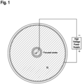
- Said devices comprise a polygonal or circular geometry.
- an epitachophoresis zone of the device may move from the edge of the polygon or circle towards the center of the polygon or circle.
- Said polygon may be selected from a triangle, quadrilateral, pentagon, hexagon, heptagon, octagon, nonagon, decagon, and/or said polygon may have 3, 4, 5, 6, 7,8, 9, 10-20, 20-50 or 50-100 or more sides.
- devices for effecting epitachophoresis may comprise any device dimensions, e.g., diameter, e.g., depth, that facilitates analysis of a desired sample volume.
- the size of a device may scale with the volume used.
- a device may comprise a diameter ranging from about 1 mm or more to about 20 mm or more.
- devices for sample analysis as described herein may comprise glass, ceramics, and/or plastics.
- glass and/or ceramics when using glass and/or ceramics in particular, these materials may result in improved heat transfer properties that can be beneficial during device operation. For example, as the flat channel of a circular or concentric ITP device has a favorable heat transfer capability compared to a narrow channel, over-heating (or boiling) of focused material can be prevented.
- current/voltage programming may also suitable for adjusting the Joule heating of the device.
- the devices for sample analysis comprise a two dimensional arrangement of electrodes, wherein said arrangement is sufficient for effecting epitachophoresis.
- Said electrodes comprise one or more ring-shaped (circular) electrodes, and/or said one or more electrodes may be arranged in a polygonal shape.
- Said polygon may be selected from a triangle, quadrilateral, pentagon, hexagon, heptagon, octagon, nonagon, decagon., and/or said polygon may have 3, 4, 5, 6, 7,8, 9, 10-20, 20-50 or 50-100 or more sides.
- said electrodes of said devices may be arranged such that said arrangement comprises a diameter or width that ranges from about 1 mm to about 20 mm.
- Said devices comprise an electrode at the center of said devices.
- said electrodes of said devices may comprise a platinum plated and/or gold-plated stainless steel ring; one or more stainless steel electrodes; and/or one or more graphite electrodes.
- said one or more electrodes may comprise wire electrodes in some embodiments.
- said arrangement of one or more electrodes may comprise an arrangement of more than one regularly spaced electrodes.
- said arrangement of one or more electrodes may comprise a non-linear, contiguous arrangement of more than one electrodes.
- said arrangement of one or more electrodes may comprise a single wire electrode formed into a desired shape, e.g., a circle.
- said arrangement of one or more electrodes may comprise an array of wire electrodes.
- a device for sample analysis may be a disposable device. Said disposable device may comprise stainless steel and/or graphite electrodes. In other exemplary embodiments, a device for sample analysis may function as a bench-top instrument, i.e., the device may comprise a workstation and/or may be re-usable.
- devices for sample analysis as described herein may comprise dimensions that accommodate 1 ⁇ l or less, 1 ⁇ l or more, 10 ⁇ l or more, 100 ⁇ l or more, 1 mL or more, 4 mL, or more, 5 mL or more, 10 mL or more, or 15 mL or more of sample volume.
- said volume may be about 15 mL.
- said sample may be injected into the device through an opening in the top of said device.
- use of said devices will result in a focused sample that collects in the center of the device, and furthermore in some embodiments said sample may be collected from the center of said device.
- sample may be collected by punching out the gel at the center of the device which may contain the focused sample.
- sample may collected in a tube located at the center of the device.
- sample may be collected by pipetting out the focused sample once it reaches the center of the device.
- said sample may comprise a target analyte.
- application of electricity to said device will focus a target analyte comprised by a sample into a focused zone, and furthermore said target analyte may be collected from said device after epitachophoresis.
- a sample for analysis using any device or method of epitachophoresis described herein may comprise a biological sample, such as blood and/or plasma.
- Said blood and/or plasma may comprise a target analyte, e.g., a target nucleic acid.
- the volume of said blood and/or said plasma may be about 4 mL.
- Said blood and/or plasma may be derived from a subject.
- a sample for analysis using any device or method of epitachophoresis described herein may comprise one or more biomarkers that may be separated and/or focused and/or collected from said device.
- said devices for sample analysis further comprise a leading electrolyte and a trailing electrolyte
- all common electrolytes known to those skilled in the art that are used for isotachophoresis may be used with the present devices when the leading ions have a higher effective electrophoretic mobility than that of the sample ion(s) of interest.
- the opposite may generally be true for the selected terminating ions.
- said devices may be used for cationic separation/epitachophoresis (positive mode) or for anionic separation/epitachophoresis (negative mode).
- common leading electrolytes for anionic separation using epitachophoresis may include, for example, chloride, sulfate, or formate, buffered to desired pH with a suitable base, e.g., histidine, TRIS, creatinine, and the like.
- concentrations of said leading electrolyte for epitachophoresis for anionic separation may range from 5 mM - 1 M with respect to the leading ion.
- said terminating ions then often may include MES, MOPS, HEPES, acetate, glutamate and other anions of weak acids and low mobility anions.
- Concentrations of said terminating electrolyte for epitachophoresis in positive mode range from: 5 mM - 10 M with respect to the terminating ion.
- said devices for sample analysis may be useful for a wide range of analytes that range from, for example, mall inorganic and organic ions to large biopolymers including peptides, proteins, polysaccharides and DNA or even particles including bacteria and viruses.
- common leading ions for epitachophoresis generally may include, for example: potassium, ammonium or sodium with acetate or formate being the most common buffering counterions.
- Reaction hydroxonium ion moving boundary then serves as a universal terminating electrolyte formed by any weak acid.
- the increase of the concentration of the leading ion may result in proportional increase of the sample zone at the expense of increased electric current (power) for a given applied voltage.
- Typical concentrations generally are in the 10-20 mM range; however, higher concentrations may also be used.
- the leading electrolyte is stabilized by a gel, viscous additive, and/or otherwise hydrodynamically separated from the terminating electrolyte.
- Said gel or hydrodynamic separation may prevent mixing of the leading and terminating electrolytes during device operation.
- said gel may comprise an uncharged material or any other material that forms a gel, such as, for example, agarose, polyacrylamide, pullulans, and the like. In particular this includes all types of hydrogels.
- the gel may be resistant to changes in pH, e.g., acid or base-stable gels.
- said device may comprise a leading electrolyte whose diameter ranges from a thickness (height) of about 10 ⁇ m to about 20 mm.
- the maximum thickness may generally be a thickness that may result in a uniform electric field over the entirety of said thickness.
- the thickness may be such that the electric field is non-uniform, the electric field may not vary linearly, however, the basic principles of epitachophoresis may still apply and separation, concentration, focusing, and/or collection of a target analyte may still occur as desired.
- a thickness of greater than 20 mm may be used, and a curved device architecture may be used to obtain linear behavior.
- the leading and/or trailing electrolyte may comprise an electrolyte with a desired buffering capacity.
- the leading electrolyte may comprise an electrolyte whose buffering capacity minimizes and/or negates any changes in pH that may occur as a result of effecting epitachophoresis, such that the pH over all of the epitachophoresis zones may be nearly the same, or the same.
- HCl histidine may be used as a leading electrolyte with desirable buffering capacity.
- a pH stable gel may be used.
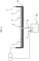
- said device may comprise an electrode in a leading electrolyte reservoir connected with a concentrator by a tube.
- Said tube may be connected directly or closed on one end by a semipermeable membrane.
- said concentrator may be connected on-line to other devices, such as, for example, capillary analyzers, chromatography, PCR devices, enzymatic reactors, and the like.
- said tube may be used to supply a countercurrent flow of the leading electrolyte in an arrangement without a gel containing said leading electrolyte.
- a device for sample analysis may comprise a gel, or other material which may be used to stabilize a leading electrolyte.
- a device for sample analysis may comprise a gel, and said gel may help to avoid unwanted sample contamination.
- a device for sample analysis may be used to extract ctDNA, and said gel may be used to help avoid contamination of ctDNA with genomic DNA.
- the gel may be of such a composition so as to allow ctDNA, but not genomic DNA, to migrate through said gel.
- Such a principle may be applied to other sample analyses where it may be beneficial to avoid contamination of a sample of interest/target analyte.
- mesh polymers and/or porous materiels may be used in a similar manner as to a gel in devices for sample analysis, such as, for example, filter paper or hydrogels.
- the selection of said mesh polymer and/or porous material may be that which helps to effect a desired separation/concentration and/or to prevent undesired sample contamination.
- a material may be selected that does not permit passage/migration of proteins but can allow passage/migration of target nucleic acids.
- a device for sample analysis may not feature a gel and still may effect epitachophoresis for sample analysis that may feature focusing and/or concentrating and/or collecting of a target analyte and/or desired sample.
- a device for sample analysis may comprise at least one electrolyte reservoir, at least two electrolyte reservoirs, or at least three electrolyte reservoirs.
- a sample may be mixed with leading electrolyte and then loaded into said device.
- a sample may be mixed with trailing electrolyte and then loading into said device.
- a sample may be mixed with a conducting solution and loaded into said device.
- a sample may contain suitable terminating ions for epitachophoresis and may be loaded into said device. Use of such a sample may eliminate a terminating electrolyte zone.
- said device may be used to concentrate a target analyte, e.g., from about 2 fold or more to about 1000 fold or more.
- said target analyte may comprise a target nucleic acid.
- said target analyte may comprise small inorganic and organic ions, peptides, proteins, polysaccharides, DNA, or microbes such as bacteria and/or viruses.
- devices for sample analysis as described herein may be operated using a constant current, a constant voltage, or a constant power.
- a device using a circular architecture e.g., devices comprising one or more circular electrodes, and further using constant current
- the epitachophoresis Boundary Velocity equation for calculating velocity v at a distance d from the start with a radius r is given by: E L ⁇ P / r ⁇ d ⁇ L S . .
- the voltage or current or power that may be used to effect epitachophoresis in said device may be varied in discrete stages.
- current or voltage or power may be applied at a first value to allow for separation and grouping of electrolytes and/or charged species within a device and/or during effecting any of the methods described herein, and after said separation and grouping occurs, the current or voltage or power may be applied at a second value to increase or to decrease the rate of epitachophoresis, as may be desired for a analysis of a given sample.
- the electric field may not vary linearly as in the case of circular or spherical architectures.
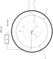
- the electric field may vary in such a manner as to produce a star-shaped arrangement of electrolyte and/or sample.
- the resulting zones of electrolyte and/or sample, etc. may form a star shape rather than a circle as a result of the electric field generated by the point electrode-based array.
- a device for sample analysis may be used to extract nucleic acids from a sample, e.g., a biological sample.
- Said sample may comprise whole blood or blood plasma.
- Said sample may comprise a cell culture from which target analytes may be harvested, such as a whole genome.
- Said nucleic acids may comprise one or more target nucleic acids, e.g., tumor DNA and/or ctDNA.
- a device for sample analysis may be used to focus and collect tumor DNA and/or circulating tumor DNA (ctDNA), and/or circulating cfDNA, e.g., those present in blood or plasma from pregnant women, and/or circulating DNAs expressing proteins over or underexpressed in specific conditions which may then optionally be subjected to further downstream analyses, such as nucleic acid sequencing and/or other in vitro diagnostic applications.
- ctDNA tumor DNA and/or circulating tumor DNA
- circulating cfDNA e.g., those present in blood or plasma from pregnant women
- DNAs expressing proteins over or underexpressed in specific conditions
- further downstream analyses such as nucleic acid sequencing and/or other in vitro diagnostic applications.
- Such downstream in vitro applications include by way of example disease detection such as cancer diagnosis and/or cancer prognosis and/or cancer staging, detection of infectious conditions, paternity analysis, detection of fetal chromosomal abnormalities such as aneuploidy, detection of fetal genetic traits, detection of pregnancy-related conditions, detection of autoimmune or inflammatory conditions, among a myriad of other potential uses.
- a device for sample analysis may be used to focus and to collect a target nucleic acid, and said target nucleic acid may be of any desired size.
- said target nucleic acid may be 5 nt or less, 10 nt or less, 20 nt or less, 30 nt or less, 50 nt or less, 100 nt or less, 1000 nt or less, 10,000 nt or less, 100,000 nt or less, 1,000,000 nt or less, or 1,000,000 nt or more.
- said device may be used to extract target nucleic acids from cell-free DNA.
- said device may be used to concentrate and collect a target analyte from a sample.
- Said sample may comprise a biological sample.
- said target analyte may used for one or more downstream in vitro diagnostic applications.
- a device for sample analysis may be connected on-line to other devices, such as, for example, capillary analyzers, chromatography, PCR devices, enzymatic reactors, and the like, and/or any other device that may be used to effect further sample analysis, e.g., a device associated with IVD applications.
- a device for sample analysis may be used in a workflow with nucleic acid sequencing library preparation.
- a device for sample analysis may be used with liquid handling robots that may optionally be used to effect downstream analysis of a sample that may have been focused and/or collected from said device.
- use of said device may result in any one or more of the following: higher extraction yields (potentially loss-less) compared to column- or bead-based extraction methods; a simpler device setup compared to the larger footprint for the MagNA Pure or other benchtop instruments; potentially faster sample turn-around and high parallelizability as compared to other devices applied to similar uses; easy integration with other microfluidics-based systems for down-stream processing of extracted nucleic acids.
- the devices disclosed herein may be used for and/or with the following applications in accordance with the present disclosure.
- the devices described herein may be used in conjunction with IHC analysis of representative samples.
- IHC analysis of representative samples from of lymph node tissue e.g., prepared from surgically removed lymph nodes
- IHC analysis of representative samples from of lymph node tissue can detect extremely small tumor micro-metastases through staining for epithelial markers combined with proliferation markers (for instance cytokeratin 8/18 dual IHC with Ki67), using markers that were positive in the primary tumor, using other markers of metastatic cells, or other diagnostic markers.
- the devices described herein may be used to analyze pre-stained cells and/or may be used to selectively stain cells and/or may be used to separate, focus, and collect desired cells as identified by the presence of a desired marker/stain in conjunction with IHC analysis techniques in some embodiments.
- metastatic tumor cells can also be detected at the nucleic acid, such as by using a Next Generation Sequencing panel looking to identify cancer-associated mutations, including mutations present in the primary tumor.
- devices and methods disclosed herein may be used to separate, focus/concentrate, and collect such nucleic acids.
- DNA purified from representative sampling from the primary and lymph nodes, as well as DNA from circulating tumor DNA from any distant metastatic cells could be separated, focused/concentrated, and/or collected in exemplary embodiments.
- representative samples derived by exemplary embodiments of the inventive devices described herein may facilitate and substantially improve the accuracy of detecting, diagnosing, and/or staging of different types of tumors, i.e., different solid tumors, irrespective of tumor tissue type, location, size or volume.
- the present devices potentially may be used to produce representative samples from supposed normal tissue samples or putative precancerous tissues (e.g., obtained from subjects at higher risk of developing cancer because of a genetic risk or a prior cancer) so as identify rare cell types such as cancer stem lines that may be present therein even before any sign of the disease has manifested in some embodiments.
- the devices described herein may be used for producing a biological sample suitable for assessing heterogeneity of cells within a tumor or lymph node and/or assessing the prognosis of a particular cancerous condition in a subject and/or determining an appropriate therapeutic protocol of a subject with a cancerous condition comprising (i) obtaining a tissue (such as a tumor sample or a lymph node) that comprises spatially distinct regions of the tissue or which comprises a whole tumor or a substantial portion thereof, and (ii) preparing the sample for analysis, and (iii) using said devices to analyze the cells comprising said sample, e.g., separating, focusing/concentrating, and/or collecting desired cells.
- a tissue such as a tumor sample or a lymph node
- preparing the sample for analysis e.g., separating, focusing/concentrating, and/or collecting desired cells.
- the devices described herein may be used for producing a biological sample suitable for assessing heterogeneity of cells within a sample (such as a tumor sample or lymph node) and/or assessing the prognosis of a particular cancerous condition in a subject comprising (i) obtaining one or more intact biopsy samples from a solid tumor or a lymph node, preferably wherein each biopsy sample comprises at least about 100-200, 200-1000, 1000-5000, 10,000-100,000, 100,000-1,000,000 or more cells, (ii) preparing the sample for analysis, and (iii) using said devices to analyze the cells comprising said sample, e.g., separating, focusing/concentrating, and/or collecting desired cells.
- a biological sample suitable for assessing heterogeneity of cells within a sample (such as a tumor sample or lymph node) and/or assessing the prognosis of a particular cancerous condition in a subject comprising (i) obtaining one or more intact biopsy samples from a solid tumor or a lymph node, preferably wherein
- the devices described herein may be used for producing a biological sample suitable for assessing whether a subject comprises a virulent form of a particular cancer and/or whether a subject with cancer comprises a virulent form of that particular cancer comprising (i) obtaining one or more intact biopsy samples from a solid tumor or a lymph node, preferably wherein each biopsy sample comprises at least about 100-200, 200-1000, 1000-5000, 10,000-100,000, 100,000-1,000,000 or more cells, and optionally fixed or preserved (such as a formalin, paraffin, or ethanol fixed or preserved sample), and (ii) preparing the sample for analysis, and (iii) using said devices to analyze the cells comprising said sample, e.g., separating, focusing/concentrating, and/or collecting desired cells, and optionally isolating or detecting the expression of at least one biomarker.
- the upregulation (such as increased expression) or downregulation (such as decreased expression) of the biomarker is associated with a virulent form of the particular cancer.
- the devices described herein may be used for characterizing a landscape within a heterogeneous tumor and/or detecting genetically distinct subclones within a heterogeneous tumor and/or identifying low prevalence events within a tumor and/or determining the prevalence of targets within a tumor comprising (i) obtaining a sample or samples of the tumor that encompasses spatially distinct regions of the tumor, (ii) preparing the sample for analysis, and (iii) using said devices to analyze the cells comprising said sample, e.g., separating, focusing/concentrating, and/or collecting a target analyte, e.g., desired cells, optionally producing samples representative of the landscape of the heterogeneous tumor and suitable for characterizing the landscape of the tumor and/or detecting genetically distinct subclones within a heterogeneous tumor and/or identifying low prevalence events within a tumor and/or determining the prevalence of targets within a tumor.
- the devices described herein may be used for detecting precancerous cells or cancerous cells in supposed normal tissues or putative precancerous tissues in a patient, e.g., one at risk of developing cancer because of a genetic mutation or previous cancer, comprising (i) obtaining a sample or samples of supposed normal tissues or putative precancerous tissues that encompass spatially distinct regions of the supposed normal tissues or putative precancerous tissues of the patient, (ii) preparing the sample for analysis, and (iii) using said devices to analyze the cells comprising said sample, e.g., separating, focusing/concentrating, and/or collecting desired cells, wherein the sample of desired cells produced by the devices and/or method is suitable for detecting rare cancerous cells or cancer stem cells, e.g., even before any sign of disease has manifested in the patient.
- the devices described herein may use representative samples and portions thereof produced by the any of the foregoing methods in different assay formats, wherein these assays may be effected in high throughput, performed simultaneously or at different times or different locations, and/or by automation (fully automated or semi-automated).
- the representative samples or portions thereof produced by the any of the foregoing devices and methods are stored for future use, e.g., frozen.
- the devices described herein may be used to produce representative samples, wherein said representative samples or portions thereof may be used to derive antibodies or antigens specific to a particular cancer cell or cell types in a patient sample which antibodies or antigens potentially may be used in personalized medicine, i.e., in the production of therapeutic or prophylactic cancer vaccines.
- any of the devices described herein may be used for detecting the expression of at least one biomarker, e.g., at least one lipid, protein, or nucleic acid biomarker, in the sample. Additionally, in further embodiments, said devices may further include detecting the percentage of tumor cells in the sample or a portion or fraction thereof that express a particular biomarker or combination of biomarkers. Optionally, tumor stem cells and/or the relative frequency or percentage of tumor subclones in the sample or a portion or fraction thereof may be detected and/or isolated in some embodiments. Additionally, in further embodiments the devices described herein may also be used for detecting a genetic target (such as a point mutation, a deletion, an addition, a translocation, a genetic fusion, or an amplification of a gene).
- a genetic target such as a point mutation, a deletion, an addition, a translocation, a genetic fusion, or an amplification of a gene.
- any of the above devices described herein may also be used to detect, isolate, and/or quantify specific immune cells (such as B lymphocytes, T lymphocytes, macrophages, NK cells, monocytes, or a combination thereof).
- specific immune cells such as B lymphocytes, T lymphocytes, macrophages, NK cells, monocytes, or a combination thereof.
- the samples used in conjunction with the subject devices and methods generally will be derived from a solid tumor or tumors. However, the devices potentially also may also be effected with non-solid tumors, e.g., blood cancers.
- Such tumor or other tissue sample or samples used in the disclosed devices and methods may e.g., be derived from breast, colon, lung, pancreas, gall bladder, skin, bone, muscle, liver, kidney, cervix, ovarian, prostate, esophageal, stomach, or other organs, e.g., a breast cancer tumor, a lung cancer tumor, liver tumor, a prostate cancer tumor, a colon cancer tumor, a bladder cancer tumor, or a kidney cancer tumor, in some embodiments.
- the tumor sample used may be of human origin.
- any of the above devices may further be used for purifying nucleic acids (such as DNA or mRNA) from the sample or a portion or fraction thereof.
- the purified nucleic acids may be subject to Northern blot, DNA sequencing, PCR, RT-PCR, microarray profiling, differential display, or in situ hybridization.
- the purified nucleic acid may be conjugated to a nanoparticle (such as quantum dots, paramagnetic nanoparticles, superparamagnetic nanoparticles, and metal nanoparticles, preferably alloyed quantum dots, including by way of example and without limitation, CdSe, ZnSSe, ZnSeTe, ZnSTe, CdSSe, CdSeTe, ScSTe, HgSSe, HgSeTe, HgSTe, ZnCdS, ZnCdSe, ZnCdTe, ZnHgS, ZnHgSe, ZnHgTe, CdHgS, CdHgSe, CdHgTe, ZnCdSSe, ZnHgSSe, ZnCdSeTe, ZnHgSeTe, CdHgSSe, CdHgSeTe, InGaAs, GaAlAs, and InGaN, by way of example).
- any of the above devices may further be used for purifying lipids from a sample or a portion or fraction thereof.
- the purified lipids may be subject to mass spectrometry or histochemistry.
- any of the above devices may further be used for purifying proteins from a sample or a portion or fraction thereof.
- the purified proteins may be subject to Western blot, ELISA, immunoprecipitation, chromatography, mass spectrometry, microarray profiling, interferometry, electrophoretic staining, or immunohistochemical staining.
- the purified proteins may be used to produce antisera specific to the tumor.
- any of the above devices may further be used for performing a genomic, transcriptomic, proteomic and/or metabolomic analysis on the sample or a portion or fraction thereof.
- any of the above devices may further be used for affinity purifying specific cell types from the sample or a portion or fraction thereof.
- the specific cell types may contain a biomarker of interest.
- biomarkers of interest may include Her2, bRaf, an ERBB2 amplification, a Pl3KCA mutation, a FGFR2 amplification, a p53 mutation, a BRCA mutation, a CCND1 amplification, a MAP2K4 mutation, an ATR mutation, or any other biomarker the expression of which is correlated to a specific cancer; at least one of AFP, ALK, BCR-ABL, BRCA1/BRCA2, BRAF, V600E, Ca-125, CA19.9, EGFR, Her-2, KIT, PSA, S100, KRAS, ER/Pr, UGT1A1, CD30,CD20, F1P1L1-PDGRF ⁇ , PDGFR, TMPT, and TMPRSS2; or at least one
- the biomarker of interest may be an immune checkpoint inhibitor such as, but not limited to, CTLA-4, PDL1, PDL2, PD1, B7-H3, B7-H4, BTLA, HVEM, KIR, TIM3, GAL9, GITR, LAG3, VISTA, KIR, 2B4, TRPO2, CD160, CGEN-15049, CHK 1, CHK2, A2aR, TL1A, and B-7 family ligands or a combination thereof or is a ligand of a checkpoint protein selected from the group consisting of CTLA-4, PDL1, PDL2, PD1, B7-H3, B7-H4, BTLA, HVEM, TIM3, GAL9, LAG3, VISTA, KIR, 2B4, CD160, CGEN-15049, CHK1, CHK2, A2aR, B-7 family ligands, or a combination thereof.
- an immune checkpoint inhibitor such as, but not limited to, CTLA-4, PDL1, PDL2, PD1, B
- the devices as described herein may further be used for detection of at least one biomarker associated with acute lymphoblastic leukemia (etv6, am11, cyclophilin b), B cell lymphoma (Ig-idiotype), glioma (E-cadherin, alpha.-catenin, beta.-catenin, .gamma.-catenin, p120 ctn), bladder cancer (p21ras), biliary cancer (p21ras), breast cancer (MUC family, HER2/neu, c-erbB-2), cervical carcinoma (p53, p21ras), colon carcinoma (p21ras, HER2/neu, c-erbB-2, MUC family), colorectal cancer (Colorectal associated antigen (CRC)-C017-1A/GA733, APC), choriocarcinoma (CEA), epithelial cell cancer (cyclophilin b), gastric cancer (HER2/neu, c-erbB-2
- the devices described herein may also use at least one detectable label selected from fluorescent molecules or fluorochromes (such as sold by Invitrogen, e.g., see, The HandbookA Guide to Fluorescent Probes and Labeling Technologies, Invitrogen Detection Technologies, Molecular Probes, Eugene, Oreg, or disclosed in U.S. Pat. No.
- fluorescent molecules or fluorochromes such as sold by Invitrogen, e.g., see, The HandbookA Guide to Fluorescent Probes and Labeling Technologies, Invitrogen Detection Technologies, Molecular Probes, Eugene, Oreg, or disclosed in U.S. Pat. No.
- rhodamine and derivatives such as 6-carboxy-X-rhodamine (ROX), 6-carboxyrhodamine (R6G), lissamine rhodamine B sulfonyl chloride, rhodamine (Rhod), rhodamine B, rhodamine 123, rhodamine X isothiocyanate, rhodamine green, sulforhodamine B, sulforhodamine 101 and sulfonyl chloride derivative of sulforhodamine 101 (Texas Red); N,N,N',N'-tetramethyl-6-carboxyrhodamine (TAMRA); tetramethyl rhodamine; tetramethyl rhodamine isothiocyanate (TRITC); riboflavin; rosolic acid and terbium chelate derivatives, thio
- fluorophores known to those skilled in the art can also be used, for example those available from Invitrogen Detection Technologies, Molecular Probes (Eugene, Oreg.) and including the ALEXA FLUOR TM series of dyes (for example, as described in U.S. Pat. Nos. 5,696,157 , 6,130,101 and 6, 716,979 ), the BODIPY series of dyes (dipyrrometheneboron difluoride dyes, for example as described in U.S. Pat. Nos.
- Cascade Blue an amine reactive derivative of the sulfonated pyrene described in U.S. Pat. No. 5,132,432
- Marina Blue U.S. Pat. No. 5,830,912
- a fluorescent nanoparticle such as a semiconductor nanocrystal, e.g., a QUANTUM DOT TM (obtained, for example, from QuantumDot Corp, Invitrogen Nanocrystal Technologies, Eugene, Oreg.; see also, U.S. Pat. Nos.
- radioisotopes such as 3 H
- metal chelates such as DOTA and DPTA chelates of radioactive or paramagnetic metal ions like Gd 3+
- liposomes enzymes, for example horseradish peroxidase, alkaline phosphatase, acid phosphatase, glucose oxidase, ⁇ -galactosidase, ⁇ -glucuronidase or ⁇ lactamase, enzyme in combination with a chromogen, fluorogenic or luminogenic compound that generates a detectable signal, for example, those sold by Invitrogen Corporation, Eugene Oreg.).
- chromogenic compounds include diaminobenzidine (DAB), 4-nitrophenylphospate (pNPP), fast red, bromochloroindolyl phosphate (BCIP), nitro blue tetrazolium (NBT), BCIP/NBT, fast red, AP Orange, AP blue, tetramethylbenzidine (TMB), 2,2'-azino-di-[3-ethylbenzothiazoline sulphonate](ABTS), o-dianisidine, 4-chloronaphthol (4-CN), nitrophenyl-.beta.-D-galactopyranoside (ONPG), o-phenylenediamine (OPD), 5-bromo-4-chloro-3-indolyl-.beta.-galactopyranoside (X-Gal), methylumbelliferyl-.beta.-D-galactopyranoside (MU-Gal), p-nitropheny
- the results obtained by use of the foregoing devices can be used in the selection of an appropriate therapeutic regimen for treating a subject.
- the therapeutic regimen can include any of chemotherapy, immunomodulator administration, radiation, cytokine administration, surgery, or a combination thereof.
- the disclosed devices can be used to select at least one therapeutic agent (such as an antibody, nucleic acid, small molecule or polypeptide that antagonizes, inhibits, or blocks the expression or functional activity of at least one detected biomarker) suitable for use in a subject whose tumor was the source for the representative sample analyzed by said devices and/or methods.
- a sample analyzed by said devices e.g., a target nucleic acid obtained using said devices, may be suitable for use in additional diagnostic tests, such as whole genome sequencing, which may be important for future pharmacological and diagnostic discoveries and for personalized medicine.
- Said analyzed sample may be used for a variety of diagnostic protocols in order to identify rare tumor sub-clones and by extension improve clinical diagnostics and personalized cancer treatment.
- the resultant analyzed sample may be used to derive antibodies or antigens useful in the development of therapeutic or prophylactic tumor vaccines.
- the detection procedures for use in conjunction with the devices for sample analysis described herein may furthermore comprise a cytochemical staining procedure rendering a chromogenic or fluorescent staining of cells or cell compartments.
- staining procedures are known to those of skill in the art and may for example comprise e.g. staining for acidophilic or basophilic structures, of subcellular regions (e.g. the nucleus, the mitochondria, the golgi, the cytoplasm etc.), of specific molecules (of chromosomes, of lipids, of glycoproteins, of polysaccharids etc.) in the cytological specimens.
- Fluorescence dyes such as DAPI, Quinacrin, Chromomycin, etc. may be employed.
- chromogenic dyes such as Azan, Acridin-orange, Hematoxylin, Eosin, Sudan-red, Thiazin-stains (Toluidin-blue, Thionin) may be applied.
- staining procedures such as Pap-staining, Giemsa-staining, Hematoxylin-Eosin staining, van-Gieson staining, Schiff-staining (using Schiff reagent), staining procedures employing precipitation of metals (such as e.g. of silver in staining procedures employing Silver Nitrate) or insoluble stains such as e.g. of Turnbulls-blue (or other insoluble metal cyanides), etc. may be used.
- the named dyes and staining methods shall be examples for the applicable methods and that any other method known in the art may be applied in conjunction with the devices for sample analysis described herein.
- the staining procedures may produce chromogenic stains for light microscopic inspection or fluorescent stains for inspection under fluorescence microscopic conditions.
- radiation emitting procedures procedures employing substances impairing the transmission of radiation or other contrast media for imaging of the cytological conditions in a sample (e.g. the generation of optical impression by means such as (micro-)autoradiographic or (micro-)radiographic picture generation) may be of use in conjunction with the devices for sample analysis as described herein.
- any of the staining and imaging procedures may be used for analysis not only in microscopic procedures but also in automated analysis procedures such flow cytometry, automated microscopic (computerized or computer aided) analysis or any other method for analysis of stained cytological specimens.
- "Automated” or “Automatic” means activity substantially computer or machine driven and substantially free of human intervention.
- Additional diagnostic methods may be applied to samples for analysis and/or analyzed by the devices described herein, and compositions comprising sample for analysis and/or analyzed samples, including, but not limited to, ELISA-based detection of proteins, affinity purification of specific cell types, etc.
- Applicants provide below an additional overview of various techniques that may be effected with the samples and subsamples or components isolated therefrom, e.g.., cells, nucleic acids, proteins, lipids et al., that are for or have been analyzed using the devices and methods described herein.
- a sample for analysis and/or analyzed by the devices described herein may be subjected to further processing steps. These include, but are not limited to, further analytical techniques, such as those detailed in the present disclosure, including further diagnostic assays where applicable.
- the following methodologies may be used in conjunction with the samples for analysis and/or analyzed by the devices described herein, which may result in information concerning the identities and biological properties of the sample, e.g., a cell contained with a heterogenous tumor cell population.
- the combined analyses provided by the devices and methods described herein and the techniques described below can allows for identification, detection, or characterization of even minor sub-clone populations within a sample, e.g., a tumor. These results can be informative for diagnosis, the selection of treatment methods, and patient management in some embodiments.
- a sample for analysis and/or sample analyzed by the devices described herein may be subjected to one or more of the following methods or steps: staining, immunohistochemical staining, flow cytometry, FACS, fluorescence-activated droplet sorting, image analysis, hybridization, DASH, molecular beacons, primer extension, microarrays, CISH, FISH, fiber FISH, quantitative FISH, flow FISH, comparative genomic hybridization, blotting, Western blotting, Southern blotting, Eastern blotting, Far-Western blotting, Soiled blotting, Northwestern blotting, and Northern blotting, enzymatic assays, ELISA, ligand binding assays, immunoprecipitation, ChIP, ChIP-seq, ChIP-ChiP, radioimmunoassays, fluorescence polarization, FRET, surface plasmon resonance, filter binding assays, affinity chromatography, immunocytochemistry, electrophoretic assays, nucleic
- Example 1 Devices for Epitachophoresis
- Devices for epitachophoresis generally use a concentric or polygonal disk architecture, for example, as depicted in FIG. 1 - FIG. 4 .
- Glass or ceramics are used for fabrication of the system (i.e. material for concentric or polygonal disks) as these materials result in improved heat transfer properties that are beneficial during device operation.
- the flat channel of a epitachophoresis device has a favorable heat transfer capability compared to a narrow channel, over-heating (or boiling) of the focused material is generally prevented.
- Current/voltage programming is also suitable for adjusting the Joule heating of the device.
- Plastic materials are also used for device fabrication. In general, devices are fabricated of such dimensions that accommodate a desired sample volume, such as milliliter-scale sample volumes, for example, up to 15 mL.
- FIG. 1 - FIG.3 two concentric disks are separated by a spacer, thereby forming a flat channel for epitachophoresis sample processing.
- Electric current is applied through multiple high voltage connections (HV connection) and the ground connection in the center of the system (see FIG. 1 and FIG. 3 , for example).
- the sample is injected into the device through an opening in the top (see, for example, FIG. 3 ).
- Application of electricity focuses the target analyte of a sample as a concentric ring that migrates to the center of the disk (discussed further below), and the target analyte is then collected through a syringe at the bottom of the device (see, for example, FIG. 3 ).
- FIG. 3 As presented in FIG.
- a preferred device setup consists of an outer circular electrode (1), terminating electrolyte (2), and leading electrolyte (3).
- the diameter of the outer circular electrode (1) is about 10 - 200 mm and the diameter of the leading electrolyte ranges from a thickness (height) of about 10 ⁇ m to about 20 mm.
- the leading electrolyte is stabilized by a gel, viscous additive, or otherwise hydrodynamically separated from the terminating electrolyte, such as, for example, by a membrane.
- the gel or hydrodynamic separation prevents mixing of the leading and terminating electrolytes during device operation. Also, in some devices mixing is prevented by using very thin ( ⁇ 100 um) layers of electrolytes, as is discussed further below in Example 2.
- an electrode reservoir (4) with electrode (5) in the center of the leading electrolyte is an electrode reservoir (4) with electrode (5).
- the assembly of the electrodes (1, 5) and electrolytes (2, 3) is placed on a flat, electrically insulating support (8).
- the electrolyte reservoir (4) is used for removal of the concentrated sample solution following a separation process, such as by pipetting the sample out of the reservoir, for example.
- the center electrode (5) is moved to a leading electrolyte reservoir (10) connected with the concentrator by a tube (9).
- the tube (9) is connected directly or closed on one end by a semipermeable membrane (not shown). This arrangement facilitates the collection by stopping migration of large molecules according the properties of the membrane used. This arrangement simplifies the sample collection and provides means of connecting the concentrator on-line to other devices, such as, for example, capillary analyzers, chromatography, PCR devices, enzymatic reactors, and the like.
- the tube (9) can also be used to supply a countercurrent flow of the leading electrolyte in an arrangement without a gel containing leading electrolyte.
- the gel for the leading electrolyte stabilization is formed by any uncharged material such as, for example agarose, polyacrylamide, pullulans, and the like.
- the top surface is left open, or in some devices the top surface is closed, depending on the nature of the separation to be performed. If closed, the material used to cover the device is preferably a heat conducting, insulating material so as to prevent evaporation during the operation of an epitachophoresis device.
- the ring (circular) electrode is preferentially a gold-plated or platinum-plated stainless steel ring as this allows for maximum chemical resistance and electric field uniformity. Additionally stainless steel and graphite electrodes are used in some devices, particularly for disposable devices. Alternatively, the ring (circular) electrode can be substituted by an array of wire electrodes. Moreover, a 2 dimensional array of regularly spaced electrodes is additionally used in epitachophoresis devices. An array of regularly spaced electrodes in a circular orientation is additionally used in epitachophoresis devices. Furthermore, other electrode configuration are also used to effect different electric field shapes based on the desired sample separation (e.g., for directing the focused zones). Such configurations are described as polygon arrangements of electrodes. When divided into electrically separated segments, a switched electric field is created for time dependent shape of the driving electric field. Such an arrangement facilitates sample collection in some devices.
- Epitachophoresis devices such as those of the designs presented in FIG. 1 - FIG.4 , are operated in either a two electrolyte reservoir arrangement, with the leading electrolyte followed by sample mixed with terminating electrolyte or with the sample mixed with the leading electrolyte followed by the terminating electrolyte, or in a three electrolyte reservoirs arrangement, as is presented in FIG. 5 .
- the sample may be mixed with any conducting solution.
- the terminating electrolyte zone can be eliminated. Referring to FIG. 2A - FIG.
- the ions upon filling the terminating electrolyte reservoir (2) with a mixture of sample and suitable terminating electrolyte and turning on the electric power supply (6), the ions start moving towards the center electrode (5) and form zones at the boundary between leading and terminating electrolytes (7).
- concentrations of the sample zones during the migration adjust according to general isotachophoretic principles [ Foret, F., Krivankova, L., Bocek, P., Capillary Zone Electrophoresis. Electrophoresis Library, (Editor Radola, B.J.) VCH, Verlagsgessellschaft, Weinheim, 1993 .].
- the low concentrated sample ions are concentrated and highly concentrated ones are diluted.
- the sample is applied in between the leading and terminating electrolytes (see, for example, FIG. 5 ), and such an arrangement results in slightly faster sample concentration and separation as compared to a two electrolyte reservoir arrangement.
- leading electrolyte and the tailing electrolyte are stabilized by a neutral (uncharged) viscous media, e . g ., agarose gel (see, for example, FIG2A - FIG2B , 3 , which represents the leading electrolyte optionally contained within a gel or hydrodynamically separated from the terminating electrolyte).
- a neutral (uncharged) viscous media e . g ., agarose gel
- the device is operated either in positive mode (separation/concentration of cationic species) or in negative mode (separation/concentration of anionic species).
- the most common leading electrolytes for anionic separation using epitachophoresis include, for example, chloride, sulfate, or formate, buffered to desired pH with a suitable base, e.g., histidine, TRIS, creatinine, and the like. Concentrations of the leading electrolyte for epitachophoresis for anionic separation range from 5 mM - 1 M with respect to the leading ion. Terminating ions then often include MES, MOPS, HEPES, acetate, glutamate and other anions of weak acids and low mobility anions. Concentrations of the terminating electrolyte for epitachophoresis in positive mode range from: 5 mM - 10 M with respect to the terminating ion.
- common leading ions for epitachophoresis include, for example: potassium, ammonium or sodium with acetate or formate being the most common buffering counterions.
- Reaction hydroxonium ion moving boundary then serves as a universal terminating electrolyte formed by any weak acid.
- the device can be operated with only one background electrolyte.
- the migrating zone is accelerated as it moves closer to the center due to increasing current density.
- HVPS high voltage power supply
- the relative velocity at a distance, d depends only on the mobility (conductivity) of the leading electrolyte, as is demonstrated by the derivation of the epitachophoresis boundary velocity at v at the distance d from the start radius r as follows:
- the relative velocity at a distance, d depends on the mobilities (conductivities) of both the LE and TE, as is demonstrated by the derivation of the epitachophoresis boundary velocity at v at the distance d from the start radius r as follows:
- U L U ⁇ Id / S ⁇ T
- U L U ⁇ U L ⁇ L d / r ⁇ d ⁇ T
- U L U r ⁇ d ⁇ T / r ⁇ d ⁇ T + ⁇ L d
- E L U L / r ⁇ d
- E L U ⁇ T / r ⁇ d ⁇ T + ⁇ L d
- v L u L
- E L v L u L U ⁇ T / r ⁇ d ⁇ T + ⁇ L d
- the relative velocity at a distance, d depends on the mobilities (conductivities) of both the LE and TE, as is demonstrated by the derivation of the epitachophoresis boundary velocity at v at the distance d from the start radius r as follows:
- An epitachophoresis device as presented in FIG.7 , was used to perform an epitachophoresis separation that focused sulfanilic acid (SPADNS) into a concentric ring.
- SPADNS focused sulfanilic acid
- X Volts/Y Watts were applied to effect epitachophoresis in the epitachophoresis device.
- SPADNS was focused into a concentric ring-shaped focused zone, which can be seen as the red zone of FIG. 7 .
- the upper half of the red circle showed that the height of the zone was approximately 5 mm.
- FIG. 8A An epitachophoresis device ( FIG. 8A ) was used to perform epitachophoresis to focus sulfanilic acid (SPADNS).
- the device of FIG. 8A had a circular architecture and a circular gold electrode with a diameter of 10.2 cm.
- 10 mM HCl-histidine (pH 6.25) was used as the leading electrolyte and was contained in 10 mL of an 0.3% agarose gel which had a diameter of 5.8 cm. 15 mL of 10 mM MES Tris (pH 8.00) was used as the trailing electrolyte.
- the syringe reservoir of the device contained the leading electrolyte HCl His (pH 6.25) at a concentration of 100 mM. 300 ⁇ l of SPADNS at a concentration of 0.137 mM was prepared in trailing electrolyte and loaded into the device. To effect epitachophoresis, a constant power of 1 W was used.
- SPADNS was focused into a concentric ring-shaped focused zone, which can be seen as the red zone of FIG. 8B .
- the epitachophoresis zone moved from the edge towards the center of the device, eventually the focused zone of the SPADNS entered the center of the device and was collected in the center of the device, thereby demonstrating focusing and recovery of a desired sample using epitachophoresis.
- the epitachophoresis device of FIG. 8A was used to perform epitachophoresis to focus a 30 nt oligomer (ROX-oligo).
- the device of FIG. 8A had a circular architecture and a circular gold electrode with a diameter of 10.2 cm.10 mM HCl-histidine (pH 6.25) was used as the leading electrolyte was contained in 10 mL of an 0.3% agarose gel which had a diameter of 5.8 cm. 15 mL of 10 mM MES Tris (pH 8.00) was used as the trailing electrolyte.
- the syringe reservoir of the device contained the leading electrolyte HCl His (pH 6.25) at a concentration of 100 mM. 75 ⁇ l of ROX-oligo at a concentration of 100 ⁇ M was prepared in trailing electrolyte and loaded into the device. To effect epitachophoresis, a constant power of 1 W was used.
- FIG. 9A - FIG. 9B An epitachophoresis device ( FIG. 9A - FIG. 9B ) was used to perform epitachophoresis to focus sulfanilic acid (SPADNS), which was subsequently collected from said device ( FIG. 9C - FIG. 9D ).
- the device of FIG. 9A - FIG. 9B had a circular architecture and a circular stainless steel wire electrode with a diameter of 11.0 cm. Referring to FIG. 9B , the numbers of the schematic represent dimensions in millimeters. 20 mM HCl-histidine (pH 6.20) was used as the leading electrolyte.
- the electrode reservoir of the device contained leading electrolyte HCl His (pH 6.25) at a concentration of 100 mM.
- SPADNS 150 ⁇ l of SPADNS at a concentration of 0.137 mM was prepared in 15 mL of trailing electrolyte and loaded into the device.
- a constant power of 2 W was used.
- SPADNS was focused into a concentric ring-shaped focused zone, which can be seen as the red zone of FIG. 9C .
- the focused zone of the SPADNS entered the center of the device and was collected in the center of the device, thereby demonstrating focusing and recovery of a desired sample using epitachophoresis.
- the recovered SPADNS had a 40-fold absorbance increase as compared to the absorbance of the initial 15 mL SPADNS-containing sample.
- SPADNS 150 ⁇ l of SPADNS at a concentration of 0.137 mM was prepared in 15 mL of trailing electrolyte and loaded into the device.
- a constant power of 2 W was used.
- SPADNS was focused into a concentric ring-shaped focused zone, which can be seen as the red zone of FIG. 9D .
- the focused zone of the SPADNS entered the center of the device and was collected in the center of the device, thereby demonstrating focusing and recovery of a desired sample using epitachophoresis.
- the recovered SPADNS had a 40-fold absorbance increase as compared to the absorbance of the initial 15 mL SPADNS-containing sample.
- the epitachophoresis device of FIG. 9A - FIG. 9B was also used to perform epitachophoresis to focus SPADNS from a physiological saline solution in a device that did not use a gel.
- 20 mM HCl-histidine (pH 6.20) was used as the leading electrolyte.
- 13 mL of 10 mM MES Tris (pH 8.00) was used as trailing electrolyte, which was further mixed with 3 mL of 0.9% NaCl.
- the electrode reservoir of the device contained leading electrolyte HCl Histidine (pH 6.25) at a concentration of 100 mM.
- 150 ⁇ l of SPADNS at a concentration of 0.137 mM was prepared in 13 mL of trailing electrolyte mixed with 3 mL of 0.9% NaCl and loaded into the device.
- a constant power of 2 W was used to effect epitachophoresis.
- SPADNS was focused into a concentric ring-shaped focused zone, which can be seen as the red zone of FIG. 10 .
- the epitachophoresis zone moved from the edge towards the center of the device, eventually the focused zone of the SPADNS entered the center of the device and was collected in the center of the device, thereby demonstrating focusing and recovery of a desired sample using epitachophoresis.
- the epitachophoresis device of FIG. 9A - FIG. 9B was also used to perform epitachophoresis to separate and to focus SPADNS and Patent Blue dye with acetic acid as a spacer.
- 20 mM HCl-histidine (pH 6.20) was used as the leading electrolyte.
- 5 mL of 10 mM MES Tris (pH 8.00) was used as trailing electrolyte, which was further mixed with 150 ⁇ l of 10 mm acetic acid, 150 ⁇ l of 0.1 mM Patent Blue dye, and 150 ⁇ l of 0.137 mM SPADNS.
- the effective mobility values (10 -9 m 2 /Vs) of SPADNS, acetic acid, and Patent Blue dye were 55, 42,7, and 32, respectively.
- the electrode reservoir of the device contained leading electrolyte HCl His (pH 6.25) at a concentration of 100 mM. No gel was used as in the device for this experiment.
- the mixture of trailing electrolyte, SPADNs, acetic acid, and Patent Blue dye was loaded into the device.
- a constant power of 2 W was used.
- SPADNS was focused into a concentric ring-shaped focused zone, which can be seen as the red zone/inner zone of FIG. 11
- Patent Blue dye was focused into a concentric ring-shaped focused zone as well, which can be seen as the blue zone/outer zone of FIG. 11 .
- the focused zones of the SPADNS and the Patent Blue dye entered the center of the device sequentially and may be collected separately in the center of the device, thereby demonstrating separation, focusing and recovery of a desired samples using epitachophoresis.
Landscapes
- Health & Medical Sciences (AREA)
- Life Sciences & Earth Sciences (AREA)
- Chemical & Material Sciences (AREA)
- Molecular Biology (AREA)
- General Health & Medical Sciences (AREA)
- Chemical Kinetics & Catalysis (AREA)
- Analytical Chemistry (AREA)
- Immunology (AREA)
- General Physics & Mathematics (AREA)
- Pathology (AREA)
- Biochemistry (AREA)
- Electrochemistry (AREA)
- Physics & Mathematics (AREA)
- Power Engineering (AREA)
- Engineering & Computer Science (AREA)
- Clinical Laboratory Science (AREA)
- Hematology (AREA)
- Measuring Or Testing Involving Enzymes Or Micro-Organisms (AREA)
- Investigating Or Analysing Biological Materials (AREA)
Description
- The present disclosure generally relates to the field of electrophoresis, and more particularly to sample analysis by selective separation, detection, extraction, and/or (pre-) concentration of samples such as, for example, biological samples, through devices and methods for epitachophoresis.
- Electrophoresis approaches have been long used in the separation and analysis of samples for a variety of purposes, such as for identifying a particular substance or for determining the size and type of molecules in a solution. For example, a variety of molecular biology applications have employed electrophoresis to separate proteins or nucleic acids, determine molecular weight, and/or prepare samples for further analysis. In these and other applications, electrophoresis generally involves the movement of an electrically-charged substance (e.g., molecules or ions) under the influence of an electric field. This movement can facilitate the separation of a sample from other samples or substances. Once separated, the sample may readily be analyzed using an optical or other approach.
- A variety of electrophoresis-based approaches typically are used in connection with different applications dependent on the particular needs of the analysis that to be performed. For example, isotachophoresis ("ITP") is a concentration and separation technique which leverages electrolytes with different electrophoretic mobility to focus, and in some cases separate, ionic analytes into distinct zones ("focused zones"). In ITP, analytes simultaneously focus and separate between high effective mobility leading electrolyte ("LE") ions and low effective mobility trailing electrolyte ("TE") ions. The balance of electromigration and diffusion at the zone boundaries in ITP typically results in sharp moving boundaries. A review of microfluidic isotachophoresis is provided by P. Smejkal et al. in Electrophoresis 34 (2013), pages 1493-1509.
- Conventionally ITP is effected through use of devices and methods that feature capillary or microfluidic channel designs. Such devices and methods are capable of handling only small volumes (µl scale) of sample for analysis, which can make the analysis of biological samples, such as the extraction of nucleic acids from blood and/or plasma, difficult. As such, further development of devices and methods for analyzing samples that may comprise a large volume would likely be beneficial. Also epitachophoresis methods which provide for more rapid analysis of samples would be beneficial.
-
US2011/247935 discloses electrophoretic separation being performed in a circular device. A circular electrode is disposed at an outer edge of the device and another electrode is disposed at the center of the circle. - The present invention provides a device as defined by
claim 1. Additional details are provided by the dependent claims. -
-
FIG. 1 provides a schematic representation of an exemplary device for effecting epitachophoresis. -
FIG. 2A provides a schematic representation of a top view of an exemplary device for effecting epitachophoresis. InFIG. 2A , numbers 1-8 refer to the following: 1. Outer circular electrode; 2. Terminating electrolyte reservoir; 3. Leading electrolyte, contained within a gel or otherwise hydrodynamically separated from the terminating electrolyte; 4. Leading electrolyte electrode/collection reservoir; 5. Central electrode; 6. Electric power supply; 7. Boundary between leading and terminating electrolytes with sample ions focused in between; and 8. Bottom support; and the symbols r and d are used to represent the leading electrolyte reservoir radius and distance migrated by the LE/TE boundary, respectively. -
FIG. 2B provides a schematic representation of a side view of an exemplary device for effecting epitachophoresis. InFIG. 2B , numbers 1-8 refer to the following: 1. Outer circular electrode; 2. Terminating electrolyte reservoir; 3. Leading electrolyte, contained within a gel or otherwise hydrodynamically separated from the terminating electrolyte; 4. Leading electrolyte electrode/collection reservoir; 5. Center electrode; 6. Electric power supply; 7. Boundary between leading and terminating electrolytes with sample ions focused in between; and 8. Bottom support; and the symbols r and d are used to represent the leading electrolyte reservoir radius and distance migrated by the LE/TE boundary, respectively. -
FIG. 3 provides a schematic representation of an exemplary device for effecting epitachophoresis. -
FIG. 4 provides a schematic representation of an exemplary device for effecting epitachophoresis. InFIG. 4 , the numbers 1-10 refer to the following: 1. Outer circular electrode; 2. Terminating electrolyte reservoir; 3. Leading electrolyte, contained within a gel or otherwise hydrodynamically separated from the terminating electrolyte; 4. Opening to leading electrolyte/collection reservoir; 5. Center electrode; 6. Electric power supply; 7. Boundary between leading and terminating electrolytes with sample ions focused in between; 8. Bottom support; 9. Tube connecting device to a leading electrolyte reservoir; 10. Leading electrolyte reservoir. -
FIG. 5 provides a schematic representation of an exemplary device for effecting epitachophoresis wherein the sample is loaded in between loading the leading and terminating electrolytes. -
FIG. 6A provides a schematic representation of a device for effecting epitachophoresis and is referred to for the equations described in Example 2. -
FIG. 6B provides a graph representing the travelled distance d in cm vs. the relative velocity at the distance d when an exemplary device for epitachophoresis (FIG. 6A ) is operated using constant current. For the example presented inFIG. 6B , a radius value of 5 and starting velocity value of 1 were used. -
FIG. 6C provides a graph representing the travelled distance d in cm vs. the relative velocity at the distance d when an exemplary device for epitachophoresis (FIG. 6A ) is operated using constant voltage. For the example presented inFIG. 6C , a radius value of 5 and starting velocity value of 1 were used. -
FIG. 6D provides a graph representing the travelled distance d in cm vs. the relative velocity at the distance d when an exemplary device for epitachophoresis (FIG. 6A ) is operated using constant power. For the example presented inFIG. 6D , a radius value of 5 and starting velocity value of 1 were used. -
FIG. 7 provides an image of a epitachophoresis device that was used to concentrate a sample in accordance with Example 3. -
FIG. 8A provides an image of an exemplary device for epitachophoresis that was used in accordance with Example 4. -
FIG. 8B provides an image of an exemplary device for epitachophoresis that was used to focus a sample into a focused zone in accordance with Example 4. -
FIG. 8C provides an image of an exemplary device for epitachophoresis that was used to focus a sample into a focused zone in accordance with Example 4. -
FIG. 9A provides an image of an exemplary device for epitachophoresis that was used in accordance with Example 5. -
FIG. 9B provides a schematic representation of an exemplary device for epitachophoresis that was used in accordance with Example 5. InFIG. 9B , the numbers refer to dimensions in millimeters. -
FIG. 9C provides an image of an exemplary device for epitachophoresis that was used to focus a sample into a focused zone in accordance with Example 5. -
FIG. 9D provides an image of an exemplary device for epitachophoresis that was used to focus a sample into a focused zone in accordance with Example 5. -
FIG. 10 provides an image of an exemplary device for epitachophoresis that was used to focus a sample into a focused zone in accordance with Example 5. -
FIG. 11 provides an image of an exemplary device for epitachophoresis that was used to separate and to focus two different samples into focused zones in accordance with Example 5. -
FIG. 12 provides an image of an exemplary device for epitachophoresis in accordance with Example 6. - As used herein the singular forms "a", "and", and "the" include plural referents unless the context clearly dictates otherwise. Thus, for example, reference to "a cell" includes a plurality of such cells and reference to "the protein" includes reference to one or more proteins and equivalents thereof known to those skilled in the art, and so forth. All technical and scientific terms used herein have the same meaning as commonly understood to one of ordinary skill in the art to which this invention belongs unless clearly indicated otherwise.
- The term "electric field" is used to mean the effect produced by the existence of an electric charge, such as an electron, ion, or proton, in the volume of space or medium that surrounds it. Each of a distribution of charges contributes to the whole field at a point on the basis of superposition. A charge placed in the volume of space or in the surrounding medium has a force exerted on it. Electric fields can be created by differences in voltage: the higher the voltage, the stronger will be the resultant field. In contrast, magnetic fields can be created when electric current flows: the greater the current, the stronger the magnetic field. An electric field can exist even when there is no current flowing. Electric fields can be measured in Volts per meter (V/m). In some embodiments, in order to cause movement of the charged particles in the present methods and devices, within a convenient time frame, the electric field strength may be about 10 V to about 10 kV with electric powers ranging from about 1 mW to about 100 W. In some embodiments, the maximum electric power applied for the fastest analysis may depend on the electric resistivity of the sample and electrolyte solutions and the cooling capabilities of the materials that may be used for construction of the devices described herein.
- As used herein, the term "isotachophoresis" generally refers to the separation of charged particles by using an electric field to create boundaries or interfaces between materials (e.g., between the charged particles and other materials in a solution). ITP generally uses multiple electrolytes, where the electrophoretic mobilities of sample ions are less than that of a leading electrolyte (LE) and greater than that of a trailing electrolyte (TE) that are placed in a device for ITP. The leading electrolyte (LE) generally contains a relatively high mobility ion, and a trailing electrolyte (TE) generally contains a relatively low mobility ion. The TE and LE ions are chosen to have effective mobilities respectively lower and higher than target analyte ions of interest. That is, the effective mobility of analyte ions is higher than that of the TE and lower than that of the LE. These target analytes have the same sign of charge as the LE and TE ions (i.e., a co-ion). An applied electric field causes LE ions to move away from TE ions and TE ions to trail behind. A moving interface forms between the adjacent and contiguous TE and LE zones. This creates a region of electric field gradient (typically from the low electric field of the LE to the high electric field of the TE). Analyte ions in the TE overtake TE ions but cannot overtake LE ions and accumulate ("focus" or form a "focused zone") at the interface between TE and LE. Alternately, target ions in the LE are overtaken by the LE ions; and also accumulate at interface. With judicious choice of LE and TE chemistry, ITP is fairly generally applicable, can be accomplished with samples initially dissolved in either or both the TE and LE electrolytes, and may not require very low electrical conductivity background electrolytes.
- As used herein, the term "epitachophoresis" generally refers to methods of electrophoretic separation that are performed using a circular or spheroid and/or concentric device and/or circular and/or concentric electrode arrangement, such as by use of the circular/concentric and/or polygonal devices as described herein. Due to a circular/concentric or another polygonal arrangement that is used during epitachophoresis; unlike conventional isotachophoresis devices, the cross section area changes during migration of ions and zones, and the velocity of the zone movement is not constant in time due to the changing cross sectional area. Thus, an epitachophoretic arrangement does not strictly follow conventional isotachophoretic principles, wherein the zones migrate with constant velocities. Notwithstanding these significant differences as shown herein epitachophoresis can be used to efficiently separate and focus charged particles by using an electric field to create boundaries or interfaces between materials that may have different electrophoretic mobilities (e.g., between the charged particles and other materials in a solution). LE and TE, as described for use with ITP, can be used for epitachophoresis as well. A description of the movement of the zones under constant current, constant voltage, and constant power for embodiments wherein circular or spheroid device architectures, e.g., devices comprising one or more circular electrodes, may be used, are presented in the Examples section infra. In exemplary embodiments, epitachophoresis may be effected using constant current, constant voltage, and/or constant power. In exemplary embodiments, epitachophoresis may be effected using varying current, varying voltage, and/or varying power. In exemplary embodiments, epitachophoresis may be effected within the context of devices and/or an arrangement of electrodes whose shape may be described in general as circular or spheroid, such that the basic principles of epitachophoresis may be accomplished as described herein. In some embodiments, epitachophoresis may be effected within the context of devices and/or an arrangement of electrodes whose shape may be described in general as polygons, such that the basic principles of epitachophoresis may be accomplished as described herein. In some embodiments, epitachophoresis may be effected by any non-linear, contiguous arrangement of electrodes, such as electrodes arranged in the shape of a circle and/or electrodes arranged in the shape of a polygon.
- As used herein, the terms "in vitro diagnostic application (IVD application)", "in vitro diagnostic method (IVD method)" and the like generally refer to any application and/or method and/or device that may evaluate a sample for a diagnostic and/or monitoring purposes, such as identifying a disease in a human subject, optionally a human subject. In exemplary embodiments, said sample may comprise blood and/or plasma from a subject. In exemplary embodiments, said sample may comprise nucleic acids and/or target nucleic acids from a subject, optionally further wherein said nucleic acids originated from blood and/or plasma from a subject. In exemplary embodiments, an epitachophoresis device may be used as an in vitro diagnostic device. In exemplary embodiments, a target analyte that has been concentrated/enriched through epitachophoresis may be used in a downstream in vitro diagnostic assay. In exemplary embodiments, an in vitro diagnostic assay may comprise nucleic acid sequencing, e.g., DNA sequencing. In further exemplary embodiments, an in vitro diagnostic method may be, but is not limited to being, any one or more of the following: staining, immunohistochemical staining, flow cytometry, FACS, fluorescence-activated droplet sorting, image analysis, hybridization, DASH, molecular beacons, primer extension, microarrays, CISH, FISH, fiber FISH, quantitative FISH, flow FISH, comparative genomic hybridization, blotting, Western blotting, Southern blotting, Eastern blotting, Far-Western blotting, Southwestern blotting, Northwestern blotting, and Northern blotting, enzymatic assays, ELISA, ligand binding assays, immunoprecipitation, ChIP, ChIP-seq, ChIP-ChiP, radioimmunoassays, fluorescence polarization, FRET, surface plasmon resonance, filter binding assays, affinity chromatography, immunocytochemistry, gene expression profiling, DNA profiling with PCR, DNA microarrays, serial analysis of gene expression, real-time polymerase chain reaction, differential display PCR, RNA-seq, mass spectrometry, DNA methylation detection, acoustic energy, lipidomic-based analyses, quantification of immune cells, detection of cancer-associated markers, affinity purification of specific cell types, DNA sequencing, next-generation sequencing, detection of cancer-associated fusion proteins, and detection of chemotherapy resistance-associated markers.
- As used herein, the terms "leading electrolyte" and "leading ion" generally refer to ions having a higher effective electrophoretic mobility as compared to that of the sample ion of interest and/or the trailing electrolyte as used during ITP and/or epitachophoresis. In exemplary embodiments, leading electrolytes for use with cationic epitachophoresis may include, but are not limited to including, chloride, sulphate and/or formate, buffered to desired pH with a suitable base, such as, for example, histidine, TRIS, creatinine, and the like. In exemplary embodiments, leading electrolytes for use with anionic epitachophoresis may include, but are not limited to including, potassium, ammonium and/or sodium with acetate or formate. In some embodiments, an increase of the concentration of the leading electrolyte may result in a proportional increase of the sample zone and a corresponding increase in electric current (power) for a given applied voltage. Typical concentrations generally may be in the 10-20 mM range; however, higher concentrations may also be used.
- As used herein, the terms "trailing electrolyte", "trailing ion", "terminating electrolyte", and "terminating ion" generally refer to ions having a lower effect electrophoretic mobility as compared to that of the sample ion of interest and/or the leading electrolyte as used during ITP and/or epitachophoresis. In exemplary embodiments, trailing electrolytes for use with cationic epitachophoresis may include, but are not limited to including, MES, MOPS, acetate, glutamate and other anions of weak acids and low mobility anions. In exemplary embodiments, trailing electrolytes for use with anionic epitachophoresis may include, but are not limited to including, reaction hydroxonium ion at the moving boundary as formed by any weak acid during epitachophoresis.
- As used herein, the term "focused zone(s)" generally refers to a volume of solution that comprises a component that has been concentrated ("focused") as a result of performing epitachophoresis. A focused zone may be collected or removed from a device, and said focused zone may comprise an enriched and/or concentrated amount of a desired sample, e.g., a target analyte, e.g., a target nucleic acid. In the epitachophoresis methods described herein the target analyte generally becomes focussed in the center of the device, e.g., a circular or spheroid or other polygonal shaped device.
- The terms "nucleic acid" and "nucleic acid molecule" may be used interchangeably throughout the disclosure. The term generally refers to polymers of nucleotides (e.g., ribonucleotides, deoxyribonucleotides, nucleotide analogs etc.) and comprising deoxyribonucleic acids (DNA), ribonucleic acids (RNA), DNA-RNA hybrids, oligonucleotides, polynucleotides, aptamers, peptide nucleic acids (PNAs), PNA-DNA conjugates, PNA-RNA conjugates, etc., that comprise nucleotides covalently linked together, either in a linear or branched fashion. A nucleic acid is typically single-stranded or double-stranded and will generally contain phosphodiester bonds, although in some cases, nucleic acid analogs are included that may have alternate backbones, including, for example, phosphoramide (Beaucage et al. (1993) Tetrahedron 49(10):1925); phosphorothioate (Mag et al. (1991) Nucleic Acids Res. 19:1437; and
U.S. Pat. No. 5,644,048 ), phosphorodithioate (Briu et al. (1989) J. Am. Chem. Soc. 111:2321), O-methylphophoroamidite linkages (see Eckstein, Oligonucleotides and Analogues: A Practical Approach, Oxford University Press (1992)), and peptide nucleic acid backbones and linkages (see, Egholm (1992) J. Am. Chem. Soc. 114:1895). Other analog nucleic acids include those with positively charged backbones (Denpcy et al. (1995) Proc. Natl. Acad. Sci. USA 92: 6097); non-ionic backbones (U.S. Pat. Nos. 5,386,023, 5,637,684 , 5,602,240 , 5,216,141 and4,469,863 ) and non-ribose backbones, including those described inU.S. Pat. Nos. 5,235,033 and5,034,506 . Nucleic acids containing one or more carbocyclic sugars are also included within the definition of nucleic acids (see Jenkins et al. (1995) Chem. Soc. Rev. pp. 169-176), and analogs are also described in, e.g., Rawls, C 8c E News Jun. 2, 1997 page 35. These modifications of the ribose-phosphate backbone may be done to facilitate the addition of additional moieties such as labels, or to alter the stability and half-life of such molecules in physiological environments. - In addition to the naturally occurring heterocyclic bases that are typically found in nucleic acids (e.g., adenine, guanine, thymine, cytosine, and uracil), nucleotide analogs also may include non-naturally occurring heterocyclic bases, such as those described in, e.g., Seela et al. (1999) Helv. Chim. Acta 82:1640. Certain bases used in nucleotide analogs act as melting temperature (Tm) modifiers. For example, some of these include 7-deazapurines (e.g., 7-deazaguanine, 7-deazaadenine, etc.), pyrazolo[3,4-d]pyrimidines, propynyl-dN (e.g., propynyl-dU, propynyl-dC, etc.), and the like, see, e.g.,
U.S. Pat. No. 5,990,303 . Other representative heterocyclic bases include, e.g., hypoxanthine, inosine, xanthine; 8-aza derivatives of 2-aminopurine, 2,6-diaminopurine, 2-amino-6-chloropurine, hypoxanthine, inosine and xanthine; 7-deaza-8-aza derivatives of adenine, guanine, 2-aminopurine, 2,6-diaminopurine, 2-amino-6-chloropurine, hypoxanthine, inosine and xanthine; 6-azacytidine; 5-fluorocytidine; 5-chlorocytidine; 5-iodocytidine; 5-bromocytidine; 5-methylcytidine; 5-propynylcytidine; 5-bromovinyluracil; 5-fluorouracil; 5-chlorouracil; 5-iodouracil; 5-bromouracil; 5-trifluoromethyluracil; 5-methoxymethyluracil; 5-ethynyluracil; 5-propynyluracil, and the like. - The terms nucleic acid and nucleic acid molecule also may generally refer to oligonucleotides, oligos, polynucleotides, genomic DNA, mitochondrial DNA (mtDNA), complementary DNA (cDNA), bacterial DNA, viral DNA, viral RNA, RNA, message RNA (mRNA), transfer RNA (tRNA), ribosomal RNA (rRNA), siRNA, catalytic RNA, clones, plasmids, M13, PI, cosmid, bacteria artificial chromosome (BAC), yeast artificial chromosome (YAC), amplified nucleic acid, amplicon, PCR product and other types of amplified nucleic acid, RNA/DNA hybrids and PNAs, all of which can be in either single- or double-stranded form, and unless otherwise limited, would encompass known analogs of natural nucleotides that can function in a similar manner as naturally occurring nucleotides and combinations and/or mixtures thereof. Thus, the term "nucleotides" refers to both naturally-occurring and modified/nonnaturally-occurring nucleotides, including nucleoside tri, di, and monophosphates as well as monophosphate monomers present within polynucleic acid or oligonucleotide. A nucleotide may also be a ribo; 2'-deoxy; 2',3'-deoxy as well as a vast array of other nucleotide mimics that are well-known in the art. Mimics include chain-terminating nucleotides, such as 3'-0-methyl, halogenated base or sugar substitutions; alternative sugar structures including nonsugar, alkyl ring structures; alternative bases including inosine; deaza-modified; chi, and psi, linker-modified; mass label-modified; phosphodiester modifications or replacements including phosphorothioate, methylphosphonate, boranophosphate, amide, ester, ether; and a basic or complete internucleotide replacements, including cleavage linkages such a photocleavable nitrophenyl moieties.
- A "nucleoside" refers to a nucleic acid component that comprises a base or basic group (comprising at least one homocyclic ring, at least one heterocyclic ring, at least one aryl group, and/or the like) covalently linked to a sugar moiety (a ribose sugar or a deoxyribose sugar), a derivative of a sugar moiety, or a functional equivalent of a sugar moiety (e.g. a carbocyclic ring). For example, when a nucleoside includes a sugar moiety, the base is typically linked to a l'-position of that sugar moiety. As described above, a base can be a naturally occurring base or a non-naturally occurring base. Exemplary nucleosides include ribonucleosides, deoxyribonucleosides, dideoxyribonucleosides and carbocyclic nucleosides.
- A "purine nucleotide" refers to a nucleotide that comprises a purine base, whereas a "pyrimidine nucleotide" refers to a nucleotide that comprises a pyrimidine base.
- A "modified nucleotide" refers to rare or minor nucleic acid bases, nucleotides and modifications, derivations, or analogs of conventional bases or nucleotides and includes synthetic nucleotides having modified base moieties and/or modified sugar moieties (see, Protocols for Oligonucleotide Conjugates, Methods in Molecular Biology, Vol. 26 (Suhier Agrawal, Ed., Humana Press, Totowa, N.J., (1994)); and Oligonucleotides and Analogues, A Practical Approach (Fritz Eckstein, Ed., IRL Press, Oxford University Press, Oxford).
- "Oligonucleotide" as used herein refers to linear oligomers of natural or modified nucleosidic monomers linked by phosphodiester bonds or analogs thereof. Oligonucleotides include deoxyribonucleosides, ribonucleosides, anomeric forms thereof, PNAs, and the like, capable of specifically binding to a target nucleic acid. Usually monomers are linked by phosphodiester bonds or analogs thereof to form oligonucleotides ranging in size from a few monomeric units, e.g., 3-4, to several tens of monomeric units, e.g., 40-60. Whenever an oligonucleotide is represented by a sequence of letters, such as "ATGCCTG," it will be understood that the nucleotides are in 5'-3' order from left to right and that "A" denotes deoxyadenosine, "C" denotes deoxycytidine, "G" denotes deoxyguanosine, "T" denotes deoxythymidine, and "U" denotes the ribonucleoside, uridine, unless otherwise noted. Usually oligonucleotides comprise the four natural deoxynucleotides; however, they may also comprise ribonucleosides or non-natural nucleotide analogs. Where an enzyme has specific oligonucleotide or polynucleotide substrate requirements for activity, e.g., single stranded DNA, RNA/DNA duplex, or the like, then selection of appropriate composition for the oligonucleotide or polynucleotide substrates is well within the knowledge of one of ordinary skill.
- As used herein "oligonucleotide primer", or simply "primer", refers to a polynucleotide sequence that hybridizes to a sequence on a target nucleic acid template and may facilitate the detection or amplification of a target nucleic acid. In amplification processes, an oligonucleotide primer serves as a point of initiation of nucleic acid synthesis. In non-amplification processes, an oligonucleotide primer may be used to create a structure that is capable of being cleaved by a cleavage agent. Primers can be of a variety of lengths and are often less than 50 nucleotides in length, for example 12-25 nucleotides, in length. The length and sequences of primers for use in PCR can be designed based on principles known to those of skill in the art.
- The term "oligonucleotide probe" as used herein refers to a polynucleotide sequence capable of hybridizing or annealing to a target nucleic acid of interest and allows for the specific detection of the target nucleic acid.
- Nucleic acids are "extended" or "elongated" when additional nucleotides are incorporated into the nucleic acids, for example by a nucleotide incorporating biocatalyst, at the 3' end of a nucleic acid.
- As used herein, the terms "hybridization" and "annealing" and the like are used interchangeably and refer to the base-pairing interaction of one polynucleotide with another polynucleotide (typically an antiparallel polynucleotide) that results in formation of a duplex or other higher-ordered structure, typically termed a hybridization complex. The primary interaction between the antiparallel polynucleotide molecules is typically base specific, e.g., A/T and G/C, by Watson/Crick and/or Hoogsteen-type hydrogen bonding. It is not a requirement that two polynucleotides have 100% complementarity over their full length to achieve hybridization. In some aspects, a hybridization complex can form from intermolecular interactions, or alternatively, can form from intramolecular interactions.
- The term "complementary" means that one nucleic acid is identical to, or hybridizes selectively to, another nucleic acid molecule. Selectivity of hybridization exists when hybridization occurs that is more selective than total lack of specificity. Typically, selective hybridization will occur when there is at least about 55% identity over a stretch of at least 14-25 nucleotides, preferably at least 65%, more preferably at least 75%, and most preferably at least 90%. Preferably, one nucleic acid hybridizes specifically to the other nucleic acid. See M. Kanehisa, Nucleic Acids Res. 12:203 (1984).
- A primer that is "perfectly complementary" has a sequence fully complementary across the entire length of the primer and has no mismatches. The primer is typically perfectly complementary to a portion (subsequence) of a target sequence and/or target nucleic acid. A "mismatch" refers to a site at which the nucleotide in the primer and the nucleotide in the target nucleic acid with which it is aligned are not complementary. The term "substantially complementary" when used in reference to a primer means that a primer is not perfectly complementary to its target sequence; instead, the primer is only sufficiently complementary to hybridize selectively to its respective strand at the desired primer-binding site.
- The term "target nucleic acid" as used herein is intended to mean any nucleic acid whose presence is to be detected, measured, amplified, and/or subject to further assays and analyses. A target nucleic acid may comprise any single and/or double-stranded nucleic acid. Target nucleic acids can exist as isolated nucleic acid fragments or be a part of a larger nucleic acid fragment. Target nucleic acids can be derived or isolated from essentially any source, such as cultured microorganisms, uncultured microorganisms, complex biological mixtures, biological samples, tissues, sera, ancient or preserved tissues or samples, environmental isolates or the like. Further, target nucleic acids include or are derived from cDNA, RNA, genomic DNA, cloned genomic DNA, genomic DNA libraries, enzymatically fragmented DNA or RNA, chemically fragmented DNA or RNA, physically fragmented DNA or RNA, or the like. In exemplary embodiments, a target nucleic acid may comprise a whole genome. In exemplary embodiments, a target nucleic acid may comprise the entire nucleic acid content of a sample and/or biological sample. In exemplary embodiments, a target nucleic acid may comprise circulating or cell-free DNA's, e.g., circulating tumor DNA ("ctDNA") present in individuals with cancer or circulating fetal or circulating maternal DNA ("cfDNA") fragments present in plasma or serum of pregnant women. Target nucleic acids can come in a variety of different forms including, for example, simple or complex mixtures, or in substantially purified forms. For example, a target nucleic acid can be part of a sample that contains other components or can be the sole or major component of the sample. Also a target nucleic acid can have either a known or unknown sequence.
- The term "amplification reaction" refers to any in vitro means for amplifying the copies of a target sequence of nucleic acid.
- The terms "amplification" and "amplifying" the like refer generally to any process that results in an increase in the copy number of a molecule or set of related molecules. Components of an amplification reaction may include, but are not limited to, e.g., primers, a polynucleotide template, nucleic acid polymerase, nucleotides, dNTPs and the like. The term "amplifying" typically refers to an "exponential" increase in target nucleic acid. However, "amplifying" as used herein can also refer to linear increases in the numbers of a select target sequence of nucleic acid. Amplification typically starts from a small amount of a target nucleic acid (e.g. a single copy of a target nucleic acid), where the amplified material is typically detectable. Amplification of target nucleic acid encompasses a variety of chemical and enzymatic processes. The generation of multiple DNA copies from one or a few copies of a target nucleic acid may be effected by a polymerase chain reaction (PCR), a hot start PCR, a strand displacement amplification (SDA) reaction, a transcription mediated amplification (TMA) reaction, a nucleic acid sequence-based amplification (NASBA) reaction, or a ligase chain reaction (LCR). Amplification is not limited to the strict duplication of the starting target nucleic acid. For example, the generation of multiple cDNA molecules from a limited amount of viral RNA in a sample using RT-PCR is a form of amplification. Furthermore, the generation of multiple RNA molecules from a single DNA molecule during the process of transcription is also a form of amplification. Amplification may optionally followed by additional steps, for example, but not limited to, labeling, sequencing, purification, isolation, hybridization, size resolution, expression, detecting and/or cloning.
- The term "target microbe" as used herein is intended to mean any unicellular or multicellular microbe, found in blood, plasma, other body fluids, samples such as biological samples, and/or tissues, e.g., one associated with an infectious condition or disease. Examples thereof include bacteria, archaea, eukaryotes, viruses, yeasts, fungi, protozoan, amoeba, and/or parasites, Further examples of diseases caused by microbes, and the microbes that may cause such diseases, can be found in Table 1 infra. As such, the term "microbe" generally refers to the microbe that may cause a disease, whether the disease is referred to or the disease-causing microbe is referred to.
TABLE 1 Disease Microbial Source of Disease Acinetobacter infections Acinetobacter baumannii Actinomycosis Actinomyces israelii, Actinomyces gerencseriae and Propionibacterium propionicus African sleeping Trypanosoma brucei sickness (African trypanosomiasis) AIDS (Acquired immunodeficiency syndrome) HIV (Human immunodeficiency virus) Amebiasis Entamoeba histolytica Anaplasmosis Anaplasma species Angiostrongyliasis Angiostrongylus Anisakiasis Anisakis Anthrax Bacillus anthracis Arcanobacterium haemolyticum infection Arcanobacterium haemolyticum Argentine hemorrhagic fever Junin virus Ascariasis Ascaris lumbricoides Aspergillosis Aspergillus species Astrovirus infection Astroviridae family Babesiosis Babesia species Bacillus cereus infection Bacillus cereus Bacterial pneumonia multiple bacteria Bacterial vaginosis List of bacterial vaginosis microbiota Bacteroides infection Bacteroides species Balantidiasis Balantidium coli Bartonellosis Bartonella Baylisascaris infection Baylisascaris species BK virus infection BK virus Black piedra Piedraia hortae Blastocystosis Blastocystis species Blastomycosis Blastomyces dermatitidis Bolivian hemorrhagic fever Machupo virus Botulism (and Infant botulism) Clostridium botulinum; Note: Botulism is not an infection by Clostridium botulinum but caused by the intake of botulinum toxin. Brazilian hemorrhagic fever Sabiá virus Brucellosis Brucella species Bubonic plague Yersinis Pestis Burkholderia infection usually Burkholderia cepacia and other Burkholderia species Buruli ulcer Mycobacterium ulcerans Calicivirus infection Caliciviridae family (Norovirus and Sapovirus) Campylobacteriosis Campylobacter species Candidiasis (Moniliasis; Thrush) usually Candida albicans and other Candida species Capillariasis Intestinal disease by Capillaria philippinensis, hepatic disease by Capillaria hepatica and pulmonary disease by Capillaria aerophila Carrion's disease Bartonella bacilliformis Cat-scratch disease Bartonella henselae Cellulitis usually Group A Streptococcus and Staphylococcus Chagas Disease (American trypanosomiasis) Trypanosoma cruzi Chancroid Haemophilus ducreyi Chickenpox Varicella zoster virus (VZV) Chikungunya Alphavirus Chlamydia Chlamydia trachomatis Chlamydophila pneumoniae infection (Taiwan acute respiratory agent or TWAR) Chlamydophila pneumoniae Cholera Vibrio cholerae Chromoblastomycosis usually Fonsecaea pedrosoi Chytridiomycosis Batrachochytrium dendrabatidis Clonorchiasis Clonorchis sinensis Clostridium difficile colitis Clostridium difficile Coccidioidomycosis Coccidioides immitis and Coccidioides posadasii Colorado tick fever (CTF) Colorado tick fever virus (CTFV) Common cold (Acute viral rhinopharyngitis; Acute coryza) usually rhinoviruses and coronaviruse s Creutzfeldt-Jakob disease (CJD) PRNP Crimean-Congo hemorrhagic fever (CCHF) Crimean-Congo hemorrhagic fever virus Cryptococcosis Cryptococcus neoformans Cryptosporidiosis Cryptosporidium species Cutaneous larva migrans (CLM) usually Ancylostoma braziliense; multiple other parasites Cyclosporiasis Cyclospora cayetanensis Cysticercosis Taenia solium Cytomegalovirus infection Cytomegalovirus Dengue fever Dengue viruses (DEN-1, DEN-2, DEN-3 and DEN-4) - Flaviviruses Desmodesmus infection Green algae Desmodesmus armatus Dientamoebiasis Dientamoeba fragilis Diphtheria Corynebacterium diphtheriae Diphyllobothriasis Diphyllobothrium Dracunculiasis Dracunculus medinensis Ebola hemorrhagic fever Ebolavirus (EBOV) Echinococcosis Echinococcus species Ehrlichiosis Ehrlichia species Enterobiasis (Pinworm infection) Enterobius vermicularis Enterococcus infection Enterococcus species Enterovirus infection Enterovirus species Epidemic typhus Rickettsia prowazekii Erythema infectiosum (Fifth disease) Parvovirus B19 Exanthem subitum (Sixth disease) Human herpesvirus 6 (HHV-6) and Human herpesvirus 7 (HHV-7) Fasciolasis Fasciola hepatica and Fasciola gigantica Fasciolopsiasis Fasciolopsis buski Fatal familial insomnia (FFI) PRNP Filariasis Filarioidea superfamily Food poisoning by Clostridium perfringens Clostridium perfringens Free-living amebic infection multiple Fusobacterium infection Fusobacterium species Gas gangrene (Clostridial myonecrosis) usually Clostridium perfringens; other Clostridium species Geotrichosis Geotrichum candidum Gerstmann-Sträussler-Scheinker syndrome (GSS) PRNP Giardiasis Giardia lamblia Glanders Burkholderia mallei Gnathostomiasis Gnathostoma spinigerum and Gnathos toma hispidum Gonorrhea Neisseria gonorrhoeae Granuloma inguinale (Donovanosis) Klebsiella granulomatis Group A streptococcal infection Streptococcus pyogenes Group B streptococcal infection Streptococcus agalactiae Haemophilus influenzae infection Haemophilus influenzae Hand, foot and mouth disease (HFMD) Enteroviruses, mainly Coxsackie A virus and Enterovirus 71 (EV71) Hantavirus Pulmonary Syndrome (HPS) Sin Nombre virus Heartland virus disease Heartland virus Helicobacter pylori infection Helicobacter pylori Hemolytic-uremic syndrome (HUS) Escherichia coli O157:H7, O111 and O104:H4 Hemorrhagic fever with renal syndrome (HFRS) Bunyaviridae family Hepatitis A Hepatitis A virus Hepatitis B Hepatitis B virus Hepatitis C Hepatitis C virus Hepatitis D Hepatitis D Virus Hepatitis E Hepatitis E virus Herpes simplex Herpes simplex virus 1 and 2 (HSV-1 and HSV-2) Histoplasmosis Histoplasma capsulatum Hookworm infection Ancylostoma duodenale and Necator americanus Human bocavirus infection Human bocavirus (HBoV) Human ewingii ehrlichiosis Ehrlichia ewingii Human granulocytic anaplasmosis (HGA) Anaplasma phagocytophilum Human metapneumovirus infection Human metapneumovirus (hMPV) Human monocytic ehrlichiosis Ehrlichia chaffeensis Human papillomavirus (HPV) infection Human papillomavirus (HPV) Human parainfluenza virus infection Human parainfluenza viruses (HPIV) Hymenolepiasis Hymenolepis nana and Hymenolepis diminuta Epstein-Barr virus infectious mononucleosis (Mono) Epstein-Barr virus (EBV) Influenza (flu) Orthomyxoviridae family Isosporiasis Isospora belli Kawasaki disease unknown; evidence supports that it is infectious Keratitis multiple Kingella kingae infection Kingella kingae Kuru PRNP Lassa fever Lassa virus Legionellosis (Legionnaires 'disease) Legionella pneumophila Legionellosis (Pontiac fever) Legionella pneumophila Leishmaniasis Leishmania species Leprosy Mycobacterium leprae and Mycobacterium lepromatosis Leptospirosis Leptospira species Listeriosis Listeria monocytogenes Lyme disease (Lyme borreliosis) Borrelia burgdorferi, Borrelia garinii, and Borrelia afzelii Lymphatic filariasis (Elephantiasis) Wuchereria bancrofti and Brugia malayi Lymphocytic choriomeningitis Lymphocytic choriomeningitis virus (LCMV) Malaria Plasmodium species Marburg hemorrhagic fever (MHF) Marburg virus Measles Measles virus Middle East respiratory syndrome (MERS) Middle East respiratory syndrome coronavirus Melioidosis (Whitmore's disease) Burkholderia pseudomallei Meningitis multiple Meningococcal disease Neisseria meningitidis Metagonimiasis usually Metagonimus yokagawai Microsporidiosis Microsporidia phylum Molluscum contagiosum (MC) Molluscum contagiosum virus (MCV) Monkeypox Monkeypox virus Mumps Mumps virus Murine typhus (Endemic typhus) Rickettsia typhi Mycoplasma pneumonia Mycoplasma pneumoniae Mycetoma (disambiguation) numerous species of bacteria (Actinomycetoma) and fungi (Eumycetoma) Myiasis parasitic dipterous fly larvae Neonatal conjunctivitis (Ophthalmia neonatorum) most commonly Chlamydia trachomatis and Neisseria gonorrhoeae Norovirus (children and babies) Norovirus (New) Variant Creutzfeldt-Jakob disease (vCJD, nvCJD) PRNP Nocardiosis usually Nocardia asteroides and other Nocardia species Onchocerciasis (River blindness) Onchocerca volvulus Opisthorchiasis Opisthorchis viverrini and Opisthorchis felineus Paracoccidioidomycosis (S outh American blastomycosis) Paracoccidioides brasiliensis Paragonimiasis usually Paragonimus westermani and other Paragonimus species Pasteurellosis Pasteurella species Pediculosis capitis (Head lice) Pediculus humanus capitis Pediculosis corporis (Body lice) Pediculus humanus corporis Pediculosis pubis (Pubic lice, Crab lice) Phthirus pubis Pelvic inflammatory disease (PID) multiple Pertussis (Whooping cough) Bordetella pertussis Plague Yersinia pestis Pneumococcal infection Streptococcus pneumoniae Pneumocystis pneumonia (PCP) Pneumocystis jirovecii Pneumonia multiple Poliomyelitis Poliovirus Prevotella infection Prevotella species Primary amoebic meningoencephalitis (PAM ) usually Naegleria fowleri Progressive multifocal leukoencephalopathy JC virus Psittacosis Chlamydophila psittaci Q fever Coxiella burnetii Rabies Rabies virus Relapsing fever Borrelia hermsii, Borrelia recurrentis, and other Borrelia species Respiratory syncytial virus infection Respiratory syncytial virus (RSV) Rhinosporidiosis Rhinosporidium seeberi Rhinovirus infection Rhinovirus Rickettsial infection Rickettsia species Rickettsialpox Rickettsia akari Rift Valley fever (RVF) Rift Valley fever virus Rocky Mountain spotted fever (RMSF) Rickettsia rickettsii Rotavirus infection Rotavirus Rubella Rubella virus Salmonellosis Salmonella species SARS (Severe Acute Respiratory Syndrome) SARS coronavirus Scabies Sarcoptes scabiei Schistosomiasis Schistosoma species Sepsis multiple Shigellosis (Bacillary dysentery) Shigella species Shingles (Herpes zoster) Varicella zoster virus (VZV) Smallpox (Variola) Variola major or Variola minor Sporotrichosis Sporothrix schenckii Staphylococcal food poisoning Staphylococcus species Staphylococcal infection Staphylococcus species Strongyloidiasis Strongyloides stercoralis Subacute sclerosing panencephalitis Measles virus Syphilis Treponema pallidum Taeniasis Taenia species Tetanus (Lockjaw) Clostridium tetani Tinea barbae (Barber's itch) usually Trichophyton species Tinea capitis (Ringworm of the Scalp) usually Trichophyton tonsurans Tinea corporis (Ringworm of the Body) usually Trichophyton species Tinea cruris (Jock itch) usually Epidermophyton floccosum, Trichophyton rubrum, and Trichophyton mentagrophytes Tinea manum (Ringworm of the Hand) Trichophyton rubrum Tinea nigra usually Hortaea werneckii Tinea pedis (Athlete's foot) usually Trichophyton species Tinea unguium (Onychomycosis) usually Trichophyton species Tinea versicolor (Pityriasis versicolor) Malassezia species Toxocariasis (Ocular Larva Migrans (OLM)) Toxocara canis or Toxocara cati Toxocariasis (Visceral Larva Migrans (VLM)) Toxocara canis or Toxocara cati Trachoma Chlamydia trachomatis Toxoplasmosis Toxoplasma gondii Trichinosis Trichinella spiralis Trichomoniasis Trichomonas vaginalis Trichuriasis (Whipworm infection) Trichuris trichiura Tuberculosis usually Mycobacterium tuberculosis Tularemia Francisella tularensis Typhoid fever Salmonella enterica subsp. enterica, serovar typhi Typhus fever Rickettsia Ureaplasma urealyticum infection Ureaplasma urealyticum Valley fever Coccidioides immitis or Coccidioides posadasii. [1] Venezuelan equine encephalitis Venezuelan equine encephalitis virus Venezuelan hemorrhagic fever Guanarito virus Vibrio vulnificus infection Vibrio vulnificus Vibrio parahaemolyticus enteritis Vibrio parahaemolyticus Viral pneumonia multiple viruses West Nile Fever West Nile virus White piedra (Tinea blanca) Trichosporon beigelii Yersinia pseudotuberculosis infectio n Yersinia pseudotuberculosis Yersiniosis Yersinia enterocolitica Yellow fever Yellow fever virus Zygomycosis Mucorales order (Mucormycosis) and Entomophthorales order (Entomophthoramycosis) - As used herein, the term "biomarker" or "biomarker of interest" refers to a biological molecule found in blood, plasma, other body fluids, and/or tissues that is a sign of a normal or abnormal process, or of a condition or disease (such as cancer). A biomarker may be used to see how well the body responds to a treatment for a disease or condition. In the context of cancer, a biomarker refers to a biological substance that is indicative of the presence of cancer in the body. A biomarker may be a molecule secreted by a tumor or a specific response of the body to the presence of cancer. Genetic, epigenetic, proteomic, glycomic, and imaging biomarkers can be used for cancer diagnosis, prognosis, and epidemiology. Such biomarkers can be assayed in non-invasively collected biofluids like blood or serum. Several gene and protein based biomarkers have already been used in patient care including but, not limited to, AFP (Liver Cancer), BCR-ABL (Chronic Myeloid Leukemia), BRCA1 / BRCA2 (Breast/Ovarian Cancer), BRAF V600E (Melanoma/Colorectal Cancer), CA-125 (Ovarian Cancer), CA19.9 (Pancreatic Cancer), CEA (Colorectal Cancer), EGFR (Non-small-cell lung carcinoma), HER-2 (Breast Cancer), KIT(Gastrointestinal stromal tumor), PSA (Prostate Specific Antigen) (Prostate Cancer), S100 (Melanoma), and many others. Biomarkers may be useful as diagnostics (to identify early stage cancers) and/or prognostics (to forecast how aggressive a cancer is and/or predict how a subject will respond to a particular treatment and/or how likely a cancer is to recur). Biomarkers of interest include, but are not limited to, such oncology biomarkers as AKAP4, ALK, APC, AR, BRAF, BRCA1, BRCA2, CCND1, CCND2, CCND3, CD274, CDK4, CDK6, CFB, CFH, CFI, DKK1, DPYD, EDNRB, EGFR, ERBB2, EPSTI1, ESR1, FCRL5, FGFR1, FGFR2, FGFR3, FLT3, FN14, HER2, HER4, HERC5, IDH1, IDH2, IDO1, KIF5B, KIT, KRAS, LGR5, LIV1, LY6E, LYPD3, MACC1, MET, MRD, MSI, MSLN, MUC16, MYC, NaPi3b, NRAS, PDGFRA, PDCD1LG2, RAF1, RNF43, NTRK1, NTSR1, OX40, PIK3CA, RET, ROS1,
Septin 9,TERT,TFRC,TROP2,TP53, TWEAK, and UGT1A1. - Further exemplary biomarkers of interest may include Her2, bRaf, an ERBB2 amplification, a Pl3KCA mutation, a FGFR2 amplification, a p53 mutation, a BRCA mutation, a CCND1 amplification, a MAP2K4 mutation, an ATR mutation, or any other biomarker the expression of which is correlated to a specific cancer; at least one of AFP, ALK, BCR-ABL, BRCA1/BRCA2, BRAF, V600E, Ca-125, CA19.9, EGFR, Her-2, KIT, PSA, S100, KRAS, ER/Pr, UGT1A1, CD30,CD20, F1P1L1-PDGRFα, PDGFR, TMPT, and TMPRSS2; or at least one biomarker selected from ABCB5, AFP-L3, Alpha-fetoprotein, Alpha-methyl acyl-CoA racemase, BRCA1, BRCA2, CA 15-3, CA 242, Ca 27-29, CA-125, CA15-3, CA19-9, Calcitonin, Carcinoembryonic antigen, Carcinoembryonic antigen peptide-1, Des-gamma carboxy prothrombin, Desmin, Early prostate cancer antigen-2, Estrogen receptor, Fibrin degradation product, Glucose-6-phosphate isomerase, an HPV antigen such as vE6, E7, L1, L2 or p16INK4a Human chorionic gonadotropin, IL-6, Keratin 19, Lactate dehydrogenase, Leucyl aminopeptidase, Lipotropin, Metanephrines, Neprilysin, NMP22, Normetanephrine, PCA3, Prostate-specific antigen, Prostatic acid phosphatase, Synaptophysin, Thyroglobulin, TNF, a transcription factor selected from ERG, ETV1 (ER81), FLI1, ETS1, ETS2, ELK1, ETV6 (TEL1), ETV7 (TEL2), GABPα, ELF1, ETV4 (E1AF; PEA3), ETV5 (ERM), ERF, PEA3/E1AF, PU.1, ESE1/ESX, SAP1 (ELK4), ETV3 (METS), EWS/FLI1, ESE1, ESE2 (ELF5), ESE3, PDEF, NET (ELK3; SAP2), NERF (ELF2), or FEV. XXX, Tumor-associated glycoprotein 72, c-kit, SCF, pAKT, pc-kit, and Vimentin. Alternatively, or in addition the biomarker of interest may be an immune checkpoint inhibitor such as, but not limited to, CTLA-4, PDL1, PDL2, PD1, B7-H3, B7-H4, BTLA, HVEM, KIR, TIM3, GAL9, GITR, LAG3, VISTA, KIR, 2B4, TRPO2, CD160, CGEN-15049,
CHK 1, CHK2, A2aR, TL1A, and B-7 family ligands or a combination thereof or is a ligand of a checkpoint protein selected from the group consisting of CTLA-4, PDL1, PDL2, PD1, B7-H3, B7-H4, BTLA, HVEM, TIM3, GAL9, LAG3, VISTA, KIR, 2B4, CD160, CGEN-15049, CHK1, CHK2, A2aR, B-7 family ligands, or a combination thereof. Further exemplary biomarkers may include, but are not limited to including, any one or more biomarkers associated with acute lymphoblastic leukemia (etv6, am11, cyclophilin b), B cell lymphoma (Ig-idiotype), glioma (E-cadherin, .alpha.-catenin, .beta.-catenin, .gamma.-catenin, p120 ctn), bladder cancer (p21ras), biliary cancer (p21ras), breast cancer (MUC family, HER2/neu, c-erbB-2), cervical carcinoma (p53, p21ras), colon carcinoma (p21ras, HER2/neu, c-erbB-2, MUC family), colorectal cancer (Colorectal associated antigen (CRC)-C017-1A/GA733, APC), choriocarcinoma (CEA), epithelial cell cancer (cyclophilin b), gastric cancer (HER2/neu, c-erbB-2, ga733 glycoprotein), hepatocellular cancer (.alpha.-fetoprotein), Hodgkin's lymphoma (Imp-1, EBNA-1), lung cancer (CEA, MAGE-3, NY-ESO-1), lymphoid cell-derived leukemia (cyclophilin b), melanoma (p5 protein, gp75, oncofetal antigen, GM2 and GD2 gangliosides, Melan-A/MART-1, cdc27, MAGE-3, p21ras, gp100.sup.Pmel117), myeloma (MUC family, p21ras), non-small cell lung carcinoma (HER2/neu, c-erbB-2), nasopharyngeal cancer (Imp-1, EBNA-1), ovarian cancer (MUC family, HER2/neu, c-erbB-2), prostate cancer (Prostate Specific Antigen (PSA) and its antigenic epitopes PSA-1, PSA-2, and PSA-3, PSMA, HER2/neu, c-erbB-2, ga733 glycoprotein), renal cancer (HER2/neu, c-erbB-2), squamous cell cancers of the cervix and esophagus (viral products such as human papilloma virus proteins), testicular cancer (NY-ESO-1), and/or T cell leukemia (HTLV-1 epitopes). - The term "sample" as used herein includes a specimen or culture (e.g., microbiological cultures) that includes nucleic acids and/or a target nucleic acid. The term "sample" is also meant to include both biological and environmental samples. A sample may include a specimen of synthetic origin. A sample may include one or more microbes from any source from which one or more microbes may be derived. A "biological sample" may include, but is not limited, to whole blood, serum, plasma, umbilical cord blood, chorionic villi, amniotic fluid, cerebrospinal fluid, spinal fluid, lavage fluid (e.g., bronchioalveolar, gastric, peritoneal, ductal, ear, arthroscopic), biopsy sample, urine, feces, sputum, saliva, nasal mucous, prostate fluid, semen, lymphatic fluid, bile, tears, sweat, breast milk, breast fluid, embryonic cells and fetal cells. The biological sample may be blood, and may be plasma. As used herein, the term "blood" encompasses whole blood or any fractions of blood, such as serum and plasma as conventionally defined. Blood plasma refers to the fraction of whole blood resulting from centrifugation of blood treated with anticoagulants. Blood serum refers to the watery portion of fluid remaining after a blood sample has coagulated. Environmental samples include environmental material such as surface matter, soil, water and industrial samples, as well as samples obtained from food and dairy processing instruments, apparatus, equipment, utensils, disposable and non-disposable items.
- A "representative" sample may comprise the different subpopulations of a sample, e.g., a sample comprising cancer cells comprised within a tumor. A "representative" sample alternatively may comprise the different subpopulations within a normal and/or control sample, e.g., normal or control cells, or may comprise a mixed sample of test and normal/control sample, e.g., tumor cells and normal cells, respectively. Further advantageously, these representative samples may be used in a plurality of assay methods, without compromising the ability to use the specimen in traditional diagnostic assays. In exemplary embodiments, representative samples may be produced by analyzing a sample, e.g., a sample comprising tumor cells, using the devices and methods described herein. In some embodiments, a representative sample may be analyzed by the devices and methods described herein. Moreover, representative samples produced by analyzing a sample using the devices and methods described herein may be used in several different assay formats simultaneously in order to detect the presence of even minor sub-populations of sample within a sample, e.g., a tumor or lymph node.
- The term "target analyte" as used herein is intended to mean any analyte whose presence is to be detected, measured, separated, concentrated, and/or subject to further assays and analyses. In exemplary embodiments, said analyte may be, but is not limited to, any ion, molecular, nucleic acid, biomarker, cell or population of cells, e.g., desired cells, and the like, whose detection, measurement, separation, concentration, and/or use in further assays is desired. In exemplary embodiments, a target analyte may be derived from any of the samples described herein.
- The term "analysis" generally refers to a process or step involving physical, chemical, biochemical, or biological analysis that includes characterization, testing, measurement, optimization, separation, synthesis, addition, filtration, dissolution, or mixing.
- The term "chemical" refers to a substance, compound, mixture, solution, emulsion, dispersion, molecule, ion, dimer, macromolecule such as a polymer or protein, biomolecule, precipitate, crystal, chemical moiety or group, particle, nanoparticle, reagent, reaction product, solvent, or fluid any one of which may exist in the solid, liquid, or gaseous state, and which is typically the subject of an analysis.
- The term "protein" generally refers to a set of amino acids linked together usually in a specific sequence. A protein can be either naturally-occurring or man-made. As used herein, the term "protein" includes amino acid sequences that have been modified to contain moieties or groups such as sugars, polymers, metalloorganic groups, fluorescent or light-emitting groups, moieties or groups that enhance or participate in a process such as intramolecular or intermolecular electron transfer, moieties or groups that facilitate or induce a protein into assuming a particular conformation or series of conformations, moieties or groups that hinder or inhibit a protein from assuming a particular conformation or series of conformations, moieties or groups that induce, enhance, or inhibit protein folding, or other moieties or groups that are incorporated into the amino acid sequence and that are intended to modify the sequence's chemical, biochemical, or biological properties. As used herein, a protein includes, but is not limited to, enzymes, structural elements, antibodies, hormones, electron carriers, and other macromolecules that are involved in processes such as cellular processes or activities. Proteins typically have up to four structural levels that include primary, secondary, tertiary, and quaternary structures.
- For purposes of the present disclosure, it will be understood that when a given component such as a layer, region, liquid or substrate is referred to herein as being disposed or formed "on", "in" or "at" another component, that given component can be directly on the other component or, alternatively, intervening components (e.g., one or more buffer layers, interlayers, electrodes or contacts) can also be present. It will be further understood that the terms "disposed on" and "formed on" are used interchangeably to describe how a given component is positioned or situated in relation to another component. Hence, the terms "disposed on" and "formed on" are not intended to introduce any limitations relating particular methods of material transport, deposition, or fabrication.
- The term "communicate" is used herein to indicate a structural, functional, mechanical, electrical, optical, thermal, or fluidic relation, or any combination thereof, between two or more components or elements. As such, the fact that one component is said to communicate with a second component is not intended to exclude the possibility that additional components may be present between, and/or operatively associated or engaged with, the first and the second component.
- As used herein, a "subject" refers to a mammalian subject (such as a human, rodent, non-human primate, canine, bovine, ovine, equine, feline, etc.) to be treated and/or one from whom a biological sample is obtained.
- The term "tumor" refers to a mass or a neoplasm, which itself is defined as an abnormal new growth of cells that usually grow more rapidly than normal cells and will continue to grow if not treated sometimes resulting in damage to adjacent structures. Tumor sizes can vary widely. A tumor may be solid or fluid-filled. A tumor can refer to benign (not cancerous, generally harmless), pre-malignant (pre-cancerous), or malignant (cancerous) growths. The dividing line between cancerous, pre-cancerous, and cancerous growths is not always clear (sometimes determining which is which may be arbitrary, especially if the tumor is in the middle of the spectrum), but there are general properties of each type of growth. Benign tumors are non-malignant/non-cancerous tumors. A benign tumor is usually localized, and does not spread (metastasize) to other parts of the body. Most benign tumors respond well to treatment. However, if left untreated, some benign tumors can grow large and lead to serious disease because of their size. In this way, benign tumors can mimic malignant tumors and, thus, are sometimes treated. A premalignant or precancerous tumor is not yet malignant, but it is primed to become so. Malignant tumors are cancerous growths. They are often resistant to treatment, may spread to other parts of the body, and sometimes recur after removal. "Cancer" is another term for a malignant growth (a malignant tumor or neoplasm).
- The virulence of different tumors varies. Certain cancers can be relatively easy to treat and/or cure, whereas other cancers are more aggressive. Tumor virulence may be determined, at least in part, by differential gene expression. In cancerous cells (cells comprising a pre-malignant and/or malignant tumor), the mechanisms that allow a cell to activate or silence genes are damaged. As a result, there is often aberrant activation of genes specific to other tissues and/or to other stages of development. For example, in lung cancers, tumorous cells that express genes specific to the production of spermatozoids, which should be silent, are extremely virulent (a high-risk cancer that exhibits increased proliferative abilities and a facility to hide from the body's immune system). It has also been shown that in almost all cancers, tens of specific genes in the germline and the placenta are aberrantly activated. See, e.g., Rousseaux et al., Ectopic Activation of Germline and Placental Genes Identifies Aggressive Metastasis-Prone Lung Cancers. Science Translational Medicine (2013) 5(186): 186. Accordingly, as the upregulation or downregulation of genes may be associated with a virulent form of a particular cancer, it is possible to be able to predict, at the diagnosis stage, which cancers have a high risk of recurrence and a fatal prognosis, even in cases where the tumor is adequately treated, at an early stage of its development.
- The present disclosure generally describes novel devices for sample analysis, for effecting epitachophoresis. Said devices comprise use of concentric or polygonal disk (e.g., circular) designs to effect epitachophoresis, in contrast to the capillary or microfluidic channel designs which may often be used for conventional isotachophoresis. The devices of the present disclosure confer numerous advantageous properties and features, as discussed herein. For example, the architecture of devices for epitachophoresis enable the analysis of large sample volumes, e.g., 15 mL or more of a sample and/or biological sample, whereas conventional capillary or microfluidic techniques are generally only equipped to handle microliter scale volumes. Furthermore, the present devices allow for whole genome and/or whole nucleic acid content extraction from a sample and/or biological sample, whereas such an extraction would be difficult when using conventional capillary or microfluidic based devices and methods, in particular ITP-based capillary or microfluidic devices and methods. Additionally, the highly efficient extraction of target nucleic acids attained through use of the devices and methods described herein is helpful for downstream in vitro diagnostic methods, in which the amount of target nucleic acid, e.g., DNA and/or RNA, directly correlates with the sensitivity that may be achieved in said down-stream IVD assay. Sometimes, spin columns or magnetic glass particles that bind nucleic acids on their surface conventionally may be used in order to effect extraction of nucleic acids. As compared to these conventional approaches, the devices and methods described herein may confer any one or more of the following advantages: higher extraction yields (potentially loss-less) compared to column- or bead-based extraction methods; a simpler device setup compared to the larger footprint for the MagNA Pure or other benchtop instruments; potentially faster sample turn-around and high parallelizability as compared to other devices applied to similar uses; easy integration with other microfluidics-based systems for down-stream processing of extracted nucleic acids. Moreover, a flat channel may generally be used in devices and methods described herein, and said channel architecture generally may have a favorable heat transfer capability as compared to the narrow channels that may generally be used in capillary and/or microfluidic devices. As such, use of said flat channel may prevent over-heating, or boiling, of a sample and/or focused sample. Furthermore, the devices described herein often allow for gentle sample collection, which typically may be an important feature when performing whole genome extraction , extraction of microbes, extraction of desired target cells such as stem cells, tumor cells, e.g., circulating tumor cells, or other rare cells wherein cell functionality is desirably preserved, or other labile analytes. Generally, conventional whole genome extraction may feature use of a pipette, which may shear genomic DNA. In some embodiments, the devices described herein allow for obtaining sample without the need for potentially damaged pipetting in some exemplary embodiments wherein damage of a sample through pipetting may be a concern. In further embodiments, the devices described herein may allow for whole genome extraction wherein shearing and/or damaging of any portion of the whole genome may not occur or be minimal as a result of using said devices
- Furthermore, the present disclosure generally relates to devices for sample analysis. The devices for sample analysis, as described herein, generally pertain to effecting epitachophoresis using said devices Devices for effecting epitachophoresis generally comprise an arrangement of electrodes sufficient for effecting said epitachophoresis. Said devices comprise a polygonal or circular geometry. During use of such exemplary devices for epitachophoresis-based analysis of a sample, an epitachophoresis zone of the device may move from the edge of the polygon or circle towards the center of the polygon or circle. Said polygon may be selected from a triangle, quadrilateral, pentagon, hexagon, heptagon, octagon, nonagon, decagon, and/or said polygon may have 3, 4, 5, 6, 7,8, 9, 10-20, 20-50 or 50-100 or more sides. Furthermore, in exemplary embodiments, devices for effecting epitachophoresis may comprise any device dimensions, e.g., diameter, e.g., depth, that facilitates analysis of a desired sample volume. In exemplary embodiments, the size of a device may scale with the volume used. In some embodiments, a device may comprise a diameter ranging from about 1 mm or more to about 20 mm or more.
- Electric current is applied in said devices through one or more high voltage connections and a ground connection in the center of the system. In further exemplary embodiments, devices for sample analysis as described herein may comprise glass, ceramics, and/or plastics. When using glass and/or ceramics in particular, these materials may result in improved heat transfer properties that can be beneficial during device operation. For example, as the flat channel of a circular or concentric ITP device has a favorable heat transfer capability compared to a narrow channel, over-heating (or boiling) of focused material can be prevented. Additionally, in further embodiments, current/voltage programming may also suitable for adjusting the Joule heating of the device.
- The devices for sample analysis comprise a two dimensional arrangement of electrodes, wherein said arrangement is sufficient for effecting epitachophoresis. Said electrodes comprise one or more ring-shaped (circular) electrodes, and/or said one or more electrodes may be arranged in a polygonal shape. Said polygon may be selected from a triangle, quadrilateral, pentagon, hexagon, heptagon, octagon, nonagon, decagon., and/or said polygon may have 3, 4, 5, 6, 7,8, 9, 10-20, 20-50 or 50-100 or more sides. In exemplary embodiments, said electrodes of said devices may be arranged such that said arrangement comprises a diameter or width that ranges from about 1 mm to about 20 mm. Said devices comprise an electrode at the center of said devices. In some embodiments, said electrodes of said devices may comprise a platinum plated and/or gold-plated stainless steel ring; one or more stainless steel electrodes; and/or one or more graphite electrodes. Furthermore, said one or more electrodes may comprise wire electrodes in some embodiments. In exemplary embodiments, said arrangement of one or more electrodes may comprise an arrangement of more than one regularly spaced electrodes. In some embodiments, said arrangement of one or more electrodes may comprise a non-linear, contiguous arrangement of more than one electrodes. In some embodiments, said arrangement of one or more electrodes may comprise a single wire electrode formed into a desired shape, e.g., a circle. In some embodiments, said arrangement of one or more electrodes may comprise an array of wire electrodes. In exemplary embodiments, a device for sample analysis may be a disposable device. Said disposable device may comprise stainless steel and/or graphite electrodes. In other exemplary embodiments, a device for sample analysis may function as a bench-top instrument, i.e., the device may comprise a workstation and/or may be re-usable.
- Moreover, in further exemplary embodiments, devices for sample analysis as described herein may comprise dimensions that accommodate 1 µl or less, 1 µl or more, 10 µl or more, 100 µl or more, 1 mL or more, 4 mL, or more, 5 mL or more, 10 mL or more, or 15 mL or more of sample volume. In exemplary embodiments, said volume may be about 15 mL. In exemplary embodiments, said sample may be injected into the device through an opening in the top of said device. In further embodiments, use of said devices will result in a focused sample that collects in the center of the device, and furthermore in some embodiments said sample may be collected from the center of said device. In some embodiments, sample may be collected by punching out the gel at the center of the device which may contain the focused sample. In some embodiments, sample may collected in a tube located at the center of the device. In some embodiments, sample may be collected by pipetting out the focused sample once it reaches the center of the device. In exemplary embodiments, said sample may comprise a target analyte. In further embodiments, application of electricity to said device will focus a target analyte comprised by a sample into a focused zone, and furthermore said target analyte may be collected from said device after epitachophoresis. In further exemplary embodiments, a sample for analysis using any device or method of epitachophoresis described herein may comprise a biological sample, such as blood and/or plasma. Said blood and/or plasma may comprise a target analyte, e.g., a target nucleic acid. In some embodiments, the volume of said blood and/or said plasma may be about 4 mL. Said blood and/or plasma may be derived from a subject. In exemplary embodiments, a sample for analysis using any device or method of epitachophoresis described herein may comprise one or more biomarkers that may be separated and/or focused and/or collected from said device.
- Furthermore, said devices for sample analysis further comprise a leading electrolyte and a trailing electrolyte In exemplary embodiments, all common electrolytes known to those skilled in the art that are used for isotachophoresis may be used with the present devices when the leading ions have a higher effective electrophoretic mobility than that of the sample ion(s) of interest. Correspondingly, the opposite may generally be true for the selected terminating ions. In exemplary embodiments, said devices may be used for cationic separation/epitachophoresis (positive mode) or for anionic separation/epitachophoresis (negative mode). In further exemplary embodiments, common leading electrolytes for anionic separation using epitachophoresis may include, for example, chloride, sulfate, or formate, buffered to desired pH with a suitable base, e.g., histidine, TRIS, creatinine, and the like. Furthermore, concentrations of said leading electrolyte for epitachophoresis for anionic separation may range from 5 mM - 1 M with respect to the leading ion. Correspondingly, said terminating ions then often may include MES, MOPS, HEPES, acetate, glutamate and other anions of weak acids and low mobility anions. Concentrations of said terminating electrolyte for epitachophoresis in positive mode range from: 5 mM - 10 M with respect to the terminating ion. In some embodiments, as the device may be operated either in positive mode (separation/concentration of cationic species) or in a negative mode (separation/concentration of anionic species), said devices for sample analysis may be useful for a wide range of analytes that range from, for example, mall inorganic and organic ions to large biopolymers including peptides, proteins, polysaccharides and DNA or even particles including bacteria and viruses.
- In some embodiments, for cationic separation common leading ions for epitachophoresis generally may include, for example: potassium, ammonium or sodium with acetate or formate being the most common buffering counterions. Reaction hydroxonium ion moving boundary then serves as a universal terminating electrolyte formed by any weak acid.
- In some embodiments, in both positive and negative modes, the increase of the concentration of the leading ion may result in proportional increase of the sample zone at the expense of increased electric current (power) for a given applied voltage. Typical concentrations generally are in the 10-20 mM range; however, higher concentrations may also be used.
- The leading electrolyte is stabilized by a gel, viscous additive, and/or otherwise hydrodynamically separated from the terminating electrolyte. Said gel or hydrodynamic separation may prevent mixing of the leading and terminating electrolytes during device operation. Additionally, said gel may comprise an uncharged material or any other material that forms a gel, such as, for example, agarose, polyacrylamide, pullulans, and the like. In particular this includes all types of hydrogels. In some embodiments the gel may be resistant to changes in pH, e.g., acid or base-stable gels. In further embodiments, said device may comprise a leading electrolyte whose diameter ranges from a thickness (height) of about 10 µm to about 20 mm. In some embodiments, the maximum thickness may generally be a thickness that may result in a uniform electric field over the entirety of said thickness. In embodiments where the thickness may be such that the electric field is non-uniform, the electric field may not vary linearly, however, the basic principles of epitachophoresis may still apply and separation, concentration, focusing, and/or collection of a target analyte may still occur as desired. In some embodiments, a thickness of greater than 20 mm may be used, and a curved device architecture may be used to obtain linear behavior. In some embodiments, the leading and/or trailing electrolyte may comprise an electrolyte with a desired buffering capacity. For example, the leading electrolyte may comprise an electrolyte whose buffering capacity minimizes and/or negates any changes in pH that may occur as a result of effecting epitachophoresis, such that the pH over all of the epitachophoresis zones may be nearly the same, or the same. For example, HCl histidine may be used as a leading electrolyte with desirable buffering capacity. Furthermore, in some embodiments, a pH stable gel may be used.
- In some embodiments, said device may comprise an electrode in a leading electrolyte reservoir connected with a concentrator by a tube. Said tube may be connected directly or closed on one end by a semipermeable membrane. Furthermore, said concentrator may be connected on-line to other devices, such as, for example, capillary analyzers, chromatography, PCR devices, enzymatic reactors, and the like. Additionally, said tube may be used to supply a countercurrent flow of the leading electrolyte in an arrangement without a gel containing said leading electrolyte.
- In exemplary embodiments, a device for sample analysis may comprise a gel, or other material which may be used to stabilize a leading electrolyte. In further embodiments, a device for sample analysis may comprise a gel, and said gel may help to avoid unwanted sample contamination. For example, a device for sample analysis may be used to extract ctDNA, and said gel may be used to help avoid contamination of ctDNA with genomic DNA. To avoid said unwanted contamination, the gel may be of such a composition so as to allow ctDNA, but not genomic DNA, to migrate through said gel. Such a principle may be applied to other sample analyses where it may be beneficial to avoid contamination of a sample of interest/target analyte. In further embodiments, mesh polymers and/or porous materiels may be used in a similar manner as to a gel in devices for sample analysis, such as, for example, filter paper or hydrogels. The selection of said mesh polymer and/or porous material may be that which helps to effect a desired separation/concentration and/or to prevent undesired sample contamination. For example a material may be selected that does not permit passage/migration of proteins but can allow passage/migration of target nucleic acids. In further exemplary embodiments, a device for sample analysis may not feature a gel and still may effect epitachophoresis for sample analysis that may feature focusing and/or concentrating and/or collecting of a target analyte and/or desired sample.
- In exemplary embodiments, a device for sample analysis may comprise at least one electrolyte reservoir, at least two electrolyte reservoirs, or at least three electrolyte reservoirs. In some embodiments, a sample may be mixed with leading electrolyte and then loaded into said device. In some embodiments, a sample may be mixed with trailing electrolyte and then loading into said device. In further embodiments, a sample may be mixed with a conducting solution and loaded into said device. Furthermore, in some embodiments, a sample may contain suitable terminating ions for epitachophoresis and may be loaded into said device. Use of such a sample may eliminate a terminating electrolyte zone.
- In further exemplary embodiments, said device may be used to concentrate a target analyte, e.g., from about 2 fold or more to about 1000 fold or more. In some embodiments, said target analyte may comprise a target nucleic acid. In further embodiments, said target analyte may comprise small inorganic and organic ions, peptides, proteins, polysaccharides, DNA, or microbes such as bacteria and/or viruses.
- Moreover, in further exemplary embodiments, devices for sample analysis as described herein may be operated using a constant current, a constant voltage, or a constant power. When operating a device using a circular architecture, e.g., devices comprising one or more circular electrodes, and further using constant current, the epitachophoresis Boundary Velocity equation for calculating velocity v at a distance d from the start with a radius r is given by: v (d) = uLI/2π(r - d)hκL = Constant/(r - d). When operating a device using a circular architecture, e.g., devices comprising one or more circular electrodes, and using constant voltage, the epitachophoresis Boundary Velocity equation for calculating velocity v at a distance d from the start with a radius r is given by: vL = uLUκT /[(r - d)κ T + κLd. When operating a device using a circular architecture, e.g., devices comprising one or more circular electrodes, and using constant power, the epitachophoresis Boundary Velocity equation for calculating velocity v at a distance d from the start with a radius r is given by:

- In further exemplary embodiments, a device for sample analysis may be used to extract nucleic acids from a sample, e.g., a biological sample. Said sample may comprise whole blood or blood plasma. Said sample may comprise a cell culture from which target analytes may be harvested, such as a whole genome. Said nucleic acids may comprise one or more target nucleic acids, e.g., tumor DNA and/or ctDNA. In exemplary embodiments, a device for sample analysis may be used to focus and collect tumor DNA and/or circulating tumor DNA (ctDNA), and/or circulating cfDNA, e.g., those present in blood or plasma from pregnant women, and/or circulating DNAs expressing proteins over or underexpressed in specific conditions which may then optionally be subjected to further downstream analyses, such as nucleic acid sequencing and/or other in vitro diagnostic applications. Such downstream in vitro applications include by way of example disease detection such as cancer diagnosis and/or cancer prognosis and/or cancer staging, detection of infectious conditions, paternity analysis, detection of fetal chromosomal abnormalities such as aneuploidy, detection of fetal genetic traits, detection of pregnancy-related conditions, detection of autoimmune or inflammatory conditions, among a myriad of other potential uses.
- In exemplary embodiments, a device for sample analysis may be used to focus and to collect a target nucleic acid, and said target nucleic acid may be of any desired size. For example, said target nucleic acid may be 5 nt or less, 10 nt or less, 20 nt or less, 30 nt or less, 50 nt or less, 100 nt or less, 1000 nt or less, 10,000 nt or less, 100,000 nt or less, 1,000,000 nt or less, or 1,000,000 nt or more. In some embodiments, said device may be used to extract target nucleic acids from cell-free DNA. Furthermore, in exemplary embodiments, said device may be used to concentrate and collect a target analyte from a sample. Said sample may comprise a biological sample. In further embodiments, said target analyte may used for one or more downstream in vitro diagnostic applications. Furthermore, in exemplary embodiments a device for sample analysis may be connected on-line to other devices, such as, for example, capillary analyzers, chromatography, PCR devices, enzymatic reactors, and the like, and/or any other device that may be used to effect further sample analysis, e.g., a device associated with IVD applications. In further exemplary embodiments, a device for sample analysis may be used in a workflow with nucleic acid sequencing library preparation. Moreover, in further embodiments, a device for sample analysis may be used with liquid handling robots that may optionally be used to effect downstream analysis of a sample that may have been focused and/or collected from said device.
- In additional exemplary embodiments, use of said device may result in any one or more of the following: higher extraction yields (potentially loss-less) compared to column- or bead-based extraction methods; a simpler device setup compared to the larger footprint for the MagNA Pure or other benchtop instruments; potentially faster sample turn-around and high parallelizability as compared to other devices applied to similar uses; easy integration with other microfluidics-based systems for down-stream processing of extracted nucleic acids.
- In further exemplary embodiments, the devices disclosed herein may be used for and/or with the following applications in accordance with the present disclosure.
- In exemplary embodiments, the devices described herein may be used in conjunction with IHC analysis of representative samples. For example, IHC analysis of representative samples from of lymph node tissue (e.g., prepared from surgically removed lymph nodes) can detect extremely small tumor micro-metastases through staining for epithelial markers combined with proliferation markers (for
instance cytokeratin 8/18 dual IHC with Ki67), using markers that were positive in the primary tumor, using other markers of metastatic cells, or other diagnostic markers. The devices described herein may be used to analyze pre-stained cells and/or may be used to selectively stain cells and/or may be used to separate, focus, and collect desired cells as identified by the presence of a desired marker/stain in conjunction with IHC analysis techniques in some embodiments. Furthermore, metastatic tumor cells can also be detected at the nucleic acid, such as by using a Next Generation Sequencing panel looking to identify cancer-associated mutations, including mutations present in the primary tumor. As described herein, in exemplary embodiments devices and methods disclosed herein may be used to separate, focus/concentrate, and collect such nucleic acids. Moreover, DNA purified from representative sampling from the primary and lymph nodes, as well as DNA from circulating tumor DNA from any distant metastatic cells could be separated, focused/concentrated, and/or collected in exemplary embodiments. - Based thereon, representative samples derived by exemplary embodiments of the inventive devices described herein may facilitate and substantially improve the accuracy of detecting, diagnosing, and/or staging of different types of tumors, i.e., different solid tumors, irrespective of tumor tissue type, location, size or volume. Also, the present devices potentially may be used to produce representative samples from supposed normal tissue samples or putative precancerous tissues (e.g., obtained from subjects at higher risk of developing cancer because of a genetic risk or a prior cancer) so as identify rare cell types such as cancer stem lines that may be present therein even before any sign of the disease has manifested in some embodiments.
- In one aspect, the devices described herein may be used for producing a biological sample suitable for assessing heterogeneity of cells within a tumor or lymph node and/or assessing the prognosis of a particular cancerous condition in a subject and/or determining an appropriate therapeutic protocol of a subject with a cancerous condition comprising (i) obtaining a tissue (such as a tumor sample or a lymph node) that comprises spatially distinct regions of the tissue or which comprises a whole tumor or a substantial portion thereof, and (ii) preparing the sample for analysis, and (iii) using said devices to analyze the cells comprising said sample, e.g., separating, focusing/concentrating, and/or collecting desired cells.
- In another aspect, the devices described herein may be used for producing a biological sample suitable for assessing heterogeneity of cells within a sample (such as a tumor sample or lymph node) and/or assessing the prognosis of a particular cancerous condition in a subject comprising (i) obtaining one or more intact biopsy samples from a solid tumor or a lymph node, preferably wherein each biopsy sample comprises at least about 100-200, 200-1000, 1000-5000, 10,000-100,000, 100,000-1,000,000 or more cells, (ii) preparing the sample for analysis, and (iii) using said devices to analyze the cells comprising said sample, e.g., separating, focusing/concentrating, and/or collecting desired cells.
- In another aspect, the devices described herein may be used for producing a biological sample suitable for assessing whether a subject comprises a virulent form of a particular cancer and/or whether a subject with cancer comprises a virulent form of that particular cancer comprising (i) obtaining one or more intact biopsy samples from a solid tumor or a lymph node, preferably wherein each biopsy sample comprises at least about 100-200, 200-1000, 1000-5000, 10,000-100,000, 100,000-1,000,000 or more cells, and optionally fixed or preserved (such as a formalin, paraffin, or ethanol fixed or preserved sample), and (ii) preparing the sample for analysis, and (iii) using said devices to analyze the cells comprising said sample, e.g., separating, focusing/concentrating, and/or collecting desired cells, and optionally isolating or detecting the expression of at least one biomarker. The upregulation (such as increased expression) or downregulation (such as decreased expression) of the biomarker is associated with a virulent form of the particular cancer.
- In yet another aspect, the devices described herein may be used for characterizing a landscape within a heterogeneous tumor and/or detecting genetically distinct subclones within a heterogeneous tumor and/or identifying low prevalence events within a tumor and/or determining the prevalence of targets within a tumor comprising (i) obtaining a sample or samples of the tumor that encompasses spatially distinct regions of the tumor, (ii) preparing the sample for analysis, and (iii) using said devices to analyze the cells comprising said sample, e.g., separating, focusing/concentrating, and/or collecting a target analyte, e.g., desired cells, optionally producing samples representative of the landscape of the heterogeneous tumor and suitable for characterizing the landscape of the tumor and/or detecting genetically distinct subclones within a heterogeneous tumor and/or identifying low prevalence events within a tumor and/or determining the prevalence of targets within a tumor.
- In yet another aspect, the devices described herein may be used for detecting precancerous cells or cancerous cells in supposed normal tissues or putative precancerous tissues in a patient, e.g., one at risk of developing cancer because of a genetic mutation or previous cancer, comprising (i) obtaining a sample or samples of supposed normal tissues or putative precancerous tissues that encompass spatially distinct regions of the supposed normal tissues or putative precancerous tissues of the patient, (ii) preparing the sample for analysis, and (iii) using said devices to analyze the cells comprising said sample, e.g., separating, focusing/concentrating, and/or collecting desired cells, wherein the sample of desired cells produced by the devices and/or method is suitable for detecting rare cancerous cells or cancer stem cells, e.g., even before any sign of disease has manifested in the patient.
- In another aspect, the devices described herein may use representative samples and portions thereof produced by the any of the foregoing methods in different assay formats, wherein these assays may be effected in high throughput, performed simultaneously or at different times or different locations, and/or by automation (fully automated or semi-automated).
- In another aspect, the representative samples or portions thereof produced by the any of the foregoing devices and methods are stored for future use, e.g., frozen.
- In another aspect, the devices described herein may be used to produce representative samples, wherein said representative samples or portions thereof may be used to derive antibodies or antigens specific to a particular cancer cell or cell types in a patient sample which antibodies or antigens potentially may be used in personalized medicine, i.e., in the production of therapeutic or prophylactic cancer vaccines.
- In exemplary embodiments, any of the devices described herein may be used for detecting the expression of at least one biomarker, e.g., at least one lipid, protein, or nucleic acid biomarker, in the sample. Additionally, in further embodiments, said devices may further include detecting the percentage of tumor cells in the sample or a portion or fraction thereof that express a particular biomarker or combination of biomarkers. Optionally, tumor stem cells and/or the relative frequency or percentage of tumor subclones in the sample or a portion or fraction thereof may be detected and/or isolated in some embodiments. Additionally, in further embodiments the devices described herein may also be used for detecting a genetic target (such as a point mutation, a deletion, an addition, a translocation, a genetic fusion, or an amplification of a gene).
- In some embodiments, any of the above devices described herein may also be used to detect, isolate, and/or quantify specific immune cells (such as B lymphocytes, T lymphocytes, macrophages, NK cells, monocytes, or a combination thereof).
- The samples used in conjunction with the subject devices and methods generally will be derived from a solid tumor or tumors. However, the devices potentially also may also be effected with non-solid tumors, e.g., blood cancers. Such tumor or other tissue sample or samples used in the disclosed devices and methods may e.g., be derived from breast, colon, lung, pancreas, gall bladder, skin, bone, muscle, liver, kidney, cervix, ovarian, prostate, esophageal, stomach, or other organs, e.g., a breast cancer tumor, a lung cancer tumor, liver tumor, a prostate cancer tumor, a colon cancer tumor, a bladder cancer tumor, or a kidney cancer tumor, in some embodiments. In some embodiments, the tumor sample used may be of human origin.
- Moreover, in some embodiments any of the above devices may further be used for purifying nucleic acids (such as DNA or mRNA) from the sample or a portion or fraction thereof. The purified nucleic acids may be subject to Northern blot, DNA sequencing, PCR, RT-PCR, microarray profiling, differential display, or in situ hybridization. Also, the purified nucleic acid may be conjugated to a nanoparticle (such as quantum dots, paramagnetic nanoparticles, superparamagnetic nanoparticles, and metal nanoparticles, preferably alloyed quantum dots, including by way of example and without limitation, CdSe, ZnSSe, ZnSeTe, ZnSTe, CdSSe, CdSeTe, ScSTe, HgSSe, HgSeTe, HgSTe, ZnCdS, ZnCdSe, ZnCdTe, ZnHgS, ZnHgSe, ZnHgTe, CdHgS, CdHgSe, CdHgTe, ZnCdSSe, ZnHgSSe, ZnCdSeTe, ZnHgSeTe, CdHgSSe, CdHgSeTe, InGaAs, GaAlAs, and InGaN, by way of example).
- It is also contemplated that any of the above devices may further be used for purifying lipids from a sample or a portion or fraction thereof. The purified lipids may be subject to mass spectrometry or histochemistry.
- Additionally, it is also contemplated that in some embodiments any of the above devices may further be used for purifying proteins from a sample or a portion or fraction thereof. The purified proteins may be subject to Western blot, ELISA, immunoprecipitation, chromatography, mass spectrometry, microarray profiling, interferometry, electrophoretic staining, or immunohistochemical staining. Alternatively, or in addition to the foregoing, the purified proteins may be used to produce antisera specific to the tumor.
- Moreover, it is contemplated that any of the above devices may further be used for performing a genomic, transcriptomic, proteomic and/or metabolomic analysis on the sample or a portion or fraction thereof.
- Furthermore, it is contemplated that any of the above devices may further be used for affinity purifying specific cell types from the sample or a portion or fraction thereof. The specific cell types may contain a biomarker of interest. Exemplary biomarkers of interest may include Her2, bRaf, an ERBB2 amplification, a Pl3KCA mutation, a FGFR2 amplification, a p53 mutation, a BRCA mutation, a CCND1 amplification, a MAP2K4 mutation, an ATR mutation, or any other biomarker the expression of which is correlated to a specific cancer; at least one of AFP, ALK, BCR-ABL, BRCA1/BRCA2, BRAF, V600E, Ca-125, CA19.9, EGFR, Her-2, KIT, PSA, S100, KRAS, ER/Pr, UGT1A1, CD30,CD20, F1P1L1-PDGRFα, PDGFR, TMPT, and TMPRSS2; or at least one biomarker selected from ABCB5, AFP-L3, Alpha-fetoprotein, Alpha-methyl acyl-CoA racemase, BRCA1, BRCA2, CA 15-3, CA 242, Ca 27-29, CA-125, CA15-3, CA19-9, Calcitonin, Carcinoembryonic antigen, Carcinoembryonic antigen peptide-1, Des-gamma carboxy prothrombin, Desmin, Early prostate cancer antigen-2, Estrogen receptor, Fibrin degradation product, Glucose-6-phosphate isomerase, an HPV antigen such as vE6, E7, L1, L2 or p16INK4a Human chorionic gonadotropin, IL-6, Keratin 19, Lactate dehydrogenase, Leucyl aminopeptidase, Lipotropin, Metanephrines, Neprilysin, NMP22, Normetanephrine, PCA3, Prostate-specific antigen, Prostatic acid phosphatase, Synaptophysin, Thyroglobulin, TNF, a transcription factor selected from ERG, ETV1 (ER81), FLI1, ETS1, ETS2, ELK1, ETV6 (TEL1), ETV7 (TEL2), GABPα, ELF1, ETV4 (E1AF; PEA3), ETV5 (ERM), ERF, PEA3/E1AF, PU.1, ESE1/ESX, SAP1 (ELK4), ETV3 (METS), EWS/FLI1, ESE1, ESE2 (ELF5), ESE3, PDEF, NET (ELK3; SAP2), NERF (ELF2), or FEV. XXX, Tumor-associated glycoprotein 72, c-kit, SCF, pAKT, pc-kit, and Vimentin.
- Alternatively, or in addition the biomarker of interest may be an immune checkpoint inhibitor such as, but not limited to, CTLA-4, PDL1, PDL2, PD1, B7-H3, B7-H4, BTLA, HVEM, KIR, TIM3, GAL9, GITR, LAG3, VISTA, KIR, 2B4, TRPO2, CD160, CGEN-15049,
CHK 1, CHK2, A2aR, TL1A, and B-7 family ligands or a combination thereof or is a ligand of a checkpoint protein selected from the group consisting of CTLA-4, PDL1, PDL2, PD1, B7-H3, B7-H4, BTLA, HVEM, TIM3, GAL9, LAG3, VISTA, KIR, 2B4, CD160, CGEN-15049, CHK1, CHK2, A2aR, B-7 family ligands, or a combination thereof. - The devices as described herein may further be used for detection of at least one biomarker associated with acute lymphoblastic leukemia (etv6, am11, cyclophilin b), B cell lymphoma (Ig-idiotype), glioma (E-cadherin, alpha.-catenin, beta.-catenin, .gamma.-catenin, p120 ctn), bladder cancer (p21ras), biliary cancer (p21ras), breast cancer (MUC family, HER2/neu, c-erbB-2), cervical carcinoma (p53, p21ras), colon carcinoma (p21ras, HER2/neu, c-erbB-2, MUC family), colorectal cancer (Colorectal associated antigen (CRC)-C017-1A/GA733, APC), choriocarcinoma (CEA), epithelial cell cancer (cyclophilin b), gastric cancer (HER2/neu, c-erbB-2, ga733 glycoprotein), hepatocellular cancer (.alpha.-fetoprotein), Hodgkin's lymphoma (Imp-1, EBNA-1), lung cancer (CEA, MAGE-3, NY-ESO-1), lymphoid cell-derived leukemia (cyclophilin b), melanoma (p5 protein, gp75, oncofetal antigen, GM2 and GD2 gangliosides, Melan-A/MART-1, cdc27, MAGE-3, p21ras, gp100.sup.Pmel117), myeloma (MUC family, p21ras), non-small cell lung carcinoma (HER2/neu, c-erbB-2), nasopharyngeal cancer (Imp-1, EBNA-1), ovarian cancer (MUC family, HER2/neu, c-erbB-2), prostate cancer (Prostate Specific Antigen (PSA) and its antigenic epitopes PSA-1, PSA-2, and PSA-3, PSMA, HER2/neu, c-erbB-2, ga733 glycoprotein), renal cancer (HER2/neu, c-erbB-2), squamous cell cancers of the cervix and esophagus (viral products such as human papilloma virus proteins), testicular cancer (NY-ESO-1), and/or T cell leukemia (HTLV-1 epitopes).
- In some embodiments, the devices described herein may also use at least one detectable label selected from fluorescent molecules or fluorochromes (such as sold by Invitrogen, e.g., see, The HandbookA Guide to Fluorescent Probes and Labeling Technologies, Invitrogen Detection Technologies, Molecular Probes, Eugene, Oreg, or disclosed in
U.S. Pat. No. 5,866,366 to Nazarenko et al .), such as 4-acetamido-4'-isothiocyanatostilbene-2,2'disulfonic acid, acridine and derivatives such as acridine and acridine isothiocyanate, 5-(2'-aminoethyl)aminonaphthalene-1-sulfonic acid (EDANS), 4-amino-N-[3-vinylsulfonyl)phenyl]naphthalimide-3,5 disulfonate (Lucifer Yellow VS), N-(4-anilino-1-naphthyl)maleimide, anthranilamide, Brilliant Yellow, coumarin and derivatives such as coumarin, 7-amino-4-methylcoumarin (AMC, Coumarin 120), 7-amino-4-trifluoromethylcouluarin (Coumaran 151); cyanosine; 4',6-diaminidino-2-phenylindole (DAPI); 5',5"-dibromopyrogallol-sulfonephthalein (Bromopyrogallol Red); 7-diethylamino-3-(4'-isothiocyanatophenyl)-4-methylcoumarin; diethylenetriamine pentaacetate; 4,4'-diisothiocyanatodihydro-stilbene-2,2'-disulfonic acid; 4,4'-diisothiocyanatostilbene-2,2'-disulfonic acid; 5-[dimethylamino]naphthalene-1-sulfonyl chloride (DNS, dansyl chloride); 4-(4'-dimethylaminophenylazo)benzoic acid (DABCYL); 4-dimethylaminophenylazophenyl-4'-isothiocyanate (DABITC); eosin and derivatives such as eosin and eosin isothiocyanate; erythrosin and derivatives such as erythrosin B and erythrosin isothiocyanate; ethidium; fluorescein and derivatives such as 5-carboxyfluorescein (FAM), 5-(4,6-dichlorotriazin-2-yl)aminofluorescein (DTAF), 2'7'-dimethoxy-4'5'-dichloro-6-carboxyfluorescein (JOE), fluorescein, fluorescein isothiocyanate (FITC), and QFITC (XRITC); 2',7'-difluorofluorescein (OREGON GREEN®); fluorescamine; IR144; IR1446; Malachite Green isothiocyanate; 4-methylumbelliferone; ortho cresolphthalein; nitrotyrosine; pararosaniline; Phenol Red; B-phycoerythrin; o-phthaldialdehyde; pyrene and derivatives such as pyrene, pyrene butyrate and succinimidyl 1-pyrene butyrate; Reactive Red 4 (Cibacron®. Brilliant Red 3B-A); rhodamine and derivatives such as 6-carboxy-X-rhodamine (ROX), 6-carboxyrhodamine (R6G), lissamine rhodamine B sulfonyl chloride, rhodamine (Rhod), rhodamine B, rhodamine 123, rhodamine X isothiocyanate, rhodamine green, sulforhodamine B, sulforhodamine 101 and sulfonyl chloride derivative of sulforhodamine 101 (Texas Red); N,N,N',N'-tetramethyl-6-carboxyrhodamine (TAMRA); tetramethyl rhodamine; tetramethyl rhodamine isothiocyanate (TRITC); riboflavin; rosolic acid and terbium chelate derivatives, thiol-reactive europium chelates which emit at approximately 617 nm (Heyduk and Heyduk, Analyt. Biochem. 248:216-27, 1997; J. Biol. Chem. 274:3315-22, 1999), as well as GFP, Lissamine™, diethylaminocoumarin, fluorescein chlorotriazinyl, naphthofluorescein, 4,7-dichlororhodamine and xanthene (as described inU.S. Pat. No. 5,800,996 to Lcc et al .) and derivatives thereof. Other fluorophores known to those skilled in the art can also be used, for example those available from Invitrogen Detection Technologies, Molecular Probes (Eugene, Oreg.) and including the ALEXA FLUOR™ series of dyes (for example, as described inU.S. Pat. Nos. 5,696,157 ,6,130,101 and6, 716,979 ), the BODIPY series of dyes (dipyrrometheneboron difluoride dyes, for example as described inU.S. Pat. Nos. 4,774,339, 5,187,288 , 5,248,782, 5,274,113 , 5,338,854, 5,451,663 and5,433,896 ), Cascade Blue (an amine reactive derivative of the sulfonated pyrene described inU.S. Pat. No. 5,132,432 ) and Marina Blue (U.S. Pat. No. 5,830,912 ), a fluorescent nanoparticle, such as a semiconductor nanocrystal, e.g., a QUANTUM DOT™ (obtained, for example, from QuantumDot Corp, Invitrogen Nanocrystal Technologies, Eugene, Oreg.; see also,U.S. Pat. Nos. 6,815,064, 6,682,596 and6,649,138 ). The semiconductor nanocrystals described in e.g.,U.S. Pat. No. 6,602,671 , Bruchez et. al. (1998) Science 281:2013-6, Chan et al. (1998) Science 281:2016-8, andU.S. Pat. No. 6,274,323 ,U.S. Pat. Nos. 6,927,069 ;6,914,256 ;6,855,202 ;6,709,929 ;6,689,338 ;6,500,622 ;6,306,736 ;6,225,198 ;6,207,392 ;6,114,038 ;6,048,616 ;5,990,479 ;5,690,807 ;5,571,018 ;5,505,928 ;5,262,357 and inU.S. Patent Publication No. 2003/0165951 as well asPCT Publication No. 99/26299 (published May 27, 1999 - Additionally, in some embodiments the results obtained by use of the foregoing devices (such as the detection of rare genetic and/or epigenetic events, rare cells, etc.) or compositions produced by any of the foregoing devices can be used in the selection of an appropriate therapeutic regimen for treating a subject. The therapeutic regimen can include any of chemotherapy, immunomodulator administration, radiation, cytokine administration, surgery, or a combination thereof. Moreover, the disclosed devices can be used to select at least one therapeutic agent (such as an antibody, nucleic acid, small molecule or polypeptide that antagonizes, inhibits, or blocks the expression or functional activity of at least one detected biomarker) suitable for use in a subject whose tumor was the source for the representative sample analyzed by said devices and/or methods.
- Additionally, a sample analyzed by said devices e.g., a target nucleic acid obtained using said devices, may be suitable for use in additional diagnostic tests, such as whole genome sequencing, which may be important for future pharmacological and diagnostic discoveries and for personalized medicine. Said analyzed sample may be used for a variety of diagnostic protocols in order to identify rare tumor sub-clones and by extension improve clinical diagnostics and personalized cancer treatment. Also, the resultant analyzed sample may be used to derive antibodies or antigens useful in the development of therapeutic or prophylactic tumor vaccines.
- The detection procedures for use in conjunction with the devices for sample analysis described herein may furthermore comprise a cytochemical staining procedure rendering a chromogenic or fluorescent staining of cells or cell compartments. Such staining procedures are known to those of skill in the art and may for example comprise e.g. staining for acidophilic or basophilic structures, of subcellular regions (e.g. the nucleus, the mitochondria, the golgi, the cytoplasm etc.), of specific molecules (of chromosomes, of lipids, of glycoproteins, of polysaccharids etc.) in the cytological specimens. Fluorescence dyes such as DAPI, Quinacrin, Chromomycin, etc. may be employed. Furthermore chromogenic dyes such as Azan, Acridin-orange, Hematoxylin, Eosin, Sudan-red, Thiazin-stains (Toluidin-blue, Thionin) may be applied. In other embodiments staining procedures such as Pap-staining, Giemsa-staining, Hematoxylin-Eosin staining, van-Gieson staining, Schiff-staining (using Schiff reagent), staining procedures employing precipitation of metals (such as e.g. of silver in staining procedures employing Silver Nitrate) or insoluble stains such as e.g. of Turnbulls-blue (or other insoluble metal cyanides), etc. may be used. It must be understood, that the named dyes and staining methods shall be examples for the applicable methods and that any other method known in the art may be applied in conjunction with the devices for sample analysis described herein.
- The staining procedures may produce chromogenic stains for light microscopic inspection or fluorescent stains for inspection under fluorescence microscopic conditions. In another embodiment radiation emitting procedures, procedures employing substances impairing the transmission of radiation or other contrast media for imaging of the cytological conditions in a sample (e.g. the generation of optical impression by means such as (micro-)autoradiographic or (micro-)radiographic picture generation) may be of use in conjunction with the devices for sample analysis as described herein.
- Any of the staining and imaging procedures may be used for analysis not only in microscopic procedures but also in automated analysis procedures such flow cytometry, automated microscopic (computerized or computer aided) analysis or any other method for analysis of stained cytological specimens. "Automated" or "Automatic" means activity substantially computer or machine driven and substantially free of human intervention.
- Additional diagnostic methods may be applied to samples for analysis and/or analyzed by the devices described herein, and compositions comprising sample for analysis and/or analyzed samples, including, but not limited to, ELISA-based detection of proteins, affinity purification of specific cell types, etc. In order to further illustrate the numerous diagnostic and therapeutic applications of the present disclosure, Applicants provide below an additional overview of various techniques that may be effected with the samples and subsamples or components isolated therefrom, e.g.., cells, nucleic acids, proteins, lipids et al., that are for or have been analyzed using the devices and methods described herein.
- A sample for analysis and/or analyzed by the devices described herein may be subjected to further processing steps. These include, but are not limited to, further analytical techniques, such as those detailed in the present disclosure, including further diagnostic assays where applicable. The following methodologies may be used in conjunction with the samples for analysis and/or analyzed by the devices described herein, which may result in information concerning the identities and biological properties of the sample, e.g., a cell contained with a heterogenous tumor cell population. The combined analyses provided by the devices and methods described herein and the techniques described below can allows for identification, detection, or characterization of even minor sub-clone populations within a sample, e.g., a tumor. These results can be informative for diagnosis, the selection of treatment methods, and patient management in some embodiments.
- In exemplary embodiments, a sample for analysis and/or sample analyzed by the devices described herein may be subjected to one or more of the following methods or steps: staining, immunohistochemical staining, flow cytometry, FACS, fluorescence-activated droplet sorting, image analysis, hybridization, DASH, molecular beacons, primer extension, microarrays, CISH, FISH, fiber FISH, quantitative FISH, flow FISH, comparative genomic hybridization, blotting, Western blotting, Southern blotting, Eastern blotting, Far-Western blotting, Southwestern blotting, Northwestern blotting, and Northern blotting, enzymatic assays, ELISA, ligand binding assays, immunoprecipitation, ChIP, ChIP-seq, ChIP-ChiP, radioimmunoassays, fluorescence polarization, FRET, surface plasmon resonance, filter binding assays, affinity chromatography, immunocytochemistry, electrophoretic assays, nucleic acid electrophoresis, polyacrylamide gel electrophoresis, native gel methods, free-flow electrophoresis, isoelectric focusing, immunoelectrophoresis, electrophoretic mobility shift assays, restriction fragment length polymorphism analysis, zymography, gene expression profiling, DNA profiling with PCR, DNA microarrays, serial analysis of gene expression, real-time polymerase chain reaction, differential display PCR, RNA-seq, mass spectrometry, DNA methylation detection, acoustic energy, lipidomic-based analyses, quantification of immune cells, detection of cancer-associated markers, affinity purification of specific cell types, DNA sequencing, next-generation sequencing, detection of cancer-associated fusion proteins, and detection of chemotherapy resistance-associated markers.
- Devices for epitachophoresis generally use a concentric or polygonal disk architecture, for example, as depicted in
FIG. 1 -FIG. 4 . Glass or ceramics are used for fabrication of the system (i.e. material for concentric or polygonal disks) as these materials result in improved heat transfer properties that are beneficial during device operation. For example, as the flat channel of a epitachophoresis device has a favorable heat transfer capability compared to a narrow channel, over-heating (or boiling) of the focused material is generally prevented. Current/voltage programming is also suitable for adjusting the Joule heating of the device. Plastic materials are also used for device fabrication. In general, devices are fabricated of such dimensions that accommodate a desired sample volume, such as milliliter-scale sample volumes, for example, up to 15 mL. - Referring to
FIG. 1 -FIG.3 , two concentric disks are separated by a spacer, thereby forming a flat channel for epitachophoresis sample processing. Electric current is applied through multiple high voltage connections (HV connection) and the ground connection in the center of the system (seeFIG. 1 andFIG. 3 , for example). The sample is injected into the device through an opening in the top (see, for example,FIG. 3 ). Application of electricity focuses the target analyte of a sample as a concentric ring that migrates to the center of the disk (discussed further below), and the target analyte is then collected through a syringe at the bottom of the device (see, for example,FIG. 3 ). As presented inFIG. 2A (top view) andFIG. 2B , a preferred device setup consists of an outer circular electrode (1), terminating electrolyte (2), and leading electrolyte (3). In general, the diameter of the outer circular electrode (1) is about 10 - 200 mm and the diameter of the leading electrolyte ranges from a thickness (height) of about 10 µm to about 20 mm. The leading electrolyte is stabilized by a gel, viscous additive, or otherwise hydrodynamically separated from the terminating electrolyte, such as, for example, by a membrane. The gel or hydrodynamic separation prevents mixing of the leading and terminating electrolytes during device operation. Also, in some devices mixing is prevented by using very thin (<100 um) layers of electrolytes, as is discussed further below in Example 2. - Referring to
FIG.2A -FIG. 2B , in the center of the leading electrolyte is an electrode reservoir (4) with electrode (5). The assembly of the electrodes (1, 5) and electrolytes (2, 3) is placed on a flat, electrically insulating support (8). The electrolyte reservoir (4) is used for removal of the concentrated sample solution following a separation process, such as by pipetting the sample out of the reservoir, for example. - In an alternative arrangement (see
FIG. 4 ) the center electrode (5) is moved to a leading electrolyte reservoir (10) connected with the concentrator by a tube (9). The tube (9) is connected directly or closed on one end by a semipermeable membrane (not shown). This arrangement facilitates the collection by stopping migration of large molecules according the properties of the membrane used. This arrangement simplifies the sample collection and provides means of connecting the concentrator on-line to other devices, such as, for example, capillary analyzers, chromatography, PCR devices, enzymatic reactors, and the like. The tube (9) can also be used to supply a countercurrent flow of the leading electrolyte in an arrangement without a gel containing leading electrolyte. - In general, the gel for the leading electrolyte stabilization is formed by any uncharged material such as, for example agarose, polyacrylamide, pullulans, and the like. In some devices, the top surface is left open, or in some devices the top surface is closed, depending on the nature of the separation to be performed. If closed, the material used to cover the device is preferably a heat conducting, insulating material so as to prevent evaporation during the operation of an epitachophoresis device.
- In general, the ring (circular) electrode is preferentially a gold-plated or platinum-plated stainless steel ring as this allows for maximum chemical resistance and electric field uniformity. Additionally stainless steel and graphite electrodes are used in some devices, particularly for disposable devices. Alternatively, the ring (circular) electrode can be substituted by an array of wire electrodes. Moreover, a 2 dimensional array of regularly spaced electrodes is additionally used in epitachophoresis devices. An array of regularly spaced electrodes in a circular orientation is additionally used in epitachophoresis devices. Furthermore, other electrode configuration are also used to effect different electric field shapes based on the desired sample separation (e.g., for directing the focused zones). Such configurations are described as polygon arrangements of electrodes. When divided into electrically separated segments, a switched electric field is created for time dependent shape of the driving electric field. Such an arrangement facilitates sample collection in some devices.
- Epitachophoresis devices, such as those of the designs presented in
FIG. 1 -FIG.4 , are operated in either a two electrolyte reservoir arrangement, with the leading electrolyte followed by sample mixed with terminating electrolyte or with the sample mixed with the leading electrolyte followed by the terminating electrolyte, or in a three electrolyte reservoirs arrangement, as is presented inFIG. 5 . In such an arrangement, the sample may be mixed with any conducting solution. Alternatively, when the sample contains suitable terminating ions the terminating electrolyte zone can be eliminated. Referring toFIG. 2A -FIG. 2B ,, upon filling the terminating electrolyte reservoir (2) with a mixture of sample and suitable terminating electrolyte and turning on the electric power supply (6), the ions start moving towards the center electrode (5) and form zones at the boundary between leading and terminating electrolytes (7). The concentrations of the sample zones during the migration adjust according to general isotachophoretic principles [Foret, F., Krivankova, L., Bocek, P., Capillary Zone Electrophoresis. Electrophoresis Library, (Editor Radola, B.J.) VCH, Verlagsgessellschaft, Weinheim, 1993 .]. Thus, the low concentrated sample ions are concentrated and highly concentrated ones are diluted. Once the sample zone enters the electrolyte reservoir (4) the separation process is stopped, and the focused material is collected in the center of the device. In practice, final concentrations of migrating zones have a concentration comparable to that of the leading ion. Typically, concentration factors of anywhere from 2 to 1000 or even more are achieved using epitachophoresis. - In a three electrolyte reservoir arrangement, the sample is applied in between the leading and terminating electrolytes (see, for example,
FIG. 5 ), and such an arrangement results in slightly faster sample concentration and separation as compared to a two electrolyte reservoir arrangement. - To avoid mixing, the leading electrolyte and the tailing electrolyte are stabilized by a neutral (uncharged) viscous media, e.g., agarose gel (see, for example,
FIG2A -FIG2B ,3 , which represents the leading electrolyte optionally contained within a gel or hydrodynamically separated from the terminating electrolyte). - All common electrolytes known to those skilled in the art that are used for isotachophoresis can be used with the present epitachophoresis devices when the leading ions have a higher effective electrophoretic mobility than that of the sample ion(s) of interest. The opposite is true for the selected terminating ions.
- The device is operated either in positive mode (separation/concentration of cationic species) or in negative mode (separation/concentration of anionic species). The most common leading electrolytes for anionic separation using epitachophoresis include, for example, chloride, sulfate, or formate, buffered to desired pH with a suitable base, e.g., histidine, TRIS, creatinine, and the like. Concentrations of the leading electrolyte for epitachophoresis for anionic separation range from 5 mM - 1 M with respect to the leading ion. Terminating ions then often include MES, MOPS, HEPES, acetate, glutamate and other anions of weak acids and low mobility anions. Concentrations of the terminating electrolyte for epitachophoresis in positive mode range from: 5 mM - 10 M with respect to the terminating ion.
- For cationic separation common leading ions for epitachophoresis include, for example: potassium, ammonium or sodium with acetate or formate being the most common buffering counterions. Reaction hydroxonium ion moving boundary then serves as a universal terminating electrolyte formed by any weak acid.
- In both positive and negative modes, the increase of the concentration of the leading ion results in proportional increase of the sample zone at the expense of increased electric current (power) for a given applied voltage. Typical concentrations are in the 10-20 mM range; however, higher concentrations are also possible.
- Furthermore, in cases where only zone electrophoretic separation is sufficient, the device can be operated with only one background electrolyte.
- Current and/or voltage programming is suitable for adjusting the migration velocity of the sample. It should be noted that in this concentric arrangement, the cross section area changes during the migration and the velocity of the zone movement is not constant in time. Thus, this arrangement does not strictly follow the isotachophoretic principle where the zones migrate with constant velocities. According to the mode of operation of the electric power supply (6) three basic cases may be distinguished: 1. Separation at Constant Current; 2. Separation at Constant Voltage; and 3. Separation at Constant Power.
- Variables for the equations described below are as follows: d = distance migrated (d < 0 ; r >); E = electric field strength; H = Electrolyte (gel) height; I = electric current; J = electric current density; κ = electrolyte conductivity; r = radius; S = cross-section area; u = electrophoretic mobility; v = velocity; and X = length from the center electrode to epitachophoresis boundary.
- In the common mode of operation that uses constant electric current supplied by a high voltage power supply (HVPS), the migrating zone is accelerated as it moves closer to the center due to increasing current density. With regard to separation at constant current and using a device comprising a circular architecture, e.g., a device comprising one or more circular electrodes, the relative velocity at a distance, d, depends only on the mobility (conductivity) of the leading electrolyte, as is demonstrated by the derivation of the epitachophoresis boundary velocity at v at the distance d from the start radius r as follows:
-


- Epitachophoresis Boundary Velocity v at the distance d from the start with radius r:

- For a plot of the relationship of the distance traveled (d) vs. the relative velocity at the distance d at constant current, see
FIG. 6B . - With regard to separation at constant voltage and using a device comprising a circular architecture, e.g., a device comprising one or more circular electrodes, the relative velocity at a distance, d, depends on the mobilities (conductivities) of both the LE and TE, as is demonstrated by the derivation of the epitachophoresis boundary velocity at v at the distance d from the start radius r as follows:
-



-





- For a plot of the relationship of the distance traveled (d) vs. the relative velocity at the distance d at constant voltage, see
FIG. 6C . - With regard to separation at constant power and a device comprising a circular architecture e.g., a device comprising one or more circular electrodes, the relative velocity at a distance, d, depends on the mobilities (conductivities) of both the LE and TE, as is demonstrated by the derivation of the epitachophoresis boundary velocity at v at the distance d from the start radius r as follows:
-


-


- κ is a small number, thus:

- For a plot of the relationship of the distance traveled (d) vs. the relative velocity at the distance d at constant power, see
FIG. 6D . - An epitachophoresis device, as presented in
FIG.7 , was used to perform an epitachophoresis separation that focused sulfanilic acid (SPADNS) into a concentric ring. X Volts/Y Watts were applied to effect epitachophoresis in the epitachophoresis device. - Referring to
FIG. 7 , SPADNS was focused into a concentric ring-shaped focused zone, which can be seen as the red zone ofFIG. 7 . The upper half of the red circle showed that the height of the zone was approximately 5 mm. As the epitachophoresis zone moved from the edge towards the center of the device, eventually the focused zone of the SPADNS entered the center of the device and was collected in the center of the device, thereby demonstrating focusing and recovery of a desired sample using epitachophoresis. - An epitachophoresis device (
FIG. 8A ) was used to perform epitachophoresis to focus sulfanilic acid (SPADNS). The device ofFIG. 8A had a circular architecture and a circular gold electrode with a diameter of 10.2 cm. 10 mM HCl-histidine (pH 6.25) was used as the leading electrolyte and was contained in 10 mL of an 0.3% agarose gel which had a diameter of 5.8 cm. 15 mL of 10 mM MES Tris (pH 8.00) was used as the trailing electrolyte. The syringe reservoir of the device contained the leading electrolyte HCl His (pH 6.25) at a concentration of 100 mM. 300 µl of SPADNS at a concentration of 0.137 mM was prepared in trailing electrolyte and loaded into the device. To effect epitachophoresis, a constant power of 1 W was used. - Referring to
FIG. 8B , SPADNS was focused into a concentric ring-shaped focused zone, which can be seen as the red zone ofFIG. 8B . As the epitachophoresis zone moved from the edge towards the center of the device, eventually the focused zone of the SPADNS entered the center of the device and was collected in the center of the device, thereby demonstrating focusing and recovery of a desired sample using epitachophoresis. - Furthermore, the epitachophoresis device of
FIG. 8A was used to perform epitachophoresis to focus a 30 nt oligomer (ROX-oligo). The device ofFIG. 8A had a circular architecture and a circular gold electrode with a diameter of 10.2 cm.10 mM HCl-histidine (pH 6.25) was used as the leading electrolyte was contained in 10 mL of an 0.3% agarose gel which had a diameter of 5.8 cm. 15 mL of 10 mM MES Tris (pH 8.00) was used as the trailing electrolyte. The syringe reservoir of the device contained the leading electrolyte HCl His (pH 6.25) at a concentration of 100 mM. 75 µl of ROX-oligo at a concentration of 100 µM was prepared in trailing electrolyte and loaded into the device. To effect epitachophoresis, a constant power of 1 W was used. - Referring to
FIG. 8C , ROX-oligo was focused into a concentric ring-shaped focused zone, which can be seen as the blue zone ofFIG. 8C . As the epitachophoresis zone moved from the edge towards the center of the device, eventually the focused zone of the ROX-oligo entered the center of the device and was collected in the center of the device, thereby demonstrating focusing and recovery of a desired sample using epitachophoresis. - An epitachophoresis device (
FIG. 9A -FIG. 9B ) was used to perform epitachophoresis to focus sulfanilic acid (SPADNS), which was subsequently collected from said device (FIG. 9C -FIG. 9D ). The device ofFIG. 9A -FIG. 9B had a circular architecture and a circular stainless steel wire electrode with a diameter of 11.0 cm. Referring toFIG. 9B , the numbers of the schematic represent dimensions in millimeters. 20 mM HCl-histidine (pH 6.20) was used as the leading electrolyte. Either 5 mL of 10 mM MES Tris (pH 8.00) was used as trailing electrolyte contained in an 0.3% agarose gel which had a diameter of 8.9 cm (FIG. 9C ) and was formed prior to introduction of TE, or 15 mL of 10 mM MES Tris (pH 8.00) was used as trailing electrolyte contained in an 0.3% gel which had a diameter of 5.8 cm (FIG. 9D ) and was formed prior to introduction of TE. The electrode reservoir of the device contained leading electrolyte HCl His (pH 6.25) at a concentration of 100 mM. - Referring to
FIG. 9C , 150 µl of SPADNS at a concentration of 0.137 mM was prepared in 15 mL of trailing electrolyte and loaded into the device. To effect epitachophoresis, a constant power of 2 W was used. SPADNS was focused into a concentric ring-shaped focused zone, which can be seen as the red zone ofFIG. 9C . As the epitachophoresis zone moved from the edge towards the center of the device, eventually the focused zone of the SPADNS entered the center of the device and was collected in the center of the device, thereby demonstrating focusing and recovery of a desired sample using epitachophoresis. The recovered SPADNS had a 40-fold absorbance increase as compared to the absorbance of the initial 15 mL SPADNS-containing sample. - Referring to
FIG. 9D , 150 µl of SPADNS at a concentration of 0.137 mM was prepared in 15 mL of trailing electrolyte and loaded into the device. To effect epitachophoresis, a constant power of 2 W was used. SPADNS was focused into a concentric ring-shaped focused zone, which can be seen as the red zone ofFIG. 9D . As the epitachophoresis zone moved from the edge towards the center of the device, eventually the focused zone of the SPADNS entered the center of the device and was collected in the center of the device, thereby demonstrating focusing and recovery of a desired sample using epitachophoresis. The recovered SPADNS had a 40-fold absorbance increase as compared to the absorbance of the initial 15 mL SPADNS-containing sample. - The epitachophoresis device of
FIG. 9A -FIG. 9B was also used to perform epitachophoresis to focus SPADNS from a physiological saline solution in a device that did not use a gel. 20 mM HCl-histidine (pH 6.20) was used as the leading electrolyte. 13 mL of 10 mM MES Tris (pH 8.00) was used as trailing electrolyte, which was further mixed with 3 mL of 0.9% NaCl. The electrode reservoir of the device contained leading electrolyte HCl Histidine (pH 6.25) at a concentration of 100 mM. - Referring to
FIG. 10 , 150 µl of SPADNS at a concentration of 0.137 mM was prepared in 13 mL of trailing electrolyte mixed with 3 mL of 0.9% NaCl and loaded into the device. To effect epitachophoresis, a constant power of 2 W was used. SPADNS was focused into a concentric ring-shaped focused zone, which can be seen as the red zone ofFIG. 10 . As the epitachophoresis zone moved from the edge towards the center of the device, eventually the focused zone of the SPADNS entered the center of the device and was collected in the center of the device, thereby demonstrating focusing and recovery of a desired sample using epitachophoresis. - The epitachophoresis device of
FIG. 9A -FIG. 9B was also used to perform epitachophoresis to separate and to focus SPADNS and Patent Blue dye with acetic acid as a spacer. 20 mM HCl-histidine (pH 6.20) was used as the leading electrolyte. 5 mL of 10 mM MES Tris (pH 8.00) was used as trailing electrolyte, which was further mixed with 150 µl of 10 mm acetic acid, 150 µl of 0.1 mM Patent Blue dye, and 150 µl of 0.137 mM SPADNS. The effective mobility values (10-9m2/Vs) of SPADNS, acetic acid, and Patent Blue dye were 55, 42,7, and 32, respectively. The electrode reservoir of the device contained leading electrolyte HCl His (pH 6.25) at a concentration of 100 mM. No gel was used as in the device for this experiment. - Referring to
FIG. 11 , the mixture of trailing electrolyte, SPADNs, acetic acid, and Patent Blue dye was loaded into the device. To effect epitachophoresis, a constant power of 2 W was used. SPADNS was focused into a concentric ring-shaped focused zone, which can be seen as the red zone/inner zone ofFIG. 11 , and Patent Blue dye was focused into a concentric ring-shaped focused zone as well, which can be seen as the blue zone/outer zone ofFIG. 11 . As the epitachophoresis zones moved from the edge towards the center of the device, eventually the focused zones of the SPADNS and the Patent Blue dye entered the center of the device sequentially and may be collected separately in the center of the device, thereby demonstrating separation, focusing and recovery of a desired samples using epitachophoresis. - An epitachophoresis device was designed for effecting epitachophoresis (
FIG. 12 ). The device ofFIG. 12 had a circular architecture and a circular copper tape electrode with a diameter of 5.8 cm. - In the preceding procedures, various steps have been described. It will, however, be evident that various modifications and changes may be made thereto, and additional procedures may be implemented, without departing from the broader scope of the exemplary procedures as set forth in the claims that follow.
Claims (14)
- A device for sample analysis, the device comprising:an electrically insulating support (8);a first electrode (1) disposed at an outer edge of the support (8), the first electrode (1) forming a first circle;a second electrode (5) in a collection reservoir (4,10); wherein the second electrode (5) is disposed at or connected with the center of the first circle;characterised bya first electrolyte (2) disposed on the support (8) within the first circle, the first electrolyte disposed closer to the first electrode than the second electrode;a second electrolyte (3) disposed on the support (8) within the first circle, the second electrolyte (3) disposed closer to the second electrode (5) than the first electrode (1),wherein:the second electrolyte (3) comprises a first ion with a higher mobility than a second ion in the first electrolyte;the second electrolyte (3) is surrounded by the first electrolyte (2), andthe first electrolyte (2) and the second electrolyte (3) are prevented from mixing as a result of:the second electrolyte (3) being stabilized by a gel or viscous additive,hydrodynamic separation, orthe first electrolyte (2) and the second electrolyte (3) having thickness less than 100 µm; anda high voltage power supply (6) which is configured to deliver a voltage difference between the first electrode (1) and the second electrode (5).
- The device of claim 1, further comprising a third electrode, wherein the third electrode forms a second circle.
- The device of claims 1 - 2, wherein the device defines an opening at the top of the device, the opening configured to receive an injected sample.
- The device of claims 1 - 3, wherein the collection reservoir (4) is in the center of the first circle.
- The device of claims 1 - 3, wherein the collection reservoir (10) is connected with the center of the first circle by a tube (9).
- The device of claims 1 - 5, wherein the second electrode is connected to ground.
- The device of claims 1-6, wherein the second electrolyte is disposed in the gel.
- The device of claims 1-7, wherein the first circle has a diameter in a range from 1 mm to 20 mm.
- The device of claims 1-8, further comprising an analyte disposed on the support.
- The device of claim 9, wherein the analyte is a nucleic acid.
- The device of claim 10, wherein the nucleic acid is RNA.
- The device of claim 10, wherein the nucleic acid is cell free DNA.
- The device of claims 1-12, wherein the second electrolyte has a thickness in a range of 10 µm to 20 mm.
- The device of claims 1-13, wherein the second electrolyte comprises chloride, sulfate, or formate in a concentration in a range from 5 mM to 1 M.
Applications Claiming Priority (2)
| Application Number | Priority Date | Filing Date | Title |
|---|---|---|---|
| US201762585219P | 2017-11-13 | 2017-11-13 | |
| PCT/EP2018/081049 WO2019092269A1 (en) | 2017-11-13 | 2018-11-13 | Devices for sample analysis using epitachophoresis |
Publications (2)
| Publication Number | Publication Date |
|---|---|
| EP3710820A1 EP3710820A1 (en) | 2020-09-23 |
| EP3710820B1 true EP3710820B1 (en) | 2025-06-11 |
Family
ID=64332284
Family Applications (1)
| Application Number | Title | Priority Date | Filing Date |
|---|---|---|---|
| EP18804263.4A Active EP3710820B1 (en) | 2017-11-13 | 2018-11-13 | Devices for sample analysis using epitachophoresis |
Country Status (5)
| Country | Link |
|---|---|
| US (2) | US11691141B2 (en) |
| EP (1) | EP3710820B1 (en) |
| JP (1) | JP7038209B2 (en) |
| CN (1) | CN111656179B (en) |
| WO (1) | WO2019092269A1 (en) |
Families Citing this family (18)
| Publication number | Priority date | Publication date | Assignee | Title |
|---|---|---|---|---|
| EP3864403A1 (en) | 2018-10-12 | 2021-08-18 | F. Hoffmann-La Roche AG | Detection methods for epitachophoresis workflow automation |
| US20200114323A1 (en) * | 2018-10-12 | 2020-04-16 | Feistel Holding Corp. | Systems and methods for treating and conditioning small volume liquid samples |
| JP7441243B2 (en) * | 2019-05-14 | 2024-02-29 | エフ. ホフマン-ラ ロシュ アーゲー | Apparatus and method for sample analysis |
| US20230348954A1 (en) * | 2020-03-26 | 2023-11-02 | Roche Sequencing Solutions, Inc. | Devices and methods for sample analysis |
| WO2021209549A1 (en) | 2020-04-17 | 2021-10-21 | F. Hoffmann-La Roche Ag | Devices and methods for urine sample analysis |
| WO2022171606A2 (en) | 2021-02-09 | 2022-08-18 | F. Hoffmann-La Roche Ag | Methods for base-level detection of methylation in nucleic acids |
| USD981004S1 (en) * | 2021-02-12 | 2023-03-14 | Roche Sequencing Solutions, Inc. | Epitachophoresis device |
| CN117062902A (en) | 2021-03-03 | 2023-11-14 | 豪夫迈·罗氏有限公司 | Devices and methods for electrophoretic extraction of nucleic acids from biological samples |
| US12596098B2 (en) | 2021-04-21 | 2026-04-07 | Roche Sequencing Solutions, Inc. | Device for volume coupling in epitachophoresis |
| WO2022248237A1 (en) | 2021-05-24 | 2022-12-01 | F. Hoffmann-La Roche Ag | Enhancer oligonucleotides for nucleic acid hybridization |
| CN113466315B (en) * | 2021-06-07 | 2024-05-14 | 江门市灿明生物科技有限公司 | Triple electrode electrophoresis device and electrophoresis tank thereof |
| EP4373929A1 (en) | 2021-07-23 | 2024-05-29 | F. Hoffmann-La Roche AG | Methods and devices for nucleic acid extraction using epitachophoresis |
| WO2023004071A1 (en) | 2021-07-23 | 2023-01-26 | F. Hoffmann-La Roche Ag | Methods and devices for isolating rna using epitachophoresis |
| TWI837599B (en) * | 2022-02-25 | 2024-04-01 | 國立清華大學 | Non-enzymatic sensor element with selectivity and non-enzymatic sensor |
| EP4540408A1 (en) | 2022-06-14 | 2025-04-23 | F. Hoffmann-La Roche AG | Detection of epigenetic cytosine modification |
| CN115440314B (en) * | 2022-09-06 | 2023-08-15 | 湖南艾科瑞生物工程有限公司 | Agarose gel electrophoresis performance detection method and related equipment |
| EP4474793A1 (en) * | 2023-06-09 | 2024-12-11 | Leica Biosystems Richmond, Inc. | Automated melanin bleaching |
| CN119413546A (en) * | 2024-07-03 | 2025-02-11 | 大连海事大学 | A device and method for purification and enrichment of bacteria based on gel electrophoresis |
Citations (4)
| Publication number | Priority date | Publication date | Assignee | Title |
|---|---|---|---|---|
| JPS613041A (en) * | 1984-06-18 | 1986-01-09 | Hitachi Ltd | Nucleic acid base sequencing device |
| US20110247935A1 (en) * | 2008-03-31 | 2011-10-13 | Alok Dhawan | Electrophoretic device for separation of charged molecules using a petri dish |
| KR20130069914A (en) * | 2011-12-19 | 2013-06-27 | 김승찬 | Electrophoresis device that has cathode at the middle and anode at the surroundings |
| CN105498540A (en) * | 2015-12-08 | 2016-04-20 | 北京大学第一医院 | Electrophoresis apparatus with concentration function |
Family Cites Families (115)
| Publication number | Priority date | Publication date | Assignee | Title |
|---|---|---|---|---|
| US3915822A (en) * | 1974-05-22 | 1975-10-28 | Grace W R & Co | Electrochemical system with bed sections having variable gradient |
| JPS5697908A (en) | 1979-12-29 | 1981-08-07 | Matsushita Electric Works Ltd | Illuminator |
| US4469863A (en) | 1980-11-12 | 1984-09-04 | Ts O Paul O P | Nonionic nucleic acid alkyl and aryl phosphonates and processes for manufacture and use thereof |
| JPS5937557U (en) * | 1982-09-01 | 1984-03-09 | 株式会社島津製作所 | Capillary gel isotachophoresis analyzer |
| US4469797A (en) | 1982-09-23 | 1984-09-04 | Miles Laboratories, Inc. | Digoxigenin immunogens, antibodies, labeled conjugates, and related derivatives |
| US5034506A (en) | 1985-03-15 | 1991-07-23 | Anti-Gene Development Group | Uncharged morpholino-based polymers having achiral intersubunit linkages |
| US5235033A (en) | 1985-03-15 | 1993-08-10 | Anti-Gene Development Group | Alpha-morpholino ribonucleoside derivatives and polymers thereof |
| DE3529478A1 (en) | 1985-08-16 | 1987-02-19 | Boehringer Mannheim Gmbh | 7-DESAZA-2'DESOXYGUANOSINE NUCLEOTIDES, METHOD FOR THE PRODUCTION THEREOF AND THEIR USE FOR NUCLEIC ACID SEQUENCING |
| JPS62167461A (en) * | 1985-11-30 | 1987-07-23 | Shimadzu Corp | Isokinetic electrophoresis device |
| US6280929B1 (en) | 1986-01-16 | 2001-08-28 | The Regents Of The University Of California | Method of detecting genetic translocations identified with chromosomal abnormalities |
| US5447841A (en) | 1986-01-16 | 1995-09-05 | The Regents Of The Univ. Of California | Methods for chromosome-specific staining |
| US5165898A (en) * | 1986-08-13 | 1992-11-24 | The Board Of Trustees Of The Leland Stanford Junior University | Electrophoresis using contour-clamped electric fields |
| US4774339A (en) | 1987-08-10 | 1988-09-27 | Molecular Probes, Inc. | Chemically reactive dipyrrometheneboron difluoride dyes |
| JPH01216248A (en) * | 1988-02-24 | 1989-08-30 | Shimadzu Corp | Isotachophoresis type preparative device |
| US5216141A (en) | 1988-06-06 | 1993-06-01 | Benner Steven A | Oligonucleotide analogs containing sulfur linkages |
| DE3836656A1 (en) | 1988-10-27 | 1990-05-03 | Boehringer Mannheim Gmbh | NEW DIGOXIGENINE DERIVATIVES AND THEIR USE |
| US5132432A (en) | 1989-09-22 | 1992-07-21 | Molecular Probes, Inc. | Chemically reactive pyrenyloxy sulfonic acid dyes |
| US5274113A (en) | 1991-11-01 | 1993-12-28 | Molecular Probes, Inc. | Long wavelength chemically reactive dipyrrometheneboron difluoride dyes and conjugates |
| US5433896A (en) | 1994-05-20 | 1995-07-18 | Molecular Probes, Inc. | Dibenzopyrrometheneboron difluoride dyes |
| US5386023A (en) | 1990-07-27 | 1995-01-31 | Isis Pharmaceuticals | Backbone modified oligonucleotide analogs and preparation thereof through reductive coupling |
| US5602240A (en) | 1990-07-27 | 1997-02-11 | Ciba Geigy Ag. | Backbone modified oligonucleotide analogs |
| US5248782A (en) | 1990-12-18 | 1993-09-28 | Molecular Probes, Inc. | Long wavelength heteroaryl-substituted dipyrrometheneboron difluoride dyes |
| US5338854A (en) | 1991-02-13 | 1994-08-16 | Molecular Probes, Inc. | Fluorescent fatty acids derived from dipyrrometheneboron difluoride dyes |
| US5427932A (en) | 1991-04-09 | 1995-06-27 | Reagents Of The University Of California | Repeat sequence chromosome specific nucleic acid probes and methods of preparing and using |
| US5187288A (en) | 1991-05-22 | 1993-02-16 | Molecular Probes, Inc. | Ethenyl-substituted dipyrrometheneboron difluoride dyes and their synthesis |
| US5262357A (en) | 1991-11-22 | 1993-11-16 | The Regents Of The University Of California | Low temperature thin films formed from nanocrystal precursors |
| US5505928A (en) | 1991-11-22 | 1996-04-09 | The Regents Of University Of California | Preparation of III-V semiconductor nanocrystals |
| US5644048A (en) | 1992-01-10 | 1997-07-01 | Isis Pharmaceuticals, Inc. | Process for preparing phosphorothioate oligonucleotides |
| US6048616A (en) | 1993-04-21 | 2000-04-11 | Philips Electronics N.A. Corp. | Encapsulated quantum sized doped semiconductor particles and method of manufacturing same |
| US5472842A (en) | 1993-10-06 | 1995-12-05 | The Regents Of The University Of California | Detection of amplified or deleted chromosomal regions |
| US6090555A (en) | 1997-12-11 | 2000-07-18 | Affymetrix, Inc. | Scanned image alignment systems and methods |
| US5637684A (en) | 1994-02-23 | 1997-06-10 | Isis Pharmaceuticals, Inc. | Phosphoramidate and phosphorothioamidate oligomeric compounds |
| US5571018A (en) | 1994-11-23 | 1996-11-05 | Motorola, Inc. | Arrangement for simulating indirect fire in combat training |
| US5872010A (en) * | 1995-07-21 | 1999-02-16 | Northeastern University | Microscale fluid handling system |
| US5690807A (en) | 1995-08-03 | 1997-11-25 | Massachusetts Institute Of Technology | Method for producing semiconductor particles |
| US5800996A (en) | 1996-05-03 | 1998-09-01 | The Perkin Elmer Corporation | Energy transfer dyes with enchanced fluorescence |
| US5696157A (en) | 1996-11-15 | 1997-12-09 | Molecular Probes, Inc. | Sulfonated derivatives of 7-aminocoumarin |
| US5830912A (en) | 1996-11-15 | 1998-11-03 | Molecular Probes, Inc. | Derivatives of 6,8-difluoro-7-hydroxycoumarin |
| US5866366A (en) | 1997-07-01 | 1999-02-02 | Smithkline Beecham Corporation | gidB |
| US6130101A (en) | 1997-09-23 | 2000-10-10 | Molecular Probes, Inc. | Sulfonated xanthene derivatives |
| US5990479A (en) | 1997-11-25 | 1999-11-23 | Regents Of The University Of California | Organo Luminescent semiconductor nanocrystal probes for biological applications and process for making and using such probes |
| US6207392B1 (en) | 1997-11-25 | 2001-03-27 | The Regents Of The University Of California | Semiconductor nanocrystal probes for biological applications and process for making and using such probes |
| WO1999063385A1 (en) | 1998-06-04 | 1999-12-09 | Board Of Regents, The University Of Texas System | Digital optical chemistry micromirror imager |
| US6617583B1 (en) | 1998-09-18 | 2003-09-09 | Massachusetts Institute Of Technology | Inventory control |
| US6372937B1 (en) | 1998-11-09 | 2002-04-16 | Mark Norman Bobrow | Enhanced catalyzed reporter deposition |
| US6114038A (en) | 1998-11-10 | 2000-09-05 | Biocrystal Ltd. | Functionalized nanocrystals and their use in detection systems |
| US6855202B2 (en) | 2001-11-30 | 2005-02-15 | The Regents Of The University Of California | Shaped nanocrystal particles and methods for making the same |
| WO2000068692A1 (en) | 1999-05-07 | 2000-11-16 | Quantum Dot Corporation | A method of detecting an analyte using semiconductor nanocrystals |
| JP2000338086A (en) | 1999-05-27 | 2000-12-08 | Hitachi Ltd | Electrophoresis device |
| US6818395B1 (en) | 1999-06-28 | 2004-11-16 | California Institute Of Technology | Methods and apparatus for analyzing polynucleotide sequences |
| US6225198B1 (en) | 2000-02-04 | 2001-05-01 | The Regents Of The University Of California | Process for forming shaped group II-VI semiconductor nanocrystals, and product formed using process |
| US6306736B1 (en) | 2000-02-04 | 2001-10-23 | The Regents Of The University Of California | Process for forming shaped group III-V semiconductor nanocrystals, and product formed using process |
| EP1276904B1 (en) | 2000-03-22 | 2013-08-28 | Life Technologies Corporation | Methods of using semiconductor nanocrystals in bead-based nucleic acid assays |
| WO2001091808A2 (en) | 2000-06-01 | 2001-12-06 | The Board Of Regents For Oklahoma State University | Bioconjugates of nanoparticles as radiopharmaceuticals |
| AU7918501A (en) | 2000-08-04 | 2002-02-18 | Molecular Probes Inc | Derivatives of 1,2-dihydro-7-hydroxyquinolines containing fused rings |
| US6942970B2 (en) | 2000-09-14 | 2005-09-13 | Zymed Laboratories, Inc. | Identifying subjects suitable for topoisomerase II inhibitor treatment |
| US6649138B2 (en) | 2000-10-13 | 2003-11-18 | Quantum Dot Corporation | Surface-modified semiconductive and metallic nanoparticles having enhanced dispersibility in aqueous media |
| US20020083888A1 (en) | 2000-12-28 | 2002-07-04 | Zehnder Donald A. | Flow synthesis of quantum dot nanocrystals |
| JP2004523243A (en) | 2001-03-12 | 2004-08-05 | カリフォルニア インスティチュート オブ テクノロジー | Method and apparatus for analyzing polynucleotide sequences by asynchronous base extension |
| US6709929B2 (en) | 2001-06-25 | 2004-03-23 | North Carolina State University | Methods of forming nano-scale electronic and optoelectronic devices using non-photolithographically defined nano-channel templates |
| EP2218762A3 (en) | 2001-07-20 | 2010-09-29 | Life Technologies Corporation | Luminescent nanoparticles and methods for their preparation |
| CN1291035C (en) * | 2001-10-09 | 2006-12-20 | 清华大学 | Integrated biological chip system for preparing and analyzing sample |
| JP2004286665A (en) | 2003-03-24 | 2004-10-14 | Olympus Corp | Free flow electrophoresis method |
| US7169560B2 (en) | 2003-11-12 | 2007-01-30 | Helicos Biosciences Corporation | Short cycle methods for sequencing polynucleotides |
| EP1893241A2 (en) | 2005-04-28 | 2008-03-05 | Ventana Medical Systems, Inc. | Fluorescent nanoparticles conjugated to antibodies via a peg linker |
| US20060246523A1 (en) | 2005-04-28 | 2006-11-02 | Christopher Bieniarz | Antibody conjugates |
| CN104090095B (en) | 2005-11-23 | 2016-06-01 | 文塔纳医疗系统公司 | Molecular conjugate |
| US7282337B1 (en) | 2006-04-14 | 2007-10-16 | Helicos Biosciences Corporation | Methods for increasing accuracy of nucleic acid sequencing |
| WO2007131191A2 (en) | 2006-05-05 | 2007-11-15 | Perkinelmer Life And Analytical Sciences | Mass spectrometry methods for multiplexed quantification of protein kinases and phosphatases |
| EP2677309B9 (en) | 2006-12-14 | 2014-11-19 | Life Technologies Corporation | Methods for sequencing a nucleic acid using large scale FET arrays, configured to measure a limited pH range |
| US8262900B2 (en) | 2006-12-14 | 2012-09-11 | Life Technologies Corporation | Methods and apparatus for measuring analytes using large scale FET arrays |
| US8349167B2 (en) | 2006-12-14 | 2013-01-08 | Life Technologies Corporation | Methods and apparatus for detecting molecular interactions using FET arrays |
| US8834697B2 (en) | 2007-04-25 | 2014-09-16 | Shimadzu Corporation | Electrophoresis apparatus and a method for electrophoresis |
| TWI325473B (en) * | 2007-06-06 | 2010-06-01 | Raydium Semiconductor Corp | Apparatus for driving fluid |
| EP2217728A4 (en) | 2007-11-30 | 2011-01-05 | Genomictree Inc | Diagnosis kit and chip for bladder cancer using bladder cancer specific methylation marker gene |
| US20100035252A1 (en) | 2008-08-08 | 2010-02-11 | Ion Torrent Systems Incorporated | Methods for sequencing individual nucleic acids under tension |
| US20100301398A1 (en) | 2009-05-29 | 2010-12-02 | Ion Torrent Systems Incorporated | Methods and apparatus for measuring analytes |
| US20100137143A1 (en) | 2008-10-22 | 2010-06-03 | Ion Torrent Systems Incorporated | Methods and apparatus for measuring analytes |
| US8546128B2 (en) | 2008-10-22 | 2013-10-01 | Life Technologies Corporation | Fluidics system for sequential delivery of reagents |
| US8673627B2 (en) | 2009-05-29 | 2014-03-18 | Life Technologies Corporation | Apparatus and methods for performing electrochemical reactions |
| US8574835B2 (en) | 2009-05-29 | 2013-11-05 | Life Technologies Corporation | Scaffolded nucleic acid polymer particles and methods of making and using |
| US9285340B2 (en) * | 2009-08-17 | 2016-03-15 | Lawrence Livermore National Security, Llc | Methods for separating particles and/or nucleic acids using isotachophoresis |
| JP2011112375A (en) | 2009-11-24 | 2011-06-09 | Hitachi High-Technologies Corp | Thermostatic device and capillary electrophoretic apparatus |
| ES2759007T3 (en) | 2010-03-04 | 2020-05-07 | Ventana Med Syst Inc | Processing system to process samples using acoustic energy |
| CA2796087A1 (en) | 2010-04-20 | 2011-10-27 | Ventana Medical Systems, Inc. | Two-color chromogenic in situ hybridization |
| WO2011139976A1 (en) | 2010-05-04 | 2011-11-10 | Ventana Medical Systems, Inc. | Cell staining with air quenched steam heating |
| US20130109019A1 (en) | 2010-07-02 | 2013-05-02 | Adrian E. Murillo | Hapten conjugates for target detection |
| DK2588144T3 (en) | 2010-07-02 | 2018-07-23 | Ventana Med Syst Inc | Detection of targets using mass markers and mass spectrometry |
| JP2014505248A (en) | 2011-01-10 | 2014-02-27 | ヴェンタナ メディカル システムズ, インク. | Hematoxylin staining method |
| US9448231B2 (en) | 2011-02-28 | 2016-09-20 | Ventana Medical Systems, Inc. | Application of quantum dots for nuclear staining |
| WO2012152747A2 (en) | 2011-05-09 | 2012-11-15 | Ventana Medical Systems, Inc. | Spectral imaging for measurement of nuclear pathology features in cancer cells prepared for in situ analysis |
| US20130116209A1 (en) | 2011-08-02 | 2013-05-09 | Ventana Medical Systems, Inc. | Method for selection of chemotherapeutic agents for adenocarcinoma cancer |
| GB2497510A (en) | 2011-11-10 | 2013-06-19 | Harry Cuppens | Methods for determining mononucleotide sequence repeats |
| WO2013079606A1 (en) | 2011-12-01 | 2013-06-06 | Ventana Medical Systems, Inc. | Automated dual stain of mirna and protein targets |
| WO2013101989A1 (en) | 2011-12-30 | 2013-07-04 | Ventana Medical Systems, Inc. | Automated analysis of circulating tumor cells |
| EP2807272A1 (en) | 2012-01-27 | 2014-12-03 | Ventana Medical Systems, Inc. | Devices and methods for detecting bio-analytes using optically decipherable patterns |
| CN104081412A (en) | 2012-02-01 | 2014-10-01 | 文塔纳医疗系统公司 | System for detecting genes in tissue samples |
| WO2013139555A1 (en) | 2012-03-21 | 2013-09-26 | Ventana Medical Systems, Inc. | Cryoembedded cell concentrates, methods for making, and methods for using |
| AU2013329655B2 (en) | 2012-10-08 | 2017-01-12 | Ventana Medical Systems, Inc. | Methods, kits, and systems for clarifying pigmented samples |
| US10138519B2 (en) | 2012-12-28 | 2018-11-27 | Quest Diagnostics Investments Incorporated | Universal sanger sequencing from next-gen sequencing amplicons |
| KR20150115013A (en) | 2013-02-07 | 2015-10-13 | 코닌클리케 필립스 엔.브이. | Processing of nucleotide sequences |
| DK2971064T3 (en) | 2013-03-12 | 2019-12-16 | Ventana Med Syst Inc | Proximity assay for in situ detection of targets |
| US20140278461A1 (en) | 2013-03-15 | 2014-09-18 | Memorial Sloan-Kettering Cancer Center | System and method for integrating a medical sequencing apparatus and laboratory system into a medical facility |
| WO2015032695A1 (en) | 2013-09-09 | 2015-03-12 | Ventana Medical Systems, Inc. | Scoring method for mesothelin protein expression |
| EP3055835B1 (en) | 2013-10-07 | 2019-12-18 | Ventana Medical Systems, Inc. | Systems and methods for comprehensive multi-assay tissue analysis |
| JP2017500004A (en) | 2013-10-18 | 2017-01-05 | セブン ブリッジズ ジェノミクス インコーポレイテッド | Methods and systems for genotyping gene samples |
| JP2014055979A (en) | 2013-12-09 | 2014-03-27 | Sharp Corp | Electric field generation device and electric field generation method |
| JP6588444B2 (en) | 2014-01-28 | 2019-10-09 | ベンタナ メディカル システムズ, インコーポレイテッド | Adaptive classification for total slide tissue segmentation |
| DK3108006T3 (en) | 2014-02-21 | 2018-10-15 | Ventana Med Syst Inc | SINGLE-STRENGTHED OIGONUCLEOTIDE PRINCIPLES FOR CHROMOSOME OR RE-COPY COUNTING |
| EP3110966A1 (en) | 2014-02-24 | 2017-01-04 | Ventana Medical Systems, Inc. | Automated rna detection using labeled 2'-o-methyl rna oligonucleotide probes and signal amplification systems |
| JP6605506B2 (en) | 2014-02-24 | 2019-11-13 | ヴェンタナ メディカル システムズ, インク. | Methods, kits, and systems for scoring immune responses against cancer by simultaneous detection of CD3, CD8, CD20 and FoxP3 |
| WO2015150278A1 (en) | 2014-03-31 | 2015-10-08 | Ventana Medical Systems, Inc. | Automated specimen processing systems and multistep processing of microscope slides |
| CA2944829C (en) | 2014-05-23 | 2022-10-25 | Ting Chen | Systems and methods for detection of biological structures and/or patterns in images |
| CA2947926C (en) | 2014-06-27 | 2020-03-10 | Ventana Medical Systems, Inc. | Automated specimen processing systems and methods of detecting specimen-bearing microscope slides |
| JP2024167461A (en) | 2023-05-22 | 2024-12-04 | シャープ株式会社 | Cooking utensils and cooking equipment |
-
2018
- 2018-11-13 WO PCT/EP2018/081049 patent/WO2019092269A1/en not_active Ceased
- 2018-11-13 US US16/763,782 patent/US11691141B2/en active Active
- 2018-11-13 CN CN201880086098.3A patent/CN111656179B/en active Active
- 2018-11-13 EP EP18804263.4A patent/EP3710820B1/en active Active
- 2018-11-13 JP JP2020525994A patent/JP7038209B2/en active Active
-
2023
- 2023-05-15 US US18/197,617 patent/US12311357B2/en active Active
Patent Citations (4)
| Publication number | Priority date | Publication date | Assignee | Title |
|---|---|---|---|---|
| JPS613041A (en) * | 1984-06-18 | 1986-01-09 | Hitachi Ltd | Nucleic acid base sequencing device |
| US20110247935A1 (en) * | 2008-03-31 | 2011-10-13 | Alok Dhawan | Electrophoretic device for separation of charged molecules using a petri dish |
| KR20130069914A (en) * | 2011-12-19 | 2013-06-27 | 김승찬 | Electrophoresis device that has cathode at the middle and anode at the surroundings |
| CN105498540A (en) * | 2015-12-08 | 2016-04-20 | 北京大学第一医院 | Electrophoresis apparatus with concentration function |
Also Published As
| Publication number | Publication date |
|---|---|
| US12311357B2 (en) | 2025-05-27 |
| JP7038209B2 (en) | 2022-03-17 |
| CN111656179B (en) | 2023-11-03 |
| US20230356211A1 (en) | 2023-11-09 |
| JP2021502556A (en) | 2021-01-28 |
| CN111656179A (en) | 2020-09-11 |
| EP3710820A1 (en) | 2020-09-23 |
| US20200282392A1 (en) | 2020-09-10 |
| US11691141B2 (en) | 2023-07-04 |
| WO2019092269A1 (en) | 2019-05-16 |
Similar Documents
| Publication | Publication Date | Title |
|---|---|---|
| US12311357B2 (en) | Devices for sample analysis using epitachophoresis | |
| US20220325268A1 (en) | Devices and methods for sample analysis | |
| US12153013B2 (en) | Detection methods for epitachophoresis workflow automation | |
| US20230014008A1 (en) | Methods for improving spatial performance | |
| EP4428246B1 (en) | Methods of measuring mislocalization of an analyte | |
| Gohil et al. | Applying high-dimensional single-cell technologies to the analysis of cancer immunotherapy | |
| US12031177B1 (en) | Methods of enhancing spatial resolution of transcripts | |
| US20210395808A1 (en) | Distinguishing rare variations in a nucleic acid sequence from a sample | |
| Dharmasiri et al. | High-throughput selection, enumeration, electrokinetic manipulation, and molecular profiling of low-abundance circulating tumor cells using a microfluidic system | |
| EP3495817B1 (en) | Molecular diagnostic screening assay | |
| US20140303005A1 (en) | Rare cell analysis after negative selection | |
| US10724082B2 (en) | Methods for analyzing DNA | |
| Trouchet et al. | Digital PCR: from early developments to its future application in clinics | |
| Park et al. | Agarose microgel-based in situ cleavable immuno-rolling circle amplification for multiplexed single-molecule quantitation on single extracellular vesicles | |
| JP2017528147A (en) | Partial lysis and assay methods | |
| US20190210641A1 (en) | Enrichment of nucleic acid targets | |
| JP2023545478A (en) | Electrophoretic devices and methods for next generation sequencing library preparation | |
| US20080003600A1 (en) | Isothermal screening of breast cancer related nucleic acid | |
| LaFranzo et al. | Predictive immune modeling of solid tumors | |
| US20230097123A1 (en) | Methods and kits for identifying, diagnosing and treating prostate carcinomas | |
| US20210123099A1 (en) | High molecular weight nucleic acid preparation and analysis methods |
Legal Events
| Date | Code | Title | Description |
|---|---|---|---|
| STAA | Information on the status of an ep patent application or granted ep patent |
Free format text: STATUS: UNKNOWN |
|
| STAA | Information on the status of an ep patent application or granted ep patent |
Free format text: STATUS: THE INTERNATIONAL PUBLICATION HAS BEEN MADE |
|
| PUAI | Public reference made under article 153(3) epc to a published international application that has entered the european phase |
Free format text: ORIGINAL CODE: 0009012 |
|
| STAA | Information on the status of an ep patent application or granted ep patent |
Free format text: STATUS: REQUEST FOR EXAMINATION WAS MADE |
|
| 17P | Request for examination filed |
Effective date: 20200615 |
|
| AK | Designated contracting states |
Kind code of ref document: A1 Designated state(s): AL AT BE BG CH CY CZ DE DK EE ES FI FR GB GR HR HU IE IS IT LI LT LU LV MC MK MT NL NO PL PT RO RS SE SI SK SM TR |
|
| AX | Request for extension of the european patent |
Extension state: BA ME |
|
| DAV | Request for validation of the european patent (deleted) | ||
| DAX | Request for extension of the european patent (deleted) | ||
| STAA | Information on the status of an ep patent application or granted ep patent |
Free format text: STATUS: EXAMINATION IS IN PROGRESS |
|
| 17Q | First examination report despatched |
Effective date: 20221125 |
|
| GRAP | Despatch of communication of intention to grant a patent |
Free format text: ORIGINAL CODE: EPIDOSNIGR1 |
|
| STAA | Information on the status of an ep patent application or granted ep patent |
Free format text: STATUS: GRANT OF PATENT IS INTENDED |
|
| INTG | Intention to grant announced |
Effective date: 20250120 |
|
| GRAS | Grant fee paid |
Free format text: ORIGINAL CODE: EPIDOSNIGR3 |
|
| GRAA | (expected) grant |
Free format text: ORIGINAL CODE: 0009210 |
|
| STAA | Information on the status of an ep patent application or granted ep patent |
Free format text: STATUS: THE PATENT HAS BEEN GRANTED |
|
| AK | Designated contracting states |
Kind code of ref document: B1 Designated state(s): AL AT BE BG CH CY CZ DE DK EE ES FI FR GB GR HR HU IE IS IT LI LT LU LV MC MK MT NL NO PL PT RO RS SE SI SK SM TR |
|
| REG | Reference to a national code |
Ref country code: GB Ref legal event code: FG4D |
|
| REG | Reference to a national code |
Ref country code: CH Ref legal event code: EP |
|
| REG | Reference to a national code |
Ref country code: IE Ref legal event code: FG4D |
|
| REG | Reference to a national code |
Ref country code: DE Ref legal event code: R096 Ref document number: 602018082550 Country of ref document: DE |
|
| PG25 | Lapsed in a contracting state [announced via postgrant information from national office to epo] |
Ref country code: FI Free format text: LAPSE BECAUSE OF FAILURE TO SUBMIT A TRANSLATION OF THE DESCRIPTION OR TO PAY THE FEE WITHIN THE PRESCRIBED TIME-LIMIT Effective date: 20250611 Ref country code: ES Free format text: LAPSE BECAUSE OF FAILURE TO SUBMIT A TRANSLATION OF THE DESCRIPTION OR TO PAY THE FEE WITHIN THE PRESCRIBED TIME-LIMIT Effective date: 20250611 |
|
| REG | Reference to a national code |
Ref country code: LT Ref legal event code: MG9D |
|
| PG25 | Lapsed in a contracting state [announced via postgrant information from national office to epo] |
Ref country code: NO Free format text: LAPSE BECAUSE OF FAILURE TO SUBMIT A TRANSLATION OF THE DESCRIPTION OR TO PAY THE FEE WITHIN THE PRESCRIBED TIME-LIMIT Effective date: 20250911 Ref country code: GR Free format text: LAPSE BECAUSE OF FAILURE TO SUBMIT A TRANSLATION OF THE DESCRIPTION OR TO PAY THE FEE WITHIN THE PRESCRIBED TIME-LIMIT Effective date: 20250912 |
|
| REG | Reference to a national code |
Ref country code: NL Ref legal event code: MP Effective date: 20250611 |
|
| PG25 | Lapsed in a contracting state [announced via postgrant information from national office to epo] |
Ref country code: BG Free format text: LAPSE BECAUSE OF FAILURE TO SUBMIT A TRANSLATION OF THE DESCRIPTION OR TO PAY THE FEE WITHIN THE PRESCRIBED TIME-LIMIT Effective date: 20250611 |
|
| PG25 | Lapsed in a contracting state [announced via postgrant information from national office to epo] |
Ref country code: HR Free format text: LAPSE BECAUSE OF FAILURE TO SUBMIT A TRANSLATION OF THE DESCRIPTION OR TO PAY THE FEE WITHIN THE PRESCRIBED TIME-LIMIT Effective date: 20250611 |
|
| PG25 | Lapsed in a contracting state [announced via postgrant information from national office to epo] |
Ref country code: RS Free format text: LAPSE BECAUSE OF FAILURE TO SUBMIT A TRANSLATION OF THE DESCRIPTION OR TO PAY THE FEE WITHIN THE PRESCRIBED TIME-LIMIT Effective date: 20250911 |
|
| PG25 | Lapsed in a contracting state [announced via postgrant information from national office to epo] |
Ref country code: LV Free format text: LAPSE BECAUSE OF FAILURE TO SUBMIT A TRANSLATION OF THE DESCRIPTION OR TO PAY THE FEE WITHIN THE PRESCRIBED TIME-LIMIT Effective date: 20250611 |
|
| PG25 | Lapsed in a contracting state [announced via postgrant information from national office to epo] |
Ref country code: NL Free format text: LAPSE BECAUSE OF FAILURE TO SUBMIT A TRANSLATION OF THE DESCRIPTION OR TO PAY THE FEE WITHIN THE PRESCRIBED TIME-LIMIT Effective date: 20250611 |
|
| REG | Reference to a national code |
Ref country code: CH Ref legal event code: U11 Free format text: ST27 STATUS EVENT CODE: U-0-0-U10-U11 (AS PROVIDED BY THE NATIONAL OFFICE) Effective date: 20251201 |
|
| PG25 | Lapsed in a contracting state [announced via postgrant information from national office to epo] |
Ref country code: PT Free format text: LAPSE BECAUSE OF FAILURE TO SUBMIT A TRANSLATION OF THE DESCRIPTION OR TO PAY THE FEE WITHIN THE PRESCRIBED TIME-LIMIT Effective date: 20251013 |
|
| REG | Reference to a national code |
Ref country code: AT Ref legal event code: MK05 Ref document number: 1802643 Country of ref document: AT Kind code of ref document: T Effective date: 20250611 |
|
| PG25 | Lapsed in a contracting state [announced via postgrant information from national office to epo] |
Ref country code: IS Free format text: LAPSE BECAUSE OF FAILURE TO SUBMIT A TRANSLATION OF THE DESCRIPTION OR TO PAY THE FEE WITHIN THE PRESCRIBED TIME-LIMIT Effective date: 20251011 |
|
| PGFP | Annual fee paid to national office [announced via postgrant information from national office to epo] |
Ref country code: DE Payment date: 20251022 Year of fee payment: 8 |
|
| PGFP | Annual fee paid to national office [announced via postgrant information from national office to epo] |
Ref country code: GB Payment date: 20251023 Year of fee payment: 8 |
|
| PG25 | Lapsed in a contracting state [announced via postgrant information from national office to epo] |
Ref country code: AT Free format text: LAPSE BECAUSE OF FAILURE TO SUBMIT A TRANSLATION OF THE DESCRIPTION OR TO PAY THE FEE WITHIN THE PRESCRIBED TIME-LIMIT Effective date: 20250611 Ref country code: SM Free format text: LAPSE BECAUSE OF FAILURE TO SUBMIT A TRANSLATION OF THE DESCRIPTION OR TO PAY THE FEE WITHIN THE PRESCRIBED TIME-LIMIT Effective date: 20250611 |
|
| PGFP | Annual fee paid to national office [announced via postgrant information from national office to epo] |
Ref country code: FR Payment date: 20251022 Year of fee payment: 8 |
|
| PGFP | Annual fee paid to national office [announced via postgrant information from national office to epo] |
Ref country code: CH Payment date: 20251201 Year of fee payment: 8 |
|
| PG25 | Lapsed in a contracting state [announced via postgrant information from national office to epo] |
Ref country code: CZ Free format text: LAPSE BECAUSE OF FAILURE TO SUBMIT A TRANSLATION OF THE DESCRIPTION OR TO PAY THE FEE WITHIN THE PRESCRIBED TIME-LIMIT Effective date: 20250611 |
|
| PG25 | Lapsed in a contracting state [announced via postgrant information from national office to epo] |
Ref country code: PL Free format text: LAPSE BECAUSE OF FAILURE TO SUBMIT A TRANSLATION OF THE DESCRIPTION OR TO PAY THE FEE WITHIN THE PRESCRIBED TIME-LIMIT Effective date: 20250611 |
|
| PG25 | Lapsed in a contracting state [announced via postgrant information from national office to epo] |
Ref country code: EE Free format text: LAPSE BECAUSE OF FAILURE TO SUBMIT A TRANSLATION OF THE DESCRIPTION OR TO PAY THE FEE WITHIN THE PRESCRIBED TIME-LIMIT Effective date: 20250611 |
|
| PG25 | Lapsed in a contracting state [announced via postgrant information from national office to epo] |
Ref country code: SK Free format text: LAPSE BECAUSE OF FAILURE TO SUBMIT A TRANSLATION OF THE DESCRIPTION OR TO PAY THE FEE WITHIN THE PRESCRIBED TIME-LIMIT Effective date: 20250611 Ref country code: RO Free format text: LAPSE BECAUSE OF FAILURE TO SUBMIT A TRANSLATION OF THE DESCRIPTION OR TO PAY THE FEE WITHIN THE PRESCRIBED TIME-LIMIT Effective date: 20250611 |
|
| REG | Reference to a national code |
Ref country code: DE Ref legal event code: R097 Ref document number: 602018082550 Country of ref document: DE |
|
| PG25 | Lapsed in a contracting state [announced via postgrant information from national office to epo] |
Ref country code: DK Free format text: LAPSE BECAUSE OF FAILURE TO SUBMIT A TRANSLATION OF THE DESCRIPTION OR TO PAY THE FEE WITHIN THE PRESCRIBED TIME-LIMIT Effective date: 20250611 |
|
| PG25 | Lapsed in a contracting state [announced via postgrant information from national office to epo] |
Ref country code: IT Free format text: LAPSE BECAUSE OF FAILURE TO SUBMIT A TRANSLATION OF THE DESCRIPTION OR TO PAY THE FEE WITHIN THE PRESCRIBED TIME-LIMIT Effective date: 20250611 |
|
| PLBE | No opposition filed within time limit |
Free format text: ORIGINAL CODE: 0009261 |
|
| STAA | Information on the status of an ep patent application or granted ep patent |
Free format text: STATUS: NO OPPOSITION FILED WITHIN TIME LIMIT |
|
| REG | Reference to a national code |
Ref country code: CH Ref legal event code: L10 Free format text: ST27 STATUS EVENT CODE: U-0-0-L10-L00 (AS PROVIDED BY THE NATIONAL OFFICE) Effective date: 20260423 |
|
| 26N | No opposition filed |
Effective date: 20260312 |
|
| PG25 | Lapsed in a contracting state [announced via postgrant information from national office to epo] |
Ref country code: LT Free format text: LAPSE BECAUSE OF FAILURE TO SUBMIT A TRANSLATION OF THE DESCRIPTION OR TO PAY THE FEE WITHIN THE PRESCRIBED TIME-LIMIT Effective date: 20250611 |
|
| PG25 | Lapsed in a contracting state [announced via postgrant information from national office to epo] |
Ref country code: MC Free format text: LAPSE BECAUSE OF FAILURE TO SUBMIT A TRANSLATION OF THE DESCRIPTION OR TO PAY THE FEE WITHIN THE PRESCRIBED TIME-LIMIT Effective date: 20250611 |
|
| PG25 | Lapsed in a contracting state [announced via postgrant information from national office to epo] |
Ref country code: LU Free format text: LAPSE BECAUSE OF NON-PAYMENT OF DUE FEES Effective date: 20251113 |
|
| PG25 | Lapsed in a contracting state [announced via postgrant information from national office to epo] |
Ref country code: SE Free format text: LAPSE BECAUSE OF FAILURE TO SUBMIT A TRANSLATION OF THE DESCRIPTION OR TO PAY THE FEE WITHIN THE PRESCRIBED TIME-LIMIT Effective date: 20250611 |