EP3704244B1 - Vektorkonstrukt - Google Patents
Vektorkonstrukt Download PDFInfo
- Publication number
- EP3704244B1 EP3704244B1 EP18804242.8A EP18804242A EP3704244B1 EP 3704244 B1 EP3704244 B1 EP 3704244B1 EP 18804242 A EP18804242 A EP 18804242A EP 3704244 B1 EP3704244 B1 EP 3704244B1
- Authority
- EP
- European Patent Office
- Prior art keywords
- sequence
- phage
- seq
- pviii
- protein
- Prior art date
- Legal status (The legal status is an assumption and is not a legal conclusion. Google has not performed a legal analysis and makes no representation as to the accuracy of the status listed.)
- Active
Links
Images


Classifications
-
- C—CHEMISTRY; METALLURGY
- C12—BIOCHEMISTRY; BEER; SPIRITS; WINE; VINEGAR; MICROBIOLOGY; ENZYMOLOGY; MUTATION OR GENETIC ENGINEERING
- C12N—MICROORGANISMS OR ENZYMES; COMPOSITIONS THEREOF; PROPAGATING, PRESERVING, OR MAINTAINING MICROORGANISMS; MUTATION OR GENETIC ENGINEERING; CULTURE MEDIA
- C12N15/00—Mutation or genetic engineering; DNA or RNA concerning genetic engineering, vectors, e.g. plasmids, or their isolation, preparation or purification; Use of hosts therefor
- C12N15/09—Recombinant DNA-technology
- C12N15/63—Introduction of foreign genetic material using vectors; Vectors; Use of hosts therefor; Regulation of expression
- C12N15/70—Vectors or expression systems specially adapted for E. coli
-
- C—CHEMISTRY; METALLURGY
- C07—ORGANIC CHEMISTRY
- C07K—PEPTIDES
- C07K14/00—Peptides having more than 20 amino acids; Gastrins; Somatostatins; Melanotropins; Derivatives thereof
- C07K14/005—Peptides having more than 20 amino acids; Gastrins; Somatostatins; Melanotropins; Derivatives thereof from viruses
-
- C—CHEMISTRY; METALLURGY
- C12—BIOCHEMISTRY; BEER; SPIRITS; WINE; VINEGAR; MICROBIOLOGY; ENZYMOLOGY; MUTATION OR GENETIC ENGINEERING
- C12N—MICROORGANISMS OR ENZYMES; COMPOSITIONS THEREOF; PROPAGATING, PRESERVING, OR MAINTAINING MICROORGANISMS; MUTATION OR GENETIC ENGINEERING; CULTURE MEDIA
- C12N15/00—Mutation or genetic engineering; DNA or RNA concerning genetic engineering, vectors, e.g. plasmids, or their isolation, preparation or purification; Use of hosts therefor
- C12N15/09—Recombinant DNA-technology
- C12N15/10—Processes for the isolation, preparation or purification of DNA or RNA
- C12N15/1034—Isolating an individual clone by screening libraries
- C12N15/1037—Screening libraries presented on the surface of microorganisms, e.g. phage display, E. coli display
-
- C—CHEMISTRY; METALLURGY
- C12—BIOCHEMISTRY; BEER; SPIRITS; WINE; VINEGAR; MICROBIOLOGY; ENZYMOLOGY; MUTATION OR GENETIC ENGINEERING
- C12N—MICROORGANISMS OR ENZYMES; COMPOSITIONS THEREOF; PROPAGATING, PRESERVING, OR MAINTAINING MICROORGANISMS; MUTATION OR GENETIC ENGINEERING; CULTURE MEDIA
- C12N7/00—Viruses; Bacteriophages; Compositions thereof; Preparation or purification thereof
-
- C—CHEMISTRY; METALLURGY
- C40—COMBINATORIAL TECHNOLOGY
- C40B—COMBINATORIAL CHEMISTRY; LIBRARIES, e.g. CHEMICAL LIBRARIES
- C40B40/00—Libraries per se, e.g. arrays, mixtures
- C40B40/04—Libraries containing only organic compounds
- C40B40/06—Libraries containing nucleotides or polynucleotides, or derivatives thereof
- C40B40/08—Libraries containing RNA or DNA which encodes proteins, e.g. gene libraries
-
- C—CHEMISTRY; METALLURGY
- C07—ORGANIC CHEMISTRY
- C07K—PEPTIDES
- C07K2319/00—Fusion polypeptide
- C07K2319/01—Fusion polypeptide containing a localisation/targetting motif
- C07K2319/02—Fusion polypeptide containing a localisation/targetting motif containing a signal sequence
-
- C—CHEMISTRY; METALLURGY
- C07—ORGANIC CHEMISTRY
- C07K—PEPTIDES
- C07K2319/00—Fusion polypeptide
- C07K2319/40—Fusion polypeptide containing a tag for immunodetection, or an epitope for immunisation
-
- C—CHEMISTRY; METALLURGY
- C07—ORGANIC CHEMISTRY
- C07K—PEPTIDES
- C07K2319/00—Fusion polypeptide
- C07K2319/40—Fusion polypeptide containing a tag for immunodetection, or an epitope for immunisation
- C07K2319/43—Fusion polypeptide containing a tag for immunodetection, or an epitope for immunisation containing a FLAG-tag
-
- C—CHEMISTRY; METALLURGY
- C07—ORGANIC CHEMISTRY
- C07K—PEPTIDES
- C07K2319/00—Fusion polypeptide
- C07K2319/60—Fusion polypeptide containing spectroscopic/fluorescent detection, e.g. green fluorescent protein [GFP]
-
- C—CHEMISTRY; METALLURGY
- C12—BIOCHEMISTRY; BEER; SPIRITS; WINE; VINEGAR; MICROBIOLOGY; ENZYMOLOGY; MUTATION OR GENETIC ENGINEERING
- C12N—MICROORGANISMS OR ENZYMES; COMPOSITIONS THEREOF; PROPAGATING, PRESERVING, OR MAINTAINING MICROORGANISMS; MUTATION OR GENETIC ENGINEERING; CULTURE MEDIA
- C12N2795/00—Bacteriophages
- C12N2795/00011—Details
- C12N2795/00022—New viral proteins or individual genes, new structural or functional aspects of known viral proteins or genes
Definitions
- the present invention relates to phage display and in particular fluorescent phage particles and nucleic acid vectors for the production of fluorescent phage particles. More particularly, the present invention relates to vector constructs in which a sequence encoding a fluorophore is fused to a sequence encoding a pVIII phage coat protein.
- phage display has become a powerful and efficient method for discovery and evolution of novel binding proteins.
- competing combinatorial technologies exist none show the high degree of versatility combined with the ease of use. Nonetheless, it is still challenging to screen for the desired phenotype following phage panning, and it is not given that the optimal binders are identified.
- FACS-based methods have yet to be integrated with phage display for a number of reasons: 1) A phage particle is too small to be detected by size (forward and side scatter) in flow, thus direct fluorescent labeling or indirect visualization such as by labeled anti-phage antibodies is needed, which significantly limits the sensitivity and utility ( Bowley et al., 2009, PNAS 106:1380-1385 ), 2) FACS requires a retrievable physical particle, thus, as a phage particle alone is too small, the target must either be expressed on a cell surface, or immobilized on a solid phase, such as beads, 3) Direct fluorescent labeling of phage by chemical coupling may disrupt target binding when free reactive groups are present at or near the binding site, or introduces sterical hindering of antigen binding, 4) Antibody detection requires additional incubation and washing steps that leads to potential loss of clones and lowers selection efficiency.
- GFP and some GFP derivatives can be expressed as a direct fusion with a truncated form (C-terminal domain only) of the pIII phage coat protein in a phagemid system.
- WO 2010/104596 relates to binding ligands with intrinsic fluorescence (termed “fluorobodies”), fluorobody libraries, and methods of preparing fluorobodies.
- pVIII is the major coat (capsid) protein on filamentous phage and thus fusion of fluorophores to the pVIII coat protein is potentially advantageous due to its high copy number per phage particle, and being non-interfering with tip display on either of the remaining four capsid proteins.
- reports on classic phage display based fusions of heterologous molecules with the pVIII coat protein are confined to the display of short peptides.
- the functional phage display of fluorophores using classic phage display techniques can be problematic.
- the fluorophore proteins are quite sensitive and are normally expressed and folded in the cytoplasm in order to be functional, which can present problems in terms of obtaining transfer of expressed fluorophore-phage coat protein fusion proteins to the periplasm and functional fluorophores displayed on the phage surface ( DeLisa et al., J. Biol. Chem., 277 (33), 29825-29831, 2002 ).
- Hess et al. (supra) provide phage particles in which pVIII proteins are fused to GFP fluorophores, it is not done by classic phage display techniques where there would be a genetic fusion within the phage particle between a nucleic acid sequence encoding the pVIII coat protein and a sequence encoding the fluorophore such that a fusion protein would be produced.
- This also means that there is no genotype-phenotype link in the Hess system which is disadvantageous for screening technologies where an important feature of the classic phage display approach is that a selected phage particle which displays a protein of interest (e.g. a protein which binds to a target ligand) contains the genetic material coding for this displayed protein of interest which can then be obtained and further analysed or manipulated.
- a protein of interest e.g. a protein which binds to a target ligand
- the present inventors have found that fluorescent proteins can be displayed on phage particles as a genetic fusion with the pVIII phage coat protein. This has been achieved by the provision of a vector construct which combines the use of the E. coli Tat secretory pathway with a fluorophore-pVIII fusion protein. Such vectors advantageously allow the production of phage particles which display functional and detectable fluorescent proteins. In other words, the phage particles are intrinsically (or inherently) fluorescent.
- this fluorescent phage system of vectors and phage produced using the vectors can advantageously be used for selection and screening in order to combine the advantages and ease of fluorescent screening with the ability to immediately and readily investigate a selected POI using the phenotype-genotype link from classic phage display.
- This fluorescent phage system has uses in non-FACS assays, as it by itself is directly compatible with classical microtiter plate-based screening approaches traditionally done as enzyme-linked immunosorbent assays (ELISA), or similar, which could now be simplified and rely on quantitative fluorescent measures instead, e.g. by way of FLISA (Fluorescent Linked Immunosorbent Assay). Other uses are described elsewhere herein.
- the inventors have shown that by building on the ability of now having a genetic fusion protein as detection module as an inherent part of the phage display system, one can also use combinatorial molecular evolution to optimize and improve the fusion protein. This was done by randomizing the sequence encoding the fluorophore and parts of pVIII, followed by phenotypic screening in FACS for increased signal in comparison with the mother construct. This allowed for the identification of a new mutant fusion protein that increased the signal obtained beyond what is seen with the mother construct. The effect appears to be due to better accommodation of the heterologous protein to the producing E.
- the inventors have shown that overexpression of the Tat transporter in E. coli host cells can significantly improve the fluorescent signal.
- the present invention provides a vector construct comprising the following components:
- the vectors of the invention are expression vectors or expression constructs, i.e. are generally comprised of nucleic acid sequences which enable the expression (protein synthesis) of desired encoded protein components in an appropriate host cell.
- the vectors of the invention can be phage vectors or phagemid vectors (plasmids) the basic construction and components of which will be well known to a person skilled in the art and selected in order to achieve expression of phage proteins and packaging of phage particles in an appropriate host cell such that the heterologous or exogenous proteins fused to the various phage coat proteins are displayed on the surface of the phage particle.
- phage vectors or phagemid vectors plasmids
- phage particles are produced which contain a number of functional fluorophore molecules fused to the pVIII phage coat protein and displayed on the surface of the phage particle, with the vector sequences or other nucleic acid sequences encoding the various phage components of the phage genome contained within the phage particle.
- a functional fluorophore displayed on the surface of the phage can be detected by conventional techniques such as fluorescent staining (detectable by for example fluorescent microscopy or flow cytometry, or a fluorescence reader or detector, e.g. a plate reader or scanner or a luminometer).
- nucleic acid sequences or nucleic acid molecules which can form part of the vectors of the invention.
- another aspect of the invention provides a nucleic acid molecule or nucleic acid sequence comprising the following components:
- the present invention further provides phage or phage particles comprising the vectors or nucleic acid molecules of the invention and expressing a fluorophore-pVlll fusion protein on the surface.
- phage or phage particles can be any filamentous phage.
- Preferred examples are Enterobacteria phage, for example M13, fd and f1 phages.
- the vectors of the present invention thus comprise a sequence encoding a signal peptide.
- This signal peptide (or signal sequence, or leader sequence, or periplasmic leader peptide, or periplasmic leader sequence) can be any appropriate sequence (amino acid sequence) which directs or targets proteins into the Twin-Arginine Translocation (Tat) export or secretory pathway (or translocation pathway), e.g by targeting or directing the protein to the Tat transporter.
- Tat signal peptides are also referred to herein as Tat signal peptides.
- this sequence encoding the signal peptide is operably linked to the sequence encoding the fluorophore-pVlll fusion protein (a fluorophore protein fused to a pVIII protein) and thus directs protein (the fluorophore-pVIll fusion protein) into the Tat secretory pathway.
- the Tat pathway is one of several translocation or secretory pathways found in bacterial host cells such as E. coli.
- the protein is folded in the reducing conditions of the cytoplasm prior to translocation of the fully folded protein across the inner membrane to the periplasm.
- the signal peptide is then removed by an appropriate signal peptidase.
- Other known translocation or secretory pathways in Gram-negative bacteria such as E. coli which are involved in translocation of proteins from the cytosolic to the periplasmic compartment are the signal recognition particle (SRP)-dependent SEC pathway, the classical secretory (Sec) pathway or the YidC-dependent pathway ( Baneyx, 2004, Nat. Biotechnol 22:1399-1408 ).
- Tat pathway Appropriate signal sequences or signal peptides for the Tat pathway would be well known to a person skilled in the art and any of these can be used, or indeed derivatives or variants thereof which display retained or improved ability to direct proteins into the Tat pathway.
- the name of the Tat pathway refers to a highly conserved twin-arginine leader motif (S/TRRXFLK, SEQ ID NO:33) which is found in the N-terminal region of proteins destined for transport via the Tat pathway.
- X can be any amino acid, preferably any polar amino acid.
- a preferred example would be the E. coli trimethylamine N-oxide reductase (Tor A) signal sequence or a derivative (or variant) thereof which retains or has an improved ability (e.g. a gain of function mutant) in comparison to the starting sequence to export proteins via the Tat pathway.
- Tor A E. coli trimethylamine N-oxide reductase
- Exemplary and preferred derivatives are for example as described in DeLisa et al. (J. Biol. Chem., 277 (33), 29825-29831, 2002 ).
- the full sequence of the wildtype Tor A leader sequence is as follows (MNNNDLFQA SRRRFLA QLGGLTVAGMLGPSLLTPRRAT, SEQ ID NO:32).
- This signal peptide sequence or any other signal peptide sequence sharing the feature of TorA to target the protein to which it is a part of to the Tat transporter can be used in the vectors of the present invention (for example as described in the interpro entry IPR006311 or Bendtsen et al., 2005, BMC Bioinformatics 6:167 ).
- the twin-arginine consensus motif is underlined and signal peptides comprising this sequence can be used in the vectors of the invention providing that targeting to the Tat pathway is retained.
- a particularly preferred signal peptide for use in the present invention is the Tor AB7 signal peptide (MNNNDLFQ T SR Q RFLAQLGGLTVAGMLGPSLLTPRRAT, SEQ ID NO:2) which has been engineered for improved periplasmic targeting of proteins via the Tat pathway compared to the wild-type TorA signal peptide.
- the Tor AB7 signal peptide has or comprises the sequence T SR Q RFLA (SEQ ID NO:34) wherein the underlined residues are the residues which differ from the wild-type TorA signal sequence, and signal peptides comprising this sequence are particularly preferred providing that targeting to the Tat pathway is retained.
- Tor A B6, E2, F1, F11 or H2 mutants as disclosed in Table II of DeLisa et al., supra may also be used.
- a preferred signal peptide for use in the present invention is the Tor AB6 signal peptide (MNNNDLFQ T SRRR L LAQLGGLTVAGMLGPSLLTPRRAT, SEQ ID NO:35) which has been engineered for improved periplasmic targeting of proteins via the Tat pathway compared to the wild-type TorA signal peptide.
- the Tor AB6 signal peptide has or comprises the sequence T SRRR L LA, SEQ ID NO:36, wherein the underlined residues are the residues which differ from the wild-type TorA signal sequence, and signal peptides comprising this sequence are particularly preferred providing that targeting to the Tat pathway is retained.
- a preferred signal peptide for use in the present invention is the Tor AE2 signal peptide (MNNND I FQASRRRFLAQ P GGLTVAGMLGPSLLTPRRAT, SEQ ID NO:37) which has been engineered for improved periplasmic targeting of proteins via the Tat pathway compared to the wild-type TorA signal peptide.
- the Tor AE2 signal peptide has or comprises the sequence I FQASRRRFLAQ P, SEQ ID NO:38, wherein the underlined residues are the residues which differ from the wild-type TorA signal sequence, and signal peptides comprising this sequence are particularly preferred providing that targeting to the Tat pathway is retained.
- a preferred signal peptide for use in the present invention is the Tor AF1 signal peptide (MNNN E LFQASRRRFLAQLGGLTVAGMLGPSLLTPRRAT, SEQ ID NO:39) which has been engineered for improved periplasmic targeting of proteins via the Tat pathway compared to the wild-type TorA signal peptide.
- the Tor AF1 signal peptide has or comprises the sequence E LFQASRRRFLA, SEQ ID NO:40, wherein the underlined residues are the residues which differ from the wild-type TorA signal sequence, and signal peptides comprising this sequence are particularly preferred providing that targeting to the Tat pathway is retained.
- a preferred signal peptide for use in the present invention is the Tor AF11 signal peptide (MNNNDLFQ TT RRRFLAQLGGLTVAGMLGPSLLTPRRAT, SEQ ID NO:41) which has been engineered for improved periplasmic targeting of proteins via the Tat pathway compared to the wild-type TorA signal peptide.
- the Tor AF11 signal peptide has or comprises the sequence TT RRRFLA, SEQ ID NO:42, wherein the underlined residues are the residues which differ from the wild-type TorA signal sequence, and signal peptides comprising this sequence are particularly preferred providing that targeting to the Tat pathway is retained.
- a preferred signal peptide for use in the present invention is the Tor AH2 signal peptide (MNNND S FQ T SRRRFLAQLGGLTVAGMLGPSLLTPRRAT, SEQ ID NO:43) which has been engineered for improved periplasmic targeting of proteins via the Tat pathway compared to the wild-type TorA signal peptide.
- the Tor AH2 signal peptide has or comprises the sequence S FQ T SRRRFLA, SEQ ID NO:44, wherein the underlined residues are the residues which differ from the wild-type TorA signal sequence, and signal peptides comprising this sequence are particularly preferred providing that targeting to the Tat pathway is retained.
- further derivatives or variants from the above signal peptides in particular the Tor AB7 signal peptide, can be used in the vectors of the invention providing that such derivative or variant sequences display retained or improved ability to direct proteins into the Tat pathway.
- the ability to direct proteins into the Tat pathway can readily be verified using any convenient technique, for example using E. coli tatC and tatAE mutant strains, or for example using the protein reporter assay as described in DeLisa et al., supra.
- Appropriate variants might comprise signal peptides which comprise or consist of an amino acid sequence, or a nucleotide sequence encoding an amino acid sequence, with a sequence identity of at least 70%, 75% or 80% to the above-mentioned signal peptide sequences, such as at least 80 %, 81 %, 82 %, 83 %, 84 %, 85 %, 86 %, 87 %, 88 %, 89 %, 90 %, 91 %, 92 %, 93 %, 94 %, 95 %, 96 %, 97 %, 98 %, or 99 % identity.
- variant sequences should retain or have the functional property to act as a signal peptide which can direct proteins into the Tat secretory pathway.
- Functional truncations or fragments of these sequences could also be used providing the ability to encode a protein which retains or has the functional property to act as a signal peptide is retained.
- Other preferred examples of mutated signal peptides are sequences containing up to 6, e.g. up to 5, 4, 3, 2, or 1 altered amino acids in the above signal peptides.
- the vectors of the invention can encode a second, non-pVIII, phage coat protein fusion, e.g. a second (different) phage coat protein fused to a protein of interest (POI), then in some of these embodiments another type of (i.e. a non-Tat) signal sequence can be used with (can be operably linked to) the non-pVIII phage coat protein fusion, or no signal sequence may be used, e.g. with a POI-pIX fusion a signal sequence need not be used and is sometimes preferably not used. In embodiments where no signal sequence is used, e.g.
- pIX fusion proteins will generally be directed into the YidC pathway.
- the non-pVIII phage coat protein fusion will be directed into a non-Tat secretory pathway as determined by the nature of the chosen signal sequence, e.g. into the Sec secretory pathway.
- the sequence encoding the signal peptide (the Tat signal peptide) is linked, e.g. operably linked, to the sequence encoding the fluorophore-pVlll fusion protein, such that it directs the fluorophore-pVlll fusion protein into the Tat secretory pathway.
- fusion protein is used herein to describe the functional joining of two or more protein components in the same polypeptide sequence or in the same open reading frame (ORF).
- Such fusion proteins can also be described as genetic fusions as they are encoded by the same nucleic acid sequence (sometimes called a "fusion gene” or “fusion nucleotide sequence”).
- nucleic acid sequence sometimes called a "fusion gene” or “fusion nucleotide sequence”
- two (or more) protein components can be directly adjacent to each other in such a fusion protein, equally the components can be joined by appropriate peptide spacers or linkers.
- spacers or linkers can be important to allow each of the individual protein components to be expressed in a functional manner, e.g. allowing them to form the appropriate three-dimensional structure to perform or maintain their native function.
- a peptide spacer is generally included between the fluorophore and the pVIII part of the fusion protein (sometimes referred to as the pVIII fusion protein) or between the protein of interest (POI) and the second phage coat protein.
- linkers or spacers need not be included, or may only be included in between some of the components.
- the sequences encoding the fluorophore or the POI can be fused to the sequences encoding pVIII or other phage protein, respectively, with or without a spacer or linker sequence between the components. All these possibilities (i.e. fusion proteins or encoding nucleic acids with or without spacer or linker sequences) are still regarded as direct fusions or direct genetic fusions.
- linker sequences may be included elsewhere in the vectors of the invention as appropriate, e.g. between other components of the vectors as discussed herein, for example between the signal peptide and the fluorophore.
- pVIII fusion protein refers to a pVIII protein (phage pVIII protein), or fragment thereof, fused to an exogenous peptide or protein, in this case a fluorophore.
- the terms "pIII fusion protein”, “pVl fusion protein”, “pVII fusion protein” or “pIX fusion protein” refer to a pIII, pVI, pVII or pIX protein (pIII, pVI, pVII or pIX phage protein) (as appropriate), or fragments thereof, fused to an exogenous peptide, e.g. a protein of interest (POI).
- POI protein of interest
- the vectors of the invention thus comprise a sequence (a nucleic acid sequence) encoding a fluorophore fused (genetically fused) to a sequence encoding a pVIII phage coat protein (sometimes referred to herein as fluorophore-pVlll).
- the fluorophore and the pVIII can be in any appropriate order or spacing in the vector providing that, once expressed and packaged into phage particles, a functional fusion protein between the fluorophore and the pVIII is formed wherein the pVIII component of the fusion protein forms part of the phage coat and the fluorophore is functionally expressed or displayed on the surface of the phage particle.
- the fluorophore part of the fusion protein is thus positioned in frame with the pVIII part of the fusion protein. This means that the fluorophore and the pVIII are expressed in the same polypeptide sequence, or, put another way, as a direct fusion.
- the fluorophore component of the fusion protein be positioned N-terminally (or at or near the N-terminus) to the pVIII component of the fusion protein.
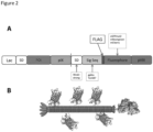
- the fluorophore As the fluorophore is fused to the major phage coat protein, pVIII employing a classical (8 + 8 wild-type (phagemid) or 88 wild-type (genomic)) complementation expression approach, the final phage particle produced by using the vectors of the invention will have multiple copies of the fluorophore randomly distributed along the length of the phage particle, together with copies of the wild-type pVIII protein. As for example M13 phages have approximately 2700 copies of the major coat protein pVIII on their surface, many copies of the fluorophore can be present. Thus, such functional expression of the fluorophore can conveniently be assessed by monitoring for fluorescence, e.g. detectable fluorescence, by any appropriate and convenient assay.
- monitoring for fluorescence can conveniently be carried out either by monitoring for fluorescence in transformed host cells such as E . coli, e.g. using fluorescence microscopy, or by analysing samples of phage particles, e.g. liquid samples, for fluorescence using a plate reader or other convenient apparatus. FACS or FLISA analysis can also be used.
- fluorophores will be biological fluorophores or other fluorophores which can be encoded by a nucleic acid sequence and thereby included in the vectors of the invention.
- fluorophores will also be intrinsically fluorescent or directly detectable. Fluorophores which can be activated or are bleachable may also be used.
- Preferred fluorophores for use in the present invention are biological fluorophores having or comprising a ⁇ -barrel structure or architecture.
- fluorophores would be well-known to a person skilled in the art (see for example the review by Shaner et al., 2007, J. Cell Sci, 120(24): 4247-4260 ) and include green fluorescent protein (GFP) or derivatives thereof with the same folding topology as GFP (also referred to as GFP derivatives).
- GFP green fluorescent protein
- such fluorophores for use in the invention include green fluorescent protein (GFP) or fluorophore derivatives thereof having a ⁇ -barrel architecture or structure or a GFP-architecture.
- Such non-GFP fluorophores can conveniently be referred to as GFP-like fluorophores or GFP derivatives or fluorophores with GFP-architecture.
- GFP green fluorescent proteins
- Such fluorophores contain one or more ⁇ -barrels as a unit, for example may contain a single ⁇ -barrel (a ⁇ -barrel monomer) or multiple ⁇ -barrels, for example ⁇ -barrel homo-tetramers.
- fluorophores have or comprise a similar 3-D cylindrical structure in which a large portion of the polypeptide backbone is wound into a ⁇ -barrel structure which generally comprises 11 strands of beta sheets that surround a central alpha helix containing the chromophore.
- the ⁇ -barrel structure forms a near-perfect cylinder creating what can be referred to as a " ⁇ -can” structure which characterises the GFP-like fluorophore family (GFP-derivatives).
- ⁇ -can which characterises the GFP-like fluorophore family
- fluorophores which contain this " ⁇ -can" structure structure are also preferred for use in the present invention.
- This ⁇ -barrel cylinder is generally approximately 24 to 30 ⁇ s wide and 40 to 42 ⁇ s long.
- the GFP family of fluorophores includes the variants blue (BFP), cyan (CFP), and yellow (YFP), depending on the spectral emission profile, as well as green variants of the original GFP.
- BFP blue
- CFP cyan
- YFP yellow
- Enhanced GFP (EGFP), enhanced CFP (ECFP) and enhanced YFP (EYFP) are further examples.
- GFP family of fluorophores (GFP plus GFP variants or derivatives, or GFP-like fluorophores or GFP related fluorophores) have some inherently different properties, e.g. emission colour, intensity of fluorescence and other properties such as stability (and indeed can be engineered so as to have such different properties), the presence and retension of the above described ⁇ -barrel structure or architecture means that they generally behave in a similar way.
- members of the GFP family of fluorophores (or variants thereof, e.g. functional variants with at least 70% sequence identity or other values as described elsewhere herein) are preferred fluorophores for use in the present invention.
- nucleic acid sequences encoding such fluorophores are well known and described in the art and can readily be obtained from the literature or standard databases (see for example pfam entry PF01353 or Interpro entry IPR011584).
- fluorophores or GFP-like fluorophores (or GFP-variant fluorophores), or ⁇ -barrel containing fluorophores, for use in the present invention are mNeonGreen (another green fluorescent protein with extremely bright yellow-green fluorescence, Shaner et al.,2013, Nature Methods, 10(5):407-409 ), mGFPmut2 (a FACS optimised version of GFP, Cormack et al., 1996, Gene 173:33-38 ), and mCherry (a red fluorescent protein, Shaner et al., 2004, Nat. Biotechnol., 22, 1567-1572 ). "m” standing for monomeric.
- EGFP or ECFP or EYFP
- Preferred fluorophores for use in the present invention are mNeonGreen, or a derivative or variant thereof, and GFP or a derivative or variant thereof, for example mGFPmut2 or some other derivative or variant, e.g. EGFP ( Heim et al., 1995, Nature 373:663-664 ).
- a more preferred fluorophore for use in the present invention is mNeonGreen or a derivative or variant thereof.
- mNeonGreen (Shaner et al., 2013, supra) is a bright monomeric green fluorescent protein derived from Branchiostoma lanceolatum.
- the original native protein from Branchiostoma lanceolatum is a tetramer and this has been modified (as described in Shaner et al., 2013, supra) to obtain the monomeric (m) NeonGreen (mNG) which is used in the present invention.
- the final mutant contains 21 substitutions relative to the tetrameric starter protein and has sharp excitation and emission peaks (at 506 nm and 517 nm).
- mNeonGreen can be imaged with essentially no loss of emission photons using standard green fluorescent protein band-pass or long-pass filter sets.
- the sequence of the synthetic construct for mNeonGreen has the GenBank accession code KC295282.1 and is repeated below for completeness.
- the nucleic acid sequence is 711 base pairs long (SEQ ID NO:45):
- the protein identifier for the encoded protein is AGG56535.1, which has the sequence as outlined below (236 amino acids, SEQ ID NO:46):
- a codon optimised version of this nucleic acid sequence is used which has been optimised for bacterial expression, e.g. in E. coli.
- any appropriate codon optimised version can be used, a specific sequence for a codon optimised version of mNeonGreen which can be used in the present invention is outlined below. (Again the start and stop codons have been omitted for better incorporation into the vectors).
- the vector comprises a sequence encoding a derivative or variant of the mNeonGreen (mNG) fluorophore, e.g. a derivative or variant of SEQ ID NO:4 or 46, or a derivative or variant of SEQ ID NO:45 or 3.
- mNG mNeonGreen
- the encoded mNG fluorophore comprises or consists of an amino acid sequence with a sequence identity of at least 70%, 75% or 80% to that of SEQ ID NO: 4, such as at least 80 %, 81 %, 82 %, 83 %, 84 %, 85 %, 86 % , 87 %, 88 %, 89 %, 90 %, 91 %, 92 %, 93 %, 94 %, 95 %, 96 %, 97 %, 98 %, or 99 % identity.
- These variant mNG sequences should retain or have the functional property to act as a fluorophore.
- Functional truncations or fragments of SEQ ID NO:4 (or these homologous sequences) or other mNG sequences could also be used providing the ability to encode a protein which retains or has the functional property to act as a fluorophore is retained.
- nucleic acid molecule encoding the mNG fluorophore may comprise or consist of a nucleotide sequence with a sequence identity of at least 70%, 75% or 80% to that of SEQ ID NO: 45 or 3, such as at least 80 %, 81 %, 82 %, 83 %, 84 %, 85 %, 86 % , 87 %, 88 %, 89 %, 90 %, 91 %, 92 %, 93 %, 94 %, 95 %, 96 %, 97 %, 98 %, or 99 % identity.
- variant mNG nucleotide sequences should encode a protein which retains or has the functional property to act as a fluorophore.
- Functional truncations or fragments of SEQ ID NO:45 or 3 (or these homologous sequences) or other mNG sequences could also be used providing the ability to encode a protein which retains or has the functional property to act as a fluorophore is retained.
- pVIII phage coat protein or "pVIII protein” or “pVIII phage protein” or “pVIII coat protein”, etc., as used herein refers to a pVIII protein originating from or derived from a filamentous phage (wild-type or native sequence), or a pVIII protein with a sequence which corresponds to the sequence of such a pVIII protein.
- Preferred filamentous phages from which the pVIII protein is derived or the pVIII protein corresponds to are M13, fd, and f1 phages. Any appropriate pVIII protein can be used providing it has the ability to display a fluorophore as a pVIII fusion protein.
- the pVIII protein encoded by the vectors of the invention corresponds to the mature (full length) pVIII protein, i.e. lacks the pVIII signal peptide.
- the pVIII proteins encoded by the vectors of the invention comprise or consist of the following amino acid sequence, which corresponds to the wild-type (full length) mature pVIII protein from the M13 filamentous phage without its signal sequence.
- AEGDDPAKAAFNSLQASATEYIGYAWAMVVVIVGATIGIKLFKKFTSKAS SEQ ID NO:8
- SEQ ID NO:7 An exemplary nucleic acid sequence encoding this sequence for inclusion in the vectors is provided elsewhere herein as SEQ ID NO:7.
- the encoded pVIII protein comprises or consists of an amino acid sequence with a sequence identity of at least 70%, 75% or 80% to that of SEQ ID NO: 8, such as at least 80 %, 81 %, 82 %, 83 %, 84 %, 85 %, 86 % , 87 %, 88 %, 89 %, 90 %, 91 %, 92 %, 93 %, 94 %, 95 %, 96 %, 97 %, 98 %, or 99 % identity.
- these variant pVIII sequences should retain or have the functional ability to display a fluorophore as a pVIII fusion protein.
- Functional truncations or fragments of SEQ ID NO:8 (or these homologous sequences) or other pVIII sequences could also be used providing the ability to display a fluorophore as a pVIII fusion protein was retained.
- nucleic acid molecule encoding the pVIII protein comprises or consists of a nucleotide sequence with a sequence identity of at least 70%, 75% or 80% to that of SEQ ID NO: 7, such as at least 80 %, 81 %, 82 %, 83 %, 84 %, 85 %, 86 % , 87 %, 88 %, 89 %, 90 %, 91 %, 92 %, 93 %, 94 %, 95 %, 96 %, 97 %, 98 %, or 99 % identity.
- variant pVIII nucleotide sequences should retain or have the functional ability to encode a fluorophore as a pVIII fusion protein which could be displayed on a phage particle.
- Functional truncations or fragments of SEQ ID NO:7 (or these homologous sequences) or other pVIII sequences could also be used providing the ability to encode a fluorophore as a pVIII fusion protein which could be displayed on a phage particle was retained.
- Preferred pVIII variants have a valine to isoleucine mutation at, or corresponding to, position 33 of SEQ ID NO:8 (i.e. have an isoleucine residue at, or corresponding to, position 33 of SEQ ID NO:8), or have an ATA isoleucine codon at, or corresponding to, residues 97-99 of SEQ ID NO:7, as described elsewhere herein.
- a particularly preferred pVIII variant is the pVIII sequence as found in the F03 clone as described elsewhere herein.
- Preferred pVIII fragments or truncations are functional fragments or truncations which include the residue corresponding to position 33 of SEQ ID NO:8 at the amino acid or nucleotide level.
- the vectors of the invention encode other non-pVIII phage coat proteins such as plll, pVI, pVII or pIX coat proteins.
- pIII phage coat protein or "pIII protein” or “pIII phage protein” or “pIII coat protein”, etc., as used herein refers to a plll protein originating from or derived from a filamentous phage (wild-type or native sequence), or a plll protein with a sequence which corresponds to the sequence of such a plll protein.
- Preferred filamentous phages from which the pIII protein is derived or the pIII protein corresponds to are M13, fd, and f1 phages. Any appropriate pIII protein can be used providing it has the ability to display a POI as a plll fusion protein.
- the plll protein encoded by the vectors of the invention corresponds to the mature plll protein, i.e. lacks the plll signal peptide.
- the plll proteins encoded by the vectors of the invention comprise or consist of the following amino acid sequence, which corresponds to the wild-type mature plll protein from the M13 filamentous phage without its signal sequence (see also (Genbank AY598820.1).
- SEQ ID NO: 11 An exemplary nucleic acid sequence encoding this amino acid sequence for inclusion in the vectors is provided below as SEQ ID NO: 11.
- the encoded plll protein comprises or consists of an amino acid sequence with a sequence identity of at least 70%, 75% or 80% to that of SEQ ID NO: 12, such as at least 80 %, 81 %, 82 %, 83 %, 84 %, 85 %, 86 % , 87 %, 88 %, 89 %, 90 %, 91 %, 92 %, 93 %, 94 %, 95 %, 96 %, 97 %, 98 %, or 99 % identity.
- These variant plll sequences should retain or have the functional ability to display a POI as a pIII fusion protein.
- pVI phage coat protein or "pVl protein” or “pVl phage protein” or “pVl coat protein”, etc., as used herein refers to a pVI protein originating from or derived from a filamentous phage (wild-type or native sequences), or a pVI protein with a sequence which corresponds to the sequence of such a pVI protein.
- Preferred filamentous phages from which the pVI protein is derived or the pVI protein corresponds to are M13, fd, and f1 phages.
- Any appropriate pVI protein can be used providing it has the ability to display a POI as a pVI fusion protein.
- the pVI proteins encoded by the vectors of the invention comprise or consist of the following amino acid sequence, which corresponds to the pVI protein from the VCSM13 helper phage (Genbank AY598820.1).
- the encoded pVI protein comprises or consists of an amino acid sequence with a sequence identity of at least 70%, 75% or 80% to that of SEQ ID NO: 13, such as at least 80 %, 81 %, 82 %, 83 %, 84 %, 85 %, 86 % , 87 %, 88 %, 89 %, 90 %, 91 %, 92 %, 93 %, 94 %, 95 %, 96 %, 97 %, 98 %, or 99 % identity.
- These variant pVI sequences should retain or have the functional ability to display a POI as a pVI fusion protein.
- Functional truncations or fragments of SEQ ID NO:13 (or these homologous sequences) or other pVI sequences, could also be used providing the ability to display a POI as a pVI fusion protein was retained.
- pVII phage coat protein or "pVII protein” or “pVII phage protein” or “pVll coat protein”, etc., as used herein refers to a pVII protein originating from or derived from a filamentous phage (wild-type or native sequences), or a pVII protein with a sequence which corresponds to the sequence of such a pVII protein.
- Preferred filamentous phages from which the pVII protein is derived or the pVII protein corresponds to are M13, fd, and f1 phages. Any appropriate pVII protein can be used providing it has the ability to display a POI as a pVII fusion protein.
- the pVII proteins encoded by the vectors of the invention comprise or consist of the following amino acid sequence, which corresponds to the wild-type pVII protein from the VCSM13 helper phage (Genbank AY598820.1). MEQVADFDTIYQAMIQISVVLCFALGIIAGGQR (SEQ ID NO:14)
- the encoded pVII protein comprises or consists of an amino acid sequence with a sequence identity of at least 70%, 75% or 80% to that of SEQ ID NO: 14 such as at least 80 %, 81 %, 82 %, 83 %, 84 %, 85 %, 86 % , 87 %, 88 %, 89 %, 90 %, 91 %, 92 %, 93 %, 94 %, 95 %, 96 %, 97 %, 98 %, or 99 % identity.
- These variant pVII sequences should retain or have the functional ability to display a POI as a pVII fusion protein.
- Functional truncations or fragments of SEQ ID NO:14 (or these homologous sequences) or other pVII sequences, could also be used providing the ability to display a POI as a pVII fusion protein was retained.
- pIX phage coat protein or "pIX protein” or “pIX phage protein” or “pIX coat protein”, etc., as used herein refers to a pIX protein originating from or derived from a filamentous phage (wild-type or native sequences), or a pIX protein with a sequence which corresponds to the sequence of such a pIX protein.
- Preferred filamentous phages from which the pVII protein is derived or the pIX protein corresponds to are M13, fd, and f1 phages. Any appropriate pIX protein can be used providing it has the ability to display a POI as a pIX fusion protein.
- the pIX protein encoded by the vectors of the invention corresponds to the pIX protein.
- the pIX proteins encoded by the vectors of the invention comprise or consist of the following amino acid sequence, which corresponds to the wild-type pIX protein from the VCSM13 helper phage (Genbank AY598820.1). MSVLVYSFASFVLGWCLRSGITYFTRLMETSS (SEQ ID NO:15)
- the encoded pIX protein comprises or consists of an amino acid sequence with a sequence identity of at least 70%, 75% or 80% to that of SEQ ID NO: 15, such as at least 80 %, 81 %, 82 %, 83 %, 84 %, 85 %, 86 % , 87 %, 88 % , 89 %, 90 %, 91 %, 92 %, 93 %, 94 %, 95 %, 96 %, 97 %, 98 %, or 99 % identity.
- These variant pIX sequences should retain or have the functional ability to display a POI as a pIX fusion protein.
- Functional truncations or fragments of SEQ ID NO:15 (or these homologous sequences) or other pIX sequences, could also be used providing the ability to display a POI as a pIX fusion protein was retained.
- sequence identity is a measure of identity between proteins at the amino acid level and a measure of identity between nucleic acids at the nucleotide level.
- the protein sequence identity may be determined by comparing the amino acid sequence in a given position in each sequence when the sequences are aligned.
- the nucleic acid sequence identity may be determined by comparing the nucleotide sequence in a given position in each sequence when the sequences are aligned.
- Methods to determine the percentage identity of two amino acid sequences or of two nucleic acid sequences are well known and described in the art, and any of these may be used.
- the sequences are aligned for optimal comparison purposes (e.g., gaps may be introduced in the sequence of a first amino acid or nucleic acid sequence for optimal alignment with a second amino acid or nucleic acid sequence).
- the amino acid residues or nucleotides at corresponding amino acid positions or nucleotide positions are then compared. When a position in the first sequence is occupied by the same amino acid residue or nucleotide as the corresponding position in the second sequence, then the molecules are identical at that position.
- alignment of two sequences for the determination of percent identity may be accomplished using a mathematical algorithm.
- Such an algorithm is incorporated into the NBLAST and XBLAST programs of (Altschul et al. 1990).
- Gapped BLAST may be utilised.
- PSI-Blast may be used to perform an iterated search which detects distant relationships between molecules.
- NBLAST NBLAST
- XBLAST XBLAST
- Gapped BLAST programs
- sequence identity may be calculated after the sequences have been aligned e.g. by the BLAST program in the EMBL database (www.ncbi.nlm.gov/cgi-bin/BLAST).
- the default settings with respect to e.g. "scoring matrix" and "gap penalty" may be used for alignment.
- the BLASTN and PSI BLAST default settings may be advantageous. In calculating percent identity, only exact matches are counted.
- one or more ribosome (ribosomal) binding sites are included in the vector constructs.
- Such components can also be referred to as a translational initiation region (TIR).
- the RBS sequence is located in the vector at an appropriate position for the RBS sequence to function.
- the role of the RBS is to recruit a ribosome during the initiation of protein translation and thus is conveniently placed at an appropriate distance upstream from the start codon of the protein it is desired to translate, or upstream of the ORF for the protein it is desired to translate.
- the RBS sequence is conveniently placed upstream of the sequence encoding the signal peptide (in embodiments where a signal peptide is part of the ORF, e.g. for the fluorophore-pVlll fusion protein). In embodiments where no signal peptide is present in the ORF, e.g.
- the RBS sequence is conveniently placed at an appropriate distance upstream of the sequence encoding the POI or the coat protein.
- the appropriate distance would be known or readily determined by a person skilled in the art depending on the RBS chosen. Exemplary distances might be seven or eight nucleotides from the ATG start codon, but this can vary.
- the RBS/TIR sequence modulates the translation intensity (level of protein expression) of the sequences located downstream and different types of RBS can produce different levels of protein expression, for example weak or strong expression.
- Weak or strong RBS/TIR sequences are well known in the art and can readily be selected by a skilled person depending on the level of protein expression desired.
- a strong RBS facilitates or induces more translation (strong translation) as compared to a weak RBS.
- a weak RBS is used, in particular to drive the translation of the fluorophore-pVlll fusion protein.
- an RBS is included upstream (or 5' or N-terminal to) to the start codon of the sequence encoding the signal peptide which directs proteins into the Tat secretory pathway.
- a preferred RBS for use in the present invention is a Shine Dalgarno (SD) sequence or a SD based sequence which can be included in the vector constructs. SD sequences are well known and described in the art and any of these may be used.
- the core SD sequence is GAGG (SEQ ID NO:47) and other consensus sequences are AGGAGG (SEQ ID NO:48) or AGGAGGU (SEQ ID NO:49).
- SD sequences comprising these core or consensus sequences can be used.
- a strong SD will be able to bind well to a ribosome.
- Such a strong SD will generally contain the SD core sequence or one of the consensus sequences outlined above exactly, or with very few changes, and/or for example will contain a number of A and/or G residues close by.
- a strong SD can be obtained by the insertion of other components in conjunction with an SD core or consensus sequence (or a highly related sequence).
- additional components would be well-known to a person skilled in the art, for example a classical or consensus SD sequence can be used in conjunction with an Epsilon sequence (e.g.
- TTAACTTTA SEQ ID NO:50.
- a preferred strong SD is the T7g10 TIR ( Olins et al., 1988, Gene 73:227-235 ) which also includes an Epsilon sequence (e.g. TTAACTTTA, SEQ ID NO:50).
- the upstream region from the ATG start site of the Tat signal peptide may have the sequence TTAACTTTA AG AAGGAG ATATACAT, SEQ ID NO:31 (Epsilon sequence underlined and SD consensus sequence in bold).
- other strong SD/TIR sequences could be readily selected by a person skilled in the art.
- a weak SD sequence will generally contain sequences which are more different to the core or consensus SD sequences outlined herein.
- a weak SD sequence will still be able to bind to a ribosome but at a lower level or lower efficiency.
- a weak SD sequence for use in the present invention may contain one or two modifications from the core consensus SD sequences outlined herein.
- a preferred weak SD sequence for use in the vectors of the present invention comprises or consists of AGGAGA, SEQ ID NO:30 (i.e. contains one nucleotide difference from the consensus sequence AGGAGG, SEQ ID NO:48). This weak SD sequence is preferably placed eight nucleotides from the ATG start site, although this distance can sometimes be varied as discussed above.
- the upstream region from the ATG start site of the Tat signal peptide may have the sequence A AGGAGA CAGTCATA, SEQ ID NO:51 (variant SD sequence shown in bold).
- SEQ ID NO:51 variant SD sequence shown in bold.
- other weak SD/TIR sequences could be readily selected by a person skilled in the art.
- a strong SD facilitates or induces more translation (strong or high translation) as compared to a weak SD which will facilitate or induce a low amount (or weak or less efficient) translation.
- Either a strong or a weak RBS or SD sequence can be used in the vector constructs of the invention.
- One of the aims of the invention is to increase the fluorescence of a phage particle by increasing the number of fluorophore-pVlll fusion proteins incorporated into the coat (on the surface) of the phage. This would generally favour the use of a strong SD sequence in order to obtain more translation.
- a weak SD (or weak RBS) sequence produces better results, e.g. in terms of better functional display of the fluorophore-pVIII fusion protein, or increased display (e.g. more copies per particle) of the fluorophore-pVlll fusion protein, preferably resulting in an increased fluorescence of phage particles.
- the use of a weak SD (or weak RBS) sequence in the vector construct upstream of the sequence encoding the ORF comprising the Tat signal peptide and the fluorophore-pVlll fusion protein is preferred.
- the RBS (or SD) controlling or driving translation of the Tat signal peptide and hence the fluorophore-pVIll fusion protein is a weak SD (or weak RBS).
- such a weak SD sequence results in more copies of the intact fluorophore-pVIII fusion protein incorporated in the coat of the phage when compared to a construct where a strong SD (or strong RBS) is used or when a consensus SD sequence, e.g AGGAGG (or other consensus sequences as defined above), is used.
- a strong SD or strong RBS
- a consensus SD sequence e.g AGGAGG (or other consensus sequences as defined above
- vector constructs where the translation efficiency of the fluorophore-pVlll fusion protein is low, weak, reduced, or non-optimal are generally preferred.
- preferred modifications/variant molecules as described elsewhere herein are those in which translation efficiency or speed, for example and in particular of the fluorophore-pVlll fusion protein, is reduced or significantly reduced, for example compared to the original parent sequence.
- reduced or weak (or decreased) translation efficiency or speed also results in more copies of the fluorophore-pVIII fusion protein on the surface of the phage and/or an increased number of functional fluorophores on the surface of the phage, and thus increased fluorescence.
- one or more tags can be included in the vector constructs.
- tags can conveniently be used for detection and thus, in these embodiments, any detectable tag could be used.
- the tags can also be used for other purposes such as enrichment of tag-containing fusion proteins, for example before screening of phage fluorescence, e.g. by FACS, takes place.
- Antigen peptide tags such as FLAG, c-myc, HA (haemaglutinin), HIS, HAT or V5 tags are particularly preferred for use in embodiments of the invention when FACS techniques or other fluorescent detection techniques are used, as these tags readily allow labelling by using known and readily available fluorescently labelled antibodies against the specific tag.
- Other antigen peptide tags known in the art can equally be used.
- a FLAG tag is a preferred tag for use in the vectors of the present invention.
- the FLAG tag is an octapeptide tag and comprises the sequence DYKDDDDK (SEQ ID NO:10).
- FLAG tag derivatives can also be used, which are well known and standard in the art.
- antibodies which can recognise the various FLAG tags and can thus be fluorescently labelled and used for detection of the FLAG tag are also well described in the art, e.g. M1 and M2 antibodies.
- biotin tags for use in the constructs of the present invention would be well known to a skilled person.
- Biotin tags for use in the methods of the present invention may comprise biotin molecules per se, e.g. biotin molecules which are attached, e.g. via chemical conjugation, to the expressed fluorophore-pVlll fusion protein on the phage surface. Methods for attaching such biotin tags are well known in the art.
- the biotin tags may comprise moieties, e.g. peptides, which can act as substrates for biotinylation reactions and thereby become attached to biotin molecules.
- moieties e.g. peptides
- Such peptide tags can readily be incorporated into the vectors of the invention.
- an exemplary biotin tag for use in the present invention is AviTag TM (MSGLNDIFEAQKIEWHE, SEQ ID NO:52), which is a commercially available tag from Avidity LLC, Aurora, Colorado, USA which becomes biotinylated in vitro or in vivo by biotin ligase.
- Strep-tag is also commercially available from IBA GmbH, Göttingen, Germany, and which is capable of binding to the biotin binding pocket of streptavidin.
- the Strep-tag comprises the 8 amino acid sequence Trp-Ser-His-Pro-Gln-Phe-Glu-Lys (WSHPQFEK, SEQ ID NO:53). Any other protein or peptide tags which are capable of binding to streptavidin or avidin could equally be used.
- Combinations of one or more of the above tags could be used to label the phage particles so that multiple labels are present.
- Preferred tags are protein tags or peptide tags which can readily be incorporated into the vector constructs (e.g. phage display constructs) of the invention, e.g. using standard recombinant and cloning techniques.
- the tag sequence can be located in the vector at any appropriate position for the tag to function, e.g. to be expressed and to be detectable.
- tags can thus be internal, N-terminal or C-terminal to the part of the construct which is being tagged and which is to be detected using the tag.
- a tag can conveniently be used to detect expression of the fluorophore-pVlll part of the construct.
- the tag can conveniently be placed within the part of the construct (ORF) encoding the fluorophore-pVlll fusion protein, for example within (internal to) or at (or near) the N-terminus, or at (or near) the C-terminus of the fluorophore-pVlll fusion protein.
- a preferred position for the sequence encoding the protein tag is at or near the N-terminus of the fluorophore in the fluorophore-pVlll fusion.
- the sequence encoding the protein tag can be located between the signal peptide and the fluorophore.
- the protein tag is a FLAG tag (or a derivative thereof), or another negatively charged tag.
- a negatively charged tag preferably located at or near the N-terminus of the fluorophore, for example between the signal peptide and the fluorophore, can result in improved stability and hence folding of the fluorophore-pVIII fusion protein, which is a phenomenon observed with other ⁇ -structures ( Schaefer, 2012, J. Mol. Biol., 417:309-335 and Dudgeon et al., 2012, PNAS, 109:10879-10884 ).
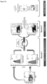
- FIG. 1 An exemplary structure for a construct of the invention with a fluorophore-pVlll fusion protein is shown in Figure 1 .
- the FLAG tag as shown in Figure 1 (or other tag) is preferred but optional.
- a sequence encoding a spacer or linker is included between the sequence encoding the fluorophore and the sequence encoding the pVIII phage coat protein.
- the sequence can aid the folding of the connected proteins, in particular the N-terminal protein (here generally the fluorophore), and the spacer or linker length can be adjusted as appropriate to enable the best or satisfactory functional folding of both components. Appropriate lengths could readily be determined by a person skilled in the art. However exemplary lengths would be between five and 15 amino acids ( Weiss et al., 2000, Protein Sci., 9:647-654 ), e.g. 6 to 10 amino acids.
- a particular linker used in the present invention is 8 amino acids long.
- the sequence of the linker is usually less relevant than the length.
- the linker used in the exemplified vectors has the sequence GGGSGGGS (SEQ ID NO: 6, encoded for example by SEQ ID NO:5). This linker is thus preferred for some embodiments of the invention, but it will be appreciated that linkers (spacers) with other sequences and lengths can also be used.
- Preferred vectors (or nucleic acid molecules) of the invention encode the sequence comprising SEQ ID NO:A or 16 (which comprises mNeonGreen, a linker and the N-terminal 30 amino acids of pVIII) or a sequence with at least 70% identity thereto (other exemplary values for percent identity are described elsewhere herein).
- SEQ ID NO:C or 18 which comprises mNeonGreen, a linker and full length pVIII
- sequence with at least 70% identity thereto other exemplary values for percent identity are described elsewhere herein.
- SEQ ID NO:E or 20 which comprises Tor AB7, mNeonGreen, a linker and full length pVIII
- sequence with at least 70% identity thereto other exemplary values for percent identity are described elsewhere herein.
- SEQ ID NO:G or 22 which comprises Tor AB7, FLAG tag, mNeonGreen, a linker and full length pVIII
- sequence with at least 70% identity thereto other exemplary values for percent identity are described elsewhere herein.
- Vectors (or nucleic acid molecules) encoding sequences comprising SEQ ID NO:K or 26 (which comprises Tor AB7, mNG, a linker, and the N-terminal 30 amino acids of pVIII) or SEQ ID NO:M or 28 (which comprises Tor AB7, FLAG tag, mNG, a linker, and the N-terminal 30 amino acids of pVIII), or a sequence with at least 70% identity thereto (other exemplary values for percent identity are described elsewhere herein) are also provided.
- SEQ ID NO:K or 26 which comprises Tor AB7, mNG, a linker, and the N-terminal 30 amino acids of pVIII
- SEQ ID NO:M or 28 which comprises Tor AB7, FLAG tag, mNG, a linker, and the N-terminal 30 amino acids of pVIII
- any one of these vector sequences are examples of sequences which can be used in a parent vector and subjected to further variation or modification as outlined below.
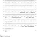
- FIG. 9 A further exemplary vector sequence for use in the invention is shown in Figure 9 (SEQ ID NO: 54 and 55).
- Preferred pVIII variants for example in the SEQ ID NOs: C or 18, E or 20, G or 22, 54 or 55, have a valine to isoleucine mutation at, or corresponding to, position 33 of SEQ ID NO:8 (i.e. have an isoleucine residue at, or corresponding to, position 33 of SEQ ID NO:8), or have an ATA isoleucine codon at, or corresponding to, residues 97-99 of SEQ ID NO:7, as described elsewhere herein.
- a particularly preferred pVIII variant is the pVIII sequence as found in the F03 clone as described elsewhere herein.
- the phage display vectors of the invention as described above may also be used to produce further modified or variant vectors that can be used to produce fluorescent phage particles.
- modified (or variant or derivative or mutant) vectors also form part of the invention.
- modifications (or mutations) involve for example the addition, deletion, substitution or insertion of one or more nucleotides in the nucleic acid sequence of a parent vector to form a new vector, wherein said parent vector is one of the vectors of the invention as defined above, and testing the resulting new vector to identify vectors that can be used to produce fluorescent phage particles with improved properties.
- Such methods can be used to form multiple new vectors (conveniently a library of new vectors) that can all be tested for their ability to produce fluorescent phage particles, preferably improved fluorescent phage particles.
- Said modifications e.g. in the form of addition, deletion, substitution or insertion of one or more nucleotides (and hence encoded amino acids) can take place in any of the functional domains of the vector, namely in one or more (or all) of the signal peptide (preferably Tor A, more preferably Tor AB7), fluorophore (preferably mNeonGreen), linker between the fluorophore and pVIII, pVIII, or the tag (preferably FLAG tag) encoding parts of the vector.
- the modifications are located in one or more (or all) of the fluorophore (preferably mNeonGreen), the linker between the fluorophore and pVIII, or the pVIII encoding region.
- the modifications are located in the pVIII encoding region, in some embodiments the mutations are located in the N-terminal half of pVIII, for example within the first 90 nucleotides (or the first 30, e.g. the first 25-28 or 25-30 amino acids of pVIII).
- Such modifications or mutations to a parent vector can be carried out in any appropriate manner using techniques well known and documented in the art, for example by carrying out methods of random or directed mutagenesis.
- the mutations made are substitutions and these can conveniently and preferably be made using random mutagenesis, in particular when it is desired to generate and screen multiple (e.g. a library of) mutants to select those which show improved properties.
- Random mutagenesis can be carried out in any appropriate way, e.g., by error-prone PCR or using mutator E. coli strains.
- a preferred and convenient technique is described in the Examples and involves the incorporation of dNTP analogues by PCR to introduce random mutations into the vector (e.g. phagemid) DNA.
- kits are available to carry out such random mutagenesis.
- mutations are made, for example the valine to isoleucine (V to I) mutation in the pVIII protein (or corresponding mutation in the encoding nucleotide sequence) as described herein, then directed mutagenesis or other types of mutagenesis where specific residues can be targeted and modified are appropriate.
- V to I valine to isoleucine
- the new vectors produced by these methods when transformed into an appropriate host cell, will preferably produce fluorescent phage particles which have improved functional properties, preferably improved fluorescent properties.
- the present invention thus further provides variant or mutated vectors which are capable of producing phage particles which exhibit an improvement in the fluorescence intensity (improved brightness) compared to the original, starting, wild-type or parental phage particle (i.e. the phage particle produced by the non-mutated or wild-type parent vector).
- improvements can be provided by a) increasing the number of fluorophores on the surface of a bacteriophage particle, for example by increased incorporation or integration of fluorophore-pVlll fusion proteins into the phage surface/coat as an inherent property of phage particle assembly from the producing E.
- coli host by virtue of the fluorophore being fused to the viral capsid protein (in other words increasing the number or average number of fluorophore-pVlll molecules per particle), or b) increasing the brightness of the individual fluorochrome, e.g. increasing the intrinsic brightness (fluorescence intensity) of the fluorescent moiety in the fusion protein, or c) a combination of both a) and b), i.e. increasing the brightness and the number of the fluorophores.
- Another option, which could be used alone, or in combination with a) and/or b), would be to improve functional display of the fluorophore, e.g. by improving (e.g. more efficient) folding or increased solubility, as heterologous protein expression in E. coli frequently results in a fraction of translated, but non-functional protein products, e.g. insoluble protein products.
- improvements can also be provided by increasing the number of functional fluorophores on the phage surface.
- any appropriate assay can be used to screen for phage particles containing mutated vectors which have an increased number of fluorophores on the surface of the bacteriophage.
- some kind of detectable tag (which is detectable on the surface of the phage) can conveniently be used to carry out such screening.
- these tags will be independent of the level of fluorescence displayed by the fluorescent moiety so that an indication of the number of fluorescent moieties on the surface of the phage can be assessed.
- detectable tags will be well known to a person skilled in the art.
- a FLAG tag can be used. Detection can conveniently be carried out using an antibody to the tag in question. FACS, e.g. using a fluorescently labelled antibody, e.g.
- an antibody to the FLAG tag is particularly preferred as it more readily allows individual particles to be screened by way of monitoring and selecting particles which have shifted in a positive direction on the relevant fluorescence axis.
- antibodies specifically recognizing the fluorophore such as anti-mNeonGreen 32F6 (ChromoTek GmbH), or similar, could be used for FACS.
- any appropriate assay can be used to screen for phage particles containing mutated vectors which have an increased brightness of the individual fluorochrome in the fusion protein expressed on the surface of the bacteriophage.
- FACS or some other kind of fluorescence-based assay can be used to measure the level of fluorescence, e.g. measuring fluorescence levels of phage particles in solution, in combination with another method of quantification of the fluorophore, e.g. use of western blot, or the use of an antibody to the fluorophore, e.g. the anti-NeonGreen nanobody as used in the FACS sorting shown in the Examples).
- FACS is particularly preferred as it more readily allows individual particles to be screened by way of monitoring and selecting particles which have shifted in a positive direction on the relevant fluorescence axis. Exemplary methods, for example FACS-based methods or FLISA, are shown in the Examples.
- Such increases are measurable increases, etc., (as appropriate), more preferably they are significant increases, preferably statistically significant increases, for example with a probability value of ⁇ 0.05, when compared to an appropriate control level or value (e.g. compared to the level of fluorescence obtained with a non-mutated or parent or wild-type vector).
- Preferred increases in fluorescence intensity might be increases of greater than or at least two-fold or three-fold, e.g. up to 5-fold, 10-fold or 20-fold, more than the parent vector.
- a convenient comparator or baseline level for a parent vector which can be used to assess such increases or improvements could be a vector comprising sequences encoding SEQ ID NO:E or 20 or SEQ ID NO: G or 22 or SEQ ID NO:C or 18 (or indeed SEQ ID NOs: A or 16, K or 26 or M or 28).
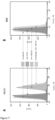
- Mutated versions of a vector which can produce fluorescent phage expressing an mNeonGreen-pVlll fusion protein on the surface have been developed which display improved fluorescence properties. Some of these mutated versions show improved brightness of the mNeonGreen fluorescent moiety on an individual basis (as evidenced by a shift up the y-axis on the FACS profiles shown in Example 2). Others of these mutated versions show improved display, i.e. an increased number of fluorophores, on the surface of the bacteriophage (as evidenced by a shift to the right along the x-axis on the FACS profiles shown in Example 2, assessed by measurement of a FLAG tag with a labelled antibody).
- the mutated residues which result in the improved fluorescence of the phage particles can be located in any part of the vector but are preferably in either the mNeonGreen or the pVIII part of the fusion protein (more preferably in the pVIII part), or in the linker between the mNeonGreen and pVIII.
- the mutations are located in the pVIII region, in some embodiments the mutations are located in the N-terminal half of pVIII, for example within the first 90 nucleotides (or the first 30, e.g. the first 25-28 or 25-30 amino acids of pVIII).
- the encoded amino acid sequence for the mNeonGreen part of the vector is provided by SEQ ID NO:4 (and an exemplary nucleotide sequence is provided by SEQ ID NO:3).
- SEQ ID NO:4 an exemplary nucleotide sequence is provided by SEQ ID NO:3.
- variant mNeonGreen components may comprise SEQ ID NO:3 or SEQ ID NO:4, or a sequence with at least 70% identity thereto at either the nucleotide or amino acid level (e.g.
- variant components may comprise a mutation level of up to 30%, 25%, 20%, 15%, 13%, 12%, 10%, 9%, 8%, 7%, 6%, 5%, 4%, 3%, 2% or 1% in SEQ ID NO:3 or SEQ ID NO:4 at either the nucleotide or amino acid level, respectively, preferably at the amino acid level.
- Preferred variant mNeonGreen components may comprise a mutation level of up to 15%, preferably up to 13% or 10%, more preferably up to 5% or 4%.
- mutated mNeonGreen sequences are sequences containing up to 30, e.g. up to 25, 20, 15, 12, or 10, e.g. 1, or up to 2, 3, 4, 5, 6, 7, 8, 9 or 10 altered amino acids in the mNeonGreen sequence, e.g. SEQ ID NO:4.
- the encoded amino acid sequence for the pVIII part of the vector is provided by SEQ ID NO:8 (and an exemplary nucleotide sequence is provided by SEQ ID NO:7).
- SEQ ID NO:8 an exemplary nucleotide sequence is provided by SEQ ID NO:7.
- variant pVIII components may comprise SEQ ID NO:7 or SEQ ID NO:8, or a sequence with at least 70% identity thereto at either the nucleotide or amino acid level (e.g.
- variant components may comprise a mutation level of up to 30%, 25%, 20%, 15%, 13%, 12%, 10%, 9%, 8%, 7%, 6%, 5%, 4%, 3%, 2% or 1% in SEQ ID NO:7 or SEQ ID NO:8 at either the nucleotide or amino acid level, respectively, preferably at the amino acid level.
- Preferred variant pVIII components may comprise a mutation level of up to 15%, preferably up to 13% or 10%, more preferably up to 5% or 4%.
- mutated pVIII sequences are sequences containing up to 10, e.g. up to 9, 8, 7, 6, 5, 4, 3, 2 or 1 altered amino acids in the pVIII sequence, e.g. SEQ ID NO:8.
- preferred mutated vector sequences contain a valine to isoleucine substitution at residue (or position) 33 of SEQ ID NO:8, or a residue corresponding thereto (i.e. have an isoleucine residue at, or corresponding to, position 33 of SEQ ID NO:8).
- the codon encoding the isoleucine is ATA.
- a valine codon GTC can be substituted with the isoleucine codon ATA.
- the mutated residues are present in the N-terminal half of the pVIII protein, for example in the encoded amino acid sequence for the pVIII part of the vector as provided by SEQ ID NO:J or 25 which contains the first 30 amino acids of pVIII (with an exemplary nucleotide sequence as provided by SEQ ID NO:I or 24, the first 90 nucleotides of pVIII).
- such variant pVIII components may comprise SEQ ID NO:I or 24 or SEQ ID NO:J or 25, or a sequence with at least 70% identity thereto at either the nucleotide or amino acid level (e.g. at least 75%, 80%, 85%, 87%, 88%, 90%, 91%, 92%, 93%, 94%, 95%, 96%, 97%, 98% or 99% identity).
- variant components may comprise a mutation level of up to 30%, 25%, 20%, 15%, 13%, 12%, 10%, 9%, 8%, 7%, 6%, 5%, 4%, 3%, 2% or 1% in SEQ ID NO:I or 24 or SEQ ID NO:J or 25 at either the nucleotide or amino acid level, respectively, preferably at the amino acid level.
- Preferred variant pVIII components may comprise a mutation level of up to 15%, preferably up to 13% or 10%, more preferably up to 5% or 4%.
- mutated pVIII sequences are sequences containing up to 5, e.g. up to 4, 3, 2 or 1 altered amino acids in the pVIII sequence, e.g. SEQ ID NO:J or 25.
- the encoded amino acid sequence for the linker part of the vector (here we are referring to the linker between the fluorophore and the pVIII components) is provided by SEQ ID NO:6 (and an exemplary nucleotide sequence is provided by SEQ ID NO:5).
- such variant linker components may comprise SEQ ID NO:5 or SEQ ID NO:6, or a sequence with at least 70% identity thereto at either the nucleotide or amino acid level (e.g.
- variant components may comprise a mutation level of up to 30%, 25%, 20%, 15%, 13%, 12%, 10%, 9%, 8%, 7%, 6%, 5%, 4%, 3%, 2% or 1% in SEQ ID NO:5 or SEQ ID NO:6 at either the nucleotide or amino acid level, respectively, preferably at the amino acid level.
- Preferred variant linker components may comprise a mutation level of up to 15%, preferably up to 13% or 10%, more preferably up to 5% or 4%.
- mutated linker sequences are sequences containing 1, 2 or 3 altered amino acids in the linker sequence, e.g. SEQ ID NO:6. In other embodiments no mutations are present in the linker sequence, e.g. SEQ ID NO:6.
- mutated TorAB7 sequences are sequences containing up to 6, e.g. up to 5, 4, 3, 2, or 1 altered amino acids in the TorAB7 sequence, e.g. SEQ ID NO:2.
- the mutations may be found in one or more (or all) of the mNeonGreen, the linker between the mNeonGreen and pVIII, and the pVIII encoding region. In some preferred embodiments, the mutations can be found in the pVIII encoding region. Where the mutations are located in the pVIII region, in some embodiments the mutations are located in the N-terminal half of pVIII, for example within the first 90 nucleotides (or the first 30, e.g. the first 25-28 or 25-30 amino acids of pVIII).
- such variant vectors may comprise SEQ ID NO:A or 16 or SEQ ID NO:C or 18 or a sequence with at least 70% identity to SEQ ID NO:A or 16 or C or 18 (e.g. at least 75%, 80%, 85%, 87%, 88%, 90%, 91%, 92%, 93%, 94%, 95%, 96%, 97%, 98% or 99% identity).
- variant components may comprise a mutation level of up to 30%, 25%, 20%, 15%, 13%, 12%, 10%, 9%, 8%, 7%, 6%, 5%, 4%, 3%, 2% or 1% in SEQ ID NO:A or C.
- Preferred variant components may comprise a mutation level of up to 15%, preferably 13% or 10%, more preferably up to 5% or 4%.
- mutated vector sequences are sequences containing up to 30, e.g. up to 25, 20, 15, 12, or 10, e.g. 1, or up to 2, 3, 4, 5, 6, 7, 8, 9 or 10 altered amino acids in the SEQ ID NO:A or 16 or C or 18.
- preferred mutated vector sequences contain a valine to isoleucine substitution at residue 276 of SEQ ID NO:C or 18, or a residue corresponding thereto (i.e. have an isoleucine residue at, or corresponding to, position 276 of SEQ ID NO:C or 18).
- the codon encoding the isoleucine is ATA.
- a valine codon GTC may be substituted with the isoleucine codon ATA.
- variant vectors may comprise SEQ ID NO:C or 18, or a sequence with at least 70% identity to SEQ ID NO:C or 18, or a mutation level of up to 15%, or a certain number, e.g. up to 35, altered amino acids in SEQ ID NO:C or 18, in preferred embodiments this isoleucine residue or codon is present.
- the mutations may be found in one or more (or all) of the mNeonGreen, the linker between the mNeonGreen and pVIII, the pVIII encoding region, and the TorAB7 signal peptide. In some preferred embodiments, the mutations can be found in the pVIII encoding region.
- such variant vectors may comprise SEQ ID NO:E or 20 or SEQ ID NO:K or 26 or a sequence with at least 70% identity to SEQ ID NO:E or 20 or SEQ ID NO:K or 26 at the amino acid level (e.g.
- variant components may comprise a mutation level of up to 30%, 25%, 20%, 15%, 13%, 12%, 10%, 9%, 8%, 7%, 6%, 5%, 4%, 3%, 2% or 1% in SEQ ID NO:E or 20 or SEQ ID NO:K or 26.
- Preferred variant components may comprise a mutation level of up to 15%, preferably 13% or 10%, more preferably up to 5% or 4%.
- mutated vector sequences are sequences containing up to 35, e.g. up to 30, 25, 20, 17, 15, 12, or 10, e.g. 1, or up to 2, 3, 4, 5, 6, 7, 8, 9 or 10 altered amino acids in the SEQ ID NO:E or 20 or K or 26.
- preferred mutated vector sequences contain a valine to isoleucine substitution at residue 317 of SEQ ID NO:E or 20, or a residue corresponding thereto (i.e. have an isoleucine residue at, or corresponding to, position 317 of SEQ ID NO:E or 20).
- the codon encoding the isoleucine is ATA.
- a valine codon GTC may be substituted with the isoleucine codon ATA.
- variant vectors may comprise SEQ ID NO:E or 20, or a sequence with at least 70% identity to SEQ ID NO:E or 20, or a mutation level of up to 15%, or a certain number, e.g. up to 35, altered amino acids in SEQ ID NO:E or 20, in preferred embodiments this isoleucine residue or codon is present.
- the mutations may be found in one or more (or all) of the mNeonGreen, the linker between the mNeonGreen and pVIII, the pVIII encoding region, the TorAB7 signal peptide and the FLAG sequence.
- such variant vectors may comprise SEQ ID NO:G or 22 or SEQ ID NO:M or 28 or a sequence with at least 70% identity to SEQ ID NO:G or 22 or SEQ ID NO:M or 28 (e.g.
- variant components may comprise a mutation level of up to 30%, 25%, 20%, 15%, 13%, 12%, 10%, 9%, 8%, 7%, 6%, 5%, 4%, 3%, 2% or 1% in SEQ ID NO:G or 22 or SEQ ID NO:M or 28.
- Preferred variant components may comprise a mutation level of up to 15%, preferably 13% or 10%, more preferably up to 5% or 4%.
- mutated vector sequences are sequences containing up to 35, e.g. up to 30, 25, 20, 17, 15, 12, or 10, e.g. 1, or up to 2, 3, 4, 5, 6, 7, 8, 9 or 10 altered amino acids in the SEQ ID NO:G or 22 or M or 28.
- preferred mutated vector sequences contain a valine to isoleucine substitution at residue 325 of SEQ ID NO:G or 22, or a residue corresponding thereto (i.e. have an isoleucine residue at, or corresponding to, position 325 of SEQ ID NO:G or 22).
- the codon encoding the isoleucine is ATA.
- a valine codon GTC can be substituted with the isoleucine codon ATA.
- variant vectors may comprise SEQ ID NO:G or 22, or a sequence with at least 70% identity to SEQ ID NO:G or 22, or a mutation level of up to 15%, or a certain number, e.g. up to 35, altered amino acids in SEQ ID NO:G or 22, in preferred embodiments this isoleucine residue or codon is present.
- the FLAG tag remains unmodified or non-mutated, e.g. retains the sequence of SEQ ID NO:10 (DYKDDDDK).
- the vectors of the invention contain mutations such that the mutated or variant sequences display reduced or significantly reduced or weaker or decreased translation efficiency or speed.
- vector constructs where the translation efficiency of the fluorophore-pVlll fusion protein is low, weak, reduced, decreased, or non-optimal are generally preferred.
- Preferred modifications/variant molecules are those in which translation efficiency, for example and in particular of the fluorophore-pVlll fusion protein, is reduced or significantly reduced (or decreased), for example compared to the original parent (non-mutated or unmodified) sequence, specific examples of which are described elsewhere herein.
- this can be achieved by having a mutated pVIII component such that the translation efficiency of the pVIII protein (and hence the whole ORF containing the fluorophore-pVIII fusion protein) is reduced or significantly reduced.
- mutations in other parts of the fluorophore-pVlll fusion protein e.g. in the fluorophore (e.g. the mNeongreen fluorophore) or in the linker sequence or in the signal peptide can result in reduced translation efficiency.
- Variants with reduced translation efficiency could be screened or tested by appropriate methods devised or known by a person skilled in the art.
- a time chase assay could be carried out on normalized host cell numbers and the protein production quantified by a means independent of protein functionality, e.g. by means of a Western Blot, to assess whether the absolute amount of protein is lower in the system being tested for reduced translation efficiency compared to a relevant control (e.g. a non-variant or wild type fusion protein).
- such decreases are measurable decreases, etc., (as appropriate), more preferably they are significant decreases, preferably statistically significant decreases, for example with a probability value of ⁇ 0.05, when compared to an appropriate control level or value (e.g. compared to the level of translation, e.g. translation efficiency or speed, obtained with a non-mutated or parent or wild-type vector, or other relevant control).
- an appropriate control level or value e.g. compared to the level of translation, e.g. translation efficiency or speed, obtained with a non-mutated or parent or wild-type vector, or other relevant control.
- a reduced translation efficiency in particular of the fluorophore-pVlll fusion protein, produces better results, e.g. in terms of better functional display of the fluorophore pVIII fusion protein, and/or increased display (e.g more copies per particle) of the fluorophore pVIII fusion protein, which can then result in an increased fluorescence of phage particles.
- One example of a mutation which results in improved fluorescence of phage particles is the modification of a V to an I at residue 33 of the pVIII protein shown in SEQ ID NO:8.
- This mutation is found in the clone F03 as described in the Examples which is shown to have improved fluorescence as compared to the parent sequence, or in other words compared to a sequence which does not have the V to I mutation at this position.
- the nucleic acid sequence is examined, the position 33 valine encoding codon, GTC, in the parent/wild type/unmodified pVIII, has been changed to the isoleucine encoding codon, ATA. This is an extremely rare codon in E.
- rare codons codons which are rare in E. coli
- T would of course be U in a corresponding RNA sequence
- Appropriate rare codons would be well-known to a person skilled in the art, however some examples might be rare Arg codons such as AGG, CGA, AGA or CGG, or CTA (Leu), CCC (Pro), or TCG (Ser).
- Another example might be to exchange the normal ATG start/initiation codon for a rarer codon, e.g. the rarer GTG codon.
- the presence of the V to I mutation at this position results in significantly improved fluorescence, for example in the order of 2 fold higher (or 100% higher) than the fluorescence observed with the wild type/parent/unmodified pVIII sequence.
- the fluorescence observed was already bright. Indeed it is believed that the fluorescence conferred by expressing the parent vector exhibited the brightest fluorescence for a phage particle known in the art. Thus, to double this fluorescence, is a significantly advantageous improvement.
- preferred mutated vector sequences have a pVIII sequence which contains a valine to isoleucine substitution at residue 33 of SEQ ID NO:8, or a residue corresponding thereto (i.e. have an isoleucine residue at, or corresponding to, position 33 of SEQ ID NO:8).
- the codon encoding the isoleucine is ATA.
- a valine codon GTC may be substituted with the isoleucine codon ATA.
- variant vectors may comprise SEQ ID NO:8, or a variant of SEQ ID NO:8, for example a sequence with at least 70% identity to SEQ ID NO:8, or a mutation level of up to 15%, or a certain number, e.g. up to 35, altered amino acids in SEQ ID NO:8, in preferred embodiments this isoleucine residue or codon is present.
- the pVIII phage coat protein or variant sequence contained (or encoded) in the vectors has a valine to isoleucine mutation (an isoleucine residue) at position 33 of SEQ ID NO:8 or a corresponding position.
- the nucleic acid sequence encoding said isoleucine residue comprises the codon ATA.
- mutated vector sequences encoding the pVIII protein contain a GTC to ATA substitution at residues 97-99 of SEQ ID NO:7, or residues corresponding thereto.
- variant vectors may comprise SEQ ID NO:7, or a variant of SEQ ID NO:7, for example a sequence with at least 70% identity to SEQ ID NO:7, then, in preferred embodiments this ATA codon encoding isoleucine is present.
- preferred vector constructs comprise a pVIII phage coat protein comprising SEQ ID NO:61, or a sequence with at least 70% identity to SEQ ID NO: 61.
- the pVIII phage coat protein is encoded by a sequence which comprises SEQ ID NO:60, or a sequence with at least 70% identity to SEQ ID NO:60.
- the encoded pVIII protein comprises or consists of an amino acid sequence with a sequence identity of at least 70%, 75% or 80% to that of SEQ ID NO: 61, such as at least 80 %, 81 %, 82 %, 83 %, 84 %, 85 %, 86 % , 87 %, 88 %, 89 %, 90 %, 91 %, 92 %, 93 %, 94 %, 95 %, 96 %, 97 %, 98 %, or 99 % identity.
- these variant pVIII sequences should retain or have the functional ability to display a fluorophore as a pVIII fusion protein.
- Functional truncations or fragments of SEQ ID NO:61 (or these homologous sequences) or other pVIII sequences could also be used providing the ability to display a fluorophore as a pVIII fusion protein was retained.
- nucleic acid molecule encoding the pVIII protein comprises or consists of a nucleotide sequence with a sequence identity of at least 70%, 75% or 80% to that of SEQ ID NO: 60, such as at least 80 %, 81 %, 82 %, 83 %, 84 %, 85 %, 86 % , 87 %, 88 %, 89 %, 90 %, 91 %, 92 %, 93 %, 94 %, 95 %, 96 %, 97 %, 98 %, or 99 % identity.
- variant pVIII nucleotide sequences should retain or have the functional ability to encode a fluorophore as a pVIII fusion protein which could be displayed on a phage particle.
- Functional truncations or fragments of SEQ ID NO:60 (or these homologous sequences) or other pVIII sequences could also be used providing the ability to encode a fluorophore as a pVIII fusion protein which could be displayed on a phage particle was retained.
- the F03 clone as shown in the Examples comprises a TorAB7 sequence of SEQ ID NO:2, a FLAG tag of SEQ ID NO:10, an mNeonGreen fluorophore of SEQ ID NO:4, a linker sequence of SEQ ID NO:6 and a pVIII phage coat protein of SEQ ID NO:61 (encoded by SEQ ID NO:60).
- vectors comprising or encoding one or more, or all, of these sequences are preferred, although the FLAG tag is optional.
- Vectors comprising sequences encoding an mNeonGreen fluorophore of SEQ ID NO:4, a linker sequence of SEQ ID NO:6 and a pVIII phage coat protein of SEQ ID NO:61 (encoded by SEQ ID NO:60) are preferred, as are vectors comprising the sequences shown in Figure 9 but wherein the pVIII phage coat protein has or comprises the amino acid sequence shown in SEQ ID NO: 61 (encoded by SEQ ID NO:60). Variants with at least 70% identity to one or more, or all, of these sequences are also contemplated, as described elsewhere herein.
- an isoleucine residue is present at residue 33 (or corresponding residues) of SEQ ID NO:61, or its variant sequence. It is also preferred that this isoleucine residue is encoded by the codon ATA (as is found in SEQ ID NO:60).
- the use of a variant exhibiting reduced translation efficiency of the fluorophore-pVlll fusion protein by way of the mutations in the pVIII (or other) components of the fluorophore-pVlll fusion protein (or encoding nucleic acid) as discussed above, in particular the V to I variants/mutants can be combined with other features that result in reduced translation efficiency of the fluorophore-pVlll fusion protein, such as the use of a weak SD (or weak RBS) sequence in the vector construct upstream of the sequence encoding the ORF comprising the Tat signal peptide and the fluorophore-pVlll fusion protein as described elsewhere herein.
- a weak SD or weak RBS
- Preferred vectors of the invention can encode a second phage coat protein fusion which comprises a protein of interest (POI) or targeting unit fused to a phage coat protein other than pVIII.
- Such second phage coat protein fusions are preferably provided on the same vector as the fluorophore-pVlll fusion protein.
- the second coat protein fusion can be provided on a separate vector.
- Exemplary non-pVIII phage coat proteins would be well-known to a person skilled in the art, for example a fusion to pIII, pVI, pVII or pIX can be used.
- Such embodiments allow the display of a POI, e.g. a targeting protein, and a fluorophore on two different coat proteins of a phage particle simultaneously.
- Preferred second fusions would be of a protein of interest (POI) or targeting unit to pIX.
- the vectors of the invention can thus further comprise a sequence (a nucleic acid sequence) encoding a POI fused (genetically fused) to a sequence encoding a non-pVIII phage coat protein.
- the POI and the non-pVIII phage coat protein can be in any appropriate order or spacing in the vector providing that, once expressed and packaged into phage particles, a functional fusion protein between the POI and the non-pVIII coat protein is formed wherein the non-pVIII coat protein component of the fusion protein forms part of the phage coat and the POI is functionally expressed or displayed on the surface of the phage particle.
- the POI part of the fusion protein is thus positioned in frame with the non-pVIII coat protein part of the fusion protein.
- the POI component of the second fusion protein can be positioned N-terminally (or at or near the N-terminus) or C-terminally (or at or near the C-terminus) to the non-pVIII phage coat protein component of the second fusion protein. This will generally be determined based on the type of non-pVIII phage coat protein used and will be readily apparent to a person skilled in the art.
- each of the coat protein fusions i.e. the pVIII and the non-pVIII fusion
- the vectors may contain two separate ribosomal binding sites (RBS) or SD or TIR regions, one directing translation of the fluorophore-pVlll fusion protein and the other directing translation of the POI-second coat protein fusion protein.
- RBS ribosomal binding sites
- Any appropriate RBS, etc. can be used to direct translation of the POI-second coat protein fusion protein (e.g strong, weak or consensus sequences as described elsewhere herein).
- the RBS sequences for the two coat protein fusions can thus be the same or different and examples are described elsewhere herein.
- sequences encoding the two fusion proteins can be in any order, although in some embodiments the POI fusion protein is positioned N-terminally to the fluorophore-pVlll fusion protein.
- a signal sequence or signal peptide can be included in the ORF for the non-pVIII fusion protein. This will largely depend on the type of non-pVIII fusion protein being used.
- a plll fusion protein will generally require a signal peptide, whereas a signal peptide is optional for pVII and pIX fusion proteins (i.e. present or absent), whereas signal sequences will generally not be used for pVI.
- pVI is generally used as a C-terminal fusion.
- a signal peptide other than Tat will be used.
- both coat protein fusions are expressed under the control of the same promoter sequence, although in other embodiments separate or different (e.g. 2) promoter sequences could be used.
- Exemplary promoter sequences would be well known to a person skilled in the art and any of these could be used.
- An exemplary promoter sequence might be a lac promoter which can for example be induced with IPTG.
- Other promoters may include tac, arabB, or psp.
- the results shown herein demonstrate that the expression of the pVIII-fluorophore fusion protein on the phage surface does not interfere with the binding capability of a POI, and in particular a targeting protein (e.g. the antigen binding capability of an antibody), displayed on a different coat protein on the same phage particle, e.g. pIX.
- a targeting protein e.g. the antigen binding capability of an antibody
- the pVIII-fluorophore fusion protein can be used in conjunction with expression of a protein of interest on any other phage coat protein.
- Any protein of interest can be encoded in the vectors of the invention providing that it is suitable for display on a phage.
- Preferred examples would be targeting molecules/targeting units or binding partners which can bind to (target) other entities.
- Some preferred examples of POIs would be antibodies or fragments thereof (e.g. Fab, scFv, nanobodies), MHC molecules (class I or class II), T cell receptors, or non-Ig derived binding proteins such as DARpins, Ankyrin family, fibronectin family, knottins, anticalins, etc., ( Hosse et al., 2006, Protein Sci 15:14-27 ) and peptides.
- the vectors of the present invention can be used for classical phage display in order to select binding partners (e.g. antibodies) for a particular target entity, e.g. target protein.
- a library of POIs can be expressed on phage particles as part of the non-pVIII fusion protein (the second coat protein fusion) and selected for binding to a target entity by standard and well-known techniques.
- the detectable phage surface fluorescence provided by the fluorescent-pVlll fusion protein can be used for screening and detection.
- the POI is typically exogenous or heterologous, as indeed is the fluorophore.
- an exogenous or heterologous protein what is meant is a protein or peptide not originally part of the relevant phage coat protein, e.g. the pIII, pVI, pVII, pIX or pVIII protein, etc., which is fused (with or without any linker amino acids) to a phage coat protein (or fragment thereof), e.g. fused to the N-terminal end of the phage coat protein.
- the vectors e.g. the phage vectors or phagemid vectors (which can collectively be termed phage display vectors or constructs) may optionally additionally contain other appropriate components, for example origins of replication, inducible or non-inducible promoters/operators for initiating transcription, enhancers, termination sequences, antibiotic resistance genes and markers, sequences encoding chaperone proteins (e.g.
- periplasmic chaperone proteins such as FkpA
- signal sequences linkers, protease sites, general tags or reporter molecules, restriction sites to enable cloning and other manipulations, primer binding sites to enable amplification of the constructs by e.g. PCR, or other desirable sequence elements, for example, DNA sequences to allow the discrimination between different libraries by e.g. PCR.
- Appropriate sources and positioning of such additional components within the phage display constructs so that they perform their desired function would be well within the normal practice of a skilled person in the art.
- bacterial hosts for phage display which can be used to express the vectors and nucleic acid sequences of the invention and to package and produce fluorescent phage particles would be well-known to a person skilled in the art and could be selected accordingly.
- Preferred bacterial host cells are Gram negative bacteria such as strains of E. coli.
- Exemplary E. coli strains would include XL-1 blue, TG1, ER2738, AVB100Fmkll', MC1061, SS320 and TOP10F'. In some embodiments XL-1 blue strains are preferred.
- Preferred bacterial hosts for use in the present invention are hosts which have been engineered to overexpress the Tat transporter protein, or components of the Tat transporter protein, for example overexpression of the proteins Tat A, Tat B and Tat C.
- Methods for such overexpression would be well known to a person skilled in the art, for example by expressing components of the Tat transporter protein, e.g. at least the proteins Tat A, Tat B and Tat C on expression vectors. Sequences and sources of these components, e.g. the proteins Tat A, Tat B and Tat C are readily available in the art, e.g. from standard databases. An exemplary method is shown in the attached Examples.
- These host cells can then be transfected or otherwise provided with the vectors of the invention and used to produce fluorescent phage particles.
- the use of such hosts can enable improved fluorescence (increased fluorescence intensity) of bacterial cells expressing the phage particles, in addition to improved fluorescence (increased fluorescence intensity) of the phage particles produced in and secreted by the host cells, e.g. as compared to the fluorescence observed when a non-Tat transporter overexpressing bacterial host cell is used.
- This improved fluorescence can be significant, for example observed and preferred increases in fluorescence intensity might be increases of greater than or at least two-fold or three-fold, e.g. up to 5-fold, 10-fold, 20-fold or 30 fold more than the fluorescence observed when a non-Tat transporter overexpressing host cell is used.
- Such bacterial host cells e.g. E. coli, e.g. XL-1 blue or others as described herein, engineered to heterologously express or overexpress the Tat transporter protein, e.g. to heterologously express or overexpress at least the proteins Tat A, Tat B and Tat C, form a yet further aspect of the invention.
- the vectors of the invention are used for phage display and can therefore be phagemid vectors or phage vectors.
- Phage display is a technique that is well known and described in the art.
- G. P. Smith established a method to display polypeptides on the surface of filamentous phage, a virus that infects E. coli cells ( Smith, G.P., 1985, Science 228, 1315-1317 ). Since then, so called phage display has evolved into a powerful technology for protein engineering and selection of peptides and proteins binding a specific target ( L ⁇ set and Sandlie, 2012, Methods 58, 40-46 ).
- the filamentous phage M13 is built from five different structural proteins.
- Protein VIII (pVIII) is the major coat protein, and the particle is capped at one end by 3-5 copies of plll and pVI, and at the other end by 3-5 copies of pVII and pIX.
- the particle infects F pilus+ E. coli by way of pIII, and its ssDNA is injected into the bacterial cell.
- phage DNA is replicated and transcribed, and new phage particles are assembled before nonlytic secretion into the growth medium.
- phage display In phage display, a gene encoding a protein of interest (POI) is normally placed between a gene encoding a coat protein (most often pIII) and its N-terminal signal sequence, to produce a POI-coat protein fusion, although in some embodiments of the present invention signal sequences are not always present.
- POI protein of interest
- phage library refers to a collection of unique phages that differ in the amino acid sequence of the POI, and is prepared by standard molecular cloning techniques. A library may well contain >10 10 members, and can be used for selection of specific binders.
- Another aspect of the present invention thus provides a library of fluorescent phages, e.g. fluorescent filamentous phages, produced by and therefore comprising the vectors (or nucleic acid molecules) of the invention as described herein.
- Said fluorescent phages comprise fluorophore-pVlll fusion proteins.
- said filamentous phages also display a POI or a library of POIs as fusions to plll, pVI, pVII or pIX.
- each individual phage particle expresses/displays the same POI, but the presence of multiple particles allows the display of multiple (or a library or a plurality of) different POIs.
- Fluorophore- and POI-coat protein fusions can be encoded either in a complete phage genome by insertion of the sequences encoding the fluorophore or the POI into the sequences encoding the relevant coat protein in the phage genome (phage vector display), or on a phagemid (phagemid display).
- a phagemid is a high copy number plasmid that can encode the fluorophore-pVlll and optionally the POI-non-pVIII coat fusion protein, and superinfection with a helper phage that provides the genetic material required for phage production, is required.
- phagemid display there are generally two sources of the coat protein that is utilized for fluorophore and optionally POI display; the helper phage encoded wild type protein, and the phagemid encoded fluorophore-pVlll and optionally POI-coat protein fusion.
- the new virions will then have a mixture of fluorophore-pVlll/POI-non-pVlll coat protein fusions and wild type coat proteins.
- wild-type phage proteins also generally need to be present, although in some embodiments of the present invention, no wild-type of the POI-non-pVIII coat protein is present.
- the phage particles can be engineered to have one or multiple copies of the POI displayed on the non-pVIII coat protein.
- This can be controlled by the helper phage which is used and in preferred embodiments of the invention phage particles with multiple copies of the POI displayed on the non-pVIII coat protein are used. This can be achieved in any appropriate manner.
- DeltaPhage that allows high valence display on pIX.
- modified helper phages contrast the use of normal helper phages such as M13K07, VCSM13, R408 or similar that only allows for low valence displays.
- the helper phage called DeltaPhage was developed by Nicolay Rustad Nilssen ( Nilssen et al., 2012, Nucleic acids research, 40, e120 ; WO 2011/036555 ).
- this helper phage at least one (e.g. two) amber mutations were inserted close to the pIX start codon, thereby conditionally inactivating the helper phage encoded pIX. If this helper phage is then superinfected into a host cell (e.g. E. coli ) transformed with a phagemid encoding a POI-pIX fusion, then in a host cell which suppresses the amber mutation (e.g.
- a supE + strain intermediate valence display of the POI-pIX is seen, whereas in a host cell which does not suppress the amber mutation (e.g. a supE- strain) high valence display of the POI-pIX is seen.
- a host cell which does not suppress the amber mutation e.g. a supE- strain
- high valence display of the POI-pIX is seen in supE + host cell strains ( e.g. supE + E. coli )
- wild type pIX from the helper phage is translated to give intermediate valence display as the wild-type pIX competes with the POI-pIX for display and a mixture of both are displayed.
- production of wild type pIX on the helper phage is blocked in supE- strains, and only the phagemid encoded POI-pIX will be present, resulting in high valence display.
- the vector construct is a phagemid vector which comprises a POI-pIX fusion protein in addition to the fluorophore-pVIll fusion protein and such a vector construct is used in combination with a helper phage which has a conditional mutation such that expression of the wild-type pIX phage protein can be controlled, which in turn can enable control of the number of POI-pIX fusion proteins on the surface of the phage.
- the conditional mutation is not suppressed then the wild-type pIX will not be expressed and only the POI-pIX will be expressed resulting in only POI-pIX fusion proteins on the surface and high valency display.
- the conditional mutation is suppressed, then the wild-type pIX protein will be expressed resulting in a mixture of wild-type pIX and POI-pIX fusion proteins on the surface.
- conditional mutations would be well known to a person skilled in the art and helper phage vectors can readily be designed so that the pIX is under control of the conditional mutation.
- helper phage vectors can readily be designed so that the pIX is under control of the conditional mutation.
- conditional mutations in the form of one or more suppressible stop codons e.g. amber mutations
- appropriate host cells e.g. amber mutations
- phagemid is a term of the art and refers to a type of cloning vector developed as a hybrid of the filamentous phage Ff and plasmids to produce a vector that can propagate as a plasmid, and also be packaged as single stranded DNA in viral particles. Similarly to a plasmid, a phagemid can be used to clone DNA fragments and be introduced into a bacterial host by a range of techniques (transformation, electroporation).
- infection of a bacterial host containing a phagemid with a 'helper' phage provides the necessary viral components to enable single stranded DNA replication and packaging of the phagemid DNA into phage particles.
- phage particles comprising the vector (or nucleic acid molecules) of the invention and expressing functional fluorophore-pVlll fusion proteins on the surface and optionally also expressing a second coat fusion protein comprising a POI.
- the phage particles may thus comprise a phage genome or a phagemid.
- Phage often called bacteriophage, is used herein in its art recognised form as meaning a virus infecting, replicating and which is secreted from bacteria.
- a filamentous bacteriophage, or filamentous phage is a phage with a single stranded DNA genome (ssDNA genome) which is packaged with phage coat proteins.
- the secreted filamentous phage particle has phenotypically a filamentous structure.
- phage or filamentous phage as used herein encompasses both phage genome derived virions and phagemid-derived virions.
- helper phage is a term of the art and refers to a virus which helps a separate and unrelated defective virus, e.g. a phagemid, which in itself is not a phage genome or a functional virus, but merely a plasmid containing one or several elements derived from a phage genome, to reproduce by infecting the same host cell that is already occupied by the defective virus (e.g. phagemid) and providing the proteins which the defective virus (e.g. phagemid) is missing and needs to complete its life cycle and form virions, e.g. containing the phagemid.
- a phagemid which in itself is not a phage genome or a functional virus, but merely a plasmid containing one or several elements derived from a phage genome
- helper phage for use in the present invention are described elsewhere herein and include M13K07 (Stratagene), Hyperphage (Progen Biotechnik GmbH), R408 (Agilent Technologies) and VCSM13 (Stratagene).
- the helper phage may be a helper phage with a conditional (or suppressible) mutation as described e.g. the DeltaPhage constructs as described herein, or Phaberge, Ex-phage, VCSM13d3, or R408d3
- phage display system comprising a vector (or nucleic acid molecule) of the invention.
- Preferred phage display systems comprise a vector (or nucleic acid molecule) of the invention, e.g. a phagemid vector of the invention and a helper phage, e.g. as defined herein.
- Other preferred phage display systems of the invention comprise a vector of the invention, e.g. a phagemid vector of the invention, and a bacterial host cell, e.g. an E. coli host cell, overexpressing the Tat transporter protein, e.g. overexpressing at least the proteins Tat A, Tat B and Tat C.
- Such host cells are also described elsewhere herein and can be included as a component in all the phage display systems, kits, methods and uses described here.
- kits comprising a vector (or nucleic acid molecule) of the invention or a kit comprising a phage display system as described above, for example composed of a phagemid of the invention and a helper phage, preferably a helper phage as described herein, e.g. a helper phage with a conditional (or suppressible) mutation as described herein, or a kit comprising a vector of the invention, e.g. a phagemid vector of the invention, and a bacterial host cell, e.g. an E. coli host cell, overexpressing the Tat transporter protein, e.g. overexpressing at least the proteins Tat A, Tat B and Tat C.
- the kit could also include be accompanied with necessary instructions for use.
- Preferred vectors (or nucleic acid molecules) of the invention for inclusion in such kits could comprise the fluorophore-pVlll vector as described herein comprising a sequence encoding a Tat signal peptide and a sequence encoding a fluorophore-pVlll fusion protein.
- the vectors (or nucleic acid molecules) could also comprise a sequence encoding a second phage coat protein (a non-pVIII phage protein) e.g. as described herein, and one or more cloning sites (e.g. a multiple cloning site) suitable for cloning in a POI which would then be fused to the second phage protein.
- a preferred kit can thus comprise or consist of a collection of reagents for generating fluorescent phage particles comprising a fluorophore-pVlll fusion protein, optionally with a second fusion protein of a POI to a non-pVIII coat protein.
- a kit could include one or more components selected from: other phagemids, helper phages, bacterial strains and instructions. Preferred options for such additional components are as described elsewhere herein.
- a yet further aspect of the invention provides the use of a vector construct, a nucleic acid molecule, a phage display system or a kit of the invention to produce fluorescent phage particles, or use in phage display.
- the present invention provides a method for producing fluorescent phage particles (or a method of phage display), said method comprising the use of a vector construct, a nucleic acid molecule, a phage display system or a kit of the invention.
- the fluorescent phage particles of the invention as defined herein may also be used as molecular tools for in vitro applications and assays.
- the particles may be used in any assay in which a detectable fluorescent reagent is desired.
- phage particles of the invention also display a POI which can be a specific binding partner or targeting unit, e.g. an antibody etc., as described elsewhere herein, these can function as members of specific binding pairs or targeting reagents, and such fluorescent phage particles can be used in any assay where the particular binding pair member or targeting unit is required.
- the fluorophore can then readily be used to detect the particles and hence detect the binding of the POI to its target.
- the display of the fluorophore-pVIII on the surface of the phage particles does not interfere with a targeting module displayed on another phage coat protein.
- the fluorescent phage particles of the invention can also be used for any assay involving fluorescence detection, in particular fluorescence staining (e.g. in solution or on cells).
- preferred assays for use of the particles are FACS analysis or immunofluorescence or FLISA assays (which have a detection system comparable to FACS analysis).
- the ability to use the particles in immunofluorescence or FLISA assays avoids the need for staining (or detection) antibodies, but only relies on the inherent fluorescence of the fluorophore-pVIII fusion for detection.
- the ability to use the particles in FACS analysis would provide a significant advantage of being able to carry out real-time selection (e.g. real-time phage display) to avoid the need for staining (or detection) antibodies, but only relying on the inherent fluorescence of the fluorophore-pVlll fusion for detection. Use for in vitro diagnosis is also contemplated.
- yet further aspects of the invention provide a reagent that comprises fluorescent phage particles of the invention as defined herein and the use of such fluorescent phage particles as molecular tools, for example in in vitro assays.
- First 90 nt of pVIII J or 25 aas correspond ing to first 90 nt of pVIII AEGDDP AKAAFNSLQASATEYIGYAW AMVV K or 26 Parts 1,2,3,4N-t (NO FLAG) (i.e. TorAB7, mNG, linker, N-t pVIII) 314 residues L or 27 nt sequence of K M or 28 Parts 1,2,3, 4N-t,5 (i.e.
- Example 1 Efficient M13 display of a functional biological fluorophore on the major capsid protein pVIII by using periplasmic targeting
- a gene fragment containing the FLAG-tag and N-terminal portion of mNeongreen was ordered from Genscript and inserted using the Snabl and Bsgl restriction sites in order to create FLAG-mNeongreen.
- the general structure of this phagemid is shown in the schematic in Figure 2A .
- Part of this vector sequence is shown in Figure 9 .
- Phagemids encoding the mCherry and the mGFPmut 2 fluorophores were made in an analogous way.
- YT-TAG Five ml of 2 ⁇ YT medium containing 100 ⁇ g/ml Ampicilline, 20 ⁇ g/ml tetracycline and 0.1 glucose (YT-TAG) were inoculated with cells from a glycerol stock of E. coli XL1-Blue containing the respective phagemid and grown overnight at 37°C on an orbital shaker. 200ml of YT-TAG were inoculated with the pre-culture at an OD 600 of 0.025 and grown at 37°C with shaking.
- Bacterial cells were removed by centrifugation and filtering through 0.2 ⁇ m vacuum driven filters (Millipore), and mixed 1:5 with polyethylene glycol (PEG) /NaCl solution (20% PEG 8000, 2.5 M NaCl). After overnight incubation on ice, the sample was centrifuged (5000 ⁇ g, 45 min, 4°C) and the pellets were dissolved in 25 ml Phosphate-buffered saline (PBS). The samples were mixed 1:5 with PEG/NaCl and incubated for 4h on ice followed by centrifugation as before, and pellets were resuspended in 1ml PBS. Virion titers were assessed by infectious spot titration as described in Koch, J., et al., 2000, Biotechniques, 29, 1196-1198, 2002 .
- Excitation/emission spectra were determined using an FP-8500 Spectrofluorometer (Jasco). In balancing the the cross-talk versus sensitivity of the instrument we set the separation of excitation and emission to 15nm while allowing the emission filter to accept ⁇ 2.5nm of variation. The measurements were done with 5nm intervals for both excitation and emission.
- Fluorescence intensity measurements of phage were done by normalizing phage samples (diluted in 1xPBS) and measuring fluorescence with either the Victor 3 multilabel reader (PerkinElmer) with FITC-filter (488/510) at 1s. excitation, or with the Varioskan multimode plate reader (Thermo Fischer) with appropriate ex/em setting for mNeongreen within the limitations of the instrument (500/525).
- Microtiter plates were coated with 1 ⁇ g/ml phOx-BSA over night at 4°C and blocked with PBS with 0.1% Tween (X) and 4% non-fat skimmed milk powder (PBSTM). 1:4 serial dilutions of phage samples starting at 2.5 ⁇ 10 12 cfu/ml diluted in PBSTM were added in triplicate. Bound phage were detected by either anti-m13 HRP (Amersham Biosciences) followed by development with TMB solution and absorbance reading at 610nm, or by fluorescence measurement in the Victor 3 multilabel reader.
- the cells were washed by adding 200 ⁇ l/well with 5% FCS/PBS, the cells pelleted by centrifuged at 300g/5 min/RT and the supernatants discarded.
- PE-conjugated chicken Anti-fd (Norwegian Antibodies) in 5% FCS/PBS was added to the appropriate wells, and unstained samples received 5% FCS/PBS, followed by a 30 min incubation at 4°C.
- the wells were washed as above and fixed with 200 ⁇ l/well 2% PFA and kept in the dark until analysis on a FACScalibur (BD Biosciences). Data analysis was done using the FlowJo (v10.2) software.
- phagemid variants were designed based on the pGALD9 ⁇ LFN (Lraset, G. ⁇ ., et al., 2011, supra) phagemid vector (see Figures 1 and 2 ).
- phagemid vector see Figures 1 and 2 .
- such phagemids encode a protein of interest (POI) (herein an antibody fragment, scFv of the antibody GB113) fused to pIX.
- POI protein of interest
- the phagemid also encodes the periplasmic chaperone FkpA for enhanced folding efficiency ( Gunnarsen, et al., 2010, BMC Biotechnol, 10, 8 ).
- the pVIII fusion was inserted directly after the termination of pIX in the phagemid. This allows the new ORF to be regulated by the same promoter (lac), thus allowing production of dual display phage particles with fusions to both pIX and pVIII ( Figure 2A and 2B ).
- gp8ss or TorAB7 as periplasmic targeting leader peptides.
- the leader peptide gp8ss provides targeting of the fusion to the Sec pathway of the secretory system, through which the protein is translocated in an unfolded state and allows folding to occur in the oxidizing environment of the periplasm ( Manting, E.H. and Driessen, A.J.,2000, Mol Microbiol, 37, 226-238 ).
- the TorAB7 leader peptide has been engineered for improved periplasmic targeting of GFP (DeLisa et al., 2002, supra), and targets the fusion to the Twin Arginine Transport (TAT) pathway where the protein is folded in the reducing conditions of the cytoplasm prior to translocation. The fully folded protein is then translocated to the periplasm ( Berks, B.C.,et al., 2005, Curr Opin Microbiol, 8, 174-181 ).
- TAT Twin Arginine Transport
- GFPmut2 Cormack, B.P., et al., 1996, Gene, 173, 33-38 .
- GFPmut2 is a FACS-optimized version of GFP that was selected for a red-shift for optimal detection in FACS selection using a standard FITC filter from an E. coli library.
- the mutant GFP also proved to be an excellent folder when expressed in E. coli.
- Another green fluorescent protein, mNeongreen Shaner, N.C., et al., 2013, Nat Methods, 10, 407-409 ) was chosen.
- the pIX-fusion was scFv GB113, an antibody fragment that exclusively reacts with the murine T cell receptor (TCR) 4B2A1 ( Bogen, B., et al., 1990, Eur J Immunol, 20, 2359-2362 .), and has previously been shown to perform well in flow cytometry when displayed on phage ( Nilssen, N.R., et al., 2012, Nucleic acids research, 40, e120 ).
- TCR murine T cell receptor
- Phagemid variants with the fluorophores mNeongreen, mCherry and mGFPmut2 were transformed into E. coli which were then grown overnight at 30° C to allow for expression of the fluorophore. These phagemid variants all contained a weak SD sequence and a TorAB7 signal sequence. Cell cultures were added to glass slides and fluorescence was visualised using a confocal microscope. The results are shown in Figure 3 where it can be seen that mNeongreen and mGFPmut2 show clear and bright fluorescence and mCherry shows weak fluorescence. The presence of detectable levels of fluorescence with all three fluorophores demonstrates that the fluorophores are expressed and correctly folded. This fluorophore expression was confirmed by western blotting (data not shown). Thus, the above-described phagemid system is working to display functional fluorophore-pVlll fusion proteins.
- Fluorescence intensity was then measured directly in live E. coli cells ( Figure 5A ). There was a clear difference in fluorescence between strains. Proteins expressed in XL1-blue gave the highest intensity, closely followed by AVB100F'. These two strains showed markedly higher fluorescence intensity than SS320 and TOP10F', although functional expression and folding was still clearly observed in these strains. To our surprise, we observed that the addition of the FLAG appeared to translate into higher overall fluorescence intensities.
- Enzyme-linked Immunosorbent assay ELISA
- Fluorescent-linked Immunosorbent assay FLISA
- Fluorophage A powerful application of the Fluorophage would be to use it in real time selection on FACS without the need for staining antibodies, but only relying on the inherent fluorescence of the fluorophore-pVlll fusion for detection.
- GB113 scFv-pIX displaying Fluorophage for specific binding to the T-cell receptor expressed on murine 4B2A1 T cell hybridoma cells using flow cytometry.
- Fluorophage exhibited strong fluorescent signal in solution, we were unable to detect it in binding to surface bound antigen using flow cytometry. This means that although this Fluorophage is highly useful and advantageous in many assays, the Fluorophage in this form does not yet reach the brightness per particle needed to allow direct staining and FACS sorting of cells.
- mNeongreen proved to be the superior fluorophore of those investigated for display on pVIII.
- the optimal conditions for functional display of biological fluorophores on pVIII involved translocation of the fusion through the Tat pathway for cytosolic folding.
- a weak SD gave higher functional display. The exact cause of this is unknown, but one might speculate that a form of overload of the E. coli folding machinery may occur.
- XL1-Blue was shown to give the highest fluorescence intensity to the produced phage particles.
- This reagent is not limited to selection and screening of antibodies. Any protein that can be displayed on a phage can be inserted into Fluorophage phagemid to produce a single layer detection reagent.
- Fluorophage exhibited strong fluorescence and will have advantageous utility in many assays due to its intrinsic fluorescence, thereby avoiding the use of labelled or staining reagents/antibodies, combined with its ability to bind to a target molecule via the second coat protein fusion to a POI, it was still not bright enough to be detected when bound to antigen.
- a possible way to achieve this might be to increase the brightness of the Fluorophage particles. This might theoretically be achieved by: 1) increasing the number of pVIII-mNeongreen per particle, or 2) improving functional display of mNeongreen by improving folding, or 3) increasing the intrinsic brightness of mNeongreen, or a combination of all.
- Random mutation of amino acids in the sequence of the original (parent) mNeonGreen protein as described herein was carried out to assess whether it was possible to produce a version that is either brighter than the existing (parent) protein or shows an improved display as a pVIIl-fusion in the phage coat. Ideally, we would identify a mutant that shows improvement in both categories. However, we were not sure that this would be possible.
- the mutagenized fragments were cloned back into the vector as shown in Figure 2 comprising a TorAB7 signal peptide and a FLAG tag.
- the sequence of the TorAB7, FLAG, mNG, Linker and pVIII components of this vector are as shown in Figure 9 .
- the complete ligation reaction was transformed into electro-competent SS320 to produce a library with high diversity. Titration based calculation of the library was 1.0E+06.
- the library was packaged with Hyperphage following standard packaging protocols and the mutant library members were screened by FACS.
- FIG. 8 An overview of the complete sorting strategy is shown in Figure 8 .
- the gating strategy in the first round was liberal regarding the signal strength of both channels.
- the sorted cells were packaged to produce the corresponding phage library.
- An anti-FLAG ® -M2 coated immunotube was then used to enrich clones with fusion proteins integrated into the phage coat.
- Fresh XL1-blue cells were transduced with the output of this enrichment and the resulting cells were prepared for sorting as in round 1.
- the gating in round 2 was much more stringent and intended to sort double positive clones only.
- the libraries prepared as described above can be screened for mutants showing improved fluorescence properties.
- One of the improved clones identified by these screenings is the so called F03 mutant as described herein.
- This F03 mutant was identified from one of the screenings which contained libraries with mutations in the full length pVIII. When sequenced the pVIII part of the F03 mutant had the nucleotide sequence:
- amino acid sequence was: AEGDDP AKAAFNSLQASATEYIGYAW AMVVVIIGAT IGIKLFKKFT SKAS (SEQ ID NO:61)
- the F03 clone contained only a single amino acid mutation in the mNeonGreen-pVlll fusion protein as compared with the parent clone, which was a V to I mutation at position 33 of the pVIII protein.
- the corresponding codon was GTC in the parent clone, which was ATA in the F03 clone.
- XL1-blue cells containing the different phagemids were cultured for 16 hours at 37 deg C in the absence of glucose to allow expression of the fusion protein. Cells were washed 3x times in PBS. The optical density of the cells was measured, and all samples were adjusted based on the optical density, to an A600 value of 1. A serial dilution with 1:2 steps was prepared.
- Panel 10A shows the comparison of fluorescence intensity measured directly in live XL-1 blue cells expressing the original Fluorophage (mNGwt) with the lead candidate from the screening (mNGF03).
- Panel 10B shows the same comparison by monitoring the fluorescence intensity of phage particles which had been packaged in the strains following the standard protocol using Deltaphage as described above. Sample concentrations were determined by infectious titration and all samples were normalized to 5,0e+12 CFU AMP /mL. Fluorescence measurement was done in the Varioskan LUX at 488ex/517em.
- the F03 clone shows increased bacterial and phage particle fluorescence which is approximately 2 fold higher than the fluorescence seen with the parent clone.
- the mNGF03-pVIII fusion cassette was cloned into the phOx-pIX fusion system as described above to form a dual expression construct.
- Bacterial cells expressing both the phOx-mNGwt as described above and the phOx-mNGF03 were assessed for fluorescence intensity ( Figure 11A ), as were phage particles ( Figure 11B ).
- Figures 11A and B show that in the phOx system the mNGwt produces good fluorescence as measured on both bacterial cells ( Figure 11A ) and phage particles ( Figure 11B).
- Figures 11A and B show that the mNGF03 also produces fluorescence in the phOx system.
- Figure 11B also shows that, once adjusted for titre, the phOx-mNGF03 phage show a better fluorescence signal per phage than the mNGwt.
- a further dual display vector was constructed in which the H57 anti-mouse TCR scFv was fused to pIX.
- the GS, cloning procedure and confirmation by sequencing was performed by Eurofins.
- the cassette containing the complete mNGF03-pVIII cassette was sub-cloned into the pGALD9 ⁇ LFN-H57 phagemid using the flanking Nhel. The correct orientation was confirmed by sequencing by Eurofins.
- a gene containing the V ⁇ and V ⁇ of the R12-C9 TCR (Straetemans, T., et al., 2012) grafted onto murine C ⁇ and C ⁇ , respectively, with alpha and beta coupled by P2A cleavable peptide (Eurofins Genomics) were cloned into pMSCV-Neo (Clontech) with a re-organized MCS on EcoRI and Xhol restriction sites essentially as described ( Holst, J. et al. Generation of T-cell receptor retrogenic mice. Nat. Protocols 1, 406-417 (2006 ).
- GP2-293 cells (Clontech) were co-transfected with pMSCV-Neo R12-C9 and pEco (Clontech). Viral supernatant was transduced into SKW-3 cells (CLS Cell Line Services, Germany). A transduced pool of SKW-3 R12-C9 cells were co-stained with PE ⁇ -hCD3 mAb (BD Biosciences) and mC ⁇ specific H57-A647 mAb (LifeTech) and positive transductants were isolated on a FACSAria Ilu (BD).
- the cells were washed by adding 200 ⁇ l/well with 0.5% BSA/PBS, the cells pelleted by centrifuged at 300g/5 min/RT and the supernatants discarded.
- APC-conjugated chicken Anti-fd (Norwegian Antibodies) in 0.5% BSA/PBS was added to the appropriate wells, followed by a 30 min incubation at 4°C.
- the wells were washed twice as above and immediately analysed on an Accuri C6 (BD Biosciences). Data analysis was done using the FlowJo (v10.5) software.
- Fluorophage exhibited strong fluorescent signal in solution, we were unable to detect it in binding to surface bound antigen using flow cytometry. This means that although this Fluorophage is highly useful and advantageous in many assays, e.g. FLISA as described below which shares many similarities and components with a FACS assay, the Fluorophage in this form does not yet reach the brightness per particle needed to allow direct staining and FACS sorting of cells.
- the vectors containing the Tat AC/B genes were designed using the Gensmart design tool (Genscript Ltd.). All constructs were produced and quality controlled by Genscript Ltd.
- the TatB sequence source is NC_000913.3: 4022218-4022733 (NCBI) and the TatAC sequence source is # X73888.1 (European Nucleotide Archive).
- TatB was fused to an C-terminal 6xHIS tag and TatC to an C-terminal FLAG tag.
- Electrocompetent XL1-blue cells were transformed with 1ng of each vector using the standard protocol for the ECM399 Electroporation System.
- XL1-blue cells were grown an LB-agar containing 30 ⁇ g/mL Chloramphenicol and 50 ⁇ g/mL Zeocin to select cells with both expression vectors.
- Expression of TatC and TatB were confirmed by standard Western Blot analysis using horse radish peroxidase conjugated anti-FLAG-M2 (1:5000 dilution) and anti-HIS (1:2000 dilution) mAB on lysates from double positive clones. 3 cell clones were chosen based on their expression levels.
- 10mL from all three cell clones were transduced with phages carrying a phagemid containing the phOx-pIX and the mNeonGreen-F03-pVIII fusion.
- 10 ⁇ l culture were incubated on LB-agar containing 30 ⁇ g/mL Chloramphenicol, 50 ⁇ g/mL Zeocin, 500mM glucose and 100 ⁇ g/mL Ampicillin. Resulting clones were tested based on their ability to fluoresce.
- FIG. 14 Some exemplary results are shown in Figure 14 , in which an overnight culture of bacterial cells in the absence of glucose shows a high increase in the fluorescence intensity of bacterial cells containing the phOx-mNGF03 phagemid when the Tat transporter is overexpressed (XL1-blue TAT; phOx-mNGF03) as compared to bacterial cells with no Tat transporter overexpression (XL1-blue; phOx-mNGF03).
- the fluorescence intensity of normalized amounts of phage particles was measured in the Varioskan LUX plate reader ( Figure 13D ) and showed very high fluorescence intensity, and only from the phages displaying the fluorophore-pVlll fusion. Again when assessed on the phage particles the F03 clone shows improved fluorescence per phage particle when compared to the parent (wt) clone.
- the data irrevocably shows that dual display of the fluorophore-pVIII fusion and scFv-pIX fusion on the same phage particle does not affect the phenotype of the scFv-pIX fusion.
Landscapes
- Life Sciences & Earth Sciences (AREA)
- Health & Medical Sciences (AREA)
- Chemical & Material Sciences (AREA)
- Genetics & Genomics (AREA)
- Organic Chemistry (AREA)
- Engineering & Computer Science (AREA)
- Wood Science & Technology (AREA)
- Bioinformatics & Cheminformatics (AREA)
- Zoology (AREA)
- Biotechnology (AREA)
- General Engineering & Computer Science (AREA)
- Biomedical Technology (AREA)
- Biochemistry (AREA)
- General Health & Medical Sciences (AREA)
- Molecular Biology (AREA)
- Biophysics (AREA)
- Virology (AREA)
- Microbiology (AREA)
- Plant Pathology (AREA)
- Physics & Mathematics (AREA)
- Medicinal Chemistry (AREA)
- Crystallography & Structural Chemistry (AREA)
- Gastroenterology & Hepatology (AREA)
- Proteomics, Peptides & Aminoacids (AREA)
- Immunology (AREA)
- Chemical Kinetics & Catalysis (AREA)
- General Chemical & Material Sciences (AREA)
- Micro-Organisms Or Cultivation Processes Thereof (AREA)
- Peptides Or Proteins (AREA)
- Ultra Sonic Daignosis Equipment (AREA)
- Measurement And Recording Of Electrical Phenomena And Electrical Characteristics Of The Living Body (AREA)
- Image Analysis (AREA)
Claims (21)
- Vektorkonstrukt, die folgenden Komponenten umfassend:(i) eine Sequenz, die ein Signalpeptid codiert, das Proteine in den Tat-Sekretionsweg lenkt; und(ii) eine Sequenz, die ein Fluorophor codiert, das mit einer Sequenz fusioniert ist, die ein pVIII-Phagen-Hüllprotein codiert.
- Vektorkonstrukt nach Anspruch 1, wobei das Fluorophor ein Fluorophor ist, das eine β-Fass-Struktur oder -Architektur aufweist oder umfasst; bevorzugtwobei das Fluorophor GFP, ein GFP-Derivat ist oder eine GFP-Architektur aufweist; bevorzugterwobei das Fluorophor mNeonGreen oder eine Variante davon ist.
- Vektorkonstrukt nach Anspruch 1 oder Anspruch 2, wobei das Signalpeptid TorA oder eine Variante davon ist; bevorzugt
wobei die TorA-Variante Tor AB7 oder eine Variante davon ist. - Vektorkonstrukt nach einem der Ansprüche 1 bis 3, wobei das pVIII-Phagen-Hüllprotein dem pVIII-Hüllprotein des M13-, fd- oder f1-Phagens oder einer Variante davon entspricht; wobei das pVIII-Phagen-Hüllprotein bevorzugt die SEQ ID Nr. 8 umfasst, oder wobei die Variantensequenz eine Sequenz mit mindestens 70 % Identität zu SEQ ID Nr. 8 ist.
- Vektorkonstrukt nach Anspruch 4, wobei das pVIII-Phagen-Hüllprotein oder die Variantensequenz eine Valin-Isoleucin-Mutation an Position 33 der SEQ ID Nr. 8 oder eine entsprechende Position aufweist; bevorzugt
wobei die Nukleinsäuresequenz, die den Isoleucin-Rest codiert, das Codon ATA umfasst. - Vektorkonstrukt nach einem der Ansprüche 1 bis 4, wobei das pVIII-Phagen-Hüllprotein die SEQ ID Nr. 25 umfasst, oder wobei die Variantensequenz eine Sequenz mit mindestens 70 % Identität zu SEQ ID Nr. 25 ist.
- Vektorkonstrukt nach einem der Ansprüche 1 bis 6, wobei der Vektor weiter eine Sequenz umfasst, die einen Linker zwischen dem Fluorophor und dem pVIII-Phagen-Hüllprotein codiert.
- Vektorkonstrukt nach einem der Ansprüche 1 bis 7, wobei der Vektor weiter eine Sequenz umfasst, die eine nachweisbare Markierung codiert; bevorzugt
wobei die Markierung eine FLAG-Markierung oder ein Derivat davon oder eine andere negativ geladene Markierung ist. - Vektorkonstrukt nach Anspruch 8, wobei sich die Markierung zwischen dem Signalpeptid und dem Fluorophor befindet.
- Vektorkonstrukt nach einem der Ansprüche 1 bis 9, wobei der Vektor weiter eine oder mehrere Ribosomenbindungsstellen (RBS) umfasst; bevorzugt
wobei die RBS, welche die Translation des Fluorophor-pVIII-Fusionsproteins steuert, eine schwache RBS ist. - Vektorkonstrukt nach einem der Ansprüche 1 bis 10, wobei das Vektorkonstrukt eine Sequenz codiert, die SEQ ID Nr. 16 oder eine Sequenz mit mindestens 70 % Identität dazu umfasst.
- Vektorkonstrukt nach einem der Ansprüche 1 bis 10, wobei das Vektorkonstrukt eine Sequenz codiert, die umfasst: (i) SEQ ID Nr. 18 oder eine Sequenz mit mindestens 70 % Identität dazu; (ii) SEQ ID Nr. 20 oder eine Sequenz mit mindestens 70 % Identität dazu; oder (iii) SEQ ID Nr. 22 oder eine Sequenz mit mindestens 70 % Identität dazu.
- Vektorkonstrukt nach Anspruch 12, wobei das pVIII-Phagen-Hüllprotein oder die Variantensequenz eine Valin-Isoleucin-Mutation an der Position aufweist, die der Position 33 der SEQ ID Nr. 8 entspricht; bevorzugt
wobei die Nukleinsäuresequenz, die den Isoleucin-Rest codiert, das Codon ATA umfasst. - Vektorkonstrukt nach einem der Ansprüche 1 bis 13, wobei das Vektorkonstrukt weiter eine Sequenz umfasst, die ein Protein von Interesse codiert, das mit einer Sequenz fusioniert ist, die ein Nicht-pVIII-Phagen-Hüllprotein codiert.
- Vektorkonstrukt nach einem der Ansprüche 1 bis 14, wobei der Vektor ein Phagemid oder ein Phagenvektor ist.
- Vektorkonstrukt nach Anspruch 2, wobei mNeonGreen die SEQ ID Nr. 4 umfasst, oder wobei die Variantensequenz eine Sequenz mit mindestens 70 % Identität zu SEQ ID Nr. 4 ist; oderdas Vektorkonstrukt nach Anspruch 3, wobei das Tor AB7 die SEQ ID Nr. 2 umfasst, oder wobei die Variantensequenz eine Sequenz mit mindestens 70 % Identität zu SEQ ID Nr. 2 ist; oderdas Vektorkonstrukt nach Anspruch 5, wobei das pVIII-Phagen-Hüllprotein die SEQ ID Nr. 61 umfasst, oder eine Sequenz mit mindestens 70 % Identität zu SEQ ID Nr. 61, oder durch eine Nukleinsäuresequenz codiert wird, welche die SEQ ID Nr. 60 oder eine Sequenz mit mindestens 70 % Identität zu SEQ ID Nr. 60 umfasst.
- Nukleinsäuremolekül, umfassend die Komponenten nach einem der Ansprüche 1 bis 16.
- Phagenpartikel, welche die Vektoren oder Nukleinsäuremoleküle nach einem der Ansprüche 1 bis 17 umfassen und ein Fluorophor-pVIII-Fusionsprotein auf der Oberfläche exprimieren; bevorzugt
wobei die Phagenpartikel Partikel eines filamentösen Phagen sind. - Bibliothek von Phagenpartikeln, wobei die Phagenpartikel nach Anspruch 18 sind, und wobei mehrere unterschiedliche Proteine von Interesse auf der Oberfläche der Phagenpartikel exprimiert werden.
- Phagenanzeigesystem, das ein Vektorkonstrukt oder Nukleinsäuremolekül nach einem der Ansprüche 1 bis 17 umfasst, und optional weiter eine E. coli-Wirtszelle umfasst, welche die Proteine Tat A, Tat B und Tat C überexprimiert.
- Verfahren zur Herstellung fluoreszierender Phagenpartikel, umfassend die Verwendung eines Vektorkonstrukts oder Nukleinsäuremoleküls nach einem der Ansprüche 1 bis 17 oder des Phagenanzeigesystems nach Anspruch 20.
Applications Claiming Priority (2)
| Application Number | Priority Date | Filing Date | Title |
|---|---|---|---|
| GBGB1718294.0A GB201718294D0 (en) | 2017-11-03 | 2017-11-03 | Vector construct |
| PCT/EP2018/080210 WO2019086681A1 (en) | 2017-11-03 | 2018-11-05 | Vector construct |
Publications (3)
| Publication Number | Publication Date |
|---|---|
| EP3704244A1 EP3704244A1 (de) | 2020-09-09 |
| EP3704244C0 EP3704244C0 (de) | 2024-08-14 |
| EP3704244B1 true EP3704244B1 (de) | 2024-08-14 |
Family
ID=60664949
Family Applications (1)
| Application Number | Title | Priority Date | Filing Date |
|---|---|---|---|
| EP18804242.8A Active EP3704244B1 (de) | 2017-11-03 | 2018-11-05 | Vektorkonstrukt |
Country Status (12)
| Country | Link |
|---|---|
| US (1) | US11306318B2 (de) |
| EP (1) | EP3704244B1 (de) |
| JP (1) | JP7087074B2 (de) |
| KR (1) | KR102554500B1 (de) |
| CN (1) | CN111479922A (de) |
| AU (1) | AU2018359655B2 (de) |
| CA (1) | CA3080885A1 (de) |
| ES (1) | ES2984906T3 (de) |
| GB (1) | GB201718294D0 (de) |
| MA (1) | MA50507A (de) |
| RU (1) | RU2020115326A (de) |
| WO (1) | WO2019086681A1 (de) |
Families Citing this family (3)
| Publication number | Priority date | Publication date | Assignee | Title |
|---|---|---|---|---|
| AU2017237163B2 (en) * | 2016-03-24 | 2021-07-29 | Marquette University | Quantitative flagellar fluorescent markers and standards |
| JP2023178884A (ja) * | 2022-06-06 | 2023-12-18 | 国立大学法人千葉大学 | 膜タンパク質のC末端に付加したHis-tagに対する高親和性モノクローナル抗体 |
| CN118420774B (zh) * | 2023-12-08 | 2025-12-09 | 深圳市龙华区人民医院 | 人源kappa轻链单链抗体的重组融合蛋白、DNA分子及建库方法和应用 |
Family Cites Families (9)
| Publication number | Priority date | Publication date | Assignee | Title |
|---|---|---|---|---|
| DE10256669B4 (de) * | 2002-12-04 | 2006-03-09 | Universitätsklinikum Charité an der Humboldt-Universität zu Berlin Technologietransferstelle | Gemisch mindestens zweier Fusionsproteine sowie ihre Herstellung und Verwendung |
| WO2009024591A1 (en) * | 2007-08-20 | 2009-02-26 | Bio-Medisinsk Innovasjon As | Pvii phage display |
| PL2401373T3 (pl) * | 2009-02-25 | 2014-02-28 | Nextera As | Prezentacja fagowa pix niezależna od sekwencji sygnalnej |
| GB0903316D0 (en) * | 2009-02-26 | 2009-04-08 | Affitech As | Method |
| WO2010104596A1 (en) * | 2009-03-13 | 2010-09-16 | Los Alamos National Security, Llc | Fluorobodies: intrinsically fluorescent binding ligands |
| US9598692B2 (en) | 2009-09-25 | 2017-03-21 | University Of Oslo | Multivalent phage display systems and methods |
| US20140030697A1 (en) * | 2012-06-14 | 2014-01-30 | Massachusetts Institute Of Technology | Sortase-mediated modification of viral surface proteins |
| WO2014018689A2 (en) | 2012-07-24 | 2014-01-30 | Allele Biotechnology And Pharmaceuticals, Inc. | Novel monomeric yellow-green fluorescent protein from cephalochordate |
| EP3014270A4 (de) * | 2013-06-26 | 2017-03-01 | Phylogica Limited | Verfahren zur überwachung der zellulären transportsteuerung von peptiden |
-
2017
- 2017-11-03 GB GBGB1718294.0A patent/GB201718294D0/en not_active Ceased
-
2018
- 2018-11-05 AU AU2018359655A patent/AU2018359655B2/en not_active Ceased
- 2018-11-05 CN CN201880080418.4A patent/CN111479922A/zh active Pending
- 2018-11-05 ES ES18804242T patent/ES2984906T3/es active Active
- 2018-11-05 EP EP18804242.8A patent/EP3704244B1/de active Active
- 2018-11-05 KR KR1020207015858A patent/KR102554500B1/ko active Active
- 2018-11-05 CA CA3080885A patent/CA3080885A1/en active Pending
- 2018-11-05 JP JP2020524514A patent/JP7087074B2/ja active Active
- 2018-11-05 WO PCT/EP2018/080210 patent/WO2019086681A1/en not_active Ceased
- 2018-11-05 US US16/760,111 patent/US11306318B2/en active Active
- 2018-11-05 MA MA050507A patent/MA50507A/fr unknown
- 2018-11-05 RU RU2020115326A patent/RU2020115326A/ru unknown
Also Published As
| Publication number | Publication date |
|---|---|
| ES2984906T3 (es) | 2024-10-31 |
| KR102554500B1 (ko) | 2023-07-12 |
| US11306318B2 (en) | 2022-04-19 |
| EP3704244C0 (de) | 2024-08-14 |
| AU2018359655A1 (en) | 2020-05-21 |
| US20200255843A1 (en) | 2020-08-13 |
| RU2020115326A (ru) | 2021-12-03 |
| JP7087074B2 (ja) | 2022-06-20 |
| GB201718294D0 (en) | 2017-12-20 |
| KR20200079300A (ko) | 2020-07-02 |
| AU2018359655B2 (en) | 2023-08-24 |
| JP2021501593A (ja) | 2021-01-21 |
| EP3704244A1 (de) | 2020-09-09 |
| CA3080885A1 (en) | 2019-05-09 |
| WO2019086681A1 (en) | 2019-05-09 |
| MA50507A (fr) | 2020-09-09 |
| RU2020115326A3 (de) | 2022-04-06 |
| CN111479922A (zh) | 2020-07-31 |
Similar Documents
| Publication | Publication Date | Title |
|---|---|---|
| US12275760B2 (en) | Amino acid-specific binder and selectively identifying an amino acid | |
| US12139816B2 (en) | Protease-resistant systems for polypeptide display and methods of making and using thereof | |
| US11078479B2 (en) | Polypeptide display libraries and methods of making and using thereof | |
| EP3704244B1 (de) | Vektorkonstrukt | |
| US6747135B1 (en) | Fluorescent dye binding peptides | |
| EP2358909B1 (de) | System zur protokollierung von wechselwirkungen zwischen proteinen im bakteriellen periplasma | |
| Speck et al. | Efficient phage display of intracellularly folded proteins mediated by the TAT pathway | |
| Nord et al. | Fluorescent detection of β-lactamase activity in living Escherichia coli cells via esterase supplementation | |
| JP5283105B2 (ja) | 高いエネルギー移動効率を有するbret発現系 | |
| WO2000023463A2 (en) | Fluorescent dye binding peptides | |
| Tos | Team: LMU-TUM Munich/project | |
| Tos | Team: LMU-TUM Munich/Localization |
Legal Events
| Date | Code | Title | Description |
|---|---|---|---|
| STAA | Information on the status of an ep patent application or granted ep patent |
Free format text: STATUS: UNKNOWN |
|
| STAA | Information on the status of an ep patent application or granted ep patent |
Free format text: STATUS: THE INTERNATIONAL PUBLICATION HAS BEEN MADE |
|
| PUAI | Public reference made under article 153(3) epc to a published international application that has entered the european phase |
Free format text: ORIGINAL CODE: 0009012 |
|
| STAA | Information on the status of an ep patent application or granted ep patent |
Free format text: STATUS: REQUEST FOR EXAMINATION WAS MADE |
|
| 17P | Request for examination filed |
Effective date: 20200603 |
|
| AK | Designated contracting states |
Kind code of ref document: A1 Designated state(s): AL AT BE BG CH CY CZ DE DK EE ES FI FR GB GR HR HU IE IS IT LI LT LU LV MC MK MT NL NO PL PT RO RS SE SI SK SM TR |
|
| AX | Request for extension of the european patent |
Extension state: BA ME |
|
| REG | Reference to a national code |
Ref country code: HK Ref legal event code: DE Ref document number: 40027046 Country of ref document: HK |
|
| STAA | Information on the status of an ep patent application or granted ep patent |
Free format text: STATUS: EXAMINATION IS IN PROGRESS |
|
| 17Q | First examination report despatched |
Effective date: 20210706 |
|
| GRAP | Despatch of communication of intention to grant a patent |
Free format text: ORIGINAL CODE: EPIDOSNIGR1 |
|
| STAA | Information on the status of an ep patent application or granted ep patent |
Free format text: STATUS: GRANT OF PATENT IS INTENDED |
|
| INTG | Intention to grant announced |
Effective date: 20230323 |
|
| GRAJ | Information related to disapproval of communication of intention to grant by the applicant or resumption of examination proceedings by the epo deleted |
Free format text: ORIGINAL CODE: EPIDOSDIGR1 |
|
| STAA | Information on the status of an ep patent application or granted ep patent |
Free format text: STATUS: EXAMINATION IS IN PROGRESS |
|
| INTC | Intention to grant announced (deleted) | ||
| GRAP | Despatch of communication of intention to grant a patent |
Free format text: ORIGINAL CODE: EPIDOSNIGR1 |
|
| STAA | Information on the status of an ep patent application or granted ep patent |
Free format text: STATUS: GRANT OF PATENT IS INTENDED |
|
| INTG | Intention to grant announced |
Effective date: 20231218 |
|
| GRAS | Grant fee paid |
Free format text: ORIGINAL CODE: EPIDOSNIGR3 |
|
| GRAA | (expected) grant |
Free format text: ORIGINAL CODE: 0009210 |
|
| STAA | Information on the status of an ep patent application or granted ep patent |
Free format text: STATUS: THE PATENT HAS BEEN GRANTED |
|
| AK | Designated contracting states |
Kind code of ref document: B1 Designated state(s): AL AT BE BG CH CY CZ DE DK EE ES FI FR GB GR HR HU IE IS IT LI LT LU LV MC MK MT NL NO PL PT RO RS SE SI SK SM TR |
|
| REG | Reference to a national code |
Ref country code: GB Ref legal event code: FG4D |
|
| REG | Reference to a national code |
Ref country code: CH Ref legal event code: EP |
|
| REG | Reference to a national code |
Ref country code: DE Ref legal event code: R096 Ref document number: 602018073135 Country of ref document: DE |
|
| REG | Reference to a national code |
Ref country code: IE Ref legal event code: FG4D |
|
| U01 | Request for unitary effect filed |
Effective date: 20240903 |
|
| U07 | Unitary effect registered |
Designated state(s): AT BE BG DE DK EE FI FR IT LT LU LV MT NL PT RO SE SI Effective date: 20240917 |
|
| REG | Reference to a national code |
Ref country code: ES Ref legal event code: FG2A Ref document number: 2984906 Country of ref document: ES Kind code of ref document: T3 Effective date: 20241031 |
|
| U20 | Renewal fee for the european patent with unitary effect paid |
Year of fee payment: 7 Effective date: 20241209 |
|
| PG25 | Lapsed in a contracting state [announced via postgrant information from national office to epo] |
Ref country code: GR Free format text: LAPSE BECAUSE OF FAILURE TO SUBMIT A TRANSLATION OF THE DESCRIPTION OR TO PAY THE FEE WITHIN THE PRESCRIBED TIME-LIMIT Effective date: 20241115 Ref country code: PL Free format text: LAPSE BECAUSE OF FAILURE TO SUBMIT A TRANSLATION OF THE DESCRIPTION OR TO PAY THE FEE WITHIN THE PRESCRIBED TIME-LIMIT Effective date: 20240814 |
|
| PG25 | Lapsed in a contracting state [announced via postgrant information from national office to epo] |
Ref country code: IS Free format text: LAPSE BECAUSE OF FAILURE TO SUBMIT A TRANSLATION OF THE DESCRIPTION OR TO PAY THE FEE WITHIN THE PRESCRIBED TIME-LIMIT Effective date: 20241214 |
|
| PG25 | Lapsed in a contracting state [announced via postgrant information from national office to epo] |
Ref country code: HR Free format text: LAPSE BECAUSE OF FAILURE TO SUBMIT A TRANSLATION OF THE DESCRIPTION OR TO PAY THE FEE WITHIN THE PRESCRIBED TIME-LIMIT Effective date: 20240814 |
|
| PG25 | Lapsed in a contracting state [announced via postgrant information from national office to epo] |
Ref country code: RS Free format text: LAPSE BECAUSE OF FAILURE TO SUBMIT A TRANSLATION OF THE DESCRIPTION OR TO PAY THE FEE WITHIN THE PRESCRIBED TIME-LIMIT Effective date: 20241114 |
|
| PG25 | Lapsed in a contracting state [announced via postgrant information from national office to epo] |
Ref country code: RS Free format text: LAPSE BECAUSE OF FAILURE TO SUBMIT A TRANSLATION OF THE DESCRIPTION OR TO PAY THE FEE WITHIN THE PRESCRIBED TIME-LIMIT Effective date: 20241114 Ref country code: PL Free format text: LAPSE BECAUSE OF FAILURE TO SUBMIT A TRANSLATION OF THE DESCRIPTION OR TO PAY THE FEE WITHIN THE PRESCRIBED TIME-LIMIT Effective date: 20240814 Ref country code: IS Free format text: LAPSE BECAUSE OF FAILURE TO SUBMIT A TRANSLATION OF THE DESCRIPTION OR TO PAY THE FEE WITHIN THE PRESCRIBED TIME-LIMIT Effective date: 20241214 Ref country code: HR Free format text: LAPSE BECAUSE OF FAILURE TO SUBMIT A TRANSLATION OF THE DESCRIPTION OR TO PAY THE FEE WITHIN THE PRESCRIBED TIME-LIMIT Effective date: 20240814 Ref country code: GR Free format text: LAPSE BECAUSE OF FAILURE TO SUBMIT A TRANSLATION OF THE DESCRIPTION OR TO PAY THE FEE WITHIN THE PRESCRIBED TIME-LIMIT Effective date: 20241115 |
|
| PG25 | Lapsed in a contracting state [announced via postgrant information from national office to epo] |
Ref country code: SM Free format text: LAPSE BECAUSE OF FAILURE TO SUBMIT A TRANSLATION OF THE DESCRIPTION OR TO PAY THE FEE WITHIN THE PRESCRIBED TIME-LIMIT Effective date: 20240814 |
|
| PG25 | Lapsed in a contracting state [announced via postgrant information from national office to epo] |
Ref country code: CZ Free format text: LAPSE BECAUSE OF FAILURE TO SUBMIT A TRANSLATION OF THE DESCRIPTION OR TO PAY THE FEE WITHIN THE PRESCRIBED TIME-LIMIT Effective date: 20240814 |
|
| PG25 | Lapsed in a contracting state [announced via postgrant information from national office to epo] |
Ref country code: SK Free format text: LAPSE BECAUSE OF FAILURE TO SUBMIT A TRANSLATION OF THE DESCRIPTION OR TO PAY THE FEE WITHIN THE PRESCRIBED TIME-LIMIT Effective date: 20240814 |
|
| VS25 | Lapsed in a validation state [announced via postgrant information from nat. office to epo] |
Ref country code: MD Free format text: LAPSE BECAUSE OF FAILURE TO SUBMIT A TRANSLATION OF THE DESCRIPTION OR TO PAY THE FEE WITHIN THE PRESCRIBED TIME-LIMIT Effective date: 20240814 |
|
| PLBE | No opposition filed within time limit |
Free format text: ORIGINAL CODE: 0009261 |
|
| STAA | Information on the status of an ep patent application or granted ep patent |
Free format text: STATUS: NO OPPOSITION FILED WITHIN TIME LIMIT |
|
| REG | Reference to a national code |
Ref country code: CH Ref legal event code: PL |
|
| PG25 | Lapsed in a contracting state [announced via postgrant information from national office to epo] |
Ref country code: MC Free format text: LAPSE BECAUSE OF FAILURE TO SUBMIT A TRANSLATION OF THE DESCRIPTION OR TO PAY THE FEE WITHIN THE PRESCRIBED TIME-LIMIT Effective date: 20240814 |
|
| PG25 | Lapsed in a contracting state [announced via postgrant information from national office to epo] |
Ref country code: NO Free format text: LAPSE BECAUSE OF NON-PAYMENT OF DUE FEES Effective date: 20241130 |
|
| REG | Reference to a national code |
Ref country code: CH Ref legal event code: PL |
|
| 26N | No opposition filed |
Effective date: 20250515 |
|
| GBPC | Gb: european patent ceased through non-payment of renewal fee |
Effective date: 20241114 |
|
| PG25 | Lapsed in a contracting state [announced via postgrant information from national office to epo] |
Ref country code: CH Free format text: LAPSE BECAUSE OF NON-PAYMENT OF DUE FEES Effective date: 20241130 |
|
| PG25 | Lapsed in a contracting state [announced via postgrant information from national office to epo] |
Ref country code: GB Free format text: LAPSE BECAUSE OF NON-PAYMENT OF DUE FEES Effective date: 20241114 |
|
| PG25 | Lapsed in a contracting state [announced via postgrant information from national office to epo] |
Ref country code: IE Free format text: LAPSE BECAUSE OF NON-PAYMENT OF DUE FEES Effective date: 20241105 |
|
| REG | Reference to a national code |
Ref country code: ES Ref legal event code: FD2A Effective date: 20251230 |
|
| PG25 | Lapsed in a contracting state [announced via postgrant information from national office to epo] |
Ref country code: ES Free format text: LAPSE BECAUSE OF NON-PAYMENT OF DUE FEES Effective date: 20241106 |