EP3693921A1 - Method for segmenting metal objects in projection images, evaluation device, computer program and electronically readable storage medium - Google Patents
Method for segmenting metal objects in projection images, evaluation device, computer program and electronically readable storage medium Download PDFInfo
- Publication number
- EP3693921A1 EP3693921A1 EP19155505.1A EP19155505A EP3693921A1 EP 3693921 A1 EP3693921 A1 EP 3693921A1 EP 19155505 A EP19155505 A EP 19155505A EP 3693921 A1 EP3693921 A1 EP 3693921A1
- Authority
- EP
- European Patent Office
- Prior art keywords
- metal
- dimensional
- projection
- region
- voxel
- Prior art date
- Legal status (The legal status is an assumption and is not a legal conclusion. Google has not performed a legal analysis and makes no representation as to the accuracy of the status listed.)
- Granted
Links
Images
Classifications
-
- G—PHYSICS
- G06—COMPUTING OR CALCULATING; COUNTING
- G06T—IMAGE DATA PROCESSING OR GENERATION, IN GENERAL
- G06T7/00—Image analysis
- G06T7/0002—Inspection of images, e.g. flaw detection
- G06T7/0012—Biomedical image inspection
-
- G—PHYSICS
- G06—COMPUTING OR CALCULATING; COUNTING
- G06T—IMAGE DATA PROCESSING OR GENERATION, IN GENERAL
- G06T12/00—Tomographic reconstruction from projections
-
- G—PHYSICS
- G06—COMPUTING OR CALCULATING; COUNTING
- G06T—IMAGE DATA PROCESSING OR GENERATION, IN GENERAL
- G06T12/00—Tomographic reconstruction from projections
- G06T12/20—Inverse problem, i.e. transformations from projection space into object space
-
- G—PHYSICS
- G06—COMPUTING OR CALCULATING; COUNTING
- G06T—IMAGE DATA PROCESSING OR GENERATION, IN GENERAL
- G06T12/00—Tomographic reconstruction from projections
- G06T12/30—Image post-processing, e.g. metal artefact correction
-
- G—PHYSICS
- G06—COMPUTING OR CALCULATING; COUNTING
- G06T—IMAGE DATA PROCESSING OR GENERATION, IN GENERAL
- G06T7/00—Image analysis
- G06T7/10—Segmentation; Edge detection
-
- G—PHYSICS
- G06—COMPUTING OR CALCULATING; COUNTING
- G06T—IMAGE DATA PROCESSING OR GENERATION, IN GENERAL
- G06T7/00—Image analysis
- G06T7/10—Segmentation; Edge detection
- G06T7/11—Region-based segmentation
-
- G—PHYSICS
- G06—COMPUTING OR CALCULATING; COUNTING
- G06T—IMAGE DATA PROCESSING OR GENERATION, IN GENERAL
- G06T7/00—Image analysis
- G06T7/10—Segmentation; Edge detection
- G06T7/136—Segmentation; Edge detection involving thresholding
-
- G—PHYSICS
- G06—COMPUTING OR CALCULATING; COUNTING
- G06T—IMAGE DATA PROCESSING OR GENERATION, IN GENERAL
- G06T2207/00—Indexing scheme for image analysis or image enhancement
- G06T2207/10—Image acquisition modality
- G06T2207/10072—Tomographic images
- G06T2207/10081—Computed x-ray tomography [CT]
-
- G—PHYSICS
- G06—COMPUTING OR CALCULATING; COUNTING
- G06T—IMAGE DATA PROCESSING OR GENERATION, IN GENERAL
- G06T2207/00—Indexing scheme for image analysis or image enhancement
- G06T2207/10—Image acquisition modality
- G06T2207/10116—X-ray image
-
- G—PHYSICS
- G06—COMPUTING OR CALCULATING; COUNTING
- G06T—IMAGE DATA PROCESSING OR GENERATION, IN GENERAL
- G06T2207/00—Indexing scheme for image analysis or image enhancement
- G06T2207/20—Special algorithmic details
- G06T2207/20081—Training; Learning
-
- G—PHYSICS
- G06—COMPUTING OR CALCULATING; COUNTING
- G06T—IMAGE DATA PROCESSING OR GENERATION, IN GENERAL
- G06T2207/00—Indexing scheme for image analysis or image enhancement
- G06T2207/20—Special algorithmic details
- G06T2207/20084—Artificial neural networks [ANN]
-
- G—PHYSICS
- G06—COMPUTING OR CALCULATING; COUNTING
- G06T—IMAGE DATA PROCESSING OR GENERATION, IN GENERAL
- G06T2207/00—Indexing scheme for image analysis or image enhancement
- G06T2207/30—Subject of image; Context of image processing
- G06T2207/30108—Industrial image inspection
- G06T2207/30136—Metal
-
- G—PHYSICS
- G06—COMPUTING OR CALCULATING; COUNTING
- G06T—IMAGE DATA PROCESSING OR GENERATION, IN GENERAL
- G06T2207/00—Indexing scheme for image analysis or image enhancement
- G06T2207/30—Subject of image; Context of image processing
- G06T2207/30196—Human being; Person
-
- G—PHYSICS
- G06—COMPUTING OR CALCULATING; COUNTING
- G06T—IMAGE DATA PROCESSING OR GENERATION, IN GENERAL
- G06T2211/00—Image generation
- G06T2211/40—Computed tomography
- G06T2211/421—Filtered back projection [FBP]
Definitions
- the invention concerns a computer-implemented method for segmenting metal objects in multiple two-dimensional projection images acquired using different projection geometries, each projection image showing a region of interest, wherein a three-dimensional x-ray image is reconstructed from the two-dimensional projection images in the region of interest.
- the invention also concerns an evaluation device, a computer program and an electronically readable storage medium.
- x-ray imaging it is known to reconstruct higher-dimensional images from lower-dimensional projection images acquired using an x-ray imaging device and different projection geometries.
- FBP filtered back-projection
- algebraic reconstruction may be used to reconstruct a three-dimensional x-ray image from two-dimensional projection images.
- the projection images can, for example, be acquired using a computed tomography device, wherein, usually, the x-ray source and the optionally an x-ray detector are rotated inside a gantry, or using a c-arm device, wherein the x-ray source and the x-ray detector are mounted to the c-arm at opposite ends.
- the projection geometry may be changed according to the acquisition trajectory, which is often chosen circular, such that the projection geometry can be described by a projection angle.
- metal artefacts are a major problem. This is particularly true for interoperative acquisitions, for example x-ray images acquired during orthopaedic surgeries. Such metal artefacts are caused by metal objects/metal structures in the region of interest to be imaged, but also by metal objects outside the region of interest, which only appear in a part of the projection images having respective projection geometries in which x-rays traverse the metal object.
- MAR headlined metal artefact reduction
- the location of the metal objects are estimated, the respective image values are removed from the projection images or the reconstructed three-dimensional x-ray image, and an interpolation process is performed, which is often called "inpainting".
- the segmentation that is, the location of the metal object, is usually performed using an initial three-dimensional reconstruction, which thus still contains the metal artefacts. Voxels showing metal may, for example, be determined by thresholding and/or other volume processing algorithms.
- the three-dimensional segmentation is forward projected using the projection geometries to yield binary metal masks describing pixels which show the segmented metal objects. For these pixels, interpolations may be performed.
- NMAR normalized metal artefact reduction
- Esther Meyer et al "Normalized metal artefact reduction (NMAR) in computed tomography" Medical Physics 37 (10), 2010, pages 5482-5493 .
- metal is also segmented in the image domain, that is, the preliminary three-dimensional reconstruction using the original projection images. The segmentation is forward projected to identify the metal traces in the original projection.
- the projections are normalized based on a 3D forward projection of a prior image. After interpolation, the original raw data are denormalized again.
- This article also discusses some possible inpainting methods.
- Algorithms working on a preliminary reconstruction data set of the region of interest that is, the three-dimensional area which is captured in all two-dimensional projection images, has two major problems to deal with.
- the metal object which causes the metal artefacts may be located outside the region of interest, that is, the area which can be (reliably) reconstructed.
- the metal objects produce artefacts in the preliminary reconstruction which can be poorly segmented using simple thresholds. While, for the second problem, approaches like automatic threshold determination and/or frequency split methods have been proposed, solutions regarding the first problem are not known yet.
- the invention thus proposes to use an artificial intelligence segmentation algorithm in the projection domain, which is also able to locate metal objects only captured by some of the two-dimensional projection images, but additionally proposes to add another mechanism increasing robustness and consistency of the segmentation of the metal compared to artificial intelligence segmentation alone or the current threshold-based segmentation in the image domain.
- the segmentation of the current invention is also obtained outside of the region of interest (ROI).
- ROI region of interest
- the second binary metal masks may be used for metal artefact reduction in the tree-dimensional x-ray image, in particular using an inpainting algorithm for pixels showing a metal object according to the second binary metal masks.
- the consistent second binary metal masks include the information which pixels of a projection image show metal, areas in the projection images showing metal objects or metal structures can be identified and interpolation approaches as known in the state of the art may be applied.
- the two-dimensional x-ray projection images are acquired using an x-ray imaging device, for example a computed tomography device or a c-arm x-ray imaging device.
- an x-ray imaging device for example a computed tomography device or a c-arm x-ray imaging device.
- a circular acquisition trajectory may be used.
- each two-dimensional projection image is associated with a projection geometry, in the case of a circular acquisition trajectory for example a projection angle.
- the projection images may preferably be acquired in a cone-beam geometry.
- a trained artificial intelligence segmentation algorithm is used to calculate first binary metal masks for each projection image.
- a convolutional neural network may be used, in particular a U-net.
- Suitable convolutional neural networks are, for example, described in an article by Olaf Ronneberger et al., "U-Net: Convolutional Networks for Biomedical Image Segmentation", arXiv: 1505.04597v1 [cs.CV], May 18, 2015 .
- projection images in which the metal objects are already annotated may be used. It is possible to enlarge the size of the training data set by generating synthetic data and/or to split the training data into a training data set and a validation data set.
- the trained artificial intelligence segmentation algorithm is then applied to the two-dimensional projection images.
- the artificial intelligence segmentation algorithm uses one of the projection images as input data and yields a first binary metal mask for this projection image as output data. For example, for a given pixel, a binary metal mask may contain "1" if the pixel shows metal, and "0" if the pixel shows no metal. Generally, for each pixel, a binary metal mask contains the binary information if a pixel shows metal or not.
- the first binary metal masks which are to be checked for consistency and improved to yield second binary metal masks, are then back-projected into a volume which is larger than the region of interest, namely the reconstruction region.
- This reconstruction is performed by counting, for each voxel of the resulting intermediate data set, how many pixels associated with a ray crossing the voxel (according to the respective, known projection geometry) and measured in a projection image are described as showing metal by the first binary metal mask.
- this metal value may be compared with a threshold. If the metal value is larger than the threshold value, many or most of the relevant projection images consistently show that metal should be present at this position. However, lower metal values, in which voxels obtain contributions which are no metal, but lie on rays which also intersect metal objects, may be discarded, as well as singular segmentation errors in the projection domain.
- the so-determined three-dimensional binary metal mask for example assigning a value of "1" for voxels being part of metal object and "0" for voxels not part of a metal object, is then forward-projected in each of the projection geometries to yield second binary metal masks for each projection image.
- the forward projection uses a maximum operator and is thus a maximum projection to yield masks also only containing "1" and "0".
- a contribution value describing the number of first binary metal masks having a pixel associated with a ray crossing the voxel may be additionally determined, wherein the threshold value for each voxel is chosen depending on the contribution value. That is, the back-projecting step regarding the first binary metal masks does not only count how many first binary metal masks show metal for voxel, but also, according to the projection geometries, how many first binary metal masks are associated with rays traversing the voxel and being measured in a pixel.
- the threshold value may be computed from the respective contribution value, preferably by using a threshold factor.
- the threshold value is determined by multiplying a threshold factor larger than 0.8 and smaller than 1 with the contribution value.
- the threshold factor may be chosen as 0.95.
- the threshold is a relative threshold. In other words, to distinguish between voxels belonging to a metal object and not belonging to a metal object in this embodiment, it is checked if a certain percentage, namely the threshold factor, of the projection geometries contributing to the voxel had first binary metal masks showing metal at the corresponding pixel.
- an absolute threshold offset for example an integer to be subtracted from the contribution value to obtain the threshold value.
- the threshold offset, as well as the threshold factor, may depend on the minimum of the contribution value allowed by the choice of the reconstruction region.
- the reconstruction region may be chosen such that a defined minimum of the contribution value over all voxels is complied with. That is, only voxels are reviewed which are imaged in at least a certain number of projection geometries, that is, the minimum number. In this manner, contribution values which are too low to provide meaningful information can be avoided.
- the invention also concerns an evaluation device, comprising a computing device configured to perform a method according to the invention.
- the computing device thus comprises:
- the computing device of the evaluation device may comprise at least one processor, in particular at least one image processor, and at least one storage means.
- the at least one processor realizes the functional units laid out above, namely the segmentation unit, the reconstruction unit, the comparison unit and the determination unit. Additional units may be added regarding additional steps or sub-steps according to the described embodiments of the method according to the invention, which can also be transferred to advantageous embodiments of the evaluation device according to the invention.
- the processor may, for example, be or comprise an integrated circuit, a CPU, a GPU or the like.
- the evaluation device is or is part of an x-ray imaging device and/or the computing device is a control device of the x-ray imaging device.
- an x-ray imaging device which is also used to acquire the two-dimensional projection images, may already be configured to evaluate the two-dimensional projection images regarding artefacts caused by metal objects.
- the x-ray imaging device, in particular its control device is also configured to perform the metal artefact correction, for example by using an MAR unit.
- the x-ray imaging device may, for example, be a computed tomography device, but is preferably a c-arm x-ray imaging device, having a c-arm with opposing ends to which an x-ray source and an x-ray detector, in particular a flat-panel detector, are mounted.
- X-ray imaging devices having a c-arm can advantageously be used to provide interoperative imaging, for example for surveying the surgery performed on a patient.
- Metal objects may, in particular in this context, comprise medical instruments and/or medical implants.
- a computer program according to the invention may, for example, be directly loaded into a storage means of a computing device, in particular a control device of an x-ray imaging device, and comprises program means to perform the steps of a method according to the invention if the computer program is executed in the computing device.
- the computer program may be stored on an electronically readably storage medium, which thus comprises electronically readable control information stored thereon.
- the control information comprises at least a computer program according to the invention and is configured such that the control information executes a method according to the invention when the storage medium is used in a computing device, in particular a control device of an x-ray imaging device.
- the electronically readably storage medium according to the invention may preferably be a non-transient medium, in particular a CD-ROM.
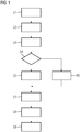
- Fig, 1 shows an exemplary flowchart of an embodiment of a method according to the invention.
- a step S1 two-dimensional projection images of a region of interest of a patient are acquired using an x-ray imaging device and multiple different projection geometries.
- the projection images may be acquired in the scope of an intervention to be performed on the patient, for example an orthopaedic surgery.
- Metal objects may thus be present in the region of interest and/or outside the region of interest in the cone-beam of at least some of the projection geometries.
- Such metal objects may, for example, comprise metal objects, for example medical instruments and/or implants.
- metal objects may cause artefacts during the reconstruction process, such that the method proposed here performs a metal artefact reduction (MAR) process to reduce these artefacts and improve the quality of the resulting x-ray image.
- MAR metal artefact reduction
- the current invention in this context, concerns the segmentation of metal objects, i.e. the detection in the projection domain, that is, in the projection images, such that in a later step corrective measures may be applied, for example the application of an inpainting algorithm.
- a trained artificial intelligence segmentation algorithm in this case a convolutional neural network, for example a U-net, is applied to each of the two-dimensional projection images, yielding first binary metal masks for each two-dimensional projection image. That is, a two-dimensional projection image is used as input data, wherein the output data is the first binary metal mask m(u, v), wherein m(u, v) equals "1" if the pixel (u, v) shows metal according to the algorithm, and m(u, v) equals "0" if the pixel (u, v) shows no metal.
- annotated projection images may be used as training data.
- segmentation results in the projection domain are checked in the following steps, yielding consistent second binary metal masks for each projection image and a more robust segmentation.
- a three-dimensional intermediate data set of a reconstruction region is reconstructed.
- the reconstruction region is larger than the region of interest, as further explained by fig. 2 .
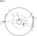
- Fig. 2 shows a schematic drawing regarding the projection geometries and regions used for reconstruction.
- a circular acquisition trajectory 1 is used, which is traversed by the x-ray source 2 having a focal spot 3 to realize different projection geometries, wherein one projection geometry is indicated by the cone beam 4 in the shown position of the focal spot 3.
- the x-ray detector 5 and its detection surface 6 are also indicated.
- an at least essentially cylinder-shaped region of interest 7 is imaged in each of the projection geometries possible along the acquisition trajectory 1. If, thus, a metal object 8 is located inside the region of interest 7, each projection image will show it. However, additionally, metal objects 9 located outside the region of interest 7 may also be imaged by some of the projection images, while other projection images will not contain them. However, also such metal objects 9 outside the region of interest 7 may induce metal artefacts into a reconstructed three-dimensional x-ray image.
- a reconstruction region 10 is defined such that all possible objects in the reconstruction region 10, which is larger than the region of interest 7, are imaged in at least a minimum number of the projection geometries used. For example, if fifty projection geometries are used and thus fifty two-dimensional projection images are acquired in step S1, the reconstruction region 10 may be chosen such that even objects, in particular metal objects, positioned at the radially outer border of the reconstruction region 10 are imaged in at least twenty of the projection geometries.
- each voxel contains two values, namely a contribution value and a metal value.
- the contribution value describes the number of projection geometries in which the voxel is traversed by a ray inside the cone beam 4 corresponding to a pixel, thus imaged.
- the metal value corresponds to the number of first binary metal masks showing metal for a pixel corresponding to the ray of the respective projection geometry traversing the voxel.
- the projection geometries are known, for each projection geometry, it is checked if a ray inside the cone beam 4 exists that traverses the voxel and to which pixel of the projection geometry it corresponds. If such a ray exists, the contribution value is increased by one. It is then referred to the pixel associated with the ray in the respective first binary metal mask. If this pixel is assigned as showing metal, the metal value is increased by one, else it is not increased.
- the ratio of the metal value to the contribution value may be understood as a probability that metal is actually present in the voxel.
- a threshold value is chosen and it is checked whether the metal value is larger than the threshold value.
- the threshold value is determined depending on the contribution value, wherein in the embodiment described here, a relative threshold value is defined using a threshold factor larger than 0.8 and smaller than 1, for example 0.95, such that the threshold value can be calculated as the threshold factor multiplied with the contribution value. If, for the currently examined voxel of the intermediate data set, the metal value actually exceeds the threshold value, in a step S5, the voxel is marked as containing metal, in particular in assigning a value of "1". In any other case, in a step S6, the voxel is assigned a value of "0" (containing no metal).
- the steps S4 and S5/S6 are repeated for each voxel in the reconstruction region 10, such that a three-dimensional binary metal mask results. The comparison with the threshold value thus allows a classification of the voxel as metal or no metal.
- the mask volume that is, the three-dimensional binary metal mask
- the maximum operator to yield second binary metal masks for each projection image.
- a step S8 the pixel values of the pixels marked as showing metal in the second binary metal mask may be replaced in each projection image, for example, by interpolation. That is, generally, in step S8, an inpainting algorithm may be used for metal artefact reduction (MAR), based on the segmentation described by the second binary metal masks.
- MAR metal artefact reduction
- the x-ray image may be reconstructed from the so-corrected projection images and, for example, displayed, stored or otherwise processed.
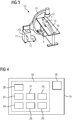
- Fig. 3 shows a mobile x-ray imaging device 11 which is especially suitable for the supervision/monitoring of a medical intervention on the patient 12, since the c-arm 13, to which in opposing positions the x-ray source 2 and the x-ray detector 5 are mounted, allows for multiple projection geometries with respect to the patient 12, who is placed on a patient table 14.
- the mobile x-ray imaging device 11 may be removed during surgical processes, or at least the c-arm 13 may be moved into a position where it does not impede the surgical process.
- the c-arm 13 may also be rotated around the region of interest 7 to acquire projection images in different projection geometries, in this case projection angles.
- control device 15 of the x-ray imaging device 11 in this case also forms a computing device 16 configured to perform the method according to the invention, the x-ray imaging device 11 also forms an evaluation device 17 according to the invention.
- Fig. 4 illustrates the functional structure of the control device 15.
- the control device 15 may comprise at least one processor 18, wherein, for example, in this case a controller and an image processor may be used as processors.
- the control device 15 further comprises at least one storage means 19.
- the control device 15 comprises, as in principle known, first of all an acquisition control unit 20, which may, for example, control the acquisition of the two-dimensional projection images S1.
- the control device 15 additionally comprises a segmentation unit 21 for performing the step S2, a reconstruction unit 22 for performing the step S3, a comparison unit 23 for performing the steps S4 to S6, and a determination unit 24 for performing the step S7.
- an MAR unit 25 for performing the step S8 and a back-projection unit 26 for reconstructing the three-dimensional x-ray images in step S9 may be provided.
Landscapes
- Engineering & Computer Science (AREA)
- Physics & Mathematics (AREA)
- General Physics & Mathematics (AREA)
- Theoretical Computer Science (AREA)
- Computer Vision & Pattern Recognition (AREA)
- General Health & Medical Sciences (AREA)
- Health & Medical Sciences (AREA)
- Medical Informatics (AREA)
- Nuclear Medicine, Radiotherapy & Molecular Imaging (AREA)
- Radiology & Medical Imaging (AREA)
- Quality & Reliability (AREA)
- Apparatus For Radiation Diagnosis (AREA)
- Image Processing (AREA)
Abstract
Description
- The invention concerns a computer-implemented method for segmenting metal objects in multiple two-dimensional projection images acquired using different projection geometries, each projection image showing a region of interest, wherein a three-dimensional x-ray image is reconstructed from the two-dimensional projection images in the region of interest. The invention also concerns an evaluation device, a computer program and an electronically readable storage medium.
- In x-ray imaging, it is known to reconstruct higher-dimensional images from lower-dimensional projection images acquired using an x-ray imaging device and different projection geometries. For example, filtered back-projection (FBP) or algebraic reconstruction may be used to reconstruct a three-dimensional x-ray image from two-dimensional projection images. The projection images can, for example, be acquired using a computed tomography device, wherein, usually, the x-ray source and the optionally an x-ray detector are rotated inside a gantry, or using a c-arm device, wherein the x-ray source and the x-ray detector are mounted to the c-arm at opposite ends. During acquisition of the projection images, the projection geometry may be changed according to the acquisition trajectory, which is often chosen circular, such that the projection geometry can be described by a projection angle.
- In computed tomography and other applications, in which three-dimensional x-ray images are reconstructed from two-dimensional projection images, metal artefacts are a major problem. This is particularly true for interoperative acquisitions, for example x-ray images acquired during orthopaedic surgeries. Such metal artefacts are caused by metal objects/metal structures in the region of interest to be imaged, but also by metal objects outside the region of interest, which only appear in a part of the projection images having respective projection geometries in which x-rays traverse the metal object. To reduce these well-known metal artefacts, different approaches have been described. These approaches are often headlined metal artefact reduction (MAR) approaches and aim at improving the quality of the reconstructed volume, that is, the x-ray image.
- In most of these approaches, in particular in flat-panel cone beam computed tomography, the location of the metal objects are estimated, the respective image values are removed from the projection images or the reconstructed three-dimensional x-ray image, and an interpolation process is performed, which is often called "inpainting". The segmentation, that is, the location of the metal object, is usually performed using an initial three-dimensional reconstruction, which thus still contains the metal artefacts. Voxels showing metal may, for example, be determined by thresholding and/or other volume processing algorithms. After the metal objects have been segmented, the three-dimensional segmentation is forward projected using the projection geometries to yield binary metal masks describing pixels which show the segmented metal objects. For these pixels, interpolations may be performed.
- As an example, an algorithm called normalized metal artefact reduction (NMAR) is disclosed in an article by Esther Meyer et al, "Normalized metal artefact reduction (NMAR) in computed tomography", Medical Physics 37 (10), 2010, pages 5482-5493. Here, metal is also segmented in the image domain, that is, the preliminary three-dimensional reconstruction using the original projection images. The segmentation is forward projected to identify the metal traces in the original projection. However, before interpolation, the projections are normalized based on a 3D forward projection of a prior image. After interpolation, the original raw data are denormalized again. This article also discusses some possible inpainting methods.
- In another conceivable approach, it may be possible to segment metal objects already in the projection domain, that is, the original two-dimensional projection images. For example, artificial intelligence segmentation algorithms might be used to segment metal and calculate binary metal masks containing the information whether a pixel contains metal or not. However, as internal studies of the applicant have shown, this approach does not yield robustness and reliability as expected.
- Algorithms working on a preliminary reconstruction data set of the region of interest, that is, the three-dimensional area which is captured in all two-dimensional projection images, has two major problems to deal with. For one thing, the metal object which causes the metal artefacts may be located outside the region of interest, that is, the area which can be (reliably) reconstructed. Additionally, the metal objects produce artefacts in the preliminary reconstruction which can be poorly segmented using simple thresholds. While, for the second problem, approaches like automatic threshold determination and/or frequency split methods have been proposed, solutions regarding the first problem are not known yet.
- It is an object of the current invention to provide a robust, reliable and consistent segmentation of metal objects shown in projection images, which also allows localisation of metal objects outside the region of interest to be reconstructed in the three-dimensional image.
- This object is achieved by providing a computer-implemented method according to
claim 1, an evaluation device according to claim 7, a computer program according toclaim 9 and an electronically readably storage medium according toclaim 10. Advantageous embodiments are described in the dependent claim A method according to the invention as initially described comprises the steps of: - using a trained artificial intelligence segmentation algorithm to calculate first binary metal masks for each projection image,
- reconstructing a three-dimensional intermediate data set of a reconstruction region, which is larger than the region of interest, by determining, for each voxel of the intermediate data set, as a metal value the number of first binary metal masks showing metal in a pixel associated with a ray crossing the voxel,
- determining a three-dimensional binary metal mask, in which a voxel shows metal when the metal value is larger than a threshold value and no metal in all other cases,
- determining second binary metal masks for each projection image by forward projecting the three-dimensional binary metal mask using the respective projection geometries.
- The invention thus proposes to use an artificial intelligence segmentation algorithm in the projection domain, which is also able to locate metal objects only captured by some of the two-dimensional projection images, but additionally proposes to add another mechanism increasing robustness and consistency of the segmentation of the metal compared to artificial intelligence segmentation alone or the current threshold-based segmentation in the image domain. The segmentation of the current invention is also obtained outside of the region of interest (ROI). By back-projecting the x-ray projection-based segmentation, that is, the first binary metal masks, thresholding and maximum projection of the threshold values, consistent second binary metal masks using all projection images are calculated. Thus, a more robust metal artefact reduction can be provided reducing the number of artefacts.
- In particular, the second binary metal masks may be used for metal artefact reduction in the tree-dimensional x-ray image, in particular using an inpainting algorithm for pixels showing a metal object according to the second binary metal masks. As the consistent second binary metal masks include the information which pixels of a projection image show metal, areas in the projection images showing metal objects or metal structures can be identified and interpolation approaches as known in the state of the art may be applied.
- In a preparatory step, the two-dimensional x-ray projection images are acquired using an x-ray imaging device, for example a computed tomography device or a c-arm x-ray imaging device. For example, a circular acquisition trajectory may be used. Generally, each two-dimensional projection image is associated with a projection geometry, in the case of a circular acquisition trajectory for example a projection angle. The projection images may preferably be acquired in a cone-beam geometry.
- Due to their nature as projection images, it is possible that metal objects localized outside the region of interest to be imaged may be visible in a fraction of the projection images. By using the combination of an artificial intelligence approach in the projection domain and a consistency check in the three-dimensional image domain, extended to a larger reconstruction region than the region of interest, these metal objects lying outside the region of interest may also be reliably detected.
- In a first step, a trained artificial intelligence segmentation algorithm is used to calculate first binary metal masks for each projection image. For example, a convolutional neural network may be used, in particular a U-net. Suitable convolutional neural networks are, for example, described in an article by Olaf Ronneberger et al., "U-Net: Convolutional Networks for Biomedical Image Segmentation", arXiv: 1505.04597v1 [cs.CV], May 18, 2015.
- As training data, projection images in which the metal objects are already annotated may be used. It is possible to enlarge the size of the training data set by generating synthetic data and/or to split the training data into a training data set and a validation data set. The trained artificial intelligence segmentation algorithm is then applied to the two-dimensional projection images. The artificial intelligence segmentation algorithm uses one of the projection images as input data and yields a first binary metal mask for this projection image as output data. For example, for a given pixel, a binary metal mask may contain "1" if the pixel shows metal, and "0" if the pixel shows no metal. Generally, for each pixel, a binary metal mask contains the binary information if a pixel shows metal or not.
- The first binary metal masks, which are to be checked for consistency and improved to yield second binary metal masks, are then back-projected into a volume which is larger than the region of interest, namely the reconstruction region. This reconstruction is performed by counting, for each voxel of the resulting intermediate data set, how many pixels associated with a ray crossing the voxel (according to the respective, known projection geometry) and measured in a projection image are described as showing metal by the first binary metal mask.
- In a third step, for each voxel, this metal value may be compared with a threshold. If the metal value is larger than the threshold value, many or most of the relevant projection images consistently show that metal should be present at this position. However, lower metal values, in which voxels obtain contributions which are no metal, but lie on rays which also intersect metal objects, may be discarded, as well as singular segmentation errors in the projection domain.
- In a last step, the so-determined three-dimensional binary metal mask, for example assigning a value of "1" for voxels being part of metal object and "0" for voxels not part of a metal object, is then forward-projected in each of the projection geometries to yield second binary metal masks for each projection image. Of course, the forward projection uses a maximum operator and is thus a maximum projection to yield masks also only containing "1" and "0".
- In an especially preferred embodiment of the invention, for each voxel of the intermediate data-set, a contribution value describing the number of first binary metal masks having a pixel associated with a ray crossing the voxel may be additionally determined, wherein the threshold value for each voxel is chosen depending on the contribution value. That is, the back-projecting step regarding the first binary metal masks does not only count how many first binary metal masks show metal for voxel, but also, according to the projection geometries, how many first binary metal masks are associated with rays traversing the voxel and being measured in a pixel. (Rays in this sense are understood as emanating from the focal point and ending on the pixel position of the detector in the respective projection geometry, for example in the middle of an actual detector pixel). As the reconstruction region is larger than the region of interest, voxels in the outer areas of the reconstruction region are not captured by all projection images. It is thus advantageous to know how many projection geometries are able to contribute to a certain voxel of the intermediate dataset, such that the threshold value may be correspondingly chosen. In other words, the quotient of the metal value and the contribution value may be understood as a probability for having a metal object at the respective voxel.
- For example, the threshold value may be computed from the respective contribution value, preferably by using a threshold factor. In an advantageous embodiment, the threshold value is determined by multiplying a threshold factor larger than 0.8 and smaller than 1 with the contribution value. For example, the threshold factor may be chosen as 0.95. In this case, the threshold is a relative threshold. In other words, to distinguish between voxels belonging to a metal object and not belonging to a metal object in this embodiment, it is checked if a certain percentage, namely the threshold factor, of the projection geometries contributing to the voxel had first binary metal masks showing metal at the corresponding pixel.
- Alternatively, it is also conceivable, but less preferred, to use an absolute threshold offset, for example an integer to be subtracted from the contribution value to obtain the threshold value. The threshold offset, as well as the threshold factor, may depend on the minimum of the contribution value allowed by the choice of the reconstruction region.
- Regarding the choice of the reconstruction region, in an embodiment of the invention, the reconstruction region may be chosen such that a defined minimum of the contribution value over all voxels is complied with. That is, only voxels are reviewed which are imaged in at least a certain number of projection geometries, that is, the minimum number. In this manner, contribution values which are too low to provide meaningful information can be avoided.
- The invention also concerns an evaluation device, comprising a computing device configured to perform a method according to the invention. The computing device thus comprises:
- a segmentation unit using the trained artificial intelligence segmentation algorithm to calculate the first binary metal masks for each projection image,
- a reconstruction unit for determining the three-dimensional intermediate data set,
- a comparison unit for determining the three-dimensional binary metal mask, and
- a determination unit for determining the second binary metal masks.
- All remarks and advantages laid out above regarding the method also apply to the evaluation device according to the invention. In particular, the computing device of the evaluation device may comprise at least one processor, in particular at least one image processor, and at least one storage means. The at least one processor realizes the functional units laid out above, namely the segmentation unit, the reconstruction unit, the comparison unit and the determination unit. Additional units may be added regarding additional steps or sub-steps according to the described embodiments of the method according to the invention, which can also be transferred to advantageous embodiments of the evaluation device according to the invention. The processor may, for example, be or comprise an integrated circuit, a CPU, a GPU or the like.
- In an advantageous embodiment, the evaluation device is or is part of an x-ray imaging device and/or the computing device is a control device of the x-ray imaging device. In this manner, an x-ray imaging device, which is also used to acquire the two-dimensional projection images, may already be configured to evaluate the two-dimensional projection images regarding artefacts caused by metal objects. In this context, it is also advantageous if the x-ray imaging device, in particular its control device, is also configured to perform the metal artefact correction, for example by using an MAR unit.
- The x-ray imaging device may, for example, be a computed tomography device, but is preferably a c-arm x-ray imaging device, having a c-arm with opposing ends to which an x-ray source and an x-ray detector, in particular a flat-panel detector, are mounted. X-ray imaging devices having a c-arm can advantageously be used to provide interoperative imaging, for example for surveying the surgery performed on a patient. Metal objects may, in particular in this context, comprise medical instruments and/or medical implants.
- A computer program according to the invention may, for example, be directly loaded into a storage means of a computing device, in particular a control device of an x-ray imaging device, and comprises program means to perform the steps of a method according to the invention if the computer program is executed in the computing device. The computer program may be stored on an electronically readably storage medium, which thus comprises electronically readable control information stored thereon. The control information comprises at least a computer program according to the invention and is configured such that the control information executes a method according to the invention when the storage medium is used in a computing device, in particular a control device of an x-ray imaging device. The electronically readably storage medium according to the invention may preferably be a non-transient medium, in particular a CD-ROM.
- Further details and advantages of the current invention become apparent from the following detailed description of preferred embodiments taken in conjunction with the drawings, in which:
- Fig. 1
- shows a flowchart of an embodiment of a method according to the invention,
- Fig. 2
- is a drawing showing a projection geometry and regions used for reconstruction,
- Fig. 3
- is a schematic drawing of an x-ray imaging device, and
- Fig. 4
- shows the functional structure of the control device of the x-ray imaging device of
fig. 2 . -
Fig, 1 shows an exemplary flowchart of an embodiment of a method according to the invention. - In a step S1, two-dimensional projection images of a region of interest of a patient are acquired using an x-ray imaging device and multiple different projection geometries. In particular, the projection images may be acquired in the scope of an intervention to be performed on the patient, for example an orthopaedic surgery. Metal objects may thus be present in the region of interest and/or outside the region of interest in the cone-beam of at least some of the projection geometries. Such metal objects may, for example, comprise metal objects, for example medical instruments and/or implants.
- As a three-dimensional x-ray image of the region of interest is to be reconstructed from the projection images, such metal objects may cause artefacts during the reconstruction process, such that the method proposed here performs a metal artefact reduction (MAR) process to reduce these artefacts and improve the quality of the resulting x-ray image. The current invention, in this context, concerns the segmentation of metal objects, i.e. the detection in the projection domain, that is, in the projection images, such that in a later step corrective measures may be applied, for example the application of an inpainting algorithm.
- To segment and thus localize the metal objects in the two-dimensional projection images acquired in step S1, in a step S2, a trained artificial intelligence segmentation algorithm, in this case a convolutional neural network, for example a U-net, is applied to each of the two-dimensional projection images, yielding first binary metal masks for each two-dimensional projection image. That is, a two-dimensional projection image is used as input data, wherein the output data is the first binary metal mask m(u, v), wherein m(u, v) equals "1" if the pixel (u, v) shows metal according to the algorithm, and m(u, v) equals "0" if the pixel (u, v) shows no metal. As training data, annotated projection images may be used.
- As it has been observed that artificial intelligence algorithms may not yield optimal, in particular consistent, results and wrong segmentations may occur, the segmentation results in the projection domain are checked in the following steps, yielding consistent second binary metal masks for each projection image and a more robust segmentation.
- In a step S3, a three-dimensional intermediate data set of a reconstruction region is reconstructed. The reconstruction region is larger than the region of interest, as further explained by
fig. 2 . -
Fig. 2 shows a schematic drawing regarding the projection geometries and regions used for reconstruction. In this case, acircular acquisition trajectory 1 is used, which is traversed by thex-ray source 2 having afocal spot 3 to realize different projection geometries, wherein one projection geometry is indicated by the cone beam 4 in the shown position of thefocal spot 3. Thex-ray detector 5 and itsdetection surface 6 are also indicated. - In this manner, an at least essentially cylinder-shaped region of interest 7 is imaged in each of the projection geometries possible along the
acquisition trajectory 1. If, thus, ametal object 8 is located inside the region of interest 7, each projection image will show it. However, additionally,metal objects 9 located outside the region of interest 7 may also be imaged by some of the projection images, while other projection images will not contain them. However, alsosuch metal objects 9 outside the region of interest 7 may induce metal artefacts into a reconstructed three-dimensional x-ray image. - Thus, in step S3 in
fig. 1 , areconstruction region 10 is defined such that all possible objects in thereconstruction region 10, which is larger than the region of interest 7, are imaged in at least a minimum number of the projection geometries used. For example, if fifty projection geometries are used and thus fifty two-dimensional projection images are acquired in step S1, thereconstruction region 10 may be chosen such that even objects, in particular metal objects, positioned at the radially outer border of thereconstruction region 10 are imaged in at least twenty of the projection geometries. - Using the so-defined
reconstruction region 10, the intermediate data set is reconstructed. In the intermediate data set, each voxel contains two values, namely a contribution value and a metal value. The contribution value describes the number of projection geometries in which the voxel is traversed by a ray inside the cone beam 4 corresponding to a pixel, thus imaged. The metal value corresponds to the number of first binary metal masks showing metal for a pixel corresponding to the ray of the respective projection geometry traversing the voxel. To determine these values, since the projection geometries are known, for each projection geometry, it is checked if a ray inside the cone beam 4 exists that traverses the voxel and to which pixel of the projection geometry it corresponds. If such a ray exists, the contribution value is increased by one. It is then referred to the pixel associated with the ray in the respective first binary metal mask. If this pixel is assigned as showing metal, the metal value is increased by one, else it is not increased. - The ratio of the metal value to the contribution value may be understood as a probability that metal is actually present in the voxel.
- In a step S4, a threshold value is chosen and it is checked whether the metal value is larger than the threshold value. The threshold value is determined depending on the contribution value, wherein in the embodiment described here, a relative threshold value is defined using a threshold factor larger than 0.8 and smaller than 1, for example 0.95, such that the threshold value can be calculated as the threshold factor multiplied with the contribution value. If, for the currently examined voxel of the intermediate data set, the metal value actually exceeds the threshold value, in a step S5, the voxel is marked as containing metal, in particular in assigning a value of "1". In any other case, in a step S6, the voxel is assigned a value of "0" (containing no metal). The steps S4 and S5/S6 are repeated for each voxel in the
reconstruction region 10, such that a three-dimensional binary metal mask results. The comparison with the threshold value thus allows a classification of the voxel as metal or no metal. - In a step S7, the mask volume, that is, the three-dimensional binary metal mask, is forward-projected for each projection geometry using the maximum operator to yield second binary metal masks for each projection image.
- Using these second binary metal masks, in a step S8, the pixel values of the pixels marked as showing metal in the second binary metal mask may be replaced in each projection image, for example, by interpolation. That is, generally, in step S8, an inpainting algorithm may be used for metal artefact reduction (MAR), based on the segmentation described by the second binary metal masks.
- Finally, in a step S9, the x-ray image may be reconstructed from the so-corrected projection images and, for example, displayed, stored or otherwise processed.
-
Fig. 3 shows a mobilex-ray imaging device 11 which is especially suitable for the supervision/monitoring of a medical intervention on thepatient 12, since the c-arm 13, to which in opposing positions thex-ray source 2 and thex-ray detector 5 are mounted, allows for multiple projection geometries with respect to thepatient 12, who is placed on a patient table 14. For example, the mobilex-ray imaging device 11 may be removed during surgical processes, or at least the c-arm 13 may be moved into a position where it does not impede the surgical process. - The c-
arm 13 may also be rotated around the region of interest 7 to acquire projection images in different projection geometries, in this case projection angles. - Since the
control device 15 of thex-ray imaging device 11 in this case also forms acomputing device 16 configured to perform the method according to the invention, thex-ray imaging device 11 also forms anevaluation device 17 according to the invention. -
Fig. 4 illustrates the functional structure of thecontrol device 15. Generally, thecontrol device 15 may comprise at least oneprocessor 18, wherein, for example, in this case a controller and an image processor may be used as processors. Thecontrol device 15 further comprises at least one storage means 19. - By the
processor 18, multiple functional units are realized. In this case, thecontrol device 15 comprises, as in principle known, first of all anacquisition control unit 20, which may, for example, control the acquisition of the two-dimensional projection images S1. Thecontrol device 15 additionally comprises asegmentation unit 21 for performing the step S2, areconstruction unit 22 for performing the step S3, acomparison unit 23 for performing the steps S4 to S6, and adetermination unit 24 for performing the step S7. Furthermore, anMAR unit 25 for performing the step S8 and a back-projection unit 26 for reconstructing the three-dimensional x-ray images in step S9 may be provided. - Although the present invention has been described in detail with reference to the preferred embodiment, the present invention is not limited by the disclosed examples from which the skilled person is able to derive other variations without departing from the scope of the invention.
Claims (10)
- Computer-implemented method for segmenting metal objects (8, 9) in multiple two-dimensional projection images acquired using different projection geometries, each projection image showing a region of interest (7), wherein a three-dimensional x-ray image is reconstructed from the two-dimensional projection images in the region of interest (7), the method comprising the steps of:- using a trained artificial intelligence segmentation algorithm to calculate first binary metal masks for each projection image,- reconstructing a three-dimensional intermediate data set of a reconstruction region (10), which is larger than the region of interest (7), by determining, for each voxel of the intermediate data set, as a metal value the number of first binary metal masks showing metal in a pixel associated with a ray crossing the voxel,- determining a three-dimensional binary metal mask, in which a voxel shows metal when the metal value is larger than a threshold value and no metal in all other cases,- determining second binary metal masks for each projection image by forward projecting the three-dimensional binary metal mask using the respective projection geometries.
- Method according to claim 1, wherein the second binary metal masks are used for metal artefact reduction in the three-dimensional x-ray image, in particular using an inpainting algorithm for pixels showing a metal object (8, 9) according to the second binary metal mask.
- Method according to claim 1 or 2, wherein, for each voxel of the intermediate data set, a contribution value describing the number of first binary metal masks having a pixel associated with a ray crossing the voxel is additionally determined, wherein the threshold value for each voxel is chosen depending on the contribution value.
- Method according to claim 3, wherein the threshold value is determined by multiplying a threshold factor larger than 0.8 and smaller than one with the contribution value.
- Method according to claim 3 or 4, wherein the reconstruction region is chosen such that a defined minimum of the contribution value over all voxels is complied with.
- Method according to one of the preceding claims, characterized in that a convolutional neural network, in particular a u-net, is used as artificial intelligence segmentation algorithm.
- Evaluation device (17), comprising a computing device (16) configured to perform a method according to one of the preceding claims, the computing device (16) comprising:- a segmentation unit (21) using the trained artificial intelligence segmentation algorithm to calculate the first binary metal masks for each projection image,- a reconstruction unit (22) for determining the three-dimensional intermediate data set- a comparison unit (23) for determining the three-dimensional binary metal mask, and- a determination unit (24) for determining the second binary metal masks.
- Evaluation device according to claim 7, wherein the evaluation device (17) is or is part of an x-ray imaging device (11) and/or the computing device (16) is a control device (15) of the x-ray imaging device (11).
- Computer program, which performs the steps of a method according to any of the claims 1 to 7 when the computer program is executed on a computing device (16).
- Electronically readable storage medium, on which a computer program according to claim 9 is stored.
Priority Applications (3)
| Application Number | Priority Date | Filing Date | Title |
|---|---|---|---|
| EP19155505.1A EP3693921B1 (en) | 2019-02-05 | 2019-02-05 | Method for segmenting metal objects in projection images, evaluation device, computer program and electronically readable storage medium |
| CN202010079569.7A CN111524200B (en) | 2019-02-05 | 2020-02-04 | Method, apparatus and medium for segmenting a metal object in a projection image |
| US16/782,359 US11321833B2 (en) | 2019-02-05 | 2020-02-05 | Segmenting metal objects in projection images |
Applications Claiming Priority (1)
| Application Number | Priority Date | Filing Date | Title |
|---|---|---|---|
| EP19155505.1A EP3693921B1 (en) | 2019-02-05 | 2019-02-05 | Method for segmenting metal objects in projection images, evaluation device, computer program and electronically readable storage medium |
Publications (2)
| Publication Number | Publication Date |
|---|---|
| EP3693921A1 true EP3693921A1 (en) | 2020-08-12 |
| EP3693921B1 EP3693921B1 (en) | 2022-04-20 |
Family
ID=65363058
Family Applications (1)
| Application Number | Title | Priority Date | Filing Date |
|---|---|---|---|
| EP19155505.1A Active EP3693921B1 (en) | 2019-02-05 | 2019-02-05 | Method for segmenting metal objects in projection images, evaluation device, computer program and electronically readable storage medium |
Country Status (3)
| Country | Link |
|---|---|
| US (1) | US11321833B2 (en) |
| EP (1) | EP3693921B1 (en) |
| CN (1) | CN111524200B (en) |
Families Citing this family (9)
| Publication number | Priority date | Publication date | Assignee | Title |
|---|---|---|---|---|
| EP3725227B1 (en) * | 2019-04-18 | 2022-12-14 | DENTSPLY SIRONA Inc. | Method of calibrating x-ray projection geometry in x-ray cone beam computed tomography |
| US11455755B2 (en) * | 2019-09-05 | 2022-09-27 | Siemens Medical Solutions Usa, Inc. | Methods and apparatus for neural network based image reconstruction |
| EP4080459A1 (en) * | 2021-04-23 | 2022-10-26 | Koninklijke Philips N.V. | Machine learning-based improvement in iterative image reconstruction |
| DE102021210356B4 (en) * | 2021-09-17 | 2024-05-16 | Siemens Healthineers Ag | Method and device for segmenting high-contrast objects in X-ray images |
| CN113962938B (en) * | 2021-09-27 | 2025-03-28 | 上海联影医疗科技股份有限公司 | Image segmentation method, device, computer equipment and readable storage medium |
| US12229927B2 (en) * | 2021-09-29 | 2025-02-18 | Siemens Healthineers International Ag | Metal artifact reduction in computed tomography |
| CN116416329A (en) * | 2021-12-31 | 2023-07-11 | 上海联影医疗科技股份有限公司 | Method and system for reconstruction of breast tomographic image |
| DE102022201003B3 (en) | 2022-01-31 | 2023-06-22 | Siemens Healthcare Gmbh | Method for defining an acquisition trajectory |
| US12475564B2 (en) | 2022-02-16 | 2025-11-18 | Proscia Inc. | Digital pathology artificial intelligence quality check |
Citations (1)
| Publication number | Priority date | Publication date | Assignee | Title |
|---|---|---|---|---|
| US20110081071A1 (en) * | 2009-10-06 | 2011-04-07 | Thomas Matthew Benson | Method and apparatus for reduction of metal artifacts in ct images |
Family Cites Families (26)
| Publication number | Priority date | Publication date | Assignee | Title |
|---|---|---|---|---|
| US6961404B2 (en) * | 2002-09-26 | 2005-11-01 | Eastman Kodak Company | Method and system for reconstructing an image from projection data acquired by a cone beam computed tomography system |
| DE102006014629A1 (en) | 2006-03-29 | 2007-10-04 | Siemens Ag | Truncation artifacts correction method in tomographic imaging, e.g. for medical diagnosis, determining extrapolated pixel outside projection image based on radiation attenuation of equivalent body placed in edge region of image |
| KR101018051B1 (en) * | 2008-10-23 | 2011-03-02 | 한국과학기술원 | How to reduce metal artifacts on x-rays |
| US8952333B2 (en) * | 2009-11-02 | 2015-02-10 | Virginia Tech Intellectual Properties, Inc. | Methods for improved single photon emission computed tomography using exact and stable region of interest reconstructions |
| CN101756707A (en) * | 2009-12-31 | 2010-06-30 | 苏州和君科技发展有限公司 | Method for carrying out scanning reconstruction on long target object by using Micro-CT imaging system |
| DE102010026374A1 (en) * | 2010-07-07 | 2012-01-12 | Siemens Aktiengesellschaft | Method for the reconstruction of a three-dimensional image data set and X-ray device |
| US8565502B2 (en) * | 2011-05-31 | 2013-10-22 | General Electric Company | Method and system for reconstruction of tomographic images |
| CN102842141B (en) * | 2012-07-03 | 2015-03-11 | 东南大学 | Rotary X-ray contrastographic picture iteration reconstruction method |
| DE102012212774B4 (en) * | 2012-07-20 | 2024-08-08 | Siemens Healthineers Ag | Procedure for correcting metal artifacts and X-ray equipment |
| CN103366389A (en) * | 2013-04-27 | 2013-10-23 | 中国人民解放军北京军区总医院 | CT (computed tomography) image reconstruction method |
| EP2997545A1 (en) * | 2013-05-14 | 2016-03-23 | Koninklijke Philips N.V. | Artifact-reduction for x-ray image reconstruction using a geometry-matched coordinate grid |
| US9349198B2 (en) * | 2013-07-26 | 2016-05-24 | General Electric Company | Robust artifact reduction in image reconstruction |
| WO2016197303A1 (en) * | 2015-06-08 | 2016-12-15 | Microsoft Technology Licensing, Llc. | Image semantic segmentation |
| US10524756B2 (en) | 2015-08-27 | 2020-01-07 | Varian Medical Systems International | Methods and systems for image artifacts reduction |
| DE102015224356B4 (en) * | 2015-12-04 | 2017-09-14 | Siemens Healthcare Gmbh | Method for image support of a practitioner, X-ray device and computer program |
| CN107730455B (en) * | 2016-08-11 | 2023-06-20 | 通用电气公司 | Method and device for obtaining MAR images |
| US10395397B2 (en) * | 2016-11-14 | 2019-08-27 | Carestream Health, Inc. | Metal artifacts reduction for cone beam CT |
| DE102017200282B3 (en) * | 2017-01-10 | 2018-02-15 | Friedrich-Alexander-Universität Erlangen-Nürnberg | Method for reducing image artifacts |
| DE102017200930A1 (en) * | 2017-01-20 | 2018-07-26 | Siemens Healthcare Gmbh | Method for determining a three-dimensional image data record |
| CN107545551B (en) * | 2017-09-07 | 2018-11-16 | 广州华端科技有限公司 | The method for reconstructing and system of digital galactophore body layer composograph |
| US20210065414A1 (en) * | 2017-09-08 | 2021-03-04 | The General Hospital Corporation | System and method for sinogram sparsified metal artifact reduction |
| US10789738B2 (en) * | 2017-11-03 | 2020-09-29 | The University Of Chicago | Method and apparatus to reduce artifacts in a computed-tomography (CT) image by iterative reconstruction (IR) using a cost function with a de-emphasis operator |
| CN107871319B (en) * | 2017-11-21 | 2021-09-17 | 上海联影医疗科技股份有限公司 | Method and device for detecting beam limiter area, X-ray system and storage medium |
| CN111656405A (en) * | 2018-01-26 | 2020-09-11 | 皇家飞利浦有限公司 | Using Deep Learning to Reduce Metal Artifacts |
| WO2019173452A1 (en) * | 2018-03-07 | 2019-09-12 | Rensselaer Polytechnic Institute | Deep neural network for ct metal artifact reduction |
| US20200151921A1 (en) * | 2018-11-14 | 2020-05-14 | Carestream Dental Llc | Methods for metal artifact reduction in cone beam reconstruction |
-
2019
- 2019-02-05 EP EP19155505.1A patent/EP3693921B1/en active Active
-
2020
- 2020-02-04 CN CN202010079569.7A patent/CN111524200B/en active Active
- 2020-02-05 US US16/782,359 patent/US11321833B2/en active Active
Patent Citations (1)
| Publication number | Priority date | Publication date | Assignee | Title |
|---|---|---|---|---|
| US20110081071A1 (en) * | 2009-10-06 | 2011-04-07 | Thomas Matthew Benson | Method and apparatus for reduction of metal artifacts in ct images |
Non-Patent Citations (4)
| Title |
|---|
| ESTHER MEYER ET AL.: "Normalized metal artefact reduction (NMAR) in computed tomography", MEDICAL PHYSICS, vol. 37, no. 10, 2010, pages 5482 - 5493 |
| GJESTEBY LARS ET AL: "Metal Artifact Reduction in CT: Where Are We After Four Decades?", IEEE ACCESS, vol. 4, 13 September 2016 (2016-09-13), pages 5826 - 5849, XP011624799, DOI: 10.1109/ACCESS.2016.2608621 * |
| OLAF RONNEBERGER ET AL.: "U-Net: Convolutional Networks for Biomedical Image Segmentation", ARXIV: 1505.04597VL [CS.CV, 18 May 2015 (2015-05-18) |
| VELDKAMP WOUTER ET AL: "Development and validation of segmentation and interpolation techniques in sinograms for metal artifact suppression in CT", MEDICAL PHYSICS, AIP, MELVILLE, NY, US, vol. 37, no. 2, 20 January 2010 (2010-01-20), pages 620 - 628, XP012135586, ISSN: 0094-2405, DOI: 10.1118/1.3276777 * |
Also Published As
| Publication number | Publication date |
|---|---|
| EP3693921B1 (en) | 2022-04-20 |
| US11321833B2 (en) | 2022-05-03 |
| US20200250820A1 (en) | 2020-08-06 |
| CN111524200A (en) | 2020-08-11 |
| CN111524200B (en) | 2024-03-22 |
Similar Documents
| Publication | Publication Date | Title |
|---|---|---|
| US11321833B2 (en) | Segmenting metal objects in projection images | |
| EP2102819B1 (en) | Apparatus, method and computer program for producing a corrected image of a region of interest from acquired projection data | |
| US9554766B2 (en) | Reducing artifacts in an image data set and X-ray device | |
| CN113226184B (en) | Methods for reducing metal artifacts in X-ray dental volume tomography | |
| US7957574B2 (en) | Methods and apparatus for generating a risk metric for soft plaque in vessels | |
| US10521934B2 (en) | Method, X-ray unit and computer program product for determining a three-dimensional image data set | |
| US7702141B2 (en) | Method for quantifying an object in a larger structure using a reconstructed image | |
| CN106683144A (en) | Image iteration reconstruction method and device | |
| CN113226181B (en) | Method for calibrating X-ray projection geometry in X-ray cone beam computed tomography | |
| CN112396700B (en) | Computer-implemented method and medium for deriving 3D image data of a reconstructed volume | |
| US20120237115A1 (en) | Method for acquiring a 3D image dataset freed of traces of a metal object | |
| US20250173844A1 (en) | Metal artifact reduction in computed tomography | |
| CN117522747A (en) | A metal artifact correction method and system for CT images | |
| US10932743B2 (en) | Determining image values in marked pixels of at least one projection image | |
| Agrawal et al. | Deep learning based projection domain metal segmentation for metal artifact reduction in cone beam computed tomography | |
| EP4026102B1 (en) | Confidence map for neural network based limited angle artifact reduction in cone beam ct | |
| US7983463B2 (en) | Methods and apparatus for suppressing tagging material in prepless CT colonography | |
| EP4160539B1 (en) | Adaptive auto-segmentation in computed tomography | |
| KR20200021156A (en) | High Quality Four Dimensional Cone Beam Computerized Tomography System Using Prior Image | |
| CN115552471A (en) | Suppression of Motion Artifacts in Computed Tomography Imaging | |
| Zhang et al. | Metal artifact reduction based on the combined prior image | |
| US20050018889A1 (en) | Systems and methods for filtering images | |
| US20060251313A1 (en) | Method of producing a cross-sectional image | |
| JP2010507438A (en) | Improved segmentation | |
| JP7743564B2 (en) | Method for generating a volume model of an object to be examined, control device, X-ray device, computer program and electronically readable data medium |
Legal Events
| Date | Code | Title | Description |
|---|---|---|---|
| PUAI | Public reference made under article 153(3) epc to a published international application that has entered the european phase |
Free format text: ORIGINAL CODE: 0009012 |
|
| STAA | Information on the status of an ep patent application or granted ep patent |
Free format text: STATUS: THE APPLICATION HAS BEEN PUBLISHED |
|
| AK | Designated contracting states |
Kind code of ref document: A1 Designated state(s): AL AT BE BG CH CY CZ DE DK EE ES FI FR GB GR HR HU IE IS IT LI LT LU LV MC MK MT NL NO PL PT RO RS SE SI SK SM TR |
|
| AX | Request for extension of the european patent |
Extension state: BA ME |
|
| STAA | Information on the status of an ep patent application or granted ep patent |
Free format text: STATUS: REQUEST FOR EXAMINATION WAS MADE |
|
| 17P | Request for examination filed |
Effective date: 20200819 |
|
| RBV | Designated contracting states (corrected) |
Designated state(s): AL AT BE BG CH CY CZ DE DK EE ES FI FR GB GR HR HU IE IS IT LI LT LU LV MC MK MT NL NO PL PT RO RS SE SI SK SM TR |
|
| RAP1 | Party data changed (applicant data changed or rights of an application transferred) |
Owner name: SIEMENS HEALTHCARE GMBH |
|
| GRAP | Despatch of communication of intention to grant a patent |
Free format text: ORIGINAL CODE: EPIDOSNIGR1 |
|
| STAA | Information on the status of an ep patent application or granted ep patent |
Free format text: STATUS: GRANT OF PATENT IS INTENDED |
|
| INTG | Intention to grant announced |
Effective date: 20211210 |
|
| GRAS | Grant fee paid |
Free format text: ORIGINAL CODE: EPIDOSNIGR3 |
|
| GRAA | (expected) grant |
Free format text: ORIGINAL CODE: 0009210 |
|
| STAA | Information on the status of an ep patent application or granted ep patent |
Free format text: STATUS: THE PATENT HAS BEEN GRANTED |
|
| AK | Designated contracting states |
Kind code of ref document: B1 Designated state(s): AL AT BE BG CH CY CZ DE DK EE ES FI FR GB GR HR HU IE IS IT LI LT LU LV MC MK MT NL NO PL PT RO RS SE SI SK SM TR |
|
| REG | Reference to a national code |
Ref country code: GB Ref legal event code: FG4D |
|
| REG | Reference to a national code |
Ref country code: CH Ref legal event code: EP |
|
| REG | Reference to a national code |
Ref country code: DE Ref legal event code: R096 Ref document number: 602019013792 Country of ref document: DE |
|
| REG | Reference to a national code |
Ref country code: IE Ref legal event code: FG4D |
|
| REG | Reference to a national code |
Ref country code: AT Ref legal event code: REF Ref document number: 1485786 Country of ref document: AT Kind code of ref document: T Effective date: 20220515 |
|
| REG | Reference to a national code |
Ref country code: LT Ref legal event code: MG9D |
|
| REG | Reference to a national code |
Ref country code: NL Ref legal event code: MP Effective date: 20220420 |
|
| REG | Reference to a national code |
Ref country code: AT Ref legal event code: MK05 Ref document number: 1485786 Country of ref document: AT Kind code of ref document: T Effective date: 20220420 |
|
| PG25 | Lapsed in a contracting state [announced via postgrant information from national office to epo] |
Ref country code: NL Free format text: LAPSE BECAUSE OF FAILURE TO SUBMIT A TRANSLATION OF THE DESCRIPTION OR TO PAY THE FEE WITHIN THE PRESCRIBED TIME-LIMIT Effective date: 20220420 |
|
| PG25 | Lapsed in a contracting state [announced via postgrant information from national office to epo] |
Ref country code: SE Free format text: LAPSE BECAUSE OF FAILURE TO SUBMIT A TRANSLATION OF THE DESCRIPTION OR TO PAY THE FEE WITHIN THE PRESCRIBED TIME-LIMIT Effective date: 20220420 Ref country code: PT Free format text: LAPSE BECAUSE OF FAILURE TO SUBMIT A TRANSLATION OF THE DESCRIPTION OR TO PAY THE FEE WITHIN THE PRESCRIBED TIME-LIMIT Effective date: 20220822 Ref country code: NO Free format text: LAPSE BECAUSE OF FAILURE TO SUBMIT A TRANSLATION OF THE DESCRIPTION OR TO PAY THE FEE WITHIN THE PRESCRIBED TIME-LIMIT Effective date: 20220720 Ref country code: LT Free format text: LAPSE BECAUSE OF FAILURE TO SUBMIT A TRANSLATION OF THE DESCRIPTION OR TO PAY THE FEE WITHIN THE PRESCRIBED TIME-LIMIT Effective date: 20220420 Ref country code: HR Free format text: LAPSE BECAUSE OF FAILURE TO SUBMIT A TRANSLATION OF THE DESCRIPTION OR TO PAY THE FEE WITHIN THE PRESCRIBED TIME-LIMIT Effective date: 20220420 Ref country code: GR Free format text: LAPSE BECAUSE OF FAILURE TO SUBMIT A TRANSLATION OF THE DESCRIPTION OR TO PAY THE FEE WITHIN THE PRESCRIBED TIME-LIMIT Effective date: 20220721 Ref country code: FI Free format text: LAPSE BECAUSE OF FAILURE TO SUBMIT A TRANSLATION OF THE DESCRIPTION OR TO PAY THE FEE WITHIN THE PRESCRIBED TIME-LIMIT Effective date: 20220420 Ref country code: ES Free format text: LAPSE BECAUSE OF FAILURE TO SUBMIT A TRANSLATION OF THE DESCRIPTION OR TO PAY THE FEE WITHIN THE PRESCRIBED TIME-LIMIT Effective date: 20220420 Ref country code: BG Free format text: LAPSE BECAUSE OF FAILURE TO SUBMIT A TRANSLATION OF THE DESCRIPTION OR TO PAY THE FEE WITHIN THE PRESCRIBED TIME-LIMIT Effective date: 20220720 Ref country code: AT Free format text: LAPSE BECAUSE OF FAILURE TO SUBMIT A TRANSLATION OF THE DESCRIPTION OR TO PAY THE FEE WITHIN THE PRESCRIBED TIME-LIMIT Effective date: 20220420 |
|
| PG25 | Lapsed in a contracting state [announced via postgrant information from national office to epo] |
Ref country code: RS Free format text: LAPSE BECAUSE OF FAILURE TO SUBMIT A TRANSLATION OF THE DESCRIPTION OR TO PAY THE FEE WITHIN THE PRESCRIBED TIME-LIMIT Effective date: 20220420 Ref country code: PL Free format text: LAPSE BECAUSE OF FAILURE TO SUBMIT A TRANSLATION OF THE DESCRIPTION OR TO PAY THE FEE WITHIN THE PRESCRIBED TIME-LIMIT Effective date: 20220420 Ref country code: LV Free format text: LAPSE BECAUSE OF FAILURE TO SUBMIT A TRANSLATION OF THE DESCRIPTION OR TO PAY THE FEE WITHIN THE PRESCRIBED TIME-LIMIT Effective date: 20220420 Ref country code: IS Free format text: LAPSE BECAUSE OF FAILURE TO SUBMIT A TRANSLATION OF THE DESCRIPTION OR TO PAY THE FEE WITHIN THE PRESCRIBED TIME-LIMIT Effective date: 20220820 |
|
| REG | Reference to a national code |
Ref country code: DE Ref legal event code: R097 Ref document number: 602019013792 Country of ref document: DE |
|
| PG25 | Lapsed in a contracting state [announced via postgrant information from national office to epo] |
Ref country code: SM Free format text: LAPSE BECAUSE OF FAILURE TO SUBMIT A TRANSLATION OF THE DESCRIPTION OR TO PAY THE FEE WITHIN THE PRESCRIBED TIME-LIMIT Effective date: 20220420 Ref country code: SK Free format text: LAPSE BECAUSE OF FAILURE TO SUBMIT A TRANSLATION OF THE DESCRIPTION OR TO PAY THE FEE WITHIN THE PRESCRIBED TIME-LIMIT Effective date: 20220420 Ref country code: RO Free format text: LAPSE BECAUSE OF FAILURE TO SUBMIT A TRANSLATION OF THE DESCRIPTION OR TO PAY THE FEE WITHIN THE PRESCRIBED TIME-LIMIT Effective date: 20220420 Ref country code: EE Free format text: LAPSE BECAUSE OF FAILURE TO SUBMIT A TRANSLATION OF THE DESCRIPTION OR TO PAY THE FEE WITHIN THE PRESCRIBED TIME-LIMIT Effective date: 20220420 Ref country code: DK Free format text: LAPSE BECAUSE OF FAILURE TO SUBMIT A TRANSLATION OF THE DESCRIPTION OR TO PAY THE FEE WITHIN THE PRESCRIBED TIME-LIMIT Effective date: 20220420 Ref country code: CZ Free format text: LAPSE BECAUSE OF FAILURE TO SUBMIT A TRANSLATION OF THE DESCRIPTION OR TO PAY THE FEE WITHIN THE PRESCRIBED TIME-LIMIT Effective date: 20220420 |
|
| PLBE | No opposition filed within time limit |
Free format text: ORIGINAL CODE: 0009261 |
|
| STAA | Information on the status of an ep patent application or granted ep patent |
Free format text: STATUS: NO OPPOSITION FILED WITHIN TIME LIMIT |
|
| 26N | No opposition filed |
Effective date: 20230123 |
|
| PG25 | Lapsed in a contracting state [announced via postgrant information from national office to epo] |
Ref country code: AL Free format text: LAPSE BECAUSE OF FAILURE TO SUBMIT A TRANSLATION OF THE DESCRIPTION OR TO PAY THE FEE WITHIN THE PRESCRIBED TIME-LIMIT Effective date: 20220420 |
|
| PG25 | Lapsed in a contracting state [announced via postgrant information from national office to epo] |
Ref country code: SI Free format text: LAPSE BECAUSE OF FAILURE TO SUBMIT A TRANSLATION OF THE DESCRIPTION OR TO PAY THE FEE WITHIN THE PRESCRIBED TIME-LIMIT Effective date: 20220420 |
|
| PG25 | Lapsed in a contracting state [announced via postgrant information from national office to epo] |
Ref country code: MC Free format text: LAPSE BECAUSE OF FAILURE TO SUBMIT A TRANSLATION OF THE DESCRIPTION OR TO PAY THE FEE WITHIN THE PRESCRIBED TIME-LIMIT Effective date: 20220420 |
|
| REG | Reference to a national code |
Ref country code: CH Ref legal event code: PL |
|
| REG | Reference to a national code |
Ref country code: BE Ref legal event code: MM Effective date: 20230228 |
|
| PG25 | Lapsed in a contracting state [announced via postgrant information from national office to epo] |
Ref country code: LU Free format text: LAPSE BECAUSE OF NON-PAYMENT OF DUE FEES Effective date: 20230205 Ref country code: LI Free format text: LAPSE BECAUSE OF NON-PAYMENT OF DUE FEES Effective date: 20230228 Ref country code: CH Free format text: LAPSE BECAUSE OF NON-PAYMENT OF DUE FEES Effective date: 20230228 |
|
| REG | Reference to a national code |
Ref country code: IE Ref legal event code: MM4A |
|
| PG25 | Lapsed in a contracting state [announced via postgrant information from national office to epo] |
Ref country code: IT Free format text: LAPSE BECAUSE OF FAILURE TO SUBMIT A TRANSLATION OF THE DESCRIPTION OR TO PAY THE FEE WITHIN THE PRESCRIBED TIME-LIMIT Effective date: 20220420 Ref country code: IE Free format text: LAPSE BECAUSE OF NON-PAYMENT OF DUE FEES Effective date: 20230205 |
|
| REG | Reference to a national code |
Ref country code: DE Ref legal event code: R081 Ref document number: 602019013792 Country of ref document: DE Owner name: SIEMENS HEALTHINEERS AG, DE Free format text: FORMER OWNER: SIEMENS HEALTHCARE GMBH, MUENCHEN, DE |
|
| PG25 | Lapsed in a contracting state [announced via postgrant information from national office to epo] |
Ref country code: BE Free format text: LAPSE BECAUSE OF NON-PAYMENT OF DUE FEES Effective date: 20230228 |
|
| PG25 | Lapsed in a contracting state [announced via postgrant information from national office to epo] |
Ref country code: BG Free format text: LAPSE BECAUSE OF FAILURE TO SUBMIT A TRANSLATION OF THE DESCRIPTION OR TO PAY THE FEE WITHIN THE PRESCRIBED TIME-LIMIT Effective date: 20220420 |
|
| PG25 | Lapsed in a contracting state [announced via postgrant information from national office to epo] |
Ref country code: BG Free format text: LAPSE BECAUSE OF FAILURE TO SUBMIT A TRANSLATION OF THE DESCRIPTION OR TO PAY THE FEE WITHIN THE PRESCRIBED TIME-LIMIT Effective date: 20220420 |
|
| PGFP | Annual fee paid to national office [announced via postgrant information from national office to epo] |
Ref country code: DE Payment date: 20250417 Year of fee payment: 7 |
|
| PG25 | Lapsed in a contracting state [announced via postgrant information from national office to epo] |
Ref country code: CY Free format text: LAPSE BECAUSE OF FAILURE TO SUBMIT A TRANSLATION OF THE DESCRIPTION OR TO PAY THE FEE WITHIN THE PRESCRIBED TIME-LIMIT; INVALID AB INITIO Effective date: 20190205 |
|
| PG25 | Lapsed in a contracting state [announced via postgrant information from national office to epo] |
Ref country code: HU Free format text: LAPSE BECAUSE OF FAILURE TO SUBMIT A TRANSLATION OF THE DESCRIPTION OR TO PAY THE FEE WITHIN THE PRESCRIBED TIME-LIMIT; INVALID AB INITIO Effective date: 20190205 |
|
| PG25 | Lapsed in a contracting state [announced via postgrant information from national office to epo] |
Ref country code: TR Free format text: LAPSE BECAUSE OF FAILURE TO SUBMIT A TRANSLATION OF THE DESCRIPTION OR TO PAY THE FEE WITHIN THE PRESCRIBED TIME-LIMIT Effective date: 20220420 |
|
| PGFP | Annual fee paid to national office [announced via postgrant information from national office to epo] |
Ref country code: GB Payment date: 20260310 Year of fee payment: 8 |
|
| PGFP | Annual fee paid to national office [announced via postgrant information from national office to epo] |
Ref country code: FR Payment date: 20260213 Year of fee payment: 8 |