EP3680667A1 - Highly selective and ultrasensitive cu2+ ion sensor - Google Patents
Highly selective and ultrasensitive cu2+ ion sensor Download PDFInfo
- Publication number
- EP3680667A1 EP3680667A1 EP19151227.6A EP19151227A EP3680667A1 EP 3680667 A1 EP3680667 A1 EP 3680667A1 EP 19151227 A EP19151227 A EP 19151227A EP 3680667 A1 EP3680667 A1 EP 3680667A1
- Authority
- EP
- European Patent Office
- Prior art keywords
- ions
- sensor
- copper
- sensor according
- ion
- Prior art date
- Legal status (The legal status is an assumption and is not a legal conclusion. Google has not performed a legal analysis and makes no representation as to the accuracy of the status listed.)
- Withdrawn
Links
- 239000010949 copper Substances 0.000 claims abstract description 122
- 229910052802 copper Inorganic materials 0.000 claims abstract description 113
- RYGMFSIKBFXOCR-UHFFFAOYSA-N Copper Chemical compound [Cu] RYGMFSIKBFXOCR-UHFFFAOYSA-N 0.000 claims abstract description 112
- 238000001514 detection method Methods 0.000 claims abstract description 94
- 239000012530 fluid Substances 0.000 claims abstract description 66
- 235000001014 amino acid Nutrition 0.000 claims abstract description 51
- 150000001413 amino acids Chemical group 0.000 claims abstract description 51
- PECYZEOJVXMISF-UHFFFAOYSA-N 3-aminoalanine Chemical compound [NH3+]CC(N)C([O-])=O PECYZEOJVXMISF-UHFFFAOYSA-N 0.000 claims abstract description 36
- CKLJMWTZIZZHCS-REOHCLBHSA-N L-aspartic acid Chemical compound OC(=O)[C@@H](N)CC(O)=O CKLJMWTZIZZHCS-REOHCLBHSA-N 0.000 claims abstract description 29
- 208000024827 Alzheimer disease Diseases 0.000 claims abstract description 28
- 208000037265 diseases, disorders, signs and symptoms Diseases 0.000 claims abstract description 16
- QNAYBMKLOCPYGJ-REOHCLBHSA-N L-alanine Chemical compound C[C@H](N)C(O)=O QNAYBMKLOCPYGJ-REOHCLBHSA-N 0.000 claims abstract description 15
- 235000004279 alanine Nutrition 0.000 claims abstract description 15
- 235000003704 aspartic acid Nutrition 0.000 claims abstract description 15
- OQFSQFPPLPISGP-UHFFFAOYSA-N beta-carboxyaspartic acid Natural products OC(=O)C(N)C(C(O)=O)C(O)=O OQFSQFPPLPISGP-UHFFFAOYSA-N 0.000 claims abstract description 15
- 201000010099 disease Diseases 0.000 claims abstract description 15
- HNDVDQJCIGZPNO-UHFFFAOYSA-N histidine Natural products OC(=O)C(N)CC1=CN=CN1 HNDVDQJCIGZPNO-UHFFFAOYSA-N 0.000 claims abstract description 15
- 230000002159 abnormal effect Effects 0.000 claims abstract description 12
- 125000000487 histidyl group Chemical group [H]N([H])C(C(=O)O*)C([H])([H])C1=C([H])N([H])C([H])=N1 0.000 claims abstract description 12
- 230000004060 metabolic process Effects 0.000 claims abstract description 9
- 238000012544 monitoring process Methods 0.000 claims abstract description 9
- 238000003745 diagnosis Methods 0.000 claims abstract description 7
- -1 Cu2+ ions Chemical class 0.000 claims description 160
- 125000005647 linker group Chemical group 0.000 claims description 53
- 239000012528 membrane Substances 0.000 claims description 41
- 229920005597 polymer membrane Polymers 0.000 claims description 34
- 238000000034 method Methods 0.000 claims description 30
- XLYOFNOQVPJJNP-UHFFFAOYSA-N water Substances O XLYOFNOQVPJJNP-UHFFFAOYSA-N 0.000 claims description 28
- 210000002700 urine Anatomy 0.000 claims description 25
- 239000007787 solid Substances 0.000 claims description 24
- 230000008859 change Effects 0.000 claims description 19
- BZTDTCNHAFUJOG-UHFFFAOYSA-N 6-carboxyfluorescein Chemical compound C12=CC=C(O)C=C2OC2=CC(O)=CC=C2C11OC(=O)C2=CC=C(C(=O)O)C=C21 BZTDTCNHAFUJOG-UHFFFAOYSA-N 0.000 claims description 13
- 208000002972 Hepatolenticular Degeneration Diseases 0.000 claims description 12
- 208000018839 Wilson disease Diseases 0.000 claims description 12
- 208000008948 Menkes Kinky Hair Syndrome Diseases 0.000 claims description 9
- 208000012583 Menkes disease Diseases 0.000 claims description 9
- 238000012360 testing method Methods 0.000 claims description 9
- 239000008139 complexing agent Substances 0.000 claims description 7
- 239000002202 Polyethylene glycol Substances 0.000 claims description 6
- 235000013305 food Nutrition 0.000 claims description 6
- 229920001223 polyethylene glycol Polymers 0.000 claims description 6
- 230000007613 environmental effect Effects 0.000 claims description 5
- 230000001172 regenerating effect Effects 0.000 claims description 2
- 150000002500 ions Chemical class 0.000 abstract description 15
- 229940024606 amino acid Drugs 0.000 description 45
- 239000011888 foil Substances 0.000 description 39
- JPVYNHNXODAKFH-UHFFFAOYSA-N Cu2+ Chemical compound [Cu+2] JPVYNHNXODAKFH-UHFFFAOYSA-N 0.000 description 34
- 239000000243 solution Substances 0.000 description 18
- 239000007987 MES buffer Substances 0.000 description 17
- VEQPNABPJHWNSG-UHFFFAOYSA-N Nickel(2+) Chemical compound [Ni+2] VEQPNABPJHWNSG-UHFFFAOYSA-N 0.000 description 15
- 108090000765 processed proteins & peptides Proteins 0.000 description 15
- 230000015572 biosynthetic process Effects 0.000 description 13
- 230000007423 decrease Effects 0.000 description 13
- 235000014304 histidine Nutrition 0.000 description 13
- PXHVJJICTQNCMI-UHFFFAOYSA-N nickel Chemical group [Ni] PXHVJJICTQNCMI-UHFFFAOYSA-N 0.000 description 13
- 238000004448 titration Methods 0.000 description 13
- 125000000524 functional group Chemical group 0.000 description 10
- YMWUJEATGCHHMB-UHFFFAOYSA-N Dichloromethane Chemical compound ClCCl YMWUJEATGCHHMB-UHFFFAOYSA-N 0.000 description 9
- LFQSCWFLJHTTHZ-UHFFFAOYSA-N Ethanol Chemical compound CCO LFQSCWFLJHTTHZ-UHFFFAOYSA-N 0.000 description 9
- 238000001218 confocal laser scanning microscopy Methods 0.000 description 9
- 238000010791 quenching Methods 0.000 description 8
- 230000000171 quenching effect Effects 0.000 description 8
- KCXVZYZYPLLWCC-UHFFFAOYSA-N EDTA Chemical compound OC(=O)CN(CC(O)=O)CCN(CC(O)=O)CC(O)=O KCXVZYZYPLLWCC-UHFFFAOYSA-N 0.000 description 7
- PIICEJLVQHRZGT-UHFFFAOYSA-N Ethylenediamine Chemical compound NCCN PIICEJLVQHRZGT-UHFFFAOYSA-N 0.000 description 7
- 210000001124 body fluid Anatomy 0.000 description 7
- 239000010839 body fluid Substances 0.000 description 7
- 125000003178 carboxy group Chemical group [H]OC(*)=O 0.000 description 7
- 238000010668 complexation reaction Methods 0.000 description 7
- 230000008878 coupling Effects 0.000 description 7
- 238000010168 coupling process Methods 0.000 description 7
- 238000005859 coupling reaction Methods 0.000 description 7
- 229960001484 edetic acid Drugs 0.000 description 7
- 238000005259 measurement Methods 0.000 description 7
- 229920000642 polymer Polymers 0.000 description 7
- HEMHJVSKTPXQMS-UHFFFAOYSA-M Sodium hydroxide Chemical compound [OH-].[Na+] HEMHJVSKTPXQMS-UHFFFAOYSA-M 0.000 description 6
- 239000012491 analyte Substances 0.000 description 6
- 239000003480 eluent Substances 0.000 description 6
- 238000002189 fluorescence spectrum Methods 0.000 description 6
- 239000007788 liquid Substances 0.000 description 6
- 239000002245 particle Substances 0.000 description 6
- 229920000139 polyethylene terephthalate Polymers 0.000 description 6
- 239000005020 polyethylene terephthalate Substances 0.000 description 6
- 230000008569 process Effects 0.000 description 6
- 102100023321 Ceruloplasmin Human genes 0.000 description 5
- DTQVDTLACAAQTR-UHFFFAOYSA-N Trifluoroacetic acid Chemical compound OC(=O)C(F)(F)F DTQVDTLACAAQTR-UHFFFAOYSA-N 0.000 description 5
- UCMIRNVEIXFBKS-UHFFFAOYSA-N beta-alanine Chemical compound NCCC(O)=O UCMIRNVEIXFBKS-UHFFFAOYSA-N 0.000 description 5
- ARUVKPQLZAKDPS-UHFFFAOYSA-L copper(II) sulfate Chemical compound [Cu+2].[O-][S+2]([O-])([O-])[O-] ARUVKPQLZAKDPS-UHFFFAOYSA-L 0.000 description 5
- 229910000366 copper(II) sulfate Inorganic materials 0.000 description 5
- 238000005530 etching Methods 0.000 description 5
- LGQLOGILCSXPEA-UHFFFAOYSA-L nickel sulfate Chemical compound [Ni+2].[O-]S([O-])(=O)=O LGQLOGILCSXPEA-UHFFFAOYSA-L 0.000 description 5
- 229910000363 nickel(II) sulfate Inorganic materials 0.000 description 5
- 239000011347 resin Substances 0.000 description 5
- 229920005989 resin Polymers 0.000 description 5
- 210000002966 serum Anatomy 0.000 description 5
- NWONKYPBYAMBJT-UHFFFAOYSA-L zinc sulfate Chemical compound [Zn+2].[O-]S([O-])(=O)=O NWONKYPBYAMBJT-UHFFFAOYSA-L 0.000 description 5
- 229910000368 zinc sulfate Inorganic materials 0.000 description 5
- 239000011686 zinc sulphate Substances 0.000 description 5
- LMDZBCPBFSXMTL-UHFFFAOYSA-N 1-ethyl-3-(3-dimethylaminopropyl)carbodiimide Chemical compound CCN=C=NCCCN(C)C LMDZBCPBFSXMTL-UHFFFAOYSA-N 0.000 description 4
- 108010075016 Ceruloplasmin Proteins 0.000 description 4
- 238000010521 absorption reaction Methods 0.000 description 4
- 125000002843 carboxylic acid group Chemical group 0.000 description 4
- 210000004027 cell Anatomy 0.000 description 4
- 238000003486 chemical etching Methods 0.000 description 4
- 229910001431 copper ion Inorganic materials 0.000 description 4
- 238000002474 experimental method Methods 0.000 description 4
- 229910052759 nickel Inorganic materials 0.000 description 4
- 238000005897 peptide coupling reaction Methods 0.000 description 4
- 238000000926 separation method Methods 0.000 description 4
- 238000001228 spectrum Methods 0.000 description 4
- PECYZEOJVXMISF-REOHCLBHSA-N 3-amino-L-alanine Chemical compound [NH3+]C[C@H](N)C([O-])=O PECYZEOJVXMISF-REOHCLBHSA-N 0.000 description 3
- WFDIJRYMOXRFFG-UHFFFAOYSA-N Acetic anhydride Chemical compound CC(=O)OC(C)=O WFDIJRYMOXRFFG-UHFFFAOYSA-N 0.000 description 3
- WEVYAHXRMPXWCK-UHFFFAOYSA-N Acetonitrile Chemical compound CC#N WEVYAHXRMPXWCK-UHFFFAOYSA-N 0.000 description 3
- DHMQDGOQFOQNFH-UHFFFAOYSA-N Glycine Chemical compound NCC(O)=O DHMQDGOQFOQNFH-UHFFFAOYSA-N 0.000 description 3
- OKKJLVBELUTLKV-UHFFFAOYSA-N Methanol Chemical compound OC OKKJLVBELUTLKV-UHFFFAOYSA-N 0.000 description 3
- JGFZNNIVVJXRND-UHFFFAOYSA-N N,N-Diisopropylethylamine (DIPEA) Chemical compound CCN(C(C)C)C(C)C JGFZNNIVVJXRND-UHFFFAOYSA-N 0.000 description 3
- 125000000218 acetic acid group Chemical group C(C)(=O)* 0.000 description 3
- 125000003277 amino group Chemical group 0.000 description 3
- DZHSAHHDTRWUTF-SIQRNXPUSA-N amyloid-beta polypeptide 42 Chemical compound C([C@@H](C(=O)N[C@@H](C)C(=O)N[C@@H](CCC(O)=O)C(=O)N[C@@H](CC(O)=O)C(=O)N[C@H](C(=O)NCC(=O)N[C@@H](CO)C(=O)N[C@@H](CC(N)=O)C(=O)N[C@@H](CCCCN)C(=O)NCC(=O)N[C@@H](C)C(=O)N[C@H](C(=O)N[C@@H]([C@@H](C)CC)C(=O)NCC(=O)N[C@@H](CC(C)C)C(=O)N[C@@H](CCSC)C(=O)N[C@@H](C(C)C)C(=O)NCC(=O)NCC(=O)N[C@@H](C(C)C)C(=O)N[C@@H](C(C)C)C(=O)N[C@@H]([C@@H](C)CC)C(=O)N[C@@H](C)C(O)=O)[C@@H](C)CC)C(C)C)NC(=O)[C@H](CC=1C=CC=CC=1)NC(=O)[C@@H](NC(=O)[C@H](CC(C)C)NC(=O)[C@H](CCCCN)NC(=O)[C@H](CCC(N)=O)NC(=O)[C@H](CC=1N=CNC=1)NC(=O)[C@H](CC=1N=CNC=1)NC(=O)[C@@H](NC(=O)[C@H](CCC(O)=O)NC(=O)[C@H](CC=1C=CC(O)=CC=1)NC(=O)CNC(=O)[C@H](CO)NC(=O)[C@H](CC(O)=O)NC(=O)[C@H](CC=1N=CNC=1)NC(=O)[C@H](CCCNC(N)=N)NC(=O)[C@H](CC=1C=CC=CC=1)NC(=O)[C@H](CCC(O)=O)NC(=O)[C@H](C)NC(=O)[C@@H](N)CC(O)=O)C(C)C)C(C)C)C1=CC=CC=C1 DZHSAHHDTRWUTF-SIQRNXPUSA-N 0.000 description 3
- 239000012490 blank solution Substances 0.000 description 3
- 150000001875 compounds Chemical class 0.000 description 3
- 239000008367 deionised water Substances 0.000 description 3
- 229910021641 deionized water Inorganic materials 0.000 description 3
- 238000013399 early diagnosis Methods 0.000 description 3
- 230000036541 health Effects 0.000 description 3
- 230000003993 interaction Effects 0.000 description 3
- 238000004949 mass spectrometry Methods 0.000 description 3
- 238000010647 peptide synthesis reaction Methods 0.000 description 3
- 102000004196 processed proteins & peptides Human genes 0.000 description 3
- 235000018102 proteins Nutrition 0.000 description 3
- 102000004169 proteins and genes Human genes 0.000 description 3
- 108090000623 proteins and genes Proteins 0.000 description 3
- 230000008929 regeneration Effects 0.000 description 3
- 238000011069 regeneration method Methods 0.000 description 3
- 239000007790 solid phase Substances 0.000 description 3
- 239000000126 substance Substances 0.000 description 3
- 230000032258 transport Effects 0.000 description 3
- 238000005406 washing Methods 0.000 description 3
- 239000003643 water by type Substances 0.000 description 3
- 208000037259 Amyloid Plaque Diseases 0.000 description 2
- 102000013455 Amyloid beta-Peptides Human genes 0.000 description 2
- 108010090849 Amyloid beta-Peptides Proteins 0.000 description 2
- 101710137189 Amyloid-beta A4 protein Proteins 0.000 description 2
- 101710151993 Amyloid-beta precursor protein Proteins 0.000 description 2
- 102100022704 Amyloid-beta precursor protein Human genes 0.000 description 2
- 108091006905 Human Serum Albumin Proteins 0.000 description 2
- 102000008100 Human Serum Albumin Human genes 0.000 description 2
- DCXYFEDJOCDNAF-REOHCLBHSA-N L-asparagine Chemical compound OC(=O)[C@@H](N)CC(N)=O DCXYFEDJOCDNAF-REOHCLBHSA-N 0.000 description 2
- WHUUTDBJXJRKMK-VKHMYHEASA-N L-glutamic acid Chemical compound OC(=O)[C@@H](N)CCC(O)=O WHUUTDBJXJRKMK-VKHMYHEASA-N 0.000 description 2
- AGPKZVBTJJNPAG-WHFBIAKZSA-N L-isoleucine Chemical compound CC[C@H](C)[C@H](N)C(O)=O AGPKZVBTJJNPAG-WHFBIAKZSA-N 0.000 description 2
- ROHFNLRQFUQHCH-YFKPBYRVSA-N L-leucine Chemical compound CC(C)C[C@H](N)C(O)=O ROHFNLRQFUQHCH-YFKPBYRVSA-N 0.000 description 2
- COLNVLDHVKWLRT-QMMMGPOBSA-N L-phenylalanine Chemical compound OC(=O)[C@@H](N)CC1=CC=CC=C1 COLNVLDHVKWLRT-QMMMGPOBSA-N 0.000 description 2
- OUYCCCASQSFEME-QMMMGPOBSA-N L-tyrosine Chemical compound OC(=O)[C@@H](N)CC1=CC=C(O)C=C1 OUYCCCASQSFEME-QMMMGPOBSA-N 0.000 description 2
- KDXKERNSBIXSRK-UHFFFAOYSA-N Lysine Natural products NCCCCC(N)C(O)=O KDXKERNSBIXSRK-UHFFFAOYSA-N 0.000 description 2
- ZMXDDKWLCZADIW-UHFFFAOYSA-N N,N-dimethylformamide Substances CN(C)C=O ZMXDDKWLCZADIW-UHFFFAOYSA-N 0.000 description 2
- 241000283973 Oryctolagus cuniculus Species 0.000 description 2
- KZSNJWFQEVHDMF-UHFFFAOYSA-N Valine Natural products CC(C)C(N)C(O)=O KZSNJWFQEVHDMF-UHFFFAOYSA-N 0.000 description 2
- 229940000635 beta-alanine Drugs 0.000 description 2
- 239000012482 calibration solution Substances 0.000 description 2
- 239000012876 carrier material Substances 0.000 description 2
- 239000001913 cellulose Substances 0.000 description 2
- 229920002678 cellulose Polymers 0.000 description 2
- 238000012512 characterization method Methods 0.000 description 2
- 238000006243 chemical reaction Methods 0.000 description 2
- HVYWMOMLDIMFJA-DPAQBDIFSA-N cholesterol Chemical compound C1C=C2C[C@@H](O)CC[C@]2(C)[C@@H]2[C@@H]1[C@@H]1CC[C@H]([C@H](C)CCCC(C)C)[C@@]1(C)CC2 HVYWMOMLDIMFJA-DPAQBDIFSA-N 0.000 description 2
- 239000011258 core-shell material Substances 0.000 description 2
- 230000002596 correlated effect Effects 0.000 description 2
- 230000000875 corresponding effect Effects 0.000 description 2
- 230000001419 dependent effect Effects 0.000 description 2
- 239000003651 drinking water Substances 0.000 description 2
- 235000020188 drinking water Nutrition 0.000 description 2
- 238000002330 electrospray ionisation mass spectrometry Methods 0.000 description 2
- 230000004907 flux Effects 0.000 description 2
- 239000011521 glass Substances 0.000 description 2
- 239000002184 metal Substances 0.000 description 2
- 229910052751 metal Inorganic materials 0.000 description 2
- 238000002156 mixing Methods 0.000 description 2
- 239000000203 mixture Substances 0.000 description 2
- 125000000538 pentafluorophenyl group Chemical group FC1=C(F)C(F)=C(*)C(F)=C1F 0.000 description 2
- 239000011148 porous material Substances 0.000 description 2
- 125000002924 primary amino group Chemical group [H]N([H])* 0.000 description 2
- 230000009467 reduction Effects 0.000 description 2
- 238000011160 research Methods 0.000 description 2
- 238000004007 reversed phase HPLC Methods 0.000 description 2
- 238000012216 screening Methods 0.000 description 2
- 230000035945 sensitivity Effects 0.000 description 2
- 239000000758 substrate Substances 0.000 description 2
- 230000007704 transition Effects 0.000 description 2
- 238000002371 ultraviolet--visible spectrum Methods 0.000 description 2
- FDKWRPBBCBCIGA-REOHCLBHSA-N (2r)-2-azaniumyl-3-$l^{1}-selanylpropanoate Chemical compound [Se]C[C@H](N)C(O)=O FDKWRPBBCBCIGA-REOHCLBHSA-N 0.000 description 1
- XXMYDXUIZKNHDT-QNGWXLTQSA-N (2s)-2-(9h-fluoren-9-ylmethoxycarbonylamino)-3-(1-tritylimidazol-4-yl)propanoic acid Chemical compound C([C@@H](C(=O)O)NC(=O)OCC1C2=CC=CC=C2C2=CC=CC=C21)C(N=C1)=CN1C(C=1C=CC=CC=1)(C=1C=CC=CC=1)C1=CC=CC=C1 XXMYDXUIZKNHDT-QNGWXLTQSA-N 0.000 description 1
- PKAUMAVONPSDRW-IBGZPJMESA-N (2s)-2-(9h-fluoren-9-ylmethoxycarbonylamino)-3-[(2-methylpropan-2-yl)oxycarbonylamino]propanoic acid Chemical compound C1=CC=C2C(COC(=O)N[C@@H](CNC(=O)OC(C)(C)C)C(O)=O)C3=CC=CC=C3C2=C1 PKAUMAVONPSDRW-IBGZPJMESA-N 0.000 description 1
- RWBLWXCGQLZKLK-USVTTYPOSA-N (2s)-2-[(2-aminoacetyl)amino]-n-[(2s)-1-[[(2s)-1-[[(2s)-1-[[(2s)-1-[[2-[[(2s)-1-[[(2s)-1-[[(2s)-1-amino-4-methylsulfanyl-1-oxobutan-2-yl]amino]-4-methyl-1-oxopentan-2-yl]amino]-3-(1h-imidazol-5-yl)-1-oxopropan-2-yl]amino]-2-oxoethyl]amino]-3-methyl-1-oxob Chemical compound C([C@@H](C(=O)N[C@@H](CC(C)C)C(=O)N[C@@H](CCSC)C(N)=O)NC(=O)CNC(=O)[C@@H](NC(=O)[C@H](C)NC(=O)[C@H](CC=1C2=CC=CC=C2NC=1)NC(=O)[C@H](CC=1NC=NC=1)NC(=O)[C@H](CC(N)=O)NC(=O)CN)C(C)C)C1=CN=CN1 RWBLWXCGQLZKLK-USVTTYPOSA-N 0.000 description 1
- VEVRNHHLCPGNDU-MUGJNUQGSA-N (2s)-2-amino-5-[1-[(5s)-5-amino-5-carboxypentyl]-3,5-bis[(3s)-3-amino-3-carboxypropyl]pyridin-1-ium-4-yl]pentanoate Chemical compound OC(=O)[C@@H](N)CCCC[N+]1=CC(CC[C@H](N)C(O)=O)=C(CCC[C@H](N)C([O-])=O)C(CC[C@H](N)C(O)=O)=C1 VEVRNHHLCPGNDU-MUGJNUQGSA-N 0.000 description 1
- JFLSOKIMYBSASW-UHFFFAOYSA-N 1-chloro-2-[chloro(diphenyl)methyl]benzene Chemical compound ClC1=CC=CC=C1C(Cl)(C=1C=CC=CC=1)C1=CC=CC=C1 JFLSOKIMYBSASW-UHFFFAOYSA-N 0.000 description 1
- SXGZJKUKBWWHRA-UHFFFAOYSA-N 2-(N-morpholiniumyl)ethanesulfonate Chemical compound [O-]S(=O)(=O)CC[NH+]1CCOCC1 SXGZJKUKBWWHRA-UHFFFAOYSA-N 0.000 description 1
- LINBWYYLPWJQHE-UHFFFAOYSA-N 3-(9h-fluoren-9-ylmethoxycarbonylamino)propanoic acid Chemical compound C1=CC=C2C(COC(=O)NCCC(=O)O)C3=CC=CC=C3C2=C1 LINBWYYLPWJQHE-UHFFFAOYSA-N 0.000 description 1
- PECYZEOJVXMISF-UWTATZPHSA-N 3-amino-D-alanine Chemical compound NC[C@@H](N)C(O)=O PECYZEOJVXMISF-UWTATZPHSA-N 0.000 description 1
- 229940117976 5-hydroxylysine Drugs 0.000 description 1
- 108010088751 Albumins Proteins 0.000 description 1
- 102000009027 Albumins Human genes 0.000 description 1
- 239000012099 Alexa Fluor family Substances 0.000 description 1
- 239000004475 Arginine Substances 0.000 description 1
- DCXYFEDJOCDNAF-UHFFFAOYSA-N Asparagine Natural products OC(=O)C(N)CC(N)=O DCXYFEDJOCDNAF-UHFFFAOYSA-N 0.000 description 1
- 108091003079 Bovine Serum Albumin Proteins 0.000 description 1
- 239000008001 CAPS buffer Substances 0.000 description 1
- 206010010957 Copper deficiency Diseases 0.000 description 1
- FDKWRPBBCBCIGA-UWTATZPHSA-N D-Selenocysteine Natural products [Se]C[C@@H](N)C(O)=O FDKWRPBBCBCIGA-UWTATZPHSA-N 0.000 description 1
- QNAYBMKLOCPYGJ-UHFFFAOYSA-N D-alpha-Ala Natural products CC([NH3+])C([O-])=O QNAYBMKLOCPYGJ-UHFFFAOYSA-N 0.000 description 1
- 108090000790 Enzymes Proteins 0.000 description 1
- 102000004190 Enzymes Human genes 0.000 description 1
- 108030000090 Ferroxidases Proteins 0.000 description 1
- BDAGIHXWWSANSR-UHFFFAOYSA-N Formic acid Chemical compound OC=O BDAGIHXWWSANSR-UHFFFAOYSA-N 0.000 description 1
- 102100036519 Gastrin-releasing peptide Human genes 0.000 description 1
- PDAWDNVHMUKWJR-ZETCQYMHSA-N Gly-Gly-His Chemical compound NCC(=O)NCC(=O)N[C@H](C(O)=O)CC1=CNC=N1 PDAWDNVHMUKWJR-ZETCQYMHSA-N 0.000 description 1
- 239000004471 Glycine Substances 0.000 description 1
- 239000007821 HATU Substances 0.000 description 1
- 241000282412 Homo Species 0.000 description 1
- PMMYEEVYMWASQN-DMTCNVIQSA-N Hydroxyproline Chemical compound O[C@H]1CN[C@H](C(O)=O)C1 PMMYEEVYMWASQN-DMTCNVIQSA-N 0.000 description 1
- AHLPHDHHMVZTML-BYPYZUCNSA-N L-Ornithine Chemical compound NCCC[C@H](N)C(O)=O AHLPHDHHMVZTML-BYPYZUCNSA-N 0.000 description 1
- 150000008575 L-amino acids Chemical class 0.000 description 1
- FFEARJCKVFRZRR-BYPYZUCNSA-N L-methionine Chemical compound CSCC[C@H](N)C(O)=O FFEARJCKVFRZRR-BYPYZUCNSA-N 0.000 description 1
- QIVBCDIJIAJPQS-VIFPVBQESA-N L-tryptophane Chemical compound C1=CC=C2C(C[C@H](N)C(O)=O)=CNC2=C1 QIVBCDIJIAJPQS-VIFPVBQESA-N 0.000 description 1
- KZSNJWFQEVHDMF-BYPYZUCNSA-N L-valine Chemical compound CC(C)[C@H](N)C(O)=O KZSNJWFQEVHDMF-BYPYZUCNSA-N 0.000 description 1
- ROHFNLRQFUQHCH-UHFFFAOYSA-N Leucine Natural products CC(C)CC(N)C(O)=O ROHFNLRQFUQHCH-UHFFFAOYSA-N 0.000 description 1
- 239000004472 Lysine Substances 0.000 description 1
- NHXYSAFTNPANFK-HDMCBQFHSA-N Neurokinin B Chemical compound C([C@@H](C(=O)N[C@H](C(=O)NCC(=O)N[C@@H](CC(C)C)C(=O)N[C@@H](CCSC)C(N)=O)C(C)C)NC(=O)[C@H](CC=1C=CC=CC=1)NC(=O)[C@H](CC(O)=O)NC(=O)[C@H](CC=1N=CNC=1)NC(=O)[C@H](CCSC)NC(=O)[C@@H](N)CC(O)=O)C1=CC=CC=C1 NHXYSAFTNPANFK-HDMCBQFHSA-N 0.000 description 1
- 102000046798 Neurokinin B Human genes 0.000 description 1
- 101800002813 Neurokinin-B Proteins 0.000 description 1
- 101800001638 Neuromedin-C Proteins 0.000 description 1
- AHLPHDHHMVZTML-UHFFFAOYSA-N Orn-delta-NH2 Natural products NCCCC(N)C(O)=O AHLPHDHHMVZTML-UHFFFAOYSA-N 0.000 description 1
- UTJLXEIPEHZYQJ-UHFFFAOYSA-N Ornithine Natural products OC(=O)C(C)CCCN UTJLXEIPEHZYQJ-UHFFFAOYSA-N 0.000 description 1
- 108090000854 Oxidoreductases Proteins 0.000 description 1
- 102000004316 Oxidoreductases Human genes 0.000 description 1
- ONIBWKKTOPOVIA-UHFFFAOYSA-N Proline Natural products OC(=O)C1CCCN1 ONIBWKKTOPOVIA-UHFFFAOYSA-N 0.000 description 1
- MTCFGRXMJLQNBG-UHFFFAOYSA-N Serine Natural products OCC(N)C(O)=O MTCFGRXMJLQNBG-UHFFFAOYSA-N 0.000 description 1
- 108010071390 Serum Albumin Proteins 0.000 description 1
- 102000007562 Serum Albumin Human genes 0.000 description 1
- AYFVYJQAPQTCCC-UHFFFAOYSA-N Threonine Natural products CC(O)C(N)C(O)=O AYFVYJQAPQTCCC-UHFFFAOYSA-N 0.000 description 1
- 239000004473 Threonine Substances 0.000 description 1
- QIVBCDIJIAJPQS-UHFFFAOYSA-N Tryptophan Natural products C1=CC=C2C(CC(N)C(O)=O)=CNC2=C1 QIVBCDIJIAJPQS-UHFFFAOYSA-N 0.000 description 1
- 238000009825 accumulation Methods 0.000 description 1
- PQLVXDKIJBQVDF-UHFFFAOYSA-N acetic acid;hydrate Chemical compound O.CC(O)=O PQLVXDKIJBQVDF-UHFFFAOYSA-N 0.000 description 1
- 230000002776 aggregation Effects 0.000 description 1
- 238000004220 aggregation Methods 0.000 description 1
- 239000003570 air Substances 0.000 description 1
- 238000003915 air pollution Methods 0.000 description 1
- 150000001450 anions Chemical class 0.000 description 1
- 230000003466 anti-cipated effect Effects 0.000 description 1
- ODKSFYDXXFIFQN-UHFFFAOYSA-N arginine Natural products OC(=O)C(N)CCCNC(N)=N ODKSFYDXXFIFQN-UHFFFAOYSA-N 0.000 description 1
- 235000009582 asparagine Nutrition 0.000 description 1
- 229960001230 asparagine Drugs 0.000 description 1
- 210000000941 bile Anatomy 0.000 description 1
- 210000004369 blood Anatomy 0.000 description 1
- 239000008280 blood Substances 0.000 description 1
- 229940098773 bovine serum albumin Drugs 0.000 description 1
- 230000004641 brain development Effects 0.000 description 1
- 239000000872 buffer Substances 0.000 description 1
- 150000001718 carbodiimides Chemical class 0.000 description 1
- UHBYWPGGCSDKFX-UHFFFAOYSA-N carboxyglutamic acid Chemical compound OC(=O)C(N)CC(C(O)=O)C(O)=O UHBYWPGGCSDKFX-UHFFFAOYSA-N 0.000 description 1
- 150000007942 carboxylates Chemical group 0.000 description 1
- 235000012000 cholesterol Nutrition 0.000 description 1
- 238000003776 cleavage reaction Methods 0.000 description 1
- 238000011109 contamination Methods 0.000 description 1
- 235000018417 cysteine Nutrition 0.000 description 1
- XUJNEKJLAYXESH-UHFFFAOYSA-N cysteine Natural products SCC(N)C(O)=O XUJNEKJLAYXESH-UHFFFAOYSA-N 0.000 description 1
- YSMODUONRAFBET-UHFFFAOYSA-N delta-DL-hydroxylysine Natural products NCC(O)CCC(N)C(O)=O YSMODUONRAFBET-UHFFFAOYSA-N 0.000 description 1
- 238000010511 deprotection reaction Methods 0.000 description 1
- 238000013461 design Methods 0.000 description 1
- 238000011161 development Methods 0.000 description 1
- 230000018109 developmental process Effects 0.000 description 1
- 230000029087 digestion Effects 0.000 description 1
- 108010009297 diglycyl-histidine Proteins 0.000 description 1
- PMMYEEVYMWASQN-UHFFFAOYSA-N dl-hydroxyproline Natural products OC1C[NH2+]C(C([O-])=O)C1 PMMYEEVYMWASQN-UHFFFAOYSA-N 0.000 description 1
- 239000000975 dye Substances 0.000 description 1
- 230000008482 dysregulation Effects 0.000 description 1
- 239000003344 environmental pollutant Substances 0.000 description 1
- YSMODUONRAFBET-UHNVWZDZSA-N erythro-5-hydroxy-L-lysine Chemical compound NC[C@H](O)CC[C@H](N)C(O)=O YSMODUONRAFBET-UHNVWZDZSA-N 0.000 description 1
- 125000003916 ethylene diamine group Chemical group 0.000 description 1
- 230000007717 exclusion Effects 0.000 description 1
- 210000003722 extracellular fluid Anatomy 0.000 description 1
- 238000002073 fluorescence micrograph Methods 0.000 description 1
- 238000001506 fluorescence spectroscopy Methods 0.000 description 1
- 239000007850 fluorescent dye Substances 0.000 description 1
- 235000019253 formic acid Nutrition 0.000 description 1
- 210000004907 gland Anatomy 0.000 description 1
- 229930195712 glutamate Natural products 0.000 description 1
- ZDXPYRJPNDTMRX-UHFFFAOYSA-N glutamine Natural products OC(=O)C(N)CCC(N)=O ZDXPYRJPNDTMRX-UHFFFAOYSA-N 0.000 description 1
- 239000003673 groundwater Substances 0.000 description 1
- 150000002411 histidines Chemical class 0.000 description 1
- 230000013632 homeostatic process Effects 0.000 description 1
- 229960002591 hydroxyproline Drugs 0.000 description 1
- 238000003384 imaging method Methods 0.000 description 1
- 238000011835 investigation Methods 0.000 description 1
- 230000037427 ion transport Effects 0.000 description 1
- AGPKZVBTJJNPAG-UHFFFAOYSA-N isoleucine Natural products CCC(C)C(N)C(O)=O AGPKZVBTJJNPAG-UHFFFAOYSA-N 0.000 description 1
- 229960000310 isoleucine Drugs 0.000 description 1
- 238000010859 live-cell imaging Methods 0.000 description 1
- 210000002751 lymph Anatomy 0.000 description 1
- 238000004519 manufacturing process Methods 0.000 description 1
- 239000003550 marker Substances 0.000 description 1
- 239000000463 material Substances 0.000 description 1
- 238000010197 meta-analysis Methods 0.000 description 1
- 229910021645 metal ion Inorganic materials 0.000 description 1
- 229930182817 methionine Natural products 0.000 description 1
- 230000004048 modification Effects 0.000 description 1
- 238000012986 modification Methods 0.000 description 1
- CMWYAOXYQATXSI-UHFFFAOYSA-N n,n-dimethylformamide;piperidine Chemical compound CN(C)C=O.C1CCNCC1 CMWYAOXYQATXSI-UHFFFAOYSA-N 0.000 description 1
- 230000004770 neurodegeneration Effects 0.000 description 1
- 208000015122 neurodegenerative disease Diseases 0.000 description 1
- 210000002569 neuron Anatomy 0.000 description 1
- 230000016273 neuron death Effects 0.000 description 1
- 229910001453 nickel ion Inorganic materials 0.000 description 1
- 238000002414 normal-phase solid-phase extraction Methods 0.000 description 1
- 229960003104 ornithine Drugs 0.000 description 1
- 230000036542 oxidative stress Effects 0.000 description 1
- COLNVLDHVKWLRT-UHFFFAOYSA-N phenylalanine Natural products OC(=O)C(N)CC1=CC=CC=C1 COLNVLDHVKWLRT-UHFFFAOYSA-N 0.000 description 1
- 239000008363 phosphate buffer Substances 0.000 description 1
- 230000007505 plaque formation Effects 0.000 description 1
- 210000002381 plasma Anatomy 0.000 description 1
- 231100000719 pollutant Toxicity 0.000 description 1
- 229920000515 polycarbonate Polymers 0.000 description 1
- 239000004417 polycarbonate Substances 0.000 description 1
- 229920001184 polypeptide Polymers 0.000 description 1
- 238000002360 preparation method Methods 0.000 description 1
- 150000003141 primary amines Chemical class 0.000 description 1
- 102000009086 protamine P2 Human genes 0.000 description 1
- 108010048206 protamine P2 Proteins 0.000 description 1
- 238000000746 purification Methods 0.000 description 1
- 230000004044 response Effects 0.000 description 1
- 230000002441 reversible effect Effects 0.000 description 1
- 230000000630 rising effect Effects 0.000 description 1
- 210000003296 saliva Anatomy 0.000 description 1
- 150000003839 salts Chemical class 0.000 description 1
- 230000007017 scission Effects 0.000 description 1
- 230000028327 secretion Effects 0.000 description 1
- ZKZBPNGNEQAJSX-UHFFFAOYSA-N selenocysteine Natural products [SeH]CC(N)C(O)=O ZKZBPNGNEQAJSX-UHFFFAOYSA-N 0.000 description 1
- 229940055619 selenocysteine Drugs 0.000 description 1
- 235000016491 selenocysteine Nutrition 0.000 description 1
- 239000002689 soil Substances 0.000 description 1
- 238000003900 soil pollution Methods 0.000 description 1
- 238000004611 spectroscopical analysis Methods 0.000 description 1
- 210000004243 sweat Anatomy 0.000 description 1
- 230000002194 synthesizing effect Effects 0.000 description 1
- ANRHNWWPFJCPAZ-UHFFFAOYSA-M thionine Chemical compound [Cl-].C1=CC(N)=CC2=[S+]C3=CC(N)=CC=C3N=C21 ANRHNWWPFJCPAZ-UHFFFAOYSA-M 0.000 description 1
- 238000000954 titration curve Methods 0.000 description 1
- 239000011573 trace mineral Substances 0.000 description 1
- 235000013619 trace mineral Nutrition 0.000 description 1
- 238000013519 translation Methods 0.000 description 1
- ZGYICYBLPGRURT-UHFFFAOYSA-N tri(propan-2-yl)silicon Chemical compound CC(C)[Si](C(C)C)C(C)C ZGYICYBLPGRURT-UHFFFAOYSA-N 0.000 description 1
- OUYCCCASQSFEME-UHFFFAOYSA-N tyrosine Natural products OC(=O)C(N)CC1=CC=C(O)C=C1 OUYCCCASQSFEME-UHFFFAOYSA-N 0.000 description 1
- 238000000870 ultraviolet spectroscopy Methods 0.000 description 1
- 239000004474 valine Substances 0.000 description 1
- 238000003911 water pollution Methods 0.000 description 1
Images


Classifications
-
- G—PHYSICS
- G01—MEASURING; TESTING
- G01N—INVESTIGATING OR ANALYSING MATERIALS BY DETERMINING THEIR CHEMICAL OR PHYSICAL PROPERTIES
- G01N33/00—Investigating or analysing materials by specific methods not covered by groups G01N1/00 - G01N31/00
- G01N33/48—Biological material, e.g. blood, urine; Haemocytometers
- G01N33/50—Chemical analysis of biological material, e.g. blood, urine; Testing involving biospecific ligand binding methods; Immunological testing
- G01N33/68—Chemical analysis of biological material, e.g. blood, urine; Testing involving biospecific ligand binding methods; Immunological testing involving proteins, peptides or amino acids
-
- G—PHYSICS
- G01—MEASURING; TESTING
- G01N—INVESTIGATING OR ANALYSING MATERIALS BY DETERMINING THEIR CHEMICAL OR PHYSICAL PROPERTIES
- G01N15/00—Investigating characteristics of particles; Investigating permeability, pore-volume, or surface-area of porous materials
- G01N15/06—Investigating concentration of particle suspensions
-
- C—CHEMISTRY; METALLURGY
- C07—ORGANIC CHEMISTRY
- C07K—PEPTIDES
- C07K5/00—Peptides containing up to four amino acids in a fully defined sequence; Derivatives thereof
- C07K5/02—Peptides containing up to four amino acids in a fully defined sequence; Derivatives thereof containing at least one abnormal peptide link
- C07K5/0202—Peptides containing up to four amino acids in a fully defined sequence; Derivatives thereof containing at least one abnormal peptide link containing the structure -NH-X-X-C(=0)-, X being an optionally substituted carbon atom or a heteroatom, e.g. beta-amino acids
-
- G—PHYSICS
- G01—MEASURING; TESTING
- G01N—INVESTIGATING OR ANALYSING MATERIALS BY DETERMINING THEIR CHEMICAL OR PHYSICAL PROPERTIES
- G01N33/00—Investigating or analysing materials by specific methods not covered by groups G01N1/00 - G01N31/00
- G01N33/48—Biological material, e.g. blood, urine; Haemocytometers
- G01N33/50—Chemical analysis of biological material, e.g. blood, urine; Testing involving biospecific ligand binding methods; Immunological testing
-
- G01N15/075—
-
- G—PHYSICS
- G01—MEASURING; TESTING
- G01N—INVESTIGATING OR ANALYSING MATERIALS BY DETERMINING THEIR CHEMICAL OR PHYSICAL PROPERTIES
- G01N15/00—Investigating characteristics of particles; Investigating permeability, pore-volume, or surface-area of porous materials
- G01N15/06—Investigating concentration of particle suspensions
- G01N2015/0687—Investigating concentration of particle suspensions in solutions, e.g. non volatile residue
Definitions
- the present application is directed to the use of a copper-binding motif for binding Cu 2+ ions in a fluid, a sensor for detection of Cu 2+ ions in a fluid, the use of the sensor in the diagnosis and monitoring of diseases and a method for qualitative and quantitative detection of Cu 2+ ions in a fluid sample.
- Copper is an essential trace element that is inevitable in biological systems and can be found in many enzymes such as amine oxidases and ferroxidases but is also required for infant growth, the ion metabolism and brain development in human organisms ( Nat Chem Biol 2008, 4, 176-185 ; J Toxicol Environ Health B Crit Rev 2001, 4, 341-394 ). Copper is also widely used in agricultural systems and therefore belongs to a major metal pollutant in our environment ( Rev Environ Health 1999, 14, 231-238 ). The contamination of ecosystems such as rivers, lakes, soil and groundwater with copper causes increased accumulation ( Chem Soc Rev 2017, 46, 7105-7123 ).
- AD ⁇ -amyloid peptide
- APP amyloid precursor protein
- CuBD binding domain
- peptides in proteins can display extraordinary and ultrasensitive key-lock behavior towards targets. Copper-binding peptides in nature are albumin (bovine serum albumin (BSA), human serum albumin (HSA), rabbit serum albumin (RSA)), neuromedin C and K, human sperm protamine P2a and histidines ( Accounts of Chemical Research 1997, 30, 123-130 ). Imperiali et al. (J Am Chem Soc 1998, 120, 609-610 ) reported polypeptide motifs for the design of selective Cu(ll) ion chemosensors. Papp et al.
- this problem has been solved by providing a sensor for detection of Cu 2+ ions in a fluid comprising a copper-binding motif 2,3-diaminopropionic acid(DAP)- ⁇ Ala-X, wherein X is an amino acid or a modified amino acid, in particular wherein X is histidine (His), alanine (Ala) or aspartic acid (Asp).
- the sensor according to the present application can be used for fast, highly selective and ultrasensitive detection of Cu 2+ ions in a fluid. With said sensor a qualitative and/or quantitative detection of Cu 2+ ions can be obtained, for example by measuring the change in fluorescence intensity, by measuring the change in the current-voltage characteristics and/or by subjecting the sensor to mass spectrometry.
- the detection method of the present invention does not require an extensive experimental set-up.
- detection of Cu 2+ ions is possible on-site and in real time.
- simply a test strip comprising the sensor having the particular copper-binding motif and a fluorescence-light source may be required.
- the qualitative and quantitative detection of Cu 2+ ions may also be performed by observing a change in the current-voltage characteristics, which even allows the detection of Cu 2+ ion concentrations in a fluid in the femtomolar range, i.e. as low as 1 fM.
- M refers to the molar concentration mol per liter (mol/L)
- sensors are devices for detecting particular analytes, in this case the analyte are Cu 2+ ions.
- the sensor provides the information regarding the analyte usually in the form of a measurable physical signal correlated with the presence and/or concentration of the analyte.
- the sensor of the present application comprises a copper-binding motif having at least two non-proteinogenic amino acids, namely 2,3-diaminopropionic acid (DAP) and ⁇ -alanine ( ⁇ Ala).
- the copper-binding motif further comprises an amino acid or modified amino acid X.
- the copper-binding motif corresponds to a peptide compound and therefore the copper-binding motif is sometimes termed peptide copper-binding motif.
- the amino acid X may be selected from the group consisting of standard amino acids, unusual amino acids or non-proteinogenic amino acids.
- L-amino acids examples include glycine (Gly), alanine (Ala), valine (Val), leucine (Leu), methionine (Met), isoleucine (Ile), serine (Ser), threonine (Thr), cysteine (Cys), proline (Pro), asparagine (Asn), glutamine (Gin), phenylalanine (Phe), tyrosine (Tyr), tryptophan (Trp), lysine (Lys), arginine (Arg), histidine (His), aspartic acid (Asp) and glutamate (Glu).
- Preferred standard amino acids according to the present invention are histidine (His), alanine (Ala) or aspartic acid (Asp).
- Examples for unusual amino acids are 4-hydroxyproline, 5-hydroxylysine, 6-N-methyllysine, ⁇ -carboxyglutamate, desmosine, selenocysteine, ornithine and citroline.
- Non-proteinogenic amino acids are amino acids which are not incorporated in proteins during translation.
- Non-proteinogenic amino acids according to the present application are for example 2,3-diaminopropionic acid (DAP) and ⁇ -alanine.
- DAP 2,3-diaminopropionic acid
- 2,3-diaminopropionic acid may be L-2,3-diaminopropionic acid, D-2,3-diaminopropionic acid or a mixtures of L- and D-DAP.
- L-2,3-diaminopropionic acid is preferred.
- a modified amino acid comprises standard or unusual amino acids, which may be for example methylated, acetylated or its diastereomeric D, cis/trans and isosteric form.
- X is preferably a standard amino acid, more preferably histidine (His), alanine (Ala) or aspartic acid (Asp).
- the senor of the present application comprises the copper-binding motif DAP- ⁇ Ala-His or DAP- ⁇ Ala-Asp.
- the sensor according to the present application may comprise means for detection.
- means for detection are not mandatory since some detection methods such as mass spectrometry do not require such means for detection.
- Detection means or means for detection are able to produce a measurable, physical signal.
- the detection means according to the present application may be commercially available fluorophores such as 5(6)-carboxyfluorescein, DyLight Fluor, Alexa Fluor or cyanine dyes.
- a preferred fluorophore of the sensor of the present application is 5(6)-carboxyfluorescein.
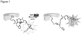
- the fluorophore may be attached to the copper-binding motif via the N-terminus of the DAP. The quenching or amount of quenching of the fluorescence intensity indicates the presence of Cu 2+ ions in the fluid, i.e.
- the senor comprises 5(6)-carboxyfluorescein-DAP- ⁇ Ala-His or 5(6)-carboxyfluorescein-DAP- ⁇ Ala-Asp.
- the detection means may also comprise a polymer membrane, foil or sheet including nanopores, preferably a functionalized polymer membrane, foil or sheet with nanopores, in particular an ion-track etched polymer membrane, foil or sheet with nanopores, even more preferably an ion-track etched polyethylene terephthalate (PET)-membrane, foil or sheet with nanopores.
- the nanopores may have a conical shape, preferably with small openings of about 30 ⁇ 5 nm and big openings of 500 ⁇ 10 nm in diameter.
- the polymer membrane, foil or sheet preferably contains about 10 4 nanopores per cm 2 .
- Ion-track etched polymer membranes, foils or sheets with conical nanopores can be obtained by the known procedure of asymmetric chemical etching of the latent ion track as described in Nucl Instrum Methods Phys Res, Sect. B 2001, 184, 337-346 .
- carboxylic acid groups are generated on the surface of the polymer membrane, foil or sheet, in particular on the nanopore surface of the ion-track etched polymer membrane, foil or sheet, which can be used for covalent attachment of functional molecules containing for example a primary amine.
- the senor of the present application comprises a fluorophore and a polymer membrane with nanopores as detection means.
- the sensor according to the present application may further comprise a linker moiety, which is preferably attached to the C-terminus of the amino acid or modified amino acid X.
- the linker moiety is for ensuring a spatial separation of the functional groups of the copper-binding motif and the fluorophore to the polymer membrane, foil or sheet as detection means, in particular ion-track etched PET-membrane having conical nanopores.
- the linker may also function to attach the sensor comprising the copper-binding motif with or without the fluorophore to a solid support, while ensuring a spatial separation of the functional groups of the copper-binding motif and the fluorophore to the surface of the solid support.
- the linker moiety comprises polyethylene glycol (PEG) units, in particular 4 PEG units.
- R 1 -[CH 2 CH 2 O] a -R 2 a may be 2 to 8, 2 to 6, 2 to 4, 4 to 10, 4 to 8 or 4 to 6, more preferably a is 3, 4, 5 or 6 and most preferable a is 4.
- the linker has the structure -HN-[CH 2 CH 2 O] 4 -CH 2 CH 2 COOH ( Figure 2(c) ) referred to as linkerPEG 4 in the present invention.
- the carboxy group of the linker may be coupled to an amino functional group on the surface of a polymer membrane or solid support via standard peptide coupling chemistry.
- the amino group R 1 allows coupling to the C-terminus of the amino acid or modified amino acid X, also by means of standard peptide coupling chemistry.
- the copper-binding motif with or without a fluorophore may be attached by means of the linker moiety to the surface of the polymer membrane with nanopores, in particular to the ion-track etched polymer membrane, foil or sheet, in particular to the surface of the nanopores. Binding of Cu 2+ ions to the copper-binding motif of the sensor changes the current-voltage characteristics of the polymer membrane, foil or sheet with the nanopores which can then be used for qualitative and quantitative detection of Cu 2+ ions in the fluid.
- the senor according to the present application comprises the copper-binding motif, detection means and a linker, wherein the detection means is a fluorophore, in particular 5(6)-carboxyfluorescein and/or a polymer membrane with nanopores, preferably an ion-track etched PET-membrane, foil or sheet with nanopores.
- the fluorophore is attached to the N-terminus of the DAP and the linker moiety is attached to the C-terminus of the amino acid or modified amino acid X.
- the copper-binding motif may be attached via the linker moiety to the surface of the polymer membrane, foil or sheet, in particular to the surface of the nanopores of the ion-track etched PET-membrane.
- the sensor according to the present application may further comprise a solid support.
- the sensor comprises the copper-binding motif, a fluorophore as detection means and a solid support.
- the copper-binding motif bound to the fluorophore may be immobilized on the solid support, which can be with or without the linker moiety, wherein immobilization or attachment by means of the linker moiety is preferred.
- the linker moiety is employed, a spatial separation of the functional groups of the copper-binding motif and of the fluorophore to the surface of the solid support is achieved whereby an undesired interaction between the functional groups on the surface of the solid support and the copper-binding motif and fluorophore can be avoided.
- the linker moiety may be attached to the copper-binding motif via the C-terminus of the amino acid or modified amino acid X to ensure a spatial separation of the functional groups of the fluorophore-copper-binding motif to the functional groups on the surface of the solid support.
- the sensor for detection of Cu 2+ ions in a fluid comprises fluorophore-DAP- ⁇ Ala-X-linkerPEG 4 attached to or immobilized on a solid support, wherein the fluorophore is preferably 5(6)-carboxyfluorescein and X is histidine, alanine or aspartic acid.
- the solid support may be for example a polymer, a resin, a glass substrate, a core-shell particle or cellulose.
- the solid support may be in the form of a membrane, foil, sheet, strip or particle.
- the sensor according to the present application may be used for detecting Cu 2+ ions in a fluid, which fluid can be obtained from any source.
- the fluid may comprise a liquid, preferably an aqueous or water-based liquid, for example from environmental sources.
- a liquid according to the present invention are drinking water and body fluids such as urine, whole blood, plasma, serum, extracellular interstitial fluid, lymph, bile, cerebrospinal liquid as well as gland secretion such as saliva or sweat, wherein urine is preferred.
- the aqueous or water-based liquid further may originate from water streams, puddles, lakes and seas.
- the sensor according to the present application is able to selectively detect Cu 2+ ions in a fluid in the presence of Ni 2+ ions and/or Zn 2+ ions.
- Selective detection of Cu 2+ ions according to the present application means that in case no Cu 2+ ions are present in the fluid, but Ni 2+ ions and/or Zn 2+ ions are, no signal indicating the presence of the desired analyte Cu 2+ ions is generated by the detection means. For example, when using 5(6)-carboxyfluorescein as fluorophore, no quenching in fluorescence intensity would be observed.
- Ni 2+ ions and/or Zn 2+ ions in addition to a particular concentration of Cu 2+ ions in the fluid does not change the signal of the detection means by more than ⁇ 5%, i.e. almost identical results are obtained for the Cu 2+ ion concentration in a solution containing Ni 2+ ions and/or Zn 2+ ions as for a solution containing Cu 2+ ions, but no Ni 2+ ions and/or Zn 2+ ions.
- the senor according to the present application is for use in the diagnosis and/or monitoring of diseases, preferably of diseases linked to abnormal Cu 2+ ion (copper) concentrations and/or dysregulated Cu 2+ ion (copper) metabolism such as Alzheimer's disease, Wilson's disease and Menke's disease.
- Cu 2+ ion concentrations in terms of diagnosis and/or monitoring of diseases, in particular when determining the Cu 2+ ion concentration in urine refer to copper, i.e. Cu 2+ ions that is not bound to ceruloplasmin (non-CP Cu). Such copper may also be termed "free Cu 2+ ions" or "free Cu 2+ ions in solution”.
- abnormal in relation to Cu 2+ ion (copper) concentration according to the present application means that the Cu 2+ ion concentration in a body fluid is higher or lower compared to healthy individuals.
- Higher Cu 2+ ion concentration in a body fluid, for example urine, is observed in individuals having a particular disease such as Alzheimer's disease or Wilsons's disease.
- Lower Cu 2+ ion concentration in a body fluid is observed in individuals having a disorder correlated to copper deficiency such as Menke's disease.
- the concentration of non-ceruloplasmin-bound Cu 2+ (non-CP Cu) in urine is in a range of 40 to 50 nM calculated based on a Cu 2+ ion amount of about 4.82 ⁇ g/day in 1.5 to 2.0 L urine ( J Trace Elem Met Biol 2018, 45, 181-188 ).
- a Cu 2+ ion concentration in urine above 40-50 nM for example at least twice as high than in healthy individuals, i.e. above 80 or 90 nM, in particular in a range of 100 to 130 nM as is often the case for AD patients is considered an abnormal copper or Cu 2+ ion concentration.
- the senor according to the present application is for use in the diagnosis and/or monitoring of Alzheimer's disease (AD), Wilson's disease and Menke's disease.
- the sensor according to the present application is for use in determining Cu 2+ ion concentration in the urine of individuals to diagnose and/or monitor diseases linked to abnormal Cu 2+ ion (copper) concentrations and/or dysregulated Cu 2+ ion (copper) metabolism, in particular Alzheimer's disease, Wilson's disease and Menke's disease.
- the use of the sensor of the present application is in particular for an ex vivo determination of Cu 2+ ion concentration in body fluids, preferably in the urine of individuals.
- the senor according to the present application may be for use in detecting Cu 2+ ions and/or determining Cu 2+ ion concentration in food products or fluids such as aqueous or water-based liquids, in particular in food/water control and/or environmental samples.
- the senor according to the present application may be used for multiple measurements, i.e. the sensor allows a repeated measurement of Cu 2+ ions or Cu 2+ ion concentration.
- the present application further discloses a method for the qualitative and quantitative detection of Cu 2+ ions in a fluid.
- the method comprises the steps of adjusting the pH of a fluid sample to be analyzed to 5.0 to 8.5, preferably to a pH of 5.5. to 8.0, 6.0 to 7.3, 6.3 to 8.0 or 6.5 to 7.5 and most preferably to a pH of 6.5 to 7.0.
- the selectivity of the sensor according to the present application towards Cu 2+ ions is ensured, in particular selectivity towards Cu 2+ ions over Ni 2+ and Zn 2+ ions. Setting the pH of the fluid sample to the claimed range, i.e.
- the sensor is brought into contact with the fluid sample. This may be done for example by immersing a test strip with the immobilized sensor in the fluid sample or by mixing a solution of the sensor of the present application with the fluid sample.
- detection of Cu 2+ ions and/or determining the Cu 2+ ion concentration of the sample takes place.
- different parameters are used as basis for qualitative and/or quantitative detection of Cu 2+ ions in the fluid sample.
- a sensor comprising a fluorophore as detection means in particular 5(6)-carboxyfluorescein is used
- quenching of the fluorescence intensity upon contact of the sensor with the analyte Cu 2+ ions is observed.
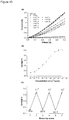
- a linear decrease of fluorescence intensity of the sensor upon addition of Cu 2+ ions in water can be clearly observed showing more and more quenching of the fluorescence intensity with increasing Cu 2+ ion concentrations.
- the change in the fluorescence intensity is measured.
- the fluorescence intensity may be reduced, i.e. quenched.
- Cu 2+ ion concentrations in the fluid sample with the sensor according to the present invention can be quantitatively detected in a range from 1 nM to 30 mM, 5 nM to 20 mM, 10 nM to 10 mM, 12 nM to 5 mM, in particular in a range from 1 nM to 60 ⁇ M, 5 nM to 40 ⁇ M, 10 nM to 10 ⁇ M, 12 nM to 5 ⁇ M, 1 nM to 500 nM, 1 nM to 200 nM or 12 nM to 150 nM.
- the quantitative detection of Cu 2+ ion concentrations in a fluid sample as low as about 1 nM is very important for application of the sensor in the detection of trace amounts of copper ions in food/water samples or for detection of copper ions in the early diagnosis of AD in urine.
- the sensor according to the present application can be used to measure the copper (Cu 2+ ) ion concentration directly from the urine sample in a simple two-step procedure, for example by immersing a test strip with the immobilized sensor in the urine sample or by mixing a solution of the sensor of the present application with the urine sample and then subjecting the sample to a fluorescence light source.
- concentration of free Cu 2+ ions in urine Cu 2+ ions not bound to ceruloplasmin (non-CP Cu)
- the Cu 2+ ion or copper concentration varies between 40 to 50 nM. All Cu 2+ ion or copper values for healthy individuals and patients of AD and Wilson's disease are within the range of detection of the sensor according to the present application.
- the qualitative and quantitative detection of Cu 2+ ions in the fluid sample may be achieved by monitoring the change in the current-voltage ( I-V ) characteristics of the sensor comprising at least a copper-binding motif, a polymer membrane, foil or sheet with nanopores as detection means, a linker moiety wherein the copper-binding motif is attached via the linker moiety to the surface of the polymer membrane, foil or sheet with nanopores and optionally a fluorophore.
- the polymer membrane, foil or sheet with nanopores is a PET membrane, foil or sheet with nanopores and even more preferably, the polymer membrane, foil or sheet with nanopores is an ion-track etched PET-membrane, foil or sheet with nanopores.
- the ion-track etched PET-membrane, foil or sheet has conical nanopores with a tip opening of 30 ⁇ 5 nm and a big opening of 500 ⁇ 10 nm in diameter.
- the polymer membrane, foil or sheet preferably contains about 10 4 nanopores per cm 2 .
- the sensor may also include a fluorophore as detection means. By measuring the changes in the current-voltage ( I-V ) characteristics of the sensor, a quantitative and qualitative detection of Cu 2+ ions in a fluid sample is feasible.
- the detection of Cu 2+ ion concentrations in a fluid sample with Cu 2+ ion concentrations as low as 1 fM can be done, in particular in a range from 1 fM to 30 mM, 1 fM to 20 mM, 1 nM to 10 mM, 1 nM to 5 mM, in particular in a range from 1 fM to 60 ⁇ M, 5 nM to 40 ⁇ M, 10 nM to 10 ⁇ M, 12 nM to 5 ⁇ M, 1nM to 500 nM, 1 nM to 200 nM or 12 nM to 150 nM.
- a qualitative and/or quantitative detection of Cu 2+ ions in a fluid sample may also be performed with mass spectrometry analysis.
- a sensor according to the present application may then comprise only the copper-binding motif and no further detection means are necessary.
- the sensor according to the present application may be regenerated to be applicable for further detection cycles. Regeneration is conducted by subjecting the sensor to a complexing agent.
- the complexing agent can be, for example, ethylene diamine tetraacetic acid (EDTA).
- EDTA ethylene diamine tetraacetic acid
- the sensor according to the present application can be regenerated at least 6 times.
- the sensor of the present application may be manufactured as follows.
- the copper-binding motif DAP- ⁇ Ala-X may be synthesized following the standard solid phase peptide synthesis (SPPS).
- SPPS solid phase peptide synthesis
- the fluorophore may also be attached via standard peptide coupling chemistry to the N-terminus of the DAP.
- the sensor comprises a linker moiety said linker moiety may also be attached via standard peptide coupling chemistry to the C-terminus of amino acid or modified amino acid X.
- the copper-binding motif may be attached via the linker moiety to the surface of the polymer membrane with nanopores.
- the sensor according to the present application can also be used in combination with a test strip for the detection of Cu 2+ ions in a fluid sample.
- the test strip may comprise the sensor according to the present application immobilized via the linker moiety to a solid support, for example a polymer foil or sheet.
- the sensor immobilized on the solid support may be applied to a carrier material.
- the solid support and/or the carrier material may have at least two slots or indentions into which a fluid sample may be applied. In one of the slots/indentions the fluid sample to be analyzed for the qualitative and/or quantitative detection of Cu 2+ ions can be applied. In the further slot/indention a control solution may be applied.
- the senor used in combination with a test strip comprises the copper-binding motif, a fluorophore as detection means, a linker moiety and a solid support.
- the calibration solution is for calibration of the fluorescence intensity.
- the copper-binding motif 2,3-diaminopropionic acid(DAP)- ⁇ Ala-X may be used for binding of Cu 2+ ions in a fluid, wherein X is an amino acid or a modified amino acid, in particular wherein X is histidine (His), alanine (Ala) or aspartic acid (Asp).
- the copper-binding motif comprises in particular DAP- ⁇ Ala-His or DAP- ⁇ Ala-Asp.
- Sensor ( 2 ) was acetylated at the N-terminus with acetic anhydride.
- the final cleavage of sensors ( 1 ) and ( 2 ) from the resin was carried out in 2/2/48/48 triisopropylsilane(TIPS)/water/trifluoroacetic acid(TFA)/DCM and agitated for 1 h at room temperature.
- RP-HPLC purification was performed on a C18 column (MultoKrom 100-5. 250 x 20 mm, 100 ⁇ pore diameter, 5.0 ⁇ m particle size) using a linear gradient of 5 % to 40 % of eluent B (eluent A: water (0.1 % TFA) and eluent B: acetonitrile (0.1 % TFA)) in 60 min.
- Sensor (2) was purified following an isocratic method of 5 % eluent B for 12 minutes followed by a linear gradient to 30 % eluent B within a total of 60 minutes.
- PET-membranes with conical nanopores were fabricated through asymmetric chemical etching of latent ion tracks as has been described in Nucl Instrum Methods Phys Res, Sect B 2001, 184, 337-346 .
- PET foils or membranes were first irradiated with single swift heavy ions (Au) of kinetic energy 11.4 MeV/nucleon at the linear accelerator UNILAC (GSI Helmholtz Centre for Heavy Ion Research, Darmstadt, Germany). Then, the latent ion tracks in the polymer foils or membranes were sensitized with soft UV light. The chemical track-etching process was performed in a conductivity cell.
- the ion tracked foils or membranes were fixed in between two chambers of the cell.
- An etching solution (9 M NaOH) was filled in one chamber and a stopping solution (1 M KCI + 1 M HCOOH) was filled in the other chamber.
- the etching process was carried out at room temperature.
- the etching process was monitored by applying a potential of -1 V across the foil or membrane.
- the etching process was stopped when the current reached a certain defined value after the breakthrough point. Then, the etched foil or membrane was washed with stopping solution and dipped in deionized water overnight to remove residual salts.
- the carboxylic acid groups on the polymer foil or membrane surface, in particular on the surface of the nanopores originated from the chemical etching. These groups were first activated through standard carbodiimide coupling chemistry.
- the track-etched foil or membrane was exposed to an ethanol solution containing 1-ethyl-3-(3-dimethylaminopropyl)carbodiimide (EDC) (100 mM) and pentafluorophenyl (PFP) (200 mM) at room temperature for 1 h. After washing with ethanol several times, the activated polymer foil or membrane was treated with ethylenediamine (EDA, 50 mM) solution overnight.
- EDC 1-ethyl-3-(3-dimethylaminopropyl)carbodiimide
- PFP pentafluorophenyl
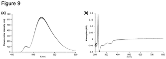
- the fluorescence spectrum in Figure 3(a) shows the decrease of fluorescence intensity of the sensor comprising 5(6)-carboxyfluorescein-DAP- ⁇ Ala-His-linkerPEG 4 (1) (1 ⁇ M, MES buffer, pH 6.50) upon addition of CuSO 4 from 0 to 25 ⁇ M. Emission (A) was determined to be 518 nm. Fluorescence titration upon addition of 20 equivalents of Cu 2+ at pH 6.50 showed quenching of the fluorescence intensity to 88% (12% remaining fluorescence intensity) ( Figure 3(b) ).
- the binding constants for Cu 2+ and Ni 2+ to the copper-binding motif of the sensor of the present application were determined by UV-Vis spectroscopy at pH 6.50 using the Benesi-Hildebrand method. The obtained binding constants were similar to the binding constants determined with fluorescence spectroscopy using the Stern-Volmer plot at pH 6.50.
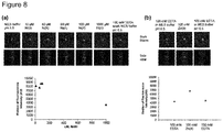
- 5(6)-carboxyfluorescein-DAP- ⁇ Ala-His-linkerPEG 4 (1) is almost exclusively present on the surface of the nanopores of the ion-track etched PET-membrane or foil as can be seen in Figure 10(a) and displays a green color when excited by the laser at 488 nm in the CLSM image ( Figure 10(b)).
- Figure 10(d) shows a titration experiment showing the dependency of decrease in fluorescence with the increase in Cu 2+ ion concentration as well as the re-usability of the sensor of the present application.
- CuSO 4 was added in concentrations from 0 to 100 ⁇ M in MES buffer solution, pH 6.50.
- the limit of detection (LOD) for Cu 2+ ions using the fluorescence signal was determined.
- the limit of detection of the sensor comprising 5(6)-carboxyfluorescein-DAP- ⁇ Ala-His-linkerPEG 4 (1) towards Cu 2+ ions in solution from fluorescence intensity titration studies is defined to be 12 nM. This value is extremely important for application of the sensor according to the present invention for detection of trace amounts of copper ions in food/water control or detection of copper ions for the early diagnosis of Alzheimer's disease in urine.
- the sensor comprises the copper-binding motif, a fluorophore and a linker moiety, i.e. 5(6)-carboxyfluorescein-DAP- ⁇ Ala-His-linkerPEG 4 whereby the copper-binding motif with the fluorophore is attached to the ion-track etched PET-membrane with nanopores by means of the linker.
- the PET-membrane with nanopores behaves as an ohmic resistor before Cu 2+ complexation meaning that the net surface charge on the nanopore surface is zero and no flux from the narrow cone opening to the wide opening takes place.
- the ion-track etched PET-membrane with conical nanopores having carboxyl groups on the surface with no immobilized copper-binding motif shows a flux from the narrow cone opening to the wide opening because of the negative carboxyl groups ( Figure 12 ).
- EDA ethylene diamine
- Immobilization of the copper-binding motif with the fluorophore via the linker to the surface of the nanopores of the ion-track etched PET-membrane results in change of the nanopore transport behavior from a rectifying to a non-rectifying due to the loss of nanopore surface charges.
- the PET-membrane with the immobilized copper-binding motif behaves as an ohmic resistor, i.e. the net surface charge on the nanopore walls is zero.
- the sensor according to the present application is able to recognize Cu 2+ ions even at concentrations as low as 1 fM and this recognition process can be transduced in an electronic signal originating from the transport behavior of the nanopore. From the data given in Figure 13 it can be seen that the sensor exhibits a remarkable interaction ability towards Cu 2+ due to the particular copper-binding motif used in the sensor according to the present application.
- the present application further relates to the following items.
Abstract
The present application is directed to a sensor for detection of Cu<sup>2+</sup>ions in a fluid comprising a copper-binding motif 2,3-diaminopropionic acid(DAP)-βAla-X, wherein X is an amino acid or a modified amino acid, in particular wherein X is histidine (His), alanine (Ala) or aspartic acid (Asp). The sensor according to the present application can be used for fast, highly selective and ultrasensitive detection of Cu<sup>2+</sup>ions in a fluid. With said sensor a qualitative and/or quantitative detection of Cu<sup>2+</sup>ions can be achieved, which can be useful in the diagnosis and/or monitoring of diseases linked to abnormal Cu<sup>2+</sup>ion concentrations and/or dysregulated copper metabolism, in particular Alzheimer's disease.
Description
- The present application is directed to the use of a copper-binding motif for binding Cu2+ ions in a fluid, a sensor for detection of Cu2+ ions in a fluid, the use of the sensor in the diagnosis and monitoring of diseases and a method for qualitative and quantitative detection of Cu2+ ions in a fluid sample.
- Copper is an essential trace element that is inevitable in biological systems and can be found in many enzymes such as amine oxidases and ferroxidases but is also required for infant growth, the ion metabolism and brain development in human organisms (Nat Chem Biol 2008, 4, 176-185; J Toxicol Environ Health B Crit Rev 2001, 4, 341-394). Copper is also widely used in agricultural systems and therefore belongs to a major metal pollutant in our environment (Rev Environ Health 1999, 14, 231-238). The contamination of ecosystems such as rivers, lakes, soil and groundwater with copper causes increased accumulation (Chem Soc Rev 2017, 46, 7105-7123).
- There are also diseases linked to an abnormal copper concentration and/or dysregulated copper metabolism in humans such as Wilson's disease, Menke's disease and Alzheimer's disease (AD) (Water, Air and Soil Pollution 1998, 108, 457-471). Dysregulation of copper homeostasis causes neurodegenerative diseases, which led to a rising interest in the investigation of the connection between copper and AD (Dalton Trans 2004, 1907-1917). The involvement of copper in AD contains two aspects: first, aggregation/plaque formation of so-called β-amyloid peptide (Aβ), which is cleaved from amyloid precursor protein (APP) upon copper binding and second, oxidative stress which is caused by reduction of Cu2+ to Cu1+ through its binding domain (CuBD) (Dalton Trans 2004, 1907-1917; Neuron 2001, 30, 665-676). Both aspects are known to aggravate AD which results in cell and especially neuronal death (J Toxicol Environ Health B Crit Rev 2001, 4, 341-394).
- During the last two decades studies have been performed in order to confirm the involvement of copper in β-amyloid plaque progression. Schreurs et al. (Proc Natl Acad Sci USA 2003, 100, 11065-11069) observed an increased formation of β-amyloid plaques by cholesterol-fed rabbits when adding trace amounts (12 ppm) of copper to their drinking water. During meta-analysis performed by Squitti et al. (J Alzheimers Dis 2014, 38, 809-822) the concentration of free copper in the serum of AD patients was determined to be higher compared to that of healthy individuals. These results were generated by studying the fraction of copper that is not bound to the ceruloplasmin (non-CP Cu), i.e. free Cu2+ in solution.
- Recent studies of Squitti et al. (J Trace Elem Med Biol 2018, 45, 181-188) show the potential of non-CP Cu detection, i.e. detection of free Cu2+ in urine as an eligible marker for patients in early stages of AD. In particular, Squitti et al. report a 2.4 times increased concentration of copper in urine of AD patients in comparison to healthy controls. Thus, determination of non-bound Cu2+ in urine and serum can be used as a basis for a screening method for the early diagnosis and/or monitoring of diseases linked to abnormal copper concentrations, for example AD or Wilson's disease.
- In recent years first steps have been taken towards the development of screening methods and sensors for the determination of copper for early AD diagnostics. For example, Hirayama et al. (Proc Nat Acad Sci USA 2012, 109, 2228-2233) developed a copper-imaging sensor inside of living cells with a sensitive fluorescence turn-on response to Cu1+. However, this method depends on live-cell imaging after digestion of the fluorescent dye and thus the detection method itself and its implementation is tied to an extensive set-up. Squitti et al. (J Alzheimers Dis 2014, 38, 809-822) designed a method using fluorescence to detect non-CP Cu2+ distributed in the serum. However, also this method requires an extensive two-step experimental set-up with a size exclusion solid-phase extraction separating non-CP Cu from protein-bound copper followed by a fluorescent method, which also requires a lengthy time period to obtain results.
- It is known that peptides in proteins can display extraordinary and ultrasensitive key-lock behavior towards targets. Copper-binding peptides in nature are albumin (bovine serum albumin (BSA), human serum albumin (HSA), rabbit serum albumin (RSA)), neuromedin C and K, human sperm protamine P2a and histidines (Accounts of Chemical Research 1997, 30, 123-130). Imperiali et al. (J Am Chem Soc 1998, 120, 609-610) reported polypeptide motifs for the design of selective Cu(ll) ion chemosensors. Papp et al. (Angewandte Chem Int Ed Engl 2018, 57, 4752-4755) were able to attach the amino terminal Cu(II) and Ni(II)-binding (ATCUN) motif (Gly-Gly-His) on track-etched polycarbonate membranes. A copper-sensing system that can be evaluated by naked eye detection with a detection limit of 0.5 µM using a "turn-on" fluorescence strategy is reported by Situ et al. (Sensors and Actuators B-Chemical 2017, 240, 560-565), however, with the disadvantage that the detection method involves several reaction steps. Further, naked eye detectable sensor systems were constructed by Ding et al. (Journal of Materials Chemistry 2011, 21, 13345-13353) presenting a colorimetric detection of copper using sensor strips at a detection limit of 5 nM.
- Due to the above-outlined disadvantages of the known copper sensing methods and the need for highly sensitive and selective detection methods for Cu2+ ions it is an object of the present application to provide a sensor allowing a fast and easy-to-handle, but highly sensitive and selective qualitative and quantitative detection of Cu2+ in a fluid, in particular for detecting elevated copper concentrations in body fluids in a quick test and in environmental samples on-site.
- According to the invention, this problem has been solved by providing a sensor for detection of Cu2+ ions in a fluid comprising a copper-binding
motif 2,3-diaminopropionic acid(DAP)-βAla-X, wherein X is an amino acid or a modified amino acid, in particular wherein X is histidine (His), alanine (Ala) or aspartic acid (Asp). The sensor according to the present application can be used for fast, highly selective and ultrasensitive detection of Cu2+ ions in a fluid. With said sensor a qualitative and/or quantitative detection of Cu2+ ions can be obtained, for example by measuring the change in fluorescence intensity, by measuring the change in the current-voltage characteristics and/or by subjecting the sensor to mass spectrometry. - The detection method of the present invention does not require an extensive experimental set-up. In fact, with the sensor of the present application detection of Cu2+ ions is possible on-site and in real time. For example, simply a test strip comprising the sensor having the particular copper-binding motif and a fluorescence-light source may be required. In addition or alternatively, the qualitative and quantitative detection of Cu2+ ions may also be performed by observing a change in the current-voltage characteristics, which even allows the detection of Cu2+ ion concentrations in a fluid in the femtomolar range, i.e. as low as 1 fM. M refers to the molar concentration mol per liter (mol/L)
- Generally, sensors are devices for detecting particular analytes, in this case the analyte are Cu2+ ions. The sensor provides the information regarding the analyte usually in the form of a measurable physical signal correlated with the presence and/or concentration of the analyte.
- The sensor of the present application comprises a copper-binding motif having at least two non-proteinogenic amino acids, namely 2,3-diaminopropionic acid (DAP) and β-alanine (βAla). The copper-binding motif further comprises an amino acid or modified amino acid X. The copper-binding motif corresponds to a peptide compound and therefore the copper-binding motif is sometimes termed peptide copper-binding motif. The amino acid X may be selected from the group consisting of standard amino acids, unusual amino acids or non-proteinogenic amino acids. Examples for standard L-amino acids are glycine (Gly), alanine (Ala), valine (Val), leucine (Leu), methionine (Met), isoleucine (Ile), serine (Ser), threonine (Thr), cysteine (Cys), proline (Pro), asparagine (Asn), glutamine (Gin), phenylalanine (Phe), tyrosine (Tyr), tryptophan (Trp), lysine (Lys), arginine (Arg), histidine (His), aspartic acid (Asp) and glutamate (Glu). Preferred standard amino acids according to the present invention are histidine (His), alanine (Ala) or aspartic acid (Asp). Examples for unusual amino acids are 4-hydroxyproline, 5-hydroxylysine, 6-N-methyllysine, γ-carboxyglutamate, desmosine, selenocysteine, ornithine and citroline. Non-proteinogenic amino acids are amino acids which are not incorporated in proteins during translation. Non-proteinogenic amino acids according to the present application are for example 2,3-diaminopropionic acid (DAP) and β-alanine. 2,3-diaminopropionic acid (DAP) may be L-2,3-diaminopropionic acid, D-2,3-diaminopropionic acid or a mixtures of L- and D-DAP. L-2,3-diaminopropionic acid is preferred. A modified amino acid comprises standard or unusual amino acids, which may be for example methylated, acetylated or its diastereomeric D, cis/trans and isosteric form. X is preferably a standard amino acid, more preferably histidine (His), alanine (Ala) or aspartic acid (Asp).
- In a preferred embodiment the sensor of the present application comprises the copper-binding motif DAP-βAla-His or DAP-βAla-Asp.
- In addition to the copper-binding motif or peptide copper-binding motif the sensor according to the present application may comprise means for detection. However, such means for detection are not mandatory since some detection methods such as mass spectrometry do not require such means for detection. Detection means or means for detection are able to produce a measurable, physical signal. In case no additional detection means are present, the N-terminus of the DAP may be capped with an acetyl (-CCH3=O) moiety.
- For example, the detection means according to the present application may be commercially available fluorophores such as 5(6)-carboxyfluorescein, DyLight Fluor, Alexa Fluor or cyanine dyes. A preferred fluorophore of the sensor of the present application is 5(6)-carboxyfluorescein. The fluorophore may be attached to the copper-binding motif via the N-terminus of the DAP. The quenching or amount of quenching of the fluorescence intensity indicates the presence of Cu2+ ions in the fluid, i.e. binding of Cu2+ ions to the copper-binding motif of the sensor and can be used to calculate the Cu2+ ions concentration in the fluid as the amount of quenching is proportional to the Cu2+ ion concentration in the fluid. The "on-off" characteristics of such a detection method using the sensor of the present application is shown in
Figure 1 . - In a preferred embodiment of the present application, the sensor comprises 5(6)-carboxyfluorescein-DAP-βAla-His or 5(6)-carboxyfluorescein-DAP-βAla-Asp.
- The detection means may also comprise a polymer membrane, foil or sheet including nanopores, preferably a functionalized polymer membrane, foil or sheet with nanopores, in particular an ion-track etched polymer membrane, foil or sheet with nanopores, even more preferably an ion-track etched polyethylene terephthalate (PET)-membrane, foil or sheet with nanopores. The nanopores may have a conical shape, preferably with small openings of about 30 ± 5 nm and big openings of 500 ± 10 nm in diameter. The polymer membrane, foil or sheet preferably contains about 104 nanopores per cm2. Ion-track etched polymer membranes, foils or sheets with conical nanopores can be obtained by the known procedure of asymmetric chemical etching of the latent ion track as described in Nucl Instrum Methods Phys Res, Sect. B 2001, 184, 337-346. After heavy ion irradiation and subsequent chemical etching process of the polymer membranes, foils or sheets, carboxylic acid groups are generated on the surface of the polymer membrane, foil or sheet, in particular on the nanopore surface of the ion-track etched polymer membrane, foil or sheet, which can be used for covalent attachment of functional molecules containing for example a primary amine.
- In a preferred embodiment, the sensor of the present application comprises a fluorophore and a polymer membrane with nanopores as detection means.
- The sensor according to the present application may further comprise a linker moiety, which is preferably attached to the C-terminus of the amino acid or modified amino acid X. The linker moiety is for ensuring a spatial separation of the functional groups of the copper-binding motif and the fluorophore to the polymer membrane, foil or sheet as detection means, in particular ion-track etched PET-membrane having conical nanopores. The linker may also function to attach the sensor comprising the copper-binding motif with or without the fluorophore to a solid support, while ensuring a spatial separation of the functional groups of the copper-binding motif and the fluorophore to the surface of the solid support. Thereby an undesired interaction between the functional groups on the surface of the polymer membrane, foil or sheet or of the functional groups on the surface of the solid support and the copper-binding motif and fluorophore can be avoided.
- Preferably, the linker moiety comprises polyethylene glycol (PEG) units, in particular 4 PEG units. The linker moiety preferably has the formula R1-[CH2CH2O]a-R2, wherein subscript a is from 2 to 10, R1 is -NH and R2 is CH2CH2COOH or CH2CH2CCH3=O. In the formula R1-[CH2CH2O]a-R2 a may be 2 to 8, 2 to 6, 2 to 4, 4 to 10, 4 to 8 or 4 to 6, more preferably a is 3, 4, 5 or 6 and most preferable a is 4. In a preferred embodiment the linker has the structure -HN-[CH2CH2O]4-CH2CH2COOH (
Figure 2(c) ) referred to as linkerPEG4 in the present invention. If the linker comprises a carboxy group R2 = COOH coupling by means of said carboxy group to a polymer membrane, foil or sheet or to a solid support can be achieved. For example, the carboxy group of the linker may be coupled to an amino functional group on the surface of a polymer membrane or solid support via standard peptide coupling chemistry. The amino group R1 allows coupling to the C-terminus of the amino acid or modified amino acid X, also by means of standard peptide coupling chemistry. - In one embodiment of the present application, the copper-binding motif with or without a fluorophore may be attached by means of the linker moiety to the surface of the polymer membrane with nanopores, in particular to the ion-track etched polymer membrane, foil or sheet, in particular to the surface of the nanopores. Binding of Cu2+ ions to the copper-binding motif of the sensor changes the current-voltage characteristics of the polymer membrane, foil or sheet with the nanopores which can then be used for qualitative and quantitative detection of Cu2+ ions in the fluid.
- In one embodiment, the sensor according to the present application comprises the copper-binding motif, detection means and a linker, wherein the detection means is a fluorophore, in particular 5(6)-carboxyfluorescein and/or a polymer membrane with nanopores, preferably an ion-track etched PET-membrane, foil or sheet with nanopores. The fluorophore is attached to the N-terminus of the DAP and the linker moiety is attached to the C-terminus of the amino acid or modified amino acid X. The copper-binding motif may be attached via the linker moiety to the surface of the polymer membrane, foil or sheet, in particular to the surface of the nanopores of the ion-track etched PET-membrane.
- The sensor according to the present application may further comprise a solid support. In one preferred embodiment, the sensor comprises the copper-binding motif, a fluorophore as detection means and a solid support. The copper-binding motif bound to the fluorophore may be immobilized on the solid support, which can be with or without the linker moiety, wherein immobilization or attachment by means of the linker moiety is preferred. When the linker moiety is employed, a spatial separation of the functional groups of the copper-binding motif and of the fluorophore to the surface of the solid support is achieved whereby an undesired interaction between the functional groups on the surface of the solid support and the copper-binding motif and fluorophore can be avoided.
- The linker moiety may be attached to the copper-binding motif via the C-terminus of the amino acid or modified amino acid X to ensure a spatial separation of the functional groups of the fluorophore-copper-binding motif to the functional groups on the surface of the solid support. In a preferred embodiment, the sensor for detection of Cu2+ ions in a fluid comprises fluorophore-DAP-βAla-X-linkerPEG4 attached to or immobilized on a solid support, wherein the fluorophore is preferably 5(6)-carboxyfluorescein and X is histidine, alanine or aspartic acid. The solid support may be for example a polymer, a resin, a glass substrate, a core-shell particle or cellulose. The solid support may be in the form of a membrane, foil, sheet, strip or particle.
- The sensor according to the present application may be used for detecting Cu2+ ions in a fluid, which fluid can be obtained from any source. The fluid may comprise a liquid, preferably an aqueous or water-based liquid, for example from environmental sources. Examples for a liquid according to the present invention are drinking water and body fluids such as urine, whole blood, plasma, serum, extracellular interstitial fluid, lymph, bile, cerebrospinal liquid as well as gland secretion such as saliva or sweat, wherein urine is preferred. The aqueous or water-based liquid further may originate from water streams, puddles, lakes and seas.
- The sensor according to the present application is able to selectively detect Cu2+ ions in a fluid in the presence of Ni2+ ions and/or Zn2+ ions. Selective detection of Cu2+ ions according to the present application means that in case no Cu2+ ions are present in the fluid, but Ni2+ ions and/or Zn2+ ions are, no signal indicating the presence of the desired analyte Cu2+ ions is generated by the detection means. For example, when using 5(6)-carboxyfluorescein as fluorophore, no quenching in fluorescence intensity would be observed. Further, the presence of Ni2+ ions and/or Zn2+ ions in addition to a particular concentration of Cu2+ ions in the fluid does not change the signal of the detection means by more than ± 5%, i.e. almost identical results are obtained for the Cu2+ ion concentration in a solution containing Ni2+ ions and/or Zn2+ ions as for a solution containing Cu2+ ions, but no Ni2+ ions and/or Zn2+ ions.
- In one embodiment, the sensor according to the present application is for use in the diagnosis and/or monitoring of diseases, preferably of diseases linked to abnormal Cu2+ ion (copper) concentrations and/or dysregulated Cu2+ ion (copper) metabolism such as Alzheimer's disease, Wilson's disease and Menke's disease. Cu2+ ion concentrations in terms of diagnosis and/or monitoring of diseases, in particular when determining the Cu2+ ion concentration in urine refer to copper, i.e. Cu2+ ions that is not bound to ceruloplasmin (non-CP Cu). Such copper may also be termed "free Cu2+ ions" or "free Cu2+ ions in solution".
- The term "abnormal" in relation to Cu2+ ion (copper) concentration according to the present application means that the Cu2+ ion concentration in a body fluid is higher or lower compared to healthy individuals. Higher Cu2+ ion concentration in a body fluid, for example urine, is observed in individuals having a particular disease such as Alzheimer's disease or Wilsons's disease. Lower Cu2+ ion concentration in a body fluid is observed in individuals having a disorder correlated to copper deficiency such as Menke's disease. In healthy individuals the concentration of non-ceruloplasmin-bound Cu2+ (non-CP Cu) in urine is in a range of 40 to 50 nM calculated based on a Cu2+ ion amount of about 4.82 µg/day in 1.5 to 2.0 L urine (J Trace ). Thus, a Cu2+ ion concentration in urine above 40-50 nM, for example at least twice as high than in healthy individuals, i.e. above 80 or 90 nM, in particular in a range of 100 to 130 nM as is often the case for AD patients is considered an abnormal copper or Cu2+ ion concentration.
- In a preferred embodiment, the sensor according to the present application is for use in the diagnosis and/or monitoring of Alzheimer's disease (AD), Wilson's disease and Menke's disease. In a further preferred embodiment, the sensor according to the present application is for use in determining Cu2+ ion concentration in the urine of individuals to diagnose and/or monitor diseases linked to abnormal Cu2+ ion (copper) concentrations and/or dysregulated Cu2+ ion (copper) metabolism, in particular Alzheimer's disease, Wilson's disease and Menke's disease. The use of the sensor of the present application is in particular for an ex vivo determination of Cu2+ ion concentration in body fluids, preferably in the urine of individuals.
- Alternatively, the sensor according to the present application may be for use in detecting Cu2+ ions and/or determining Cu2+ ion concentration in food products or fluids such as aqueous or water-based liquids, in particular in food/water control and/or environmental samples.
- Particularly, the sensor according to the present application may be used for multiple measurements, i.e. the sensor allows a repeated measurement of Cu2+ ions or Cu2+ ion concentration.
- The present application further discloses a method for the qualitative and quantitative detection of Cu2+ ions in a fluid. The method comprises the steps of adjusting the pH of a fluid sample to be analyzed to 5.0 to 8.5, preferably to a pH of 5.5. to 8.0, 6.0 to 7.3, 6.3 to 8.0 or 6.5 to 7.5 and most preferably to a pH of 6.5 to 7.0. By adjusting the pH of the fluid sample to be analyzed the selectivity of the sensor according to the present application towards Cu2+ ions is ensured, in particular selectivity towards Cu2+ ions over Ni2+ and Zn2+ ions. Setting the pH of the fluid sample to the claimed range, i.e. to a pH range of 5.0 to 8.5 leads to formation of a complex only between Cu2+ ions and the copper-binding motif of the sensor. The formation of Ni2+- or Zn2+-complexes at the claimed pH-range with the copper-binding motif of the sensor is insignificant.
- Without being bound by theory, it is assumed that upon binding of the copper-binding motif, in particular DAP-βAla-His of the sensor to the Cu2+ ions a square planar complex is formed having a d-d transition band at 537 nm (
Figure 2(c) ). As can be seen inFigure 2(a) of the present application Ni-complexation of the sensor according to the present application significantly starts at around > pH 7, which Ni-complex shows a transition band at 438 nm (Figure 2(b) ). Thus, adjusting the pH in relation to the pH-dependent formation of the Cu2+- and Ni2+-complex, taking into account up to which pH range the Ni2+-complex is not formed, a highly selective detection method for Cu2+ ions in a fluid is enabled. - Performing quantitative and/or qualitative detection of Cu2+ ions with the sensor according to the present application in a fluid with a pH range of 5.0 to 8.5, preferably in a pH range of 5.5. to 8.0, 6.0 to 7.3, 6.3 to 8.0 or 6.5 to 7.5 and even more preferably at a pH range of 6.5 to 7.0 corresponding to pH50 of the copper-sensor, i.e. the pH value at which 50% of the Cu2+-complexes have been formed, allows for selective detection of Cu2+ ions also in the presence of Ni2+ ions in the fluid sample, since at the claimed pH range only insignificant or no complexation of Ni2+ is present.
- In a further step, following adjustment of the pH of the fluid sample, the sensor is brought into contact with the fluid sample. This may be done for example by immersing a test strip with the immobilized sensor in the fluid sample or by mixing a solution of the sensor of the present application with the fluid sample. In the next step, detection of Cu2+ ions and/or determining the Cu2+ ion concentration of the sample takes place. Depending on the detection means different parameters are used as basis for qualitative and/or quantitative detection of Cu2+ ions in the fluid sample. In case a sensor comprising a fluorophore as detection means, in particular 5(6)-carboxyfluorescein is used, quenching of the fluorescence intensity upon contact of the sensor with the analyte Cu2+ ions is observed. As can be seen in
Figure 3(a),(b) a linear decrease of fluorescence intensity of the sensor upon addition of Cu2+ ions in water can be clearly observed showing more and more quenching of the fluorescence intensity with increasing Cu2+ ion concentrations. - When using a fluorophore as the detection means in the sensor according to the present application, the change in the fluorescence intensity is measured. Upon binding of the Cu2+ ions to the copper-binding motif of the sensor, the fluorescence intensity may be reduced, i.e. quenched. Cu2+ ion concentrations in the fluid sample with the sensor according to the present invention can be quantitatively detected in a range from 1 nM to 30 mM, 5 nM to 20 mM, 10 nM to 10 mM, 12 nM to 5 mM, in particular in a range from 1 nM to 60 µM, 5 nM to 40 µM, 10 nM to 10 µM, 12 nM to 5 µM, 1 nM to 500 nM, 1 nM to 200 nM or 12 nM to 150 nM. The quantitative detection of Cu2+ ion concentrations in a fluid sample as low as about 1 nM is very important for application of the sensor in the detection of trace amounts of copper ions in food/water samples or for detection of copper ions in the early diagnosis of AD in urine. A copper value, i.e. amount of Cu2+ ions of about 12 µg/day in the 24 h urine of AD patients (J Trace ) and taking into account that 24 h collected urine is around 1.5 to 2.0 L leads to a copper concentration in urine in a range of 100 to 130 nM for AD patients. The sensor according to the present application can be used to measure the copper (Cu2+) ion concentration directly from the urine sample in a simple two-step procedure, for example by immersing a test strip with the immobilized sensor in the urine sample or by mixing a solution of the sensor of the present application with the urine sample and then subjecting the sample to a fluorescence light source. For individuals with Wilson's disease the concentration of free Cu2+ ions in urine (Cu2+ ions not bound to ceruloplasmin (non-CP Cu)) is determined to be about 5 to 10 µM, which is even higher than in AD patients. For healthy individuals with a copper value of 4.82 µg/day the Cu2+ ion or copper concentration varies between 40 to 50 nM. All Cu2+ ion or copper values for healthy individuals and patients of AD and Wilson's disease are within the range of detection of the sensor according to the present application.
- In an alternative embodiment, the qualitative and quantitative detection of Cu2+ ions in the fluid sample may be achieved by monitoring the change in the current-voltage (I-V) characteristics of the sensor comprising at least a copper-binding motif, a polymer membrane, foil or sheet with nanopores as detection means, a linker moiety wherein the copper-binding motif is attached via the linker moiety to the surface of the polymer membrane, foil or sheet with nanopores and optionally a fluorophore. Preferably, the polymer membrane, foil or sheet with nanopores is a PET membrane, foil or sheet with nanopores and even more preferably, the polymer membrane, foil or sheet with nanopores is an ion-track etched PET-membrane, foil or sheet with nanopores. In a preferred embodiment, the ion-track etched PET-membrane, foil or sheet has conical nanopores with a tip opening of 30 ± 5 nm and a big opening of 500 ± 10 nm in diameter. The polymer membrane, foil or sheet preferably contains about 104 nanopores per cm2. Optionally, the sensor may also include a fluorophore as detection means. By measuring the changes in the current-voltage (I-V) characteristics of the sensor, a quantitative and qualitative detection of Cu2+ ions in a fluid sample is feasible. In particular the detection of Cu2+ ion concentrations in a fluid sample with Cu2+ ion concentrations as low as 1 fM can be done, in particular in a range from 1 fM to 30 mM, 1 fM to 20 mM, 1 nM to 10 mM, 1 nM to 5 mM, in particular in a range from 1 fM to 60 µM, 5 nM to 40 µM, 10 nM to 10 µM, 12 nM to 5 µM, 1nM to 500 nM, 1 nM to 200 nM or 12 nM to 150 nM.
- A qualitative and/or quantitative detection of Cu2+ ions in a fluid sample may also be performed with mass spectrometry analysis. A sensor according to the present application may then comprise only the copper-binding motif and no further detection means are necessary.
- The sensor according to the present application may be regenerated to be applicable for further detection cycles. Regeneration is conducted by subjecting the sensor to a complexing agent. The complexing agent can be, for example, ethylene diamine tetraacetic acid (EDTA). In a preferred embodiment, the sensor according to the present application can be regenerated at least 6 times.
- The sensor of the present application may be manufactured as follows. The copper-binding motif DAP-βAla-X may be synthesized following the standard solid phase peptide synthesis (SPPS). In case a fluorophore is used as detection means, the fluorophore may also be attached via standard peptide coupling chemistry to the N-terminus of the DAP. In case the sensor comprises a linker moiety said linker moiety may also be attached via standard peptide coupling chemistry to the C-terminus of amino acid or modified amino acid X. In case the sensor comprises a copper-binding motif, a linker moiety, a polymer membrane with nanopores as detection means and optionally a fluorophore as detection means, the copper-binding motif may be attached via the linker moiety to the surface of the polymer membrane with nanopores.
- The sensor according to the present application can also be used in combination with a test strip for the detection of Cu2+ ions in a fluid sample. The test strip may comprise the sensor according to the present application immobilized via the linker moiety to a solid support, for example a polymer foil or sheet. Optionally, the sensor immobilized on the solid support may be applied to a carrier material. The solid support and/or the carrier material may have at least two slots or indentions into which a fluid sample may be applied. In one of the slots/indentions the fluid sample to be analyzed for the qualitative and/or quantitative detection of Cu2+ ions can be applied. In the further slot/indention a control solution may be applied. In case a quantitative detection of Cu2+ ions in the fluid sample should be performed in a further slot/indention a calibration solution may be applied. In a preferred embodiment, the sensor used in combination with a test strip comprises the copper-binding motif, a fluorophore as detection means, a linker moiety and a solid support. In that case, the calibration solution is for calibration of the fluorescence intensity.
- The copper-binding
motif 2,3-diaminopropionic acid(DAP)-βAla-X may be used for binding of Cu2+ ions in a fluid, wherein X is an amino acid or a modified amino acid, in particular wherein X is histidine (His), alanine (Ala) or aspartic acid (Asp). The copper-binding motif comprises in particular DAP-βAla-His or DAP-βAla-Asp. -
-
Figure 1 shows the "on-off" characteristics of a detection method using the sensor of the present application comprising 5(6)-carboxyfluorescein as detection means. -
Figure 2(a) is an UV-vis spectrum showing the pH dependent formation of the Cu2+ complex (black square) at 537 nm of the sensor comprising Ac-DAP-βAla-His-linkerPEG4 (1 mM, water) in relation to the formation of the corresponding Ni2+ complex at 438 nm of the sensor (grey dots). Boltzman fit (black and grey curves) show the sigmoidal trend of the pH titration curves.Figure 2(b) shows UV-vis spectra of the Cu2+ complex at pH 8 (black line) and Ni2+ complex at pH 10.5 (grey line) of the sensor comprising Ac-DAP-βAla-His-linkerPEG4, the insert showing a magnification at 350 to 650 nm.Figure 2(c) is a scheme of the complex between the copper-binding motif DAP-βAla-His and the metal M2+ (Cu2+), wherein R is either 5(6)-carboxyfluorescein or acetyl. -
Figure 3(a) is a fluorescence spectrum showing the decrease of fluorescence intensity of the sensor comprising 5(6)-carboxyfluorescein-DAP-βAla-His-linkerPEG4 (1 µM, 2-(N-morpholino)ethanesulfonic acid (MES) buffer, pH 6.50) upon addition of CuSO4 from 0 to 25 µM in water.Figure 3(b) shows the change in fluorescence intensity as a function of the molar ratio of Cu2+ to sensor (sensor is termed "peptide" in the description of the x-axis).Figure 3(c) is a fluorescence spectrum showing the decrease of fluorescence intensity of the sensor comprising 5(6)-carboxyfluorescein-DAP-βAla-His-linkerPEG4 (1 µM, MES buffer, pH 6.50) upon addition of NiSO4 from 0 to 3 mM in water.Figure 3(d) shows the change in fluorescence intensity as a function of the molar ratio of Ni2+ to sensor (sensor is termed "peptide" in the description of the x-axis). -
Figure 4(a) is a fluorescence spectrum showing the decrease of fluorescence intensity of the sensor comprising 5(6)-carboxyfluorescein-DAP-βAla-His-linkerPEG4 (0.1 µM, phosphate buffer, pH 8.0) upon addition of CuCI2 from 0 to 0.15 µM in water.Figure 4(b) shows the change in fluorescence intensity as a function of the molar ratio of Cu2+ to sensor (sensor is termed "peptide" in the description of the x-axis). -
Figure 5(a) is a fluorescence spectrum showing the decrease of fluorescence intensity of the sensor comprising 5(6)-carboxyfluorescein-DAP-βAla-His-linkerPEG4 (0.025 µM, CAPS buffer, pH 10.55) upon addition of Ni2+ from 0 to 0.038 µM in water.Figure 5(b) shows the change in fluorescence intensity as a function of the molar ratio of Ni2+ to sensor (sensor is termed "peptide" in the description of the x-axis). -
Figure 6(a) is an UV-Vis titration spectrum of the sensor comprising Ac-DAP-βAla-His-linkerPEG4 (0.68 mM, MES buffer pH 6.50) upon addition of Cu2+ (0 to 0.7 mM) in water.Figure 6(b) shows the absorption coefficient ε of the sensor-Cu2+ complex as a function of the molar ratio of Cu2+ to sensor (sensor is termed "peptide" in the description of the x-axis). -
Figure 7(a) is an UV-Vis titration spectrum of the sensor comprising Ac-DAP-βAla-His-linkerPEG4 (0.68 mM, MES buffer pH 6.50) upon addition of NiSO4 (0 to 16 mM) in water.Figure 7(b) shows the change in absorption as a function of the molar ratio of Ni2+ to sensor (sensor is termed "peptide" in the description of the x-axis). -
Figure 8(a) shows fluorescence emission images obtained with confocal laser scanning microscopy (CLSM) after addition of different amounts of NiSO4 (top). It also shows the median of fluorescence intensity in relation to different amounts of NiSO4 (bottom).Figure 8(b) shows a fluorescence image obtained with CLSM after addition of 100 mM ZnSO4 (top). It also shows the median of fluorescence intensity after addition of 100 mM ZnSO4 in relation to reference "washed out" nanopores fluorescence intensities with 100 mM EDTA (bottom). -
Figure 9(a) is a fluorescence spectrum showing fluorescence titration results of 5(6)-carboxyfluorescein-DAP-βAla-His-linkerPEG4 (1 µM, 100 mM MES buffer, pH 6.50) upon addition of ZnSO4 from 0 to 3 mM in water.Figure 9(b) shows an UV-vis titration spectrum of the sensor comprising Ac-DAP-βAla-His-linkerPEG4 (1 mM, 100 mM MES buffer pH 6.50) upon addition of ZnSO4 (0 to 2 mM) in water. -
Figure 10(a) shows the surface of an ion-track etched PET-membrane with nanopores upon which 5(6)-carboxyfluorescein-DAP-βAla-His-linkerPEG4 was immobilized (via linkerPEG4 as the linker moiety) (reflected light).Figure 10(b) shows confocal laser scanning microscopy (CLSM) images of a sensor comprising 5(6)-carboxyfluorescein-DAP-βAla-His-linkerPEG4 linked (via linkerPEG4) to an ion-track etched PET-membrane with nanopores. The area on the right hand side represents nanopores which have not been etched and thus no 5(6)-carboxyfluorescein-DAP-βAla-His-linkerPEG4 was immobilized thereon.Figure 10(c) shows the linear dependency of decrease in median fluorescence intensity with increase in Cu2+ ion concentration.Figure 10(d) shows CLSM fluorescence emission images of the "on-off" characteristics the sensor, in particular the re-usability of the sensor after regeneration with 1 mM EDTA and washing with water and MES buffer, pH 6.50. -
Figure 11 shows a plot of F0 - F in relation to Cu2+ ion concentration for determining the limit of detection (LOD) of the sensor comprising 5(6)-carboxyfluorescein-DAP-βAla-His-linkerPEG4 (1 µM, 100 mM MES buffer pH 6.50; addition of CuSO4 (0 to 60 nM) in water), wherein F0 is the fluorescence intensity of the solution without analyte, i.e. without Cu2+ ions and F is the fluorescence intensity of the solution with Cu2+ ions. -
Figure 12 shows the current-voltage (I-V) characteristics of the ion-track etched PET-membrane with nanopores without any sensor immobilized thereon (as-prepared), the ion-track etched PET-membrane with nanopores modified with EDA (EDA modified) and the ion-track etched PET-membrane with nanopores upon which 5(6)-carboxyfluorescein-DAP-βAla-His-linkerPEG4 was immobilized (peptide modified). -
Figure 13(a) shows the current-voltage (I-V) characteristics of the sensor comprising the ion-track etched PET-membrane with nanopores upon which 5(6)-carboxyfluorescein-DAP-βAla-His-linkerPEG4 was immobilized upon application of different concentrations of Cu2+ ions.Figure 13(b) shows the conductance depending on the Cu2+ ion concentration.Figure 13(c) shows the reversible complexation/decomplexation behavior of Cu2+ ions when using EDTAas complexing agent. - 5(6)-carboxyfluorescein-DAP-βAla-His-linkerPEG4 (1) and Acetyl(Ac)-DAP-βAla-His-linkerPEG4 (2) were synthesized following the standard Fmoc-SPPS using chlorotrityl chloride resin (0.966 mmol/g). After linkerPEG4 coupling the resin was capped with 8.5:1:0.5 dichloromethane (DCM)/methanol/N-ethyl-N-(propan-2-yl)propan-2-amine (DIEA) followed by coupling of Fmoc-His(Trt)-OH, Fmoc-βAla-OH, Fmoc-Dap(Boc) and 5(6)-carboxyfluorescein (fluorophore only for (1)), correspondingly. Deprotection was obtained by 20 % piperidine in N,N-dimethylformamide (DMF) and the coupling efficiency was monitored by UV-Vis-spectrometry at 301 nm. Sensor (2) was acetylated at the N-terminus with acetic anhydride. The final cleavage of sensors (1) and (2) from the resin was carried out in 2/2/48/48 triisopropylsilane(TIPS)/water/trifluoroacetic acid(TFA)/DCM and agitated for 1 h at room temperature.
- RP-HPLC purification was performed on a C18 column (MultoKrom 100-5. 250 x 20 mm, 100 Å pore diameter, 5.0 µm particle size) using a linear gradient of 5 % to 40 % of eluent B (eluent A: water (0.1 % TFA) and eluent B: acetonitrile (0.1 % TFA)) in 60 min. Sensor (2) was purified following an isocratic method of 5 % eluent B for 12 minutes followed by a linear gradient to 30 % eluent B within a total of 60 minutes. The molecular mass was confirmed by ESI-MS ((1): m/z: [M+H]+ calculated for C44H51N7O15 918.92, found 918.35; (2): m/z: [M+H]+ calculated for C25H43N7O10 602.31, found 602.31).
- Collected fractions were combined, freeze-dried and stored at -28°C. Purity of the collected fractions was confirmed by analytical RP-HPLC on a Waters XC e2695 system (Waters, Milford, MA, USA) employing a Waters PDA 2998 diode array detector equipped with ISAspher 100-3 C18 (C18, 3.0 µm particle size, 100 Å pore size, 50x4.6 mm, Isera GmbH, Düren, Germany). The sensor was eluted with a gradient of 0 % - 30 % eluent B in 10 min at a flow rate of 2 mL/min. Chromatograms were extracted at 214 nm. The molecular weight of the purified sensors as well as complexation of sensor (1) and (2) with Cu(II), Ni(ll) was confirmed by ESI mass spectrometry on a TOF-Q impact II spectrometer (Bruker Daltonik GmbH, Bremen, Germany) and calibrated using Bruker's ESI-Tune-Mix.
- Ion-track etched PET-membranes with conical nanopores were fabricated through asymmetric chemical etching of latent ion tracks as has been described in Nucl Instrum Methods Phys Res, Sect B 2001, 184, 337-346. PET foils or membranes were first irradiated with single swift heavy ions (Au) of kinetic energy 11.4 MeV/nucleon at the linear accelerator UNILAC (GSI Helmholtz Centre for Heavy Ion Research, Darmstadt, Germany). Then, the latent ion tracks in the polymer foils or membranes were sensitized with soft UV light. The chemical track-etching process was performed in a conductivity cell. The ion tracked foils or membranes were fixed in between two chambers of the cell. An etching solution (9 M NaOH) was filled in one chamber and a stopping solution (1 M KCI + 1 M HCOOH) was filled in the other chamber. The etching process was carried out at room temperature. The etching process was monitored by applying a potential of -1 V across the foil or membrane. The etching process was stopped when the current reached a certain defined value after the breakthrough point. Then, the etched foil or membrane was washed with stopping solution and dipped in deionized water overnight to remove residual salts.
- The carboxylic acid groups on the polymer foil or membrane surface, in particular on the surface of the nanopores originated from the chemical etching. These groups were first activated through standard carbodiimide coupling chemistry. The track-etched foil or membrane was exposed to an ethanol solution containing 1-ethyl-3-(3-dimethylaminopropyl)carbodiimide (EDC) (100 mM) and pentafluorophenyl (PFP) (200 mM) at room temperature for 1 h. After washing with ethanol several times, the activated polymer foil or membrane was treated with ethylenediamine (EDA, 50 mM) solution overnight. During this reaction period, amine-reactive PFP-esters were covalently coupled with amine group of the EDA. Subsequently, the modified polymer foil or membrane was washed thoroughly with ethanol followed by careful rinsing with deionized water.
- Then, the EDA-modified foil was used for the immobilization of the sensor having carboxylic acid groups at the terminus of the linker moiety (
Figure 2(c) ). Carboxylic acid groups of the linker were activated with 1-[bis(dimethylamino)methylene]-1H-1,2,3-triazolo[4,5]pyridinium-3-oxide hexa-fluorophosphate (HATU)/DIEA and reacted with sensor (1) or (2) (0.5 mM) in DMF, and left to react overnight at room temperature. - Fluorescence studies performed with sensor (1) at pH 8.0 at which pH optimal formation of the Cu-sensor-complex was observed, show that when adding one equivalent of Cu2+ ions, fluorescence intensity is quenched to 89 % (11 % remained intensity) by formation of a non-fluorescent complex between the Cu2+ ions and the copper binding motif (
Figures 4(a),(b) ). For Ni2+ ions pH 10.55 was evaluated as the lowest pH at which complete formation of the Ni-complex was achieved, and upon addition of 1 equivalent Ni2+ ions a remaining fluorescence of 29 % is observed (Figures 5(a),(b) ). To obtain a distinguished selectivity of the sensor towards Cu2+ ions, a pH of 6.50 was determined for copper-binding motif DAP-βAla-His. This can be seen in the pH titration study shown inFigure 2(a) . pH Titration studies were performed using a 1 mM sensor solution in deionized water. One equivalent of the according metal ion, Cu2+ or Ni2+, was added and the pH lowered to pH < 3 using 0.1 mM HCI. Adding small aliquots of 0.1 mM NaOH the pH was increased and with each step a spectrum recorded. - At pH of 6.50 nickel ion complexation is insignificant whereas for copper this pH represents the pH50. UV-Vis titration using different Cu2+ ion concentrations shows the anticipated behavior at pH50 illustrating a saturation after addition of around 0.5 equivalents Cu2+ ions (
Figures 6(a),(b) ). - The fluorescence spectrum in
Figure 3(a) shows the decrease of fluorescence intensity of the sensor comprising 5(6)-carboxyfluorescein-DAP-βAla-His-linkerPEG4 (1) (1 µM, MES buffer, pH 6.50) upon addition of CuSO4 from 0 to 25 µM. Emission (A) was determined to be 518 nm. Fluorescence titration upon addition of 20 equivalents of Cu2+ at pH 6.50 showed quenching of the fluorescence intensity to 88% (12% remaining fluorescence intensity) (Figure 3(b) ). In comparison thereto, fluorescence titration upon addition of nickel at pH 6.50 showed quenching of the fluorescence intensity after addition of 1364 equivalents of nickel to only 43% (57% remaining fluorescence intensity) (Figures 3(c),(d) ) which can be explained by the insufficient formation of the sensor-nickel complex at pH 6.50. - Further, an experiment with Ni2+ and the sensor comprising Ac-DAP-βAla-His-linkerPEG4 (2) at a pH of 6.50 did not show the required maximum absorption at 438 nm which indicates the formation of the planar coordination of Ni2+ to the sensor, but rather showed a maximum absorption at 394 nm indicating that the complex between the copper-binding motif and the Ni2+ ions of the sensor is not assembled but rather a non-specific octahedral binding is achieved (
Figures 7(a),(b) ). Thus, it was confirmed that when adjusting the pH to the claimed range, a selectivity of the sensor according to the present application is guaranteed for Cu2+ over Ni2+ and the almost exclusive formation of the copper-binding complex is confirmed. - In
Figure 8(a) it is demonstrated that there is no decrease in fluorescence intensity visible for Ni2+ ions in a range of 0 to 200 µM in MES buffer, pH 6.50. Only upon addition of 1 mM (1000 µM) NiSO4 in MES buffer a reduction in fluorescence intensity is observed. With regard to Zn (Figure 8(b) ) it can be seen that even addition of 100 mM ZnSO4 in MES buffer, pH 6.50 does not affect the fluorescence intensity. The titration experiments were performed at pH 6.50 to demonstrate selectivity towards Cu2+ ions. - The binding constants for Cu2+ and Ni2+ to the copper-binding motif of the sensor of the present application were determined by UV-Vis spectroscopy at pH 6.50 using the Benesi-Hildebrand method. The obtained binding constants were similar to the binding constants determined with fluorescence spectroscopy using the Stern-Volmer plot at pH 6.50. Binding constants of the sensor are as follows logKCu = 6.8 for 5(6)-carboxyfluorescein-DAP-βAla-His-linkerPEG4 (1) with Kb = 6.27 × 10⁶ M-1 and logKCu = 6.56 for Ac-DAP-βAla-His-linkerPEG4 (2) with Kb = 4.00 × 10⁶ M-1 showing a slightly better binding to the sensor comprising the fluorophore. At pH 6.50 the binding constants towards nickel were determined to be logKNi = 3.26 with Kb = 1.80 × 10³ M-1 for (1) and logKNi = 2.76 with with Kb = 5.80 × 10² M-1 for (2) demonstrating again the outstanding selectivity towards copper. By optimizing the pH value a superior copper binding of the copper-binding motif of the sensor of the present application was achieved, leading to a selective formation of the Cu-complex at the respective pH value.
- No binding of Zn2+ ions was detected for 5(6)-carboxyfluorescein-DAP-βAla-His-linkerPEG4 (1) or Ac-DAP-βAla-His-linkerPEG4 (2) (
Figures 9(a),(b) ). - When using fluorescence intensity as parameter for determining the presence and/or concentration of Cu2+ ions, in particular when using 5(6)-carboxyfluorescein as fluorophore a decrease in fluorescence intensity indicates the presence of Cu2+ ions. The higher the Cu2+ concentration the lower the fluorescence intensity. This has been shown in
Figures 10(a)-(d). Figures 10(a)-(d) show 5(6)-carboxyfluorescein-DAP-βAla-His-linkerPEG4 (1) attached to an ion-track etched PET-membrane with nanopores (via the linker moiety linkerPEG4) which was subjected to confocal laser scanning microscopy (CLSM) measurements. - 5(6)-carboxyfluorescein-DAP-βAla-His-linkerPEG4 (1) is almost exclusively present on the surface of the nanopores of the ion-track etched PET-membrane or foil as can be seen in
Figure 10(a) and displays a green color when excited by the laser at 488 nm in the CLSM image (Figure 10(b)). Figure 10(d) shows a titration experiment showing the dependency of decrease in fluorescence with the increase in Cu2+ ion concentration as well as the re-usability of the sensor of the present application. CuSO4 was added in concentrations from 0 to 100 µM in MES buffer solution, pH 6.50. In particular, the "on-off" characteristics of the sensor of the present application showing re-usebility after regeneration with 1 mM EDTA and washing with water and MES buffer, pH 6.50 can be seen in said figure.Figure 10(c) shows clearly the linear dependency of decrease in fluorescence with the increase in Cu2+ ion concentration. This experiment was performed by measuring the decrease in median fluorescence intensity after addition of CuSO4 from 0 to 60 µM in MES buffer, pH 6.5. - As can be seen in
Figures 10(c) and (d) fluorescence intensity decreases with addition of Cu2+ with a linear dependency. Furthermore, when EDTA was used as complexing agent it was possible to regenerate the Cu2+ sensitivity of the sensor. As can be seen inFigure 10(d) the sensing towards Cu2+ could be revived for at least seven times. Further, a shelf life of the sensor of at least six months was confirmed. - Also the limit of detection (LOD) for Cu2+ ions using the fluorescence signal was determined. In order to determine the sensitivity of the sensor according to the present application the limit of detection of the sensor comprising 5(6)-carboxyfluorescein-DAP-βAla-His-linkerPEG4 (1) towards Cu2+ ions in solution from fluorescence intensity titration studies is defined to be 12 nM. This value is extremely important for application of the sensor according to the present invention for detection of trace amounts of copper ions in food/water control or detection of copper ions for the early diagnosis of Alzheimer's disease in urine.
- The LOD was calculated using the following equation: LOD = (3 × σ)/slope, wherein σ = standard deviation of blank solution. 1 µL of a blank solution (100 mM MES buffer pH 6.50) was pipetted to a 1 µM peptide solution in 100 mM MES buffer and the difference between fluorescence intensity F0-F relating to the starting intensity was determined (
Figure 11 ). From a six fold titration study the standard deviation of the change in fluorescence intensity with the addition of blank solution is determined and used as the σ value (1.28) (Eur J Biochem 2002, 269, 1323-1331) - In the following the measurement of the current-voltage (I-V) characteristics of the sensor according to the present application is outlined. The sensor comprises the copper-binding motif, a fluorophore and a linker moiety, i.e. 5(6)-carboxyfluorescein-DAP-βAla-His-linkerPEG4 whereby the copper-binding motif with the fluorophore is attached to the ion-track etched PET-membrane with nanopores by means of the linker. The PET-membrane with nanopores behaves as an ohmic resistor before Cu2+ complexation meaning that the net surface charge on the nanopore surface is zero and no flux from the narrow cone opening to the wide opening takes place. The ion-track etched PET-membrane with conical nanopores having carboxyl groups on the surface with no immobilized copper-binding motif (as-prepared) shows a flux from the narrow cone opening to the wide opening because of the negative carboxyl groups (
Figure 12 ). After modification with ethylene diamine (EDA) the surface charge switches from negative to positive resulting in an anion selectivity and the concomitant inversion of the rectification behavior of the nanopore (EDA modified). Immobilization of the copper-binding motif with the fluorophore via the linker to the surface of the nanopores of the ion-track etched PET-membrane results in change of the nanopore transport behavior from a rectifying to a non-rectifying due to the loss of nanopore surface charges. The PET-membrane with the immobilized copper-binding motif behaves as an ohmic resistor, i.e. the net surface charge on the nanopore walls is zero. - Upon complexation of Cu2+ by the sensor, i.e. by the copper-binding motif, a positive charge is generated on the nanopore surface, which concomitantly changes the ion transport behavior of the nanopore. The nanopore becomes ion selective as evidenced from the successive increase in positive current by increasing the Cu2+ concentration ranging from fM to nM concentrations as shown in
Figures 13(a) and (b) . Thus, the presence of the generated Cu complex on the surface of the nanopores switches the nanopore transport behavior from a non-conducting "off" state to a conducting "on" state. As can be further seen inFigures 13(a) and (b) the sensor according to the present application is able to recognize Cu2+ ions even at concentrations as low as 1 fM and this recognition process can be transduced in an electronic signal originating from the transport behavior of the nanopore. From the data given inFigure 13 it can be seen that the sensor exhibits a remarkable interaction ability towards Cu2+ due to the particular copper-binding motif used in the sensor according to the present application. - The reproducibility of the conductance measurements over at least six cycles is shown in
Figure 13(c) . - The present application further relates to the following items.
-
Item 1. A sensor for detection of Cu2+ ions in a fluid comprising a copper-bindingmotif 2,3-diaminopropionic acid(DAP)-βAla-X, wherein X is an amino acid or a modified amino acid, in particular wherein X is histidine (His), alanine (Ala) or aspartic acid (Asp). -
Item 2. The sensor according toitem 1 comprising the copper-bindingmotif 2,3-diaminopropionic acid(DAP)-βAla-Hisor DAP-βAla-Asp, wherein DAP is preferably L-DAP. -
Item 3. The sensor according toitems -
Item 4. The sensor according toitem 3, wherein the detection means comprises a fluorophore, in particular 5(6)-carboxyfluorescein. -
Item 5. The sensor according toitem -
Item 6. The sensor according to any one ofitems 3 to 5 comprising the copper-binding motif DAP-βAla-X, wherein X is an amino acid or a modified amino acid, in particular wherein X is histidine (His), alanine (Ala) or aspartic acid (Asp) and DAP is L-DAP, and a fluorophore, wherein the fluorophore is preferably attached to the N-terminus of the DAP. - Item 7. The sensor according to
item 3, wherein the linker moiety is attached to the C-terminus of the X. -
Item 8. The sensor according to any one ofitems 3 to 7 comprising the copper-binding motif DAP-βAla-X, wherein X is an amino acid or a modified amino acid, in particular wherein X is histidine (His), alanine (Ala) or aspartic acid (Asp), a fluorophore, wherein the fluorophore is preferably attached to the N-terminus of the DAP, and a linker moiety, wherein the linker moiety is preferably attached to the C-terminus of the amino acid or a modified amino acid X. -
Item 9. The sensor according to any one ofitems 3 to 8, wherein the detection means comprises a polymer membrane with nanopores, preferably a functionalized polymer membrane with nanopores and more preferably an ion-track etched PET membrane with nanopores. -
Item 10. The sensor according to any one ofitems 3 to 9 comprising the copper-binding motif DAP-βAla-X, wherein X is an amino acid or a modified amino acid, in particular wherein X is histidine (His), alanine (Ala) or aspartic acid (Asp), a linker moiety, wherein the linker moiety is preferably attached to the C-terminus of the amino acid or a modified amino acid X, and a polymer membrane with nanopores, preferably a functionalized polymer membrane with nanopores and more preferably an ion-track etched PET membrane with nanopores and optionally a fluorophore, wherein the fluorophore is preferably attached to the N-terminus of the DAP. - Item 11. The sensor according to
item 10, wherein a functionalized polymer membrane with nanopores is a membrane with functional groups such as carboxylate and/or amino groups on the surface of the membrane, in particular on the surface of the nanopores. -
Item 12. The sensor according to any one ofitems 9 to 11, wherein the copper-binding motif, optionally with a fluorophore, is attached via the linker moiety to the surface of the polymer membrane, in particular to the surface of the nanopores. - Item 13. The sensor according to any one of
items 3 to 12, wherein the linker moiety comprises polyethylene glycol, in particular wherein the linker moiety has the formula R1-[CH2CH2O]a-R2, wherein a is preferably 4, R1 is preferably -NH and/or R2 is preferably COOH, wherein a linker moiety having the structure -HN-[CH2CH2O]4-CH2CH2COOH (linkerPEG4) or -HN-[CH2CH2O]4-CH2CH2COO- is most preferred. -
Item 14. The sensor according to any one ofitems 3 to 13 comprising 5(6)-carboxyfluorescein-DAP-βAla-His-linkerPEG4 or 5(6)-carboxyfluorescein-DAP-βAla-Asp-linkerPEG4. -
Item 15. The sensor according to any one of items 1-14 further comprising a solid support. - Item 16. The sensor according to
item 15, wherein the copper-binding motif optionally with a fluorophore is attached via the linker moiety to the solid support, in particular wherein the linker moiety comprises polyethylene glycol, in particular wherein the linker moiety has the structure -HN-[CH2CH2O]4-CH2CH2COO-. - Item 17. The sensor according to
items 15 or 16 comprising the copper-binding motif DAP-βAla-X, wherein X is an amino acid or a modified amino acid, in particular wherein X is histidine (His), alanine (Ala) or aspartic acid (Asp) and a fluorophore, wherein the fluorophore is preferably attached to the N-terminus of the DAP, and wherein the copper-binding motif with the fluorophore is attached to the solid support via the linker moiety. - Item 18. The sensor according to any one of
items 15 to 17, wherein the solid support is a polymer, a resin, a glass substrate, a core-shell particle or cellulose and/or wherein the solid support is in the form of a membrane, foil, sheet, strip or particle. - Item 19. The sensor according to any one of items 1-18 for selectively detecting Cu2+ ions in a fluid in the presence of Ni2+ ions and/or Zn2+ ions.
-
Item 20. The sensor according to any one of items 1-19 for use in the diagnosis and/or monitoring of diseases, preferably of diseases linked to abnormal Cu2+ ion concentrations and/or dysregulated Cu2+ ion metabolism, in particular Alzheimer's disease, Wilson's disease and Menke's disease. - Item 21. The sensor according to any one of items 1-19 for use in determining Cu2+ ion concentration in the urine of individuals to diagnose and/or monitor diseases linked to abnormal Cu2+ ion concentrations and/or dysregulated Cu2+ ion metabolism, in particular Alzheimer's disease, Wilson's disease and Menke's disease.
- Item 22. The sensor according to any one of items 1-19 for use in determining Cu2+ ion concentration ex vivo in the urine of individuals to diagnose and/or monitor diseases linked to abnormal Cu2+ ion concentrations and/or dysregulated Cu2+ ion metabolism, in particular Alzheimer's disease, Wilson's disease and Menke's disease.
- Item 23. The sensor according to any one of items 20-22 for use, wherein an abnormal Cu2+ ion concentration relates to a Cu2+ ion concentration in a body fluid such as urine and/or serum which is higher or lower than in a healthy individual.
- Item 24. The sensor according to any one of items 1-19 for use in detecting Cu2+ ions and/or determining Cu2+ ion concentration in food products or fluids, in particular in environmental water samples.
-
Item 25. A method for detection of Cu2+ ions and/or for determining Cu2+ ion concentration in a fluid sample comprising the steps of
adjusting the pH of a fluid sample to be analyzed to 5.0 to 8.5, in particular to a pH of 6.3 to 7.0,
contacting the sensor according to any one of items 1-19 with the fluid sample, and
determining the presence and/or concentration of Cu2+ ions in the fluid sample by measuring a change in the parameter of the detection means, and
optionally further comprising a step of regenerating the sensor by subjecting the sensor to a complexing agent. - Item 26. The method according to
item 25, wherein the complexing agent comprises EDTA. - Item 27. The method according to
item 25, wherein the change in fluorescence intensity is measured, and/or wherein the Cu2+ ion concentration in the fluid sample is in a range of 1 nM to 60 µM, in particular in a range of 1 nM to 200 nM. - Item 28. The method according to item 18, wherein the change in the current-voltage characteristics is measured, and/or wherein the Cu2+ ion concentration in the fluid sample is in a range of 1 fM to 60 µM, in particular in a range of 1 fM to 200 nM.
- Item 29. Test strip for the detection of Cu2+ ions in a fluid sample comprising the sensor according to any one of items 1-19.
-
Item 30. Use of a copper-bindingmotif 2,3-diaminopropionic acid(DAP)-βAla-X for binding of Cu2+ ions in a fluid, wherein X is an amino acid or a modified amino acid, in particular wherein X is histidine (His), alanine (Ala) or aspartic acid (Asp) and/or DAP is L-DAP. - Item 31. Use of the copper-binding motif according to
item 30, wherein the copper binding motif comprises 2,3-diaminopropionic acid(DAP)-βAla-His or 2,3-diaminopropionic acid(DAP)-βAla-Asp, wherein DAP is preferably L-DAP. - Item 32. A method for manufacturing the peptide sensor according to items 3-19 comprising the steps of
synthesizing a compound including the Cu-binding motif DAP-βAla-X, the linker moiety and the fluorophore according toitems 1 to 19 by solid phase peptide synthesis,
providing a polymer membrane, foil or sheet, and
coupling the compound via the linker moiety to the surface of the polymer membrane.
Claims (15)
- A sensor for detection of Cu2+ ions in a fluid comprising a copper-binding motif 2,3-diaminopropionic acid(DAP)-βAla-X, wherein X is an amino acid or a modified amino acid, in particular wherein X is histidine (His), alanine (Ala) or aspartic acid (Asp).
- The sensor according to claim 1 further comprising detection means and/or a linker moiety.
- The sensor according to claim 2, wherein the detection means comprises a fluorophore, in particular 5(6)-carboxyfluorescein.
- The sensor according to claims 2 or 3, wherein the detection means comprises a polymer membrane with nanopores, preferably an ion-track etched PET membrane with nanopores.
- The sensor according to any one of claims 2-4, wherein the linker moiety comprises polyethylene glycol, in particular wherein the linker moiety has the formula R1-[CH2CH2O]a-R2, wherein a is preferably from 2 to 10, R1 is preferably -NH and R2 is preferably CH2CH2COOH.
- The sensor according to any one of claims 1-3 and 5 further comprising a solid support.
- The sensor according to any one of claims 1-6 for selectively detecting Cu2+ ions in a fluid in the presence of Ni2+ ions and/or Zn2+ ions.
- The sensor according to any one of claims 1-7 for use in the diagnosis and/or monitoring of diseases, preferably of diseases linked to abnormal Cu2+ ion concentrations and/or dysregulated Cu2+ ion metabolism, in particular Alzheimer's disease, Wilson's disease and Menke's disease.
- The sensor according to any one of claims 1-7 for use in determining Cu2+ ion concentration in the urine of individuals to diagnose and/or monitor Alzheimer's disease.
- The sensor according to any one of claims 1-7 for use in detecting Cu2+ ions and/or determining Cu2+ ion concentration in food products or fluids, in particular in environmental water samples.
- A method for detection of Cu2+ ions and/or for determining Cu2+ ion concentration in a fluid sample comprising the steps of
adjusting the pH of a fluid sample to be analyzed to 5.0 to 8.5, in particular to a pH of 6.3 to 7.0,
contacting the sensor according to any one of claims 1-7 with the fluid sample, and
determining the presence and/or concentration of Cu2+ ions in the fluid sample by measuring a change in the parameter of the detection means, and
optionally further comprising a step of regenerating the sensor by subjecting the sensor to a complexing agent. - The method according to claim 11, wherein the change in fluorescence intensity is measured, and/or wherein the Cu2+ ion concentration in the fluid sample is in a range of 1 nM to 60 µM, in particular in a range of 1 nM to 200 nM.
- The method according to claim 11, wherein the change in the current-voltage characteristics is measured, and/or wherein the Cu2+ ion concentration in the fluid sample is in a range of 1 fM to 60 µM, in particular in a range of 1 fM to 200 nM.
- Test strip for the detection of Cu2+ ions in a fluid sample comprising the sensor according to any one of claims 1-7.
- Use of a copper-binding motif 2,3-diaminopropionic acid(DAP)-βAla-X for binding of Cu2+ ions in a fluid, wherein X is an amino acid or a modified amino acid, in particular wherein X is histidine (His), alanine (Ala) or aspartic acid (Asp).
Priority Applications (5)
| Application Number | Priority Date | Filing Date | Title |
|---|---|---|---|
| EP19151227.6A EP3680667A1 (en) | 2019-01-10 | 2019-01-10 | Highly selective and ultrasensitive cu2+ ion sensor |
| EP20700084.5A EP3908840A1 (en) | 2019-01-10 | 2020-01-09 | Highly selective and ultrasensitive metal ion sensor |
| US17/421,837 US11977016B2 (en) | 2019-01-10 | 2020-01-09 | Highly selective and ultrasensitive metal ion sensor |
| PCT/EP2020/050461 WO2020144294A1 (en) | 2019-01-10 | 2020-01-09 | Highly selective and ultrasensitive metal ion sensor |
| EA202191917A EA202191917A1 (en) | 2019-01-10 | 2020-01-09 | HIGHLY SELECTIVE AND SUPER SENSITIVE METAL ION SENSOR |
Applications Claiming Priority (1)
| Application Number | Priority Date | Filing Date | Title |
|---|---|---|---|
| EP19151227.6A EP3680667A1 (en) | 2019-01-10 | 2019-01-10 | Highly selective and ultrasensitive cu2+ ion sensor |
Publications (1)
| Publication Number | Publication Date |
|---|---|
| EP3680667A1 true EP3680667A1 (en) | 2020-07-15 |
Family
ID=65241109
Family Applications (2)
| Application Number | Title | Priority Date | Filing Date |
|---|---|---|---|
| EP19151227.6A Withdrawn EP3680667A1 (en) | 2019-01-10 | 2019-01-10 | Highly selective and ultrasensitive cu2+ ion sensor |
| EP20700084.5A Pending EP3908840A1 (en) | 2019-01-10 | 2020-01-09 | Highly selective and ultrasensitive metal ion sensor |
Family Applications After (1)
| Application Number | Title | Priority Date | Filing Date |
|---|---|---|---|
| EP20700084.5A Pending EP3908840A1 (en) | 2019-01-10 | 2020-01-09 | Highly selective and ultrasensitive metal ion sensor |
Country Status (3)
| Country | Link |
|---|---|
| EP (2) | EP3680667A1 (en) |
| EA (1) | EA202191917A1 (en) |
| WO (1) | WO2020144294A1 (en) |
-
2019
- 2019-01-10 EP EP19151227.6A patent/EP3680667A1/en not_active Withdrawn
-
2020
- 2020-01-09 WO PCT/EP2020/050461 patent/WO2020144294A1/en unknown
- 2020-01-09 EP EP20700084.5A patent/EP3908840A1/en active Pending
- 2020-01-09 EA EA202191917A patent/EA202191917A1/en unknown
Non-Patent Citations (24)
| Title |
|---|
| ACCOUNTS OF CHEMICAL RESEARCH, vol. 30, 1997, pages 123 - 130 |
| ALICIA TORRADO ET AL: "Exploiting Polypeptide Motifs for the Design of Selective Cu(II) Ion Chemosensors", JOURNAL OF THE AMERICAN CHEMICAL SOCIETY, vol. 120, no. 3, 1998, pages 609 - 610, XP055572649, ISSN: 0002-7863, DOI: 10.1021/ja973357k * |
| C. WENDE ET AL: "Fluorophore ATCUN complexes: combining agent and probe for oxidative DNA cleavage", CHEMICAL COMMUNICATIONS, vol. 51, no. 62, 22 June 2015 (2015-06-22), pages 12395 - 12398, XP055572647, ISSN: 1359-7345, DOI: 10.1039/C5CC04508H * |
| CHEM SOC REV, vol. 46, 2017, pages 7105 - 7123 |
| CHRISTIAN WENDE ET AL: "Copper Complexes of N-Donor Ligands as Artificial Nucleases : Copper Complexes of N-Donor Ligands as Artificial Nucleases", EUROPEAN JOURNAL OF INORGANIC CHEMISTRY - CHEMISCHE BERICHTE, vol. 2014, no. 16, 1 June 2014 (2014-06-01), DE, pages 2597 - 2612, XP055573437, ISSN: 1434-1948, DOI: 10.1002/ejic.201400032 * |
| DALTON TRANS, 2004, pages 1907 - 1917 |
| DING ET AL., JOURNAL OF MATERIALS CHEMISTRY, vol. 21, 2011, pages 13345 - 13353 |
| EUR J BIOCHEM, vol. 269, 2002, pages 1323 - 1331 |
| HIRAYAMA ET AL., PROC NAT ACAD SCI USA, vol. 109, 2012, pages 2228 - 2233 |
| IMPERIALI ET AL., J AM CHEM SOC, vol. 120, 1998, pages 609 - 610 |
| J TOXICOL ENVIRON HEALTH B CRIT REV, vol. 4, 2001, pages 341 - 394 |
| J TRACE BERN MET BIOL, vol. 45, 2018, pages 181 - 188 |
| J TRACE ELEM MET BIOL, vol. 45, 2018, pages 181 - 188 |
| NAT CHEM BIOL, vol. 4, 2008, pages 176 - 185 |
| NEURON, vol. 30, 2001, pages 665 - 676 |
| NUCL INSTRUM METHODS PHYS RES, vol. 184, 2001, pages 337 - 346 |
| PAPP ET AL., ANGEWANDTE CHEM INT ED ENGL, vol. 57, 2018, pages 4752 - 4755 |
| PAULINA GONZALEZ ET AL: "N-Terminal Cu-Binding Motifs (Xxx-Zzz-His, Xxx-His) and Their Derivatives: Chemistry, Biology and Medicinal Applications", CHEMISTRY - A EUROPEAN JOURNAL, vol. 24, no. 32, 24 March 2018 (2018-03-24), DE, pages 8029 - 8041, XP055572644, ISSN: 0947-6539, DOI: 10.1002/chem.201705398 * |
| REV ENVIRON HEALTH, vol. 14, 1999, pages 231 - 238 |
| SCHREURS ET AL., PROC NATL ACAD SCI USA, vol. 100, 2003, pages 11065 - 11069 |
| SITU ET AL., SENSORS AND ACTUATORS Β-CHEMICAL, vol. 240, 2017, pages 560 - 565 |
| SQUITTI ET AL., J ALZHEIMERS DIS, vol. 38, 2014, pages 809 - 822 |
| SQUITTI ET AL., J TRACE ELEM MED BIOL, vol. 45, 2018, pages 181 - 188 |
| WATER, AIR AND SOIL POLLUTION, vol. 108, 1998, pages 457 - 471 |
Also Published As
| Publication number | Publication date |
|---|---|
| US20220099548A1 (en) | 2022-03-31 |
| EA202191917A1 (en) | 2021-11-17 |
| WO2020144294A1 (en) | 2020-07-16 |
| EP3908840A1 (en) | 2021-11-17 |
Similar Documents
| Publication | Publication Date | Title |
|---|---|---|
| Chow et al. | Peptide modified electrodes as electrochemical metal ion sensors | |
| Pagliari et al. | Enantioselective fluorescence sensing of amino acids by modified cyclodextrins: role of the cavity and sensing mechanism | |
| Naumann et al. | Proton transport through a peptide-tethered bilayer lipid membrane by the H+-ATP synthase from chloroplasts measured by impedance spectroscopy | |
| Wang et al. | Highly selective and sensitive detection of hydrogen sulfide in aqueous medium and live cells using peptide-based bioprobe to mimic the binding sites of the ceruloplasmin for Cu (II) ions | |
| Zhou et al. | A DNA–scaffolded silver nanocluster/Cu 2+ ensemble as a turn-on fluorescent probe for histidine | |
| Bian et al. | Doped zinc sulfide quantum dots based phosphorescence turn-off/on probe for detecting histidine in biological fluid | |
| Mozsolits et al. | Surface plasmon resonance spectroscopy in the study of membrane‐mediated cell signalling | |
| Cai et al. | Histidine-triggered turning-on of gold/copper nanocluster fluorescence for the sensitive and selective detection of histidine | |
| Chen et al. | Sensitive detection of copper (II) ions based on the conformational change of peptides by surface plasmon resonance spectroscopy | |
| Wang et al. | Molecularly imprinted polymer thin film based surface plasmon resonance sensor to detect hemoglobin | |
| Müller et al. | Ultrasensitive and selective copper (ii) detection: Introducing a bioinspired and robust sensor | |
| Miao et al. | Peptide-based electrochemical approach for apoptosis evaluation | |
| Yang et al. | Highly selective ratiometric peptide-based chemosensors for zinc ions and applications in living cell imaging: a study for reasonable structure design | |
| Zhu et al. | Turn-on fluorescent assay for antioxidants based on their inhibiting polymerization of dopamine on graphene quantum dots | |
| Kurzątkowska et al. | Voltammetric detection of the S100B protein using his-tagged RAGE domain immobilized onto a gold electrode modified with a dipyrromethene–cu (II) complex and different diluents | |
| Joshi et al. | Recyclable sensitive fluorimetric detection of specific metal ions using a functionalized PEG-PS resin with a fluorescent peptide sensor | |
| Kim et al. | Al3+ ion sensing at attomole level via surface-potential mapping of gold nanoparticle complexes | |
| Songa et al. | Amperometric nanobiosensor for quantitative determination of glyphosate and glufosinate residues in corn samples | |
| Wang et al. | An electrochemical sensing platform based on gold nanostars for the detection of Alzheimer's disease marker Aβ oligomers (Aβo) | |
| Li et al. | A magnetic nanoparticle-based aptasensor for selective and sensitive determination of lysozyme with strongly scattering silver nanoparticles | |
| US11977016B2 (en) | Highly selective and ultrasensitive metal ion sensor | |
| EP3680667A1 (en) | Highly selective and ultrasensitive cu2+ ion sensor | |
| US20060222565A1 (en) | Method for producing an arrangement comprising a plurality of layers on the base of semiconductor substrate, multi-layer arrangement, and biosensor | |
| Wu et al. | Determination of isoelectric point and investigation of immunoreaction in peanut allergenic proteins–rabbit IgG antibody system by whole‐column imaged capillary isoelectric focusing | |
| WO2011060528A1 (en) | Heteropeptides useful for reducing nonspecific adsorption |
Legal Events
| Date | Code | Title | Description |
|---|---|---|---|
| PUAI | Public reference made under article 153(3) epc to a published international application that has entered the european phase |
Free format text: ORIGINAL CODE: 0009012 |
|
| STAA | Information on the status of an ep patent application or granted ep patent |
Free format text: STATUS: THE APPLICATION HAS BEEN PUBLISHED |
|
| AK | Designated contracting states |
Kind code of ref document: A1 Designated state(s): AL AT BE BG CH CY CZ DE DK EE ES FI FR GB GR HR HU IE IS IT LI LT LU LV MC MK MT NL NO PL PT RO RS SE SI SK SM TR |
|
| AX | Request for extension of the european patent |
Extension state: BA ME |
|
| STAA | Information on the status of an ep patent application or granted ep patent |
Free format text: STATUS: THE APPLICATION IS DEEMED TO BE WITHDRAWN |
|
| 18D | Application deemed to be withdrawn |
Effective date: 20210116 |