EP3663757B1 - Rapid tumorigenicity screening system - Google Patents
Rapid tumorigenicity screening system Download PDFInfo
- Publication number
- EP3663757B1 EP3663757B1 EP18841316.5A EP18841316A EP3663757B1 EP 3663757 B1 EP3663757 B1 EP 3663757B1 EP 18841316 A EP18841316 A EP 18841316A EP 3663757 B1 EP3663757 B1 EP 3663757B1
- Authority
- EP
- European Patent Office
- Prior art keywords
- cells
- cell
- test
- tumorigenicity
- eye
- Prior art date
- Legal status (The legal status is an assumption and is not a legal conclusion. Google has not performed a legal analysis and makes no representation as to the accuracy of the status listed.)
- Active
Links
- 238000012216 screening Methods 0.000 title description 8
- 210000004027 cell Anatomy 0.000 claims description 127
- 238000012360 testing method Methods 0.000 claims description 50
- 206010028980 Neoplasm Diseases 0.000 claims description 39
- 201000011510 cancer Diseases 0.000 claims description 32
- 210000002159 anterior chamber Anatomy 0.000 claims description 24
- 230000005740 tumor formation Effects 0.000 claims description 21
- 238000000034 method Methods 0.000 claims description 19
- 241001465754 Metazoa Species 0.000 claims description 16
- 150000001875 compounds Chemical class 0.000 claims description 11
- 230000000259 anti-tumor effect Effects 0.000 claims description 5
- 210000001082 somatic cell Anatomy 0.000 claims description 5
- 210000000130 stem cell Anatomy 0.000 claims description 5
- 238000011156 evaluation Methods 0.000 description 33
- 210000001508 eye Anatomy 0.000 description 33
- 241000700159 Rattus Species 0.000 description 32
- 238000002054 transplantation Methods 0.000 description 21
- 206010043276 Teratoma Diseases 0.000 description 9
- 239000002246 antineoplastic agent Substances 0.000 description 9
- 229940041181 antineoplastic drug Drugs 0.000 description 8
- 230000015572 biosynthetic process Effects 0.000 description 8
- 238000010171 animal model Methods 0.000 description 6
- 230000036210 malignancy Effects 0.000 description 6
- 210000001519 tissue Anatomy 0.000 description 6
- 239000003814 drug Substances 0.000 description 4
- 238000001727 in vivo Methods 0.000 description 4
- 239000006285 cell suspension Substances 0.000 description 3
- 210000004087 cornea Anatomy 0.000 description 3
- 230000004069 differentiation Effects 0.000 description 3
- 230000000694 effects Effects 0.000 description 3
- 238000007490 hematoxylin and eosin (H&E) staining Methods 0.000 description 3
- 210000005260 human cell Anatomy 0.000 description 3
- 210000002901 mesenchymal stem cell Anatomy 0.000 description 3
- 210000001778 pluripotent stem cell Anatomy 0.000 description 3
- 239000000243 solution Substances 0.000 description 3
- 230000004083 survival effect Effects 0.000 description 3
- 206010002091 Anaesthesia Diseases 0.000 description 2
- 208000023940 X-Linked Combined Immunodeficiency disease Diseases 0.000 description 2
- 230000037005 anaesthesia Effects 0.000 description 2
- 210000005252 bulbus oculi Anatomy 0.000 description 2
- 230000001186 cumulative effect Effects 0.000 description 2
- 230000003247 decreasing effect Effects 0.000 description 2
- 229940079593 drug Drugs 0.000 description 2
- 210000001654 germ layer Anatomy 0.000 description 2
- 238000012750 in vivo screening Methods 0.000 description 2
- 210000004263 induced pluripotent stem cell Anatomy 0.000 description 2
- 230000006698 induction Effects 0.000 description 2
- 239000007924 injection Substances 0.000 description 2
- 238000002347 injection Methods 0.000 description 2
- 238000004519 manufacturing process Methods 0.000 description 2
- 108010082117 matrigel Proteins 0.000 description 2
- VPNGEIHDPSLNMU-UHFFFAOYSA-N medetomidine hydrochloride Chemical compound Cl.C=1C=CC(C)=C(C)C=1C(C)C1=CNC=N1 VPNGEIHDPSLNMU-UHFFFAOYSA-N 0.000 description 2
- WEXRUCMBJFQVBZ-UHFFFAOYSA-N pentobarbital Chemical compound CCCC(C)C1(CC)C(=O)NC(=O)NC1=O WEXRUCMBJFQVBZ-UHFFFAOYSA-N 0.000 description 2
- 230000001172 regenerating effect Effects 0.000 description 2
- 210000001988 somatic stem cell Anatomy 0.000 description 2
- 238000007920 subcutaneous administration Methods 0.000 description 2
- XYGVIBXOJOOCFR-BTJKTKAUSA-N (z)-but-2-enedioic acid;8-chloro-6-(2-fluorophenyl)-1-methyl-4h-imidazo[1,5-a][1,4]benzodiazepine Chemical compound OC(=O)\C=C/C(O)=O.C12=CC(Cl)=CC=C2N2C(C)=NC=C2CN=C1C1=CC=CC=C1F XYGVIBXOJOOCFR-BTJKTKAUSA-N 0.000 description 1
- 241000272525 Anas platyrhynchos Species 0.000 description 1
- 210000002237 B-cell of pancreatic islet Anatomy 0.000 description 1
- 241000283690 Bos taurus Species 0.000 description 1
- 208000005623 Carcinogenesis Diseases 0.000 description 1
- 241000725101 Clea Species 0.000 description 1
- 108010037362 Extracellular Matrix Proteins Proteins 0.000 description 1
- 102000010834 Extracellular Matrix Proteins Human genes 0.000 description 1
- 241000282326 Felis catus Species 0.000 description 1
- 241000287828 Gallus gallus Species 0.000 description 1
- 208000010412 Glaucoma Diseases 0.000 description 1
- GSDSWSVVBLHKDQ-JTQLQIEISA-N Levofloxacin Chemical compound C([C@@H](N1C2=C(C(C(C(O)=O)=C1)=O)C=C1F)C)OC2=C1N1CCN(C)CC1 GSDSWSVVBLHKDQ-JTQLQIEISA-N 0.000 description 1
- 206010064912 Malignant transformation Diseases 0.000 description 1
- 241000699666 Mus <mouse, genus> Species 0.000 description 1
- 241000699660 Mus musculus Species 0.000 description 1
- 241000699670 Mus sp. Species 0.000 description 1
- FAPWRFPIFSIZLT-UHFFFAOYSA-M Sodium chloride Chemical compound [Na+].[Cl-] FAPWRFPIFSIZLT-UHFFFAOYSA-M 0.000 description 1
- 241000282898 Sus scrofa Species 0.000 description 1
- 208000012827 T-B+ severe combined immunodeficiency due to gamma chain deficiency Diseases 0.000 description 1
- 210000001744 T-lymphocyte Anatomy 0.000 description 1
- 201000007146 X-linked severe combined immunodeficiency Diseases 0.000 description 1
- 238000004458 analytical method Methods 0.000 description 1
- 210000004102 animal cell Anatomy 0.000 description 1
- 239000003242 anti bacterial agent Substances 0.000 description 1
- 210000001742 aqueous humor Anatomy 0.000 description 1
- 238000004166 bioassay Methods 0.000 description 1
- 229960000074 biopharmaceutical Drugs 0.000 description 1
- 238000001574 biopsy Methods 0.000 description 1
- 210000000601 blood cell Anatomy 0.000 description 1
- GMTYREVWZXJPLF-AFHUBHILSA-N butorphanol D-tartrate Chemical compound OC(=O)[C@@H](O)[C@H](O)C(O)=O.N1([C@@H]2CC3=CC=C(C=C3[C@@]3([C@]2(CCCC3)O)CC1)O)CC1CCC1 GMTYREVWZXJPLF-AFHUBHILSA-N 0.000 description 1
- 229960001590 butorphanol tartrate Drugs 0.000 description 1
- 230000036952 cancer formation Effects 0.000 description 1
- 231100000504 carcinogenesis Toxicity 0.000 description 1
- 210000004413 cardiac myocyte Anatomy 0.000 description 1
- 230000003915 cell function Effects 0.000 description 1
- 238000002659 cell therapy Methods 0.000 description 1
- 238000013461 design Methods 0.000 description 1
- 238000011161 development Methods 0.000 description 1
- 210000001671 embryonic stem cell Anatomy 0.000 description 1
- 210000002889 endothelial cell Anatomy 0.000 description 1
- 238000002474 experimental method Methods 0.000 description 1
- 210000002744 extracellular matrix Anatomy 0.000 description 1
- 239000003889 eye drop Substances 0.000 description 1
- 230000002440 hepatic effect Effects 0.000 description 1
- 210000003494 hepatocyte Anatomy 0.000 description 1
- 210000000987 immune system Anatomy 0.000 description 1
- 238000012744 immunostaining Methods 0.000 description 1
- 238000000338 in vitro Methods 0.000 description 1
- 238000000099 in vitro assay Methods 0.000 description 1
- 238000005462 in vivo assay Methods 0.000 description 1
- 238000007912 intraperitoneal administration Methods 0.000 description 1
- 239000000644 isotonic solution Substances 0.000 description 1
- 210000003292 kidney cell Anatomy 0.000 description 1
- 229960003376 levofloxacin Drugs 0.000 description 1
- SUIQUYDRLGGZOL-RCWTXCDDSA-N levofloxacin hemihydrate Chemical compound O.C([C@@H](N1C2=C(C(C(C(O)=O)=C1)=O)C=C1F)C)OC2=C1N1CCN(C)CC1.C([C@@H](N1C2=C(C(C(C(O)=O)=C1)=O)C=C1F)C)OC2=C1N1CCN(C)CC1 SUIQUYDRLGGZOL-RCWTXCDDSA-N 0.000 description 1
- 230000002197 limbic effect Effects 0.000 description 1
- 230000007774 longterm Effects 0.000 description 1
- 230000036212 malign transformation Effects 0.000 description 1
- 239000003550 marker Substances 0.000 description 1
- 229960004882 medetomidine hydrochloride Drugs 0.000 description 1
- DDLIGBOFAVUZHB-UHFFFAOYSA-N midazolam Chemical compound C12=CC(Cl)=CC=C2N2C(C)=NC=C2CN=C1C1=CC=CC=C1F DDLIGBOFAVUZHB-UHFFFAOYSA-N 0.000 description 1
- 229960003793 midazolam Drugs 0.000 description 1
- 239000011259 mixed solution Substances 0.000 description 1
- 238000012544 monitoring process Methods 0.000 description 1
- 210000004165 myocardium Anatomy 0.000 description 1
- 210000001178 neural stem cell Anatomy 0.000 description 1
- 238000011580 nude mouse model Methods 0.000 description 1
- CMHHMUWAYWTMGS-UHFFFAOYSA-N oxybuprocaine Chemical compound CCCCOC1=CC(C(=O)OCCN(CC)CC)=CC=C1N CMHHMUWAYWTMGS-UHFFFAOYSA-N 0.000 description 1
- PRGUDWLMFLCODA-UHFFFAOYSA-N oxybuprocaine hydrochloride Chemical compound [Cl-].CCCCOC1=CC(C(=O)OCC[NH+](CC)CC)=CC=C1N PRGUDWLMFLCODA-UHFFFAOYSA-N 0.000 description 1
- 210000004923 pancreatic tissue Anatomy 0.000 description 1
- 229960001412 pentobarbital Drugs 0.000 description 1
- 244000144977 poultry Species 0.000 description 1
- 108090000623 proteins and genes Proteins 0.000 description 1
- 238000011552 rat model Methods 0.000 description 1
- 210000005084 renal tissue Anatomy 0.000 description 1
- 238000011160 research Methods 0.000 description 1
- 210000003583 retinal pigment epithelium Anatomy 0.000 description 1
- 239000011780 sodium chloride Substances 0.000 description 1
- 241000894007 species Species 0.000 description 1
- 238000012453 sprague-dawley rat model Methods 0.000 description 1
- 238000010186 staining Methods 0.000 description 1
- 239000007858 starting material Substances 0.000 description 1
- 239000000758 substrate Substances 0.000 description 1
- 230000001629 suppression Effects 0.000 description 1
- 230000009885 systemic effect Effects 0.000 description 1
- 238000002560 therapeutic procedure Methods 0.000 description 1
- 238000011200 topical administration Methods 0.000 description 1
- 238000002691 topical anesthesia Methods 0.000 description 1
- 210000001585 trabecular meshwork Anatomy 0.000 description 1
- 231100000588 tumorigenic Toxicity 0.000 description 1
- 230000000381 tumorigenic effect Effects 0.000 description 1
Images


Classifications
-
- C—CHEMISTRY; METALLURGY
- C12—BIOCHEMISTRY; BEER; SPIRITS; WINE; VINEGAR; MICROBIOLOGY; ENZYMOLOGY; MUTATION OR GENETIC ENGINEERING
- C12Q—MEASURING OR TESTING PROCESSES INVOLVING ENZYMES, NUCLEIC ACIDS OR MICROORGANISMS; COMPOSITIONS OR TEST PAPERS THEREFOR; PROCESSES OF PREPARING SUCH COMPOSITIONS; CONDITION-RESPONSIVE CONTROL IN MICROBIOLOGICAL OR ENZYMOLOGICAL PROCESSES
- C12Q1/00—Measuring or testing processes involving enzymes, nucleic acids or microorganisms; Compositions therefor; Processes of preparing such compositions
- C12Q1/02—Measuring or testing processes involving enzymes, nucleic acids or microorganisms; Compositions therefor; Processes of preparing such compositions involving viable microorganisms
- C12Q1/025—Measuring or testing processes involving enzymes, nucleic acids or microorganisms; Compositions therefor; Processes of preparing such compositions involving viable microorganisms for testing or evaluating the effect of chemical or biological compounds, e.g. drugs, cosmetics
-
- A—HUMAN NECESSITIES
- A01—AGRICULTURE; FORESTRY; ANIMAL HUSBANDRY; HUNTING; TRAPPING; FISHING
- A01K—ANIMAL HUSBANDRY; AVICULTURE; APICULTURE; PISCICULTURE; FISHING; REARING OR BREEDING ANIMALS, NOT OTHERWISE PROVIDED FOR; NEW BREEDS OF ANIMALS
- A01K67/00—Rearing or breeding animals, not otherwise provided for; New or modified breeds of animals
- A01K67/027—New or modified breeds of vertebrates
- A01K67/0271—Chimeric vertebrates, e.g. comprising exogenous cells
-
- A—HUMAN NECESSITIES
- A61—MEDICAL OR VETERINARY SCIENCE; HYGIENE
- A61P—SPECIFIC THERAPEUTIC ACTIVITY OF CHEMICAL COMPOUNDS OR MEDICINAL PREPARATIONS
- A61P43/00—Drugs for specific purposes, not provided for in groups A61P1/00-A61P41/00
-
- G—PHYSICS
- G01—MEASURING; TESTING
- G01N—INVESTIGATING OR ANALYSING MATERIALS BY DETERMINING THEIR CHEMICAL OR PHYSICAL PROPERTIES
- G01N33/00—Investigating or analysing materials by specific methods not covered by groups G01N1/00 - G01N31/00
- G01N33/15—Medicinal preparations ; Physical properties thereof, e.g. dissolubility
-
- A—HUMAN NECESSITIES
- A01—AGRICULTURE; FORESTRY; ANIMAL HUSBANDRY; HUNTING; TRAPPING; FISHING
- A01K—ANIMAL HUSBANDRY; AVICULTURE; APICULTURE; PISCICULTURE; FISHING; REARING OR BREEDING ANIMALS, NOT OTHERWISE PROVIDED FOR; NEW BREEDS OF ANIMALS
- A01K2207/00—Modified animals
- A01K2207/12—Animals modified by administration of exogenous cells
-
- A—HUMAN NECESSITIES
- A01—AGRICULTURE; FORESTRY; ANIMAL HUSBANDRY; HUNTING; TRAPPING; FISHING
- A01K—ANIMAL HUSBANDRY; AVICULTURE; APICULTURE; PISCICULTURE; FISHING; REARING OR BREEDING ANIMALS, NOT OTHERWISE PROVIDED FOR; NEW BREEDS OF ANIMALS
- A01K2227/00—Animals characterised by species
- A01K2227/10—Mammal
- A01K2227/105—Murine
-
- A—HUMAN NECESSITIES
- A01—AGRICULTURE; FORESTRY; ANIMAL HUSBANDRY; HUNTING; TRAPPING; FISHING
- A01K—ANIMAL HUSBANDRY; AVICULTURE; APICULTURE; PISCICULTURE; FISHING; REARING OR BREEDING ANIMALS, NOT OTHERWISE PROVIDED FOR; NEW BREEDS OF ANIMALS
- A01K2267/00—Animals characterised by purpose
- A01K2267/03—Animal model, e.g. for test or diseases
- A01K2267/0331—Animal model for proliferative diseases
Definitions
- the present invention relates to a novel method for evaluating tumorigenicity of a test cell in a short period.
- the present invention also relates to evaluating efficacy of an anticancer drug using the aforementioned method.
- stem cells such as iPS cell, mesenchymal stem cell, neural stem cell and the like
- ensuring the safety of the cells to be transplanted is one of the most important problems.
- transplantation therapy using stem cells and differentiated cells induced therefrom it is necessary to certainly check the tumorigenicity of the cell.
- the risk of malignant transformation is higher in the order of ES/iPS cells, somatic stem cells, and somatic cells.
- ES/iPS cell-derived products need to be evaluated for the risk of teratoma formation due to residual pluripotent stem cells.
- tumorigenicity is one of the endpoints also in the evaluation of the malignancy of various cancer cells, and judgment of the effect of an anticancer agent and the like.
- a method for evaluating tumorforming ability by using immunodeficient animals such as mouse and the like and subcutaneously transplanting test cells into the animal body is widely practiced.
- this method requires a large number of animals and long-term observation for several months because the tumor cannot be detected until the tumor size becomes very large.
- it is necessary to subcutaneously administer 10 ⁇ 7 cells to 10 animals such as nude mice and the like and examine by comparison with HeLa cells and the like for 16 weeks.
- Non-patent document 8 discloses that transplantation of iPSC-derived trabecular meshwork cells rescues glaucoma phenotypes in vivo in mice.
- Non-patent document 9 discusses design of a tumorigenicity test for induced pluripotent stem cell-derived cell products.
- a screening system capable of evaluating tumorigenicity conveniently in a short period is still required.
- the present invention aims to provide a novel screening system capable of evaluating tumorigenicity conveniently in a short period.
- the present inventors have conducted intensive studies in an attempt to achieve the above-mentioned objective. As a result, they took note of good visibility of the anterior chamber of an eye, transplanted iPS cells as test cells into the anterior chamber of an eye of a rat, and observed the progress. As a result, they successfully detected tumorigenicity (teratoma formation) with almost 100% accuracy only in 4 weeks, which resulted in the completion of the present invention.
- the present invention is as follows.
- tumorigenicity evaluation which previously required several months of observation period and a large number of animals, can be shortened to about 4 to 8 weeks and the number of animals can be decreased.
- the present invention is a method for evaluating tumorigenicity of a test cell, comprising transplanting the cell into anterior chamber of an eye of a rat, and observing the presence or absence of tumor formation (hereinafter to be also referred to as "the evaluation method of the present invention”).
- the "tumorigenicity” is used to encompass the ability to form teratomas of not only cancer cells but also undifferentiated cells.
- the rat to be used in the present invention is an experimental animal having an anterior chamber of an eye having a sufficient volume for receiving injection of about 1 ⁇ 10 6 cells.
- the rat to be used in the present invention is desirably genetically and microbiologically controlled such that it is suitable for undergoing the obtained evaluation experiment of the present invention using the rat model. That is, genetically, use of an inbred strain is preferable. For example, Wistar strain, Sprague-Dawley strain and the like can be exemplified. Microbiologically, SPF or one of a gnotobiotic grade is preferably used.
- Sex, age and the like of the rat is not particularly limited as long as the volume of the anterior chamber of an eye can meet the above-mentioned conditions.
- the test cell to be the evaluation target of the evaluation method of the present invention is preferably a human cell in consideration of applicability to regenerative medicine and use as a screening system for anticancer drugs.
- the rat to be used for the evaluation method of the present invention is desirably an immunodeficient animal so that human cells are not rejected.
- F344/NJcl-rnu/rnu rat lacking T cell function SCID rat, X-SCID rat, FSG rat lacking both genes of SCID and XSCID and the like can be mentioned.
- a normal rat having a normal immune system can also be used as an experimental animal. Since immunodeficient animals are limited in species and expensive, the use of normal experimental animals is extremely significant in increasing the broad utility of the evaluation method of the present invention.
- the test cell to be the evaluation target of the evaluation method of the present invention is a stem cell or a differentiated somatic cell induced therefrom or is a cancer cell.
- the test cell is exemplified by pluripotent stem cells such as embryonic stem cell (ES cell), induced pluripotent stem cell (iPS cell), mesenchymal stem cell (MSC) and the like, somatic stem cell (e.g., neural stem cell etc.), somatic cell (e.g., corneal cell, hepatocyte, kidney cell, pancreatic ⁇ cell, cardiac muscle cell, blood cells etc.) obtained therefrom by differentiation induction, and tissues (e.g., cornea, retinal pigment epithelium, hepatic tissue, kidney tissue, pancreatic tissue, cardiac muscle etc.) formed therefrom.
- pluripotent stem cells such as embryonic stem cell (ES cell), induced pluripotent stem cell (iPS cell), mesenchymal stem cell (MSC) and the like
- somatic stem cell e.g.
- the test cell is desirably a cell or tissue constituting the eye. Even cells constituting other tissues can be applied to the evaluation method of the present invention as a primary screening means prior to a test at the transplantation site.
- the evaluation method of the present invention is used for evaluation of cancer malignancy or as a screening system for anticancer drugs.
- various cancer cells e.g., cancer cell collected from patients by biopsy and the like, cancer cell line etc.
- cancer cell line etc. can also be used as the test cells.
- the test cell may be derived from any animal.
- the number of test cells necessary for the evaluation method of the present invention is not less than 10 5 cells. In consideration of the volume of the anterior chamber of an eye which is the transplantation site, it is preferably 10 5 cells - 10 7 cells. International guidelines proposed by WHO indicate transplantation of 10 7 cells. Using the evaluation method of the present invention, however, tumorigenicity can be detected with about 60% accuracy by using 10 5 cells, and nearly 100% accuracy by using 10 6 cells. More preferably, therefore, the number of the test cells is 10 5 cells - 10 6 cells, particularly preferably about 10 6 cells (e.g., not less than 5 ⁇ 10 5 cells and less than 5 ⁇ 10 6 cells).
- test cells can be used for transplantation by suspending the above-mentioned amount in, for example, 5 - 10 ⁇ l of a suitable medium or isotonic solution (e.g., saline, PBS etc.).
- a suitable medium or isotonic solution e.g., saline, PBS etc.
- the cell suspension can be transplanted into the anterior chamber of an eye after mixing with an extracellular matrix known per se such as Matrigel and the like.
- the anterior chamber of an eye is a region inside the eye between the iris and the endothelial cells in the innermost layer of the cornea and is filled with aqueous humor.
- the anterior chamber of an eye allows easy monitoring of the state of the transplanted cells at any time through the cornea, and can detect tumor formation conveniently and early.
- the test cells can be transplanted into the anterior chamber of an eye by injecting the cell suspension prepared above from the corneal limbic area of the eye. Transplantation may be performed for one eye, and the other eye can be used as a control.
- rats After transplantation, rats can be bred in the same way as before transplantation.
- the follow-up can be performed, for example, by visually observing tumor formation under a stereomicroscope at appropriate intervals.
- tumor formation under a stereomicroscope at appropriate intervals.
- tumor formed occupies 1/4 or more of the inner volume of the anterior chamber of an eye, (tumorigenic) death can be determined.
- the eyeballs may be removed and formation of tumor including teratomas can be confirmed by tissue staining (e.g., HE staining, immunostaining by tissue specific marker) and the like.
- tissue staining e.g., HE staining, immunostaining by tissue specific marker
- a tumor In conventional evaluation by subcutaneous transplantation, a tumor cannot be determined until it grows to a size of about 20 mm, and during that time, observation is not possible since it is under the skin.
- tumor formation can be detected sharply since transplanted cells can be observed at any time while the rats are alive. Therefore, nearly 100% of tumor formation can be detected at 4 weeks post-transplantation, depending on the kind of test cells and the number of transplanted cells. Even when the number of cells to be transplanted is as small as about 10 5 cells, tumor formation can be detected with high accuracy within 8 weeks after transplantation.
- the period of follow-up in the evaluation method of the present invention is preferably 4 - 8 weeks.
- the presence or absence of tumorigenicity can be evaluated with sufficient accuracy when the number of rats is 5.
- International guidelines proposed by WHO indicate testing using 10 individuals. Since the evaluation method of the present invention has less variation between individuals, it is also advantageous in that the number of individuals used for evaluation can be reduced from those used conventionally.
- the evaluation method of the present invention by using a cancer cell, for example, a cancer cell collected from a patient as a test cell, malignancy of the cancer cell can be evaluated.
- a cancer cell for example, a cancer cell collected from a patient as a test cell
- the evaluation method of the present invention is performed on cancer cells known to have low clinical malignancy, the number of transplanted cells in which cancer cells do not proliferate is determined, after which the test cancer cells in the above number are transplanted into the anterior chamber of an eye of the experimental animal and follow-up is performed. When tumor formation is found, the test cancer cell can be judged to have high malignancy.
- a cancer cell for example, various cancer cell lines conventionally used for the evaluation of anticancer drugs
- a candidate compound for an anticancer drug is administered as a test compound to animal rat
- tumor formation in the anterior chamber of an eye of the rat is examined, whereby a test compound having an antitumor activity can be screened for.
- the number of transplanted cells in which a test cancer cell line can form a tumor is first determined, after which the test cancer cells in the above number are transplanted and the test compound is administered into the anterior chamber of an eye of the rat and follow-up is performed. When suppression of tumor formation is observed, the test compound can be judged to have an antitumor activity.
- the test compound can be administered to the rat by any administration route and dose. In preferable one embodiment, it can be administered topically to the eye. Topical administration to the eye may be performed by injection into the anterior chamber of the eye as in the cell transplantation, or may be performed by instillation. In this case, cancer cells may be transplanted into both eyes, the drug may be administered to only one eye, and the suppressive effect on tumor formation can also be evaluated by comparing the both eyes.
- an anticancer drug effective for the patient can be selected, and a personalized cancer treatment can be performed.
- an immunodeficient rat F344/NJcl-rnu/rnu 5-week-old, CLEA Japan, Inc.
- a mixed anesthesia solution of three types of Domitor (medetomidine hydrochloride) (75 ⁇ g/ml), Dormicum (Midazolam) (0.4 mg/ml), Betorphal (butorphanol tartrate) (0.5 mg/ml) was intraperitoneally injected at 500 ⁇ l/100 g to perform systemic anesthesia.
- topical anesthesia was performed with Benoxil (0.4%, Oxybuprocaine Hydrochloride eye drop) instillation.
- rats After transplantation, rats were followed-up for a certain period (immediately thereafter to several months). Thereafter, for more detailed analysis, the rats were euthanized by intraperitoneal overdose of pentobarbital (120 mg/kg). The eyeballs were collected, HE staining etc. was performed, and teratoma formation (differentiation into lineages of three germ layers) was confirmed. The protocol of this test is shown in Fig. 1 .
- a tumor In conventional evaluation by subcutaneous transplantation, a tumor cannot be determined until it grows to a size of about 20 mm, and during that time, observation is not possible since it is under the skin. In contrast, in the evaluation method of the present invention, tumor formation can be detected sharply since transplanted cells can be observed at any time while the rats are alive. Thus, tumor formation was detectable with 100% accuracy at 4 weeks post-transplantation.
- tumorigenicity evaluation which previously required several months of observation period and a large number of animals, can be shortened to about 4 to 8 weeks and the number of animals can be decreased.
- the present invention is useful for in vivo screening for tumorigenicity of cells for transplantation.
- the present invention is extremely useful since it can be utilized for evaluation of cancer malignancy or as a screening system for anticancer drugs.
Landscapes
- Life Sciences & Earth Sciences (AREA)
- Chemical & Material Sciences (AREA)
- Health & Medical Sciences (AREA)
- Engineering & Computer Science (AREA)
- Zoology (AREA)
- Organic Chemistry (AREA)
- Bioinformatics & Cheminformatics (AREA)
- Environmental Sciences (AREA)
- Medicinal Chemistry (AREA)
- General Health & Medical Sciences (AREA)
- Animal Behavior & Ethology (AREA)
- Immunology (AREA)
- Molecular Biology (AREA)
- Physics & Mathematics (AREA)
- Analytical Chemistry (AREA)
- Biophysics (AREA)
- Proteomics, Peptides & Aminoacids (AREA)
- Biochemistry (AREA)
- Wood Science & Technology (AREA)
- Pharmacology & Pharmacy (AREA)
- Animal Husbandry (AREA)
- Biodiversity & Conservation Biology (AREA)
- Cell Biology (AREA)
- Food Science & Technology (AREA)
- Biotechnology (AREA)
- General Engineering & Computer Science (AREA)
- Genetics & Genomics (AREA)
- Pathology (AREA)
- Microbiology (AREA)
- General Physics & Mathematics (AREA)
- Toxicology (AREA)
- General Chemical & Material Sciences (AREA)
- Veterinary Medicine (AREA)
- Public Health (AREA)
- Nuclear Medicine, Radiotherapy & Molecular Imaging (AREA)
- Chemical Kinetics & Catalysis (AREA)
- Measuring Or Testing Involving Enzymes Or Micro-Organisms (AREA)
- Investigating Or Analysing Biological Materials (AREA)
Description
- The present invention relates to a novel method for evaluating tumorigenicity of a test cell in a short period. The present invention also relates to evaluating efficacy of an anticancer drug using the aforementioned method.
- In regenerative medicine using stem cells such as iPS cell, mesenchymal stem cell, neural stem cell and the like, ensuring the safety of the cells to be transplanted (especially reducing the risk of tumorigenesis) is one of the most important problems. When transplantation therapy using stem cells and differentiated cells induced therefrom is performed, it is necessary to certainly check the tumorigenicity of the cell. Based on cells used as the starting material, the risk of malignant transformation is higher in the order of ES/iPS cells, somatic stem cells, and somatic cells. In addition, ES/iPS cell-derived products need to be evaluated for the risk of teratoma formation due to residual pluripotent stem cells.
- In addition, tumorigenicity is one of the endpoints also in the evaluation of the malignancy of various cancer cells, and judgment of the effect of an anticancer agent and the like.
- As a tumorigenicity test, a method for evaluating tumorforming ability by using immunodeficient animals such as mouse and the like and subcutaneously transplanting test cells into the animal body is widely practiced. However, this method requires a large number of animals and long-term observation for several months because the tumor cannot be detected until the tumor size becomes very large. According to the international guidelines for in vivo tumorigenicity test (non-patent document 1), it is necessary to subcutaneously administer 10^7 cells to 10 animals such as nude mice and the like and examine by comparison with HeLa cells and the like for 16 weeks.
- When tumor formation is observed in the first tumorigenicity test, or when the protocol such as induction method, manufacturing method or the like is modified after the tumorigenicity test, the tumorigenicity test must be repeated from the beginning. Therefore, the development of a novel in vivo screening system that can evaluate tumorigenicity in a shorter period of time and has small variation among individuals is desired. In vitro and in vivo assay systems for evaluating tumorigenicity including teratoma formation of cancer cells, pluripotent stem cells and differentiated cells induced therefrom conveniently and in a short period have been reported (non-patent documents 2 - 7). The former eventually requires an in vivo test to evaluate tumorigenicity in the microenvironment where the transplanted cells are engrafted. In the latter, since the cells are transplanted subcutaneously and the like, the evaluation of tumorigenicity becomes complicated.
- Non-patent
document 8 discloses that transplantation of iPSC-derived trabecular meshwork cells rescues glaucoma phenotypes in vivo in mice. Non-patent document 9 discusses design of a tumorigenicity test for induced pluripotent stem cell-derived cell products. - A screening system capable of evaluating tumorigenicity conveniently in a short period is still required.
-
- non-patent document 1: "Requirements for Use of Animal Cells as in vitro Substrates for the Production of Biologicals" in WHO Expert Committee on Biological Standardization, 47th Report (1998) (technical report series number 878, TRS 878)
- non-patent document 2: Cell Med., 3: 103-112 (2012)
- non-patent document 3: Exp. Anim., 63(1): 55-62 (2014)
- non-patent document 4: PLOS ONE, 9(6): e98319 (2014)
- non-patent document 5: Cytotherapy, 15:578-585 (2013)
- non-patent document 6: PLOS ONE, 9(8): e106110 (2014)
- non-patent document 7: PLOS ONE, 9(1): e85336 (2014)
- non-patent document 8: PNAS, 113(25): E3492-E3500 (2016)
- non-patent document 9: J. Clinical Medicine, 4(1): 159-171 (2015).
- Therefore, the present invention aims to provide a novel screening system capable of evaluating tumorigenicity conveniently in a short period.
- The present inventors have conducted intensive studies in an attempt to achieve the above-mentioned objective. As a result, they took note of good visibility of the anterior chamber of an eye, transplanted iPS cells as test cells into the anterior chamber of an eye of a rat, and observed the progress. As a result, they successfully detected tumorigenicity (teratoma formation) with almost 100% accuracy only in 4 weeks, which resulted in the completion of the present invention.
- Accordingly, the present invention is as follows.
- [1] A method for evaluating tumorigenicity of a test cell, comprising transplanting the cell into anterior chamber of an eye of a rat, and observing the presence or absence of tumor formation, wherein the number of the cells to be transplanted is not less than 105 cells and wherein the test cell is a stem cell or a differentiated somatic cell induced therefrom.
- [2] The method of [1], wherein the observation period is 4 - 8 weeks.
- [3] The method of [1] or [2], wherein the number of the cells to be transplanted is 105 cells - 107 cells.
- [4] The method of any of [1] to [3], wherein the experimental animal is an immunodeficient animal.
- [5] A method for evaluating tumorigenicity of a test cell, comprising transplanting the cell into anterior chamber of an eye of a rat, and observing the presence or absence of tumor formation,
wherein the number of the cells to be transplanted is not less than 105 cells and wherein the test cell is a cancer cell. - [6] The method of [5], comprising further administering a test compound to the rat, and evaluating an antitumor activity of the compound by using tumor formation as an index.
- According to the present invention, tumorigenicity evaluation, which previously required several months of observation period and a large number of animals, can be shortened to about 4 to 8 weeks and the number of animals can be decreased.
-
-
Fig. 1 shows the protocol of a tumorigenicity test using the anterior chamber of an eye. -
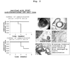
Fig. 2 shows teratoma formation (right) and cumulative survival rate (left) at one month after administration of human iPS cell 201B7 line within the anterior chamber of an eye of an immunodeficient rat. -
Fig. 3 shows teratoma formation (lower) and cumulative survival rate (upper) at one month after administration of human iPS cell FFI-01 line within the anterior chamber of an eye of an immunodeficient rat. - The present invention is explained in more detail in the following. Unless particularly indicated, the terms used in the present specification have the meanings generally used in the pertinent field.
- The present invention is a method for evaluating tumorigenicity of a test cell, comprising transplanting the cell into anterior chamber of an eye of a rat, and observing the presence or absence of tumor formation (hereinafter to be also referred to as "the evaluation method of the present invention"). As used herein, the "tumorigenicity" is used to encompass the ability to form teratomas of not only cancer cells but also undifferentiated cells.
- The rat to be used in the present invention is an experimental animal having an anterior chamber of an eye having a sufficient volume for receiving injection of about 1×10⁶ cells.
- The rat to be used in the present invention is desirably genetically and microbiologically controlled such that it is suitable for undergoing the obtained evaluation experiment of the present invention using the rat model. That is, genetically, use of an inbred strain is preferable. For example, Wistar strain, Sprague-Dawley strain and the like can be exemplified. Microbiologically, SPF or one of a gnotobiotic grade is preferably used.
- Sex, age and the like of the rat is not particularly limited as long as the volume of the anterior chamber of an eye can meet the above-mentioned conditions.
- The test cell to be the evaluation target of the evaluation method of the present invention is preferably a human cell in consideration of applicability to regenerative medicine and use as a screening system for anticancer drugs. Thus, in one embodiment, the rat to be used for the evaluation method of the present invention is desirably an immunodeficient animal so that human cells are not rejected. For example, F344/NJcl-rnu/rnu rat lacking T cell function, SCID rat, X-SCID rat, FSG rat lacking both genes of SCID and XSCID and the like can be mentioned.
- However, eye is an immune-privileged site and is known to be less likely to reject transplanted cells. Thus, in another embodiment, a normal rat having a normal immune system can also be used as an experimental animal. Since immunodeficient animals are limited in species and expensive, the use of normal experimental animals is extremely significant in increasing the broad utility of the evaluation method of the present invention.
- The test cell to be the evaluation target of the evaluation method of the present invention is a stem cell or a differentiated somatic cell induced therefrom or is a cancer cell. In preferable one embodiment, since the evaluation method of the present invention is used for evaluating safety of cells for transplantation, the test cell is exemplified by pluripotent stem cells such as embryonic stem cell (ES cell), induced pluripotent stem cell (iPS cell), mesenchymal stem cell (MSC) and the like, somatic stem cell (e.g., neural stem cell etc.), somatic cell (e.g., corneal cell, hepatocyte, kidney cell, pancreatic β cell, cardiac muscle cell, blood cells etc.) obtained therefrom by differentiation induction, and tissues (e.g., cornea, retinal pigment epithelium, hepatic tissue, kidney tissue, pancreatic tissue, cardiac muscle etc.) formed therefrom. In consideration of the fact that the in vivo test is necessary for evaluating tumorigenicity under influence of the microenvironment of the transplantation site, preferably, the test cell is desirably a cell or tissue constituting the eye. Even cells constituting other tissues can be applied to the evaluation method of the present invention as a primary screening means prior to a test at the transplantation site.
- In another embodiment, the evaluation method of the present invention is used for evaluation of cancer malignancy or as a screening system for anticancer drugs. Thus, various cancer cells (e.g., cancer cell collected from patients by biopsy and the like, cancer cell line etc.) can also be used as the test cells.
- The test cell may be derived from any animal. Preferred are cells derived from warm-blooded animal such as human and pet animal (e.g., dog, cat etc.) or domestic animal (e.g., bovine, swine etc.) or poultry (e.g., chicken, duck etc.) and the like. More preferred is a human cell.
- The number of test cells necessary for the evaluation method of the present invention is not less than 105 cells. In consideration of the volume of the anterior chamber of an eye which is the transplantation site, it is preferably 105 cells - 107 cells. International guidelines proposed by WHO indicate transplantation of 107 cells. Using the evaluation method of the present invention, however, tumorigenicity can be detected with about 60% accuracy by using 105 cells, and nearly 100% accuracy by using 106 cells. More preferably, therefore, the number of the test cells is 105 cells - 106 cells, particularly preferably about 106 cells (e.g., not less than 5×10⁵ cells and less than 5×10⁶ cells).
- The test cells can be used for transplantation by suspending the above-mentioned amount in, for example, 5 - 10 µl of a suitable medium or isotonic solution (e.g., saline, PBS etc.). To increase the retentivity of the test cells in the anterior chamber of an eye, the cell suspension can be transplanted into the anterior chamber of an eye after mixing with an extracellular matrix known per se such as Matrigel and the like.
- The anterior chamber of an eye is a region inside the eye between the iris and the endothelial cells in the innermost layer of the cornea and is filled with aqueous humor. The anterior chamber of an eye allows easy monitoring of the state of the transplanted cells at any time through the cornea, and can detect tumor formation conveniently and early. The test cells can be transplanted into the anterior chamber of an eye by injecting the cell suspension prepared above from the corneal limbic area of the eye. Transplantation may be performed for one eye, and the other eye can be used as a control.
- After transplantation, rats can be bred in the same way as before transplantation. The follow-up can be performed, for example, by visually observing tumor formation under a stereomicroscope at appropriate intervals. In the evaluation method of the present invention, for example, when the tumor formed occupies 1/4 or more of the inner volume of the anterior chamber of an eye, (tumorigenic) death can be determined.
- In addition, after a certain period of time, the eyeballs may be removed and formation of tumor including teratomas can be confirmed by tissue staining (e.g., HE staining, immunostaining by tissue specific marker) and the like.
- In conventional evaluation by subcutaneous transplantation, a tumor cannot be determined until it grows to a size of about 20 mm, and during that time, observation is not possible since it is under the skin. In contrast, in the evaluation method of the present invention, tumor formation can be detected sharply since transplanted cells can be observed at any time while the rats are alive. Therefore, nearly 100% of tumor formation can be detected at 4 weeks post-transplantation, depending on the kind of test cells and the number of transplanted cells. Even when the number of cells to be transplanted is as small as about 105 cells, tumor formation can be detected with high accuracy within 8 weeks after transplantation.
- Therefore, the period of follow-up in the evaluation method of the present invention is preferably 4 - 8 weeks.
- In the evaluation method of the present invention, the presence or absence of tumorigenicity can be evaluated with sufficient accuracy when the number of rats is 5. International guidelines proposed by WHO indicate testing using 10 individuals. Since the evaluation method of the present invention has less variation between individuals, it is also advantageous in that the number of individuals used for evaluation can be reduced from those used conventionally.
- In the evaluation method of the present invention, by using a cancer cell, for example, a cancer cell collected from a patient as a test cell, malignancy of the cancer cell can be evaluated. For example, first, the evaluation method of the present invention is performed on cancer cells known to have low clinical malignancy, the number of transplanted cells in which cancer cells do not proliferate is determined, after which the test cancer cells in the above number are transplanted into the anterior chamber of an eye of the experimental animal and follow-up is performed. When tumor formation is found, the test cancer cell can be judged to have high malignancy.
- In the evaluation method of the present invention, a cancer cell, for example, various cancer cell lines conventionally used for the evaluation of anticancer drugs, is used as a test cell, a candidate compound for an anticancer drug is administered as a test compound to animal rat, tumor formation in the anterior chamber of an eye of the rat is examined, whereby a test compound having an antitumor activity can be screened for. For example, the number of transplanted cells in which a test cancer cell line can form a tumor is first determined, after which the test cancer cells in the above number are transplanted and the test compound is administered into the anterior chamber of an eye of the rat and follow-up is performed. When suppression of tumor formation is observed, the test compound can be judged to have an antitumor activity. The test compound can be administered to the rat by any administration route and dose. In preferable one embodiment, it can be administered topically to the eye. Topical administration to the eye may be performed by injection into the anterior chamber of the eye as in the cell transplantation, or may be performed by instillation. In this case, cancer cells may be transplanted into both eyes, the drug may be administered to only one eye, and the suppressive effect on tumor formation can also be evaluated by comparing the both eyes.
- By practicing the evaluation method of the present invention for a compound selected as having an antitumor activity as a result of primary screening using a cancer cell line, or an existing anticancer drug, and by changing the test cells to a cancer cell derived from a patient, an anticancer drug effective for the patient can be selected, and a personalized cancer treatment can be performed.
- While the present invention is more specifically explained by referring to the following Examples, it is needless to say that the present invention is not limited thereto.
- As an experimental animal, an immunodeficient rat F344/NJcl-rnu/rnu (5-week-old, CLEA Japan, Inc.) was used. A mixed anesthesia solution of three types of Domitor (medetomidine hydrochloride) (75 µg/ml), Dormicum (Midazolam) (0.4 mg/ml), Betorphal (butorphanol tartrate) (0.5 mg/ml) was intraperitoneally injected at 500 µl/100 g to perform systemic anesthesia. In addition, topical anesthesia was performed with Benoxil (0.4%, Oxybuprocaine Hydrochloride eye drop) instillation. A mixed solution of a cell suspension containing 5 µl human iPS cell line 201B7 (http://cell.brc.riken.jp/ja/hps/hps0063_info) or FFI-01 (http://www.cira.kyoto-u.ac.jp/ ciRA Center for iPS Research, Kyoto University) (1x10^4 cells - 1×10^7 cells), and a solution containing Matrigel (5 ul) was injected into one eye of rats (5 rats in each group) placed under a stereomicroscope. As an antibacterial agent, a cravit instillation solution (0.5%, levofloxacin hydrate) was instilled. After transplantation, rats were followed-up for a certain period (immediately thereafter to several months). Thereafter, for more detailed analysis, the rats were euthanized by intraperitoneal overdose of pentobarbital (120 mg/kg). The eyeballs were collected, HE staining etc. was performed, and teratoma formation (differentiation into lineages of three germ layers) was confirmed. The protocol of this test is shown in
Fig. 1 . - Tumor formation occupying 1/4 or more of the anterior chamber inner volume was determined to mean dead, and Kaplan-Meier survival curve was generated. In the case of 201B7 line, when 1×10^6 cells were transplanted, all five rats were judged dead (with teratoma formation ability) at 4 weeks after transplantation (
Fig. 2 , upper left). As a result of HE staining, differentiation into lineages of three germ layers was confirmed (Fig. 2 , right). Even when 1×10^5 cells were transplanted, 60% (3/5 rats) were judged dead within 8 weeks (Fig. 2 , lower left). Even when FFI-01 line was transplanted, similar results were obtained (Fig. 3 ). When 1×10^7 cells were transplanted, all rats were judged dead at 4 weeks post-transplantation for any iPS cell line. On the other hand, when 1×10^4 cells were transplanted, tumor formation was not found in any cell line. - In conventional evaluation by subcutaneous transplantation, a tumor cannot be determined until it grows to a size of about 20 mm, and during that time, observation is not possible since it is under the skin. In contrast, in the evaluation method of the present invention, tumor formation can be detected sharply since transplanted cells can be observed at any time while the rats are alive. Thus, tumor formation was detectable with 100% accuracy at 4 weeks post-transplantation.
- According to the present invention, tumorigenicity evaluation, which previously required several months of observation period and a large number of animals, can be shortened to about 4 to 8 weeks and the number of animals can be decreased. Thus, the present invention is useful for in vivo screening for tumorigenicity of cells for transplantation. In addition, the present invention is extremely useful since it can be utilized for evaluation of cancer malignancy or as a screening system for anticancer drugs.
Claims (6)
- A method for evaluating tumorigenicity of a test cell, comprising transplanting the cell into anterior chamber of an eye of a rat, and observing the presence or absence of tumor formation,wherein the number of the cells to be transplanted is not less than 105 cells andwherein the test cell is a stem cell or a differentiated somatic cell induced therefrom.
- The method according to claim 1, wherein the observation period is 4 - 8 weeks.
- The method according to claim 1 or 2, wherein the number of the cells to be transplanted is 105 cells - 107 cells.
- The method according to any one of claims 1 to 3, wherein the rat is an immunodeficient animal.
- A method for evaluating tumorigenicity of a test cell, comprising transplanting the cell into anterior chamber of an eye of a rat, and observing the presence or absence of tumor formation,wherein the number of the cells to be transplanted is not less than 105 cells andwherein the test cell is a cancer cell.
- The method according to claim 5, comprising further administering a test compound to the rat, and evaluating an antitumor activity of the compound by using tumor formation as an index.
Applications Claiming Priority (2)
| Application Number | Priority Date | Filing Date | Title |
|---|---|---|---|
| JP2017148605 | 2017-07-31 | ||
| PCT/JP2018/028654 WO2019026903A1 (en) | 2017-07-31 | 2018-07-31 | Rapid tumorigenicity screening system |
Publications (3)
| Publication Number | Publication Date |
|---|---|
| EP3663757A1 EP3663757A1 (en) | 2020-06-10 |
| EP3663757A4 EP3663757A4 (en) | 2021-04-14 |
| EP3663757B1 true EP3663757B1 (en) | 2024-02-21 |
Family
ID=65233976
Family Applications (1)
| Application Number | Title | Priority Date | Filing Date |
|---|---|---|---|
| EP18841316.5A Active EP3663757B1 (en) | 2017-07-31 | 2018-07-31 | Rapid tumorigenicity screening system |
Country Status (4)
| Country | Link |
|---|---|
| US (1) | US11332773B2 (en) |
| EP (1) | EP3663757B1 (en) |
| JP (1) | JP7193865B2 (en) |
| WO (1) | WO2019026903A1 (en) |
Family Cites Families (1)
| Publication number | Priority date | Publication date | Assignee | Title |
|---|---|---|---|---|
| JP5208916B2 (en) | 2006-03-31 | 2013-06-12 | キュー エル ティー インク. | Drug delivery methods, structures and compositions for the nasolacrimal system |
-
2018
- 2018-07-31 US US16/635,094 patent/US11332773B2/en active Active
- 2018-07-31 EP EP18841316.5A patent/EP3663757B1/en active Active
- 2018-07-31 WO PCT/JP2018/028654 patent/WO2019026903A1/en unknown
- 2018-07-31 JP JP2019534529A patent/JP7193865B2/en active Active
Also Published As
| Publication number | Publication date |
|---|---|
| WO2019026903A1 (en) | 2019-02-07 |
| US20200308621A1 (en) | 2020-10-01 |
| EP3663757A4 (en) | 2021-04-14 |
| EP3663757A1 (en) | 2020-06-10 |
| US11332773B2 (en) | 2022-05-17 |
| JPWO2019026903A1 (en) | 2020-06-11 |
| JP7193865B2 (en) | 2022-12-21 |
Similar Documents
| Publication | Publication Date | Title |
|---|---|---|
| Lin et al. | Reactive astrocytes protect melanoma cells from chemotherapy by sequestering intracellular calcium through gap junction communication channels | |
| Joseph et al. | Enteric glia are multipotent in culture but primarily form glia in the adult rodent gut | |
| Tzameret et al. | Transplantation of human bone marrow mesenchymal stem cells as a thin subretinal layer ameliorates retinal degeneration in a rat model of retinal dystrophy | |
| Biressi et al. | Myf5 expression during fetal myogenesis defines the developmental progenitors of adult satellite cells | |
| Li et al. | Allogeneic bone marrow stromal cells promote glial–axonal remodeling without immunologic sensitization after stroke in rats | |
| He et al. | Mesenchymal stem cells-derived exosomes ameliorate blue light stimulation in retinal pigment epithelium cells and retinal laser injury by VEGF-dependent mechanism | |
| Kaneko et al. | Characteristics of bone marrow–derived microglia in the normal and injured retina | |
| EP2745840B1 (en) | Composition including stem cell-derived microvesicles for use in promoting neurogenesis | |
| JP5400381B2 (en) | Carcinogenic stem cell fusion model | |
| Tracy et al. | Intravitreal implantation of TPP1-transduced stem cells delays retinal degeneration in canine CLN2 neuronal ceroid lipofuscinosis | |
| Li et al. | Endogenous bone marrow–derived cells express retinal pigment epithelium cell markers and migrate to focal areas of RPE damage | |
| CN107988141A (en) | liver fibrosis model and its construction method and application | |
| Sefton et al. | Fibroblast-derived Hgf controls recruitment and expansion of muscle during morphogenesis of the mammalian diaphragm | |
| Toda et al. | In vivo fluorescence visualization of anterior chamber injected human corneal endothelial cells labeled with quantum dots | |
| KR20120034167A (en) | Method and compositions for increasing trichogenic potency of dermal cells | |
| EP3663757B1 (en) | Rapid tumorigenicity screening system | |
| Feng et al. | Vascular disrupting effects of combretastatin A4 phosphate on murine endometriotic lesions | |
| Huang et al. | Functional and morphological analysis of the subretinal injection of human retinal progenitor cells under Cyclosporin A treatment | |
| JP6489517B2 (en) | Differentiation-promoting agents and brain tumor therapeutic agents for cancer stem cells | |
| Kumar et al. | Isolation of tumor cells based on their distance from blood vessels | |
| Lv et al. | Tcap deficiency in zebrafish leads to ROS production and Mitophagy, and Idebenone improves its phenotypes | |
| Lapi et al. | Effects of bone marrow mesenchymal stem cells (BM-MSCs) on rat pial microvascular remodeling after transient middle cerebral artery occlusion | |
| WO2017028602A1 (en) | Use of butylidenephthalide | |
| Liu et al. | Characterization and allogeneic transplantation of a novel transgenic cone-rich donor mouse line | |
| Hua et al. | Xenograft model of human brain tumor |
Legal Events
| Date | Code | Title | Description |
|---|---|---|---|
| STAA | Information on the status of an ep patent application or granted ep patent |
Free format text: STATUS: THE INTERNATIONAL PUBLICATION HAS BEEN MADE |
|
| PUAI | Public reference made under article 153(3) epc to a published international application that has entered the european phase |
Free format text: ORIGINAL CODE: 0009012 |
|
| STAA | Information on the status of an ep patent application or granted ep patent |
Free format text: STATUS: REQUEST FOR EXAMINATION WAS MADE |
|
| 17P | Request for examination filed |
Effective date: 20200219 |
|
| AK | Designated contracting states |
Kind code of ref document: A1 Designated state(s): AL AT BE BG CH CY CZ DE DK EE ES FI FR GB GR HR HU IE IS IT LI LT LU LV MC MK MT NL NO PL PT RO RS SE SI SK SM TR |
|
| AX | Request for extension of the european patent |
Extension state: BA ME |
|
| DAV | Request for validation of the european patent (deleted) | ||
| DAX | Request for extension of the european patent (deleted) | ||
| A4 | Supplementary search report drawn up and despatched |
Effective date: 20210312 |
|
| RIC1 | Information provided on ipc code assigned before grant |
Ipc: G01N 33/48 20060101AFI20210305BHEP Ipc: C12N 5/09 20100101ALI20210305BHEP Ipc: C12N 5/095 20100101ALI20210305BHEP Ipc: C12Q 1/02 20060101ALI20210305BHEP Ipc: G01N 33/15 20060101ALI20210305BHEP Ipc: G01N 33/50 20060101ALI20210305BHEP Ipc: A61K 35/13 20150101ALI20210305BHEP Ipc: A61K 35/545 20150101ALI20210305BHEP Ipc: A61P 43/00 20060101ALI20210305BHEP Ipc: A01K 67/027 20060101ALI20210305BHEP |
|
| STAA | Information on the status of an ep patent application or granted ep patent |
Free format text: STATUS: EXAMINATION IS IN PROGRESS |
|
| 17Q | First examination report despatched |
Effective date: 20220425 |
|
| GRAP | Despatch of communication of intention to grant a patent |
Free format text: ORIGINAL CODE: EPIDOSNIGR1 |
|
| STAA | Information on the status of an ep patent application or granted ep patent |
Free format text: STATUS: GRANT OF PATENT IS INTENDED |
|
| INTG | Intention to grant announced |
Effective date: 20230925 |
|
| GRAS | Grant fee paid |
Free format text: ORIGINAL CODE: EPIDOSNIGR3 |
|
| GRAA | (expected) grant |
Free format text: ORIGINAL CODE: 0009210 |
|
| STAA | Information on the status of an ep patent application or granted ep patent |
Free format text: STATUS: THE PATENT HAS BEEN GRANTED |
|
| AK | Designated contracting states |
Kind code of ref document: B1 Designated state(s): AL AT BE BG CH CY CZ DE DK EE ES FI FR GB GR HR HU IE IS IT LI LT LU LV MC MK MT NL NO PL PT RO RS SE SI SK SM TR |
|
| REG | Reference to a national code |
Ref country code: GB Ref legal event code: FG4D |
|
| REG | Reference to a national code |
Ref country code: CH Ref legal event code: EP |
|
| REG | Reference to a national code |
Ref country code: DE Ref legal event code: R096 Ref document number: 602018065638 Country of ref document: DE |
|
| REG | Reference to a national code |
Ref country code: IE Ref legal event code: FG4D |
|
| REG | Reference to a national code |
Ref country code: LT Ref legal event code: MG9D |
|
| REG | Reference to a national code |
Ref country code: NL Ref legal event code: MP Effective date: 20240221 |
|
| PG25 | Lapsed in a contracting state [announced via postgrant information from national office to epo] |
Ref country code: IS Free format text: LAPSE BECAUSE OF FAILURE TO SUBMIT A TRANSLATION OF THE DESCRIPTION OR TO PAY THE FEE WITHIN THE PRESCRIBED TIME-LIMIT Effective date: 20240621 |
|
| PGFP | Annual fee paid to national office [announced via postgrant information from national office to epo] |
Ref country code: GB Payment date: 20240606 Year of fee payment: 7 |
|
| PG25 | Lapsed in a contracting state [announced via postgrant information from national office to epo] |
Ref country code: LT Free format text: LAPSE BECAUSE OF FAILURE TO SUBMIT A TRANSLATION OF THE DESCRIPTION OR TO PAY THE FEE WITHIN THE PRESCRIBED TIME-LIMIT Effective date: 20240221 |
|
| PG25 | Lapsed in a contracting state [announced via postgrant information from national office to epo] |
Ref country code: GR Free format text: LAPSE BECAUSE OF FAILURE TO SUBMIT A TRANSLATION OF THE DESCRIPTION OR TO PAY THE FEE WITHIN THE PRESCRIBED TIME-LIMIT Effective date: 20240522 |
|
| REG | Reference to a national code |
Ref country code: AT Ref legal event code: MK05 Ref document number: 1659527 Country of ref document: AT Kind code of ref document: T Effective date: 20240221 |
|
| PG25 | Lapsed in a contracting state [announced via postgrant information from national office to epo] |
Ref country code: NL Free format text: LAPSE BECAUSE OF FAILURE TO SUBMIT A TRANSLATION OF THE DESCRIPTION OR TO PAY THE FEE WITHIN THE PRESCRIBED TIME-LIMIT Effective date: 20240221 Ref country code: RS Free format text: LAPSE BECAUSE OF FAILURE TO SUBMIT A TRANSLATION OF THE DESCRIPTION OR TO PAY THE FEE WITHIN THE PRESCRIBED TIME-LIMIT Effective date: 20240521 Ref country code: HR Free format text: LAPSE BECAUSE OF FAILURE TO SUBMIT A TRANSLATION OF THE DESCRIPTION OR TO PAY THE FEE WITHIN THE PRESCRIBED TIME-LIMIT Effective date: 20240221 |
|
| PG25 | Lapsed in a contracting state [announced via postgrant information from national office to epo] |
Ref country code: ES Free format text: LAPSE BECAUSE OF FAILURE TO SUBMIT A TRANSLATION OF THE DESCRIPTION OR TO PAY THE FEE WITHIN THE PRESCRIBED TIME-LIMIT Effective date: 20240221 |
|
| PG25 | Lapsed in a contracting state [announced via postgrant information from national office to epo] |
Ref country code: AT Free format text: LAPSE BECAUSE OF FAILURE TO SUBMIT A TRANSLATION OF THE DESCRIPTION OR TO PAY THE FEE WITHIN THE PRESCRIBED TIME-LIMIT Effective date: 20240221 |
|
| PG25 | Lapsed in a contracting state [announced via postgrant information from national office to epo] |
Ref country code: RS Free format text: LAPSE BECAUSE OF FAILURE TO SUBMIT A TRANSLATION OF THE DESCRIPTION OR TO PAY THE FEE WITHIN THE PRESCRIBED TIME-LIMIT Effective date: 20240521 Ref country code: NO Free format text: LAPSE BECAUSE OF FAILURE TO SUBMIT A TRANSLATION OF THE DESCRIPTION OR TO PAY THE FEE WITHIN THE PRESCRIBED TIME-LIMIT Effective date: 20240521 Ref country code: NL Free format text: LAPSE BECAUSE OF FAILURE TO SUBMIT A TRANSLATION OF THE DESCRIPTION OR TO PAY THE FEE WITHIN THE PRESCRIBED TIME-LIMIT Effective date: 20240221 Ref country code: LT Free format text: LAPSE BECAUSE OF FAILURE TO SUBMIT A TRANSLATION OF THE DESCRIPTION OR TO PAY THE FEE WITHIN THE PRESCRIBED TIME-LIMIT Effective date: 20240221 Ref country code: IS Free format text: LAPSE BECAUSE OF FAILURE TO SUBMIT A TRANSLATION OF THE DESCRIPTION OR TO PAY THE FEE WITHIN THE PRESCRIBED TIME-LIMIT Effective date: 20240621 Ref country code: HR Free format text: LAPSE BECAUSE OF FAILURE TO SUBMIT A TRANSLATION OF THE DESCRIPTION OR TO PAY THE FEE WITHIN THE PRESCRIBED TIME-LIMIT Effective date: 20240221 Ref country code: GR Free format text: LAPSE BECAUSE OF FAILURE TO SUBMIT A TRANSLATION OF THE DESCRIPTION OR TO PAY THE FEE WITHIN THE PRESCRIBED TIME-LIMIT Effective date: 20240522 Ref country code: FI Free format text: LAPSE BECAUSE OF FAILURE TO SUBMIT A TRANSLATION OF THE DESCRIPTION OR TO PAY THE FEE WITHIN THE PRESCRIBED TIME-LIMIT Effective date: 20240221 Ref country code: ES Free format text: LAPSE BECAUSE OF FAILURE TO SUBMIT A TRANSLATION OF THE DESCRIPTION OR TO PAY THE FEE WITHIN THE PRESCRIBED TIME-LIMIT Effective date: 20240221 Ref country code: BG Free format text: LAPSE BECAUSE OF FAILURE TO SUBMIT A TRANSLATION OF THE DESCRIPTION OR TO PAY THE FEE WITHIN THE PRESCRIBED TIME-LIMIT Effective date: 20240221 Ref country code: AT Free format text: LAPSE BECAUSE OF FAILURE TO SUBMIT A TRANSLATION OF THE DESCRIPTION OR TO PAY THE FEE WITHIN THE PRESCRIBED TIME-LIMIT Effective date: 20240221 |
|
| PGFP | Annual fee paid to national office [announced via postgrant information from national office to epo] |
Ref country code: FR Payment date: 20240530 Year of fee payment: 7 |
|
| PG25 | Lapsed in a contracting state [announced via postgrant information from national office to epo] |
Ref country code: PL Free format text: LAPSE BECAUSE OF FAILURE TO SUBMIT A TRANSLATION OF THE DESCRIPTION OR TO PAY THE FEE WITHIN THE PRESCRIBED TIME-LIMIT Effective date: 20240221 Ref country code: PT Free format text: LAPSE BECAUSE OF FAILURE TO SUBMIT A TRANSLATION OF THE DESCRIPTION OR TO PAY THE FEE WITHIN THE PRESCRIBED TIME-LIMIT Effective date: 20240621 |
|
| PG25 | Lapsed in a contracting state [announced via postgrant information from national office to epo] |
Ref country code: SE Free format text: LAPSE BECAUSE OF FAILURE TO SUBMIT A TRANSLATION OF THE DESCRIPTION OR TO PAY THE FEE WITHIN THE PRESCRIBED TIME-LIMIT Effective date: 20240221 Ref country code: PT Free format text: LAPSE BECAUSE OF FAILURE TO SUBMIT A TRANSLATION OF THE DESCRIPTION OR TO PAY THE FEE WITHIN THE PRESCRIBED TIME-LIMIT Effective date: 20240621 Ref country code: PL Free format text: LAPSE BECAUSE OF FAILURE TO SUBMIT A TRANSLATION OF THE DESCRIPTION OR TO PAY THE FEE WITHIN THE PRESCRIBED TIME-LIMIT Effective date: 20240221 Ref country code: LV Free format text: LAPSE BECAUSE OF FAILURE TO SUBMIT A TRANSLATION OF THE DESCRIPTION OR TO PAY THE FEE WITHIN THE PRESCRIBED TIME-LIMIT Effective date: 20240221 |