EP3662086B1 - Optisches supermultiplexing durch polyyne - Google Patents
Optisches supermultiplexing durch polyyne Download PDFInfo
- Publication number
- EP3662086B1 EP3662086B1 EP18842004.6A EP18842004A EP3662086B1 EP 3662086 B1 EP3662086 B1 EP 3662086B1 EP 18842004 A EP18842004 A EP 18842004A EP 3662086 B1 EP3662086 B1 EP 3662086B1
- Authority
- EP
- European Patent Office
- Prior art keywords
- polyynes
- alkyl
- cells
- library
- imaging
- Prior art date
- Legal status (The legal status is an assumption and is not a legal conclusion. Google has not performed a legal analysis and makes no representation as to the accuracy of the status listed.)
- Active
Links
Images





Classifications
-
- C—CHEMISTRY; METALLURGY
- C12—BIOCHEMISTRY; BEER; SPIRITS; WINE; VINEGAR; MICROBIOLOGY; ENZYMOLOGY; MUTATION OR GENETIC ENGINEERING
- C12Q—MEASURING OR TESTING PROCESSES INVOLVING ENZYMES, NUCLEIC ACIDS OR MICROORGANISMS; COMPOSITIONS OR TEST PAPERS THEREFOR; PROCESSES OF PREPARING SUCH COMPOSITIONS; CONDITION-RESPONSIVE CONTROL IN MICROBIOLOGICAL OR ENZYMOLOGICAL PROCESSES
- C12Q1/00—Measuring or testing processes involving enzymes, nucleic acids or microorganisms; Compositions therefor; Processes of preparing such compositions
- C12Q1/68—Measuring or testing processes involving enzymes, nucleic acids or microorganisms; Compositions therefor; Processes of preparing such compositions involving nucleic acids
- C12Q1/6813—Hybridisation assays
- C12Q1/6816—Hybridisation assays characterised by the detection means
-
- C—CHEMISTRY; METALLURGY
- C12—BIOCHEMISTRY; BEER; SPIRITS; WINE; VINEGAR; MICROBIOLOGY; ENZYMOLOGY; MUTATION OR GENETIC ENGINEERING
- C12Q—MEASURING OR TESTING PROCESSES INVOLVING ENZYMES, NUCLEIC ACIDS OR MICROORGANISMS; COMPOSITIONS OR TEST PAPERS THEREFOR; PROCESSES OF PREPARING SUCH COMPOSITIONS; CONDITION-RESPONSIVE CONTROL IN MICROBIOLOGICAL OR ENZYMOLOGICAL PROCESSES
- C12Q1/00—Measuring or testing processes involving enzymes, nucleic acids or microorganisms; Compositions therefor; Processes of preparing such compositions
- C12Q1/68—Measuring or testing processes involving enzymes, nucleic acids or microorganisms; Compositions therefor; Processes of preparing such compositions involving nucleic acids
- C12Q1/6806—Preparing nucleic acids for analysis, e.g. for polymerase chain reaction [PCR] assay
-
- C—CHEMISTRY; METALLURGY
- C12—BIOCHEMISTRY; BEER; SPIRITS; WINE; VINEGAR; MICROBIOLOGY; ENZYMOLOGY; MUTATION OR GENETIC ENGINEERING
- C12Q—MEASURING OR TESTING PROCESSES INVOLVING ENZYMES, NUCLEIC ACIDS OR MICROORGANISMS; COMPOSITIONS OR TEST PAPERS THEREFOR; PROCESSES OF PREPARING SUCH COMPOSITIONS; CONDITION-RESPONSIVE CONTROL IN MICROBIOLOGICAL OR ENZYMOLOGICAL PROCESSES
- C12Q1/00—Measuring or testing processes involving enzymes, nucleic acids or microorganisms; Compositions therefor; Processes of preparing such compositions
- C12Q1/68—Measuring or testing processes involving enzymes, nucleic acids or microorganisms; Compositions therefor; Processes of preparing such compositions involving nucleic acids
- C12Q1/6813—Hybridisation assays
- C12Q1/6827—Hybridisation assays for detection of mutation or polymorphism
-
- C—CHEMISTRY; METALLURGY
- C12—BIOCHEMISTRY; BEER; SPIRITS; WINE; VINEGAR; MICROBIOLOGY; ENZYMOLOGY; MUTATION OR GENETIC ENGINEERING
- C12Q—MEASURING OR TESTING PROCESSES INVOLVING ENZYMES, NUCLEIC ACIDS OR MICROORGANISMS; COMPOSITIONS OR TEST PAPERS THEREFOR; PROCESSES OF PREPARING SUCH COMPOSITIONS; CONDITION-RESPONSIVE CONTROL IN MICROBIOLOGICAL OR ENZYMOLOGICAL PROCESSES
- C12Q1/00—Measuring or testing processes involving enzymes, nucleic acids or microorganisms; Compositions therefor; Processes of preparing such compositions
- C12Q1/68—Measuring or testing processes involving enzymes, nucleic acids or microorganisms; Compositions therefor; Processes of preparing such compositions involving nucleic acids
- C12Q1/6813—Hybridisation assays
- C12Q1/6834—Enzymatic or biochemical coupling of nucleic acids to a solid phase
-
- C—CHEMISTRY; METALLURGY
- C12—BIOCHEMISTRY; BEER; SPIRITS; WINE; VINEGAR; MICROBIOLOGY; ENZYMOLOGY; MUTATION OR GENETIC ENGINEERING
- C12Q—MEASURING OR TESTING PROCESSES INVOLVING ENZYMES, NUCLEIC ACIDS OR MICROORGANISMS; COMPOSITIONS OR TEST PAPERS THEREFOR; PROCESSES OF PREPARING SUCH COMPOSITIONS; CONDITION-RESPONSIVE CONTROL IN MICROBIOLOGICAL OR ENZYMOLOGICAL PROCESSES
- C12Q1/00—Measuring or testing processes involving enzymes, nucleic acids or microorganisms; Compositions therefor; Processes of preparing such compositions
- C12Q1/68—Measuring or testing processes involving enzymes, nucleic acids or microorganisms; Compositions therefor; Processes of preparing such compositions involving nucleic acids
- C12Q1/6844—Nucleic acid amplification reactions
- C12Q1/686—Polymerase chain reaction [PCR]
-
- G—PHYSICS
- G01—MEASURING; TESTING
- G01N—INVESTIGATING OR ANALYSING MATERIALS BY DETERMINING THEIR CHEMICAL OR PHYSICAL PROPERTIES
- G01N21/00—Investigating or analysing materials by the use of optical means, i.e. using sub-millimetre waves, infrared, visible or ultraviolet light
- G01N21/62—Systems in which the material investigated is excited whereby it emits light or causes a change in wavelength of the incident light
- G01N21/63—Systems in which the material investigated is excited whereby it emits light or causes a change in wavelength of the incident light optically excited
- G01N21/65—Raman scattering
-
- B—PERFORMING OPERATIONS; TRANSPORTING
- B82—NANOTECHNOLOGY
- B82Y—SPECIFIC USES OR APPLICATIONS OF NANOSTRUCTURES; MEASUREMENT OR ANALYSIS OF NANOSTRUCTURES; MANUFACTURE OR TREATMENT OF NANOSTRUCTURES
- B82Y15/00—Nanotechnology for interacting, sensing or actuating, e.g. quantum dots as markers in protein assays or molecular motors
-
- B—PERFORMING OPERATIONS; TRANSPORTING
- B82—NANOTECHNOLOGY
- B82Y—SPECIFIC USES OR APPLICATIONS OF NANOSTRUCTURES; MEASUREMENT OR ANALYSIS OF NANOSTRUCTURES; MANUFACTURE OR TREATMENT OF NANOSTRUCTURES
- B82Y30/00—Nanotechnology for materials or surface science, e.g. nanocomposites
-
- C—CHEMISTRY; METALLURGY
- C12—BIOCHEMISTRY; BEER; SPIRITS; WINE; VINEGAR; MICROBIOLOGY; ENZYMOLOGY; MUTATION OR GENETIC ENGINEERING
- C12Q—MEASURING OR TESTING PROCESSES INVOLVING ENZYMES, NUCLEIC ACIDS OR MICROORGANISMS; COMPOSITIONS OR TEST PAPERS THEREFOR; PROCESSES OF PREPARING SUCH COMPOSITIONS; CONDITION-RESPONSIVE CONTROL IN MICROBIOLOGICAL OR ENZYMOLOGICAL PROCESSES
- C12Q2565/00—Nucleic acid analysis characterised by mode or means of detection
- C12Q2565/60—Detection means characterised by use of a special device
- C12Q2565/632—Detection means characterised by use of a special device being a surface enhanced, e.g. resonance, Raman spectrometer
-
- H—ELECTRICITY
- H04—ELECTRIC COMMUNICATION TECHNIQUE
- H04Q—SELECTING
- H04Q11/00—Selecting arrangements for multiplex systems
- H04Q11/0001—Selecting arrangements for multiplex systems using optical switching
- H04Q11/0005—Switch and router aspects
Definitions
- the present disclosure generally relates to libraries of polyynes useful in optical super-multiplexing applications, as well as articles of manufacture and particles comprising the polyynes.
- optical detection is one of the most efficient methods for non-invasive information readout at far field.
- many optical multiplexing techniques have been developed, such as multiplexed microscopy and barcoding. These techniques are primarily based on fluorescence of organic dyes, fluorescent proteins or inorganic luminescent nanomaterials including quantum dots, rare earth nanoparticles and up-conversion nanocrystals.
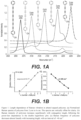
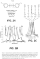
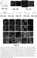
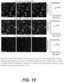
- polyynes As shown in the working examples, a small library of polyynes has been demonstrated to show 20 distinct frequencies, with single strong peak and minimal crosstalk. These polyynes were prepared using rational engineering of conjugation length, isotope doping, and substitution variations. Using these polyynes, up to 30-channel parallel optical detections can be achieved in tandem with fluorescence. With super-multiplexed polyynes, 15-color live-cell imaging can be demonstrated without the need of complicated un-mixing and 10-color optical imaging of organelles can be achieved in a single living cell with excellent specificity, sensitivity, and photostability and live-cell compatibility.
- the polyynes are used to image cultured live cells.
- the methods involve culturing live cells, which can be derived from an in vitro tissue cell culture of interest, extracted from an in vivo subject of interest, seeding the cultured live cells in wells, and labelling each well with a single color of polyynes in culture media, wherein the polyynes comprise one or more colors.
- the labelled cultured live cells are then added to an imaging chamber for stimulated Raman microscopy, and the cells are imaged using stimulated Raman microscopy, wherein each cell is maintained with a single color during the imaging period.
- the polyynes are used to image organelles in cultured live cells.
- the methods involve culturing live cells, which can be derived from an in vitro tissue cell culture of interest or extracted from an in vivo subject of interest, incubating the cultured live cells with polyynes in culture media, wherein the polyynes each comprise a specific organelle-targeted probe, labelling the cultured live cells with the polyynes, and imaging the cells using stimulated Raman microscopy.
- the cultured live cells are seeded in a well before they are incubated and labelled with the polyynes, and then the cells are labelled with the polyynes, wherein the polyynes each comprise a specific organelle-targeted probe, which probes specifically target different organelles.
- Representative organelle-targeted probes include probes which target the plasma membrane, endoplasmic reticulum (ER), Golgi, mitochondria, lipid droplets, lysosome, nucleus, and tubulin in the cultured live cells.
- the polyynes are used in methods of live-cell tagging using spectral barcoded beads.
- the methods involve mixing polymeric microbeads, such as polystyrene beads, with spectral barcoded polyynes, binding the beads with the spectral barcoded polyynes to form barcoded beads, and culturing live cells, which can be derived from in vitro tissue cell culture of interest, or extracted from an in vivo subject of interest.
- the barcoded beads are incubated with the cultured live cells in culture media, which labels the cultured live cells with the barcoded beads.
- the cultured live cells can then be imaged using stimulated Raman microscopy, and the spectral barcodes of the barcoded beads in the whole field of view can be decoded based on hyperspectral SRS (Stimulated Raman scattering) images.
- SRS Stimulated Raman scattering
- the polyynes are used to identify a population of cancer cells using flow cytometry, or other methods for sorting/counting cells.
- the methods involve associating one or more polyynes (also referred to herein as "carbon atom wires") with live cells of a subject of interest, wherein the carbon atom wires are conjugated with antibodies that detect, and bind to, specific cell surface markers of the live cells, and further wherein the antibodies bind to cell surface markers of the live cells.
- the live cells are positioned in a focused flow stream of a flow cytometer; and illuminated within the focused flow stream of the flow cytometer with a predetermined wavelength of light. The cells are exposed to the illumination, and light is deflected as it comes into contact with the cells.
- the resulting scattered light from the live cells is detected in the frequency domain, and the Raman scattering spectrum is analyzed to identify individual groups of live cells.
- Cells that are not cancer cells are not linked to the carbon atom wire-conjugated antibodies, whereas cancer cells are linked to the carbon atom wire-conjugated antibodies.
- the cancer cells are detected based on the Raman spectra of scattered light produced by the carbon atom wires conjugated with antibodies.
- the particular cell type can be detected based on the Raman spectra of scattered light, rather than the absorption, at a single wavelength.
- the scattering is not Rayleigh scattering, but rather, Raman scattering, which has different wavelengths compared to that of excitation light. By looking at the Raman spectrum, it is possible to separately identify cancer cells and non-cancer cells.
- the scattered light is detected using a detector which detects a broad range of wavelength, for example, using spectrograph and/or a camera.
- the polyynes are used in methods of flow cytometry-based high throughput medical diagnosis.
- the methods involve associating one or more carbon atom wires (which may be conjugated to one or more antibodies, nucleic acids or other molecules which form a complex with specific biological markers on specific subpopulations of cells which are indicative of a particular disease state with live cells of a subject of interest, wherein specific subpopulations of the live cells are specifically associated with distinct sets of carbon atom wires.
- the live cells are positioned in a flow cytometer, and the live cells are illuminated within the focused flow stream of the flow cytometer, wherein the carbon atom wires are exposed to the illumination. Scattered light from the live cells is detected, and analyzed to identify individual groups of live cells.
- An analysis of the Raman scattering spectrum can provide a diagnosis of a specific disease, based on the binding of the carbon atom wires to specific subpopulations of cells, if present in the cell population.
- the members of the specific subpopulations of cells When bound to the carbon atom wires, the members of the specific subpopulations of cells will scatter light at specific wavelengths that unbound cells will not scatter.
- the presence of cells within one or more sub-populations can be detected, and correlated with a particular disease state.
- carbon atom wires conjugated to antibodies, nucleic acids or other molecules can be used which bind to bacteria, preferably to one or more specific bacteria, and if a bacteria, ideally a specific type of bacteria, is detected, appropriate antibiotics can be administered, and if a bacteria is not detected, antibiotic administration can be avoided.
- This can be particularly advantageous where a patient has sepsis or bacterial meningitis, and real-time assays can be particularly preferred over typical cell culture-based approaches, which often cause delays in patients receiving appropriate care.
- Another exemplary embodiment involves methods of cell sorting.
- the methods involve associating carbon atom wires with live cells of a subject of interest, wherein specific subpopulations of the live cells are specifically associated with distinct sets of carbon atom wires.
- the specific subpopulations of live cells are sorted using spectroscopy, based on the specific association of the subpopulation of cells with a distinct set of carbon atom wires. Rather than using fluorescence to identify the cells, stimulated Raman spectroscopy can be used.
- libraries of carbon atom wires are used, each of which binds to a different subpopulation of cells, and each of which has a different "spectral barcode," a single biological sample can be screened for multiple subpopulations of cell types.
- the polyynes are used to produce a cell atlas in vivo.
- the methods involve associating carbon atom wires with live cells of a subject of interest, wherein specific subpopulations of the live cells are specifically associated with distinct sets of carbon atom wires; mapping relationships between the specific subpopulations of live cells, wherein the specific subpopulations of live cells are distinguishable by their specific association with a distinct set of carbon atom wires.
- the polyynes are attached to a solid support, such as a nanoparticle, microparticle, or metal nanostructure, such as a gold particle.
- the diameter of the particle is between about 2 and about 200 nm, more typically, between about 2 and about 100 nm, and still more typically, between about 4 and about 50 nm.
- Ranges may be expressed herein as from “about” one particular value, and/or to "about” another particular value. When such a range is expressed, another exemplary embodiment includes from the one particular value and/or to the other particular value. Similarly, when values are expressed as approximations, by use of the antecedent "about,” it will be understood that the particular value forms another exemplary embodiment. It will be further understood that the endpoints of each of the ranges are significant both in relation to the other endpoint, and independently of the other endpoint. It is also understood that there are a number of values disclosed herein, and that each value is also herein disclosed as “about” that particular value in addition to the value itself. For example, if the value “10” is disclosed, then “about 10" is also disclosed.
- alkyl as used herein, unless otherwise specified, can refer to a saturated straight, branched, or cyclic, primary, secondary, or tertiary hydrocarbons, including both substituted and unsubstituted alkyl groups.
- the alkyl group can be optionally substituted with any moiety that does not otherwise interfere with the reaction or that provides an improvement in the process, including but not limited to but limited to halo, haloalkyl, hydroxyl, carboxyl, acyl, aryl, acyloxy, amino, amido, carboxyl derivatives, alkylamino, dialkylamino, arylamino, alkoxy, aryloxy, nitro, cyano, sulfonic acid, thiol, imine, sulfonyl, sulfanyl, sulfinyl, sulfamonyl, ester, carboxylic acid, amide, phosphonyl, phosphinyl, phosphoryl, phosphine, thioester, thioether, acid halide, anhydride, oxime, hydrozine, carbamate, carbonate, urea, phosphonic acid, or phosphonate, either unprotected, or
- alkyl can include C 1-22 alkyl moieties, and the term “lower alkyl” includes C 1-6 alkyl moieties. It is understood to those of ordinary skill in the art that the relevant alkyl radical is named by replacing the suffix "-ane” with the suffix "-yl”.
- alkenyl can refer to an unsaturated, hydrocarbon radical, linear or branched, in so much as it contains one or more double bonds.
- the alkenyl group disclosed herein can be optionally substituted with any moiety that does not adversely affect the reaction process, including but not limited to but not limited to those described for substituents on alkyl moieties.
- Non-limiting examples of alkenyl groups include ethylene, methylethylene, isopropylidene, 1,2-ethane-diyl, 1,1-ethane-diyl, 1,3-propane- diyl, 1,2-propane-diyl, 1,3-butane-diyl, and 1,4-butane-diyl.
- alkynyl can refer to an unsaturated, acyclic hydrocarbon radical, linear or branched, in so much as it contains one or more triple bonds.
- the alkynyl group can be optionally substituted with any moiety that does not adversely affect the reaction process, including but not limited to those described above for alkyl moieties.
- Non-limiting examples of suitable alkynyl groups include ethynyl, propynyl, hydroxypropynyl, butyn-1-yl, butyn-2-yl, pentyn-1-yl, pentyn-2-yl, 4-methoxypentyn-2-yl, 3-methylbutyn-1-yl, hexyn-1-yl, hexyn-2-yl, and hexyn-3-yl, 3,3-dimethylbutyn-1-yl radicals.
- alkylamino or arylamino can refer to an amino group that has one or two alkyl or aryl substituents, respectively.
- acyl can refer to a carboxylic acid ester in which the non-carbonyl moiety of the ester group is selected from the group consisting of straight, branched, or cyclic alkyl or lower alkyl, alkoxyalkyl, including, but not limited to methoxymethyl, aralkyl, including, but not limited to, benzyl, aryloxyalkyl, such as phenoxymethyl, aryl, including, but not limited to, phenyl, optionally substituted with halogen (F, Cl, Br, or I), alkyl (including but not limited to C 1 , C 2 , C 3 , and C 4 ) or alkoxy (including but not limited to C 1 , C 2 , C 3 , and C 4 ), sulfonate esters such as alkyl or aralkyl sulphonyl including but not limited to methanesulfonyl, the mono, di or triphosphate ester, trityl or mono
- alkoxy and “alkoxyalkyl” can embrace linear or branched oxy-containing radicals having alkyl moieties, such as methoxy radical.
- alkoxyalkyl also embraces alkyl radicals having one or more alkoxy radicals attached to the alkyl radical, that is, to form monoalkoxyalkyl and dialkoxyalkyl radicals.
- the "alkoxy” radicals can be further substituted with one or more halo atoms, such as fluoro, chloro or bromo, to provide "haloalkoxy" radicals.
- alkylamino can denote “monoalkylamino” and “dialkylamino” containing one or two alkyl radicals, respectively, attached to an amino radical.
- arylamino denotes “monoarylamino” and “diarylamino” containing one or two aryl radicals, respectively, attached to an amino radical.
- aralkylamino embraces aralkyl radicals attached to an amino radical.
- aralkylamino denotes “monoaralkylamino” and “diaralkylamino” containing one or two aralkyl radicals, respectively, attached to an amino radical.
- aralkylamino further denotes "monoaralkyl monoalkylamino" containing one aralkyl radical and one alkyl radical attached to an amino radical.
- aryl alone or in combination, can mean a carbocyclic aromatic system containing one, two or three rings wherein such rings can be attached together in a pendent manner or can be fused.
- Non-limiting examples of aryl include phenyl, biphenyl, or naphthyl, or other aromatic groups that remain after the removal of a hydrogen from an aromatic ring.
- aryl includes both substituted and unsubstituted moieties.
- alkaryl or “alkylaryl” can refer to an alkyl group with an aryl substituent.
- aralkyl or arylalkyl refer to an aryl group with an alkyl substituent.
- heteroatom can refer to oxygen, sulfur, nitrogen and phosphorus.
- heteroaryl or “heteroaromatic,” as used herein, can refer to an aromatic that includes at least one sulfur, oxygen, nitrogen or phosphorus in the aromatic ring.
- the heteroaryl ring can be a a moiety with 5-10 ring atoms, of which 1-5 ring atoms are heteroatoms.
- heteroarylalkyl can refer to a heteroaryl moiety attached to a C1-12 alkyl group.
- aryl, arylalkyl, alkylaryl, heteroaryl, and heterarylalkyl groups can optionally be substituted with any moiety that does not adversely affect the ability of the polyynes to be detected, including but not limited to those described above for alkyl moieties.
- Non-limiting examples of substituted aryl include heteroarylamino, N-aryl-N- alkylamino, N-heteroarylamino-N-alkylamino, heteroaralkoxy, arylamino, arylalkylamino, arylthio, monoarylamidosulfonyl, arylsulfonamido, diarylamidosulfonyl, monoaryl amidosulfonyl, arylsulfinyl, arylsulfonyl, heteroarylthio, heteroarylsulfinyl, heteroarylsulfonyl, aroyl, heteroaroyl, aralkanoyl, heteroaralkanoyl, hydroxyaralkyl, hydoxyheteroaralkyl, haloalkoxyalkyl, aryl, aralkyl, aryloxy, aralkoxy, aryloxyalkyl, saturated heterocyclyl,
- heterocyclic can refer to a nonaromatic cyclic group wherein there is at least one heteroatom, such as oxygen, sulfur, nitrogen, or phosphorus in the ring.
- heteroaryl and heterocyclic groups can include furyl, furanyl, pyridyl, pyrimidyl, thienyl, isothiazolyl, imidazolyl, tetrazolyl, pyrazinyl, benzofuranyl, benzothiophenyl, quinolyl, isoquinolyl, benzothienyl, isobenzofuryl, pyrazolyl, indolyl, isoindolyl, benzimidazolyl, purinyl, carbazolyl, oxazolyl, thiazolyl, isothiazolyl, 1,2,4- thiadiazolyl, isooxazolyl, pyrrolyl, quinazolinyl, cinnolinyl, phthalazinyl, xanthinyl, hypoxanthinyl, thiophene, furan, pyrrole, isopyrrole, pyrazole, imid
- the heteroaromatic group can be optionally substituted as described above for aryl.
- the heterocyclic or heteroaromatic group can be optionally substituted with one or more substituents selected from the group consisting of halogen, haloalkyl, alkyl, alkoxy, hydroxy, carboxyl derivatives, amido, amino, alkylamino, and dialkylamino.
- the heteroaromatic can be partially or totally hydrogenated as desired.
- dihydropyridine can be used in place of pyridine. Functional oxygen and nitrogen groups on the heterocyclic or heteroaryl group can be protected as necessary or desired.
- Suitable protecting groups are well known to those skilled in the art, and include trimethylsilyl, dimethylhexylsilyl, t-butyldimethylsilyl, and t-butyldiphenylsilyl, trityl or substituted trityl, alkyl groups, acyl groups such as acetyl and propionyl, methanesulfonyl, and p-toluenelsulfonyl.
- the heterocyclic or heteroaromatic group can be substituted with any moiety that does not adversely affect the reaction, including but not limited to but not limited to those described above for aryl.
- nucleic acid can refer to DNA, including genomic DNA and cDNA, RNA, such as siRNA, mRNA, tRNA, tmRNA, ncRNA, rRNA, shRNA, ribozymes, and the like, riboswitches, aptamers, primers, probes, and artificial nucleic acid analogues, such as peptide nucleic acids, morpholino- and locked nucleic acids, glycol nucleic acids, and threose nucleic acids.
- peptide can refer to a natural or synthetic compound containing two to one hundred amino acids linked by the carboxyl group of one amino acid to the amino group of another.
- protected can refer to a group that is added to an oxygen, nitrogen, or phosphorus atom to prevent its further reaction or for other purposes.
- oxygen and nitrogen protecting groups are known to those skilled in the art of organic synthesis, and are described, for example, in Greene et al., Protective Groups in Organic Synthesis , supra.
- spectral barcoded polyynes can relate to where multiple polyynes are used, each of which has a unique spectral band. These multiple polyynes can be conjugate to, or encapsulated within, a solid support, such as a nanoparticle or microparticle. Nanoparticles typically have a size between about 2 and about 100 nm, and microparticles typically have a diameter between about 0.1 and 100 ⁇ m.
- the solid support can also be conjugated to a binding member which is known to bind specifically to a biological particle of interest.
- the presence of multiple polyynes allows for the generation of multiple spectral bands, which provides a unique "barcode" to the solid support.
- the spectral barcoding of polyynes in solid supports can be achieved by controlled physical absorption, such as using microfluidics, or controlled chemical synthesis.
- solid support refers to any type of solid support to which one or more of the polyynes, and, ideally, a binding member, such as an antibody, nucleic acid, and the like, are bound.
- a binding member such as an antibody, nucleic acid, and the like. Examples include, but are not limited to, nanoparticles, including nanobeads, nanorods, nanostars and nanowires, such as metallic nanostructures and carbon nanotubes, microparticles, including microbeads, such as polystyrene microbeads, and chips.
- flow cytometry can be a laser- or impedance-based, biophysical technology employed in cell counting, cell sorting, biomarker detection and protein engineering.
- Cells are suspended in a stream of fluid, and passed through an electronic detection apparatus.
- a flow cytometer allows simultaneous multiparametric analysis of the physical and chemical characteristics of up to thousands of particles per second.
- a common variation involves linking the analytical capability of the flow cytometer to a sorting device, to physically separate and thereby purify particles of interest based on their optical properties. Such a process is called cell sorting, and the instrument is commonly termed a "cell sorter".
- Modern flow cytometers can be used to analyze many thousand particles per second, in "real time,” and, if configured as cell sorters, can actively separate and isolate particles at similar rates having specified optical properties.
- a flow cytometer can have, e.g., five main components: a flow cell, a measuring system, a detector, an amplification system, and a computer for analysis of the signals.
- the flow cell has a liquid stream (sheath fluid), which carries and aligns the cells so that they pass single file through the light beam for sensing.
- the measuring system commonly use measurement of impedance (or conductivity) and optical systems - lamps (mercury, xenon); high-power water-cooled lasers (argon, krypton, dye laser); low-power air-cooled lasers (argon (488 nm), red-HeNe (633 nm), green-HeNe, HeCd (UV)); diode lasers (blue, green, red, violet) resulting in light signals.
- the detector and analog-to-digital conversion (ADC) system converts analog measurements of forward-scattered light (FSC) and side-scattered light (SSC) as well as dye-specific fluorescence signals into digital signals that can be processed by a computer.
- the amplification system can be linear or logarithmic.
- the process of collecting data from samples using the flow cytometer can be termed 'acquisition'. Acquisition can be mediated by a computer physically connected to the flow cytometer, and the software which handles the digital interface with the cytometer.
- the software is capable of adjusting parameters (e.g., voltage, compensation) for the sample being tested, and also assists in displaying initial sample information while acquiring sample data to ensure that parameters are set correctly.
- Flow cytometers can typically have multiple lasers and fluorescence detectors. Increasing the number of lasers and detectors allows for multiple antibody labeling, and can more precisely identify a target population by their phenotypic markers. Certain instruments can even take digital images of individual cells, allowing for the analysis of fluorescent signal location within or on the surface of cells.
- stimulated Raman spectroscopy flow cytometry can be a technique which combines stimulated Raman spectroscopy with flow cytometry.
- SRS flow cytometry and, more specifically, a multiplex stimulated Raman scattering process, is described, for example, in Zhang et al., "Stimulated Raman scattering flow cytometry for label-free single-particle analysis," Optica, Vol. 4, No. 1 / January 2017 .
- an SRS flow cytometer can detect single particles with spectral coverage of 200 cm -1 and a throughput over 10,000 particles per second.
- Stimulated Raman scattering (SRS) microscopy can be an imaging technique that looks at the vibrational frequencies of chemical bonds. Different types of bonds will have different frequencies based on the surrounding molecular environment. For example, a C-H bond on a DNA molecule (2,956 cm-1) is going to have a slightly different vibrational frequency compared to a C-H bond on proteins (2,931 cm-1) or lipids (2,854 cm-1). Unlike traditional Raman spectroscopy, SRS can obtain data on a sample rapidly, allowing for real-time, in vivo studies.
- Hyperspectral imaging can be part of a class of techniques commonly referred to as spectral imaging or spectral analysis. Hyperspectral imaging is related to multispectral imaging. Hyperspectral imaging (HSI) uses continuous and contiguous ranges of wavelengths (e.g. 400 - 1100 nm in steps of 0.1 nm) whilst multispectral imaging (MSI) uses a subset of targeted wavelengths at chosen locations (e.g. 400 - 1100 nm in steps of 20 nm).
- HSUMI hyperspectral imaging
- MSI multispectral imaging
- Multispectral imaging deals with several images at discrete and somewhat narrow bands. Being “discrete and somewhat narrow” is what distinguishes multispectral imaging in the visible wavelength from color photography.
- a multispectral sensor may have many bands covering the spectrum from the visible to the longwave infrared. Multispectral images do not produce the "spectrum" of an object.
- Hyperspectral imaging can deal with imaging narrow spectral bands over a continuous spectral range, producing the spectra of all pixels in the scene.
- a sensor with only 20 bands can also be hyperspectral when it covers the range from 500 to 700 nm with 20 bands each 10 nm wide.
- the recorded spectra have fine wavelength resolution and cover a wide range of wavelengths.
- Hyperspectral imaging measures contiguous spectral bands, as opposed to multispectral imaging which measures spaced spectral bands.
- vibrational microscopy can include spontaneous Raman microscopy, such as confocal Raman and line-scan Raman microscopy, surface-enhanced Raman scattering (SERS) microscopy, Tip-enhanced Raman scattering (TERS) microscopy, infrared microscopy, infrared-AFM microscopy, infrared photothermal microscopy, and coherent Raman microscopy, such as coherent anti-Stokes Raman scattering (CARS) microscopy.
- spontaneous Raman microscopy such as confocal Raman and line-scan Raman microscopy, surface-enhanced Raman scattering (SERS) microscopy, Tip-enhanced Raman scattering (TERS) microscopy, infrared microscopy, infrared-AFM microscopy, infrared photothermal microscopy, and coherent Raman microscopy, such as coherent anti-Stokes Raman scattering (CARS) microscopy.
- SERS
- polyynes conjugates of the polyynes with various biological and non-biological moieties, libraries comprising a plurality of the polyynes, the use of the polyynes in biological applications, and the use of the polyynes in non-biological applications, including non-imaging applications.
- polyynes described herein can represent a new class of 1-D optical material with wide application in super-multiplexed imaging and analysis. These compounds extend the current limit of optical super-multiplexing techniques and provides new opportunities in life science, medical diagnostics, drug discovery and data security.
- polyyne compounds with phenyl, alkyl, or other end-capping groups has been designed, synthesized and characterized, and their unique optical properties for super-multiplexed detection have been exploited.
- Polyynes are linear chains of sp-hybridized carbon atoms with alternating single and tripe bonds, which are also referred to herein as carbon-atom wires.
- the polyynes include anywhere from 2 to 20, preferably 3 to 20, more typically, 3 to 10, triple bonds in alternation with single bonds, preferably from 2 to 6 triple bonds.
- the polyynes include one or more end caps, which can be present at one or both ends of the polyynes.
- the polyynes can include an end cap at either or both ends of the molecules.
- one or both end cap(s) are, independently, aryl or heteroaryl rings, and in other embodiments, one or both end caps are alkyl, alkylaryl, arylalkyl, heterocyclic, heterarylalkyl, silyl groups, metal atoms (i.e., metal acetylides, such as silver or mercury acetylides, whether covalently bound to the terminal carbon atom or complexed to the triple bond in the terminal acetylene) or metal complex groups
- the alkyl groups are branched or cyclic alkyl groups rather than linear alkyl groups, and on information and belief, the branching, or steric hindrance provided by the cycloalkyl groups, can stabilize the polyyne molecules.
- the aryl and/or heteroaryl rings are functionalized with one or more functional groups, which can be electron donating or electron withdrawing groups.
- the polyynes have the structure:
- substituents on one or more of the aryl, C 1-20 alkyl, aryl-C 1-20 alkyl, C 1-20 alkyl-aryl, heteroaryl, heteroaryl- C 1-20 alkyl, or C 1-20 alkyl-heteroaryl groups include, but are not limited to, one or more of halo, haloalkyl, hydroxyl, carboxyl, acyl, aryl, acyloxy, amino, amido, carboxyl derivatives, alkylamino, dialkylamino, arylamino, alkoxy, aryloxy, nitro, cyano, sulfonic acid, thiol, imine, sulfonyl, sulfanyl, sulfinyl, sulfamonyl, ester, carboxylic acid, amide, phosphonyl, phosphinyl, phosphoryl, phosphine, thioester, thioether, acid halide
- Representative polyynes include, but are not limited to, Carbow2172, Carbow2141, Carbow 2128, Carbow2100, Carbow2086, Carbow2066, Carbow2049, and Carbow2017, as described in the working examples.
- the polyynes have well-resolved Raman peaks with less than 10% cross-talk.
- binding members are suitable for binding to the polyynes described herein, which can allow the polyynes to be used in various detections methods described herein.
- the binding members are selected for their ability to specifically bind to a target of interest, and also to be capable of being attached to a polyyne.
- Suitable binding members include, for example, an antigen, an antibody, biotin, avidin, streptavidin, anti-biotin, folate, folate-binding protein, IgG, Protein A, Protein G, Protein L, a carbohydrate, lectin, lipid, and a nucleic acid.
- the binding members also include nucleic acids, including DNA, including genomic DNA and cDNA, RNA, such as siRNA, mRNA, tRNA, tmRNA, ncRNA, rRNA, shRNA ribozymes, and the like, riboswitches, aptamers, primers, probes, and artificial nucleic acid analogues, such as peptide nucleic acids, morpholino- and locked nucleic acids, glycol nucleic acids, and threose nucleic acids.
- nucleic acids including DNA, including genomic DNA and cDNA, RNA, such as siRNA, mRNA, tRNA, tmRNA, ncRNA, rRNA, shRNA ribozymes, and the like, riboswitches, aptamers, primers, probes, and artificial nucleic acid analogues, such as peptide nucleic acids, morpholino- and locked nucleic acids, glycol nucleic acids, and thre
- the polyynes can be bonded to the binding members either directly, as an end cap, or by attachment to a functional group on one or both end caps.
- Those of skill in the art can readily appreciate how to attach a binding member to an alkyne, or to an end cap.
- carbamate moieties on an aryl end cap can be used to conjugate antibodies or peptides.
- Terminal alkyne groups in the polyynes can be bonded to aryl/heteroaryl halides, for example, using palladium coupling reactions.
- this type of coupling reaction can be used to attach primers or probes directly to a polyyne as an end cap.
- this type of chemistry can be used to attach other nucleic acids to the polyynes.
- amine, carboxyl, and thiol based crosslinking chemistry such as using NHS ester and maleimide groups, or click chemistry can be used to conjugate peptides, proteins and nucleic acids to polyynes.
- the polyynes have a neutral scaffold and high membrane permeability, and, as such, can be functionalized into live-cell, organelle-specific imaging probes.
- An appropriate linker or functional group can be used to attach different targeting groups to the polyynes.
- the linker or functional group is present on an aryl or alkyl end cap.
- Triphenylphosphonium is a motif with high affinity to mitochondrial matrix due to the positive charge.
- Other positively charged motifs can be used, which can also target the mitochondrial matrix.
- the lysosome lumen is acidic, and basic motifs can be protonated and trapped inside lysosomes.
- Mono and dialkylamines are representative motifs which can be used. Examples include, but are not limited to, dimethylamine, diethylamine, dipropylamine, diisopropylamine, dibutylamine, and morpholine groups.
- the plasma membrane includes anionic phosphate headgroups.
- Cationic diammonium groups can be used to stain the plasma membrane through interaction with these anionic phosphate headgroups.
- An ethylene glycol chain with different terminal groups such as a pentafluorobenzamide group, or a glibenclamide group can be used to target the endoplasmic reticulum.
- Lipid droplets are known to interact with fatty acids, so carboxylic acid and/or carboxylate-terminated fatty acid chains (typically between 8 and 21 carbons in length) can be attached either directly to the end of the polyynes, or to an aryl end cap. These fatty acid chains mimic fatty acids, allowing polyynes with this functionality to function as probes which can be efficiently incorporated into lipid droplets.
- next-generation polyyne probes for super-multiplexed detection can be developed through multiple nanoscale signal amplification approaches.
- Dendrimers and polymeric nanostructures such as bottle-brush and star-shaped nanomaterials can be grafted with multiple polyynes for topological signal amplification.
- dendrimers are prepared in step-wise fashion, using divergent methods, convergent methods, and click methods.
- divergent synthesis the dendrimer is assembled from a multifunctional core, which is extended outward by a series of reactions, commonly a Michael reaction.
- convergent synthesis dendrimers are built from small molecules that end up at the surface of a sphere, and reactions proceed inward, building inward, and are eventually attached to a core.
- This method tends to provide dendrimers which are more monodisperse than when divergent synthesis is used, but the dendrimers also tend to be smaller.
- Click chemistry uses, for example, Diels-Alder reactions, thiol-ene and thiol-yne reactions, and azide-alkyne reactions. Since the polyynes (before an end cap is attached) include terminal acetylenic groups, these groups can be used in thiol-yne reactions, azide-alkyne reactions, and Diels-Alder reactions to attach the polyynes to a dendrimer.
- Noble metal nanostructures such as, e.g., nanospheres, nanorods and nanostars can be coated with polyyne molecules using physical adsorption or chemical modification for signal amplification through surface-enhanced Raman scattering.
- Traditional coating methods for coating nanostructures can be employed.
- one method of coating nanoparticles comprises subjecting a) nanoparticles, b) a coating precursor and c) one or more polyynes which include one or more reactive groups which can react with the coating precursor, to shear, wherein the coating precursor and the one or more polyynes react to provide a coating on the nanostructures.
- polyynes are conjugated to nanoparticles and/or microparticles (such as nanoparticles, polystyrene beads, gold particles, and the like).
- polyynes can be prepared which include functional groups which react with the functional groups on the nano or microparticles, and a covalent attachment between the nano or microparticles and the polyynes can be formed.
- the amount of polyynes attached to the particles can vary depending on the number of groups on each particle, which itself can vary, for example, depending on the particle size, and the percentage of such groups which is successfully coupled with the polyynes.
- the dendrimers can include a known quantity of the polyynes, by virtue of the active functional groups at the terminus on the dendrimers.
- the polyynes can be conjugated to nanoparticles/microparticles by chemical means.
- a range of functionalized groups can be present on the polyynes, and used to attach them to the nanoparticles/microparticles, including low molecular weight ligands ( Chen et al., Ligand conjugated low-density lipoprotein nanoparticles for enhanced optical cancer imaging in vivo, J. Am. Chem. Soc., 129 (18) (2007), pp.
- peptides Mu, et al., Anti-HER2/neu peptide-conjugated iron oxide nanoparticles for targeted delivery of paclitaxel to breast cancer cells, Nanoscale (2015), proteins (Meziani, and Sun, Protein-conjugated nanoparticles from rapid expansion of supercritical fluid solution into aqueous solution, J. Am. Chem. Soc., 125 (26) (2003), pp. 8015-8018 , polysaccharides ( Lemarchand, et al., Polysaccharide-decorated nanoparticles, Eur. J. Pharm. Biopharm., 58 (2) (2004), pp.
- the two main strategies for conjugating proteins/peptides/antibodies to gold nanoparticles are classic passive adsorption, and covalent conjugation.
- the polyynes described herein are present in libraries, for example, libraries with between 2 and 50,000 members, more typically, between 2 and about 200 members, still more typically, between 2 and about 100 members, and even more typically, between about 4 and about 50 members.
- the polyyne libraries comprise a plurality of polyynes linked to antibodies, which antibodies are specific for different types of cells, including stem cells, cancer cells, bacteria, viruses, or fungi.
- the polyyne libraries comprise a plurality of polyynes linked to nucleic acids, which are specific for different types of cells, including stem cells, cancer cells, bacteria, viruses, or fungi.
- the polyyne libraries comprise a plurality of polyynes linked to primers, which primers can be used in genetic screening applications to identify the presence of single nucleotide polymorphisms, such as cytochrome P450 mutations, to identify types of mutations present in viruses, such as HIV, which can be indicative of drug resistance, or in cancer cells, such as HER2 and other mutations, which can be useful in determining an appropriate course of treatment for a patient.
- the use of polyyne labels replaces the use of fluorescent labels in PCR techniques, and the use of Raman spectroscopy replaces the detection of fluorescence. Because multiple "barcoded" primers can be screened for at once, all with distinguishable signals, this allows for the simultaneous detection of multiple mutations in a single scan.
- the individual polyynes can include structural differences, such as isotopic labeling, difference in the numbers of alkyne moieties, and end cap functionalization, so as to provide a plurality of polyynes, which provide signals in Raman spectroscopy with minimal "crosstalk" with respect to each other.
- the libraries can include polyynes with up to 50 distinct frequencies, ranging from about 1850 to about 2600 cm -1 , with single strong peak and minimal crosstalk. Up to 50-channel parallel optical detections can be achieved in tandem with fluorescence within this frequency range. With super-multiplexed polyynes, up to 50 color live cell imaging, typically up to 30 color live cell imaging, and, more typically, up to 15-color live-cell imaging can be achieved, without the need of complicated un-mixing, within this frequency range.
- libraries useful for optical data storage and identification applications can include sufficient polyynes to generate a significant number of distinct barcodes, for example, up to around 10 13 barcodes, and, in some exemplary embodiments, at least around 50,000 or more distinct barcodes. These distinct barcodes can be achieved, for example, even with libraries including only up to around 200 individual polyynes.
- a barcode can include one or more polyynes, each of which has its own signal, preferably a signal which does not "cross-talk" more than about 10% with a signal associated with an adjacent polyyne (i.e., a polyyne which has a Raman spectral peak adjacent to a Raman spectral peak of another polyyne).
- polyynes in a library With just three polyynes in a library, one can create numerous barcodes. For example, if all of the polyynes are present at the same concentration, one can have polyyne 1, polyyne 2, polyyne 3, polyynes 1 and 2, polyynes 1 and 3, polyynes 2 and 3, and polyynes 1, 2, and 3, a total of seven combinations. However, the polyynes can be present in different concentrations, which will provide Raman spectral peaks with different heights (signal intensities). Typically, one can distinguish up to about 5 different concentrations. So, with just three polyynes, at five different possible concentrations, one has 6 3 possible combinations (i.e., more than 200 possible combinations). Thus, with up to 200 polyynes, it is possible to have a very significant number of possible combinations of polyynes, at multiple possible concentrations of the polyynes.
- an intimate mixture of the polyynes in a solid structure such as an ID badge, passport, driver's license, credit card, merchandise tag, currency, bonds, such as bearer bonds or other financial instruments, and articles of manufacture, such as medicines, containers, clothing, shoes, handbags, DVDs and the like.
- a solid structure such as an ID badge, passport, driver's license, credit card, merchandise tag, currency, bonds, such as bearer bonds or other financial instruments, and articles of manufacture, such as medicines, containers, clothing, shoes, handbags, DVDs and the like.
- the unique Raman signature of thus-labeled articles can help prevent identity theft, property theft, counterfeiting, document forgery, and the sale and/or consumption of adulterated drugs.
- the libraries include polyynes complexed with nano- and/or microparticles, and in other embodiments, the libraries include polyynes which are not complexed with nano- and/or microparticles. In still other embodiments, the libraries include combinations of polyynes, complexed and not complexed with nano- and/or microparticles.
- One aspect of the polyyne synthesis involves coupling two or more acetylene moieties together to form a polyyne.
- Representative coupling chemistry includes Glaser coupling, the Eglinton Reaction, Hay coupling, and Cadiot-Chodkiewicz Coupling.
- the Eglinton Reaction is an oxidative coupling of terminal alkynes, and allows the synthesis of symmetric or cyclic bisacetylenes via reaction of the terminal alkyne with a stoichiometric amount of a copper(II) salt in pyridine.
- Glaser Coupling is a synthesis of symmetric or cyclic bisacetylenes via a coupling reaction of terminal alkynes.
- the reaction is similar to the Eglinton Reaction; the difference being the use of catalytic copper(I), which is reoxidized in the catalytic cycle by oxygen in the reaction medium.
- the related Hay Coupling which involves using a copper-TMEDA (N,N,N',N-Tetramethylethylenediamine) complex to perform the coupling step, has several advantages as compared with the Glaser Coupling.
- the copper-TMEDA complex used is soluble in a wider range of solvents, so that the reaction is more versatile.
- Cadiot-Chodkiewicz Coupling allows for the formation of asymmetric acetylenes, and involves the copper(I)-catalyzed coupling of a terminal alkyne and an alkynyl halide.
- Cadiot-Chodkiewicz Coupling chemistry can be used to form asymmetric acetylenes, and the other reactions tend to form symmetric acetylenes, they can also be used to form asymmetric acetylenes when mixtures of acetylenic monomers are used. For example, a mixture of acetylene A and acetylene B will form dimers AA, AB, and BB, which can be separated into individual dimers.
- haloalkynes themselves can be prepared, for example, by reacting N-bromosuccinimide (NBS) with an acetylene in the presence of silver nitrate.
- NBS N-bromosuccinimide
- a 13 C label is present on one or more individual alkynes when the alkynes are coupled together to form the polyynes. Further, a deuterium or tritium label can be present on one or both end caps.
- Friedel Crafts acylation is similar, but uses an acid halide, such as an acid chloride, to place a ketone moiety on the ring.
- the acid halide can be an alkyl acid, such as acetic acid, propionic acid, butyric acid, and the like, or can be an aromatic acid, such as benzoic acid, p-toluic acid, and the like.
- electron donating groups include, but are not limited to, phenol/phenoxide, tertiary amines, secondary amines, primary amine, ethers, alkyl groups, aryl groups, and vinyl groups.
- Representative electron withdrawing groups include, but are not limited to, triflates, nosylates, brosylates, tosylates, trihalides, such as - CF3 and -CCl3, nitriles, sulfonates, nitro groups, ammonium salts, quaternary ammonium salts, aldehydes, ketones, carboxylic acids, acyl halides, such as acyl chlorides, esters, amides, and halides.
- one or both end caps are alkyl groups, preferably branched alkyl groups, as the branching can lend stability to the molecules.
- an acetylenic ion can be used in a nucleophilic displacement reaction to provide an alkyl end cap.
- Sonogashira coupling of alkynes with alkyl halides can also be performed, and the coupling is tolerant to a wide range of functional groups, including ether, ester, amide, nitrile, keto, heterocycle, acetal, and aryl halide, in both coupling partners.
- antibodies, peptides, nucleic acids, and other materials which bind to biological particles of interest can be labeled with one or more polyynes as described herein, and used to determine the presence or absence of a biological particle of interest.
- the methods are used to determine whether or not a biological particle of interest, such as a cancer cell, stem cell, immune cell, neuron, glia, bacteria, virus, or fungi is present or absent in a biological sample taken from a patient.
- a biological particle of interest such as a cancer cell, stem cell, immune cell, neuron, glia, bacteria, virus, or fungi is present or absent in a biological sample taken from a patient.
- Representative biological samples include, but are not limited to, whole blood, blood products, such as plasma or serum, cerebral spinal fluid, urine, seminal fluid, saliva, nipple aspirate, lymph, fine needle aspirate, water, cerebrospinal fluid, ascites, pleural fluid, and synovial fluid.
- Representative biological particles include, but are not limited to, stem cells, cancer cells, red blood cells, white blood cells, granulocytes, platelets, monocytes, neutrophils, lymphocytes, bacteria, viruses, and fungi.
- antibodies, nucleic acids or other materials which bind to biological particles of interest are conjugated to one or more polyynes as described herein to form polyyne-labeled antibodies, nucleic acids and the like.
- a biological sample from a patient can then be incubated with the polyyne-labeled antibodies, nucleic acids, and the like. If the biological sample includes a biological particle of interest, it will form a conjugate with the polyyne-labeled antibody, nucleic acid, and the like.
- Conjugate formation (and, thus, the presence of the biological particle of interest) can be confirmed by identifying the presence of a polyyne-labeled antibody, nucleic acid, and the like, on the biological particle.
- This detection can be accomplished using techniques known to those of skill in the art.
- polyyne-conjugated antibodies, oligonucleotides, and the like can be subjected to super-multiplexed imaging of antigen proteins and nucleic acids in fixed cells and tissues through immunohistochemistry, immunocytometry in situ hybridization, and the like.
- These techniques can optionally be further combined with sequential immunolabeling and imaging for a higher level of multiplexing, as well as specimen expansion and clearing techniques for super-resolution and deep-tissue super-multiplexed imaging and detection.
- polyynes can be used in cell sorting applications, particularly when combined with flow cytometry or other related techniques. For example, normal cells and cancer cells can be easily separated due to the presence of distinct protein types on their respective surfaces.
- conjugate antibodies, nucleic acids and the like with the polyynes described herein also referred to as "carbon atom wires”
- the cell sorting technique is similar to fluorescence-activated cell sorting (FACS), which is a specialized type of flow cytometry, but rather than using fluorescence, the technique uses Raman spectroscopy to identify cells with polyyne labels.
- FACS fluorescence-activated cell sorting
- bacteria, viruses, and fungi are known to have various targets, such as receptors, on their respective surfaces. These targets can be conjugated with various antibodies, nucleic acids, and like binding members.
- a biological sample is incubated with one or more polyyne-labeled antibodies, nucleic acids, and the like, which specifically bind to cancer cells, stem cells, bacteria, fungi, or viruses, and if the biological sample includes a cancer cell, stem cell, bacteria, fungi, or virus which binds to the labeled antibodies, nucleic acids and the like, a conjugate will form.
- This conjugate will have an identifiable polyyne label, which can be detected, for example, using stimulated Raman spectroscopy, spontaneous Raman spectroscopy, infrared spectroscopy or other coherent Raman techniques.
- the particles will flow through a tube and can pass by a source of radiation.
- the presence of the polyyne (as detected using the source of radiation) in the conjugate means that the biological particle associated with this antibody/nucleic acid is present in the biological sample.
- a particular polyyne is not detected, this means that the biological particle associated with this antibody/nucleic acid is not present in the biological sample.
- Libraries of polyyne-labeled antibodies, nucleic acids and the like can be prepared, for example, containing a plurality of different antibodies, nucleic acids, and the like, which can bind specifically to a number of biological particles if such are present in the biological sample.
- polyynes each of which is bound to a different antibody/nucleic acid, one can identify the presence or absence of specific types of biological particles in a biological sample.
- This technique can allow for rapid detection not only of a bacterial, viral, or fungal infection, but can also allow for the rapid determination of the specific type of bacteria, virus, or fungi.
- This technique can also be used to rapidly detect the presence or absence of cancer cells.
- samples of tissues for example, tissues taken from near the surgical site, can be evaluated for the presence or absence of cancer, to ensure that wide enough margins were taken to ensure the entire tumor was removed.
- the polyynes are used to image cultured live cells.
- the methods involve culturing live cells, which can be derived from an in vitro tissue cell culture of interest, extracted from an in vivo subject of interest, seeding the cultured live cells in wells, and labelling each well with a single color of polyynes in culture media, wherein the polyynes comprise one or more colors.
- the labelled cultured live cells are then added to an imaging chamber for stimulated Raman microscopy, and the cells are imaged using stimulated Raman microscopy, wherein each cell is maintained with a single color during the imaging period.
- the polyynes are used to image organelles in cultured live cells.
- the methods involve culturing live cells, which can be derived from an in vitro tissue cell culture of interest or extracted from an in vivo subject of interest, incubating the cultured live cells with polyynes in culture media, wherein the polyynes each comprise a specific organelle-targeted probe, labelling the cultured live cells with the polyynes, and imaging the cells using stimulated Raman microscopy.
- the cultured live cells are seeded in a well before they are incubated and labelled with the polyynes, and then the cells are labelled with the polyynes, wherein the polyynes each comprise a specific organelle-targeted probe, which probes specifically target different organelles.
- Representative organelle-targeted probes include probes which target the plasma membrane, endoplasmic reticulum (ER), Golgi, mitochondria, lipid droplets, lysosome, nucleus, and tubulin in the cultured live cells.
- the polyynes are used in methods of live-cell tagging using spectral barcoded beads.
- the methods involve mixing polymeric microbeads, such as polystyrene beads, with spectral barcoded polyynes, binding the beads with the spectral barcoded polyynes to form barcoded beads, and culturing live cells, which can be derived from in vitro tissue cell culture of interest, or extracted from an in vivo subject of interest.
- the barcoded beads are incubated with the cultured live cells in culture media, which labels the cultured live cells with the barcoded beads.
- the cultured live cells can then be imaged using stimulated Raman microscopy, and the spectral barcodes of the barcoded beads in the whole field of view can be decoded based on hyperspectral SRS (Stimulated Raman scattering) images.
- SRS Stimulated Raman scattering
- the polyynes are used to identify a population of cancer cells using flow cytometry, or other methods for sorting/counting cells.
- the methods involve associating one or more polyynes (also referred to herein as "carbon atom wires") with live cells of a subject of interest, wherein the carbon atom wires are conjugated with antibodies that detect, and bind to, specific cell surface markers of the live cells, and further wherein the antibodies bind to cell surface markers of the live cells.
- the live cells are positioned in a focused flow stream of a flow cytometer; and illuminated within the focused flow stream of the flow cytometer with a predetermined wavelength of light. The cells are exposed to the illumination, and light is deflected as it comes into contact with the cells.
- the resulting scattered light from the live cells is detected, and the Raman scattering spectrum is analyzed to identify individual groups of live cells.
- Cells that are not cancer cells are not linked to the carbon atom wire-conjugated antibodies, whereas cancer cells are linked to the carbon atom wire-conjugated antibodies.
- the cancer cells are detected based on the Raman spectra of scattered light produced by the carbon atom wires conjugated with antibodies.
- the polyynes are used in methods of flow cytometry-based high throughput medical diagnosis.
- the methods involve associating one or more carbon atom wires, in some aspects, conjugated to a solid support, such as a nano- or microparticle, and which may be conjugated to one or more antibodies, nucleic acids or other binding molecules which form a complex with specific biological markers on specific subpopulations of cells which are indicative of a particular disease state, with live cells of a subject of interest, wherein specific subpopulations of the live cells are specifically associated with distinct sets of carbon atom wires.
- the live cells are positioned in a flow cytometer, and the live cells are illuminated within the focused flow stream of the flow cytometer, wherein the carbon atom wires are exposed to the illumination. Scattered light from the live cells is detected, and analyzed to identify individual groups of live cells.
- An analysis of the scattered light can provide a diagnosis of a specific disease, based on the binding of the carbon atom wires to specific subpopulations of cells, if present in the cell population.
- the members of the specific subpopulations of cells When bound to the carbon atom wires, the members of the specific subpopulations of cells will scatter light at specific wavelengths that unbound cells will not scatter.
- the presence of cells within one or more sub-populations can be detected, and correlated with a particular disease state.
- carbon atom wires can be selected which bind to bacteria, preferably to one or more specific bacteria, and if a bacteria, ideally a specific type of bacteria, is detected, appropriate antibiotics can be administered, and if a bacteria is not detected, antibiotic administration can be avoided.
- This can be particularly advantageous where a patient has sepsis or bacterial meningitis, and real-time assays can be particularly preferred over typical cell culture-based approaches, which often cause delays in patients receiving appropriate care.
- Another exemplary embodiment involves methods of cell sorting. The methods involve associating carbon atom wires with live cells of a subject of interest, wherein specific subpopulations of the live cells are specifically associated with distinct sets of carbon atom wires.
- the specific subpopulations of live cells are sorted using spectroscopy, based on the specific association of the subpopulation of cells with a distinct set of carbon atom wires. Rather than using fluorescence to identify the cells, stimulated Raman spectroscopy can be used. When libraries of carbon atom wires are used, each of which binds to a different subpopulation of cells, and each of which has a different "spectral barcode," a single biological sample can be screened for multiple subpopulations of cell types.
- the polyynes are used to produce a cell atlas in vivo.
- the methods involve associating carbon atom wires with live cells of a subject of interest, wherein specific subpopulations of the live cells are specifically associated with distinct sets of carbon atom wires; mapping relationships between the specific subpopulations of live cells, wherein the specific subpopulations of live cells are distinguishable by their specific association with a distinct set of carbon atom wires.
- CSF cerebrospinal fluid
- Representative ways to covalently attach a plurality of polyynes to a polymer include providing a polymer with a plurality of reactive groups, and a plurality of polyynes with a plurality of functional groups capable of participating in coupling reactions with the reactive groups on the polymer, and performing a coupling reaction to couple the polymer to the plurality of polyynes. It can be difficult to control the amount of polyynes which are added to a particular polymer, particularly if the polymer is a polydisperse polymer. In those embodiments where control of the amount of polyyne attached to a polymer, it can be preferred to use a dendrimer, or a monomer with a low degree of polydispersity.
- Representative ways to physically entrap the polyynes into nano- and/or microparticles include, but are not limited to, having the polyyne(s) present during a polymerization reaction which convert monomers to polymer particles; spray drying conditions which convert polymer solutions to polymer nano- or microparticles; and precipitation-based approaches, where a polymer and one or more polyynes as described herein are dissolved in a solution.
- the polymer and polyyne(s) are precipitated from solution, ideally while the solution is rapidly stirred, and the precipitation can be accomplished, for example, by removing a solvent or co-solvent, adding a non-solvent, adjusting the pH, adjusting the ionic strength of the solution, and the like. This causes the polymer and the one or more polyynes to precipitate out of solution and form nano- or microparticles.
- These techniques for forming nano- and microparticles are well known to those of skill in the art.
- the polyynes are not conjugated to nanoparticles or microparticles, but the signal is amplified using various biological techniques, such as polymerase amplification when a target being screened is genetic material, and Hybridization Chain Reaction (HCR) when the target is a peptide or protein.
- the screens can be conducted in a high-throughput manner by using chip-based screening methods and/or multiplexed polymerase amplification or HCR techniques.
- a target such as a target nucleic acid, a protein, or a peptide
- a target nucleic acid such as a target nucleic acid, a protein, or a peptide
- target nucleic acids include nucleic acid sequences, epigenetic modifications, single nucleotide polymorphisms, deletions, and additions
- representative proteins and/or peptides include antigens, proteins with post-translational modifications, as well as primary antibodies which bind to antigens.
- biological techniques useful for signal amplification. Some biological techniques, such as polymerase amplification, are specific for amplification related to nucleic acid targets. Other biological techniques, such as hybridization chain reaction, are specific for amplification related to protein and peptide targets.
- Representative common methods for detecting PCR products in real-time PCR include, but are not limited to: (1) non-specific fluorescent dyes that intercalate with any double-stranded DNA, and (2) sequence-specific DNA probes consisting of oligonucleotides that are labelled with a fluorescent reporter which permits detection only after hybridization of the probe with its complementary sequence.
- the fluorescent dyes/reporters can be replaced with one or more of the polyyne labels described herein.
- Multiplex PCR multiplex polymerase chain reaction
- PCR multiplex polymerase chain reaction
- This process amplifies DNA in samples using multiple primers, and a temperature-mediated DNA polymerase, in a thermal cycler.
- the sequencing is performed in a multiplexed manner.
- Multiplex-PCR can use multiple primer sets within a single PCR mixture to produce amplicons of varying sizes, which are specific to different DNA sequences. By targeting multiple sequences at once, additional information may be gained from a single test run that otherwise would require several runs. Annealing temperatures for each of the primer sets must be optimized to work correctly within a single reaction. In some exemplary embodiments, particularly those where amplicon sizes overlap, different amplicons can be differentiated and visualized using labeled primers, each of which is labeled with a different polyyne described herein.
- One difference between this approach and conventional multiplex-PCR is that conventional multiplex-PCR uses fluorescent dyes, but in this exemplary embodiment, one or more primers in a primer set is labeled with a polyyne label.
- kits for performing multiplex PCR using one or more primers labeled a polyyne as described herein include between two and about fifty primers, such as between two and about twenty primers, one or more of which is labeled with a polyyne as described herein, as well as a DNA polymerase, such as a Taq polymerase.
- the kits may also include appropriate buffers, which can optionally also include optimized salt concentrations for PCR amplification.
- Representative salts which can be used include magnesium salts, such as magnesium chloride, and potassium salts, such as potassium chloride, and/or ammonium salts, such as ammonium sulfate.
- PCR additives that facilitate amplification of difficult templates by modifying the melting behavior of DNA can also be present.
- Dimethylsulfoxide (DMSO) is another commonly used additive in PCR kits. Such additives are well known to those of skill in the art.
- Representative applications for performing multiplex PCR using the polyyne-labeled primers, and multiplex PCR kits described herein include detection of cancer, typing and analysis of transgenic organisms, amplification and analysis of microsatellites, typing and detection of bacteria and viruses, and amplification of multiple DNA regions for analyzing single nucleotide polymorphisms (SNPs), deletions, such as single feature polymorphisms (SFP-InDel (deletions), repeats, and epigenetic modifications.
- SNPs single nucleotide polymorphisms
- SFP-InDel single feature polymorphisms
- Repeats and epigenetic modifications.
- Multiplex PCR using the reagents described herein can be used to discover novel SNPs, and detect known SNPs.
- the presence of the primer binding can be determined by the presence of a Raman spectral peak associated with a specific polyyne label
- the absence of the primer binding can be determined by the absence of this Raman spectral peak.
- one can determine whether or not a target nucleic acid of interest is present by incubating the primer with the target nucleic acid, forming an amplicon if the primer binds to the target nucleic acid, and detecting the label on the amplicon, if present, using Raman spectroscopy.
- this technique can be used to simultaneously determine the presence or absence of multiple target nucleic acids.
- the number of primers coupled to a polyyne of Formula I is between 2 and 100.
- Fluorescence in situ hybridization is a laboratory method used to detect and locate a DNA sequence, often on a particular chromosome.
- fluorescent labels used in this technique are replaced with the polyyne labels described herein.
- HCR Hybridization Chain Reaction
- Hybridization chain reaction amplifications can use metastable nucleic acid monomers which self-assemble upon exposure to an initiator, such as a target analyte, as a way to amplify a signal so as to detect an analyte in a sample.
- the sample can be contacted with a first metastable monomer comprising an initiator complement region and a second metastable monomer comprising a region that is complementary to a portion of the first monomer.
- Representative monomers include, for example, hairpin nucleic acid structures comprising a loop region and a duplex region.
- the first and second monomers polymerize in the presence of an initiator.
- hybridization of the initiator to the initiator complement region of the first monomer initiates polymerization.
- Polymerization continues until the supply of one of the monomers is exhausted.
- the identification of polymers comprising the first and second monomers is indicative of the presence of the analyte in the sample. While polymers may be identified, for example, by gel electrophoresis, the presence of the polyyne dye enables one to detect the polymers using spectroscopic techniques, such as Raman spectroscopy, including SRS.
- the initiator can be a nucleic acid.
- the analyte comprises the initiator.
- the sample can additionally be contacted with an initiation trigger.
- the initiation trigger can include the initiator and a binding molecule, such as an aptamer, that is able to specifically recognize the analyte of interest.
- the initiator can hybridize to the first monomer and trigger polymerization when the binding molecule is bound by the analyte.
- the analyte is a nucleic acid that is associated with a pathogen, such as a bacteria, fungi, or a virus, such as HIV.
- the sample may be a biological sample from a patient.
- an antigen of interest binds to a primary antibody
- the primary antibody binds to a DNA initiator-conjugated secondary antibody.
- the DNA initiator-conjugated secondary antibody only forms a complex with the primary antibody if the primary antibody has formed a complex with an antigen of interest. It does not form a complex with the primary antibody if it has not formed a complex with the antigen of interest.
- a biological sample from a mammal such as a human
- a primary antibody which specifically binds to an antigen of interest if the antigen of interest is present in the biological sample
- the mammal has not developed a primary antibody to the antigen of interest.
- the mammal has already developed a primary antibody to the antigen of interest, so there is no need to incubate the biological sample with a primary antibody.
- the biological sample included an antigen of interest, but no primary antibody, and the sample was incubated with a primary antibody to form a complex, or the biological sample already included a complex between a primary antibody and an antigen of interest, one can then add a DNA initiator-conjugated secondary antibody.
- the DNA initiator-conjugated secondary antibody forms a complex with the primary antibody if the primary antibody has formed a complex with the antigen of interest.
- nucleic acid monomers include hairpin structures, typically two hairpin structures, at least of which is labeled with a polyyne as described herein, which has a known Raman scattering peak.
- the DNA initiator-conjugated secondary antibody has formed a complex with the primary antibody, the initiator then starts a chain reaction in which the two hairpins assemble sequentially into a double-stranded amplification polymer, which double-stranded amplification polymer comprises at least one polyyne of Formula I.
- the presence or absence of the double-stranded amplification polymer can then using Raman spectroscopy, by determining the presence or absence of the at least one polyyne of Formula I based on the known Raman scattering peak(s).
- the HCR amplification reaction is carried out in a multiplex fashion.
- the number of nucleic acid monomers coupled to a polyyne, and the number of secondary antibodies is between 2 and 100.
- suitable initiators can start a chain reaction in which two polyyne-labeled hairpins assemble sequentially into a long double-stranded amplification polymer.
- polyyne-labeled monomers such as polyyne-labeled hairpin nucleic acid structures, one can achieve significantly higher signal amplification with the absence of self-quenching relative to where the monomers are labeled with a fluorescent label.
- the presence or absence of an antigen of interest in a biological sample can be determined without the need for amplification.
- a primary antibody can be used to bind to an antigen in a biological sample, if the antigen is present.
- conjugating one or more polyynes to the primary antibody either directly, or by binding the antibody and one or more polyynes to a solid support, one can detect the presence of an antigen of interest by its binding to the (directly or indirectly) labeled primary antibody.
- a primary antibody which binds to an antigen if present in a biological sample, can either be added to the biological sample, or can already be present, in the event the subject from which the biological sample has been derived had been exposed to the antigen, and already developed an antibody response.
- a secondary antibody, which binds to the primary antibody if the primary antibody has bound to an antigen of interest, can be added.
- This second antibody can be labeled with one or more polyynes, either directly, or by binding the antibody and the one or more polyynes to a solid support.
- the presence of the antigen can be determined by screening for the presence of the polyynes, which are present on the secondary antibody, which binds to the first antibody, which binds to the antigen. If the antigen is not present, the primary antibody will not be bound to an antigen, so the secondary antibody will not bind to the primary antibody, so there will be no signal to detect that shows the presence of the antigen of interest.
- Polyynes can be applied to informatics by super-multiplexed spectral barcoding, demonstrating optical data storage and identification.
- bead-based suspension assays hold great promise to achieve high-throughput multiplexed analysis, the core of which is the capacity of distinguishable barcodes for accurate detection.
- Polyynes not only have a high number of well-resolved peaks for straightforward spectral encoding and decoding, but also Raman scattering is intrinsically free from photobleaching or the complication of resonance energy transfer in fluorescence, due to the ultrashort lifetime of virtue state.
- frequency-domain barcoding can reach very high number through combinatory encoding.
- polyynes have been engineered into imaging and barcoding probes for optical supermultiplexing, surpassing the existing 'multiplexing ceiling'.
- the strong signals of polyynes arise from superlinear second hyperpolarizability enhancement of the linearly conjugated triple bonds under electronic nonresonant conditions, whereas MARS dyes derive their signals from electronic preresonant enhancement of planarly conjugated chromophores.
- stringent laser preresonance conditions are not required, and these applications further expand and better separate frequencies with less cross-talk and no need of unmixing.
- Carbow probes are also more suitable for live-cell application and spectral barcoding because of their neutral scaffolds of polyynes. The current technology can be further improved.
- polyynes represent a new class of 1-D optical material that can have wide application in super-multiplexed imaging and analysis, providing exciting opportunities in life science, medicine, and information storage.
- the exemplary embodiments of the present disclosure can also be used in blockchain applications; polyynes can be conjugated to metal nanostructures to greatly enhance the Raman scattering signal by orders of magnitude. This new structure of polyynes-attached metal nanostructures can be used for immunohistochemistry of human tissues and offers multiplex imaging of a large number of markers.
- Exemplary embodiments of the present disclosure can provide a new set of material for optical super-multiplexing applications, such as imaging probes, diagnostic assays, personalized medicine and data security.
- Multiple organelle-targeted probes are developed for multicolor microscopy in live cells and bead-based spectral encoding demonstrated in the exemplary embodiments of the present disclosure can be widely applied in multiplexed suspension assays for medical diagnostics and drug discovery.
- the exemplary embodiments of the present disclosure can be used in optical information storage and document security for anti-counterfeiting application.
- the exemplary embodiments of the present disclosure can be used in multiplexed immuno-imaging and detection of antigens and nucleic acids for disease diagnostics and personalized medicine.
- Polyyne encoded bead surfaces are functionalized with different bioactive molecules such as antibody and enzyme for in vitro assays.
- Surface-functionalized beads are combined with SRS flow cytometry in bead-based suspension assays for high-throughput multiplexed detection in clinical diagnostics and drug discovery.
- Information encryption techniques are based on super-multiplexed polyynes for data security. Nanoscale signal amplification techniques are used to enhance the signal of polyynes.
- Signal enhanced polyyne probes are conjugated to antibodies and oligonucleotides for super-multiplexed immuno-imaging of antigens and nucleic acids in fixed cells and tissues.
- polyynes can be used to generate tens of trillion optical IDs for individual cell labeling in human cell atlas, which are many orders of magnitudes greater than the upper limit of current state-of-the-art techniques based on fluorescent dyes, quantum dots and up-conversion nanocrystals.
- super-multiplexed polyynes can be applied to optical data storage and information encryption for potential document security and anti-counterfeiting applications.
- phenyl-capped polyynes exhibit unique vibrational spectroscopic properties. They all display an intense Raman peak with narrow linewidth (13 cm -1 ) ( Figure 1a ), originated from a collective out-of-phase bond-length alternation oscillation of both single and triple bonds. Such a single sharp peak in the Raman-silent spectral region promises sensitive and specific detection. In addition, as the number of triple bonds increases from two to six, the Raman intensity grows superlinearly with a power-law exponent of 2.77 ⁇ 0.06 ( Figure 1b ). This trend is similar to the dependence observed in the second hyperpolarizability of other conjugated oligomers, which suggests much higher detection sensitivity than a single alkyne.
- Vibrational frequency is known to be determined by two independent factors, one is the force constant of the vibrational mode, related to the electron density between nucleus, and the other is the reduced mass of the mode, linked to the weight of individual nucleus.
- the force constant of the vibrational mode related to the electron density between nucleus
- the reduced mass of the mode linked to the weight of individual nucleus.
- the 13 C isotope labeling pattern can be optimized and one or more triple bond at selective positions can be modularly doped, which can precisely tune the frequency of polyynes in a large range of 20-80 cm -1 ; the frequency-shifting effect of doping multiple triple bonds appears to be additive, as illustrated by singly, doubly and triply 13C-labeled 4-yne series ( Figure 2b ).
- the appearance of minor peaks in isotope edited polyynes is attributed to the IR-active modes resulted from violation of mutual exclusion, likely due to the breakdown of polyyne centrosymmetry with nonuniform 13 C labeling.
- Ortho-position (2,133 cm -1 ) is next, and meta-position (2,139 cm -1 ) is the weakest.
- end-capping substitution provides the frequency fine-tuning (2-20 cm -1 ), which is complementary to the coarse-tuning (20-80 cm -1 ) from 13C doping.
- the multiplexing capability of Carbow is useful in biological imaging for the simultaneous visualization of multiple species.
- SRS stimulated Raman scattering
- organelle-targeted Carbow probes and five fluorescent reporters were combined to achieve tandem ten-color optical imaging of subcellular structures in live cells ( Figure 4i ), which including plasma membrane, endoplasmic reticulum, Golgi, mitochondria, lysosome, lipid droplets, nucleus, tubulin and actin). Recognizing the essential roles of the organelle interactome in cellular activities, up to six-color organelle imaging has recently been achieved by fluorescence microscopy with spectral unmixing and color compensation. Ten-color organelle imaging in live cells is achieved without any unmixing or color compensation in image processing, which is difficult to achieve by other means. This is the highest degree of multiplexing demonstrated for multitarget imaging inside living cells and relies critically on Carbow's well-resolved frequencies and live-cell compatibility.
- reaction solution was stirred at 50 °C for 2 h, then the solvent was evaporated under reduced pressure, and the residue was further purified by column chromatography with petrol ether as the eluent to afford S2 (1.47 g, 64%) as a yellow liquid.
- the reaction mixture was stirred at rt for 1 h and was concentrated to yield the amine product without further purification.
- the amine was dissolved in CH 2 Cl 2 (1 mL) and cooled to 0 °C. Triethylamine (28 ⁇ L, 0.2 mmol) and pentafluorobenzoyl chloride (6 ⁇ L, 0.04 mmol) was added. After the mixture was stirred at 0 °C for 1 h, H 2 O was added and the mixture was extracted with CH 2 Cl 2 . The organic phase was dried over Na 2 SO 4 and the solvent was removed under reduced pressure. The residue was further purified by column chromatography to afford 6a (6.3 mg, 48%).
- An integrated laser system (picoEMERALD, Applied Physics & Electronics, Inc.) is used to produce two synchronized laser beams at 80 MHz repetition rate.
- a fundamental Stokes beam (1064 nm, 6 ps pulse width) is intensity modulated at 8 MHz by an electro-optic-modulator with >90% modulation depth, and a tunable pump beam (720-990 nm, 5-6 ps pulse width) is produced by a build-in optical parametric oscillator.
- the pump and Stokes beams are spatially and temporally overlapped using two dichroic mirrors and a delay stage inside the laser system, and coupled into an inverted laser-scanning multiphoton microscope (FV1200MPE, Olympus) with optimized near-IR throughput.
- FV1200MPE inverted laser-scanning multiphoton microscope
- the lasers are focused on the sample through a 25 ⁇ water objective (XLPlan N, 1.05 N.A. MP, Olympus) or a 60 ⁇ water objective (UPlanAPO/IR, 1.2 N.A., Olympus) with high near-IR transmission.
- the beam sizes of pump and Stokes laser are adjusted to match the back-aperture of the objective.
- both beams are effectively collected by a high N.A. oil condenser lens (1.4 N.A., Olympus) in Kohler illumination and the laser-scanning motion is descanned with a telescope.
- bandpass filter (890/220 CARS, Chroma Technology), only the pump beam is detected by a large-area (10 mm ⁇ 10 mm) silicon photodiode (FDS1010, Thorlabs) reverse-biased at 64 DC voltage to maximize saturation threshold and response bandwidth.
- the output current of photodiode is filtered with a 8 MHz electronic bandpass filter (KR 2724, KR electronics), and terminated with 50 ⁇ before entering a RF lock-in amplifier (SR844, Stanford Research Systems or HF2LI, Zurich instrument).
- the stimulated Raman loss signal is extracted from the pump beam by demodulation at the 8 MHz frequency with near short-noise-limited sensitivity.
- the in-phase signal from the lock-in amplifier at each pixel is sent to the analog interface box (FV10-ANALOG, Olympus) of the microscope to generate the SRS image by scanning across the whole field of view.
- SRS images are acquired using 25 ⁇ objective with 30 ⁇ s time constant from the lock-in amplifier and 100 ⁇ s pixel dwell time (10.2 s per 320 ⁇ 320 frame), unless otherwise specified.
- the pump power is from 12-48 mW and the Stokes power is from 40-120 mW for all cell images ( Figure 4 ).
- the pump power is from 12-24 mW and the Stokes power is 20 mW.
- 100 ⁇ s time constant and 200 ⁇ s pixel dwell time is used.
- 10-color live-cell organelle imaging ( Figure 4i ) is performed using 60 ⁇ objective and 80 ⁇ s time constant.
- Photostability experiments ( Figure 10 ) are performed with 3 ⁇ s time constant and 4 ⁇ s pixel dwell time with 0.4 s per frame (320 ⁇ 320 pixels).
- Stimulated Raman scattering spectra are acquired by scanning the pump beam across the selected wavelength range point by point.
- Goat-anti-Rabbit secondary antibodies (2 mg/mL, Millipore, AP132) are adjusted to pH ⁇ 8.3 with sodium bicarbonate solution. 50 ⁇ L 4 mg/mL 4-yne NHS ester in DMSO solution is added dropwise to 250 ⁇ L protein solution while stirring. Reaction is kept under gentle stirring at RT for 1 h.
- the labeled antibodies are purified using gel permeation chromatography with Sephadex ® G-25 (Sigma, G25150). Sephadex ® G-25 gel is first swelled in PBS buffer at 90°C for 1 h and settled down at room temperature. The gel is then exchanged with fresh PBS buffer and stored at 4 °C.
- the column (diameter ⁇ 1 cm and length >12 cm) is loaded with swelled gels and equilibrated with PBS buffer.
- the antibody solution is applied and eluted with PBS.
- the first band with light color for 4-yne conjugated antibodies is collected.
- the solution is centrifuged briefly and the supernatant is concentrated using Amicon ® Ultra Centrifugal Filters (Millipore, UFC501096) to a final concentration of ⁇ 2 mg/mL in PBS with 5 mM sodium azide and stored at -20 °C.
- HeLa cells are cultured with DMEM medium (Invitrogen, 11965) supplemented with 10% fetal bovine serum (Invitrogen, 16000) and 1% penicillin-streptomycin (Invitrogen, 15140). All cell cultures are maintained in a humidified environment at 37 °C and 5% CO 2 .
- HeLa cells are seeded on a glass coverslip in a 4-well plate with ⁇ 1 mL of culture media for 24 h. Cells are fixed with methanol for 25 min, washed with 10% goat serum added with 1% bovine serum albumin (BSA) and 0.3M glycine twice and permeabilized with 0.1% Tween PBS solution for 40 min. Anti- ⁇ -tubulin primary antibody in rabbit (Abcam, ab18251) is added to the cells at 1:50 dilution in 3% BSA solution and incubated overnight at 4 °C. The cells are then blocked with 10% goat serum for 30 min and incubated with 4-yne conjugated goat-anti-rabbit secondary antibody by 1:25 dilution in 10% goat serum at 4 °C overnight. The cells are washed with PBS twice before imaging.
- BSA bovine serum albumin
- HeLa cells are seeded in 15 wells of a 24-well plate for 24 h. Each well is labeled with a single color of fluorescent dyes or polyynes in culture media at 37 °C, including 2-yne Mito (5 ⁇ M), Carbow2172 (3 ⁇ M), Carbow2141 (10 ⁇ M), Carbow 2128 (2 ⁇ M), Carbow2100 (4 ⁇ M), Carbow2086 (10 ⁇ M), Carbow2066 (10 ⁇ M), Carbow2049 (5 ⁇ M), and Carbow2017 (10 ⁇ M) for 4 h, 2-yne 1213 LD (4 ⁇ M) for 24 h, MitoTracker Green (100 nM, Invitrogen, M7514), MitoTracker Orange CMTMRos (50 nM, Invitrogen, M7510), and MitoTracker Deep Red (50 nM, Invitrogen, M22426) for 1 h, NucBlue ® Live ReadyProbes Reagent (1 drop, Invitrogen, R37605)
- Cells are then washed with PBS once, detached from each well with trypsin for 2 min, and mixed together in fresh culture media.
- the cell mixture is centrifuged at 1000 rpm for 1.5 min and the cell pellet is mixed with PBS.
- Cells are added to the imaging chamber and settled for 1 h to reduce the movement before imaging, and each cell is maintained with a single color during the imaging period.
- Cells were cultured on a glass coverslip in a 4-well plate for 24h, and 4 ⁇ M Carbow2141 Lyso is added to cells for 30 min at 37 °C. Cells are washed with PBS for three times before imaging. For co-localization, 100 nM LysoTracker Red (Invitrogen, L7528) is used to stain lysosome for 30 min.
- LysoTracker Red Invitrogen, L7528
- Cell are incubated with 3 ⁇ M Carbow2141 PM in the culture media for 20 min at 37 °C and are washed with PBS for three times before imaging.
- 0.5 ⁇ g/mL CellMask Deep Red (Invitrogen, C10046) is used to stain cell membrane for 5 min.
- Cells were cultured on a glass coverslip in a 4-well plate for 24 h and incubated with 10 ⁇ M Carbow2226 ER for 2 h at 37 °C. Cells were washed with PBS for three times before imaging. For colocalization, 1 ⁇ M ERTracker Green (Invitrogen, E34251) was used to stain endoplasmic reticulum for 2 h.
- Cells were cultured on a glass coverslip in a 4-well plate for 24 h, and 10 ⁇ M Carbow2202 LD was added to cell media for overnight at 37 °C. Cells were washed with PBS for three times before imaging. For colocalization, 1 ⁇ M Nile red (Invitrogen, N1142) was used to stain lipid droplets for 10 min.
- cells were labeled with 5 ⁇ M BODIPY TR Ceramide (Invitrogen D7540) and 0.1% Pluronic F-127 in Hanks' buffered salt solution (HBSS, Invitrogen, 14025) for 10 min at 37 °C. After the incubation, cells were washed with PBS solution twice, quickly immersed in 5 ⁇ g/mL FM 4-64 HBSS solution (without magnesium or calcium) on ice for 1 min before imaging.
- HBSS Hanks' buffered salt solution
- n values represent the number of compounds ( Fig. 1b ) and concentrations ( Fig. 4a ) measured independently in the experiments.
- Fig. 4b For the immunostaining and imaging experiments ( FIG. 4b ), experiments were repeated four times independently with similar results.
- Fig. 4c For the 15-color imaging experiments ( Fig. 4c ), experiments were repeated three times independently with similar results.
- Fig. 4d-h For the live-cell, organelle-targeted experiments ( Fig. 4d-h ), experiments were repeated five times independently with similar results.
- Fig. 4i experiments were repeated three times independently with similar results.
- Fig. 5c For the bead labeling in live cells by spontaneous Raman readout ( Fig. 5c ) or SRS imaging ( Fig. 5d ), experiments were repeated three times independently with similar results.
- Figure 15 shows a block diagram of an exemplary embodiment of a system according to the present disclosure.
- exemplary procedures in accordance with the present disclosure described herein can be performed by a processing arrangement and/or a computing arrangement 1505.
- Such processing/computing arrangement 1505 can be, for example entirely or a part of, or include, but not limited to, a computer/processor 1510 that can include, for example one or more microprocessors, and use instructions stored on a computer-accessible medium (e.g., RAM, ROM, hard drive, or other storage device).
- a computer-accessible medium e.g., RAM, ROM, hard drive, or other storage device.
- a computer-accessible medium 1515 e.g., as described herein above, a storage device such as a hard disk, floppy disk, memory stick, CD-ROM, RAM, ROM, etc., or a collection thereof
- the computer-accessible medium 1515 can contain executable instructions 1520 thereon.
- a storage arrangement 1525 can be provided separately from the computer-accessible medium 1515, which can provide the instructions to the processing arrangement 1505 so as to configure the processing arrangement to execute certain exemplary procedures, processes, and methods, as described herein above, for example.
- the exemplary processing arrangement 1505 can be provided with or include an input/output arrangement 1535, which can include, for example a wired network, a wireless network, the internet, an intranet, a data collection probe, a sensor, etc.
- the exemplary processing arrangement 1505 can be in communication with an exemplary display arrangement 1530, which, according to certain exemplary embodiments of the present disclosure, can be a touch-screen configured for inputting information to the processing arrangement in addition to outputting information from the processing arrangement, for example.
- the exemplary display 1530 and/or a storage arrangement 1525 can be used to display and/or store data in a user-accessible format and/or user-readable format.
- EXAMPLE 8 Demonstration of an amplification method for SRS imaging of proteins
- HCR Hybridization Chain Reaction
Landscapes
- Chemical & Material Sciences (AREA)
- Life Sciences & Earth Sciences (AREA)
- Organic Chemistry (AREA)
- Health & Medical Sciences (AREA)
- Engineering & Computer Science (AREA)
- Proteomics, Peptides & Aminoacids (AREA)
- Zoology (AREA)
- Wood Science & Technology (AREA)
- General Health & Medical Sciences (AREA)
- Analytical Chemistry (AREA)
- Physics & Mathematics (AREA)
- Biochemistry (AREA)
- Immunology (AREA)
- Molecular Biology (AREA)
- Bioinformatics & Cheminformatics (AREA)
- Genetics & Genomics (AREA)
- Microbiology (AREA)
- Biotechnology (AREA)
- General Engineering & Computer Science (AREA)
- Biophysics (AREA)
- Chemical Kinetics & Catalysis (AREA)
- Nanotechnology (AREA)
- Pathology (AREA)
- Nuclear Medicine, Radiotherapy & Molecular Imaging (AREA)
- General Physics & Mathematics (AREA)
- Crystallography & Structural Chemistry (AREA)
- Investigating, Analyzing Materials By Fluorescence Or Luminescence (AREA)
- Measuring Or Testing Involving Enzymes Or Micro-Organisms (AREA)
- Pyrrole Compounds (AREA)
- Organic Low-Molecular-Weight Compounds And Preparation Thereof (AREA)
- Peptides Or Proteins (AREA)
Claims (15)
- Bibliothek, die zwischen 2 und etwa 200 Polyine umfasst, wobei die in der Bibliothek enthaltenen Polyine die Formel haben:
 wobei n eine ganze Zahl von 1 bis 19 ist,R1 und R2 unabhängig voneinander Aryl, C1-20-Alkyl, Trialkylsilyl, Aryldialkylsilyl, Diarylalkylsilyl, Metall, ein Metallkomplex, Aryl-C1-20-Alkyl, C1-20-Alkyl-Aryl, Heteroaryl, Heteroaryl-C1-20-Alkyl oder C1-20-Alkyl-Heteroaryl sind, wobei jede dieser Gruppen gegebenenfalls mit einem oder mehreren Substituenten substituiert ist,einer oder mehrere der Wasserstoffe in diesen Gruppen gegebenenfalls deuteriert ist/sind, undein Paar Kohlenstoffatome an derselben Kohlenstoff-Kohlenstoff-Dreifachbindung in einer oder mehreren der Alkineinheiten mit 13C markiert sind undwobei die Substituenten an einer oder mehreren der Aryl-, C1-20-Alkyl-, Aryl-C1-20-Alkyl-, C1-20-Alkyl-Aryl-, Heteroaryl-, Heteroaryl-C1-20-Alkyl- oder C1-20-Alkyl-Heteroaryl-Gruppen ausgewählt sind aus der Gruppe bestehend aus Fluoreszenzmarkern, Antigenen, Antikörpern, Antikörperfragmenten, Antikörpermimetika, Peptiden, Proteinen, Nukleinsäuren, Lipiden, Kohlenhydraten, Biotin, Streptavidin, Avidin, Anti-Biotin, Folat, Folat-bindendem Protein, IgG, Protein A, Protein G, Protein L, ein Kohlenhydrat, Lektin, ein Lipid, eine Nukleinsäure, Halogen, Halogenalkyl, Hydroxyl, Carboxyl, Acyl, Aryl, Acyloxy, Amino, Amido, Carboxylderivate, Alkylamino, Dialkylamino, Arylamino, Alkoxy, Aryloxy, Nitro, Cyano, Sulfonsäure, Thiol, Imin, Sulfonyl, Sulfanyl, Sulfinyl, Sulfamonyl, Ester, Carbonsäure, Amid, Phosphonyl, Phosphinyl, Phosphoryl, Phosphin, Thioester, Thioether, Säurehalogenid, Anhydrid, Oxim, Hydrozin, Carbamat, Carbonat, Harnstoff, Phosphonsäure, und/oder Phosphonat.
 - Bibliothek nach Anspruch 1, wobei einer oder mehrere der Substituenten an den Aryl-, C1-20-Alkyl-, Aryl-C1-20-Alkyl-, C1-20-Alkyl-Aryl-, Heteroaryl-, Heteroaryl-C1-20-Alkyl- oder C1-20-Alkyl-Heteroaryl-Gruppen mindestens eine der folgenden Gruppen sind: (i) die mit funktionellen Gruppen auf Nanopartikeln, Mikropartikeln, Antikörpern, Peptiden, Proteinen, Nukleinsäuren, Lipiden oder Kohlenhydraten konjugiert werden können, (ii) elektronenabziehende oder elektronenabgebende Gruppen, die das Raman-Spektrum der Polyine im Vergleich zu unsubstituierten Gruppen verschieben können, und/oder (iii) Substituenten, die auf spezifische Organellen oder zelluläre Kompartimente abzielen.
- Bibliothek nach Anspruch 1, wobei n eine ganze Zahl von 2 bis 19 ist; vorzugsweise n eine ganze Zahl von 2 bis 5 ist.
- Bibliothek nach Anspruch 1, wobei die in der Bibliothek enthaltenen Polyine ausgewählt sind aus der Gruppe bestehend aus:

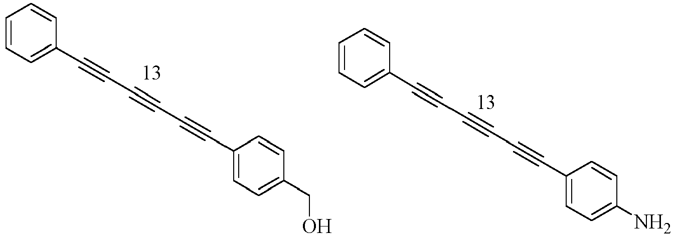


- Bibliothek nach Anspruch 1, wobei die Anzahl der Polyine zwischen 2 und etwa 100 liegt; vorzugsweise liegt die Anzahl der Polyine zwischen 4 und etwa 50.
- Bibliothek nach Anspruch 1, wobei die in der Bibliothek enthaltenen Polyine ausgewählt sind aus der Gruppe bestehend aus:


- Bibliothek nach Anspruch 1, wobei das eine oder mehrere der Polyine in oder auf einem festen Träger vorhanden sind, der ein Nanopartikel oder Mikropartikel ist.
- Bibliothek nach Anspruch 7, wobei das Nanopartikel oder Mikropartikel mindestens eines der folgenden ist: (i) gebildet aus einem Polymer, das eine Vielzahl von funktionellen Gruppen umfasst, von denen zwei oder mehr mit einem Polyin konjugiert sind, (ii) konjugiert mit einem oder mehreren Fluoreszenzmarkern, Antigenen, Antikörper, Antikörperfragmente, Antikörpermimetika, Peptide, Proteinen, Nukleinsäuren, Lipiden, Kohlenhydraten, Biotin, Streptavidin, Avidin, Anti-Biotin, Folat, folatbindendem Protein, IgG, Protein A, Protein G, Protein L, einem Kohlenhydrat, Lektin, Lipid oder Nukleinsäuren konjugiert ist, und/oder (iii) aus einem Polymer gebildet ist und zwei oder mehr Polyine in dem polymeren Nanopartikel oder Mikropartikel eingekapselt sind.
- Bibliothek nach Anspruch 7, wobei das Nanopartikel einen Durchmesser zwischen etwa 2 und etwa 100 nm hat und das Mikropartikel einen Durchmesser von mehr als etwa 100 nm und weniger als etwa 100 µm hat.
- Bibliothek nach Anspruch 7, wobei das Nanopartikel ausgewählt ist aus der Gruppe bestehend aus Nanokörpern, Nanokügelchen, Nanostäbchen, Nanodrähten, metallischen Nanostrukturen und Kohlenstoff-Nanoröhren.
- Bibliothek nach Anspruch 7, wobei das Nanopartikel ein Metall- oder Polymer-Nanopartikel ist.
- Bibliothek nach Anspruch 7, wobei der feste Träger mindestens ein Chip oder ein Mikropartikel, wie ein polymeres Mikropartikel, ist.
- Bibliothek nach einem der vorhergehenden Ansprüche, wobei die Polyine gut aufgelöste oberflächenverstärkte Raman-Peaks mit weniger als 10 % Übersprechen aufweisen.
- Herstellungsgegenstand mit einer Markierung, wobei die Markierung zwischen einem und 200 Polyine in der Bibliothek nach einem der Ansprüche 1-12 umfasst.
- Ein Teilchen, umfassend:einen Träger; undein oder mehrere Mmarkierungen, die mindestens ein Polyin in der Bibliothek nach einem der Ansprüche 1-12 enthalten.
Applications Claiming Priority (3)
| Application Number | Priority Date | Filing Date | Title |
|---|---|---|---|
| US201762540953P | 2017-08-03 | 2017-08-03 | |
| US201862616624P | 2018-01-12 | 2018-01-12 | |
| PCT/US2018/045271 WO2019028430A1 (en) | 2017-08-03 | 2018-08-03 | OPTICAL SUPER-MULTIPLEXING BY POLYYNESES |
Publications (3)
| Publication Number | Publication Date |
|---|---|
| EP3662086A1 EP3662086A1 (de) | 2020-06-10 |
| EP3662086A4 EP3662086A4 (de) | 2021-04-28 |
| EP3662086B1 true EP3662086B1 (de) | 2025-06-25 |
Family
ID=65233299
Family Applications (1)
| Application Number | Title | Priority Date | Filing Date |
|---|---|---|---|
| EP18842004.6A Active EP3662086B1 (de) | 2017-08-03 | 2018-08-03 | Optisches supermultiplexing durch polyyne |
Country Status (4)
| Country | Link |
|---|---|
| US (2) | US12065692B2 (de) |
| EP (1) | EP3662086B1 (de) |
| JP (1) | JP2020532497A (de) |
| WO (1) | WO2019028430A1 (de) |
Families Citing this family (9)
| Publication number | Priority date | Publication date | Assignee | Title |
|---|---|---|---|---|
| CN120795059A (zh) | 2018-08-27 | 2025-10-17 | 瑞泽恩制药公司 | 拉曼光谱在下游纯化中的应用 |
| CN112683874B (zh) * | 2019-10-18 | 2023-12-15 | 华中师范大学 | 一种超容量信息编码系统及其应用 |
| JP2023522891A (ja) * | 2020-04-15 | 2023-06-01 | クイーンズランド ユニバーシティ オブ テクノロジー | 蛍光高分子およびその使用 |
| US20220065791A1 (en) * | 2020-09-01 | 2022-03-03 | Cytoveris Inc. | Stimulated raman spectroscopy based multiplexed virtual immunohistology using alkynic, nitrile, or azide probes |
| CN112949799B (zh) * | 2021-01-08 | 2021-10-26 | 南京大学 | 一种基于偏振拉曼光谱编码的纳米条码智能标签 |
| EP4450638A1 (de) * | 2023-04-18 | 2024-10-23 | Leica Microsystems CMS GmbH | Etikett und markierungsstruktur zum markieren einer zielstruktur |
| GB202314282D0 (en) | 2023-09-18 | 2023-11-01 | Imperial College Innovations Ltd | New compounds and uses thereof |
| WO2025101682A1 (en) * | 2023-11-10 | 2025-05-15 | The Trustees Of Columbia University In The City Of New York | Device and methods for high-throughput micro-nano particle analysis |
| CN118405960A (zh) * | 2024-04-24 | 2024-07-30 | 振电(苏州)医疗科技有限公司 | 一种拉曼染料探针分子及其制备方法与应用 |
Family Cites Families (11)
| Publication number | Priority date | Publication date | Assignee | Title |
|---|---|---|---|---|
| US6610351B2 (en) | 2000-04-12 | 2003-08-26 | Quantag Systems, Inc. | Raman-active taggants and their recognition |
| US6783940B2 (en) * | 2001-10-31 | 2004-08-31 | Applera Corporation | Method of reducing non-specific amplification in PCR |
| US20090186343A1 (en) * | 2003-01-28 | 2009-07-23 | Visigen Biotechnologies, Inc. | Methods for preparing modified biomolecules, modified biomolecules and methods for using same |
| US7727721B2 (en) * | 2005-03-08 | 2010-06-01 | California Institute Of Technology | Hybridization chain reaction amplification for in situ imaging |
| WO2008062479A2 (en) * | 2006-11-21 | 2008-05-29 | Jawaharlal Nehru Centre For Advanced Scientific Research | A high sensitivity assay for molecular typing of biological sample, probes and a kit thereof |
| EP2040075A1 (de) * | 2007-09-24 | 2009-03-25 | Julius-Maximilians-Universität Würzburg | Verbindungen und Marker für oberflächenverstärkte Raman-Streuung |
| US20090136594A1 (en) * | 2007-11-27 | 2009-05-28 | Board Of Regents, The University Of Texas System | Functionalization of Micro-And Nano Particles for Selective Attachment to Calcium Biomineral Surfaces |
| US9034257B2 (en) * | 2008-10-27 | 2015-05-19 | Nodality, Inc. | High throughput flow cytometry system and method |
| US8941062B2 (en) * | 2010-11-16 | 2015-01-27 | 1087 Systems, Inc. | System for identifying and sorting living cells |
| US11408892B2 (en) | 2013-06-18 | 2022-08-09 | The Trustees Of Columbia University In The City Of New York | Devices, compositions and methods for imaging with raman scattering |
| JP7038676B2 (ja) * | 2016-03-18 | 2022-03-18 | キューティー ホールディングス コーポレーション | 細胞分離のための組成物、装置、及び方法 |
-
2018
- 2018-08-03 US US16/624,682 patent/US12065692B2/en active Active
- 2018-08-03 JP JP2020505264A patent/JP2020532497A/ja active Pending
- 2018-08-03 EP EP18842004.6A patent/EP3662086B1/de active Active
- 2018-08-03 WO PCT/US2018/045271 patent/WO2019028430A1/en not_active Ceased
-
2024
- 2024-07-10 US US18/768,612 patent/US20250002976A1/en active Pending
Also Published As
| Publication number | Publication date |
|---|---|
| US20200199657A1 (en) | 2020-06-25 |
| US20250002976A1 (en) | 2025-01-02 |
| US12065692B2 (en) | 2024-08-20 |
| EP3662086A1 (de) | 2020-06-10 |
| EP3662086A4 (de) | 2021-04-28 |
| JP2020532497A (ja) | 2020-11-12 |
| WO2019028430A1 (en) | 2019-02-07 |
Similar Documents
| Publication | Publication Date | Title |
|---|---|---|
| US20250002976A1 (en) | Optical super-multiplexing by polyynes | |
| US12140540B2 (en) | Stimulated Raman coupled fluorescence spectroscopy and microscopy system and method | |
| Mohammadi et al. | Carbon dots hybrid for dual fluorescent detection of microRNA-21 integrated bioimaging of MCF-7 using a microfluidic platform | |
| Zhao et al. | Gold nanostructures encoded by non-fluorescent small molecules in polyA-mediated nanogaps as universal SERS nanotags for recognizing various bioactive molecules | |
| JP5701896B2 (ja) | ラマン活性分子がナノ粒子二量体の接合部に位置する二量体コア−シェルナノ粒子、その用途およびその製造方法 | |
| Li et al. | Encoding quantized fluorescence states with fractal DNA frameworks | |
| CN100413977C (zh) | Serrs活性微粒 | |
| Jana et al. | One pot synthesis of intriguing fluorescent carbon dots for sensing and live cell imaging | |
| CN106147753B (zh) | 噻唑橙苯乙烯类化合物作为g-四链体核酸荧光探针 | |
| Guo et al. | Ultrasensitive simultaneous detection of multiplex disease-related nucleic acids using double-enhanced surface-enhanced Raman scattering nanosensors | |
| US11597842B2 (en) | Labeling dye and kit including same | |
| US20160266104A1 (en) | Heterodimeric core-shell nanoparticle in which raman-active molecules are located at a binding portion of a nanoparticle heterodimer, use thereof, and method for preparing same | |
| Tang et al. | Luminescent lanthanide coordination polymer as a platform for DNA colorimetric detection | |
| Li et al. | Breaking through bead-supported assay: integration of optical tweezers assisted fluorescence imaging and luminescence confined upconversion nanoparticles triggered luminescent resonance energy transfer (LRET) | |
| Pan et al. | Monochromatic fluorescent barcodes hierarchically assembled from modular DNA origami nanorods | |
| You et al. | Micron-sized surface enhanced Raman scattering reporter/fluorescence probe encoded colloidal microspheres for sensitive DNA detection | |
| Li et al. | Autonomously driving multiplexed hierarchical hybridization chain reaction of a DNA cobweb sensor for monitoring intracellular microRNA | |
| US20200385790A1 (en) | Heterodimeric core-shell nanoparticle in which raman-active molecules are located at a binding portion of a nanoparticle heterodimer, use thereof, and method for preparing same | |
| CN103509857A (zh) | 一种dna检测试剂盒及其制备方法和应用 | |
| Cui et al. | An intelligent, autocatalytic, DNAzyme biocircuit for amplified imaging of intracellular microRNAs | |
| Jannah et al. | A diaminotriazine-functionalized polydiacetylene for colorimetric sensing of thymine and oligothymidine | |
| Wang et al. | Fluorescence coding techniques for RNA detection | |
| Wahan et al. | Harnessing click chemistry for the discovery of fluorescent materials | |
| Guo et al. | Modern optical techniques provide a bright outlook for cell analysis | |
| Krishna et al. | Fluorescent biotin analogues for microstructure patterning and selective protein immobilization |
Legal Events
| Date | Code | Title | Description |
|---|---|---|---|
| STAA | Information on the status of an ep patent application or granted ep patent |
Free format text: STATUS: THE INTERNATIONAL PUBLICATION HAS BEEN MADE |
|
| PUAI | Public reference made under article 153(3) epc to a published international application that has entered the european phase |
Free format text: ORIGINAL CODE: 0009012 |
|
| STAA | Information on the status of an ep patent application or granted ep patent |
Free format text: STATUS: REQUEST FOR EXAMINATION WAS MADE |
|
| 17P | Request for examination filed |
Effective date: 20200228 |
|
| AK | Designated contracting states |
Kind code of ref document: A1 Designated state(s): AL AT BE BG CH CY CZ DE DK EE ES FI FR GB GR HR HU IE IS IT LI LT LU LV MC MK MT NL NO PL PT RO RS SE SI SK SM TR |
|
| AX | Request for extension of the european patent |
Extension state: BA ME |
|
| DAV | Request for validation of the european patent (deleted) | ||
| DAX | Request for extension of the european patent (deleted) | ||
| RIN1 | Information on inventor provided before grant (corrected) |
Inventor name: WIN, WEI Inventor name: HU, FANGHAO |
|
| REG | Reference to a national code |
Ref country code: DE Ref legal event code: R079 Free format text: PREVIOUS MAIN CLASS: C12Q0001700000 Ipc: G01J0003440000 Ref document number: 602018082984 Country of ref document: DE |
|
| A4 | Supplementary search report drawn up and despatched |
Effective date: 20210326 |
|
| RIC1 | Information provided on ipc code assigned before grant |
Ipc: G01J 3/44 20060101AFI20210322BHEP Ipc: G01N 21/65 20060101ALI20210322BHEP Ipc: C12Q 1/6816 20180101ALI20210322BHEP Ipc: C12Q 1/6806 20180101ALI20210322BHEP |
|
| STAA | Information on the status of an ep patent application or granted ep patent |
Free format text: STATUS: EXAMINATION IS IN PROGRESS |
|
| 17Q | First examination report despatched |
Effective date: 20220812 |
|
| P01 | Opt-out of the competence of the unified patent court (upc) registered |
Effective date: 20230314 |
|
| GRAP | Despatch of communication of intention to grant a patent |
Free format text: ORIGINAL CODE: EPIDOSNIGR1 |
|
| STAA | Information on the status of an ep patent application or granted ep patent |
Free format text: STATUS: GRANT OF PATENT IS INTENDED |
|
| RIC1 | Information provided on ipc code assigned before grant |
Ipc: B82Y 30/00 20110101ALI20250113BHEP Ipc: B82Y 15/00 20110101ALI20250113BHEP Ipc: C12Q 1/6806 20180101ALI20250113BHEP Ipc: C12Q 1/6816 20180101ALI20250113BHEP Ipc: G01N 21/65 20060101ALI20250113BHEP Ipc: G01J 3/44 20060101AFI20250113BHEP |
|
| INTG | Intention to grant announced |
Effective date: 20250122 |
|
| GRAS | Grant fee paid |
Free format text: ORIGINAL CODE: EPIDOSNIGR3 |
|
| GRAA | (expected) grant |
Free format text: ORIGINAL CODE: 0009210 |
|
| STAA | Information on the status of an ep patent application or granted ep patent |
Free format text: STATUS: THE PATENT HAS BEEN GRANTED |
|
| AK | Designated contracting states |
Kind code of ref document: B1 Designated state(s): AL AT BE BG CH CY CZ DE DK EE ES FI FR GB GR HR HU IE IS IT LI LT LU LV MC MK MT NL NO PL PT RO RS SE SI SK SM TR |
|
| REG | Reference to a national code |
Ref country code: GB Ref legal event code: FG4D |
|
| REG | Reference to a national code |
Ref country code: CH Ref legal event code: EP |
|
| REG | Reference to a national code |
Ref country code: CH Ref legal event code: EP |
|
| REG | Reference to a national code |
Ref country code: IE Ref legal event code: FG4D |
|
| REG | Reference to a national code |
Ref country code: DE Ref legal event code: R096 Ref document number: 602018082984 Country of ref document: DE |
|
| PG25 | Lapsed in a contracting state [announced via postgrant information from national office to epo] |
Ref country code: FI Free format text: LAPSE BECAUSE OF FAILURE TO SUBMIT A TRANSLATION OF THE DESCRIPTION OR TO PAY THE FEE WITHIN THE PRESCRIBED TIME-LIMIT Effective date: 20250625 |
|
| PGFP | Annual fee paid to national office [announced via postgrant information from national office to epo] |
Ref country code: DE Payment date: 20250820 Year of fee payment: 8 |
|
| REG | Reference to a national code |
Ref country code: LT Ref legal event code: MG9D |
|
| PG25 | Lapsed in a contracting state [announced via postgrant information from national office to epo] |
Ref country code: NO Free format text: LAPSE BECAUSE OF FAILURE TO SUBMIT A TRANSLATION OF THE DESCRIPTION OR TO PAY THE FEE WITHIN THE PRESCRIBED TIME-LIMIT Effective date: 20250925 Ref country code: GR Free format text: LAPSE BECAUSE OF FAILURE TO SUBMIT A TRANSLATION OF THE DESCRIPTION OR TO PAY THE FEE WITHIN THE PRESCRIBED TIME-LIMIT Effective date: 20250926 |
|
| PG25 | Lapsed in a contracting state [announced via postgrant information from national office to epo] |
Ref country code: BG Free format text: LAPSE BECAUSE OF FAILURE TO SUBMIT A TRANSLATION OF THE DESCRIPTION OR TO PAY THE FEE WITHIN THE PRESCRIBED TIME-LIMIT Effective date: 20250625 |
|
| PGFP | Annual fee paid to national office [announced via postgrant information from national office to epo] |
Ref country code: GB Payment date: 20250820 Year of fee payment: 8 |
|
| PG25 | Lapsed in a contracting state [announced via postgrant information from national office to epo] |
Ref country code: HR Free format text: LAPSE BECAUSE OF FAILURE TO SUBMIT A TRANSLATION OF THE DESCRIPTION OR TO PAY THE FEE WITHIN THE PRESCRIBED TIME-LIMIT Effective date: 20250625 |
|
| PGFP | Annual fee paid to national office [announced via postgrant information from national office to epo] |
Ref country code: FR Payment date: 20250828 Year of fee payment: 8 |
|
| PG25 | Lapsed in a contracting state [announced via postgrant information from national office to epo] |
Ref country code: RS Free format text: LAPSE BECAUSE OF FAILURE TO SUBMIT A TRANSLATION OF THE DESCRIPTION OR TO PAY THE FEE WITHIN THE PRESCRIBED TIME-LIMIT Effective date: 20250925 |
|
| PG25 | Lapsed in a contracting state [announced via postgrant information from national office to epo] |
Ref country code: LV Free format text: LAPSE BECAUSE OF FAILURE TO SUBMIT A TRANSLATION OF THE DESCRIPTION OR TO PAY THE FEE WITHIN THE PRESCRIBED TIME-LIMIT Effective date: 20250625 |
|
| REG | Reference to a national code |
Ref country code: NL Ref legal event code: MP Effective date: 20250625 |
|
| PG25 | Lapsed in a contracting state [announced via postgrant information from national office to epo] |
Ref country code: NL Free format text: LAPSE BECAUSE OF FAILURE TO SUBMIT A TRANSLATION OF THE DESCRIPTION OR TO PAY THE FEE WITHIN THE PRESCRIBED TIME-LIMIT Effective date: 20250625 |
|
| PG25 | Lapsed in a contracting state [announced via postgrant information from national office to epo] |
Ref country code: PT Free format text: LAPSE BECAUSE OF FAILURE TO SUBMIT A TRANSLATION OF THE DESCRIPTION OR TO PAY THE FEE WITHIN THE PRESCRIBED TIME-LIMIT Effective date: 20251027 |
|
| REG | Reference to a national code |
Ref country code: AT Ref legal event code: MK05 Ref document number: 1806871 Country of ref document: AT Kind code of ref document: T Effective date: 20250625 |
|
| PG25 | Lapsed in a contracting state [announced via postgrant information from national office to epo] |
Ref country code: IS Free format text: LAPSE BECAUSE OF FAILURE TO SUBMIT A TRANSLATION OF THE DESCRIPTION OR TO PAY THE FEE WITHIN THE PRESCRIBED TIME-LIMIT Effective date: 20251025 |
|
| PG25 | Lapsed in a contracting state [announced via postgrant information from national office to epo] |
Ref country code: AT Free format text: LAPSE BECAUSE OF FAILURE TO SUBMIT A TRANSLATION OF THE DESCRIPTION OR TO PAY THE FEE WITHIN THE PRESCRIBED TIME-LIMIT Effective date: 20250625 Ref country code: SM Free format text: LAPSE BECAUSE OF FAILURE TO SUBMIT A TRANSLATION OF THE DESCRIPTION OR TO PAY THE FEE WITHIN THE PRESCRIBED TIME-LIMIT Effective date: 20250625 |
|
| PG25 | Lapsed in a contracting state [announced via postgrant information from national office to epo] |
Ref country code: CZ Free format text: LAPSE BECAUSE OF FAILURE TO SUBMIT A TRANSLATION OF THE DESCRIPTION OR TO PAY THE FEE WITHIN THE PRESCRIBED TIME-LIMIT Effective date: 20250625 |
|
| PG25 | Lapsed in a contracting state [announced via postgrant information from national office to epo] |
Ref country code: PL Free format text: LAPSE BECAUSE OF FAILURE TO SUBMIT A TRANSLATION OF THE DESCRIPTION OR TO PAY THE FEE WITHIN THE PRESCRIBED TIME-LIMIT Effective date: 20250625 |
|
| PG25 | Lapsed in a contracting state [announced via postgrant information from national office to epo] |
Ref country code: EE Free format text: LAPSE BECAUSE OF FAILURE TO SUBMIT A TRANSLATION OF THE DESCRIPTION OR TO PAY THE FEE WITHIN THE PRESCRIBED TIME-LIMIT Effective date: 20250625 |
|
| PG25 | Lapsed in a contracting state [announced via postgrant information from national office to epo] |
Ref country code: SK Free format text: LAPSE BECAUSE OF FAILURE TO SUBMIT A TRANSLATION OF THE DESCRIPTION OR TO PAY THE FEE WITHIN THE PRESCRIBED TIME-LIMIT Effective date: 20250625 |
|
| PG25 | Lapsed in a contracting state [announced via postgrant information from national office to epo] |
Ref country code: ES Free format text: LAPSE BECAUSE OF FAILURE TO SUBMIT A TRANSLATION OF THE DESCRIPTION OR TO PAY THE FEE WITHIN THE PRESCRIBED TIME-LIMIT Effective date: 20250625 |
|
| PG25 | Lapsed in a contracting state [announced via postgrant information from national office to epo] |
Ref country code: RO Free format text: LAPSE BECAUSE OF FAILURE TO SUBMIT A TRANSLATION OF THE DESCRIPTION OR TO PAY THE FEE WITHIN THE PRESCRIBED TIME-LIMIT Effective date: 20250625 |
|
| REG | Reference to a national code |
Ref country code: CH Ref legal event code: H13 Free format text: ST27 STATUS EVENT CODE: U-0-0-H10-H13 (AS PROVIDED BY THE NATIONAL OFFICE) Effective date: 20260324 |
|
| PG25 | Lapsed in a contracting state [announced via postgrant information from national office to epo] |
Ref country code: MC Free format text: LAPSE BECAUSE OF FAILURE TO SUBMIT A TRANSLATION OF THE DESCRIPTION OR TO PAY THE FEE WITHIN THE PRESCRIBED TIME-LIMIT Effective date: 20250625 |
|
| PG25 | Lapsed in a contracting state [announced via postgrant information from national office to epo] |
Ref country code: DK Free format text: LAPSE BECAUSE OF FAILURE TO SUBMIT A TRANSLATION OF THE DESCRIPTION OR TO PAY THE FEE WITHIN THE PRESCRIBED TIME-LIMIT Effective date: 20250625 |
|
| PG25 | Lapsed in a contracting state [announced via postgrant information from national office to epo] |
Ref country code: LU Free format text: LAPSE BECAUSE OF NON-PAYMENT OF DUE FEES Effective date: 20250803 Ref country code: IT Free format text: LAPSE BECAUSE OF FAILURE TO SUBMIT A TRANSLATION OF THE DESCRIPTION OR TO PAY THE FEE WITHIN THE PRESCRIBED TIME-LIMIT Effective date: 20250625 |
|
| PG25 | Lapsed in a contracting state [announced via postgrant information from national office to epo] |
Ref country code: CH Free format text: LAPSE BECAUSE OF NON-PAYMENT OF DUE FEES Effective date: 20250831 |
|
| PLBE | No opposition filed within time limit |
Free format text: ORIGINAL CODE: 0009261 |
|
| STAA | Information on the status of an ep patent application or granted ep patent |
Free format text: STATUS: NO OPPOSITION FILED WITHIN TIME LIMIT |
|
| REG | Reference to a national code |
Ref country code: CH Ref legal event code: L10 Free format text: ST27 STATUS EVENT CODE: U-0-0-L10-L00 (AS PROVIDED BY THE NATIONAL OFFICE) Effective date: 20260507 |