EP3651635B1 - Oral care evaluation system and process - Google Patents
Oral care evaluation system and process Download PDFInfo
- Publication number
- EP3651635B1 EP3651635B1 EP18752647.0A EP18752647A EP3651635B1 EP 3651635 B1 EP3651635 B1 EP 3651635B1 EP 18752647 A EP18752647 A EP 18752647A EP 3651635 B1 EP3651635 B1 EP 3651635B1
- Authority
- EP
- European Patent Office
- Prior art keywords
- image
- light
- spectrum
- oral
- hygiene
- Prior art date
- Legal status (The legal status is an assumption and is not a legal conclusion. Google has not performed a legal analysis and makes no representation as to the accuracy of the status listed.)
- Active
Links
Images




Classifications
-
- A—HUMAN NECESSITIES
- A61—MEDICAL OR VETERINARY SCIENCE; HYGIENE
- A61B—DIAGNOSIS; SURGERY; IDENTIFICATION
- A61B5/00—Measuring for diagnostic purposes; Identification of persons
- A61B5/0059—Measuring for diagnostic purposes; Identification of persons using light, e.g. diagnosis by transillumination, diascopy, fluorescence
- A61B5/0071—Measuring for diagnostic purposes; Identification of persons using light, e.g. diagnosis by transillumination, diascopy, fluorescence by measuring fluorescence emission
-
- A—HUMAN NECESSITIES
- A61—MEDICAL OR VETERINARY SCIENCE; HYGIENE
- A61B—DIAGNOSIS; SURGERY; IDENTIFICATION
- A61B5/00—Measuring for diagnostic purposes; Identification of persons
- A61B5/0059—Measuring for diagnostic purposes; Identification of persons using light, e.g. diagnosis by transillumination, diascopy, fluorescence
- A61B5/0082—Measuring for diagnostic purposes; Identification of persons using light, e.g. diagnosis by transillumination, diascopy, fluorescence adapted for particular medical purposes
- A61B5/0088—Measuring for diagnostic purposes; Identification of persons using light, e.g. diagnosis by transillumination, diascopy, fluorescence adapted for particular medical purposes for oral or dental tissue
-
- A—HUMAN NECESSITIES
- A61—MEDICAL OR VETERINARY SCIENCE; HYGIENE
- A61B—DIAGNOSIS; SURGERY; IDENTIFICATION
- A61B5/00—Measuring for diagnostic purposes; Identification of persons
- A61B5/74—Details of notification to user or communication with user or patient; User input means
- A61B5/742—Details of notification to user or communication with user or patient; User input means using visual displays
- A61B5/7445—Display arrangements, e.g. multiple display units
-
- G—PHYSICS
- G16—INFORMATION AND COMMUNICATION TECHNOLOGY [ICT] SPECIALLY ADAPTED FOR SPECIFIC APPLICATION FIELDS
- G16H—HEALTHCARE INFORMATICS, i.e. INFORMATION AND COMMUNICATION TECHNOLOGY [ICT] SPECIALLY ADAPTED FOR THE HANDLING OR PROCESSING OF MEDICAL OR HEALTHCARE DATA
- G16H30/00—ICT specially adapted for the handling or processing of medical images
- G16H30/40—ICT specially adapted for the handling or processing of medical images for processing medical images, e.g. editing
-
- A—HUMAN NECESSITIES
- A61—MEDICAL OR VETERINARY SCIENCE; HYGIENE
- A61B—DIAGNOSIS; SURGERY; IDENTIFICATION
- A61B2576/00—Medical imaging apparatus involving image processing or analysis
-
- A—HUMAN NECESSITIES
- A61—MEDICAL OR VETERINARY SCIENCE; HYGIENE
- A61C—DENTISTRY; APPARATUS OR METHODS FOR ORAL OR DENTAL HYGIENE
- A61C17/00—Devices for cleaning, polishing, rinsing or drying teeth, teeth cavities or prostheses; Saliva removers; Dental appliances for receiving spittle
- A61C17/16—Power-driven cleaning or polishing devices
- A61C17/22—Power-driven cleaning or polishing devices with brushes, cushions, cups, or the like
Definitions
- Dental professionals typically advise individuals to perform a regular brushing routine which includes brushing both the teeth and soft tissues within the oral cavity. It is widely recognized that bacteria on both teeth and soft tissues contributes to poor oral hygiene, which in turn can contribute to malodorous breath. Dental professionals have systems which are used to evaluate gingival plaque. Such systems must be used in the dental office and are generally not available for use at home. While systems and processes for evaluating the hygiene of soft tissues using other markers have been investigated, none exist which are convenient enough for use by dental professionals, let alone for home use. It is therefore desirable to have systems and processes which facilitate the evaluation of oral hygiene with respect to soft tissue, particularly the tongue, through the use of other markers that may be naturally present. Evaluations of this type can not only improve oral hygiene, and thus breath freshness, when used by dental professionals, but it can also be used at home for the same purposes.
- Exemplary embodiments according to the present disclosure are directed to oral care evaluation systems and methods which are used to evaluate the amount of bacteria present on oral tissue such as the tongue.
- the system illuminates oral tissue within the oral cavity using a wavelength which induces fluorescence in an organic compound and detects and analyzes the light resulting from the fluorescence in order to evaluate oral hygiene associated with the oral tissue.
- the disclosure refers to an oral care evaluation system including: a light source configured to emit a spectrum of light which induces fluorescence in an organic compound, the light source illuminating soft oral tissue within an oral cavity; an image sensor receiving light generated by fluorescence of the organic compound on the soft oral tissue, the image sensor generating image data for an image of the soft oral tissue, the image including a plurality of pixels; and an image processor receiving the image data from the image sensor and programmed to evaluate oral hygiene of the soft oral tissue by assessing an attribute of each pixel in the image.
- the disclosure refers to an oral care evaluation process including: illuminating soft oral tissue within an oral cavity with a light source emitting a spectrum of light which induces fluorescence in an organic compound; generating image data for an image of the soft oral tissue while illuminating the soft oral tissue, the image including a plurality of pixels; and evaluating oral hygiene using a programmable processor programmed to assess an attribute of each pixel of the image.
- an oral care evaluation system including: an imaging subsystem including: a light source configured to emit a spectrum of light which induces fluorescence in an organic compound and to illuminate oral tissue within an oral cavity; and an image sensor positioned to receive light generated by fluorescence of the organic compound on the oral tissue and configured to generate image data for an image of the oral tissue, the image including a plurality of pixels; and an image processing subsystem configured to receive the image data from the image sensor and programmed to evaluate oral hygiene of the oral tissue by performing an assessment of an attribute of each pixel of the image, the assessment including: assigning one of a plurality of intensity values to each pixel; and counting a number of pixels associated with each of the plurality of intensity values.
- the disclosure refers to an oral care evaluation system including: a light source configured to emit a spectrum of light which induces fluorescence in an organic compound, the light source illuminating soft oral tissue within an oral cavity and the organic compound having a fluorescence spectrum; an image sensor receiving light in the fluorescence spectrum generated by fluorescence of the organic compound on the soft oral tissue, the image sensor generating image data for an image of the soft oral tissue, the image including a plurality of pixels; and an image processor receiving the image data from the image sensor and programmed to evaluate oral hygiene of the soft oral tissue by quantitative analysis to assess attributes of light in the fluorescence spectrum present in the image data.
- programmable processes described herein are not limited to any particular embodiment, and may be implemented in an operating system, application program, foreground or background processes, driver, or any combination thereof
- the computer programmable processes may be executed on a single processor or on or across multiple processors.
- processors described herein may be any central processing unit (CPU), microprocessor, micro-controller, computational, or programmable device or circuit configured for executing computer program instructions (e.g. code).
- Various processors may be embodied in computer and/or server hardware and/or computing device of any suitable type (e.g. desktop, laptop, notebook, tablet, cellular phone, smart phone, PDA, etc.) and may include all the usual ancillary components necessary to form a functional data processing device including without limitation a bus, software and data storage such as volatile and non-volatile memory, input/output devices, a display screen, graphical user interfaces (GUIs), removable data storage, and wired and/or wireless communication interface devices including Wi-Fi, Bluetooth, LAN, etc.
- GUIs graphical user interfaces
- Computer-executable instructions or programs may be programmed into and tangibly embodied in a non-transitory computer-readable medium that is accessible to and retrievable by a respective processor as described herein which configures and directs the processor to perform the desired functions and processes by executing the instructions encoded in the medium.
- a device embodying a programmable processor configured to such non-transitory computer-executable instructions or programs is referred to hereinafter as a "programmable device", or just a “device” for short, and multiple programmable devices in mutual communication is referred to as a "programmable system”.
- non-transitory "computer-readable medium” as described herein may include, without limitation, any suitable volatile or non-volatile memory including random access memory (RAM) and various types thereof, read-only memory (ROM) and various types thereof, USB flash memory, and magnetic or optical data storage devices (e.g. internal/external hard disks, floppy discs, magnetic tape CD-ROM, DVD-ROM, optical disk, ZIP TM drive, Blu-ray disk, and others), which may be written to and/or read by a processor operably connected to the medium.
- RAM random access memory
- ROM read-only memory
- USB flash memory e.g. internal/external hard disks, floppy discs, magnetic tape CD-ROM, DVD-ROM, optical disk, ZIP TM drive, Blu-ray disk, and others
- the present invention may be embodied in the form of computer-implemented processes and apparatuses such as processor-based data processing and communication systems or computer systems for practicing those processes.
- the present invention may also be embodied in the form of software or computer program code embodied in a non-transitory computer-readable storage medium, which when loaded into and executed by the data processing and communications systems or computer systems, the computer program code segments configure the processor to create specific logic circuits configured for implementing the processes.
- FIG. 1 illustrates an oral care evaluation system 101 in accordance with an embodiment of the present invention.
- the oral care evaluation system 101 includes a light source 103 configured to emit a spectrum of light which induces fluorescence in an organic compound.
- the light source 103 may be any type of light source which is capable of emitting light in the desired spectrum, such as an LED or an incandescent light bulb. Other types of light sources may also be used, with the light source not limiting the scope of the invention unless otherwise expressly stated in the claims.
- the emission spectrum of the light source 103 is selected based upon the absorption spectrum of the targeted organic compound.
- the light source 103 may also include a light filter which limits the emission of light from the light source 103 to the desired emission spectrum.
- the targeted organic compound is a porphyrin compound.
- other fluorescing organic compounds may be targeted.
- Previous research has shown that many of the malodorous bacteria on oral tissue within the oral cavity, such as on the tongue, contain porphyrin compounds. Whereas the prior art uses subjective observations of fluorescence from porphyrin compounds in order to evaluate the hygiene of oral tissue, such as the tongue, the objective measures of fluorescence described herein provide better and more consistent evaluation results. To this end, the oral care evaluation system 101 is able to objectively evaluate the amount of the porphyrin compounds on oral tissues. And, because the porphyrin compounds are contained by bacteria, the objective evaluation of the porphyrin compounds may in turn be used as an objective evaluation of the amount of bacteria present on the oral tissue.
- Porphyrin compounds are known to have an absorption spectrum with several absorption bands, with one band commonly referred to as the Soret band, and one or more other bands commonly referred to as the Q bands.
- the Soret band is used by the oral care evaluation system 101 to evaluate the amount of bacteria present on the tongue.
- the absorption spectrum of the Soret band is centered at a wavelength of about 400 nm and ranges in wavelength from about 350 nm to 450 nm.
- the light source 103 is selected and/or configured to emit a spectrum of light having wavelengths of between 350 nm to 450 nm. In certain embodiments, the light source 103 may emit a narrower spectrum, so long as the emitted spectrum still has an effective overlap with the absorption spectrum of the organic compound.
- the spectrum of light emitted from the light source 103 may include wavelengths less than 350 nm, and/or it may include wavelengths over 450 nm.
- a VELscope ® Vx device manufactured by LED Medical Diagnostics Inc. of Atlanta, GA, may be used as the light source 103.
- the VELscope ® Vx device is commonly used to detect soft tissue abnormalities, and it emits light in the spectral range of about 400 nm to 460 nm.
- Porphyrin compounds are also known to have a fluorescence spectrum ranging in wavelength from about 625 nm to about 750 nm.
- the image sensor 105 is therefore positioned and configured to detect light in this spectral range.
- oral tissue with more bacteria generates more fluorescent light in this spectral range.
- the image sensor 105 is configured to detect light in the fluorescence spectrum of the targeted organic compound.
- the image sensor 105 generates image data for an image of the oral tissue while the oral tissue is being illuminated by light emitted from the light source 103, i.e., while the organic compound is fluorescing.
- a light filter 107 is included as part of the oral care evaluation system 101 to filter light incident on the image sensor 105, with the purpose being to limit the incident light to the fluorescence spectrum of the porphyrin compound.
- the light filter 107 may be omitted where the image sensor 105 has limited or no sensitivity to light outside of the fluorescence spectrum of the organic compound.
- the image sensor 105 may be a broad spectrum image sensor, with data relating to the fluorescence spectrum of the organic compound being determined and analyzed through subsequent image processing.
- the image sensor 105 may be a charge-coupled device (CCD). In certain other embodiments, the image sensor 105 may be a digital camera (which itself may include a CCD). In such embodiments, the flash of the digital camera may be configured to serve as the light source 103 for the oral care evaluation system 101.
- CCD charge-coupled device
- the flash of the digital camera may be configured to serve as the light source 103 for the oral care evaluation system 101.
- Other types of image sensors may also be used, with the image sensor not limiting the scope of the invention unless otherwise expressly stated in the claims.
- the image processor 109 is a programmable processor which is communicably coupled to the image sensor 105 so that the image data for the image of the oral tissue may be transmitted from the image sensor 105 and received by the image processor 109.
- the image sensor 105 and the image processor 109 may be communicably coupled through a wired connection.
- the communication connection between the image sensor 105 and the image processor 109 allows the image sensor 105 to send image data to the image processor 109, and it allows the image processor 109 to send command signals to control operation of the image sensor 105.
- the image processor 109 and the image sensor 105 may communicate using a wireless connection.
- the wireless connection may use any type of appropriate wireless protocol for communications, such as, for example, Bluetooth ® , WiFi, cellular, and the like.
- the image processor 109 is also communicably coupled to a control module 111 and to the light source 103. Through the communication connection between the image processor 109 and the light source 103, the image processor is able to control operation of the light source 103, such as for turning the light source 103 on and off.
- the control module 111 serves as a user interface for the system 101, allowing the user to turn the light source 103 on and off, to control when the image sensor 105 is operational to generate image data for the oral tissue, and to otherwise interact with the image processor 109.
- the control module 111 may take any different form, such as one or more buttons or switches, a keypad or keyboard, or even a touch screen. Other types of control modules may also be used, with the control module not limiting the scope of the invention unless otherwise expressly stated in the claims. In certain embodiments, the control module may also provide feedback to the user about the oral hygiene evaluation being performed.
- the image processor 109 is also communicably coupled to a display 113, through which the image processor 109 is able to display an image of the oral tissue and/or results of the oral hygiene evaluation.
- the results of the oral hygiene evaluation may be a hygiene grade.
- the communication connections between the image processor 109, on the one hand, and any one or more of the light source 103, the control module 111, and the display 113 may be through a wired connection or through a wireless connection.
- the light source 103 emits a spectrum of light which induces fluorescence in an organic compound, with the light illuminating oral tissue, such as the tongue 123, within the oral cavity 121.
- the image sensor 105 detects light from the fluorescing organic compound and generates image data for an image of the oral tissue.
- the image processor 109 is programmed to asses an attribute of each pixel in the image in order to evaluate the oral hygiene associated with the oral tissue.
- the image processor 109 is further programmed to communicate the evaluation to a user, such as by displaying the image of the oral tissue and/or the hygiene grade on a display screen.
- FIG. 2 illustrates an oral care evaluation system 201 in accordance with another embodiment of the present invention.
- the oral care evaluation system 201 includes an imaging subsystem 203 and an image processing subsystem 211.
- the imaging subsystem 203 includes a light source 205, an image sensor 207, and a light filter 209.
- the light source 205 may be any type of light source which is capable of emitting light in the desired spectrum, such as an LED or an incandescent light bulb. Other types of light sources may also be used, with the light source not limiting the scope of the invention unless otherwise expressly stated in the claims.
- the emission spectrum of the light source 205 is selected based upon the absorption spectrum of the targeted organic compound, and the targeted organic compound, in certain embodiments, may be a porphyrin compound.
- the light source 205 is selected and/or configured to emit a spectrum of light having wavelength of between 350 nm to 450 nm. In certain other embodiments, the light source 205 may emit a narrower spectrum, so long as the emitted spectrum still has an effective overlap with the absorption spectrum of the targeted organic compound. In still other embodiments, the spectrum of light emitted from the light source 205 may include wavelengths less than 350 nm, and/or it may include wavelengths over 450 nm. In certain embodiments, the light source 205 may also include a light filter which limits the emission of light from the light source 205 to the desired emission spectrum.
- the image sensor 207 is configured to detect light in the spectral range of light emitted from the fluorescing organic compound.
- the light filter 209 is included to filter light incident on the image sensor 207, with the purpose being to limit the incident light to the fluorescence spectrum of the porphyrin compound. In certain embodiments, the light filter 209 may be omitted where the image sensor 207 has limited or no sensitivity to light outside of the fluorescence spectrum of the organic compound.
- the image sensor 207 generates image data for an image of the oral tissue while the organic compound is fluorescing. In certain embodiments, the image sensor 207 may be a CCD. Other types of image sensors may also be used, with the image sensor not limiting the scope of the invention unless otherwise expressly stated in the claims.
- the image processing subsystem 211 is communicably coupled to the imaging subsystem 203 so that image data for the image of the oral tissue may be transmitted from the imaging subsystem 203 and received by the image processing subsystem 21 1.
- the imaging subsystem 203 and the image processing subsystem 211 may be communicably coupled through a wired connection.
- the communication connection between the imaging subsystem 203 and the image processing subsystem 211 also allows the image processing subsystem 211 to send command signals to control operation of the imaging subsystem 203.
- the image processing subsystem 211 and the imaging subsystem 203 may communicate using a wireless connection.
- the wireless connection may use any type of appropriate wireless protocol for communications, such as, for example, Bluetooth ® , WiFi, cellular, and the like.
- the image processing subsystem 211 includes a programmable processor 213 and a display screen 215 communicably coupled to each other.
- the programmable processor 213 is programmed to control the light source 205 and the image sensor 207, such that during operation of the oral care evaluation system 201, the light source 205 emits a spectrum of light which induces fluorescence in an organic compound, with the light illuminating oral tissue, such as the tongue 223, within the oral cavity 221.
- the programmable processor 213 is able to obtain image data while the organic compound is fluorescing.
- the programmable processor 213 is also programmed to control the display screen 215 so that the image of the oral tissue and/or a hygiene grade may be displayed to the user.
- the programmed functionality of the programmable processor 213 may be as described below in connection with the process for evaluating oral hygiene.
- the display screen 215 may be a touch sensitive screen which accepts input from a user in response to touches on the touch sensitive screen.
- the programmable processor 213 may control the light source 205 and the image sensor 207 in response to input received from the user through the display screen 215.
- the image processing subsystem 211 may also include a volatile and/or non-volatile memory, which may be used for storing programming and for storing data. In such embodiments, the memory may also be used to store historical data.
- the image processing subsystem 211 may be a computing device such as, for example, a laptop, a smart phone, a tablet, a PDA, and the like. In still other embodiments, the image processing subsystem 211 may also communicate with a server (not shown) for purposes of storing historical data and/or to provide server-side processing functionality.
- FIG. 3 illustrates an oral care evaluation system 301 in accordance with another embodiment of the present invention.
- the oral care evaluation system 301 includes an imaging subsystem 303 and an image processing subsystem 311.
- the imaging subsystem 303 includes a light source 305, an image sensor 307, and a light filter 309.
- the light source 305 may be any type of light source which is capable of emitting light in the desired spectrum, such as an LED or an incandescent light bulb. Other types of light sources may also be used, with the light source not limiting the scope of the invention unless otherwise expressly stated in the claims.
- the emission spectrum of the light source 305 is selected based upon the absorption spectrum of the targeted organic compound, and the targeted organic compound, in certain embodiments, may be a porphyrin compound.
- the light source 305 is selected and/or configured to emit a spectrum of light having wavelength of between 350 nm to 450 nm. In certain other embodiments, the light source 305 may emit a narrower spectrum, so long as the emitted spectrum still has an effective overlap with the absorption spectrum of the targeted organic compound. In still other embodiments, the spectrum of light emitted from the light source 305 may include wavelengths less than 350 nm, and/or it may include wavelengths over 450 nm. In certain embodiments, the light source 305 may also include a light filter which limits the emission of light from the light source 305 to the desired emission spectrum.
- the image sensor 307 is configured to detect light in the spectral range of light emitted from the fluorescing organic compound.
- the light filter 309 is included to filter light incident on the image sensor 307, with the purpose being to limit the incident light to the fluorescence spectrum of the porphyrin compound. In certain embodiments, the light filter 309 may be omitted where the image sensor 307 has limited or no sensitivity to light outside of the fluorescence spectrum of the organic compound.
- the image sensor 307 generates image data for an image of the oral tissue while the organic compound is fluorescing. In certain embodiments, the image sensor 307 may be a CCD. Other types of image sensors may also be used, with the image sensor not limiting the scope of the invention unless otherwise expressly stated in the claims.
- the image processing subsystem 311 is communicably coupled to the imaging subsystem 303 so that image data for the image of the oral tissue may be transmitted from the imaging subsystem 303 and received by the image processing subsystem 311.
- the communication connection between the imaging subsystem 303 and the image processing subsystem 311 also allows the image processing subsystem 311 to send command signals to control operation of the imaging subsystem 303.
- the imaging subsystem 303 and the image processing subsystem 311 are communicably coupled through a wired connection.
- the image processing subsystem 311 includes a programmable processor 313 and a wireless transceiver 315 which are communicably to each other.
- the image processing subsystem 311 may also include a volatile and/or non-volatile memory, which may be used for storing programming and/or data.
- the wireless transceiver 315 may be configured to transmit the image data using any type of appropriate wireless protocol for communications. Non-limiting examples of wireless protocols include Bluetooth ® , WiFi, cellular, and the like.
- the programmable processor 313 is programmed to control the light source 305 and the image sensor 307, such that during operation of the oral care evaluation system 301, the light source 305 emits a spectrum of light which induces fluorescence in an organic compound, with the light illuminating oral tissue, such as the tongue 319, within the oral cavity 317.
- the programmable processor 313 is able to generate image data while the organic compound is fluorescing.
- the programmed functionality of the programmable processor 313 for generating the image data is described in greater detail below.
- Evaluation of the generated image data may be performed by the programmable processor 313, by the remote device 321, or by a combination of the programmable processor 313 and the remote device 321.
- the remote device 321 is a general computing device such as, for example, a laptop, a smart phone, a tablet, a PDA, and the like. In certain embodiments, the remote device 321 may be programmed with functionality to fully evaluate the image data as described herein.
- the remote device 321 is also programmed to display the evaluation results on the display screen 323. In certain embodiments, the evaluation results displayed on the display screen 323 may be the image of the oral cavity 325, which includes the oral tissue being evaluated, and/or the hygiene grade 327.
- the remote device 321 may also be used to control the functionality of the programmable processor 313.
- the remote device 321 may accept input from a user and send control commands to the programmable processor 313 in response to the user input.
- the remote device 321 may also communicate with a cloud server 329 using one or more public or private local area networks (LAN) and/or wide area networks (WAN).
- the remote device 321 may communicate one or more of the image data, the evaluation results, and any meta data associated with the evaluation with the cloud server 329.
- the cloud server 329 may be used to store historical data associated with oral care evaluations.
- the cloud server 329 may be used as a data aggregator, and the cloud server 329 may be used to perform additional data analysis, both on individual evaluations and on aggregated evaluations.
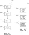
- FIGS. 4A-B are flowcharts 401, 411 showing a process for evaluating oral hygiene.
- the programmable processors described above in connection with embodiments of the invention may be programmed to follow the process of the flowcharts 401, 411.
- the capabilities and parameters of the embodiments described above may be incorporated into the process of the flowcharts 401, 411.
- the processes shown and described herein may performed by a plurality of processors, with each processor being programmed to perform only a portion of the process, and with all the processors together being programmed to perform the entirety of the process.
- the process begins with a step of illuminating 403 oral tissue within an oral cavity with a light source emitting a spectrum of light which induces fluorescence in an organic compound.
- the light source may be any type of light source which emits light in a spectrum, as described above, which induces fluorescence in an organic compound.
- the targeted organic compound is a porphyrin compound, and the light spectrum emitted by the light source overlaps, at least partially, with the absorption spectrum of the porphyrin compound.
- the process continues with a step of generating image data 405 for an image of the oral tissue.
- the image data may be generated by an image sensor receiving light from the fluorescing porphyrin compound.
- the image of the oral tissue is a digital image, and as such the digital image includes a plurality of pixels.
- the digital image has a pixel resolution of at least 640 ⁇ 480.
- the digital image may have a pixel resolution of less than 640 ⁇ 480, such as 320 ⁇ 240 or even less. It should be understood that the tradeoff for using a digital image with a lower pixel resolution is that the accuracy of the evaluation may decrease as the pixel resolution decreases.
- each pixel may be independently defined within the image data. In certain other embodiments, such as for image data that represents a compressed image, each pixel is independently defined only after the compressed image data is decompressed. In certain embodiments, the image data may conform to a standardized image format, such as gif, jpg, tif, png, and the like.
- the process continues with evaluating 407 oral hygiene.
- the results of the evaluation may be in the form of a hygiene grade, which is an assigned value used to inform the user about the results of the oral hygiene evaluation.
- the assigned value is a hygiene grade, which may be a numeric score ranging from 1-5, or 1-10, or it may be an alphabetical score ranging from A-F.
- FIG. 4B is a flowchart 411 which illustrates details of the evaluation 407 step in FIG. 4A .
- an attribute of each pixel of the image is assessed and an attribute value is assigned 413 to each pixel.
- the attribute assessed is the intensity of each pixel.
- other attributes of each pixel may be assessed in addition to or instead of the intensity.
- the assigned attribute value is based on the assessed attribute of each individual pixel and the overall range of the assessed attributes amongst all pixels.
- the assigned attribute values have a predetermined range, such as from 0-255, with 0 representing a lower intensity than 255.
- the number of attribute values within the range may vary, however, the range should include a sufficient number of attribute values to create a meaningful distribution for the assessed attribute.
- FIG. 5 is a graph 501 which shows four different sample numerical distributions for assessed intensity based on fluorescing images generated from tongues.
- the x-axis of the graph 501 represents the range of attribute values that are assigned to the pixels based on the assessed intensity, and the y-axis represents the number of pixels counted as a function of the attribute value.
- dirty tongues 1 and 2 have a larger number of pixels in the higher intensity region above the attribute value of 150, and therefore a corresponding lower number of pixels in the lower intensity region below the attribute value of 150.
- tongues 1 and 2 nearly all pixels are assigned an attribute value below 150.
- the graph 501 illustrates that a distinct difference exists between the attribute value distribution for a dirty tongue as compared to the attribute value distribution for a clean tongue when the attribute is the intensity of the pixels.
- a hygiene grade may be generated for each of the numerical distributions. For example, on a scale of 1-5, a 1 on this hygiene scale may represent an image having more than a nominal number of pixels counted for intensity values over 200, such as is seen for the dirty tongues 1 and 2. Also, a 5 on this hygiene scale may represent an image having any intensity value between 0-100 which also has a pixel count for any intensity value above 2500, such as is seen for the clean tongues 3 and 4.
- FIG. 6 is an image showing a tongue before cleaning
- FIG. 7 is an image showing a tongue after cleaning.
- the image of FIG. 6 has a greater number of high intensity pixels as compared to the image of FIG. 7 .
- ranges are used as shorthand for describing each and every value that is within the range. Any value within the range can be selected as the terminus of the range. In the event of a conflict in a definition in the present disclosure and that of a cited reference, the present disclosure controls.
Landscapes
- Health & Medical Sciences (AREA)
- Life Sciences & Earth Sciences (AREA)
- General Health & Medical Sciences (AREA)
- Public Health (AREA)
- Veterinary Medicine (AREA)
- Engineering & Computer Science (AREA)
- Medical Informatics (AREA)
- Animal Behavior & Ethology (AREA)
- Heart & Thoracic Surgery (AREA)
- Molecular Biology (AREA)
- Surgery (AREA)
- Biomedical Technology (AREA)
- Pathology (AREA)
- Physics & Mathematics (AREA)
- Biophysics (AREA)
- Dentistry (AREA)
- Audiology, Speech & Language Pathology (AREA)
- Oral & Maxillofacial Surgery (AREA)
- Epidemiology (AREA)
- Nuclear Medicine, Radiotherapy & Molecular Imaging (AREA)
- Primary Health Care (AREA)
- Radiology & Medical Imaging (AREA)
- Investigating, Analyzing Materials By Fluorescence Or Luminescence (AREA)
- Measuring And Recording Apparatus For Diagnosis (AREA)
Description
- Dental professionals typically advise individuals to perform a regular brushing routine which includes brushing both the teeth and soft tissues within the oral cavity. It is widely recognized that bacteria on both teeth and soft tissues contributes to poor oral hygiene, which in turn can contribute to malodorous breath. Dental professionals have systems which are used to evaluate gingival plaque. Such systems must be used in the dental office and are generally not available for use at home. While systems and processes for evaluating the hygiene of soft tissues using other markers have been investigated, none exist which are convenient enough for use by dental professionals, let alone for home use. It is therefore desirable to have systems and processes which facilitate the evaluation of oral hygiene with respect to soft tissue, particularly the tongue, through the use of other markers that may be naturally present. Evaluations of this type can not only improve oral hygiene, and thus breath freshness, when used by dental professionals, but it can also be used at home for the same purposes.
- The invention is defined by the claims.
- Exemplary embodiments according to the present disclosure are directed to oral care evaluation systems and methods which are used to evaluate the amount of bacteria present on oral tissue such as the tongue. In one embodiment, the system illuminates oral tissue within the oral cavity using a wavelength which induces fluorescence in an organic compound and detects and analyzes the light resulting from the fluorescence in order to evaluate oral hygiene associated with the oral tissue.
- In one aspect, the disclosure refers to an oral care evaluation system including: a light source configured to emit a spectrum of light which induces fluorescence in an organic compound, the light source illuminating soft oral tissue within an oral cavity; an image sensor receiving light generated by fluorescence of the organic compound on the soft oral tissue, the image sensor generating image data for an image of the soft oral tissue, the image including a plurality of pixels; and an image processor receiving the image data from the image sensor and programmed to evaluate oral hygiene of the soft oral tissue by assessing an attribute of each pixel in the image.
- In another aspect, the disclosure refers to an oral care evaluation process including:
illuminating soft oral tissue within an oral cavity with a light source emitting a spectrum of light which induces fluorescence in an organic compound; generating image data for an image of the soft oral tissue while illuminating the soft oral tissue, the image including a plurality of pixels; and evaluating oral hygiene using a programmable processor programmed to assess an attribute of each pixel of the image. - In still another aspect, the disclosure refers to an oral care evaluation system including: an imaging subsystem including: a light source configured to emit a spectrum of light which induces fluorescence in an organic compound and to illuminate oral tissue within an oral cavity; and an image sensor positioned to receive light generated by fluorescence of the organic compound on the oral tissue and configured to generate image data for an image of the oral tissue, the image including a plurality of pixels; and an image processing subsystem configured to receive the image data from the image sensor and programmed to evaluate oral hygiene of the oral tissue by performing an assessment of an attribute of each pixel of the image, the assessment including: assigning one of a plurality of intensity values to each pixel; and counting a number of pixels associated with each of the plurality of intensity values.
- In yet another aspect, the disclosure refers to an oral care evaluation system including: a light source configured to emit a spectrum of light which induces fluorescence in an organic compound, the light source illuminating soft oral tissue within an oral cavity and the organic compound having a fluorescence spectrum; an image sensor receiving light in the fluorescence spectrum generated by fluorescence of the organic compound on the soft oral tissue, the image sensor generating image data for an image of the soft oral tissue, the image including a plurality of pixels; and an image processor receiving the image data from the image sensor and programmed to evaluate oral hygiene of the soft oral tissue by quantitative analysis to assess attributes of light in the fluorescence spectrum present in the image data.
- Further areas of applicability of the present invention will become apparent from the detailed description provided hereinafter. It should be understood that the detailed description and specific examples, while indicating the preferred embodiment of the invention, are intended for purposes of illustration only and are not intended to limit the scope of the invention.
- The foregoing summary, as well as the following detailed description of the exemplary embodiments, will be better understood when read in conjunction with the appended drawings. It should be understood, however, that the invention is not limited to the precise arrangements and instrumentalities shown in the following figures:
-
Figure 1 schematically illustrates an oral care evaluation system in accordance with a first embodiment of the present invention; -
Figure 2 schematically illustrates an oral care evaluation system in accordance with a second embodiment of the present invention; -
Figure 3 schematically illustrates an oral care evaluation system in accordance with a third embodiment of the present invention; -
Figure 4A is a flowchart showing a process for evaluating oral hygiene; -
Figure 4B is a flowchart showing an image assessment process; -
Figure 5 illustrates a graph illustrating sample data comparing a plurality of image assessments; -
Figure 6 is a grayscale image of a tongue before being cleaned; and -
Figure 7 is a grayscale image of a tongue after being cleaned. - The following description of the preferred embodiment(s) is merely exemplary in nature and is in no way intended to limit the invention, its application, or uses.
- The description of illustrative embodiments according to principles of the present invention is intended to be read in connection with the accompanying drawings, which are to be considered part of the entire written description. In the description of embodiments of the invention disclosed herein, any reference to direction or orientation is merely intended for convenience of description and is not intended in any way to limit the scope of the present invention. Relative terms such as "lower," "upper," "horizontal," "vertical," "above," "below," "up," "down," "left," "right," "top" and "bottom" as well as derivatives thereof (e.g., "horizontally," "downwardly," "upwardly," etc.) should be construed to refer to the orientation as then described or as shown in the drawing under discussion. These relative terms are for convenience of description only and do not require that the apparatus be constructed or operated in a particular orientation unless explicitly indicated as such. Terms such as "attached," "affixed," "connected," "coupled," "interconnected," and similar refer to a relationship wherein structures are secured or attached to one another either directly or indirectly through intervening structures, as well as both movable or rigid attachments or relationships, unless expressly described otherwise. Moreover, the features and benefits of the invention are illustrated by reference to the preferred embodiments. Accordingly, the invention expressly should not be limited to such preferred embodiments illustrating some possible non-limiting combinations of features that may exist alone or in other combinations of features; the scope of the invention being defined by the claims appended hereto.
- Features of the present invention may be implemented in software, hardware, firmware, or combinations thereof. The programmable processes described herein are not limited to any particular embodiment, and may be implemented in an operating system, application program, foreground or background processes, driver, or any combination thereof The computer programmable processes may be executed on a single processor or on or across multiple processors.
- Processors described herein may be any central processing unit (CPU), microprocessor, micro-controller, computational, or programmable device or circuit configured for executing computer program instructions (e.g. code). Various processors may be embodied in computer and/or server hardware and/or computing device of any suitable type (e.g. desktop, laptop, notebook, tablet, cellular phone, smart phone, PDA, etc.) and may include all the usual ancillary components necessary to form a functional data processing device including without limitation a bus, software and data storage such as volatile and non-volatile memory, input/output devices, a display screen, graphical user interfaces (GUIs), removable data storage, and wired and/or wireless communication interface devices including Wi-Fi, Bluetooth, LAN, etc.
- Computer-executable instructions or programs (e.g. software or code) and data described herein may be programmed into and tangibly embodied in a non-transitory computer-readable medium that is accessible to and retrievable by a respective processor as described herein which configures and directs the processor to perform the desired functions and processes by executing the instructions encoded in the medium. A device embodying a programmable processor configured to such non-transitory computer-executable instructions or programs is referred to hereinafter as a "programmable device", or just a "device" for short, and multiple programmable devices in mutual communication is referred to as a "programmable system". It should be noted that non-transitory "computer-readable medium" as described herein may include, without limitation, any suitable volatile or non-volatile memory including random access memory (RAM) and various types thereof, read-only memory (ROM) and various types thereof, USB flash memory, and magnetic or optical data storage devices (e.g. internal/external hard disks, floppy discs, magnetic tape CD-ROM, DVD-ROM, optical disk, ZIP™ drive, Blu-ray disk, and others), which may be written to and/or read by a processor operably connected to the medium.
- In certain embodiments, the present invention may be embodied in the form of computer-implemented processes and apparatuses such as processor-based data processing and communication systems or computer systems for practicing those processes. The present invention may also be embodied in the form of software or computer program code embodied in a non-transitory computer-readable storage medium, which when loaded into and executed by the data processing and communications systems or computer systems, the computer program code segments configure the processor to create specific logic circuits configured for implementing the processes.
- Turning in detail to the drawings,
FIG. 1 illustrates an oralcare evaluation system 101 in accordance with an embodiment of the present invention. The oralcare evaluation system 101 includes alight source 103 configured to emit a spectrum of light which induces fluorescence in an organic compound. Thelight source 103 may be any type of light source which is capable of emitting light in the desired spectrum, such as an LED or an incandescent light bulb. Other types of light sources may also be used, with the light source not limiting the scope of the invention unless otherwise expressly stated in the claims. The emission spectrum of thelight source 103 is selected based upon the absorption spectrum of the targeted organic compound. In certain embodiments, thelight source 103 may also include a light filter which limits the emission of light from thelight source 103 to the desired emission spectrum. - In certain embodiments, the targeted organic compound is a porphyrin compound. In certain other embodiments, other fluorescing organic compounds may be targeted. Previous research has shown that many of the malodorous bacteria on oral tissue within the oral cavity, such as on the tongue, contain porphyrin compounds. Whereas the prior art uses subjective observations of fluorescence from porphyrin compounds in order to evaluate the hygiene of oral tissue, such as the tongue, the objective measures of fluorescence described herein provide better and more consistent evaluation results. To this end, the oral
care evaluation system 101 is able to objectively evaluate the amount of the porphyrin compounds on oral tissues. And, because the porphyrin compounds are contained by bacteria, the objective evaluation of the porphyrin compounds may in turn be used as an objective evaluation of the amount of bacteria present on the oral tissue. - Porphyrin compounds are known to have an absorption spectrum with several absorption bands, with one band commonly referred to as the Soret band, and one or more other bands commonly referred to as the Q bands. The Soret band is used by the oral
care evaluation system 101 to evaluate the amount of bacteria present on the tongue. The absorption spectrum of the Soret band is centered at a wavelength of about 400 nm and ranges in wavelength from about 350 nm to 450 nm. Thus, thelight source 103 is selected and/or configured to emit a spectrum of light having wavelengths of between 350 nm to 450 nm. In certain embodiments, thelight source 103 may emit a narrower spectrum, so long as the emitted spectrum still has an effective overlap with the absorption spectrum of the organic compound. In still other embodiments, the spectrum of light emitted from thelight source 103 may include wavelengths less than 350 nm, and/or it may include wavelengths over 450 nm. In certain other embodiments, a VELscope® Vx device, manufactured by LED Medical Diagnostics Inc. of Atlanta, GA, may be used as thelight source 103. The VELscope® Vx device is commonly used to detect soft tissue abnormalities, and it emits light in the spectral range of about 400 nm to 460 nm. - Porphyrin compounds are also known to have a fluorescence spectrum ranging in wavelength from about 625 nm to about 750 nm. The
image sensor 105 is therefore positioned and configured to detect light in this spectral range. As should be appreciated, oral tissue with more bacteria generates more fluorescent light in this spectral range. In certain other embodiments in which other organic compounds are induced to fluoresce, theimage sensor 105 is configured to detect light in the fluorescence spectrum of the targeted organic compound. - The
image sensor 105 generates image data for an image of the oral tissue while the oral tissue is being illuminated by light emitted from thelight source 103, i.e., while the organic compound is fluorescing. Alight filter 107 is included as part of the oralcare evaluation system 101 to filter light incident on theimage sensor 105, with the purpose being to limit the incident light to the fluorescence spectrum of the porphyrin compound. In certain embodiments, thelight filter 107 may be omitted where theimage sensor 105 has limited or no sensitivity to light outside of the fluorescence spectrum of the organic compound. In other embodiments, theimage sensor 105 may be a broad spectrum image sensor, with data relating to the fluorescence spectrum of the organic compound being determined and analyzed through subsequent image processing. - In certain embodiments, the
image sensor 105 may be a charge-coupled device (CCD). In certain other embodiments, theimage sensor 105 may be a digital camera (which itself may include a CCD). In such embodiments, the flash of the digital camera may be configured to serve as thelight source 103 for the oralcare evaluation system 101. Other types of image sensors may also be used, with the image sensor not limiting the scope of the invention unless otherwise expressly stated in the claims. - The
image processor 109 is a programmable processor which is communicably coupled to theimage sensor 105 so that the image data for the image of the oral tissue may be transmitted from theimage sensor 105 and received by theimage processor 109. In certain embodiments, theimage sensor 105 and theimage processor 109 may be communicably coupled through a wired connection. The communication connection between theimage sensor 105 and theimage processor 109 allows theimage sensor 105 to send image data to theimage processor 109, and it allows theimage processor 109 to send command signals to control operation of theimage sensor 105. In alternative embodiments, theimage processor 109 and theimage sensor 105 may communicate using a wireless connection. In such embodiments, the wireless connection may use any type of appropriate wireless protocol for communications, such as, for example, Bluetooth®, WiFi, cellular, and the like. - The
image processor 109 is also communicably coupled to acontrol module 111 and to thelight source 103. Through the communication connection between theimage processor 109 and thelight source 103, the image processor is able to control operation of thelight source 103, such as for turning thelight source 103 on and off. Thecontrol module 111 serves as a user interface for thesystem 101, allowing the user to turn thelight source 103 on and off, to control when theimage sensor 105 is operational to generate image data for the oral tissue, and to otherwise interact with theimage processor 109. Thecontrol module 111 may take any different form, such as one or more buttons or switches, a keypad or keyboard, or even a touch screen. Other types of control modules may also be used, with the control module not limiting the scope of the invention unless otherwise expressly stated in the claims. In certain embodiments, the control module may also provide feedback to the user about the oral hygiene evaluation being performed. - The
image processor 109 is also communicably coupled to adisplay 113, through which theimage processor 109 is able to display an image of the oral tissue and/or results of the oral hygiene evaluation. In certain embodiments, the results of the oral hygiene evaluation may be a hygiene grade. The communication connections between theimage processor 109, on the one hand, and any one or more of thelight source 103, thecontrol module 111, and thedisplay 113 may be through a wired connection or through a wireless connection. - As is described in greater detail below, during operation of the oral
care evaluation system 101, thelight source 103 emits a spectrum of light which induces fluorescence in an organic compound, with the light illuminating oral tissue, such as thetongue 123, within theoral cavity 121. Theimage sensor 105 detects light from the fluorescing organic compound and generates image data for an image of the oral tissue. Theimage processor 109 is programmed to asses an attribute of each pixel in the image in order to evaluate the oral hygiene associated with the oral tissue. Theimage processor 109 is further programmed to communicate the evaluation to a user, such as by displaying the image of the oral tissue and/or the hygiene grade on a display screen. -
FIG. 2 illustrates an oralcare evaluation system 201 in accordance with another embodiment of the present invention. The oralcare evaluation system 201 includes animaging subsystem 203 and animage processing subsystem 211. Theimaging subsystem 203 includes alight source 205, animage sensor 207, and alight filter 209. Thelight source 205 may be any type of light source which is capable of emitting light in the desired spectrum, such as an LED or an incandescent light bulb. Other types of light sources may also be used, with the light source not limiting the scope of the invention unless otherwise expressly stated in the claims. As previously described, the emission spectrum of thelight source 205 is selected based upon the absorption spectrum of the targeted organic compound, and the targeted organic compound, in certain embodiments, may be a porphyrin compound. In certain embodiments, thelight source 205 is selected and/or configured to emit a spectrum of light having wavelength of between 350 nm to 450 nm. In certain other embodiments, thelight source 205 may emit a narrower spectrum, so long as the emitted spectrum still has an effective overlap with the absorption spectrum of the targeted organic compound. In still other embodiments, the spectrum of light emitted from thelight source 205 may include wavelengths less than 350 nm, and/or it may include wavelengths over 450 nm. In certain embodiments, thelight source 205 may also include a light filter which limits the emission of light from thelight source 205 to the desired emission spectrum. - The
image sensor 207 is configured to detect light in the spectral range of light emitted from the fluorescing organic compound. Thelight filter 209 is included to filter light incident on theimage sensor 207, with the purpose being to limit the incident light to the fluorescence spectrum of the porphyrin compound. In certain embodiments, thelight filter 209 may be omitted where theimage sensor 207 has limited or no sensitivity to light outside of the fluorescence spectrum of the organic compound. Theimage sensor 207 generates image data for an image of the oral tissue while the organic compound is fluorescing. In certain embodiments, theimage sensor 207 may be a CCD. Other types of image sensors may also be used, with the image sensor not limiting the scope of the invention unless otherwise expressly stated in the claims. - The
image processing subsystem 211 is communicably coupled to theimaging subsystem 203 so that image data for the image of the oral tissue may be transmitted from theimaging subsystem 203 and received by the image processing subsystem 21 1. In certain embodiments, theimaging subsystem 203 and theimage processing subsystem 211 may be communicably coupled through a wired connection. The communication connection between theimaging subsystem 203 and theimage processing subsystem 211 also allows theimage processing subsystem 211 to send command signals to control operation of theimaging subsystem 203. In alternative embodiments, theimage processing subsystem 211 and theimaging subsystem 203 may communicate using a wireless connection. In such embodiments, the wireless connection may use any type of appropriate wireless protocol for communications, such as, for example, Bluetooth®, WiFi, cellular, and the like. - The
image processing subsystem 211 includes aprogrammable processor 213 and adisplay screen 215 communicably coupled to each other. Theprogrammable processor 213 is programmed to control thelight source 205 and theimage sensor 207, such that during operation of the oralcare evaluation system 201, thelight source 205 emits a spectrum of light which induces fluorescence in an organic compound, with the light illuminating oral tissue, such as thetongue 223, within theoral cavity 221. By controlling thelight source 205 and theimage sensor 207 in this manner, theprogrammable processor 213 is able to obtain image data while the organic compound is fluorescing. Theprogrammable processor 213 is also programmed to control thedisplay screen 215 so that the image of the oral tissue and/or a hygiene grade may be displayed to the user. IN certain embodiments, the programmed functionality of theprogrammable processor 213 may be as described below in connection with the process for evaluating oral hygiene. In certain embodiments, thedisplay screen 215 may be a touch sensitive screen which accepts input from a user in response to touches on the touch sensitive screen. In such embodiments, theprogrammable processor 213 may control thelight source 205 and theimage sensor 207 in response to input received from the user through thedisplay screen 215. - In certain embodiments, the
image processing subsystem 211 may also include a volatile and/or non-volatile memory, which may be used for storing programming and for storing data. In such embodiments, the memory may also be used to store historical data. In certain embodiments, theimage processing subsystem 211 may be a computing device such as, for example, a laptop, a smart phone, a tablet, a PDA, and the like. In still other embodiments, theimage processing subsystem 211 may also communicate with a server (not shown) for purposes of storing historical data and/or to provide server-side processing functionality. -
FIG. 3 illustrates an oralcare evaluation system 301 in accordance with another embodiment of the present invention. The oralcare evaluation system 301 includes animaging subsystem 303 and animage processing subsystem 311. Theimaging subsystem 303 includes alight source 305, animage sensor 307, and alight filter 309. Thelight source 305 may be any type of light source which is capable of emitting light in the desired spectrum, such as an LED or an incandescent light bulb. Other types of light sources may also be used, with the light source not limiting the scope of the invention unless otherwise expressly stated in the claims. As previously described, the emission spectrum of thelight source 305 is selected based upon the absorption spectrum of the targeted organic compound, and the targeted organic compound, in certain embodiments, may be a porphyrin compound. In certain embodiments, thelight source 305 is selected and/or configured to emit a spectrum of light having wavelength of between 350 nm to 450 nm. In certain other embodiments, thelight source 305 may emit a narrower spectrum, so long as the emitted spectrum still has an effective overlap with the absorption spectrum of the targeted organic compound. In still other embodiments, the spectrum of light emitted from thelight source 305 may include wavelengths less than 350 nm, and/or it may include wavelengths over 450 nm. In certain embodiments, thelight source 305 may also include a light filter which limits the emission of light from thelight source 305 to the desired emission spectrum. - The
image sensor 307 is configured to detect light in the spectral range of light emitted from the fluorescing organic compound. Thelight filter 309 is included to filter light incident on theimage sensor 307, with the purpose being to limit the incident light to the fluorescence spectrum of the porphyrin compound. In certain embodiments, thelight filter 309 may be omitted where theimage sensor 307 has limited or no sensitivity to light outside of the fluorescence spectrum of the organic compound. Theimage sensor 307 generates image data for an image of the oral tissue while the organic compound is fluorescing. In certain embodiments, theimage sensor 307 may be a CCD. Other types of image sensors may also be used, with the image sensor not limiting the scope of the invention unless otherwise expressly stated in the claims. - The
image processing subsystem 311 is communicably coupled to theimaging subsystem 303 so that image data for the image of the oral tissue may be transmitted from theimaging subsystem 303 and received by theimage processing subsystem 311. The communication connection between theimaging subsystem 303 and theimage processing subsystem 311 also allows theimage processing subsystem 311 to send command signals to control operation of theimaging subsystem 303. In this embodiment, theimaging subsystem 303 and theimage processing subsystem 311 are communicably coupled through a wired connection. - The
image processing subsystem 311 includes aprogrammable processor 313 and awireless transceiver 315 which are communicably to each other. In certain embodiments, theimage processing subsystem 311 may also include a volatile and/or non-volatile memory, which may be used for storing programming and/or data. Thewireless transceiver 315 may be configured to transmit the image data using any type of appropriate wireless protocol for communications. Non-limiting examples of wireless protocols include Bluetooth®, WiFi, cellular, and the like. Theprogrammable processor 313 is programmed to control thelight source 305 and theimage sensor 307, such that during operation of the oralcare evaluation system 301, thelight source 305 emits a spectrum of light which induces fluorescence in an organic compound, with the light illuminating oral tissue, such as thetongue 319, within theoral cavity 317. By controlling thelight source 305 and theimage sensor 307 in this manner, theprogrammable processor 313 is able to generate image data while the organic compound is fluorescing. The programmed functionality of theprogrammable processor 313 for generating the image data is described in greater detail below. - Evaluation of the generated image data may be performed by the
programmable processor 313, by theremote device 321, or by a combination of theprogrammable processor 313 and theremote device 321. Theremote device 321 is a general computing device such as, for example, a laptop, a smart phone, a tablet, a PDA, and the like. In certain embodiments, theremote device 321 may be programmed with functionality to fully evaluate the image data as described herein. Theremote device 321 is also programmed to display the evaluation results on thedisplay screen 323. In certain embodiments, the evaluation results displayed on thedisplay screen 323 may be the image of theoral cavity 325, which includes the oral tissue being evaluated, and/or thehygiene grade 327. - In certain embodiments, the
remote device 321 may also be used to control the functionality of theprogrammable processor 313. In such embodiments, theremote device 321 may accept input from a user and send control commands to theprogrammable processor 313 in response to the user input. - The
remote device 321 may also communicate with acloud server 329 using one or more public or private local area networks (LAN) and/or wide area networks (WAN). In certain embodiments, theremote device 321 may communicate one or more of the image data, the evaluation results, and any meta data associated with the evaluation with thecloud server 329. In certain embodiments, thecloud server 329 may be used to store historical data associated with oral care evaluations. In still other embodiments, thecloud server 329 may be used as a data aggregator, and thecloud server 329 may be used to perform additional data analysis, both on individual evaluations and on aggregated evaluations. -
FIGS. 4A-B areflowcharts flowcharts flowcharts - As shown in
FIG. 4A , the process begins with a step of illuminating 403 oral tissue within an oral cavity with a light source emitting a spectrum of light which induces fluorescence in an organic compound. In certain embodiments, the light source may be any type of light source which emits light in a spectrum, as described above, which induces fluorescence in an organic compound. In certain embodiments, the targeted organic compound is a porphyrin compound, and the light spectrum emitted by the light source overlaps, at least partially, with the absorption spectrum of the porphyrin compound. - While the organic compound is fluorescing, the process continues with a step of generating
image data 405 for an image of the oral tissue. In certain embodiments, the image data may be generated by an image sensor receiving light from the fluorescing porphyrin compound. The image of the oral tissue is a digital image, and as such the digital image includes a plurality of pixels. In certain embodiments, the digital image has a pixel resolution of at least 640×480. In certain other embodiments, the digital image may have a pixel resolution of less than 640×480, such as 320×240 or even less. It should be understood that the tradeoff for using a digital image with a lower pixel resolution is that the accuracy of the evaluation may decrease as the pixel resolution decreases. In certain embodiments, each pixel may be independently defined within the image data. In certain other embodiments, such as for image data that represents a compressed image, each pixel is independently defined only after the compressed image data is decompressed. In certain embodiments, the image data may conform to a standardized image format, such as gif, jpg, tif, png, and the like. - In the next step, the process continues with evaluating 407 oral hygiene. In certain embodiments, the results of the evaluation may be in the form of a hygiene grade, which is an assigned value used to inform the user about the results of the oral hygiene evaluation. In such embodiments the assigned value is a hygiene grade, which may be a numeric score ranging from 1-5, or 1-10, or it may be an alphabetical score ranging from A-F. Once the
evaluation 407 is completed, the image and/or the results of the evaluation, such as the hygiene grade, are displayed 409 on a display screen. -
FIG. 4B is aflowchart 411 which illustrates details of theevaluation 407 step inFIG. 4A . As a first step during theevaluation 407, an attribute of each pixel of the image is assessed and an attribute value is assigned 413 to each pixel. In certain embodiments, the attribute assessed is the intensity of each pixel. In still other embodiments, other attributes of each pixel may be assessed in addition to or instead of the intensity. The assigned attribute value is based on the assessed attribute of each individual pixel and the overall range of the assessed attributes amongst all pixels. The assigned attribute values have a predetermined range, such as from 0-255, with 0 representing a lower intensity than 255. The number of attribute values within the range may vary, however, the range should include a sufficient number of attribute values to create a meaningful distribution for the assessed attribute. Once the attribute values are assigned 413, then in the next step the number of pixels associated with each attribute value are counted 415. Once all the attribute values are counted, the evaluation process ends with identifying 417 the numerical distribution of the counted and assigned attribute values. -
FIG. 5 is agraph 501 which shows four different sample numerical distributions for assessed intensity based on fluorescing images generated from tongues. The x-axis of thegraph 501 represents the range of attribute values that are assigned to the pixels based on the assessed intensity, and the y-axis represents the number of pixels counted as a function of the attribute value. In thegraph 501,dirty tongues tongues graph 501 illustrates that a distinct difference exists between the attribute value distribution for a dirty tongue as compared to the attribute value distribution for a clean tongue when the attribute is the intensity of the pixels. From these differences, and because the intensity value distribution correlates with the amount of bacteria present on the tongue, a hygiene grade may be generated for each of the numerical distributions. For example, on a scale of 1-5, a 1 on this hygiene scale may represent an image having more than a nominal number of pixels counted for intensity values over 200, such as is seen for thedirty tongues - Two images are presented to illustrate the differences seen in the intensity distributions in the graph of
FIG. 5 ;FIG. 6 is an image showing a tongue before cleaning, andFIG. 7 is an image showing a tongue after cleaning. As can be seen by comparison of these two images, the image ofFIG. 6 has a greater number of high intensity pixels as compared to the image ofFIG. 7 . These differences are reflected in the differences in the dirty tongues versus the clean tongues in thegraph 501 ofFIG. 5 . - As used throughout, ranges are used as shorthand for describing each and every value that is within the range. Any value within the range can be selected as the terminus of the range. In the event of a conflict in a definition in the present disclosure and that of a cited reference, the present disclosure controls.
- While the invention has been described with respect to specific examples including presently preferred modes of carrying out the invention, those skilled in the art will appreciate that there are numerous variations and permutations of the above described systems and techniques. It is to be understood that other embodiments may be utilized and structural and functional modifications may be made without departing from the scope of the present invention. Thus, the scope of the invention should be construed broadly as set forth in the appended claims.
Claims (13)
- An oral care evaluation system (101, 201, 301) comprising:a light source (103, 205, 305) configured to emit a spectrum of light which induces fluorescence in an organic compound, the light source (103, 205, 305) illuminating soft oral tissue within an oral cavity;an image sensor (105, 207, 307) receiving light generated by fluorescence of the organic compound on the soft oral tissue, the image sensor (105, 207, 307) generating image data for an image of the soft oral tissue, the image comprising a plurality of pixels; andan image processor (109, 213, 313) receiving the image data from the image sensor (105, 207, 307) and programmed to evaluate oral hygiene of the soft oral tissue by assessing an attribute of each pixel in the image;wherein the system (101, 201, 301) further comprisesa display screen (113, 215, 323) communicably coupled to the image processor, wherein the image processor is programmed to display the image of the soft oral tissue on the display screen (113, 215, 323); whereinthe image processor is programmed to display a hygiene grade on the display screen, the hygiene grade resulting from the evaluation of oral hygiene;wherein while assessing the attributes of the pixels, the image processor (109, 213, 313) is programmed to assign one of a plurality of attribute values to each pixel and to count a number of pixels associated with each of the plurality of attribute values, wherein the image processor (109, 213, 313) is further programmed to evaluate oral hygiene based upon a numerical distribution of counted pixels versus the attribute values.
- The system (101, 201, 301) of claim 1, wherein the soft oral tissue comprises a tongue.
- The system (101, 201, 301) of claims 1 or 2, wherein the attribute comprises intensity.
- The system (101, 201, 301) of any one of claims 1 to 3, wherein the organic compound comprises a porphyrin compound.
- The system (101, 201, 301) of any one of claims 1 to 4, wherein the spectrum of light comprises light having a wavelength in the range of 400 nm to 460 nm.
- The system (101, 201, 301) of any one of claims 1 to 5, further comprising a light filter (107, 209, 309) positioned to limit light received by the image sensor (105, 207, 307) to a predetermined spectrum of light, wherein the predetermined spectrum comprises the spectrum of fluorescence of a porphyrin compound.
- The system (101, 201, 301) of any one of claims 1 to 6, wherein the image data is wirelessly transmitted between the image sensor (105, 207, 307) and the image processor (109, 213, 313).
- An oral care evaluation process comprising:illuminating soft oral tissue within an oral cavity with a light source (103, 205, 305) emitting a spectrum of light which induces fluorescence in an organic compound;generating image data for an image of the soft oral tissue while illuminating the soft oral tissue, the image comprising a plurality of pixels;displaying the image of the soft oral tissue on a display screen (113, 215, 323); andevaluating oral hygiene using a programmable processor programmed to assess an attribute of each pixel of the image,wherein evaluating oral hygiene results in a hygiene grade, the process further comprising displaying the hygiene grade on the display screen (113, 215, 323);wherein assessing the attribute of each pixel comprises:assigning one of a plurality of attribute values to each pixel; andcounting a number of pixels associated with each of the plurality of attribute values; andfurther comprising evaluating oral hygiene based upon a numerical distribution of counted pixels versus the attribute values.
- The process of claim 8, wherein the soft oral tissue comprises a tongue.
- The process of claims 8 or 9, wherein the attribute comprises intensity.
- The process of any one of claims 8 to 10, wherein the organic compound comprises a porphyrin compound.
- The process of any one of claims 8 to 11, wherein the spectrum of light comprises light having a wavelength in the range of 400 nm to 460 nm.
- The process of any one of claims 8 to 12, wherein generating the image data comprises imaging the soft oral tissue with an image sensor (105, 207, 307) while the organic compound fluoresces, wherein a light filter (107, 209, 309) is positioned to limit light received by the image sensor (105, 207, 307) to a predetermined spectrum of light, and wherein the predetermined spectrum comprises the spectrum of fluorescence of a porphyrin compound.
Applications Claiming Priority (2)
| Application Number | Priority Date | Filing Date | Title |
|---|---|---|---|
| US15/646,257 US10376149B2 (en) | 2017-07-11 | 2017-07-11 | Oral care evaluation system and process |
| PCT/US2018/041263 WO2019014109A1 (en) | 2017-07-11 | 2018-07-09 | Oral care evaluation system and process |
Publications (2)
| Publication Number | Publication Date |
|---|---|
| EP3651635A1 EP3651635A1 (en) | 2020-05-20 |
| EP3651635B1 true EP3651635B1 (en) | 2024-11-06 |
Family
ID=63145182
Family Applications (1)
| Application Number | Title | Priority Date | Filing Date |
|---|---|---|---|
| EP18752647.0A Active EP3651635B1 (en) | 2017-07-11 | 2018-07-09 | Oral care evaluation system and process |
Country Status (6)
| Country | Link |
|---|---|
| US (2) | US10376149B2 (en) |
| EP (1) | EP3651635B1 (en) |
| CN (1) | CN110868914A (en) |
| AU (1) | AU2018299882B2 (en) |
| IL (1) | IL271887B (en) |
| WO (1) | WO2019014109A1 (en) |
Citations (5)
| Publication number | Priority date | Publication date | Assignee | Title |
|---|---|---|---|---|
| KR20130003359A (en) * | 2011-06-30 | 2013-01-09 | 상지대학교산학협력단 | Tongue coat analyzer using light source with range of specific wavelength and method thereof |
| WO2015060486A1 (en) * | 2013-10-25 | 2015-04-30 | 한국 한의학 연구원 | Apparatus and method for diagnosing image |
| US20150305624A1 (en) * | 2012-10-01 | 2015-10-29 | Inspektor Research Systems B.V. | Fluorescence filter spectrum compensation |
| US20160210746A1 (en) * | 2013-10-01 | 2016-07-21 | Konica Minolta, Inc. | Organ imaging device |
| US20170164888A1 (en) * | 2014-01-30 | 2017-06-15 | Konica Minolta, Inc. | Organ imaging device |
Family Cites Families (39)
| Publication number | Priority date | Publication date | Assignee | Title |
|---|---|---|---|---|
| US6276934B1 (en) | 1998-02-19 | 2001-08-21 | Miratech Dental Imaging, Ltd. | Dental camera |
| DE19827417B4 (en) * | 1998-06-19 | 2004-10-28 | Hahn, Rainer, Dr.Med.Dent. | Material for different modification of the optical properties of different cells |
| US7057639B2 (en) | 2001-02-28 | 2006-06-06 | Eastman Kodak Company | Intra-oral camera with integral display |
| US8116845B2 (en) * | 2005-08-04 | 2012-02-14 | Dune Medical Devices Ltd. | Tissue-characterization probe with effective sensor-to-tissue contact |
| US20070038206A1 (en) * | 2004-12-09 | 2007-02-15 | Palomar Medical Technologies, Inc. | Photocosmetic device |
| AU2003273668A1 (en) * | 2002-10-02 | 2004-04-23 | Ifire Technology Corp.Lumen Health Innovations, Inc. | Apparatus and methods relating to high speed spectroscopy and excitation-emission matrices |
| KR100506085B1 (en) * | 2002-12-28 | 2005-08-05 | 삼성전자주식회사 | Apparatus for processing tongue image and health care service apparatus using tongue image |
| JP2006521875A (en) * | 2003-02-10 | 2006-09-28 | パロマー・メディカル・テクノロジーズ・インコーポレイテッド | Luminescent oral appliance and method of use |
| US7144247B2 (en) * | 2003-04-25 | 2006-12-05 | Oralum, Llc | Hygienic treatments of structures in body cavities |
| CN102499620A (en) * | 2004-04-14 | 2012-06-20 | Led医学诊断有限公司 | System and method for dectecting oral diseases, and environment light management system |
| CN101193605B (en) * | 2005-03-09 | 2013-04-10 | 宝洁公司 | Sensor response electric toothbrush and using method |
| WO2006106509A2 (en) | 2005-04-04 | 2006-10-12 | Hadasit Ltd. | Medical imaging method and system |
| JP4813898B2 (en) | 2005-12-26 | 2011-11-09 | 株式会社カナガワファニチュア | Digital camera for intraoral photography |
| WO2008059435A2 (en) | 2006-11-13 | 2008-05-22 | The Procter & Gamble Company | Products and methods for disclosing conditions in the oral cavity |
| US20120078524A1 (en) * | 2007-08-08 | 2012-03-29 | Chemlmage Corporation | System and method for diagnosis tissue samples using fluorescence and raman techniques |
| JPWO2009028136A1 (en) * | 2007-08-29 | 2010-11-25 | パナソニック株式会社 | Fluorescence observation equipment |
| US20090195385A1 (en) * | 2008-02-04 | 2009-08-06 | Ching Ching Huang | Proactive hand hygiene monitoring system |
| US9883833B2 (en) * | 2008-05-13 | 2018-02-06 | Spectral Image, Inc. | Systems and methods for hyperspectral medical imaging using real-time projection of spectral information |
| WO2009140757A1 (en) * | 2008-05-20 | 2009-11-26 | University Health Network | Device and method for fluorescence-based imaging and monitoring |
| ES2649488T3 (en) * | 2008-07-24 | 2018-01-12 | Massachusetts Institute Of Technology | Systems and methods for obtaining images using absorption |
| ES2639429T3 (en) * | 2008-11-20 | 2017-10-26 | Braun Gmbh | Personal hygiene device for the body |
| CA2762886A1 (en) * | 2009-05-22 | 2010-11-25 | British Columbia Cancer Agency Branch | Selective excitation light fluorescence imaging methods and apparatus |
| EP2687257A3 (en) * | 2009-12-21 | 2014-10-08 | Colgate-Palmolive Company | Method of treating and/or preventing conditions caused by microorganisms using an oral light device |
| US9486141B2 (en) * | 2011-08-09 | 2016-11-08 | Carestream Health, Inc. | Identification of dental caries in live video images |
| JP5796408B2 (en) | 2011-08-24 | 2015-10-21 | オムロンヘルスケア株式会社 | Oral care device |
| US20130203008A1 (en) * | 2012-02-07 | 2013-08-08 | Braun Gmbh (A German Corporation) | Oral Health Detection Device |
| US8998609B2 (en) * | 2012-02-11 | 2015-04-07 | The Board Of Trustees Of The Leland Stanford Jr. University | Techniques for standardized imaging of oral cavity |
| US20130323671A1 (en) * | 2012-05-30 | 2013-12-05 | Ormco Corporation | Spectral filter for an intra-oral imaging system |
| JP6015501B2 (en) * | 2012-06-01 | 2016-10-26 | ソニー株式会社 | Dental device and medical device |
| EP2866644B1 (en) * | 2012-06-27 | 2023-11-08 | 3Shape A/S | 3d intraoral scanner measuring fluorescence |
| CN105050537B (en) * | 2012-09-14 | 2017-11-03 | 3M创新有限公司 | Dentistry irradiation unit, dentistry irradiation system |
| KR102241837B1 (en) * | 2013-02-13 | 2021-04-19 | 쓰리세이프 에이/에스 | Focus scanning apparatus recording color |
| US9675428B2 (en) | 2013-07-12 | 2017-06-13 | Carestream Health, Inc. | Video-based auto-capture for dental surface imaging apparatus |
| US10080484B2 (en) * | 2014-01-31 | 2018-09-25 | University Of Washington | Multispectral wide-field endoscopic imaging of fluorescence |
| EP3171765B1 (en) * | 2014-07-24 | 2021-09-01 | University Health Network | Collection and analysis of data for diagnostic purposes |
| KR101709058B1 (en) * | 2015-02-09 | 2017-02-22 | 연세대학교 산학협력단 | Method of providing the information for diagnosing of oral malodor |
| EP3274675B1 (en) * | 2015-03-25 | 2021-07-21 | 3M Innovative Properties Company | A dental imaging and illumination device |
| US20170116665A1 (en) * | 2015-10-23 | 2017-04-27 | Abdulrahman Obaid ALZAHRANI | Smart oral hygiene aid |
| US9757020B1 (en) * | 2016-05-26 | 2017-09-12 | Dental Smartmirror, Inc. | Using an intraoral mirror with an integrated camera to record dental impressions, and applicants thereof |
-
2017
- 2017-07-11 US US15/646,257 patent/US10376149B2/en active Active
-
2018
- 2018-07-09 WO PCT/US2018/041263 patent/WO2019014109A1/en not_active Ceased
- 2018-07-09 EP EP18752647.0A patent/EP3651635B1/en active Active
- 2018-07-09 AU AU2018299882A patent/AU2018299882B2/en active Active
- 2018-07-09 CN CN201880045484.8A patent/CN110868914A/en active Pending
-
2019
- 2019-07-11 US US16/508,427 patent/US10980424B2/en active Active
-
2020
- 2020-01-07 IL IL271887A patent/IL271887B/en unknown
Patent Citations (5)
| Publication number | Priority date | Publication date | Assignee | Title |
|---|---|---|---|---|
| KR20130003359A (en) * | 2011-06-30 | 2013-01-09 | 상지대학교산학협력단 | Tongue coat analyzer using light source with range of specific wavelength and method thereof |
| US20150305624A1 (en) * | 2012-10-01 | 2015-10-29 | Inspektor Research Systems B.V. | Fluorescence filter spectrum compensation |
| US20160210746A1 (en) * | 2013-10-01 | 2016-07-21 | Konica Minolta, Inc. | Organ imaging device |
| WO2015060486A1 (en) * | 2013-10-25 | 2015-04-30 | 한국 한의학 연구원 | Apparatus and method for diagnosing image |
| US20170164888A1 (en) * | 2014-01-30 | 2017-06-15 | Konica Minolta, Inc. | Organ imaging device |
Also Published As
| Publication number | Publication date |
|---|---|
| EP3651635A1 (en) | 2020-05-20 |
| IL271887B (en) | 2021-07-29 |
| WO2019014109A1 (en) | 2019-01-17 |
| US10980424B2 (en) | 2021-04-20 |
| CN110868914A (en) | 2020-03-06 |
| AU2018299882A1 (en) | 2020-01-30 |
| AU2018299882B2 (en) | 2020-09-10 |
| IL271887A (en) | 2020-02-27 |
| US20190014988A1 (en) | 2019-01-17 |
| US10376149B2 (en) | 2019-08-13 |
| US20190328236A1 (en) | 2019-10-31 |
Similar Documents
| Publication | Publication Date | Title |
|---|---|---|
| CN114128243B (en) | Hyperspectral and fluorescence imaging using topological laser scanning in light-deficient environments | |
| US11266304B2 (en) | Minimizing image sensor input/output in a pulsed hyperspectral imaging system | |
| US20220218189A1 (en) | Controlling integral energy of a laser pulse in a fluorescence imaging system | |
| CN114025656B (en) | Noise-sensing edge enhancement in pulsed hyperspectral, fluorescence, and laser mapping imaging systems | |
| CN114173641B (en) | Image rotation in endoscopic fluoroscopic imaging system | |
| CN114449940B (en) | Laser scanning and tool tracking imaging in low-light environments | |
| WO2020257027A1 (en) | Pulsed illumination in a hyperspectral imaging system | |
| JP7299726B2 (en) | Skin color measuring device and program | |
| AU2022209692B2 (en) | Intraoral diagnostic device and method of using same | |
| US11399698B2 (en) | Light source system, control device, and control method | |
| US12539042B2 (en) | Multi-modal imaging system and method therefor | |
| US10980424B2 (en) | Oral care evaluation system and process | |
| EP3050517A1 (en) | Optical measurement device | |
| KR102612266B1 (en) | Method and system for measuring local inflammation using an oral care device | |
| TW201704722A (en) | Optical sensor device, optical sensor unit, and optical sensor system | |
| CN111479498A (en) | Method and system for automatic brightness/gain control when measuring localized oral inflammation | |
| JP2021505303A (en) | Methods and systems for improved measurement of localized oral inflammation using dimming | |
| JP2014236421A (en) | Camera apparatus and imaging method | |
| CN117297521A (en) | Image acquisition method, image acquisition device, computer device, storage medium, and endoscope | |
| WO2025004624A1 (en) | Detection device | |
| CN121656151A (en) | Coffee particle color value detection method and colorimeter | |
| KR20130115634A (en) | Reflectivity measuring device for color information with led and phototransistor | |
| JP2019187522A (en) | Biological information measuring apparatus and program |
Legal Events
| Date | Code | Title | Description |
|---|---|---|---|
| STAA | Information on the status of an ep patent application or granted ep patent |
Free format text: STATUS: UNKNOWN |
|
| STAA | Information on the status of an ep patent application or granted ep patent |
Free format text: STATUS: THE INTERNATIONAL PUBLICATION HAS BEEN MADE |
|
| PUAI | Public reference made under article 153(3) epc to a published international application that has entered the european phase |
Free format text: ORIGINAL CODE: 0009012 |
|
| STAA | Information on the status of an ep patent application or granted ep patent |
Free format text: STATUS: REQUEST FOR EXAMINATION WAS MADE |
|
| 17P | Request for examination filed |
Effective date: 20200108 |
|
| AK | Designated contracting states |
Kind code of ref document: A1 Designated state(s): AL AT BE BG CH CY CZ DE DK EE ES FI FR GB GR HR HU IE IS IT LI LT LU LV MC MK MT NL NO PL PT RO RS SE SI SK SM TR |
|
| AX | Request for extension of the european patent |
Extension state: BA ME |
|
| DAV | Request for validation of the european patent (deleted) | ||
| DAX | Request for extension of the european patent (deleted) | ||
| STAA | Information on the status of an ep patent application or granted ep patent |
Free format text: STATUS: EXAMINATION IS IN PROGRESS |
|
| 17Q | First examination report despatched |
Effective date: 20220602 |
|
| GRAP | Despatch of communication of intention to grant a patent |
Free format text: ORIGINAL CODE: EPIDOSNIGR1 |
|
| STAA | Information on the status of an ep patent application or granted ep patent |
Free format text: STATUS: GRANT OF PATENT IS INTENDED |
|
| INTG | Intention to grant announced |
Effective date: 20240529 |
|
| GRAS | Grant fee paid |
Free format text: ORIGINAL CODE: EPIDOSNIGR3 |
|
| GRAA | (expected) grant |
Free format text: ORIGINAL CODE: 0009210 |
|
| STAA | Information on the status of an ep patent application or granted ep patent |
Free format text: STATUS: THE PATENT HAS BEEN GRANTED |
|
| AK | Designated contracting states |
Kind code of ref document: B1 Designated state(s): AL AT BE BG CH CY CZ DE DK EE ES FI FR GB GR HR HU IE IS IT LI LT LU LV MC MK MT NL NO PL PT RO RS SE SI SK SM TR |
|
| REG | Reference to a national code |
Ref country code: GB Ref legal event code: FG4D |
|
| P01 | Opt-out of the competence of the unified patent court (upc) registered |
Free format text: CASE NUMBER: APP_53729/2024 Effective date: 20240927 |
|
| REG | Reference to a national code |
Ref country code: CH Ref legal event code: EP |
|
| REG | Reference to a national code |
Ref country code: DE Ref legal event code: R096 Ref document number: 602018076274 Country of ref document: DE |
|
| REG | Reference to a national code |
Ref country code: IE Ref legal event code: FG4D |
|
| REG | Reference to a national code |
Ref country code: LT Ref legal event code: MG9D |
|
| REG | Reference to a national code |
Ref country code: NL Ref legal event code: MP Effective date: 20241106 |
|
| PG25 | Lapsed in a contracting state [announced via postgrant information from national office to epo] |
Ref country code: PT Free format text: LAPSE BECAUSE OF FAILURE TO SUBMIT A TRANSLATION OF THE DESCRIPTION OR TO PAY THE FEE WITHIN THE PRESCRIBED TIME-LIMIT Effective date: 20250306 Ref country code: HR Free format text: LAPSE BECAUSE OF FAILURE TO SUBMIT A TRANSLATION OF THE DESCRIPTION OR TO PAY THE FEE WITHIN THE PRESCRIBED TIME-LIMIT Effective date: 20241106 Ref country code: IS Free format text: LAPSE BECAUSE OF FAILURE TO SUBMIT A TRANSLATION OF THE DESCRIPTION OR TO PAY THE FEE WITHIN THE PRESCRIBED TIME-LIMIT Effective date: 20250306 |
|
| PG25 | Lapsed in a contracting state [announced via postgrant information from national office to epo] |
Ref country code: FI Free format text: LAPSE BECAUSE OF FAILURE TO SUBMIT A TRANSLATION OF THE DESCRIPTION OR TO PAY THE FEE WITHIN THE PRESCRIBED TIME-LIMIT Effective date: 20241106 Ref country code: NL Free format text: LAPSE BECAUSE OF FAILURE TO SUBMIT A TRANSLATION OF THE DESCRIPTION OR TO PAY THE FEE WITHIN THE PRESCRIBED TIME-LIMIT Effective date: 20241106 |
|
| REG | Reference to a national code |
Ref country code: AT Ref legal event code: MK05 Ref document number: 1738422 Country of ref document: AT Kind code of ref document: T Effective date: 20241106 |
|
| PG25 | Lapsed in a contracting state [announced via postgrant information from national office to epo] |
Ref country code: BG Free format text: LAPSE BECAUSE OF FAILURE TO SUBMIT A TRANSLATION OF THE DESCRIPTION OR TO PAY THE FEE WITHIN THE PRESCRIBED TIME-LIMIT Effective date: 20241106 |
|
| PG25 | Lapsed in a contracting state [announced via postgrant information from national office to epo] |
Ref country code: ES Free format text: LAPSE BECAUSE OF FAILURE TO SUBMIT A TRANSLATION OF THE DESCRIPTION OR TO PAY THE FEE WITHIN THE PRESCRIBED TIME-LIMIT Effective date: 20241106 |
|
| PG25 | Lapsed in a contracting state [announced via postgrant information from national office to epo] |
Ref country code: NO Free format text: LAPSE BECAUSE OF FAILURE TO SUBMIT A TRANSLATION OF THE DESCRIPTION OR TO PAY THE FEE WITHIN THE PRESCRIBED TIME-LIMIT Effective date: 20250206 |
|
| PG25 | Lapsed in a contracting state [announced via postgrant information from national office to epo] |
Ref country code: LV Free format text: LAPSE BECAUSE OF FAILURE TO SUBMIT A TRANSLATION OF THE DESCRIPTION OR TO PAY THE FEE WITHIN THE PRESCRIBED TIME-LIMIT Effective date: 20241106 Ref country code: GR Free format text: LAPSE BECAUSE OF FAILURE TO SUBMIT A TRANSLATION OF THE DESCRIPTION OR TO PAY THE FEE WITHIN THE PRESCRIBED TIME-LIMIT Effective date: 20250207 Ref country code: AT Free format text: LAPSE BECAUSE OF FAILURE TO SUBMIT A TRANSLATION OF THE DESCRIPTION OR TO PAY THE FEE WITHIN THE PRESCRIBED TIME-LIMIT Effective date: 20241106 |
|
| PG25 | Lapsed in a contracting state [announced via postgrant information from national office to epo] |
Ref country code: PL Free format text: LAPSE BECAUSE OF FAILURE TO SUBMIT A TRANSLATION OF THE DESCRIPTION OR TO PAY THE FEE WITHIN THE PRESCRIBED TIME-LIMIT Effective date: 20241106 |
|
| PG25 | Lapsed in a contracting state [announced via postgrant information from national office to epo] |
Ref country code: RS Free format text: LAPSE BECAUSE OF FAILURE TO SUBMIT A TRANSLATION OF THE DESCRIPTION OR TO PAY THE FEE WITHIN THE PRESCRIBED TIME-LIMIT Effective date: 20250206 |
|
| PG25 | Lapsed in a contracting state [announced via postgrant information from national office to epo] |
Ref country code: SM Free format text: LAPSE BECAUSE OF FAILURE TO SUBMIT A TRANSLATION OF THE DESCRIPTION OR TO PAY THE FEE WITHIN THE PRESCRIBED TIME-LIMIT Effective date: 20241106 |
|
| PG25 | Lapsed in a contracting state [announced via postgrant information from national office to epo] |
Ref country code: DK Free format text: LAPSE BECAUSE OF FAILURE TO SUBMIT A TRANSLATION OF THE DESCRIPTION OR TO PAY THE FEE WITHIN THE PRESCRIBED TIME-LIMIT Effective date: 20241106 |
|
| PG25 | Lapsed in a contracting state [announced via postgrant information from national office to epo] |
Ref country code: EE Free format text: LAPSE BECAUSE OF FAILURE TO SUBMIT A TRANSLATION OF THE DESCRIPTION OR TO PAY THE FEE WITHIN THE PRESCRIBED TIME-LIMIT Effective date: 20241106 |
|
| PG25 | Lapsed in a contracting state [announced via postgrant information from national office to epo] |
Ref country code: RO Free format text: LAPSE BECAUSE OF FAILURE TO SUBMIT A TRANSLATION OF THE DESCRIPTION OR TO PAY THE FEE WITHIN THE PRESCRIBED TIME-LIMIT Effective date: 20241106 |
|
| PG25 | Lapsed in a contracting state [announced via postgrant information from national office to epo] |
Ref country code: SK Free format text: LAPSE BECAUSE OF FAILURE TO SUBMIT A TRANSLATION OF THE DESCRIPTION OR TO PAY THE FEE WITHIN THE PRESCRIBED TIME-LIMIT Effective date: 20241106 |
|
| PG25 | Lapsed in a contracting state [announced via postgrant information from national office to epo] |
Ref country code: CZ Free format text: LAPSE BECAUSE OF FAILURE TO SUBMIT A TRANSLATION OF THE DESCRIPTION OR TO PAY THE FEE WITHIN THE PRESCRIBED TIME-LIMIT Effective date: 20241106 |
|
| PG25 | Lapsed in a contracting state [announced via postgrant information from national office to epo] |
Ref country code: IT Free format text: LAPSE BECAUSE OF FAILURE TO SUBMIT A TRANSLATION OF THE DESCRIPTION OR TO PAY THE FEE WITHIN THE PRESCRIBED TIME-LIMIT Effective date: 20241106 |
|
| REG | Reference to a national code |
Ref country code: DE Ref legal event code: R097 Ref document number: 602018076274 Country of ref document: DE |
|
| PG25 | Lapsed in a contracting state [announced via postgrant information from national office to epo] |
Ref country code: SE Free format text: LAPSE BECAUSE OF FAILURE TO SUBMIT A TRANSLATION OF THE DESCRIPTION OR TO PAY THE FEE WITHIN THE PRESCRIBED TIME-LIMIT Effective date: 20241106 |
|
| PLBE | No opposition filed within time limit |
Free format text: ORIGINAL CODE: 0009261 |
|
| STAA | Information on the status of an ep patent application or granted ep patent |
Free format text: STATUS: NO OPPOSITION FILED WITHIN TIME LIMIT |
|
| PGFP | Annual fee paid to national office [announced via postgrant information from national office to epo] |
Ref country code: DE Payment date: 20250729 Year of fee payment: 8 |
|
| 26N | No opposition filed |
Effective date: 20250807 |
|
| PGFP | Annual fee paid to national office [announced via postgrant information from national office to epo] |
Ref country code: GB Payment date: 20250728 Year of fee payment: 8 |
|
| PGFP | Annual fee paid to national office [announced via postgrant information from national office to epo] |
Ref country code: FR Payment date: 20250725 Year of fee payment: 8 |
|
| REG | Reference to a national code |
Ref country code: CH Ref legal event code: H13 Free format text: ST27 STATUS EVENT CODE: U-0-0-H10-H13 (AS PROVIDED BY THE NATIONAL OFFICE) Effective date: 20260224 |
|
| PG25 | Lapsed in a contracting state [announced via postgrant information from national office to epo] |
Ref country code: LU Free format text: LAPSE BECAUSE OF NON-PAYMENT OF DUE FEES Effective date: 20250709 |
|
| REG | Reference to a national code |
Ref country code: BE Ref legal event code: MM Effective date: 20250731 |
|
| PG25 | Lapsed in a contracting state [announced via postgrant information from national office to epo] |
Ref country code: BE Free format text: LAPSE BECAUSE OF NON-PAYMENT OF DUE FEES Effective date: 20250731 |
|
| PG25 | Lapsed in a contracting state [announced via postgrant information from national office to epo] |
Ref country code: CH Free format text: LAPSE BECAUSE OF NON-PAYMENT OF DUE FEES Effective date: 20250731 |