EP3635098B1 - T-cells modified to overexpress phf19 - Google Patents
T-cells modified to overexpress phf19 Download PDFInfo
- Publication number
- EP3635098B1 EP3635098B1 EP18737062.2A EP18737062A EP3635098B1 EP 3635098 B1 EP3635098 B1 EP 3635098B1 EP 18737062 A EP18737062 A EP 18737062A EP 3635098 B1 EP3635098 B1 EP 3635098B1
- Authority
- EP
- European Patent Office
- Prior art keywords
- cell
- phf19
- cells
- antigen
- modified
- Prior art date
- Legal status (The legal status is an assumption and is not a legal conclusion. Google has not performed a legal analysis and makes no representation as to the accuracy of the status listed.)
- Active
Links
Images












Classifications
-
- C—CHEMISTRY; METALLURGY
- C12—BIOCHEMISTRY; BEER; SPIRITS; WINE; VINEGAR; MICROBIOLOGY; ENZYMOLOGY; MUTATION OR GENETIC ENGINEERING
- C12N—MICROORGANISMS OR ENZYMES; COMPOSITIONS THEREOF; PROPAGATING, PRESERVING, OR MAINTAINING MICROORGANISMS; MUTATION OR GENETIC ENGINEERING; CULTURE MEDIA
- C12N15/00—Mutation or genetic engineering; DNA or RNA concerning genetic engineering, vectors, e.g. plasmids, or their isolation, preparation or purification; Use of hosts therefor
- C12N15/09—Recombinant DNA-technology
- C12N15/63—Introduction of foreign genetic material using vectors; Vectors; Use of hosts therefor; Regulation of expression
-
- A—HUMAN NECESSITIES
- A61—MEDICAL OR VETERINARY SCIENCE; HYGIENE
- A61K—PREPARATIONS FOR MEDICAL, DENTAL OR TOILETRY PURPOSES
- A61K40/00—Cellular immunotherapy
- A61K40/10—Cellular immunotherapy characterised by the cell type used
- A61K40/11—T-cells, e.g. tumour infiltrating lymphocytes [TIL] or regulatory T [Treg] cells; Lymphokine-activated killer [LAK] cells
-
- A—HUMAN NECESSITIES
- A61—MEDICAL OR VETERINARY SCIENCE; HYGIENE
- A61K—PREPARATIONS FOR MEDICAL, DENTAL OR TOILETRY PURPOSES
- A61K40/00—Cellular immunotherapy
- A61K40/40—Cellular immunotherapy characterised by antigens that are targeted or presented by cells of the immune system
- A61K40/41—Vertebrate antigens
- A61K40/42—Cancer antigens
- A61K40/4271—Melanoma antigens
- A61K40/4273—Glycoprotein 100 [Gp100]
-
- A—HUMAN NECESSITIES
- A61—MEDICAL OR VETERINARY SCIENCE; HYGIENE
- A61P—SPECIFIC THERAPEUTIC ACTIVITY OF CHEMICAL COMPOUNDS OR MEDICINAL PREPARATIONS
- A61P31/00—Antiinfectives, i.e. antibiotics, antiseptics, chemotherapeutics
- A61P31/12—Antivirals
-
- A—HUMAN NECESSITIES
- A61—MEDICAL OR VETERINARY SCIENCE; HYGIENE
- A61P—SPECIFIC THERAPEUTIC ACTIVITY OF CHEMICAL COMPOUNDS OR MEDICINAL PREPARATIONS
- A61P35/00—Antineoplastic agents
-
- C—CHEMISTRY; METALLURGY
- C07—ORGANIC CHEMISTRY
- C07K—PEPTIDES
- C07K14/00—Peptides having more than 20 amino acids; Gastrins; Somatostatins; Melanotropins; Derivatives thereof
- C07K14/435—Peptides having more than 20 amino acids; Gastrins; Somatostatins; Melanotropins; Derivatives thereof from animals; from humans
-
- C—CHEMISTRY; METALLURGY
- C12—BIOCHEMISTRY; BEER; SPIRITS; WINE; VINEGAR; MICROBIOLOGY; ENZYMOLOGY; MUTATION OR GENETIC ENGINEERING
- C12N—MICROORGANISMS OR ENZYMES; COMPOSITIONS THEREOF; PROPAGATING, PRESERVING, OR MAINTAINING MICROORGANISMS; MUTATION OR GENETIC ENGINEERING; CULTURE MEDIA
- C12N5/00—Undifferentiated human, animal or plant cells, e.g. cell lines; Tissues; Cultivation or maintenance thereof; Culture media therefor
- C12N5/06—Animal cells or tissues; Human cells or tissues
- C12N5/0602—Vertebrate cells
- C12N5/0634—Cells from the blood or the immune system
- C12N5/0636—T lymphocytes
-
- C—CHEMISTRY; METALLURGY
- C12—BIOCHEMISTRY; BEER; SPIRITS; WINE; VINEGAR; MICROBIOLOGY; ENZYMOLOGY; MUTATION OR GENETIC ENGINEERING
- C12N—MICROORGANISMS OR ENZYMES; COMPOSITIONS THEREOF; PROPAGATING, PRESERVING, OR MAINTAINING MICROORGANISMS; MUTATION OR GENETIC ENGINEERING; CULTURE MEDIA
- C12N5/00—Undifferentiated human, animal or plant cells, e.g. cell lines; Tissues; Cultivation or maintenance thereof; Culture media therefor
- C12N5/06—Animal cells or tissues; Human cells or tissues
- C12N5/0602—Vertebrate cells
- C12N5/0634—Cells from the blood or the immune system
- C12N5/0636—T lymphocytes
- C12N5/0638—Cytotoxic T lymphocytes [CTL] or lymphokine activated killer cells [LAK]
-
- C—CHEMISTRY; METALLURGY
- C12—BIOCHEMISTRY; BEER; SPIRITS; WINE; VINEGAR; MICROBIOLOGY; ENZYMOLOGY; MUTATION OR GENETIC ENGINEERING
- C12N—MICROORGANISMS OR ENZYMES; COMPOSITIONS THEREOF; PROPAGATING, PRESERVING, OR MAINTAINING MICROORGANISMS; MUTATION OR GENETIC ENGINEERING; CULTURE MEDIA
- C12N5/00—Undifferentiated human, animal or plant cells, e.g. cell lines; Tissues; Cultivation or maintenance thereof; Culture media therefor
- C12N5/10—Cells modified by introduction of foreign genetic material
-
- C—CHEMISTRY; METALLURGY
- C12—BIOCHEMISTRY; BEER; SPIRITS; WINE; VINEGAR; MICROBIOLOGY; ENZYMOLOGY; MUTATION OR GENETIC ENGINEERING
- C12N—MICROORGANISMS OR ENZYMES; COMPOSITIONS THEREOF; PROPAGATING, PRESERVING, OR MAINTAINING MICROORGANISMS; MUTATION OR GENETIC ENGINEERING; CULTURE MEDIA
- C12N2501/00—Active agents used in cell culture processes, e.g. differentation
- C12N2501/998—Proteins not provided for elsewhere
-
- C—CHEMISTRY; METALLURGY
- C12—BIOCHEMISTRY; BEER; SPIRITS; WINE; VINEGAR; MICROBIOLOGY; ENZYMOLOGY; MUTATION OR GENETIC ENGINEERING
- C12N—MICROORGANISMS OR ENZYMES; COMPOSITIONS THEREOF; PROPAGATING, PRESERVING, OR MAINTAINING MICROORGANISMS; MUTATION OR GENETIC ENGINEERING; CULTURE MEDIA
- C12N2510/00—Genetically modified cells
Definitions
- T cell exhaustion is a state of T cell dysfunction that arises during many chronic infections and cancer. It is defined by poor effector function, sustained expression of inhibitory receptors, and a transcriptional state distinct from that of functional effector or memory T cells.
- An embodiment of the invention provides a T cell (which can be isolated or purified) modified to express Phf19 at a level that is higher than the level of Phf19 expressed by a T cell that has not been modified to express Phf19.
- the T cell comprises a genetic expression vector encoding Phf19.
- An embodiment of the invention provides a genetically-modified mammalian T cell (which also can be isolated or purified) comprising a genetic expression vector comprising (a) virally-, bacterially-, or both virally- and bacterially-derived genetic sequences and (b) a genetic sequence encoding Phf19, whereby the genetic sequence encoding Phf19 within the vector is expressed within the T cell.
- the T cell is a CD8 + T cell.
- the T cell comprises an antigen-specific receptor, wherein the antigen-specific receptor is a T cell receptor (TCR) or a chimeric antigen receptor (CAR).
- TCR T cell receptor
- CAR chimeric antigen receptor
- an embodiment of the invention provides a pharmaceutical composition comprising at least one or two of the inventive T cells (which also can be isolated or purified), which have been modified to express Phf19 at a level that is higher than the level of Phf19 expressed by a T cell that has not been modified to express Phf19, and a pharmaceutically acceptable carrier.
- the T cells in the pharmaceutical composition comprise a genetic expression vector encoding Phf19, such as a vector comprising (a) virally-, bacterially-, or both virally- and bacterially-derived genetic sequences and (b) a genetic sequence encoding Phf19, whereby the genetic sequence encoding Phf19 within the vector is expressed within the T cell.
- the T cells in the pharmaceutical composition are CD8 + T cells.
- An embodiment of the invention provides a method for treating cancer or chronic viral disease in a mammal.
- the method comprises administering to the mammal an effective amount of T cells modified to express Phf19 at a level that is higher than the level of Phf19 expressed by a T cell that has not been modified to express Phf19, wherein the T cells can also be isolated or purified.
- the method comprises administering to the mammal a pharmaceutical composition comprising at least one or two T cells, which have been modified to express Phf19 at a level that is higher than the level of Phf19 expressed by a T cell that has not been modified to express Phf19, and a pharmaceutically acceptable carrier.
- the method comprises administering to the mammal an effective amount of genetically-modified mammalian T cells comprising a genetic expression vector comprising (a) virally-, bacterially-, or both virally- and bacterially-derived genetic sequences and (b) a genetic sequence encoding Phf19, whereby the genetic sequence encoding Phf19 within the vector is expressed within the T cell(s).
- the method comprises administering to the mammal a pharmaceutical composition comprising at least one or two T cells comprising a genetic expression vector comprising (a) virally-, bacterially-, or both virally- and bacterially-derived genetic sequences and (b) a genetic sequence encoding Phf19, whereby the genetic sequence encoding Phf19 within the vector is expressed within the isolated or purified T cell, and a pharmaceutically acceptable carrier.
- An embodiment of the invention provides a method for inhibiting T cell terminal differentiation and exhaustion.
- this method comprises modifying the T cell to express Phf19 at a level that is higher than the level of Phf19 expressed by a T cell that has not been modified, wherein the increased expression of Phf19 inhibits T cell terminal differentiation and exhaustion when compared with a T cell not modified to express Phf19.
- this method comprises introducing a genetic expression vector into the T cell, wherein the vector comprises (a) virally-, bacterially-, or both virally- and bacterially-derived genetic sequences and (b) a genetic sequence encoding Phf19, whereby the genetic sequence encoding Phf19 within the vector is expressed within the T cell.
- An embodiment of the invention provides a T cell, wherein the T cell has been modified to express Phf19 at a level that is higher than the level of Phf19 expressed by a T cell that has not been modified to express Phf19.
- the Phf19 used to modify the T cell may be Phf19, functional variants of Phf19, or functional fragments of Phf19.
- the modified T cell also can be isolated or purified.
- the modified T cell comprises an antigen-specific receptor, wherein the antigen-specific receptor is a T cell receptor (TCR) or a chimeric antigen receptor (CAR).
- the inventive T cells have been modified to overexpress Phf19.
- the modified T cell expresses Phf19 at a level that is higher than the level of Phf19 expressed by a T cell that has not been modified with respect to Phf19 expression (e.g., wild-type T cells).
- the modified T cell including such vector expresses Phf19 at a level that is higher than the level of Phf19 expressed by a control T cell that does not contain such vector.
- the level of expression of Phf19 may be determined using any methods known in the art. For example, the expression level of Phf19 may be determined using quantitative RT-PCR. The level of Phf19 expression may also be determined using antibodies. Antibodies that recognize Phf19 are available from several vendors including Abcam (Cambridge, UK), Cell Signaling Technologies (Danvers, MA), Bethyl Laboratories (Montgomery, TX), among others.
- the inventive T cells may provide many advantages, for example, an increase of any one or more of in vivo proliferation, survival, persistence, anti-tumor activity, and antiviral activity as compared to T cells that have not been modified to overexpress Phf19 (e.g., T cells that lack a vector encoding Phf19, as described in more detail below).
- CD8 + T cells overexpressing Phf19 present augmented cell expansion, reduced effector senescence, increased polyfunctionality, and superior anti-tumor responses.
- Example 4 suggests that such effects may be influenced by the chromatin-binding capacity of Phf19 via two key amino acids located in the Aromatic cage within its6.1 domain.
- the T cell may be isolated or purified.
- isolated means having been removed from its natural environment.
- An “isolated” cell therefore, can be a cell “in vitro” or “ ex vivo,” even if in the presence of other cells or derivative products of an organism (e.g., serum, growth factors, etc.).
- purified means being separated from at least some other natural components.
- a “purified” T cell refers to a T cell which has been separated from at least some other natural components, such as tissues, cells, proteins, nucleic acids, etc.
- the inventive T cells are in vitro or ex vivo.
- the T cell can be any T cell, such as a cultured T cell, e.g., a primary T cell, or a T cell from a cultured T cell line, e.g., Jurkat, SupT1, etc., or a T cell obtained from a mammal. If obtained from a mammal, the T cell can be obtained from numerous sources, including but not limited to blood, bone marrow, lymph node, thymus, spleen, tumor, or other tissues or fluids. The T cells can also be enriched or purified. Methods for enriching or purifying T cells are well known in the art and include, for example, immunomagnetic selection, centrifugation and resuspension, column separation, and immunoprecipitation.
- CD8 T cells may be purified from peripheral blood mononuclear cells (PBMCs) by incubating the PBMCs with antibodies against CD4 T cells, B cells, NK cells, dendritic cells, monocytes, granulocytes, and erythrocytes, and subsequently running the mixture through a magnetic column. Cells labeled with the antibodies will be bound to the column, while unlabeled cells, the CD8 T cells, will elute through.
- PBMCs peripheral blood mononuclear cells
- the T cell is a human T cell.
- the T cell can be any type of T cell and can be of any developmental stage, including but not limited to, CD4 + /CD8 + double positive T cells, CD4 + helper T cells, e.g., Th1, Th2, Th9, Th17, Th22 cells, CD4 + T cells, CD8 + T cells (e.g., cytotoxic T cells), peripheral blood mononuclear cells (PBMCs), peripheral blood leukocytes (PBLs), tumor infiltrating cells (TILs), memory T cells, na ⁇ ve T cells, and the like.
- the cell is a CD8 + T cell.
- An embodiment of the invention provides a pharmaceutical composition
- a pharmaceutical composition comprising at least one or two T cells as described herein - i.e., being modified to express Phf19 at a level that is higher than the level of Phf19 expressed by a T cell that has not been modified to express Phf19 and/or comprising a genetic expression vector comprising (a) virally-, bacterially-, or both virally- and bacterially-derived genetic sequences and (b) a genetic sequence encoding Phf19, whereby the genetic sequence encoding Phf19 within the vector is expressed within the T cell(s).
- Such pharmaceutical composition also comprises a pharmaceutically-acceptable carrier.
- the inventive compositions can comprise a single T cell or a population thereof.
- the population of T cells can be a heterogeneous population comprising the T cell that has been modified to overexpress Phf19 (e.g., a T cell comprising a vector encoding Phf19, as described in more detail below), in addition to at least one other cell, e.g., a T cell, which has not been modified to overexpress Phf19 (e.g., a T cell lacking such a vector encoding Phf19, as described in more detail below,), or a cell other than a T cell, e.g., a B cell, a macrophage, a neutrophil, an erythrocyte, a melanocyte, a hepatocyte, an endothelial cell, an epithelial cell, a muscle cell, a brain cell, etc.
- the population of cells can be a substantially homogeneous population, in which the population mainly comprises T cells that have been modified to overexpress Phf19 and/or comprise the genetic expression vector comprising (a) virally-, bacterially-, or both virally- and bacterially-derived genetic sequences and (b) a genetic sequence encoding Phf19, as described herein.
- the population also can be a clonal population of T cells, in which all T cells of the population are clones of a single T cell that has been modified to overexpress Phf19.
- a T cell of the invention can be present in a population of cells or a composition in an amount of about 10% or more, e.g., about 30% or more, about 50% or more, about 60% or more, about 70% or more, about 75% or more, about 80% or more, about 85% or more, or about 90% or more, based on the total number of cells in the population or composition.
- the T cell of the invention can be present in a population of cells or a composition in an amount of about 95% or less, e.g., about 90% or less, about 85% or less, about 80% or less, about 75% or less, about 70% or less, about 60% or less, about 40% or less, or about 30% or less based on the total number of cells in the population or composition.
- the T cell of the invention can be present in a population of cells or a composition in an amount bounded by any two of the above endpoints.
- the T cell of the invention can be present in a population of cells or a composition in an amount of about 30 to about 60%, about 50 to about 90%, about 60 to about 80%, about 80 to about 90%, or about 75 to about 85%.
- a method for treating cancer or chronic viral disease in a mammal comprises administering to the mammal an effective amount of the T cell of an embodiment of the invention.
- the T cell comprises an antigen-specific receptor.
- antigen-specific and antigenic specificity mean that the receptor can specifically bind to and immunologically recognize an antigen, or an epitope thereof, such that binding of the receptor to antigen, or the epitope thereof, elicits an immune response.
- the antigen-specific receptor is a T cell receptor (TCR).
- TCR T cell receptor
- the antigen-specific TCR generally comprises two polypeptides (i.e., polypeptide chains), such as an ⁇ -chain of a TCR, a ⁇ -chain of a TCR, a ⁇ -chain of a TCR, a ⁇ -chain of a TCR, or a combination thereof. Such polypeptide chains of TCRs are known in the art.
- the antigen-specific receptor can comprise any amino acid sequence, provided that the receptor can specifically bind to and immunologically recognize an antigen, such as a disease-associated antigen or epitope thereof.
- the antigen-specific receptor can be an endogenous TCR, i.e., the antigen-specific TCR that is endogenous or native to (naturally-occurring on) the T cell.
- the T cell comprising the endogenous TCR can be a T cell that has been isolated from a mammal which is known to express the particular disease-specific antigen.
- the T cell is a primary T cell isolated from a host afflicted with a cancer.
- the T cell is a tumor infiltrating lymphocyte (TIL) or a peripheral blood lymphocyte (PBL) isolated from a human cancer patient.
- TIL tumor infiltrating lymphocyte
- PBL peripheral blood lymphocyte
- the mammal from which a T cell is isolated is immunized with an antigen of, or specific for, a disease.
- the mammal is immunized prior to obtaining the T cell from the mammal.
- the isolated T cells can include T cells induced to have specificity for the disease to be treated, or can include a higher proportion of cells specific for the disease.
- a T cell comprising an endogenous antigen-specific TCR can be a T cell within a mixed population of cells isolated from a mammal, and the mixed population can be exposed to the antigen which is recognized by the endogenous TCR while being cultured in vitro.
- the T cell comprising the TCR that recognizes the disease-specific antigen expands or proliferates in vitro, thereby increasing the number of T cells having the endogenous antigen-specific receptor.
- the antigen-specific TCR can be an exogenous TCR, i.e., an antigen-specific TCR that is not native to (not naturally-occurring on) the T cell.
- a recombinant TCR is a TCR which has been generated through recombinant expression of one or more exogenous TCR ⁇ -, ⁇ -, ⁇ -, and/or ⁇ -chain encoding genes.
- a recombinant TCR can comprise polypeptide chains derived entirely from a single mammalian species, or the antigen-specific TCR can be a chimeric or hybrid TCR comprised of amino acid sequences derived from TCRs from two different mammalian species.
- the antigen-specific TCR can comprise a variable region derived from a murine TCR, and a constant region of a human TCR such that the TCR is "humanized.”
- Methods of making recombinant TCRs are known in the art. See, for example, U.S. Patents 7,820,174 , 8,785,601 , 8,216,565 , and 9,345,748 .
- a T cell of the invention comprising an endogenous antigen-specific TCR can also be transformed, e.g., transduced or transfected, with one or more nucleic acids encoding an exogenous (e.g., recombinant) TCR or other recombinant chimeric receptor.
- exogenous chimeric receptors e.g., chimeric TCRs
- the antigen-specific receptor is a "chimeric antigen receptor" (CAR).
- CAR comprises the antigen binding domain of an antibody, e.g., a single-chain variable fragment (scFv), fused to the transmembrane and intracellular domains of a TCR.
- scFv single-chain variable fragment
- the antigenic specificity of a TCR of the invention can be encoded by a scFv which specifically binds to the antigen, or an epitope thereof.
- Methods of making such chimeric TCRs are known in the art. See, for example, U.S. Patents 8,465,743 and 9,266,960 and U.S. Patent Application Publication No. 2014/0274909 .
- nucleic acid encoding a CAR, TCR, or TCR-like protein or polypeptide can be used.
- transformation with a nucleic acid encoding Phf19, as discussed below, can occur before, after, or simultaneously with, antigen-specific receptor transformation.
- the antigen-specific receptor encoded by the transformed nucleic acids can be of any suitable form including for example, a single-chain TCR or a fusion with other proteins or polypeptides (e.g., without limitation co-stimulatory molecules).
- the antigen which is specifically recognized by the antigen-specific receptor can be any antigen which is characteristic of a disease.
- the antigen may be, but is not limited to, a cancer antigen (also termed a tumor antigen or a tumor associated antigen) or a foreign antigen (viral, bacterial, parasite antigens).
- a cancer antigen also termed a tumor antigen or a tumor associated antigen
- a foreign antigen viral, bacterial, parasite antigens.
- Foreign antigens are known in the art and include, for example, any viral protein, e.g., env, gag, pol, gp120, thymidine kinase, and the like.
- cancer antigen refers to any molecule (e.g., protein, polypeptide, peptide, lipid, carbohydrate, etc.) solely or predominantly expressed or over-expressed by a tumor cell or cancer cell, such that the antigen is associated with the tumor or cancer.
- the cancer antigen can additionally be expressed by normal, non-tumor, or non-cancerous cells.
- normal, non-tumor, or non-cancerous cells are not as robust as the expression by tumor or cancer cells.
- the tumor or cancer cells can over-express the antigen or express the antigen at a significantly higher level, as compared to the expression of the antigen by normal, non-tumor, or non-cancerous cells.
- the cancer antigen can additionally be expressed by cells of a different state of development or maturation.
- the cancer antigen can be additionally expressed by cells of the embryonic or fetal stage, which cells are not normally found in an adult host.
- the cancer antigen can be additionally expressed by stem cells or precursor cells, whether or not normally found in an adult host.
- Cancer antigens include, for instance, mesothelin, CD19, CD22, CD276 (B7H3), gp100, MART-1, Epidermal Growth Factor Receptor Variant III (EGFRVIII), TRP-1, TRP-2, tyrosinase, NY-ESO-1 (also known as CAG-3), MAGE-1, MAGE-3, etc.
- the cancer antigen can be an antigen expressed by any cell of any cancer or tumor, including the cancers and tumors described herein.
- the cancer antigen may be a cancer antigen of only one type of cancer or tumor, such that the cancer antigen is associated with or characteristic of only one type of cancer or tumor.
- the cancer antigen may be a cancer antigen (e.g., may be characteristic) of more than one type of cancer or tumor.
- the cancer antigen may be expressed by both breast and prostate cancer cells and not expressed at all by normal, non-tumor, or non-cancer cells.
- the disease which is associated with or is characterized by the antigen recognized by the antigen-specific receptor can be any disease.
- the disease can be a cancer or a viral disease, as discussed herein.
- the cancer may be any cancer, including any of acute lymphocytic cancer, acute myeloid leukemia, alveolar rhabdomyosarcoma, bone cancer, brain cancer, breast cancer, cancer of the anus, anal canal, or anorectum, cancer of the eye, cancer of the intrahepatic bile duct, cancer of the joints, cancer of the neck, gallbladder, or pleura, cancer of the nose, nasal cavity, or middle ear, cancer of the oral cavity, cancer of the vulva, chronic lymphocytic leukemia, chronic myeloid cancer, colon cancer, esophageal cancer, cervical cancer, gastrointestinal carcinoid tumor, Hodgkin lymphoma, HPV carcinoma, hypopharynx cancer, kidney cancer, larynx cancer, leukemia, liver cancer, lung cancer, malignant mesothelioma, melanoma, multiple myeloma, nasopharynx cancer, non-Hodgkin lymphoma, ova
- viral disease means a disease that can be transmitted from person to person or from organism to organism, and is caused by a virus.
- the viral disease is caused by a virus selected from the group consisting of herpes viruses, pox viruses, hepadnaviruses, papilloma viruses, adenoviruses, coronoviruses, orthomyxoviruses, paramyxoviruses, flaviviruses, and caliciviruses.
- the viral disease may be caused by a virus selected from the group consisting of respiratory syncytial virus (RSV), influenza virus, herpes simplex virus, Epstein-Barr virus, varicella virus, cytomegalovirus, hepatitis A virus, hepatitis B virus, hepatitis C virus, human immunodeficiency virus (HIV), human T-lymphotropic virus, calicivirus, adenovirus, and Arena virus.
- RSV respiratory syncytial virus
- influenza virus herpes simplex virus
- Epstein-Barr virus varicella virus
- cytomegalovirus cytomegalovirus
- hepatitis A virus hepatitis B virus
- hepatitis C virus hepatitis C virus
- human immunodeficiency virus HAV
- human T-lymphotropic virus human T-lymphotropic virus
- calicivirus calicivirus
- adenovirus adenovirus
- Arena virus Arena virus
- the viral disease may be, for example, influenza, pneumonia, herpes, hepatitis, hepatitis A, hepatitis B, hepatitis C, chronic fatigue syndrome, sudden acute respiratory syndrome (SARS), gastroenteritis, enteritis, carditis, encephalitis, bronchiolitis, respiratory papillomatosis, meningitis, HIV/AIDS, and mononucleosis.
- influenza influenza, pneumonia, herpes, hepatitis, hepatitis A, hepatitis B, hepatitis C, chronic fatigue syndrome, sudden acute respiratory syndrome (SARS), gastroenteritis, enteritis, carditis, encephalitis, bronchiolitis, respiratory papillomatosis, meningitis, HIV/AIDS, and mononucleosis.
- SARS sudden acute respiratory syndrome
- An isolated or purified T cell may be modified to overexpress Phf19.
- the T cell may be modified to overexpress Phf19 in any suitable manner.
- the T cell may be modified to overexpress Phf19 using genome editing techniques.
- Genome editing techniques can modify gene expression in a target cell by inserting, replacing, or removing DNA in the genome using an artificially engineered nuclease.
- nucleases may include zinc finger nucleases (ZFNs), transcription activator-like effector nucleases (TALENs), the CRISPR/Cas system, and engineered meganucleases.
- ZFNs zinc finger nucleases
- TALENs transcription activator-like effector nucleases
- CRISPR/Cas system CRISPR/Cas system
- engineered meganucleases engineered meganucleases.
- the nucleases create specific double-stranded breaks (DSBs) at targeted locations in the genome, and use endogenous mechanisms in the cell to repair the induced break by homologous recombination (HR) and nonhomologous end-joining (NHEJ).
- HR homologous recombination
- NHEJ nonhomologous end-joining
- the T cell may be modified (e.g., transduced or transfected) so as to comprise a nucleic acid encoding Phf19.
- Variants of Phf19 have been identified in Homo sapiens , and sequences encoding homologs of Phf19 have been identified at least for Poeciliopsis prolifica, Microcebus murinus, Meleagris gallopavo, Aptenodytes forsteri , Fukomys damarensis , Gorilla, Xenopus tropicalis, and Callithrix jacchus.
- the amino acid sequence of mouse Phf19, NCBI reference sequence NP_082992.1 is: METQALEPGTLEAFGATSPNKGGLSKTKKNFKDLMSKVTEGQFVL CRWTDGLYYLGKIKRVSSPKQSCLVTFEDNSKYWVLWKDIQHAGVPGEEPKCDVC MGKTSGPMNEILICGKCGLGYHQQCHIPIAVDANWPLLTHWFCRRCIFALAVRKGG ALKKGAIAKTLQAVKMVLSYQPEELDWDSPHRTNQQQCYCYCGGPGEWYLRMLQ CYRCRQWFHEACTQCLSEPMVFGDRFYLFFCSVCNQGPEYIERLPLRWVDIVHLALY NLGVQSKKRYFDFEEILAFVNHHWELLQLGKLTSTPMTERGPHLLNALNSYKSRFLC GKEIKKKKCIFRLRIRVPPAPPGKLLPDRALMPSDKGTSELLRKKGKSKPGLLPQEPQ Q
- the vector may be a plasmid, a viral vector, a cosmid, or an artificial chromosome.
- Viral vectors that can be used to deliver nucleic acids into the genetic makeup of cells include engineered retrovirus, lentivirus, adenovirus, adeno-associated virus and herpes simplex virus.
- genetic vectors for introduction and expression of foreign genetic sequences into mammalian cells typically contain elements of virally-, bacterially-, or both virally- and bacterially-derived DNA (or RNA) in addition to containing or comprising mammalian genetic elements (such as promoters, enhancers, and the like) and the Phf19 -encoding sequence.
- mammalian genetic elements such as promoters, enhancers, and the like
- the nucleic acid is a recombinant nucleic acid.
- the term "recombinant” refers to (i) molecules that are constructed outside living cells by joining natural or synthetic nucleic acid segments to nucleic acid molecules that can replicate in a living cell, or (ii) molecules that result from the replication of those described in (i) above.
- the replication can be in vitro replication or in vivo replication.
- nucleic acid and “polynucleotide,” as used herein, refer to a polymeric form of nucleotides of any length, either ribonucleotides (RNA) or deoxyribonucleotides (DNA). These terms refer to the primary structure of the molecule, and thus include double- and single-stranded DNA, double- and single-stranded RNA, and double-stranded DNA-RNA hybrids. The terms include, as equivalents, analogs of either RNA or DNA made from nucleotide analogs and modified polynucleotides such as, though not limited to, methylated and/or capped polynucleotides. Suitable nucleotide analogs are known.
- the nucleic acid may comprise any suitable Phf19 nucleotide sequence, which may encode any suitable Phf19 amino acid sequence from any mammal, examples are discussed above.
- An embodiment of the invention provides a method for inhibiting T cell terminal differentiation and exhaustion, comprising epigenetically reprogramming the T cell to express Phf19 at a level that is higher than the level of Phf19 expressed by a T cell that has not been epigenetically reprogrammed, wherein the increased expression of Phf19 inhibits T cell terminal differentiation and exhaustion when compared with a T cell not epigenetically reprogrammed by Phf19 overexpression.
- Phf19 As seen in in the Figures and described herein, particularly in these Examples, it appears as if, in the absence of Phf19, CD8 + T cells consistently display impaired expansion and enhanced exhaustion. Also seen in the figures and described herein, overexpression of Phf19 appears to enhance T cell expansion while restraining terminal differentiation and senescence. Thus, enforced expression of Phf19 can greatly augment cellular engraftment, restrict senescence, and sustain cytokine production, resulting in enhanced antitumor immunity by epigenetically reprogramming T cell fate.
- C57BL/6 mice were obtained from Charles River Laboratories (Wilmington, MA); although the C57BL/6J strain of mice is refractory to many tumors, it is a permissive background for maximal expression of most mutations.
- Pmel-1 (B6.Cg-Thyla/Cy Tg (TcraTcrb)8Rest/J) mice were obtained from the Jackson Laboratory (Bar Harbor, ME); these transgenic mice provide a tumor model system for studies related to immunotherapy, and for studying in vivo T-cell biology Phf19 - / - cells were obtained from the Mouse Biology Program at the University of California, Davis (Davis, CA).
- B16 (H-2 b ) a gp100 + mouse melanoma, was obtained from the National Cancer Institute Tumor Repository.
- B16 (H-2 b )-hgp100 was obtained from K. Hanada, (Surgery Branch, NCI/NIH, Bethesda, MD). All mouse experiments were done with the approval of the National Cancer Institute Animal Use and Care Committee.
- Anti-CD8 ⁇ , anti-CD62L, anti-KLRG1, anti-CD3, anti-CD44, anti-CD25, anti-Ly-5.1, anti-Ly-5.2, anti-Thy-1.1, anti-IL-2, anti-IFN-y, anti-TNF-a, were obtained from BD Biosciences (Franklin Lakes, NJ).
- LEUKOCYTE ACTIVATION COCKTAIL containing phorbol myristate acetate and ionomycin was used for the stimulation of T cells for intracellular cytokine staining.
- a FACSCANTO II or LSR II (BD Biosciences) was used for flow cytometry acquisition.
- CD8 + GFP + T cells CD8 + GFP + Thy-1.1 + T cells
- CD8 + Ly-5.1 + T cells were sorted with a FACSAria (BD Biosciences).
- Proteins were separated by 4-20% sodium dodecyl sulfate polyacrylamide gel electrophoresis (SDS-PAGE), followed by standard immunoblot analysis.
- Anti-pAkt, anti-Ship1, anti-Ezh2 and anti-Suz12 were obtained from Cell Signaling Technology (Danvers, MA).
- Anti-Jarid2 and anti-H3 were obtained from Abcam (Cambridge, MA).
- Anti-H3K27me3 and anti-Gapdh were obtained from EMD Millipore (Kankakee, IL).
- Anti-V5 was obtained from Invitrogen (Carlsbad, CA).
- horseradish peroxidase-conjugated goat anti-mouse IgG was obtained from Santa Cruz Biotechnology (Dallas, TX). Cells were analyzed by immunoblot 72 hours after transduction.
- Phf19 cDNA, Phf19 mut cDNA, or Thy1.1 cDNA linked by the sequence encoding the picornavirus 2A ribosomal skip peptide were cloned into the MSGV1 vector ( Hughes, M. et al., Hum. Gene Ther. 16: 457-472 (2005 )).
- PLATINUM ECO cell lines (Cell Biolabs, Inc. San Diego, CA) were used for gamma-retroviral production by transfection with DNA plasmids through the use of LIPOFECTAMINE 2000 (Invitrogen) and collection of virus 40 hours after transfection.
- the nucleotide sequence of the mouse Phf19 Thy1.1 vector is set forth in SEQ ID NO: 11; Figure 9 shows a diagram of this Phf19 Thy1.1 vector.
- CD8 + T cells were separated from non-CD8 + T cells with a MACS negative selection kit (Miltenyi Biotech, Bergisch Gladbach, Germany) and were activated on plates coated with anti-CD3 ⁇ (2 ⁇ g/ml; BD Biosciences) and soluble anti-CD28 (1 ⁇ g/ml; BD Biosciences) in culture medium containing rhIL-2 (120 IU/ml; Chiron, Emeryville, CA). Virus was 'spin-inoculated' at 2,000 g for 2 h at 32 °C onto plates coated with RETRONECTIN recombinant human fibronectin (Takara, Shiga, Japan). CD8 + T cells activated for 24 h were transduced following standard protocols.
- Cells (3 ⁇ 10 5 to 2 x 10 6 cells) were adoptively transferred into hosts followed by infection with recombinant vaccinia virus or fowlpox virus expressing human gp100 (Virapur; San Diego, CA) together with the indicated combination of exogenous cytokines (2.4e 5 IU/dose of rhIL-2 for 6 doses every 12 hours).
- Female C57BL/6 mice were injected subcutaneously with 3 ⁇ 10 5 B16 melanoma cells.
- mice were euthanized after infection. Splenocytes were counted by TRYPAN blue exclusion cell viability assay. The frequency of transferred T cells was determined by measurement of the expression of CD8 and GFP (green fluorescent protein), Thy-1.1, by flow cytometry. The absolute number of pmel-1 cells was calculated by multiplication of the total cell count with the percentage of CD8 + GFP + Thy-1.1 + cells.
- RNA was isolated with a miRNEASY Mini kit (Qiagen, Velno, Netherlands). Ten ng total RNA was used for Nanostring analysis following the NanoString® nCounter®Expression CodeSet Design Manual. Background levels were calculated and subtracted from the samples, which were then normalized against the positive control and housekeeping gene probes.
- Expression heat maps were generated with the R package ComplexHeatmap (Bioconductor, Open Source Software for Bioinformatics), as described in Gu et., "Complex heatmaps reveal patterns and correlations in multidimensional genomic data, Bioinformatics, 32(18):2847-9 (2016 ), which is herein incorporated by reference.
- RNA concentration was determined with the QUBIT RNA broad range assay in the QUBIT Fluorometer (Invitrogen).
- the RNA integrity was determined with Eukaryote TOTAL RNA NANO SERIES II ChIP on a 2100 Bioanalyzer (Agilent Technologies, Santa Clara, CA).
- RNA-seq libraries were prepared from 4 ⁇ g of total RNA via the TRUSEQ RNA sample prep kit according to manufacturer's protocol (Illumina, San Diego, CA). In brief, oligo-dT purified mRNA was fragmented and subjected to first and second strand cDNA synthesis.
- cDNA fragments were blunt-ended, ligated to ILLUMINA adaptors, and PCR amplified to enrich for the fragments ligated to adaptors.
- the resulting cDNA libraries were verified and quantified on Agilent Bioanalyzer and single-end 96 cycle RNA-seq was conducted with the GAIIX Genome Analyzer (Illumina).
- RNA-seq analyses were performed using ⁇ 2 biological replicates.
- total RNA was prepared from cells using the RNEASY PLUS Mini Kit (Qiagen). 200 ng total RNA was subsequently used to prepare an RNA-seq library by using TRUSEQ RNA sample prep kit (FC-122-1001, Illumina) according to the manufacturer's instructions. Sequenced reads were aligned to the mouse genome (NCBI37/mm9) using TOPHAT 2.0.11 (Jonhs Hopkins University Center for Computational Biology; Baltimore, MD), and uniquely mapped reads were used to calculate gene expression.
- REFSEQ gene database was downloaded from the UCSC genome browser for RNA-seq analysis.
- GSEA Gene-Set Enrichment Analysis
- Mouse gene symbols were first mapped to the orthologous human genes using homology information available from Mouse Genome Informatics (MGI) (see http:// www.informatics. jax. org and ftp:// ftp. informatics, jax. org) and were ranked by the fold changes of the gene expression as profiled by RNA-seq. Then, gene set enrichment was analyzed using GSEA software available from the Broad Institute website (www.software. broadinstitute. org).
- MMI Mouse Genome Informatics
- T cells were crosslinked with 2% paraformaldehyde at room temperature for 10 min and lysed in Farnham buffer (5 mM PIPES pH 8.0; 85 mM KCl; 0.5% NP-40) and subsequently in RIPA buffer (1x PBS; 1% NP-40; 0.5% sodium deoxycholate; 0.1% SDS).
- Farnham buffer 5 mM PIPES pH 8.0; 85 mM KCl; 0.5% NP-40
- RIPA buffer (1x PBS; 1% NP-40; 0.5% sodium deoxycholate; 0.1% SDS.
- Sheared chromatin was immunoprecipitated with anti-H3K27me3 antibody (Millipore, 07-449) and washed successively in buffer I (20 mM Tris HCl pH 8.0, 150 mM NaCl, 2 mM EDTA, 0.1% SDS, 1% Triton X-100); buffer II (20 mM Tris HCl pH 8.0, 500 mM NaCl, 2 mM EDTA, 0.1% SDS, 1% Triton X-100); three times of buffer III (100 mM Tris HCl pH 8.0, 500 mM LiCl, 1% NP-40; 1% sodium deoxycholate).
- buffer I (20 mM Tris HCl pH 8.0, 150 mM NaCl, 2 mM EDTA, 0.1% SDS, 1% Triton X-100
- buffer II (20 mM Tris HCl pH 8.0, 500 mM NaCl, 2 mM EDTA, 0.1%
- DNA fragments were blunt-end ligated to Illumina adaptors, amplified, and sequenced by using the HiSeq 2000 platform (Illumina). Single-end reads of 50 bp were obtained by using the Illumina Pipeline (Illumina). Sequenced reads were aligned to the mouse genome (NCBI37/mm9) with Bowtie 2.2.8; only uniquely mapped reads were retained.
- H3K27me3 enriched regions were detected using SICER algorithm, described in Xu et al., "Spatial clustering for identification of ChIP-enriched regions (SICER) to map regions of histone methylation patterns in embryonic stem cells," Methods Mol Biol., 1150:97-11 (2014 ), herein incorporated by reference, and the window size, gap size, and FDR were set to 200 bp, 600 bp, and 5%, respectively.
- Genomic graphs were generated and viewed using the INTEGRATIVE GENOMICS VIEWR (IGV), described in Robinson et al., “Integrative Genomics Viewer," Nature Biotechnology, 29, 24-26 (2011 ).
- a two-tailed student t test was used for comparison of data such as gene expression levels, cell proliferation and functionality (numbers and percent), and tumor growth slopes.
- a Log-rank (Mantel-Cox) Test was used for comparison of survival curves.
- Table 1 lists the nucleotide sequences of the qPCR primers used in the experiments described herein.
- Table 1 Name qPCR Primer sequence: SEQ ID NO: Phf19 F TGACAGAGGGACAGTTCGTG 1 Phf19 R GATCTCGTTCATAGGCCCTGA 2 miR-155 mmu-miR-155-5p (002571, Life Technologies) UUAAUGCUAAUUGUGAUAGGGGU 3
- U6 U6 snRNA (001973, Life Technologies) 4 Rpl13 F CGAGGCATGCTGCCCCACAA 5 Rpl13 R AGCAGGGACCACCATCCGCT 6 Ezh2F AGTGACTTGGATTTTCCAGCAC 16 Ezh2R AATTCTGTTGTAAGGGCGACC 17
- ChIP PCR Primer sequence (from mouse genome assembly GRCm38.p5) SEQ ID NO: EomesF GAGCTTGCTCTAGGGGTAGG (Chromosome 9: 118,476,227-118,476,247) 18 EomesR ACAGCCAGAAGTAAGGTCCC (Chromosome 9: 118,476,388-118,476,408) 19 Id2F CGCCACAATTCCGACCTTAG (Chromosome 12: 25,100,336-25,100,356) 20 Id2R AAATATTTGCGGCGCTCCAT (Chromosome 12: 25,100,545-25,100,565) 21 Prdm1F TTGGGGCACAGATACCATGT (Chromosome 10: 44,460,649-44,460,669) 22 Prdm1R TCCTCCCTAGACTCAAGCCT (Chromosome 10: 44,460,84
- Phf19 is a critical downstream factor of miR-155 in CD8 + T cells.
- RNA-seq data set was re-examined comparing KLRG1 - CD62L - CD8 + T cells overexpressing miR-155 (miR-155 cells) and the CD8 + T cells not overexpressing miR-155 (Ctrl cells).
- the PHD finger protein 19 ( Phf19 ) is a polycomb-like protein that recruits the PRC2 to specific genomic targets by binding to histone H3 trimethylated at Lysine 36 (H3K36me3) ( Bellaré C et al., Nat. Struct. Mol. Biol. 19: 1257-1265 (2012 )).
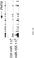
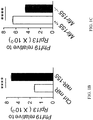
- Phf19 was strongly upregulated in miR-155 cells as compared to Ctrl cells ( Figure 1A ). This observation was further validated by qPCR analysis, which demonstrated that, as seen in Figure 1B , miR-155 cells contained nearly 3-fold higher Phf19 transcripts than Ctrl cells. Conversely, Phf19 levels were significantly reduced in miR-155-deficient CD8 + T cells compared to wild-type cells ( Figure 1C ).
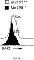
- Phf19 expression has been reported to be upregulated by pAkt in cancer cells, and Akt signaling is heightened by miR-155 through repression of inositol polyphosphate-5-phosphatase D (also known as Ship-1), a well-established negative regulator of Akt ( Ji Y et al., P. N. A. S. USA, 112:476-481(2015 ); published electronically as doi: 10.1073/pnas.1422916112. Epub (2014) ( Figures 1D and 1E ).
- Phf19 expression was measured in miR-155 cells and Ctrl miR-overexpressing cells that were concomitantly transduced with a constitutively active form of Akt (AktCA), or the Thy1.1 control. It was found that miR-155 upregulated Phf19 in Thy1.1 co-transduced cells ( Figure IF). Also shown in Figure IF, constitutive Akt signaling drove Phf19 expression to saturation, abrogating any further upregulation of Phf19 by miR-155. Next, whether downregulation of Ship1 alone would induce Phf19 was tested.
- AktCA constitutively active form of Akt
- Phf19 expression was dynamically regulated in CD8 + T cells responding to gp100-VV infection. It was found that Phf19 was strongly induced at the early stages of acute immune response, sharply downregulated at peak effector response and maintained at low levels throughout transition to memory phase ( Figure 2A ). These findings suggested a potential role of Phf19 in regulating CD8 + T cell effector differentiation. The rapid spike of induction was similarly observed for Ezh2, indicating coordinated expression of PRC2 and its associated factor Phf19 during the immune response ( Figure 2B ). Mir155 followed a virtually identical pattern of expression, emphasizing the interplay between these molecules during physiologic immune response ( Figure 2B ).
- Phf19 is essential to enhance proliferation and restrict exhaustion of miR-155 cells.
- Phf19 is a downstream factor of miR-155 in mediating T cell expansion and restricting exhaustion and senescence.
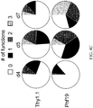
- Phf19 -overexpressing cells displayed enhanced proliferation ( Figure 4A ), reduced terminal differentiation ( Figure 4B ), sustained polyfunctionality, and IFN- ⁇ production ( Figure 4C ).
- Figure 4A enhanced proliferation
- Figure 4B reduced terminal differentiation
- Figure 4C sustained polyfunctionality
- Figure 4C IFN- ⁇ production
- Figure 4D and 4E enforced expression of Phf19 enhances CD8 + T cell long-term memory.
- Figures 4F and 4G Phf19 -overexpressing cells demonstrated a dramatic and long-lasting antitumor activity resulting in better overall survival.
- Phf19 overexpression enhances CD8 + T cell antitumor immunity by epigenetically reprogramming T cell differentiation.
- Phf19 for regulating PRC2 activity is to recruit the PRC2 to histone through its chromatin binding capacity. Therefore, a Phf19 mutant ( Phf19 mut), with attenuated chromatin binding-capacity to test whether the effects mediated by Phf19 overexpression were dependent on epigenetic mechanisms was generated.
- this mutant form of Phf19 two amino acids (W41 and Y47) in the aromatic cage of the Tale domain required to bind to chromatin, were mutated to W41C and Y47A, respectively ( Figure 5A ).
- the amino acid sequence of the wild-type region of Phf19 is set forth in SEQ ID NO: 7 (Wt).
- SEQ ID NO: 8 is SKVTEGQFVLCRCTDGLYALGKIKRVSSPKQ.
- Phf19 but not Phf19 mut enhanced Ezh2 association to chromatin and H3K27me3 deposition in CD8+ T cells.
- Figures 5C and 5D Following gp100-VV infection, pmel-1 cells transduced with Phf19Thy 1.1 , Phf19 mutThy1.1, or Thy1.1 alone were adoptively transferred into wild-type mice and T cell expansion, function, and differentiation in response to gp100-VV infection were measured.
- Phf19 -transduced cells expanded more robustly than Thy.1.1 controls ( Figures 5E and 5F ), and displayed limited senescence ( Figures 5G and 5H ) and sustained cytokine production ( Figures 5I and 5J ), and augmented memory precursor formation ( Figures 5K and 5L ).
- Figures 5E-5J displayed limited senescence
- Figures 5I and 5J sustained cytokine production
- Figures 5K and 5L augmented memory precursor formation
- Phf19 resultsed in augmented antitumor function
- pmel-1 cells overexpressing Phf19, Phf19 mut , or Thy1.1 were adoptively transferred into tumor-bearing mice in conjunction with administration of gp100-VV and IL-2. It was found that Phf19 cells mediated a dramatic and long-lasting antitumor response resulting in increased mice survival compared to Thy1.1 controls ( Figures 5M and 5N ).
- miR-155 enhances the anti-tumor response by epigenetically restricting CD8 + T cell differentiation and functional exhaustion.
- miR-155 promoted PRC2 activity to silence key TFs known to drive terminal differentiation and exhaustion.
- the miR-155-Jarid2-PRC2 axis had only a minor inhibitory role in CD8 + T cell immune responses to both virus and cancer. Instead, it was discovered that miR-155 influenced PRC2 function by indirectly inducing Phf19 expression ( Figure 6 ).
- Phf19 also restrains the differentiation of human T cells, as shown in Figure 7 .
- a highly conserved core of genes regulated in mouse and human T cells by Phf19 is shown in Figure 8 .
- the restriction of T cell terminal differentiation by Phf19 is highly conserved between mouse and human.
Landscapes
- Health & Medical Sciences (AREA)
- Life Sciences & Earth Sciences (AREA)
- Engineering & Computer Science (AREA)
- Chemical & Material Sciences (AREA)
- Organic Chemistry (AREA)
- Genetics & Genomics (AREA)
- Biomedical Technology (AREA)
- General Health & Medical Sciences (AREA)
- Zoology (AREA)
- Biotechnology (AREA)
- Wood Science & Technology (AREA)
- Bioinformatics & Cheminformatics (AREA)
- Immunology (AREA)
- Public Health (AREA)
- Animal Behavior & Ethology (AREA)
- Veterinary Medicine (AREA)
- General Engineering & Computer Science (AREA)
- Biochemistry (AREA)
- Microbiology (AREA)
- Cell Biology (AREA)
- Medicinal Chemistry (AREA)
- Epidemiology (AREA)
- Hematology (AREA)
- Pharmacology & Pharmacy (AREA)
- Nuclear Medicine, Radiotherapy & Molecular Imaging (AREA)
- General Chemical & Material Sciences (AREA)
- Chemical Kinetics & Catalysis (AREA)
- Biophysics (AREA)
- Molecular Biology (AREA)
- Virology (AREA)
- Physics & Mathematics (AREA)
- Plant Pathology (AREA)
- Toxicology (AREA)
- Gastroenterology & Hepatology (AREA)
- Proteomics, Peptides & Aminoacids (AREA)
- Communicable Diseases (AREA)
- Oncology (AREA)
- Medicines Containing Material From Animals Or Micro-Organisms (AREA)
- Developmental Biology & Embryology (AREA)
Description
- Adoptive cell transfer has emerged as a promising therapy for patients with advanced cancer. However, obstacles to the overall success of adoptive cell therapy still exist. For example, the in vivo proliferation, persistence, and release of high amounts of proinflammatory cytokines can, in some cases, decrease following adoptive transfer. Alternatively or additionally, in some cases, transferred cells, such as T cells, rapidly lose their proliferative and effector capacities following adoptive transfer as they enter into a state of functional exhaustion. T cell exhaustion is a state of T cell dysfunction that arises during many chronic infections and cancer. It is defined by poor effector function, sustained expression of inhibitory receptors, and a transcriptional state distinct from that of functional effector or memory T cells.
- In spite of considerable research into methods of producing cells for adoptive cell transfer therapy and treatments for cancer and viral diseases, there still exists a need for improved methods for producing cells for adoptive cell transfer therapy and treating and/or preventing cancer and viral diseases.
- An embodiment of the invention provides a T cell (which can be isolated or purified) modified to express Phf19 at a level that is higher than the level of Phf19 expressed by a T cell that has not been modified to express Phf19. In some embodiments of the invention, the T cell comprises a genetic expression vector encoding Phf19. An embodiment of the invention provides a genetically-modified mammalian T cell (which also can be isolated or purified) comprising a genetic expression vector comprising (a) virally-, bacterially-, or both virally- and bacterially-derived genetic sequences and (b) a genetic sequence encoding Phf19, whereby the genetic sequence encoding Phf19 within the vector is expressed within the T cell. In some embodiments of the invention, the T cell is a CD8+ T cell. In some embodiments, the T cell comprises an antigen-specific receptor, wherein the antigen-specific receptor is a T cell receptor (TCR) or a chimeric antigen receptor (CAR).
- An embodiment of the invention provides a pharmaceutical composition comprising at least one or two of the inventive T cells (which also can be isolated or purified), which have been modified to express Phf19 at a level that is higher than the level of Phf19 expressed by a T cell that has not been modified to express Phf19, and a pharmaceutically acceptable carrier. In some embodiments of the invention, the T cells in the pharmaceutical composition comprise a genetic expression vector encoding Phf19, such as a vector comprising (a) virally-, bacterially-, or both virally- and bacterially-derived genetic sequences and (b) a genetic sequence encoding Phf19, whereby the genetic sequence encoding Phf19 within the vector is expressed within the T cell. In some embodiments of the invention, the T cells in the pharmaceutical composition are CD8+ T cells.
- An embodiment of the invention provides a method for treating cancer or chronic viral disease in a mammal. In some embodiments of the invention, the method comprises administering to the mammal an effective amount of T cells modified to express Phf19 at a level that is higher than the level of Phf19 expressed by a T cell that has not been modified to express Phf19, wherein the T cells can also be isolated or purified. In some embodiments of the invention, the method comprises administering to the mammal a pharmaceutical composition comprising at least one or two T cells, which have been modified to express Phf19 at a level that is higher than the level of Phf19 expressed by a T cell that has not been modified to express Phf19, and a pharmaceutically acceptable carrier. In some embodiments of the invention, the method comprises administering to the mammal an effective amount of genetically-modified mammalian T cells comprising a genetic expression vector comprising (a) virally-, bacterially-, or both virally- and bacterially-derived genetic sequences and (b) a genetic sequence encoding Phf19, whereby the genetic sequence encoding Phf19 within the vector is expressed within the T cell(s). In some embodiments of the invention, the method comprises administering to the mammal a pharmaceutical composition comprising at least one or two T cells comprising a genetic expression vector comprising (a) virally-, bacterially-, or both virally- and bacterially-derived genetic sequences and (b) a genetic sequence encoding Phf19, whereby the genetic sequence encoding Phf19 within the vector is expressed within the isolated or purified T cell, and a pharmaceutically acceptable carrier.
- An embodiment of the invention provides a method for inhibiting T cell terminal differentiation and exhaustion. In one aspect, this method comprises modifying the T cell to express Phf19 at a level that is higher than the level of Phf19 expressed by a T cell that has not been modified, wherein the increased expression of Phf19 inhibits T cell terminal differentiation and exhaustion when compared with a T cell not modified to express Phf19. In another aspect, this method comprises introducing a genetic expression vector into the T cell, wherein the vector comprises (a) virally-, bacterially-, or both virally- and bacterially-derived genetic sequences and (b) a genetic sequence encoding Phf19, whereby the genetic sequence encoding Phf19 within the vector is expressed within the T cell.
-
-
Figure 1A shows RNA-seq reads of Phf19 mRNA in miR-155 and Ctrl-miR-overexpressing cells. -
Figure 1B is a bar graph showing RT-PCR of Phf19 mRNA in miR-155 and Ctrl-miR-overexpressing cells. Bars (mean ± s.e.m. of technical triplicates) are relative to Rpl13 mRNA. -
Figure 1C is a bar graph showing RT-PCR of Phf19 mRNA in in vitro activated KLRG1-miR-155 sufficient and deficient CD8+ T cells. Bars (mean ± s.e.m. of technical triplicates) are relative to Rpl13 mRNA. - Figure ID is an image of a gel showing Shipl and pAkt levels in miR-155-overexpressing cells assessed by Immunoblot. Gapdh was used as the normalizing control.
-
Figure 1E is a histogram showing pAkt levels of in vitro activated KLRG1- miR-155 sufficient and deficient CD8+ T cells assessed by flow cytometry. -
Figure 1F is a bar graph showing RT-PCR of the expression of Phf19 mRNA in CD8+ T cells overexpressing miR-155 and Ctrl-miR together with Thy1.1 or constitutively active Akt (AktCA). Bars represent the mean ± s.e.m. of technical triplicates. -
Figure 1G is an image of a gel showing Shipl and pAkt levels in Cas9+ CD8+ T cells transduced with Ship1-specific gRNA assessed by Immunoblot. Gapdh was used as the normalizing control. -
Figure 1H is a bar graph showing RT-PCR of Phf19 mRNA in Cas9+ CD8+ T cells transduced with Ship1-specific gRNA or control. Bars represent the mean ± s.e.m. of technical triplicates. Data are representative of two independent experiments. * = P < 0.05; ** = P < 0.01; **** = P < 0.001 (unpaired two-tailed Student's t-test). -
Figure 2A is a graph showing the expression of Phf19 mRNA at indicated time points after transferring 105 pmel-1 CD8+Ly5.1+ T cells into wild-type mice in conjunction with gp100-VV. -
Figure 2B is a graph showing the expressions of Mir155, Ezh2 and Phf19 mRNA at indicated time points after transferring 105 pmel-1 CD8+LyS.1+ T cells into wild-type mice in conjunction with gp100-VV. Ezh2 and Phf19 levels are relative to Rpl13, Mir155 levels are relative to U6. -
Figure 2C is a bar graph showing RT-PCR of Phf19 levels in Phf19 +/+ or Phf19 -/- CD8+ T cells. Bars (mean ± s.e.m. of technical triplicates) are relative to Rpl13 mRNA. -
Figure 2D shows density plots representing flow cytometry of thymocytes from Phf19 +/+ or Phf19 -/- thymus. Numbers adjacent to outlined areas indicate percentage after gating on CD3+ cells (top), CD3+CD4-CD8-cells (bottom). -
Figure 2E shows density plots of representing flow cytometry of splenocytes from Phf19 +/+ or Phf19 -/- spleen. Numbers adjacent to outlined areas indicate percentage after gating on CD3+ cells (top), CD3+CD8+ cells (bottom) -
Figure 2F shows density plots representing flow cytometry analysis of splenocytes from pmel-1 Phf19 +/+ or Phf19 -/- spleen. Numbers adjacent to outlined areas indicate percentage after gating on CD8+ cells. Data are presented as the mean of three to four individual mice in two independent experiments. *** = P < 0.005 (unpaired two-tailed Student's t-test). -
Figure 2G is a density plot showing flow cytometry of live CD8+Ly5.1+ T cells in thespleen 5 days after transfer of 3 × 10⁵ naive pmel-1 CD8+Ly5.1+ Phf19 +/+ or Phf19 -/- T cells into wild-type mice in conjunction with gp100-VV. Numbers adjacent to outlined areas indicate percentage after gating on live CD8+Ly5.1+ T cells. -
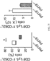
Figure 2H is a box plot showing percentages (left) and a bar graph showing the number (right) of pmel-1 CD8+Ly5.1+TE cells in thespleen 5 days after transfer of 3 × 10⁵ naïve pmel-1 CD8+Ly5.1+ Phf19 +/+ or Phf19 -/- T cells into wild-type mice in conjunction with gp100-VV. -
Figure 2I is a dot plot showing the percentage (left) and a bar graph showing the number (right) of CD8+Ly5.1+CD62L+ T cells in thespleen 5 days after transfer of 3 × 10⁵ naïve pmel-1 CD8+Ly5.1+ Phf19 +/+ or Phf19 -/- T cells into wild-type mice in conjunction with gp100-VV. Data are presented as box plots extending as a range. Bands inside the boxes represent median values of three mice. -
Figure 2J shows density plots representing flow cytometry of CD8+Ly5.1+ T cells in thespleen 30 days after adoptive transfer of 105 Phf19 +/+ and Phf19 -/- naïve pmel-1 CD8+ T cells into wild-type mice in conjunction with gp100-VV. Numbers adjacent to outlined areas indicate percentage after gating on live CD8+Ly5.1+ T cells. Data are presented as the mean of three to four mice in two independent experiments. * = P < 0.05 (unpaired two-tailed Student's t-test). -
Figure 2K is a bar graph showing the number of CD8+Ly5.1+ T cells in thespleen 30 days after adoptive transfer of 105 Phf19 +/+ and Phf19 -/- naïve pmel-1 CD8+ T cells into wild-type mice in conjunction with gp100-VV. Bars represent the mean ± s.e.m. of three mice. Data are presented as the mean of three to four mice in two independent experiments. * = P < 0.05 (unpaired two-tailed Student's t-test). -
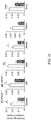
Figure 2L is a series of bar graphs showing ChIP-qPCR analyses using H3K27me3 antibody on in vitro activated non TE KLRG1- Phf19 +/+ or Phf19 -/- T cells with primers specific to the transcription start site of selected TFs. ChIP enrichments are presented as the percentage of protein bound, normalized to input. Bars represent the mean ± s.e.m. of technical triplicates. Data are representative of two independent experiments. * = P < 0.05; ** = P < 0.01; **** = P < 0.001 (unpaired two-tailed Student's t-test). -
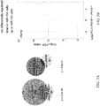
Figure 3A shows Venn diagrams depicting the number of differentially expressed genes in miR-155-overexpressing and Phf19 -/- KLRG1-CD62L- CD8+ T cells 5 days after transfer of 3 x 10⁵ pmel-1 Phf19 +/+/Phf19 -/- cells or pmel-1 cells overexpressing Ctrl-miR/miR-155 into wild-type mice in conjunction with gp100-VV. RNA-seq data were obtained from triplicated groups of three mice. -
Figure 3B is a volcano plot depicting the number of differentially expressed genes in miR-155-overexpressing and Phf19 -/- KLRG1-CD62L- CD8+ T cells 5 days after transfer of 3 x 10⁵pmel-1 Phf19 +/+/Phf19 -/- cells or pmel-1 cells overexpressing Ctrl-miR/miR-155 into wild-type mice in conjunction with gp100-VV. RNA-seq data were obtained from triplicated groups of three mice. -
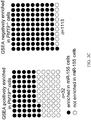
Figure 3C shows gene sets significantly enriched (FDR<0.25) in Phf19 -/- CD8+ T cells. Gene sets also enriched in miR-155 overexpressing cells are highlighted in black. -
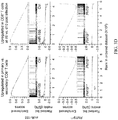
Figure 3D shows a graphs representing the enrichment of genes upregulated in CD8+ T cells responding to primary vs secondary LCMV infections (left) and enrichment of genes upregulated in CD8+ T cells at d6 vs d10 post LmOVA infections (right) in miR-155-overexpressing and Phf19 -/- CD8+ T cells. -
Figure 3E shows density plots representing flow cytometry of live pmel-1 Phf19 +/+ Ly5.2+/+ and pmel-1 Phf19 -/- Ly5.1+/- cells transduced with either miR-155 or Ctrl-miR assessed pre-transfer and 5 days after co-transfer of 3 × 10⁵ cells into Ly5.1+/+ mice in conjunction with gp100-VV. Numbers adjacent to outlined areas indicate percentage after gating on live CD8+GFP+ T cells. -
Figure 3F is a graph showing percentages of live pmel-1 Phf19 +/+Ly5.2+/+ and pmel-1 Phf19 -/- Ly5.1+/- cells transduced with either miR-155 or Ctrl-miR assessed at indicated time points after co-transfer of 3 × 10⁵ cells into Ly5.1+/+ mice in conjunction with gp100-VV. Symbols represent the mean ± s.e.m. of three mice; small horizontal lines (right panel) indicate the mean ± s.e.m. Data are representative of two independent experiments. *** = P < 0.005; **** = P < 0.001 (unpaired two-tailed Student's t-test). -
Figure 3G is a box plot showing percentages of live pmel-1 CD8+ GFP+ TE cells at indicated time points after co-transferring of 3 × 10⁵ pmel-1 Phf19 +/+Ly5.2+/+ and pmel-1 Phf19 -/- Ly5.1+/- cells transduced with either miR-155 or Ctrl-miR into Ly5.1+/+ mice in conjunction with gp100-VV Symbols represent the mean ± s.e.m. of three mice; small horizontal lines (right panel) indicate the mean ± s.e.m. Data are representative of two independent experiments. *** = P < 0.005; **** = P < 0.001 (unpaired two-tailed Student's t-test). -
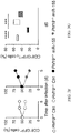
Figure 4A is a bar graph of the number of pmel-1 CD8+Thy1.1+ T cells (x 106) in the spleen following adoptive transfer of 3 × 10⁵ Phf19Thy1.1 (Phf19) or Thy1.1 transduced pmel-1 CD8+ T cells after gp100-VV infection. ** = P < 0.01 (a two-tailed Student t test). -
Figure 4B is a box plot of the percentage of pmel-1 CD8+Thy1.1+KLRG1+ T cells in the spleen at indicated time points following adoptive transfer of 3 × 10⁵ Phf19Thy1.1 (Phf19) or Thy1.1 (Thy1.1) transduced pmel-1 CD8+ T cells after gp100-VV infection. * = P < 0.05; ** = P < 0.01 (an unpaired two-tailed Student's t test). -
Figure 4C presents pie charts depicting the quality of the cytokine response in CD8+Thy1.1+ T cells transduced with the Phf19Thy1.1 (Phf19), or Thy1.1 control. # functions = # cytokines (IFN-γ+, IL-2+, and TNF-α+) produced by the cell. -
Figure 4D shows density plots depicting flow cytometry analysis of CD8+Thy1.1+ T cells in the spleen, lung, andlymphoid node 30 days after adoptive transfer of 3 × 10⁵ pmel-1 CD8+ T cells overexpressing Phf19Thy1.1 or Thy1.1 into wild-type mice in conjunction with gp100-VV. Numbers adjacent to outlined areas indicate percent after gating on CD8+Thy1.1+ T cells. -
Figure 4E is a bar graph presenting the number of CD8+Thy1.1+ cells in thespleen 30 days after transfer and 5 days after re-challenging hosts 30 days post infection as described in 4D. Bars represent the mean ± s.e.m. of three individual mice. -
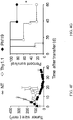
Figure 4F depicts the tumor size in mm2 of B16 tumor-bearing mice receiving 2 x 10⁶ cells Phf19Thy1.1 (Phf19) or Thy1.1 transduced pmel-1 CD8+ T in conjunction with gp100-VV and IL-2. NT = no treatment. *= P < 0.05 (a Log-rank (Mantel-Cox) Test). -
Figure 4G depicts the survival curve of B16 tumor-bearing mice receiving 2 x 10⁶ cells Phf19Thy1.1 (Phf19) or Thy1.1 transduced pmel-1 CD8+ T in conjunction with gp100-VV and IL-2. NT = no treatment. *= P < 0.05 (a Log-rank (Mantel-Cox) Test). -
Figure 5A is an amino acid sequence alignment of the partial aromatic cage of Phf19 indicating the location of the tryptophan (W) and tyrosine (Y) that were mutated to cysteine (C) and alanine (A), respectively, to abrogate chromatin binding of Phf19. The amino acid sequence of the wild-type region of Phf19 is set forth in SEQ ID NO: 7 (Wt), and the amino acid sequence of the mutated Phf19 region is set forth in SEQ ID NO: 8 (Mut). -
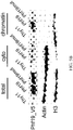
Figure 5B is an image of a gel depicting an immunoblot of total, soluble, and chromatin-bound proteins from CD8+ T cells transduced with the Phf19Thy1.1, Phf19mutThy1.1, and Thy1.1 control. -
Figure 5C shows histograms representing flow cytometry of Thy1.1 percentage in CD8+ T cells overexpressing Phf19, Phf19mut, or Thy1.1. Numbers adjacent to outlined areas indicate percentage after gating on CD8+ T cells. -
Figure 5D is an image of a gel depicting an immunoblot of Ezh2, H3K27me3, and H3 levels in chromatin fraction of CD8+ T cells transduced with Phf19, Phf19mut or Thy1.1. -
Figure 5E shows density plots representing flow cytometry of T cells in the spleen following transfer of 3 × 10⁵ pmel-1 CD8+ T cells transduced with Phf19Thy1.1, Phf19mutThy1.1, or Thy1.1 into wild-type mice in conjunction with gp100-VV. Numbers adjacent to outlined areas indicate percentage after gating on CD8+GFP+ T cells. -
Figure 5F is a bar graph showing the number of live pmel-1 CD8+Thy1.1+ T cells in thespleen 5 days following transfer of 3 × 10⁵ pmel-1 CD8+ T cells transduced with Phf19Thy1.1, Phf19mutThy1.1, or Thy1.1 into wild-type mice in conjunction with gp100-VV. Bars represent the mean ± s.e.m. of three mice. Data are representative of two independent experiments. * = P < 0.05; ** = P < 0.01; *** = P < 0.005; **** = P < 0.001 (unpaired two-tailed Student's t-test). -
Figure 5G shows density plots representing flow cytometry of live CD8+Thy1.1+ T cells in thespleen 5 days following transfer of 3 × 10⁵ pmel-1 CD8+ T cells transduced with Phf19Thy1.1, Phf19mutThy1.1, or Thy1.1 into wild-type mice in conjunction with gp100-VV. Numbers adjacent to outlined areas indicate percentage after gating on live CD8+Thy1.1+ T cells. -
Figure 5H is a box plot representing the percentage of live pmel-1 CD8+Thy1.1+ TE cells in thespleen 5 days following transfer of 3 × 10⁵ pmel-1 CD8+ T cells transduced with Phf19Thy1.1, Phf19mutThy1.1, or Thy1.1 into wild-type mice in conjunction with gp100-VV. Data are presented as box plots extending as a range. Bands inside the boxes represent median values of three mice. -
Figure 5I shows density plots representing flow cytometry of cytokine production of live CD8+Thy1.1+ T cells in thespleen 5 days after transfer of 3 × 10⁵ pmel-1 CD8+ T cells transduced with Phf19Thy1.1, Phf19mutThy1.1, or Thy1.1 into wild-type mice in conjunction with gp100-VV. Numbers adjacent to outlined areas indicate percentage after gating on live CD8+Thy1.1+ T cells. -
Figure 5J presents pie charts depicting the quality of the cytokine response in live CD8+Thy1.1+ T cells in thespleen 5 days after transfer of 3 × 10⁵ pmel-1 CD8+ T cells transduced with Phf19Thy1.1, Phf19mutThy1.1, or Thy1.1 into wild-type mice in conjunction with gp100-VV, as determined by the Boolean combination of gates identifying IFN-γ+, IL-2+, and TNF-α+ cells. Data are presented as the mean of three mice. Data are representative of two independent experiments. * = P < 0.05; ** = P < 0.01; *** = P < 0.005; **** = P < 0.001 (unpaired two-tailed Student's t-test). -
Figure 5K is a box plot showing the percentage of CD8+Thy1.1+KLRG1+ T cells in thespleen 5 days after re-challenging hosts 30 days following adoptive transfer of 3 × 10⁵ Phf19Thy1.1 (Phf19), Phf19mutThy1.1 (Phf19mut), or Thy1.1 (Thy1.1) transduced pmel-1 CD8+ T cells into wild-type mice in conjunction with gp100-VV. ** = P < 0.01 (a two-tailed Student t test). -
Figure 5L is a box plot showing the percentage of CD8+Thy1.1+CD62L+ T cells in thespleen 5 days after re-challenging hosts 30 days following adoptive transfer of 3 × 10⁵ Phf19Thy1.1 (Phf19), Phf19mutThy1.1 (Phf19mut), or Thy1.1 (Thy1.1) transduced pmel-1 CD8+ T cells into wild-type mice in conjunction with gp100-VV. ** = P < 0.01 (a two-tailed Student t test). -
Figure 5M is a graph showing tumor size (mean ± s.e.m.) of B16 tumor-bearing mice receiving 2 x 10⁶ pmel-1 CD8+ T cells transduced with Phf19Thy1.1, Phf19mutThy1.1, or Thy1.1 into wild-type mice in conjunction with gp100-VV and IL-2. NT, no treatment (n= 7 mice/group). Data are representative of two independent experiments. * = P < 0.05; ** = P < 0.01; *** = P < 0.005; **** = P < 0.001 (unpaired two-tailed Student's t-test). -
Figure 5N is a graph showing a survival curve of B16 tumor-bearing mice receiving 2 x 10⁶ pmel-1 CD8+ T cells transduced with Phf19Thy1.1, Phf19mutThy1.1, or Thy1.1 into wild-type mice in conjunction with gp100-VV and IL-2. NT, no treatment (n= 7 mice/group). Data are representative of two independent experiments. *= P < 0.05 (a Log-rank (Mantel-Cox) Test). -
Figure 6 is a schematic depicting the regulatory circuitry by which miR-155 epigenetically reprograms CD8+ T cell fate and function via enhancement of PRC2 activity. TFs, Transcription factors. -
Figure 7 shows density plots representing flow cytometry of human live CD8+ T cells transduced with Phf19-LNGFR, or LNGFR control and cultured in vitro for 8 days. Numbers adjacent to outlined areas indicate percentage after gating on live CD8+ T cells. -
Figure 8 is a Venn diagram showing a conserved core of genes regulated in mouse and human T cells by Phf19. -
Figure 9 is a diagram of the vector comprising mouse Phf19Thy1.1. - An embodiment of the invention provides a T cell, wherein the T cell has been modified to express Phf19 at a level that is higher than the level of Phf19 expressed by a T cell that has not been modified to express Phf19. The Phf19 used to modify the T cell may be Phf19, functional variants of Phf19, or functional fragments of Phf19. The modified T cell also can be isolated or purified. The modified T cell comprises an antigen-specific receptor, wherein the antigen-specific receptor is a T cell receptor (TCR) or a chimeric antigen receptor (CAR).
- The inventive T cells have been modified to overexpress Phf19. In this regard, the modified T cell expresses Phf19 at a level that is higher than the level of Phf19 expressed by a T cell that has not been modified with respect to Phf19 expression (e.g., wild-type T cells). For example, if the T cell has been modified to comprise a vector encoding Phf19, the modified T cell including such vector expresses Phf19 at a level that is higher than the level of Phf19 expressed by a control T cell that does not contain such vector.
- The level of expression of Phf19 may be determined using any methods known in the art. For example, the expression level of Phf19 may be determined using quantitative RT-PCR. The level of Phf19 expression may also be determined using antibodies. Antibodies that recognize Phf19 are available from several vendors including Abcam (Cambridge, UK), Cell Signaling Technologies (Danvers, MA), Bethyl Laboratories (Montgomery, TX), among others.
- The inventive T cells may provide many advantages, for example, an increase of any one or more of in vivo proliferation, survival, persistence, anti-tumor activity, and antiviral activity as compared to T cells that have not been modified to overexpress Phf19 (e.g., T cells that lack a vector encoding Phf19, as described in more detail below). CD8+ T cells overexpressing Phf19 present augmented cell expansion, reduced effector senescence, increased polyfunctionality, and superior anti-tumor responses. Also, while not wishing to be bound by theory, Example 4, below suggests that such effects may be influenced by the chromatin-binding capacity of Phf19 via two key amino acids located in the Aromatic cage within its Tudor domain. This suggests that the ability of Phf19 to bind to chromatin and recruit the PRC2 complex to the specific targets for H3K27me3 deposition may be implicated in its role in regulating CD8+ T cell differentiation and functional exhaustion. These findings support a conclusion that T-cell based immunotherapy can be enhanced through epigenetic reprogramming of T cell fate.
- The T cell may be isolated or purified. The term "isolated," as used herein, means having been removed from its natural environment. An "isolated" cell, therefore, can be a cell "in vitro" or "ex vivo," even if in the presence of other cells or derivative products of an organism (e.g., serum, growth factors, etc.). The term "purified," as used herein, means being separated from at least some other natural components. A "purified" T cell refers to a T cell which has been separated from at least some other natural components, such as tissues, cells, proteins, nucleic acids, etc. Preferably, the inventive T cells are in vitro or ex vivo.
- The T cell can be any T cell, such as a cultured T cell, e.g., a primary T cell, or a T cell from a cultured T cell line, e.g., Jurkat, SupT1, etc., or a T cell obtained from a mammal. If obtained from a mammal, the T cell can be obtained from numerous sources, including but not limited to blood, bone marrow, lymph node, thymus, spleen, tumor, or other tissues or fluids. The T cells can also be enriched or purified. Methods for enriching or purifying T cells are well known in the art and include, for example, immunomagnetic selection, centrifugation and resuspension, column separation, and immunoprecipitation. CD8 T cells may be purified from peripheral blood mononuclear cells (PBMCs) by incubating the PBMCs with antibodies against CD4 T cells, B cells, NK cells, dendritic cells, monocytes, granulocytes, and erythrocytes, and subsequently running the mixture through a magnetic column. Cells labeled with the antibodies will be bound to the column, while unlabeled cells, the CD8 T cells, will elute through.
- Preferably, the T cell is a human T cell. The T cell can be any type of T cell and can be of any developmental stage, including but not limited to, CD4+/CD8+ double positive T cells, CD4+ helper T cells, e.g., Th1, Th2, Th9, Th17, Th22 cells, CD4+ T cells, CD8+ T cells (e.g., cytotoxic T cells), peripheral blood mononuclear cells (PBMCs), peripheral blood leukocytes (PBLs), tumor infiltrating cells (TILs), memory T cells, naïve T cells, and the like. Preferably, the cell is a CD8+ T cell.
- An embodiment of the invention provides a pharmaceutical composition comprising at least one or two T cells as described herein - i.e., being modified to express Phf19 at a level that is higher than the level of Phf19 expressed by a T cell that has not been modified to express Phf19 and/or comprising a genetic expression vector comprising (a) virally-, bacterially-, or both virally- and bacterially-derived genetic sequences and (b) a genetic sequence encoding Phf19, whereby the genetic sequence encoding Phf19 within the vector is expressed within the T cell(s). Such pharmaceutical composition also comprises a pharmaceutically-acceptable carrier.
- The inventive compositions can comprise a single T cell or a population thereof. The population of T cells can be a heterogeneous population comprising the T cell that has been modified to overexpress Phf19 (e.g., a T cell comprising a vector encoding Phf19, as described in more detail below), in addition to at least one other cell, e.g., a T cell, which has not been modified to overexpress Phf19 (e.g., a T cell lacking such a vector encoding Phf19, as described in more detail below,), or a cell other than a T cell, e.g., a B cell, a macrophage, a neutrophil, an erythrocyte, a melanocyte, a hepatocyte, an endothelial cell, an epithelial cell, a muscle cell, a brain cell, etc. Alternatively, the population of cells can be a substantially homogeneous population, in which the population mainly comprises T cells that have been modified to overexpress Phf19 and/or comprise the genetic expression vector comprising (a) virally-, bacterially-, or both virally- and bacterially-derived genetic sequences and (b) a genetic sequence encoding Phf19, as described herein. The population also can be a clonal population of T cells, in which all T cells of the population are clones of a single T cell that has been modified to overexpress Phf19.
- A T cell of the invention can be present in a population of cells or a composition in an amount of about 10% or more, e.g., about 30% or more, about 50% or more, about 60% or more, about 70% or more, about 75% or more, about 80% or more, about 85% or more, or about 90% or more, based on the total number of cells in the population or composition. Alternatively, or in addition, the T cell of the invention can be present in a population of cells or a composition in an amount of about 95% or less, e.g., about 90% or less, about 85% or less, about 80% or less, about 75% or less, about 70% or less, about 60% or less, about 40% or less, or about 30% or less based on the total number of cells in the population or composition. Thus, the T cell of the invention can be present in a population of cells or a composition in an amount bounded by any two of the above endpoints. For example, the T cell of the invention can be present in a population of cells or a composition in an amount of about 30 to about 60%, about 50 to about 90%, about 60 to about 80%, about 80 to about 90%, or about 75 to about 85%.
- A method for treating cancer or chronic viral disease in a mammal is disclosed. The method comprises administering to the mammal an effective amount of the T cell of an embodiment of the invention.
- In an embodiment of the invention, the T cell comprises an antigen-specific receptor. The phrases "antigen-specific" and "antigenic specificity," as used herein, mean that the receptor can specifically bind to and immunologically recognize an antigen, or an epitope thereof, such that binding of the receptor to antigen, or the epitope thereof, elicits an immune response. In an embodiment of the invention, the antigen-specific receptor is a T cell receptor (TCR). The antigen-specific TCR generally comprises two polypeptides (i.e., polypeptide chains), such as an α-chain of a TCR, a β-chain of a TCR, a γ-chain of a TCR, a δ-chain of a TCR, or a combination thereof. Such polypeptide chains of TCRs are known in the art. The antigen-specific receptor can comprise any amino acid sequence, provided that the receptor can specifically bind to and immunologically recognize an antigen, such as a disease-associated antigen or epitope thereof.
- The antigen-specific receptor can be an endogenous TCR, i.e., the antigen-specific TCR that is endogenous or native to (naturally-occurring on) the T cell. In such a case, the T cell comprising the endogenous TCR can be a T cell that has been isolated from a mammal which is known to express the particular disease-specific antigen. In certain embodiments, the T cell is a primary T cell isolated from a host afflicted with a cancer. In some embodiments, the T cell is a tumor infiltrating lymphocyte (TIL) or a peripheral blood lymphocyte (PBL) isolated from a human cancer patient.
- In some embodiments, the mammal from which a T cell is isolated is immunized with an antigen of, or specific for, a disease. Desirably, the mammal is immunized prior to obtaining the T cell from the mammal. In this way, the isolated T cells can include T cells induced to have specificity for the disease to be treated, or can include a higher proportion of cells specific for the disease.
- Alternatively, a T cell comprising an endogenous antigen-specific TCR can be a T cell within a mixed population of cells isolated from a mammal, and the mixed population can be exposed to the antigen which is recognized by the endogenous TCR while being cultured in vitro. In this manner, the T cell comprising the TCR that recognizes the disease-specific antigen, expands or proliferates in vitro, thereby increasing the number of T cells having the endogenous antigen-specific receptor.
- The antigen-specific TCR can be an exogenous TCR, i.e., an antigen-specific TCR that is not native to (not naturally-occurring on) the T cell. A recombinant TCR is a TCR which has been generated through recombinant expression of one or more exogenous TCR α-, β-, γ-, and/or δ-chain encoding genes. A recombinant TCR can comprise polypeptide chains derived entirely from a single mammalian species, or the antigen-specific TCR can be a chimeric or hybrid TCR comprised of amino acid sequences derived from TCRs from two different mammalian species. For example, the antigen-specific TCR can comprise a variable region derived from a murine TCR, and a constant region of a human TCR such that the TCR is "humanized." Methods of making recombinant TCRs are known in the art. See, for example,
U.S. Patents 7,820,174 ,8,785,601 ,8,216,565 , and9,345,748 - A T cell of the invention comprising an endogenous antigen-specific TCR can also be transformed, e.g., transduced or transfected, with one or more nucleic acids encoding an exogenous (e.g., recombinant) TCR or other recombinant chimeric receptor. Such exogenous chimeric receptors, e.g., chimeric TCRs, can confer specificity for additional antigens to the transformed T cell beyond the antigens for which the endogenous TCR is naturally specific. This can, but need not, result in the production of T cell having dual antigen specificities.
- In an embodiment of the invention, the antigen-specific receptor is a "chimeric antigen receptor" (CAR). Typically, a CAR comprises the antigen binding domain of an antibody, e.g., a single-chain variable fragment (scFv), fused to the transmembrane and intracellular domains of a TCR. Thus, the antigenic specificity of a TCR of the invention can be encoded by a scFv which specifically binds to the antigen, or an epitope thereof. Methods of making such chimeric TCRs are known in the art. See, for example,
U.S. Patents 8,465,743 and9,266,960 U.S. Patent Application Publication No. 2014/0274909 . - Any suitable nucleic acid encoding a CAR, TCR, or TCR-like protein or polypeptide can be used. In these embodiments, transformation with a nucleic acid encoding Phf19, as discussed below, can occur before, after, or simultaneously with, antigen-specific receptor transformation. The antigen-specific receptor encoded by the transformed nucleic acids can be of any suitable form including for example, a single-chain TCR or a fusion with other proteins or polypeptides (e.g., without limitation co-stimulatory molecules).
- The antigen which is specifically recognized by the antigen-specific receptor can be any antigen which is characteristic of a disease. For example, the antigen may be, but is not limited to, a cancer antigen (also termed a tumor antigen or a tumor associated antigen) or a foreign antigen (viral, bacterial, parasite antigens). Foreign antigens are known in the art and include, for example, any viral protein, e.g., env, gag, pol, gp120, thymidine kinase, and the like.
- The term "cancer antigen," as used herein, refers to any molecule (e.g., protein, polypeptide, peptide, lipid, carbohydrate, etc.) solely or predominantly expressed or over-expressed by a tumor cell or cancer cell, such that the antigen is associated with the tumor or cancer. The cancer antigen can additionally be expressed by normal, non-tumor, or non-cancerous cells. However, in such cases, the expression of the cancer antigen by normal, non-tumor, or non-cancerous cells is not as robust as the expression by tumor or cancer cells. In this regard, the tumor or cancer cells can over-express the antigen or express the antigen at a significantly higher level, as compared to the expression of the antigen by normal, non-tumor, or non-cancerous cells. Also, the cancer antigen can additionally be expressed by cells of a different state of development or maturation. For instance, the cancer antigen can be additionally expressed by cells of the embryonic or fetal stage, which cells are not normally found in an adult host. Alternatively, the cancer antigen can be additionally expressed by stem cells or precursor cells, whether or not normally found in an adult host. Cancer antigens are known in the art and include, for instance, mesothelin, CD19, CD22, CD276 (B7H3), gp100, MART-1, Epidermal Growth Factor Receptor Variant III (EGFRVIII), TRP-1, TRP-2, tyrosinase, NY-ESO-1 (also known as CAG-3), MAGE-1, MAGE-3, etc.
- The cancer antigen can be an antigen expressed by any cell of any cancer or tumor, including the cancers and tumors described herein. The cancer antigen may be a cancer antigen of only one type of cancer or tumor, such that the cancer antigen is associated with or characteristic of only one type of cancer or tumor. Alternatively, the cancer antigen may be a cancer antigen (e.g., may be characteristic) of more than one type of cancer or tumor. For example, the cancer antigen may be expressed by both breast and prostate cancer cells and not expressed at all by normal, non-tumor, or non-cancer cells.
- The disease which is associated with or is characterized by the antigen recognized by the antigen-specific receptor can be any disease. For instance, the disease can be a cancer or a viral disease, as discussed herein.
- The cancer may be any cancer, including any of acute lymphocytic cancer, acute myeloid leukemia, alveolar rhabdomyosarcoma, bone cancer, brain cancer, breast cancer, cancer of the anus, anal canal, or anorectum, cancer of the eye, cancer of the intrahepatic bile duct, cancer of the joints, cancer of the neck, gallbladder, or pleura, cancer of the nose, nasal cavity, or middle ear, cancer of the oral cavity, cancer of the vulva, chronic lymphocytic leukemia, chronic myeloid cancer, colon cancer, esophageal cancer, cervical cancer, gastrointestinal carcinoid tumor, Hodgkin lymphoma, HPV carcinoma, hypopharynx cancer, kidney cancer, larynx cancer, leukemia, liver cancer, lung cancer, malignant mesothelioma, melanoma, multiple myeloma, nasopharynx cancer, non-Hodgkin lymphoma, ovarian cancer, pancreatic cancer, peritoneum, omentum, and mesentery cancer, pharynx cancer, prostate cancer, rectal cancer, renal cancer (e.g., renal cell carcinoma (RCC)), small intestine cancer, soft tissue cancer, stomach cancer, testicular cancer, thyroid cancer, ureter cancer, and urinary bladder cancer..
- For purposes herein, "viral disease" means a disease that can be transmitted from person to person or from organism to organism, and is caused by a virus. In an embodiment of the invention, the viral disease is caused by a virus selected from the group consisting of herpes viruses, pox viruses, hepadnaviruses, papilloma viruses, adenoviruses, coronoviruses, orthomyxoviruses, paramyxoviruses, flaviviruses, and caliciviruses. For example, the viral disease may be caused by a virus selected from the group consisting of respiratory syncytial virus (RSV), influenza virus, herpes simplex virus, Epstein-Barr virus, varicella virus, cytomegalovirus, hepatitis A virus, hepatitis B virus, hepatitis C virus, human immunodeficiency virus (HIV), human T-lymphotropic virus, calicivirus, adenovirus, and Arena virus.
- The viral disease may be, for example, influenza, pneumonia, herpes, hepatitis, hepatitis A, hepatitis B, hepatitis C, chronic fatigue syndrome, sudden acute respiratory syndrome (SARS), gastroenteritis, enteritis, carditis, encephalitis, bronchiolitis, respiratory papillomatosis, meningitis, HIV/AIDS, and mononucleosis.
- An isolated or purified T cell may be modified to overexpress Phf19. The T cell may be modified to overexpress Phf19 in any suitable manner. In an embodiment of the invention, the T cell may be modified to overexpress Phf19 using genome editing techniques.
- Genome editing techniques can modify gene expression in a target cell by inserting, replacing, or removing DNA in the genome using an artificially engineered nuclease. Examples of such nucleases may include zinc finger nucleases (ZFNs), transcription activator-like effector nucleases (TALENs), the CRISPR/Cas system, and engineered meganucleases. The nucleases create specific double-stranded breaks (DSBs) at targeted locations in the genome, and use endogenous mechanisms in the cell to repair the induced break by homologous recombination (HR) and nonhomologous end-joining (NHEJ). Such techniques may be used to achieve overexpression of Phf19 in T cells.
- In another embodiment of the invention, the T cell may be modified (e.g., transduced or transfected) so as to comprise a nucleic acid encoding Phf19. Variants of Phf19 have been identified in Homo sapiens, and sequences encoding homologs of Phf19 have been identified at least for Poeciliopsis prolifica, Microcebus murinus, Meleagris gallopavo, Aptenodytes forsteri, Fukomys damarensis, Gorilla, Xenopus tropicalis, and Callithrix jacchus.
- The sequence of Homo sapiens Phf19 isoform e is found at GenBank accession No. NP_001273772.1: MENRALDPGTRDSYGATSHLPNKGALAKVKNNFKDLMSKLTEGQYVLCRWTDGLY YLGKIKRVSSSKQSCLVTFEDNSKYWVLWKDIQHAGVPGEEPKCNICLGKTSGPLNE ILICGKCGLVPHPHSGQC, and it is set forth in SEQ ID NO: 12.
- The amino acid sequence of Homo sapiens Phf19 isoform d is found at GenBank accession No. NP_001273771.1: MLQCYRCRQWFHEACTQCLNEPMMFGDRFYLFFCS VCNQGPEYIERLPLRWVDVVHLALYNLGVQSKKKYFDFEEILAFVNHHWELLQLGK LTSTPVTDRGPHLLNALNSYKSRFLCGKEIKKKKCIFRLRIRVPPNPPGKLLPDKGLLP NENSASSELKRGKSKPGLLPHEFQQQKRRVYRRKRSKFLLEDAIPSSDFTSAWSTNH HLASIFDFTLDEIQSLKSASSGQTFFSDVDSTDAASTSGSASTSLSYDSRWTVGSRKRK LAAKAYMPLRAKRWAAELDGRCPSDSSAEGASVPERPDEGIDSHTFESISEDDSSLSH LKSSITNYFGAAGRLACGEKYQVLARRVTPEGKVQYLVEWEGTTPY, and it is set forth in SEQ ID NO: 13.
- The amino acid sequence of Homo sapiens Phf19 isoform c is found at GenBank accession No. NP_001273769.1: MLVLVIRGPYPSAQCQGKLMENRALDPGTRDSYGA TSHLPNKGALAKVKN NFKDLMSKLTEGQYVLCRWTDGLYYLGKIKRVSSSKQSCL VTFEDNSKYWVLWKDIQHAGVPGEEPKCNICLGKTSGPLNEILICGKCGLGYHQQCH IPIAGSADQPLLTPWFCRRCIFALAVRKGGALKKGAIARTLQAVKMVLSYQPEELEW DSPHRTNQQQCYCYCGGPGEWYLRMLQCYRCRQWFHEACTQCLNEPMMFGDRFY LFFCSVCNQGPEYIERLPLRWVDVVHLALYNLGVQSKKKYFDFEEILAFVNHHWELL QLGKLTSTPVTDRGPHLLNALNSYKSRFLCGKEIKKKKCIFRLRIRVPPNPPGKLLPDK GLLPNENSASSELRKRGKSKPGLLPHEFQQQKRRVYRRKRSKFLLEDAIPSSDFTSAW STNHHLASIFDFTLDEIQSLKSASSGQTFFSDVDSTDAASTSGSASTSLSYDSRWTVG SRKRKLAAKAYMPLRAKRWAAELDGRCPSDSSAEGASVPERPDEGIDSHTFESISED DSSLSHLKSSITNYFGAAGRLACGEKYQVLARRVTPEGKVQYLVEWEGTTPY, and it is set forth in SEQ ID NO: 14.
- The amino acid sequence of Homo sapiens Phf19 isoform b is found at GenBank accession No. NP_001009936.1: MENRALDPGTRDSYGATSHLPNKGALAKVKNNFKD LMSKLTEGQYVLCRWTDGLYYLGKIKRVSSSKQSCLVTFEDNSKYWVLWKDIQHA GVPGEEPKCNICLGKTSGPLNEILICGKCGLGYHQQCHIPIAGSADQPLLTPWFCRRCI FALAVRVSLPSSPVPASPASSSGADQRLPSQSLSSKQKGHTWALETDSASATVLGQD L, and it is set forth in SEQ ID NO: 15.
- The amino acid sequence of Homo sapiens Phf19 isoform a is found at GenBank accession No. NP_056466.1: MENRALDPGTRDSYGATSHLPNKGALAKVKNNFKDLM SKLTEGQYVLCRWTDGLYYLGKIKRVSSSKQSCLVTFEDNSKYWVLWKDIQHAGVP GEEPKCNICLGKTSGPLNEILICGKCGLGYHQQCHIPIAGSADQPLLTPWFCRRCIFAL AVRKGGALKKGAIARTLQAVKMVLSYQPEELEWDSPHRTNQQQCYCYCGGPGEW YLRMLQCYRCRQWFHEACTQCLNEPMMFGDRFYLFFCSVCNQGPEYIERLPLRWVD VVHLALYNLGVQSKKKYFDFEEILAFVNHHWELLQLGKLTSTPVTDRGPHLLNALN SYKSRFLCGKEIKKKKCIFRLRIRVPPNPPGKLLPDKGLLPNENSASSELRKRGKSKPG LLPHEFQQQKRRVYRRKRSKFLLEDAIPSSDFTSAWSTNHHLASIFDFTLDEIQSLKSA SSGQTFFSDVDSTDAASTSGSASTSLSYDSRWTVGSRKRKLAAKAYMPLRAKRWAA ELDGRCPSDSSAEGASVPERPDEGIDSHTFESISEDDSSLSHLKSSITNYFGAAGRLAC GEKYQVLARRVTPEGKVQYLVEWEGTTPY, and it is set forth in SEQ ID NO: 9.
- The amino acid sequence of mouse Phf19, NCBI reference sequence NP_082992.1 is: METQALEPGTLEAFGATSPNKGGLSKTKKNFKDLMSKVTEGQFVL CRWTDGLYYLGKIKRVSSPKQSCLVTFEDNSKYWVLWKDIQHAGVPGEEPKCDVC MGKTSGPMNEILICGKCGLGYHQQCHIPIAVDANWPLLTHWFCRRCIFALAVRKGG ALKKGAIAKTLQAVKMVLSYQPEELDWDSPHRTNQQQCYCYCGGPGEWYLRMLQ CYRCRQWFHEACTQCLSEPMVFGDRFYLFFCSVCNQGPEYIERLPLRWVDIVHLALY NLGVQSKKRYFDFEEILAFVNHHWELLQLGKLTSTPMTERGPHLLNALNSYKSRFLC GKEIKKKKCIFRLRIRVPPAPPGKLLPDRALMPSDKGTSELLRKKGKSKPGLLPQEPQ QQKRRVYRRKRSKFLLEDAIPSSDFTSAWSTDHHLASIFDFTLDEIQSLKSGSSGQTFF SDVDSTDAASTSGSASTSLSYDSRWTVGSRKRKLTAKVHRPLRAKQRAAELEGRCA SDSNAEGAVGPEQPDEGIDSHTLESISGDDSSLSHLKSSITNYFGAAGRLACGEKYRV LARRVTPEGKVQYLLEWEGTTPY, and it is set forth in SEQ ID NO: 10.
- Genetic expression vectors useful to overexpress Phf19 are well known in the art. The vector may be a plasmid, a viral vector, a cosmid, or an artificial chromosome. Viral vectors that can be used to deliver nucleic acids into the genetic makeup of cells include engineered retrovirus, lentivirus, adenovirus, adeno-associated virus and herpes simplex virus. It will be apparent to persons or ordinary skill that genetic vectors for introduction and expression of foreign genetic sequences into mammalian cells, such as T cells, typically contain elements of virally-, bacterially-, or both virally- and bacterially-derived DNA (or RNA) in addition to containing or comprising mammalian genetic elements (such as promoters, enhancers, and the like) and the Phf19-encoding sequence. Preferred are any techniques that can efficiently introduce Phf19 into antigen-specific T cells.
- Preferably, the nucleic acid is a recombinant nucleic acid. As used herein, the term "recombinant" refers to (i) molecules that are constructed outside living cells by joining natural or synthetic nucleic acid segments to nucleic acid molecules that can replicate in a living cell, or (ii) molecules that result from the replication of those described in (i) above. For purposes herein, the replication can be in vitro replication or in vivo replication.
- The terms "nucleic acid" and "polynucleotide," as used herein, refer to a polymeric form of nucleotides of any length, either ribonucleotides (RNA) or deoxyribonucleotides (DNA). These terms refer to the primary structure of the molecule, and thus include double- and single-stranded DNA, double- and single-stranded RNA, and double-stranded DNA-RNA hybrids. The terms include, as equivalents, analogs of either RNA or DNA made from nucleotide analogs and modified polynucleotides such as, though not limited to, methylated and/or capped polynucleotides. Suitable nucleotide analogs are known.
- Methods of preparing polynucleotides are within the ordinary skill in the art (Green and Sambrook, Molecular Cloning: A Laboratory Manual, (4th Ed.) Cold Spring Harbor Laboratory Press, New York (2012)).
- The nucleic acid may comprise any suitable Phf19 nucleotide sequence, which may encode any suitable Phf19 amino acid sequence from any mammal, examples are discussed above.
- An embodiment of the invention provides a method for inhibiting T cell terminal differentiation and exhaustion, comprising epigenetically reprogramming the T cell to express Phf19 at a level that is higher than the level of Phf19 expressed by a T cell that has not been epigenetically reprogrammed, wherein the increased expression of Phf19 inhibits T cell terminal differentiation and exhaustion when compared with a T cell not epigenetically reprogrammed by Phf19 overexpression.
- The following examples further illustrate the invention but, of course, should not be construed as in any way limiting its scope.
- As seen in in the Figures and described herein, particularly in these Examples, it appears as if, in the absence of Phf19, CD8+ T cells consistently display impaired expansion and enhanced exhaustion. Also seen in the figures and described herein, overexpression of Phf19 appears to enhance T cell expansion while restraining terminal differentiation and senescence. Thus, enforced expression of Phf19 can greatly augment cellular engraftment, restrict senescence, and sustain cytokine production, resulting in enhanced antitumor immunity by epigenetically reprogramming T cell fate.
- The following materials and methods were employed in the experiments described in Examples 1 through 4, below.
- C57BL/6 mice were obtained from Charles River Laboratories (Wilmington, MA); although the C57BL/6J strain of mice is refractory to many tumors, it is a permissive background for maximal expression of most mutations. Pmel-1 (B6.Cg-Thyla/Cy Tg (TcraTcrb)8Rest/J) mice were obtained from the Jackson Laboratory (Bar Harbor, ME); these transgenic mice provide a tumor model system for studies related to immunotherapy, and for studying in vivo T-cell biology Phf19 -/- cells were obtained from the Mouse Biology Program at the University of California, Davis (Davis, CA). B16 (H-2b), a gp100+ mouse melanoma, was obtained from the National Cancer Institute Tumor Repository. B16 (H-2b)-hgp100 was obtained from K. Hanada, (Surgery Branch, NCI/NIH, Bethesda, MD). All mouse experiments were done with the approval of the National Cancer Institute Animal Use and Care Committee.
- Anti-CD8α, anti-CD62L, anti-KLRG1, anti-CD3, anti-CD44, anti-CD25, anti-Ly-5.1, anti-Ly-5.2, anti-Thy-1.1, anti-IL-2, anti-IFN-y, anti-TNF-a, were obtained from BD Biosciences (Franklin Lakes, NJ). LEUKOCYTE ACTIVATION COCKTAIL containing phorbol myristate acetate and ionomycin (BD Biosciences) was used for the stimulation of T cells for intracellular cytokine staining. A FACSCANTO II or LSR II (BD Biosciences) was used for flow cytometry acquisition. Samples were analyzed with FLOWJO software (TreeStar, Ashland, OR). CD8+ GFP+ T cells, CD8+ GFP+ Thy-1.1+ T cells, CD8+ Ly-5.1+ T cells were sorted with a FACSAria (BD Biosciences).
- RNA was isolated with a MIRNEASY Mini kit (Qiagen, Germantown, MD) and cDNA was generated by reverse transcription (Applied BioSystems, Foster City, CA), following the manufacturer's protocol. Primers from Applied BioSystems (listed in Table 1), and a PRISM 7900HT (Applied BioSytems) were used for real-time PCR analysis of all genes; results are presented relative to U6 expression.
- Proteins were separated by 4-20% sodium dodecyl sulfate polyacrylamide gel electrophoresis (SDS-PAGE), followed by standard immunoblot analysis. Anti-pAkt, anti-Ship1, anti-Ezh2 and anti-Suz12 were obtained from Cell Signaling Technology (Danvers, MA). Anti-Jarid2 and anti-H3 were obtained from Abcam (Cambridge, MA). Anti-H3K27me3 and anti-Gapdh were obtained from EMD Millipore (Kankakee, IL). Anti-V5 was obtained from Invitrogen (Carlsbad, CA). Anti-Actin, horseradish peroxidase-conjugated goat anti-mouse IgG, and horseradish peroxidase-conjugated goat anti-rabbit IgG were obtained from Santa Cruz Biotechnology (Dallas, TX). Cells were analyzed by
immunoblot 72 hours after transduction. - Phf19 cDNA, Phf19mut cDNA, or Thy1.1 cDNA linked by the sequence encoding the
picornavirus 2A ribosomal skip peptide were cloned into the MSGV1 vector (Hughes, M. et al., Hum. Gene Ther. 16: 457-472 (2005)). PLATINUM ECO cell lines (Cell Biolabs, Inc. San Diego, CA) were used for gamma-retroviral production by transfection with DNA plasmids through the use of LIPOFECTAMINE 2000 (Invitrogen) and collection ofvirus 40 hours after transfection. The nucleotide sequence of the mouse Phf19Thy1.1 vector is set forth in SEQ ID NO: 11;Figure 9 shows a diagram of this Phf19Thy1.1 vector. - CD8+ T cells were separated from non-CD8+ T cells with a MACS negative selection kit (Miltenyi Biotech, Bergisch Gladbach, Germany) and were activated on plates coated with anti-CD3ε (2 µg/ml; BD Biosciences) and soluble anti-CD28 (1 µg/ml; BD Biosciences) in culture medium containing rhIL-2 (120 IU/ml; Chiron, Emeryville, CA). Virus was 'spin-inoculated' at 2,000 g for 2 h at 32 °C onto plates coated with RETRONECTIN recombinant human fibronectin (Takara, Shiga, Japan). CD8+ T cells activated for 24 h were transduced following standard protocols.
- Cells (3 × 10⁵ to 2 x 10⁶ cells) were adoptively transferred into hosts followed by infection with recombinant vaccinia virus or fowlpox virus expressing human gp100 (Virapur; San Diego, CA) together with the indicated combination of exogenous cytokines (2.4e5 IU/dose of rhIL-2 for 6 doses every 12 hours). Female C57BL/6 mice were injected subcutaneously with 3 × 10⁵ B16 melanoma cells.
- Mice were euthanized after infection. Splenocytes were counted by TRYPAN blue exclusion cell viability assay. The frequency of transferred T cells was determined by measurement of the expression of CD8 and GFP (green fluorescent protein), Thy-1.1, by flow cytometry. The absolute number of pmel-1 cells was calculated by multiplication of the total cell count with the percentage of CD8+GFP+Thy-1.1+ cells.
- Cells were sorted ex vivo and total RNA was isolated with a miRNEASY Mini kit (Qiagen, Velno, Netherlands). Ten ng total RNA was used for Nanostring analysis following the NanoString® nCounter®Expression CodeSet Design Manual. Background levels were calculated and subtracted from the samples, which were then normalized against the positive control and housekeeping gene probes. Expression heat maps were generated with the R package ComplexHeatmap (Bioconductor, Open Source Software for Bioinformatics), as described in Gu et., "Complex heatmaps reveal patterns and correlations in multidimensional genomic data, Bioinformatics, 32(18):2847-9 (2016), which is herein incorporated by reference.
- Ribonucleic acid (RNA) concentration was determined with the QUBIT RNA broad range assay in the QUBIT Fluorometer (Invitrogen). The RNA integrity was determined with Eukaryote TOTAL RNA NANO SERIES II ChIP on a 2100 Bioanalyzer (Agilent Technologies, Santa Clara, CA). RNA-seq libraries were prepared from 4 µg of total RNA via the TRUSEQ RNA sample prep kit according to manufacturer's protocol (Illumina, San Diego, CA). In brief, oligo-dT purified mRNA was fragmented and subjected to first and second strand cDNA synthesis. cDNA fragments were blunt-ended, ligated to ILLUMINA adaptors, and PCR amplified to enrich for the fragments ligated to adaptors. The resulting cDNA libraries were verified and quantified on Agilent Bioanalyzer and single-end 96 cycle RNA-seq was conducted with the GAIIX Genome Analyzer (Illumina).
- RNA-seq analyses were performed using ≥2 biological replicates. For RNA sequencing, total RNA was prepared from cells using the RNEASY PLUS Mini Kit (Qiagen). 200 ng total RNA was subsequently used to prepare an RNA-seq library by using TRUSEQ RNA sample prep kit (FC-122-1001, Illumina) according to the manufacturer's instructions. Sequenced reads were aligned to the mouse genome (NCBI37/mm9) using TOPHAT 2.0.11 (Jonhs Hopkins University Center for Computational Biology; Baltimore, MD), and uniquely mapped reads were used to calculate gene expression. REFSEQ gene database (mm9) was downloaded from the UCSC genome browser for RNA-seq analysis. Raw counts that fell on exons of each gene were calculated, and differentially expressed genes were identified with the statistical R PACKAGE EDGER52 or CUFFDIFF53. Fisher's exact test or t-test were used to evaluate significance with indicated P value and fold-change thresholds. Expression heat maps were generated with the R package PHEATMAP.
- Mouse gene symbols were first mapped to the orthologous human genes using homology information available from Mouse Genome Informatics (MGI) (see http:// www.informatics. jax. org and ftp:// ftp. informatics, jax. org) and were ranked by the fold changes of the gene expression as profiled by RNA-seq. Then, gene set enrichment was analyzed using GSEA software available from the Broad Institute website (www.software. broadinstitute. org).
- T cells were crosslinked with 2% paraformaldehyde at room temperature for 10 min and lysed in Farnham buffer (5 mM PIPES pH 8.0; 85 mM KCl; 0.5% NP-40) and subsequently in RIPA buffer (1x PBS; 1% NP-40; 0.5% sodium deoxycholate; 0.1% SDS). Sheared chromatin was immunoprecipitated with anti-H3K27me3 antibody (Millipore, 07-449) and washed successively in buffer I (20 mM Tris HCl pH 8.0, 150 mM NaCl, 2 mM EDTA, 0.1% SDS, 1% Triton X-100); buffer II (20 mM Tris HCl pH 8.0, 500 mM NaCl, 2 mM EDTA, 0.1% SDS, 1% Triton X-100); three times of buffer III (100 mM Tris HCl pH 8.0, 500 mM LiCl, 1% NP-40; 1% sodium deoxycholate). For sequencing of immunoprecipitated DNA, DNA fragments were blunt-end ligated to Illumina adaptors, amplified, and sequenced by using the HiSeq 2000 platform (Illumina). Single-end reads of 50 bp were obtained by using the Illumina Pipeline (Illumina). Sequenced reads were aligned to the mouse genome (NCBI37/mm9) with Bowtie 2.2.8; only uniquely mapped reads were retained. H3K27me3 enriched regions were detected using SICER algorithm, described in Xu et al., "Spatial clustering for identification of ChIP-enriched regions (SICER) to map regions of histone methylation patterns in embryonic stem cells," Methods Mol Biol., 1150:97-11 (2014), herein incorporated by reference, and the window size, gap size, and FDR were set to 200 bp, 600 bp, and 5%, respectively. Genomic graphs were generated and viewed using the INTEGRATIVE GENOMICS VIEWR (IGV), described in Robinson et al., "Integrative Genomics Viewer," Nature Biotechnology, 29, 24-26 (2011).
- A two-tailed student t test was used for comparison of data such as gene expression levels, cell proliferation and functionality (numbers and percent), and tumor growth slopes. A Log-rank (Mantel-Cox) Test was used for comparison of survival curves.
- Table 1 lists the nucleotide sequences of the qPCR primers used in the experiments described herein.
Table 1 Name qPCR Primer sequence: SEQ ID NO: Phf19 F TGACAGAGGGACAGTTCGTG 1 Phf19R GATCTCGTTCATAGGCCCTGA 2 miR-155 mmu-miR-155-5p (002571, Life Technologies) UUAAUGCUAAUUGUGAUAGGGGU 3 U6: U6 snRNA (001973, Life Technologies)
4 Rpl13 F CGAGGCATGCTGCCCCACAA 5 Rpl13R AGCAGGGACCACCATCCGCT 6 Ezh2F AGTGACTTGGATTTTCCAGCAC 16 Ezh2R AATTCTGTTGTAAGGGCGACC 17 - Table 2 lists the nucleotide sequences of the ChIP primers used in the experiments described herein.
Table 2 Name ChIP PCR Primer sequence: (from mouse genome assembly GRCm38.p5) SEQ ID NO: EomesF GAGCTTGCTCTAGGGGTAGG (Chromosome 9: 118,476,227-118,476,247) 18 EomesR ACAGCCAGAAGTAAGGTCCC (Chromosome 9: 118,476,388-118,476,408) 19 Id2F CGCCACAATTCCGACCTTAG (Chromosome 12: 25,100,336-25,100,356) 20 Id2R AAATATTTGCGGCGCTCCAT (Chromosome 12: 25,100,545-25,100,565) 21 Prdm1F TTGGGGCACAGATACCATGT (Chromosome 10: 44,460,649-44,460,669) 22 Prdm1R TCCTCCCTAGACTCAAGCCT (Chromosome 10: 44,460,841-44,460,861) 23 MafF CTGCAGACATTTTGAGGCGT (Chromosome 8: 118,236,772-118,236,792) 24 MafR TCTAACTGAGCCGGTGTTGT (Chromosome 8: 118,236,930-118,236,950) 25 Nr4a2F TGGTTGTTCTAGGGCGTGAT (Chromosome 2: 56,944,999-56,945,019) 26 Nr4a2R TACCCGGCCAAACTCTCAAT (Chromosome 2: 56,944,999-56,945,019) 27 Zeb2F TGAAATTCCACCTCCCTCCC (Chromosome 2: 56,945,135-56,945,155) 28 Zeb2R TCCCTTTAACTTTCGCCCCT (Chromosome 2: 45,113,439-45,113,459) 29 - This example demonstrates that Phf19 is a critical downstream factor of miR-155 in CD8+ T cells.
- To identify potential downstream targets of miR-155 involved in Polycomb Repressive Complex 2 (PRC2) function, the RNA-seq data set was re-examined comparing KLRG1-CD62L-CD8+T cells overexpressing miR-155 (miR-155 cells) and the CD8+ T cells not overexpressing miR-155 (Ctrl cells). The PHD finger protein 19 (Phf19) is a polycomb-like protein that recruits the PRC2 to specific genomic targets by binding to histone H3 trimethylated at Lysine 36 (H3K36me3) (Bellaré C et al., Nat. Struct. Mol. Biol. 19: 1257-1265 (2012)). Phf19 was strongly upregulated in miR-155 cells as compared to Ctrl cells (
Figure 1A ). This observation was further validated by qPCR analysis, which demonstrated that, as seen inFigure 1B , miR-155 cells contained nearly 3-fold higher Phf19 transcripts than Ctrl cells. Conversely, Phf19 levels were significantly reduced in miR-155-deficient CD8+ T cells compared to wild-type cells (Figure 1C ). - Phf19 expression has been reported to be upregulated by pAkt in cancer cells, and Akt signaling is heightened by miR-155 through repression of inositol polyphosphate-5-phosphatase D (also known as Ship-1), a well-established negative regulator of Akt (Ji Y et al., P. N. A. S. USA, 112:476-481(2015); published electronically as doi: 10.1073/pnas.1422916112. Epub (2014) (
Figures 1D and1E ). To determine whether miR-155 induced Phf19 by enhancing Akt signaling, Phf19 expression was measured in miR-155 cells and Ctrl miR-overexpressing cells that were concomitantly transduced with a constitutively active form of Akt (AktCA), or the Thy1.1 control. It was found that miR-155 upregulated Phf19 in Thy1.1 co-transduced cells (Figure IF). Also shown in Figure IF, constitutive Akt signaling drove Phf19 expression to saturation, abrogating any further upregulation of Phf19 by miR-155. Next, whether downregulation of Ship1 alone would induce Phf19 was tested. To delete Shipl in CD8+ T cells, Cas9+ CD8+ T cells were transduced with a retroviral vector encoding a sgRNA targeting Shipl exon 5 (Figure 1G ). Shipl knockdown resulted in both enhanced pAkt levels and a substantial upregulation of Phf19 expression in CD8+ T cells (Figures 1G and1H ). - Next, whether Phf19 expression was dynamically regulated in CD8+ T cells responding to gp100-VV infection was determined. It was found that Phf19 was strongly induced at the early stages of acute immune response, sharply downregulated at peak effector response and maintained at low levels throughout transition to memory phase (
Figure 2A ). These findings suggested a potential role of Phf19 in regulating CD8+ T cell effector differentiation. The rapid spike of induction was similarly observed for Ezh2, indicating coordinated expression of PRC2 and its associated factor Phf19 during the immune response (Figure 2B ). Mir155 followed a virtually identical pattern of expression, emphasizing the interplay between these molecules during physiologic immune response (Figure 2B ). - To investigate whether Phf19 phenocopies miR-155 effects in restraining CD8+ T cell differentiation, the induction of short-lived KLRG1+CD62L- effectors was evaluated in cells lacking Phf19 after transfer of naive CD8+ T cells into wild-type mice infected with a recombinant strain of vaccinia virus encoding the cognate antigen gp100 (gp100-VV). As seen in
Figures 2G and2H , the Phf19 -/- T cells were more prone to undergo terminal differentiation as shown by the increased frequency and number of TE cells (Figs. 2G and2H ) and reduced percentages of KLRG1-CD62L+ memory precursors (Fig. 2I ). Although expressing minimum amounts of Phf19 no gross alterations in T cell development and homeostasis were found in Phf19 deficient animals (Figures 2C-2F ). Consistent with this defect in memory precursor formation, reduced frequency and absolute number of memory cells in Phf19 -/- cells was observed, though no major difference in the distribution of memory subsets was detected (Figures 2J and 2K ). - To test whether Phf19 promoted the silencing of the pro-effector and pro-exhaustion TFs that were suppressed by miR-155 overexpression, a H3K27me3 ChIP qPCR analysis was performed on KLRG1- T cells in the presence and absence of Phf19. In Phf19 -/- CD8+ T cells a reduced deposition of H3K27me3 was observed at all previously shown TFs targeted in miR-155-overxpressing cells (
Figure 2L ), suggesting that Phf19 and miR-155 regulate a common core molecular program. - Taken together, these results suggest that similar to miR-155, Phf19 enhances T cell expansion while restraining terminal differentiation and senescence.
- To determine whether the transcriptional program underlying Phf19 activity overlaid with the gene expression profile of miR-155 cells, the transcriptome of KLRG1-CD62L- Phf19 -/- cells and miR-155 cells isolated at the peak of the immune response were compared. There were 346 genes significantly changed in Phf19 -/- cells compared to wild-type cells. Of these significantly changed genes, 166 genes were also differentially regulated in miR-155 cells compared to Ctrl cells. Nearly 65% of the genes downregulated in Phf19 -/- T cells, were significantly upregulated in miR-155-overexpressing cells (P = 2.2e-16) (
Figures 3A and 3B ). About 1/6 of genes upregulated in Phf19 -/- cells were downregulated in miR-155-overexpressing cells (P = 4.9e-11) (Figures 3A and 3B ). This high degree of overlap in the transcriptional programs of miR-155 and Phf19 was further supported by Gene Set Enrichment Analyses (GSEA analyses) which revealed that numerous gene sets displayed opposite enrichment pattern in miR-155-overexpressing cells and Phf19 deficient cells (Figure 3C and3D and Tables 3 and 4, provided herein). - Among 32 datasets positively enriched in Phf19 -/- cells, >50% were also negatively enriched in miR-155-overexpressing cells (P = 1.43e-6). Of 1,113 datasets, 1,012 negatively enriched in Phf19 -/- cells were positively enriched in miR-155-overexpressing cells (P = 2.2e-16) (
Figure 3C ). For instance, genes upregulated in primary versus secondary CD8+ T cell responses against LCMV infection, which represent a molecular signature of less-differentiated cells, were enriched in miR-155-overexpressing cells but depleted in Phf19 -/- cells (Figure 3D , left panels). Likewise, genes upregulated in d6 versus d10 post LmOVA infections, which represent a molecular signature of more-proliferative cells, were enriched in miR-155-overexpressing cells but depleted in Phf19 -/- cells (Figure 3D , right panels). These results demonstrate that Phf19 and miR-155 orchestrate an extensively shared transcriptional program, establishing Phf19 as a downstream factor of miR-155. - This example demonstrates that Phf19 is essential to enhance proliferation and restrict exhaustion of miR-155 cells.
- The experiments in this example were performed to test whether the enhanced immune responses mediated by miR-155 were dependent on Phf19 function. The immune response of miR-155 or Ctrl-miR overexpressing T cells in Phf19 sufficient and deficient pmel-1 cells. The results are consistent with the view that Phf19 is a downstream factor contributing to the benefits conferred by miR-155 overexpression. As seen in
Figures 3E ,3F, and 3G , the miR-155 overexpressing Phf19 -/- T cells had a defect in expansion compared to miR-155 Phf19 +/+ T cells, which exceeded that observed in Ctrl Phf19 -/- T cells compared to Ctrl Phf19 +/+ T cells. As seen inFigure 3G , Phf19 -/- T cells were more prone to undergo terminal differentiation even with enforced miR-155 expression as shown by the increased frequencies of KLRG1+CD62L- cells. - These results indicate that Phf19 is a downstream factor of miR-155 in mediating T cell expansion and restricting exhaustion and senescence.
- This example demonstrates that enforced expression of Phf19 restricts CD8+ T cell senescence and functional exhaustion.
- Whether the enforced expression of Phf19 would mimic the functional effects induced by miR-155 overexpression was evaluated. Following gp100-VV infection, pmel-1 cells transduced with Phf19Thy1.1 or Thy1.1 alone were adoptively transferred into wild-type mice. Reminiscent of miR-155 T cells, Phf19-overexpressing cells displayed enhanced proliferation (
Figure 4A ), reduced terminal differentiation (Figure 4B ), sustained polyfunctionality, and IFN-γ production (Figure 4C ). As shown inFigures 4D and 4E , enforced expression of Phf19 enhances CD8+ T cell long-term memory. Furthermore, as seen inFigures 4F and 4G , Phf19-overexpressing cells demonstrated a dramatic and long-lasting antitumor activity resulting in better overall survival. - Altogether, these findings indicate that enforced expression of Phf19 can greatly augment cellular engraftment, restrict senescence, and sustain cytokine production, resulting in enhanced efficacy of T cell-based immunotherapy.
- This example demonstrates that Phf19 overexpression enhances CD8+ T cell antitumor immunity by epigenetically reprogramming T cell differentiation.
- One important feature of Phf19 for regulating PRC2 activity is to recruit the PRC2 to histone through its chromatin binding capacity. Therefore, a Phf19 mutant (Phf19mut), with attenuated chromatin binding-capacity to test whether the effects mediated by Phf19 overexpression were dependent on epigenetic mechanisms was generated. In this mutant form of Phf19, two amino acids (W41 and Y47) in the aromatic cage of the Tudor domain required to bind to chromatin, were mutated to W41C and Y47A, respectively (
Figure 5A ). The amino acid sequence of the wild-type region of Phf19 is set forth in SEQ ID NO: 7 (Wt). Considerably less Phf19 protein in the chromatin fraction was consistently detected when CD8+ T cells were transduced with Phf19mut (Figure 5B ). The amino acid sequence of the mutated Phf19 region is set forth in SEQ ID NO: 8 (Mut). SEQ ID NO: 8 is SKVTEGQFVLCRCTDGLYALGKIKRVSSPKQ. - Overexpression of Phf19 but not Phf19mut enhanced Ezh2 association to chromatin and H3K27me3 deposition in CD8+ T cells. (
Figures 5C and 5D ). Following gp100-VV infection, pmel-1 cells transduced with Phf19Thy1.1, Phf19mutThy1.1, or Thy1.1 alone were adoptively transferred into wild-type mice and T cell expansion, function, and differentiation in response to gp100-VV infection were measured. Reminiscent of miR-155-overexpressing T cells, Phf19-transduced cells expanded more robustly than Thy.1.1 controls (Figures 5E and 5F ), and displayed limited senescence (Figures 5G and 5H ) and sustained cytokine production (Figures 5I and 5J ), and augmented memory precursor formation (Figures 5K and 5L ). These functional advantages were almost abrogated in cells transduced with Phf19mut (Figures 5E-5J ), indicating that Phf19 activity was dependent on its capacity to remodeling chromatin. As seen inFigure 5J , Phf19mut-overexpressing cells displayed certain advantages in sustaining polyfunctionality, but the capacity to enhance cellular engraftment and reduce terminal differentiation mediated by its wild-type allele was greatly attenuated. - Lastly, to evaluate whether the epigenetic reprogramming mediated by Phf19 resulted in augmented antitumor function, pmel-1 cells overexpressing Phf19, Phf19mut, or Thy1.1 were adoptively transferred into tumor-bearing mice in conjunction with administration of gp100-VV and IL-2. It was found that Phf19 cells mediated a dramatic and long-lasting antitumor response resulting in increased mice survival compared to Thy1.1 controls (
Figures 5M and 5N ). These therapeutic benefits, however, were greatly reduced in mice receiving Phf19mut cells resulting in increased mice survival compared to Thy1.1 controls (Figures 5M and 5N ), indicating that the chromatin binding capacity of Phf19 was critical for the enhanced CD8+ T cell antitumor immunity. - These examples demonstrate that miR-155 enhances the anti-tumor response by epigenetically restricting CD8+ T cell differentiation and functional exhaustion. These examples further demonstrate that miR-155 promoted PRC2 activity to silence key TFs known to drive terminal differentiation and exhaustion. The miR-155-Jarid2-PRC2 axis had only a minor inhibitory role in CD8+ T cell immune responses to both virus and cancer. Instead, it was discovered that miR-155 influenced PRC2 function by indirectly inducing Phf19 expression (
Figure 6 ). - Furthermore, Phf19 also restrains the differentiation of human T cells, as shown in
Figure 7 . A highly conserved core of genes regulated in mouse and human T cells by Phf19 is shown inFigure 8 . Taken together, the restriction of T cell terminal differentiation by Phf19 is highly conserved between mouse and human.


-
- <110> THE UNITED STATES OF AMERICA, AS REPRESENTED BY THE SECRETARY, DEPARTMENT OF HEALTH AND HUMAN SERVICES
- <120> T CELLS MODIFIED TO OVEREXPRESS PHF19
- <130> 739092
- <150>
62/515,105
<151> 2017-06-05 - <160> 29
- <170> PatentIn version 3.5
- <210> 1
<211> 20
<212> DNA
<213> Artificial Sequence - <220>
<223> synthetic - <400> 1
tgacagaggg acagttcgtg 20 - <210> 2
<211> 21
<212> DNA
<213> Artificial Sequence - <220>
<223> synthetic - <400> 2
gatctcgttc ataggccctg a 21 - <210> 3
<211> 23
<212> RNA
<213> Mus musculus - <400> 3
uuaaugcuaa uugugauagg ggu 23 - <210> 4
<211> 106
<212> DNA
<213> Homo sapiens - <400> 4

- <210> 5
<211> 20
<212> DNA
<213> Artificial Sequence - <220>
<223> synthetic - <400> 5
cgaggcatgc tgccccacaa 20 - <210> 6
<211> 20
<212> DNA
<213> Artificial Sequence - <220>
<223> synthetic - <400> 6
agcagggacc accatccgct 20 - <210> 7
<211> 31
<212> PRT
<213> Mus musculus - <400> 7

- <210> 8
<211> 31
<212> PRT
<213> Artificial Sequence - <220>
<223> synthetic - <400> 8

- <210> 9
<211> 580
<212> PRT
<213> Homo sapiens - <400> 9


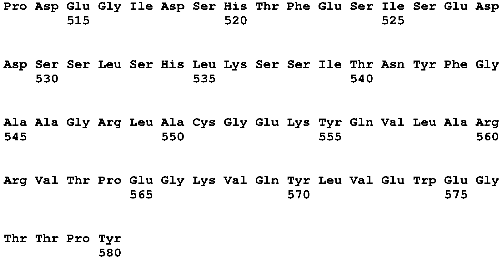
- <210> 10
<211> 578
<212> PRT
<213> Mus musculus - <400> 10

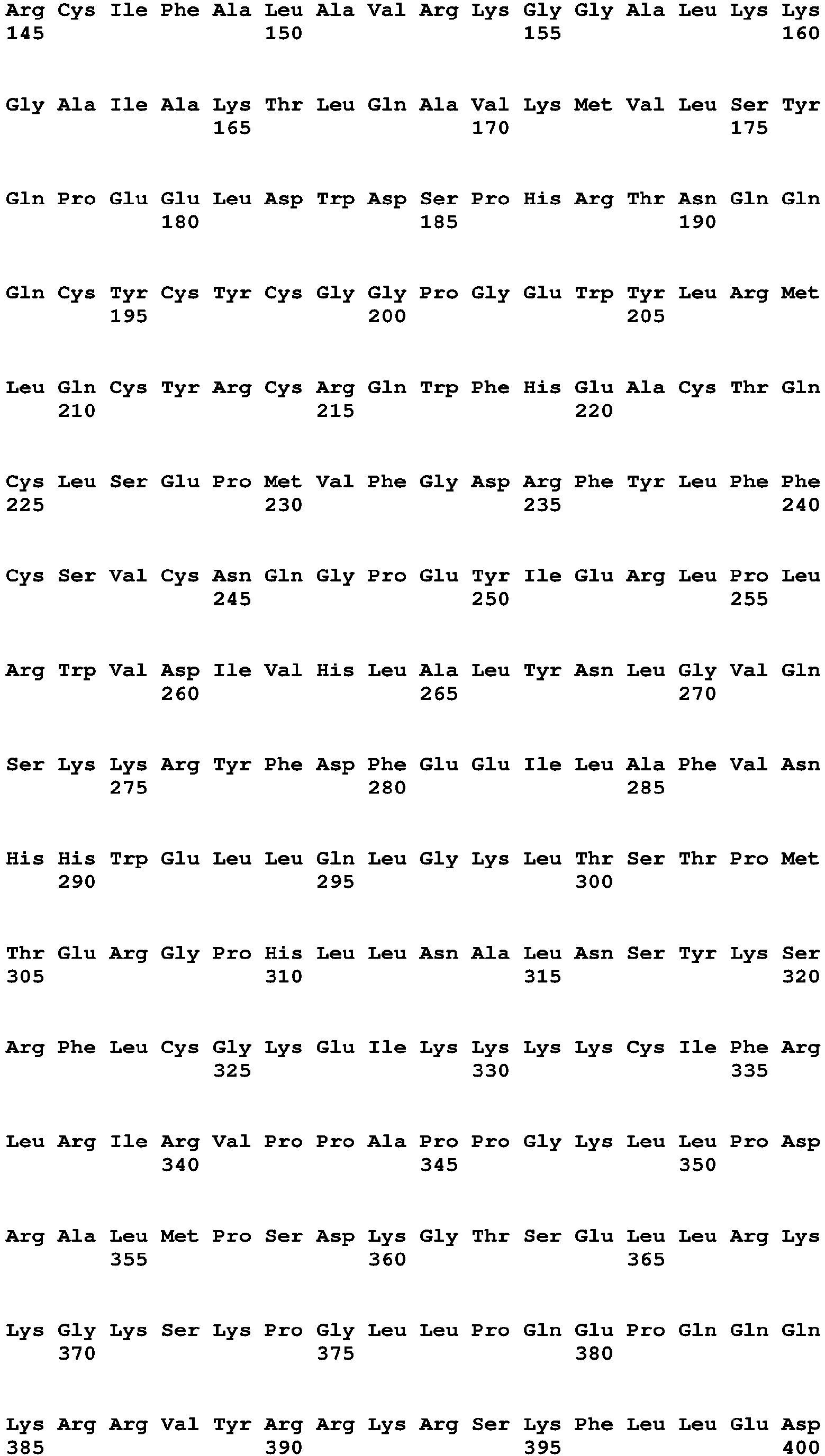

- <210> 11
<211> 7956
<212> DNA
<213> Artificial Sequence - <220>
<223> synthetic - <400> 11


- <210> 12
<211> 130
<212> PRT
<213> Homo sapiens - <400> 12

- <210> 13
<211> 370
<212> PRT
<213> Homo sapiens - <400> 13


- <210> 14
<211> 599
<212> PRT
<213> Homo sapiens - <400> 14


- <210> 15
<211> 207
<212> PRT
<213> Homo sapiens - <400> 15

- <210> 16
<211> 22
<212> DNA
<213> Artificial Sequence - <220>
<223> synthetic - <400> 16
agtgacttgg attttccagc ac 22 - <210> 17
<211> 21
<212> DNA
<213> Artificial Sequence - <220>
<223> synthetic - <400> 17
aattctgttg taagggcgacc 21 - <210> 18
<211> 20
<212> DNA
<213> Mus musculus - <400> 18
gagcttgctc taggggtagg 20 - <210> 19
<211> 20
<212> DNA
<213> Mus musculus - <400> 19
acagccagaa gtaaggtccc 20 - <210> 20
<211> 20
<212> DNA
<213> Mus musculus - <400> 20
cgccacaatt ccgaccttag 20 - <210> 21
<211> 20
<212> DNA
<213> Mus musculus - <400> 21
aaatatttgc ggcgctccat 20 - <210> 22
<211> 20
<212> DNA
<213> Mus musculus - <400> 22
ttggggcaca gataccatgt 20 - <210> 23
<211> 20
<212> DNA
<213> Mus musculus - <400> 23
tcctccctag actcaagcct 20 - <210> 24
<211> 20
<212> DNA
<213> Mus musculus - <400> 24
ctgcagacat tttgaggcgt 20 - <210> 25
<211> 20
<212> DNA
<213> Mus musculus - <400> 25
tctaactgag ccggtgttgt 20 - <210> 26
<211> 20
<212> DNA
<213> Mus musculus - <400> 26
tggttgttct agggcgtgat 20 - <210> 27
<211> 20
<212> DNA
<213> Mus musculus - <400> 27
tacccggcca aactctcaat 20 - <210> 28
<211> 20
<212> DNA
<213> Mus musculus - <400> 28
tgaaattcca cctccctccc 20 - <210> 29
<211> 20
<212> DNA
<213> Mus musculus - <400> 29
tccctttaac tttcgcccct 20
Claims (15)
- A mammalian T cell, comprising an antigen-specific receptor, wherein the antigen-specific receptor is a T cell receptor (TCR) or a chimeric antigen receptor (CAR), wherein the T cell has been modified to express Phf19 at a level that is higher than the level of Phf19 expressed by a T cell that has not been modified to express Phf19.
- A mammalian T cell, comprising an antigen-specific receptor, wherein the antigen-specific receptor is a T cell receptor (TCR) or a chimeric antigen receptor (CAR), wherein the T cell comprises a genetic expression vector comprising a genetic sequence encoding Phf19.
- A genetically-modified mammalian T cell, comprising an antigen-specific receptor, wherein the antigen-specific receptor is a T cell receptor (TCR) or a chimeric antigen receptor (CAR), wherein the T cell comprises a genetic expression vector comprising (a) virally-, bacterially-, or both virally- and bacterially-derived genetic sequences and (b) a genetic sequence encoding Phf19, wherein the genetic sequence encoding Phf19 within the vector is expressed within the T cell.
- The T cell of any one of claims 1-3, wherein the T cell is a CD8+ T cell.
- The T cell of any of one claims 1-4, wherein the T cell is human.
- The T cell of any of one claims 1-5, wherein the Phf19 is a human isoform of Phf19.
- The T cell of any one of claims 1-6, wherein the T cell is in vitro or ex vivo.
- The T cell of any one of claims 1-7, wherein the antigen-specific receptor has antigenic specificity for a cancer antigen, or wherein the antigen-specific receptor has antigenic specificity for a viral, bacterial, or parasite antigen.
- The T cell of any one of claims 1-8, wherein the antigen-specific receptor is a recombinant TCR, or wherein the antigen-specific receptor is an endogenous TCR, or wherein the antigen-specific receptor is a recombinant CAR.
- A population of T cells comprising at least two T cells of any one of claims 1-9.
- A pharmaceutical composition comprising the T cells of any of claims 1-9 or the population of cells of claim 10, and a pharmaceutically acceptable carrier.
- An effective amount of the T cell of any of claims 1-9, the population of cells of claim 10, or the pharmaceutical composition of claim 11, for use in the treatment of cancer or chronic viral disease in a mammal, optionally wherein the cancer is selected from the group consisting of melanoma, leukemia, and HPV carcinoma.
- An in vitro method for inhibiting T cell terminal differentiation and exhaustion epigenetically, comprising modifying the T cell to express Phf19 at a level that is higher than the level of Phf19 expressed by a T cell that has not been modified,wherein the increased expression of Phf19 inhibits the modified T cell's terminal differentiation and exhaustion when compared with a T cell not modified to express Phf19,optionally wherein the modified T-cell comprises a vector comprising a genetic sequence encoding Phf19,or optionally wherein the modified T-cell comprises a genetic expression vector comprising (a) virally-, bacterially-, or both virally- and bacterially-derived genetic sequences and (b) a genetic sequence encoding Phf19, wherein the genetic sequence encoding Phf19 within the vector is expressed within the modified T cell.
- The effective amount, population, or pharmaceutical composition for use of claim 12, or the method of claim 13, wherein the T cell is a CD8+ T cell.
- The effective amount, population, or pharmaceutical composition for use of claim 12 or 14, or the method of claim 13, wherein the T cell is human.
Applications Claiming Priority (2)
| Application Number | Priority Date | Filing Date | Title |
|---|---|---|---|
| US201762515105P | 2017-06-05 | 2017-06-05 | |
| PCT/US2018/036125 WO2018226741A1 (en) | 2017-06-05 | 2018-06-05 | T-cells modified to overexpress phf19 |
Publications (2)
| Publication Number | Publication Date |
|---|---|
| EP3635098A1 EP3635098A1 (en) | 2020-04-15 |
| EP3635098B1 true EP3635098B1 (en) | 2021-09-15 |
Family
ID=62815130
Family Applications (1)
| Application Number | Title | Priority Date | Filing Date |
|---|---|---|---|
| EP18737062.2A Active EP3635098B1 (en) | 2017-06-05 | 2018-06-05 | T-cells modified to overexpress phf19 |
Country Status (3)
| Country | Link |
|---|---|
| US (1) | US11883431B2 (en) |
| EP (1) | EP3635098B1 (en) |
| WO (1) | WO2018226741A1 (en) |
Families Citing this family (1)
| Publication number | Priority date | Publication date | Assignee | Title |
|---|---|---|---|---|
| WO2025146440A1 (en) * | 2024-01-02 | 2025-07-10 | Leibniz-Institut Für Immuntherapie (Lit) | Use of lmo4 to bolster stemness and antitumor efficacy of t-cells |
Family Cites Families (9)
| Publication number | Priority date | Publication date | Assignee | Title |
|---|---|---|---|---|
| US7820174B2 (en) | 2006-02-24 | 2010-10-26 | The United States Of America As Represented By The Department Of Health And Human Services | T cell receptors and related materials and methods of use |
| AU2008206442B2 (en) | 2007-01-12 | 2012-10-18 | Government Of The United States Of America, Represented By The Secretary, Department Of Health And Human Services | gp100-specific T cell receptors and related materials and methods of use |
| WO2010088160A1 (en) | 2009-01-28 | 2010-08-05 | The United States Of America, As Represented By The Secretary, Department Of Health And Human Services | T cell receptors and related materials and methods of use |
| US8465743B2 (en) | 2009-10-01 | 2013-06-18 | The United States Of America, As Represented By The Secretary, Department Of Health And Human Services | Anti-vascular endothelial growth factor receptor-2 chimeric antigen receptors and use of same for the treatment of cancer |
| CN103124740B (en) | 2010-09-21 | 2015-09-02 | 美国卫生和人力服务部 | Anti-SSX-2T cell receptor and associated materials and using method |
| TR201815488T4 (en) | 2011-04-08 | 2018-11-21 | Health | Use of anti-epidermal growth factor receptor variant III for chimeric antigen receptors and cancer treatment. |
| IN2014CN02906A (en) | 2011-10-20 | 2015-07-03 | Us Health | |
| WO2014059248A1 (en) * | 2012-10-12 | 2014-04-17 | Philadelphia Health & Education Corporation D/B/A/ Drexel | Mir-155 enhancement of cd8+ t cell immunity |
| WO2014066137A1 (en) * | 2012-10-22 | 2014-05-01 | The United States Of America, As Represented By The Secretary, Department Of Health And Human Services | Compositions and methods for enhancing cancer immunotherapy |
-
2018
- 2018-06-05 EP EP18737062.2A patent/EP3635098B1/en active Active
- 2018-06-05 US US16/619,570 patent/US11883431B2/en active Active
- 2018-06-05 WO PCT/US2018/036125 patent/WO2018226741A1/en not_active Ceased
Also Published As
| Publication number | Publication date |
|---|---|
| EP3635098A1 (en) | 2020-04-15 |
| WO2018226741A1 (en) | 2018-12-13 |
| US20200289562A1 (en) | 2020-09-17 |
| US11883431B2 (en) | 2024-01-30 |
Similar Documents
| Publication | Publication Date | Title |
|---|---|---|
| Mathewson et al. | Inhibitory CD161 receptor identified in glioma-infiltrating T cells by single-cell analysis | |
| Siddiqui et al. | Epithelial miR-141 regulates IL-13–induced airway mucus production | |
| Li et al. | Regulatory T-cells regulate neonatal heart regeneration by potentiating cardiomyocyte proliferation in a paracrine manner | |
| Meraviglia-Crivelli et al. | IL-6/STAT3 signaling in tumor cells restricts the expression of frameshift-derived neoantigens by SMG1 induction | |
| JP2020517247A (en) | Platform for manipulation of T lymphocyte genome and its in vivo high throughput screening method | |
| JP2021522188A (en) | Compositions and Methods for Multiple Tumor Vaccination with Endogenous Gene Activation | |
| US20240327826A1 (en) | Compositions and methods for improving t cell persistence and function | |
| Kabir et al. | ZBTB46 coordinates angiogenesis and immunity to control tumor outcome | |
| JP2024544003A (en) | Methods for Treating Acute Myeloid Leukemia | |
| CN111166867B (en) | Function and application of PD-1 ubiquitination agonist | |
| CN111526894B (en) | Pharmaceutical composition for preventing or treating lung metastasis of cancer using a CHI3L1 inhibitor as an active ingredient | |
| WO2024103313A1 (en) | Cancer therapy based on targeting irg1 | |
| Feng et al. | iNKT cells with high PLZF expression are recruited into the lung via CCL21‐CCR7 signaling to facilitate the development of asthma tolerance in mice | |
| WO2020128006A1 (en) | Mirna for use in therapy | |
| Li et al. | MCRS1 sensitizes T cell–dependent immunotherapy by augmenting MHC-I expression in solid tumors | |
| Jahangiri et al. | Interleukin-12 inhibits tumor growth and metastasis promoted by tumor-associated mesenchymal stem cells in triple-negative breast cancer | |
| US11883431B2 (en) | T-cells modified to overexpress PHF19 | |
| US20250270546A1 (en) | Guide rnas and uses thereof | |
| WO2024059821A2 (en) | Car t cell compositions for treatment of cancer | |
| Humer et al. | NR2F6 deletion revives CAR-T cell function and induces antigen-agnostic immune memory in solid tumors | |
| Chen et al. | In vivo CRISPR screening identifies Fli1 as a transcriptional safeguard that restrains effector CD8 T cell differentiation during infection and cancer | |
| CN117866906B (en) | Use of FOXR A inhibitor in preparation of medicament for treating tumor | |
| Notaro | Lentiviral vector-based tumor vaccines to expand and activate tumor-reactive T cells | |
| Redkiewicz et al. | immunogenicity of Dna Vaccine against h5n1 containing extended Kappa B site: In Vivo study in Mice and chickens | |
| Tan et al. | Dual-antigen DoggyboneTM DNA vaccine induces potent anti-tumor immunity against immunosuppressive oral cancer |
Legal Events
| Date | Code | Title | Description |
|---|---|---|---|
| STAA | Information on the status of an ep patent application or granted ep patent |
Free format text: STATUS: UNKNOWN |
|
| STAA | Information on the status of an ep patent application or granted ep patent |
Free format text: STATUS: THE INTERNATIONAL PUBLICATION HAS BEEN MADE |
|
| PUAI | Public reference made under article 153(3) epc to a published international application that has entered the european phase |
Free format text: ORIGINAL CODE: 0009012 |
|
| STAA | Information on the status of an ep patent application or granted ep patent |
Free format text: STATUS: REQUEST FOR EXAMINATION WAS MADE |
|
| 17P | Request for examination filed |
Effective date: 20191219 |
|
| AK | Designated contracting states |
Kind code of ref document: A1 Designated state(s): AL AT BE BG CH CY CZ DE DK EE ES FI FR GB GR HR HU IE IS IT LI LT LU LV MC MK MT NL NO PL PT RO RS SE SI SK SM TR |
|
| AX | Request for extension of the european patent |
Extension state: BA ME |
|
| DAV | Request for validation of the european patent (deleted) | ||
| DAX | Request for extension of the european patent (deleted) | ||
| GRAP | Despatch of communication of intention to grant a patent |
Free format text: ORIGINAL CODE: EPIDOSNIGR1 |
|
| STAA | Information on the status of an ep patent application or granted ep patent |
Free format text: STATUS: GRANT OF PATENT IS INTENDED |
|
| INTG | Intention to grant announced |
Effective date: 20210329 |
|
| GRAS | Grant fee paid |
Free format text: ORIGINAL CODE: EPIDOSNIGR3 |
|
| GRAA | (expected) grant |
Free format text: ORIGINAL CODE: 0009210 |
|
| STAA | Information on the status of an ep patent application or granted ep patent |
Free format text: STATUS: THE PATENT HAS BEEN GRANTED |
|
| AK | Designated contracting states |
Kind code of ref document: B1 Designated state(s): AL AT BE BG CH CY CZ DE DK EE ES FI FR GB GR HR HU IE IS IT LI LT LU LV MC MK MT NL NO PL PT RO RS SE SI SK SM TR |
|
| REG | Reference to a national code |
Ref country code: CH Ref legal event code: EP |
|
| REG | Reference to a national code |
Ref country code: DE Ref legal event code: R096 Ref document number: 602018023611 Country of ref document: DE |
|
| REG | Reference to a national code |
Ref country code: IE Ref legal event code: FG4D |
|
| REG | Reference to a national code |
Ref country code: AT Ref legal event code: REF Ref document number: 1430549 Country of ref document: AT Kind code of ref document: T Effective date: 20211015 |
|
| REG | Reference to a national code |
Ref country code: LT Ref legal event code: MG9D |
|
| REG | Reference to a national code |
Ref country code: NL Ref legal event code: MP Effective date: 20210915 |
|
| PG25 | Lapsed in a contracting state [announced via postgrant information from national office to epo] |
Ref country code: NO Free format text: LAPSE BECAUSE OF FAILURE TO SUBMIT A TRANSLATION OF THE DESCRIPTION OR TO PAY THE FEE WITHIN THE PRESCRIBED TIME-LIMIT Effective date: 20211215 Ref country code: LT Free format text: LAPSE BECAUSE OF FAILURE TO SUBMIT A TRANSLATION OF THE DESCRIPTION OR TO PAY THE FEE WITHIN THE PRESCRIBED TIME-LIMIT Effective date: 20210915 Ref country code: BG Free format text: LAPSE BECAUSE OF FAILURE TO SUBMIT A TRANSLATION OF THE DESCRIPTION OR TO PAY THE FEE WITHIN THE PRESCRIBED TIME-LIMIT Effective date: 20211215 Ref country code: SE Free format text: LAPSE BECAUSE OF FAILURE TO SUBMIT A TRANSLATION OF THE DESCRIPTION OR TO PAY THE FEE WITHIN THE PRESCRIBED TIME-LIMIT Effective date: 20210915 Ref country code: RS Free format text: LAPSE BECAUSE OF FAILURE TO SUBMIT A TRANSLATION OF THE DESCRIPTION OR TO PAY THE FEE WITHIN THE PRESCRIBED TIME-LIMIT Effective date: 20210915 Ref country code: HR Free format text: LAPSE BECAUSE OF FAILURE TO SUBMIT A TRANSLATION OF THE DESCRIPTION OR TO PAY THE FEE WITHIN THE PRESCRIBED TIME-LIMIT Effective date: 20210915 Ref country code: FI Free format text: LAPSE BECAUSE OF FAILURE TO SUBMIT A TRANSLATION OF THE DESCRIPTION OR TO PAY THE FEE WITHIN THE PRESCRIBED TIME-LIMIT Effective date: 20210915 |
|
| REG | Reference to a national code |
Ref country code: AT Ref legal event code: MK05 Ref document number: 1430549 Country of ref document: AT Kind code of ref document: T Effective date: 20210915 |
|
| PG25 | Lapsed in a contracting state [announced via postgrant information from national office to epo] |
Ref country code: LV Free format text: LAPSE BECAUSE OF FAILURE TO SUBMIT A TRANSLATION OF THE DESCRIPTION OR TO PAY THE FEE WITHIN THE PRESCRIBED TIME-LIMIT Effective date: 20210915 Ref country code: GR Free format text: LAPSE BECAUSE OF FAILURE TO SUBMIT A TRANSLATION OF THE DESCRIPTION OR TO PAY THE FEE WITHIN THE PRESCRIBED TIME-LIMIT Effective date: 20211216 |
|
| PG25 | Lapsed in a contracting state [announced via postgrant information from national office to epo] |
Ref country code: AT Free format text: LAPSE BECAUSE OF FAILURE TO SUBMIT A TRANSLATION OF THE DESCRIPTION OR TO PAY THE FEE WITHIN THE PRESCRIBED TIME-LIMIT Effective date: 20210915 |
|
| PG25 | Lapsed in a contracting state [announced via postgrant information from national office to epo] |
Ref country code: IS Free format text: LAPSE BECAUSE OF FAILURE TO SUBMIT A TRANSLATION OF THE DESCRIPTION OR TO PAY THE FEE WITHIN THE PRESCRIBED TIME-LIMIT Effective date: 20220115 Ref country code: SM Free format text: LAPSE BECAUSE OF FAILURE TO SUBMIT A TRANSLATION OF THE DESCRIPTION OR TO PAY THE FEE WITHIN THE PRESCRIBED TIME-LIMIT Effective date: 20210915 Ref country code: SK Free format text: LAPSE BECAUSE OF FAILURE TO SUBMIT A TRANSLATION OF THE DESCRIPTION OR TO PAY THE FEE WITHIN THE PRESCRIBED TIME-LIMIT Effective date: 20210915 Ref country code: RO Free format text: LAPSE BECAUSE OF FAILURE TO SUBMIT A TRANSLATION OF THE DESCRIPTION OR TO PAY THE FEE WITHIN THE PRESCRIBED TIME-LIMIT Effective date: 20210915 Ref country code: PT Free format text: LAPSE BECAUSE OF FAILURE TO SUBMIT A TRANSLATION OF THE DESCRIPTION OR TO PAY THE FEE WITHIN THE PRESCRIBED TIME-LIMIT Effective date: 20220117 Ref country code: PL Free format text: LAPSE BECAUSE OF FAILURE TO SUBMIT A TRANSLATION OF THE DESCRIPTION OR TO PAY THE FEE WITHIN THE PRESCRIBED TIME-LIMIT Effective date: 20210915 Ref country code: NL Free format text: LAPSE BECAUSE OF FAILURE TO SUBMIT A TRANSLATION OF THE DESCRIPTION OR TO PAY THE FEE WITHIN THE PRESCRIBED TIME-LIMIT Effective date: 20210915 Ref country code: ES Free format text: LAPSE BECAUSE OF FAILURE TO SUBMIT A TRANSLATION OF THE DESCRIPTION OR TO PAY THE FEE WITHIN THE PRESCRIBED TIME-LIMIT Effective date: 20210915 Ref country code: EE Free format text: LAPSE BECAUSE OF FAILURE TO SUBMIT A TRANSLATION OF THE DESCRIPTION OR TO PAY THE FEE WITHIN THE PRESCRIBED TIME-LIMIT Effective date: 20210915 Ref country code: CZ Free format text: LAPSE BECAUSE OF FAILURE TO SUBMIT A TRANSLATION OF THE DESCRIPTION OR TO PAY THE FEE WITHIN THE PRESCRIBED TIME-LIMIT Effective date: 20210915 Ref country code: AL Free format text: LAPSE BECAUSE OF FAILURE TO SUBMIT A TRANSLATION OF THE DESCRIPTION OR TO PAY THE FEE WITHIN THE PRESCRIBED TIME-LIMIT Effective date: 20210915 |
|
| REG | Reference to a national code |
Ref country code: DE Ref legal event code: R097 Ref document number: 602018023611 Country of ref document: DE |
|
| PLBE | No opposition filed within time limit |
Free format text: ORIGINAL CODE: 0009261 |
|
| STAA | Information on the status of an ep patent application or granted ep patent |
Free format text: STATUS: NO OPPOSITION FILED WITHIN TIME LIMIT |
|
| PG25 | Lapsed in a contracting state [announced via postgrant information from national office to epo] |
Ref country code: DK Free format text: LAPSE BECAUSE OF FAILURE TO SUBMIT A TRANSLATION OF THE DESCRIPTION OR TO PAY THE FEE WITHIN THE PRESCRIBED TIME-LIMIT Effective date: 20210915 |
|
| 26N | No opposition filed |
Effective date: 20220616 |
|
| PG25 | Lapsed in a contracting state [announced via postgrant information from national office to epo] |
Ref country code: SI Free format text: LAPSE BECAUSE OF FAILURE TO SUBMIT A TRANSLATION OF THE DESCRIPTION OR TO PAY THE FEE WITHIN THE PRESCRIBED TIME-LIMIT Effective date: 20210915 |
|
| PG25 | Lapsed in a contracting state [announced via postgrant information from national office to epo] |
Ref country code: MC Free format text: LAPSE BECAUSE OF FAILURE TO SUBMIT A TRANSLATION OF THE DESCRIPTION OR TO PAY THE FEE WITHIN THE PRESCRIBED TIME-LIMIT Effective date: 20210915 Ref country code: IT Free format text: LAPSE BECAUSE OF FAILURE TO SUBMIT A TRANSLATION OF THE DESCRIPTION OR TO PAY THE FEE WITHIN THE PRESCRIBED TIME-LIMIT Effective date: 20210915 |
|
| REG | Reference to a national code |
Ref country code: CH Ref legal event code: PL |
|
| REG | Reference to a national code |
Ref country code: BE Ref legal event code: MM Effective date: 20220630 |
|
| PG25 | Lapsed in a contracting state [announced via postgrant information from national office to epo] |
Ref country code: LU Free format text: LAPSE BECAUSE OF NON-PAYMENT OF DUE FEES Effective date: 20220605 Ref country code: LI Free format text: LAPSE BECAUSE OF NON-PAYMENT OF DUE FEES Effective date: 20220630 Ref country code: IE Free format text: LAPSE BECAUSE OF NON-PAYMENT OF DUE FEES Effective date: 20220605 Ref country code: CH Free format text: LAPSE BECAUSE OF NON-PAYMENT OF DUE FEES Effective date: 20220630 |
|
| PG25 | Lapsed in a contracting state [announced via postgrant information from national office to epo] |
Ref country code: BE Free format text: LAPSE BECAUSE OF NON-PAYMENT OF DUE FEES Effective date: 20220630 |
|
| P01 | Opt-out of the competence of the unified patent court (upc) registered |
Effective date: 20230526 |
|
| PG25 | Lapsed in a contracting state [announced via postgrant information from national office to epo] |
Ref country code: MK Free format text: LAPSE BECAUSE OF FAILURE TO SUBMIT A TRANSLATION OF THE DESCRIPTION OR TO PAY THE FEE WITHIN THE PRESCRIBED TIME-LIMIT Effective date: 20210915 Ref country code: CY Free format text: LAPSE BECAUSE OF FAILURE TO SUBMIT A TRANSLATION OF THE DESCRIPTION OR TO PAY THE FEE WITHIN THE PRESCRIBED TIME-LIMIT Effective date: 20210915 |
|
| PG25 | Lapsed in a contracting state [announced via postgrant information from national office to epo] |
Ref country code: HU Free format text: LAPSE BECAUSE OF FAILURE TO SUBMIT A TRANSLATION OF THE DESCRIPTION OR TO PAY THE FEE WITHIN THE PRESCRIBED TIME-LIMIT; INVALID AB INITIO Effective date: 20180605 |
|
| PG25 | Lapsed in a contracting state [announced via postgrant information from national office to epo] |
Ref country code: TR Free format text: LAPSE BECAUSE OF FAILURE TO SUBMIT A TRANSLATION OF THE DESCRIPTION OR TO PAY THE FEE WITHIN THE PRESCRIBED TIME-LIMIT Effective date: 20210915 |
|
| PG25 | Lapsed in a contracting state [announced via postgrant information from national office to epo] |
Ref country code: MT Free format text: LAPSE BECAUSE OF FAILURE TO SUBMIT A TRANSLATION OF THE DESCRIPTION OR TO PAY THE FEE WITHIN THE PRESCRIBED TIME-LIMIT Effective date: 20210915 |
|
| PGFP | Annual fee paid to national office [announced via postgrant information from national office to epo] |
Ref country code: DE Payment date: 20250627 Year of fee payment: 8 |
|
| PGFP | Annual fee paid to national office [announced via postgrant information from national office to epo] |
Ref country code: GB Payment date: 20250627 Year of fee payment: 8 |
|
| PGFP | Annual fee paid to national office [announced via postgrant information from national office to epo] |
Ref country code: FR Payment date: 20250625 Year of fee payment: 8 |