EP3631011B1 - Multiplex pcr detection of alk, ret, and ros fusions - Google Patents
Multiplex pcr detection of alk, ret, and ros fusions Download PDFInfo
- Publication number
- EP3631011B1 EP3631011B1 EP18730279.9A EP18730279A EP3631011B1 EP 3631011 B1 EP3631011 B1 EP 3631011B1 EP 18730279 A EP18730279 A EP 18730279A EP 3631011 B1 EP3631011 B1 EP 3631011B1
- Authority
- EP
- European Patent Office
- Prior art keywords
- exon
- ros1
- alk
- ret
- fusion
- Prior art date
- Legal status (The legal status is an assumption and is not a legal conclusion. Google has not performed a legal analysis and makes no representation as to the accuracy of the status listed.)
- Active
Links
- 230000004927 fusion Effects 0.000 title claims description 278
- 238000001514 detection method Methods 0.000 title claims description 57
- 238000007403 mPCR Methods 0.000 title 1
- 108090000623 proteins and genes Proteins 0.000 claims description 165
- 239000000523 sample Substances 0.000 claims description 144
- 101000686031 Homo sapiens Proto-oncogene tyrosine-protein kinase ROS Proteins 0.000 claims description 63
- 102100023347 Proto-oncogene tyrosine-protein kinase ROS Human genes 0.000 claims description 63
- 230000003321 amplification Effects 0.000 claims description 60
- 238000003199 nucleic acid amplification method Methods 0.000 claims description 60
- 101001050559 Homo sapiens Kinesin-1 heavy chain Proteins 0.000 claims description 58
- 102100023422 Kinesin-1 heavy chain Human genes 0.000 claims description 58
- 102100027100 Echinoderm microtubule-associated protein-like 4 Human genes 0.000 claims description 51
- 101001057929 Homo sapiens Echinoderm microtubule-associated protein-like 4 Proteins 0.000 claims description 51
- 239000000203 mixture Substances 0.000 claims description 44
- 238000000034 method Methods 0.000 claims description 39
- 206010028980 Neoplasm Diseases 0.000 claims description 37
- 239000012472 biological sample Substances 0.000 claims description 34
- 108091006576 SLC34A2 Proteins 0.000 claims description 33
- 102100038437 Sodium-dependent phosphate transport protein 2B Human genes 0.000 claims description 33
- 101000740519 Homo sapiens Syndecan-4 Proteins 0.000 claims description 32
- 102100037220 Syndecan-4 Human genes 0.000 claims description 32
- 201000011510 cancer Diseases 0.000 claims description 28
- 238000007837 multiplex assay Methods 0.000 claims description 19
- 102100030595 HLA class II histocompatibility antigen gamma chain Human genes 0.000 claims description 18
- 101001082627 Homo sapiens HLA class II histocompatibility antigen gamma chain Proteins 0.000 claims description 18
- 230000015572 biosynthetic process Effects 0.000 claims description 11
- 102100031048 Coiled-coil domain-containing protein 6 Human genes 0.000 claims description 10
- 101000777370 Homo sapiens Coiled-coil domain-containing protein 6 Proteins 0.000 claims description 10
- 108020004414 DNA Proteins 0.000 claims description 9
- 101001017855 Homo sapiens Leucine-rich repeats and immunoglobulin-like domains protein 3 Proteins 0.000 claims description 9
- 101000974343 Homo sapiens Nuclear receptor coactivator 4 Proteins 0.000 claims description 9
- 101000850794 Homo sapiens Tropomyosin alpha-3 chain Proteins 0.000 claims description 9
- 102100033284 Leucine-rich repeats and immunoglobulin-like domains protein 3 Human genes 0.000 claims description 9
- 102100022927 Nuclear receptor coactivator 4 Human genes 0.000 claims description 9
- 102100033080 Tropomyosin alpha-3 chain Human genes 0.000 claims description 9
- 229940043355 kinase inhibitor Drugs 0.000 claims description 9
- 239000003757 phosphotransferase inhibitor Substances 0.000 claims description 9
- 102000016928 DNA-directed DNA polymerase Human genes 0.000 claims description 8
- 108010014303 DNA-directed DNA polymerase Proteins 0.000 claims description 8
- 238000002560 therapeutic procedure Methods 0.000 claims description 8
- 102100034343 Integrase Human genes 0.000 claims description 5
- 108010092799 RNA-directed DNA polymerase Proteins 0.000 claims description 5
- 238000003757 reverse transcription PCR Methods 0.000 claims description 5
- 230000004044 response Effects 0.000 claims description 4
- 230000035945 sensitivity Effects 0.000 claims description 3
- 102100033793 ALK tyrosine kinase receptor Human genes 0.000 description 76
- 101710168331 ALK tyrosine kinase receptor Proteins 0.000 description 76
- 230000002441 reversible effect Effects 0.000 description 50
- 150000007523 nucleic acids Chemical class 0.000 description 34
- 108020004707 nucleic acids Proteins 0.000 description 30
- 102000039446 nucleic acids Human genes 0.000 description 30
- 238000011282 treatment Methods 0.000 description 29
- 108091032973 (ribonucleotides)n+m Proteins 0.000 description 26
- 238000006243 chemical reaction Methods 0.000 description 21
- -1 RET Proteins 0.000 description 17
- 210000002381 plasma Anatomy 0.000 description 17
- 238000003556 assay Methods 0.000 description 16
- 125000003729 nucleotide group Chemical group 0.000 description 16
- 108091034117 Oligonucleotide Proteins 0.000 description 14
- 210000004369 blood Anatomy 0.000 description 14
- 239000008280 blood Substances 0.000 description 14
- 239000002773 nucleotide Substances 0.000 description 14
- UDGUGZTYGWUUSG-UHFFFAOYSA-N 4-[4-[[2,5-dimethoxy-4-[(4-nitrophenyl)diazenyl]phenyl]diazenyl]-n-methylanilino]butanoic acid Chemical compound COC=1C=C(N=NC=2C=CC(=CC=2)N(C)CCCC(O)=O)C(OC)=CC=1N=NC1=CC=C([N+]([O-])=O)C=C1 UDGUGZTYGWUUSG-UHFFFAOYSA-N 0.000 description 12
- 230000002068 genetic effect Effects 0.000 description 12
- 238000012360 testing method Methods 0.000 description 12
- 210000001519 tissue Anatomy 0.000 description 12
- 238000011529 RT qPCR Methods 0.000 description 11
- JLCPHMBAVCMARE-UHFFFAOYSA-N [3-[[3-[[3-[[3-[[3-[[3-[[3-[[3-[[3-[[3-[[3-[[5-(2-amino-6-oxo-1H-purin-9-yl)-3-[[3-[[3-[[3-[[3-[[3-[[5-(2-amino-6-oxo-1H-purin-9-yl)-3-[[5-(2-amino-6-oxo-1H-purin-9-yl)-3-hydroxyoxolan-2-yl]methoxy-hydroxyphosphoryl]oxyoxolan-2-yl]methoxy-hydroxyphosphoryl]oxy-5-(5-methyl-2,4-dioxopyrimidin-1-yl)oxolan-2-yl]methoxy-hydroxyphosphoryl]oxy-5-(6-aminopurin-9-yl)oxolan-2-yl]methoxy-hydroxyphosphoryl]oxy-5-(6-aminopurin-9-yl)oxolan-2-yl]methoxy-hydroxyphosphoryl]oxy-5-(6-aminopurin-9-yl)oxolan-2-yl]methoxy-hydroxyphosphoryl]oxy-5-(6-aminopurin-9-yl)oxolan-2-yl]methoxy-hydroxyphosphoryl]oxyoxolan-2-yl]methoxy-hydroxyphosphoryl]oxy-5-(5-methyl-2,4-dioxopyrimidin-1-yl)oxolan-2-yl]methoxy-hydroxyphosphoryl]oxy-5-(4-amino-2-oxopyrimidin-1-yl)oxolan-2-yl]methoxy-hydroxyphosphoryl]oxy-5-(5-methyl-2,4-dioxopyrimidin-1-yl)oxolan-2-yl]methoxy-hydroxyphosphoryl]oxy-5-(5-methyl-2,4-dioxopyrimidin-1-yl)oxolan-2-yl]methoxy-hydroxyphosphoryl]oxy-5-(6-aminopurin-9-yl)oxolan-2-yl]methoxy-hydroxyphosphoryl]oxy-5-(6-aminopurin-9-yl)oxolan-2-yl]methoxy-hydroxyphosphoryl]oxy-5-(4-amino-2-oxopyrimidin-1-yl)oxolan-2-yl]methoxy-hydroxyphosphoryl]oxy-5-(4-amino-2-oxopyrimidin-1-yl)oxolan-2-yl]methoxy-hydroxyphosphoryl]oxy-5-(4-amino-2-oxopyrimidin-1-yl)oxolan-2-yl]methoxy-hydroxyphosphoryl]oxy-5-(6-aminopurin-9-yl)oxolan-2-yl]methoxy-hydroxyphosphoryl]oxy-5-(4-amino-2-oxopyrimidin-1-yl)oxolan-2-yl]methyl [5-(6-aminopurin-9-yl)-2-(hydroxymethyl)oxolan-3-yl] hydrogen phosphate Polymers Cc1cn(C2CC(OP(O)(=O)OCC3OC(CC3OP(O)(=O)OCC3OC(CC3O)n3cnc4c3nc(N)[nH]c4=O)n3cnc4c3nc(N)[nH]c4=O)C(COP(O)(=O)OC3CC(OC3COP(O)(=O)OC3CC(OC3COP(O)(=O)OC3CC(OC3COP(O)(=O)OC3CC(OC3COP(O)(=O)OC3CC(OC3COP(O)(=O)OC3CC(OC3COP(O)(=O)OC3CC(OC3COP(O)(=O)OC3CC(OC3COP(O)(=O)OC3CC(OC3COP(O)(=O)OC3CC(OC3COP(O)(=O)OC3CC(OC3COP(O)(=O)OC3CC(OC3COP(O)(=O)OC3CC(OC3COP(O)(=O)OC3CC(OC3COP(O)(=O)OC3CC(OC3COP(O)(=O)OC3CC(OC3COP(O)(=O)OC3CC(OC3CO)n3cnc4c(N)ncnc34)n3ccc(N)nc3=O)n3cnc4c(N)ncnc34)n3ccc(N)nc3=O)n3ccc(N)nc3=O)n3ccc(N)nc3=O)n3cnc4c(N)ncnc34)n3cnc4c(N)ncnc34)n3cc(C)c(=O)[nH]c3=O)n3cc(C)c(=O)[nH]c3=O)n3ccc(N)nc3=O)n3cc(C)c(=O)[nH]c3=O)n3cnc4c3nc(N)[nH]c4=O)n3cnc4c(N)ncnc34)n3cnc4c(N)ncnc34)n3cnc4c(N)ncnc34)n3cnc4c(N)ncnc34)O2)c(=O)[nH]c1=O JLCPHMBAVCMARE-UHFFFAOYSA-N 0.000 description 11
- 208000002154 non-small cell lung carcinoma Diseases 0.000 description 11
- 208000029729 tumor suppressor gene on chromosome 11 Diseases 0.000 description 11
- 210000004027 cell Anatomy 0.000 description 10
- 108091033319 polynucleotide Proteins 0.000 description 8
- 239000002157 polynucleotide Substances 0.000 description 8
- 102000040430 polynucleotide Human genes 0.000 description 8
- 238000001574 biopsy Methods 0.000 description 7
- 239000000975 dye Substances 0.000 description 7
- 238000003753 real-time PCR Methods 0.000 description 7
- 239000002118 L01XE12 - Vandetanib Substances 0.000 description 6
- 230000000295 complement effect Effects 0.000 description 6
- 229960000241 vandetanib Drugs 0.000 description 6
- UHTHHESEBZOYNR-UHFFFAOYSA-N vandetanib Chemical compound COC1=CC(C(/N=CN2)=N/C=3C(=CC(Br)=CC=3)F)=C2C=C1OCC1CCN(C)CC1 UHTHHESEBZOYNR-UHFFFAOYSA-N 0.000 description 6
- AILRADAXUVEEIR-UHFFFAOYSA-N 5-chloro-4-n-(2-dimethylphosphorylphenyl)-2-n-[2-methoxy-4-[4-(4-methylpiperazin-1-yl)piperidin-1-yl]phenyl]pyrimidine-2,4-diamine Chemical compound COC1=CC(N2CCC(CC2)N2CCN(C)CC2)=CC=C1NC(N=1)=NC=C(Cl)C=1NC1=CC=CC=C1P(C)(C)=O AILRADAXUVEEIR-UHFFFAOYSA-N 0.000 description 5
- 206010025323 Lymphomas Diseases 0.000 description 5
- 229950004272 brigatinib Drugs 0.000 description 5
- 239000000872 buffer Substances 0.000 description 5
- 239000003153 chemical reaction reagent Substances 0.000 description 5
- 238000002512 chemotherapy Methods 0.000 description 5
- 239000000306 component Substances 0.000 description 5
- 238000001959 radiotherapy Methods 0.000 description 5
- 210000002966 serum Anatomy 0.000 description 5
- YBJHBAHKTGYVGT-ZKWXMUAHSA-N (+)-Biotin Chemical compound N1C(=O)N[C@@H]2[C@H](CCCCC(=O)O)SC[C@@H]21 YBJHBAHKTGYVGT-ZKWXMUAHSA-N 0.000 description 4
- WCKQPPQRFNHPRJ-UHFFFAOYSA-N 4-[[4-(dimethylamino)phenyl]diazenyl]benzoic acid Chemical compound C1=CC(N(C)C)=CC=C1N=NC1=CC=C(C(O)=O)C=C1 WCKQPPQRFNHPRJ-UHFFFAOYSA-N 0.000 description 4
- 208000010507 Adenocarcinoma of Lung Diseases 0.000 description 4
- 206010005003 Bladder cancer Diseases 0.000 description 4
- 206010006187 Breast cancer Diseases 0.000 description 4
- 208000026310 Breast neoplasm Diseases 0.000 description 4
- WSFSSNUMVMOOMR-UHFFFAOYSA-N Formaldehyde Chemical compound O=C WSFSSNUMVMOOMR-UHFFFAOYSA-N 0.000 description 4
- 208000032612 Glial tumor Diseases 0.000 description 4
- 206010018338 Glioma Diseases 0.000 description 4
- 208000008839 Kidney Neoplasms Diseases 0.000 description 4
- 102100034671 L-lactate dehydrogenase A chain Human genes 0.000 description 4
- 239000002146 L01XE16 - Crizotinib Substances 0.000 description 4
- 108010088350 Lactate Dehydrogenase 5 Proteins 0.000 description 4
- 206010058467 Lung neoplasm malignant Diseases 0.000 description 4
- 108091028043 Nucleic acid sequence Proteins 0.000 description 4
- 206010033128 Ovarian cancer Diseases 0.000 description 4
- 206010061535 Ovarian neoplasm Diseases 0.000 description 4
- 206010061902 Pancreatic neoplasm Diseases 0.000 description 4
- 102100028251 Phosphoglycerate kinase 1 Human genes 0.000 description 4
- 101710139464 Phosphoglycerate kinase 1 Proteins 0.000 description 4
- 206010060862 Prostate cancer Diseases 0.000 description 4
- 208000000236 Prostatic Neoplasms Diseases 0.000 description 4
- 206010038389 Renal cancer Diseases 0.000 description 4
- 208000033781 Thyroid carcinoma Diseases 0.000 description 4
- 208000024770 Thyroid neoplasm Diseases 0.000 description 4
- 201000001531 bladder carcinoma Diseases 0.000 description 4
- 229960005061 crizotinib Drugs 0.000 description 4
- KTEIFNKAUNYNJU-GFCCVEGCSA-N crizotinib Chemical compound O([C@H](C)C=1C(=C(F)C=CC=1Cl)Cl)C(C(=NC=1)N)=CC=1C(=C1)C=NN1C1CCNCC1 KTEIFNKAUNYNJU-GFCCVEGCSA-N 0.000 description 4
- 208000037265 diseases, disorders, signs and symptoms Diseases 0.000 description 4
- 230000000694 effects Effects 0.000 description 4
- 208000005017 glioblastoma Diseases 0.000 description 4
- 201000010536 head and neck cancer Diseases 0.000 description 4
- 208000014829 head and neck neoplasm Diseases 0.000 description 4
- 201000010982 kidney cancer Diseases 0.000 description 4
- 208000032839 leukemia Diseases 0.000 description 4
- 201000005249 lung adenocarcinoma Diseases 0.000 description 4
- 201000005202 lung cancer Diseases 0.000 description 4
- 208000020816 lung neoplasm Diseases 0.000 description 4
- 201000005243 lung squamous cell carcinoma Diseases 0.000 description 4
- 208000015486 malignant pancreatic neoplasm Diseases 0.000 description 4
- 201000002528 pancreatic cancer Diseases 0.000 description 4
- 208000008443 pancreatic carcinoma Diseases 0.000 description 4
- PYWVYCXTNDRMGF-UHFFFAOYSA-N rhodamine B Chemical compound [Cl-].C=12C=CC(=[N+](CC)CC)C=C2OC2=CC(N(CC)CC)=CC=C2C=1C1=CC=CC=C1C(O)=O PYWVYCXTNDRMGF-UHFFFAOYSA-N 0.000 description 4
- 201000002510 thyroid cancer Diseases 0.000 description 4
- 208000013077 thyroid gland carcinoma Diseases 0.000 description 4
- 208000010570 urinary bladder carcinoma Diseases 0.000 description 4
- 108091093088 Amplicon Proteins 0.000 description 3
- 102000004190 Enzymes Human genes 0.000 description 3
- 108090000790 Enzymes Proteins 0.000 description 3
- 239000002137 L01XE24 - Ponatinib Substances 0.000 description 3
- 239000002176 L01XE26 - Cabozantinib Substances 0.000 description 3
- 108091000080 Phosphotransferase Proteins 0.000 description 3
- 108010012901 Succinate Dehydrogenase Proteins 0.000 description 3
- 102000019259 Succinate Dehydrogenase Human genes 0.000 description 3
- 241000589497 Thermus sp. Species 0.000 description 3
- 229960001611 alectinib Drugs 0.000 description 3
- KDGFLJKFZUIJMX-UHFFFAOYSA-N alectinib Chemical compound CCC1=CC=2C(=O)C(C3=CC=C(C=C3N3)C#N)=C3C(C)(C)C=2C=C1N(CC1)CCC1N1CCOCC1 KDGFLJKFZUIJMX-UHFFFAOYSA-N 0.000 description 3
- 150000001413 amino acids Chemical class 0.000 description 3
- 229960003982 apatinib Drugs 0.000 description 3
- 229960001292 cabozantinib Drugs 0.000 description 3
- ONIQOQHATWINJY-UHFFFAOYSA-N cabozantinib Chemical compound C=12C=C(OC)C(OC)=CC2=NC=CC=1OC(C=C1)=CC=C1NC(=O)C1(C(=O)NC=2C=CC(F)=CC=2)CC1 ONIQOQHATWINJY-UHFFFAOYSA-N 0.000 description 3
- 229960001602 ceritinib Drugs 0.000 description 3
- VERWOWGGCGHDQE-UHFFFAOYSA-N ceritinib Chemical compound CC=1C=C(NC=2N=C(NC=3C(=CC=CC=3)S(=O)(=O)C(C)C)C(Cl)=CN=2)C(OC(C)C)=CC=1C1CCNCC1 VERWOWGGCGHDQE-UHFFFAOYSA-N 0.000 description 3
- GLNDAGDHSLMOKX-UHFFFAOYSA-N coumarin 120 Chemical compound C1=C(N)C=CC2=C1OC(=O)C=C2C GLNDAGDHSLMOKX-UHFFFAOYSA-N 0.000 description 3
- 201000010099 disease Diseases 0.000 description 3
- 230000009977 dual effect Effects 0.000 description 3
- 238000000338 in vitro Methods 0.000 description 3
- 229960003784 lenvatinib Drugs 0.000 description 3
- WOSKHXYHFSIKNG-UHFFFAOYSA-N lenvatinib Chemical compound C=12C=C(C(N)=O)C(OC)=CC2=NC=CC=1OC(C=C1Cl)=CC=C1NC(=O)NC1CC1 WOSKHXYHFSIKNG-UHFFFAOYSA-N 0.000 description 3
- 229950001290 lorlatinib Drugs 0.000 description 3
- IIXWYSCJSQVBQM-LLVKDONJSA-N lorlatinib Chemical compound N=1N(C)C(C#N)=C2C=1CN(C)C(=O)C1=CC=C(F)C=C1[C@@H](C)OC1=CC2=CN=C1N IIXWYSCJSQVBQM-LLVKDONJSA-N 0.000 description 3
- WPEWQEMJFLWMLV-UHFFFAOYSA-N n-[4-(1-cyanocyclopentyl)phenyl]-2-(pyridin-4-ylmethylamino)pyridine-3-carboxamide Chemical compound C=1C=CN=C(NCC=2C=CN=CC=2)C=1C(=O)NC(C=C1)=CC=C1C1(C#N)CCCC1 WPEWQEMJFLWMLV-UHFFFAOYSA-N 0.000 description 3
- 239000013642 negative control Substances 0.000 description 3
- 239000012188 paraffin wax Substances 0.000 description 3
- 102000020233 phosphotransferase Human genes 0.000 description 3
- 229960001131 ponatinib Drugs 0.000 description 3
- PHXJVRSECIGDHY-UHFFFAOYSA-N ponatinib Chemical compound C1CN(C)CCN1CC(C(=C1)C(F)(F)F)=CC=C1NC(=O)C1=CC=C(C)C(C#CC=2N3N=CC=CC3=NC=2)=C1 PHXJVRSECIGDHY-UHFFFAOYSA-N 0.000 description 3
- 239000013641 positive control Substances 0.000 description 3
- 238000010839 reverse transcription Methods 0.000 description 3
- 230000001225 therapeutic effect Effects 0.000 description 3
- 108020004465 16S ribosomal RNA Proteins 0.000 description 2
- HBEDSQVIWPRPAY-UHFFFAOYSA-N 2,3-dihydrobenzofuran Chemical compound C1=CC=C2OCCC2=C1 HBEDSQVIWPRPAY-UHFFFAOYSA-N 0.000 description 2
- PXBFMLJZNCDSMP-UHFFFAOYSA-N 2-Aminobenzamide Chemical compound NC(=O)C1=CC=CC=C1N PXBFMLJZNCDSMP-UHFFFAOYSA-N 0.000 description 2
- OBYNJKLOYWCXEP-UHFFFAOYSA-N 2-[3-(dimethylamino)-6-dimethylazaniumylidenexanthen-9-yl]-4-isothiocyanatobenzoate Chemical compound C=12C=CC(=[N+](C)C)C=C2OC2=CC(N(C)C)=CC=C2C=1C1=CC(N=C=S)=CC=C1C([O-])=O OBYNJKLOYWCXEP-UHFFFAOYSA-N 0.000 description 2
- HZTYDQRUAWIZRE-UHFFFAOYSA-N 2-[[2-[[1-[2-(dimethylamino)-1-oxoethyl]-5-methoxy-2,3-dihydroindol-6-yl]amino]-7H-pyrrolo[2,3-d]pyrimidin-4-yl]amino]-6-fluoro-N-methylbenzamide Chemical compound CNC(=O)C1=C(F)C=CC=C1NC1=C2C=CN=C2NC(NC=2C(=CC=3CCN(C=3C=2)C(=O)CN(C)C)OC)=N1 HZTYDQRUAWIZRE-UHFFFAOYSA-N 0.000 description 2
- HSHNITRMYYLLCV-UHFFFAOYSA-N 4-methylumbelliferone Chemical compound C1=C(O)C=CC2=C1OC(=O)C=C2C HSHNITRMYYLLCV-UHFFFAOYSA-N 0.000 description 2
- ZWONWYNZSWOYQC-UHFFFAOYSA-N 5-benzamido-3-[[5-[[4-chloro-6-(4-sulfoanilino)-1,3,5-triazin-2-yl]amino]-2-sulfophenyl]diazenyl]-4-hydroxynaphthalene-2,7-disulfonic acid Chemical compound OC1=C(N=NC2=CC(NC3=NC(NC4=CC=C(C=C4)S(O)(=O)=O)=NC(Cl)=N3)=CC=C2S(O)(=O)=O)C(=CC2=C1C(NC(=O)C1=CC=CC=C1)=CC(=C2)S(O)(=O)=O)S(O)(=O)=O ZWONWYNZSWOYQC-UHFFFAOYSA-N 0.000 description 2
- NJYVEMPWNAYQQN-UHFFFAOYSA-N 5-carboxyfluorescein Chemical compound C12=CC=C(O)C=C2OC2=CC(O)=CC=C2C21OC(=O)C1=CC(C(=O)O)=CC=C21 NJYVEMPWNAYQQN-UHFFFAOYSA-N 0.000 description 2
- QQWUGDVOUVUTOY-UHFFFAOYSA-N 5-chloro-N2-[2-methoxy-4-[4-(4-methyl-1-piperazinyl)-1-piperidinyl]phenyl]-N4-(2-propan-2-ylsulfonylphenyl)pyrimidine-2,4-diamine Chemical compound COC1=CC(N2CCC(CC2)N2CCN(C)CC2)=CC=C1NC(N=1)=NC=C(Cl)C=1NC1=CC=CC=C1S(=O)(=O)C(C)C QQWUGDVOUVUTOY-UHFFFAOYSA-N 0.000 description 2
- 102100022900 Actin, cytoplasmic 1 Human genes 0.000 description 2
- 108010085238 Actins Proteins 0.000 description 2
- 108700028369 Alleles Proteins 0.000 description 2
- MLDQJTXFUGDVEO-UHFFFAOYSA-N BAY-43-9006 Chemical compound C1=NC(C(=O)NC)=CC(OC=2C=CC(NC(=O)NC=3C=C(C(Cl)=CC=3)C(F)(F)F)=CC=2)=C1 MLDQJTXFUGDVEO-UHFFFAOYSA-N 0.000 description 2
- 108091026890 Coding region Proteins 0.000 description 2
- 230000004544 DNA amplification Effects 0.000 description 2
- XPDXVDYUQZHFPV-UHFFFAOYSA-N Dansyl Chloride Chemical compound C1=CC=C2C(N(C)C)=CC=CC2=C1S(Cl)(=O)=O XPDXVDYUQZHFPV-UHFFFAOYSA-N 0.000 description 2
- 108700039887 Essential Genes Proteins 0.000 description 2
- ZRALSGWEFCBTJO-UHFFFAOYSA-N Guanidine Chemical compound NC(N)=N ZRALSGWEFCBTJO-UHFFFAOYSA-N 0.000 description 2
- 101000896557 Homo sapiens Eukaryotic translation initiation factor 3 subunit B Proteins 0.000 description 2
- 101000988834 Homo sapiens Hypoxanthine-guanine phosphoribosyltransferase Proteins 0.000 description 2
- 101000741800 Homo sapiens Peptidyl-prolyl cis-trans isomerase H Proteins 0.000 description 2
- 102100029098 Hypoxanthine-guanine phosphoribosyltransferase Human genes 0.000 description 2
- 239000002147 L01XE04 - Sunitinib Substances 0.000 description 2
- 239000005511 L01XE05 - Sorafenib Substances 0.000 description 2
- ZDZOTLJHXYCWBA-VCVYQWHSSA-N N-debenzoyl-N-(tert-butoxycarbonyl)-10-deacetyltaxol Chemical compound O([C@H]1[C@H]2[C@@](C([C@H](O)C3=C(C)[C@@H](OC(=O)[C@H](O)[C@@H](NC(=O)OC(C)(C)C)C=4C=CC=CC=4)C[C@]1(O)C3(C)C)=O)(C)[C@@H](O)C[C@H]1OC[C@]12OC(=O)C)C(=O)C1=CC=CC=C1 ZDZOTLJHXYCWBA-VCVYQWHSSA-N 0.000 description 2
- 108700020796 Oncogene Proteins 0.000 description 2
- 238000012408 PCR amplification Methods 0.000 description 2
- 229930012538 Paclitaxel Natural products 0.000 description 2
- 102100038827 Peptidyl-prolyl cis-trans isomerase H Human genes 0.000 description 2
- 238000010802 RNA extraction kit Methods 0.000 description 2
- AUNGANRZJHBGPY-SCRDCRAPSA-N Riboflavin Chemical compound OC[C@@H](O)[C@@H](O)[C@@H](O)CN1C=2C=C(C)C(C)=CC=2N=C2C1=NC(=O)NC2=O AUNGANRZJHBGPY-SCRDCRAPSA-N 0.000 description 2
- 108010090804 Streptavidin Proteins 0.000 description 2
- 241000589499 Thermus thermophilus Species 0.000 description 2
- IQFYYKKMVGJFEH-XLPZGREQSA-N Thymidine Chemical compound O=C1NC(=O)C(C)=CN1[C@@H]1O[C@H](CO)[C@@H](O)C1 IQFYYKKMVGJFEH-XLPZGREQSA-N 0.000 description 2
- ISAKRJDGNUQOIC-UHFFFAOYSA-N Uracil Chemical compound O=C1C=CNC(=O)N1 ISAKRJDGNUQOIC-UHFFFAOYSA-N 0.000 description 2
- 238000010521 absorption reaction Methods 0.000 description 2
- DZBUGLKDJFMEHC-UHFFFAOYSA-N acridine Chemical compound C1=CC=CC2=CC3=CC=CC=C3N=C21 DZBUGLKDJFMEHC-UHFFFAOYSA-N 0.000 description 2
- 238000007792 addition Methods 0.000 description 2
- OIRDTQYFTABQOQ-KQYNXXCUSA-N adenosine Chemical compound C1=NC=2C(N)=NC=NC=2N1[C@@H]1O[C@H](CO)[C@@H](O)[C@H]1O OIRDTQYFTABQOQ-KQYNXXCUSA-N 0.000 description 2
- 238000004458 analytical method Methods 0.000 description 2
- 230000008901 benefit Effects 0.000 description 2
- 229960002685 biotin Drugs 0.000 description 2
- 235000020958 biotin Nutrition 0.000 description 2
- 239000011616 biotin Substances 0.000 description 2
- HFCFMRYTXDINDK-WNQIDUERSA-N cabozantinib malate Chemical compound OC(=O)[C@@H](O)CC(O)=O.C=12C=C(OC)C(OC)=CC2=NC=CC=1OC(C=C1)=CC=C1NC(=O)C1(C(=O)NC=2C=CC(F)=CC=2)CC1 HFCFMRYTXDINDK-WNQIDUERSA-N 0.000 description 2
- 229960004562 carboplatin Drugs 0.000 description 2
- 190000008236 carboplatin Chemical compound 0.000 description 2
- 230000002759 chromosomal effect Effects 0.000 description 2
- 210000000349 chromosome Anatomy 0.000 description 2
- 229960004316 cisplatin Drugs 0.000 description 2
- DQLATGHUWYMOKM-UHFFFAOYSA-L cisplatin Chemical compound N[Pt](N)(Cl)Cl DQLATGHUWYMOKM-UHFFFAOYSA-L 0.000 description 2
- ZYGHJZDHTFUPRJ-UHFFFAOYSA-N coumarin Chemical compound C1=CC=C2OC(=O)C=CC2=C1 ZYGHJZDHTFUPRJ-UHFFFAOYSA-N 0.000 description 2
- OPTASPLRGRRNAP-UHFFFAOYSA-N cytosine Chemical compound NC=1C=CNC(=O)N=1 OPTASPLRGRRNAP-UHFFFAOYSA-N 0.000 description 2
- 230000007423 decrease Effects 0.000 description 2
- 238000012217 deletion Methods 0.000 description 2
- 230000037430 deletion Effects 0.000 description 2
- 230000001419 dependent effect Effects 0.000 description 2
- 238000013461 design Methods 0.000 description 2
- 238000003745 diagnosis Methods 0.000 description 2
- 229960003668 docetaxel Drugs 0.000 description 2
- VYXSBFYARXAAKO-UHFFFAOYSA-N ethyl 2-[3-(ethylamino)-6-ethylimino-2,7-dimethylxanthen-9-yl]benzoate;hydron;chloride Chemical compound [Cl-].C1=2C=C(C)C(NCC)=CC=2OC2=CC(=[NH+]CC)C(C)=CC2=C1C1=CC=CC=C1C(=O)OCC VYXSBFYARXAAKO-UHFFFAOYSA-N 0.000 description 2
- 239000012530 fluid Substances 0.000 description 2
- 239000012634 fragment Substances 0.000 description 2
- 238000003364 immunohistochemistry Methods 0.000 description 2
- 238000005304 joining Methods 0.000 description 2
- 239000000463 material Substances 0.000 description 2
- 238000012986 modification Methods 0.000 description 2
- 230000004048 modification Effects 0.000 description 2
- 238000012544 monitoring process Methods 0.000 description 2
- 239000000178 monomer Substances 0.000 description 2
- 229950003968 motesanib Drugs 0.000 description 2
- RAHBGWKEPAQNFF-UHFFFAOYSA-N motesanib Chemical compound C=1C=C2C(C)(C)CNC2=CC=1NC(=O)C1=CC=CN=C1NCC1=CC=NC=C1 RAHBGWKEPAQNFF-UHFFFAOYSA-N 0.000 description 2
- HAYYBYPASCDWEQ-UHFFFAOYSA-N n-[5-[(3,5-difluorophenyl)methyl]-1h-indazol-3-yl]-4-(4-methylpiperazin-1-yl)-2-(oxan-4-ylamino)benzamide Chemical compound C1CN(C)CCN1C(C=C1NC2CCOCC2)=CC=C1C(=O)NC(C1=C2)=NNC1=CC=C2CC1=CC(F)=CC(F)=C1 HAYYBYPASCDWEQ-UHFFFAOYSA-N 0.000 description 2
- 210000005170 neoplastic cell Anatomy 0.000 description 2
- 229960001592 paclitaxel Drugs 0.000 description 2
- 229920000642 polymer Polymers 0.000 description 2
- 238000006116 polymerization reaction Methods 0.000 description 2
- 238000002360 preparation method Methods 0.000 description 2
- 230000002265 prevention Effects 0.000 description 2
- 230000008569 process Effects 0.000 description 2
- 102000004169 proteins and genes Human genes 0.000 description 2
- BBEAQIROQSPTKN-UHFFFAOYSA-N pyrene Chemical compound C1=CC=C2C=CC3=CC=CC4=CC=C1C2=C43 BBEAQIROQSPTKN-UHFFFAOYSA-N 0.000 description 2
- 229960003787 sorafenib Drugs 0.000 description 2
- 239000000126 substance Substances 0.000 description 2
- 229960001796 sunitinib Drugs 0.000 description 2
- WINHZLLDWRZWRT-ATVHPVEESA-N sunitinib Chemical compound CCN(CC)CCNC(=O)C1=C(C)NC(\C=C/2C3=CC(F)=CC=C3NC\2=O)=C1C WINHZLLDWRZWRT-ATVHPVEESA-N 0.000 description 2
- 230000004083 survival effect Effects 0.000 description 2
- 208000024891 symptom Diseases 0.000 description 2
- 238000003786 synthesis reaction Methods 0.000 description 2
- RCINICONZNJXQF-MZXODVADSA-N taxol Chemical compound O([C@@H]1[C@@]2(C[C@@H](C(C)=C(C2(C)C)[C@H](C([C@]2(C)[C@@H](O)C[C@H]3OC[C@]3([C@H]21)OC(C)=O)=O)OC(=O)C)OC(=O)[C@H](O)[C@@H](NC(=O)C=1C=CC=CC=1)C=1C=CC=CC=1)O)C(=O)C1=CC=CC=C1 RCINICONZNJXQF-MZXODVADSA-N 0.000 description 2
- ABZLKHKQJHEPAX-UHFFFAOYSA-N tetramethylrhodamine Chemical compound C=12C=CC(N(C)C)=CC2=[O+]C2=CC(N(C)C)=CC=C2C=1C1=CC=CC=C1C([O-])=O ABZLKHKQJHEPAX-UHFFFAOYSA-N 0.000 description 2
- 238000013518 transcription Methods 0.000 description 2
- 230000035897 transcription Effects 0.000 description 2
- 210000002700 urine Anatomy 0.000 description 2
- GIANIJCPTPUNBA-QMMMGPOBSA-N (2s)-3-(4-hydroxyphenyl)-2-nitramidopropanoic acid Chemical compound [O-][N+](=O)N[C@H](C(=O)O)CC1=CC=C(O)C=C1 GIANIJCPTPUNBA-QMMMGPOBSA-N 0.000 description 1
- DUFUXAHBRPMOFG-UHFFFAOYSA-N 1-(4-anilinonaphthalen-1-yl)pyrrole-2,5-dione Chemical compound O=C1C=CC(=O)N1C(C1=CC=CC=C11)=CC=C1NC1=CC=CC=C1 DUFUXAHBRPMOFG-UHFFFAOYSA-N 0.000 description 1
- ZTTARJIAPRWUHH-UHFFFAOYSA-N 1-isothiocyanatoacridine Chemical compound C1=CC=C2C=C3C(N=C=S)=CC=CC3=NC2=C1 ZTTARJIAPRWUHH-UHFFFAOYSA-N 0.000 description 1
- RUDINRUXCKIXAJ-UHFFFAOYSA-N 2,2,3,3,4,4,5,5,6,6,7,7,8,8,9,9,10,10,11,11,12,12,13,13,14,14,14-heptacosafluorotetradecanoic acid Chemical compound OC(=O)C(F)(F)C(F)(F)C(F)(F)C(F)(F)C(F)(F)C(F)(F)C(F)(F)C(F)(F)C(F)(F)C(F)(F)C(F)(F)C(F)(F)C(F)(F)F RUDINRUXCKIXAJ-UHFFFAOYSA-N 0.000 description 1
- IOOMXAQUNPWDLL-UHFFFAOYSA-N 2-[6-(diethylamino)-3-(diethyliminiumyl)-3h-xanthen-9-yl]-5-sulfobenzene-1-sulfonate Chemical compound C=12C=CC(=[N+](CC)CC)C=C2OC2=CC(N(CC)CC)=CC=C2C=1C1=CC=C(S(O)(=O)=O)C=C1S([O-])(=O)=O IOOMXAQUNPWDLL-UHFFFAOYSA-N 0.000 description 1
- LAXVMANLDGWYJP-UHFFFAOYSA-N 2-amino-5-(2-aminoethyl)naphthalene-1-sulfonic acid Chemical compound NC1=CC=C2C(CCN)=CC=CC2=C1S(O)(=O)=O LAXVMANLDGWYJP-UHFFFAOYSA-N 0.000 description 1
- OALHHIHQOFIMEF-UHFFFAOYSA-N 3',6'-dihydroxy-2',4',5',7'-tetraiodo-3h-spiro[2-benzofuran-1,9'-xanthene]-3-one Chemical compound O1C(=O)C2=CC=CC=C2C21C1=CC(I)=C(O)C(I)=C1OC1=C(I)C(O)=C(I)C=C21 OALHHIHQOFIMEF-UHFFFAOYSA-N 0.000 description 1
- CPBJMKMKNCRKQB-UHFFFAOYSA-N 3,3-bis(4-hydroxy-3-methylphenyl)-2-benzofuran-1-one Chemical compound C1=C(O)C(C)=CC(C2(C3=CC=CC=C3C(=O)O2)C=2C=C(C)C(O)=CC=2)=C1 CPBJMKMKNCRKQB-UHFFFAOYSA-N 0.000 description 1
- IGRCWJPBLWGNPX-UHFFFAOYSA-N 3-(2-chlorophenyl)-n-(4-chlorophenyl)-n,5-dimethyl-1,2-oxazole-4-carboxamide Chemical compound C=1C=C(Cl)C=CC=1N(C)C(=O)C1=C(C)ON=C1C1=CC=CC=C1Cl IGRCWJPBLWGNPX-UHFFFAOYSA-N 0.000 description 1
- DORJQZDOULKINH-QNBGGDODSA-N 3-[4-[(2r)-2-aminopropoxy]phenyl]-n-[(1r)-1-(3-fluorophenyl)ethyl]imidazo[1,2-b]pyridazin-6-amine;hexanedioic acid Chemical compound OC(=O)CCCCC(O)=O.C1=CC(OC[C@H](N)C)=CC=C1C1=CN=C2N1N=C(N[C@H](C)C=1C=C(F)C=CC=1)C=C2 DORJQZDOULKINH-QNBGGDODSA-N 0.000 description 1
- WEVYNIUIFUYDGI-UHFFFAOYSA-N 3-[6-[4-(trifluoromethoxy)anilino]-4-pyrimidinyl]benzamide Chemical compound NC(=O)C1=CC=CC(C=2N=CN=C(NC=3C=CC(OC(F)(F)F)=CC=3)C=2)=C1 WEVYNIUIFUYDGI-UHFFFAOYSA-N 0.000 description 1
- FWBHETKCLVMNFS-UHFFFAOYSA-N 4',6-Diamino-2-phenylindol Chemical compound C1=CC(C(=N)N)=CC=C1C1=CC2=CC=C(C(N)=N)C=C2N1 FWBHETKCLVMNFS-UHFFFAOYSA-N 0.000 description 1
- YSCNMFDFYJUPEF-OWOJBTEDSA-N 4,4'-diisothiocyano-trans-stilbene-2,2'-disulfonic acid Chemical compound OS(=O)(=O)C1=CC(N=C=S)=CC=C1\C=C\C1=CC=C(N=C=S)C=C1S(O)(=O)=O YSCNMFDFYJUPEF-OWOJBTEDSA-N 0.000 description 1
- YJCCSLGGODRWKK-NSCUHMNNSA-N 4-Acetamido-4'-isothiocyanostilbene-2,2'-disulphonic acid Chemical compound OS(=O)(=O)C1=CC(NC(=O)C)=CC=C1\C=C\C1=CC=C(N=C=S)C=C1S(O)(=O)=O YJCCSLGGODRWKK-NSCUHMNNSA-N 0.000 description 1
- OSWZKAVBSQAVFI-UHFFFAOYSA-N 4-[(4-isothiocyanatophenyl)diazenyl]-n,n-dimethylaniline Chemical compound C1=CC(N(C)C)=CC=C1N=NC1=CC=C(N=C=S)C=C1 OSWZKAVBSQAVFI-UHFFFAOYSA-N 0.000 description 1
- SJQRQOKXQKVJGJ-UHFFFAOYSA-N 5-(2-aminoethylamino)naphthalene-1-sulfonic acid Chemical compound C1=CC=C2C(NCCN)=CC=CC2=C1S(O)(=O)=O SJQRQOKXQKVJGJ-UHFFFAOYSA-N 0.000 description 1
- YERWMQJEYUIJBO-UHFFFAOYSA-N 5-chlorosulfonyl-2-[3-(diethylamino)-6-diethylazaniumylidenexanthen-9-yl]benzenesulfonate Chemical compound C=12C=CC(=[N+](CC)CC)C=C2OC2=CC(N(CC)CC)=CC=C2C=1C1=CC=C(S(Cl)(=O)=O)C=C1S([O-])(=O)=O YERWMQJEYUIJBO-UHFFFAOYSA-N 0.000 description 1
- AXGKYURDYTXCAG-UHFFFAOYSA-N 5-isothiocyanato-2-[2-(4-isothiocyanato-2-sulfophenyl)ethyl]benzenesulfonic acid Chemical compound OS(=O)(=O)C1=CC(N=C=S)=CC=C1CCC1=CC=C(N=C=S)C=C1S(O)(=O)=O AXGKYURDYTXCAG-UHFFFAOYSA-N 0.000 description 1
- HWQQCFPHXPNXHC-UHFFFAOYSA-N 6-[(4,6-dichloro-1,3,5-triazin-2-yl)amino]-3',6'-dihydroxyspiro[2-benzofuran-3,9'-xanthene]-1-one Chemical compound C=1C(O)=CC=C2C=1OC1=CC(O)=CC=C1C2(C1=CC=2)OC(=O)C1=CC=2NC1=NC(Cl)=NC(Cl)=N1 HWQQCFPHXPNXHC-UHFFFAOYSA-N 0.000 description 1
- WQZIDRAQTRIQDX-UHFFFAOYSA-N 6-carboxy-x-rhodamine Chemical compound OC(=O)C1=CC=C(C([O-])=O)C=C1C(C1=CC=2CCCN3CCCC(C=23)=C1O1)=C2C1=C(CCC1)C3=[N+]1CCCC3=C2 WQZIDRAQTRIQDX-UHFFFAOYSA-N 0.000 description 1
- YALJZNKPECPZAS-UHFFFAOYSA-N 7-(diethylamino)-3-(4-isothiocyanatophenyl)-4-methylchromen-2-one Chemical compound O=C1OC2=CC(N(CC)CC)=CC=C2C(C)=C1C1=CC=C(N=C=S)C=C1 YALJZNKPECPZAS-UHFFFAOYSA-N 0.000 description 1
- JBNOVHJXQSHGRL-UHFFFAOYSA-N 7-amino-4-(trifluoromethyl)coumarin Chemical compound FC(F)(F)C1=CC(=O)OC2=CC(N)=CC=C21 JBNOVHJXQSHGRL-UHFFFAOYSA-N 0.000 description 1
- 101150023956 ALK gene Proteins 0.000 description 1
- FYEHYMARPSSOBO-UHFFFAOYSA-N Aurin Chemical compound C1=CC(O)=CC=C1C(C=1C=CC(O)=CC=1)=C1C=CC(=O)C=C1 FYEHYMARPSSOBO-UHFFFAOYSA-N 0.000 description 1
- 208000032791 BCR-ABL1 positive chronic myelogenous leukemia Diseases 0.000 description 1
- DWRXFEITVBNRMK-UHFFFAOYSA-N Beta-D-1-Arabinofuranosylthymine Natural products O=C1NC(=O)C(C)=CN1C1C(O)C(O)C(CO)O1 DWRXFEITVBNRMK-UHFFFAOYSA-N 0.000 description 1
- 239000002126 C01EB10 - Adenosine Substances 0.000 description 1
- 241000282472 Canis lupus familiaris Species 0.000 description 1
- 208000016718 Chromosome Inversion Diseases 0.000 description 1
- AUNGANRZJHBGPY-UHFFFAOYSA-N D-Lyxoflavin Natural products OCC(O)C(O)C(O)CN1C=2C=C(C)C(C)=CC=2N=C2C1=NC(=O)NC2=O AUNGANRZJHBGPY-UHFFFAOYSA-N 0.000 description 1
- 241000192091 Deinococcus radiodurans Species 0.000 description 1
- 102000016911 Deoxyribonucleases Human genes 0.000 description 1
- 108010053770 Deoxyribonucleases Proteins 0.000 description 1
- AHCYMLUZIRLXAA-SHYZEUOFSA-N Deoxyuridine 5'-triphosphate Chemical compound O1[C@H](COP(O)(=O)OP(O)(=O)OP(O)(O)=O)[C@@H](O)C[C@@H]1N1C(=O)NC(=O)C=C1 AHCYMLUZIRLXAA-SHYZEUOFSA-N 0.000 description 1
- 241000258955 Echinodermata Species 0.000 description 1
- 241000588724 Escherichia coli Species 0.000 description 1
- QTANTQQOYSUMLC-UHFFFAOYSA-O Ethidium cation Chemical compound C12=CC(N)=CC=C2C2=CC=C(N)C=C2[N+](CC)=C1C1=CC=CC=C1 QTANTQQOYSUMLC-UHFFFAOYSA-O 0.000 description 1
- 102100020903 Ezrin Human genes 0.000 description 1
- 241000282326 Felis catus Species 0.000 description 1
- 108700039691 Genetic Promoter Regions Proteins 0.000 description 1
- 241000193385 Geobacillus stearothermophilus Species 0.000 description 1
- 241000282412 Homo Species 0.000 description 1
- 101000854648 Homo sapiens Ezrin Proteins 0.000 description 1
- 101001021527 Homo sapiens Huntingtin-interacting protein 1 Proteins 0.000 description 1
- 101000605496 Homo sapiens Kinesin light chain 1 Proteins 0.000 description 1
- 101000800847 Homo sapiens Protein TFG Proteins 0.000 description 1
- 101000685323 Homo sapiens Succinate dehydrogenase [ubiquinone] flavoprotein subunit, mitochondrial Proteins 0.000 description 1
- 102100035957 Huntingtin-interacting protein 1 Human genes 0.000 description 1
- 102100038306 Kinesin light chain 1 Human genes 0.000 description 1
- 241000124008 Mammalia Species 0.000 description 1
- 102000009664 Microtubule-Associated Proteins Human genes 0.000 description 1
- 108010020004 Microtubule-Associated Proteins Proteins 0.000 description 1
- 241000699670 Mus sp. Species 0.000 description 1
- KWYHDKDOAIKMQN-UHFFFAOYSA-N N,N,N',N'-tetramethylethylenediamine Chemical compound CN(C)CCN(C)C KWYHDKDOAIKMQN-UHFFFAOYSA-N 0.000 description 1
- QPCDCPDFJACHGM-UHFFFAOYSA-N N,N-bis{2-[bis(carboxymethyl)amino]ethyl}glycine Chemical compound OC(=O)CN(CC(O)=O)CCN(CC(=O)O)CCN(CC(O)=O)CC(O)=O QPCDCPDFJACHGM-UHFFFAOYSA-N 0.000 description 1
- CHJJGSNFBQVOTG-UHFFFAOYSA-N N-methyl-guanidine Natural products CNC(N)=N CHJJGSNFBQVOTG-UHFFFAOYSA-N 0.000 description 1
- 206010028813 Nausea Diseases 0.000 description 1
- 108020004711 Nucleic Acid Probes Proteins 0.000 description 1
- 241000283973 Oryctolagus cuniculus Species 0.000 description 1
- BELBBZDIHDAJOR-UHFFFAOYSA-N Phenolsulfonephthalein Chemical compound C1=CC(O)=CC=C1C1(C=2C=CC(O)=CC=2)C2=CC=CC=C2S(=O)(=O)O1 BELBBZDIHDAJOR-UHFFFAOYSA-N 0.000 description 1
- 108010004729 Phycoerythrin Proteins 0.000 description 1
- 102100033661 Protein TFG Human genes 0.000 description 1
- 102000004022 Protein-Tyrosine Kinases Human genes 0.000 description 1
- 108090000412 Protein-Tyrosine Kinases Proteins 0.000 description 1
- MTVVRWVOXZSVBW-UHFFFAOYSA-M QSY21 succinimidyl ester Chemical compound [Cl-].C1CN(S(=O)(=O)C=2C(=CC=CC=2)C2=C3C=CC(C=C3OC3=CC(=CC=C32)N2CC3=CC=CC=C3C2)=[N+]2CC3=CC=CC=C3C2)CCC1C(=O)ON1C(=O)CCC1=O MTVVRWVOXZSVBW-UHFFFAOYSA-M 0.000 description 1
- BDJDTKYGKHEMFF-UHFFFAOYSA-M QSY7 succinimidyl ester Chemical compound [Cl-].C=1C=C2C(C=3C(=CC=CC=3)S(=O)(=O)N3CCC(CC3)C(=O)ON3C(CCC3=O)=O)=C3C=C\C(=[N+](\C)C=4C=CC=CC=4)C=C3OC2=CC=1N(C)C1=CC=CC=C1 BDJDTKYGKHEMFF-UHFFFAOYSA-M 0.000 description 1
- 108091028733 RNTP Proteins 0.000 description 1
- 241000700159 Rattus Species 0.000 description 1
- 108091028664 Ribonucleotide Proteins 0.000 description 1
- 102000042303 SPRING family Human genes 0.000 description 1
- 108091078185 SPRING family Proteins 0.000 description 1
- 102100023155 Succinate dehydrogenase [ubiquinone] flavoprotein subunit, mitochondrial Human genes 0.000 description 1
- 108010006785 Taq Polymerase Proteins 0.000 description 1
- 241000206213 Thermosipho africanus Species 0.000 description 1
- 241000204666 Thermotoga maritima Species 0.000 description 1
- 241000204664 Thermotoga neapolitana Species 0.000 description 1
- 241000589501 Thermus caldophilus Species 0.000 description 1
- 241000589498 Thermus filiformis Species 0.000 description 1
- 108091036066 Three prime untranslated region Proteins 0.000 description 1
- 241000193758 [Bacillus] caldotenax Species 0.000 description 1
- 230000005856 abnormality Effects 0.000 description 1
- 239000002253 acid Substances 0.000 description 1
- 150000007513 acids Chemical class 0.000 description 1
- 229960005305 adenosine Drugs 0.000 description 1
- 125000000217 alkyl group Chemical group 0.000 description 1
- 238000013459 approach Methods 0.000 description 1
- 238000002820 assay format Methods 0.000 description 1
- IQFYYKKMVGJFEH-UHFFFAOYSA-N beta-L-thymidine Natural products O=C1NC(=O)C(C)=CN1C1OC(CO)C(O)C1 IQFYYKKMVGJFEH-UHFFFAOYSA-N 0.000 description 1
- 239000012503 blood component Substances 0.000 description 1
- 210000001772 blood platelet Anatomy 0.000 description 1
- 230000030833 cell death Effects 0.000 description 1
- 239000013592 cell lysate Substances 0.000 description 1
- 108091092259 cell-free RNA Proteins 0.000 description 1
- 239000013522 chelant Substances 0.000 description 1
- 239000003795 chemical substances by application Substances 0.000 description 1
- 210000000038 chest Anatomy 0.000 description 1
- 208000037516 chromosome inversion disease Diseases 0.000 description 1
- 239000002299 complementary DNA Substances 0.000 description 1
- 239000000470 constituent Substances 0.000 description 1
- 230000006552 constitutive activation Effects 0.000 description 1
- 229960000956 coumarin Drugs 0.000 description 1
- 235000001671 coumarin Nutrition 0.000 description 1
- 230000001351 cycling effect Effects 0.000 description 1
- 229940104302 cytosine Drugs 0.000 description 1
- SUYVUBYJARFZHO-RRKCRQDMSA-N dATP Chemical compound C1=NC=2C(N)=NC=NC=2N1[C@H]1C[C@H](O)[C@@H](COP(O)(=O)OP(O)(=O)OP(O)(O)=O)O1 SUYVUBYJARFZHO-RRKCRQDMSA-N 0.000 description 1
- SUYVUBYJARFZHO-UHFFFAOYSA-N dATP Natural products C1=NC=2C(N)=NC=NC=2N1C1CC(O)C(COP(O)(=O)OP(O)(=O)OP(O)(O)=O)O1 SUYVUBYJARFZHO-UHFFFAOYSA-N 0.000 description 1
- HAAZLUGHYHWQIW-KVQBGUIXSA-N dGTP Chemical compound C1=NC=2C(=O)NC(N)=NC=2N1[C@H]1C[C@H](O)[C@@H](COP(O)(=O)OP(O)(=O)OP(O)(O)=O)O1 HAAZLUGHYHWQIW-KVQBGUIXSA-N 0.000 description 1
- NHVNXKFIZYSCEB-XLPZGREQSA-N dTTP Chemical compound O=C1NC(=O)C(C)=CN1[C@@H]1O[C@H](COP(O)(=O)OP(O)(=O)OP(O)(O)=O)[C@@H](O)C1 NHVNXKFIZYSCEB-XLPZGREQSA-N 0.000 description 1
- 239000005547 deoxyribonucleotide Substances 0.000 description 1
- 125000002637 deoxyribonucleotide group Chemical group 0.000 description 1
- 238000011161 development Methods 0.000 description 1
- 230000018109 developmental process Effects 0.000 description 1
- 238000012631 diagnostic technique Methods 0.000 description 1
- 239000005546 dideoxynucleotide Substances 0.000 description 1
- 238000006471 dimerization reaction Methods 0.000 description 1
- SWSQBOPZIKWTGO-UHFFFAOYSA-N dimethylaminoamidine Natural products CN(C)C(N)=N SWSQBOPZIKWTGO-UHFFFAOYSA-N 0.000 description 1
- OOYIOIOOWUGAHD-UHFFFAOYSA-L disodium;2',4',5',7'-tetrabromo-4,5,6,7-tetrachloro-3-oxospiro[2-benzofuran-1,9'-xanthene]-3',6'-diolate Chemical compound [Na+].[Na+].O1C(=O)C(C(=C(Cl)C(Cl)=C2Cl)Cl)=C2C21C1=CC(Br)=C([O-])C(Br)=C1OC1=C(Br)C([O-])=C(Br)C=C21 OOYIOIOOWUGAHD-UHFFFAOYSA-L 0.000 description 1
- KPBGWWXVWRSIAY-UHFFFAOYSA-L disodium;2',4',5',7'-tetraiodo-6-isothiocyanato-3-oxospiro[2-benzofuran-1,9'-xanthene]-3',6'-diolate Chemical compound [Na+].[Na+].O1C(=O)C2=CC=C(N=C=S)C=C2C21C1=CC(I)=C([O-])C(I)=C1OC1=C(I)C([O-])=C(I)C=C21 KPBGWWXVWRSIAY-UHFFFAOYSA-L 0.000 description 1
- 208000035475 disorder Diseases 0.000 description 1
- 238000010828 elution Methods 0.000 description 1
- 238000005516 engineering process Methods 0.000 description 1
- YQGOJNYOYNNSMM-UHFFFAOYSA-N eosin Chemical compound [Na+].OC(=O)C1=CC=CC=C1C1=C2C=C(Br)C(=O)C(Br)=C2OC2=C(Br)C(O)=C(Br)C=C21 YQGOJNYOYNNSMM-UHFFFAOYSA-N 0.000 description 1
- XHXYXYGSUXANME-UHFFFAOYSA-N eosin 5-isothiocyanate Chemical compound O1C(=O)C2=CC(N=C=S)=CC=C2C21C1=CC(Br)=C(O)C(Br)=C1OC1=C(Br)C(O)=C(Br)C=C21 XHXYXYGSUXANME-UHFFFAOYSA-N 0.000 description 1
- GVEPBJHOBDJJJI-UHFFFAOYSA-N fluoranthrene Natural products C1=CC(C2=CC=CC=C22)=C3C2=CC=CC3=C1 GVEPBJHOBDJJJI-UHFFFAOYSA-N 0.000 description 1
- ZFKJVJIDPQDDFY-UHFFFAOYSA-N fluorescamine Chemical compound C12=CC=CC=C2C(=O)OC1(C1=O)OC=C1C1=CC=CC=C1 ZFKJVJIDPQDDFY-UHFFFAOYSA-N 0.000 description 1
- GNBHRKFJIUUOQI-UHFFFAOYSA-N fluorescein Chemical compound O1C(=O)C2=CC=CC=C2C21C1=CC=C(O)C=C1OC1=CC(O)=CC=C21 GNBHRKFJIUUOQI-UHFFFAOYSA-N 0.000 description 1
- MHMNJMPURVTYEJ-UHFFFAOYSA-N fluorescein-5-isothiocyanate Chemical compound O1C(=O)C2=CC(N=C=S)=CC=C2C21C1=CC=C(O)C=C1OC1=CC(O)=CC=C21 MHMNJMPURVTYEJ-UHFFFAOYSA-N 0.000 description 1
- 239000007850 fluorescent dye Substances 0.000 description 1
- 125000000524 functional group Chemical group 0.000 description 1
- 230000012010 growth Effects 0.000 description 1
- 229960004198 guanidine Drugs 0.000 description 1
- 238000009396 hybridization Methods 0.000 description 1
- 230000000984 immunochemical effect Effects 0.000 description 1
- 230000006872 improvement Effects 0.000 description 1
- 238000007901 in situ hybridization Methods 0.000 description 1
- 239000003112 inhibitor Substances 0.000 description 1
- 230000000977 initiatory effect Effects 0.000 description 1
- 230000003834 intracellular effect Effects 0.000 description 1
- 238000002955 isolation Methods 0.000 description 1
- 150000002540 isothiocyanates Chemical class 0.000 description 1
- 229910052747 lanthanoid Inorganic materials 0.000 description 1
- 150000002602 lanthanoids Chemical class 0.000 description 1
- 239000003446 ligand Substances 0.000 description 1
- 239000000891 luminescent agent Substances 0.000 description 1
- 239000012139 lysis buffer Substances 0.000 description 1
- 229940107698 malachite green Drugs 0.000 description 1
- 238000004519 manufacturing process Methods 0.000 description 1
- 239000011159 matrix material Substances 0.000 description 1
- 238000005259 measurement Methods 0.000 description 1
- 230000001404 mediated effect Effects 0.000 description 1
- 108010003855 mesentericopeptidase Proteins 0.000 description 1
- 230000001394 metastastic effect Effects 0.000 description 1
- MYWUZJCMWCOHBA-VIFPVBQESA-N methamphetamine Chemical compound CN[C@@H](C)CC1=CC=CC=C1 MYWUZJCMWCOHBA-VIFPVBQESA-N 0.000 description 1
- 238000010369 molecular cloning Methods 0.000 description 1
- 230000035772 mutation Effects 0.000 description 1
- 230000008693 nausea Effects 0.000 description 1
- 238000007481 next generation sequencing Methods 0.000 description 1
- QJGQUHMNIGDVPM-UHFFFAOYSA-N nitrogen group Chemical group [N] QJGQUHMNIGDVPM-UHFFFAOYSA-N 0.000 description 1
- 239000002853 nucleic acid probe Substances 0.000 description 1
- AFAIELJLZYUNPW-UHFFFAOYSA-N pararosaniline free base Chemical compound C1=CC(N)=CC=C1C(C=1C=CC(N)=CC=1)=C1C=CC(=N)C=C1 AFAIELJLZYUNPW-UHFFFAOYSA-N 0.000 description 1
- 239000013610 patient sample Substances 0.000 description 1
- 230000000144 pharmacologic effect Effects 0.000 description 1
- 229960003531 phenolsulfonphthalein Drugs 0.000 description 1
- 125000001997 phenyl group Chemical group [H]C1=C([H])C([H])=C(*)C([H])=C1[H] 0.000 description 1
- 125000002467 phosphate group Chemical group [H]OP(=O)(O[H])O[*] 0.000 description 1
- ZWLUXSQADUDCSB-UHFFFAOYSA-N phthalaldehyde Chemical compound O=CC1=CC=CC=C1C=O ZWLUXSQADUDCSB-UHFFFAOYSA-N 0.000 description 1
- 238000003752 polymerase chain reaction Methods 0.000 description 1
- 229920001184 polypeptide Polymers 0.000 description 1
- 230000003652 pro-growth Effects 0.000 description 1
- 102000004196 processed proteins & peptides Human genes 0.000 description 1
- 108090000765 processed proteins & peptides Proteins 0.000 description 1
- 238000004393 prognosis Methods 0.000 description 1
- 230000002250 progressing effect Effects 0.000 description 1
- 238000000746 purification Methods 0.000 description 1
- AJMSJNPWXJCWOK-UHFFFAOYSA-N pyren-1-yl butanoate Chemical compound C1=C2C(OC(=O)CCC)=CC=C(C=C3)C2=C2C3=CC=CC2=C1 AJMSJNPWXJCWOK-UHFFFAOYSA-N 0.000 description 1
- 239000002096 quantum dot Substances 0.000 description 1
- 239000011541 reaction mixture Substances 0.000 description 1
- 230000008707 rearrangement Effects 0.000 description 1
- 230000009467 reduction Effects 0.000 description 1
- TUFFYSFVSYUHPA-UHFFFAOYSA-M rhodamine 123 Chemical compound [Cl-].COC(=O)C1=CC=CC=C1C1=C(C=CC(N)=C2)C2=[O+]C2=C1C=CC(N)=C2 TUFFYSFVSYUHPA-UHFFFAOYSA-M 0.000 description 1
- 229940043267 rhodamine b Drugs 0.000 description 1
- 229960002477 riboflavin Drugs 0.000 description 1
- 235000019192 riboflavin Nutrition 0.000 description 1
- 239000002151 riboflavin Substances 0.000 description 1
- 239000003161 ribonuclease inhibitor Substances 0.000 description 1
- 239000002336 ribonucleotide Substances 0.000 description 1
- 125000002652 ribonucleotide group Chemical group 0.000 description 1
- 210000003296 saliva Anatomy 0.000 description 1
- 210000000582 semen Anatomy 0.000 description 1
- 238000010008 shearing Methods 0.000 description 1
- 230000019491 signal transduction Effects 0.000 description 1
- 230000011664 signaling Effects 0.000 description 1
- 210000003491 skin Anatomy 0.000 description 1
- 241000894007 species Species 0.000 description 1
- 238000006467 substitution reaction Methods 0.000 description 1
- COIVODZMVVUETJ-UHFFFAOYSA-N sulforhodamine 101 Chemical compound OS(=O)(=O)C1=CC(S([O-])(=O)=O)=CC=C1C1=C(C=C2C3=C4CCCN3CCC2)C4=[O+]C2=C1C=C1CCCN3CCCC2=C13 COIVODZMVVUETJ-UHFFFAOYSA-N 0.000 description 1
- YBBRCQOCSYXUOC-UHFFFAOYSA-N sulfuryl dichloride Chemical class ClS(Cl)(=O)=O YBBRCQOCSYXUOC-UHFFFAOYSA-N 0.000 description 1
- 238000001356 surgical procedure Methods 0.000 description 1
- 210000001138 tear Anatomy 0.000 description 1
- MPLHNVLQVRSVEE-UHFFFAOYSA-N texas red Chemical compound [O-]S(=O)(=O)C1=CC(S(Cl)(=O)=O)=CC=C1C(C1=CC=2CCCN3CCCC(C=23)=C1O1)=C2C1=C(CCC1)C3=[N+]1CCCC3=C2 MPLHNVLQVRSVEE-UHFFFAOYSA-N 0.000 description 1
- 238000011285 therapeutic regimen Methods 0.000 description 1
- ANRHNWWPFJCPAZ-UHFFFAOYSA-M thionine Chemical compound [Cl-].C1=CC(N)=CC2=[S+]C3=CC(N)=CC=C3N=C21 ANRHNWWPFJCPAZ-UHFFFAOYSA-M 0.000 description 1
- 229940104230 thymidine Drugs 0.000 description 1
- 238000001890 transfection Methods 0.000 description 1
- 230000005945 translocation Effects 0.000 description 1
- 108010002164 tyrosine receptor Proteins 0.000 description 1
- 229940035893 uracil Drugs 0.000 description 1
- 230000003612 virological effect Effects 0.000 description 1
- 238000011179 visual inspection Methods 0.000 description 1
Images


Classifications
-
- C—CHEMISTRY; METALLURGY
- C12—BIOCHEMISTRY; BEER; SPIRITS; WINE; VINEGAR; MICROBIOLOGY; ENZYMOLOGY; MUTATION OR GENETIC ENGINEERING
- C12Q—MEASURING OR TESTING PROCESSES INVOLVING ENZYMES, NUCLEIC ACIDS OR MICROORGANISMS; COMPOSITIONS OR TEST PAPERS THEREFOR; PROCESSES OF PREPARING SUCH COMPOSITIONS; CONDITION-RESPONSIVE CONTROL IN MICROBIOLOGICAL OR ENZYMOLOGICAL PROCESSES
- C12Q1/00—Measuring or testing processes involving enzymes, nucleic acids or microorganisms; Compositions therefor; Processes of preparing such compositions
- C12Q1/68—Measuring or testing processes involving enzymes, nucleic acids or microorganisms; Compositions therefor; Processes of preparing such compositions involving nucleic acids
- C12Q1/6876—Nucleic acid products used in the analysis of nucleic acids, e.g. primers or probes
- C12Q1/6883—Nucleic acid products used in the analysis of nucleic acids, e.g. primers or probes for diseases caused by alterations of genetic material
- C12Q1/6886—Nucleic acid products used in the analysis of nucleic acids, e.g. primers or probes for diseases caused by alterations of genetic material for cancer
-
- C—CHEMISTRY; METALLURGY
- C12—BIOCHEMISTRY; BEER; SPIRITS; WINE; VINEGAR; MICROBIOLOGY; ENZYMOLOGY; MUTATION OR GENETIC ENGINEERING
- C12Q—MEASURING OR TESTING PROCESSES INVOLVING ENZYMES, NUCLEIC ACIDS OR MICROORGANISMS; COMPOSITIONS OR TEST PAPERS THEREFOR; PROCESSES OF PREPARING SUCH COMPOSITIONS; CONDITION-RESPONSIVE CONTROL IN MICROBIOLOGICAL OR ENZYMOLOGICAL PROCESSES
- C12Q1/00—Measuring or testing processes involving enzymes, nucleic acids or microorganisms; Compositions therefor; Processes of preparing such compositions
- C12Q1/68—Measuring or testing processes involving enzymes, nucleic acids or microorganisms; Compositions therefor; Processes of preparing such compositions involving nucleic acids
- C12Q1/6844—Nucleic acid amplification reactions
- C12Q1/6851—Quantitative amplification
-
- C—CHEMISTRY; METALLURGY
- C12—BIOCHEMISTRY; BEER; SPIRITS; WINE; VINEGAR; MICROBIOLOGY; ENZYMOLOGY; MUTATION OR GENETIC ENGINEERING
- C12Q—MEASURING OR TESTING PROCESSES INVOLVING ENZYMES, NUCLEIC ACIDS OR MICROORGANISMS; COMPOSITIONS OR TEST PAPERS THEREFOR; PROCESSES OF PREPARING SUCH COMPOSITIONS; CONDITION-RESPONSIVE CONTROL IN MICROBIOLOGICAL OR ENZYMOLOGICAL PROCESSES
- C12Q1/00—Measuring or testing processes involving enzymes, nucleic acids or microorganisms; Compositions therefor; Processes of preparing such compositions
- C12Q1/68—Measuring or testing processes involving enzymes, nucleic acids or microorganisms; Compositions therefor; Processes of preparing such compositions involving nucleic acids
- C12Q1/6844—Nucleic acid amplification reactions
- C12Q1/686—Polymerase chain reaction [PCR]
-
- C—CHEMISTRY; METALLURGY
- C12—BIOCHEMISTRY; BEER; SPIRITS; WINE; VINEGAR; MICROBIOLOGY; ENZYMOLOGY; MUTATION OR GENETIC ENGINEERING
- C12Q—MEASURING OR TESTING PROCESSES INVOLVING ENZYMES, NUCLEIC ACIDS OR MICROORGANISMS; COMPOSITIONS OR TEST PAPERS THEREFOR; PROCESSES OF PREPARING SUCH COMPOSITIONS; CONDITION-RESPONSIVE CONTROL IN MICROBIOLOGICAL OR ENZYMOLOGICAL PROCESSES
- C12Q2537/00—Reactions characterised by the reaction format or use of a specific feature
- C12Q2537/10—Reactions characterised by the reaction format or use of a specific feature the purpose or use of
- C12Q2537/143—Multiplexing, i.e. use of multiple primers or probes in a single reaction, usually for simultaneously analyse of multiple analysis
-
- C—CHEMISTRY; METALLURGY
- C12—BIOCHEMISTRY; BEER; SPIRITS; WINE; VINEGAR; MICROBIOLOGY; ENZYMOLOGY; MUTATION OR GENETIC ENGINEERING
- C12Q—MEASURING OR TESTING PROCESSES INVOLVING ENZYMES, NUCLEIC ACIDS OR MICROORGANISMS; COMPOSITIONS OR TEST PAPERS THEREFOR; PROCESSES OF PREPARING SUCH COMPOSITIONS; CONDITION-RESPONSIVE CONTROL IN MICROBIOLOGICAL OR ENZYMOLOGICAL PROCESSES
- C12Q2600/00—Oligonucleotides characterized by their use
- C12Q2600/112—Disease subtyping, staging or classification
-
- C—CHEMISTRY; METALLURGY
- C12—BIOCHEMISTRY; BEER; SPIRITS; WINE; VINEGAR; MICROBIOLOGY; ENZYMOLOGY; MUTATION OR GENETIC ENGINEERING
- C12Q—MEASURING OR TESTING PROCESSES INVOLVING ENZYMES, NUCLEIC ACIDS OR MICROORGANISMS; COMPOSITIONS OR TEST PAPERS THEREFOR; PROCESSES OF PREPARING SUCH COMPOSITIONS; CONDITION-RESPONSIVE CONTROL IN MICROBIOLOGICAL OR ENZYMOLOGICAL PROCESSES
- C12Q2600/00—Oligonucleotides characterized by their use
- C12Q2600/118—Prognosis of disease development
-
- C—CHEMISTRY; METALLURGY
- C12—BIOCHEMISTRY; BEER; SPIRITS; WINE; VINEGAR; MICROBIOLOGY; ENZYMOLOGY; MUTATION OR GENETIC ENGINEERING
- C12Q—MEASURING OR TESTING PROCESSES INVOLVING ENZYMES, NUCLEIC ACIDS OR MICROORGANISMS; COMPOSITIONS OR TEST PAPERS THEREFOR; PROCESSES OF PREPARING SUCH COMPOSITIONS; CONDITION-RESPONSIVE CONTROL IN MICROBIOLOGICAL OR ENZYMOLOGICAL PROCESSES
- C12Q2600/00—Oligonucleotides characterized by their use
- C12Q2600/156—Polymorphic or mutational markers
-
- C—CHEMISTRY; METALLURGY
- C12—BIOCHEMISTRY; BEER; SPIRITS; WINE; VINEGAR; MICROBIOLOGY; ENZYMOLOGY; MUTATION OR GENETIC ENGINEERING
- C12Q—MEASURING OR TESTING PROCESSES INVOLVING ENZYMES, NUCLEIC ACIDS OR MICROORGANISMS; COMPOSITIONS OR TEST PAPERS THEREFOR; PROCESSES OF PREPARING SUCH COMPOSITIONS; CONDITION-RESPONSIVE CONTROL IN MICROBIOLOGICAL OR ENZYMOLOGICAL PROCESSES
- C12Q2600/00—Oligonucleotides characterized by their use
- C12Q2600/16—Primer sets for multiplex assays
-
- C—CHEMISTRY; METALLURGY
- C12—BIOCHEMISTRY; BEER; SPIRITS; WINE; VINEGAR; MICROBIOLOGY; ENZYMOLOGY; MUTATION OR GENETIC ENGINEERING
- C12Q—MEASURING OR TESTING PROCESSES INVOLVING ENZYMES, NUCLEIC ACIDS OR MICROORGANISMS; COMPOSITIONS OR TEST PAPERS THEREFOR; PROCESSES OF PREPARING SUCH COMPOSITIONS; CONDITION-RESPONSIVE CONTROL IN MICROBIOLOGICAL OR ENZYMOLOGICAL PROCESSES
- C12Q2600/00—Oligonucleotides characterized by their use
- C12Q2600/166—Oligonucleotides used as internal standards, controls or normalisation probes
Definitions
- tyrosine receptor kinase ALK anaplastic lymphoma kinase
- EML4-ALK echinoderm microtubule-associated protein-like 4-anaplastic lymphoma kinase
- NSCLC non-small cell lung cancer
- EML4 KIF5B, HIP1, KLC1, TFG can also fuse with ALK in a similar manner.
- the expression of the resulting fusion gene is driven by the strong EML4 promoter, resulting in higher expression of the intracellular tyrosine kinase domain of ALK.
- EML4 forms a coiled-coil that results in ligand-independent dimerization, and constitutive activation of the ALK tyrosine kinase domain. Additional examples of activated kinase fusions involve RET (rearranged during transfection) and ROS1.
- Detection of a gene fusion can be used to direct therapy. Most methods of detection require biopsy of tumor tissue, which is not feasible for many cancer patients, especially in later stages. Detection in biopsied tissue sections is typically carried out by fluorescence in situ hybridization (FISH) or immunohistochemistry (IHC). The tests have high false positive rates and background, in part because of shearing during the sectioning process. Skilled cytologists are thus required to observe multiple tissue sections, which necessitates a sizable biopsy from a weakened patient. Similarly, a difficulty with using RT-PCR is the amount and quality of genetic material from tumor tissue, e.g., in formalin fixed paraffin embedded (FFPE) form. See, e.g., Liu et al. (2015) PLoSOne 10: e0117032 .
- FFPE formalin fixed paraffin embedded
- ALK fusions are very sensitive to ALK inhibitors such as crizotinib and ceretinib.
- Gene fusions with Rearranged during Transcription (RET), such as with KIF5B or CCDC6, are also sensitive to therapy, e.g., with vandetanib (see Matsubara et al. (2007) J. Thorac. Oncol. 7:1872 ). The low rate of testing for gene fusions thus represents a great lost opportunity for treatment.
- CN104818320 A (AMOY DIAGNOSTICS CO LTD) discloses primers and probes for PCR detection of the three genes (ALK, RET, ROS1), including multiplex amplification of several variants of the same gene fusions in a single tube, but no multiplex of 2 or 3 genes.
- WO 2016/166269 A1 (ROCHE DIAGNOSTICS GMBH [DE]; F HOFFMANN-LA ROCHE AG [CH]; ROCHE MOLECU) discloses methods for detection of all three gene fusions by measuring the imbalance in expression of the two parts of the fusion. The method uses several primer pairs specific for said fusions, but no specific probes for detection of the amplicons and no multiplex amplification of the three genes.
- multiplex methods and compositions for detecting fusion genes in particular those involving ALK, RET, and ROS1.
- multiplex assay compositions comprising: (A) at least one primer set and labeled probe that specifically amplify and detect at least one ALK fusion gene; (B) at least one primer set and labeled probe that specifically amplify and detect at least one RET fusion gene; (C) at least one primer set and labeled probe that specifically amplify and detect at least one ROS1 fusion gene; and (D) a primer set and labeled probe that specifically amplify and detect an internal control.
- multiplex assay compositions comprising: (A) at least one primer set and labeled probe that specifically amplify and detect at least one ALK fusion gene; (B) at least one primer set and labeled probe that specifically amplify and detect at least one RET fusion gene; and (C) a primer set and labeled probe that specifically amplify and detect an internal control. Also disclosed herein are multiplex assay compositions comprising: (A) at least one primer set and labeled probe that specifically amplify and detect at least one RET fusion gene; and (B) a primer set and labeled probe that specifically amplify and detect an internal control.
- the at least one ALK fusion gene is selected from the group consisting of: EML4 exon 13-ALK exon 20, EML4 exon 20-ALK exon 20, EML4 exon 6a/b-ALK exon 20, EML4 exon 2-ALK exon 20, EML4 exon 18-ALK exon 20, KIF5B exon 17-ALK exon 20, and KIF5B exon 24-ALK exon 20;
- the at least one RET fusion gene is selected from the group consisting of: KIF5B exon 15-RET exon 12, KIF5B exon 16-RET exon 12, KIF5B exon 22-RET exon 12, KIF5B exon 23-RET exon 12, CCDC6 exon 1-RET exon 12, and NCOA4 exon 6-RET exon 12;
- the at least one ROS1 fusion gene is selected from the group consisting of: CD74 exon 6-ROS1 exon 34, CD74 exon 6-ROS1 exon 32, EZR exon 10-ROS1 exon
- the composition comprises at least one primer set and probe that amplify and detect more than 2 ALK fusion genes, more than 2 RET fusion genes, and/ or more than 2 ROS1 fusion genes.
- the composition comprises at least one primer set and probe that amplify and detect EML4 exon 13-ALK exon 20, EML4 exon 20-ALK exon 20, EML4 exon 6a/b-ALK exon 20, KIF5B exon 15-RET exon 12, KIF5B exon 16-RET exon 12, KIF5B exon 22-RET exon 12, CD74 exon 6-ROS1 exon 34, and EZR exon 10-ROS1 exon 34.
- the at least one ALK fusion gene include: EML4 exon 13-ALK exon 20, EML4 exon 20-ALK exon 20, EML4 exon 6a/b-ALK exon 20, EML4 exon 2-ALK exon 20, EML4 exon 18-ALK exon 20, KIF5B exon 17-ALK exon 20, and KIF5B exon 24-ALK exon 20;
- the at least one RET fusion gene includes: KIF5B exon 15-RET exon 12, KIF5B exon 16-RET exon 12, KIF5B exon 22-RET exon 12, KIF5B exon 23-RET exon 12, CCDC6 exon 1-RET exon 12, and NCOA4 exon 6-RET exon 12;
- the at least one ROS1 fusion gene includes: CD74 exon 6-ROS1 exon 34, CD74 exon 6-ROS1 exon 32, EZR exon 10-ROS1 exon 34, TPM3 exon 8-ROS1 exon 35, SDC4 exon
- the forward primer and reverse primer have sequences selected from the group consisting of SEQ ID NOs:1-50, and SEQ ID NOs:52-61 and 181, respectively.
- the probe sequence is selected from the group consisting of SEQ ID NOs:182-186.
- the forward and reverse primer sequences and probe sequences can be used together in any appropriate combination to detect any 1, 2, 3, 4, 5, 6, or 7 ALK fusion variants in any combination.
- the forward primer and reverse primer have sequences selected from the group consisting of SEQ ID NOs:83-145 and 187, and SEQ ID NOs:161-180, respectively.
- the probe sequence is selected from the group consisting of: 189-194. The forward and reverse primer sequences and probe sequences can be used together in any combination to detect any 1, 2, 3, 4, 5, or 6 RET fusion variants in any combination.
- the forward primer and reverse primer have sequences selected from the group consisting of SEQ ID NOs:195-212, and SEQ ID NOs:213-226, respectively.
- the probe sequence is selected from the group consisting of:227-230 and 51. The forward and reverse primer sequences and probe sequences can be used together in any combination to detect any 1, 2, 3, 4, 5, 6, 7, 8, 9, 10, 11, 12, or 13 ROS1 fusion variants in any combination.
- the label on labeled probe that detects the internal control is different from the labels on the labeled probes that detect the fusion genes. In some embodiments, the labels on all of the labeled probes are different from each other. In some embodiments, a single labeled probe is used to detect all of the at least one ALK fusion genes. In some embodiments, a single labeled probe is used to detect all of the at least one RET fusion genes. In some embodiments, a single labeled probe is used to detect all of the at least one ROS1 fusion genes. In some embodiments, the labeled probe is attached to a primer in the at least one primer set. In some embodiments, the labeled probe is separate from the primer set.
- all of the primer sets that amplify the ALK fusion genes include a single common primer. In some embodiments, where more than one ALK fusion gene is amplified and detected, the primer sets include unique primers. In some embodiments, where more than one RET fusion gene is amplified and detected, all of the primer sets that amplify the RET fusion genes include a single common primer. In some embodiments, where more than one RET fusion gene is amplified and detected, the primer sets include unique primers. In some embodiments, where more than one ROS1 fusion gene is amplified and detected, all of the primer sets that amplify the ROS1 fusion genes include a single common primer. In some embodiments, where more than one ROS1 fusion gene is amplified and detected, the primer sets include unique primers.
- multiplex assay compositions comprising: (A) at least one primer set and labeled probe that specifically amplify and detect at least one ALK fusion gene; (B) at least one primer set and labeled probe that specifically amplify and detect at least one RET fusion gene; and (C) a primer set and labeled probe that specifically amplify and detect an internal control. Also provided herein are multiplex assay compositions comprising: (A) at least one primer set and labeled probe that specifically amplify and detect at least one RET fusion gene; and (B) a primer set and labeled probe that specifically amplify and detect an internal control.
- At least one ROS1 fusion gene is amplified and detected in a separate multiplex assay.
- the at least one ALK fusion gene is selected from the group consisting of: EML4 exon 13-ALK exon 20, EML4 exon 20-ALK exon 20, EML4 exon 6a/b-ALK exon 20, EML4 exon 2-ALK exon 20, EML4 exon 18-ALK exon 20, KIF5B exon 17-ALK exon 20, and KIF5B exon 24-ALK exon 20; and the at least one RET fusion gene is selected from the group consisting of: KIF5B exon 15-RET exon 12, KIF5B exon 16-RET exon 12, KIF5B exon 22-RET exon 12, KIF5B exon 23-RET exon 12, CCDC6 exon 1-RET exon 12, and NCOA4 exon 6-RET exon 12, in any combination.
- the at least one ROS1 fusion gene is selected from the group consisting of: CD74 exon 6-ROS1 exon 34, CD74 exon 6-ROS1 exon 32, EZR exon 10-ROS1 exon 34, TPM3 exon 8-ROS1 exon 35, SDC4 exon 2-ROS1 exon 34, SDC4 exon 4-ROS1 exon 32, SDC4 exon 2-ROS1 exon 32, SDC4 exon 4-ROS1 exon 34, SLC34A2 exon 13-ROS1 exon 34, SLC34A2 exon 13-ROS1 exon 32, SLC34A2 exon 4-ROS1 exon 32, SLC34A2 exon 4-ROS1 exon 35, and LRIG3 exon 16-ROS1 exon 35.
- the composition comprises at least one primer set and probe that amplify and detect more than 2 ALK fusion genes and more than 2 RET fusion genes. In some embodiments, the composition comprises at least one primer set and probe that amplify and detect EML4 exon 13-ALK exon 20, EML4 exon 20-ALK exon 20, EML4 exon 6a/b-ALK exon 20, KIF5B exon 15-RET exon 12, KIF5B exon 16-RET exon 12, and KIF5B exon 22-RET exon 12.
- the at least one ALK fusion gene include: EML4 exon 13-ALK exon 20, EML4 exon 20-ALK exon 20, EML4 exon 6a/b-ALK exon 20, EML4 exon 2-ALK exon 20, EML4 exon 18-ALK exon 20, KIF5B exon 17-ALK exon 20, and KIF5B exon 24-ALK exon 20; and the at least one RET fusion gene includes: KIF5B exon 15-RET exon 12, KIF5B exon 16-RET exon 12, KIF5B exon 22-RET exon 12, KIF5B exon 23-RET exon 12, CCDC6 exon 1-RET exon 12, and NCOA4 exon 6-RET exon 12.
- the label on labeled probe that detects the internal control is different from the labels on the labeled probes that detect the fusion genes. In some embodiments, the labels on all of the labeled probes are different from each other. In some embodiments, a single labeled probe is used to detect all of the at least one ALK fusion genes. In some embodiments, a single labeled probe is used to detect all of the at least one RET fusion genes. In some embodiments, the labeled probe is attached to a primer in the at least one primer set. In some embodiments, the labeled probe is separate from the primer set.
- all of the primer sets that amplify the ALK fusion genes include a single common primer. In some embodiments, where more than one ALK fusion gene is amplified and detected, the primer sets include unique primers. In some embodiments, where more than one RET fusion gene is amplified and detected, all of the primer sets that amplify the RET fusion genes include a single common primer. In some embodiments, where more than one RET fusion gene is amplified and detected, the primer sets include unique primers.
- Examples of internal controls that can be used for the presently disclosed assays include, but are not limited to, SDHA (succinate dehydrogenase), LDHA (lactate dehydrogenase A), NONO, PGK (phosphoglycerate kinase 1), PPIH, HPRT1, beta-actin, GADPH, ACTB, and 16S rRNA.
- the composition further comprises a DNA polymerase, e.g., a thermostable DNA polymerase such as Taq or a Taq derivative.
- the composition further comprises reverse transcriptase.
- the composition further comprises dNTPs.
- the composition further comprises buffer amenable to polymerization by the DNA polymerase and reverse transcriptase.
- the composition further comprises a biological sample from an individual or group of individuals.
- the individual has been diagnosed with cancer, e.g., lung cancer (e.g., non-small cell lung cancer (NSCLC), lung squamous cell carcinoma, lung adenocarcinoma), bladder carcinoma, glioblastoma, head and neck cancer, glioma, thyroid carcinoma, ovarian cancer, leukemia, lymphoma, prostate cancer, pancreatic cancer, renal cancer, or breast cancer.
- lung cancer e.g., non-small cell lung cancer (NSCLC), lung squamous cell carcinoma, lung adenocarcinoma), bladder carcinoma, glioblastoma, head and neck cancer, glioma, thyroid carcinoma, ovarian cancer, leukemia, lymphoma, prostate cancer, pancreatic cancer, renal cancer, or breast cancer.
- NSCLC non-small cell lung cancer
- lung squamous cell carcinoma e.g., lung squamous cell carcinoma,
- the sample is enriched or isolated nucleic acid, e.g., DNA or RNA.
- the sample is RNA, e.g., isolated from blood (e.g., serum, plasma, other blood fraction), bronchoalveolar lavage, or tissue biopsy.
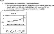
- the biological sample includes 100 nM or less of the polynucleotide comprising the fusion gene, e.g., 0.01-100 nM, 0.01-25nM, 0.01-5 nM, 0.02-0.5 nM, or 0.02-0.1 nM.
- an individual e.g ., an individual diagnosed with cancer
- any of the multiplex assay compositions described herein comprising: (A) at least one primer set and labeled probe that specifically amplify and detect at least one ALK fusion gene; (B) at least one primer set and labeled probe that specifically amplify and detect at least one RET fusion gene; (C) at least one primer set and labeled probe that specifically amplify and detect at least one ROS1 fusion gene; and (D) a primer set and labeled probe that specifically amplify and detect an internal control); carrying out amplification and detection under conditions that allow formation and detection of an amplification product in the presence of at least one fusion gene in the biological sample; determining that at least one fusion gene is present if a fusion gene is detected; and treating the individual if at least one fusion gene is present.
- an individual e.g ., an individual diagnosed with cancer
- any of the multiplex assay compositions described herein e.g ., comprising: (A) at least one primer set and labeled probe that specifically amplify and detect at least one ALK fusion gene; (B) at least one primer set and labeled probe that specifically amplify and detect at least one RET fusion gene; and (C) a primer set and labeled probe that specifically amplify and detect an internal control); carrying out amplification and detection under conditions that allow formation and detection of an amplification product in the presence of at least one fusion gene in the biological sample; determining that at least one fusion gene is present if a fusion gene is detected; and treating the individual if at least one fusion gene is present.
- an individual e.g ., an individual diagnosed with cancer
- any of the multiplex assay compositions described herein e.g ., comprising: (A) at least one primer set and labeled probe that specifically amplify and detect at least one RET fusion gene; and (B) a primer set and labeled probe that specifically amplify and detect an internal control); carrying out amplification and detection under conditions that allow formation and detection of an amplification product in the presence of at least one fusion gene in the biological sample; determining that at least one fusion gene is present if a fusion gene is detected; and treating the individual if at least one fusion gene is present.
- the treatment is with a kinase inhibitor, e.g., a selective kinase inhibitor such as alectinib, crizotinib, ceritinib, lorlatinib, brigatinib, cabozantinib, apatinib, vandetanib, ponatinib, lenvatinib, DS6051b, or variants or combinations thereof.
- the course of treatment includes radiation therapy or chemotherapy (e.g ., cisplatin, carboplatin, paclitaxel, docetaxel).
- the treatment is with GSK1838705A, TAE-684, CEP-14083, AP26113, NMS-E628, sorafenib, vandetanib, motesanib, sunitinib, and XL-184 (see, e.g., Mologni (2011) Curr. Med. Chem. 18:162 ).
- the individual is monitored throughout treatment, e.g., to determine if the amount of fusion gene amplification product increases or decreases, or if a different fusion gene is detected.
- the treatment is changed if the amount of fusion gene amplification product changes, or if a different fusion gene is detected. For example, if the amount of the originally detected fusion gene decreases but the cancer is progressing, treatment can be changed to be less targeted, e.g., radio- or chemotherapy. If the individual's condition has improved, treatment can be reduced.
- the biological sample includes DNA or RNA, e.g., separated or purified nucleic acids.
- the biological sample is RNA from blood, e.g., plasma, serum, or other blood fraction.
- the amplification and detection are carried out using qRT-PCR.
- the individual is diagnosed with lung cancer (e.g ., non-small cell lung cancer (NSCLC), lung squamous cell carcinoma, lung adenocarcinoma), bladder carcinoma, glioblastoma, head and neck cancer, glioma, thyroid carcinoma, ovarian cancer, leukemia, lymphoma, prostate cancer, pancreatic cancer, renal cancer, or breast cancer.
- lung cancer e.g ., non-small cell lung cancer (NSCLC), lung squamous cell carcinoma, lung adenocarcinoma), bladder carcinoma, glioblastoma, head and neck cancer, glioma, thyroid carcinoma, ovarian cancer, leukemia, lymphoma, prostate cancer, pancreatic cancer, renal cancer, or breast cancer.
- determining the presence of at least one fusion gene in a sample from an individual comprising contacting a biological sample from the individual with any of the multiplex assay compositions described herein (e.g ., comprising: (A) at least one primer set and labeled probe that specifically amplify and detect at least one ALK fusion gene; (B) at least one primer set and labeled probe that specifically amplify and detect at least one RET fusion gene; (C) at least one primer set and labeled probe that specifically amplify and detect at least one ROS1 fusion gene; and (D) a primer set and labeled probe that specifically amplify and detect an internal control); carrying out amplification and detection under conditions that allow formation and detection of an amplification product in the presence of at least one fusion gene in the biological sample; determining that at least one fusion gene is present if a fusion gene is detected.
- the multiplex assay compositions described herein e.g ., comprising: (A) at least one primer set
- determining the presence of at least one fusion gene in a sample from an individual comprising contacting a biological sample from the individual with any of the multiplex assay compositions described herein (e.g ., comprising: (A) at least one primer set and labeled probe that specifically amplify and detect at least one ALK fusion gene; (B) at least one primer set and labeled probe that specifically amplify and detect at least one RET fusion gene; and (C) a primer set and labeled probe that specifically amplify and detect an internal control); carrying out amplification and detection under conditions that allow formation and detection of an amplification product in the presence of at least one fusion gene in the biological sample; determining that at least one fusion gene is present if a fusion gene is detected.
- the multiplex assay compositions described herein e.g ., comprising: (A) at least one primer set and labeled probe that specifically amplify and detect at least one ALK fusion gene; (B) at least one primer set and
- determining the presence of at least one fusion gene in a sample from an individual comprising contacting a biological sample from the individual with any of the multiplex assay compositions described herein (e.g ., comprising: (A) at least one primer set and labeled probe that specifically amplify and detect at least one RET fusion gene; and (B) a primer set and labeled probe that specifically amplify and detect an internal control); carrying out amplification and detection under conditions that allow formation and detection of an amplification product in the presence of at least one fusion gene in the biological sample; determining that at least one fusion gene is present if a fusion gene is detected.
- the biological sample includes DNA or RNA, e.g., separated or purified nucleic acids.
- the biological sample is RNA from blood, e.g., plasma, serum, or other blood fraction.
- the amplification and detection are carried out using qRT-PCR.
- the individual is diagnosed with lung cancer (e.g ., non-small cell lung cancer (NSCLC), lung squamous cell carcinoma, lung adenocarcinoma), bladder carcinoma, glioblastoma, head and neck cancer, glioma, thyroid carcinoma, ovarian cancer, leukemia, lymphoma, prostate cancer, pancreatic cancer, renal cancer, or breast cancer.
- lung cancer e.g ., non-small cell lung cancer (NSCLC), lung squamous cell carcinoma, lung adenocarcinoma), bladder carcinoma, glioblastoma, head and neck cancer, glioma, thyroid carcinoma, ovarian cancer, leukemia, lymphoma, prostate cancer, pancreatic cancer, renal cancer, or breast cancer.
- the method further comprises determining a course of treatment if at least one fusion gene is detected.
- the treatment is with a kinase inhibitor, e.g ., a selective kinase inhibitor such as alectinib, crizotinib, ceritinib, lorlatinib, brigatinib, cabozantinib, apatinib, vandetanib, ponatinib, lenvatinib, DS6051b, or variants or combinations thereof.
- the course of treatment includes radiation therapy or chemotherapy (e.g ., cisplatin, carboplatin, paclitaxel, docetaxel).
- the treatment is with GSK1838705A, TAE-684, CEP-14083, AP26113, NMS-E628, sorafenib, vandetanib, motesanib, sunitinib, and XL-184.
- the inventors have discovered a novel, quantitative, and multiplex method of detecting fusions between genetic regions.
- the presently disclosed methods require only a small amount of patient sample that can be gathered non-invasively, e.g ., circulating free RNA (cfRNA) from plasma.
- cfRNA free RNA
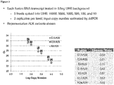
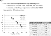
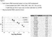
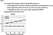
- the presently described methods allow for an extremely sensitive (down to ⁇ 25 copies), one-tube assay to detect multiple gene fusions that are predictive of cancer and response to therapy.
- the present assays can be used for identification of a fusion variant, as well as monitoring and surveillance during treatment and/ or progression.
- a “genetic fusion” is hybrid chromosomal sequence formed by joining of two chromosomal locations that were previously separate. Fusion can occur between genes on the same chromosome (e.g., interstitial deletion or chromosomal inversion) or on different chromosomes (e.g., translocation).
- a “fusion gene” is a hybrid gene formed by the joining of two genes that were previously separate, leading to a structural rearrangement and/or variant in the tumor genome.
- the fusion gene need not necessarily include coding sequence from both genes, but can include non-coding sequence from one of the genes, e.g., promoter or 3' untranslated regions.
- the denomination of genes that comprise a fusion gene as "gene 1," “gene 2,” “gene A,” “gene B,” etc., is used to distinguish between genes that make up the fusion and does not necessarily refer to the position of the genes in the fusion.
- the terms ALK fusion, RET fusion, and ROS1 fusion refer to fusion genes that include ALK, RET, and ROS1 as a member, respectively.
- fusion site refers to the point in a genetic fusion where a nucleotide from one gene or genetic location is found adjacent to a nucleotide from another gene or genetic location.
- target region refers to a region of a target nucleic acid sequence that is to be amplified and/or analyzed.
- nucleic acid refers to polymers of nucleotides (e.g., ribonucleotides or deoxyribo-nucleotides) and includes naturally-occurring (adenosine, guanidine, cytosine, uracil and thymidine), non-naturally occurring, and modified nucleic acids.
- the term is not limited by length (e.g., number of monomers) of the polymer.
- a nucleic acid may be single-stranded or double-stranded and will generally contain 5'-3' phosphodiester bonds, although in some cases, nucleotide analogs may have other linkages.
- nucleotides Monomers are typically referred to as nucleotides.
- non-natural nucleotide or “modified nucleotide” refers to a nucleotide that contains a modified nitrogenous base, sugar or phosphate group, or that incorporates a non-natural moiety in its structure.
- non-natural nucleotides include dideoxynucleotides, biotinylated, aminated, deaminated, alkylated, benzylated and fluorophor-labeled nucleotides.
- primer refers to a short nucleic acid (an oligonucleotide) that acts as a point of initiation of polynucleotide strand synthesis by a nucleic acid polymerase under suitable conditions.
- Polynucleotide synthesis and amplification reactions typically include an appropriate buffer, dNTPs and/or rNTPs, and one or more optional cofactors, and are carried out at a suitable temperature.
- a primer typically includes at least one target-hybridized region that is at least substantially complementary to the target sequence. This region of is typically about 15 to about 40 nucleotides in length.
- a “primer pair” refers to a forward primer and reverse primer (sometimes called 5' and 3' primers) that are complementary to opposite strands of a target sequence and designed to amplify the target sequence.
- the forward and reverse primers are arranged within an amplifiable distance of each other on the target sequence, e.g., about 10-5000 nucleotides, about 25-500, or about 60-120 nucleotides.
- a “primer set” refers to one or more primer pairs, or a combination of at least one forward primer and at least one reverse primer.
- a primer set can include 3 forward primers and 1 reverse primer, so that 3 distinct amplification products can potentially be produced.
- a primer set or primer pair that is specific for a sequence (or portion of a gene) that is 5' (or 3') of a fusion site (or breakpoint) refers to primers used to amplify a sequence that does not include the fusion site or breakpoint.
- probe means any molecule that is capable of selectively binding to a specifically intended target biomolecule, for example, a nucleic acid sequence of interest to be bound, captured or hybridized by the probes. Probes are typically labeled with a non-naturally occurring moiety, e.g., a fluorophore, chromophore, affinity tag (e.g., streptavidin or biotin), and/or a quencher.
- a non-naturally occurring moiety e.g., a fluorophore, chromophore, affinity tag (e.g., streptavidin or biotin), and/or a quencher.
- complementarity refers to the ability of a nucleic acid in a polynucleotide to form a base pair with another nucleic acid in a second polynucleotide.
- sequence A-G-T A-G-U for RNA
- T-C-A U-C-A for RNA
- Complementarity may be partial, in which only some of the nucleic acids match according to base pairing, or complete, where all the nucleic acids match according to base pairing.
- a probe or primer is considered “specific for" a target sequence if it is at least partially complementary to the target sequence.
- the degree of complementarity to the target sequence is typically higher for a shorter nucleic acid such as a primer (e.g., greater than 80%, 90%, 95%, or higher) than for a longer sequence.
- nucleic acids in the context of two or more nucleic acids, or two or more polypeptides, refer to two or more sequences or subsequences that are the same or have a specified percentage of nucleotides, or amino acids, that are the same (e.g ., about 60% identity, e.g., at least any of 65%, 70%, 75%, 80%, 85%, 90%, 91%, 92%, 93%, 94%, 95%, 96%, 97%, 98%, 99%, or higher identity over a specified region, when compared and aligned for maximum correspondence over a comparison window or designated region) as measured using a BLAST or BLAST 2.0 sequence comparison algorithms with default parameters, or by manual alignment and visual inspection.
- Percent identity is typically determined over optimally aligned sequences, so that the definition applies to sequences that have deletions and/or additions, as well as those that have substitutions.
- the algorithms commonly used in the art account for gaps and the like.
- identity exists over a region comprising a sequence that is at least about 8-25 amino acids or nucleotides in length, or over a region that is 50-100 amino acids or nucleotides in length, or over the entire length of the reference sequence.
- allele refers to a sequence variant of a gene. One or more genetic differences can constitute an allele.
- kit refers to any manufacture (e.g., a package or a container) including at least one reagent, such as a nucleic acid probe or probe pool or the like, for specifically amplifying, capturing, tagging/converting or detecting RNA or DNA as described herein.
- manufacture e.g., a package or a container
- reagent such as a nucleic acid probe or probe pool or the like
- amplification conditions refers to conditions in a nucleic acid amplification reaction (e.g., PCR amplification) that allow for hybridization and template-dependent extension of the primers.
- amplicon and amplification product refer to a nucleic acid molecule that contains all or a fragment of the target nucleic acid sequence and that is formed as the product of in vitro amplification by any suitable amplification method.
- the borders of a given amplicon are typically defined by the position of the complementary portion of the forward and reverse primers used for amplification.
- PCR Strategies Innis et al., 1995, Academic Press, San Diego, CA) at Chapter 14 ; PCR Protocols: A Guide to Methods and Applications (Innis et al., Academic Press, NY, 1990 )
- thermostable nucleic acid polymerase or “thermostable polymerase” refers to a polymerase enzyme, which is relatively stable at elevated temperatures when compared, for example, to polymerases from E. coli.
- a thermostable polymerase is suitable for use under temperature cycling conditions typical of the polymerase chain reaction ("PCR").
- Exemplary thermostable polymerases include those from Thermus thermophilus, Thermus caldophilus, Thermus sp. Z05 (see, e.g., U.S. Patent No. 5,674,738 ) and mutants of the Thermus sp. Z05 polymerase, Thermus aquaticus, Thermus flavus, Thermus filiformis, Thermus sp.
- sample refers to any composition containing or presumed to contain nucleic acid from an individual.
- the term includes purified or separated components of cells, tissues, or blood, e.g., DNA, RNA, proteins, cell-free portions, or cell lysates.
- analysis is conducted on plasma samples isolated from blood; the terms "detected in patient's blood” and “detected in patient's plasma” are used interchangeably to mean that blood is obtained from the patient and plasma derived therefrom is used for the analysis.
- a sample can also refer to other types of biological samples, e.g., skin, plasma, serum, whole blood and blood components (e.g., platelets, buffy coat), saliva, urine, tears, seminal fluid, vaginal fluids, tissue biopsies, and other fluids and tissues, including paraffin embedded tissues. Samples also may include constituents and components of in vitro cultures of cells obtained from an individual, including cell lines.
- biological samples e.g., skin, plasma, serum, whole blood and blood components (e.g., platelets, buffy coat), saliva, urine, tears, seminal fluid, vaginal fluids, tissue biopsies, and other fluids and tissues, including paraffin embedded tissues.
- Samples also may include constituents and components of in vitro cultures of cells obtained from an individual, including cell lines.
- a "control" sample or value refers to a sample that serves as a reference, usually a known reference, for comparison to a test sample or test conditions.
- a test sample can be taken from a test condition, e.g., from an individual suspected of having cancer, and compared to samples from known conditions, e.g., from a cancer-free individual (negative control), or from an individual known to have cancer and/ or a particular genetic abnormality (positive control).
- a negative control would be a biological sample from a known healthy (non-cancer, non-mutated) individual
- an example of a positive control would be a biological sample from a patient or cell line known to have a particular gene fusion.
- a control can also represent an average value or a range gathered from a number of tests or results.
- a control can also be prepared for reaction conditions.
- a positive control for the presence of nucleic acid could include primers or probes that will detect a sequence known to be present in the sample, while a negative control would be free of nucleic acids.
- controls can be designed for assessment of any number of parameters. For example, a control can be devised to compare therapeutic benefit based on pharmacological data (e.g., half-life) or therapeutic measures (e.g., comparison of benefit and/or side effects). Controls can be designed for in vitro applications.
- Controls are valuable in a given situation and be able to analyze data based on comparisons to control values. Controls are also valuable for determining the significance of data. For example, if values for a given parameter are widely variant in controls, variation in test samples will not be considered as significant.
- An “internal control” refers to a nucleic acid that is expected to be present in the sample, such as a housekeeping gene that is expressed or present at a fairly standard level across samples.
- the internal control can be used to standardize the amount and quality of nucleic acid in the sample with that of other samples and ensure that the amplification and detection reaction is functioning.
- Examples of internal controls include SDH (succinate dehydrogenase), LDHA (lactate dehydrogenase A), NONO, PGK (phosphoglycerate kinase 1), PPIH, HPRT1, beta-actin, GADPH, ACTB, and 16S rRNA.
- diagnosis refers to a relative probability that a subject has a disorder such as cancer or certain type of cancer (e.g ., resulting from a gene fusion).
- prognosis refers to a relative probability that a certain future outcome may occur in the subject.
- diagnosis can refer to classification of a cancer or the likelihood that an individual will be responsive to a particular therapy. The terms are not intended to be absolute, as will be appreciated by any one of skill in the field of medical diagnostics.
- treatment can refer to, e.g., reducing tumor size, number of cancer cells, growth rate, metastatic activity, reducing cell death of non-cancer cells, reduced nausea and other chemotherapy or radiotherapy side effects, etc.
- treat and “prevent” are not intended to be absolute terms. Treatment and prevention can refer to any delay in onset, amelioration of symptoms, improvement in patient survival, increase in survival time or rate, etc.
- Treatment and prevention can be complete (undetectable levels of neoplastic cells) or partial, such that fewer neoplastic cells are found in a patient than would have occurred without the treatment.
- the effect of treatment can be compared to an individual or pool of individuals not receiving the treatment (e.g., individuals having the same genetic fusion), or to the same patient prior to treatment or at a different time during treatment.
- the severity of disease is reduced by at least 10%, as compared, e.g., to the individual before administration or to a control individual not undergoing treatment.
- the severity of disease is reduced by at least 25%, 50%, 75%, 80%, or 90%, or in some cases, no longer detectable using standard diagnostic techniques.
- treat and “administer,” with reference to a patient, include recommending, providing, or prescribing a particular treatment to the patient, and are not limited to directly, physically treating the patient.
- threshold cycle is a measure of relative concentration and is commonly used in real-time PCR (also referred to as qPCR).
- Ct refers to the intersection of an amplification curve and a threshold line.
- the threshold line is often set at a point when signal can be detected above background, or when an amplification reaction enters the exponential phase.
- Ct can be affected by concentration of target and amplification conditions, e.g., the effect of conditions on detectable labels and amplification efficiency. A higher Ct corresponds to a longer time to reach the threshold, be it due to low target concentration or inefficient amplification.
- the terms "individual,” “subject,” “patient,” and like terms are used interchangeably and refer to humans, except where indicated. Other mammals can be considered subjects, such as non-human primates, as well as rabbits, rats, mice, dogs, cats, and other mammalian species. The term does not necessarily indicate that the subject has been diagnosed with a particular disease, but typically refers to an individual under medical supervision.
- a patient can be seeking treatment, monitoring, adjustment or modification of an existing therapeutic regimen, etc.
- a patient can include individuals that have not received treatment, are currently receiving treatment, have had surgery, and those that have discontinued treatment.
- label refers to a composition detectable by spectroscopic, photochemical, biochemical, immunochemical, chemical, or other physical means.
- useful labels include fluorescent dyes, luminescent agents, radioisotopes (e.g., 32 P, 3 H), electron-dense reagents, or an affinity-based moiety, e.g., a "His tag” for purification, or a "strepavidin tag” that interacts with biotin.
- a number of cancer-associated fusion genes are known, and appear in all manner of cancers. Examples include lung cancer (e.g., non-small cell lung cancer (NSCLC), lung squamous cell carcinoma, lung adenocarcinoma), bladder carcinoma, glioblastoma, head and neck cancer, glioma, thyroid carcinoma, ovarian cancer, leukemia, lymphoma, prostate cancer, pancreatic cancer, renal cancer, and breast cancer.
- Cancer-associated fusion genes commonly occur where one member of the fusion is a kinase involved in a pro-growth signaling pathway, and the other member contributes to elevated or constitutive expression or signaling. This is the case for fusions of ALK, RET, and ROS1.
- Common fusion partners for ALK are EML4 and KIF5B.
- Common fusion partners for RET are KIF5B, CCDC6, and NCOA4.
- ROS1 including CD74, EZR, TPM3, SDC4, SLC34A2, and LRIG3 (see, e.g., Yoshihara et al. (2015) Oncogene 34:4845 ).
- compositions and methods focus on design of multiplex assays to detect ALK, RET, and ROS1 fusions. Invasive biopsy or excessive blood collection is often not feasible for cancer patients.
- present compositions and methods allow for detection of several actionable gene fusions with a relatively small sample from the patient, which can be a non-invasive plasma sample.
- the design of these highly multiplexed assays can vary. Where multiple ALK fusions are detected, for example, a common primer and probe that hybridize to sequences in the ALK gene near the fusion point, and primers specific for various fusion partners, can be used. Thus, for example, if 5 different ALK fusions are detected, the assay can include 15 oligonucleotides (10 primers and 5 probes) or 7 oligonucleotides (1 common primer, 1 common probe, and 5 specific primers).
- the multiplex assay detects 2, 3, 4, 5, 6, or 7 ALK fusions and 2, 3, 4, 5, or 6 RET fusions in a single amplification and detection reaction. In some embodiments, the multiplex assay further detects 2, 3, 4, 5, 6, 7, 8, 9, 10, 11, 12, or 13 ROS1 fusions in the same reaction. In some embodiments, the ROS1 fusions are detected in a separate amplification and detection reaction. In some embodiments, the amplification and detection reaction further includes an internal control (e.g., a housekeeping gene).
- an internal control e.g., a housekeeping gene
- ALK, RET and ROS1 fusions indicate that a cancer patient will be responsive to a selective kinase inhibitor.
- a selective kinase inhibitor include alectinib, crizotinib, ceritinib, lorlatinib, brigatinib, cabozantinib, apatinib, vandetanib, ponatinib, lenvatinib, DS-6051b, and variants or combinations thereof.
- the fusion status of a patient can be monitored throughout treatment to determine if the therapeutic approach can be changed, e.g., to a different kinase inhibitor or more standard chemo- or radio-therapy.
- Samples for testing for genetic fusions can be obtained from any source, but are advantageously obtained in a non-invasive manner, e.g., from blood or a blood fraction (e.g ., plasma, serum, platelets, etc.). Samples for the present methods can also be taken from urine, bronchoalveolar lavage, or tissue biopsy. Methods for isolating nucleic acids from biological samples are known, e.g., as described in Sambrook, and several kits are commercially available (e.g., High Pure RNA Isolation Kit, High Pure Viral Nucleic Acid Kit, and MagNA Pure LC Total Nucleic Acid Isolation Kit from Roche).
- DNA is prepared, and used as template for the presently disclosed amplification and detection methods.
- RNA is prepared. When RNA is used as template for amplification by PCR, a reverse transcription step is required to prepare cDNA. A DNA polymerase such as Taq or another thermostable polymerase can then be used to carry out amplification.
- a DNA polymerase such as Taq or another thermostable polymerase can then be used to carry out amplification.
- the sample is RNA is isolated from blood plasma. Depending on the condition of the patient, about 1-10 mL of plasma can be obtained for testing (usually about 2 mL). Kits for isolating circulating free RNA are commercially available, e.g., from Norgen Biotek Corp or Qiagen.
- the presently disclosed methods for sample preparation and amplification/ detection with custom target-specific oligos are extraordinarily sensitive, and can be used to detect gene fusion mutations from as few as about 50 - and in some cases about 20 - copies in a sample diluted 1:4000 in wild type RNA background.
- This allows for detection of fusion variants in samples where the target sequence is very rare, e.g., circulating cell-free RNA (cfRNA). Varying backgrounds of RNA and DNA in plasma do not detract from the specificity of detection even at low copy numbers.
- Nucleic acid amplification can be carried out using any primer-dependent method.
- the amplification is quantitative, so that the relative or actual abundance of a given amplification target can be determined by the amount of amplification product.
- DNA-based methods can be used for the presently disclosed amplification and detection methods, e.g., PCR.
- PCR real time or quantitative PCR
- qPCR allows for reliable detection and measurement of products generated during each cycle of PCR process.
- kits and reagents are commercially available, e.g., from Roche Molecular Systems, Life Technologies, Bio-Rad, etc. See, e.g., Pfaffl (2010) Methods: The ongoing evolution of qPCR vol. 50 .
- the amplification and detection are carried out in the presence of a dual labeled probe (e.g., a TaqMan, CPT, LNA, or MGB probe) labeled with a quencher and a fluorophore (see, e.g., Gasparic et al. (2010) Anal. Bioanal. Chem. 396:2023 ).
- a dual labeled probe e.g., a TaqMan, CPT, LNA, or MGB probe
- a quencher and a fluorophore see, e.g., Gasparic et al. (2010) Anal. Bioanal. Chem. 396:2023 .
- a preliminary reverse transcription step is carried out (also referred to as RT-PCR, not to be confused with real time PCR). See, e.g., Hierro et al. (2006) 72:7148.
- qRT-PCR refers to reverse transcription followed by quantitative PCR. Both reactions can be carried out in a single tube without interruption, e.g ., to add reagents.
- RNA-based amplification methods can also be used, e.g., transcription mediated amplification (TMA) or nucleic acid sequence based amplification (NASBA).
- TMA transcription mediated amplification
- NASBA nucleic acid sequence based amplification
- oligonucleotides used in the present assays include alkyl base modifications to enhance selective amplification, in particular in a multiplex format.
- a probe, or one or both primers in a primer pair can be labeled with any substance or component that directly or indirectly emits or generates a detectable signal.
- the labels are fluorophores (dyes), many of which are reported in the literature and known to those skilled in the art, and many of which are commercially available. Fluorophores are described, e.g., in Cardullo et al. (1988) Proc. Natl. Acad. Sci. USA 85: 8790 ; Hochstrasser et al. (1992) Biophysical Chemistry 45: 133 ; Selvin (1995) Methods in Enzymology 246: 300 ; Steinberg, Ann. Rev. Biochem., 40: 83- 114 (1971 ); and Wang et al., Anal. Chem. 67: 1197-1203 (1995 ).
- fluorophores that can be used as labels: 4-acetamido-4'-isothiocyanatostilbene-2,2'disulfonic acid; acridine; acridine isothiocyanate; 5-(2'-aminoethyl)aminonaphthalene-1-sulfonic acid (EDANS); 4-amino-N-[3-vinylsulfonyl) phenyl]naphthalimide-3,5 disulfonate [0070] N-(4-anilino-1-naphthyl)maleimide; anthranilamide; BODIPY; Brilliant Yellow; coumarin; 7-amino-4-methylcoumarin (AMC, Coumarin 120)/ 7-amino-4-trifluoromethylcoumarin (Coumaran 151); cyanine dyes; cyanosine 4',6-diaminidino-2-phenylindole (DAPI); 5',5"
- Fluorophores can be used in the presently described assays to label a nucleic acid as described herein. Fluorophores can be attached by conventional covalent bonding, using appropriate functional groups on the fluorophore and/or nucleic acid.
- a dual labeled probe can be used for detection.
- the dual labeled probe can comprise a fluorophore, such any of the fluorophores listed above, and a quencher.
- Suitable quenchers include but are not limited to DDQ-I, Dabcyl, Eclipse, Iowa Black FQ, BHQ-1, QSY-7, BHQ-2, DDQ-II, Iowa Black RQ, QSY-21, and BHQ-3.
- fluorophores having an emission maximum between 500 and 550 nm e.g., FAM, TET, and HEX
- a quencher with an absorption maxima between 450 and 500 nm can be selected (e.g ., dabcyl or BHQ-1).
- a quencher with an absorption maxima above 550 nm can be selected (e.g ., BHQ-2). See, e.g., Johansson (2003) Meth. Mol. Biol. 335:17 for considerations in selecting dye-quencher pairs.
- Detection devices are known in the art and can be selected as appropriate for the selected labels. Detection devices appropriate for quantitative PCR include the cobas® and Light Cycler® systems (Roche), PRISM 7000 and 7300 real-time PCR systems (Applied Biosystems), etc.
- kits for carrying out the presently disclosed methods are included in a kit.
- the kit includes components for obtaining, storing, and/ or preparing sample.
- Such components include, e.g., sterile needles and syringes, EDTA-lined tubes, buffers (e.g., for binding nucleic acid to, and elution from a matrix), RNase inhibitors, and/ or DNase, etc.
- the kit includes forward primer(s) and reverse primer(s) for amplifying ALK fusion variant(s) having sequences selected from the group consisting of SEQ ID NOs:1-50, and SEQ ID NOs:52-61 and 181, respectively.
- the kit includes probe(s) for detecting ALK fusion variant(s) having sequences selected from the group consisting of SEQ ID NOs:182-186.
- the forward and reverse primer sequences and probe sequences can be used together in any appropriate combination to detect any 1, 2, 3, 4, 5, 6, or 7 ALK fusion variants in any combination.
- the kit includes forward primer(s) and reverse primer(s) for amplifying RET fusion variant(s) having sequences selected from the group consisting of SEQ ID NOs:83-145 and 187, and SEQ ID NOs:161-180, respectively.
- the kit includes probe(s) for detecting RET fusion variant(s) having sequences selected from the group consisting of:189-194. The forward and reverse primer sequences and probe sequences can be used together in any combination to detect any 1, 2, 3, 4, 5, or 6 RET fusion variants in any combination.
- the kit includes forward primer(s) and reverse primer(s) for amplifying ROS1 fusion variant(s) having sequences selected from the group consisting of SEQ ID NOs:195-212, and SEQ ID NOs:213-226, respectively.
- the kit includes probe(s) for detecting ROS1 fusion variants having sequences selected from the group consisting of:227-230 and 51.
- the forward and reverse primer sequences and probe sequences can be used together in any combination to detect any 1, 2, 3, 4, 5, 6, 7, 8, 9, 10, 11, 12, or 13 ROS1 fusion variants in any combination.
- the kit includes a forward primer and reverse primer for amplifying an EML exon 13- ALK exon 20 fusion variant having sequences selected from the group consisting of SEQ ID NOs:1-10 and SEQ ID NOs:52-61 and 181, respectively.
- the kit includes a forward primer and reverse primer for amplifying an EML exon 20- ALK exon 20 fusion variant having sequences selected from the group consisting of SEQ ID NOs:11-20 and SEQ ID NOs:52-61 and 181, respectively.
- the kit includes a forward primer and reverse primer for amplifying an EML exon 6- ALK exon 20 fusion variant having sequences selected from the group consisting of SEQ ID NOs:21-30 and SEQ ID NOs:52-61 and 181, respectively.
- the kit includes a forward primer and reverse primer for amplifying an EML exon 2- ALK exon 20 fusion variant having sequences selected from the group consisting of SEQ ID NOs:31-35 and SEQ ID NOs:52-61 and 181, respectively.
- the kit includes a forward primer and reverse primer for amplifying an EML exon 18- ALK exon 20 fusion variant having sequences selected from the group consisting of SEQ ID NOs:36-40 and SEQ ID NOs:52-61 and 181, respectively.
- the kit includes a forward primer and reverse primer for amplifying a KIF exon 24- ALK exon 20 fusion variant having sequences selected from the group consisting of SEQ ID NOs:41-45 and SEQ ID NOs:52-61 and 181, respectively.
- the kit includes a forward primer and reverse primer for amplifying a KIF exon 17- ALK exon 20 fusion variant having sequences selected from the group consisting of SEQ ID NOs:46-50 and SEQ ID NOs:52-61 and 181, respectively.
- the kit includes a probe for detecting an ALK fusion having a sequence selected from group consisting of SEQ ID NOs:182-186.
- the kit includes a forward primer and reverse primer for amplifying a KIF exon 15- RET exon 12 fusion variant having sequences selected from the group consisting of SEQ ID NOs:83-97 and SEQ ID NOs:161-180, respectively.
- the kit includes a forward primer and reverse primer for amplifying a KIF exon 16- RET exon 12 fusion variant having sequences selected from the group consisting of SEQ ID NOs:98-107 and SEQ ID NOs:161-180, respectively.
- the kit includes a forward primer and reverse primer for amplifying a KIF exon 22- RET exon 12 fusion variant having sequences selected from the group consisting of SEQ ID NOs:108-117 and SEQ ID NOs:161-180, respectively.
- the kit includes a forward primer and reverse primer for amplifying a KIF exon 23- RET exon 12 fusion variant having sequences selected from the group consisting of SEQ ID NOs:118-127 and 187, and SEQ ID NOs:161-180, respectively.
- the kit includes a forward primer and reverse primer for amplifying a CCDC exon 1- RET exon 12 fusion variant having sequences selected from the group consisting of SEQ ID NOs:128-135 and 118, and SEQ ID NOs:161-180, respectively.
- the kit includes a forward primer and reverse primer for amplifying an NCO exon 6- RET exon 12 fusion variant having sequences selected from the group consisting of SEQ ID NOs:136-145 and SEQ ID NOs:161-180, respectively.
- the kit includes a probe for detecting a RET fusion having a sequence selected from group consisting of SEQ ID NOs:189-194.
- the kit includes a forward primer and reverse primer for amplifying a CD74 exon 6- ROS1 exon 34 fusion variant having sequences selected from the group consisting of SEQ ID NOs:195-197 and SEQ ID NOs:222-226, respectively.
- the kit includes a forward primer and reverse primer for amplifying a CD74 exon 6- ROS1 exon 32 fusion variant having sequences selected from the group consisting of SEQ ID NOs:195-197 and SEQ ID NOs:213-215, respectively.
- the kit includes a forward primer and reverse primer for amplifying an EZR exon 10- ROS1 exon 34 fusion variant having sequences selected from the group consisting of SEQ ID NO:208 and SEQ ID NOs:222-226, respectively.
- the kit includes a forward primer and reverse primer for amplifying a TPM3 exon 8- ROS1 exon 35 fusion variant having sequences selected from the group consisting of SEQ ID NOs:211-212 and SEQ ID NOs:216-221, respectively.
- the kit includes a forward primer and reverse primer for amplifying an SDC4 exon 4- ROS1 exon 34 fusion variant having sequences selected from the group consisting of SEQ ID NOs:200-202 and SEQ ID NOs:222-226, respectively.
- the kit includes a forward primer and reverse primer for amplifying an SDC4 exon 2- ROS1 exon 32 fusion variant having sequences selected from the group consisting of SEQ ID NOs:198-199 and SEQ ID NOs:213-215, respectively.
- the kit includes a forward primer and reverse primer for amplifying an SDC4 exon 2-ROS1 exon 34 fusion variant having sequences selected from the group consisting of SEQ ID NOs:198-199 and SEQ ID NOs:222-226, respectively.
- the kit includes a forward primer and reverse primer for amplifying an SDC4 exon 4- ROS1 exon 32 fusion variant having sequences selected from the group consisting of SEQ ID NOs:200-202 and SEQ ID NOs:213-215, respectively.
- the kit includes a forward primer and reverse primer for amplifying an SLC34A2 exon 13- ROS1 exon 34 fusion variant having sequences selected from the group consisting of SEQ ID NOs:203-205 and SEQ ID NOs:222-226, respectively.
- the kit includes a forward primer and reverse primer for amplifying an SLC34A2 exon 13- ROS1 exon 32 fusion variant having sequences selected from the group consisting of SEQ ID NOs:203-205 and SEQ ID NOs:213-215, respectively.
- the kit includes a forward primer and reverse primer for amplifying an SLC34A2 exon 4- ROS1 exon 32 fusion variant having sequences selected from the group consisting of SEQ ID NOs:206-207 and SEQ ID NOs:213-215, respectively.
- the kit includes a forward primer and reverse primer for amplifying an SLC34A2 exon 4- ROS1 exon 34 fusion variant having sequences selected from the group consisting of SEQ ID NOs:206-207 and SEQ ID NOs:222-226, respectively.
- the kit includes a forward primer and reverse primer for amplifying an LRIG3 exon 16- ROS1 exon 35 fusion variant having sequences selected from the group consisting of SEQ ID NOs:209-210 and SEQ ID NOs:216-221, respectively.
- each of the primer sets is packaged in separate tubes, e.g., to be added in ratios to be determined by the user. In some embodiments, one or more or all of the primer sets are packaged in a single tube with predetermined ratios.
- the kit can also include enzymes, such as reverse transcriptase and or DNA polymerase.
- the DNA polymerase is a thermostable DNA polymerase capable of amplifying in thermocycling conditions, e.g., Taq or a Taq derivative.
- the kit includes dNTPs.
- the kit includes buffers conducive to polymerization/ amplification by the selected polymerases.
- the kit includes controls, e.g., a polynucleotide that is wild type at the genetic fusion to be detected (i.e., no genetic fusion), or a polynucleotide that includes the genetic fusion to be detected.
- controls e.g., a polynucleotide that is wild type at the genetic fusion to be detected (i.e., no genetic fusion), or a polynucleotide that includes the genetic fusion to be detected.
- the kit can also include consumables such as sample tubes or vials; reaction containers (e.g., tubes, multiwell plates, microfluidic chips or chambers, etc), as well as directions for use or reference to a website.
- consumables such as sample tubes or vials; reaction containers (e.g., tubes, multiwell plates, microfluidic chips or chambers, etc), as well as directions for use or reference to a website.
- Example 1 Multiplex assays for detection of ALK, RET, and ROS1 fusion panel
- These four sets correspond to (i) ALK (detected with one or more probes labeled with a first label), (ii) RET (detected with one or more probes labeled with a second label), (iii) ROS1 (detected with one or more probes labeled with a third label), and (iv) an internal control (detected with a probe labeled with a forth label).
- the labels can be selected from those disclosed herein and in some embodiments are distinguishable from one other.
- ALK fusions are detected with a FAM-labeled probe
- RET fusions are detected with a HEX-labeled probe
- ROS1 fusions are detected with a JA270-labeled probe
- the internal control is detected with a Cy5.5-labeled probe.
- Table 1 Label Gene Fusion Coverage Oligonucleotides FAM ALK EML4 exon 13-ALK exon 20 (V1) 7 fusions 15 primers EML4 exon 20-ALK exon 20 (V2) 94% ALK fusions 2 probes EML4 exon 6a/b-ALK exon 20 (V3) EML4 exon 2-ALK exon 20 (V5) EML4 exon 18-ALK exon 20 (V8) KIF5B exon 17-ALK exon 20 (V6) KIF5B exon 24-ALK exon 20 (V7) HEX RET KIF5B exon 15-RET exon 12 (V1) 6 fusions KIF5B exon 16-RET exon 12 (V2) 97% RET fusions KIF5B exon 22-RET exon 12 (V3) KIF5B exon 23-RET exon 12 (V4) CCDC6 exon 1-RET exon
- the multiplex may include various gene fusion detection combinations, and in some embodiments, fewer fusions are assayed and detected.
- An example of an assay format for detection ALK and RET fusions is shown in Table 2. Fusions in ROS1 can be detected separately, or in a parallel assay, for example, as shown in Table 3.
- the oligonucleotides shown in Tables 4-6 can be selected for use in the assays.
- the first set of forward and reverse primers amplifies across EML4-ALK and KIF5B-ALK fusions.
- the primers are designated with the gene name (e.g. EML for EML4), exon (e.g., 13 for exon 13), and designation (e.g., F1 for Forward 1).
- the symbols ⁇ t_bb_dA>, ⁇ t_bb_dC>, ⁇ t_bb_dT>, ⁇ t_bb_dG> refer to p-tert butylbenzyl modified A, C, T, and G, respectively.
- Forward and reverse primers can be used in single pairs or in any combination to amplify different fusion products, as will be appreciated by one of skill in the art.
- the number of oligonucleotides in the reaction was minimized, as indicated in Tables 1-3.
- the reverse primers in all reactions served as primers for the reverse transcriptase reactions.
- reaction conditions were as follows. For each reaction, 25 uL of input RNA was added to a RT-PCR reaction mix comprising forward and reverse primers, labeled probe, buffer, dUTP, dTTP, dATP, dGTP, UNG, and Z05 enzyme to a final volume of 50uL. The reactions were run in multiplex, each with primers and probes specific for every fusion variant indicated in Table 1.
- Results were confirmed using a Next Generation Sequencing assay that detects the fusion variants covered in the qRT-PCR assay.
- Figure 1A shows that each ALK fusion variant is detectable at 50 copies.
- Figure 1B shows that each RET fusion variant is detectable at 50 copies.
- Figure 1C shows that each ROS1 fusion variant is detectable at 50 copies.
- Figure 1D shows that the reaction efficiency and input was equivalent, as indicated by the Internal Control Ct's.
- Sensitivity, or Limit of Detection (LOD) studies are shown and described in Figures 6-8 .
- the LOD for each assay is shown in Tables 7 and 8. All 7 ALK, 6 RET, and 13 ROS1 fusion variants are detectable down to less than 10 copies. The predominant fusion variants are marked with an *.
Description
- A number of cancers are associated with gene fusions (Yoshihara et al. (2015) Oncogene 34:4845). Perhaps the earliest reported example is the association of BCR-ABL with chronic myelogenous leukemia (CML) in the '60s (Nowell and Hungerford (1960) J. Natl. Cancer Inst. 25:85). Since then, hundreds more gene fusions have been reported for cancers in many different tissues (Presner and Chinnaiyan (2009) Curr. Opin Genet. Dev. 19:82).
- Another example is the tyrosine receptor kinase ALK (anaplastic lymphoma kinase). EML4-ALK (echinoderm microtubule-associated protein-like 4-anaplastic lymphoma kinase) fusions are associated with non-small cell lung cancer (NSCLC). In this case, the N terminal, extracellular portion of ALK is replaced by EML4 (KIF5B, HIP1, KLC1, TFG can also fuse with ALK in a similar manner). The expression of the resulting fusion gene is driven by the strong EML4 promoter, resulting in higher expression of the intracellular tyrosine kinase domain of ALK. In addition, EML4 forms a coiled-coil that results in ligand-independent dimerization, and constitutive activation of the ALK tyrosine kinase domain. Additional examples of activated kinase fusions involve RET (rearranged during transfection) and ROS1.
- Detection of a gene fusion can be used to direct therapy. Most methods of detection require biopsy of tumor tissue, which is not feasible for many cancer patients, especially in later stages. Detection in biopsied tissue sections is typically carried out by fluorescence in situ hybridization (FISH) or immunohistochemistry (IHC). The tests have high false positive rates and background, in part because of shearing during the sectioning process. Skilled cytologists are thus required to observe multiple tissue sections, which necessitates a sizable biopsy from a weakened patient. Similarly, a difficulty with using RT-PCR is the amount and quality of genetic material from tumor tissue, e.g., in formalin fixed paraffin embedded (FFPE) form. See, e.g., Liu et al. (2015) PLoSOne 10: e0117032.
- Because detection is time and resource intensive, the testing rate is relatively low. Cancers associated with ALK fusions are very sensitive to ALK inhibitors such as crizotinib and ceretinib. Gene fusions with Rearranged during Transcription (RET), such as with KIF5B or CCDC6, are also sensitive to therapy, e.g., with vandetanib (see Matsubara et al. (2007) J. Thorac. Oncol. 7:1872). The low rate of testing for gene fusions thus represents a great lost opportunity for treatment.
-
CN104818320 A (AMOY DIAGNOSTICS CO LTD) discloses primers and probes for PCR detection of the three genes (ALK, RET, ROS1), including multiplex amplification of several variants of the same gene fusions in a single tube, but no multiplex of 2 or 3 genes.WO 2016/166269 A1 (ROCHE DIAGNOSTICS GMBH [DE]; F HOFFMANN-LA ROCHE AG [CH]; ROCHE MOLECU) discloses methods for detection of all three gene fusions by measuring the imbalance in expression of the two parts of the fusion. The method uses several primer pairs specific for said fusions, but no specific probes for detection of the amplicons and no multiplex amplification of the three genes. - The description below may in some respects go beyond the content of the invention as such, which is defined exclusively by the appended claims. This description is provided as additional information to place the actual invention in a broader technical context and to illustrate possible related tools, materials or technical developments. Additional technical information which does not fall within the scope of the appended claims is not part of the invention, even if it is described as "embodiment"of the invention.
- Provided herein are multiplex methods and compositions for detecting fusion genes, in particular those involving ALK, RET, and ROS1.
- Provided herein are multiplex assay compositions comprising: (A) at least one primer set and labeled probe that specifically amplify and detect at least one ALK fusion gene; (B) at least one primer set and labeled probe that specifically amplify and detect at least one RET fusion gene; (C) at least one primer set and labeled probe that specifically amplify and detect at least one ROS1 fusion gene; and (D) a primer set and labeled probe that specifically amplify and detect an internal control. Further provided are multiplex assay compositions comprising: (A) at least one primer set and labeled probe that specifically amplify and detect at least one ALK fusion gene; (B) at least one primer set and labeled probe that specifically amplify and detect at least one RET fusion gene; and (C) a primer set and labeled probe that specifically amplify and detect an internal control. Also disclosed herein are multiplex assay compositions comprising: (A) at least one primer set and labeled probe that specifically amplify and detect at least one RET fusion gene; and (B) a primer set and labeled probe that specifically amplify and detect an internal control.
- In some embodiments, the at least one ALK fusion gene is selected from the group consisting of: EML4 exon 13-ALK exon 20, EML4 exon 20-ALK exon 20, EML4 exon 6a/b-ALK exon 20, EML4 exon 2-ALK exon 20, EML4 exon 18-ALK exon 20, KIF5B exon 17-ALK exon 20, and KIF5B exon 24-ALK exon 20; the at least one RET fusion gene is selected from the group consisting of: KIF5B exon 15-RET exon 12, KIF5B exon 16-RET exon 12, KIF5B exon 22-RET exon 12, KIF5B exon 23-RET exon 12, CCDC6 exon 1-RET exon 12, and NCOA4 exon 6-RET exon 12; and the at least one ROS1 fusion gene is selected from the group consisting of: CD74 exon 6-ROS1 exon 34, CD74 exon 6-ROS1 exon 32, EZR exon 10-ROS1 exon 34, TPM3 exon 8-ROS1 exon 35, SDC4 exon 4-ROS1 exon 32, SDC4 exon 2-ROS1 exon 32, SDC4 exon 2-ROS1 exon 34, SDC4 exon 4-ROS1 exon 34, SLC34A2 exon 13-ROS1 exon 34, SLC34A2 exon 13-ROS1 exon 32, SLC34A2 exon 4-ROS1 exon 32, SLC34A2 exon 4-ROS1 exon 35, and LRIG3 exon 16-ROS1 exon 35, in any combination.
- In some embodiments, the composition comprises at least one primer set and probe that amplify and detect more than 2 ALK fusion genes, more than 2 RET fusion genes, and/ or more than 2 ROS1 fusion genes. In some embodiments, the composition comprises at least one primer set and probe that amplify and detect EML4 exon 13-
ALK exon 20, EML4 exon 20-ALK exon 20, EML4 exon 6a/b-ALK exon 20, KIF5B exon 15-RET exon 12, KIF5B exon 16-RET exon 12, KIF5B exon 22-RET exon 12, CD74 exon 6-ROS1 exon 34, and EZR exon 10-ROS1 exon 34. - In some embodiments, the at least one ALK fusion gene include: EML4 exon 13-
ALK exon 20, EML4 exon 20-ALK exon 20, EML4 exon 6a/b-ALK exon 20, EML4 exon 2-ALK exon 20, EML4 exon 18-ALK exon 20, KIF5B exon 17-ALK exon 20, and KIF5B exon 24-ALK exon 20; the at least one RET fusion gene includes: KIF5B exon 15-RET exon 12, KIF5B exon 16-RET exon 12, KIF5B exon 22-RET exon 12, KIF5B exon 23-RET exon 12, CCDC6 exon 1-RET exon 12, and NCOA4 exon 6-RET exon 12; and the at least one ROS1 fusion gene includes: CD74 exon 6-ROS1 exon 34, CD74 exon 6-ROS1 exon 32, EZR exon 10-ROS1 exon 34, TPM3 exon 8-ROS1 exon 35, SDC4 exon 4-ROS1 exon 32, SDC4 exon 2-ROS1 exon 34, SDC4 exon 2-ROS1 exon 32, SDC4 exon 4-ROS1 exon 32, SLC34A2 exon 13-ROS1 exon 34, SLC34A2 exon 13-ROS1 exon 32, SLC34A2 exon 4-ROS1 exon 32, SLC34A2 exon 4-ROS1 exon 35, and LRIG3 exon 16-ROS1 exon 35. That is, the assay composition includes primer sets and probes to amplify and detect all of the listed fusion genes. - In some embodiments, for the primer set to amplify at least one ALK fusion gene, the forward primer and reverse primer have sequences selected from the group consisting of SEQ ID NOs:1-50, and SEQ ID NOs:52-61 and 181, respectively. In some embodiments, for the probe to detect at least one ALK fusion gene, the probe sequence is selected from the group consisting of SEQ ID NOs:182-186. The forward and reverse primer sequences and probe sequences can be used together in any appropriate combination to detect any 1, 2, 3, 4, 5, 6, or 7 ALK fusion variants in any combination. In some embodiments, for the primer set to amplify at least one RET fusion gene, the forward primer and reverse primer have sequences selected from the group consisting of SEQ ID NOs:83-145 and 187, and SEQ ID NOs:161-180, respectively. In some embodiments, for the probe to detect at least one RET fusion gene, the probe sequence is selected from the group consisting of: 189-194. The forward and reverse primer sequences and probe sequences can be used together in any combination to detect any 1, 2, 3, 4, 5, or 6 RET fusion variants in any combination. In some embodiments, for the primer set to detect at least one ROS1 fusion gene, the forward primer and reverse primer have sequences selected from the group consisting of SEQ ID NOs:195-212, and SEQ ID NOs:213-226, respectively. In some embodiments, for the probe to detect at least one ROS1 fusion gene, the probe sequence is selected from the group consisting of:227-230 and 51. The forward and reverse primer sequences and probe sequences can be used together in any combination to detect any 1, 2, 3, 4, 5, 6, 7, 8, 9, 10, 11, 12, or 13 ROS1 fusion variants in any combination.
- In some embodiments, the label on labeled probe that detects the internal control is different from the labels on the labeled probes that detect the fusion genes. In some embodiments, the labels on all of the labeled probes are different from each other. In some embodiments, a single labeled probe is used to detect all of the at least one ALK fusion genes. In some embodiments, a single labeled probe is used to detect all of the at least one RET fusion genes. In some embodiments, a single labeled probe is used to detect all of the at least one ROS1 fusion genes. In some embodiments, the labeled probe is attached to a primer in the at least one primer set. In some embodiments, the labeled probe is separate from the primer set.
- In some embodiments, where more than one ALK fusion gene is amplified and detected, all of the primer sets that amplify the ALK fusion genes include a single common primer. In some embodiments, where more than one ALK fusion gene is amplified and detected, the primer sets include unique primers. In some embodiments, where more than one RET fusion gene is amplified and detected, all of the primer sets that amplify the RET fusion genes include a single common primer. In some embodiments, where more than one RET fusion gene is amplified and detected, the primer sets include unique primers. In some embodiments, where more than one ROS1 fusion gene is amplified and detected, all of the primer sets that amplify the ROS1 fusion genes include a single common primer. In some embodiments, where more than one ROS1 fusion gene is amplified and detected, the primer sets include unique primers.
- Further provided herein are multiplex assay compositions comprising: (A) at least one primer set and labeled probe that specifically amplify and detect at least one ALK fusion gene; (B) at least one primer set and labeled probe that specifically amplify and detect at least one RET fusion gene; and (C) a primer set and labeled probe that specifically amplify and detect an internal control. Also provided herein are multiplex assay compositions comprising: (A) at least one primer set and labeled probe that specifically amplify and detect at least one RET fusion gene; and (B) a primer set and labeled probe that specifically amplify and detect an internal control. In some embodiments, at least one ROS1 fusion gene is amplified and detected in a separate multiplex assay. In some embodiments, the at least one ALK fusion gene is selected from the group consisting of: EML4 exon 13-
ALK exon 20, EML4 exon 20-ALK exon 20, EML4 exon 6a/b-ALK exon 20, EML4 exon 2-ALK exon 20, EML4 exon 18-ALK exon 20, KIF5B exon 17-ALK exon 20, and KIF5B exon 24-ALK exon 20; and the at least one RET fusion gene is selected from the group consisting of: KIF5B exon 15-RET exon 12, KIF5B exon 16-RET exon 12, KIF5B exon 22-RET exon 12, KIF5B exon 23-RET exon 12, CCDC6 exon 1-RET exon 12, and NCOA4 exon 6-RET exon 12, in any combination. In some embodiments, the at least one ROS1 fusion gene is selected from the group consisting of: CD74 exon 6-ROS1 exon 34, CD74 exon 6-ROS1 exon 32, EZR exon 10-ROS1 exon 34, TPM3 exon 8-ROS1 exon 35, SDC4 exon 2-ROS1 exon 34, SDC4 exon 4-ROS1 exon 32, SDC4 exon 2-ROS1 exon 32, SDC4 exon 4-ROS1 exon 34, SLC34A2 exon 13-ROS1 exon 34, SLC34A2 exon 13-ROS1 exon 32, SLC34A2 exon 4-ROS1 exon 32, SLC34A2 exon 4-ROS1 exon 35, and LRIG3 exon 16-ROS1 exon 35. - In some embodiments, the composition comprises at least one primer set and probe that amplify and detect more than 2 ALK fusion genes and more than 2 RET fusion genes. In some embodiments, the composition comprises at least one primer set and probe that amplify and detect EML4 exon 13-
ALK exon 20, EML4 exon 20-ALK exon 20, EML4 exon 6a/b-ALK exon 20, KIF5B exon 15-RET exon 12, KIF5B exon 16-RET exon 12, and KIF5B exon 22-RET exon 12. - In some embodiments, the at least one ALK fusion gene include: EML4 exon 13-
ALK exon 20, EML4 exon 20-ALK exon 20, EML4 exon 6a/b-ALK exon 20, EML4 exon 2-ALK exon 20, EML4 exon 18-ALK exon 20, KIF5B exon 17-ALK exon 20, and KIF5B exon 24-ALK exon 20; and the at least one RET fusion gene includes: KIF5B exon 15-RET exon 12, KIF5B exon 16-RET exon 12, KIF5B exon 22-RET exon 12, KIF5B exon 23-RET exon 12, CCDC6 exon 1-RET exon 12, and NCOA4 exon 6-RET exon 12. - In some embodiments, the label on labeled probe that detects the internal control is different from the labels on the labeled probes that detect the fusion genes. In some embodiments, the labels on all of the labeled probes are different from each other. In some embodiments, a single labeled probe is used to detect all of the at least one ALK fusion genes. In some embodiments, a single labeled probe is used to detect all of the at least one RET fusion genes. In some embodiments, the labeled probe is attached to a primer in the at least one primer set. In some embodiments, the labeled probe is separate from the primer set.
- In some embodiments, where more than one ALK fusion gene is amplified and detected, all of the primer sets that amplify the ALK fusion genes include a single common primer. In some embodiments, where more than one ALK fusion gene is amplified and detected, the primer sets include unique primers. In some embodiments, where more than one RET fusion gene is amplified and detected, all of the primer sets that amplify the RET fusion genes include a single common primer. In some embodiments, where more than one RET fusion gene is amplified and detected, the primer sets include unique primers.
- Examples of internal controls that can be used for the presently disclosed assays include, but are not limited to, SDHA (succinate dehydrogenase), LDHA (lactate dehydrogenase A), NONO, PGK (phosphoglycerate kinase 1), PPIH, HPRT1, beta-actin, GADPH, ACTB, and 16S rRNA.
- In some embodiments, the composition further comprises a DNA polymerase, e.g., a thermostable DNA polymerase such as Taq or a Taq derivative. In some embodiments, the composition further comprises reverse transcriptase. In some embodiments, the composition further comprises dNTPs. In some embodiments, the composition further comprises buffer amenable to polymerization by the DNA polymerase and reverse transcriptase.
- In some embodiments, the composition further comprises a biological sample from an individual or group of individuals. In some embodiments, the individual has been diagnosed with cancer, e.g., lung cancer (e.g., non-small cell lung cancer (NSCLC), lung squamous cell carcinoma, lung adenocarcinoma), bladder carcinoma, glioblastoma, head and neck cancer, glioma, thyroid carcinoma, ovarian cancer, leukemia, lymphoma, prostate cancer, pancreatic cancer, renal cancer, or breast cancer.
- In some embodiments, the sample is enriched or isolated nucleic acid, e.g., DNA or RNA. In some embodiments, the sample is RNA, e.g., isolated from blood (e.g., serum, plasma, other blood fraction), bronchoalveolar lavage, or tissue biopsy. In some embodiments, the biological sample includes 100 nM or less of the polynucleotide comprising the fusion gene, e.g., 0.01-100 nM, 0.01-25nM, 0.01-5 nM, 0.02-0.5 nM, or 0.02-0.1 nM.
- Further disclosed are methods of treating an individual, e.g., an individual diagnosed with cancer, comprising contacting a biological sample from the individual with any of the multiplex assay compositions described herein (e.g., comprising: (A) at least one primer set and labeled probe that specifically amplify and detect at least one ALK fusion gene; (B) at least one primer set and labeled probe that specifically amplify and detect at least one RET fusion gene; (C) at least one primer set and labeled probe that specifically amplify and detect at least one ROS1 fusion gene; and (D) a primer set and labeled probe that specifically amplify and detect an internal control); carrying out amplification and detection under conditions that allow formation and detection of an amplification product in the presence of at least one fusion gene in the biological sample; determining that at least one fusion gene is present if a fusion gene is detected; and treating the individual if at least one fusion gene is present. Further provided are methods of treating an individual, e.g., an individual diagnosed with cancer, comprising contacting a biological sample from the individual with any of the multiplex assay compositions described herein (e.g., comprising: (A) at least one primer set and labeled probe that specifically amplify and detect at least one ALK fusion gene; (B) at least one primer set and labeled probe that specifically amplify and detect at least one RET fusion gene; and (C) a primer set and labeled probe that specifically amplify and detect an internal control); carrying out amplification and detection under conditions that allow formation and detection of an amplification product in the presence of at least one fusion gene in the biological sample; determining that at least one fusion gene is present if a fusion gene is detected; and treating the individual if at least one fusion gene is present. Further provided are methods of treating an individual, e.g., an individual diagnosed with cancer, comprising contacting a biological sample from the individual with any of the multiplex assay compositions described herein (e.g., comprising: (A) at least one primer set and labeled probe that specifically amplify and detect at least one RET fusion gene; and (B) a primer set and labeled probe that specifically amplify and detect an internal control); carrying out amplification and detection under conditions that allow formation and detection of an amplification product in the presence of at least one fusion gene in the biological sample; determining that at least one fusion gene is present if a fusion gene is detected; and treating the individual if at least one fusion gene is present.
- In some instances the treatment is with a kinase inhibitor, e.g., a selective kinase inhibitor such as alectinib, crizotinib, ceritinib, lorlatinib, brigatinib, cabozantinib, apatinib, vandetanib, ponatinib, lenvatinib, DS6051b, or variants or combinations thereof. In some embodiments, the course of treatment includes radiation therapy or chemotherapy (e.g., cisplatin, carboplatin, paclitaxel, docetaxel). In some embodiments, the treatment is with GSK1838705A, TAE-684, CEP-14083, AP26113, NMS-E628, sorafenib, vandetanib, motesanib, sunitinib, and XL-184 (see, e.g., Mologni (2011) Curr. Med. Chem. 18:162).
- In some instances the individual is monitored throughout treatment, e.g., to determine if the amount of fusion gene amplification product increases or decreases, or if a different fusion gene is detected. In some embodiments, the treatment is changed if the amount of fusion gene amplification product changes, or if a different fusion gene is detected. For example, if the amount of the originally detected fusion gene decreases but the cancer is progressing, treatment can be changed to be less targeted, e.g., radio- or chemotherapy. If the individual's condition has improved, treatment can be reduced.
- In some embodiments, the biological sample includes DNA or RNA, e.g., separated or purified nucleic acids. In some embodiments, the biological sample is RNA from blood, e.g., plasma, serum, or other blood fraction. In some embodiments, the amplification and detection are carried out using qRT-PCR.
- In some embodiments, the individual is diagnosed with lung cancer (e.g., non-small cell lung cancer (NSCLC), lung squamous cell carcinoma, lung adenocarcinoma), bladder carcinoma, glioblastoma, head and neck cancer, glioma, thyroid carcinoma, ovarian cancer, leukemia, lymphoma, prostate cancer, pancreatic cancer, renal cancer, or breast cancer.
- Further provided are methods for determining the presence of at least one fusion gene in a sample from an individual, e.g., an individual diagnosed with cancer, comprising contacting a biological sample from the individual with any of the multiplex assay compositions described herein (e.g., comprising: (A) at least one primer set and labeled probe that specifically amplify and detect at least one ALK fusion gene; (B) at least one primer set and labeled probe that specifically amplify and detect at least one RET fusion gene; (C) at least one primer set and labeled probe that specifically amplify and detect at least one ROS1 fusion gene; and (D) a primer set and labeled probe that specifically amplify and detect an internal control); carrying out amplification and detection under conditions that allow formation and detection of an amplification product in the presence of at least one fusion gene in the biological sample; determining that at least one fusion gene is present if a fusion gene is detected. Further provided are methods for determining the presence of at least one fusion gene in a sample from an individual, e.g., an individual diagnosed with cancer, comprising contacting a biological sample from the individual with any of the multiplex assay compositions described herein (e.g., comprising: (A) at least one primer set and labeled probe that specifically amplify and detect at least one ALK fusion gene; (B) at least one primer set and labeled probe that specifically amplify and detect at least one RET fusion gene; and (C) a primer set and labeled probe that specifically amplify and detect an internal control); carrying out amplification and detection under conditions that allow formation and detection of an amplification product in the presence of at least one fusion gene in the biological sample; determining that at least one fusion gene is present if a fusion gene is detected. Further provided are methods for determining the presence of at least one fusion gene in a sample from an individual, e.g., an individual diagnosed with cancer, comprising contacting a biological sample from the individual with any of the multiplex assay compositions described herein (e.g., comprising: (A) at least one primer set and labeled probe that specifically amplify and detect at least one RET fusion gene; and (B) a primer set and labeled probe that specifically amplify and detect an internal control); carrying out amplification and detection under conditions that allow formation and detection of an amplification product in the presence of at least one fusion gene in the biological sample; determining that at least one fusion gene is present if a fusion gene is detected.
- In some embodiments, the biological sample includes DNA or RNA, e.g., separated or purified nucleic acids. In some embodiments, the biological sample is RNA from blood, e.g., plasma, serum, or other blood fraction. In some embodiments, the amplification and detection are carried out using qRT-PCR.
- In some embodiments, the individual is diagnosed with lung cancer (e.g., non-small cell lung cancer (NSCLC), lung squamous cell carcinoma, lung adenocarcinoma), bladder carcinoma, glioblastoma, head and neck cancer, glioma, thyroid carcinoma, ovarian cancer, leukemia, lymphoma, prostate cancer, pancreatic cancer, renal cancer, or breast cancer.
- In some instances the method further comprises determining a course of treatment if at least one fusion gene is detected. In some embodiments, the treatment is with a kinase inhibitor, e.g., a selective kinase inhibitor such as alectinib, crizotinib, ceritinib, lorlatinib, brigatinib, cabozantinib, apatinib, vandetanib, ponatinib, lenvatinib, DS6051b, or variants or combinations thereof. In some embodiments, the course of treatment includes radiation therapy or chemotherapy (e.g., cisplatin, carboplatin, paclitaxel, docetaxel). In some embodiments, the treatment is with GSK1838705A, TAE-684, CEP-14083, AP26113, NMS-E628, sorafenib, vandetanib, motesanib, sunitinib, and XL-184.
-
-
Figure 1A shows that the indicated ALK fusion variants (FAM) are detectable at 50 copies in 0.1ng WT RNA (n=3).Figure 1B shows that the indicated RET fusion variants (HEX) are detectable at 50 copies in 0.1ng WT RNA (n=3).Figure 1C shows that the indicated ROS1 fusion variants (JA270) are detectable at 50 copies in 0.1ng WT RNA (n=3).Figure 1D shows the internal control Ct values for each input RNA. -
Figure 2 shows the limit of detection of ALK and RET fusions in a multiplex assay as described herein. The assay is able to detect 25 copies of fusion transcript diluted in UHR. -
Figure 3 shows linearity data for representative ALK fusion variants -
Figure 4 shows linearity data for representative RET fusion variants. -
Figure 5 shows linearity data for representative ROS1 fusion variants -
Figure 6 shows LOD data for a representative ALK fusion variant. -
Figure 7 shows LOD data for a representative RET fusion variant. -
Figure 8 shows LOD data for a representative ROS1 fusion variant. - The inventors have discovered a novel, quantitative, and multiplex method of detecting fusions between genetic regions. The presently disclosed methods require only a small amount of patient sample that can be gathered non-invasively, e.g., circulating free RNA (cfRNA) from plasma.
- Current tests require either biopsy or large amounts of plasma, due to the limited amount of circulating nucleic acids originating from a tumor. The presently described methods allow for an extremely sensitive (down to ∼25 copies), one-tube assay to detect multiple gene fusions that are predictive of cancer and response to therapy. The present assays can be used for identification of a fusion variant, as well as monitoring and surveillance during treatment and/ or progression.
- A "genetic fusion" is hybrid chromosomal sequence formed by joining of two chromosomal locations that were previously separate. Fusion can occur between genes on the same chromosome (e.g., interstitial deletion or chromosomal inversion) or on different chromosomes (e.g., translocation).
- A "fusion gene" is a hybrid gene formed by the joining of two genes that were previously separate, leading to a structural rearrangement and/or variant in the tumor genome. The fusion gene need not necessarily include coding sequence from both genes, but can include non-coding sequence from one of the genes, e.g., promoter or 3' untranslated regions. The denomination of genes that comprise a fusion gene as "gene 1," "
gene 2," "gene A," "gene B," etc., is used to distinguish between genes that make up the fusion and does not necessarily refer to the position of the genes in the fusion. The terms ALK fusion, RET fusion, and ROS1 fusion refer to fusion genes that include ALK, RET, and ROS1 as a member, respectively. - The terms "fusion site," "fusion point," "breakpoint" and like terms refer to the point in a genetic fusion where a nucleotide from one gene or genetic location is found adjacent to a nucleotide from another gene or genetic location.
- The terms "target region," "target portion," "target fragment," and like terms refer to a region of a target nucleic acid sequence that is to be amplified and/or analyzed.
- The terms "nucleic acid," "polynucleotide," and "oligonucleotide" refer to polymers of nucleotides (e.g., ribonucleotides or deoxyribo-nucleotides) and includes naturally-occurring (adenosine, guanidine, cytosine, uracil and thymidine), non-naturally occurring, and modified nucleic acids. The term is not limited by length (e.g., number of monomers) of the polymer. A nucleic acid may be single-stranded or double-stranded and will generally contain 5'-3' phosphodiester bonds, although in some cases, nucleotide analogs may have other linkages. Monomers are typically referred to as nucleotides. The term "non-natural nucleotide" or "modified nucleotide" refers to a nucleotide that contains a modified nitrogenous base, sugar or phosphate group, or that incorporates a non-natural moiety in its structure. Examples of non-natural nucleotides include dideoxynucleotides, biotinylated, aminated, deaminated, alkylated, benzylated and fluorophor-labeled nucleotides.
- The term "primer" refers to a short nucleic acid (an oligonucleotide) that acts as a point of initiation of polynucleotide strand synthesis by a nucleic acid polymerase under suitable conditions. Polynucleotide synthesis and amplification reactions typically include an appropriate buffer, dNTPs and/or rNTPs, and one or more optional cofactors, and are carried out at a suitable temperature. A primer typically includes at least one target-hybridized region that is at least substantially complementary to the target sequence. This region of is typically about 15 to about 40 nucleotides in length. A "primer pair" refers to a forward primer and reverse primer (sometimes called 5' and 3' primers) that are complementary to opposite strands of a target sequence and designed to amplify the target sequence. The forward and reverse primers are arranged within an amplifiable distance of each other on the target sequence, e.g., about 10-5000 nucleotides, about 25-500, or about 60-120 nucleotides. A "primer set" refers to one or more primer pairs, or a combination of at least one forward primer and at least one reverse primer. For example, a primer set can include 3 forward primers and 1 reverse primer, so that 3 distinct amplification products can potentially be produced.
- A primer set or primer pair that is specific for a sequence (or portion of a gene) that is 5' (or 3') of a fusion site (or breakpoint) refers to primers used to amplify a sequence that does not include the fusion site or breakpoint.
- As used herein, "probe" means any molecule that is capable of selectively binding to a specifically intended target biomolecule, for example, a nucleic acid sequence of interest to be bound, captured or hybridized by the probes. Probes are typically labeled with a non-naturally occurring moiety, e.g., a fluorophore, chromophore, affinity tag (e.g., streptavidin or biotin), and/or a quencher.
- The words "complementary" or "complementarity" refer to the ability of a nucleic acid in a polynucleotide to form a base pair with another nucleic acid in a second polynucleotide. For example, the sequence A-G-T (A-G-U for RNA) is complementary to the sequence T-C-A (U-C-A for RNA). Complementarity may be partial, in which only some of the nucleic acids match according to base pairing, or complete, where all the nucleic acids match according to base pairing. A probe or primer is considered "specific for" a target sequence if it is at least partially complementary to the target sequence. Depending on the conditions, the degree of complementarity to the target sequence is typically higher for a shorter nucleic acid such as a primer (e.g., greater than 80%, 90%, 95%, or higher) than for a longer sequence.
- The terms "identical" or "percent identity," in the context of two or more nucleic acids, or two or more polypeptides, refer to two or more sequences or subsequences that are the same or have a specified percentage of nucleotides, or amino acids, that are the same (e.g., about 60% identity, e.g., at least any of 65%, 70%, 75%, 80%, 85%, 90%, 91%, 92%, 93%, 94%, 95%, 96%, 97%, 98%, 99%, or higher identity over a specified region, when compared and aligned for maximum correspondence over a comparison window or designated region) as measured using a BLAST or BLAST 2.0 sequence comparison algorithms with default parameters, or by manual alignment and visual inspection. See e.g., the NCBI web site at ncbi.nlm.nih.gov/BLAST. Such sequences are then said to be "substantially identical." Percent identity is typically determined over optimally aligned sequences, so that the definition applies to sequences that have deletions and/or additions, as well as those that have substitutions. The algorithms commonly used in the art account for gaps and the like. Typically, identity exists over a region comprising a sequence that is at least about 8-25 amino acids or nucleotides in length, or over a region that is 50-100 amino acids or nucleotides in length, or over the entire length of the reference sequence.
- The term "allele" refers to a sequence variant of a gene. One or more genetic differences can constitute an allele.
- The term "kit" refers to any manufacture (e.g., a package or a container) including at least one reagent, such as a nucleic acid probe or probe pool or the like, for specifically amplifying, capturing, tagging/converting or detecting RNA or DNA as described herein.
- The term "amplification conditions" refers to conditions in a nucleic acid amplification reaction (e.g., PCR amplification) that allow for hybridization and template-dependent extension of the primers. The terms "amplicon" and "amplification product" refer to a nucleic acid molecule that contains all or a fragment of the target nucleic acid sequence and that is formed as the product of in vitro amplification by any suitable amplification method. The borders of a given amplicon are typically defined by the position of the complementary portion of the forward and reverse primers used for amplification. Suitable PCR conditions are described in PCR Strategies (Innis et al., 1995, Academic Press, San Diego, CA) at Chapter 14; PCR Protocols: A Guide to Methods and Applications (Innis et al., Academic Press, NY, 1990)
- The term "thermostable nucleic acid polymerase" or "thermostable polymerase" refers to a polymerase enzyme, which is relatively stable at elevated temperatures when compared, for example, to polymerases from E. coli. A thermostable polymerase is suitable for use under temperature cycling conditions typical of the polymerase chain reaction ("PCR"). Exemplary thermostable polymerases include those from Thermus thermophilus, Thermus caldophilus, Thermus sp. Z05 (see, e.g.,
U.S. Patent No. 5,674,738 ) and mutants of the Thermus sp. Z05 polymerase, Thermus aquaticus, Thermus flavus, Thermus filiformis, Thermus sp. sps17, Deinococcus radiodurans, Hot Spring family B/clone 7, Bacillus stearothermophilus, Bacillus caldotenax, Thermotoga maritima, Thermotoga neapolitana and Thermosipho africanus, and modified versions thereof. - The term "sample" or "biological sample" refers to any composition containing or presumed to contain nucleic acid from an individual. The term includes purified or separated components of cells, tissues, or blood, e.g., DNA, RNA, proteins, cell-free portions, or cell lysates. In some embodiments, analysis is conducted on plasma samples isolated from blood; the terms "detected in patient's blood" and "detected in patient's plasma" are used interchangeably to mean that blood is obtained from the patient and plasma derived therefrom is used for the analysis. A sample can also refer to other types of biological samples, e.g., skin, plasma, serum, whole blood and blood components (e.g., platelets, buffy coat), saliva, urine, tears, seminal fluid, vaginal fluids, tissue biopsies, and other fluids and tissues, including paraffin embedded tissues. Samples also may include constituents and components of in vitro cultures of cells obtained from an individual, including cell lines.
- A "control" sample or value refers to a sample that serves as a reference, usually a known reference, for comparison to a test sample or test conditions. For example, a test sample can be taken from a test condition, e.g., from an individual suspected of having cancer, and compared to samples from known conditions, e.g., from a cancer-free individual (negative control), or from an individual known to have cancer and/ or a particular genetic abnormality (positive control). In the context of the present disclosure, an example of a negative control would be a biological sample from a known healthy (non-cancer, non-mutated) individual, and an example of a positive control would be a biological sample from a patient or cell line known to have a particular gene fusion. A control can also represent an average value or a range gathered from a number of tests or results. A control can also be prepared for reaction conditions. For example, a positive control for the presence of nucleic acid could include primers or probes that will detect a sequence known to be present in the sample, while a negative control would be free of nucleic acids. One of skill in the art will recognize that controls can be designed for assessment of any number of parameters. For example, a control can be devised to compare therapeutic benefit based on pharmacological data (e.g., half-life) or therapeutic measures (e.g., comparison of benefit and/or side effects). Controls can be designed for in vitro applications. One of skill in the art will understand which controls are valuable in a given situation and be able to analyze data based on comparisons to control values. Controls are also valuable for determining the significance of data. For example, if values for a given parameter are widely variant in controls, variation in test samples will not be considered as significant.
- An "internal control" (IC) refers to a nucleic acid that is expected to be present in the sample, such as a housekeeping gene that is expressed or present at a fairly standard level across samples. The internal control can be used to standardize the amount and quality of nucleic acid in the sample with that of other samples and ensure that the amplification and detection reaction is functioning. Examples of internal controls include SDH (succinate dehydrogenase), LDHA (lactate dehydrogenase A), NONO, PGK (phosphoglycerate kinase 1), PPIH, HPRT1, beta-actin, GADPH, ACTB, and 16S rRNA.
- The term "diagnosis" refers to a relative probability that a subject has a disorder such as cancer or certain type of cancer (e.g., resulting from a gene fusion). Similarly, the term "prognosis" refers to a relative probability that a certain future outcome may occur in the subject. For example, in the context of the present disclosure, diagnosis can refer to classification of a cancer or the likelihood that an individual will be responsive to a particular therapy. The terms are not intended to be absolute, as will be appreciated by any one of skill in the field of medical diagnostics.
- The terms "response to therapy," "response to treatment," "amelioration," and like terms refer to any reduction in the severity of symptoms. In the case of treating cancer, treatment can refer to, e.g., reducing tumor size, number of cancer cells, growth rate, metastatic activity, reducing cell death of non-cancer cells, reduced nausea and other chemotherapy or radiotherapy side effects, etc. The terms "treat" and "prevent" are not intended to be absolute terms. Treatment and prevention can refer to any delay in onset, amelioration of symptoms, improvement in patient survival, increase in survival time or rate, etc. Treatment and prevention can be complete (undetectable levels of neoplastic cells) or partial, such that fewer neoplastic cells are found in a patient than would have occurred without the treatment. The effect of treatment can be compared to an individual or pool of individuals not receiving the treatment (e.g., individuals having the same genetic fusion), or to the same patient prior to treatment or at a different time during treatment. In some aspects, the severity of disease is reduced by at least 10%, as compared, e.g., to the individual before administration or to a control individual not undergoing treatment. In some aspects the severity of disease is reduced by at least 25%, 50%, 75%, 80%, or 90%, or in some cases, no longer detectable using standard diagnostic techniques.
- The terms "treat" and "administer," with reference to a patient, include recommending, providing, or prescribing a particular treatment to the patient, and are not limited to directly, physically treating the patient.
- The term "threshold cycle" or "Ct" is a measure of relative concentration and is commonly used in real-time PCR (also referred to as qPCR). Ct refers to the intersection of an amplification curve and a threshold line. The threshold line is often set at a point when signal can be detected above background, or when an amplification reaction enters the exponential phase. Ct can be affected by concentration of target and amplification conditions, e.g., the effect of conditions on detectable labels and amplification efficiency. A higher Ct corresponds to a longer time to reach the threshold, be it due to low target concentration or inefficient amplification.
- The terms "individual," "subject," "patient," and like terms are used interchangeably and refer to humans, except where indicated. Other mammals can be considered subjects, such as non-human primates, as well as rabbits, rats, mice, dogs, cats, and other mammalian species. The term does not necessarily indicate that the subject has been diagnosed with a particular disease, but typically refers to an individual under medical supervision. A patient can be seeking treatment, monitoring, adjustment or modification of an existing therapeutic regimen, etc. A patient can include individuals that have not received treatment, are currently receiving treatment, have had surgery, and those that have discontinued treatment.
- The terms "label," "tag," "detectable moiety," and like terms refer to a composition detectable by spectroscopic, photochemical, biochemical, immunochemical, chemical, or other physical means. For example, useful labels include fluorescent dyes, luminescent agents, radioisotopes (e.g., 32P, 3H), electron-dense reagents, or an affinity-based moiety, e.g., a "His tag" for purification, or a "strepavidin tag" that interacts with biotin.
- Unless defined otherwise, technical and scientific terms used herein have the same meaning as commonly understood by a person of ordinary skill in the art. See, e.g., Pfaffl, Methods: The ongoing evolution of qPCR, vol. 50 (2010); van Pelt-Verkuil et al. Principles and Technical Aspects of PCR Amplification, Springer (2010); Lackie, DICTIONARY OF CELL AND MOLECULAR BIOLOGY, Elsevier (4th ed. 2007); Sambrook et al., MOLECULAR CLONING, A LABORATORY MANUAL, Cold Springs Harbor Press (Cold Springs Harbor, N.Y. 1989). The term "a" or "an" is intended to mean "one or more." The terms "comprise," "comprises," and "comprising," when preceding the recitation of a step or an element, are intended to mean that the addition of further steps or elements is optional and not excluded.
- A number of cancer-associated fusion genes are known, and appear in all manner of cancers. Examples include lung cancer (e.g., non-small cell lung cancer (NSCLC), lung squamous cell carcinoma, lung adenocarcinoma), bladder carcinoma, glioblastoma, head and neck cancer, glioma, thyroid carcinoma, ovarian cancer, leukemia, lymphoma, prostate cancer, pancreatic cancer, renal cancer, and breast cancer. Cancer-associated fusion genes commonly occur where one member of the fusion is a kinase involved in a pro-growth signaling pathway, and the other member contributes to elevated or constitutive expression or signaling. This is the case for fusions of ALK, RET, and ROS1. Common fusion partners for ALK are EML4 and KIF5B. Common fusion partners for RET are KIF5B, CCDC6, and NCOA4. Several genes are known to fuse with ROS1, including CD74, EZR, TPM3, SDC4, SLC34A2, and LRIG3 (see, e.g., Yoshihara et al. (2015) Oncogene 34:4845).
- The present compositions and methods focus on design of multiplex assays to detect ALK, RET, and ROS1 fusions. Invasive biopsy or excessive blood collection is often not feasible for cancer patients. The present compositions and methods allow for detection of several actionable gene fusions with a relatively small sample from the patient, which can be a non-invasive plasma sample.
- The design of these highly multiplexed assays can vary. Where multiple ALK fusions are detected, for example, a common primer and probe that hybridize to sequences in the ALK gene near the fusion point, and primers specific for various fusion partners, can be used. Thus, for example, if 5 different ALK fusions are detected, the assay can include 15 oligonucleotides (10 primers and 5 probes) or 7 oligonucleotides (1 common primer, 1 common probe, and 5 specific primers).
- In some embodiments, the multiplex assay detects 2, 3, 4, 5, 6, or 7 ALK fusions and 2, 3, 4, 5, or 6 RET fusions in a single amplification and detection reaction. In some embodiments, the multiplex assay further detects 2, 3, 4, 5, 6, 7, 8, 9, 10, 11, 12, or 13 ROS1 fusions in the same reaction. In some embodiments, the ROS1 fusions are detected in a separate amplification and detection reaction. In some embodiments, the amplification and detection reaction further includes an internal control (e.g., a housekeeping gene).
- The presence of ALK, RET and ROS1 fusions indicate that a cancer patient will be responsive to a selective kinase inhibitor. These include alectinib, crizotinib, ceritinib, lorlatinib, brigatinib, cabozantinib, apatinib, vandetanib, ponatinib, lenvatinib, DS-6051b, and variants or combinations thereof. The fusion status of a patient can be monitored throughout treatment to determine if the therapeutic approach can be changed, e.g., to a different kinase inhibitor or more standard chemo- or radio-therapy.
- Samples for testing for genetic fusions can be obtained from any source, but are advantageously obtained in a non-invasive manner, e.g., from blood or a blood fraction (e.g., plasma, serum, platelets, etc.). Samples for the present methods can also be taken from urine, bronchoalveolar lavage, or tissue biopsy. Methods for isolating nucleic acids from biological samples are known, e.g., as described in Sambrook, and several kits are commercially available (e.g., High Pure RNA Isolation Kit, High Pure Viral Nucleic Acid Kit, and MagNA Pure LC Total Nucleic Acid Isolation Kit from Roche).
- In some embodiments, DNA is prepared, and used as template for the presently disclosed amplification and detection methods. In some embodiments, RNA is prepared. When RNA is used as template for amplification by PCR, a reverse transcription step is required to prepare cDNA. A DNA polymerase such as Taq or another thermostable polymerase can then be used to carry out amplification.
- In some embodiments, the sample is RNA is isolated from blood plasma. Depending on the condition of the patient, about 1-10 mL of plasma can be obtained for testing (usually about 2 mL). Kits for isolating circulating free RNA are commercially available, e.g., from Norgen Biotek Corp or Qiagen.
- As shown in the Examples, the presently disclosed methods for sample preparation and amplification/ detection with custom target-specific oligos are extraordinarily sensitive, and can be used to detect gene fusion mutations from as few as about 50 - and in some cases about 20 - copies in a sample diluted 1:4000 in wild type RNA background. This allows for detection of fusion variants in samples where the target sequence is very rare, e.g., circulating cell-free RNA (cfRNA). Varying backgrounds of RNA and DNA in plasma do not detract from the specificity of detection even at low copy numbers.
- Nucleic acid amplification can be carried out using any primer-dependent method. In some embodiments, the amplification is quantitative, so that the relative or actual abundance of a given amplification target can be determined by the amount of amplification product.
- DNA-based methods can be used for the presently disclosed amplification and detection methods, e.g., PCR. In some embodiments, real time or quantitative PCR is used (RTPCR or qPCR). qPCR allows for reliable detection and measurement of products generated during each cycle of PCR process. Such techniques are well known in the art, and kits and reagents are commercially available, e.g., from Roche Molecular Systems, Life Technologies, Bio-Rad, etc. See, e.g., Pfaffl (2010) Methods: The ongoing evolution of qPCR vol. 50. In some embodiments, the amplification and detection are carried out in the presence of a dual labeled probe (e.g., a TaqMan, CPT, LNA, or MGB probe) labeled with a quencher and a fluorophore (see, e.g., Gasparic et al. (2010) Anal. Bioanal. Chem. 396:2023).
- In some embodiments, a preliminary reverse transcription step is carried out (also referred to as RT-PCR, not to be confused with real time PCR). See, e.g., Hierro et al. (2006) 72:7148. The term "qRT-PCR" as used herein refers to reverse transcription followed by quantitative PCR. Both reactions can be carried out in a single tube without interruption, e.g., to add reagents.
- RNA-based amplification methods can also be used, e.g., transcription mediated amplification (TMA) or nucleic acid sequence based amplification (NASBA). See, e.g., Fakruddin et al. (2013) J Pharm Bioallied Sci. 5:245; van Deursen et al. (1999) Nucl. Acids Res. 27:e15; Kamisango et al. (1999) J Clin. Microbiol. 37:310.
- Some of the oligonucleotides used in the present assays (primers and probes) include alkyl base modifications to enhance selective amplification, in particular in a multiplex format.
- A probe, or one or both primers in a primer pair can be labeled with any substance or component that directly or indirectly emits or generates a detectable signal. In some embodiments, the labels are fluorophores (dyes), many of which are reported in the literature and known to those skilled in the art, and many of which are commercially available. Fluorophores are described, e.g., in Cardullo et al. (1988) Proc. Natl. Acad. Sci. USA 85: 8790; Hochstrasser et al. (1992) Biophysical Chemistry 45: 133; Selvin (1995) Methods in Enzymology 246: 300; Steinberg, Ann. Rev. Biochem., 40: 83- 114 (1971); and Wang et al., Anal. Chem. 67: 1197-1203 (1995).
- The following are examples of fluorophores that can be used as labels: 4-acetamido-4'-isothiocyanatostilbene-2,2'disulfonic acid; acridine; acridine isothiocyanate; 5-(2'-aminoethyl)aminonaphthalene-1-sulfonic acid (EDANS); 4-amino-N-[3-vinylsulfonyl) phenyl]naphthalimide-3,5 disulfonate [0070] N-(4-anilino-1-naphthyl)maleimide; anthranilamide; BODIPY; Brilliant Yellow; coumarin; 7-amino-4-methylcoumarin (AMC, Coumarin 120)/ 7-amino-4-trifluoromethylcoumarin (Coumaran 151); cyanine dyes; cyanosine 4',6-diaminidino-2-phenylindole (DAPI); 5',5"-dibromopyrogallol-sulfonaphthalein (Bromopyrogallol Red); 7-diethylamino-3-(4'-isothiocyanatophenyl)-4-methylcoumarin; diethylenetriamine pentaacetate; 4,4'-diisothiocyanatodihydro-stilbene-2,2'-disulfonic acid; 4,4'-diisothiocyanatostilbene-2,2'-disulfonic acid; 5-[dimethylamino] naphthalene-1-sulfonyl chloride (DNS, dansylchloride); 4-(4'-dimethylaminophenylazo)benzoic acid (DABCYL); 4-dimethylaminophenylazophenyl-4'-isothiocyanate (DABITC); eosin; eosin isothiocyanate; erythrosin B; erythrosin isothiocyanate; ethidium; 5-carboxyfluorescein (FAM); 5-(4,6-dichlorotriazin-2-yl)aminofluorescein (DTAF); 2',7'-dimethoxy-4'5'-dichloro-6-carboxyfluorescein (JOE); fluorescein; fluorescein isothiocyanate; fluorescamine; IR144; IR1446; Malachite Green isothiocyanate; 4-methylumbelliferone; ortho cresolphthalein; nitrotyrosine; pararosaniline; Phenol Red; phycoerythrin (including but not limited to B and R types); o-phthaldialdehyde; pyrene; pyrene butyrate; succinimidyl 1-pyrene butyrate; quantum dots; Reactive Red 4 (Cibacron Brilliant Red 3B-A); 6-carboxy-X-rhodamine (ROX); 6-carboxyrhodamine (R6G); lissamine rhodamine B sulfonyl chloride rhodamine; rhodamine B; rhodamine 123; rhodamine X isothiocyanate; sulforhodamine B; sulforhodamine 101; sulfonyl chloride derivative of sulforhodamine 101 (Texas Red); N,N,N',N'-tetramethyl-6-carboxyrhodamine (TAMRA); tetramethyl rhodamine; tetramethyl rhodamine isothiocyanate (TRITC); riboflavin; rosolic acid; and lanthanide chelate derivatives.
- Any of the listed fluorophores (dyes) can be used in the presently described assays to label a nucleic acid as described herein. Fluorophores can be attached by conventional covalent bonding, using appropriate functional groups on the fluorophore and/or nucleic acid.
- As noted above, a dual labeled probe can be used for detection. The dual labeled probe can comprise a fluorophore, such any of the fluorophores listed above, and a quencher. Suitable quenchers include but are not limited to DDQ-I, Dabcyl, Eclipse, Iowa Black FQ, BHQ-1, QSY-7, BHQ-2, DDQ-II, Iowa Black RQ, QSY-21, and BHQ-3. For fluorophores having an emission maximum between 500 and 550 nm (e.g., FAM, TET, and HEX), a quencher with an absorption maxima between 450 and 500 nm can be selected (e.g., dabcyl or BHQ-1). For fluorophores having an emission maximum above 550 nm (e.g., rhodamine and Cy dyes), a quencher with an absorption maxima above 550 nm can be selected (e.g., BHQ-2). See, e.g., Johansson (2003) Meth. Mol. Biol. 335:17 for considerations in selecting dye-quencher pairs. Detection devices are known in the art and can be selected as appropriate for the selected labels. Detection devices appropriate for quantitative PCR include the cobas® and Light Cycler® systems (Roche), PRISM 7000 and 7300 real-time PCR systems (Applied Biosystems), etc.
- In some aspects of this disclosure reagents and materials for carrying out the presently disclosed methods are included in a kit. In some instances the kit includes components for obtaining, storing, and/ or preparing sample. Such components include, e.g., sterile needles and syringes, EDTA-lined tubes, buffers (e.g., for binding nucleic acid to, and elution from a matrix), RNase inhibitors, and/ or DNase, etc.
- In some instances the kit includes forward primer(s) and reverse primer(s) for amplifying ALK fusion variant(s) having sequences selected from the group consisting of SEQ ID NOs:1-50, and SEQ ID NOs:52-61 and 181, respectively. In some embodiments, the kit includes probe(s) for detecting ALK fusion variant(s) having sequences selected from the group consisting of SEQ ID NOs:182-186. The forward and reverse primer sequences and probe sequences can be used together in any appropriate combination to detect any 1, 2, 3, 4, 5, 6, or 7 ALK fusion variants in any combination. In some embodiments, the kit includes forward primer(s) and reverse primer(s) for amplifying RET fusion variant(s) having sequences selected from the group consisting of SEQ ID NOs:83-145 and 187, and SEQ ID NOs:161-180, respectively. In some embodiments, the kit includes probe(s) for detecting RET fusion variant(s) having sequences selected from the group consisting of:189-194. The forward and reverse primer sequences and probe sequences can be used together in any combination to detect any 1, 2, 3, 4, 5, or 6 RET fusion variants in any combination. In some embodiments, the kit includes forward primer(s) and reverse primer(s) for amplifying ROS1 fusion variant(s) having sequences selected from the group consisting of SEQ ID NOs:195-212, and SEQ ID NOs:213-226, respectively. In some embodiments, the kit includes probe(s) for detecting ROS1 fusion variants having sequences selected from the group consisting of:227-230 and 51. The forward and reverse primer sequences and probe sequences can be used together in any combination to detect any 1, 2, 3, 4, 5, 6, 7, 8, 9, 10, 11, 12, or 13 ROS1 fusion variants in any combination.
- In some instances the kit includes a forward primer and reverse primer for amplifying an EML exon 13-
ALK exon 20 fusion variant having sequences selected from the group consisting of SEQ ID NOs:1-10 and SEQ ID NOs:52-61 and 181, respectively. In some embodiments, the kit includes a forward primer and reverse primer for amplifying an EML exon 20-ALK exon 20 fusion variant having sequences selected from the group consisting of SEQ ID NOs:11-20 and SEQ ID NOs:52-61 and 181, respectively. In some embodiments, the kit includes a forward primer and reverse primer for amplifying an EML exon 6-ALK exon 20 fusion variant having sequences selected from the group consisting of SEQ ID NOs:21-30 and SEQ ID NOs:52-61 and 181, respectively. the kit includes a forward primer and reverse primer for amplifying an EML exon 2-ALK exon 20 fusion variant having sequences selected from the group consisting of SEQ ID NOs:31-35 and SEQ ID NOs:52-61 and 181, respectively. In some embodiments, the kit includes a forward primer and reverse primer for amplifying an EML exon 18-ALK exon 20 fusion variant having sequences selected from the group consisting of SEQ ID NOs:36-40 and SEQ ID NOs:52-61 and 181, respectively. In some embodiments, the kit includes a forward primer and reverse primer for amplifying a KIF exon 24-ALK exon 20 fusion variant having sequences selected from the group consisting of SEQ ID NOs:41-45 and SEQ ID NOs:52-61 and 181, respectively. In some embodiments, the kit includes a forward primer and reverse primer for amplifying a KIF exon 17-ALK exon 20 fusion variant having sequences selected from the group consisting of SEQ ID NOs:46-50 and SEQ ID NOs:52-61 and 181, respectively. In some embodiments, the kit includes a probe for detecting an ALK fusion having a sequence selected from group consisting of SEQ ID NOs:182-186. - In some instances the kit includes a forward primer and reverse primer for amplifying a KIF exon 15-
RET exon 12 fusion variant having sequences selected from the group consisting of SEQ ID NOs:83-97 and SEQ ID NOs:161-180, respectively. In some embodiments, the kit includes a forward primer and reverse primer for amplifying a KIF exon 16-RET exon 12 fusion variant having sequences selected from the group consisting of SEQ ID NOs:98-107 and SEQ ID NOs:161-180, respectively. In some embodiments, the kit includes a forward primer and reverse primer for amplifying a KIF exon 22-RET exon 12 fusion variant having sequences selected from the group consisting of SEQ ID NOs:108-117 and SEQ ID NOs:161-180, respectively. In some embodiments, the kit includes a forward primer and reverse primer for amplifying a KIF exon 23-RET exon 12 fusion variant having sequences selected from the group consisting of SEQ ID NOs:118-127 and 187, and SEQ ID NOs:161-180, respectively. In some embodiments, the kit includes a forward primer and reverse primer for amplifying a CCDC exon 1-RET exon 12 fusion variant having sequences selected from the group consisting of SEQ ID NOs:128-135 and 118, and SEQ ID NOs:161-180, respectively. In some embodiments, the kit includes a forward primer and reverse primer for amplifying an NCO exon 6-RET exon 12 fusion variant having sequences selected from the group consisting of SEQ ID NOs:136-145 and SEQ ID NOs:161-180, respectively. In some embodiments, the kit includes a probe for detecting a RET fusion having a sequence selected from group consisting of SEQ ID NOs:189-194. - In some instances the kit includes a forward primer and reverse primer for amplifying a CD74 exon 6-
ROS1 exon 34 fusion variant having sequences selected from the group consisting of SEQ ID NOs:195-197 and SEQ ID NOs:222-226, respectively. In some embodiments, the kit includes a forward primer and reverse primer for amplifying a CD74 exon 6-ROS1 exon 32 fusion variant having sequences selected from the group consisting of SEQ ID NOs:195-197 and SEQ ID NOs:213-215, respectively. In some embodiments, the kit includes a forward primer and reverse primer for amplifying an EZR exon 10-ROS1 exon 34 fusion variant having sequences selected from the group consisting of SEQ ID NO:208 and SEQ ID NOs:222-226, respectively. In some embodiments, the kit includes a forward primer and reverse primer for amplifying a TPM3 exon 8-ROS1 exon 35 fusion variant having sequences selected from the group consisting of SEQ ID NOs:211-212 and SEQ ID NOs:216-221, respectively. In some embodiments, the kit includes a forward primer and reverse primer for amplifying an SDC4 exon 4-ROS1 exon 34 fusion variant having sequences selected from the group consisting of SEQ ID NOs:200-202 and SEQ ID NOs:222-226, respectively. In some embodiments, the kit includes a forward primer and reverse primer for amplifying an SDC4 exon 2-ROS1 exon 32 fusion variant having sequences selected from the group consisting of SEQ ID NOs:198-199 and SEQ ID NOs:213-215, respectively. In some embodiments, the kit includes a forward primer and reverse primer for amplifying an SDC4 exon 2-ROS1 exon 34 fusion variant having sequences selected from the group consisting of SEQ ID NOs:198-199 and SEQ ID NOs:222-226, respectively. In some embodiments, the kit includes a forward primer and reverse primer for amplifying an SDC4 exon 4-ROS1 exon 32 fusion variant having sequences selected from the group consisting of SEQ ID NOs:200-202 and SEQ ID NOs:213-215, respectively. In some embodiments, the kit includes a forward primer and reverse primer for amplifying an SLC34A2 exon 13-ROS1 exon 34 fusion variant having sequences selected from the group consisting of SEQ ID NOs:203-205 and SEQ ID NOs:222-226, respectively. In some embodiments, the kit includes a forward primer and reverse primer for amplifying an SLC34A2 exon 13-ROS1 exon 32 fusion variant having sequences selected from the group consisting of SEQ ID NOs:203-205 and SEQ ID NOs:213-215, respectively. In some embodiments, the kit includes a forward primer and reverse primer for amplifying an SLC34A2 exon 4-ROS1 exon 32 fusion variant having sequences selected from the group consisting of SEQ ID NOs:206-207 and SEQ ID NOs:213-215, respectively. In some embodiments, the kit includes a forward primer and reverse primer for amplifying an SLC34A2 exon 4-ROS1 exon 34 fusion variant having sequences selected from the group consisting of SEQ ID NOs:206-207 and SEQ ID NOs:222-226, respectively. In some embodiments, the kit includes a forward primer and reverse primer for amplifying an LRIG3 exon 16-ROS1 exon 35 fusion variant having sequences selected from the group consisting of SEQ ID NOs:209-210 and SEQ ID NOs:216-221, respectively. - In some instances each of the primer sets is packaged in separate tubes, e.g., to be added in ratios to be determined by the user. In some embodiments, one or more or all of the primer sets are packaged in a single tube with predetermined ratios.
- The kit can also include enzymes, such as reverse transcriptase and or DNA polymerase. In some embodiments, the DNA polymerase is a thermostable DNA polymerase capable of amplifying in thermocycling conditions, e.g., Taq or a Taq derivative. In some embodiments, the kit includes dNTPs. In some embodiments, the kit includes buffers conducive to polymerization/ amplification by the selected polymerases.
- In some instances the kit includes controls, e.g., a polynucleotide that is wild type at the genetic fusion to be detected (i.e., no genetic fusion), or a polynucleotide that includes the genetic fusion to be detected.
- The kit can also include consumables such as sample tubes or vials; reaction containers (e.g., tubes, multiwell plates, microfluidic chips or chambers, etc), as well as directions for use or reference to a website.
- In this example, we tested a multiplex, quantitative RT-PCR method to detect ALK, RET, and ROS1 fusions (ALK/ RET/ ROS1 panel). Four different sets of primers and probes are used in a single-tube (or vessel, well, chamber, compartment) assay to reduce the amount of sample needed to achieve measurable, reliable results. These four sets correspond to (i) ALK (detected with one or more probes labeled with a first label), (ii) RET (detected with one or more probes labeled with a second label), (iii) ROS1 (detected with one or more probes labeled with a third label), and (iv) an internal control (detected with a probe labeled with a forth label). The labels can be selected from those disclosed herein and in some embodiments are distinguishable from one other. In the present example, ALK fusions are detected with a FAM-labeled probe, RET fusions are detected with a HEX-labeled probe, ROS1 fusions are detected with a JA270-labeled probe, and the internal control is detected with a Cy5.5-labeled probe.
- The coverage of the highly multiplexed assay is shown in Table 1 with the fusion variant number indicated in parenthesis.
Table 1 Label Gene Fusion Coverage Oligonucleotides FAM ALK EML4 exon 13-ALK exon 20 (V1) 7 fusions 15 primers EML4 exon 20-ALK exon 20 (V2) 94% ALK fusions 2 probes EML4 exon 6a/b-ALK exon 20 (V3) EML4 exon 2-ALK exon 20 (V5) EML4 exon 18-ALK exon 20 (V8) KIF5B exon 17-ALK exon 20 (V6) KIF5B exon 24-ALK exon 20 (V7) HEX RET KIF5B exon 15-RET exon 12 (V1) 6 fusions KIF5B exon 16-RET exon 12 (V2) 97% RET fusions KIF5B exon 22-RET exon 12 (V3) KIF5B exon 23-RET exon 12 (V4) CCDC6 exon 1-RET exon 12 (V8) NCOA4 exon 6-RET exon 12 (V9) JA270 ROS1 CD74 exon 6-ROS1 exon 34 (V2) 12 fusions 11 primers CD74 exon 6-ROS1 exon 32 (V1) 95% ROS1 fusions 3 probes EZR exon 10-ROS1 exon 34 (V10) TPM3 exon 8-ROS1 exon 35 (V13) SDC4 exon 4-ROS1 exon 34 (V5) SDC4 exon 2-ROS1 exon 32 (V3) SDC4 exon 2-ROS1 exon 34 (V14) SDC4 exon 4-ROS1 exon 32 (V4) SLC34A2 exon 13-ROS1 exon 34 (V7) SLC34A2 exon 13-ROS1 exon 32 (V6) SLC34A2 exon 4-ROS1 exon 32 (V8) SLC34A2 exon 4-ROS1 exon 34 (V9) LRIG3 exon 16-ROS1 exon 35 (V11) CY5.5 IC IC N/A 2 primers 1 probe - The multiplex may include various gene fusion detection combinations, and in some embodiments, fewer fusions are assayed and detected. An example of an assay format for detection ALK and RET fusions is shown in Table 2. Fusions in ROS1 can be detected separately, or in a parallel assay, for example, as shown in Table 3.
Table 2 Label Gene Fusion Coverage Oligonucleotides FAM ALK EML4 exon 13-ALK exon 20 (V1) 7 fusions 15 primers EML4 exon 20-ALK exon 20 (V2) 94% ALK fusions 2 probes EML4 exon 6a/b-ALK exon 20 (V3) EML4 exon 2-ALK exon 20 (V5) EML4 exon 18-ALK exon 20 (V8) KIF5B exon 17-ALK exon 20 (V6) KIF5B exon 24-ALK exon 20 (V7) HEX RET KIF5B exon 15-RET exon 12 (V1) 6 fusions KIF5B exon 16-RET exon 12 (V2) 97% RET fusions KIF5B exon 22-RET exon 12 (V3) KIF5B exon 23-RET exon 12 (V4) CCDC6 exon 1-RET exon 12 (V8) NCOA4 exon 6-RET exon 12 (V9) CY5.5 IC IC N/A 2 primers 1 probe Table 3 Label Gene Fusion Coverage Oligonucleotides FAM ROS1 CD74 exon 6-ROS1 exon 32 (V1) 12 fusions 11 primers SDC4 exon 2-ROS1 exon 32 (V3) 95% ROS1 fusions 3 probes SDC4 exon 4-ROS1 exon 32 (V4) SLC34A2 exon 13-ROS1 exon 32 (V6) SLC34A2 exon 4-ROS1 exon 32 (V8) HEX ROS1 CD74 exon 6-ROS1 exon 34 (V2) EZR exon 10-ROS1 exon 34 (V10) SDC4 exon 4-ROS1 exon 34 (V5) SLC34A2 exon 13-ROS1 exon 34 (V7) SLC34A2 exon 4-ROS1 exon 34 (V9) SDC4 exon 2-ROS1 exon 34 (V14) JA270 ROS1 TPM3 exon 8-ROS1 exon 35 (V13) LRIG3 exon 16-ROS1 exon 35 (V11) CY5.5 IC IC N/A 2 primers 1 probe - The oligonucleotides shown in Tables 4-6 can be selected for use in the assays. The first set of forward and reverse primers amplifies across EML4-ALK and KIF5B-ALK fusions. The primers are designated with the gene name (e.g. EML for EML4), exon (e.g., 13 for exon 13), and designation (e.g., F1 for Forward 1). The symbols <t_bb_dA>, <t_bb_dC>, <t_bb_dT>, <t_bb_dG> refer to p-tert butylbenzyl modified A, C, T, and G, respectively. Forward and reverse primers can be used in single pairs or in any combination to amplify different fusion products, as will be appreciated by one of skill in the art. In the present example, the number of oligonucleotides in the reaction was minimized, as indicated in Tables 1-3. The reverse primers in all reactions served as primers for the reverse transcriptase reactions.
Table 4: Oligonucleotides for use in amplification and detection of ALK fusions Probe dye (for example) Forward primer SEQ ID NO Sequence FAM EML13F1 1 ACACCTGGGAAAGGACCTAAA EML13F2 2 CACACCTGGGAAAGGACCTAAA EML13F3 3 CCACACCTGGGAAAGGACCTA EML13F4 4 CCACACCTGGGAAAGGACCT EML13F5 5 CCACACCTGGGAAAGGACC EML13F6 6 CCACACCTGGGAAAGGAC EML13F7 7 CCCACACCTGGGAAAGGAC EML13F8 8 GCCCACACCTGGGAAAGGA EML13F9 9 AGCCCACACCTGGGAAAG EML13F10 10 GAGCCCACACCTGGGAAA EML20F1 11 CTCGGGAGACTATGAAATATTGTACT EML20F2 12 TCGGGAGACTATGAAATATTGTACT EML20F3 13 CGGGAGACTATGAAATATTGTACT EML20F4 14 CTCGGGAGACTATGAAATATTGTAC EML20F5 15 ACTCGGGAGACTATGAAATATTGTA EML20F6 16 AACTCGGGAGACTATGAAATATTGTA EML20F7 17 TAACTCGGGAGACTATGAAATATTGTA EML20F8 18 TAACTCGGGAGACTATGAAATATTGT EML20F9 19 TAACTCGGGAGACTATGAAATATTGTA EML20F10 20 ACTCGGGAGACTATGAAATATTGTAC EML6F1 21 AAGCATAAAGATGTCATCATCAACCAA EML6F2 22 AGCATAAAGATGTCATCATCAACCAA EML6F3 23 GCATAAAGATGTCATCATCAACCAA EML6F4 24 CATAAAGATGTCATCATCAACCAAG EML6F5 25 GCATAAAGATGTCATCATCAACCAAG EML6F6 26 GCATAAAGATGTCATCATCAACCA EML6F7 27 GCATAAAGATGTCATCATCAACC EML6F8 28 AGCATAAAGATGTCATCATCAACC EML6F9 29 AAGCATAAAGATGTCATCATCAACC EML6F10 30 AAGCATAAAGATGTCATCATCAAC EML2F1 31 CTCAGTGAAAAAATCAGTCTCAAG EML2F2 32 CTCAGTGAAAAAATCAGTCTCAAGT EML2F3 33 TCAGTGAAAAAATCAGTCTCAAGTA EML2F4 34 TCAGTGAAAAAATCAGTCTCAAGTAA EML2F5 35 CAGTGAAAAAATCAGTCTCAAGTAAAG EML18F1 36 CAGCTCTCTGTGATGCGCTA EML18F2 37 CTCTCTGTGATGCGCTACT EML18F3 38 TCTCTGTGATGCGCTACTCAA EML18F4 39 GCTCTCTGTGATGCGCTAC EML18F5 40 CTGTGATGCGCTACTCAATAG KIF24F 1 41 AGAAGAGGGCATTCTGCACA KIF24F2 42 GAGGGCATTCTGCACAGA KIF24F3 43 GAGGGCATTCTGCACAGAT KIF24F4 44 GAAGAGGGCATTCTGCACAG KIF24F5 45 GGGCATTCTGCACAGATTG KIF17F1 46 GAACTAGTCCAGCTTCGAGCA KIF17F2 47 TGAAGAACTAGTCCAGCTTCGA KIF17F3 48 CTAGTCCAGCTTCGAGCACAA KIF17F4 49 AAGAACTAGTCCAGCTTCGAG KIF17F5 50 GTCCAGCTTCGAGCACAAG Reverse primer ALK20R1 52 GCTCTGCAGCTCCATCTG ALK20R2 53 GGCTCTGCAGCTCCATCT ALK20R3 54 GGGCTCTGCAGCTCCATC ALK20R4 55 GGGCTCTGCAGCTCCAT ALK20R5 56 GGGCTCTGCAGCTCCA ALK20R6 57 TGCAGCTCCATCTGCATGG ALK20R7 58 GCAGCTCCATCTGCATGG ALK20R8 59 CAGCTCCATCTGCATGGC ALK20R9 60 AGCTCCATCTGCATGGC ALK20R10 61 GCTCCATCTGCATGGCT ALK20R11 181 TGCAGCTCCATCTGCATGGCT TGCAGCTCCATCTGCATGG<t bb dC>T Probe ALK20RP9_Q6 182 <DYE-Thr>CCGCCG<BHQ_2>GAAGCACCAGGAGC ALK20P4 183 <DYE-Thr>TACCGCC <BHQ_2>GGAAGCACCAGGAGCTGCA ALK20P5 184 <DYE-Thr>TACCGCC <BHQ_2>GGAAGCACCAGGAGCTGC ALK20P6 185 <DYE-Thr>TACCGCC <BHQ_2>GGAAGCACCAGGAGCTG ALK20P7 186 <DYE-Thr>TACCGCC <BHQ 2>GGAAGCACCAGGAGCT Table 5: Oligonucleotides for use in amplification and detection of RET fusions Probe dye (for example) Forward primer SEQ ID NO Sequence HEX KIF15F1 83 GAATTGCTGTGGGAAATAATGATG KIF15F2 84 GAATTGCTGTGGGAAATAATGAT KIF15F3 85 ATTGCTGTGGGAAATAATGATGTAAAG KIF15F4 86 TTGCTGTGGGAAATAATGATGTAAAG KIF15F5 87 TGCTGTGGGAAATAATGATGTAAAG KIF15F6 88 GCTGTGGGAAATAATGATGTAAAG KIF15F7 89 GAATTGCTGTGGGAAATAATGATGTAAA KIF15F8 90 GAATTGCTGTGGGAAATAATGATGTAA KIF15F9 91 AATTGCTGTGGGAAATAATGATGTAAA KIF15F10 92 ATTGCTGTGGGAAATAATGATGTAAA KIF15F11 93 ATTGCTGTGGGAAATAATGATGTAA KIF15F12 94 AATTGCTGTGGGAAATAATGATGTA KIF15F13 95 ATTGCTGTGGGAAATAATGATGTA KIF15F14 96 GAATTGCTGTGGGAAATAATGATGTA KIF15F15 97 GAATTGCTGTGGGAAATAATGATGT KIF16F1 98 CATGTCAGCTTCGTATCTCTCAA KIF16F2 99 ATGTCAGCTTCGTATCTCTCAA KIF16F3 100 CATGTCAGCTTCGTATCTCTCA KIF16F4 101 GCATGTCAGCTTCGTATCTCTC KIF16F5 102 CATGTCAGCTTCGTATCTCTC KIF16F6 103 GCATGTCAGCTTCGTATCTCT KIF16F7 104 GCATGTCAGCTTCGTATCTC KIF16F8 105 CAGCATGTCAGCTTCGTATC KIF16F9 106 TAGCAGCATGTCAGCTTCGTA KIF16F10 107 AGCAGCATGTCAGCTTCG KIF22F1 108 AGGACCTGGCTACAAGAGTTAA KIF22F2 109 GGACCTGGCTACAAGAGTTAA KIF22F3 110 GGACCTGGCTACAAGAGTTAAA KIF22F4 111 AGGACCTGGCTACAAGAGTTAAA KIF22F5 112 AGGACCTGGCTACAAGAGTTA KIF22F6 113 GGACCTGGCTACAAGAGTTA KIF22F7 114 GACCTGGCTACAAGAGTTAAAAAG KIF22F8 115 ACCTGGCTACAAGAGTTAAAAAG KIF22F9 116 AGGACCTGGCTACAAGAGTT KIF22F10 117 GGACCTGGCTACAAGAGTT KIF23F1 118 TTGAACAGCTCACTAAAGTGCACAAA KIF23F2 119 TGAACAGCTCACTAAAGTGCACAAA KIF23F3 120 GAACAGCTCACT AAAGTGCACAAA KIF23F4 121 AACAGCTCACTAAAGTGCACAAA KIF23F5 122 ACAGCTCACTAAAGTGCACAAA KIF23F6 123 GAACAGCTCACT AAAGTGCACAA KIF23F7 124 AACAGCTCACTAAAGTGCACAA KIF23F8 125 ACAGCTCACTAAAGTGCACAA KIF23F9 126 GAACAGCTCACTAAAGTGCACA KIF23F10 127 AACAGCTCACTAAAGTGCACA KIF23F13 187 TTGAACAGCTCACTAAAGTGCA CCDC1F1 128 TGCGCAAAGCCAGCGT CCDC1F2 129 CGACCTGCGCAAAGCCA CCDC1F3 130 GACCTGCGCAAAGCCAG CCDC1F4 131 CCTGCGCAAAGCCAGC CCDC1F5 132 ACCTGCGCAAAGCCAGC CCDC1F6 133 CTGCGCAAAGCCAGCGT CCDC1F7 134 GACCTGCGCAAAGCCAGC CCDC1F8 135 CGACCTGCGCAAAGCC CCDC1F14 188 CAAAGCCAGCGTGACCA NCO6F1 136 TGTATCTCCATGCCAGAGCAG NCO6F2 137 GTATCTCCATGCCAGAGCAG NCO6F3 138 CTGTATCTCCATGCCAGAGCA NCO6F4 139 GCTGTATCTCCATGCCAGAG NCO6F5 140 
NCO6F6 141 GGCTGTATCTCCATGCCAG NCO6F7 142 AGGCTGTATCTCCATGCCA NCO6F8 143 GAGGCTGTATCTCCATGCCA NCO6F9 144 AGAGGCTGTATCTCCATGC NCO6F10 145 GAGAGGCTGTATCTCCATGC Reverse primer RET12R1 161 AGAGTTTTTCCAAGAACCAAGTTCT RET12R2 162 CTAGAGTTTTTCCAAGAACCAAGTTCT RET12R3 163 CTAGAGTTTTTCCAAGAACCAAGTTC RET12R4 164 CTAGAGTTTTTCCAAGAACCAAGTT RET12R5 165 CTAGAGTTTTTCCAAGAACCAAGT RET12R6 166 CTAGAGTTTTTCCAAGAACCAAG RET12R7 167 TAGAGTTTTTCCAAGAACCAAGTTCTT RET12R8 168 GAGTTTTTCCAAGAACCAAGTTCTT RET12R9 169 AGTTTTTCCAAGAACCAAGTTCTT RET12R10 170 GTTTTTCCAAGAACCAAGTTCTT RET12R11 171 TAGAGTTTTTCCAAGAACCAAGTTCT RET12R12 172 TAGAGTTTTTCCAAGAACCAAGTTC RET12R13 173 AGAGTTTTTCCAAGAACCAAGTTC RET12R14 174 AGAGTTTTTCCAAGAACCAAGTT RET12R15 175 AGAGTTTTTCCAAGAACCAAGT RET12R16 176 CTCCTAGAGTTTTTCCAAGAACCAA RET12R17 177 CTCCTAGAGTTTTTCCAAGAACCA RET12R18 178 TCCTAGAGTTTTTCCAAGAACCAA RET12R19 179 CCTAGAGTTTTTCCAAGAACCAA RET12R20 180 GAGTTTTTCCAAGAACCAAGTTCT Probe RET12P3_HEX 189 
RET12P4_HEX 190 
RET12P8_HEX 191 
RET12P14_HEX 192 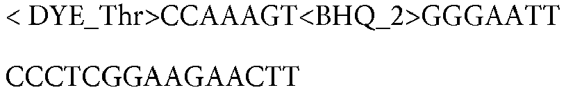
RET12P18_HEX 193 
RET12P13_HEX 194 
Table 6: Oligonucleotides for use in amplification and detection of ROS1 fusions Probe dye (for example) Forward primer SEQ ID NO Sequence JA270 CD74ex6F2 195 CACTGACGCTCCACCGAA CD74ex6F1 196 AAGCCCACTGACGCTCCA CD74ex6F3 197 ACTGACGCTCCACCGAAA SDC4ex2F1 198 GAGCTGTCTGGCTCTGG<t_BB_dA> SDC4ex2F2 199 
SDC4ex4F1 200 TTGAGAGAACGGAGGTCCT SDC4exF2 201 TGAGAGAACGGAGGTCCT SDC4ex4F3 202 TTGAGAGAACGGAGGTCCTG SLC34A2ex13F1 203 ATAACCATTAGCAGAGAGGCT SLC34A2ex13F2 204 AACCATTAGCAGAGAGGCTCA SLC34A2ex13F3 205 ATAACCATTAGCAGAGAGGCT SLC34A2ex4F1 206 AGTAGCGCCTTCCAGCT SLC34A2ex4F2 207 GCCTTCCAGCTGGTTGGA FZRex10F2 208 GAAGACAAAGAAGGCAGAGAGA LRIG3ex16F1 209 TTCTTACCACAACATGACAGTAGT LRIG3ex16F2 210 TCTTACCACAACATGACAGTAGTG TPM3ex8F1 211 
TPM3ex8F5 212 AAGCTGGAAAAGACAATTGATGAC Reverse primer ROSlex32Rl 213 GTATTGAATTTTTACTCCCTTCTAGTAATTTG ROSlex32R2 214 GTATTGAATTTTTACTCCCTTCTAGTAATTT ROSlex32R3 215 GTATTGAATTTTTACTCCCTTCTAGTAATT ROSlex35Rl 216 TATAAGCACTGTCACCCCTT ROSlex35R2 217 ATAAGCACTGTCACCCCTT ROSlex35R3 218 TATAAGCACTGTCACCCCT ROSlex35R4 219 CTTTGTCTTCGTTTATAAGCACTGTCA ROSlex35R5 220 AACTCTTTGTCTTCGTTTATAAGCACTGT ROSlex35R6 221 AGCCAACTCTTTGTCTTCGTTTATAAGCA ROSlex34LArevl 222 CAGTGGGATTGTAACAACCAGAAAT ROS1ex34LArev2 223 GTCAGTGGGATTGTAACAACCAGA ROS1ex34LArev3 224 GTCAGTGGGATTGTAACAACCA ROS1ex34LArev4 225 CAGTGGGATTGTAACAACCAGAAA ROS1ex34LArev5 226 CAGTGGGATTGTAACAACCAGAA Probe ROS1EX32P2 227 < DYE_Thr>TGGAGTCCCAAA<BHQ_2> TAAACCAGGCATTCCCA ROS1EX34P1 228 < DYE_Thr>TGATTTTTGGAT<BHQ_2> ACCAGAAACAAGTTTCATAC ROS1EX32P3 229 <DYE_Thr>TGGAGTC<BHQ_2> CCAAAT AAACCAGGC<t_BB_dA>TTCCCA ROS1EX34P3 230 <DYE_Thr>TGATTTT<BHQ_2> TGGATACCAGAAACAAGTTTCATAC ROS1EX35P1 51 < DYE_Thr>TCTGGCATAGAA<BHQ_2> GATTAAAGAATCAAAAAAGTGCCAAG - We have tested this method using RNA from EML4-ALK positive cell lines NCI-H2228 and EML4-ALK Fusion Variant 1 cell line from Horizon Discovery, CCDC6-RET cell line LC2AD, as well as from NSCLC formalin fixed paraffin embedded tissue (FFPET) and plasma specimens.
- In the case of plasma, we extracted cfRNA using the Roche High Pure FFPET RNA extraction kit with MagNA Pure Lysis Buffer and Esperase enzyme. Because the yield of cfRNA is too low to be measured accurately, we input a fixed volume (1/24 of total) of the extracted plasma cfRNA into the qRT-PCR.
- The reaction conditions were as follows. For each reaction, 25 uL of input RNA was added to a RT-PCR reaction mix comprising forward and reverse primers, labeled probe, buffer, dUTP, dTTP, dATP, dGTP, UNG, and Z05 enzyme to a final volume of 50uL. The reactions were run in multiplex, each with primers and probes specific for every fusion variant indicated in Table 1.
- Results were confirmed using a Next Generation Sequencing assay that detects the fusion variants covered in the qRT-PCR assay.
- Maximum Ct (threshold cycle) was set at 38, meaning that a signal must be detectable over background within a Ct of 38. Data is shown in
Figures 1A ,1B ,1C , and1D . The input RNA for each reaction was known to have the indicated fusion variant. Each reaction was repeated three times. -
Figure 1A shows that each ALK fusion variant is detectable at 50 copies.Figure 1B shows that each RET fusion variant is detectable at 50 copies.Figure 1C shows that each ROS1 fusion variant is detectable at 50 copies.Figure 1D shows that the reaction efficiency and input was equivalent, as indicated by the Internal Control Ct's. - We tested the multiplex qRT-PCR for the limit of detection of the ALK and RET fusion variants shown in Example 1, Table 1. We tested the multiplex assay by titering ALK or RET fusion positive transcripts into 0.1 ng Universal Human RNA (UHR) at 250, 100, 50, or 25 copies. The amplification and detection reactions were repeated 3 times.
- As shown in
Figure 2 , all of the ALK and RET fusion variants tested was detectable down to 25 copies. - Further studies were carried out to determine the linearity of detection for ALK, RET, and ROS1 fusions, as shown and described in
Figures 3-5 . - Sensitivity, or Limit of Detection (LOD) studies are shown and described in
Figures 6-8 . The LOD for each assay is shown in Tables 7 and 8. All 7 ALK, 6 RET, and 13 ROS1 fusion variants are detectable down to less than 10 copies. The predominant fusion variants are marked with an *.Table 7 Fusion Hit Rate LOD for 95% Probability E13:A20* 12/12 all levels tested <6.25 copies E20:A20* 12/12 all levels tested <6.25 copies E6:A20* 12/12 all levels tested <6.25 copies E2:A20 11/12 at 6.25 copies 6.45 copies K17:A20 12/12 at 6.25 copies (11/12 at 12.5 copies) 4.78 copies K24:A20 12/12 all levels tested <6.25 copies E18:A20 12/12 all levels tested <6.25 copies K15:R12* 11/12 at 6.25 copies 6.45 copies K16:R12* 11/12 at 6.25 copies 6.45 copies K22:R12 12/12 all levels tested <6.25 copies K23:R12 11/12 at 6.25 copies 6.45 copies C1:R12* 12/12 all levels tested <6.25 copies N6:R12 12/12 all levels tested <6.25 copies Table 8 Fusion variant Hit Rate LOD for 95% Probability C6:R32 12/12 all levels tested <6.25 copies C6:R34* 12/12 all levels tested <6.25 copies SD2:R32 11/12 at 6.25 and 12.5 copies 11.07 copies SD4:R34 12/12 all levels tested <6.25 copies SL13:R32 12/12 all levels tested <6.25 copies SL13:R34 12/12 all levels tested <6.25 copies SL4:R32 12/12 all levels tested <6.25 copies SL4:R34 11/12 at 6.25 copies 6.45 copies E10:R34* 11/12 at 6.25 copies 6.45 copies L16:R35 11/12 at 12.5 copies; 12/12 at 6.25 copies 4.78 copies T8:R35 12/12 all levels tested <6.25 copies SD2:R34 12/12 all levels tested <6.25 copies
Claims (21)
- A multiplex assay composition comprising:A. at least one primer set and probe that specifically amplify and detect at least one ALK fusion gene;B. at least one primer set and probe that specifically amplify and detect at least one RET fusion gene; andC. a primer set and probe that specifically amplify and detect an internal control.
- The composition of claim 1 further comprising
D. at least one primer set and probe that specifically amplify and detect at least one ROS1 fusion gene; - The composition of any one of claims 1 and 2, wherein the at least one ALK fusion gene is selected from the group consisting of: EML4 exon 13-ALK exon 20, EML4 exon 20-ALK exon 20, EML4 exon 6a/b-ALK exon 20, EML4 exon 2-ALK exon 20, EML4 exon 18-ALK exon 20, KIF5B exon 17-ALK exon 20, and KIF5B exon 24-ALK exon 20.
- The composition of any one of claims 1 to 3, wherein A includes at least one primer set and probe that amplify and detect more than 2 ALK fusion genes.
- The composition of any one of claims 1 to 4, wherein A includes at least one primer set and probe that amplify and detect EML4 exon 13-ALK exon 20, EML4 exon 20-ALK exon 20, EML4 exon 6a/b-ALK exon 20, EML4 exon 2-ALK exon 20, EML4 exon 18-ALK exon 20, KIF5B exon 17-ALK exon 20, and KIF5B exon 24-ALK exon 20.
- The composition of any one of claims 1 to 5, wherein the at least one RET fusion gene is selected from the group consisting of: KIF5B exon 15-RET exon 12, KIF5B exon 16-RET exon 12, KIF5B exon 22-RET exon 12, KIF5B exon 23-RET exon 12, CCDC6 exon 1-RET exon 12, and NCOA4 exon 6-RET exon 12.
- The composition of any one of claims 1 to 6, wherein B includes at least one primer set and probe that amplify and detect more than 2 RET fusion genes.
- The composition of any one of claims 1 to 7, wherein B includes at least one primer set and probe that amplify and detect KIF5B exon 15-RET exon 12, KIF5B exon 16-RET exon 12, KIF5B exon 22-RET exon 12, KIF5B exon 23-RET exon 12, CCDC6 exon 1-RET exon 12, and NCOA4 exon 6-RET exon 12.
- The composition of any one of claims 2 to 8, wherein the at least one ROS1 fusion gene is selected from the group consisting of: CD74 exon 6-ROS1 exon 34, CD74 exon 6-ROS1 exon 32, EZR exon 10-ROS1 exon 34, TPM3 exon 8-ROS1 exon 35, SDC4 exon 4-ROS1 exon 34, SDC4 exon 2-ROS1 exon 34, SDC4 exon 2-ROS1 exon 32, SDC4 exon 4-ROS1 exon 32, SLC34A2 exon 13-ROS1 exon 34, SLC34A2 exon 13-ROS1 exon 32v2, SLC34A2 exon 4-ROS1 exon 32, SLC34A2 exon 4-ROS1 exon 35, and LRIG3 exon 16-ROS1 exon 35.
- The composition of any one of claims 2 to 9, wherein D includes at least one primer set and probe that amplify and detect more than 2 ROS1 fusion genes.
- The composition of any one of claims 2 to 10, wherein D includes at least one primer set and probe that amplify and detect CD74 exon 6-ROS1 exon 34, CD74 exon 6-ROS1 exon 32, EZR exon 10-ROS1 exon 34, TPM3 exon 8-ROS1 exon 35, SDC4 exon 4-ROS1 exon 34, SDC4 exon 2-ROS1 exon 32v2, SDC4 exon 2-ROS1 exon 32, SLC34A2 exon 13-ROS1 exon 34, SLC34A2 exon 13-ROS1 exon 32v2, SLC34A2 exon 4-ROS1 exon 32, SLC34A2 exon 4-ROS1 exon 35, and LRIG3 exon 16-ROS1 exon 35.
- The composition of any one of claims 1 to 11, further comprising a thermostable DNA polymerase.
- The composition of any one of claims 1 to 12, further comprising reverse transcriptase.
- The composition of any one of claims 1 to 13, further comprising a biological sample from an individual.
- The composition of claim 14, wherein the biological sample includes RNA from plasma.
- A method of identifying an individual with cancer comprising:(a) contacting a biological sample from the individual with the composition of any one of claims 1-13;(b) carrying out amplification and detection under conditions that allow formation and detection of an amplification product in the presence of at least one fusion gene in the biological sample;(c) determining that at least one fusion gene is present if a fusion gene is detected in step (b);(d) whereby the presence of at least one fusion gene in said individual's sample indicates sensitivity of said individual to a kinase inhibitor therapy if at least one fusion gene is present.
- A method of determining the likelihood of response of an individual with cancer to kinase inhibitor therapy comprising:(a) contacting a biological sample from the individual with the composition of any one of claims 1-13;(b) carrying out amplification and detection under conditions that allow formation and detection of an amplification product in the presence of at least one fusion gene in the biological sample;(c) determining that at least one fusion gene is present if a fusion gene is detected in step (b); and(d) determining that the individual will likely respond to the kinase inhibitor therapy.
- A method for determining the presence of at least one fusion gene in a biological sample from an individual with cancer comprising:(a) contacting a biological sample from the individual with the composition of any one of claims 1-13;(b) carrying out amplification and detection under conditions that allow formation and detection of an amplification product in the presence of at least one fusion gene in the biological sample;(c) determining the presence of at least one fusion gene if a fusion gene is detected in step (b).
- The method of any one of claims 16-18, wherein the biological sample includes DNA or RNA.
- The method of any one of claims 16-18, wherein the biological sample is RNA from plasma of the individual.
- The method of any one of claims 16-18, wherein the amplification and detection are carried out using quantitative reverse transcription polymerase chain reaction (qRT-PCR).
Applications Claiming Priority (2)
| Application Number | Priority Date | Filing Date | Title |
|---|---|---|---|
| US201762513226P | 2017-05-31 | 2017-05-31 | |
| PCT/EP2018/064172 WO2018220004A1 (en) | 2017-05-31 | 2018-05-30 | Multiplex pcr detection of alk, ret, and ros fusions |
Publications (2)
| Publication Number | Publication Date |
|---|---|
| EP3631011A1 EP3631011A1 (en) | 2020-04-08 |
| EP3631011B1 true EP3631011B1 (en) | 2021-06-23 |
Family
ID=62567625
Family Applications (1)
| Application Number | Title | Priority Date | Filing Date |
|---|---|---|---|
| EP18730279.9A Active EP3631011B1 (en) | 2017-05-31 | 2018-05-30 | Multiplex pcr detection of alk, ret, and ros fusions |
Country Status (8)
| Country | Link |
|---|---|
| US (2) | US11505830B2 (en) |
| EP (1) | EP3631011B1 (en) |
| JP (2) | JP2020521440A (en) |
| CN (1) | CN110691852A (en) |
| AU (1) | AU2018277210B2 (en) |
| CA (1) | CA3059119C (en) |
| ES (1) | ES2883934T3 (en) |
| WO (1) | WO2018220004A1 (en) |
Families Citing this family (5)
| Publication number | Priority date | Publication date | Assignee | Title |
|---|---|---|---|---|
| CA3059119C (en) | 2017-05-31 | 2024-01-23 | F. Hoffman-La Roche Ag | Multiplex pcr detection of alk, ret, and ros fusions |
| CN109837275B (en) * | 2019-01-08 | 2022-09-06 | 大连医科大学附属第二医院 | Preparation method of fusion gene positive control standard substance |
| CN111718994A (en) * | 2019-03-22 | 2020-09-29 | 广州金域医学检验中心有限公司 | Tumor fusion gene targeted detection probe, kit and application |
| CN110964830A (en) * | 2019-12-31 | 2020-04-07 | 中山大学达安基因股份有限公司 | Kit and method for multiple detection of ROS1 gene mutation |
| CN117144016A (en) * | 2023-10-27 | 2023-12-01 | 神州医疗科技股份有限公司 | Primer probe, kit and detection system for detecting RET fusion gene in peripheral blood circulation tumor RNA |
Citations (10)
| Publication number | Priority date | Publication date | Assignee | Title |
|---|---|---|---|---|
| WO2004099434A2 (en) | 2003-04-30 | 2004-11-18 | The Public Health Research Institute Of The City Of New York, Inc. | Homogeneous multiplex screening assays and kits |
| US20130102006A1 (en) | 2010-06-22 | 2013-04-25 | Astellas Pharma Inc. | Detection method for novel ros1 fusions |
| WO2014144121A2 (en) | 2013-03-15 | 2014-09-18 | Life Technologies Corporation | Classification and actionability indices for lung cancer |
| WO2014150300A2 (en) | 2013-03-15 | 2014-09-25 | Abbott Molecular Inc. | Method for amplification and assay of rna fusion gene variants, method of distinguishing same and related primers, probes, and kits |
| CN104818320A (en) | 2014-12-03 | 2015-08-05 | 厦门艾德生物医药科技有限公司 | Primers, probes, detection system and kit for one time detection of lung cancer multiple genes |
| WO2015149034A2 (en) | 2014-03-27 | 2015-10-01 | Life Technologies Corporation | Gene fusions and gene variants associated with cancer |
| WO2015148494A1 (en) | 2014-03-25 | 2015-10-01 | Quest Diagnostics Investments Incorporated | Detection of gene fusions by intragenic differential expression (ide) using average cycle thresholds |
| US20150315657A1 (en) | 2013-04-17 | 2015-11-05 | Life Technologies Corporation | Gene fusions and gene variants associated with cancer |
| WO2016166269A1 (en) | 2015-04-17 | 2016-10-20 | Roche Diagnostics Gmbh | Multiplex pcr to detect gene fusions |
| US20180346994A1 (en) | 2017-05-31 | 2018-12-06 | Roche Molecular Systems, Inc. | Multiplex pcr detection of alk, ret, and ros fusions |
Family Cites Families (10)
| Publication number | Priority date | Publication date | Assignee | Title |
|---|---|---|---|---|
| US5455170A (en) | 1986-08-22 | 1995-10-03 | Hoffmann-La Roche Inc. | Mutated thermostable nucleic acid polymerase enzyme from Thermus species Z05 |
| CA2371818C (en) * | 1999-02-18 | 2009-01-06 | The Regents Of The University Of California | Salicylamide-lanthanide complexes for use as luminescent markers |
| WO2001077390A2 (en) * | 2000-04-05 | 2001-10-18 | Molecular Staging, Inc. | Process for allele discrimation utilizing primer extension |
| PT2450437T (en) * | 2006-04-14 | 2017-08-25 | Cell Signaling Technology Inc | Gene defects and mutant alk kinase in human solid tumors |
| CA2982018C (en) * | 2006-04-14 | 2023-10-17 | Cell Signaling Technology, Inc. | Gene defects and mutant alk kinase in human solid tumors |
| EP2775300A3 (en) * | 2009-08-28 | 2015-04-01 | Asuragen, INC. | miRNA Biomarkers of Lung Disease |
| EP2599878A4 (en) * | 2010-07-26 | 2014-02-12 | Astellas Pharma Inc | Detection method of novel ret fusion |
| US20130059762A1 (en) * | 2011-04-28 | 2013-03-07 | Life Technologies Corporation | Methods and compositions for multiplex pcr |
| US20130237442A1 (en) * | 2012-03-07 | 2013-09-12 | University Of Miami | Methods and Compositions for Diagnosis of Non-Viable Early Pregnancy |
| CA2946416C (en) * | 2014-04-25 | 2022-07-19 | Exelixis, Inc. | Method of treating lung adenocarcinoma |
-
2018
- 2018-05-30 CA CA3059119A patent/CA3059119C/en active Active
- 2018-05-30 EP EP18730279.9A patent/EP3631011B1/en active Active
- 2018-05-30 ES ES18730279T patent/ES2883934T3/en active Active
- 2018-05-30 CN CN201880035309.0A patent/CN110691852A/en active Pending
- 2018-05-30 AU AU2018277210A patent/AU2018277210B2/en active Active
- 2018-05-30 US US15/993,375 patent/US11505830B2/en active Active
- 2018-05-30 WO PCT/EP2018/064172 patent/WO2018220004A1/en active Application Filing
- 2018-05-30 JP JP2019559727A patent/JP2020521440A/en active Pending
-
2022
- 2022-10-06 US US17/961,200 patent/US20230121541A1/en active Pending
-
2023
- 2023-12-07 JP JP2023206878A patent/JP2024019433A/en active Pending
Patent Citations (13)
| Publication number | Priority date | Publication date | Assignee | Title |
|---|---|---|---|---|
| WO2004099434A2 (en) | 2003-04-30 | 2004-11-18 | The Public Health Research Institute Of The City Of New York, Inc. | Homogeneous multiplex screening assays and kits |
| US20130102006A1 (en) | 2010-06-22 | 2013-04-25 | Astellas Pharma Inc. | Detection method for novel ros1 fusions |
| EP2971093B1 (en) | 2013-03-15 | 2018-09-19 | Life Technologies Corporation | Classification and actionability indices for lung cancer |
| WO2014144121A2 (en) | 2013-03-15 | 2014-09-18 | Life Technologies Corporation | Classification and actionability indices for lung cancer |
| US20140288116A1 (en) | 2013-03-15 | 2014-09-25 | Life Technologies Corporation | Classification and Actionability Indices for Lung Cancer |
| WO2014150300A2 (en) | 2013-03-15 | 2014-09-25 | Abbott Molecular Inc. | Method for amplification and assay of rna fusion gene variants, method of distinguishing same and related primers, probes, and kits |
| US20150315657A1 (en) | 2013-04-17 | 2015-11-05 | Life Technologies Corporation | Gene fusions and gene variants associated with cancer |
| EP3123380B1 (en) | 2014-03-25 | 2020-11-04 | Quest Diagnostics Investments Incorporated | Detection of gene fusions by intragenic differential expression (ide) using average cycle thresholds |
| WO2015148494A1 (en) | 2014-03-25 | 2015-10-01 | Quest Diagnostics Investments Incorporated | Detection of gene fusions by intragenic differential expression (ide) using average cycle thresholds |
| WO2015149034A2 (en) | 2014-03-27 | 2015-10-01 | Life Technologies Corporation | Gene fusions and gene variants associated with cancer |
| CN104818320A (en) | 2014-12-03 | 2015-08-05 | 厦门艾德生物医药科技有限公司 | Primers, probes, detection system and kit for one time detection of lung cancer multiple genes |
| WO2016166269A1 (en) | 2015-04-17 | 2016-10-20 | Roche Diagnostics Gmbh | Multiplex pcr to detect gene fusions |
| US20180346994A1 (en) | 2017-05-31 | 2018-12-06 | Roche Molecular Systems, Inc. | Multiplex pcr detection of alk, ret, and ros fusions |
Non-Patent Citations (28)
| Title |
|---|
| ANONYMOUS: "AmoyDx EML4-ALK Fusion gene detection kit", AMOY DIAGNOSTICS - INSTRUCTION FOR USE, 1 July 2014 (2014-07-01), pages 1 - 8, XP055909180, [retrieved on 20220405] |
| ANONYMOUS: "AmoyDx RET gene fusions detection kit", AMOY DIAGNOSTICS - INSTRUCTION FOR USE, 1 July 2015 (2015-07-01), pages 1 - 6, XP055909173, [retrieved on 20220405] |
| ANONYMOUS: "cobas@ EGFR Mutation Test v2", ROCHE MOLECULAR SYSTEMS, 1 January 2015 (2015-01-01), XP055909197, Retrieved from the Internet <URL:https://www.accessdata.fda.gov/cdrh_docs/pdf12/P120019S007c.pdf> [retrieved on 20220405] |
| ANONYMOUS: "Critical Factors for Successful Real-Time PCR", REAL-TIME PCR BROCHURE 07/2010, QIAGEN, XP055749716, Retrieved from the Internet <URL:https://www.gene-quantification.de/qiagen-qpcr-sample-assay-tech-guide-2010.pdf> [retrieved on 20201112] |
| ANONYMOUS: "The Archer™ Solution. A New Frontier in Fusion Detection", ENZYMATICS - BROCHURE, 1 January 2014 (2014-01-01), pages 1 - 4, XP055909247, Retrieved from the Internet <URL:http://www.enzymatics.com/wp-content/uploads/2014/01/Archer_Brochure_2014.pdf> [retrieved on 20220405] |
| BEYOND: "Beyond ALK-RET, ROS1 and other oncogene fusions in lung cancer", TRANSL LUNG CANCER RES, vol. 4, no. 2, 1 January 2015 (2015-01-01), pages 156 - 164, XP055240876, Retrieved from the Internet <URL:http://www.ncbi.nlm.nih.gov/pmc/articles/PMC4384213/pdf/tlcr-04-02-156.pdf> DOI: 10.3978/j.issn.2218-6751.2014.11.11 |
| BLACK ET AL.: "NSCLC: An update of driver mutations, their role in pathogenesis and clinical significance", R I MED J, vol. 98, no. 10, 1 October 2015 (2015-10-01), pages 25 - 8, XP055883015 |
| BUBENDORF L ET AL.: "Testing for ROS1 in non-small cell lung cancer: a review with recommendations", VIRCHOWS ARCH, vol. 469, no. 5, November 2016 (2016-11-01), pages 489 - 503, XP036084887, DOI: 10.1007/s00428-016-2000-3 |
| CAMIDGE D ROSS, BANG YUNG-JUE, KWAK EUNICE L, IAFRATE A JOHN, VARELLA-GARCIA MARILEILA, FOX STEPHEN B, RIELY GREGORY J, SOLOMON BE: "Activity and safety of crizotinib in patients with ALK-positive non-small-cell lung cancer: updated results from a phase 1 study", THE LANCET ONCOLOGY, ELSEVIER, AMSTERDAM, NL, vol. 13, no. 10, 1 October 2012 (2012-10-01), AMSTERDAM, NL , pages 1011 - 1019, XP055909189, ISSN: 1470-2045, DOI: 10.1016/S1470-2045(12)70344-3 |
| DERVEAUX S ET AL.: "How to do successful gene expression analysis using real-time PCR", METHODS, vol. 50, no. 4, April 2010 (2010-04-01), pages 227 - 30, XP026941713, DOI: 10.1016/j.ymeth.2009.11.001 |
| DRILON A ET AL.: "Response to Cabozantinib in patients with RET fusion-positive lung adenocarcinomas", CANCER DISCOV, vol. 3, no. 6, June 2013 (2013-06-01), pages 630 - 5, XP055195155, DOI: 10.1158/2159-8290.CD-13-0035 |
| HITOSHI ICHIKAWA: "Clinical sequencing using an original cancer gene panel in National Cancer Center", RESEARCH & CLINICAL TRIAL CENTER, vol. 249, no. 10, 7 June 2014 (2014-06-07), pages 1139 - 1144, XP055909222 |
| INOUE YOSHIAKI, SHIIHARA JUN, MIYAZAWA HITOSHI, OHTA HIROMITSU, HIGO MEGUMI, NAGAI YOSHIAKI, KOBAYASHI KUNIHIKO, SAIJO YASUO, TSUC: "A highly specific and sensitive massive parallel sequencer-based test for somatic mutations in non-small cell lung cancer", PLOS ONE, vol. 12, no. 4, 27 April 2017 (2017-04-27), pages e0176525, XP055909235, DOI: 10.1371/journal.pone.0176525 |
| KHOO C ET AL.: "Molecular methods for somatic mutation testing in lung adenocarcinoma: EGFR and beyond. 2015", TRANSL LUNG CANCER RES, vol. 4, no. 2, pages 126 - 141, XP055799240, DOI: 10.3978/j.issn.2218-6751.2015.01.10 |
| KWAK EL ET AL.: "Anaplastic lymphoma kinase inhibition in non-small- cell lung cancer", N ENGL J MED, vol. 363, no. 18, 28 October 2010 (2010-10-28), pages 1693 - 703, XP055081495, DOI: 10.1056/NEJMoa1006448 |
| LI C ET AL.: "Spectrum of oncogenic driver mutations in lung adenocarcinomas from East Asian never smokers", PLOS ONE, vol. 6, no. 11, 2011, pages e28204, XP055209333, DOI: 10.1371/journal.pone.0028204 |
| LIRA ME ET AL.: "A Single-Tube Multiplexed Assay for Detecting ALK, ROS1, and RET Fusions in Lung Cancer. 2014", THE JOURNAL OF MOLECULAR DIAGNOSTICS, vol. 16, no. 2, pages 229 - 243, XP055280268, DOI: 10.1016/j.jmoldx.2013.11.007 |
| MOTONOBU SAITO: "Gene aberrations for precision medicine against lung adenocarcinoma", CANCER SCIENCE, JAPANESE CANCER ASSOCIATION, TOKYO, JP, vol. 107, no. 6, 1 June 2016 (2016-06-01), JP , pages 713 - 720, XP093155691, ISSN: 1347-9032, DOI: 10.1111/cas.12941 |
| OKAMOTO I ET AL.: "Multiplex genomic profiling of non-small cell lung cancers from the LETS phase III trial of first-line S-1/carboplatin versus paclitaxel/carboplatin: results of a West Japan Oncology Group study", ONCOTARGET, vol. 5, no. 8, 30 April 2014 (2014-04-30), pages 2293 - 304, XP055498814, DOI: 10.18632/oncotarget.1906 |
| PETER M ET AL.: "A multiplex real-time per assay for the detection of gene fusions observed in solid tumors", LAB INVEST, vol. 81, no. 6, June 2001 (2001-06-01), pages 905 - 12, XP002985561 |
| REGUART NOEMÍ, TEIXIDÓ CRISTINA, GIMÉNEZ-CAPITÁN ANA, PARÉ LAIA, GALVÁN PATRICIA, VITERI SANTIAGO, RODRÍGUEZ SONIA, PEG VICENTE, A: "Identification of ALK, ROS1, and RET Fusions by a Multiplexed mRNA-Based Assay in Formalin-Fixed, Paraffin-Embedded Samples from Advanced Non–Small-Cell Lung Cancer Patients", CLINICAL CHEMISTRY, OXFORD UNIVERSITY PRESS, US, vol. 63, no. 3, 1 March 2017 (2017-03-01), US , pages 751 - 760, XP055909237, ISSN: 0009-9147, DOI: 10.1373/clinchem.2016.265314 |
| SEQUIST L.V., HEIST R.S., SHAW A.T., FIDIAS P., ROSOVSKY R., TEMEL J.S., LENNES I.T., DIGUMARTHY S., WALTMAN B.A., BAST E., TAMMIR: "Implementing multiplexed genotyping of non-small-cell lung cancers into routine clinical practice", ANNALS OF ONCOLOGY, KLUWER DORDRECHT, NL, vol. 22, no. 12, 1 December 2011 (2011-12-01), NL , pages 2616 - 2624, XP055909186, ISSN: 0923-7534, DOI: 10.1093/annonc/mdr489 |
| SUNAMI KUNIKO, FURUTA KOH, TSUTA KOJI, SASADA SHINJI, IZUMO TAKEHIRO, NAKAOKU TAKASHI, SHIMADA YOKO, SAITO MOTONOBU, NOKIHARA HIRO: "Multiplex Diagnosis of Oncogenic Fusion and MET Exon Skipping by Molecular Counting Using Formalin-Fixed Paraffin Embedded Lung Adenocarcinoma Tissues", JOURNAL OF THORACIC ONCOLOGY, ELSEVIER INC., US, vol. 11, no. 2, 1 February 2016 (2016-02-01), US , pages 203 - 212, XP055909234, ISSN: 1556-0864, DOI: 10.1016/j.jtho.2015.10.005 |
| TAKEUCHI K ET AL.: "RET, ROS1 and ALK fusions in lung cance r", NAT MED, vol. 18, no. 3, 12 February 2012 (2012-02-12), pages 378 - 81, XP037532160, DOI: 10.1038/nm.2658 |
| TONI-MAREE ROGERS, GISELA MIR ARNAU, GEORGINA L. RYLAND, STEPHEN HUANG, MARUJA E. LIRA, YVETTE EMMANUEL, OMAR D. PEREZ, DARRYL IRW: "Multiplexed transcriptome analysis to detect ALK, ROS1 and RET rearrangements in lung cancer", SCIENTIFIC REPORTS, vol. 7, no. 1, 1 December 2017 (2017-12-01), XP055498617, DOI: 10.1038/srep42259 |
| VANGUILDER H D; VRANA K E; FREEMAN W M: "Twenty-five years of quantitative PCR for gene expression analysis", BIOTECHNIQUES, INFORMA HEALTHCARE, US, vol. 44, no. 5, 1 April 2008 (2008-04-01), US , pages 619 - 626, XP001537018, ISSN: 0736-6205, DOI: 10.2144/000112776 |
| ZHAO C ET AL.: "Detecting ALK, ROS1 and RET Fusion Genes in Cell Block Samples", TRANSL ONCOL, vol. 7, no. 3, 17 June 2014 (2014-06-17), pages 363 - 7, XP055498635, DOI: 10.1016/j.tranon.2014.04.013 |
| ZHENG LI-MOU, WHYTE DAVID B., RUAN LI, SONG ROMAN, FEI LUO, YANG NANCY, ZHOU CAICUN: "Screening of NSCLC samples from Chinese lung cancer patients for activating rearrangements of the ALK, RET , and ROS1 genes.", JOURNAL OF CLINICAL ONCOLOGY, AMERICAN SOCIETY OF CLINICAL ONCOLOGY, US, vol. 31, no. 15_suppl, 20 May 2013 (2013-05-20), US , pages e22066 - e22066, XP055909193, ISSN: 0732-183X, DOI: 10.1200/jco.2013.31.15_suppl.e22066 |
Also Published As
| Publication number | Publication date |
|---|---|
| US20230121541A1 (en) | 2023-04-20 |
| JP2020521440A (en) | 2020-07-27 |
| AU2018277210A1 (en) | 2019-10-17 |
| US20180346994A1 (en) | 2018-12-06 |
| EP3631011A1 (en) | 2020-04-08 |
| WO2018220004A1 (en) | 2018-12-06 |
| AU2018277210B2 (en) | 2024-03-28 |
| CN110691852A (en) | 2020-01-14 |
| CA3059119C (en) | 2024-01-23 |
| CA3059119A1 (en) | 2018-12-06 |
| US11505830B2 (en) | 2022-11-22 |
| JP2024019433A (en) | 2024-02-09 |
| ES2883934T3 (en) | 2021-12-09 |
Similar Documents
| Publication | Publication Date | Title |
|---|---|---|
| CA2982870C (en) | Multiplex pcr to detect gene fusions | |
| EP3631011B1 (en) | Multiplex pcr detection of alk, ret, and ros fusions | |
| EP3755813B1 (en) | Improved detection of microsatellite instability | |
| CN101815789A (en) | The enrichment of target sequence | |
| CA2569502A1 (en) | Diagnosing or predicting the course of breast cancer | |
| Wang et al. | DNA degradation test predicts success in whole-genome amplification from diverse clinical samples | |
| EP2523965A1 (en) | Oligonucleotides and methods for detecting kras and pik3ca mutations | |
| US20200277651A1 (en) | Nucleic Acid Preparation and Analysis | |
| EP2198057B1 (en) | Detection of atypical pneumonia | |
| US20160097103A1 (en) | Primer reagent for amplifying ALK fusion gene, ALK fusion gene amplification reagent kit including the same, and ALK fusion gene amplification method and ALK fusion gene analysis method using the same | |
| WO2011059895A1 (en) | Hexa mutations | |
| US20210254172A1 (en) | Methods of detecting mlh1 methylation | |
| WO2009018225A2 (en) | Internal positive control for nucleic acid assays | |
| CN104395468A (en) | Method and kit for detecting hla-a*24:02 | |
| US20120252024A1 (en) | Probe for Detection of Polymorphism in C-Kit Gene and Use Thereof | |
| KR20230012467A (en) | Looped primers and loop-de-loop methods for detecting target nucleic acids | |
| US20190078136A1 (en) | Multiplex Detection of Short Nucleic Acids | |
| WO2008033776A1 (en) | Probes and methods for detecting gleevec-resistant bcr-abl mutations | |
| EP2496710B1 (en) | Determination of 17q gain in neuroblastoma patients by analysis of circulating dna | |
| JP6533128B2 (en) | Primer reagent for amplifying ALK fusion gene, amplification reagent kit for ALK fusion gene containing the same, amplification method and analysis method of ALK fusion gene using it |
Legal Events
| Date | Code | Title | Description |
|---|---|---|---|
| STAA | Information on the status of an ep patent application or granted ep patent |
Free format text: STATUS: UNKNOWN |
|
| STAA | Information on the status of an ep patent application or granted ep patent |
Free format text: STATUS: THE INTERNATIONAL PUBLICATION HAS BEEN MADE |
|
| PUAI | Public reference made under article 153(3) epc to a published international application that has entered the european phase |
Free format text: ORIGINAL CODE: 0009012 |
|
| STAA | Information on the status of an ep patent application or granted ep patent |
Free format text: STATUS: REQUEST FOR EXAMINATION WAS MADE |
|
| 17P | Request for examination filed |
Effective date: 20200102 |
|
| AK | Designated contracting states |
Kind code of ref document: A1 Designated state(s): AL AT BE BG CH CY CZ DE DK EE ES FI FR GB GR HR HU IE IS IT LI LT LU LV MC MK MT NL NO PL PT RO RS SE SI SK SM TR |
|
| AX | Request for extension of the european patent |
Extension state: BA ME |
|
| DAV | Request for validation of the european patent (deleted) | ||
| DAX | Request for extension of the european patent (deleted) | ||
| GRAP | Despatch of communication of intention to grant a patent |
Free format text: ORIGINAL CODE: EPIDOSNIGR1 |
|
| STAA | Information on the status of an ep patent application or granted ep patent |
Free format text: STATUS: GRANT OF PATENT IS INTENDED |
|
| INTG | Intention to grant announced |
Effective date: 20210122 |
|
| TPAC | Observations filed by third parties |
Free format text: ORIGINAL CODE: EPIDOSNTIPA |
|
| GRAS | Grant fee paid |
Free format text: ORIGINAL CODE: EPIDOSNIGR3 |
|
| GRAA | (expected) grant |
Free format text: ORIGINAL CODE: 0009210 |
|
| STAA | Information on the status of an ep patent application or granted ep patent |
Free format text: STATUS: THE PATENT HAS BEEN GRANTED |
|
| AK | Designated contracting states |
Kind code of ref document: B1 Designated state(s): AL AT BE BG CH CY CZ DE DK EE ES FI FR GB GR HR HU IE IS IT LI LT LU LV MC MK MT NL NO PL PT RO RS SE SI SK SM TR |
|
| REG | Reference to a national code |
Ref country code: GB Ref legal event code: FG4D |
|
| REG | Reference to a national code |
Ref country code: CH Ref legal event code: EP |
|
| REG | Reference to a national code |
Ref country code: DE Ref legal event code: R096 Ref document number: 602018019028 Country of ref document: DE Ref country code: AT Ref legal event code: REF Ref document number: 1404361 Country of ref document: AT Kind code of ref document: T Effective date: 20210715 |
|
| REG | Reference to a national code |
Ref country code: IE Ref legal event code: FG4D |
|
| REG | Reference to a national code |
Ref country code: LT Ref legal event code: MG9D |
|
| PG25 | Lapsed in a contracting state [announced via postgrant information from national office to epo] |
Ref country code: FI Free format text: LAPSE BECAUSE OF FAILURE TO SUBMIT A TRANSLATION OF THE DESCRIPTION OR TO PAY THE FEE WITHIN THE PRESCRIBED TIME-LIMIT Effective date: 20210623 Ref country code: LT Free format text: LAPSE BECAUSE OF FAILURE TO SUBMIT A TRANSLATION OF THE DESCRIPTION OR TO PAY THE FEE WITHIN THE PRESCRIBED TIME-LIMIT Effective date: 20210623 Ref country code: HR Free format text: LAPSE BECAUSE OF FAILURE TO SUBMIT A TRANSLATION OF THE DESCRIPTION OR TO PAY THE FEE WITHIN THE PRESCRIBED TIME-LIMIT Effective date: 20210623 Ref country code: BG Free format text: LAPSE BECAUSE OF FAILURE TO SUBMIT A TRANSLATION OF THE DESCRIPTION OR TO PAY THE FEE WITHIN THE PRESCRIBED TIME-LIMIT Effective date: 20210923 |
|
| REG | Reference to a national code |
Ref country code: AT Ref legal event code: MK05 Ref document number: 1404361 Country of ref document: AT Kind code of ref document: T Effective date: 20210623 |
|
| PG25 | Lapsed in a contracting state [announced via postgrant information from national office to epo] |
Ref country code: SE Free format text: LAPSE BECAUSE OF FAILURE TO SUBMIT A TRANSLATION OF THE DESCRIPTION OR TO PAY THE FEE WITHIN THE PRESCRIBED TIME-LIMIT Effective date: 20210623 Ref country code: RS Free format text: LAPSE BECAUSE OF FAILURE TO SUBMIT A TRANSLATION OF THE DESCRIPTION OR TO PAY THE FEE WITHIN THE PRESCRIBED TIME-LIMIT Effective date: 20210623 Ref country code: LV Free format text: LAPSE BECAUSE OF FAILURE TO SUBMIT A TRANSLATION OF THE DESCRIPTION OR TO PAY THE FEE WITHIN THE PRESCRIBED TIME-LIMIT Effective date: 20210623 Ref country code: NO Free format text: LAPSE BECAUSE OF FAILURE TO SUBMIT A TRANSLATION OF THE DESCRIPTION OR TO PAY THE FEE WITHIN THE PRESCRIBED TIME-LIMIT Effective date: 20210923 Ref country code: GR Free format text: LAPSE BECAUSE OF FAILURE TO SUBMIT A TRANSLATION OF THE DESCRIPTION OR TO PAY THE FEE WITHIN THE PRESCRIBED TIME-LIMIT Effective date: 20210924 |
|
| REG | Reference to a national code |
Ref country code: NL Ref legal event code: MP Effective date: 20210623 |
|
| REG | Reference to a national code |
Ref country code: ES Ref legal event code: FG2A Ref document number: 2883934 Country of ref document: ES Kind code of ref document: T3 Effective date: 20211209 |
|
| PG25 | Lapsed in a contracting state [announced via postgrant information from national office to epo] |
Ref country code: EE Free format text: LAPSE BECAUSE OF FAILURE TO SUBMIT A TRANSLATION OF THE DESCRIPTION OR TO PAY THE FEE WITHIN THE PRESCRIBED TIME-LIMIT Effective date: 20210623 Ref country code: SK Free format text: LAPSE BECAUSE OF FAILURE TO SUBMIT A TRANSLATION OF THE DESCRIPTION OR TO PAY THE FEE WITHIN THE PRESCRIBED TIME-LIMIT Effective date: 20210623 Ref country code: AT Free format text: LAPSE BECAUSE OF FAILURE TO SUBMIT A TRANSLATION OF THE DESCRIPTION OR TO PAY THE FEE WITHIN THE PRESCRIBED TIME-LIMIT Effective date: 20210623 Ref country code: CZ Free format text: LAPSE BECAUSE OF FAILURE TO SUBMIT A TRANSLATION OF THE DESCRIPTION OR TO PAY THE FEE WITHIN THE PRESCRIBED TIME-LIMIT Effective date: 20210623 Ref country code: NL Free format text: LAPSE BECAUSE OF FAILURE TO SUBMIT A TRANSLATION OF THE DESCRIPTION OR TO PAY THE FEE WITHIN THE PRESCRIBED TIME-LIMIT Effective date: 20210623 Ref country code: RO Free format text: LAPSE BECAUSE OF FAILURE TO SUBMIT A TRANSLATION OF THE DESCRIPTION OR TO PAY THE FEE WITHIN THE PRESCRIBED TIME-LIMIT Effective date: 20210623 Ref country code: PT Free format text: LAPSE BECAUSE OF FAILURE TO SUBMIT A TRANSLATION OF THE DESCRIPTION OR TO PAY THE FEE WITHIN THE PRESCRIBED TIME-LIMIT Effective date: 20211025 Ref country code: SM Free format text: LAPSE BECAUSE OF FAILURE TO SUBMIT A TRANSLATION OF THE DESCRIPTION OR TO PAY THE FEE WITHIN THE PRESCRIBED TIME-LIMIT Effective date: 20210623 |
|
| PG25 | Lapsed in a contracting state [announced via postgrant information from national office to epo] |
Ref country code: PL Free format text: LAPSE BECAUSE OF FAILURE TO SUBMIT A TRANSLATION OF THE DESCRIPTION OR TO PAY THE FEE WITHIN THE PRESCRIBED TIME-LIMIT Effective date: 20210623 |
|
| REG | Reference to a national code |
Ref country code: DE Ref legal event code: R026 Ref document number: 602018019028 Country of ref document: DE |
|
| PLBI | Opposition filed |
Free format text: ORIGINAL CODE: 0009260 |
|
| PLAX | Notice of opposition and request to file observation + time limit sent |
Free format text: ORIGINAL CODE: EPIDOSNOBS2 |
|
| PLAN | Information deleted related to communication of a notice of opposition and request to file observations + time limit |
Free format text: ORIGINAL CODE: EPIDOSDOBS2 |
|
| PLAX | Notice of opposition and request to file observation + time limit sent |
Free format text: ORIGINAL CODE: EPIDOSNOBS2 |
|
| 26 | Opposition filed |
Opponent name: STRAWMAN LIMITED Effective date: 20220322 |
|
| PG25 | Lapsed in a contracting state [announced via postgrant information from national office to epo] |
Ref country code: DK Free format text: LAPSE BECAUSE OF FAILURE TO SUBMIT A TRANSLATION OF THE DESCRIPTION OR TO PAY THE FEE WITHIN THE PRESCRIBED TIME-LIMIT Effective date: 20210623 |
|
| PG25 | Lapsed in a contracting state [announced via postgrant information from national office to epo] |
Ref country code: AL Free format text: LAPSE BECAUSE OF FAILURE TO SUBMIT A TRANSLATION OF THE DESCRIPTION OR TO PAY THE FEE WITHIN THE PRESCRIBED TIME-LIMIT Effective date: 20210623 |
|
| PLBB | Reply of patent proprietor to notice(s) of opposition received |
Free format text: ORIGINAL CODE: EPIDOSNOBS3 |
|
| REG | Reference to a national code |
Ref country code: BE Ref legal event code: MM Effective date: 20220531 |
|
| PG25 | Lapsed in a contracting state [announced via postgrant information from national office to epo] |
Ref country code: MC Free format text: LAPSE BECAUSE OF FAILURE TO SUBMIT A TRANSLATION OF THE DESCRIPTION OR TO PAY THE FEE WITHIN THE PRESCRIBED TIME-LIMIT Effective date: 20210623 Ref country code: LU Free format text: LAPSE BECAUSE OF NON-PAYMENT OF DUE FEES Effective date: 20220530 |
|
| PG25 | Lapsed in a contracting state [announced via postgrant information from national office to epo] |
Ref country code: IE Free format text: LAPSE BECAUSE OF NON-PAYMENT OF DUE FEES Effective date: 20220530 |
|
| PG25 | Lapsed in a contracting state [announced via postgrant information from national office to epo] |
Ref country code: BE Free format text: LAPSE BECAUSE OF NON-PAYMENT OF DUE FEES Effective date: 20220531 |
|
| PGFP | Annual fee paid to national office [announced via postgrant information from national office to epo] |
Ref country code: IT Payment date: 20230515 Year of fee payment: 6 Ref country code: FR Payment date: 20230412 Year of fee payment: 6 Ref country code: ES Payment date: 20230608 Year of fee payment: 6 Ref country code: DE Payment date: 20230412 Year of fee payment: 6 Ref country code: CH Payment date: 20230602 Year of fee payment: 6 |
|
| PGFP | Annual fee paid to national office [announced via postgrant information from national office to epo] |
Ref country code: GB Payment date: 20230412 Year of fee payment: 6 |
|
| PLCK | Communication despatched that opposition was rejected |
Free format text: ORIGINAL CODE: EPIDOSNREJ1 |
|
| APAJ | Date of receipt of notice of appeal modified |
Free format text: ORIGINAL CODE: EPIDOSCNOA2O |
|
| APBM | Appeal reference recorded |
Free format text: ORIGINAL CODE: EPIDOSNREFNO |
|
| APBP | Date of receipt of notice of appeal recorded |
Free format text: ORIGINAL CODE: EPIDOSNNOA2O |
|
| APAH | Appeal reference modified |
Free format text: ORIGINAL CODE: EPIDOSCREFNO |