EP3621694B1 - Lrrc33 inhibitors and use thereof - Google Patents
Lrrc33 inhibitors and use thereof Download PDFInfo
- Publication number
- EP3621694B1 EP3621694B1 EP18727561.5A EP18727561A EP3621694B1 EP 3621694 B1 EP3621694 B1 EP 3621694B1 EP 18727561 A EP18727561 A EP 18727561A EP 3621694 B1 EP3621694 B1 EP 3621694B1
- Authority
- EP
- European Patent Office
- Prior art keywords
- lrrc33
- cells
- cell
- inhibitor
- antibody
- Prior art date
- Legal status (The legal status is an assumption and is not a legal conclusion. Google has not performed a legal analysis and makes no representation as to the accuracy of the status listed.)
- Active
Links
- 239000003112 inhibitor Substances 0.000 title claims description 163
- 101150079786 NRROS gene Proteins 0.000 title 1
- 210000004027 cell Anatomy 0.000 claims description 483
- 101001004913 Homo sapiens Transforming growth factor beta activator LRRC33 Proteins 0.000 claims description 443
- 102100025954 Transforming growth factor beta activator LRRC33 Human genes 0.000 claims description 441
- 206010028980 Neoplasm Diseases 0.000 claims description 178
- 208000037265 diseases, disorders, signs and symptoms Diseases 0.000 claims description 162
- 102000004887 Transforming Growth Factor beta Human genes 0.000 claims description 115
- 108090001012 Transforming Growth Factor beta Proteins 0.000 claims description 115
- 210000002540 macrophage Anatomy 0.000 claims description 110
- 201000010099 disease Diseases 0.000 claims description 99
- 208000031261 Acute myeloid leukaemia Diseases 0.000 claims description 91
- 238000000034 method Methods 0.000 claims description 68
- 238000002560 therapeutic procedure Methods 0.000 claims description 67
- 208000035475 disorder Diseases 0.000 claims description 63
- 201000011510 cancer Diseases 0.000 claims description 62
- 210000001519 tissue Anatomy 0.000 claims description 56
- 238000011282 treatment Methods 0.000 claims description 55
- 208000032839 leukemia Diseases 0.000 claims description 51
- 239000012634 fragment Substances 0.000 claims description 44
- 230000002062 proliferating effect Effects 0.000 claims description 40
- 206010028537 myelofibrosis Diseases 0.000 claims description 39
- 208000024893 Acute lymphoblastic leukemia Diseases 0.000 claims description 35
- 206010035226 Plasma cell myeloma Diseases 0.000 claims description 35
- 210000001616 monocyte Anatomy 0.000 claims description 33
- 230000004913 activation Effects 0.000 claims description 31
- 230000001506 immunosuppresive effect Effects 0.000 claims description 29
- 230000002489 hematologic effect Effects 0.000 claims description 28
- 239000008194 pharmaceutical composition Substances 0.000 claims description 28
- 230000001225 therapeutic effect Effects 0.000 claims description 28
- 208000010839 B-cell chronic lymphocytic leukemia Diseases 0.000 claims description 27
- 208000032791 BCR-ABL1 positive chronic myelogenous leukemia Diseases 0.000 claims description 27
- 208000010833 Chronic myeloid leukaemia Diseases 0.000 claims description 27
- 208000033761 Myelogenous Chronic BCR-ABL Positive Leukemia Diseases 0.000 claims description 27
- 102100025243 Myeloid cell surface antigen CD33 Human genes 0.000 claims description 27
- 210000004985 myeloid-derived suppressor cell Anatomy 0.000 claims description 27
- 206010025323 Lymphomas Diseases 0.000 claims description 25
- 206010016654 Fibrosis Diseases 0.000 claims description 24
- 230000004761 fibrosis Effects 0.000 claims description 24
- 210000000440 neutrophil Anatomy 0.000 claims description 24
- 101000934338 Homo sapiens Myeloid cell surface antigen CD33 Proteins 0.000 claims description 22
- 208000003476 primary myelofibrosis Diseases 0.000 claims description 21
- 230000001988 toxicity Effects 0.000 claims description 20
- 231100000419 toxicity Toxicity 0.000 claims description 20
- 210000004981 tumor-associated macrophage Anatomy 0.000 claims description 19
- 208000034578 Multiple myelomas Diseases 0.000 claims description 18
- 230000002159 abnormal effect Effects 0.000 claims description 18
- 210000003719 b-lymphocyte Anatomy 0.000 claims description 18
- 230000003176 fibrotic effect Effects 0.000 claims description 16
- 238000002512 chemotherapy Methods 0.000 claims description 15
- 230000008901 benefit Effects 0.000 claims description 14
- 230000002018 overexpression Effects 0.000 claims description 14
- 206010062016 Immunosuppression Diseases 0.000 claims description 13
- 230000002829 reductive effect Effects 0.000 claims description 13
- 101100500100 Arabidopsis thaliana DSE1 gene Proteins 0.000 claims description 12
- 101150112384 TAN gene Proteins 0.000 claims description 12
- 230000000779 depleting effect Effects 0.000 claims description 11
- 238000011275 oncology therapy Methods 0.000 claims description 11
- 238000009097 single-agent therapy Methods 0.000 claims description 11
- 229940127089 cytotoxic agent Drugs 0.000 claims description 10
- 208000015181 infectious disease Diseases 0.000 claims description 10
- 210000004980 monocyte derived macrophage Anatomy 0.000 claims description 9
- 239000002254 cytotoxic agent Substances 0.000 claims description 8
- 231100000599 cytotoxic agent Toxicity 0.000 claims description 8
- 229940076838 Immune checkpoint inhibitor Drugs 0.000 claims description 7
- 230000034994 death Effects 0.000 claims description 7
- 210000003979 eosinophil Anatomy 0.000 claims description 7
- 230000007236 host immunity Effects 0.000 claims description 7
- 239000012274 immune-checkpoint protein inhibitor Substances 0.000 claims description 7
- 210000000822 natural killer cell Anatomy 0.000 claims description 7
- 238000001959 radiotherapy Methods 0.000 claims description 7
- 208000032843 Hemorrhage Diseases 0.000 claims description 6
- 208000007502 anemia Diseases 0.000 claims description 6
- 102000048304 human NRROS Human genes 0.000 claims description 6
- 210000003593 megakaryocyte Anatomy 0.000 claims description 6
- -1 MDSCs Proteins 0.000 claims description 5
- 206010019851 Hepatotoxicity Diseases 0.000 claims description 4
- 101001004924 Homo sapiens Transforming growth factor beta activator LRRC32 Proteins 0.000 claims description 4
- 210000003651 basophil Anatomy 0.000 claims description 4
- 230000007686 hepatotoxicity Effects 0.000 claims description 4
- 231100000304 hepatotoxicity Toxicity 0.000 claims description 4
- 230000036039 immunity Effects 0.000 claims description 4
- 238000001565 modulated differential scanning calorimetry Methods 0.000 claims description 4
- 206010051792 Infusion related reaction Diseases 0.000 claims description 3
- 208000021642 Muscular disease Diseases 0.000 claims description 3
- 201000009623 Myopathy Diseases 0.000 claims description 3
- 208000005069 pulmonary fibrosis Diseases 0.000 claims description 3
- 101000771075 Homo sapiens Cyclic nucleotide-gated cation channel beta-1 Proteins 0.000 claims description 2
- 206010023421 Kidney fibrosis Diseases 0.000 claims description 2
- 206010050207 Skin fibrosis Diseases 0.000 claims description 2
- 230000009787 cardiac fibrosis Effects 0.000 claims description 2
- 208000019425 cirrhosis of liver Diseases 0.000 claims description 2
- 102000052917 human LRRC32 Human genes 0.000 claims description 2
- 210000000135 megakaryocyte-erythroid progenitor cell Anatomy 0.000 claims description 2
- 206010067269 Uterine fibrosis Diseases 0.000 claims 1
- 208000033776 Myeloid Acute Leukemia Diseases 0.000 description 86
- 239000000427 antigen Substances 0.000 description 86
- 108091007433 antigens Proteins 0.000 description 85
- 102000036639 antigens Human genes 0.000 description 85
- 230000014509 gene expression Effects 0.000 description 76
- 210000001185 bone marrow Anatomy 0.000 description 61
- 230000027455 binding Effects 0.000 description 59
- 102100025946 Transforming growth factor beta activator LRRC32 Human genes 0.000 description 53
- 101710169732 Transforming growth factor beta activator LRRC32 Proteins 0.000 description 51
- 239000000203 mixture Substances 0.000 description 40
- 108010047041 Complementarity Determining Regions Proteins 0.000 description 39
- 239000003795 chemical substances by application Substances 0.000 description 39
- 230000010056 antibody-dependent cellular cytotoxicity Effects 0.000 description 37
- 230000000694 effects Effects 0.000 description 36
- 210000003289 regulatory T cell Anatomy 0.000 description 36
- 230000002401 inhibitory effect Effects 0.000 description 34
- 150000001413 amino acids Chemical class 0.000 description 32
- 208000014697 Acute lymphocytic leukaemia Diseases 0.000 description 30
- 208000006664 Precursor Cell Lymphoblastic Leukemia-Lymphoma Diseases 0.000 description 30
- 210000000349 chromosome Anatomy 0.000 description 30
- 108090000623 proteins and genes Proteins 0.000 description 30
- 239000011230 binding agent Substances 0.000 description 29
- 102000004169 proteins and genes Human genes 0.000 description 28
- 239000012636 effector Substances 0.000 description 27
- 230000006870 function Effects 0.000 description 27
- 235000018102 proteins Nutrition 0.000 description 27
- 230000008685 targeting Effects 0.000 description 27
- 210000001744 T-lymphocyte Anatomy 0.000 description 25
- 238000002648 combination therapy Methods 0.000 description 24
- 208000031422 Lymphocytic Chronic B-Cell Leukemia Diseases 0.000 description 22
- 102000007651 Macrophage Colony-Stimulating Factor Human genes 0.000 description 22
- 108010046938 Macrophage Colony-Stimulating Factor Proteins 0.000 description 22
- 229940049595 antibody-drug conjugate Drugs 0.000 description 21
- 239000012071 phase Substances 0.000 description 21
- 230000011664 signaling Effects 0.000 description 21
- 230000005945 translocation Effects 0.000 description 21
- 230000001404 mediated effect Effects 0.000 description 20
- 125000000174 L-prolyl group Chemical group [H]N1C([H])([H])C([H])([H])C([H])([H])[C@@]1([H])C(*)=O 0.000 description 19
- 208000032852 chronic lymphocytic leukemia Diseases 0.000 description 19
- 201000005787 hematologic cancer Diseases 0.000 description 19
- 102000010834 Extracellular Matrix Proteins Human genes 0.000 description 18
- 108010037362 Extracellular Matrix Proteins Proteins 0.000 description 18
- 201000000050 myeloid neoplasm Diseases 0.000 description 18
- 238000013459 approach Methods 0.000 description 17
- 210000002744 extracellular matrix Anatomy 0.000 description 17
- 210000000066 myeloid cell Anatomy 0.000 description 17
- 239000003814 drug Substances 0.000 description 16
- 239000003102 growth factor Substances 0.000 description 16
- 238000003556 assay Methods 0.000 description 15
- 210000000601 blood cell Anatomy 0.000 description 15
- 102000004127 Cytokines Human genes 0.000 description 14
- 108090000695 Cytokines Proteins 0.000 description 14
- 230000001419 dependent effect Effects 0.000 description 14
- 210000002865 immune cell Anatomy 0.000 description 14
- 210000000265 leukocyte Anatomy 0.000 description 14
- 210000004698 lymphocyte Anatomy 0.000 description 14
- 208000017604 Hodgkin disease Diseases 0.000 description 13
- 208000021519 Hodgkin lymphoma Diseases 0.000 description 13
- 208000010747 Hodgkins lymphoma Diseases 0.000 description 13
- 101001054659 Homo sapiens Latent-transforming growth factor beta-binding protein 1 Proteins 0.000 description 13
- 102100027000 Latent-transforming growth factor beta-binding protein 1 Human genes 0.000 description 13
- 210000003169 central nervous system Anatomy 0.000 description 13
- 238000001943 fluorescence-activated cell sorting Methods 0.000 description 13
- 230000007246 mechanism Effects 0.000 description 13
- 230000004044 response Effects 0.000 description 13
- 230000004069 differentiation Effects 0.000 description 12
- 208000024200 hematopoietic and lymphoid system neoplasm Diseases 0.000 description 12
- 238000001727 in vivo Methods 0.000 description 12
- 230000001939 inductive effect Effects 0.000 description 12
- 230000005764 inhibitory process Effects 0.000 description 12
- 230000035772 mutation Effects 0.000 description 12
- 210000000130 stem cell Anatomy 0.000 description 12
- 230000004083 survival effect Effects 0.000 description 12
- 229940121358 tyrosine kinase inhibitor Drugs 0.000 description 12
- 239000005483 tyrosine kinase inhibitor Substances 0.000 description 12
- 208000031229 Cardiomyopathies Diseases 0.000 description 11
- 101001054649 Homo sapiens Latent-transforming growth factor beta-binding protein 2 Proteins 0.000 description 11
- 101001054646 Homo sapiens Latent-transforming growth factor beta-binding protein 3 Proteins 0.000 description 11
- 102100027020 Latent-transforming growth factor beta-binding protein 3 Human genes 0.000 description 11
- 208000014767 Myeloproliferative disease Diseases 0.000 description 11
- 108091022873 acetoacetate decarboxylase Proteins 0.000 description 11
- 235000001014 amino acid Nutrition 0.000 description 11
- 230000002559 cytogenic effect Effects 0.000 description 11
- 210000002950 fibroblast Anatomy 0.000 description 11
- 238000004519 manufacturing process Methods 0.000 description 11
- 230000002206 pro-fibrotic effect Effects 0.000 description 11
- 230000035755 proliferation Effects 0.000 description 11
- 230000007115 recruitment Effects 0.000 description 11
- 230000002411 adverse Effects 0.000 description 10
- 239000000611 antibody drug conjugate Substances 0.000 description 10
- 230000006378 damage Effects 0.000 description 10
- 230000012010 growth Effects 0.000 description 10
- 210000003958 hematopoietic stem cell Anatomy 0.000 description 10
- 210000004072 lung Anatomy 0.000 description 10
- 238000012423 maintenance Methods 0.000 description 10
- 239000002243 precursor Substances 0.000 description 10
- 238000004393 prognosis Methods 0.000 description 10
- 210000000952 spleen Anatomy 0.000 description 10
- 208000024891 symptom Diseases 0.000 description 10
- NFGXHKASABOEEW-UHFFFAOYSA-N 1-methylethyl 11-methoxy-3,7,11-trimethyl-2,4-dodecadienoate Chemical group COC(C)(C)CCCC(C)CC=CC(C)=CC(=O)OC(C)C NFGXHKASABOEEW-UHFFFAOYSA-N 0.000 description 9
- 102100039939 Growth/differentiation factor 8 Human genes 0.000 description 9
- 108060003951 Immunoglobulin Proteins 0.000 description 9
- 102000025171 antigen binding proteins Human genes 0.000 description 9
- 108091000831 antigen binding proteins Proteins 0.000 description 9
- 239000000872 buffer Substances 0.000 description 9
- 230000001413 cellular effect Effects 0.000 description 9
- 230000011132 hemopoiesis Effects 0.000 description 9
- 210000004408 hybridoma Anatomy 0.000 description 9
- 102000018358 immunoglobulin Human genes 0.000 description 9
- 230000001976 improved effect Effects 0.000 description 9
- 230000001965 increasing effect Effects 0.000 description 9
- 238000001764 infiltration Methods 0.000 description 9
- 230000009885 systemic effect Effects 0.000 description 9
- 102100031151 C-C chemokine receptor type 2 Human genes 0.000 description 8
- 241000699670 Mus sp. Species 0.000 description 8
- 108010056852 Myostatin Proteins 0.000 description 8
- 208000015914 Non-Hodgkin lymphomas Diseases 0.000 description 8
- 108010029485 Protein Isoforms Proteins 0.000 description 8
- 102000001708 Protein Isoforms Human genes 0.000 description 8
- 238000011374 additional therapy Methods 0.000 description 8
- 239000002671 adjuvant Substances 0.000 description 8
- 210000004369 blood Anatomy 0.000 description 8
- 239000008280 blood Substances 0.000 description 8
- 231100000433 cytotoxic Toxicity 0.000 description 8
- 230000001472 cytotoxic effect Effects 0.000 description 8
- 238000012217 deletion Methods 0.000 description 8
- 230000037430 deletion Effects 0.000 description 8
- 229940079593 drug Drugs 0.000 description 8
- 210000000987 immune system Anatomy 0.000 description 8
- 108020004201 indoleamine 2,3-dioxygenase Proteins 0.000 description 8
- 102000006639 indoleamine 2,3-dioxygenase Human genes 0.000 description 8
- 230000006698 induction Effects 0.000 description 8
- 230000008595 infiltration Effects 0.000 description 8
- 108090000765 processed proteins & peptides Proteins 0.000 description 8
- 238000012216 screening Methods 0.000 description 8
- 239000003053 toxin Substances 0.000 description 8
- 231100000765 toxin Toxicity 0.000 description 8
- 108700012359 toxins Proteins 0.000 description 8
- 210000004881 tumor cell Anatomy 0.000 description 8
- 150000004917 tyrosine kinase inhibitor derivatives Chemical class 0.000 description 8
- 208000031404 Chromosome Aberrations Diseases 0.000 description 7
- 206010061818 Disease progression Diseases 0.000 description 7
- 210000004322 M2 macrophage Anatomy 0.000 description 7
- 206010027476 Metastases Diseases 0.000 description 7
- 230000001154 acute effect Effects 0.000 description 7
- 230000033115 angiogenesis Effects 0.000 description 7
- 230000004540 complement-dependent cytotoxicity Effects 0.000 description 7
- 239000013078 crystal Substances 0.000 description 7
- 210000004443 dendritic cell Anatomy 0.000 description 7
- 230000005750 disease progression Effects 0.000 description 7
- 210000004185 liver Anatomy 0.000 description 7
- 230000003211 malignant effect Effects 0.000 description 7
- 230000009401 metastasis Effects 0.000 description 7
- 210000004180 plasmocyte Anatomy 0.000 description 7
- 229920001184 polypeptide Polymers 0.000 description 7
- 102000004196 processed proteins & peptides Human genes 0.000 description 7
- 101710149815 C-C chemokine receptor type 2 Proteins 0.000 description 6
- 102100021943 C-C motif chemokine 2 Human genes 0.000 description 6
- 102000014914 Carrier Proteins Human genes 0.000 description 6
- CMSMOCZEIVJLDB-UHFFFAOYSA-N Cyclophosphamide Chemical compound ClCCN(CCCl)P1(=O)NCCCO1 CMSMOCZEIVJLDB-UHFFFAOYSA-N 0.000 description 6
- 101000716102 Homo sapiens T-cell surface glycoprotein CD4 Proteins 0.000 description 6
- 201000003793 Myelodysplastic syndrome Diseases 0.000 description 6
- 102100036011 T-cell surface glycoprotein CD4 Human genes 0.000 description 6
- 208000027418 Wounds and injury Diseases 0.000 description 6
- 238000004458 analytical method Methods 0.000 description 6
- 230000005809 anti-tumor immunity Effects 0.000 description 6
- 108091008324 binding proteins Proteins 0.000 description 6
- 230000022534 cell killing Effects 0.000 description 6
- 230000001684 chronic effect Effects 0.000 description 6
- 229960004397 cyclophosphamide Drugs 0.000 description 6
- 239000000539 dimer Substances 0.000 description 6
- LOKCTEFSRHRXRJ-UHFFFAOYSA-I dipotassium trisodium dihydrogen phosphate hydrogen phosphate dichloride Chemical compound P(=O)(O)(O)[O-].[K+].P(=O)(O)([O-])[O-].[Na+].[Na+].[Cl-].[K+].[Cl-].[Na+] LOKCTEFSRHRXRJ-UHFFFAOYSA-I 0.000 description 6
- 230000008629 immune suppression Effects 0.000 description 6
- 208000014674 injury Diseases 0.000 description 6
- 230000003993 interaction Effects 0.000 description 6
- 230000035800 maturation Effects 0.000 description 6
- 230000037361 pathway Effects 0.000 description 6
- 239000002953 phosphate buffered saline Substances 0.000 description 6
- 230000008569 process Effects 0.000 description 6
- 102000005962 receptors Human genes 0.000 description 6
- 108020003175 receptors Proteins 0.000 description 6
- 230000004614 tumor growth Effects 0.000 description 6
- 230000003442 weekly effect Effects 0.000 description 6
- 208000036762 Acute promyelocytic leukaemia Diseases 0.000 description 5
- UHDGCWIWMRVCDJ-CCXZUQQUSA-N Cytarabine Chemical compound O=C1N=C(N)C=CN1[C@H]1[C@@H](O)[C@H](O)[C@@H](CO)O1 UHDGCWIWMRVCDJ-CCXZUQQUSA-N 0.000 description 5
- 206010061218 Inflammation Diseases 0.000 description 5
- 241000699666 Mus <mouse, genus> Species 0.000 description 5
- 201000007224 Myeloproliferative neoplasm Diseases 0.000 description 5
- 230000009471 action Effects 0.000 description 5
- 239000005557 antagonist Substances 0.000 description 5
- 230000008859 change Effects 0.000 description 5
- 229960000684 cytarabine Drugs 0.000 description 5
- 238000011161 development Methods 0.000 description 5
- 230000018109 developmental process Effects 0.000 description 5
- 230000009977 dual effect Effects 0.000 description 5
- 210000003162 effector t lymphocyte Anatomy 0.000 description 5
- 108020001507 fusion proteins Proteins 0.000 description 5
- 102000037865 fusion proteins Human genes 0.000 description 5
- 230000028993 immune response Effects 0.000 description 5
- 230000004054 inflammatory process Effects 0.000 description 5
- 239000003446 ligand Substances 0.000 description 5
- 210000001165 lymph node Anatomy 0.000 description 5
- 210000001167 myeloblast Anatomy 0.000 description 5
- 210000000056 organ Anatomy 0.000 description 5
- 230000007170 pathology Effects 0.000 description 5
- 210000002966 serum Anatomy 0.000 description 5
- 239000007790 solid phase Substances 0.000 description 5
- 229940124597 therapeutic agent Drugs 0.000 description 5
- 108091032973 (ribonucleotides)n+m Proteins 0.000 description 4
- 208000003950 B-cell lymphoma Diseases 0.000 description 4
- 208000019838 Blood disease Diseases 0.000 description 4
- 102000008186 Collagen Human genes 0.000 description 4
- 108010035532 Collagen Proteins 0.000 description 4
- AOJJSUZBOXZQNB-TZSSRYMLSA-N Doxorubicin Chemical compound O([C@H]1C[C@@](O)(CC=2C(O)=C3C(=O)C=4C=CC=C(C=4C(=O)C3=C(O)C=21)OC)C(=O)CO)[C@H]1C[C@H](N)[C@H](O)[C@H](C)O1 AOJJSUZBOXZQNB-TZSSRYMLSA-N 0.000 description 4
- 208000036566 Erythroleukaemia Diseases 0.000 description 4
- 208000032027 Essential Thrombocythemia Diseases 0.000 description 4
- 101000916644 Homo sapiens Macrophage colony-stimulating factor 1 receptor Proteins 0.000 description 4
- 102000003814 Interleukin-10 Human genes 0.000 description 4
- 108090000174 Interleukin-10 Proteins 0.000 description 4
- FBOZXECLQNJBKD-ZDUSSCGKSA-N L-methotrexate Chemical compound C=1N=C2N=C(N)N=C(N)C2=NC=1CN(C)C1=CC=C(C(=O)N[C@@H](CCC(O)=O)C(O)=O)C=C1 FBOZXECLQNJBKD-ZDUSSCGKSA-N 0.000 description 4
- 102100028198 Macrophage colony-stimulating factor 1 receptor Human genes 0.000 description 4
- 229940124060 PD-1 antagonist Drugs 0.000 description 4
- 102100024216 Programmed cell death 1 ligand 1 Human genes 0.000 description 4
- 208000033826 Promyelocytic Acute Leukemia Diseases 0.000 description 4
- 101710169743 Transforming growth factor beta activator LRRC33 Proteins 0.000 description 4
- 230000005856 abnormality Effects 0.000 description 4
- 238000009825 accumulation Methods 0.000 description 4
- 208000017733 acquired polycythemia vera Diseases 0.000 description 4
- 230000003213 activating effect Effects 0.000 description 4
- 208000021841 acute erythroid leukemia Diseases 0.000 description 4
- 210000001132 alveolar macrophage Anatomy 0.000 description 4
- 230000004071 biological effect Effects 0.000 description 4
- 230000008827 biological function Effects 0.000 description 4
- 230000000903 blocking effect Effects 0.000 description 4
- 210000001772 blood platelet Anatomy 0.000 description 4
- 210000000988 bone and bone Anatomy 0.000 description 4
- 229920001436 collagen Polymers 0.000 description 4
- 238000001514 detection method Methods 0.000 description 4
- 210000003743 erythrocyte Anatomy 0.000 description 4
- 238000000684 flow cytometry Methods 0.000 description 4
- 229960000390 fludarabine Drugs 0.000 description 4
- GIUYCYHIANZCFB-FJFJXFQQSA-N fludarabine phosphate Chemical compound C1=NC=2C(N)=NC(F)=NC=2N1[C@@H]1O[C@H](COP(O)(O)=O)[C@@H](O)[C@@H]1O GIUYCYHIANZCFB-FJFJXFQQSA-N 0.000 description 4
- 229960003297 gemtuzumab ozogamicin Drugs 0.000 description 4
- 230000002068 genetic effect Effects 0.000 description 4
- 230000003394 haemopoietic effect Effects 0.000 description 4
- 230000036541 health Effects 0.000 description 4
- 208000014951 hematologic disease Diseases 0.000 description 4
- 238000011534 incubation Methods 0.000 description 4
- 230000002147 killing effect Effects 0.000 description 4
- 210000000274 microglia Anatomy 0.000 description 4
- 230000007935 neutral effect Effects 0.000 description 4
- 230000003472 neutralizing effect Effects 0.000 description 4
- 230000001575 pathological effect Effects 0.000 description 4
- 210000004214 philadelphia chromosome Anatomy 0.000 description 4
- 208000037244 polycythemia vera Diseases 0.000 description 4
- 238000007634 remodeling Methods 0.000 description 4
- 230000002441 reversible effect Effects 0.000 description 4
- 239000007787 solid Substances 0.000 description 4
- 230000009870 specific binding Effects 0.000 description 4
- 230000000638 stimulation Effects 0.000 description 4
- UCSJYZPVAKXKNQ-HZYVHMACSA-N streptomycin Chemical compound CN[C@H]1[C@H](O)[C@@H](O)[C@H](CO)O[C@H]1O[C@@H]1[C@](C=O)(O)[C@H](C)O[C@H]1O[C@@H]1[C@@H](NC(N)=N)[C@H](O)[C@@H](NC(N)=N)[C@H](O)[C@H]1O UCSJYZPVAKXKNQ-HZYVHMACSA-N 0.000 description 4
- 239000006228 supernatant Substances 0.000 description 4
- 230000003827 upregulation Effects 0.000 description 4
- 229960004528 vincristine Drugs 0.000 description 4
- OGWKCGZFUXNPDA-XQKSVPLYSA-N vincristine Chemical compound C([N@]1C[C@@H](C[C@]2(C(=O)OC)C=3C(=CC4=C([C@]56[C@H]([C@@]([C@H](OC(C)=O)[C@]7(CC)C=CCN([C@H]67)CC5)(O)C(=O)OC)N4C=O)C=3)OC)C[C@@](C1)(O)CC)CC1=C2NC2=CC=CC=C12 OGWKCGZFUXNPDA-XQKSVPLYSA-N 0.000 description 4
- OGWKCGZFUXNPDA-UHFFFAOYSA-N vincristine Natural products C1C(CC)(O)CC(CC2(C(=O)OC)C=3C(=CC4=C(C56C(C(C(OC(C)=O)C7(CC)C=CCN(C67)CC5)(O)C(=O)OC)N4C=O)C=3)OC)CN1CCC1=C2NC2=CC=CC=C12 OGWKCGZFUXNPDA-UHFFFAOYSA-N 0.000 description 4
- 102100024222 B-lymphocyte antigen CD19 Human genes 0.000 description 3
- 108010074708 B7-H1 Antigen Proteins 0.000 description 3
- 208000011691 Burkitt lymphomas Diseases 0.000 description 3
- 102100035875 C-C chemokine receptor type 5 Human genes 0.000 description 3
- 101710149870 C-C chemokine receptor type 5 Proteins 0.000 description 3
- 101710155857 C-C motif chemokine 2 Proteins 0.000 description 3
- 102100029968 Calreticulin Human genes 0.000 description 3
- 108090000549 Calreticulin Proteins 0.000 description 3
- 102000000844 Cell Surface Receptors Human genes 0.000 description 3
- 108010001857 Cell Surface Receptors Proteins 0.000 description 3
- 206010008805 Chromosomal abnormalities Diseases 0.000 description 3
- 208000034951 Genetic Translocation Diseases 0.000 description 3
- 101000980825 Homo sapiens B-lymphocyte antigen CD19 Proteins 0.000 description 3
- 101001057504 Homo sapiens Interferon-stimulated gene 20 kDa protein Proteins 0.000 description 3
- 101001055144 Homo sapiens Interleukin-2 receptor subunit alpha Proteins 0.000 description 3
- 101000633784 Homo sapiens SLAM family member 7 Proteins 0.000 description 3
- 102100026120 IgG receptor FcRn large subunit p51 Human genes 0.000 description 3
- 108010021625 Immunoglobulin Fragments Proteins 0.000 description 3
- 102000008394 Immunoglobulin Fragments Human genes 0.000 description 3
- 102100027268 Interferon-stimulated gene 20 kDa protein Human genes 0.000 description 3
- 102000004889 Interleukin-6 Human genes 0.000 description 3
- 108090001005 Interleukin-6 Proteins 0.000 description 3
- 108010019437 Janus Kinase 2 Proteins 0.000 description 3
- 241001465754 Metazoa Species 0.000 description 3
- 208000035489 Monocytic Acute Leukemia Diseases 0.000 description 3
- 206010057249 Phagocytosis Diseases 0.000 description 3
- 102100040678 Programmed cell death protein 1 Human genes 0.000 description 3
- 101710089372 Programmed cell death protein 1 Proteins 0.000 description 3
- 206010038748 Restrictive cardiomyopathy Diseases 0.000 description 3
- 239000006146 Roswell Park Memorial Institute medium Substances 0.000 description 3
- 102100029198 SLAM family member 7 Human genes 0.000 description 3
- 101150096245 SRL1 gene Proteins 0.000 description 3
- 108010029180 Sialic Acid Binding Ig-like Lectin 3 Proteins 0.000 description 3
- 102100034196 Thrombopoietin receptor Human genes 0.000 description 3
- 102100033444 Tyrosine-protein kinase JAK2 Human genes 0.000 description 3
- 208000012634 Venoocclusive liver disease Diseases 0.000 description 3
- 208000037833 acute lymphoblastic T-cell leukemia Diseases 0.000 description 3
- 230000000996 additive effect Effects 0.000 description 3
- 239000000556 agonist Substances 0.000 description 3
- 230000000259 anti-tumor effect Effects 0.000 description 3
- 230000033228 biological regulation Effects 0.000 description 3
- 230000015572 biosynthetic process Effects 0.000 description 3
- 210000004556 brain Anatomy 0.000 description 3
- 230000000747 cardiac effect Effects 0.000 description 3
- 150000005829 chemical entities Chemical class 0.000 description 3
- 210000002808 connective tissue Anatomy 0.000 description 3
- 238000007596 consolidation process Methods 0.000 description 3
- 238000003745 diagnosis Methods 0.000 description 3
- 230000017188 evasion or tolerance of host immune response Effects 0.000 description 3
- 239000012530 fluid Substances 0.000 description 3
- 238000009472 formulation Methods 0.000 description 3
- 230000004927 fusion Effects 0.000 description 3
- 210000003714 granulocyte Anatomy 0.000 description 3
- 230000004217 heart function Effects 0.000 description 3
- 210000005003 heart tissue Anatomy 0.000 description 3
- 208000018645 hepatic veno-occlusive disease Diseases 0.000 description 3
- 210000005260 human cell Anatomy 0.000 description 3
- 102000006495 integrins Human genes 0.000 description 3
- 108010044426 integrins Proteins 0.000 description 3
- 230000002452 interceptive effect Effects 0.000 description 3
- 238000001990 intravenous administration Methods 0.000 description 3
- 230000005865 ionizing radiation Effects 0.000 description 3
- 229940043355 kinase inhibitor Drugs 0.000 description 3
- 230000000670 limiting effect Effects 0.000 description 3
- 230000007434 lytic lesion Effects 0.000 description 3
- 239000011159 matrix material Substances 0.000 description 3
- 210000003519 mature b lymphocyte Anatomy 0.000 description 3
- 238000005259 measurement Methods 0.000 description 3
- 239000002609 medium Substances 0.000 description 3
- 108020004999 messenger RNA Proteins 0.000 description 3
- 230000001394 metastastic effect Effects 0.000 description 3
- 206010061289 metastatic neoplasm Diseases 0.000 description 3
- 229960000485 methotrexate Drugs 0.000 description 3
- 230000002107 myocardial effect Effects 0.000 description 3
- 108020004707 nucleic acids Proteins 0.000 description 3
- 102000039446 nucleic acids Human genes 0.000 description 3
- 150000007523 nucleic acids Chemical class 0.000 description 3
- 210000003819 peripheral blood mononuclear cell Anatomy 0.000 description 3
- 230000008782 phagocytosis Effects 0.000 description 3
- 239000003757 phosphotransferase inhibitor Substances 0.000 description 3
- 230000010287 polarization Effects 0.000 description 3
- 230000003389 potentiating effect Effects 0.000 description 3
- 230000001737 promoting effect Effects 0.000 description 3
- 238000011321 prophylaxis Methods 0.000 description 3
- 210000003625 skull Anatomy 0.000 description 3
- 150000003384 small molecules Chemical class 0.000 description 3
- 241000894007 species Species 0.000 description 3
- 150000003431 steroids Chemical class 0.000 description 3
- 230000000153 supplemental effect Effects 0.000 description 3
- 230000001629 suppression Effects 0.000 description 3
- 230000002195 synergetic effect Effects 0.000 description 3
- 238000012360 testing method Methods 0.000 description 3
- 206010043554 thrombocytopenia Diseases 0.000 description 3
- 230000007704 transition Effects 0.000 description 3
- MFRNYXJJRJQHNW-DEMKXPNLSA-N (2s)-2-[[(2r,3r)-3-methoxy-3-[(2s)-1-[(3r,4s,5s)-3-methoxy-5-methyl-4-[methyl-[(2s)-3-methyl-2-[[(2s)-3-methyl-2-(methylamino)butanoyl]amino]butanoyl]amino]heptanoyl]pyrrolidin-2-yl]-2-methylpropanoyl]amino]-3-phenylpropanoic acid Chemical compound CN[C@@H](C(C)C)C(=O)N[C@@H](C(C)C)C(=O)N(C)[C@@H]([C@@H](C)CC)[C@H](OC)CC(=O)N1CCC[C@H]1[C@H](OC)[C@@H](C)C(=O)N[C@H](C(O)=O)CC1=CC=CC=C1 MFRNYXJJRJQHNW-DEMKXPNLSA-N 0.000 description 2
- GVJHHUAWPYXKBD-UHFFFAOYSA-N (±)-α-Tocopherol Chemical compound OC1=C(C)C(C)=C2OC(CCCC(C)CCCC(C)CCCC(C)C)(C)CCC2=C1C GVJHHUAWPYXKBD-UHFFFAOYSA-N 0.000 description 2
- WEVYNIUIFUYDGI-UHFFFAOYSA-N 3-[6-[4-(trifluoromethoxy)anilino]-4-pyrimidinyl]benzamide Chemical compound NC(=O)C1=CC=CC(C=2N=CN=C(NC=3C=CC(OC(F)(F)F)=CC=3)C=2)=C1 WEVYNIUIFUYDGI-UHFFFAOYSA-N 0.000 description 2
- TVZGACDUOSZQKY-LBPRGKRZSA-N 4-aminofolic acid Chemical compound C1=NC2=NC(N)=NC(N)=C2N=C1CNC1=CC=C(C(=O)N[C@@H](CCC(O)=O)C(O)=O)C=C1 TVZGACDUOSZQKY-LBPRGKRZSA-N 0.000 description 2
- STQGQHZAVUOBTE-UHFFFAOYSA-N 7-Cyan-hept-2t-en-4,6-diinsaeure Natural products C1=2C(O)=C3C(=O)C=4C(OC)=CC=CC=4C(=O)C3=C(O)C=2CC(O)(C(C)=O)CC1OC1CC(N)C(O)C(C)O1 STQGQHZAVUOBTE-UHFFFAOYSA-N 0.000 description 2
- 102000000452 Acetyl-CoA carboxylase Human genes 0.000 description 2
- 108010016219 Acetyl-CoA carboxylase Proteins 0.000 description 2
- 206010000830 Acute leukaemia Diseases 0.000 description 2
- 208000010581 Acute myeloid leukemia with minimal differentiation Diseases 0.000 description 2
- 206010000890 Acute myelomonocytic leukaemia Diseases 0.000 description 2
- 206010067484 Adverse reaction Diseases 0.000 description 2
- 108700028369 Alleles Proteins 0.000 description 2
- 206010002198 Anaphylactic reaction Diseases 0.000 description 2
- 206010073478 Anaplastic large-cell lymphoma Diseases 0.000 description 2
- 208000002150 Arrhythmogenic Right Ventricular Dysplasia Diseases 0.000 description 2
- 201000006058 Arrhythmogenic right ventricular cardiomyopathy Diseases 0.000 description 2
- 208000023275 Autoimmune disease Diseases 0.000 description 2
- 208000032800 BCR-ABL1 positive blast phase chronic myelogenous leukemia Diseases 0.000 description 2
- 108010018763 Biotin carboxylase Proteins 0.000 description 2
- 206010006002 Bone pain Diseases 0.000 description 2
- 108091003079 Bovine Serum Albumin Proteins 0.000 description 2
- 208000016778 CD4+/CD56+ hematodermic neoplasm Diseases 0.000 description 2
- 201000004085 CLL/SLL Diseases 0.000 description 2
- 241000282472 Canis lupus familiaris Species 0.000 description 2
- 208000005623 Carcinogenesis Diseases 0.000 description 2
- 201000009030 Carcinoma Diseases 0.000 description 2
- 229940122444 Chemokine receptor antagonist Drugs 0.000 description 2
- 108010012236 Chemokines Proteins 0.000 description 2
- 102000019034 Chemokines Human genes 0.000 description 2
- 206010008583 Chloroma Diseases 0.000 description 2
- 206010056370 Congestive cardiomyopathy Diseases 0.000 description 2
- 108010009392 Cyclin-Dependent Kinase Inhibitor p16 Proteins 0.000 description 2
- 102100024458 Cyclin-dependent kinase inhibitor 2A Human genes 0.000 description 2
- 201000010046 Dilated cardiomyopathy Diseases 0.000 description 2
- 238000002965 ELISA Methods 0.000 description 2
- 208000031637 Erythroblastic Acute Leukemia Diseases 0.000 description 2
- 108010021472 Fc gamma receptor IIB Proteins 0.000 description 2
- 102100027581 Forkhead box protein P3 Human genes 0.000 description 2
- 108010017213 Granulocyte-Macrophage Colony-Stimulating Factor Proteins 0.000 description 2
- 102100039620 Granulocyte-macrophage colony-stimulating factor Human genes 0.000 description 2
- 102100040898 Growth/differentiation factor 11 Human genes 0.000 description 2
- 206010019280 Heart failures Diseases 0.000 description 2
- 102100026122 High affinity immunoglobulin gamma Fc receptor I Human genes 0.000 description 2
- 101000861452 Homo sapiens Forkhead box protein P3 Proteins 0.000 description 2
- 101000893545 Homo sapiens Growth/differentiation factor 11 Proteins 0.000 description 2
- 101000913074 Homo sapiens High affinity immunoglobulin gamma Fc receptor I Proteins 0.000 description 2
- 101000946889 Homo sapiens Monocyte differentiation antigen CD14 Proteins 0.000 description 2
- 101000874179 Homo sapiens Syndecan-1 Proteins 0.000 description 2
- 101001074035 Homo sapiens Zinc finger protein GLI2 Proteins 0.000 description 2
- 101710177940 IgG receptor FcRn large subunit p51 Proteins 0.000 description 2
- 102000003815 Interleukin-11 Human genes 0.000 description 2
- 108090000177 Interleukin-11 Proteins 0.000 description 2
- OUYCCCASQSFEME-QMMMGPOBSA-N L-tyrosine Chemical compound OC(=O)[C@@H](N)CC1=CC=C(O)C=C1 OUYCCCASQSFEME-QMMMGPOBSA-N 0.000 description 2
- 208000031671 Large B-Cell Diffuse Lymphoma Diseases 0.000 description 2
- 208000032004 Large-Cell Anaplastic Lymphoma Diseases 0.000 description 2
- 102000008121 Latent TGF-beta Binding Proteins Human genes 0.000 description 2
- 108010049807 Latent TGF-beta Binding Proteins Proteins 0.000 description 2
- 206010024291 Leukaemias acute myeloid Diseases 0.000 description 2
- 102100029205 Low affinity immunoglobulin gamma Fc region receptor II-b Human genes 0.000 description 2
- 102100029193 Low affinity immunoglobulin gamma Fc region receptor III-A Human genes 0.000 description 2
- 101710099301 Low affinity immunoglobulin gamma Fc region receptor III-A Proteins 0.000 description 2
- 102100029185 Low affinity immunoglobulin gamma Fc region receptor III-B Human genes 0.000 description 2
- 108010058398 Macrophage Colony-Stimulating Factor Receptor Proteins 0.000 description 2
- 241000124008 Mammalia Species 0.000 description 2
- 208000025205 Mantle-Cell Lymphoma Diseases 0.000 description 2
- 208000035490 Megakaryoblastic Acute Leukemia Diseases 0.000 description 2
- 206010051696 Metastases to meninges Diseases 0.000 description 2
- 102100035877 Monocyte differentiation antigen CD14 Human genes 0.000 description 2
- 108010085220 Multiprotein Complexes Proteins 0.000 description 2
- 102000007474 Multiprotein Complexes Human genes 0.000 description 2
- 241001529936 Murinae Species 0.000 description 2
- 101100407308 Mus musculus Pdcd1lg2 gene Proteins 0.000 description 2
- 208000033835 Myelomonocytic Acute Leukemia Diseases 0.000 description 2
- 102000004140 Oncostatin M Human genes 0.000 description 2
- 108090000630 Oncostatin M Proteins 0.000 description 2
- 229940123751 PD-L1 antagonist Drugs 0.000 description 2
- 206010033661 Pancytopenia Diseases 0.000 description 2
- 102000015094 Paraproteins Human genes 0.000 description 2
- 108010064255 Paraproteins Proteins 0.000 description 2
- 229920001213 Polysorbate 20 Polymers 0.000 description 2
- 108700030875 Programmed Cell Death 1 Ligand 2 Proteins 0.000 description 2
- 102100024213 Programmed cell death 1 ligand 2 Human genes 0.000 description 2
- 108010076504 Protein Sorting Signals Proteins 0.000 description 2
- 206010037660 Pyrexia Diseases 0.000 description 2
- 241000700159 Rattus Species 0.000 description 2
- PXIPVTKHYLBLMZ-UHFFFAOYSA-N Sodium azide Chemical compound [Na+].[N-]=[N+]=[N-] PXIPVTKHYLBLMZ-UHFFFAOYSA-N 0.000 description 2
- 206010041660 Splenomegaly Diseases 0.000 description 2
- 102100021669 Stromal cell-derived factor 1 Human genes 0.000 description 2
- QAOWNCQODCNURD-UHFFFAOYSA-N Sulfuric acid Chemical compound OS(O)(=O)=O QAOWNCQODCNURD-UHFFFAOYSA-N 0.000 description 2
- 102100035721 Syndecan-1 Human genes 0.000 description 2
- 230000006044 T cell activation Effects 0.000 description 2
- 208000029052 T-cell acute lymphoblastic leukemia Diseases 0.000 description 2
- IQFYYKKMVGJFEH-XLPZGREQSA-N Thymidine Chemical compound O=C1NC(=O)C(C)=CN1[C@@H]1O[C@H](CO)[C@@H](O)C1 IQFYYKKMVGJFEH-XLPZGREQSA-N 0.000 description 2
- 102000046299 Transforming Growth Factor beta1 Human genes 0.000 description 2
- 101800002279 Transforming growth factor beta-1 Proteins 0.000 description 2
- 101000980463 Treponema pallidum (strain Nichols) Chaperonin GroEL Proteins 0.000 description 2
- 208000012346 Venoocclusive disease Diseases 0.000 description 2
- 206010047700 Vomiting Diseases 0.000 description 2
- 102100035558 Zinc finger protein GLI2 Human genes 0.000 description 2
- IEDXPSOJFSVCKU-HOKPPMCLSA-N [4-[[(2S)-5-(carbamoylamino)-2-[[(2S)-2-[6-(2,5-dioxopyrrolidin-1-yl)hexanoylamino]-3-methylbutanoyl]amino]pentanoyl]amino]phenyl]methyl N-[(2S)-1-[[(2S)-1-[[(3R,4S,5S)-1-[(2S)-2-[(1R,2R)-3-[[(1S,2R)-1-hydroxy-1-phenylpropan-2-yl]amino]-1-methoxy-2-methyl-3-oxopropyl]pyrrolidin-1-yl]-3-methoxy-5-methyl-1-oxoheptan-4-yl]-methylamino]-3-methyl-1-oxobutan-2-yl]amino]-3-methyl-1-oxobutan-2-yl]-N-methylcarbamate Chemical compound CC[C@H](C)[C@@H]([C@@H](CC(=O)N1CCC[C@H]1[C@H](OC)[C@@H](C)C(=O)N[C@H](C)[C@@H](O)c1ccccc1)OC)N(C)C(=O)[C@@H](NC(=O)[C@H](C(C)C)N(C)C(=O)OCc1ccc(NC(=O)[C@H](CCCNC(N)=O)NC(=O)[C@@H](NC(=O)CCCCCN2C(=O)CCC2=O)C(C)C)cc1)C(C)C IEDXPSOJFSVCKU-HOKPPMCLSA-N 0.000 description 2
- 208000037832 acute lymphoblastic B-cell leukemia Diseases 0.000 description 2
- 208000013593 acute megakaryoblastic leukemia Diseases 0.000 description 2
- 208000020700 acute megakaryocytic leukemia Diseases 0.000 description 2
- 208000026784 acute myeloblastic leukemia with maturation Diseases 0.000 description 2
- 208000010816 acute myeloblastic leukemia without maturation Diseases 0.000 description 2
- 208000011912 acute myelomonocytic leukemia M4 Diseases 0.000 description 2
- 230000006838 adverse reaction Effects 0.000 description 2
- 229940100198 alkylating agent Drugs 0.000 description 2
- 239000002168 alkylating agent Substances 0.000 description 2
- SHGAZHPCJJPHSC-YCNIQYBTSA-N all-trans-retinoic acid Chemical compound OC(=O)\C=C(/C)\C=C\C=C(/C)\C=C\C1=C(C)CCCC1(C)C SHGAZHPCJJPHSC-YCNIQYBTSA-N 0.000 description 2
- 229960003896 aminopterin Drugs 0.000 description 2
- 206010002022 amyloidosis Diseases 0.000 description 2
- 230000036783 anaphylactic response Effects 0.000 description 2
- 208000003455 anaphylaxis Diseases 0.000 description 2
- 230000001093 anti-cancer Effects 0.000 description 2
- 239000002246 antineoplastic agent Substances 0.000 description 2
- 238000003491 array Methods 0.000 description 2
- 230000009286 beneficial effect Effects 0.000 description 2
- 230000031018 biological processes and functions Effects 0.000 description 2
- 239000000090 biomarker Substances 0.000 description 2
- 238000001574 biopsy Methods 0.000 description 2
- 210000003969 blast cell Anatomy 0.000 description 2
- 208000034158 bleeding Diseases 0.000 description 2
- 230000000740 bleeding effect Effects 0.000 description 2
- 210000004204 blood vessel Anatomy 0.000 description 2
- 210000002798 bone marrow cell Anatomy 0.000 description 2
- 208000015322 bone marrow disease Diseases 0.000 description 2
- 238000007469 bone scintigraphy Methods 0.000 description 2
- 229930195731 calicheamicin Natural products 0.000 description 2
- 230000036952 cancer formation Effects 0.000 description 2
- 125000003178 carboxy group Chemical group [H]OC(*)=O 0.000 description 2
- 231100000504 carcinogenesis Toxicity 0.000 description 2
- 230000024245 cell differentiation Effects 0.000 description 2
- 230000032823 cell division Effects 0.000 description 2
- 230000004663 cell proliferation Effects 0.000 description 2
- 230000005889 cellular cytotoxicity Effects 0.000 description 2
- 238000006243 chemical reaction Methods 0.000 description 2
- 239000002559 chemokine receptor antagonist Substances 0.000 description 2
- 231100000005 chromosome aberration Toxicity 0.000 description 2
- 208000037976 chronic inflammation Diseases 0.000 description 2
- 230000006020 chronic inflammation Effects 0.000 description 2
- 208000023738 chronic lymphocytic leukemia/small lymphocytic lymphoma Diseases 0.000 description 2
- 230000024203 complement activation Effects 0.000 description 2
- 230000004154 complement system Effects 0.000 description 2
- 238000012790 confirmation Methods 0.000 description 2
- 230000006552 constitutive activation Effects 0.000 description 2
- 238000011443 conventional therapy Methods 0.000 description 2
- 230000000875 corresponding effect Effects 0.000 description 2
- 230000004940 costimulation Effects 0.000 description 2
- STQGQHZAVUOBTE-VGBVRHCVSA-N daunorubicin Chemical compound O([C@H]1C[C@@](O)(CC=2C(O)=C3C(=O)C=4C=CC=C(C=4C(=O)C3=C(O)C=21)OC)C(C)=O)[C@H]1C[C@H](N)[C@H](O)[C@H](C)O1 STQGQHZAVUOBTE-VGBVRHCVSA-N 0.000 description 2
- 229960000975 daunorubicin Drugs 0.000 description 2
- 230000008021 deposition Effects 0.000 description 2
- 230000001627 detrimental effect Effects 0.000 description 2
- 206010012818 diffuse large B-cell lymphoma Diseases 0.000 description 2
- 230000003828 downregulation Effects 0.000 description 2
- 229960004679 doxorubicin Drugs 0.000 description 2
- 239000003937 drug carrier Substances 0.000 description 2
- VQNATVDKACXKTF-XELLLNAOSA-N duocarmycin Chemical class COC1=C(OC)C(OC)=C2NC(C(=O)N3C4=CC(=O)C5=C([C@@]64C[C@@H]6C3)C=C(N5)C(=O)OC)=CC2=C1 VQNATVDKACXKTF-XELLLNAOSA-N 0.000 description 2
- 230000008482 dysregulation Effects 0.000 description 2
- 230000002708 enhancing effect Effects 0.000 description 2
- 238000009093 first-line therapy Methods 0.000 description 2
- 229960000578 gemtuzumab Drugs 0.000 description 2
- 208000019622 heart disease Diseases 0.000 description 2
- 210000003701 histiocyte Anatomy 0.000 description 2
- JYGXADMDTFJGBT-VWUMJDOOSA-N hydrocortisone Chemical compound O=C1CC[C@]2(C)[C@H]3[C@@H](O)C[C@](C)([C@@](CC4)(O)C(=O)CO)[C@@H]4[C@@H]3CCC2=C1 JYGXADMDTFJGBT-VWUMJDOOSA-N 0.000 description 2
- FDGQSTZJBFJUBT-UHFFFAOYSA-N hypoxanthine Chemical compound O=C1NC=NC2=C1NC=N2 FDGQSTZJBFJUBT-UHFFFAOYSA-N 0.000 description 2
- 238000003384 imaging method Methods 0.000 description 2
- 230000007124 immune defense Effects 0.000 description 2
- 230000001900 immune effect Effects 0.000 description 2
- 230000006058 immune tolerance Effects 0.000 description 2
- 238000002649 immunization Methods 0.000 description 2
- 230000003053 immunization Effects 0.000 description 2
- 238000003018 immunoassay Methods 0.000 description 2
- 230000002163 immunogen Effects 0.000 description 2
- 229940072221 immunoglobulins Drugs 0.000 description 2
- 230000003834 intracellular effect Effects 0.000 description 2
- 238000007913 intrathecal administration Methods 0.000 description 2
- 150000002500 ions Chemical class 0.000 description 2
- 210000003734 kidney Anatomy 0.000 description 2
- 210000001865 kupffer cell Anatomy 0.000 description 2
- 239000007788 liquid Substances 0.000 description 2
- 210000004324 lymphatic system Anatomy 0.000 description 2
- 230000000527 lymphocytic effect Effects 0.000 description 2
- 230000036210 malignancy Effects 0.000 description 2
- 239000003550 marker Substances 0.000 description 2
- GLVAUDGFNGKCSF-UHFFFAOYSA-N mercaptopurine Chemical compound S=C1NC=NC2=C1NC=N2 GLVAUDGFNGKCSF-UHFFFAOYSA-N 0.000 description 2
- 229960001428 mercaptopurine Drugs 0.000 description 2
- 238000012544 monitoring process Methods 0.000 description 2
- 108010093470 monomethyl auristatin E Proteins 0.000 description 2
- 108010059074 monomethylauristatin F Proteins 0.000 description 2
- 238000010172 mouse model Methods 0.000 description 2
- 208000025113 myeloid leukemia Diseases 0.000 description 2
- 201000005987 myeloid sarcoma Diseases 0.000 description 2
- 210000004967 non-hematopoietic stem cell Anatomy 0.000 description 2
- 230000009437 off-target effect Effects 0.000 description 2
- 230000014207 opsonization Effects 0.000 description 2
- 230000008520 organization Effects 0.000 description 2
- 210000002997 osteoclast Anatomy 0.000 description 2
- 230000036961 partial effect Effects 0.000 description 2
- 230000001717 pathogenic effect Effects 0.000 description 2
- 210000005259 peripheral blood Anatomy 0.000 description 2
- 239000011886 peripheral blood Substances 0.000 description 2
- 239000000546 pharmaceutical excipient Substances 0.000 description 2
- 239000000256 polyoxyethylene sorbitan monolaurate Substances 0.000 description 2
- 235000010486 polyoxyethylene sorbitan monolaurate Nutrition 0.000 description 2
- 229960005205 prednisolone Drugs 0.000 description 2
- OIGNJSKKLXVSLS-VWUMJDOOSA-N prednisolone Chemical compound O=C1C=C[C@]2(C)[C@H]3[C@@H](O)C[C@](C)([C@@](CC4)(O)C(=O)CO)[C@@H]4[C@@H]3CCC2=C1 OIGNJSKKLXVSLS-VWUMJDOOSA-N 0.000 description 2
- 230000000750 progressive effect Effects 0.000 description 2
- 238000000746 purification Methods 0.000 description 2
- 230000005855 radiation Effects 0.000 description 2
- 238000003127 radioimmunoassay Methods 0.000 description 2
- 238000004064 recycling Methods 0.000 description 2
- 230000009467 reduction Effects 0.000 description 2
- 210000002707 regulatory b cell Anatomy 0.000 description 2
- 229930002330 retinoic acid Natural products 0.000 description 2
- 229960004641 rituximab Drugs 0.000 description 2
- 231100000241 scar Toxicity 0.000 description 2
- 230000037390 scarring Effects 0.000 description 2
- 208000011581 secondary neoplasm Diseases 0.000 description 2
- 230000019491 signal transduction Effects 0.000 description 2
- 210000003491 skin Anatomy 0.000 description 2
- 210000004988 splenocyte Anatomy 0.000 description 2
- 238000013517 stratification Methods 0.000 description 2
- 229960005322 streptomycin Drugs 0.000 description 2
- 239000000126 substance Substances 0.000 description 2
- ZRKFYGHZFMAOKI-QMGMOQQFSA-N tgfbeta Chemical compound C([C@H](NC(=O)[C@H](C(C)C)NC(=O)CNC(=O)[C@H](CCC(O)=O)NC(=O)[C@H](CCCNC(N)=N)NC(=O)[C@H](CC(N)=O)NC(=O)[C@H](CC(C)C)NC(=O)[C@H]([C@@H](C)O)NC(=O)[C@H](CCC(O)=O)NC(=O)[C@H]([C@@H](C)O)NC(=O)[C@H](CC(C)C)NC(=O)CNC(=O)[C@H](C)NC(=O)[C@H](CO)NC(=O)[C@H](CCC(N)=O)NC(=O)[C@@H](NC(=O)[C@H](C)NC(=O)[C@H](C)NC(=O)[C@@H](NC(=O)[C@H](CC(C)C)NC(=O)[C@@H](N)CCSC)C(C)C)[C@@H](C)CC)C(=O)N[C@@H]([C@@H](C)O)C(=O)N[C@@H](C(C)C)C(=O)N[C@@H](CC=1C=CC=CC=1)C(=O)N[C@@H](C)C(=O)N1[C@@H](CCC1)C(=O)N[C@@H]([C@@H](C)O)C(=O)N[C@@H](CC(N)=O)C(=O)N[C@@H](CCC(O)=O)C(=O)N[C@@H](C)C(=O)N[C@@H](CC=1C=CC=CC=1)C(=O)N[C@@H](CCCNC(N)=N)C(=O)N[C@@H](C)C(=O)N[C@@H](CC(C)C)C(=O)N1[C@@H](CCC1)C(=O)N1[C@@H](CCC1)C(=O)N[C@@H](CCCNC(N)=N)C(=O)N[C@@H](CCC(O)=O)C(=O)N[C@@H](CCCNC(N)=N)C(=O)N[C@@H](CO)C(=O)N[C@@H](CCCNC(N)=N)C(=O)N[C@@H](CC(C)C)C(=O)N[C@@H](CC(C)C)C(O)=O)C1=CC=C(O)C=C1 ZRKFYGHZFMAOKI-QMGMOQQFSA-N 0.000 description 2
- 210000001541 thymus gland Anatomy 0.000 description 2
- 230000030968 tissue homeostasis Effects 0.000 description 2
- 230000009466 transformation Effects 0.000 description 2
- 229940099456 transforming growth factor beta 1 Drugs 0.000 description 2
- 102000035160 transmembrane proteins Human genes 0.000 description 2
- 108091005703 transmembrane proteins Proteins 0.000 description 2
- 229960001727 tretinoin Drugs 0.000 description 2
- OUYCCCASQSFEME-UHFFFAOYSA-N tyrosine Natural products OC(=O)C(N)CC1=CC=C(O)C=C1 OUYCCCASQSFEME-UHFFFAOYSA-N 0.000 description 2
- 238000010626 work up procedure Methods 0.000 description 2
- GVJHHUAWPYXKBD-IEOSBIPESA-N α-tocopherol Chemical compound OC1=C(C)C(C)=C2O[C@@](CCC[C@H](C)CCC[C@H](C)CCCC(C)C)(C)CCC2=C1C GVJHHUAWPYXKBD-IEOSBIPESA-N 0.000 description 2
- YYGNTYWPHWGJRM-UHFFFAOYSA-N (6E,10E,14E,18E)-2,6,10,15,19,23-hexamethyltetracosa-2,6,10,14,18,22-hexaene Chemical compound CC(C)=CCCC(C)=CCCC(C)=CCCC=C(C)CCC=C(C)CCC=C(C)C YYGNTYWPHWGJRM-UHFFFAOYSA-N 0.000 description 1
- NHBKXEKEPDILRR-UHFFFAOYSA-N 2,3-bis(butanoylsulfanyl)propyl butanoate Chemical compound CCCC(=O)OCC(SC(=O)CCC)CSC(=O)CCC NHBKXEKEPDILRR-UHFFFAOYSA-N 0.000 description 1
- UEJJHQNACJXSKW-UHFFFAOYSA-N 2-(2,6-dioxopiperidin-3-yl)-1H-isoindole-1,3(2H)-dione Chemical compound O=C1C2=CC=CC=C2C(=O)N1C1CCC(=O)NC1=O UEJJHQNACJXSKW-UHFFFAOYSA-N 0.000 description 1
- WYWHKKSPHMUBEB-UHFFFAOYSA-N 6-Mercaptoguanine Natural products N1C(N)=NC(=S)C2=C1N=CN2 WYWHKKSPHMUBEB-UHFFFAOYSA-N 0.000 description 1
- 102100026802 72 kDa type IV collagenase Human genes 0.000 description 1
- 102100031585 ADP-ribosyl cyclase/cyclic ADP-ribose hydrolase 1 Human genes 0.000 description 1
- 208000030507 AIDS Diseases 0.000 description 1
- MNFPZBOQEWMBOK-UHFFFAOYSA-N AS-I-145 Chemical compound C1=CC=CC2=C(CCCl)C(NC(=O)C3=CC=4C=C(C(=C(OC)C=4N3)OC)OC)=CC(N)=C21 MNFPZBOQEWMBOK-UHFFFAOYSA-N 0.000 description 1
- 229940127254 ASK1 inhibitor Drugs 0.000 description 1
- 102100030835 AT-rich interactive domain-containing protein 5B Human genes 0.000 description 1
- 206010000599 Acromegaly Diseases 0.000 description 1
- 208000016557 Acute basophilic leukemia Diseases 0.000 description 1
- 206010000871 Acute monocytic leukaemia Diseases 0.000 description 1
- 208000021494 Acute myeloid leukemia with CEBPA somatic mutations Diseases 0.000 description 1
- 208000016585 Acute panmyelosis with myelofibrosis Diseases 0.000 description 1
- 102100034540 Adenomatous polyposis coli protein Human genes 0.000 description 1
- 102100022749 Aminopeptidase N Human genes 0.000 description 1
- 101100136628 Arabidopsis thaliana PIF4 gene Proteins 0.000 description 1
- 102000015790 Asparaginase Human genes 0.000 description 1
- 108010024976 Asparaginase Proteins 0.000 description 1
- 208000004736 B-Cell Leukemia Diseases 0.000 description 1
- 101100049203 Bacillus subtilis (strain 168) veg gene Proteins 0.000 description 1
- 208000035143 Bacterial infection Diseases 0.000 description 1
- 208000033775 Basophilic Acute Leukemia Diseases 0.000 description 1
- 102100027314 Beta-2-microglobulin Human genes 0.000 description 1
- DWRXFEITVBNRMK-UHFFFAOYSA-N Beta-D-1-Arabinofuranosylthymine Natural products O=C1NC(=O)C(C)=CN1C1C(O)C(O)C(CO)O1 DWRXFEITVBNRMK-UHFFFAOYSA-N 0.000 description 1
- 208000027188 Bifid uvula Diseases 0.000 description 1
- 229940122361 Bisphosphonate Drugs 0.000 description 1
- 206010005949 Bone cancer Diseases 0.000 description 1
- 206010061728 Bone lesion Diseases 0.000 description 1
- 206010065553 Bone marrow failure Diseases 0.000 description 1
- 208000018084 Bone neoplasm Diseases 0.000 description 1
- 102100031650 C-X-C chemokine receptor type 4 Human genes 0.000 description 1
- 102100034800 CCAAT/enhancer-binding protein epsilon Human genes 0.000 description 1
- 102100038077 CD226 antigen Human genes 0.000 description 1
- 102100032912 CD44 antigen Human genes 0.000 description 1
- 101150041972 CDKN2A gene Proteins 0.000 description 1
- 229940045513 CTLA4 antagonist Drugs 0.000 description 1
- 108090000835 CX3C Chemokine Receptor 1 Proteins 0.000 description 1
- 102100039196 CX3C chemokine receptor 1 Human genes 0.000 description 1
- KLWPJMFMVPTNCC-UHFFFAOYSA-N Camptothecin Natural products CCC1(O)C(=O)OCC2=C1C=C3C4Nc5ccccc5C=C4CN3C2=O KLWPJMFMVPTNCC-UHFFFAOYSA-N 0.000 description 1
- 208000013627 Camurati-Engelmann disease Diseases 0.000 description 1
- 241000283707 Capra Species 0.000 description 1
- 235000002566 Capsicum Nutrition 0.000 description 1
- 208000031976 Channelopathies Diseases 0.000 description 1
- 206010008531 Chills Diseases 0.000 description 1
- 102100035932 Cocaine- and amphetamine-regulated transcript protein Human genes 0.000 description 1
- 102100027995 Collagenase 3 Human genes 0.000 description 1
- 108010034753 Complement Membrane Attack Complex Proteins 0.000 description 1
- 108010069112 Complement System Proteins Proteins 0.000 description 1
- 102000000989 Complement System Proteins Human genes 0.000 description 1
- 206010010774 Constipation Diseases 0.000 description 1
- 108010043471 Core Binding Factor Alpha 2 Subunit Proteins 0.000 description 1
- 102000008147 Core Binding Factor beta Subunit Human genes 0.000 description 1
- 108010060313 Core Binding Factor beta Subunit Proteins 0.000 description 1
- 241000557626 Corvus corax Species 0.000 description 1
- 108020004414 DNA Proteins 0.000 description 1
- 102100037799 DNA-binding protein Ikaros Human genes 0.000 description 1
- 208000013558 Developmental Bone disease Diseases 0.000 description 1
- OFDNQWIFNXBECV-UHFFFAOYSA-N Dolastatin 10 Natural products CC(C)C(N(C)C)C(=O)NC(C(C)C)C(=O)N(C)C(C(C)CC)C(OC)CC(=O)N1CCCC1C(OC)C(C)C(=O)NC(C=1SC=CN=1)CC1=CC=CC=C1 OFDNQWIFNXBECV-UHFFFAOYSA-N 0.000 description 1
- 102100034583 Dolichyl-diphosphooligosaccharide-protein glycosyltransferase subunit 1 Human genes 0.000 description 1
- 201000010374 Down Syndrome Diseases 0.000 description 1
- 239000006144 Dulbecco’s modified Eagle's medium Substances 0.000 description 1
- 102100040278 E3 ubiquitin-protein ligase RNF19A Human genes 0.000 description 1
- 101150029707 ERBB2 gene Proteins 0.000 description 1
- 102000004190 Enzymes Human genes 0.000 description 1
- 108090000790 Enzymes Proteins 0.000 description 1
- 206010014950 Eosinophilia Diseases 0.000 description 1
- 206010014961 Eosinophilic myocarditis Diseases 0.000 description 1
- 241000283086 Equidae Species 0.000 description 1
- 208000010201 Exanthema Diseases 0.000 description 1
- 208000024720 Fabry Disease Diseases 0.000 description 1
- 108010021468 Fc gamma receptor IIA Proteins 0.000 description 1
- 108010087819 Fc receptors Proteins 0.000 description 1
- 102000009109 Fc receptors Human genes 0.000 description 1
- 108091006020 Fc-tagged proteins Proteins 0.000 description 1
- 241000282326 Felis catus Species 0.000 description 1
- 102000018233 Fibroblast Growth Factor Human genes 0.000 description 1
- 108050007372 Fibroblast Growth Factor Proteins 0.000 description 1
- 102100024785 Fibroblast growth factor 2 Human genes 0.000 description 1
- 108090000379 Fibroblast growth factor 2 Proteins 0.000 description 1
- 208000024412 Friedreich ataxia Diseases 0.000 description 1
- 206010018691 Granuloma Diseases 0.000 description 1
- 206010019233 Headaches Diseases 0.000 description 1
- 208000018565 Hemochromatosis Diseases 0.000 description 1
- 206010019842 Hepatomegaly Diseases 0.000 description 1
- 102100039121 Histone-lysine N-methyltransferase MECOM Human genes 0.000 description 1
- 241000282412 Homo Species 0.000 description 1
- 101000590272 Homo sapiens 26S proteasome non-ATPase regulatory subunit 2 Proteins 0.000 description 1
- 101000627872 Homo sapiens 72 kDa type IV collagenase Proteins 0.000 description 1
- 101000777636 Homo sapiens ADP-ribosyl cyclase/cyclic ADP-ribose hydrolase 1 Proteins 0.000 description 1
- 101000792947 Homo sapiens AT-rich interactive domain-containing protein 5B Proteins 0.000 description 1
- 101000924577 Homo sapiens Adenomatous polyposis coli protein Proteins 0.000 description 1
- 101000757160 Homo sapiens Aminopeptidase N Proteins 0.000 description 1
- 101000922348 Homo sapiens C-X-C chemokine receptor type 4 Proteins 0.000 description 1
- 101000945969 Homo sapiens CCAAT/enhancer-binding protein epsilon Proteins 0.000 description 1
- 101000884298 Homo sapiens CD226 antigen Proteins 0.000 description 1
- 101100005713 Homo sapiens CD4 gene Proteins 0.000 description 1
- 101000868273 Homo sapiens CD44 antigen Proteins 0.000 description 1
- 101000715592 Homo sapiens Cocaine- and amphetamine-regulated transcript protein Proteins 0.000 description 1
- 101000577887 Homo sapiens Collagenase 3 Proteins 0.000 description 1
- 101000599038 Homo sapiens DNA-binding protein Ikaros Proteins 0.000 description 1
- 101000848781 Homo sapiens Dolichyl-diphosphooligosaccharide-protein glycosyltransferase subunit 1 Proteins 0.000 description 1
- 101000886562 Homo sapiens Growth/differentiation factor 8 Proteins 0.000 description 1
- 101001033728 Homo sapiens Histone-lysine N-methyltransferase MECOM Proteins 0.000 description 1
- 101000599951 Homo sapiens Insulin-like growth factor I Proteins 0.000 description 1
- 101000619661 Homo sapiens Leucine-rich repeat-containing protein 3 Proteins 0.000 description 1
- 101000917839 Homo sapiens Low affinity immunoglobulin gamma Fc region receptor III-B Proteins 0.000 description 1
- 101000990912 Homo sapiens Matrilysin Proteins 0.000 description 1
- 101000990902 Homo sapiens Matrix metalloproteinase-9 Proteins 0.000 description 1
- 101000591286 Homo sapiens Myocardin-related transcription factor A Proteins 0.000 description 1
- 101001000104 Homo sapiens Myosin-11 Proteins 0.000 description 1
- 101001109501 Homo sapiens NKG2-D type II integral membrane protein Proteins 0.000 description 1
- 101000581981 Homo sapiens Neural cell adhesion molecule 1 Proteins 0.000 description 1
- 101000996563 Homo sapiens Nuclear pore complex protein Nup214 Proteins 0.000 description 1
- 101000601724 Homo sapiens Paired box protein Pax-5 Proteins 0.000 description 1
- 101001001531 Homo sapiens Phosphatidylinositol 5-phosphate 4-kinase type-2 alpha Proteins 0.000 description 1
- 101001117317 Homo sapiens Programmed cell death 1 ligand 1 Proteins 0.000 description 1
- 101000610551 Homo sapiens Prominin-1 Proteins 0.000 description 1
- 101000959489 Homo sapiens Protein AF-9 Proteins 0.000 description 1
- 101001062093 Homo sapiens RNA-binding protein 15 Proteins 0.000 description 1
- 101100078258 Homo sapiens RUNX1T1 gene Proteins 0.000 description 1
- 101000738771 Homo sapiens Receptor-type tyrosine-protein phosphatase C Proteins 0.000 description 1
- 101000617130 Homo sapiens Stromal cell-derived factor 1 Proteins 0.000 description 1
- 101000914514 Homo sapiens T-cell-specific surface glycoprotein CD28 Proteins 0.000 description 1
- 101000819111 Homo sapiens Trans-acting T-cell-specific transcription factor GATA-3 Proteins 0.000 description 1
- 101000813738 Homo sapiens Transcription factor ETV6 Proteins 0.000 description 1
- 101000801234 Homo sapiens Tumor necrosis factor receptor superfamily member 18 Proteins 0.000 description 1
- 108010001336 Horseradish Peroxidase Proteins 0.000 description 1
- 241000598436 Human T-cell lymphotropic virus Species 0.000 description 1
- 241000701044 Human gammaherpesvirus 4 Species 0.000 description 1
- 206010048643 Hypereosinophilic syndrome Diseases 0.000 description 1
- 208000010086 Hypertelorism Diseases 0.000 description 1
- 206010020771 Hypertelorism of orbit Diseases 0.000 description 1
- 206010020772 Hypertension Diseases 0.000 description 1
- 206010020850 Hyperthyroidism Diseases 0.000 description 1
- 206010020880 Hypertrophy Diseases 0.000 description 1
- UGQMRVRMYYASKQ-UHFFFAOYSA-N Hypoxanthine nucleoside Natural products OC1C(O)C(CO)OC1N1C(NC=NC2=O)=C2N=C1 UGQMRVRMYYASKQ-UHFFFAOYSA-N 0.000 description 1
- 108010073807 IgG Receptors Proteins 0.000 description 1
- 208000024781 Immune Complex disease Diseases 0.000 description 1
- 206010061598 Immunodeficiency Diseases 0.000 description 1
- 208000029462 Immunodeficiency disease Diseases 0.000 description 1
- 208000026350 Inborn Genetic disease Diseases 0.000 description 1
- 229940113303 Indoleamine 2,3-dioxygenase inhibitor Drugs 0.000 description 1
- 206010061216 Infarction Diseases 0.000 description 1
- 108091008026 Inhibitory immune checkpoint proteins Proteins 0.000 description 1
- 102000037984 Inhibitory immune checkpoint proteins Human genes 0.000 description 1
- 102100037852 Insulin-like growth factor I Human genes 0.000 description 1
- 102100037850 Interferon gamma Human genes 0.000 description 1
- 108010074328 Interferon-gamma Proteins 0.000 description 1
- 102000004553 Interleukin-11 Receptors Human genes 0.000 description 1
- 108010017521 Interleukin-11 Receptors Proteins 0.000 description 1
- 102000000588 Interleukin-2 Human genes 0.000 description 1
- 108010002350 Interleukin-2 Proteins 0.000 description 1
- 108010038453 Interleukin-2 Receptors Proteins 0.000 description 1
- 102000010789 Interleukin-2 Receptors Human genes 0.000 description 1
- 108090000978 Interleukin-4 Proteins 0.000 description 1
- 102000004388 Interleukin-4 Human genes 0.000 description 1
- 102000010781 Interleukin-6 Receptors Human genes 0.000 description 1
- 108010038501 Interleukin-6 Receptors Proteins 0.000 description 1
- 206010048858 Ischaemic cardiomyopathy Diseases 0.000 description 1
- 230000004163 JAK-STAT signaling pathway Effects 0.000 description 1
- 229940122245 Janus kinase inhibitor Drugs 0.000 description 1
- 102000001399 Kallikrein Human genes 0.000 description 1
- 108060005987 Kallikrein Proteins 0.000 description 1
- ODKSFYDXXFIFQN-BYPYZUCNSA-N L-arginine Chemical compound OC(=O)[C@@H](N)CCCN=C(N)N ODKSFYDXXFIFQN-BYPYZUCNSA-N 0.000 description 1
- 229930064664 L-arginine Natural products 0.000 description 1
- 235000014852 L-arginine Nutrition 0.000 description 1
- 125000002842 L-seryl group Chemical group O=C([*])[C@](N([H])[H])([H])C([H])([H])O[H] 0.000 description 1
- 102100022177 Leucine-rich repeat-containing protein 3 Human genes 0.000 description 1
- 206010024305 Leukaemia monocytic Diseases 0.000 description 1
- 102100029204 Low affinity immunoglobulin gamma Fc region receptor II-a Human genes 0.000 description 1
- 208000028018 Lymphocytic leukaemia Diseases 0.000 description 1
- 208000030289 Lymphoproliferative disease Diseases 0.000 description 1
- 229940119500 Lysyl oxidase-like 2 inhibitor Drugs 0.000 description 1
- 229940126560 MAPK inhibitor Drugs 0.000 description 1
- 102100028123 Macrophage colony-stimulating factor 1 Human genes 0.000 description 1
- 101710127797 Macrophage colony-stimulating factor 1 Proteins 0.000 description 1
- 101710141347 Major envelope glycoprotein Proteins 0.000 description 1
- 102100030417 Matrilysin Human genes 0.000 description 1
- 108010000684 Matrix Metalloproteinases Proteins 0.000 description 1
- 102000002274 Matrix Metalloproteinases Human genes 0.000 description 1
- 102100030412 Matrix metalloproteinase-9 Human genes 0.000 description 1
- 229930126263 Maytansine Natural products 0.000 description 1
- 102000018697 Membrane Proteins Human genes 0.000 description 1
- 108010052285 Membrane Proteins Proteins 0.000 description 1
- 108010006035 Metalloproteases Proteins 0.000 description 1
- 102000005741 Metalloproteases Human genes 0.000 description 1
- 108090000192 Methionyl aminopeptidases Proteins 0.000 description 1
- 102000029749 Microtubule Human genes 0.000 description 1
- 108091022875 Microtubule Proteins 0.000 description 1
- 206010028116 Mucosal inflammation Diseases 0.000 description 1
- 201000010927 Mucositis Diseases 0.000 description 1
- 206010028561 Myeloid metaplasia Diseases 0.000 description 1
- 102100034099 Myocardin-related transcription factor A Human genes 0.000 description 1
- 208000009525 Myocarditis Diseases 0.000 description 1
- 102100036639 Myosin-11 Human genes 0.000 description 1
- 102100022680 NKG2-D type II integral membrane protein Human genes 0.000 description 1
- 108010004222 Natural Cytotoxicity Triggering Receptor 3 Proteins 0.000 description 1
- 102100032852 Natural cytotoxicity triggering receptor 3 Human genes 0.000 description 1
- 206010028813 Nausea Diseases 0.000 description 1
- 206010061309 Neoplasm progression Diseases 0.000 description 1
- 102000003729 Neprilysin Human genes 0.000 description 1
- 108090000028 Neprilysin Proteins 0.000 description 1
- 102100027347 Neural cell adhesion molecule 1 Human genes 0.000 description 1
- 206010029748 Noonan syndrome Diseases 0.000 description 1
- 102100033819 Nuclear pore complex protein Nup214 Human genes 0.000 description 1
- 108091005461 Nucleic proteins Proteins 0.000 description 1
- 208000008589 Obesity Diseases 0.000 description 1
- 201000000023 Osteosclerosis Diseases 0.000 description 1
- 101710160107 Outer membrane protein A Proteins 0.000 description 1
- 239000012661 PARP inhibitor Substances 0.000 description 1
- 239000012270 PD-1 inhibitor Substances 0.000 description 1
- 239000012668 PD-1-inhibitor Substances 0.000 description 1
- 239000012828 PI3K inhibitor Substances 0.000 description 1
- 229930012538 Paclitaxel Natural products 0.000 description 1
- 208000002193 Pain Diseases 0.000 description 1
- 102100037504 Paired box protein Pax-5 Human genes 0.000 description 1
- 229930040373 Paraformaldehyde Natural products 0.000 description 1
- 206010033885 Paraparesis Diseases 0.000 description 1
- 208000002774 Paraproteinemias Diseases 0.000 description 1
- 208000007542 Paresis Diseases 0.000 description 1
- 208000010995 Partial deletion of the long arm of chromosome 11 Diseases 0.000 description 1
- 208000010992 Partial deletion of the long arm of chromosome 13 Diseases 0.000 description 1
- 208000010954 Partial deletion of the long arm of chromosome 7 Diseases 0.000 description 1
- 239000006002 Pepper Substances 0.000 description 1
- 208000037581 Persistent Infection Diseases 0.000 description 1
- 102100036146 Phosphatidylinositol 5-phosphate 4-kinase type-2 alpha Human genes 0.000 description 1
- 235000016761 Piper aduncum Nutrition 0.000 description 1
- 235000017804 Piper guineense Nutrition 0.000 description 1
- 244000203593 Piper nigrum Species 0.000 description 1
- 235000008184 Piper nigrum Nutrition 0.000 description 1
- 208000007452 Plasmacytoma Diseases 0.000 description 1
- 206010035774 Poikilocytosis Diseases 0.000 description 1
- 229940121906 Poly ADP ribose polymerase inhibitor Drugs 0.000 description 1
- 208000009052 Precursor T-Cell Lymphoblastic Leukemia-Lymphoma Diseases 0.000 description 1
- 208000017414 Precursor T-cell acute lymphoblastic leukemia Diseases 0.000 description 1
- 241000288906 Primates Species 0.000 description 1
- 102100040120 Prominin-1 Human genes 0.000 description 1
- 102100038280 Prostaglandin G/H synthase 2 Human genes 0.000 description 1
- 108050003267 Prostaglandin G/H synthase 2 Proteins 0.000 description 1
- 102100039686 Protein AF-9 Human genes 0.000 description 1
- 102100024952 Protein CBFA2T1 Human genes 0.000 description 1
- 102000003923 Protein Kinase C Human genes 0.000 description 1
- 206010037714 Quadriplegia Diseases 0.000 description 1
- 102100029244 RNA-binding protein 15 Human genes 0.000 description 1
- 238000003559 RNA-seq method Methods 0.000 description 1
- 108700040655 RUNX1 Translocation Partner 1 Proteins 0.000 description 1
- 102100037422 Receptor-type tyrosine-protein phosphatase C Human genes 0.000 description 1
- 108020004511 Recombinant DNA Proteins 0.000 description 1
- 108010008281 Recombinant Fusion Proteins Proteins 0.000 description 1
- 102000007056 Recombinant Fusion Proteins Human genes 0.000 description 1
- 241000283984 Rodentia Species 0.000 description 1
- 206010039238 Rouleaux formation Diseases 0.000 description 1
- 102100025373 Runt-related transcription factor 1 Human genes 0.000 description 1
- 101150032106 SRL2 gene Proteins 0.000 description 1
- 229940124639 Selective inhibitor Drugs 0.000 description 1
- 206010072610 Skeletal dysplasia Diseases 0.000 description 1
- 206010066286 Stress cardiomyopathy Diseases 0.000 description 1
- 101710088580 Stromal cell-derived factor 1 Proteins 0.000 description 1
- 230000020385 T cell costimulation Effects 0.000 description 1
- 230000006052 T cell proliferation Effects 0.000 description 1
- 108091008874 T cell receptors Proteins 0.000 description 1
- 230000005867 T cell response Effects 0.000 description 1
- 102000016266 T-Cell Antigen Receptors Human genes 0.000 description 1
- 102100027213 T-cell-specific surface glycoprotein CD28 Human genes 0.000 description 1
- 210000000662 T-lymphocyte subset Anatomy 0.000 description 1
- 102000043168 TGF-beta family Human genes 0.000 description 1
- 108091085018 TGF-beta family Proteins 0.000 description 1
- 101150000629 TGFB1 gene Proteins 0.000 description 1
- 208000009609 Takotsubo Cardiomyopathy Diseases 0.000 description 1
- BHEOSNUKNHRBNM-UHFFFAOYSA-N Tetramethylsqualene Natural products CC(=C)C(C)CCC(=C)C(C)CCC(C)=CCCC=C(C)CCC(C)C(=C)CCC(C)C(C)=C BHEOSNUKNHRBNM-UHFFFAOYSA-N 0.000 description 1
- 108010070774 Thrombopoietin Receptors Proteins 0.000 description 1
- 108020000411 Toll-like receptor Proteins 0.000 description 1
- 102000002689 Toll-like receptor Human genes 0.000 description 1
- 102100021386 Trans-acting T-cell-specific transcription factor GATA-3 Human genes 0.000 description 1
- 108091023040 Transcription factor Proteins 0.000 description 1
- 102000040945 Transcription factor Human genes 0.000 description 1
- 102100039580 Transcription factor ETV6 Human genes 0.000 description 1
- 102000009618 Transforming Growth Factors Human genes 0.000 description 1
- 108010009583 Transforming Growth Factors Proteins 0.000 description 1
- 208000008963 Transient myeloproliferative syndrome Diseases 0.000 description 1
- 108060008682 Tumor Necrosis Factor Proteins 0.000 description 1
- 102000000852 Tumor Necrosis Factor-alpha Human genes 0.000 description 1
- 102100033728 Tumor necrosis factor receptor superfamily member 18 Human genes 0.000 description 1
- 102100022153 Tumor necrosis factor receptor superfamily member 4 Human genes 0.000 description 1
- 101710165473 Tumor necrosis factor receptor superfamily member 4 Proteins 0.000 description 1
- 206010045170 Tumour lysis syndrome Diseases 0.000 description 1
- 208000031655 Uniparental Disomy Diseases 0.000 description 1
- 102000005789 Vascular Endothelial Growth Factors Human genes 0.000 description 1
- 108010019530 Vascular Endothelial Growth Factors Proteins 0.000 description 1
- 206010058990 Venous occlusion Diseases 0.000 description 1
- 208000036142 Viral infection Diseases 0.000 description 1
- 229930003427 Vitamin E Natural products 0.000 description 1
- 238000002835 absorbance Methods 0.000 description 1
- 231100000230 acceptable toxicity Toxicity 0.000 description 1
- NFXWJYUDIOHFAW-UHFFFAOYSA-N acetic acid;tetradecanoic acid Chemical compound CC(O)=O.CCCCCCCCCCCCCC(O)=O NFXWJYUDIOHFAW-UHFFFAOYSA-N 0.000 description 1
- 230000002378 acidificating effect Effects 0.000 description 1
- 239000012190 activator Substances 0.000 description 1
- 208000020683 acute myeloid leukemia with mutated NPM1 Diseases 0.000 description 1
- 230000006978 adaptation Effects 0.000 description 1
- 230000033289 adaptive immune response Effects 0.000 description 1
- 239000000654 additive Substances 0.000 description 1
- 210000004982 adipose tissue macrophage Anatomy 0.000 description 1
- 208000014619 adult acute lymphoblastic leukemia Diseases 0.000 description 1
- 201000011184 adult acute lymphocytic leukemia Diseases 0.000 description 1
- 229940087168 alpha tocopherol Drugs 0.000 description 1
- 230000004075 alteration Effects 0.000 description 1
- 125000000539 amino acid group Chemical group 0.000 description 1
- 230000003042 antagnostic effect Effects 0.000 description 1
- 229940124650 anti-cancer therapies Drugs 0.000 description 1
- 229940121363 anti-inflammatory agent Drugs 0.000 description 1
- 239000002260 anti-inflammatory agent Substances 0.000 description 1
- 230000006023 anti-tumor response Effects 0.000 description 1
- 230000009830 antibody antigen interaction Effects 0.000 description 1
- 230000005875 antibody response Effects 0.000 description 1
- 238000011319 anticancer therapy Methods 0.000 description 1
- 230000000890 antigenic effect Effects 0.000 description 1
- 208000025150 arthrogryposis multiplex congenita Diseases 0.000 description 1
- 230000001174 ascending effect Effects 0.000 description 1
- 229960003272 asparaginase Drugs 0.000 description 1
- DCXYFEDJOCDNAF-UHFFFAOYSA-M asparaginate Chemical compound [O-]C(=O)C(N)CC(N)=O DCXYFEDJOCDNAF-UHFFFAOYSA-M 0.000 description 1
- 238000000429 assembly Methods 0.000 description 1
- 230000000712 assembly Effects 0.000 description 1
- QVGXLLKOCUKJST-UHFFFAOYSA-N atomic oxygen Chemical compound [O] QVGXLLKOCUKJST-UHFFFAOYSA-N 0.000 description 1
- 201000010878 atypical lipomatous tumor Diseases 0.000 description 1
- 230000003376 axonal effect Effects 0.000 description 1
- 208000022362 bacterial infectious disease Diseases 0.000 description 1
- 230000006399 behavior Effects 0.000 description 1
- 229960002707 bendamustine Drugs 0.000 description 1
- YTKUWDBFDASYHO-UHFFFAOYSA-N bendamustine Chemical compound ClCCN(CCCl)C1=CC=C2N(C)C(CCCC(O)=O)=NC2=C1 YTKUWDBFDASYHO-UHFFFAOYSA-N 0.000 description 1
- 108010081355 beta 2-Microglobulin Proteins 0.000 description 1
- IQFYYKKMVGJFEH-UHFFFAOYSA-N beta-L-thymidine Natural products O=C1NC(=O)C(C)=CN1C1OC(CO)C(O)C1 IQFYYKKMVGJFEH-UHFFFAOYSA-N 0.000 description 1
- 239000003139 biocide Substances 0.000 description 1
- 150000004663 bisphosphonates Chemical class 0.000 description 1
- 210000004271 bone marrow stromal cell Anatomy 0.000 description 1
- 238000010322 bone marrow transplantation Methods 0.000 description 1
- 229940098773 bovine serum albumin Drugs 0.000 description 1
- 210000004899 c-terminal region Anatomy 0.000 description 1
- 244000309466 calf Species 0.000 description 1
- HXCHCVDVKSCDHU-LULTVBGHSA-N calicheamicin Chemical compound C1[C@H](OC)[C@@H](NCC)CO[C@H]1O[C@H]1[C@H](O[C@@H]2C\3=C(NC(=O)OC)C(=O)C[C@](C/3=C/CSSSC)(O)C#C\C=C/C#C2)O[C@H](C)[C@@H](NO[C@@H]2O[C@H](C)[C@@H](SC(=O)C=3C(=C(OC)C(O[C@H]4[C@@H]([C@H](OC)[C@@H](O)[C@H](C)O4)O)=C(I)C=3C)OC)[C@@H](O)C2)[C@@H]1O HXCHCVDVKSCDHU-LULTVBGHSA-N 0.000 description 1
- VSJKWCGYPAHWDS-FQEVSTJZSA-N camptothecin Chemical compound C1=CC=C2C=C(CN3C4=CC5=C(C3=O)COC(=O)[C@]5(O)CC)C4=NC2=C1 VSJKWCGYPAHWDS-FQEVSTJZSA-N 0.000 description 1
- 229940127093 camptothecin Drugs 0.000 description 1
- 230000005880 cancer cell killing Effects 0.000 description 1
- 230000009400 cancer invasion Effects 0.000 description 1
- 239000012830 cancer therapeutic Substances 0.000 description 1
- 229940022399 cancer vaccine Drugs 0.000 description 1
- 238000009566 cancer vaccine Methods 0.000 description 1
- 230000020411 cell activation Effects 0.000 description 1
- 238000000423 cell based assay Methods 0.000 description 1
- 239000006143 cell culture medium Substances 0.000 description 1
- 230000030833 cell death Effects 0.000 description 1
- 230000003915 cell function Effects 0.000 description 1
- 230000010261 cell growth Effects 0.000 description 1
- 230000004709 cell invasion Effects 0.000 description 1
- 230000011748 cell maturation Effects 0.000 description 1
- 210000000170 cell membrane Anatomy 0.000 description 1
- 230000012292 cell migration Effects 0.000 description 1
- 230000010001 cellular homeostasis Effects 0.000 description 1
- 238000011342 chemoimmunotherapy Methods 0.000 description 1
- 239000002604 chemokine receptor CCR2 antagonist Substances 0.000 description 1
- 239000003067 chemokine receptor CCR5 antagonist Substances 0.000 description 1
- 208000015319 chromosome 12p deletion Diseases 0.000 description 1
- 208000024207 chronic leukemia Diseases 0.000 description 1
- 238000003776 cleavage reaction Methods 0.000 description 1
- 230000007012 clinical effect Effects 0.000 description 1
- 210000001728 clone cell Anatomy 0.000 description 1
- 238000010367 cloning Methods 0.000 description 1
- 239000011248 coating agent Substances 0.000 description 1
- 238000000576 coating method Methods 0.000 description 1
- 230000002016 colloidosmotic effect Effects 0.000 description 1
- 238000009096 combination chemotherapy Methods 0.000 description 1
- 238000005056 compaction Methods 0.000 description 1
- 230000001447 compensatory effect Effects 0.000 description 1
- 238000012875 competitive assay Methods 0.000 description 1
- 230000000295 complement effect Effects 0.000 description 1
- 238000002591 computed tomography Methods 0.000 description 1
- 238000009108 consolidation therapy Methods 0.000 description 1
- 230000001276 controlling effect Effects 0.000 description 1
- 230000002596 correlated effect Effects 0.000 description 1
- 230000008878 coupling Effects 0.000 description 1
- 238000010168 coupling process Methods 0.000 description 1
- 238000005859 coupling reaction Methods 0.000 description 1
- 238000002425 crystallisation Methods 0.000 description 1
- 230000008025 crystallization Effects 0.000 description 1
- 238000011461 current therapy Methods 0.000 description 1
- 208000031513 cyst Diseases 0.000 description 1
- 230000001461 cytolytic effect Effects 0.000 description 1
- 210000000805 cytoplasm Anatomy 0.000 description 1
- 210000001151 cytotoxic T lymphocyte Anatomy 0.000 description 1
- 229960002806 daclizumab Drugs 0.000 description 1
- 230000007423 decrease Effects 0.000 description 1
- 230000006866 deterioration Effects 0.000 description 1
- 229960003957 dexamethasone Drugs 0.000 description 1
- UREBDLICKHMUKA-CXSFZGCWSA-N dexamethasone Chemical compound C1CC2=CC(=O)C=C[C@]2(C)[C@]2(F)[C@@H]1[C@@H]1C[C@@H](C)[C@@](C(=O)CO)(O)[C@@]1(C)C[C@@H]2O UREBDLICKHMUKA-CXSFZGCWSA-N 0.000 description 1
- 206010012601 diabetes mellitus Diseases 0.000 description 1
- 238000002059 diagnostic imaging Methods 0.000 description 1
- 230000008034 disappearance Effects 0.000 description 1
- 230000006806 disease prevention Effects 0.000 description 1
- 238000010494 dissociation reaction Methods 0.000 description 1
- 230000005593 dissociations Effects 0.000 description 1
- 208000037748 distal type 1A arthrogryposis Diseases 0.000 description 1
- VSJKWCGYPAHWDS-UHFFFAOYSA-N dl-camptothecin Natural products C1=CC=C2C=C(CN3C4=CC5=C(C3=O)COC(=O)C5(O)CC)C4=NC2=C1 VSJKWCGYPAHWDS-UHFFFAOYSA-N 0.000 description 1
- PRAKJMSDJKAYCZ-UHFFFAOYSA-N dodecahydrosqualene Natural products CC(C)CCCC(C)CCCC(C)CCCCC(C)CCCC(C)CCCC(C)C PRAKJMSDJKAYCZ-UHFFFAOYSA-N 0.000 description 1
- OFDNQWIFNXBECV-VFSYNPLYSA-N dolastatin 10 Chemical compound CC(C)[C@H](N(C)C)C(=O)N[C@@H](C(C)C)C(=O)N(C)[C@@H]([C@@H](C)CC)[C@H](OC)CC(=O)N1CCC[C@H]1[C@H](OC)[C@@H](C)C(=O)N[C@H](C=1SC=CN=1)CC1=CC=CC=C1 OFDNQWIFNXBECV-VFSYNPLYSA-N 0.000 description 1
- 108010045524 dolastatin 10 Proteins 0.000 description 1
- 229960005501 duocarmycin Drugs 0.000 description 1
- 229930184221 duocarmycin Natural products 0.000 description 1
- 238000001962 electrophoresis Methods 0.000 description 1
- 230000008030 elimination Effects 0.000 description 1
- 238000003379 elimination reaction Methods 0.000 description 1
- 230000002124 endocrine Effects 0.000 description 1
- 201000010048 endomyocardial fibrosis Diseases 0.000 description 1
- 230000003511 endothelial effect Effects 0.000 description 1
- 238000005516 engineering process Methods 0.000 description 1
- 230000002255 enzymatic effect Effects 0.000 description 1
- 229940088598 enzyme Drugs 0.000 description 1
- 102000052116 epidermal growth factor receptor activity proteins Human genes 0.000 description 1
- 108700015053 epidermal growth factor receptor activity proteins Proteins 0.000 description 1
- 210000002919 epithelial cell Anatomy 0.000 description 1
- 230000007705 epithelial mesenchymal transition Effects 0.000 description 1
- 210000003237 epithelioid cell Anatomy 0.000 description 1
- 230000008029 eradication Effects 0.000 description 1
- 230000000925 erythroid effect Effects 0.000 description 1
- VJJPUSNTGOMMGY-MRVIYFEKSA-N etoposide Chemical compound COC1=C(O)C(OC)=CC([C@@H]2C3=CC=4OCOC=4C=C3[C@@H](O[C@H]3[C@@H]([C@@H](O)[C@@H]4O[C@H](C)OC[C@H]4O3)O)[C@@H]3[C@@H]2C(OC3)=O)=C1 VJJPUSNTGOMMGY-MRVIYFEKSA-N 0.000 description 1
- 229960005420 etoposide Drugs 0.000 description 1
- 238000011156 evaluation Methods 0.000 description 1
- 201000005884 exanthem Diseases 0.000 description 1
- 230000007717 exclusion Effects 0.000 description 1
- 238000002474 experimental method Methods 0.000 description 1
- 201000005219 extrinsic cardiomyopathy Diseases 0.000 description 1
- 229940121360 farnesoid X receptor (fxr) agonists Drugs 0.000 description 1
- 230000002349 favourable effect Effects 0.000 description 1
- 239000012091 fetal bovine serum Substances 0.000 description 1
- 229940126864 fibroblast growth factor Drugs 0.000 description 1
- 210000000630 fibrocyte Anatomy 0.000 description 1
- WIGCFUFOHFEKBI-UHFFFAOYSA-N gamma-tocopherol Natural products CC(C)CCCC(C)CCCC(C)CCCC1CCC2C(C)C(O)C(C)C(C)C2O1 WIGCFUFOHFEKBI-UHFFFAOYSA-N 0.000 description 1
- 238000003209 gene knockout Methods 0.000 description 1
- 238000012239 gene modification Methods 0.000 description 1
- 230000007274 generation of a signal involved in cell-cell signaling Effects 0.000 description 1
- 208000016361 genetic disease Diseases 0.000 description 1
- 230000005017 genetic modification Effects 0.000 description 1
- 235000013617 genetically modified food Nutrition 0.000 description 1
- 210000004602 germ cell Anatomy 0.000 description 1
- 238000003881 globally optimized alternating phase rectangular pulse Methods 0.000 description 1
- 208000035474 group of disease Diseases 0.000 description 1
- 239000003966 growth inhibitor Substances 0.000 description 1
- 239000007952 growth promoter Substances 0.000 description 1
- 239000003630 growth substance Substances 0.000 description 1
- 210000003128 head Anatomy 0.000 description 1
- 231100000869 headache Toxicity 0.000 description 1
- 210000002216 heart Anatomy 0.000 description 1
- 238000011134 hematopoietic stem cell transplantation Methods 0.000 description 1
- 210000004024 hepatic stellate cell Anatomy 0.000 description 1
- 239000000833 heterodimer Substances 0.000 description 1
- HNDVDQJCIGZPNO-UHFFFAOYSA-N histidine Natural products OC(=O)C(N)CC1=CN=CN1 HNDVDQJCIGZPNO-UHFFFAOYSA-N 0.000 description 1
- 230000013632 homeostatic process Effects 0.000 description 1
- 239000000710 homodimer Substances 0.000 description 1
- 229960000890 hydrocortisone Drugs 0.000 description 1
- 206010020871 hypertrophic cardiomyopathy Diseases 0.000 description 1
- 230000005746 immune checkpoint blockade Effects 0.000 description 1
- 230000008102 immune modulation Effects 0.000 description 1
- 230000003832 immune regulation Effects 0.000 description 1
- 230000037451 immune surveillance Effects 0.000 description 1
- 229940127121 immunoconjugate Drugs 0.000 description 1
- 230000007813 immunodeficiency Effects 0.000 description 1
- 238000003318 immunodepletion Methods 0.000 description 1
- 230000005847 immunogenicity Effects 0.000 description 1
- 238000003364 immunohistochemistry Methods 0.000 description 1
- 230000006054 immunological memory Effects 0.000 description 1
- 238000013394 immunophenotyping Methods 0.000 description 1
- 208000025095 immunoproliferative disease Diseases 0.000 description 1
- 229960003444 immunosuppressant agent Drugs 0.000 description 1
- 230000001861 immunosuppressant effect Effects 0.000 description 1
- 239000003018 immunosuppressive agent Substances 0.000 description 1
- 238000009169 immunotherapy Methods 0.000 description 1
- 230000006872 improvement Effects 0.000 description 1
- 238000000338 in vitro Methods 0.000 description 1
- 231100000405 induce cancer Toxicity 0.000 description 1
- 238000011419 induction treatment Methods 0.000 description 1
- 230000007574 infarction Effects 0.000 description 1
- 210000004969 inflammatory cell Anatomy 0.000 description 1
- 230000004968 inflammatory condition Effects 0.000 description 1
- 238000001802 infusion Methods 0.000 description 1
- 238000002347 injection Methods 0.000 description 1
- 239000007924 injection Substances 0.000 description 1
- 201000007170 intrinsic cardiomyopathy Diseases 0.000 description 1
- 230000009545 invasion Effects 0.000 description 1
- 238000011835 investigation Methods 0.000 description 1
- 229960004768 irinotecan Drugs 0.000 description 1
- UWKQSNNFCGGAFS-XIFFEERXSA-N irinotecan Chemical compound C1=C2C(CC)=C3CN(C(C4=C([C@@](C(=O)OC4)(O)CC)C=4)=O)C=4C3=NC2=CC=C1OC(=O)N(CC1)CCC1N1CCCCC1 UWKQSNNFCGGAFS-XIFFEERXSA-N 0.000 description 1
- 238000009533 lab test Methods 0.000 description 1
- 238000002372 labelling Methods 0.000 description 1
- 210000001821 langerhans cell Anatomy 0.000 description 1
- 230000002045 lasting effect Effects 0.000 description 1
- 229960004942 lenalidomide Drugs 0.000 description 1
- GOTYRUGSSMKFNF-UHFFFAOYSA-N lenalidomide Chemical compound C1C=2C(N)=CC=CC=2C(=O)N1C1CCC(=O)NC1=O GOTYRUGSSMKFNF-UHFFFAOYSA-N 0.000 description 1
- 230000003902 lesion Effects 0.000 description 1
- 231100001233 less severe toxicity Toxicity 0.000 description 1
- 231100000518 lethal Toxicity 0.000 description 1
- 230000001665 lethal effect Effects 0.000 description 1
- 238000011866 long-term treatment Methods 0.000 description 1
- 230000007774 longterm Effects 0.000 description 1
- 238000009593 lumbar puncture Methods 0.000 description 1
- 208000003747 lymphoid leukemia Diseases 0.000 description 1
- 208000019420 lymphoid neoplasm Diseases 0.000 description 1
- 229920002521 macromolecule Polymers 0.000 description 1
- 238000002595 magnetic resonance imaging Methods 0.000 description 1
- 238000009115 maintenance therapy Methods 0.000 description 1
- 201000000638 mature B-cell neoplasm Diseases 0.000 description 1
- WKPWGQKGSOKKOO-RSFHAFMBSA-N maytansine Chemical compound CO[C@@H]([C@@]1(O)C[C@](OC(=O)N1)([C@H]([C@@H]1O[C@@]1(C)[C@@H](OC(=O)[C@H](C)N(C)C(C)=O)CC(=O)N1C)C)[H])\C=C\C=C(C)\CC2=CC(OC)=C(Cl)C1=C2 WKPWGQKGSOKKOO-RSFHAFMBSA-N 0.000 description 1
- 238000002483 medication Methods 0.000 description 1
- 210000004379 membrane Anatomy 0.000 description 1
- 239000012528 membrane Substances 0.000 description 1
- 210000002418 meninge Anatomy 0.000 description 1
- 210000003584 mesangial cell Anatomy 0.000 description 1
- 210000002901 mesenchymal stem cell Anatomy 0.000 description 1
- 230000002503 metabolic effect Effects 0.000 description 1
- 230000004060 metabolic process Effects 0.000 description 1
- 238000000386 microscopy Methods 0.000 description 1
- 210000004688 microtubule Anatomy 0.000 description 1
- 230000005012 migration Effects 0.000 description 1
- 238000013508 migration Methods 0.000 description 1
- 238000012986 modification Methods 0.000 description 1
- 230000004048 modification Effects 0.000 description 1
- 201000005328 monoclonal gammopathy of uncertain significance Diseases 0.000 description 1
- 201000006894 monocytic leukemia Diseases 0.000 description 1
- 210000005087 mononuclear cell Anatomy 0.000 description 1
- 210000002864 mononuclear phagocyte Anatomy 0.000 description 1
- 210000004877 mucosa Anatomy 0.000 description 1
- 201000006938 muscular dystrophy Diseases 0.000 description 1
- 208000022744 myeloid leukemia associated with Down Syndrome Diseases 0.000 description 1
- 210000003643 myeloid progenitor cell Anatomy 0.000 description 1
- 210000000651 myofibroblast Anatomy 0.000 description 1
- YOHYSYJDKVYCJI-UHFFFAOYSA-N n-[3-[[6-[3-(trifluoromethyl)anilino]pyrimidin-4-yl]amino]phenyl]cyclopropanecarboxamide Chemical compound FC(F)(F)C1=CC=CC(NC=2N=CN=C(NC=3C=C(NC(=O)C4CC4)C=CC=3)C=2)=C1 YOHYSYJDKVYCJI-UHFFFAOYSA-N 0.000 description 1
- 210000000581 natural killer T-cell Anatomy 0.000 description 1
- 210000002501 natural regulatory T cell Anatomy 0.000 description 1
- 230000008693 nausea Effects 0.000 description 1
- 230000017095 negative regulation of cell growth Effects 0.000 description 1
- 108010068617 neonatal Fc receptor Proteins 0.000 description 1
- 208000010915 neoplasm of mature B-cells Diseases 0.000 description 1
- 230000001613 neoplastic effect Effects 0.000 description 1
- 230000000626 neurodegenerative effect Effects 0.000 description 1
- 230000002232 neuromuscular Effects 0.000 description 1
- 238000006386 neutralization reaction Methods 0.000 description 1
- 208000004235 neutropenia Diseases 0.000 description 1
- 229960004378 nintedanib Drugs 0.000 description 1
- XZXHXSATPCNXJR-ZIADKAODSA-N nintedanib Chemical compound O=C1NC2=CC(C(=O)OC)=CC=C2\C1=C(C=1C=CC=CC=1)\NC(C=C1)=CC=C1N(C)C(=O)CN1CCN(C)CC1 XZXHXSATPCNXJR-ZIADKAODSA-N 0.000 description 1
- 239000002773 nucleotide Substances 0.000 description 1
- 125000003729 nucleotide group Chemical group 0.000 description 1
- 239000007764 o/w emulsion Substances 0.000 description 1
- 235000020824 obesity Nutrition 0.000 description 1
- 244000309459 oncolytic virus Species 0.000 description 1
- 229940124624 oral corticosteroid Drugs 0.000 description 1
- 230000011164 ossification Effects 0.000 description 1
- 238000012261 overproduction Methods 0.000 description 1
- 229910052760 oxygen Inorganic materials 0.000 description 1
- 239000001301 oxygen Substances 0.000 description 1
- 108700042657 p16 Genes Proteins 0.000 description 1
- 229960001592 paclitaxel Drugs 0.000 description 1
- 229920002866 paraformaldehyde Polymers 0.000 description 1
- 244000052769 pathogen Species 0.000 description 1
- 230000008506 pathogenesis Effects 0.000 description 1
- 230000003950 pathogenic mechanism Effects 0.000 description 1
- 230000007310 pathophysiology Effects 0.000 description 1
- 239000013610 patient sample Substances 0.000 description 1
- 229940121655 pd-1 inhibitor Drugs 0.000 description 1
- 210000003668 pericyte Anatomy 0.000 description 1
- 230000002093 peripheral effect Effects 0.000 description 1
- 210000003200 peritoneal cavity Anatomy 0.000 description 1
- 210000003024 peritoneal macrophage Anatomy 0.000 description 1
- 230000002688 persistence Effects 0.000 description 1
- 230000002085 persistent effect Effects 0.000 description 1
- 230000003094 perturbing effect Effects 0.000 description 1
- 239000000575 pesticide Substances 0.000 description 1
- 210000001986 peyer's patch Anatomy 0.000 description 1
- 229940043441 phosphoinositide 3-kinase inhibitor Drugs 0.000 description 1
- LFGREXWGYUGZLY-UHFFFAOYSA-N phosphoryl Chemical group [P]=O LFGREXWGYUGZLY-UHFFFAOYSA-N 0.000 description 1
- 229960003073 pirfenidone Drugs 0.000 description 1
- ISWRGOKTTBVCFA-UHFFFAOYSA-N pirfenidone Chemical compound C1=C(C)C=CC(=O)N1C1=CC=CC=C1 ISWRGOKTTBVCFA-UHFFFAOYSA-N 0.000 description 1
- 210000002826 placenta Anatomy 0.000 description 1
- 239000004033 plastic Substances 0.000 description 1
- 102000054765 polymorphisms of proteins Human genes 0.000 description 1
- 238000010837 poor prognosis Methods 0.000 description 1
- 239000013641 positive control Substances 0.000 description 1
- 231100000683 possible toxicity Toxicity 0.000 description 1
- 201000011174 precursor lymphoblastic lymphoma/leukemia Diseases 0.000 description 1
- 125000002924 primary amino group Chemical group [H]N([H])* 0.000 description 1
- 208000037821 progressive disease Diseases 0.000 description 1
- 230000002035 prolonged effect Effects 0.000 description 1
- 108010006693 promyelocytic leukemia-retinoic acid receptor alpha fusion oncoprotein Proteins 0.000 description 1
- 230000001681 protective effect Effects 0.000 description 1
- 210000003456 pulmonary alveoli Anatomy 0.000 description 1
- YUOCYTRGANSSRY-UHFFFAOYSA-N pyrrolo[2,3-i][1,2]benzodiazepine Chemical class C1=CN=NC2=C3C=CN=C3C=CC2=C1 YUOCYTRGANSSRY-UHFFFAOYSA-N 0.000 description 1
- 206010037844 rash Diseases 0.000 description 1
- 229940044551 receptor antagonist Drugs 0.000 description 1
- 239000002464 receptor antagonist Substances 0.000 description 1
- 230000000306 recurrent effect Effects 0.000 description 1
- 210000004984 red pulp macrophage Anatomy 0.000 description 1
- 210000001350 reed-sternberg cell Anatomy 0.000 description 1
- 238000009877 rendering Methods 0.000 description 1
- 230000003362 replicative effect Effects 0.000 description 1
- 238000011160 research Methods 0.000 description 1
- 210000002345 respiratory system Anatomy 0.000 description 1
- 230000004043 responsiveness Effects 0.000 description 1
- 108090000064 retinoic acid receptors Proteins 0.000 description 1
- 102000003702 retinoic acid receptors Human genes 0.000 description 1
- 238000013341 scale-up Methods 0.000 description 1
- 210000004761 scalp Anatomy 0.000 description 1
- 230000007017 scission Effects 0.000 description 1
- 210000004983 sinus histiocyte Anatomy 0.000 description 1
- 229940126586 small molecule drug Drugs 0.000 description 1
- 210000000329 smooth muscle myocyte Anatomy 0.000 description 1
- 210000004872 soft tissue Anatomy 0.000 description 1
- 238000001228 spectrum Methods 0.000 description 1
- 230000002269 spontaneous effect Effects 0.000 description 1
- 230000007480 spreading Effects 0.000 description 1
- 238000003892 spreading Methods 0.000 description 1
- 229940031439 squalene Drugs 0.000 description 1
- TUHBEKDERLKLEC-UHFFFAOYSA-N squalene Natural products CC(=CCCC(=CCCC(=CCCC=C(/C)CCC=C(/C)CC=C(C)C)C)C)C TUHBEKDERLKLEC-UHFFFAOYSA-N 0.000 description 1
- 230000000087 stabilizing effect Effects 0.000 description 1
- 238000010186 staining Methods 0.000 description 1
- 238000011301 standard therapy Methods 0.000 description 1
- 238000011272 standard treatment Methods 0.000 description 1
- 238000011476 stem cell transplantation Methods 0.000 description 1
- 238000003860 storage Methods 0.000 description 1
- 210000002536 stromal cell Anatomy 0.000 description 1
- 239000000758 substrate Substances 0.000 description 1
- 125000000472 sulfonyl group Chemical group *S(*)(=O)=O 0.000 description 1
- 239000013589 supplement Substances 0.000 description 1
- 238000001356 surgical procedure Methods 0.000 description 1
- 238000010408 sweeping Methods 0.000 description 1
- 230000008961 swelling Effects 0.000 description 1
- 208000011580 syndromic disease Diseases 0.000 description 1
- 231100000057 systemic toxicity Toxicity 0.000 description 1
- 238000002626 targeted therapy Methods 0.000 description 1
- 125000002456 taxol group Chemical group 0.000 description 1
- 229960003433 thalidomide Drugs 0.000 description 1
- 208000019112 therapy-related myeloid neoplasm Diseases 0.000 description 1
- 229940104230 thymidine Drugs 0.000 description 1
- 229960003087 tioguanine Drugs 0.000 description 1
- MNRILEROXIRVNJ-UHFFFAOYSA-N tioguanine Chemical compound N1C(N)=NC(=S)C2=NC=N[C]21 MNRILEROXIRVNJ-UHFFFAOYSA-N 0.000 description 1
- 230000019432 tissue death Effects 0.000 description 1
- 230000009772 tissue formation Effects 0.000 description 1
- 208000037816 tissue injury Diseases 0.000 description 1
- 229960000984 tocofersolan Drugs 0.000 description 1
- 231100000331 toxic Toxicity 0.000 description 1
- 230000002588 toxic effect Effects 0.000 description 1
- 238000013518 transcription Methods 0.000 description 1
- 230000035897 transcription Effects 0.000 description 1
- 238000013519 translation Methods 0.000 description 1
- 230000014616 translation Effects 0.000 description 1
- 238000002054 transplantation Methods 0.000 description 1
- 238000011269 treatment regimen Methods 0.000 description 1
- 230000005747 tumor angiogenesis Effects 0.000 description 1
- 230000005748 tumor development Effects 0.000 description 1
- 208000010380 tumor lysis syndrome Diseases 0.000 description 1
- 230000005751 tumor progression Effects 0.000 description 1
- 230000002100 tumorsuppressive effect Effects 0.000 description 1
- 230000028063 type III hypersensitivity Effects 0.000 description 1
- 208000025883 type III hypersensitivity disease Diseases 0.000 description 1
- 230000004222 uncontrolled growth Effects 0.000 description 1
- 210000004291 uterus Anatomy 0.000 description 1
- 208000029761 vertebral disease Diseases 0.000 description 1
- 230000003612 virological effect Effects 0.000 description 1
- 229940046009 vitamin E Drugs 0.000 description 1
- 235000019165 vitamin E Nutrition 0.000 description 1
- 239000011709 vitamin E Substances 0.000 description 1
- 230000008673 vomiting Effects 0.000 description 1
- 239000011534 wash buffer Substances 0.000 description 1
- 238000005406 washing Methods 0.000 description 1
- 208000022752 well-differentiated liposarcoma Diseases 0.000 description 1
- 238000002424 x-ray crystallography Methods 0.000 description 1
- 239000002076 α-tocopherol Substances 0.000 description 1
- 235000004835 α-tocopherol Nutrition 0.000 description 1
Images
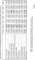
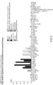

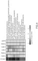
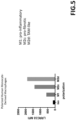


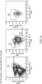
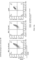


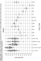
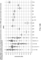

Classifications
-
- A—HUMAN NECESSITIES
- A61—MEDICAL OR VETERINARY SCIENCE; HYGIENE
- A61P—SPECIFIC THERAPEUTIC ACTIVITY OF CHEMICAL COMPOUNDS OR MEDICINAL PREPARATIONS
- A61P35/00—Antineoplastic agents
-
- C—CHEMISTRY; METALLURGY
- C07—ORGANIC CHEMISTRY
- C07K—PEPTIDES
- C07K16/00—Immunoglobulins [IGs], e.g. monoclonal or polyclonal antibodies
- C07K16/18—Immunoglobulins [IGs], e.g. monoclonal or polyclonal antibodies against material from animals or humans
- C07K16/22—Immunoglobulins [IGs], e.g. monoclonal or polyclonal antibodies against material from animals or humans against growth factors ; against growth regulators
-
- A—HUMAN NECESSITIES
- A61—MEDICAL OR VETERINARY SCIENCE; HYGIENE
- A61K—PREPARATIONS FOR MEDICAL, DENTAL OR TOILETRY PURPOSES
- A61K33/00—Medicinal preparations containing inorganic active ingredients
- A61K33/20—Elemental chlorine; Inorganic compounds releasing chlorine
-
- A—HUMAN NECESSITIES
- A61—MEDICAL OR VETERINARY SCIENCE; HYGIENE
- A61K—PREPARATIONS FOR MEDICAL, DENTAL OR TOILETRY PURPOSES
- A61K45/00—Medicinal preparations containing active ingredients not provided for in groups A61K31/00 - A61K41/00
- A61K45/06—Mixtures of active ingredients without chemical characterisation, e.g. antiphlogistics and cardiaca
-
- A—HUMAN NECESSITIES
- A61—MEDICAL OR VETERINARY SCIENCE; HYGIENE
- A61P—SPECIFIC THERAPEUTIC ACTIVITY OF CHEMICAL COMPOUNDS OR MEDICINAL PREPARATIONS
- A61P11/00—Drugs for disorders of the respiratory system
-
- A—HUMAN NECESSITIES
- A61—MEDICAL OR VETERINARY SCIENCE; HYGIENE
- A61P—SPECIFIC THERAPEUTIC ACTIVITY OF CHEMICAL COMPOUNDS OR MEDICINAL PREPARATIONS
- A61P21/00—Drugs for disorders of the muscular or neuromuscular system
-
- A—HUMAN NECESSITIES
- A61—MEDICAL OR VETERINARY SCIENCE; HYGIENE
- A61P—SPECIFIC THERAPEUTIC ACTIVITY OF CHEMICAL COMPOUNDS OR MEDICINAL PREPARATIONS
- A61P35/00—Antineoplastic agents
- A61P35/02—Antineoplastic agents specific for leukemia
-
- A—HUMAN NECESSITIES
- A61—MEDICAL OR VETERINARY SCIENCE; HYGIENE
- A61P—SPECIFIC THERAPEUTIC ACTIVITY OF CHEMICAL COMPOUNDS OR MEDICINAL PREPARATIONS
- A61P35/00—Antineoplastic agents
- A61P35/04—Antineoplastic agents specific for metastasis
-
- A—HUMAN NECESSITIES
- A61—MEDICAL OR VETERINARY SCIENCE; HYGIENE
- A61P—SPECIFIC THERAPEUTIC ACTIVITY OF CHEMICAL COMPOUNDS OR MEDICINAL PREPARATIONS
- A61P37/00—Drugs for immunological or allergic disorders
- A61P37/02—Immunomodulators
- A61P37/04—Immunostimulants
-
- C—CHEMISTRY; METALLURGY
- C07—ORGANIC CHEMISTRY
- C07K—PEPTIDES
- C07K16/00—Immunoglobulins [IGs], e.g. monoclonal or polyclonal antibodies
- C07K16/18—Immunoglobulins [IGs], e.g. monoclonal or polyclonal antibodies against material from animals or humans
- C07K16/28—Immunoglobulins [IGs], e.g. monoclonal or polyclonal antibodies against material from animals or humans against receptors, cell surface antigens or cell surface determinants
-
- A—HUMAN NECESSITIES
- A61—MEDICAL OR VETERINARY SCIENCE; HYGIENE
- A61K—PREPARATIONS FOR MEDICAL, DENTAL OR TOILETRY PURPOSES
- A61K39/00—Medicinal preparations containing antigens or antibodies
- A61K2039/505—Medicinal preparations containing antigens or antibodies comprising antibodies
-
- C—CHEMISTRY; METALLURGY
- C07—ORGANIC CHEMISTRY
- C07K—PEPTIDES
- C07K2317/00—Immunoglobulins specific features
- C07K2317/20—Immunoglobulins specific features characterized by taxonomic origin
- C07K2317/24—Immunoglobulins specific features characterized by taxonomic origin containing regions, domains or residues from different species, e.g. chimeric, humanized or veneered
-
- C—CHEMISTRY; METALLURGY
- C07—ORGANIC CHEMISTRY
- C07K—PEPTIDES
- C07K2317/00—Immunoglobulins specific features
- C07K2317/70—Immunoglobulins specific features characterized by effect upon binding to a cell or to an antigen
- C07K2317/73—Inducing cell death, e.g. apoptosis, necrosis or inhibition of cell proliferation
- C07K2317/732—Antibody-dependent cellular cytotoxicity [ADCC]
-
- C—CHEMISTRY; METALLURGY
- C07—ORGANIC CHEMISTRY
- C07K—PEPTIDES
- C07K2317/00—Immunoglobulins specific features
- C07K2317/70—Immunoglobulins specific features characterized by effect upon binding to a cell or to an antigen
- C07K2317/76—Antagonist effect on antigen, e.g. neutralization or inhibition of binding
-
- C—CHEMISTRY; METALLURGY
- C07—ORGANIC CHEMISTRY
- C07K—PEPTIDES
- C07K2317/00—Immunoglobulins specific features
- C07K2317/70—Immunoglobulins specific features characterized by effect upon binding to a cell or to an antigen
- C07K2317/77—Internalization into the cell
Definitions
- This invention relates generally to LRRC33-inhibiting agents and use thereof.
- TGF ⁇ Transforming growth factor-beta superfamily of growth factors are involved in a number of signaling cascades that regulate diverse biological processes including, but not limited to: immune modulation/suppression, inhibition of cell growth, tissue homeostasis, extracellular matrix (ECM) remodeling, endothelial to mesenchymal transition (EMT), cell migration and invasion, as well as mesenchymal to epithelial transition.
- ECM extracellular matrix
- EMT endothelial to mesenchymal transition
- EMT endothelial to mesenchymal transition
- TGF ⁇ ligand modulates T regulatory cell function and maintenance of immune precursor cell growth and homeostasis.
- TGF ⁇ is a potent growth inhibitor and promoter of cellular differentiation.
- TGF ⁇ may become a promoter of tumor development due to its ability to stimulate angiogenesis, alter the stromal environment, and induce local and systemic immunosuppression.
- TGF ⁇ signaling may increase fibroblast populations and ECM deposition (e.g., collagen).
- ECM deposition e.g., collagen
- the TGF ⁇ family within the superfamily is comprised of three isoforms of TGF ⁇ , namely, TGF ⁇ 1, TGF ⁇ 2 and TGF ⁇ 3. All three isoforms signal through the same receptors to trigger a wide array of cellular effects. While gene knock-out studies of these isoforms offer some clues, only limited information is available as to the discrete biological significance of each of these isoforms in healthy and disease contexts.
- TGF ⁇ regulation is thought to occur via its interactions with so-called "presenting molecules.” These molecules appear to be expressed in a tissue-specific manner and sequester and "present” inactive (e.g., latent) forms of TGF ⁇ in the extracellular (e.g., cell surface or extra cellular matrix) microenvironment. There, TGF ⁇ becomes activated in response to certain stimuli and is released from the latent complex at the site of action to exert its effect. In this way, TGF ⁇ may regulate local signaling in a context-specific manner.
- presenting molecules These molecules appear to be expressed in a tissue-specific manner and sequester and "present” inactive (e.g., latent) forms of TGF ⁇ in the extracellular (e.g., cell surface or extra cellular matrix) microenvironment. There, TGF ⁇ becomes activated in response to certain stimuli and is released from the latent complex at the site of action to exert its effect. In this way, TGF ⁇ may regulate local signaling in a context-specific manner.
- TGF ⁇ presenting molecules have previously been described in the literature. These include latent TGF- ⁇ binding protein 1 (“LTBP1”), latent TGF- ⁇ binding protein 3 (“LTBP3”), glycoprotein A repetitions predominant (or “GARP” also known as LRRC32) and Leucine-Rich Repeat-Containing 33 (“LRRC33”).
- LTBP1 and LTBP3 are components of the extracellular matrix, whilst GARP is a transmembrane protein expressed on FOXP3+ regulatory T cells (Treg). See, for example: WO 2014/182676 ; WO 2017/156500 ; and, WO 2018/081287 .
- LRRC33 as a novel therapeutic target for certain proliferative disorders, including solid tumors and hematologic proliferative disorders, such as blood cancers and bone marrow disorders, as well as fibrotic disorders in which activated macrophages play a role.
- LRRC33 is preferentially expressed in a subset of cells and tissues with a surprising degree of selectivity. More specifically, LRRC33 expression is limited predominantly to cells of myeloid and lymphoid lineages (e.g., white blood cells). In particular, overexpression of LRRC33 has been observed in a number of tumor cell types of these lineages.
- LRRC33 in these cells/tissues may provide a therapeutic opportunity for treating various disorders involving disease-associated myeloid cells expressing LRRC33 on the cell surface.
- diseases involving disease-associated myeloid cells expressing LRRC33 on the cell surface include conditions, in which TGF ⁇ plays an immunosuppressive role, without broadly affecting essential TGF ⁇ function mediated by the other presenting molecules, which is important for normal tissue homeostasis.
- LRRC33 in monocytes, e.g., macrophages, was previously reported. However, subsequent studies in polarized macrophages have unexpectedly revealed that not all so-called "pro-fibrotic" M2-like macrophages express LRRC33. Rather, among polarized M2 macrophages, LRRC33 cell surface expression appears to be restricted predominantly to the sub-types, M2c and TAM-like (M2d), which are phenotypes associated with pro-fibrotic and cancer microenvironments, respectively. Moreover, such expression appears to become markedly uniform upon exposure to certain disease-derived factors (e.g., cytokines).
- M2c sub-types
- M2d TAM-like
- the target cells express receptors for such cytokines.
- the target cells express CCR2/CD192, which is a receptor for CCL2/MCP-1.
- the target cells express CD115, which is a receptor for M-CSF.
- the present invention provides LRRC33 inhibitors for achieving selective targeting of LRRC33-expressing cells.
- One class of such inhibitors selectively inhibits activation of LRRC33-presented proTGF ⁇ .
- a second class of such inhibitors selectively binds LRRC33 expressed on cells and induce cytotoxic effects.
- a third class of such inhibitors induces internalization of cell-surface LRRC33 or an LRRC33-containing complex for removal of the same from the cell surface.
- such an inhibitor includes a cytotoxic agent such that upon selective binding to cell-surface LRRC33 and subsequent internalization of the complex, it causes destruction of the cell (i.e., cell killing) leading to depletion of cells expressing LRRC33.
- such an inhibitor is an LRRC33 binding agent that causes the cell-surface LRRC33 or an LRRC33-containing complex to be removed from the cell surface, e.g., via internalization, while sparing the cell.
- the LRRC33 inhibitors described herein aim to i) reduce the availability of TGF ⁇ or LRRC33-proTGF ⁇ complex in the disease environment, e.g., fibrotic tissue, tumor microenvironment, etc.; ii) reduce or reverse immune suppression; and/or, iii) promote or boost the host's immunity, e.g., anti-tumor immunity.
- the present disclosure provides a class of antibodies or antigen-binding portions thereof that specifically inhibit activation of TGF ⁇ 1 associated with (e.g., presented by) LRRC33, but not with other presenting molecules.
- the antibody or fragment thereof binds the inactive (e.g., latent) proTGF ⁇ 1 complex that comprises LRRC33 in a context-specific manner, thereby inhibiting release of free, active TGF ⁇ 1 from the latent complex.
- the antibody or fragment thereof is context-permissive, in that it can bind and inhibit activation of TGF ⁇ 1 from a latent proTGF ⁇ 1 complex associated with two or more different presenting molecules.
- the antibody or fragment thereof can inhibit release of TGF ⁇ 1 from the latent complex comprising LRRC33, GARP, LTBP1 or LTBP3.
- the antibody or fragment thereof can inhibit release of TGF ⁇ 1 from the latent complex comprising either LRRC33 or GARP.
- Non-limiting examples of such antibodies and fragments thereof suitable for carrying out the present invention are provided in PCT/US2017/021972 , which may be used to achieve the effects described herein.
- the disclosure provides a class of cytotoxic antibodies or fragments thereof that target and ablate or eradicate cells expressing LRRC33 through effector function.
- such antibodies are engineered to provide or modulate effector functions (e.g., ADCC, ADCP, and CDC) and/or pharmacokinetics.
- ADCC effector functions
- ADCP ADCP
- CDC pharmacokinetics
- such antibodies induce ADCC by binding to target cells expressing LRRC33 thereby causing cell lyses.
- such an antibody may be an IgG1 isotype or IgG4 isotype.
- the antibody is a fully human antibody or variant thereof, or a humanized antibody.
- the antibody contains one or more mutations to modulate effector function.
- certain mutations cause altered binding to FcRn, Fc ⁇ RI, Fc ⁇ RIIIa, C1q, Fc ⁇ RIIa, Fc ⁇ RIIb.
- certain mutations may alter the half-life of the antibody in vivo; alter ADCC and/or CDC; alter macrophage phagocytosis; and/or alter Fab-arm exchange.
- such antibodies or fragments are pH-sensitive antibodies, which readily bind respective antigen at a neutral pH and dissociate at an acidic pH. These include so-called “sweeping” antibodies and "recycling" antibodies.
- target cells comprise cell surface LRRC33.
- target cells comprise cell surface LRRC33-proTGF ⁇ 1.
- the target cells are of myeloid lineage.
- the target cells are monocytes.
- the target cells are macrophages.
- the target cells are dendritic cells.
- the target cells are hematologic cancer cells.
- the target cells are leukemia cells, e.g., acute lymphoblastic leukemia (ALL) cells, acute myeloid leukemia (AML) cells, chronic lymphocytic leukemia (CLL) cells and chronic myeloid leukemia (CML) cells.
- ALL acute lymphoblastic leukemia
- AML acute myeloid leukemia
- CLL chronic lymphocytic leukemia
- CML chronic myeloid leukemia
- the target cells are myeloblasts.
- the AML cells are myeloblastic (M0) cells, myeloblastic (M1) cells without maturation; myeloblastic (M2) cells with maturation; promyeloctic (M3) cells; myelomonocytic (M4) cells; monocytic (M5) cells; erythroleukemia (M6) cells; and/or megakaryocytic (M7) cells.
- the target cells are in circulation and/or in the bone marrow.
- the target cells are macrophages, e.g., activated or M2-like macrophages, such as M2c and TAM-like macrophages.
- the target cell is an activated macrophage that differentiated from monocyte upon recruitment to the site of disease/injury, such as a tumor microenvironment (TME).
- TEE tumor microenvironment
- Such macrophage may be exposed to various factors (e.g., growth factors, cytokines, chemokines and ECM remodeling components, etc.) that are present in the local niche, which can further promote tumor-associated phenotype of the macrophage.
- Factors/cytokines present in disease environments include but are not limited to: indoleamine 2,3-dioxygenase (IDO), IFN ⁇ , IL-10, IL-6, IL-11, Oncostatin M (OSM), TGF ⁇ such as TGF ⁇ 1, CCL2/MCP-1, SDF-1/CXCL12, CXCR4, VEG, PDGF, COX-2, metalloproteinases (MMPs) such as MMP2, MMP7, MMP13 and MMP9, Kallikreins, IL-4, bFGF, TNF ⁇ , and M-CSF/CSF-1.
- IDO indoleamine 2,3-dioxygenase
- IFN ⁇ IFN ⁇
- IL-10 interleamine 2,3-dioxygenase
- OSM Oncostatin M
- TGF ⁇ such as TGF ⁇ 1, CCL2/MCP-1, SDF-1/CXCL12, CXCR4, VEG, PDGF, COX-2
- the target cells express cell-surface receptor(s) for one or more of the above-listed factors/cytokines.
- the target cells express indoleamine 2,3-dioxygenase (IDO).
- IDO indoleamine 2,3-dioxygenase
- target cells are neutrophils; in particular, activated neutrophils.
- target cells are myeloid-derived suppressor cells (MDSCs). MDSCs may be associated with cancer and/or infection. For example, MDSCs may be present in the tumor microenvironment.
- Cells to be targeted are a subset of (but not all) macrophages.
- the subset of macrophages is a monocyte-derived population.
- Cells to be targeted (target cells) by the use of the antibodies or fragments of the invention include B cells.
- the target cell is a regulatory B cell.
- the target cell is a CD19+ cell.
- the target cell is a B cell expressing IL-10.
- the target cell is an IL-10-producing regulatory B cell, capable of lowering the number of regulatory T cells (Tregs). Data suggest that normal B cells isolated from healthy individuals do not show elevated cell-surface LRRC33 protein as measured by FACS, despite the relatively high mRNA levels observed in these cells at the transcription level.
- activated or disease-associated B cells may upregulate LRRC33 expression that leads to increased cell-surface LRRC33 levels, rendering these cells responsive to an LRRC33 inhibition therapy described herein.
- Cells to be targeted by the use of the antibodies or fragments of the invention may express GARP, which presents proTGF ⁇ on the cell surface (a "GARP-proTGF ⁇ complex").
- the target cells express GARP-proTGF ⁇ 1 complex.
- the target cells are of lymphoid lineage.
- the target cells are lymphocytes.
- the target cells are Tregs, such as natural Tregs developed in the thymus and na ⁇ ve T cell-derived Tregs.
- the target cells are CD4+/Foxp3+.
- the target cells are CD4+/CD25+/Foxp3+.
- the target cells are tumor-associated ( i . e ., infiltrated or reside within tumors) or accumulate in the peripheral blood.
- the target cells are IDO-sensitive.
- preferred target cells express LRRC33 or LRRC33-proTGF ⁇ complex on cell surface at a level greater than cells which are non-target cells.
- the invention aims to preferentially inhibit cells expressing high levels of cell- surface LRRC33 or LRRC33-containing complex (e.g., LRRC33-proTGF ⁇ ) over cells expressing lower levels of cell surface LRRC33 or LRRC33-containing complex.
- cell surface expression of LRRC33 is at least equal or comparable to and preferably greater than cell surface expression of CD133, CD44, CD33, CD13, and/or CD64.
- LRRC33 or LRRC33-containing complex e.g., LRRC33-proTGF ⁇
- LRRC33-proTGF ⁇ cell-surface expression of LRRC33 or LRRC33-containing complex
- LRRC33 inhibitors as described herein may be used in therapy.
- LRRC3 inhibitors may be formulated in a pharmaceutical composition.
- the inhibitors may be used in the treatment of certain proliferative disorders, including hematologic proliferative disorders, such as blood cancers and bone marrow disorders, solid tumors, and/or fibrosis.
- the LRRC33 inhibitors may selectively inhibit LRRC33.
- the LRRC33 inhibitors may selectively target LRRC33-expressing cells.
- the cells to be targeted may be selected from target cells described above.
- the cells to be targeted may in some embodiments be tumor cells of the type showing overexpression of LRRC33 (e.g., based on mRNA expression) relative to the average expression level of LRRC33 in a range of cancer cell lines (e.g., as described herein). Overexpression may be significant (q ⁇ 0.05) overexpression. Overexpression may be 4-16-fold higher vs. the average expression level in the range of cancer cell lines.
- the cancer cell lines may be those tested as described in Figure 2 .
- LRRC33 inhibitors for use as described herein may selectively target LRRC33-expressing cells in one or more disorders as described herein (e.g., hematologic proliferative disorders) which are characterized by the presence of LRRC33-expressing acute myeloid leukemia cells, plasma cell myeloma cells, acute lymphoblastic T cell leukemia cells, blast phase chronic myeloid leukemia cells, acute lymphoblastic B cell leukemia cells, chronic lymphocytic leukemia/small lymphocytic lymphoma cells, B cell lymphoma cells, Burkitt lymphoma cells, mantle cell lymphoma cells, acute lymphoblastic leukemia cells.
- disorders as described herein e.g., hematologic proliferative disorders
- acute myeloid leukemia cells plasma cell myeloma cells, acute lymphoblastic T cell leukemia cells, blast phase chronic myeloid leukemia cells, acute lymphoblastic B cell leukemia cells, chronic lymph
- LRRC33 inhibitors for use as described herein may selectively target LRRC33-expressing cells in one or more disorders as described herein (e.g., proliferative disorders, such as a solid tumor) which are characterized by the presence of LRRC33-expressing polarized macrophages, including M2c and TAM-like cells.
- the macrophages may have profibrotic and tumor-associated-macrophage-like phenotypes, respectively.
- LRRC33 inhibitors provided herein may deplete the LRRC33-expressing cells in vivo.
- LRRC33 Human monocytes and neutrophils exhibit little to no surface expression of LRRC33 directly ex vivo, in contrast to CD33.
- the LRRC33 inhibitors do not target monocytes and/or neutrophils.
- the LRRC33 inhibitors do not deplete (e.g., do not significantly deplete) monocytes and/or neutrophils.
- LRRC33 cell surface expression appears to be restricted predominantly to the sub-types, M2c and TAM-like (M2d).
- M2a, M2b, M2c e.g., pro-fibrotic
- M2d pro-tumor or TAM-like).
- the LRRC33 inhibitors do not target M2a or M2b (or M1) macrophages. In some embodiments, the LRRC33 inhibitors do not deplete (e.g., do not significantly deplete) M2a or M2b (or M1) macrophages.
- compositions formulated with one or more of the antibodies (or fragments) disclosed in the disclosure are described.
- pharmaceutical compositions may comprise or may be used in conjunction with an adjuvant. It is contemplated that certain adjuvants can boost the subject's immune responses to, for example, tumor antigens, and facilitate Teffector function, DC differentiation from monocytes, enhanced antigen uptake and presentation by APCs, etc.
- Suitable adjuvants include but are not limited to retinoic acid-based adjuvants and derivatives thereof, oil-in-water emulsion-based adjuvants, such as MF59 and other squalene-containing adjuvants, Toll-like receptor (TRL) ligands, ⁇ -tocopherol (vitamin E) and derivatives thereof.
- retinoic acid-based adjuvants and derivatives thereof oil-in-water emulsion-based adjuvants, such as MF59 and other squalene-containing adjuvants, Toll-like receptor (TRL) ligands, ⁇ -tocopherol (vitamin E) and derivatives thereof.
- TRL Toll-like receptor
- vitamin E ⁇ -tocopherol
- compositions may further comprise an excipient.
- Methods for manufacturing such pharmaceutical compositions, as well as kits including the same, are included in the invention.
- host immune suppression in the tumor microenvironment may be mediated by TGF ⁇ .
- TGF ⁇ particularly TGF ⁇ 1
- TGF ⁇ 1 may contribute to host immune evasion by creating an immunosuppressive or immune excluded environment via one or more of the following mechanisms: i) recruiting monocytes and inducing tumor-associated macrophage (TAM) phenotypes (LRRC33-dependent TGF ⁇ signaling); ii) inducing tumor-infiltrating Tregs that suppress T effector cells (GARP-dependent TGF ⁇ signaling); and, iii) promoting extracellular remodeling that favors tumor growth and/or invasion (matrix-associated or LTBP1/3-dependent TGF ⁇ signaling).
- TAM tumor-associated macrophage
- GARP tumor-associated TGF ⁇ signaling
- promoting extracellular remodeling that favors tumor growth and/or invasion
- An LRRC33 inhibitor according to the present disclosure may be used to reverse the immuno-suppression to create an immuno-permissive environment so as to allow effector cells to access target cells in the disease environment (e.g., TME). This may be achieved by inhibiting the disease-associated TGF ⁇ pathway, and/or, by depleting cells expressing cell-surface LRRC33 or LRRC33-containing complex (e.g., activated macrophages).
- the present invention includes the recognition that selective inhibition of LRRC33 may be useful for the treatment of various proliferative disorders. Additionally or alternatively, LRRC33 inhibition may help reduce or reverse immune suppression in the host either directly or indirectly. Accordingly, the disclosure provides methods for treating proliferative disorders, such as a hematologic proliferative disorder in a subject.
- the hematologic proliferative disorder is associated with myeloid or lymphoid cells.
- the hematologic disorder is a systemic disease or a localized disorder.
- the disorder comprises a solid tumor.
- a further aspect of the disclosure relates to methods for inhibiting cancer progression by intervening TGF ⁇ effects.
- the TGF ⁇ effects are mediated by LRRC33.
- the TGF ⁇ effects are mediated by GARP.
- interplay of both LRRC33 and GARP is involved in the process.
- treatment contributes to overcoming or reducing immunosuppression in the subject.
- the methods enhance or boost the body's immune response to treat cancer.
- LRRC33 LRRC33-presented TGF ⁇ 1.
- the hematologic disorder is blood cancer and/or cancer of the bone marrow.
- the hematologic disorder is a systemic disorder, localized disorder (such as solid tumor) or both.
- LRRC33-expressing and/or GARP-expressing immune cells are enriched in the site of tumor to create a tumor-associated local microenvironment.
- TGF ⁇ 1 concentrations are elevated in the niche.
- LRRC33 inhibitors as cancer therapeutic.
- the disclosure provides the use of an LRRC33 inhibitor in the treatment of cancer that comprises a solid tumor.
- LRRC33 inhibitors include neutralizing agents, such as antibodies or antigen-binding portions thereof that bind LRRC33 on cell surface, thereby neutralizing or interfering with its biological function. Binding of such neutralizing agents to LRRC33 may effectively deplete target cells expressing LRRC33 in vivo.
- LRRC33-neutralizing agents such as monoclonal antibodies or antigen-binding portions thereof that specifically bind LRRC33
- LRRC33-expressing cells include but are not limited to activated (polarized) macrophages, e.g., tumor-associated macrophages ("TAMs"), activated neutrophils, e . g ., tumor-associated neutrophils ("TANs"), and/or cancer-associated fibroblasts ("CAFs"), the phenotype of which is myofibroblast-like.
- CAFs express integrin comprising an alpha-11 ( ⁇ 11) chain.
- CAFs express cell-surface receptors for integrin comprising an alpha-11 ( ⁇ 11) chain.
- CAFs express IL-11 and/or IL-6.
- CAFs express IL-11 receptors and/or IL-6 receptors.
- LRRC33 inhibitors e.g., LRRC33-neutralizing agents
- inhibition of LRRC33 may prevent or reduce recruitment of immune cells to the site of tumor (e.g., tumor microenvironment).
- inhibition of LRRC33 may prevent or reduce tumor-infiltration of macrophages.
- LRRC33-binding agents such as those described above, bind and neutralize biological function of LRRC33 in vivo without directly affecting TGF ⁇ activities.
- LRRC33-binding agents work as inhibitors of TGF ⁇ , e.g., TGF ⁇ 1.
- TGF ⁇ TGF ⁇ 1
- LRRC33-binding agents that inhibit activation of TGF ⁇ e.g., TGF ⁇ 1, such as monoclonal antibodies or antigen-binding portions thereof that bind inactive or latent pro-protein complex of TGF ⁇ , thereby preventing release of mature TGF ⁇ growth factor from the complex.
- such disease comprises a solid tumor.
- LRRC33-expressing immune cells such as macrophages
- macrophages are enriched in the tumor.
- the macrophages are M2c and/or TAM-like phenotypes.
- these macrophages are capable of recruiting suppressive T lymphocytes to the tumor microenvironment.
- the suppressive T lymphocytes are Tregs expressing GARP.
- the hematologic disorder comprises abnormal proliferation of poorly differentiated blood cells in the bone marrow.
- the disorder is a myeloproliferative disorder.
- the myeloproliferative disorder is myelofibrosis.
- the disclosure provides combination therapies comprising a composition comprising an LRRC33 inhibitor and at least an additional therapy.
- the combination therapy in some embodiments achieves supplemental clinical benefits, as compared to monotherapy. In some embodiments, the combination therapy achieves additive or synergistic effects.
- Suitable subjects to be administered with a pharmaceutical composition suffer from one or more hematologic proliferative disorders.
- the subject is non-responsive or poorly responsive to conventional therapy.
- the conventional therapy may comprise an additional therapeutic as described herein, such as a PD-1 antagonist (or an antagonist of PD-1 signaling) or a tyrosine kinase inhibitor (e.g., a Bcr/Abl inhibitor).
- the subject has relapse following a remission of the disorder.
- a pharmaceutical composition comprising an antibody that binds human LRRC33 expressed on a cell, wherein the antibody optionally comprises an Fc domain, and wherein the antibody does not bind GARP.
- the antibody may be an LRRC33 inhibitor antibody as defined herein.
- the pharmaceutical composition comprising the LRRC33 inhibitor antibody may be for use in therapeutic methods, as described herein.
- an LRRC33 inhibitor for use in a method of depleting cells expressing cell-surface LRRC33 in a subject, the method comprising a step of administering to the subject the LRRC33 inhibitor in an amount effective to reduce the number of cells expressing LRRC33 on the cell surface, wherein the subject suffers from a disease associated with LRRC33 overexpression and/or abnormal macrophage activation.
- the LRRC33 inhibitor a) kills the cells expressing LRRC33 on the cell surface, optionally by inducing cytotoxic effects; and/or, b) induces internalization of the cell-surface LRRC33, so as to reduce the number of cells expressing LRRC33 on the cell surface in the subject.
- the subject has a hematologic proliferative disorder (e.g., leukemia, myelofibrosis and multiple myeloma), solid tumor, and/or fibrosis.
- a hematologic proliferative disorder e.g., leukemia, myelofibrosis and multiple myeloma
- the hematologic proliferative disorder is a leukemia.
- the leukemia is AML, ALL, CLL or CML.
- the solid tumor is enriched with tumor-associated macrophages (TAMs).
- TAMs tumor-associated macrophages
- the fibrosis comprises a fibrotic tissue enriched with monocyte-derived macrophages, wherein optionally the fibrosis is lung fibrosis, kidney fibrosis, liver fibrosis, cardiac fibrosis, bone marrow fibrosis and/or skin fibrosis.
- the cells expressing cell-surface LRRC33 are: TAMs, TANs, CAFs, leukemic cells, hematopoietic stem cells, myeloid progenitor cells, lymphoid progenitor cells, megakaryocyte-erythroid progenitor cells, megakaryocytes, monocytes, B cells, NK cells, neutrophils, eosinophils, basophils, and/or macrophages.
- an LRRC33 inhibitor for use in a method for reversing an immunosuppressive disease environment, so as to boost/facilitate/promote body's immunity.
- the disease environment is a TME or fibrotic tissue.
- the LRRC33 inhibitor induces ADCC. In one embodiment, the LRRC33 inhibitor induces internalization of cell-surface LRRC33.
- the LRRC33 inhibitor further comprises a cytotoxic agent.
- an LRRC33 inhibitor for use in a method for treating a leukemia in a subject, wherein administration of the LRRC33 inhibitor at an effective dose causes less toxicities as compared to a CD33 therapy.
- an LRRC33 inhibitor for use in a method for boosting host immunity against cancer in a subject, wherein the subject is treated with a cancer therapy, wherein optionally, the cancer therapy is a CAR-T therapy, a checkpoint inhibitor therapy, a chemotherapy, or a radiation therapy.
- the host immunity is boosted by reducing immunosuppression.
- the use of the LRRC33 inhibitor renders the cancer more responsive to the cancer therapy.
- the subject receives a reduced amount of the cancer therapy as compared to a monotherapy to achieve the same or equivalent therapeutic benefit/efficacy.
- the cell-surface LRRC33 is associated with a latent TGF ⁇ complex, wherein optionally the latent TGF ⁇ complex is a latent TGF ⁇ 1 complex.
- a method for selectively inhibiting TGF ⁇ 1 expressed on hematopoietic cells comprising a step of contacting a plurality of cells comprising TGF ⁇ 1-expressing hematopoietic cells and TGF ⁇ 1-expressing non-hematopoietic cells, with an LRRC33 inhibitor, thereby inhibiting TGF ⁇ 1 in the TGF ⁇ 1-expressing hematopoietic cells but not in the TGF ⁇ 1-expressing non-hematopoietic cells, wherein the LRRC33 inhibitor is an isolated antibody or fragment thereof that binds a large latent complex of TGF ⁇ 1 associated with LRRC33, thereby inhibiting release of active TGF ⁇ 1 from the complex; and, wherein the isolated antibody or fragment thereof does not bind a large latent complex of TGF ⁇ 1 associated with GARP or an LTBP.
- the hematopoietic cells are myeloid and/or lymphoid cells. In one embodiment, the hematopoietic cells are myeloma cells. In one embodiment, the hematopoietic cells are lymphoma cells.
- the antibody binds proTGF ⁇ 1, LRRC33, or combination thereof, but does not bind free, mature TGF ⁇ 1.
- a method for treating a blood cancer comprising a step of administering to a subject suffering from a blood cancer an effective amount of an LRRC33 inhibitor to inhibit the blood cancer in the subject.
- the blood cancer is selected from the group consisting of leukemia, lymphoma and multiple myeloma.
- leukemia is selected from the group consisting of: acute lymphoblastic leukemia (ALL), acute myeloid leukemia (AML), chronic lymphocytic leukemia (CLL) and chronic myeloid leukemia (CML).
- the LRRC33 inhibitor is an inhibitory antibody of LRRC33.
- the inhibitory antibody is a) an isolated antibody or fragment thereof that binds a large latent complex of TGF ⁇ 1, thereby inhibiting release of active TGF ⁇ 1 from the complex; or b) an isolated antibody that binds LRRC33 expressed on a cell, wherein the antibody comprises an Fc domain so as to induce ADCC of the cell.
- the inhibitory antibody is a fully human antibody or a humanized antibody. In one embodiment, the inhibitory antibody is a monoclonal antibody or a multimeric antibody. In one embodiment, the multimeric antibody is a bispecific antibody.
- the subject has received a bone marrow transplant.
- the subject is treated with an additional therapeutic for the blood cancer.
- the subject is non-responsive or poorly responsive to the additional therapeutic.
- the additional therapeutic is a PD-1 antagonist or a tyrosine kinase inhibitor.
- the tyrosine kinase inhibitor is a Bcr/Abl inhibitor.
- the LRRC33 inhibitor is administered as a combination therapy.
- the present disclosure provides therapeutics that are useful for treating proliferative disorders associated with hematologic cells/tissues. These disorders include but are not limited to cancers of blood cells and blood-forming tissues (e.g., the bone marrow), as well as immune cells that regulate the patient's anti-tumor immunity.
- the invention therefore includes related compositions, use thereof, related manufacture methods, as well as treatment methods.
- the compositions disclosed herein advantageously have the ability to modulate immune cell activity in tumors, thereby providing, in one aspect, a method to treat cancer by affecting a cell population that directly or indirectly affects growth of the tumor.
- Acute myeloid leukemia is a cancer of the myeloid line of blood cells, characterized by the rapid growth of abnormal white blood cells that build up in the bone marrow and interfere with the production of normal blood cells.
- AML is the most common acute leukemia affecting adults. Within AML, multiple subtypes are identified, as well as genetic mutations associated with each category.
- Antibody encompasses immunoglobulins or native antibodies, fragments thereof, variants thereof, as well as engineered antigen-binding agents. Such antibodies therefore may be any isotypes or chimeric constructs.
- An antibody of the invention may comprise a human light chain, e.g., kappa and lambda light chains.
- An antibody of the invention may comprise a heavy chain, e.g., mu, delta, gamma, alpha, and epsilon, which typically define the antibody's isotype as IgM, IgD, IgG, IgA, and IgE, respectively.
- An antibody may include a region to effectuate effector function, such as an Fc region that is primarily responsible for effector function.
- ADCC antibody-dependent cellular cytotoxicity
- NK natural killer
- ADCC is part of the adaptive immune response due to its dependence on a prior antibody response.
- the coating of target cells with antibodies is sometimes referred to as opsonization.
- antibody-drug conjugate refers to antibodies or functionally equivalent molecules (e.g., binding agents) linked to one or more cytotoxic or cell-killing agents.
- cytotoxic or cell-killing agents include a monoclonal antibody for a tumor-associated antigen that has restricted or no expression on normal (healthy) cells, where the antibody is conjugated with a potent cytotoxic agent (generally a small molecule drug with a high systemic toxicity) aimed to induce target cell death after being internalized in the tumor cell and released.
- a potent cytotoxic agent generally a small molecule drug with a high systemic toxicity
- Antibody fragments or antigen-binding portions include, inter alia, Fab, Fab', F(ab')2, Fv, domain antibody (dAb), complementarity determining region (CDR) fragments, CDR-grafted antibodies, single-chain antibodies (scFv), single chain antibody fragments, chimeric antibodies, diabodies, triabodies, tetrabodies, minibody, linear antibody; chelating recombinant antibody, a tribody or bibody, an intrabody, a nanobody, a small modular immunopharmaceutical (SMIP), an antigen-binding-domain immunoglobulin fusion protein, a camelized antibody, a VHH containing antibody, or a variant or a derivative thereof, and polypeptides that contain at least a portion of an immunoglobulin that is sufficient to confer specific antigen binding to the polypeptide, such as one, two, three, four, five or six CDR sequences, as long as the antibody retains the desired biological activity.
- dAb domain antibody
- Bone marrow proliferative disorder includes conditions in which cells of the bone marrow undergo abnormal cell division. Such proliferative disorders include cancerous (malignant) as well as non-cancerous (benign) conditions of the bone marrow.
- Cancer-associated fibroblasts are also referred to as carcinoma-associated fibroblasts, and these terms are used interchangeably herein. They represent a subpopulation of heterogeneous cells that reside within the tumor microenvironment. Phenotypically, CAFs may be myofibroblast-like and are redirected towards carcinogenesis, e.g., promotes the transformation process by encouraging tumor growth, angiogenesis, inflammation, and/or metastasis.
- CAFs are usually derived from the normal fibroblasts in the surrounding stroma but can also come from pericytes, smooth muscle cells, fibrocytes, mesenchymal stem cells (MSCs, often derived from bone marrow), or via epithelial-mesenchymal transition (EMT) or endothelial-mesenchymal transition (EndMT).
- EMT epithelial-mesenchymal transition
- EndMT endothelial-mesenchymal transition
- Cell-surface or cell-associated TGF ⁇ As used herein, the pro-protein form (hence latent) TGF ⁇ is said to be cell-associated, as opposed to ECM- or matrix-associated, when it is presented by a presenting molecule that is localized on cell surface, e.g., cell membrane-associated presenting molecules.
- a presenting molecule that is localized on cell surface, e.g., cell membrane-associated presenting molecules.
- Examples of cell-surface proTGF ⁇ include GARP-presented proTGF ⁇ and LRRC33-presented proTGF ⁇ .
- Clinical benefits As used herein, the term is intended to include both efficacy and safety of a therapy. Thus, therapeutic treatment that achieves a desirable clinical benefit is both efficacious and safe (e.g., with tolerable or acceptable toxicities or adverse events).
- Combination therapy refers to treatment regimens for a clinical indication that comprise two or more therapeutic agents.
- Complement dependent cytotoxicity refers to the process by which the complement cascade is activated, where one or more membrane attack complexes (MAC) is inserted into a target (e.g., pathogen) which cause lethal colloid-osmotic swelling.
- MAC membrane attack complexes
- Chronic myeloid leukemia Chronic myeloid (or myelogenous or myelocytic) leukemia (CML), also known as chronic granulocytic leukemia (CGL), is a cancer of the white blood cells. It is a form of leukemia characterized by the increased and unregulated growth of predominantly myeloid cells in the bone marrow and the accumulation of these cells in the blood. CML is a clonal bone marrow stem cell disorder in which a proliferation of mature granulocytes (neutrophils, eosinophils and basophils) and their precursors is found. It is a type of myeloproliferative disease associated with a characteristic chromosomal translocation called the Philadelphia chromosome.
- depletion of cells expressing cell-surface LRRC33 refers to i) killing or destruction of such cells by targeting the cell-surface LRRC33 or complex comprising the same; and/or, ii) removing cell-surface LRRC33 or complex comprising the same from such cells (i.e., from the cell surface) by, for example, inducing internalization.
- a common outcome is a reduced number of cells that express LRRC33 on the cell surface, whilst the latter may spare the cells themselves.
- ECM-associated TGF ⁇ As used herein, the pro-protein form (hence latent) TGF ⁇ is said to be ECM-associated (or "matrix-associated"), as opposed to cell-associated (or localized to cell-surface), when it is presented by a presenting molecule that is extracellularly localized, e.g., components of the ECM.
- LTBP1- or LTBP3-presented proTGF ⁇ is an example of ECM-associated TGF ⁇ .
- an effective amount is a dosage or dosing regimen that achieves statistically significant clinical benefits in a patient population.
- an effective amount of an LRRC33 inhibitor achieves a sufficient efficacy with tolerable toxicities when administered to a subject or a patient population.
- Antibodies or fragments encompassed by the present disclosure may contain a region/domain that confers effector function.
- the Fc region of an antibody mediates its serum half-life and effector functions, such as complement-dependent cytotoxicity (CDC), antibody-dependent cellular cytotoxicity (ADCC) and antibody-dependent cell phagocytosis (ADCP).
- Therapeutic antibodies engineered to contain an Fc or Fc-like region are encompassed by this invention.
- GARP-TGF ⁇ 1 complex refers to a protein complex comprising a pro-protein form or latent form of a transforming growth factor- ⁇ 1 (TGF ⁇ 1) protein and a glycoprotein-A repetitions predominant protein (GARP).
- TGF ⁇ 1 transforming growth factor- ⁇ 1
- GARP glycoprotein-A repetitions predominant protein
- a pro-protein form or latent form of TGF ⁇ 1 protein may be referred to as "pro/latent TGF ⁇ 1 protein”.
- a GARP-TGF ⁇ 1 complex comprises GARP covalently linked with pro/latent TGF ⁇ 1 via one or more disulfide bonds.
- a GARP-TGF ⁇ 1 complex comprises GARP non-covalently linked with pro/latent TGF ⁇ 1.
- a GARP-TGF ⁇ 1 complex is a naturally-occurring complex, for example a GARP-TGF ⁇ 1 complex in a cell.
- Hematologic cancer refers to a cancer that begins in blood-forming tissue, such as the bone marrow, or in the cells of the immune system.
- examples of hematologic cancer include leukemia, lymphoma, myelofibrosis, and multiple myeloma.
- Hematologic proliferative disorder refers to conditions characterized by abnormal cell division (malignant or benign) in blood cells or blood-forming tissues, e.g., cells in the bone marrow.
- Leukemia is cancer of the body's blood-forming tissues, including the bone marrow and the lymphatic system, and may be acute or chronic. Some forms of leukemia are more common in children, while other forms of leukemia occur mostly in adults. Leukemia usually involves the white blood cells (e.g., myeloid cells and lymphoid cells). The four main types of leukemia are: Acute myeloid (or myelogenous) leukemia (AML); Chronic myeloid (or myelogenous) leukemia (CML); Acute lymphocytic (or lymphoblastic) leukemia (ALL); and. Chronic lymphocytic leukemia (CLL).
- AML Acute myeloid (or myelogenous) leukemia
- CML Chronic myeloid (or myelogenous) leukemia
- ALL Acute lymphocytic leukemia
- CLL Chronic lymphocytic leukemia
- Localized tumor refers to anatomically isolated or isolatable abnormalities, such as solid malignancies, as opposed to systemic disease.
- Certain leukemia for example, may have both a localized component (for instance the bone marrow) and a systemic component (for instance circulating blood cells) to the disease.
- LRRC33 inhibitor refers to an agent that binds LRRC33 or a complex comprising LRRC33, thereby perturbing or interfering with its function or availability.
- an agent is an antibody or fragment thereof that specifically binds an epitope on LRRC33 or LRRC33-containing complex present on cell-surface.
- LRRC33 inhibitors may or may not exert inhibitory effects by modulating TGF ⁇ growth factor availability or activation.
- LRRC33-TGF ⁇ 1 complex refers to a complex between a pro-protein form or latent form of transforming growth factor- ⁇ 1 (TGF ⁇ 1) protein and a Leucine-Rich Repeat-Containing Protein 33 (LRRC33; also known as Negative Regulator Of Reactive Oxygen Species or NRROS).
- a LRRC33-TGF ⁇ 1 complex comprises LRRC33 covalently linked with pro/latent TGF ⁇ 1 via one or more disulfide bonds.
- a LRRC33-TGF ⁇ 1 complex comprises LRRC33 non-covalently linked with pro/latent TGF ⁇ 1.
- a LRRC33-TGF ⁇ 1 complex is a naturally-occurring complex, for example a LRRC33-TGF ⁇ 1 complex in a cell.
- Macrophages are a type of white blood cells of the immune system and includes heterogeneous, phenotypically diverse subpopulations of cells. Some macrophages differentiate from bone marrow-derived, circulating monocytes, while others are tissue-specific macrophages that reside within particular anatomical or tissue locations ("resident" macrophages).
- Tissue-specific macrophages include but are not limited to: Adipose tissue macrophages; Kupffer cells (Liver); Sinus histiocytes (Lymph nodes); Alveolar macrophages (or dust cells, Pulmonary alveoli of lungs); Tissue macrophages (histiocytes) leading to giant cells (Connective tissue); Langerhans cells (Skin and mucosa); Microglia (Central nervous system); Hofbauer cells (Placenta); Intraglomerular mesangial cells (Kidney); Osteoclasts (Bone); Epithelioid cells (Granulomas); Red pulp macrophages (or Sinusoidal lining cells, Red pulp of spleen); Peritoneal macrophages (Peritoneal cavity); and, LysoMac (Peyer's patch).
- Adipose tissue macrophages Kupffer cells (Liver); Sinus histiocytes (Lymph nodes); Alveolar macrophage
- Macrophages e.g., bone-marrow derived monocytes
- can be activated by certain stimuli such as cytokines
- polarized phenotypes e.g. , M1 and M2.
- M2-biased macrophages are further classified into several phenotypically distinct subtypes, such as M2a, M2b, M2c (e.g., pro-fibrotic) and M2d (pro-tumor or TAM-like).
- Microenvironment refers to a local niche in a biological system associated with particular feature(s) of interest, such as certain cellular/tissue compartments or disease loci, as in “disease microenvironment.”
- disease or disease loci may include fibrotic tissues (e.g., fibrotic organs or bone marrow), tumors, myopathies (e.g., a cardiomyopathy), etc.
- tumor microenvironment means the cellular/tissue environment in which the tumor exists, including surrounding blood vessels, immune cells, fibroblasts, bone marrow-derived inflammatory cells, lymphocytes, signaling molecules and the extracellular matrix (ECM).
- ECM extracellular matrix
- the tumor and the surrounding components of the microenvironment are closely related and interact constantly. Tumors can influence the microenvironment by releasing extracellular signals, promoting tumor angiogenesis and inducing peripheral immune tolerance, while components of the microenvironment, such as immune cells, can affect the growth and evolution of cancerous cells.
- Multiple myeloma also known as plasma cell myeloma, is a cancer of plasma cells, a type of white blood cell normally responsible for producing antibodies. Initially, often no symptoms are noticed. When advanced, bone pain, bleeding, frequent infections, and anemia may occur. Complications may include amyloidosis.
- Myelofibrosis also known as osteomyelofibrosis, is a relatively rare bone marrow proliferative disorder, which belongs to a group of diseases called myeloproliferative disorders. Myelofibrosis is classified into the Philadelphia chromosome-negative (-) branch of myeloproliferative neoplasms. Myelofibrosis is characterized by the proliferation of an abnormal clone of hematopoietic stem cells in the bone marrow and other sites results in fibrosis, or the replacement of the marrow with scar tissue.
- the term myelofibrosis unless otherwise specified, refers to primary myelofibrosis (PMF).
- myelofibrosis This may also be referred to as chronic idiopathic myelofibrosis (cIMF) (the terms idiopathic and primary mean that in these cases the disease is of unknown or spontaneous origin). This is in contrast with myelofibrosis that develops secondary to polycythemia vera or essential thrombocythaemia.
- Myelofibrosis is a form of myeloid metaplasia, which refers to a change in cell type in the blood-forming tissue of the bone marrow, and often the two terms are used synonymously.
- the terms agnogenic myeloid metaplasia and myelofibrosis with myeloid metaplasia (MMM) are also used to refer to primary myelofibrosis.
- MDSC Myeloid-derived suppressor cell
- MDSCs Myeloid-derived suppressor cells, or MDSCs
- MDSCs are a heterogeneous group of immune cells of the myeloid lineage. MDSCs have immunosuppressive properties and are reported to expand in pathological situations such as chronic infections and cancer, as a result of an altered hematopoiesis. MDSCs have immunosuppressive properties. Clinical and experimental evidence indicates that cancer tissues with high infiltration of MDSCs are associated with poor patient prognosis and resistance to therapies. MDSCs include multiple subtypes.
- one subtype of MDSCs is a population of immature mononuclear cells which are phenotypically and morphologically similar to monocytes (e.g., at "monocytic” hence M-MDSCs).
- Another subtype is a population of immature polynuclear cells which phenotypically and morphologically resemble neutrophils.
- MDSC suppressor function includes their ability to inhibit T cell proliferation and activation. In healthy individuals, immature myeloid cells formed in the bone marrow differentiate to dendritic cells, macrophages and neutrophils. However, under chronic inflammatory conditions (viral and bacterial infections) or cancer, myeloid differentiation is skewed towards the expansion of MDSCs.
- MDSCs infiltrate inflammation sites and tumors, where they stop immune responses by inhibiting T cells and NK cells, for example. MDSCs also accelerate angiogenesis, tumor progression and metastasis through the expression of cytokines and factors such as TGF-beta
- MDSC activity was originally described as suppressors of T cells, in particular of CD8+ T-cell responses.
- the spectrum of action of MDSC activity also encompasses NK cells, dendritic cells and macrophages.
- Suppressor activity of MDSC is determined by their ability to inhibit the effector function of lymphocytes. Inhibition can be caused different mechanisms. It is primarily attributed to the effects of the metabolism of L-arginine. Another important factor influencing the activity of MDSC is oppressive ROS
- Tregs are characterized by the expression of the biomarkers CD4, FOXP3, and CD25. Tregs are sometimes referred to as suppressor T cells and represent a subpopulation of T cells that modulate the immune system, maintain tolerance to self-antigens, and prevent autoimmune disease. Tregs are immunosuppressive and generally suppress or downregulate induction and proliferation of effector T cells. Tregs can develop in the thymus (so-called CD4+ Foxp3+ “natural” Tregs) or differentiate from na ⁇ ve CD4+ T cells in the periphery, for example, following exposure to TGF ⁇ or retinoic acid.
- Solid tumor refers to proliferative disorders resulting in an abnormal growth or mass of tissue that usually does not contain cysts or liquid areas.
- Solid tumors may be benign (noncancerous), or malignant (cancerous).
- Solid tumor may refer to a primary tumor (the original growth) or a secondary tumor (resulting from metastasis of a primary tumor).
- a solid tumor may comprise: cancerous (malignant) cells, surrounding stroma, which may include activated fibroblasts (e.g., myofibroblast-like phenotype), infiltrated immune cells, such as macrophages and neutrophils, as well as blood vessels.
- activated fibroblasts e.g., myofibroblast-like phenotype
- infiltrated immune cells such as macrophages and neutrophils, as well as blood vessels.
- Target cell refers to a cell whose function is to be pharmacologically perturbed.
- target cells express LRRC33 on the cell surface.
- An LRRC33 inhibitor according to the invention is capable of depleting or antagonizing LRRC33 expressed on a target cell.
- the target cell may express cell surface receptor for disease-derived cytokines such as CCL2/MCP-1 and M-CSF.
- Treating, treatment is intended to broadly mean: causing therapeutic benefits in a patient by, for example, enhancing or boosting the body's immunity; reducing or reversing immune suppression; reducing, removing (e.g., depleting) or eradicating harmful cells (e.g., tumor-associated cells, such as TAMs, TANs and CAFs) or substances from the body; reducing disease burden (e.g., tumor burden); preventing recurrence or relapse; and/or otherwise improving survival.
- harmful cells e.g., tumor-associated cells, such as TAMs, TANs and CAFs
- disease burden e.g., tumor burden
- preventing recurrence or relapse preventing recurrence or relapse; and/or otherwise improving survival.
- “Depletion" of harmful cells does not necessarily require physical removal of such cells from the affected patient; rather, depletion may be achieved by neutralization of certain activities or biological function associated with such harmful cells.
- the term includes therapeutic treatments, prophylactic treatments, and applications in which one reduces the risk that a subject will develop a disorder or other risk factor. Treatment does not require the complete curing of a disorder and encompasses embodiments in which one reduces symptoms or underlying risk factors.
- TGF ⁇ growth factors (TGF ⁇ 1, TGF ⁇ 2 and TGF ⁇ 3) are members of the TGF ⁇ superfamily of growth factors and are widely expressed by most, if not all, cell types and tissues. TGF ⁇ growth factors mediate an array of biological processes, including cell differentiation and proliferation.
- TGF ⁇ pathway dysregulation has been implicated in multiple diseases, several drugs that target the TGF ⁇ pathway have been developed and tested in patients, but with limited success.
- TGF ⁇ regulation involves its differential associations with tissue-specific presenting molecules, by which TGF ⁇ can exert context-specific biological effects within a local niche or microenvironment.
- tissue-specific presenting molecules by which TGF ⁇ can exert context-specific biological effects within a local niche or microenvironment.
- These proteins that interact with and hence "present" an inactive, latent form of TGF ⁇ complex include: LTBP1, LTBP3, GARP (also known as LRRC32) and LRRC33.
- LTBP1 and LTBP3 are secreted proteins and are components of the extracellular matrix. They were originally identified by their association with the latent form of transforming growth factors.
- GARP and LRRC33 are expressed on the cell surface of a small subset of cell types where they "present" latent TGF ⁇ as a GARP-proTGF ⁇ or LRRC33-proTGF ⁇ complex.
- LRRC33 may contribute to TGF ⁇ bioavailability and signaling in selected niches or microenvironments.
- LRRC33 Leucine-rich repeat-containing protein 33
- NROS Native Regulator of Reactive Oxygen Species
- LRRC33-/- mice ascending paraparesis was observed, followed by quadriplegia and death by five months of age ( Timothy Springer, Int. BMP Conference, October 2016 ). Consistent with the neurodegenerative phenotype observed in the LRRC33-/- mice, the Springer group found that LRRC33 is highly expressed in microglia, which is known to play a crucial role in axonal growth and guidance in the brain.
- Representative cell lines with significant change in TGF ⁇ presenting molecule expression include but are not limited to: acute myeloid leukemia cells, plasma cell myeloma cells, acute lymphoblastic T cell leukemia cells, blast phase chronic myeloid leukemia cells, acute lymphoblastic B cell leukemia cells, chronic lymphocytic leukemia/small lymphocytic lymphoma cells, B cell lymphoma cells, Burkitt lymphoma cells, mantle cell lymphoma cells, acute lymphoblastic leukemia cells, anaplastic large cell lymphoma cells, diffuse large B cell lymphoma cells and Hodgkin lymphoma cells. Concurrent overexpression of TGF ⁇ 1 observed in these cells has suggested that LRRC33-presented TGF ⁇ 1 activities may contribute to the pathology.
- preferential LRRC33 overexpression is found on the cell surface of a subset , but not all, of polarized macrophages, namely, M2c and TAM-like cells, which are profibrotic and tumor-associated-macrophage-like phenotypes, respectively.
- polarized macrophages namely, M2c and TAM-like cells, which are profibrotic and tumor-associated-macrophage-like phenotypes, respectively.
- These LRRC33-expressing macrophage subpopulations may directly interact with and influence the recruitment and/or differentiation of various cell types, such as immunosuppressive T cells (Tregs) and myofibroblasts or CAFs, in the disease environment such as TME and fibrotic tissues.
- Tregs immunosuppressive T cells
- myofibroblasts or CAFs
- Tregs which upon stimulation express GARP-presented TGF ⁇ 1
- LRRC33-expressing macrophages are known to infiltrate multiple types of solid tumors, raising the possibility that LRRC33-expressing macrophages may be involved in triggering or facilitating this process.
- macrophages exposed to certain disease-associated cytokines, such as M-CSF/CSF-1 show a robust increase in the level of cell surface LRRC33, raising the possibility that in a TME in vivo, tumor-derived CSF-1 may further induce macrophage activation into pro-tumor phenotypes, leading to disease progression.
- compositions comprising an isolated antibody or functional fragment thereof, which are useful for inhibiting a subset of TGF ⁇ 1 signaling, namely, LRRC33-mediated.
- antibody may refer to an immunoglobulin molecule that specifically binds to a target antigen, and includes, for instance, chimeric, humanized, fully human, and bispecific antibodies.
- An intact antibody will generally comprise at least two full-length heavy chains and two full-length light chains, but in some instances can include fewer chains such as antibodies naturally occurring in camelids which can comprise only heavy chains.
- Antibodies can be derived solely from a single source, or can be "chimeric,” that is, different portions of the antibody can be derived from two different antibodies. Antibodies, or antigen binding portions thereof, can be produced in hybridomas, by recombinant DNA techniques, or by enzymatic or chemical cleavage of intact antibodies.
- the term antibodies, as used herein, includes monoclonal antibodies, bispecific antibodies, minibodies, domain antibodies, synthetic antibodies (sometimes referred to herein as "antibody mimetics"), chimeric antibodies, humanized antibodies, human antibodies, antibody fusions (sometimes referred to herein as "antibody conjugates”), respectively. In some embodiments, the term also encompasses peptibodies.
- Naturally-occurring antibody structural units typically comprise a tetramer.
- Each such tetramer typically is composed of two identical pairs of polypeptide chains, each pair having one full-length "light” (in certain embodiments, about 25 kDa) and one full-length "heavy” chain (in certain embodiments, about 50-70 kDa).
- the amino-terminal portion of each chain typically includes a variable region of about 100 to 110 or more amino acids that typically is responsible for antigen recognition.
- the carboxy-terminal portion of each chain typically defines a constant region that can be responsible for effector function.
- Human antibody light chains are typically classified as kappa and lambda light chains.
- Heavy chains are typically classified as mu, delta, gamma, alpha, or epsilon, and define the isotype of the antibody.
- An antibody can be of any type (e.g., IgM, IgD, IgG, IgA, IgY, and IgE) and class (e.g., IgG1, IgG2, IgG3, IgG4, IgM1, IgM2, IgA1, and IgA2).
- the variable and constant regions are joined by a "J" region of about 12 or more amino acids, with the heavy chain also including a "D" region of about 10 more amino acids (see, e . g ., Fundamental Immunology, Ch. 7 (Paul, W., ed., 2nd ed. Raven Press, N.Y. (1989 )).
- the variable regions of each light/heavy chain pair typically form the antigen binding site.
- variable regions typically exhibit the same general structure of relatively conserved framework regions (FR) joined by three hyper variable regions, also called complementarity determining regions or CDRs.
- the CDRs from the two chains of each pair typically are aligned by the framework regions, which can enable binding to a specific epitope.
- both light and heavy chain variable regions typically comprise the domains FR1, CDR1, FR2, CDR2, FR3, CDR3 and FR4.
- the assignment of amino acids to each domain is typically in accordance with the definitions of Kabat Sequences of Proteins of Immunological Interest (National Institutes of Health, Bethesda, Md. (1987 and 1991 )), or Chothia & Lesk (1987) J. Mol. Biol.
- an antibody can comprise a small number of amino acid deletions from the carboxy end of the heavy chain(s). In some embodiments, an antibody comprises a heavy chain having 1-5 amino acid deletions in the carboxy end of the heavy chain.
- definitive delineation of a CDR and identification of residues comprising the binding site of an antibody is accomplished by solving the structure of the antibody and/or solving the structure of the antibody-ligand complex. In certain embodiments, that can be accomplished by any of a variety of techniques known to those skilled in the art, such as X-ray crystallography. In some embodiments, various methods of analysis can be employed to identify or approximate the CDR regions. Examples of such methods include, but are not limited to, the Kabat definition, the Chothia definition, the AbM definition, and the contact definition.
- a "functional antigen binding site" of a binding protein is one that that can bind to a target, antigen, or ligand.
- the antigen binding affinity of the antigen binding site is not necessarily as strong as the parent binding protein from which the antigen binding site is derived, but the ability to bind antigen must be measurable using any one of a variety of methods known for evaluating binding protein binding to an antigen.
- the antigen binding affinity of each of the antigen binding sites of a multispecific binding protein herein need not be quantitatively the same.
- variable region refers to a portion of the light and/or heavy chains of an antibody, typically including approximately the amino-terminal 120 to 130 amino acids in the heavy chain and about 100 to 110 amino terminal amino acids in the light chain.
- variable regions of different antibodies differ extensively in amino acid sequence even among antibodies of the same species.
- the variable region of an antibody typically determines specificity of a particular antibody for its target.
- An immunoglobulin constant domain refers to a heavy or light chain constant domain.
- Human IgG heavy chain and light chain constant domain amino acid sequences are known in the art.
- Compet when used in the context of antigen binding proteins (e.g., an antibody or antigen binding portion thereof) that compete for the same epitope means competition between antigen binding proteins as determined by an assay in which the antigen binding protein being tested prevents or inhibits (e.g., reduces) specific binding of a reference antigen binding protein to a common antigen (e.g., TGF ⁇ 1 or a fragment thereof).
- a common antigen e.g., TGF ⁇ 1 or a fragment thereof.
- ⁇ solid phase direct or indirect radioimmunoassay
- EIA solid phase direct or indirect enzyme immunoassay
- sandwich competition assay solid phase direct biotin-avidin EIA
- solid phase direct labeled assay solid phase direct labeled sandwich assay.
- a competing antigen binding protein when present in excess, it will inhibit (e.g., reduce) specific binding of a reference antigen binding protein to a common antigen by at least 40-45%, 45-50%, 50-55%, 55-60%, 60-65%, 65-70%, 70-75% or 75% or more. In some instances, binding is inhibited by at least 80-85%, 85-90%, 90-95%, 95-97%, or 97% or more.
- an antigen refers to a molecular structure that provides an epitope, e.g., a molecule or a portion of a molecule, or a complex of molecules or portions of molecules, capable of being bound by a selective binding agent, such as an antigen binding protein (including, e.g., an antibody).
- a selective binding agent may specifically bind to an antigen that is formed by two or more components in a complex.
- the antigen is capable of being used in an animal to produce antibodies capable of binding to that antigen.
- An antigen can possess one or more epitopes that are capable of interacting with different antigen binding proteins, e.g., antibodies.
- the antigen comprises LRRC33 or a fragment thereof, or a protein complex comprising the same. More specifically, an LRRC33 inhibitor for carrying out the invention described herein can bind an epitope present on the extracellular portion of cell-surface LRRC33 or a complex comprising the same.
- CDR refers to the complementarity determining region within antibody variable sequences. There are three CDRs in each of the variable regions of the heavy chain and the light chain, which are designated CDR1, CDR2 and CDR3, for each of the variable regions.
- CDR set refers to a group of three CDRs that occur in a single variable region that can bind the antigen. The exact boundaries of these CDRs have been defined differently according to different systems. The system described by Kabat ( Kabat et al.
- CDR boundary definitions may not strictly follow one of the herein systems, but will nonetheless overlap with the Kabat CDRs, although they may be shortened or lengthened in light of prediction or experimental findings that particular residues or groups of residues or even entire CDRs do not significantly impact antigen binding.
- the methods used herein may utilize CDRs defined according to any of these systems, although certain embodiments use Kabat or Chothia defined CDRs.
- crystal and “crystallized” as used herein, refer to a binding protein (e.g., an antibody), or antigen binding portion thereof, that exists in the form of a crystal.
- Crystals are one form of the solid state of matter, which is distinct from other forms such as the amorphous solid state or the liquid crystalline state. Crystals are composed of regular, repeating, three-dimensional arrays of atoms, ions, molecules (e.g., proteins such as antibodies), or molecular assemblies (e.g., antigen/antibody complexes). These three-dimensional arrays are arranged according to specific mathematical relationships that are well-understood in the field. The fundamental unit, or building block, that is repeated in a crystal is called the asymmetric unit.
- Repetition of the asymmetric unit in an arrangement that conforms to a given, well-defined crystallographic symmetry provides the "unit cell" of the crystal. Repetition of the unit cell by regular translations in all three dimensions provides the crystal. See Giege, R. and Ducruix, A. Barrett, Crystallization of Nucleic Acids and Proteins, a Practical Approach, 2nd ea., pp. 201-16, Oxford University Press, New York, New York, (1999 ).
- epitope includes any molecular determinant (e.g., polypeptide determinant) that can specifically bind to a binding agent, immunoglobulin or T-cell receptor.
- epitope determinants include chemically active surface groupings of molecules, such as amino acids, sugar side chains, phosphoryl, or sulfonyl, and, in certain embodiments, may have specific three-dimensional structural characteristics, and/or specific charge characteristics.
- An epitope is a region of an antigen that is bound by a binding protein. An epitope thus consists of the amino acid residues of a region of an antigen (or fragment thereof) known to bind to the complementary site on the specific binding partner. An antigenic fragment can contain more than one epitope.
- an antibody is the to specifically bind an antigen when it recognizes its target antigen in a complex mixture of proteins and/or macromolecules.
- antibodies are said to "bind to the same epitope” if the antibodies cross-compete (one prevents the binding or modulating effect of the other).
- structural definitions of epitopes are informative, but functional definitions are often more relevant as they encompass structural (binding) and functional (modulation, competition) parameters.
- Mechanisms of inhibitory activities of a particular antibody may vary.
- the inhibitory antibody works by inducing internalization of the antigen-antibody complex, thereby facilitating clearance of the target antigen (e.g., LRRC33 or a complex comprising LRRC33) from the cell surface.
- the target antigen e.g., LRRC33 or a complex comprising LRRC33
- the present disclosure includes certain "context-selective" inhibitors that can block a subset (but not all) of TGF ⁇ signaling in vivo.
- Such inhibitors include LRRC33 inhibitors and GARP inhibitors.
- the selectivity (or specificity) enabled by the present invention may render improved safety (e.g., limited toxicity) as compared to more conventional inhibitors of TGF ⁇ that do not discriminate functional contexts.
- LRRC33-specific inhibitors are provided.
- the aspect provides isolated antibodies or antigen-binding portions thereof that bind an LRRC33-TGF ⁇ 1 complex, which is an inactive, latent form of the complex.
- Such an antibody (or fragment) can inhibit the release of an active form of TGF ⁇ 1 growth factor from the latent complex, thereby specifically inhibiting TGF ⁇ 1 signaling associated with LRRC33.
- the inhibitory antibody works by stabilizing the inactive complex.
- the inhibitory antibody works by inducing internalization of the antigen-antibody complex, thereby facilitating clearance of the target antigen (LRRC33 or LRRC33-containing complex).
- the antibody does not bind the free, mature form of TGF ⁇ 1 growth factor that is not associated with the latent complex.
- the antibody is isoform-specific in that it inhibits activation of TGF ⁇ 1 without affecting TGF ⁇ 2 or TGF ⁇ 3.
- Such antibodies or antigen-binding portions thereof are suitable for the treatment of diseases associated with overexpression of LRRC33-presented TGF ⁇ 1.
- monoclonal antibodies that specifically binds LRRC33-TGF ⁇ 1 and inhibits the step of TGF ⁇ 1 activation are suitable for use in the treatment of hematologic proliferative disorders.
- context-selective antibodies or antigen-binding portions thereof can bind and inhibit activation (release) of TGF ⁇ 1 that is presented by either LRRC33 or GARP, but not LTBP1/3.
- careful designing of an antigen comprising a proTGF ⁇ 1 complex can generate inhibitory antibodies that recognize one or both types of proTGF ⁇ 1 complexes of interest. The resulting antibodies are suitable for use in modulating immune responses.
- Suitable antigens used to generate such antibodies include a protein complex comprising proTGF ⁇ 1 complex bound to a presenting molecule of interest (such as LRRC33 and GARP).
- the inactive form (e.g., a precursor) of a proTGF ⁇ complex typically comprises a dimer of proTGF ⁇ proprotein polypeptides, which associates with a single presenting molecule in a 2-to-1 stoichiometry.
- the dimer is a homodimer.
- the dimer is a heterodimer.
- the antibody or fragment thereof described herein binds each of the dimer.
- the antibody or fragment thereof described herein binds the dimer or the complex.
- the epitope recognized by such an antibody is a combinatorial epitope formed by two or more components of the complex, where the components are selected from portions of the proTGF ⁇ dimer and the presenting molecule (e.g., LRRC33).
- the epitope recognized by such an antibody is a conformational epitope that is dependent on the three-dimensional structure of the complex.
- the antibody specifically binds to the complex but not each of the components of the complex alone.
- Exemplary LRRC33 and GARP amino acid sequences are provided in the table below: Protein Sequence GARP sGARP LRRC33 (also known as NRROS; Uniprot Accession No. Q86YC3) soluble LRRC33 (sLRRC33) * Modified human kappa light chain signal peptide is depicted in bold font. ** Histidine tag is underlined. Human LRRC33-GARP chimera * Modified human kappa light chain signal peptide is depicted in bold font. ** LRRC33 ectodomain is underlined. # GARP transmembrane domain is italicized. ## GARP intracellular tail is double underlined.
- TGF ⁇ amino acid sequences are provided in the table below: Protein Sequence proTGF ⁇ 1 proTGF ⁇ 1 C4S proTGF ⁇ 1 D2G proTGF ⁇ 1 C4S D2G proTGF ⁇ 2 proTGF ⁇ 2 C5S proTGF ⁇ 2 C5S D2G proTGF ⁇ 2 D2G proTGF ⁇ 3 proTGF ⁇ 3 C7S proTGF ⁇ 3 C7S D2G proTGF ⁇ 3 D2G
- LRRC33 inhibitors are specific binders of LRRC33 or LRRC33-containing complex expressed on the cell surface. Such inhibitors are aimed to specifically recognize target cells expressing LRRC33 on the cell surface thereby inhibiting its availability for function. Such specific binders of LRRC33 or LRRC33-containing complex may or may not have the ability to inhibit TGF ⁇ activation.
- LRRC33 inhibitors are LRRC33-binding agents that specifically bind LRRC33 associated with latent TGF ⁇ (e.g., LRRC33-proTGF ⁇ 1 complex).
- LRRC33 cell-surface expression of a target molecule (such as LRRC33) is sufficiently restricted or selective to disease-associated cells over healthy cells.
- LRRC33 is advantageous over other cell-surface antigens which have been commercially exploited to date, such as CD33, due to more restricted expression to certain cell subpopulations. In this way, healthy cells may be spared, potentially reducing unwanted side effects or toxicities.
- the cell surface expression should be robust such that the inhibitor (LRRC33 binder) can readily and reliably engage target cells to ensure sufficient opsonization.
- LRRC33 Cell surface density of LRRC33 in cells evaluated so far shows expression levels that are equivalent to the targets of commercially available therapies that have been successfully exploited as ACDs to date (such as CD33, EGFR and Her2).
- ACDs such as CD33, EGFR and Her2
- M-CSF-matured human monocyte-derived macrophages show robust and uniform cell surface expression of LRRC33, making this an advantageous target for depleting disease-associated cells.
- only subpopulations of monocytes and/or macrophages will be targeted by LRRC33 inhibitors.
- LRRC33 Even within M2-polarized macrophages, cell-surface expression of LRRC33 is selectively upregulated in the M2c ("pro-fibrotic") and M2d ("pro-tumor” or “TAM-like") subpopulations, without significantly affecting the M2a and M2b subpopulations. This allows preferential targeting of disease-associated cell populations, as compared to broad targets such as myeloid markers.
- such inhibitors may bind cell-surface LRRC33 or LRRC33-containing complex thereby neutralizing (e.g., blocking) its function.
- such inhibitors may bind cell-surface LRRC33 or LRRC33-containing complex thereby inducing internalization of the LRRC33 (or LRRC33-containing complex) so as to remove or clear it from the cell surface. Internalization-dependent depletion of cell-surface LRRC33 may be exploited as an ADC-mediated cell killing.
- At least 35% (e.g., at least 40%, 45%, 50%, 60%, 70% and 80%) of the cell-surface target becomes internalized within 30 minutes, within 60 minutes, within 90 minutes or within 120 minutes at a physiological temperature, where preferably a majority of internalization (e.g., internalization of at least 50% of the cell-surface target) occurs within one hour of target engagement.
- such inhibitors may bind cell-surface LRRC33 thereby inducing antibody-dependent cell-mediated cytotoxicity (ADCC) of the target cells.
- ADCC is a mechanism of cell-mediated immune defense whereby an effector cell of the immune system actively lyses a target cell, whose membrane-surface antigens (e.g., LRRC33) have been bound by specific antibodies (e.g., anti-LRRC33 antibodies or fragments thereof).
- ADCC is independent of the immune complement system that also lyses targets but does not require any other cell.
- ADCC requires an effector cell (e.g., natural killer (NK) cells) that typically interact with IgG antibodies.
- NK natural killer
- macrophages, neutrophils and eosinophils may also mediate ADCC.
- the specific binders are monoclonal antibodies or fragment thereof that recognize an epitope available for binding on the extracellular portion of LRRC33 or a complex (e.g., protein complex, such as LRRC33-proTGF ⁇ ) on the cell surface of target cells.
- a complex e.g., protein complex, such as LRRC33-proTGF ⁇
- Such specific binders of LRRC33 encompassed by the present invention may comprise an effector region that triggers cellular cytotoxicity (e.g., ADCC) and other effector functions.
- monoclonal antibodies can target disease-associated cells/tissues (e.g., tumors, fibrotic tissues, injured tissues, etc.) through specific recognition of disease-associated antigens such as tumor-associated antigens and subsequent recruitment of effector elements including macrophages, dendritic cells, natural killer (NK) cells, T-cells, and the complement pathway components.
- disease-associated antigens such as tumor-associated antigens
- effector elements including macrophages, dendritic cells, natural killer (NK) cells, T-cells, and the complement pathway components.
- Such recruitments are achieved by interactions among the immunoglobulin gamma (IgG)-crystallizable fragment (Fc) and the immune cell receptors like Fcy receptors (Fc ⁇ Rs) and the complement protein C1q of the complement system.
- IgG immunoglobulin gamma
- Fc ⁇ Rs Fcy receptors
- the present disclosure provides an LRRC33 antibody comprising a heavy chain variable domain sequence that is at least 95% identical to the amino acid sequence SEQ ID NO. 1; and a light chain variable domain sequence that is at least 95% identical to the amino acid sequence SEQ ID NO. 2.
- the present disclosure provides an LRRC33 antibody comprising a heavy chain variable domain sequence that is at least 96%, at least 97%, at least 98%, or at least 99% identical to the amino acid sequence SEQ ID NO. 1; and a light chain variable domain sequence that is at least 96%, at least 97%, at least 98%, or at least 99% identical to the amino acid sequence SEQ ID NO. 2.
- the disclosure provides an LRRC33 antibody comprising a heavy chain variable domain sequence that is identical to the amino acid sequence SEQ ID NO. 1; and a light chain variable domain sequence that is identical to the amino acid sequence SEQ ID NO. 2.
- the present disclosure provides an LRRC33 antibody comprising a heavy chain variable domain sequence that is at least 95% identical to the amino acid sequence SEQ ID NO. 3; and a light chain variable domain sequence that is at least 95% identical to the amino acid sequence SEQ ID NO. 4.
- the present disclosure provides an LRRC33 antibody comprising a heavy chain variable domain sequence that is at least 96%, at least 97%, at least 98%, or at least 99% identical to the amino acid sequence SEQ ID NO. 3; and a light chain variable domain sequence that is at least 96%, at least 97%, at least 98%, at least 99% identical to the amino acid sequence SEQ ID NO. 4.
- the present disclosure provides an LRRC33 antibody comprising a heavy chain variable domain sequence that is identical to the amino acid sequence SEQ ID NO. 3; and a light chain variable domain sequence that is identical to the amino acid sequence SEQ ID NO. 4.
- the LRRC33 antibody comprises both a heavy chain and a light chain, wherein the antibody has a heavy chain/light chain variable domain sequence selected from the group consisting of SEQ ID NO. 1/SEQ ID NO. 2 (SRL1) and SEQ ID NO. 3/SEQ ID NO. 4 (SRL2).
- the present disclosure provides an LRRC33 antibody comprising a heavy chain variable domain comprising complementarity determining regions (CDRs) as set forth in SEQ ID NO. 1; and comprising a light chain variable domain comprising CDRs as set forth in SEQ ID NO. 2.
- CDRs complementarity determining regions
- the heavy chain variable domain sequence may be at least 95% identical to the amino acid sequence SEQ ID NO. 1 and the light chain variable domain sequence may be at least 95% identical to the amino acid sequence SEQ ID NO. 2, as disclosed above.
- the present disclosure provides an LRRC33 antibody comprising a heavy chain variable domain comprising complementarity determining regions (CDRs) as set forth in SEQ ID NO. 3; and comprising a light chain variable domain comprising CDRs as set forth in SEQ ID NO. 4.
- CDRs complementarity determining regions
- the heavy chain variable domain sequence may be at least 95% identical to the amino acid sequence SEQ ID NO. 3 and the light chain variable domain sequence may be at least 95% identical to the amino acid sequence SEQ ID NO. 4, as disclosed above.
- the present disclosure provides an LRRC33 antibody comprising a heavy chain variable domain comprising a heavy chain CDR set (CDR1, CDR2, and CDR3) selected from the group consisting of SEQ ID Nos: 5, 6 and 7, and a light chain variable domain comprising a light chain CDR set (CDR1, CDR2, and CDR3) selected from the group consisting of SEQ ID Nos: 8, 9 and 10.
- the present disclosure provides an LRRC33 antibody comprising a heavy chain variable domain comprising a heavy chain CDR set (CDR1, CDR2, and CDR3) selected from the group consisting of SEQ ID Nos: 11, 12 and 13 and a light chain variable domain comprising a light chain CDR set (CDR1, CDR2, and CDR3) selected from the group consisting of SEQ ID Nos: 14, 15 and 16.
- CDR1, CDR2, and CDR3 selected from the group consisting of SEQ ID Nos: 11, 12 and 13
- a light chain variable domain comprising a light chain CDR set (CDR1, CDR2, and CDR3) selected from the group consisting of SEQ ID Nos: 14, 15 and 16.
- LRRC33 inhibitors that specifically bind LRRC33 may be used to deplete cells expressing LRRC33 on the cell surface e.g., (immuno-depletion). Such inhibitors, used as an immune-depletion agent, may be useful for interfering with or limiting the recruitment of circulating monocytes to the site of disease (e.g., tumors and fibrotic or injured tissues) and/or depleting activated macrophage subpopulations at the site of disease (e.g., tumors and fibrotic or injured tissues).
- the site of disease e.g., tumors and fibrotic or injured tissues
- activated macrophage subpopulations e.g., tumors and fibrotic or injured tissues
- LRRC33-binding agents e.g., anti-LRRC33 antibodies or fragments thereof
- the FcyRs consisting of Fc ⁇ RI (CD64), FcyRII (CD32), and Fc ⁇ RIII (CD16) classes, are heterogeneous in terms of their cellular expression and Fc binding affinities.
- FcyRI binds to the Fc region with K D ⁇ 10 -8 -10 -9 M and is expressed on mononuclear phagocytes, dendritic cells, and IFN- ⁇ -activated neutrophils.
- FcyRII binds to the Fc region with relatively lower affinity (K D ⁇ 10 -7 M) and exists in five isoforms; among them, activating (FcyRlla, harboring an immunoreceptor tyrosine-based activation motif on neutrophils) or inhibitory (Fc ⁇ RIIb, harboring an immunoreceptor tyrosine-based inhibitory motif predominantly on B-lymphocytes) are critical for immune regulation.
- FcyRIII expressed in two isoforms, binds the Fc region with the lowest affinities (K D ⁇ 10 -5 M).
- Fc ⁇ RIIIa has a moderate Fc binding allele (V158) and a low binding allele (F158), and is expressed on NK cells, macrophages, and T-cell subsets and activates NK and T cell-mediated ADCC response; Fc ⁇ RIIIb is exclusively present on neutrophils and lacks signal generation capacity.
- Such LRRC33 binding agents are useful for eliminating target cells that express LRRC33.
- the same approach can be taken to engineer specific inhibitors of GARP that specifically bind target cells that express GARP and mediate cellular cytotoxic effects.
- binding agents can be used to reduce the number of target cells expressing LRRC33 and/or GARP in disease conditions in which it is beneficial to reduce the overexpression or proliferation of these target cells.
- compositions comprising the antibodies or antigen-binding portions thereof encompassed by the invention include antibody-drug-conjugates (ADCs).
- ADCs antibody-drug-conjugates
- an antibody or fragment thereof may be engineered to be linked or coupled to a drug or "payload" to be delivered to the site of interest via the antibody-antigen interaction.
- the antibody or antigen-binding portion thereof may include an additional moiety for carrying "a payload" of interest.
- suitable payload include, but are not limited to: therapeutics/drugs, toxins, markers and detection/imaging labels, and any other functional moieties of interest.
- payload may be chemical entities, small molecules, polypeptides, nucleic acids, radio-isotopes, etc.
- the invention includes an approach to induce antibody-independent (e.g., non-ADCC-mediated) killing of target cells by including a moiety (e.g., chemical entities) that works as a toxin.
- a moiety e.g., chemical entities
- Moieties of antibody-drug-conjugates (ADCs) may be attached or conjugated to the antibody structure via covalent or non-covalent linkages.
- ADCs contain cleavable or non-cleavable linkages.
- non-covalent interactions e.g., association and dissociation
- Any suitable toxins may be employed and engineered into ADC molecules.
- ADC molecules may be produced by coupling the LRRC33 binder with a cytotoxic agent.
- the binding moiety such as an anti-LRRC33 antibody or fragment thereof
- the cytotoxic moiety are conjugated via a linker.
- the linker may be a cleavable linker or non-cleavable linker.
- the ADC may contain one or more non-natural amino acids.
- the linker of the ADC may comprise one or more non-natural amino acids.
- cytotoxic agents may be employed.
- microtubule disrupting agents includes, without limitation, dolastatin 10 and its derivatives; monomethyl auristatin E (MMAE), monomethyl auristatin F (MMAF), maytansine and its derivatives.
- MMAE monomethyl auristatin E
- MMAF monomethyl auristatin F
- maytansine and its derivatives includes, without limitation, duocarmycin analogues and calicheamicin.
- Other agents used in the development of ADCs include, without limitation, pyrrolobenzodiazepines, duocarmycin, centanamycin, irinotecan, camptothecin, and doxorubicin.
- ADCs comprising an antibody or antigen-binding portion thereof, conjugated to a toxin, wherein the antibody or antigen-binding portion binds an epitope on LRRC33, an epitope on GARP, or an epitope common to both.
- Such antibodies may be used to preferentially target disease tissues and to reduce unwanted toxicities (e.g., adverse events) in patients.
- the antibody or the antigen-binding portion is a "dual function" antibody or fragment thereof, which functions as both TGF ⁇ activation inhibitor and further comprises a second function, such as killing of target cells, e.g., ADCC and toxin-dependent effects.
- dual function antibody is characterized in that it binds a latent proTGF ⁇ complex associated with a presenting molecule of interest, thereby inhibiting the mature TGF ⁇ to be released from the latent complex, which results in local reduction of free TGF ⁇ available in the disease microenvironment.
- Suitable presenting molecules may be selected on the basis of mechanisms of TGF ⁇ activation that drive the pathology of a particular disease or disorder.
- antibodies of the invention can specifically target abnormal cells or disease tissues, without adversely affecting normal or healthy counterpart.
- Non-limiting examples of such antibodies target a proTGF ⁇ 1 complex associated with LRRC33, associated with GARP, or permissive to bind either LRRC33- or GARP-associated proTGF ⁇ 1 complex.
- such antibodies are used to treat a hematologic proliferative disorder, such as leukemia and myelofibrosis.
- Tregs To counter immunosuppressive Treg accumulation in cancer-bearing patients, efforts have been made to develop a strategy to deplete Tregs. For example, several groups targeted IL-2 receptor as a target, which is expressed by a majority of Tregs, to deliver a toxin conjugated to the IL-2 ligand as a carrier. Results were mixed with limited success. Similarly, Daclizumab, an anti-CD25 antibody was used to target Tregs; however, this approach produced unwanted side effects of inadvertently depleting CD-25-expressing T cells, demonstrating the importance of specificity in targeting cancer cells.
- Targeting GARP-selective TGF ⁇ signaling with the use of antibodies described herein may provide clinical benefits in enhancing anti-tumor immunity, while allowing an improved safety profile due to the level of specificity this approach provides.
- LRRC33 is overexpressed in a very selective set of cell/tissue types. For example, only a subset of polarized monocytes, namely, the pro-fibrotic M2c subtype and the tumor-associated TAM-like subtype, show elevated levels of LRRC33 expression.
- LRRC33 e.g., LRRC33 or an LRRC33-containing complex
- pathological and/or pathogenic (disease-associated) cells e.g., myeloid leukemia such as AML and tumor-associated macrophages or disease/injury-associated macrophages
- LRRC33+ cells i.e., cells expressing LRRC33 on cell surface
- AML blasts may be potently targeted for destruction with minimal toxic side effects, as LRRC33- cells will not be engaged by the CAR-Ts.
- targeting LRRC33 should provide a safer alternative, as compared to existing AML therapies, due to more selective expression that restricts target cells to narrower subpopulations. This should spare healthy platelets and myeloid cells that would be otherwise targeted by existing therapies, which target more broadly expressed cell-surface markers, such as CD33, and which are needed for normal function.
- CAR-T cells typically they consist of an scFV recognizing a specific antigen (in this case LRRC33 or LRRC33-containing complex, such as LRRC33-proTGF ⁇ 1) fused to intracellular activating and co-stimulatory domains, such as CD28, CD3z and/or 4-1BB/ICOS/OX40 or other T cell activation domain.
- LRRC33 or LRRC33-containing complex such as LRRC33-proTGF ⁇ 1
- intracellular activating and co-stimulatory domains such as CD28, CD3z and/or 4-1BB/ICOS/OX40 or other T cell activation domain.
- CD8 T cell may or may not carry other genetic modifications to make it a potent and/or long lived anti-tumor effector cell (example: 'Armored CAR-Ts' express IL-12).
- CAR-T cells transfused into a patient would specifically recognize LRRC33 or a complex comprising the same, become activated and respond via the elaboration of cytokines and cytotoxic proteins.
- similar approaches may be taken to engineer CAR-NKT and/or CAR-NK cells in which a similar scFV fused to activating domains are cloned into NKT or NK cells as the source of anti-tumor effector mechanism.
- alternative activation domains may be used to leverage the activation pathways intrinsic to those cells, such as NKG2D, NKp30 and CD226.
- CAR-T therapies in blood cancer cells, such as AML, and in response to solid tumors may be more efficacious if used in combination with one or more cancer therapy/therapies.
- Additional cancer therapies used in conjunction with CAR-T include, but are not limited to checkpoint inhibitors, such as anti-PD1 or anti-PDL1 as some patients AML blasts show upregulated PDL1 as a mechanism for immunosuppression.
- CAR-T targeting of LRRC33 may provide a more durable response than single antigen targeting, since cancer cells, such as leukemic cells, in some instances develop an immunosuppressive environment and immune evasion through the production of TGF ⁇ 1.
- Downregulation of surface target antigens by leukemic cells, for instance T-ALL and NHL has been shown to be a mechanism of escape from immune cell killing.
- Downregulation of surface LRRC33-proTGF ⁇ may reverse or lessen TGF ⁇ 1-driven immunosuppression thus making the disease cells (such as cancer cells) exposed to immune surveillance that facilitates the body's immunity to combat the disease.
- LRRC33 as a target for BiTes, or Bi-specific T cell engagers.
- BiTes are fused scFvs, with one end specific for a tumor antigen (LRRC33) and the other specific for a T cell antigen, such as CD3.
- LRRC33 tumor antigen
- CD3 T cell antigen
- Useful antibodies and fragments thereof encompassed by the present application may be generated according to the methods described previously. Exemplary methods are provided in, for example, international patent applications PCT/US2013/068613 , PCT/US2014/036933 , PCT/US2015/059468 , PCT/US2016/052014 , PCT/US2017/021972 .
- such antibodies can be formulated into pharmaceutical compositions that further comprise at least one excipient.
- manufacture shall encompass various steps, methods and processes carried out, for example, to generate antigens or antigen complexes, screening step(s) (e.g., positive selection and negative selection steps) to identify and evaluate candidate agents (e.g., binders and inhibitors) of certain profiles, scale-up process, antibody engineering and/or modifications, purification steps, assays, and formulating or admixing components into pharmaceutical compositions which are suitable for administration to human and non-human subjects.
- candidate agents e.g., binders and inhibitors
- Such pharmaceutical compositions are sterile.
- candidate agents are screened for specific and selective binding for LRRC33, e.g., an extracellular potion(s) of LRRC33.
- Such step is generally referred to as positive selection.
- useful antigens or immunogens include recombinant LRRC33 or an extracellular fragment thereof, a protein complex comprising the same (e.g., LRRC33-proTGF ⁇ ), as well as cell-based antigens (cells expressing LRRC33 on cell surface, with or without proTGF ⁇ ).
- LRRC33-proTGF ⁇ e.g., LRRC33-proTGF ⁇
- cell-based antigens cells expressing LRRC33 on cell surface, with or without proTGF ⁇ .
- negative selection may be included in the method of producing or identifying LRRC33 inhibitors.
- Negative selection also referred to as “counter-selection” or “counter-screening” generally refers to the removal of undesirable binders or candidates from a pool.
- the invention includes methods for identifying LRRC33 inhibitors.
- the methods may comprise a step of selecting one or more binders that specifically bind LRRC33 from a pool (e.g., library).
- the method may further comprise a step of carrying out negative selection, or removing undesirable binders that are not specific or selective to LRRC33.
- undesirable binders may include those that cross-react with GARP.
- the method further comprises a step of confirming selectivity for LRRC33.
- the confirmation step may include confirming species-selectivity, such as murine LRRC33 and/or human LRRC33 (preferably human LRRC33).
- the confirmation step comprises confirming species-selectivity for human LRRC33.
- the LRRC33 inhibitor is cross-reactive for both human and rodent (e.g., murine) counterparts so as to facilitate in vivo evaluations and/or translatability.
- the method may comprise a FACS-based screening so as to confirm a candidate/test binder is capable of binding to cell-surface LRRC33.
- the method may comprise one or more cell-based assays, including but are not limited to internalization assays and cell killing (cytotoxic) assays so as to evaluate or confirm the ability of a candidate binder to deplete target cells.
- compositions and related methods described herein are useful as therapeutic include disease or disorder characterized by upregulation of cell-surface LRRC33 expression.
- disease or disorder involves monocyte-derived macrophage activation at the site of injury (e.g., fibrotic tissues, myopathies, tumors, etc.).
- an LRRC33 inhibitor may be used for the treatment of various proliferative conditions, such as cancer, e.g., systemic cancer and localized cancer.
- cancer e.g., systemic cancer and localized cancer.
- systemic cancer include hematologic proliferative disorders (e.g., cancer that begins in blood-forming tissue, such as the bone marrow, blood cells, and/or in the cells of the immune system).
- localized cancer include cancers that cause a solid tumor, e.g., primary tumors as well as secondary tumors (metastasis).
- Solid tumors may include, in addition to cancerous (i.e., malignant) cells, infiltrated immune cells or leukocytes, such as macrophages (e.g., tumor-associated macrophages or TAMs) and immunosuppressive Tregs, as well as activated neutrophils (e.g., tumor-associated neutrophils or TANs) and cancer-associated fibroblasts (CAFs).
- cancerous i.e., malignant
- leukocytes such as macrophages (e.g., tumor-associated macrophages or TAMs) and immunosuppressive Tregs, as well as activated neutrophils (e.g., tumor-associated neutrophils or TANs) and cancer-associated fibroblasts (CAFs).
- macrophages e.g., tumor-associated macrophages or TAMs
- immunosuppressive Tregs e.g., activated neutrophils (e.g., tumor-associated neutrophils or TANs)
- TANs are also recruited to a tumor site, where they may play a role in facilitating angiogenesis and other aspects of disease progression. Elevated TANs appear to correlate with poor prognosis in various types of carcinoma. Reminiscent to macrophage polarization, there are tumor-suppressive and tumor-promoting subtypes of TANs, and TGF ⁇ is thought to be involved in this conversion. Activated TANs also may recruit macrophages, suggesting that there may be a cascade of LRRC33lTGF ⁇ 1-mediated events that may drive cancer progression.
- an LRRC33 inhibitor is used in a method for depleting cells that express LRRC33 on cell surface in a subject.
- M2-polarized macrophages exposed to disease-associated (e.g., tumor-derived) cytokines such as M-CSF exhibit robust cell-surface expression of LRRC33.
- Cell surface density of LRRC33 on AML cells shows expression levels that are equivalent to targets of other cancer types in the clinic exploited as ADCs to date.
- the cell-surface density of LRRC33 in M-CSF-treated macrophages becomes markedly increased selectively in TAM-like macrophage subpopulation.
- Such selective induction of cell-surface LRRC33 in the highly restricted subset of cells provides an opportunity for selectively targeting the disease-associated macrophage subpopulation, without affecting non-disease-associated subpopulations that are required for normal biological functions.
- LRRC33 and CD115 show more uniform, hence less selective, cell-surface expression on different macrophage subtypes, e.g., M1-polarized and M-CSF-induced TAM-like macrophages, indicating that targeting these more broadly expressed markers may affect both disease-associated (e.g., TAMs) and non-disease-associated cells, thereby increasing potentially unwanted side effects.
- the use of the LRRC33 inhibitors described herein can selectively target various cell types associated with disease features, e.g., pro-tumor phenotype.
- target cells may include, but are not limited to: bone marrow-originated monocytes, polarized/activated macrophages at disease site, differentiate/tissue-specific or resident macrophages, e.g., alveolar macrophages (in the lung), osteoclasts (in the bone marrow), microglia (CNS), histiocytes (in connective tissue) and Kupffer cells (in the liver), MDSCs, as well as activated TANs and CAFs.
- differentiate/tissue-specific or resident macrophages e.g., alveolar macrophages (in the lung), osteoclasts (in the bone marrow), microglia (CNS), histiocytes (in connective tissue) and Kupffer cells (in the liver), MDSCs, as well as activated TANs and CAFs.
- LRRC33 expression in tumor may be a useful biomarker to identify a patient population that are poor responders of checkpoint inhibitor therapy. Indeed, it has been reported that greater degree of myeloid infiltration into tumor correlates with less responsiveness to PD-1 inhibitor.
- Current clinical development involving M-CSF receptor antagonists show significant but not great survival/tumor growth response, which is likely mediated by inhibition of M2 macrophage recruitment. However, this approach does not include MDSCs as target because these cells are not inhibited by M-CSF receptor inhibition.
- multiple cell types that contribute to disease progression can be inhibited, including TAMs, TANs, MDSCs, and CAFs, thereby improving the likelihood of clinical effects.
- the disclosure includes a method for treating the hematologic proliferative disorder comprising a step of administering to a patient (in vivo or ex vivo) a pharmaceutical composition comprising an antibody or antigen-binding fragment thereof that inhibits LRRC33, GARP, or both, in an amount sufficient to treat the disorder.
- compositions provided in the present disclosure may be used to enhance or boost the host immunity by reducing or reversing immunosuppression.
- One approach to achieve the reversal of immunosuppression is to reduce immunosuppressive regulatory T cells that are elevated in certain disease conditions, such as tumor microenvironment.
- the disease may also be associated with chronic inflammation of the affected tissue and/or fibrosis.
- such methods are aimed to achieve immune-mediated elimination of tumor/cancer cells in the host.
- the tumor/cancer cells are systemic cells, such as blood cancer cells or localized cells, such as a solid tumor.
- LRRC33-expressing macrophages of pro-fibrotic phenotype and/or tumor-associated phenotype are present in a disease tissue/cells. Overexpression of LRRC33 may lead to increased TGF ⁇ at the site of the disease. It is contemplated that targeting LRRC33 in these contexts may provide clinical benefits.
- an elevated number of LRRC33-expressing polarized macrophages may promote recruitment of Tregs which express GARP to the disease niche.
- administration of the composition can influence the progression of the disease, e.g., prolong remission or delay relapse of the malignancies. In some embodiments, this is achieved by reversing immune tolerance in the host, and/or via direct eradication of cancer cells to potentially overcome evasion of the host immunity by cancer cells through, for example, immune-editing.
- the antibody or fragment thereof may suppress Tregs, thereby promoting effector T cells.
- Tregs directly respond to IDO-expressing cells, such as dendritic cells and macrophages, which can stimulate Treg cell maturation. Elevated IDO stimulation is reported to correlate with greater recruitment of immunosuppressive Tregs to tumor microenvironment, leading to tumor growth. Conversely, reduced IDO stimulation is correlated with fewer Tregs infiltrating the tumor, which enhances T cell-mediated anti-tumor immunity. Therefore, antibodies that specifically inhibit GARP-presented TGF ⁇ in these scenarios can inhibit suppressor cells, thereby anti-tumor immunity of effector T cells can be maintained to combat the disease.
- inhibiting GARP-dependent TGF ⁇ signaling may block recruitment of Tregs to the site of tumor microenvironment, resulting in less tumor infiltration by Tregs. Clearance of suppressive cells from the tumor may promote anti-tumor effector function by effector T cells. These cells may directly recruit and activate Treg to corresponding disease microenvironments. Antibodies that specifically target the LRRC33-expressing macrophages may therefore help prevent immunosuppressive effects.
- target cells that express LRRC33 are blood cancer cells, e.g., leukemia cells (see more details below).
- LRRC33 and TGF ⁇ 1 appear to be selectively upregulated in many myeloid and lymphoid cancer cells. This selective expression provides a means for directly targeting LRRC33 in these cells to induce cancer cell killing by, for example, ADCC.
- the method comprises a step of administering an effective amount of a pharmaceutical composition comprising an antibody or antigen-binding portion thereof that specifically binds LRRC33 and comprises an Fc domain, which is capable of inducing ADCC in target cells.
- a pharmaceutical composition comprising an antibody or antigen-binding portion thereof that specifically binds LRRC33 and comprises an Fc domain, which is capable of inducing ADCC in target cells.
- hematologic cancer e.g., blood cancer
- hematologic cancer which may be targeted in this way include but are not limited to leukemia, lymphoma, and multiple myeloma.
- Suitable subjects who may be administered a pharmaceutical composition according to the present disclosure include those suffering from one or more of hematologic proliferative disorders and those having a solid or localized tumor comprising infiltration of myeloid and/or lymphoid cells expressing LRRC33 and/or GARP on the cell surface.
- the subject to be treated by the methods described herein can be a mammal, more preferably a human.
- Mammals include, but are not limited to, farm animals, sport animals, pets, primates, horses, dogs, cats, mice and rats.
- a human subject who needs the treatment may be a human patient having, at risk for, or suspected of having a TGF ⁇ -related indication, such as those noted above.
- the antibody or antigen-binding portion thereof is preferably designed to cause minimal unwanted immunogenicity in the subject to be treated. Therefore, for a human subject, a fully human or humanized antibody or antigen-binding portion thereof is preferred.
- such constructs may be engineered to closely match the particular species, so as to avoid unwanted immune responses to the therapeutic.
- the subject is treated with the composition as monotherapy.
- the subject to be administered with the composition described herein has received another therapy.
- the subject receives combination therapy that includes the composition.
- the subject has received bone marrow transplantation and or stem cell transplantation.
- the subject is in remission.
- the subject has experienced a relapse.
- the subject is non-responsive or has become resistant to a standard therapy.
- the subject is treated with the composition at a dose of about 1-20 mg of mAb/kg per dose, for example about 1 mg of mAb/kg, 2 mg of mAb/kg, 3 mg of mAb/kg, 4 mg of mAb/kg, 5 mg of mAb/kg, 6 mg of mAb/kg, 7 mg of mAb/kg, 8 mg of mAb/kg, 9 mg of mAb/kg, 10 mg of mAb/kg, 11 mg of mAb/kg, 12 mg of mAb/kg, 13 mg of mAb/kg, 14 mg of mAb/kg, 15 mg of mAb/kg, 16 mg of mAb/kg, 17 mg of mAb/kg, 18 mg of mAb/kg, 19 mg of mAb/kg, or 20 mg of mAb/kg.
- the dose may be about 1-15 mAb/kg, 1-10 mAb/kg, or 1-5 mAb/kg.
- the subject is treated with the composition at a dose of about 1-20 mg of mAb/kg per dose, for example about 1 mg of mAb/kg, 2 mg of mAb/kg, 3 mg of mAb/kg, 4 mg of mAb/kg, 5 mg of mAb/kg, 6 mg of mAb/kg, 7 mg of mAb/kg, 8 mg of mAb/kg, 9 mg of mAb/kg, or 10 mg of mAb/kg.
- the dose is about 1-10 mg of mAb/kg.
- Administration of the composition can be by any route, preferably intravenous or infusion.
- the subject is treated with the composition once a week. In some embodiments, the subject is treated with the composition every other week. In some embodiments, the subject is treated with the composition every 4 weeks. In some embodiments, the subject is treated with the composition once a month.
- dosing occurs in an initial phase (e.g., a period of time when the subject first starts treatment), followed by a maintenance phase.
- the frequency of dosing is the same in the initial phase and the maintenance phase.
- the frequency of dosing is different in the initial phase compared to the maintenance phase.
- dosing in the initial phase is on day 1 and day 4, followed by weekly or monthly dosing in the maintenance phase.
- dosing in the initial phase is on day 1 and day 7, followed by weekly or monthly dosing in the maintenance phase.
- dosing in the initial phase is on day 1, day 4 and day 7, followed by weekly or monthly dosing in the maintenance phase.
- dosing in the initial phase is on day 1, day 3, day 5 and day 7, followed by weekly or monthly dosing in the maintenance phase. In one embodiment, dosing in the initial phase is on day 1- day 7, followed by weekly or monthly dosing in the maintenance phase.
- Such disorders include but are not limited to the following.
- compositions and related methods are useful for treating a subject with a solid tumor.
- the disclosure includes a pharmaceutical composition comprising an LRRC33 inhibitor for use in the treatment of solid tumor in a subject, which comprises administering an effective amount of the composition to the subject.
- the LRRC33 inhibitor binds a proTGF ⁇ complex thereby inhibiting the release of active TGF ⁇ growth factor from the complex.
- the proTGF ⁇ is proTGF ⁇ 1.
- the hematologic proliferative disorder which may be treated in accordance with the present invention is leukemia.
- Leukemia is cancer of the body's blood-forming tissues, including the bone marrow and the lymphatic system, and may be acute or chronic. Leukemia usually involves the white blood cells (e.g., myeloid cells and lymphoid cells).
- the four main types of leukemia are: Acute myeloid (or myelogenous) leukemia (AML); Chronic myeloid (or myelogenous) leukemia (CML); Acute lymphocytic (or lymphoblastic) leukemia (ALL); and. Chronic lymphocytic leukemia (CLL).
- the hematologic proliferative disorder which may be treated in accordance with the present invention is Acute myeloid leukemia (AML).
- AML is the most common type of leukemia afflicting adults, which can develop from myeloid stem cells or myeloid blasts.
- the World Health Organization categorizes AML into several types.
- Acute myeloid leukemia with recurrent genetic abnormalities includes:
- AML with myelodysplasia-related changes includes patients who have had a prior documented myelodysplastic syndrome (MDS) or myeloproliferative disease (MPD) that then has transformed into AML, or who have cytogenetic abnormalities characteristic for this type of AML (with previous history of MDS or MPD that has gone unnoticed in the past, but the cytogenetics is still suggestive of MDS/MPD history).
- MDS myelodysplastic syndrome
- MPD myeloproliferative disease
- Therapy-related myeloid neoplasms includes patients who have had prior chemotherapy and/or radiation and subsequently develop AML or MDS. These leukemias may be characterized by specific chromosomal abnormalities, and often carry a worse prognosis.
- Myeloid proliferations related to Down syndrome includes so-called “transient abnormal myelopoiesis” and “Myeloid leukemia associated with Down syndrome.”
- Blastic plasmacytoid dendritic cell neoplasm includes so-called "blastic plasmacytoid dendritic cell neoplasm.”
- AML not otherwise categorized includes subtypes of AML that do not fall into the above categories, such as:
- the French-American-British (FAB) classification system divides AML into eight subtypes, M0 through to M7, based on the type of cell from which the leukemia developed and its degree of maturity. This is done by examining the appearance of the malignant cells with light microscopy and/or by using cytogenetics to characterize any underlying chromosomal abnormalities.
- the subtypes have varying prognoses and responses to therapy.
- Cytogenetics M0 acute myeloblastic leukemia, minimally differentiated M1 acute myeloblastic leukemia, without maturation M2 acute myeloblastic leukemia, with granulocytic maturation t(8;21)(q22;q22), t(6;9) M3 promyelocytic, or acute promyelocytic leukemia (APL) t(15;17) M4 acute myelomonocytic leukemia inv(16)(p13q22), del(16q) M4eo myelomonocytic together with bone marrow eosinophilia inv(16), t(16;16) M5 acute monoblastic leukemia (M5a) or acute monocytic leukemia (M5b) del (11q), t(9;11), t(11;19) M6 acute erythroid leukemias, including erythroleukemia (M6a) and very
- the pathophysiology of AML is complex.
- the malignant cell in AML is the myeloblast.
- the myeloblast In normal hematopoiesis, the myeloblast is an immature precursor of myeloid white blood cells; a normal myeloblast will gradually mature into a mature white blood cell.
- AML though, a single myeloblast accumulates genetic changes which halt the cell in its immature state and prevent differentiation.
- Such a mutation alone does not cause leukemia; however, when such a "differentiation arrest" is combined with other mutations which disrupt genes controlling proliferation, the result is the uncontrolled growth of an immature clone of cells, leading to the clinical entity of AML.
- cytogenetic abnormalities can be found in many people with AML; the types of chromosomal abnormalities often have prognostic significance.
- the chromosomal translocations encode abnormal fusion proteins, usually transcription factors whose altered properties may cause the "differentiation arrest".
- the t(15;17) translocation produces a PML-RAR ⁇ fusion protein which binds to the retinoic acid receptor element in the promoters of several myeloid-specific genes and inhibits myeloid differentiation.
- AML The clinical signs and symptoms of AML result from the growth of leukemic clone cells, which tends to displace or interfere with the development of normal blood cells in the bone marrow. This leads to neutropenia, anemia, and thrombocytopenia.
- the symptoms of AML are, in turn, often due to the low numbers of these normal blood elements.
- people with AML can develop a chloroma, or solid tumor of leukemic cells outside the bone marrow, which can cause various symptoms depending on its location.
- first-line treatment of AML consists primarily of chemotherapy, and is divided into two phases: induction and post-remission (or consolidation) therapy.
- induction therapy is to achieve a complete remission by reducing the number of leukemic cells to an undetectable level;
- consolidation therapy is to eliminate any residual undetectable disease and achieve a cure.
- Hematopoietic stem cell transplantation is usually considered if induction chemotherapy fails or after a person relapses, although transplantation is also sometimes used as front-line therapy for people with high-risk disease. Efforts to use tyrosine kinase inhibitors in AML continue.
- Gemtuzumab ozogamicin was approved by the FDA for the treatment of CD33-positive relapsed or refractory AML.
- Gemtuzumab is a monoclonal antibody to CD33 linked to a cytotoxic agent from the class of calicheamicins.
- CD33 or Siglec-3 sialic acid binding Ig-like lectin 3, SIGLEC3, SIGLEC-3, gp67, p67
- CD33 is typically considered a myeloid cell surface antigen but is also found on some lymphoid cells, such as activated T cells and natural killer (NK) cells.
- CD33 is also expressed in most leukemic blast cells, making this antigen a useful therapeutic target, but its expression is also observed in normal hematopoietic cells, although the expression generally diminishes with maturation of stem cells.
- Common side effects associated with anti-CD33 therapy included shivering, fever, nausea and vomiting.
- Serious side effects included severe myelosuppression (suppressed activity of bone marrow, which is involved in formation of various blood cells [found in 98% of patients]), disorder of the respiratory system, tumor lysis syndrome, Type III hypersensitivity, venous occlusion, infection, and death.
- the approved drug is marketed as Mylotarg ® , and the label contains a box warning for hepatotoxicity including severe or fatal hepatic veno-occlusive disease (VOD), also known as sinusoidal obstruction syndrome (SOS).
- VOD hepatic veno-occlusive disease
- SOS sinusoidal obstruction syndrome
- LRRC33 expression is restricted to a limited subset of myeloid cells presents the opportunity to more narrowly target disease-associated cells without adversely affecting normal/healthy cells that are affected by the existing approaches, such as anti-CD33 therapy.
- the present invention includes the use of LRRC33 inhibitors for the treatment of AML.
- An LRRC33 inhibitor according to the invention is used in the treatment of AMC (e.g. , newly diagnosed/de novo AML, relapsed AML, and refractory AML) in a human subject (e.g., adults and pediatric subjects).
- treatment methods involve administering to a patient with AML an effective amount of a pharmaceutical composition comprising an antibody or fragment thereof that includes a binding domain for LRRC33 and an Fc domain that binds FcR and is capable of triggering ADCC of target cells that express LRRC33 on cell surface.
- a pharmaceutical composition comprising an antibody or fragment thereof that includes a binding domain for LRRC33 and an Fc domain that binds FcR and is capable of triggering ADCC of target cells that express LRRC33 on cell surface.
- Such therapy is aimed to selectively target and kill cells expressing LRRC33, e.g., AML cells, both in circulation and within the bone marrow.
- Such therapy may be administered in conjunction with additional therapy, such as a tyrosine kinase inhibitor and/or a transplant (e.g., bone marrow transplant, stem cell transplant, etc.).
- an LRRC33 inhibitor of the present disclosure is used in a method for reducing toxicities associated with current AML therapy (anti-CD33 therapy) in a human subject.
- an LRRC33 inhibitor therapy is expected to cause less adverse events, including but are not limited to: hepatotoxicity (such as veno-occlusive liver disease); infusion-related reactions (such as anaphylaxis); hemorrhage (e.g., due to prolonged thrombocytopenia); QT interval prolongation; infection (less frequency and/or less severity); death (i.e., increased survival) and, embryo-fetal toxicity.
- an LRRC33 inhibitor for use as described herein may elicit reduced adverse side effects (e.g., selected from those described above) relative to an anti-CD33 therapy.
- An LRRC33 inhibitor for use as described herein may elicit reduced toxicity relative to an anti-CD33 therapy, for example when using an effective amount of the LRRC33 inhibitor and the anti-CD33 therapy.
- the anti-CD33 therapy may be Gemtuzumab ozogamicin.
- the AML may be CD33-positive relapsed or refractory AML.
- the LRRC33 inhibitor therapy may improve event-free survival in the subgroup of patients having adverse-risk cytogenetics.
- the LRRC33 inhibitor therapy may provide improved efficacy over currently available therapies. Efficacy may be established on the basis of event-free survival, measured for example, from the date of randomization until induction failure, relapse, or death by any cause. It is contemplated that the LRRC33 inhibitor may achieve improved overall survival in patient populations such as those with newly diagnosed AML (receiving either monotherapy or combination therapy), those with relapsed and/or refractory AML. In some embodiments, the LRRC33 inhibitor therapy achieves survival probability of at least 0 . 4 ( e.g., 0.40, 0.45, 0.50, 0.55, 0.60, etc.) at 24 months as determined by any suitable measures, such as Kaplan-Meier Plot of event-free survival within a patient population.
- any suitable measures such as Kaplan-Meier Plot of event-free survival within a patient population.
- the hematologic proliferative disorder which may be treated in accordance with the present invention is Chronic myeloid leukemia (CML).
- CML is a form of leukemia characterized by the increased and unregulated growth of predominantly myeloid cells in the bone marrow and the accumulation of these cells in the blood.
- CML is a clonal bone marrow stem cell disorder in which a proliferation of mature granulocytes (neutrophils, eosinophils and basophils) and their precursors is found. It is a type of myeloproliferative disease associated with a characteristic chromosomal translocation called the Philadelphia chromosome.
- CML is now largely treated with targeted tyrosine kinase inhibitors (TKIs) (Bcr-Abl) which have led to dramatically improved long-term survival rates.
- TKIs targeted tyrosine kinase inhibitors
- an LRRC33-targeting therapy described herein may provide an effective treatment option for CML patients.
- the present invention includes the use of LRRC33 inhibitors for the treatment of CML.
- treatment methods involve administering to a patient with CML an effective amount of a pharmaceutical composition comprising an antibody or fragment thereof that includes a binding domain for LRRC33 and an Fc domain that binds FcR and is capable of triggering ADCC of target cells that express LRRC33 on cell surface.
- Such therapy is aimed to selectively target and kill cells expressing LRRC33, e.g., CML cells, both in circulation and within the bone marrow.
- Such therapy may be administered in conjunction with additional therapy, such as a tyrosine kinase inhibitor and/or a transplant (e.g., bone marrow transplant, stem cell transplant, etc.).
- an LRRC33 inhibitor is employed as an ADC agent to treat CML.
- the LRRC33 inhibitor is used for the treatment of patients with newly diagnosed, relapsed or refractory CML. As compared to a therapy that broadly targets myeloid cells, the LRRC33 inhibitor does not attack normal myeloid cells. Thus, LRRC33 is a more selective target for disease-associated cells and may provide improved safety and tolerability (reduced toxicities), relative to other therapy.
- the hematologic proliferative disorder which may be treated in accordance with the present invention is Acute lymphocytic leukemia (ALL).
- Acute lymphoblastic leukemia also known as acute lymphocytic leukemia or acute lymphoid leukemia (ALL)
- ALL acute lymphocytic leukemia
- lymphoblasts are overproduced in the bone marrow and continuously multiply, causing damage and death by inhibiting the production of normal cells (such as red and white blood cells and platelets) in the bone marrow and by spreading (infiltrating) to other organs. ALL is most common in childhood.
- B-lymphoblastic ALL this category can be subdivided according to the correlation of the ALL cell immunophenotype with the stages of normal B-cell development
- Burkitt ALL corresponds to ALL-L3
- T-cell ALL T-cell ALL.
- Early detection of ALL is crucial for effective treatment. The aim is typically to induce a lasting remission, defined as the absence of detectable cancer cells in the body (usually less than 5% blast cells in the bone marrow).
- Treatment for acute leukemia can include chemotherapy, steroids, radiation therapy, intensive combined treatments (including bone marrow or stem cell transplants), and growth factors.
- the aim of remission induction is to rapidly kill most tumor cells and get the patient into remission. This is defined as the presence of less than 5% leukemic blasts in the bone marrow, normal blood cells and absence of tumor cells from blood, and absence of other signs and symptoms of the disease.
- Central nervous system (CNS) prophylaxis should begin during this phase of treatment and continue during the consolidation/intensification period. The rationale is based on the presence of CNS involvement in 10%-40% of adult patients at diagnosis. Combination of prednisolone or dexamethasone, vincristine, asparaginase (better tolerance in pediatric patients), and daunorubicin (used in Adult ALL) is used to induce remission.
- Central nervous system prophylaxis can be achieved via irradiation, cytarabine + methotrexate, or liposomal cytarabine.
- the intensity of initial induction treatment may be less than has been traditionally given.
- Typical intensification protocols use vincristine, cyclophosphamide, cytarabine, daunorubicin, etoposide, thioguanine or mercaptopurine given as blocks in different combinations.
- intrathecal methotrexate or cytarabine is usually used combined with or without cranio-spinal irradiation (the use of radiation therapy to the head and spine).
- Central nervous system relapse is treated with intrathecal administration of hydrocortisone, methotrexate, and cytarabine.
- the aim is to kill any residual cell that was not killed by remission induction and intensification regimens. Although such cells are few, they will cause relapse if not eradicated.
- daily oral mercaptopurine, once weekly oral methotrexate, once monthly 5-day course of intravenous vincristine and oral corticosteroids are usually used.
- the length of maintenance therapy is 2-3 years.
- the present disclosure includes the use of LRRC33 inhibitors for the treatment of ALL.
- treatment methods involve administering to a patient with ALL an effective amount of a pharmaceutical composition comprising an antibody or fragment thereof that includes a binding domain for LRRC33 and an Fc domain that binds FcR and is capable of triggering ADCC of target cells that express LRRC33 on cell surface.
- a pharmaceutical composition comprising an antibody or fragment thereof that includes a binding domain for LRRC33 and an Fc domain that binds FcR and is capable of triggering ADCC of target cells that express LRRC33 on cell surface.
- Such therapy is aimed to selectively target and kill cells expressing LRRC33, e.g., ALL cells, both in circulation and within the bone marrow.
- Such therapy may be administered in conjunction with additional therapy, such as a tyrosine kinase inhibitor and/or a transplant (e.g., bone marrow transplant, stem cell transplant, etc.).
- an LRRC33 inhibitor is employed as an ADC agent to treat ALL.
- the LRRC33 inhibitor is used for the treatment of patients with newly diagnosed, relapsed or refractory B-cell precursor acute lymphoblastic leukemia (ALL).
- ALL acute lymphoblastic leukemia
- the LRRC33 inhibitor does not attack normal B cells. This is supported by data showing that normal B cells isolated from healthy individuals do not show elevated cell-surface LRRC33 protein as measured by FACS. This indicates that LRRC33 is a more selective target for disease-associated cells and may provide improved safety and tolerability (reduced toxicities), relative to anti-CD22 therapy.
- Cytogenetics (the study of characteristic large changes in the chromosomes of cancer cells) is an important predictor of outcome.
- suitable patient population falls within one or more of the following cytogenetic subtypes:
- the treatment methods may be combined with additional therapy to treat ALL, such as chemotherapy, steroids, radiation therapy, intensive combined treatments (including bone marrow or stem cell transplants), growth factors and immunotherapy.
- additional therapy such as chemotherapy, steroids, radiation therapy, intensive combined treatments (including bone marrow or stem cell transplants), growth factors and immunotherapy.
- the hematologic proliferative disorder which may be treated in accordance with the present invention is B-cell chronic lymphocytic leukemia (B-CLL), also known as chronic lymphoid leukemia (CLL).
- B-CLL B-cell chronic lymphocytic leukemia
- CLL chronic lymphoid leukemia
- CLL is a common type of leukemia in adults. CLL affects B cell lymphocytes, which originate in the bone marrow, develop in the lymph nodes, and normally fight infection by producing antibodies.
- CLL small lymphocytic lymphoma
- SLL small lymphocytic lymphoma
- Combination chemotherapy regimens are effective in both newly diagnosed and relapsed CLL.
- Combinations of fludarabine with alkylating agents produce higher response rates and a longer progression-free survival than single agents, which include: FC (fludarabine with cyclophosphamide), FR (fludarabine with rituximab), FCR (fludarabine, cyclophosphamide, and rituximab), and CHOP (cyclophosphamide, doxorubicin, vincristine, and prednisolone).
- Chemoimmunotherapy with FCR has shown to improve response rates, progression-free survival, and overall survival in a large randomized trial in CLL patients selected for good physical fitness.
- Alkylating agents approved for CLL include bendamustine and cyclophosphamide.
- treatment methods involve administering to a patient with CLL an effective amount of a pharmaceutical composition comprising an antibody or fragment thereof that includes a binding domain for LRRC33 and an Fc domain that binds FcR and is capable of triggering ADCC of target cells that express LRRC33 on cell surface.
- a pharmaceutical composition comprising an antibody or fragment thereof that includes a binding domain for LRRC33 and an Fc domain that binds FcR and is capable of triggering ADCC of target cells that express LRRC33 on cell surface.
- Such therapy is aimed to selectively target and kill cells expressing LRRC33, e.g., CLL cells, both in circulation and within the bone marrow.
- Such therapy may be administered in conjunction with additional therapy, such as a those listed above.
- an LRRC33 inhibitor is employed as an ADC agent to treat CLL.
- the LRRC33 inhibitor is used for the treatment of patients with newly diagnosed, relapsed or refractory CLL. As compared to a therapy that targets mature B lymphocytes, the LRRC33 inhibitor does not attack normal B cells. This is supported by data showing that normal B cells isolated from healthy individuals do not show elevated cell-surface LRRC33 protein as measured by FACS. This indicates that LRRC33 is a more selective target for disease-associated cells and may provide improved safety and tolerability (reduced toxicities), relative to other therapy.
- the hematologic proliferative disorder which may be treated in accordance with the present invention is Multiple myeloma (MM).
- MM also known as plasma cell myeloma, is a cancer of plasma cells, a type of white blood cell normally responsible for producing antibodies. In advanced stages, bone pain, bleeding, frequent infections, and anemia may occur. Complications may include amyloidosis. Multiple myeloma is considered treatable but generally incurable. Remissions may be brought about with steroids, chemotherapy, thalidomide or lenalidomide, and stem cell transplant. Bisphosphonates and radiation therapy are sometimes used to reduce pain from bone lesions.
- the workup of suspected multiple myeloma includes a skeletal survey. This is a series of X-rays of the skull, axial skeleton and proximal long bones. Myeloma activity sometimes appears as "lytic lesions” (with local disappearance of normal bone due to resorption), and on the skull X-ray as "punched-out lesions” (pepper pot skull). Magnetic resonance imaging (MRI) is more sensitive than simple X-ray in the detection of lytic lesions, and may supersede skeletal survey, especially when vertebral disease is suspected. Occasionally a CT scan is performed to measure the size of soft tissue plasmacytomas. Bone scans are typically not of any additional value in the workup of myeloma patients (no new bone formation; lytic lesions not well visualized on bone scan).
- MRI Magnetic resonance imaging
- a bone marrow biopsy is usually performed to estimate the percentage of bone marrow occupied by plasma cells. This percentage is used in the diagnostic criteria for myeloma.
- Immunohistochemistry staining particular cell types using antibodies against surface proteins
- myeloma cells are typically CD56, CD38, CD138, CD319 positive and CD19 and CD45 negative.
- Cytogenetics may also be performed in myeloma for prognostic purposes, including a myeloma-specific FISH and Virtual Karyotype.
- the patient is diagnosed with the CD138 and/or the surface antigen CD319 (SLAMF7) as markers for myeloma cells.
- SLAMF7 surface antigen CD319
- Other useful laboratory tests include quantitative measurement of IgA, IgG, IgM (immunoglobulins) to look for immune paresis, and beta-2 microglobulin which provides prognostic information.
- IgA, IgG, IgM immunoglobulins
- beta-2 microglobulin which provides prognostic information.
- peripheral blood smear the rouleaux formation of red blood cells is commonly seen, though this is not specific.
- This assay the serum free light chain assay, has recently been recommended by the International Myeloma Working Group for the screening, diagnosis, prognosis, and monitoring of plasma cell dyscrasias.
- the prognosis of myeloma varies widely depending upon various risk factors.
- the Mayo Clinic has developed a risk-stratification model termed Mayo Stratification for Myeloma and Risk-adapted Therapy (mSMART) which divides patients into high-risk and standard-risk categories.
- mSMART Risk-adapted Therapy
- Patients with deletion of chromosome 13 or hypodiploidy by conventional cytogenetics, t(4;14), t(14;16) or 17p- by molecular genetic studies, or with a high plasma cell labeling index (3% or more) are considered to have high-risk myeloma.
- the hematologic proliferative disorder which may be treated in accordance with the present invention is Lymphoma.
- Lymphoma is a group of blood cell tumors that develop from lymphocytes. There are a number of subtypes of lymphomas, but the two main categories of lymphomas are Hodgkin's lymphomas (HL) and the non-Hodgkin lymphomas (NHL).
- HL Hodgkin's lymphomas
- NHL non-Hodgkin lymphomas
- the World Health Organization (WHO) includes two other categories as types of lymphoma: multiple myeloma and immunoproliferative diseases. About 90% of lymphomas are non-Hodgkin lymphomas.
- Risk factors for Hodgkin lymphoma include infection with Epstein-Barr virus and a history of the disease in the family. Risk factors for common types of non-Hodgkin lymphomas include autoimmune diseases, HIV/AIDS, infection with human T-lymphotropic virus, immunosuppressant medications, and some pesticides. Medical imaging may then be done to determine if and where the cancer has spread. Lymphoma most often spreads to the lungs, liver, and/or brain.
- Treatment may involve one or more of the following: chemotherapy, radiation therapy, targeted therapy, and surgery.
- the subject has received at least one additional such therapy.
- Classification, treatment and prognosis may differ according to: Whether or not it is a Hodgkin lymphoma; Whether the cell that is replicating is a T cell or B cell; The site from which the cell arises; Lymphoma can also spread to the central nervous system, often around the brain in the meninges, known as lymphomatous meningitis (LM).
- LM lymphomatous meningitis
- Hodgkin lymphoma is one of the most commonly known types of lymphoma and differs from other forms of lymphoma in its prognosis and several pathological characteristics. A division into Hodgkin and non-Hodgkin lymphomas is used in several of the older classification systems. A Hodgkin lymphoma is marked by the presence of a type of cell called the Reed-Sternberg cell.
- Non-Hodgkin lymphomas which are defined as being all lymphomas except Hodgkin lymphoma, are more common than Hodgkin lymphoma.
- a wide variety of lymphomas are in this class, and the causes, the types of cells involved, and the prognosis vary by type. The incidence of non-Hodgkin lymphoma increases with age. It is further divided into several subtypes.
- the WHO classification published in 2001 and updated in 2008, is based upon the foundations laid within the "revised European-American lymphoma classification" (REAL).
- RRL European-American lymphoma classification
- This system groups lymphomas by cell type (i.e. the normal cell type that most resembles the tumor) and defining phenotypic, molecular, or cytogenetic characteristics. The five groups are shown in the table.
- Hodgkin lymphoma is considered separately within the WHO and preceding classifications, although it is recognized as being a tumor of, albeit markedly abnormal, lymphocytes of mature B cell lineage.
- lymphoma Of the many forms of lymphoma, some are categorized as indolent (e.g. small lymphocytic lymphoma), compatible with a long life even without treatment, whereas other forms are aggressive (e.g. Burkitt's lymphoma), causing rapid deterioration and death. However, most of the aggressive lymphomas respond well to treatment and are curable. The prognosis, therefore, depends on the correct diagnosis and classification of the disease, which is established after examination of a biopsy by a pathologist (usually a hematopathologist).
- pathologist usually a hematopathologist
- Lymphoma subtypes according to the WHO classification include: Mature B cell neoplasms; Mature T cell and natural killer (NK) cell neoplasms; Precursor lymphoid neoplasms; Hodgkin lymphoma; Immunodeficiency-associated lymphoproliferative disorders.
- NK natural killer
- the hematologic proliferative disorders which may be treated in accordance with the present invention include myeloproliferative disorders, such as myelofibrosis.
- myeloproliferative disorders such as myelofibrosis.
- So-called "classical" group of BCR-ABL (Ph) negative chronic myeloproliferative disorders includes essential thrombocythemia (ET), polycythemia vera (PV) and primary myelofibrosis (PMF).
- Myelofibrosis is a type of chronic leukemia, e.g., a cancer that affects the blood-forming tissues in the body, and is a clonal neoplastic disorder of hematopoiesis, the formation of blood cellular components, which disrupts the body's normal production of blood cells. The result is extensive scarring in your bone marrow, leading to severe anemia, weakness, fatigue and often an enlarged spleen. It is one of the myeloproliferative disorders, diseases of the bone marrow in which excess cells are produced at some stage.
- cytokines such as fibroblast growth factor by the abnormal hematopoietic cell clone (particularly by megakaryocytes) leads to replacement of the hematopoietic tissue of the bone marrow by connective tissue via collagen fibrosis.
- the decrease in hematopoietic tissue impairs the patient's ability to generate new blood cells, resulting in progressive pancytopenia, a shortage of all blood cell types.
- the proliferation of fibroblasts and deposition of collagen is a secondary phenomenon, and the fibroblasts themselves are not part of the abnormal cell clone.
- Myelofibrosis may be caused by abnormal blood stem cells in the bone marrow.
- the abnormal stem cells produce mature and poorly differentiated cells that grow quickly and take over the bone marrow, causing both fibrosis (scar tissue formation) and chronic inflammation.
- hepatomegaly Primary myelofibrosis is associated with mutations in Janus kinase 2 (JAK2), thrombopoietin receptor (MPL) and calreticulin (CALR), which can lead to constitutive activation of the JAK-STAT pathway, progressive scarring, or fibrosis, of the bone marrow occurs.
- JAK-STAT pathway Janus kinase 2
- MPL thrombopoietin receptor
- CAR calreticulin
- Patients may develop extramedullary hematopoiesis, i.e., blood cell formation occurring in sites other than the bone marrow, as the haemopoetic cells are forced to migrate to other areas, particularly the liver and spleen. This causes an enlargement of these organs. In the liver, the abnormal size is called hepatomegaly.
- splenomegaly Enlargement of the spleen is called splenomegaly, which also contributes to causing pancytopenia, particularly thrombocytopenia and anemia.
- pancytopenia particularly thrombocytopenia and anemia.
- Another complication of extramedullary hematopoiesis is poikilocytosis, or the presence of abnormally shaped red blood cells.
- the principal site of extramedullary hematopoiesis in myelofibrosis is the spleen, which is usually markedly enlarged in patients suffering from myelofibrosis.
- the spleen contains red blood cell precursors, granulocyte precursors and megakaryocytes, with the megakaryocytes prominent in their number and in their abnormal shapes. Megakaryocytes may be involved in causing the secondary fibrosis seen in this condition.
- TGF ⁇ may be involved in the fibrotic aspect of the pathogenesis of myelofibrosis (see, for example, Agarwal et al., "Bone marrow fibrosis in primary myelofibrosis: pathogenic mechanisms and the role of TGF ⁇ " (2016) Stem Cell Investig 3:5 ). Bone marrow pathology in primary myelofibrosis is characterized by fibrosis, neoangeogenesis and osteosclerosis, and the fibrosis is associated with an increase in production of collagens deposited in the ECM.
- TGF ⁇ is a component of the leukemic bone marrow niche
- targeting the bone marrow microenvironment with TGF ⁇ inhibitors may be a promising approach to reduce leukemic cells expressing presenting molecules that regulate local TGF ⁇ availability in the effected tissue.
- one aspect relates to methods for treating primary myelofibrosis.
- the method comprises administering to a patient suffering from primary myelofibrosis a therapeutically effective amount of a composition comprising a TGF ⁇ inhibitor that causes reduced TGF ⁇ availability.
- the TGF ⁇ inhibitor is an antibody or antigen-binding portion thereof that binds an inactive (e.g., latent) proTGF ⁇ complex, thereby preventing the release of active or mature TGF ⁇ from the complex, effectively inhibiting the activation step.
- an antibody or antigen-binding portion specifically binds a proTGF ⁇ complex that is associated with LRRC33, GARP, LTBP1, LTBP3 or any combination thereof.
- the antibody or portion thereof selectively binds a proTGF ⁇ complex that is associated with either LRRC33 or GARP (but not with LTBP1 or LTBP3).
- the antibody or portion thereof specifically binds a proTGF ⁇ complex that is associated with LRRC33.
- the antibody or portion thereof specifically binds a proTGF ⁇ complex that is associated with GARP.
- the TGF ⁇ inhibitor is an antibody or antigen-binding portion thereof that binds LRRC33 or GARP and comprises a domain for additional effector functions.
- the domain for additional effector function may be an Fc or Fc-like domain to mediate ADCC in target cells.
- the antibody or antigen-binding portion thereof may include an additional moiety for carrying "a payload" of interest.
- suitable payload include, but are not limited to: therapeutics/drugs, toxins, markers and detection/imaging labels, etc.
- payload may be chemical entities, small molecules, polypeptides, nucleic acids, radio-isotopes, etc.
- suitable therapy includes an LRRC33 inhibitor described herein, which specifically targets hematopoietic cells expressing LRRC33.
- LRRC33 inhibitor described herein, which specifically targets hematopoietic cells expressing LRRC33. This may be achieved by administration of a composition comprising an antibody that specifically binds an LRRC33-presented proTGF ⁇ complex and inhibits activation of TGF ⁇ in the patient. It can also be achieved by administration of a composition comprising an antibody that specifically binds an LRRC33 and inducing killing of target cells in the patient.
- these approaches may be combined to use an antibody that is a TGF ⁇ activation inhibitor and also contains an additional moiety to mediate cellular cytotoxicity.
- the additional moiety may be an Fc or Fc-like domain to induce ADCC or a toxin conjugated to the antibody as a payload (e.g., ADC).
- TGF ⁇ is known to regulate aspects of ECM homeostasis, the dysregulation of which can lead to tissue fibrosis, it is contemplated that in some embodiments, it is desirable to inhibit TGF ⁇ activities associated with the ECM. Accordingly, antibodies or fragments thereof that bind and inhibit proTGF ⁇ presented by LTBPs (such as LTBP1 and LTBP3) are encompassed by this invention.
- antibodies or fragments thereof suitable for treating myelofibrosis are "context-permissive" in that they can bind multiple contexts of proTGF ⁇ complex, such as those associated with LRRC33, GARP, LTBP1, LTBP3, or any combination thereof.
- Suitable patient populations of myeloproliferative neoplasms who may be treated with the compositions and methods described herein include: a) a patient population that is Philadelphia (+); b) a patient population that is Philadelphia (-); c) a patient population that is categorized "classical" (PV, ET and PMF); d) a patient population carrying the mutation JAK2V617F(+); e) a patient population carrying JAK2V617F(-); f) a patient population with JAK2 exon 12(+); g) a patient population with MPL(+); and h) a patient population with CALR(+).
- the patient has extramedullary hematopoiesis.
- the extramedullary hematopoiesis is in the liver, lung, spleen, and/or lymph nodes.
- the pharmaceutical composition of the present invention is administered locally to one or more of the localized sites of disease manifestation.
- the LRRC33 inhibitors and related compositions described herein are used as therapeutic in a method for treating fibrotic conditions, such as organ fibrosis of the lung, kidney, liver, heart, uterus, and/or the skin, and/or bone marrow fibrosis.
- fibrotic conditions such as organ fibrosis of the lung, kidney, liver, heart, uterus, and/or the skin, and/or bone marrow fibrosis.
- activated macrophages such as monocyte-derived macrophages that are recruited to the injured/fibrotic tissue, differentiate and contribute to persistent tissue fibrosis (see, for example, Misharin et al. (2017) "Monocyte-derived alveolar macrophages drive lung fibrosis and persist in the lung over the life span" J. Exp. Med. 214(8): 2387-2404 ).
- selectively targeting M2-polarized macrophage subpopulations may provide beneficial effects for ameliorating fibrosis, without depleting normal, circulating monocytes.
- LRRC33 expression is highly restricted to disease-associated macrophage subpopulations
- the use of an LRRC33 inhibitor as contemplated herein would advantageously provide therapeutic benefits while minimizing off-target effects (e.g., reducing toxicities).
- systemic depletion of myeloid cells, such as monocytes, or broad inhibition of monocyte migration to sites of tissue injury may cause unwanted side effects (e.g., toxicities).
- Cardiomyopathy such as nonischemic cardiomyopathy (NICM) resulting from long-standing hypertension, valvular disease, and/or genetic mutations is a major cause of heart failure.
- monocyte-derived macrophages e.g., nonresident infiltrating macrophages that are recruited to the site of injury
- can have detrimental impact on cardiac function see: Liao et al., (2016) "Distinct roles of resident and nonresident macrophages in nonischemic cardiomyopathy.” Proc Nat'l Acad Sci https://doi.ora/10.1073/pnas.1720065115 ).
- an LRRC33 inhibitor may be useful for targeting activated macrophages that are recruited to the cardiac tissue.
- the LRRC33 inhibitor can block the disease-associated macrophages, thereby improving myocardial angiogenesis and preserving cardiac function, or otherwise to prevent heart failure or improve symptoms of heart disease.
- the invention includes use of an LRRC33 inhibitor in a method for the treatment of cardiomyopathy in a patient comprising administering an effective amount of the LRRC33 inhibitor to the subject so as to treat heart disease.
- Cardiomyopathy to be treated with the use of an LRRC33 inhibitor includes, but are not limited to the following: Primary/intrinsic cardiomyopathies may be genetic (e . g ., Hypertrophic cardiomyopathy; Arrhythmogenic right ventricular cardiomyopathy (ARVC); LV non-compaction; Ion Channelopathies; Dilated cardiomyopathy (DCM); Restrictive cardiomyopathy (RCM)); Acquired (e.g ., Stress cardiomyopathy; Myocarditis, inflammation of and injury to heart tissue due in part to its infiltration by lymphocytes and monocytes; Eosinophilic myocarditis, inflammation of and injury to heart tissue due in part to its infiltration by eosinophils; Ischemic cardiomyopathy (not formally included in the classification as a direct result of another cardiac problem)).
- Primary/intrinsic cardiomyopathies may be genetic (e . g ., Hypertrophic cardiomyopathy; Arrhythmogenic right ventricular cardiomyopathy (ARVC); LV non-
- Secondary/extrinsic cardiomyopathies may include: Metabolic/storage (e.g ., Fabry's disease; hemochromatosis); Endomyocardial (e.g., Endomyocardial fibrosis; Hypereosinophilic syndrome); Endocrine ( e.g., diabetes mellitus; hyperthyroidism; acromegaly); Cardiofacial ( e.g., Noonan syndrome); Neuromuscular ( e.g ., muscular dystrophy; Friedreich's ataxia); as well as Obesity-associated cardiomyopathy.
- Metabolic/storage e.g Fabry's disease; hemochromatosis
- Endomyocardial e.g., Endomyocardial fibrosis; Hypereosinophilic syndrome
- Endocrine e.g., diabetes mellitus; hyperthyroidism; acromegaly
- Cardiofacial e.g., Noon
- target cells are Ly6C hi , CX3CR1-positive, and/or CCR2/CD192-positive.
- the LRRC33 inhibitor is able to preferentially deplete the disease-associated, monocyte-derived, activated macrophages that are recruited to the injured tissue over cardiac resident macrophages that play a protective role.
- compositions and related methods used as combination therapies for treating subjects who may benefit from TGF ⁇ inhibition in vivo may receive combination therapies that include a first composition comprising at least one TGF ⁇ inhibitor, e.g ., antibody or antigen-binding portion thereof, described herein, in conjunction with a second composition comprising at least one additional therapeutic intended to treat the same or overlapping disease or clinical condition.
- the first and second compositions may both act on the same cellular target, or discrete cellular targets.
- the first and second compositions may treat or alleviate the same or overlapping set of symptoms or aspects of a disease or clinical condition.
- the first and second compositions may treat or alleviate a separate set of symptoms or aspects of a disease or clinical condition.
- the first composition may treat a disease or condition associated with TGF ⁇ signaling, while the second composition may treat inflammation or fibrosis associated with the same disease, etc.
- Such combination therapies may be administered in conjunction with each other.
- the phrase "in conjunction with,” in the context of combination therapies, means that therapeutic effects of a first therapy overlaps temporarily and/or spatially with therapeutic effects of a second therapy in the subject receiving the combination therapy.
- the combination therapies may be formulated as a single formulation for concurrent administration, or as separate formulations, for sequential administration of the therapies.
- the additional agent added to a monotherapy provides supplemental clinical benefits, relative to the monotherapy.
- the supplemental clinical benefits are additive effects.
- combination therapies produce synergistic effects in the treatment of a disease.
- the term "synergistic" refers to effects that are greater than additive effects (e.g ., greater efficacy) of each monotherapy in aggregate.
- combination therapies comprising a pharmaceutical composition described herein produce efficacy that is overall equivalent to that produced by another therapy (such as monotherapy of a second agent) but are associated with fewer unwanted adverse effect or less severe toxicity associated with the second agent, as compared to the mono therapy of the second agent.
- such combination therapies allow lower dosage of the second agent but maintain overall efficacy.
- Such combination therapies may be particularly suitable for patient populations where a long-term treatment is warranted and/or involving pediatric patients.
- the disclosure provides pharmaceutical compositions and methods for use in combination therapies for the reduction of TGF ⁇ 1 protein activation and the treatment or prevention of diseases or conditions associated with TGF ⁇ 1 signaling, as described herein.
- the methods or the pharmaceutical compositions further comprise a second therapy.
- the second therapy may be useful in treating or preventing diseases or conditions associated with TGF ⁇ 1 signaling.
- the second therapy may diminish or treat at least one symptom(s) associated with the targeted disease.
- the first and second therapies may exert their biological effects by similar or unrelated mechanisms of action; or either one or both of the first and second therapies may exert their biological effects by a multiplicity of mechanisms of action.
- compositions described herein may have the first and second therapies in the same pharmaceutically acceptable carrier or in a different pharmaceutically acceptable carrier for each described embodiment. It further should be understood that the first and second therapies may be administered simultaneously or sequentially within described embodiments.
- the one or more anti-LRRC33 antibodies, or antigen binding portions thereof, or LRRC33 inhibitors of the invention may be used in combination with one or more of additional therapeutic agents.
- additional therapeutic agents which can be used with an anti-LRRC33 antibody, or LRRC33 inhibitor of the invention include, but are not limited to, an indoleamine 2,3-dioxygenase inhibitor, a tyrosine kinase inhibitor, Ser/Thr kinase inhibitor, a dual-specific kinase inhibitor.
- such an agent may be a PI3K inhibitor, a PKC inhibitor, a MAPK inhibitor, a p38 kinase inhibitor, a JAK inhibitor.
- such an agent may be a myostatin inhibitor, a VEGF agonist, an IGFI agonist, an FXR agonist, a CCR2 inhibitor, a CCR5 inhibitor, a dual CCR2/CCR5 inhibitor, a lysyl oxidase-like-2 inhibitor, an ASK1 inhibitor, an Acetyl-CoA Carboxylase (ACC) inhibitor, Pirfenidone, Nintedanib, a GDF11 inhibitor, and the like.
- a myostatin inhibitor a VEGF agonist, an IGFI agonist, an FXR agonist, a CCR2 inhibitor, a CCR5 inhibitor, a dual CCR2/CCR5 inhibitor, a lysyl oxidase-like-2 inhibitor, an ASK1 inhibitor, an Acetyl-CoA Carboxylase (ACC) inhibitor, Pirfenidone, Nintedanib, a GDF11 inhibitor, and the like.
- ACC Acetyl-CoA Car
- the additional agent is a checkpoint inhibitor. In some embodiments, the additional agent binds a T-cell costimulation molecule, such as inhibitory costimulation molecules and activating costimulation molecules. In some embodiments, the additional agent is selected from the group consisting of a PD-1 antagonist, a PD-L1 antagonist, a PD-L1 or PD-L2 fusion protein, a CTLA4 antagonist, a GITR agonist, an anti-ICOS antibody, an anti-ICOSL antibody, an anti-B7H3 antibody, an anti-B7H4 antibody, an anti-TIM3 antibody, an anti-LAG3 antibody, an anti-OX40 antibody, an anti-CD27 antibody, an anti-CD70 antibody, an anti-CD47 antibody, an anti-41 BB antibody, an anti-PD-1 antibody, an anti-CD40 antibody, an anti-CD38 antibody, an anti-KIR antibody, an anti-CD33 antibody, an anti-CD137 antibody, an anti-CD74 antibody, an anti-CD68
- the additional agent is an antagonist of PD-1 signaling, e.g., selected from the group consisting of a PD-1 antagonist, a PD-L1 antagonist, a PD-L1 or PD-L2 fusion protein.
- the additional agent is a tyrosine kinase inhibitor, e.g ., a Bcr/Abl inhibitor.
- the additional therapy is radiation.
- the additional agent is a chemotherapeutic agent.
- the chemotherapeutic agent is Taxol.
- the additional agent is an anti-inflammatory agent.
- the additional agent inhibits the process of monocyte/macrophage recruitment and/or tissue infiltration.
- the additional agent is an inhibitor of hepatic stellate cell activation.
- the additional agent is a chemokine receptor antagonist, e.g ., CCR2 antagonists and CCR5 antagonists.
- such chemokine receptor antagonist is a dual specific antagonist, such as a CCR2/CCR5 antagonist.
- the additional agent to be administered as combination therapy is or comprises a member of the TGF ⁇ superfamily of growth factors or regulators thereof.
- such agent is selected from modulators (e.g ., inhibitors and activators) of GDF8/myostatin and GDF11.
- such agent is an inhibitor of GDFS/myostatin signaling.
- such agent is a monoclonal antibody that specifically binds a pro/latent myostatin complex and blocks activation of myostatin.
- the monoclonal antibody that specifically binds a pro/latent myostatin complex and blocks activation of myostatin does not bind free, mature myostatin.
- an additional therapy comprises CAR-T therapy.
- such combination therapies may comprise a cancer vaccine (i.e ., an immunogenic composition comprising a tumor antigen).
- a cancer vaccine i.e ., an immunogenic composition comprising a tumor antigen.
- Such combination therapies may further comprise an immune checkpoint inhibitor. It is contemplated that the LRRC33 inhibitor according to the present disclosure may enhance anti-cancer effects by boosting anti-tumor immunity while facilitating effector cell access within the TME.
- Such combination therapies may advantageously utilize lower dosages of the administered therapeutic agents, thus avoiding possible toxicities or complications associated with the various monotherapies or conventional combination therapies that lack the degree of selectivity/specificity achieved by the present invention.
- Example 1 LRRC33 expression in certain cell types and cancer cells.
- LRRC33 is a recently described presenting molecule for TGF ⁇ , keeping the TGF ⁇ growth factor tethered to the cell surface and held in a latent form until activation by integrins or other extracellular signals.
- HPA HPA
- GTex GTex
- FANTOM5 FANTOM5
- LRRC33 may be a means of targeting TGF ⁇ signaling in myeloid and lymphoid cancers. Because these cancers are highly immunosuppressive, one means of targeting could be by inhibiting LRRC33-presented TGF ⁇ on these cells, thereby preventing the global effects of TGF ⁇ : TGF ⁇ -mediated suppression of effector T cells, Treg induction and immunosuppression by these cells, and perpetuation of immunosuppression by M2 macrophages. Alternatively, LRRC33 could serve as a target for homing ADCC-mediated cell killing to lymphoid or myeloid tumors through Fc-mediated or other mechanisms.
- Example 2 A subset of polarized macrophages selectively express LRRC33.
- M-CSF macrophage colony-stimulating factor
- CSF-1 a known tumor-derived factor, which may regulate disease phenotype, such as TAM activation.
- Fluorescence-activated cell sorting (FACS) analyses were carried out to examine effects of an M-CSF exposure on LRRC33 expression in macrophages. Briefly, human PBMCs were collected from healthy donors. The primary cells were cultured for one week, in a medium containing 10% human serum, plus GM-CSF or M-CSF. To induce various M2 macrophage phenotypes, cells were cultured for additional 2-3 days in the presence of IL-10 and TGF ⁇ for the M2c subtype, and IL-6 for the M2d subtype. Antibodies against the cell surface markers as indicated in the figure were used in the FACS analyses. CD14+ immunomagnetic selection indicates monocytes.
- FIG.11A shows that M-CSF-treated macrophages are uniformly that of M2-polarized macrophages. Moreover, M-CSF exposure causes macrophages to uniformly express LRRC33 on the cell surface (see FIG.11B ). As summarized in FIG.11C , robust LRRC33 expression on the cell surface of M-CSF-activated macrophages was observed.
- LRRC33 is a specific marker of M-CSF-matured human monocyte-derived macrophages. On average there are 163,731 surface molecules of LRRC33 per cell on the surface of these macrophages. We found that other myeloid markers that are current therapeutic targets show similar cell surface density but do not specifically label a tumor-promoting macrophage subpopulation over other macrophage subpopulations.
- GM-CSF + IFNy skewed "M1" macrophages in red and MCSF skewed macrophages in green show similar surface expression for CD33 over isotype control in gray ( center ), while M1 and M-CSF macrophages show similar levels of CD115 or CSF1R ( right ) .
- LRRC33 inhibitor provides an advantageous approach that enables a greater level of selectivity among cell types (subpopulations of cells).
- Bloodspot data show that LRRC33 RNA is highly and uniformly expressed across a variety of AML types and at lower levels across hematopoietic precursor populations. Average CD33 expression across various AML subtypes is similar to LRRC33, however expression is more variable ( FIG.14 ).
- Treg suppression assay was carried out to assess TGF ⁇ -dependent Treg function in vitro. Assay conditions were established to reliably evaluate TGF ⁇ -dependent Treg activities. As shown in FIG.7 , T effector cell proliferation is suppressed by either addition of TGF ⁇ or GARP-expressing Tregs. This effect can be reversed by addition of our inhibitory antibody that inhibits activation of GARP-associated TGF ⁇ .
- the overall assay protocol is provided in FIG.8 .
- PBMC peripheral blood mononuclear cells
- Example 5 4T1 metastatic model.
- 4T1 cell metastatic model was used to explore TGF ⁇ 1 inhibition in vivo. This model offers a well characterized TGF ⁇ and Treg-dependent biology.
- FIG.15 provides results from an internalization study.
- THP1 cells were stimulated overnight with PMA to induce LRRC33 surface expression and adherence to tissue culture plastic. The next day, cells were stained with antibodies at 10ug/mL for 1 hour. A subset of cells were washed and moved to cell culture media and to 37°C for 1 hour or two hours, while another subset (time 0) were immediately fixed. Another subset of cells were washed and kept at 4°C. Internalization was detected by using fluorophore conjugated secondary antibodies against the primary antibody isotypes. Gemtuzumab and SRL1, a human specific mouse antibody against LRRC33, internalize similarly.
- Example 7 Generation of LRRC33 binders.
- LRRC33 binders were produced using the following methods.
- Recombinant proteins and/or protein complexes provided below were expressed in Expi293F cells (available from Thermo) and purified to be used as antigens. Both the presenting molecule (e.g ., LRRC33) and proTGF ⁇ (containing the prodomain and the growth factor domain) were transfected for optimal expression. In some cases, the protein construct contained a His tag for purification purposes.
- a cohort of five eight-week-old BALB/cJ mice (Jackson Laboratory) were dosed subcutaneously with LRRC33 covalently complexed with TGF ⁇ 1 and its prodomain. Mice were immunized eight times using human LRRC33-proTGF ⁇ 1 and twice with mouse LRRC33-proTGF ⁇ 1 over six weeks (10 ⁇ g per dose). The described immunogen was emulsified in Complete Freund's Adjuvant (CFA), for the first immunization, and Incomplete Freund's Adjuvant (IFA; Sigma) for successive boosts. Mice were bled on day 15 and 33, and serum titers were analyzed for specific binding to LRRC33. Four days following the final boost, two mice were euthanized, and splenocytes were prepared for fusion.
- CFA Complete Freund's Adjuvant
- IFA Incomplete Freund's Adjuvant
- splenocytes were fused to the Sp2/0-Ag14 (ATCC ® ), myeloma cell line at a 1:1 ratio using a standard fusion protocol. Newly fused cells were seeded in 102 96-well plates containing Roswell Park Memorial Institute medium (RPMI-1640) with 10% bovine calf serum, 10,000 Units/ml pencillin, 10,000 ⁇ g/ml streptomycin, 3.5% hybridoma cloning supplement (Sigma), 100 ⁇ M hypoxanthine, 0.4 ⁇ M aminopterin, and 16 ⁇ M thymidine. Medium was replaced on day seven using the same medium formulation described above without aminopterin.
- hybridoma supernatants were collected and screened for LRRC33 specificity by ELISA. Briefly, 384-well plates were coated with 20 ⁇ l of recombinant antigen in phosphate buffered saline (PBS) at 1 ⁇ g/ml overnight at 4°C. Plates were washed three times using washing buffer (PBS containing 0.05% Tween 20) and blocked using 80 ⁇ l of blocking buffer (PBS containing 1% bovine serum albumin and 0.05% Tween 20). Following a one-hour incubation at room temperature, plates were washed three times, and 20 ⁇ l of hybridoma supernatant was transferred to each well.
- PBS phosphate buffered saline
- THP1 cells in RPMI-1640 and P388D1 cells in DMEM containing 10% fetal bovine serum, and pencillin/streptomycin were stimulated overnight in 5% CO 2 at 37°C with 100 nM phorbal myristate acetate (PMA).
- PMA phorbal myristate acetate
Description
- The subject matter of this application relates to
U.S. Provisional Application Nos. 62/503,785 filed on May 9, 2017 62/558,048 filed on September 13, 2017 62/663,030 filed on April 26, 2018 62/666,182 filed on May 3, 2018 - This invention relates generally to LRRC33-inhibiting agents and use thereof.
- Transforming growth factor-beta (TGFβ) superfamily of growth factors are involved in a number of signaling cascades that regulate diverse biological processes including, but not limited to: immune modulation/suppression, inhibition of cell growth, tissue homeostasis, extracellular matrix (ECM) remodeling, endothelial to mesenchymal transition (EMT), cell migration and invasion, as well as mesenchymal to epithelial transition.
- In the immune system, TGFβ ligand modulates T regulatory cell function and maintenance of immune precursor cell growth and homeostasis. In normal epithelial cells, TGFβ is a potent growth inhibitor and promoter of cellular differentiation. However, as tumors develop and progress, they frequently lose their negative growth response to TGFβ. In this setting, TGFβ may become a promoter of tumor development due to its ability to stimulate angiogenesis, alter the stromal environment, and induce local and systemic immunosuppression. In relation to ECM remodeling, TGFβ signaling may increase fibroblast populations and ECM deposition (e.g., collagen). For these and other reasons, TGFβ has been a therapeutic target for a number of clinical indications. Despite much effort made to date by a number of groups, clinical development of a TGFβ therapeutic has been challenging.
- Observations from preclinical studies, including in rats and dogs, have revealed certain toxicities associated with inhibition of TGFβ in vivo. Indeed, most of the clinical programs targeting TGFβ (e.g., small molecule and biologic inhibitors) have been discontinued due to risk of adverse side effects.
- The TGFβ family within the superfamily is comprised of three isoforms of TGFβ, namely, TGFβ1, TGFβ2 and TGFβ3. All three isoforms signal through the same receptors to trigger a wide array of cellular effects. While gene knock-out studies of these isoforms offer some clues, only limited information is available as to the discrete biological significance of each of these isoforms in healthy and disease contexts.
- An additional facet of TGFβ regulation is thought to occur via its interactions with so-called "presenting molecules." These molecules appear to be expressed in a tissue-specific manner and sequester and "present" inactive (e.g., latent) forms of TGFβ in the extracellular (e.g., cell surface or extra cellular matrix) microenvironment. There, TGFβ becomes activated in response to certain stimuli and is released from the latent complex at the site of action to exert its effect. In this way, TGFβ may regulate local signaling in a context-specific manner.
- Several TGFβ presenting molecules have previously been described in the literature. These include latent TGF-β binding protein 1 ("LTBP1"), latent TGF-β binding protein 3 ("LTBP3"), glycoprotein A repetitions predominant (or "GARP" also known as LRRC32) and Leucine-Rich Repeat-Containing 33 ("LRRC33"). LTBP1 and LTBP3 are components of the extracellular matrix, whilst GARP is a transmembrane protein expressed on FOXP3+ regulatory T cells (Treg). See, for example:
WO 2014/182676 ;WO 2017/156500 ; and,WO 2018/081287 . - The invention is defined by the claims. The present disclosure encompasses the identification of LRRC33 as a novel therapeutic target for certain proliferative disorders, including solid tumors and hematologic proliferative disorders, such as blood cancers and bone marrow disorders, as well as fibrotic disorders in which activated macrophages play a role.
- The present disclosure is based at least in part on the finding that LRRC33 is preferentially expressed in a subset of cells and tissues with a surprising degree of selectivity. More specifically, LRRC33 expression is limited predominantly to cells of myeloid and lymphoid lineages (e.g., white blood cells). In particular, overexpression of LRRC33 has been observed in a number of tumor cell types of these lineages.
- The inventor of the present disclosure therefore reasoned that selective targeting of LRRC33 in these cells/tissues may provide a therapeutic opportunity for treating various disorders involving disease-associated myeloid cells expressing LRRC33 on the cell surface. These include conditions, in which TGFβ plays an immunosuppressive role, without broadly affecting essential TGFβ function mediated by the other presenting molecules, which is important for normal tissue homeostasis.
- Expression of LRRC33 in monocytes, e.g., macrophages, was previously reported. However, subsequent studies in polarized macrophages have unexpectedly revealed that not all so-called "pro-fibrotic" M2-like macrophages express LRRC33. Rather, among polarized M2 macrophages, LRRC33 cell surface expression appears to be restricted predominantly to the sub-types, M2c and TAM-like (M2d), which are phenotypes associated with pro-fibrotic and cancer microenvironments, respectively. Moreover, such expression appears to become markedly uniform upon exposure to certain disease-derived factors (e.g., cytokines). This presents an opportunity to selectively inhibit LRRC33 or LRRC33-associated TGFβ activities in pro-fibrotic and/or proliferative disorders, without adversely affecting normal cells, e.g., monocytes and platelets. In some embodiments, the target cells express receptors for such cytokines. In some embodiments, the target cells express CCR2/CD192, which is a receptor for CCL2/MCP-1. In some embodiments, the target cells express CD115, which is a receptor for M-CSF.
- To this end, the present invention provides LRRC33 inhibitors for achieving selective targeting of LRRC33-expressing cells.
- One class of such inhibitors selectively inhibits activation of LRRC33-presented proTGFβ.
- A second class of such inhibitors selectively binds LRRC33 expressed on cells and induce cytotoxic effects.
- A third class of such inhibitors induces internalization of cell-surface LRRC33 or an LRRC33-containing complex for removal of the same from the cell surface. In one mode, such an inhibitor includes a cytotoxic agent such that upon selective binding to cell-surface LRRC33 and subsequent internalization of the complex, it causes destruction of the cell (i.e., cell killing) leading to depletion of cells expressing LRRC33. In another mode, such an inhibitor is an LRRC33 binding agent that causes the cell-surface LRRC33 or an LRRC33-containing complex to be removed from the cell surface, e.g., via internalization, while sparing the cell.
- In any of the scenarios above, the LRRC33 inhibitors described herein aim to i) reduce the availability of TGFβ or LRRC33-proTGFβ complex in the disease environment, e.g., fibrotic tissue, tumor microenvironment, etc.; ii) reduce or reverse immune suppression; and/or, iii) promote or boost the host's immunity, e.g., anti-tumor immunity.
- Accordingly, in one aspect, the present disclosure provides a class of antibodies or antigen-binding portions thereof that specifically inhibit activation of TGFβ1 associated with (e.g., presented by) LRRC33, but not with other presenting molecules. In some embodiments, the antibody or fragment thereof binds the inactive (e.g., latent) proTGFβ1 complex that comprises LRRC33 in a context-specific manner, thereby inhibiting release of free, active TGFβ1 from the latent complex. In some embodiments, the antibody or fragment thereof is context-permissive, in that it can bind and inhibit activation of TGFβ1 from a latent proTGFβ1 complex associated with two or more different presenting molecules. For example, in some embodiments, the antibody or fragment thereof can inhibit release of TGFβ1 from the latent complex comprising LRRC33, GARP, LTBP1 or LTBP3. In some embodiments, the antibody or fragment thereof can inhibit release of TGFβ1 from the latent complex comprising either LRRC33 or GARP. Non-limiting examples of such antibodies and fragments thereof suitable for carrying out the present invention are provided in
PCT/US2017/021972 , which may be used to achieve the effects described herein. - In another aspect, the disclosure provides a class of cytotoxic antibodies or fragments thereof that target and ablate or eradicate cells expressing LRRC33 through effector function. In some embodiments, such antibodies are engineered to provide or modulate effector functions (e.g., ADCC, ADCP, and CDC) and/or pharmacokinetics. In some embodiments, such antibodies induce ADCC by binding to target cells expressing LRRC33 thereby causing cell lyses. In some embodiments, such an antibody may be an IgG1 isotype or IgG4 isotype. Preferably, the antibody is a fully human antibody or variant thereof, or a humanized antibody. In some embodiments, the antibody contains one or more mutations to modulate effector function. For example, certain mutations cause altered binding to FcRn, FcγRI, FcγRIIIa, C1q, FcγRIIa, FcγRIIb. Additionally or alternatively, certain mutations may alter the half-life of the antibody in vivo; alter ADCC and/or CDC; alter macrophage phagocytosis; and/or alter Fab-arm exchange. In some embodiments, such antibodies or fragments are pH-sensitive antibodies, which readily bind respective antigen at a neutral pH and dissociate at an acidic pH. These include so-called "sweeping" antibodies and "recycling" antibodies.
- Cells to be targeted (i.e., target cells) by the use of the antibodies or fragments express LRRC33 which presents proTGFβ (referred to as a "LRRC33-proTGFβ complex") on the cell surface. In some embodiments, target cells comprise cell surface LRRC33. In some embodiments, target cells comprise cell surface LRRC33-proTGFβ1. In some embodiments, the target cells are of myeloid lineage. In some embodiments, the target cells are monocytes. In some embodiments, the target cells are macrophages. In some embodiments, the target cells are dendritic cells. In some embodiments, the target cells are hematologic cancer cells. In some embodiments, the target cells are leukemia cells, e.g., acute lymphoblastic leukemia (ALL) cells, acute myeloid leukemia (AML) cells, chronic lymphocytic leukemia (CLL) cells and chronic myeloid leukemia (CML) cells. In some embodiments, the target cells are myeloblasts. In some embodiments, the AML cells are myeloblastic (M0) cells, myeloblastic (M1) cells without maturation; myeloblastic (M2) cells with maturation; promyeloctic (M3) cells; myelomonocytic (M4) cells; monocytic (M5) cells; erythroleukemia (M6) cells; and/or megakaryocytic (M7) cells. In some embodiments, the target cells are in circulation and/or in the bone marrow. In some embodiments, the target cells are macrophages, e.g., activated or M2-like macrophages, such as M2c and TAM-like macrophages. In some embodiments, the target cell is an activated macrophage that differentiated from monocyte upon recruitment to the site of disease/injury, such as a tumor microenvironment (TME). Such macrophage may be exposed to various factors (e.g., growth factors, cytokines, chemokines and ECM remodeling components, etc.) that are present in the local niche, which can further promote tumor-associated phenotype of the macrophage. Factors/cytokines present in disease environments, such as the tumor microenvironment, and may affect macrophage polarization/differentiation include but are not limited to:
indoleamine 2,3-dioxygenase (IDO), IFNγ, IL-10, IL-6, IL-11, Oncostatin M (OSM), TGFβ such as TGFβ1, CCL2/MCP-1, SDF-1/CXCL12, CXCR4, VEG, PDGF, COX-2, metalloproteinases (MMPs) such as MMP2, MMP7, MMP13 and MMP9, Kallikreins, IL-4, bFGF, TNFα, and M-CSF/CSF-1. Thus, in some embodiments, the target cells express cell-surface receptor(s) for one or more of the above-listed factors/cytokines. In some embodiments, the target cells expressindoleamine 2,3-dioxygenase (IDO). In some embodiments, target cells are neutrophils; in particular, activated neutrophils. In some embodiments, target cells are myeloid-derived suppressor cells (MDSCs). MDSCs may be associated with cancer and/or infection. For example, MDSCs may be present in the tumor microenvironment. - Cells to be targeted (target cells) are a subset of (but not all) macrophages. In some embodiments, the subset of macrophages is a monocyte-derived population.
- Cells to be targeted (target cells) by the use of the antibodies or fragments of the invention include B cells. In some embodiments, the target cell is a regulatory B cell. In some embodiments, the target cell is a CD19+ cell. In some embodiments, the target cell is a B cell expressing IL-10. In some embodiments, the target cell is an IL-10-producing regulatory B cell, capable of lowering the number of regulatory T cells (Tregs). Data suggest that normal B cells isolated from healthy individuals do not show elevated cell-surface LRRC33 protein as measured by FACS, despite the relatively high mRNA levels observed in these cells at the transcription level. However, activated or disease-associated B cells (such as B cell cancer, e.g., B cell lymphoma and B cell leukemia) may upregulate LRRC33 expression that leads to increased cell-surface LRRC33 levels, rendering these cells responsive to an LRRC33 inhibition therapy described herein. Cells to be targeted by the use of the antibodies or fragments of the invention may express GARP, which presents proTGFβ on the cell surface (a "GARP-proTGFβ complex"). In some embodiments, the target cells express GARP-proTGFβ1 complex. In some embodiments, the target cells are of lymphoid lineage. In some embodiments, the target cells are lymphocytes. In some embodiments, the target cells are Tregs, such as natural Tregs developed in the thymus and naïve T cell-derived Tregs. In some embodiments, the target cells are CD4+/Foxp3+. In some embodiments, the target cells are CD4+/CD25+/Foxp3+. In some embodiments, the target cells are tumor-associated (i.e., infiltrated or reside within tumors) or accumulate in the peripheral blood. In some embodiments, the target cells are IDO-sensitive.
- According to the present disclosure, preferred target cells express LRRC33 or LRRC33-proTGFβ complex on cell surface at a level greater than cells which are non-target cells. In this way, the invention aims to preferentially inhibit cells expressing high levels of cell- surface LRRC33 or LRRC33-containing complex (e.g., LRRC33-proTGFβ) over cells expressing lower levels of cell surface LRRC33 or LRRC33-containing complex. In some embodiments, cell surface expression of LRRC33 is at least equal or comparable to and preferably greater than cell surface expression of CD133, CD44, CD33, CD13, and/or CD64. Importantly, cell-surface expression of LRRC33 or LRRC33-containing complex (e.g., LRRC33-proTGFβ) is more selective to certain cell subpopulations, e.g., restricted only or predominantly to disease-associated cells, as compared to therapeutic targets pursued to date. The ability to target a defined subpopulation of cells provides improved clinical benefits to achieve both efficacy and safety (reduced toxicities stemming from off-target effects).
- Thus, LRRC33 inhibitors as described herein may be used in therapy. LRRC3 inhibitors may be formulated in a pharmaceutical composition. The inhibitors may be used in the treatment of certain proliferative disorders, including hematologic proliferative disorders, such as blood cancers and bone marrow disorders, solid tumors, and/or fibrosis. The LRRC33 inhibitors may selectively inhibit LRRC33. The LRRC33 inhibitors may selectively target LRRC33-expressing cells. The cells to be targeted may be selected from target cells described above. The cells to be targeted may in some embodiments be tumor cells of the type showing overexpression of LRRC33 (e.g., based on mRNA expression) relative to the average expression level of LRRC33 in a range of cancer cell lines (e.g., as described herein). Overexpression may be significant (q<0.05) overexpression. Overexpression may be 4-16-fold higher vs. the average expression level in the range of cancer cell lines. The cancer cell lines may be those tested as described in
Figure 2 . LRRC33 inhibitors for use as described herein may selectively target LRRC33-expressing cells in one or more disorders as described herein (e.g., hematologic proliferative disorders) which are characterized by the presence of LRRC33-expressing acute myeloid leukemia cells, plasma cell myeloma cells, acute lymphoblastic T cell leukemia cells, blast phase chronic myeloid leukemia cells, acute lymphoblastic B cell leukemia cells, chronic lymphocytic leukemia/small lymphocytic lymphoma cells, B cell lymphoma cells, Burkitt lymphoma cells, mantle cell lymphoma cells, acute lymphoblastic leukemia cells. Further cells characterizing disorders to be treated may include anaplastic large cell lymphoma cells, diffuse large B cell lymphoma cells and Hodgkin lymphoma cells. LRRC33 inhibitors for use as described herein may selectively target LRRC33-expressing cells in one or more disorders as described herein (e.g., proliferative disorders, such as a solid tumor) which are characterized by the presence of LRRC33-expressing polarized macrophages, including M2c and TAM-like cells. The macrophages may have profibrotic and tumor-associated-macrophage-like phenotypes, respectively. LRRC33 inhibitors provided herein may deplete the LRRC33-expressing cells in vivo. Human monocytes and neutrophils exhibit little to no surface expression of LRRC33 directly ex vivo, in contrast to CD33. In some embodiments, the LRRC33 inhibitors do not target monocytes and/or neutrophils. In some embodiments, the LRRC33 inhibitors do not deplete (e.g., do not significantly deplete) monocytes and/or neutrophils. Among polarized M2 macrophages, LRRC33 cell surface expression appears to be restricted predominantly to the sub-types, M2c and TAM-like (M2d). M2a, M2b, M2c (e.g., pro-fibrotic) and M2d (pro-tumor or TAM-like). In some embodiments, the LRRC33 inhibitors do not target M2a or M2b (or M1) macrophages. In some embodiments, the LRRC33 inhibitors do not deplete (e.g., do not significantly deplete) M2a or M2b (or M1) macrophages. - Pharmaceutical compositions formulated with one or more of the antibodies (or fragments) disclosed in the disclosure are described. In some embodiments, pharmaceutical compositions may comprise or may be used in conjunction with an adjuvant. It is contemplated that certain adjuvants can boost the subject's immune responses to, for example, tumor antigens, and facilitate Teffector function, DC differentiation from monocytes, enhanced antigen uptake and presentation by APCs, etc. Suitable adjuvants include but are not limited to retinoic acid-based adjuvants and derivatives thereof, oil-in-water emulsion-based adjuvants, such as MF59 and other squalene-containing adjuvants, Toll-like receptor (TRL) ligands, α-tocopherol (vitamin E) and derivatives thereof.
- In some embodiments, pharmaceutical compositions may further comprise an excipient. Methods for manufacturing such pharmaceutical compositions, as well as kits including the same, are included in the invention.
- According to the invention, host immune suppression in the tumor microenvironment may be mediated by TGFβ. More specifically, TGFβ, particularly TGFβ1,may contribute to host immune evasion by creating an immunosuppressive or immune excluded environment via one or more of the following mechanisms: i) recruiting monocytes and inducing tumor-associated macrophage (TAM) phenotypes (LRRC33-dependent TGFβ signaling); ii) inducing tumor-infiltrating Tregs that suppress T effector cells (GARP-dependent TGFβ signaling); and, iii) promoting extracellular remodeling that favors tumor growth and/or invasion (matrix-associated or LTBP1/3-dependent TGFβ signaling). An LRRC33 inhibitor according to the present disclosure may be used to reverse the immuno-suppression to create an immuno-permissive environment so as to allow effector cells to access target cells in the disease environment (e.g., TME). This may be achieved by inhibiting the disease-associated TGFβ pathway, and/or, by depleting cells expressing cell-surface LRRC33 or LRRC33-containing complex (e.g., activated macrophages).
- Thus, the present invention includes the recognition that selective inhibition of LRRC33 may be useful for the treatment of various proliferative disorders. Additionally or alternatively, LRRC33 inhibition may help reduce or reverse immune suppression in the host either directly or indirectly. Accordingly, the disclosure provides methods for treating proliferative disorders, such as a hematologic proliferative disorder in a subject. In some embodiments, the hematologic proliferative disorder is associated with myeloid or lymphoid cells. In some embodiments, the hematologic disorder is a systemic disease or a localized disorder. In some embodiments, the disorder comprises a solid tumor.
- Accordingly, a further aspect of the disclosure relates to methods for inhibiting cancer progression by intervening TGFβ effects. In some embodiments, the TGFβ effects are mediated by LRRC33. In some embodiments, the TGFβ effects are mediated by GARP. In some embodiments, interplay of both LRRC33 and GARP is involved in the process. In some embodiments, treatment contributes to overcoming or reducing immunosuppression in the subject. Thus, the methods enhance or boost the body's immune response to treat cancer.
- Based at least on the unexpected finding that certain hematologic cancer cells selectively upregulate LRRC33 expression, methods for administering one or more of such LRRC33 inhibitors to patients are provided, where the patients suffer from a disease or condition associated with overexpression of LRRC33. In some embodiments, the progression of the disease or disorder is mediated in part by LRRC33-presented TGFβ1.
- In some embodiments, the hematologic disorder is blood cancer and/or cancer of the bone marrow. In some embodiments, the hematologic disorder is a systemic disorder, localized disorder (such as solid tumor) or both. In some embodiments, LRRC33-expressing and/or GARP-expressing immune cells are enriched in the site of tumor to create a tumor-associated local microenvironment. In some embodiments, TGFβ1 concentrations are elevated in the niche.
- Aspects of the disclosure relate generally to LRRC33 inhibitors as cancer therapeutic. Thus, in one aspect, the disclosure provides the use of an LRRC33 inhibitor in the treatment of cancer that comprises a solid tumor. Such LRRC33 inhibitors include neutralizing agents, such as antibodies or antigen-binding portions thereof that bind LRRC33 on cell surface, thereby neutralizing or interfering with its biological function. Binding of such neutralizing agents to LRRC33 may effectively deplete target cells expressing LRRC33 in vivo. In some embodiments, LRRC33-neutralizing agents (such as monoclonal antibodies or antigen-binding portions thereof that specifically bind LRRC33) may be used to treat cancer in a subject by depleting LRRC33-expressing cells. LRRC33-expressing cells include but are not limited to activated (polarized) macrophages, e.g., tumor-associated macrophages ("TAMs"), activated neutrophils, e.g., tumor-associated neutrophils ("TANs"), and/or cancer-associated fibroblasts ("CAFs"), the phenotype of which is myofibroblast-like. In some embodiments, CAFs express integrin comprising an alpha-11 (α11) chain. In some embodiments, CAFs express cell-surface receptors for integrin comprising an alpha-11 (α11) chain. In some embodiments, CAFs express IL-11 and/or IL-6. In some embodiments, CAFs express IL-11 receptors and/or IL-6 receptors. It is contemplated that such LRRC33 inhibitors (e.g., LRRC33-neutralizing agents) may have anti-cancer effects in vivo, such as reducing tumorigenesis (e.g., tumor growth), metastasis, angiogenesis, vascularity, invasion, and/or immunosuppression. In some embodiments, inhibition of LRRC33 may prevent or reduce recruitment of immune cells to the site of tumor (e.g., tumor microenvironment). In some embodiments, inhibition of LRRC33 may prevent or reduce tumor-infiltration of macrophages.
- In some embodiments, LRRC33-binding agents, such as those described above, bind and neutralize biological function of LRRC33 in vivo without directly affecting TGFβ activities. In other embodiments, LRRC33-binding agents work as inhibitors of TGFβ, e.g., TGFβ1. These include LRRC33-binding agents that inhibit activation of TGFβ, e.g., TGFβ1, such as monoclonal antibodies or antigen-binding portions thereof that bind inactive or latent pro-protein complex of TGFβ, thereby preventing release of mature TGFβ growth factor from the complex.
- In some embodiments, such disease comprises a solid tumor. In some embodiments, LRRC33-expressing immune cells, such as macrophages, are enriched in the tumor. In some embodiments, the macrophages are M2c and/or TAM-like phenotypes. In some embodiments, these macrophages are capable of recruiting suppressive T lymphocytes to the tumor microenvironment. In some embodiments, the suppressive T lymphocytes are Tregs expressing GARP.
- In some embodiments, the hematologic disorder comprises abnormal proliferation of poorly differentiated blood cells in the bone marrow. In some embodiments, the disorder is a myeloproliferative disorder. In some embodiments, the myeloproliferative disorder is myelofibrosis.
- In a further aspect, the disclosure provides combination therapies comprising a composition comprising an LRRC33 inhibitor and at least an additional therapy. The combination therapy in some embodiments achieves supplemental clinical benefits, as compared to monotherapy. In some embodiments, the combination therapy achieves additive or synergistic effects.
- Suitable subjects to be administered with a pharmaceutical composition suffer from one or more hematologic proliferative disorders. In some embodiments, the subject is non-responsive or poorly responsive to conventional therapy. The conventional therapy may comprise an additional therapeutic as described herein, such as a PD-1 antagonist (or an antagonist of PD-1 signaling) or a tyrosine kinase inhibitor (e.g., a Bcr/Abl inhibitor). In some embodiments, the subject has relapse following a remission of the disorder.
- Also provided herein is a pharmaceutical composition comprising an antibody that binds human LRRC33 expressed on a cell, wherein the antibody optionally comprises an Fc domain, and wherein the antibody does not bind GARP. The antibody may be an LRRC33 inhibitor antibody as defined herein. The pharmaceutical composition comprising the LRRC33 inhibitor antibody may be for use in therapeutic methods, as described herein.
- In one aspect, disclosed herein is an LRRC33 inhibitor for use in a method of depleting cells expressing cell-surface LRRC33 in a subject, the method comprising a step of administering to the subject the LRRC33 inhibitor in an amount effective to reduce the number of cells expressing LRRC33 on the cell surface, wherein the subject suffers from a disease associated with LRRC33 overexpression and/or abnormal macrophage activation.
- In one embodiment, the LRRC33 inhibitor a) kills the cells expressing LRRC33 on the cell surface, optionally by inducing cytotoxic effects; and/or, b) induces internalization of the cell-surface LRRC33, so as to reduce the number of cells expressing LRRC33 on the cell surface in the subject.
- In another embodiment, the subject has a hematologic proliferative disorder (e.g., leukemia, myelofibrosis and multiple myeloma), solid tumor, and/or fibrosis. In one embodiment, the hematologic proliferative disorder is a leukemia. In one embodiment, the leukemia is AML, ALL, CLL or CML. In one embodiment, the solid tumor is enriched with tumor-associated macrophages (TAMs). In one embodiment, the fibrosis comprises a fibrotic tissue enriched with monocyte-derived macrophages, wherein optionally the fibrosis is lung fibrosis, kidney fibrosis, liver fibrosis, cardiac fibrosis, bone marrow fibrosis and/or skin fibrosis.
- In one embodiment, the cells expressing cell-surface LRRC33 are: TAMs, TANs, CAFs, leukemic cells, hematopoietic stem cells, myeloid progenitor cells, lymphoid progenitor cells, megakaryocyte-erythroid progenitor cells, megakaryocytes, monocytes, B cells, NK cells, neutrophils, eosinophils, basophils, and/or macrophages.
- In one aspect, disclosed herein is an LRRC33 inhibitor for use in a method for reversing an immunosuppressive disease environment, so as to boost/facilitate/promote body's immunity. In one embodiment, the disease environment is a TME or fibrotic tissue.
- In one embodiment, the LRRC33 inhibitor induces ADCC. In one embodiment, the LRRC33 inhibitor induces internalization of cell-surface LRRC33.
- In one embodiment, the LRRC33 inhibitor further comprises a cytotoxic agent.
- In another aspect, disclosed herein is an LRRC33 inhibitor for use in a method for treating a leukemia in a subject, wherein administration of the LRRC33 inhibitor at an effective dose causes less toxicities as compared to a CD33 therapy.
- In another aspect, disclosed herein is an LRRC33 inhibitor for use in a method for boosting host immunity against cancer in a subject, wherein the subject is treated with a cancer therapy, wherein optionally, the cancer therapy is a CAR-T therapy, a checkpoint inhibitor therapy, a chemotherapy, or a radiation therapy.
- In one embodiment, the host immunity is boosted by reducing immunosuppression.
- In one embodiment, the use of the LRRC33 inhibitor renders the cancer more responsive to the cancer therapy.
- In one embodiment, the subject receives a reduced amount of the cancer therapy as compared to a monotherapy to achieve the same or equivalent therapeutic benefit/efficacy.
- In one embodiment, the cell-surface LRRC33 is associated with a latent TGFβ complex, wherein optionally the latent TGFβ complex is a latent TGFβ1 complex.
- In another aspect, disclosed herein is a method for selectively inhibiting TGFβ1 expressed on hematopoietic cells, the method comprising a step of contacting a plurality of cells comprising TGFβ1-expressing hematopoietic cells and TGFβ1-expressing non-hematopoietic cells, with an LRRC33 inhibitor, thereby inhibiting TGFβ1 in the TGFβ1-expressing hematopoietic cells but not in the TGFβ1-expressing non-hematopoietic cells, wherein the LRRC33 inhibitor is an isolated antibody or fragment thereof that binds a large latent complex of TGFβ1 associated with LRRC33, thereby inhibiting release of active TGFβ1 from the complex; and, wherein the isolated antibody or fragment thereof does not bind a large latent complex of TGFβ1 associated with GARP or an LTBP.
- In one embodiment, the hematopoietic cells are myeloid and/or lymphoid cells. In one embodiment, the hematopoietic cells are myeloma cells. In one embodiment, the hematopoietic cells are lymphoma cells.
- In one embodiment, the antibody binds proTGFβ1, LRRC33, or combination thereof, but does not bind free, mature TGFβ1.
- In one aspect, disclosed herein is a method for treating a blood cancer, the method comprising a step of administering to a subject suffering from a blood cancer an effective amount of an LRRC33 inhibitor to inhibit the blood cancer in the subject.
- In one embodiment, the blood cancer is selected from the group consisting of leukemia, lymphoma and multiple myeloma. In one embodiment, the leukemia is selected from the group consisting of: acute lymphoblastic leukemia (ALL), acute myeloid leukemia (AML), chronic lymphocytic leukemia (CLL) and chronic myeloid leukemia (CML).
- In one embodiment, the LRRC33 inhibitor is an inhibitory antibody of LRRC33.
- In one embodiment, the inhibitory antibody is a) an isolated antibody or fragment thereof that binds a large latent complex of TGFβ1, thereby inhibiting release of active TGFβ1 from the complex; or b) an isolated antibody that binds LRRC33 expressed on a cell, wherein the antibody comprises an Fc domain so as to induce ADCC of the cell.
- In one embodiment, the inhibitory antibody is a fully human antibody or a humanized antibody. In one embodiment, the inhibitory antibody is a monoclonal antibody or a multimeric antibody. In one embodiment, the multimeric antibody is a bispecific antibody.
- In one embodiment, the subject has received a bone marrow transplant.
- In one embodiment, the subject is treated with an additional therapeutic for the blood cancer. In one embodiment, the subject is non-responsive or poorly responsive to the additional therapeutic. In one embodiment, the additional therapeutic is a PD-1 antagonist or a tyrosine kinase inhibitor. In one embodiment, the tyrosine kinase inhibitor is a Bcr/Abl inhibitor.
- In one embodiment, the LRRC33 inhibitor is administered as a combination therapy.
- For all purposes of the present disclosure, relevant contents of the international patent applications
PCT/US2013/068613 ,PCT/US2014/036933 ,PCT/US2015/059468 ,PCT/US2016/052014 ,PCT/US2017/021972 , are cited. -
-
FIG.1 provides a summary of informatics analysis listing representative cell lines with statistically significant change in TGFβ presenting molecule expression. -
FIG.2 provides a bar graph showing LRRC33 expression in cancer cell lines. -
FIG.3 provides mRNA expression of LRRC33 in cancer cell lines. -
FIG.4 provides a partial list of cancer cells showing elevated expression of LRRC33 and concurrent expression of TGFβ1. -
FIG.5 provides a graph showing relative cell surface expression of LRRC33 in polarized/activated macrophages. -
FIG.6 provides a schematic of polarized macrophage subtypes and phenotypes (adapted from O'Neill et al., 2015). -
FIG.7 provides two panels showing Treg suppression of T effector proliferation and effects of inhibitory antibody. -
FIG.8 provides data showing that Tregs upregulate GARP expression upon stimulation. -
FIG.9 provides activation-mediated phenotypic change in human Tregs. -
FIG.10 shows presence of significant levels of Tregs and macrophages in mouse lungs from 4T1 metastasis model. -
FIG.11A ,FIG.11B andFIG.11C show macrophage polarization induced by a panel of cytokines: (FIG.11A ) LRRC33 expression in four subtypes of macrophages; (FIG.11B ) FACS data; (FIG. 11C ) number of surface molecules of LRRC33 on surface of macrophages. -
FIG.12A andFIG.12B provide data showing that LRRC33 is a specific marker for M-CSF-activated "TAM" macrophages, providing a highly selective target as compared to broad myeloid markers that are currently used as therapeutic targets in immune-oncology. -
FIG.13A and FIG.13B provides data showing selective cell-surface expression of LRRC33, as compared with broader expression of CD33. Human monocytes and neutrophils exhibit little to no surface expression of LRRC33 directly ex vivo in contrast to CD33. -
FIG.14A andFIG.14B provides Bloodspot RNA data, showing that LRRC33 RNA is highly and uniformly expressed across a variety of AML types and at lower levels across hematopoietic precursor populations. -
FIG.15 provides data from an internalization assay using LRRC33 antibody SRL1. An anti-CD33 antibody was used as positive control in each case. - The present disclosure provides therapeutics that are useful for treating proliferative disorders associated with hematologic cells/tissues. These disorders include but are not limited to cancers of blood cells and blood-forming tissues (e.g., the bone marrow), as well as immune cells that regulate the patient's anti-tumor immunity. The invention therefore includes related compositions, use thereof, related manufacture methods, as well as treatment methods. The compositions disclosed herein advantageously have the ability to modulate immune cell activity in tumors, thereby providing, in one aspect, a method to treat cancer by affecting a cell population that directly or indirectly affects growth of the tumor.
- In order that the disclosure may be more readily understood, certain terms are first defined. These definitions should be read in light of the remainder of the disclosure and as understood by a person of ordinary skill in the art. Unless defined otherwise, all technical and scientific terms used herein have the same meaning as commonly understood by a person of ordinary skill in the art. Additional definitions are set forth throughout the detailed description.
- Acute myeloid leukemia: Acute myeloid leukemia (AML) is a cancer of the myeloid line of blood cells, characterized by the rapid growth of abnormal white blood cells that build up in the bone marrow and interfere with the production of normal blood cells. AML is the most common acute leukemia affecting adults. Within AML, multiple subtypes are identified, as well as genetic mutations associated with each category.
- Antibody: The term "antibody" as used herein encompasses immunoglobulins or native antibodies, fragments thereof, variants thereof, as well as engineered antigen-binding agents. Such antibodies therefore may be any isotypes or chimeric constructs. An antibody of the invention may comprise a human light chain, e.g., kappa and lambda light chains. An antibody of the invention may comprise a heavy chain, e.g., mu, delta, gamma, alpha, and epsilon, which typically define the antibody's isotype as IgM, IgD, IgG, IgA, and IgE, respectively. An antibody may include a region to effectuate effector function, such as an Fc region that is primarily responsible for effector function.
- Antibody-dependent cellular cytotoxicity: The antibody-dependent cellular cytotoxicity (ADCC), also referred to as antibody-dependent cell-mediated cytotoxicity, is a mechanism of cell-mediated immune defense whereby an effector cell of the immune system actively lyses a target cell, whose membrane-surface antigens have been bound by specific antibodies. ADCC requires an effector cell, such as natural killer (NK) cells, macrophages, neutrophils and eosinophils. ADCC is part of the adaptive immune response due to its dependence on a prior antibody response. The coating of target cells with antibodies is sometimes referred to as opsonization.
- Antibody drug conjugate: The term antibody-drug conjugate, or ADC, refers to antibodies or functionally equivalent molecules (e.g., binding agents) linked to one or more cytotoxic or cell-killing agents. Examples include a monoclonal antibody for a tumor-associated antigen that has restricted or no expression on normal (healthy) cells, where the antibody is conjugated with a potent cytotoxic agent (generally a small molecule drug with a high systemic toxicity) aimed to induce target cell death after being internalized in the tumor cell and released.
- Antibody fragments: Antibody fragments or antigen-binding portions include, inter alia, Fab, Fab', F(ab')2, Fv, domain antibody (dAb), complementarity determining region (CDR) fragments, CDR-grafted antibodies, single-chain antibodies (scFv), single chain antibody fragments, chimeric antibodies, diabodies, triabodies, tetrabodies, minibody, linear antibody; chelating recombinant antibody, a tribody or bibody, an intrabody, a nanobody, a small modular immunopharmaceutical (SMIP), an antigen-binding-domain immunoglobulin fusion protein, a camelized antibody, a VHH containing antibody, or a variant or a derivative thereof, and polypeptides that contain at least a portion of an immunoglobulin that is sufficient to confer specific antigen binding to the polypeptide, such as one, two, three, four, five or six CDR sequences, as long as the antibody retains the desired biological activity.
- Bone marrow proliferative disorder: As used herein, the term "bone marrow proliferative disorder" includes conditions in which cells of the bone marrow undergo abnormal cell division. Such proliferative disorders include cancerous (malignant) as well as non-cancerous (benign) conditions of the bone marrow.
- Cancer-associated fibroblast: Cancer-associated fibroblasts (CAFs) are also referred to as carcinoma-associated fibroblasts, and these terms are used interchangeably herein. They represent a subpopulation of heterogeneous cells that reside within the tumor microenvironment. Phenotypically, CAFs may be myofibroblast-like and are redirected towards carcinogenesis, e.g., promotes the transformation process by encouraging tumor growth, angiogenesis, inflammation, and/or metastasis. CAFs are usually derived from the normal fibroblasts in the surrounding stroma but can also come from pericytes, smooth muscle cells, fibrocytes, mesenchymal stem cells (MSCs, often derived from bone marrow), or via epithelial-mesenchymal transition (EMT) or endothelial-mesenchymal transition (EndMT).
- Cell-surface or cell-associated TGFβ: As used herein, the pro-protein form (hence latent) TGFβ is said to be cell-associated, as opposed to ECM- or matrix-associated, when it is presented by a presenting molecule that is localized on cell surface, e.g., cell membrane-associated presenting molecules. Examples of cell-surface proTGFβ include GARP-presented proTGFβ and LRRC33-presented proTGFβ.
- Clinical benefits: As used herein, the term is intended to include both efficacy and safety of a therapy. Thus, therapeutic treatment that achieves a desirable clinical benefit is both efficacious and safe (e.g., with tolerable or acceptable toxicities or adverse events).
- Combination therapy: As used herein, the term refers to treatment regimens for a clinical indication that comprise two or more therapeutic agents.
- Complement dependent cytotoxicity: The term Complement-dependent cytotoxicity, or CDC, refers to the process by which the complement cascade is activated, where one or more membrane attack complexes (MAC) is inserted into a target (e.g., pathogen) which cause lethal colloid-osmotic swelling.
- Chronic myeloid leukemia: Chronic myeloid (or myelogenous or myelocytic) leukemia (CML), also known as chronic granulocytic leukemia (CGL), is a cancer of the white blood cells. It is a form of leukemia characterized by the increased and unregulated growth of predominantly myeloid cells in the bone marrow and the accumulation of these cells in the blood. CML is a clonal bone marrow stem cell disorder in which a proliferation of mature granulocytes (neutrophils, eosinophils and basophils) and their precursors is found. It is a type of myeloproliferative disease associated with a characteristic chromosomal translocation called the Philadelphia chromosome.
- Depletion: In the context of the present disclosure, "depletion of cells expressing cell-surface LRRC33" or the like refers to i) killing or destruction of such cells by targeting the cell-surface LRRC33 or complex comprising the same; and/or, ii) removing cell-surface LRRC33 or complex comprising the same from such cells (i.e., from the cell surface) by, for example, inducing internalization. In either mode of action, a common outcome is a reduced number of cells that express LRRC33 on the cell surface, whilst the latter may spare the cells themselves.
- ECM-associated TGFβ: As used herein, the pro-protein form (hence latent) TGFβ is said to be ECM-associated (or "matrix-associated"), as opposed to cell-associated (or localized to cell-surface), when it is presented by a presenting molecule that is extracellularly localized, e.g., components of the ECM. LTBP1- or LTBP3-presented proTGFβ is an example of ECM-associated TGFβ.
- Effective amount: An effective amount (or therapeutically effective amount) is a dosage or dosing regimen that achieves statistically significant clinical benefits in a patient population. Thus, an effective amount of an LRRC33 inhibitor achieves a sufficient efficacy with tolerable toxicities when administered to a subject or a patient population.
- Fc regions: Antibodies or fragments encompassed by the present disclosure may contain a region/domain that confers effector function. The Fc region of an antibody mediates its serum half-life and effector functions, such as complement-dependent cytotoxicity (CDC), antibody-dependent cellular cytotoxicity (ADCC) and antibody-dependent cell phagocytosis (ADCP). Therapeutic antibodies engineered to contain an Fc or Fc-like region (e.g., monoclonal antibodies and Fc fusion proteins) are encompassed by this invention.
- GARP-TGFβ1 complex: As used herein, the term "GARP-TGFβ1 complex" refers to a protein complex comprising a pro-protein form or latent form of a transforming growth factor-β1 (TGFβ1) protein and a glycoprotein-A repetitions predominant protein (GARP). In some embodiments, a pro-protein form or latent form of TGFβ1 protein may be referred to as "pro/latent TGFβ1 protein". In some embodiments, a GARP-TGFβ1 complex comprises GARP covalently linked with pro/latent TGFβ1 via one or more disulfide bonds. In other embodiments, a GARP-TGFβ1 complex comprises GARP non-covalently linked with pro/latent TGFβ1. In some embodiments, a GARP-TGFβ1 complex is a naturally-occurring complex, for example a GARP-TGFβ1 complex in a cell.
- Hematologic cancer: The term "hematologic cancer" refers to a cancer that begins in blood-forming tissue, such as the bone marrow, or in the cells of the immune system. Examples of hematologic cancer include leukemia, lymphoma, myelofibrosis, and multiple myeloma.
- Hematologic proliferative disorder: The term "hematologic proliferative disorder" as used herein refers to conditions characterized by abnormal cell division (malignant or benign) in blood cells or blood-forming tissues, e.g., cells in the bone marrow.
- Leukemia: Leukemia is cancer of the body's blood-forming tissues, including the bone marrow and the lymphatic system, and may be acute or chronic. Some forms of leukemia are more common in children, while other forms of leukemia occur mostly in adults. Leukemia usually involves the white blood cells (e.g., myeloid cells and lymphoid cells). The four main types of leukemia are: Acute myeloid (or myelogenous) leukemia (AML); Chronic myeloid (or myelogenous) leukemia (CML); Acute lymphocytic (or lymphoblastic) leukemia (ALL); and. Chronic lymphocytic leukemia (CLL).
- Localized tumor: In the context of the present disclosure, the term "localized" (as in "localized tumor") refers to anatomically isolated or isolatable abnormalities, such as solid malignancies, as opposed to systemic disease. Certain leukemia, for example, may have both a localized component (for instance the bone marrow) and a systemic component (for instance circulating blood cells) to the disease.
- LRRC33 inhibitor:As used herein, the term "LRRC33 inhibitor" refers to an agent that binds LRRC33 or a complex comprising LRRC33, thereby perturbing or interfering with its function or availability. Preferably, such an agent is an antibody or fragment thereof that specifically binds an epitope on LRRC33 or LRRC33-containing complex present on cell-surface. LRRC33 inhibitors may or may not exert inhibitory effects by modulating TGFβ growth factor availability or activation.
- LRRC33-TGFβ1 complex: As used herein, the term "LRRC33-TGFβ1 complex" refers to a complex between a pro-protein form or latent form of transforming growth factor-β1 (TGFβ1) protein and a Leucine-Rich Repeat-Containing Protein 33 (LRRC33; also known as Negative Regulator Of Reactive Oxygen Species or NRROS). In some embodiments, a LRRC33-TGFβ1 complex comprises LRRC33 covalently linked with pro/latent TGFβ1 via one or more disulfide bonds. In other embodiments, a LRRC33-TGFβ1 complex comprises LRRC33 non-covalently linked with pro/latent TGFβ1. In some embodiments, a LRRC33-TGFβ1 complex is a naturally-occurring complex, for example a LRRC33-TGFβ1 complex in a cell.
- Macrophage: Macrophages are a type of white blood cells of the immune system and includes heterogeneous, phenotypically diverse subpopulations of cells. Some macrophages differentiate from bone marrow-derived, circulating monocytes, while others are tissue-specific macrophages that reside within particular anatomical or tissue locations ("resident" macrophages). Tissue-specific macrophages include but are not limited to: Adipose tissue macrophages; Kupffer cells (Liver); Sinus histiocytes (Lymph nodes); Alveolar macrophages (or dust cells, Pulmonary alveoli of lungs); Tissue macrophages (histiocytes) leading to giant cells (Connective tissue); Langerhans cells (Skin and mucosa); Microglia (Central nervous system); Hofbauer cells (Placenta); Intraglomerular mesangial cells (Kidney); Osteoclasts (Bone); Epithelioid cells (Granulomas); Red pulp macrophages (or Sinusoidal lining cells, Red pulp of spleen); Peritoneal macrophages (Peritoneal cavity); and, LysoMac (Peyer's patch). Macrophages, e.g., bone-marrow derived monocytes, can be activated by certain stimuli (such as cytokines) resulting in polarized phenotypes, e.g., M1 and M2. M2-biased macrophages are further classified into several phenotypically distinct subtypes, such as M2a, M2b, M2c (e.g., pro-fibrotic) and M2d (pro-tumor or TAM-like).
- Microenvironment: The term "microenvironment" as used herein refers to a local niche in a biological system associated with particular feature(s) of interest, such as certain cellular/tissue compartments or disease loci, as in "disease microenvironment." Such disease or disease loci may include fibrotic tissues (e.g., fibrotic organs or bone marrow), tumors, myopathies (e.g., a cardiomyopathy), etc. Accordingly, "tumor microenvironment" means the cellular/tissue environment in which the tumor exists, including surrounding blood vessels, immune cells, fibroblasts, bone marrow-derived inflammatory cells, lymphocytes, signaling molecules and the extracellular matrix (ECM). The tumor and the surrounding components of the microenvironment are closely related and interact constantly. Tumors can influence the microenvironment by releasing extracellular signals, promoting tumor angiogenesis and inducing peripheral immune tolerance, while components of the microenvironment, such as immune cells, can affect the growth and evolution of cancerous cells.
- Multiple myeloma: Multiple myeloma (MM), also known as plasma cell myeloma, is a cancer of plasma cells, a type of white blood cell normally responsible for producing antibodies. Initially, often no symptoms are noticed. When advanced, bone pain, bleeding, frequent infections, and anemia may occur. Complications may include amyloidosis.
- Myelofibrosis: Myelofibrosis, also known as osteomyelofibrosis, is a relatively rare bone marrow proliferative disorder, which belongs to a group of diseases called myeloproliferative disorders. Myelofibrosis is classified into the Philadelphia chromosome-negative (-) branch of myeloproliferative neoplasms. Myelofibrosis is characterized by the proliferation of an abnormal clone of hematopoietic stem cells in the bone marrow and other sites results in fibrosis, or the replacement of the marrow with scar tissue. The term myelofibrosis, unless otherwise specified, refers to primary myelofibrosis (PMF). This may also be referred to as chronic idiopathic myelofibrosis (cIMF) (the terms idiopathic and primary mean that in these cases the disease is of unknown or spontaneous origin). This is in contrast with myelofibrosis that develops secondary to polycythemia vera or essential thrombocythaemia. Myelofibrosis is a form of myeloid metaplasia, which refers to a change in cell type in the blood-forming tissue of the bone marrow, and often the two terms are used synonymously. The terms agnogenic myeloid metaplasia and myelofibrosis with myeloid metaplasia (MMM) are also used to refer to primary myelofibrosis.
- Myeloid-derived suppressor cell (MDSC): Myeloid-derived suppressor cells, or MDSCs, are a heterogeneous group of immune cells of the myeloid lineage. MDSCs have immunosuppressive properties and are reported to expand in pathological situations such as chronic infections and cancer, as a result of an altered hematopoiesis. MDSCs have immunosuppressive properties. Clinical and experimental evidence indicates that cancer tissues with high infiltration of MDSCs are associated with poor patient prognosis and resistance to therapies. MDSCs include multiple subtypes. For example, one subtype of MDSCs is a population of immature mononuclear cells which are phenotypically and morphologically similar to monocytes (e.g., at "monocytic" hence M-MDSCs). Another subtype is a population of immature polynuclear cells which phenotypically and morphologically resemble neutrophils. MDSC suppressor function includes their ability to inhibit T cell proliferation and activation. In healthy individuals, immature myeloid cells formed in the bone marrow differentiate to dendritic cells, macrophages and neutrophils. However, under chronic inflammatory conditions (viral and bacterial infections) or cancer, myeloid differentiation is skewed towards the expansion of MDSCs. These MDSCs infiltrate inflammation sites and tumors, where they stop immune responses by inhibiting T cells and NK cells, for example. MDSCs also accelerate angiogenesis, tumor progression and metastasis through the expression of cytokines and factors such as TGF-beta
- MDSC activity was originally described as suppressors of T cells, in particular of CD8+ T-cell responses. The spectrum of action of MDSC activity also encompasses NK cells, dendritic cells and macrophages. Suppressor activity of MDSC is determined by their ability to inhibit the effector function of lymphocytes. Inhibition can be caused different mechanisms. It is primarily attributed to the effects of the metabolism of L-arginine. Another important factor influencing the activity of MDSC is oppressive ROS
- Regulatory T cell (Treg): Tregs are characterized by the expression of the biomarkers CD4, FOXP3, and CD25. Tregs are sometimes referred to as suppressor T cells and represent a subpopulation of T cells that modulate the immune system, maintain tolerance to self-antigens, and prevent autoimmune disease. Tregs are immunosuppressive and generally suppress or downregulate induction and proliferation of effector T cells. Tregs can develop in the thymus (so-called CD4+ Foxp3+ "natural" Tregs) or differentiate from naïve CD4+ T cells in the periphery, for example, following exposure to TGFβ or retinoic acid.
- Solid tumor: The term "solid tumor" (or "a tumor") refers to proliferative disorders resulting in an abnormal growth or mass of tissue that usually does not contain cysts or liquid areas. Solid tumors may be benign (noncancerous), or malignant (cancerous). Solid tumor may refer to a primary tumor (the original growth) or a secondary tumor (resulting from metastasis of a primary tumor). A solid tumor may comprise: cancerous (malignant) cells, surrounding stroma, which may include activated fibroblasts (e.g., myofibroblast-like phenotype), infiltrated immune cells, such as macrophages and neutrophils, as well as blood vessels.
- Target cell: As used herein, a target cell refers to a cell whose function is to be pharmacologically perturbed. In the context of the present disclosure, target cells express LRRC33 on the cell surface. An LRRC33 inhibitor according to the invention is capable of depleting or antagonizing LRRC33 expressed on a target cell. The target cell may express cell surface receptor for disease-derived cytokines such as CCL2/MCP-1 and M-CSF.
- Treating, treatment: The term "treat" or the like is intended to broadly mean: causing therapeutic benefits in a patient by, for example, enhancing or boosting the body's immunity; reducing or reversing immune suppression; reducing, removing (e.g., depleting) or eradicating harmful cells (e.g., tumor-associated cells, such as TAMs, TANs and CAFs) or substances from the body; reducing disease burden (e.g., tumor burden); preventing recurrence or relapse; and/or otherwise improving survival. "Depletion" of harmful cells does not necessarily require physical removal of such cells from the affected patient; rather, depletion may be achieved by neutralization of certain activities or biological function associated with such harmful cells. The term includes therapeutic treatments, prophylactic treatments, and applications in which one reduces the risk that a subject will develop a disorder or other risk factor. Treatment does not require the complete curing of a disorder and encompasses embodiments in which one reduces symptoms or underlying risk factors.
- TGFβ growth factors (TGFβ1, TGFβ2 and TGFβ3) are members of the TGFβ superfamily of growth factors and are widely expressed by most, if not all, cell types and tissues. TGFβ growth factors mediate an array of biological processes, including cell differentiation and proliferation.
- The biological importance of the TGFβ pathway in humans has been validated by genetic diseases. Camurati-Engelman disease results in bone dysplasia due to an autosomal dominant mutation in the TGFB1 gene, leading to constitutive activation of TGFβ1 signaling (Janssens, K., et al., J Med Genet, 2006. 43(1): p. 1-11). Patients with Loeys/Dietz syndrome carry autosomal dominant mutations in components of the TGFβ signaling pathway, which cause aortic aneurism, hypertelorism, and bifid uvula (Van Laer, L., H. Dietz, and B. Loeys, Adv Exp Med Biol, 2014. 802: p. 95-105). As TGFβ pathway dysregulation has been implicated in multiple diseases, several drugs that target the TGFβ pathway have been developed and tested in patients, but with limited success.
- As described previously (see, for example:
WO 2014/074532 ,WO 2014/182676 , andWO 2017/156500 ,), one facet of TGFβ regulation involves its differential associations with tissue-specific presenting molecules, by which TGFβ can exert context-specific biological effects within a local niche or microenvironment. These proteins that interact with and hence "present" an inactive, latent form of TGFβ complex (e.g., proTGFβ complexes) include: LTBP1, LTBP3, GARP (also known as LRRC32) and LRRC33. LTBP1 and LTBP3 are secreted proteins and are components of the extracellular matrix. They were originally identified by their association with the latent form of transforming growth factors. They interact with a variety of extracellular matrix proteins and may play a role in the regulation of TGFβ bioavailability. GARP and LRRC33 on the other hand are expressed on the cell surface of a small subset of cell types where they "present" latent TGFβ as a GARP-proTGFβ or LRRC33-proTGFβ complex. Thus, through its association with TGFβ (TGFβ1, TGFβ2 or TGFβ3, particularly TGFβ1), LRRC33 may contribute to TGFβ bioavailability and signaling in selected niches or microenvironments. - Leucine-rich repeat-containing protein 33 ("LRRC33"; also referred to as "Negative Regulator of Reactive Oxygen Species" or "NRROS") is a transmembrane protein that is closely related to GARP and has only recently been confirmed to function as a presenting molecule for TGFβ1 (Wang, R., et al., Mol Biol Cell, 2012. 23(6): 1129-39; and, T.A. Springer, Int. BMP Conference, October 2016;
WO 2018/081287 ). It has been reported that LRRC33 protein expression is largely limited to cells of the monocyte lineage (e.g., macrophages and microglia). In the LRRC33-/- mice, ascending paraparesis was observed, followed by quadriplegia and death by five months of age (Timothy Springer, Int. BMP Conference, October 2016). Consistent with the neurodegenerative phenotype observed in the LRRC33-/- mice, the Springer group found that LRRC33 is highly expressed in microglia, which is known to play a crucial role in axonal growth and guidance in the brain. - Subsequent investigations by the inventors of the present disclosure have revealed that in disease contexts, various hematologic cancer cells express high levels of LRRC33. Representative cell lines with significant change in TGFβ presenting molecule expression include but are not limited to: acute myeloid leukemia cells, plasma cell myeloma cells, acute lymphoblastic T cell leukemia cells, blast phase chronic myeloid leukemia cells, acute lymphoblastic B cell leukemia cells, chronic lymphocytic leukemia/small lymphocytic lymphoma cells, B cell lymphoma cells, Burkitt lymphoma cells, mantle cell lymphoma cells, acute lymphoblastic leukemia cells, anaplastic large cell lymphoma cells, diffuse large B cell lymphoma cells and Hodgkin lymphoma cells. Concurrent overexpression of TGFβ1 observed in these cells has suggested that LRRC33-presented TGFβ1 activities may contribute to the pathology.
- Moreover, preferential LRRC33 overexpression is found on the cell surface of a subset, but not all, of polarized macrophages, namely, M2c and TAM-like cells, which are profibrotic and tumor-associated-macrophage-like phenotypes, respectively. These LRRC33-expressing macrophage subpopulations may directly interact with and influence the recruitment and/or differentiation of various cell types, such as immunosuppressive T cells (Tregs) and myofibroblasts or CAFs, in the disease environment such as TME and fibrotic tissues. Indeed, Tregs, which upon stimulation express GARP-presented TGFβ1, are known to infiltrate multiple types of solid tumors, raising the possibility that LRRC33-expressing macrophages may be involved in triggering or facilitating this process. Furthermore, macrophages exposed to certain disease-associated cytokines, such as M-CSF/CSF-1 show a robust increase in the level of cell surface LRRC33, raising the possibility that in a TME in vivo, tumor-derived CSF-1 may further induce macrophage activation into pro-tumor phenotypes, leading to disease progression. These observations together suggested that the LRRC33-TGFβ1 axis may be a common factor that mediates immune suppression/exclusion of effector cells in the tumor microenvironment, leading to immune evasion of tumor cells and disease progression.
- Accordingly, some embodiments of the present disclosure include compositions (e.g., pharmaceutical compositions) comprising an isolated antibody or functional fragment thereof, which are useful for inhibiting a subset of TGFβ1 signaling, namely, LRRC33-mediated. In any of the embodiments, the term "antibody" may refer to an immunoglobulin molecule that specifically binds to a target antigen, and includes, for instance, chimeric, humanized, fully human, and bispecific antibodies. An intact antibody will generally comprise at least two full-length heavy chains and two full-length light chains, but in some instances can include fewer chains such as antibodies naturally occurring in camelids which can comprise only heavy chains. Antibodies can be derived solely from a single source, or can be "chimeric," that is, different portions of the antibody can be derived from two different antibodies. Antibodies, or antigen binding portions thereof, can be produced in hybridomas, by recombinant DNA techniques, or by enzymatic or chemical cleavage of intact antibodies. The term antibodies, as used herein, includes monoclonal antibodies, bispecific antibodies, minibodies, domain antibodies, synthetic antibodies (sometimes referred to herein as "antibody mimetics"), chimeric antibodies, humanized antibodies, human antibodies, antibody fusions (sometimes referred to herein as "antibody conjugates"), respectively. In some embodiments, the term also encompasses peptibodies.
- Naturally-occurring antibody structural units typically comprise a tetramer. Each such tetramer typically is composed of two identical pairs of polypeptide chains, each pair having one full-length "light" (in certain embodiments, about 25 kDa) and one full-length "heavy" chain (in certain embodiments, about 50-70 kDa). The amino-terminal portion of each chain typically includes a variable region of about 100 to 110 or more amino acids that typically is responsible for antigen recognition. The carboxy-terminal portion of each chain typically defines a constant region that can be responsible for effector function. Human antibody light chains are typically classified as kappa and lambda light chains. Heavy chains are typically classified as mu, delta, gamma, alpha, or epsilon, and define the isotype of the antibody. An antibody can be of any type (e.g., IgM, IgD, IgG, IgA, IgY, and IgE) and class (e.g., IgG1, IgG2, IgG3, IgG4, IgM1, IgM2, IgA1, and IgA2). Within full-length light and heavy chains, typically, the variable and constant regions are joined by a "J" region of about 12 or more amino acids, with the heavy chain also including a "D" region of about 10 more amino acids (see, e.g., Fundamental Immunology, Ch. 7 (Paul, W., ed., 2nd ed. Raven Press, N.Y. (1989)). The variable regions of each light/heavy chain pair typically form the antigen binding site.
- The variable regions typically exhibit the same general structure of relatively conserved framework regions (FR) joined by three hyper variable regions, also called complementarity determining regions or CDRs. The CDRs from the two chains of each pair typically are aligned by the framework regions, which can enable binding to a specific epitope. From N-terminal to C-terminal, both light and heavy chain variable regions typically comprise the domains FR1, CDR1, FR2, CDR2, FR3, CDR3 and FR4. The assignment of amino acids to each domain is typically in accordance with the definitions of Kabat Sequences of Proteins of Immunological Interest (National Institutes of Health, Bethesda, Md. (1987 and 1991)), or Chothia & Lesk (1987) J. Mol. Biol. 196: 901-917; Chothia et al. (1989) Nature 342: 878-883. The CDRs of a light chain can also be referred to as CDR-L1, CDR-L2, and CDR-L3, and the CDRs of a heavy chain can also be referred to as CDR-H1, CDR-H2, and CDR-H3. In some embodiments, an antibody can comprise a small number of amino acid deletions from the carboxy end of the heavy chain(s). In some embodiments, an antibody comprises a heavy chain having 1-5 amino acid deletions in the carboxy end of the heavy chain. In certain embodiments, definitive delineation of a CDR and identification of residues comprising the binding site of an antibody is accomplished by solving the structure of the antibody and/or solving the structure of the antibody-ligand complex. In certain embodiments, that can be accomplished by any of a variety of techniques known to those skilled in the art, such as X-ray crystallography. In some embodiments, various methods of analysis can be employed to identify or approximate the CDR regions. Examples of such methods include, but are not limited to, the Kabat definition, the Chothia definition, the AbM definition, and the contact definition.
- A "functional antigen binding site" of a binding protein is one that that can bind to a target, antigen, or ligand. The antigen binding affinity of the antigen binding site is not necessarily as strong as the parent binding protein from which the antigen binding site is derived, but the ability to bind antigen must be measurable using any one of a variety of methods known for evaluating binding protein binding to an antigen. Moreover, the antigen binding affinity of each of the antigen binding sites of a multispecific binding protein herein need not be quantitatively the same.
- The term "variable region" or "variable domain" refers to a portion of the light and/or heavy chains of an antibody, typically including approximately the amino-terminal 120 to 130 amino acids in the heavy chain and about 100 to 110 amino terminal amino acids in the light chain. In certain embodiments, variable regions of different antibodies differ extensively in amino acid sequence even among antibodies of the same species. The variable region of an antibody typically determines specificity of a particular antibody for its target.
- An immunoglobulin constant domain refers to a heavy or light chain constant domain. Human IgG heavy chain and light chain constant domain amino acid sequences are known in the art.
- The term "compete" when used in the context of antigen binding proteins (e.g., an antibody or antigen binding portion thereof) that compete for the same epitope means competition between antigen binding proteins as determined by an assay in which the antigen binding protein being tested prevents or inhibits (e.g., reduces) specific binding of a reference antigen binding protein to a common antigen (e.g., TGFβ1 or a fragment thereof). Numerous types of competitive binding assays can be used to determine if one antigen binding protein competes with another, for example: solid phase direct or indirect radioimmunoassay (RIA), solid phase direct or indirect enzyme immunoassay (EIA), sandwich competition assay; solid phase direct biotin-avidin EIA; solid phase direct labeled assay, and solid phase direct labeled sandwich assay. Usually, when a competing antigen binding protein is present in excess, it will inhibit (e.g., reduce) specific binding of a reference antigen binding protein to a common antigen by at least 40-45%, 45-50%, 50-55%, 55-60%, 60-65%, 65-70%, 70-75% or 75% or more. In some instances, binding is inhibited by at least 80-85%, 85-90%, 90-95%, 95-97%, or 97% or more.
- The term "antigen" refers to a molecular structure that provides an epitope, e.g., a molecule or a portion of a molecule, or a complex of molecules or portions of molecules, capable of being bound by a selective binding agent, such as an antigen binding protein (including, e.g., an antibody). Thus, a selective binding agent may specifically bind to an antigen that is formed by two or more components in a complex. In some embodiments, the antigen is capable of being used in an animal to produce antibodies capable of binding to that antigen. An antigen can possess one or more epitopes that are capable of interacting with different antigen binding proteins, e.g., antibodies. In the context of the present disclosure, therefore, the antigen comprises LRRC33 or a fragment thereof, or a protein complex comprising the same. More specifically, an LRRC33 inhibitor for carrying out the invention described herein can bind an epitope present on the extracellular portion of cell-surface LRRC33 or a complex comprising the same.
- As used herein, the term "CDR" refers to the complementarity determining region within antibody variable sequences. There are three CDRs in each of the variable regions of the heavy chain and the light chain, which are designated CDR1, CDR2 and CDR3, for each of the variable regions. The term "CDR set" as used herein refers to a group of three CDRs that occur in a single variable region that can bind the antigen. The exact boundaries of these CDRs have been defined differently according to different systems. The system described by Kabat (Kabat et al. (1987; 1991) Sequences of Proteins of Immunological Interest (National Institutes of Health, Bethesda, Md.) not only provides an unambiguous residue numbering system applicable to any variable region of an antibody, but also provides precise residue boundaries defining the three CDRs. These CDRs may be referred to as Kabat CDRs. Chothia and coworkers (Chothia & Lesk (1987) J. Mol. Biol. 196: 901-917; and Chothia et al. (1989) Nature 342: 877-883) found that certain sub-portions within Kabat CDRs adopt nearly identical peptide backbone conformations, despite having great diversity at the level of amino acid sequence. These sub-portions were designated as L1, L2 and L3 or H1, H2 and H3, where the "L" and the "H" designate the light chain and the heavy chain regions, respectively. These regions may be referred to as Chothia CDRs, which have boundaries that overlap with Kabat CDRs. Other boundaries defining CDRs overlapping with the Kabat CDRs have been described by Padlan (1995) FASEB J. 9: 133-139 and MacCallum (1996) J. Mol. Biol. 262(5): 732-45. Still other CDR boundary definitions may not strictly follow one of the herein systems, but will nonetheless overlap with the Kabat CDRs, although they may be shortened or lengthened in light of prediction or experimental findings that particular residues or groups of residues or even entire CDRs do not significantly impact antigen binding. The methods used herein may utilize CDRs defined according to any of these systems, although certain embodiments use Kabat or Chothia defined CDRs.
- The terms "crystal" and "crystallized" as used herein, refer to a binding protein (e.g., an antibody), or antigen binding portion thereof, that exists in the form of a crystal. Crystals are one form of the solid state of matter, which is distinct from other forms such as the amorphous solid state or the liquid crystalline state. Crystals are composed of regular, repeating, three-dimensional arrays of atoms, ions, molecules (e.g., proteins such as antibodies), or molecular assemblies (e.g., antigen/antibody complexes). These three-dimensional arrays are arranged according to specific mathematical relationships that are well-understood in the field. The fundamental unit, or building block, that is repeated in a crystal is called the asymmetric unit. Repetition of the asymmetric unit in an arrangement that conforms to a given, well-defined crystallographic symmetry provides the "unit cell" of the crystal. Repetition of the unit cell by regular translations in all three dimensions provides the crystal. See Giege, R. and Ducruix, A. Barrett, Crystallization of Nucleic Acids and Proteins, a Practical Approach, 2nd ea., pp. 201-16, Oxford University Press, New York, New York, (1999).
- The term "epitope" includes any molecular determinant (e.g., polypeptide determinant) that can specifically bind to a binding agent, immunoglobulin or T-cell receptor. In certain embodiments, epitope determinants include chemically active surface groupings of molecules, such as amino acids, sugar side chains, phosphoryl, or sulfonyl, and, in certain embodiments, may have specific three-dimensional structural characteristics, and/or specific charge characteristics. An epitope is a region of an antigen that is bound by a binding protein. An epitope thus consists of the amino acid residues of a region of an antigen (or fragment thereof) known to bind to the complementary site on the specific binding partner. An antigenic fragment can contain more than one epitope. In certain embodiments, an antibody is the to specifically bind an antigen when it recognizes its target antigen in a complex mixture of proteins and/or macromolecules. For example, antibodies are said to "bind to the same epitope" if the antibodies cross-compete (one prevents the binding or modulating effect of the other). In addition, structural definitions of epitopes (overlapping, similar, identical) are informative, but functional definitions are often more relevant as they encompass structural (binding) and functional (modulation, competition) parameters.
- Mechanisms of inhibitory activities of a particular antibody may vary. In some embodiments, the inhibitory antibody works by inducing internalization of the antigen-antibody complex, thereby facilitating clearance of the target antigen (e.g., LRRC33 or a complex comprising LRRC33) from the cell surface.
- Accordingly, the present disclosure includes certain "context-selective" inhibitors that can block a subset (but not all) of TGFβ signaling in vivo. Such inhibitors include LRRC33 inhibitors and GARP inhibitors. Without wishing to be bound by particular theory, it is contemplated that the selectivity (or specificity) enabled by the present invention may render improved safety (e.g., limited toxicity) as compared to more conventional inhibitors of TGFβ that do not discriminate functional contexts.
- In one aspect, LRRC33-specific inhibitors are provided.
- In some embodiments, the aspect provides isolated antibodies or antigen-binding portions thereof that bind an LRRC33-TGFβ1 complex, which is an inactive, latent form of the complex. Such an antibody (or fragment) can inhibit the release of an active form of TGFβ1 growth factor from the latent complex, thereby specifically inhibiting TGFβ1 signaling associated with LRRC33. In some embodiments, the inhibitory antibody works by stabilizing the inactive complex. In some embodiments, the inhibitory antibody works by inducing internalization of the antigen-antibody complex, thereby facilitating clearance of the target antigen (LRRC33 or LRRC33-containing complex). In some embodiments, the antibody does not bind the free, mature form of TGFβ1 growth factor that is not associated with the latent complex. In some embodiments, the antibody is isoform-specific in that it inhibits activation of TGFβ1 without affecting TGFβ2 or TGFβ3. Such antibodies or antigen-binding portions thereof are suitable for the treatment of diseases associated with overexpression of LRRC33-presented TGFβ1. For example, as discussed in further detail herein, monoclonal antibodies that specifically binds LRRC33-TGFβ1 and inhibits the step of TGFβ1 activation are suitable for use in the treatment of hematologic proliferative disorders.
- In some embodiments, context-selective antibodies or antigen-binding portions thereof can bind and inhibit activation (release) of TGFβ1 that is presented by either LRRC33 or GARP, but not LTBP1/3. Based on the structural homology shared between the two presenting molecules, careful designing of an antigen comprising a proTGFβ1 complex can generate inhibitory antibodies that recognize one or both types of proTGFβ1 complexes of interest. The resulting antibodies are suitable for use in modulating immune responses.
- Suitable antigens used to generate such antibodies include a protein complex comprising proTGFβ1 complex bound to a presenting molecule of interest (such as LRRC33 and GARP). The inactive form (e.g., a precursor) of a proTGFβ complex typically comprises a dimer of proTGFβ proprotein polypeptides, which associates with a single presenting molecule in a 2-to-1 stoichiometry. In some embodiments, the dimer is a homodimer. In some embodiments, the dimer is a heterodimer. In some embodiments, the antibody or fragment thereof described herein binds each of the dimer. In some embodiments, the antibody or fragment thereof described herein binds the dimer or the complex. In some embodiments, the epitope recognized by such an antibody is a combinatorial epitope formed by two or more components of the complex, where the components are selected from portions of the proTGFβ dimer and the presenting molecule (e.g., LRRC33). In some embodiments, the epitope recognized by such an antibody is a conformational epitope that is dependent on the three-dimensional structure of the complex. In some embodiments, the antibody specifically binds to the complex but not each of the components of the complex alone.
- Exemplary LRRC33 and GARP amino acid sequences are provided in the table below:
Protein Sequence GARP 

sGARP 
LRRC33 (also known as NRROS; Uniprot Accession No. Q86YC3) 
soluble LRRC33 (sLRRC33) * Modified human kappa light chain signal peptide is depicted in bold font.
** Histidine tag is underlined. Human LRRC33-GARP chimera 
* Modified human kappa light chain signal peptide is depicted in bold font. ** LRRC33 ectodomain is underlined. # GARP transmembrane domain is italicized. ## GARP intracellular tail is double underlined. - Exemplary TGFβ amino acid sequences are provided in the table below:
Protein Sequence proTGFβ1 
proTGFβ1 C4S 
proTGFβ1 D2G 
proTGFβ1 C4S D2G 
proTGFβ2 
proTGFβ2 C5S 
proTGFβ2 C5S D2G 
proTGFβ2 D2G 
proTGFβ3 
proTGFβ3 C7S 
proTGFβ3 C7S D2G 
proTGFβ3 D2G 
- In some embodiments, LRRC33 inhibitors are specific binders of LRRC33 or LRRC33-containing complex expressed on the cell surface. Such inhibitors are aimed to specifically recognize target cells expressing LRRC33 on the cell surface thereby inhibiting its availability for function. Such specific binders of LRRC33 or LRRC33-containing complex may or may not have the ability to inhibit TGFβ activation.
- In some embodiments, LRRC33 inhibitors are LRRC33-binding agents that specifically bind LRRC33 associated with latent TGFβ (e.g., LRRC33-proTGFβ1 complex).
- To be effective as a therapeutic agent, it is desirable that cell-surface expression of a target molecule (such as LRRC33) is sufficiently restricted or selective to disease-associated cells over healthy cells. In this regard, LRRC33 is advantageous over other cell-surface antigens which have been commercially exploited to date, such as CD33, due to more restricted expression to certain cell subpopulations. In this way, healthy cells may be spared, potentially reducing unwanted side effects or toxicities. In addition, the cell surface expression should be robust such that the inhibitor (LRRC33 binder) can readily and reliably engage target cells to ensure sufficient opsonization.
- Cell surface density of LRRC33 in cells evaluated so far shows expression levels that are equivalent to the targets of commercially available therapies that have been successfully exploited as ACDs to date (such as CD33, EGFR and Her2). As demonstrated in Examples herein, M-CSF-matured human monocyte-derived macrophages show robust and uniform cell surface expression of LRRC33, making this an advantageous target for depleting disease-associated cells. Notably, only subpopulations of monocytes and/or macrophages will be targeted by LRRC33 inhibitors. Even within M2-polarized macrophages, cell-surface expression of LRRC33 is selectively upregulated in the M2c ("pro-fibrotic") and M2d ("pro-tumor" or "TAM-like") subpopulations, without significantly affecting the M2a and M2b subpopulations. This allows preferential targeting of disease-associated cell populations, as compared to broad targets such as myeloid markers.
- In some embodiments, such inhibitors may bind cell-surface LRRC33 or LRRC33-containing complex thereby neutralizing (e.g., blocking) its function.
- In some embodiments, such inhibitors may bind cell-surface LRRC33 or LRRC33-containing complex thereby inducing internalization of the LRRC33 (or LRRC33-containing complex) so as to remove or clear it from the cell surface. Internalization-dependent depletion of cell-surface LRRC33 may be exploited as an ADC-mediated cell killing. As exemplified in Examples herein, in some embodiments, upon engaging cell-surface LRRC33 or LRRC33-containing complex by the inhibitor, at least 35% (e.g., at least 40%, 45%, 50%, 60%, 70% and 80%) of the cell-surface target becomes internalized within 30 minutes, within 60 minutes, within 90 minutes or within 120 minutes at a physiological temperature, where preferably a majority of internalization (e.g., internalization of at least 50% of the cell-surface target) occurs within one hour of target engagement.
- In some embodiments, such inhibitors may bind cell-surface LRRC33 thereby inducing antibody-dependent cell-mediated cytotoxicity (ADCC) of the target cells. ADCC is a mechanism of cell-mediated immune defense whereby an effector cell of the immune system actively lyses a target cell, whose membrane-surface antigens (e.g., LRRC33) have been bound by specific antibodies (e.g., anti-LRRC33 antibodies or fragments thereof). ADCC is independent of the immune complement system that also lyses targets but does not require any other cell. ADCC requires an effector cell (e.g., natural killer (NK) cells) that typically interact with IgG antibodies. However, macrophages, neutrophils and eosinophils may also mediate ADCC.
- In preferred embodiments, the specific binders are monoclonal antibodies or fragment thereof that recognize an epitope available for binding on the extracellular portion of LRRC33 or a complex (e.g., protein complex, such as LRRC33-proTGFβ) on the cell surface of target cells. Such specific binders of LRRC33 encompassed by the present invention may comprise an effector region that triggers cellular cytotoxicity (e.g., ADCC) and other effector functions. For example, monoclonal antibodies (mAbs) can target disease-associated cells/tissues (e.g., tumors, fibrotic tissues, injured tissues, etc.) through specific recognition of disease-associated antigens such as tumor-associated antigens and subsequent recruitment of effector elements including macrophages, dendritic cells, natural killer (NK) cells, T-cells, and the complement pathway components. Such recruitments are achieved by interactions among the immunoglobulin gamma (IgG)-crystallizable fragment (Fc) and the immune cell receptors like Fcy receptors (FcγRs) and the complement protein C1q of the complement system. These interactions lead to the activation of immune cells for enhanced antibody-dependent cellular cytotoxicity (ADCC)/antibody-dependent cell-mediated phagocytosis (ADCP), formation of the membrane attack complex, and more efficient presentation of antigen to the dendritic cells. Through a recycling mechanism, the neonatal Fc receptor (FcRn) prolongs the half-life of mAbs in a pH-dependent interaction with the Fc region.
- In one embodiment, the present disclosure provides an LRRC33 antibody comprising a heavy chain variable domain sequence that is at least 95% identical to the amino acid sequence SEQ ID NO. 1; and a light chain variable domain sequence that is at least 95% identical to the amino acid sequence SEQ ID NO. 2.


- In one embodiment, the present disclosure provides an LRRC33 antibody comprising a heavy chain variable domain sequence that is at least 96%, at least 97%, at least 98%, or at least 99% identical to the amino acid sequence SEQ ID NO. 1; and a light chain variable domain sequence that is at least 96%, at least 97%, at least 98%, or at least 99% identical to the amino acid sequence SEQ ID NO. 2. In one embodiment, the disclosure provides an LRRC33 antibody comprising a heavy chain variable domain sequence that is identical to the amino acid sequence SEQ ID NO. 1; and a light chain variable domain sequence that is identical to the amino acid sequence SEQ ID NO. 2.
- In one embodiment, the present disclosure provides an LRRC33 antibody comprising a heavy chain variable domain sequence that is at least 95% identical to the amino acid sequence SEQ ID NO. 3; and a light chain variable domain sequence that is at least 95% identical to the amino acid sequence SEQ ID NO. 4.


- In one embodiment, the present disclosure provides an LRRC33 antibody comprising a heavy chain variable domain sequence that is at least 96%, at least 97%, at least 98%, or at least 99% identical to the amino acid sequence SEQ ID NO. 3; and a light chain variable domain sequence that is at least 96%, at least 97%, at least 98%, at least 99% identical to the amino acid sequence SEQ ID NO. 4. In one embodiment, the present disclosure provides an LRRC33 antibody comprising a heavy chain variable domain sequence that is identical to the amino acid sequence SEQ ID NO. 3; and a light chain variable domain sequence that is identical to the amino acid sequence SEQ ID NO. 4.
- In one embodiment, the LRRC33 antibody comprises both a heavy chain and a light chain, wherein the antibody has a heavy chain/light chain variable domain sequence selected from the group consisting of SEQ ID NO. 1/SEQ ID NO. 2 (SRL1) and SEQ ID NO. 3/SEQ ID NO. 4 (SRL2).
- In another embodiment, the present disclosure provides an LRRC33 antibody comprising a heavy chain variable domain comprising complementarity determining regions (CDRs) as set forth in SEQ ID NO. 1; and comprising a light chain variable domain comprising CDRs as set forth in SEQ ID NO. 2. In addition, the heavy chain variable domain sequence may be at least 95% identical to the amino acid sequence SEQ ID NO. 1 and the light chain variable domain sequence may be at least 95% identical to the amino acid sequence SEQ ID NO. 2, as disclosed above.
- In another embodiment, the present disclosure provides an LRRC33 antibody comprising a heavy chain variable domain comprising complementarity determining regions (CDRs) as set forth in SEQ ID NO. 3; and comprising a light chain variable domain comprising CDRs as set forth in SEQ ID NO. 4. In addition, the heavy chain variable domain sequence may be at least 95% identical to the amino acid sequence SEQ ID NO. 3 and the light chain variable domain sequence may be at least 95% identical to the amino acid sequence SEQ ID NO. 4, as disclosed above.
- In one embodiment, the present disclosure provides an LRRC33 antibody comprising a heavy chain variable domain comprising a heavy chain CDR set (CDR1, CDR2, and CDR3) selected from the group consisting of SEQ ID Nos: 5, 6 and 7, and a light chain variable domain comprising a light chain CDR set (CDR1, CDR2, and CDR3) selected from the group consisting of SEQ ID Nos: 8, 9 and 10.
- GYTFTYYW (SEQ ID NO:5)
- IYPGNSDT (SEQ ID NO:6)
- TNTNWEAMDY (SEQ ID NO:7)
- QSLLDSDGKTY (SEQ ID NO:8)
- LVS (SEQ ID NO:9)
- WQGTHFPT (SEQ ID NO:10)
- In one embodiment, the present disclosure provides an LRRC33 antibody comprising a heavy chain variable domain comprising a heavy chain CDR set (CDR1, CDR2, and CDR3) selected from the group consisting of SEQ ID Nos: 11, 12 and 13 and a light chain variable domain comprising a light chain CDR set (CDR1, CDR2, and CDR3) selected from the group consisting of SEQ ID Nos: 14, 15 and 16.
- GYSFTGYF (SEQ ID NO:11)
- INPYNGDT (SEQ ID NO:12)
- GRGGYDYDFDY (SEQ ID NO:13)
- KSLLQSYGITY (SEQ ID NO:14)
- QMS (SEQ ID NO:15)
- AQNLELPFT (SEQ ID NO:16)
- LRRC33 inhibitors that specifically bind LRRC33 may be used to deplete cells expressing LRRC33 on the cell surface e.g., (immuno-depletion). Such inhibitors, used as an immune-depletion agent, may be useful for interfering with or limiting the recruitment of circulating monocytes to the site of disease (e.g., tumors and fibrotic or injured tissues) and/or depleting activated macrophage subpopulations at the site of disease (e.g., tumors and fibrotic or injured tissues).
- For ADCC applications, LRRC33-binding agents (e.g., anti-LRRC33 antibodies or fragments thereof) may be engineered to include suitable features. The FcyRs, consisting of FcγRI (CD64), FcyRII (CD32), and FcγRIII (CD16) classes, are heterogeneous in terms of their cellular expression and Fc binding affinities. FcyRI binds to the Fc region with KD ~10-8-10-9 M and is expressed on mononuclear phagocytes, dendritic cells, and IFN-γ-activated neutrophils. FcyRII binds to the Fc region with relatively lower affinity (KD ~10-7 M) and exists in five isoforms; among them, activating (FcyRlla, harboring an immunoreceptor tyrosine-based activation motif on neutrophils) or inhibitory (FcγRIIb, harboring an immunoreceptor tyrosine-based inhibitory motif predominantly on B-lymphocytes) are critical for immune regulation. FcyRIII, expressed in two isoforms, binds the Fc region with the lowest affinities (KD ~10-5 M). Among these, FcγRIIIa has a moderate Fc binding allele (V158) and a low binding allele (F158), and is expressed on NK cells, macrophages, and T-cell subsets and activates NK and T cell-mediated ADCC response; FcγRIIIb is exclusively present on neutrophils and lacks signal generation capacity.
- Such LRRC33 binding agents are useful for eliminating target cells that express LRRC33. Similarly, the same approach can be taken to engineer specific inhibitors of GARP that specifically bind target cells that express GARP and mediate cellular cytotoxic effects. Such binding agents can be used to reduce the number of target cells expressing LRRC33 and/or GARP in disease conditions in which it is beneficial to reduce the overexpression or proliferation of these target cells.
- Compositions comprising the antibodies or antigen-binding portions thereof encompassed by the invention include antibody-drug-conjugates (ADCs). Thus, such an antibody or fragment thereof may be engineered to be linked or coupled to a drug or "payload" to be delivered to the site of interest via the antibody-antigen interaction. For example, the antibody or antigen-binding portion thereof may include an additional moiety for carrying "a payload" of interest. Examples of suitable payload include, but are not limited to: therapeutics/drugs, toxins, markers and detection/imaging labels, and any other functional moieties of interest. Such payload may be chemical entities, small molecules, polypeptides, nucleic acids, radio-isotopes, etc. Thus, the invention includes an approach to induce antibody-independent (e.g., non-ADCC-mediated) killing of target cells by including a moiety (e.g., chemical entities) that works as a toxin. Moieties of antibody-drug-conjugates (ADCs) may be attached or conjugated to the antibody structure via covalent or non-covalent linkages. In some embodiments, ADCs contain cleavable or non-cleavable linkages. In some embodiments, non-covalent interactions (e.g., association and dissociation) may be pH-dependent. Any suitable toxins may be employed and engineered into ADC molecules.
- ADC molecules may be produced by coupling the LRRC33 binder with a cytotoxic agent. Typically, the binding moiety (such as an anti-LRRC33 antibody or fragment thereof) and the cytotoxic moiety are conjugated via a linker. The linker may be a cleavable linker or non-cleavable linker. In some embodiments, the ADC may contain one or more non-natural amino acids. For example, the linker of the ADC may comprise one or more non-natural amino acids.
- Any suitable cytotoxic agents may be employed. For anti-cancer therapies, two categories of such agents include microtubule disrupting agents and DNA modifying agents. The former includes, without limitation,
dolastatin 10 and its derivatives; monomethyl auristatin E (MMAE), monomethyl auristatin F (MMAF), maytansine and its derivatives. The latter includes, without limitation, duocarmycin analogues and calicheamicin. Other agents used in the development of ADCs include, without limitation, pyrrolobenzodiazepines, duocarmycin, centanamycin, irinotecan, camptothecin, and doxorubicin. - Accordingly, some embodiments are drawn to ADCs comprising an antibody or antigen-binding portion thereof, conjugated to a toxin, wherein the antibody or antigen-binding portion binds an epitope on LRRC33, an epitope on GARP, or an epitope common to both. Such antibodies may be used to preferentially target disease tissues and to reduce unwanted toxicities (e.g., adverse events) in patients.
- In some embodiments, the antibody or the antigen-binding portion is a "dual function" antibody or fragment thereof, which functions as both TGFβ activation inhibitor and further comprises a second function, such as killing of target cells, e.g., ADCC and toxin-dependent effects. Such dual function antibody is characterized in that it binds a latent proTGFβ complex associated with a presenting molecule of interest, thereby inhibiting the mature TGFβ to be released from the latent complex, which results in local reduction of free TGFβ available in the disease microenvironment. Suitable presenting molecules may be selected on the basis of mechanisms of TGFβ activation that drive the pathology of a particular disease or disorder. Additional consideration should include assessment of the extent of the expression of the particular presenting molecule within diseased and normal tissues in the body. Preferably, antibodies of the invention can specifically target abnormal cells or disease tissues, without adversely affecting normal or healthy counterpart. Non-limiting examples of such antibodies target a proTGFβ1 complex associated with LRRC33, associated with GARP, or permissive to bind either LRRC33- or GARP-associated proTGFβ1 complex. In some embodiments, such antibodies are used to treat a hematologic proliferative disorder, such as leukemia and myelofibrosis.
- To counter immunosuppressive Treg accumulation in cancer-bearing patients, efforts have been made to develop a strategy to deplete Tregs. For example, several groups targeted IL-2 receptor as a target, which is expressed by a majority of Tregs, to deliver a toxin conjugated to the IL-2 ligand as a carrier. Results were mixed with limited success. Similarly, Daclizumab, an anti-CD25 antibody was used to target Tregs; however, this approach produced unwanted side effects of inadvertently depleting CD-25-expressing T cells, demonstrating the importance of specificity in targeting cancer cells.
- Targeting GARP-selective TGFβ signaling with the use of antibodies described herein may provide clinical benefits in enhancing anti-tumor immunity, while allowing an improved safety profile due to the level of specificity this approach provides.
- As described herein, one of the surprising findings is that LRRC33 is overexpressed in a very selective set of cell/tissue types. For example, only a subset of polarized monocytes, namely, the pro-fibrotic M2c subtype and the tumor-associated TAM-like subtype, show elevated levels of LRRC33 expression.
- In the same manner that LRRC33 (e.g., LRRC33 or an LRRC33-containing complex) is an attractive target to be employed for an ADC or ADCC approach to target and destroy pathological and/or pathogenic (disease-associated) cells (e.g., myeloid leukemia such as AML and tumor-associated macrophages or disease/injury-associated macrophages), so too can it be an attractive target for CAR-T therapies. In this way LRRC33+ cells (i.e., cells expressing LRRC33 on cell surface), such as AML blasts may be potently targeted for destruction with minimal toxic side effects, as LRRC33- cells will not be engaged by the CAR-Ts.
- It is contemplated that targeting LRRC33 should provide a safer alternative, as compared to existing AML therapies, due to more selective expression that restricts target cells to narrower subpopulations. This should spare healthy platelets and myeloid cells that would be otherwise targeted by existing therapies, which target more broadly expressed cell-surface markers, such as CD33, and which are needed for normal function.
- Currently there are many permutations of CAR-T cells, but typically they consist of an scFV recognizing a specific antigen (in this case LRRC33 or LRRC33-containing complex, such as LRRC33-proTGFβ1) fused to intracellular activating and co-stimulatory domains, such as CD28, CD3z and/or 4-1BB/ICOS/OX40 or other T cell activation domain. These are cloned into a CD8 T cell that may or may not carry other genetic modifications to make it a potent and/or long lived anti-tumor effector cell (example: 'Armored CAR-Ts' express IL-12). In this way all CAR-T cells transfused into a patient would specifically recognize LRRC33 or a complex comprising the same, become activated and respond via the elaboration of cytokines and cytotoxic proteins. In additional embodiments, similar approaches may be taken to engineer CAR-NKT and/or CAR-NK cells in which a similar scFV fused to activating domains are cloned into NKT or NK cells as the source of anti-tumor effector mechanism. In the instance of the CAR-NKs alternative activation domains may be used to leverage the activation pathways intrinsic to those cells, such as NKG2D, NKp30 and CD226.
- CAR-T therapies in blood cancer cells, such as AML, and in response to solid tumors may be more efficacious if used in combination with one or more cancer therapy/therapies. Additional cancer therapies used in conjunction with CAR-T (and/or CAR-NK, CAR-NKT, etc.) include, but are not limited to checkpoint inhibitors, such as anti-PD1 or anti-PDL1 as some patients AML blasts show upregulated PDL1 as a mechanism for immunosuppression.
- CAR-T targeting of LRRC33 may provide a more durable response than single antigen targeting, since cancer cells, such as leukemic cells, in some instances develop an immunosuppressive environment and immune evasion through the production of TGFβ1. Downregulation of surface target antigens by leukemic cells, for instance T-ALL and NHL has been shown to be a mechanism of escape from immune cell killing. Downregulation of surface LRRC33-proTGFβ may reverse or lessen TGFβ1-driven immunosuppression thus making the disease cells (such as cancer cells) exposed to immune surveillance that facilitates the body's immunity to combat the disease.
- The population of CAR-T cells will naturally contract once antigen disappears. There are many strategies being investigated to improve immunological memory generation from these cells. Natural CD8 T cell die-off has proven to be a challenge for durable CAR-T protection if cancer cells take up residence in an un-surveyed niche. In this way protection may not be durable. While high LRRC33 expression is a feature of pathological myeloid cells, a small percentage (~5%) of circulating monocytes have detectable LRRC33 expression. This low level of expression may be sufficient for antigen persistence that will retain a circulating population of CARTs and constant surveillance.
- An additional permutation would be LRRC33 as a target for BiTes, or Bi-specific T cell engagers. As an example, BiTes are fused scFvs, with one end specific for a tumor antigen (LRRC33) and the other specific for a T cell antigen, such as CD3. In this way a CD8+ T cell is brought into contact with a target cell and through engagement with CD3 the T cell becomes activated to elaborate a cytolytic response.
- Useful antibodies and fragments thereof encompassed by the present application may be generated according to the methods described previously. Exemplary methods are provided in, for example, international patent applications
PCT/US2013/068613 ,PCT/US2014/036933 ,PCT/US2015/059468 ,PCT/US2016/052014 ,PCT/US2017/021972 . - In preferred embodiments, such antibodies can be formulated into pharmaceutical compositions that further comprise at least one excipient. In the context of the present disclosure, manufacture shall encompass various steps, methods and processes carried out, for example, to generate antigens or antigen complexes, screening step(s) (e.g., positive selection and negative selection steps) to identify and evaluate candidate agents (e.g., binders and inhibitors) of certain profiles, scale-up process, antibody engineering and/or modifications, purification steps, assays, and formulating or admixing components into pharmaceutical compositions which are suitable for administration to human and non-human subjects. Such pharmaceutical compositions are sterile.
- Typically, to produce or identify LRRC33-specific inhibitors, candidate agents are screened for specific and selective binding for LRRC33, e.g., an extracellular potion(s) of LRRC33. Such step is generally referred to as positive selection. Thus, useful antigens or immunogens include recombinant LRRC33 or an extracellular fragment thereof, a protein complex comprising the same (e.g., LRRC33-proTGFβ), as well as cell-based antigens (cells expressing LRRC33 on cell surface, with or without proTGFβ). A non-limiting example of such methods is provided in Examples herein.
- In some embodiments, negative selection may be included in the method of producing or identifying LRRC33 inhibitors. "Negative selection" (also referred to as "counter-selection" or "counter-screening") generally refers to the removal of undesirable binders or candidates from a pool.
- Thus, the invention includes methods for identifying LRRC33 inhibitors. The methods may comprise a step of selecting one or more binders that specifically bind LRRC33 from a pool (e.g., library). The method may further comprise a step of carrying out negative selection, or removing undesirable binders that are not specific or selective to LRRC33. For example, undesirable binders may include those that cross-react with GARP. In some embodiments, the method further comprises a step of confirming selectivity for LRRC33. In some embodiments, the confirmation step may include confirming species-selectivity, such as murine LRRC33 and/or human LRRC33 (preferably human LRRC33). In some embodiments, the confirmation step comprises confirming species-selectivity for human LRRC33. However, in some embodiments, it is preferred that the LRRC33 inhibitor is cross-reactive for both human and rodent (e.g., murine) counterparts so as to facilitate in vivo evaluations and/or translatability. In some embodiments, the method may comprise a FACS-based screening so as to confirm a candidate/test binder is capable of binding to cell-surface LRRC33. In some embodiments, the method may comprise one or more cell-based assays, including but are not limited to internalization assays and cell killing (cytotoxic) assays so as to evaluate or confirm the ability of a candidate binder to deplete target cells.
- Clinical indications for which the compositions and related methods described herein are useful as therapeutic include disease or disorder characterized by upregulation of cell-surface LRRC33 expression. In some embodiments, such disease or disorder involves monocyte-derived macrophage activation at the site of injury (e.g., fibrotic tissues, myopathies, tumors, etc.).
- Accordingly, an LRRC33 inhibitor may be used for the treatment of various proliferative conditions, such as cancer, e.g., systemic cancer and localized cancer. Examples of systemic cancer include hematologic proliferative disorders (e.g., cancer that begins in blood-forming tissue, such as the bone marrow, blood cells, and/or in the cells of the immune system). Examples of localized cancer include cancers that cause a solid tumor, e.g., primary tumors as well as secondary tumors (metastasis). Solid tumors may include, in addition to cancerous (i.e., malignant) cells, infiltrated immune cells or leukocytes, such as macrophages (e.g., tumor-associated macrophages or TAMs) and immunosuppressive Tregs, as well as activated neutrophils (e.g., tumor-associated neutrophils or TANs) and cancer-associated fibroblasts (CAFs).
- Evidence in the literature suggests that, like monocytes, TANs are also recruited to a tumor site, where they may play a role in facilitating angiogenesis and other aspects of disease progression. Elevated TANs appear to correlate with poor prognosis in various types of carcinoma. Reminiscent to macrophage polarization, there are tumor-suppressive and tumor-promoting subtypes of TANs, and TGFβ is thought to be involved in this conversion. Activated TANs also may recruit macrophages, suggesting that there may be a cascade of LRRC33lTGFβ1-mediated events that may drive cancer progression.
- Thus, the present disclosure provides methods for targeting LRRC33-expressing pro-cancer cells, such as TAMs, TANs and CAFs, to treat cancer. According to the present disclosure, an LRRC33 inhibitor is used in a method for depleting cells that express LRRC33 on cell surface in a subject. As evidenced by data presented herein (see for example
FIGs.11 and12 ), M2-polarized macrophages exposed to disease-associated (e.g., tumor-derived) cytokines such as M-CSF exhibit robust cell-surface expression of LRRC33. Cell surface density of LRRC33 on AML cells, for example, shows expression levels that are equivalent to targets of other cancer types in the clinic exploited as ADCs to date. However, as described further in Example 2, on average, the cell-surface density of LRRC33 in M-CSF-treated macrophages (the number of cell-surface LRRC33 molecules per cell) becomes markedly increased selectively in TAM-like macrophage subpopulation. Such selective induction of cell-surface LRRC33 in the highly restricted subset of cells provides an opportunity for selectively targeting the disease-associated macrophage subpopulation, without affecting non-disease-associated subpopulations that are required for normal biological functions. By comparison, other known markers that have been employed as a myeloid target (e.g., for AML therapy), such as CD33 and CD115, show more uniform, hence less selective, cell-surface expression on different macrophage subtypes, e.g., M1-polarized and M-CSF-induced TAM-like macrophages, indicating that targeting these more broadly expressed markers may affect both disease-associated (e.g., TAMs) and non-disease-associated cells, thereby increasing potentially unwanted side effects. The use of the LRRC33 inhibitors described herein can selectively target various cell types associated with disease features, e.g., pro-tumor phenotype. Thus, target cells may include, but are not limited to: bone marrow-originated monocytes, polarized/activated macrophages at disease site, differentiate/tissue-specific or resident macrophages, e.g., alveolar macrophages (in the lung), osteoclasts (in the bone marrow), microglia (CNS), histiocytes (in connective tissue) and Kupffer cells (in the liver), MDSCs, as well as activated TANs and CAFs. - LRRC33 expression in tumor may be a useful biomarker to identify a patient population that are poor responders of checkpoint inhibitor therapy. Indeed, it has been reported that greater degree of myeloid infiltration into tumor correlates with less responsiveness to PD-1 inhibitor. Current clinical development involving M-CSF receptor antagonists show significant but not great survival/tumor growth response, which is likely mediated by inhibition of M2 macrophage recruitment. However, this approach does not include MDSCs as target because these cells are not inhibited by M-CSF receptor inhibition. By using the novel approach described here, i.e., targeting LRRC33, however, multiple cell types that contribute to disease progression can be inhibited, including TAMs, TANs, MDSCs, and CAFs, thereby improving the likelihood of clinical effects.
- Accordingly, provided herein are use of such compositions in the treatment of hematologic proliferative disorder and disease tissues that harbor TGFβ-expressing immune cells that infiltrate and exasperate the disease progression. Thus, the disclosure includes a method for treating the hematologic proliferative disorder comprising a step of administering to a patient (in vivo or ex vivo) a pharmaceutical composition comprising an antibody or antigen-binding fragment thereof that inhibits LRRC33, GARP, or both, in an amount sufficient to treat the disorder.
- The compositions provided in the present disclosure may be used to enhance or boost the host immunity by reducing or reversing immunosuppression. One approach to achieve the reversal of immunosuppression is to reduce immunosuppressive regulatory T cells that are elevated in certain disease conditions, such as tumor microenvironment. In some embodiments, the disease may also be associated with chronic inflammation of the affected tissue and/or fibrosis. In some embodiments, such methods are aimed to achieve immune-mediated elimination of tumor/cancer cells in the host. In some embodiments, the tumor/cancer cells are systemic cells, such as blood cancer cells or localized cells, such as a solid tumor. In some embodiments, LRRC33-expressing macrophages of pro-fibrotic phenotype and/or tumor-associated phenotype are present in a disease tissue/cells. Overexpression of LRRC33 may lead to increased TGFβ at the site of the disease. It is contemplated that targeting LRRC33 in these contexts may provide clinical benefits.
- In some embodiments, an elevated number of LRRC33-expressing polarized macrophages may promote recruitment of Tregs which express GARP to the disease niche.
- Thus, in some embodiments of the invention, administration of the composition can influence the progression of the disease, e.g., prolong remission or delay relapse of the malignancies. In some embodiments, this is achieved by reversing immune tolerance in the host, and/or via direct eradication of cancer cells to potentially overcome evasion of the host immunity by cancer cells through, for example, immune-editing.
- Where the compositions work by reducing immunosuppression in the host, the antibody or fragment thereof may suppress Tregs, thereby promoting effector T cells. In some embodiments, Tregs directly respond to IDO-expressing cells, such as dendritic cells and macrophages, which can stimulate Treg cell maturation. Elevated IDO stimulation is reported to correlate with greater recruitment of immunosuppressive Tregs to tumor microenvironment, leading to tumor growth. Conversely, reduced IDO stimulation is correlated with fewer Tregs infiltrating the tumor, which enhances T cell-mediated anti-tumor immunity. Therefore, antibodies that specifically inhibit GARP-presented TGFβ in these scenarios can inhibit suppressor cells, thereby anti-tumor immunity of effector T cells can be maintained to combat the disease. In parallel, inhibiting GARP-dependent TGFβ signaling may block recruitment of Tregs to the site of tumor microenvironment, resulting in less tumor infiltration by Tregs. Clearance of suppressive cells from the tumor may promote anti-tumor effector function by effector T cells. These cells may directly recruit and activate Treg to corresponding disease microenvironments. Antibodies that specifically target the LRRC33-expressing macrophages may therefore help prevent immunosuppressive effects.
- In some embodiments, target cells that express LRRC33 are blood cancer cells, e.g., leukemia cells (see more details below). As disclosed herein, surprisingly, both LRRC33 and TGFβ1 appear to be selectively upregulated in many myeloid and lymphoid cancer cells. This selective expression provides a means for directly targeting LRRC33 in these cells to induce cancer cell killing by, for example, ADCC.
- Accordingly, methods for inducing ADCC in LRRC33-expressing cells are provided. The method comprises a step of administering an effective amount of a pharmaceutical composition comprising an antibody or antigen-binding portion thereof that specifically binds LRRC33 and comprises an Fc domain, which is capable of inducing ADCC in target cells. Examples of hematologic cancer (e.g., blood cancer) which may be targeted in this way include but are not limited to leukemia, lymphoma, and multiple myeloma.
- Suitable subjects who may be administered a pharmaceutical composition according to the present disclosure include those suffering from one or more of hematologic proliferative disorders and those having a solid or localized tumor comprising infiltration of myeloid and/or lymphoid cells expressing LRRC33 and/or GARP on the cell surface.
- The subject to be treated by the methods described herein can be a mammal, more preferably a human. Mammals include, but are not limited to, farm animals, sport animals, pets, primates, horses, dogs, cats, mice and rats. A human subject who needs the treatment may be a human patient having, at risk for, or suspected of having a TGFβ-related indication, such as those noted above. It is readily understood by one of ordinary skill in the art that where such pharmaceutical compositions contain an antibody or antigen-binding portion thereof for the treatment of a TGFβ-related indication, the antibody or antigen-binding portion thereof is preferably designed to cause minimal unwanted immunogenicity in the subject to be treated. Therefore, for a human subject, a fully human or humanized antibody or antigen-binding portion thereof is preferred. Similarly, for subjects of other species, such constructs may be engineered to closely match the particular species, so as to avoid unwanted immune responses to the therapeutic.
- In some embodiments, the subject is treated with the composition as monotherapy. In some embodiments, the subject to be administered with the composition described herein has received another therapy. In some embodiments, the subject receives combination therapy that includes the composition. In some embodiment, the subject has received bone marrow transplantation and or stem cell transplantation. In some embodiments, the subject is in remission. In some embodiments, the subject has experienced a relapse. In some embodiments, the subject is non-responsive or has become resistant to a standard therapy.
- In some embodiments, the subject is treated with the composition at a dose of about 1-20 mg of mAb/kg per dose, for example about 1 mg of mAb/kg, 2 mg of mAb/kg, 3 mg of mAb/kg, 4 mg of mAb/kg, 5 mg of mAb/kg, 6 mg of mAb/kg, 7 mg of mAb/kg, 8 mg of mAb/kg, 9 mg of mAb/kg, 10 mg of mAb/kg, 11 mg of mAb/kg, 12 mg of mAb/kg, 13 mg of mAb/kg, 14 mg of mAb/kg, 15 mg of mAb/kg, 16 mg of mAb/kg, 17 mg of mAb/kg, 18 mg of mAb/kg, 19 mg of mAb/kg, or 20 mg of mAb/kg. In one embodiment, the dose may be about 1-15 mAb/kg, 1-10 mAb/kg, or 1-5 mAb/kg. In particular, embodiments, the subject is treated with the composition at a dose of about 1-20 mg of mAb/kg per dose, for example about 1 mg of mAb/kg, 2 mg of mAb/kg, 3 mg of mAb/kg, 4 mg of mAb/kg, 5 mg of mAb/kg, 6 mg of mAb/kg, 7 mg of mAb/kg, 8 mg of mAb/kg, 9 mg of mAb/kg, or 10 mg of mAb/kg. In some embodiments, the dose is about 1-10 mg of mAb/kg. Administration of the composition can be by any route, preferably intravenous or infusion.
- In some embodiments, the subject is treated with the composition once a week. In some embodiments, the subject is treated with the composition every other week. In some embodiments, the subject is treated with the composition every 4 weeks. In some embodiments, the subject is treated with the composition once a month.
- In some embodiments, dosing occurs in an initial phase (e.g., a period of time when the subject first starts treatment), followed by a maintenance phase. In one embodiment, the frequency of dosing is the same in the initial phase and the maintenance phase. In one embodiment, the frequency of dosing is different in the initial phase compared to the maintenance phase. For example, in one embodiment, dosing in the initial phase is on
day 1 andday 4, followed by weekly or monthly dosing in the maintenance phase. In one embodiment, dosing in the initial phase is onday 1 and day 7, followed by weekly or monthly dosing in the maintenance phase. In one embodiment, dosing in the initial phase is onday 1,day 4 and day 7, followed by weekly or monthly dosing in the maintenance phase. In one embodiment, dosing in the initial phase is onday 1,day 3, day 5 and day 7, followed by weekly or monthly dosing in the maintenance phase. In one embodiment, dosing in the initial phase is on day 1- day 7, followed by weekly or monthly dosing in the maintenance phase. - Such disorders include but are not limited to the following.
- Compositions and related methods are useful for treating a subject with a solid tumor. Thus, the disclosure includes a pharmaceutical composition comprising an LRRC33 inhibitor for use in the treatment of solid tumor in a subject, which comprises administering an effective amount of the composition to the subject. In some embodiments, the LRRC33 inhibitor binds a proTGFβ complex thereby inhibiting the release of active TGFβ growth factor from the complex. In preferred embodiments, the proTGFβ is proTGFβ1.
- In some embodiments, the hematologic proliferative disorder which may be treated in accordance with the present invention is leukemia.
- Leukemia is cancer of the body's blood-forming tissues, including the bone marrow and the lymphatic system, and may be acute or chronic. Leukemia usually involves the white blood cells (e.g., myeloid cells and lymphoid cells). The four main types of leukemia are: Acute myeloid (or myelogenous) leukemia (AML); Chronic myeloid (or myelogenous) leukemia (CML); Acute lymphocytic (or lymphoblastic) leukemia (ALL); and. Chronic lymphocytic leukemia (CLL).
- In some preferred embodiments, the hematologic proliferative disorder which may be treated in accordance with the present invention is Acute myeloid leukemia (AML). AML is the most common type of leukemia afflicting adults, which can develop from myeloid stem cells or myeloid blasts. The World Health Organization categorizes AML into several types.
- Acute myeloid leukemia with recurrent genetic abnormalities includes:
- AML with translocations between
chromosome 8 and 21 - [t(8;21)(q22;q22);] RUNX1/RUNX1T1; (ICD-O 9896/3); - AML with inversions in chromosome 16 - [inv(16)(p13.1q22)] or internal translocations in it - [t(16;16)(p13.1;q22);] CBFB/MYH11; (ICD-O 9871/3);
- Acute promyelocytic leukemia with translocations between
chromosome 15 and 17 - [t(15;17)(q22;q12);] RARA/PML; (ICD-O 9866/3); - AML with translocations between
chromosome 9 and 11 - [t(9;11)(p22;q23);] MLLT3/MLL; - AML with translocations between
chromosome 6 and 9 - [t(6;9)(p23;q34);] DEK/NUP214; - AML with inversions in chromosome 3 - [inv(3)(q21q26.2)] or internal translocations in it - [t(3;3)(q21;q26.2);] RPN1/EVI1;
- Megakaryoblastic AML with translocations between
chromosome 1 and 22 - [t(1;22)(p13;q13);] RBM15/MKL1; - AML with mutated NPM1
- AML with mutated CEBPA.
- AML with myelodysplasia-related changes includes patients who have had a prior documented myelodysplastic syndrome (MDS) or myeloproliferative disease (MPD) that then has transformed into AML, or who have cytogenetic abnormalities characteristic for this type of AML (with previous history of MDS or MPD that has gone unnoticed in the past, but the cytogenetics is still suggestive of MDS/MPD history). This category of AML occurs most often in elderly people and often has a worse prognosis.:
- AML with complex karyotype
- Unbalanced abnormalities
- AML with deletions of chromosome 7 - [del(7q);]
- AML with deletions of chromosome 5 - [del(5q);]
- AML with unbalanced chromosomal aberrations in chromosome 17 - [i(17q)/t(17p);]
- AML with deletions of chromosome 13 - [del(13q);]
- AML with deletions of chromosome 11 - [del(11q);]
- AML with unbalanced chromosomal aberrations in chromosome 12 - [del(12p)/t(12p);]
- AML with deletions of chromosome 9 - [del(9q);]
- AML with aberrations in chromosome X - [idic(X)(q13);]
- Balanced abnormalities
- AML with translocations between
chromosome 11 and 16 - [t(11;16)(q23;q13.3);], unrelated to previous chemotherapy or ionizing radiation - AML with translocations between
chromosome 3 and 21 - [t(3;21)(q26.2;q22.1);], unrelated to previous chemotherapy or ionizing radiation - AML with translocations between
chromosome 1 and 3 - [t(1;3)(p36.3;q21.1);] - AML with translocations between
chromosome 2 and 11 - [t(2;11)(p21;q23);], unrelated to previous chemotherapy or ionizing radiation - AML with translocations between chromosome 5 and 12 - [t(5;12)(q33;p12);]
- AML with translocations between chromosome 5 and 7 - [t(5;7)(q33;q11.2);]
- AML with translocations between chromosome 5 and 17 - [t(5;17)(q33;p13);]
- AML with translocations between chromosome 5 and 10 - [t(5;10)(q33;q21);]
- AML with translocations between
chromosome 3 and 5 - [t(3;5)(q25;q34);]
- AML with translocations between
- Therapy-related myeloid neoplasms includes patients who have had prior chemotherapy and/or radiation and subsequently develop AML or MDS. These leukemias may be characterized by specific chromosomal abnormalities, and often carry a worse prognosis.
- Myeloid proliferations related to Down syndrome includes so-called "transient abnormal myelopoiesis" and "Myeloid leukemia associated with Down syndrome."
- Blastic plasmacytoid dendritic cell neoplasm includes so-called "blastic plasmacytoid dendritic cell neoplasm."
- AML not otherwise categorized includes subtypes of AML that do not fall into the above categories, such as:
- AML with minimal differentiation
- AML without maturation
- AML with maturation
- Acute myelomonocytic leukemia
- Acute monoblastic and monocytic leukemia
- Acute erythroid leukemia
- Acute megakaryoblastic leukemia
- Acute basophilic leukemia
- Acute panmyelosis with myelofibrosis.
- The French-American-British (FAB) classification system divides AML into eight subtypes, M0 through to M7, based on the type of cell from which the leukemia developed and its degree of maturity. This is done by examining the appearance of the malignant cells with light microscopy and/or by using cytogenetics to characterize any underlying chromosomal abnormalities. The subtypes have varying prognoses and responses to therapy.
Type Name Cytogenetics M0 acute myeloblastic leukemia, minimally differentiated M1 acute myeloblastic leukemia, without maturation M2 acute myeloblastic leukemia, with granulocytic maturation t(8;21)(q22;q22), t(6;9) M3 promyelocytic, or acute promyelocytic leukemia (APL) t(15;17) M4 acute myelomonocytic leukemia inv(16)(p13q22), del(16q) M4eo myelomonocytic together with bone marrow eosinophilia inv(16), t(16;16) M5 acute monoblastic leukemia (M5a) or acute monocytic leukemia (M5b) del (11q), t(9;11), t(11;19) M6 acute erythroid leukemias, including erythroleukemia (M6a) and very rare pure erythroid leukemia (M6b) M7 acute megakaryoblastic leukemia t(1;22) - The pathophysiology of AML is complex. The malignant cell in AML is the myeloblast. In normal hematopoiesis, the myeloblast is an immature precursor of myeloid white blood cells; a normal myeloblast will gradually mature into a mature white blood cell. In AML, though, a single myeloblast accumulates genetic changes which halt the cell in its immature state and prevent differentiation. Such a mutation alone does not cause leukemia; however, when such a "differentiation arrest" is combined with other mutations which disrupt genes controlling proliferation, the result is the uncontrolled growth of an immature clone of cells, leading to the clinical entity of AML.
- Much of the diversity and heterogeneity of AML is because leukemic transformation can occur at a number of different steps along the differentiation pathway. Modern classification schemes for AML recognize the characteristics and behavior of the leukemic cell (and the leukemia) may depend on the stage at which differentiation was halted.
- Specific cytogenetic abnormalities can be found in many people with AML; the types of chromosomal abnormalities often have prognostic significance. The chromosomal translocations encode abnormal fusion proteins, usually transcription factors whose altered properties may cause the "differentiation arrest". For example, in acute promyelocytic leukemia, the t(15;17) translocation produces a PML-RARα fusion protein which binds to the retinoic acid receptor element in the promoters of several myeloid-specific genes and inhibits myeloid differentiation.
- The clinical signs and symptoms of AML result from the growth of leukemic clone cells, which tends to displace or interfere with the development of normal blood cells in the bone marrow. This leads to neutropenia, anemia, and thrombocytopenia. The symptoms of AML are, in turn, often due to the low numbers of these normal blood elements. In rare cases, people with AML can develop a chloroma, or solid tumor of leukemic cells outside the bone marrow, which can cause various symptoms depending on its location.
- Typically, first-line treatment of AML consists primarily of chemotherapy, and is divided into two phases: induction and post-remission (or consolidation) therapy. The goal of induction therapy is to achieve a complete remission by reducing the number of leukemic cells to an undetectable level; the goal of consolidation therapy is to eliminate any residual undetectable disease and achieve a cure. Hematopoietic stem cell transplantation is usually considered if induction chemotherapy fails or after a person relapses, although transplantation is also sometimes used as front-line therapy for people with high-risk disease. Efforts to use tyrosine kinase inhibitors in AML continue.
- More recently, Gemtuzumab ozogamicin was approved by the FDA for the treatment of CD33-positive relapsed or refractory AML. Gemtuzumab is a monoclonal antibody to CD33 linked to a cytotoxic agent from the class of calicheamicins. CD33 or Siglec-3 (sialic acid binding Ig-
like lectin 3, SIGLEC3, SIGLEC-3, gp67, p67) is typically considered a myeloid cell surface antigen but is also found on some lymphoid cells, such as activated T cells and natural killer (NK) cells. CD33 is also expressed in most leukemic blast cells, making this antigen a useful therapeutic target, but its expression is also observed in normal hematopoietic cells, although the expression generally diminishes with maturation of stem cells. - Common side effects associated with anti-CD33 therapy included shivering, fever, nausea and vomiting. Serious side effects included severe myelosuppression (suppressed activity of bone marrow, which is involved in formation of various blood cells [found in 98% of patients]), disorder of the respiratory system, tumor lysis syndrome, Type III hypersensitivity, venous occlusion, infection, and death. The approved drug is marketed as Mylotarg®, and the label contains a box warning for hepatotoxicity including severe or fatal hepatic veno-occlusive disease (VOD), also known as sinusoidal obstruction syndrome (SOS). The most common adverse reactions (>15%) were hemorrhage, infection, fever, nausea, vomiting, constipation, headache, increased AST, increased ALT, rash, and mucositis. Serious adverse reactions associated with gemtuzumab ozogamicin are hepatotoxicity (including veno-occlusive disease), infusion-related reactions (including anaphylaxis), and hemorrhage.
- The inventors of the present disclosure contemplated that toxicities associated with targeting broad myeloid markers, such as CD33, may be effectively reduced by selecting a target whose expression is more restrictive to disease-associated cells while excluding normal/healthy cells of myeloid lineage, including platelets. The recognition that LRRC33 expression is restricted to a limited subset of myeloid cells presents the opportunity to more narrowly target disease-associated cells without adversely affecting normal/healthy cells that are affected by the existing approaches, such as anti-CD33 therapy.
- Accordingly, while the heterogeneity of the AML pathology has made universal treatment a challenge, the finding that LRRC33 is highly expressed in AML cells advantageously provides a novel therapeutic opportunity. Thus, the present invention includes the use of LRRC33 inhibitors for the treatment of AML. An LRRC33 inhibitor according to the invention is used in the treatment of AMC (e.g., newly diagnosed/de novo AML, relapsed AML, and refractory AML) in a human subject (e.g., adults and pediatric subjects). In some embodiments, treatment methods involve administering to a patient with AML an effective amount of a pharmaceutical composition comprising an antibody or fragment thereof that includes a binding domain for LRRC33 and an Fc domain that binds FcR and is capable of triggering ADCC of target cells that express LRRC33 on cell surface. Such therapy is aimed to selectively target and kill cells expressing LRRC33, e.g., AML cells, both in circulation and within the bone marrow. Such therapy may be administered in conjunction with additional therapy, such as a tyrosine kinase inhibitor and/or a transplant (e.g., bone marrow transplant, stem cell transplant, etc.).
- It is contemplated that targeting LRRC33-expressing cells, as compared to targeting CD33-expressing cells, will provide a safer therapeutic approach that is likely to show reduced adverse side effects. Thus, an LRRC33 inhibitor of the present disclosure is used in a method for reducing toxicities associated with current AML therapy (anti-CD33 therapy) in a human subject. As compared to an anti-CD33 therapy, an LRRC33 inhibitor therapy is expected to cause less adverse events, including but are not limited to: hepatotoxicity (such as veno-occlusive liver disease); infusion-related reactions (such as anaphylaxis); hemorrhage (e.g., due to prolonged thrombocytopenia); QT interval prolongation; infection (less frequency and/or less severity); death (i.e., increased survival) and, embryo-fetal toxicity. Accordingly, an LRRC33 inhibitor for use as described herein (particularly for use in treating leukemia, such as AML) may elicit reduced adverse side effects (e.g., selected from those described above) relative to an anti-CD33 therapy. An LRRC33 inhibitor for use as described herein (particularly for use in treating leukemia, such as AML) may elicit reduced toxicity relative to an anti-CD33 therapy, for example when using an effective amount of the LRRC33 inhibitor and the anti-CD33 therapy. The anti-CD33 therapy may be Gemtuzumab ozogamicin. The AML may be CD33-positive relapsed or refractory AML. In addition, the LRRC33 inhibitor therapy may improve event-free survival in the subgroup of patients having adverse-risk cytogenetics.
- Due to its ability to target a more selective subpopulation of cells in patients, the LRRC33 inhibitor therapy may provide improved efficacy over currently available therapies. Efficacy may be established on the basis of event-free survival, measured for example, from the date of randomization until induction failure, relapse, or death by any cause. It is contemplated that the LRRC33 inhibitor may achieve improved overall survival in patient populations such as those with newly diagnosed AML (receiving either monotherapy or combination therapy), those with relapsed and/or refractory AML. In some embodiments, the LRRC33 inhibitor therapy achieves survival probability of at least 0.4 (e.g., 0.40, 0.45, 0.50, 0.55, 0.60, etc.) at 24 months as determined by any suitable measures, such as Kaplan-Meier Plot of event-free survival within a patient population.
- In some embodiments, the hematologic proliferative disorder which may be treated in accordance with the present invention is Chronic myeloid leukemia (CML). CML is a form of leukemia characterized by the increased and unregulated growth of predominantly myeloid cells in the bone marrow and the accumulation of these cells in the blood. CML is a clonal bone marrow stem cell disorder in which a proliferation of mature granulocytes (neutrophils, eosinophils and basophils) and their precursors is found. It is a type of myeloproliferative disease associated with a characteristic chromosomal translocation called the Philadelphia chromosome. CML is now largely treated with targeted tyrosine kinase inhibitors (TKIs) (Bcr-Abl) which have led to dramatically improved long-term survival rates.
- Notwithstanding, a significant fraction of the patient population is non-responsive or resistant to the standard treatment comprising tyrosine kinase inhibitors. Therefore, an LRRC33-targeting therapy described herein may provide an effective treatment option for CML patients. Accordingly, the present invention includes the use of LRRC33 inhibitors for the treatment of CML. In some embodiments, treatment methods involve administering to a patient with CML an effective amount of a pharmaceutical composition comprising an antibody or fragment thereof that includes a binding domain for LRRC33 and an Fc domain that binds FcR and is capable of triggering ADCC of target cells that express LRRC33 on cell surface. Such therapy is aimed to selectively target and kill cells expressing LRRC33, e.g., CML cells, both in circulation and within the bone marrow. Such therapy may be administered in conjunction with additional therapy, such as a tyrosine kinase inhibitor and/or a transplant (e.g., bone marrow transplant, stem cell transplant, etc.).
- In some embodiments, an LRRC33 inhibitor is employed as an ADC agent to treat CML. In some embodiments, the LRRC33 inhibitor is used for the treatment of patients with newly diagnosed, relapsed or refractory CML. As compared to a therapy that broadly targets myeloid cells, the LRRC33 inhibitor does not attack normal myeloid cells. Thus, LRRC33 is a more selective target for disease-associated cells and may provide improved safety and tolerability (reduced toxicities), relative to other therapy.
- In some embodiments, the hematologic proliferative disorder which may be treated in accordance with the present invention is Acute lymphocytic leukemia (ALL). Acute lymphoblastic leukemia, also known as acute lymphocytic leukemia or acute lymphoid leukemia (ALL), is an acute form of leukemia, or cancer of the white blood cells, characterized by the overproduction and accumulation of cancerous, immature white blood cells, known as lymphoblasts. In persons with ALL, lymphoblasts are overproduced in the bone marrow and continuously multiply, causing damage and death by inhibiting the production of normal cells (such as red and white blood cells and platelets) in the bone marrow and by spreading (infiltrating) to other organs. ALL is most common in childhood.
- Functional germline mutations of some cancer-related genes have been found in familial ALL or enriched in cases involving radiation exposure (e.g., PAX5, ETV6 and CDKN2A), accounting for ALL susceptibility for a small proportion of people, while large genome wide association studies revealed multiple inherited predisposition to ALL risk, including single nucleotide polymorphisms (SNPs) at ARID5B, IKZF1, CEBPE, PIP4K2A, GATA3, and CDKN2A loci among diverse populations.
- The three therapeutically distinct categories of ALL which can be identified by immunophenotyping of surface markers of the abnormal lymphocytes are: B-lymphoblastic ALL (this category can be subdivided according to the correlation of the ALL cell immunophenotype with the stages of normal B-cell development); Burkitt ALL (corresponds to ALL-L3); and, T-cell ALL. Early detection of ALL is crucial for effective treatment. The aim is typically to induce a lasting remission, defined as the absence of detectable cancer cells in the body (usually less than 5% blast cells in the bone marrow). Treatment for acute leukemia can include chemotherapy, steroids, radiation therapy, intensive combined treatments (including bone marrow or stem cell transplants), and growth factors.
- The aim of remission induction is to rapidly kill most tumor cells and get the patient into remission. This is defined as the presence of less than 5% leukemic blasts in the bone marrow, normal blood cells and absence of tumor cells from blood, and absence of other signs and symptoms of the disease. Central nervous system (CNS) prophylaxis should begin during this phase of treatment and continue during the consolidation/intensification period. The rationale is based on the presence of CNS involvement in 10%-40% of adult patients at diagnosis. Combination of prednisolone or dexamethasone, vincristine, asparaginase (better tolerance in pediatric patients), and daunorubicin (used in Adult ALL) is used to induce remission. Central nervous system prophylaxis can be achieved via irradiation, cytarabine + methotrexate, or liposomal cytarabine. In Philadelphia chromosome-positive ALL, the intensity of initial induction treatment may be less than has been traditionally given.
- In the consolidation/intensification phase of treatment, high doses of intravenous multidrug chemotherapy are used to further reduce tumor burden. Since ALL cells sometimes penetrate the CNS, most protocols include delivery of chemotherapy into the CNS fluid (e.g., intrathecal chemotherapy). Some centers deliver the drug through Ommaya reservoir (a device surgically placed under the scalp and used to deliver drugs to the CNS fluid and to extract CNS fluid for various tests). Other centers would perform multiple lumbar punctures as needed for testing and treatment delivery. Typical intensification protocols use vincristine, cyclophosphamide, cytarabine, daunorubicin, etoposide, thioguanine or mercaptopurine given as blocks in different combinations. For CNS protection, intrathecal methotrexate or cytarabine is usually used combined with or without cranio-spinal irradiation (the use of radiation therapy to the head and spine). Central nervous system relapse is treated with intrathecal administration of hydrocortisone, methotrexate, and cytarabine.
- During the maintenance phase, the aim is to kill any residual cell that was not killed by remission induction and intensification regimens. Although such cells are few, they will cause relapse if not eradicated. For this purpose, daily oral mercaptopurine, once weekly oral methotrexate, once monthly 5-day course of intravenous vincristine and oral corticosteroids are usually used. The length of maintenance therapy is 2-3 years.
- Accordingly, the present disclosure includes the use of LRRC33 inhibitors for the treatment of ALL. In some embodiments, treatment methods involve administering to a patient with ALL an effective amount of a pharmaceutical composition comprising an antibody or fragment thereof that includes a binding domain for LRRC33 and an Fc domain that binds FcR and is capable of triggering ADCC of target cells that express LRRC33 on cell surface. Such therapy is aimed to selectively target and kill cells expressing LRRC33, e.g., ALL cells, both in circulation and within the bone marrow. Such therapy may be administered in conjunction with additional therapy, such as a tyrosine kinase inhibitor and/or a transplant (e.g., bone marrow transplant, stem cell transplant, etc.).
- In some embodiments, an LRRC33 inhibitor is employed as an ADC agent to treat ALL. In some embodiments, the LRRC33 inhibitor is used for the treatment of patients with newly diagnosed, relapsed or refractory B-cell precursor acute lymphoblastic leukemia (ALL). As compared to current therapy such as anti-CD22 therapy, which targets mature B lymphocytes, the LRRC33 inhibitor does not attack normal B cells. This is supported by data showing that normal B cells isolated from healthy individuals do not show elevated cell-surface LRRC33 protein as measured by FACS. This indicates that LRRC33 is a more selective target for disease-associated cells and may provide improved safety and tolerability (reduced toxicities), relative to anti-CD22 therapy.
- Cytogenetics (the study of characteristic large changes in the chromosomes of cancer cells) is an important predictor of outcome. In some embodiments, suitable patient population falls within one or more of the following cytogenetic subtypes:
- A translocation between
chromosomes - A translocation between
chromosomes - Not all translocations of chromosomes carry a poorer prognosis. Some translocations are relatively favorable. For example, hyperdiploidy (>50 chromosomes) is a good prognostic factor.
- Genome-wide copy number changes can be assessed by conventional cytogenetics or virtual karyotyping. SNP array virtual karyotyping can detect copy number changes and LOH status, while arrayCGH can detect only copy number changes. Copy neutral LOH (acquired uniparental disomy) has been reported at key loci in ALL, such as CDKN2A gene, which have prognostic significance. SNP array virtual karyotyping can readily detect copy neutral LOH. Array CGH, FISH, and conventional cytogenetics cannot detect copy neutral LOH.
- The treatment methods may be combined with additional therapy to treat ALL, such as chemotherapy, steroids, radiation therapy, intensive combined treatments (including bone marrow or stem cell transplants), growth factors and immunotherapy.
- In some embodiments, the hematologic proliferative disorder which may be treated in accordance with the present invention is B-cell chronic lymphocytic leukemia (B-CLL), also known as chronic lymphoid leukemia (CLL). CLL is a common type of leukemia in adults. CLL affects B cell lymphocytes, which originate in the bone marrow, develop in the lymph nodes, and normally fight infection by producing antibodies.
- In CLL, B cells grow in an uncontrolled manner and accumulate in the bone marrow and blood, where they crowd out healthy blood cells. CLL is a stage of small lymphocytic lymphoma (SLL), a type of B-cell lymphoma, which may present primarily in the lymph nodes. CLL and SLL are considered the same underlying disease.
- Combination chemotherapy regimens are effective in both newly diagnosed and relapsed CLL. Combinations of fludarabine with alkylating agents (cyclophosphamide) produce higher response rates and a longer progression-free survival than single agents, which include: FC (fludarabine with cyclophosphamide), FR (fludarabine with rituximab), FCR (fludarabine, cyclophosphamide, and rituximab), and CHOP (cyclophosphamide, doxorubicin, vincristine, and prednisolone).
- Chemoimmunotherapy with FCR has shown to improve response rates, progression-free survival, and overall survival in a large randomized trial in CLL patients selected for good physical fitness. Alkylating agents approved for CLL include bendamustine and cyclophosphamide.
- The present disclosure includes the use of LRRC33 inhibitors for the treatment of CLL. In some embodiments, treatment methods involve administering to a patient with CLL an effective amount of a pharmaceutical composition comprising an antibody or fragment thereof that includes a binding domain for LRRC33 and an Fc domain that binds FcR and is capable of triggering ADCC of target cells that express LRRC33 on cell surface. Such therapy is aimed to selectively target and kill cells expressing LRRC33, e.g., CLL cells, both in circulation and within the bone marrow. Such therapy may be administered in conjunction with additional therapy, such as a those listed above.
- In some embodiments, an LRRC33 inhibitor is employed as an ADC agent to treat CLL. In some embodiments, the LRRC33 inhibitor is used for the treatment of patients with newly diagnosed, relapsed or refractory CLL. As compared to a therapy that targets mature B lymphocytes, the LRRC33 inhibitor does not attack normal B cells. This is supported by data showing that normal B cells isolated from healthy individuals do not show elevated cell-surface LRRC33 protein as measured by FACS. This indicates that LRRC33 is a more selective target for disease-associated cells and may provide improved safety and tolerability (reduced toxicities), relative to other therapy.
- In some embodiments, the hematologic proliferative disorder which may be treated in accordance with the present invention is Multiple myeloma (MM). MM, also known as plasma cell myeloma, is a cancer of plasma cells, a type of white blood cell normally responsible for producing antibodies. In advanced stages, bone pain, bleeding, frequent infections, and anemia may occur. Complications may include amyloidosis. Multiple myeloma is considered treatable but generally incurable. Remissions may be brought about with steroids, chemotherapy, thalidomide or lenalidomide, and stem cell transplant. Bisphosphonates and radiation therapy are sometimes used to reduce pain from bone lesions.
- The workup of suspected multiple myeloma includes a skeletal survey. This is a series of X-rays of the skull, axial skeleton and proximal long bones. Myeloma activity sometimes appears as "lytic lesions" (with local disappearance of normal bone due to resorption), and on the skull X-ray as "punched-out lesions" (pepper pot skull). Magnetic resonance imaging (MRI) is more sensitive than simple X-ray in the detection of lytic lesions, and may supersede skeletal survey, especially when vertebral disease is suspected. Occasionally a CT scan is performed to measure the size of soft tissue plasmacytomas. Bone scans are typically not of any additional value in the workup of myeloma patients (no new bone formation; lytic lesions not well visualized on bone scan).
- A bone marrow biopsy is usually performed to estimate the percentage of bone marrow occupied by plasma cells. This percentage is used in the diagnostic criteria for myeloma. Immunohistochemistry (staining particular cell types using antibodies against surface proteins) can detect plasma cells which express immunoglobulin in the cytoplasm and occasionally on the cell surface; myeloma cells are typically CD56, CD38, CD138, CD319 positive and CD19 and CD45 negative. Cytogenetics may also be performed in myeloma for prognostic purposes, including a myeloma-specific FISH and Virtual Karyotype.
- In some embodiments, the patient is diagnosed with the CD138 and/or the surface antigen CD319 (SLAMF7) as markers for myeloma cells. Other useful laboratory tests include quantitative measurement of IgA, IgG, IgM (immunoglobulins) to look for immune paresis, and beta-2 microglobulin which provides prognostic information. On peripheral blood smear, the rouleaux formation of red blood cells is commonly seen, though this is not specific.
- The recent introduction of a commercial immunoassay for measurement of free light chains potentially offers an improvement in monitoring disease progression and response to treatment, particularly where the paraprotein is difficult to measure accurately by electrophoresis (for example in light chain myeloma, or where the paraprotein level is very low). Initial research also suggests that measurement of free light chains may also be used, in conjunction with other markers, for assessment of the risk of progression from monoclonal gammopathy of undetermined significance (MGUS) to multiple myeloma.
- This assay, the serum free light chain assay, has recently been recommended by the International Myeloma Working Group for the screening, diagnosis, prognosis, and monitoring of plasma cell dyscrasias.
- The prognosis of myeloma varies widely depending upon various risk factors. The Mayo Clinic has developed a risk-stratification model termed Mayo Stratification for Myeloma and Risk-adapted Therapy (mSMART) which divides patients into high-risk and standard-risk categories. Patients with deletion of
chromosome 13 or hypodiploidy by conventional cytogenetics, t(4;14), t(14;16) or 17p- by molecular genetic studies, or with a high plasma cell labeling index (3% or more) are considered to have high-risk myeloma. - In some embodiments, the hematologic proliferative disorder which may be treated in accordance with the present invention is Lymphoma. Lymphoma is a group of blood cell tumors that develop from lymphocytes. There are a number of subtypes of lymphomas, but the two main categories of lymphomas are Hodgkin's lymphomas (HL) and the non-Hodgkin lymphomas (NHL). The World Health Organization (WHO) includes two other categories as types of lymphoma: multiple myeloma and immunoproliferative diseases. About 90% of lymphomas are non-Hodgkin lymphomas.
- Risk factors for Hodgkin lymphoma include infection with Epstein-Barr virus and a history of the disease in the family. Risk factors for common types of non-Hodgkin lymphomas include autoimmune diseases, HIV/AIDS, infection with human T-lymphotropic virus, immunosuppressant medications, and some pesticides. Medical imaging may then be done to determine if and where the cancer has spread. Lymphoma most often spreads to the lungs, liver, and/or brain.
- Treatment may involve one or more of the following: chemotherapy, radiation therapy, targeted therapy, and surgery. In some embodiments, the subject has received at least one additional such therapy.
- Classification, treatment and prognosis may differ according to: Whether or not it is a Hodgkin lymphoma; Whether the cell that is replicating is a T cell or B cell; The site from which the cell arises; Lymphoma can also spread to the central nervous system, often around the brain in the meninges, known as lymphomatous meningitis (LM).
- Hodgkin lymphoma is one of the most commonly known types of lymphoma and differs from other forms of lymphoma in its prognosis and several pathological characteristics. A division into Hodgkin and non-Hodgkin lymphomas is used in several of the older classification systems. A Hodgkin lymphoma is marked by the presence of a type of cell called the Reed-Sternberg cell.
- Non-Hodgkin lymphomas, which are defined as being all lymphomas except Hodgkin lymphoma, are more common than Hodgkin lymphoma. A wide variety of lymphomas are in this class, and the causes, the types of cells involved, and the prognosis vary by type. The incidence of non-Hodgkin lymphoma increases with age. It is further divided into several subtypes.
- The WHO classification, published in 2001 and updated in 2008, is based upon the foundations laid within the "revised European-American lymphoma classification" (REAL). This system groups lymphomas by cell type (i.e. the normal cell type that most resembles the tumor) and defining phenotypic, molecular, or cytogenetic characteristics. The five groups are shown in the table. Hodgkin lymphoma is considered separately within the WHO and preceding classifications, although it is recognized as being a tumor of, albeit markedly abnormal, lymphocytes of mature B cell lineage.
- Of the many forms of lymphoma, some are categorized as indolent (e.g. small lymphocytic lymphoma), compatible with a long life even without treatment, whereas other forms are aggressive (e.g. Burkitt's lymphoma), causing rapid deterioration and death. However, most of the aggressive lymphomas respond well to treatment and are curable. The prognosis, therefore, depends on the correct diagnosis and classification of the disease, which is established after examination of a biopsy by a pathologist (usually a hematopathologist).
- Lymphoma subtypes according to the WHO classification include: Mature B cell neoplasms; Mature T cell and natural killer (NK) cell neoplasms; Precursor lymphoid neoplasms; Hodgkin lymphoma; Immunodeficiency-associated lymphoproliferative disorders.
- In some embodiments, the hematologic proliferative disorders which may be treated in accordance with the present invention include myeloproliferative disorders, such as myelofibrosis. So-called "classical" group of BCR-ABL (Ph) negative chronic myeloproliferative disorders includes essential thrombocythemia (ET), polycythemia vera (PV) and primary myelofibrosis (PMF).
- Myelofibrosis is a type of chronic leukemia, e.g., a cancer that affects the blood-forming tissues in the body, and is a clonal neoplastic disorder of hematopoiesis, the formation of blood cellular components, which disrupts the body's normal production of blood cells. The result is extensive scarring in your bone marrow, leading to severe anemia, weakness, fatigue and often an enlarged spleen. It is one of the myeloproliferative disorders, diseases of the bone marrow in which excess cells are produced at some stage. Production of cytokines such as fibroblast growth factor by the abnormal hematopoietic cell clone (particularly by megakaryocytes) leads to replacement of the hematopoietic tissue of the bone marrow by connective tissue via collagen fibrosis. The decrease in hematopoietic tissue impairs the patient's ability to generate new blood cells, resulting in progressive pancytopenia, a shortage of all blood cell types. However, the proliferation of fibroblasts and deposition of collagen is a secondary phenomenon, and the fibroblasts themselves are not part of the abnormal cell clone.
- Myelofibrosis may be caused by abnormal blood stem cells in the bone marrow. The abnormal stem cells produce mature and poorly differentiated cells that grow quickly and take over the bone marrow, causing both fibrosis (scar tissue formation) and chronic inflammation.
- Primary myelofibrosis is associated with mutations in Janus kinase 2 (JAK2), thrombopoietin receptor (MPL) and calreticulin (CALR), which can lead to constitutive activation of the JAK-STAT pathway, progressive scarring, or fibrosis, of the bone marrow occurs. Patients may develop extramedullary hematopoiesis, i.e., blood cell formation occurring in sites other than the bone marrow, as the haemopoetic cells are forced to migrate to other areas, particularly the liver and spleen. This causes an enlargement of these organs. In the liver, the abnormal size is called hepatomegaly. Enlargement of the spleen is called splenomegaly, which also contributes to causing pancytopenia, particularly thrombocytopenia and anemia. Another complication of extramedullary hematopoiesis is poikilocytosis, or the presence of abnormally shaped red blood cells.
- The principal site of extramedullary hematopoiesis in myelofibrosis is the spleen, which is usually markedly enlarged in patients suffering from myelofibrosis. As a result of massive enlargement of the spleen, multiple subcapsular infarcts often occur in the spleen, meaning that due to interrupted oxygen supply to the spleen partial or complete tissue death happens. On the cellular level, the spleen contains red blood cell precursors, granulocyte precursors and megakaryocytes, with the megakaryocytes prominent in their number and in their abnormal shapes. Megakaryocytes may be involved in causing the secondary fibrosis seen in this condition.
- It has been suggested that TGFβ may be involved in the fibrotic aspect of the pathogenesis of myelofibrosis (see, for example, Agarwal et al., "Bone marrow fibrosis in primary myelofibrosis: pathogenic mechanisms and the role of TGFβ" (2016) Stem Cell Investig 3:5). Bone marrow pathology in primary myelofibrosis is characterized by fibrosis, neoangeogenesis and osteosclerosis, and the fibrosis is associated with an increase in production of collagens deposited in the ECM.
- Based on the finding that TGFβ is a component of the leukemic bone marrow niche, it is contemplated that targeting the bone marrow microenvironment with TGFβ inhibitors may be a promising approach to reduce leukemic cells expressing presenting molecules that regulate local TGFβ availability in the effected tissue.
- Accordingly, one aspect relates to methods for treating primary myelofibrosis. The method comprises administering to a patient suffering from primary myelofibrosis a therapeutically effective amount of a composition comprising a TGFβ inhibitor that causes reduced TGFβ availability.
- In some embodiments, the TGFβ inhibitor is an antibody or antigen-binding portion thereof that binds an inactive (e.g., latent) proTGFβ complex, thereby preventing the release of active or mature TGFβ from the complex, effectively inhibiting the activation step. In some embodiments, such an antibody or antigen-binding portion specifically binds a proTGFβ complex that is associated with LRRC33, GARP, LTBP1, LTBP3 or any combination thereof. In some embodiments, the antibody or portion thereof selectively binds a proTGFβ complex that is associated with either LRRC33 or GARP (but not with LTBP1 or LTBP3). In some embodiments, the antibody or portion thereof specifically binds a proTGFβ complex that is associated with LRRC33. In some embodiments, the antibody or portion thereof specifically binds a proTGFβ complex that is associated with GARP.
- Alternatively or additionally to the embodiments discussed above, the TGFβ inhibitor is an antibody or antigen-binding portion thereof that binds LRRC33 or GARP and comprises a domain for additional effector functions. In some embodiments, the domain for additional effector function may be an Fc or Fc-like domain to mediate ADCC in target cells.
- Alternatively or additionally to the embodiments discussed above, the antibody or antigen-binding portion thereof may include an additional moiety for carrying "a payload" of interest. Examples of suitable payload include, but are not limited to: therapeutics/drugs, toxins, markers and detection/imaging labels, etc. Such payload may be chemical entities, small molecules, polypeptides, nucleic acids, radio-isotopes, etc.
- Because myelofibrosis is a progressive disease that manifests many facets of pathology in multiple affected tissues or organs, therapeutic approach may vary depending on the disease progression. For example, at the primary site of the disease (the bone marrow), it is contemplated that suitable therapy includes an LRRC33 inhibitor described herein, which specifically targets hematopoietic cells expressing LRRC33. This may be achieved by administration of a composition comprising an antibody that specifically binds an LRRC33-presented proTGFβ complex and inhibits activation of TGFβ in the patient. It can also be achieved by administration of a composition comprising an antibody that specifically binds an LRRC33 and inducing killing of target cells in the patient. Alternatively, these approaches may be combined to use an antibody that is a TGFβ activation inhibitor and also contains an additional moiety to mediate cellular cytotoxicity. For example, the additional moiety may be an Fc or Fc-like domain to induce ADCC or a toxin conjugated to the antibody as a payload (e.g., ADC).
- While myelofibrosis may be considered a type of leukemia, it is characterized by the manifestation of secondary fibrosis. Because TGFβ is known to regulate aspects of ECM homeostasis, the dysregulation of which can lead to tissue fibrosis, it is contemplated that in some embodiments, it is desirable to inhibit TGFβ activities associated with the ECM. Accordingly, antibodies or fragments thereof that bind and inhibit proTGFβ presented by LTBPs (such as LTBP1 and LTBP3) are encompassed by this invention. In some embodiments, antibodies or fragments thereof suitable for treating myelofibrosis are "context-permissive" in that they can bind multiple contexts of proTGFβ complex, such as those associated with LRRC33, GARP, LTBP1, LTBP3, or any combination thereof.
- Suitable patient populations of myeloproliferative neoplasms who may be treated with the compositions and methods described herein include: a) a patient population that is Philadelphia (+); b) a patient population that is Philadelphia (-); c) a patient population that is categorized "classical" (PV, ET and PMF); d) a patient population carrying the mutation JAK2V617F(+); e) a patient population carrying JAK2V617F(-); f) a patient population with JAK2 exon 12(+); g) a patient population with MPL(+); and h) a patient population with CALR(+). In some embodiments, the patient has extramedullary hematopoiesis. In some embodiments, the extramedullary hematopoiesis is in the liver, lung, spleen, and/or lymph nodes. In some embodiments, the pharmaceutical composition of the present invention is administered locally to one or more of the localized sites of disease manifestation.
- The LRRC33 inhibitors and related compositions described herein are used as therapeutic in a method for treating fibrotic conditions, such as organ fibrosis of the lung, kidney, liver, heart, uterus, and/or the skin, and/or bone marrow fibrosis. Evidence in literature suggests that activated macrophages, such as monocyte-derived macrophages that are recruited to the injured/fibrotic tissue, differentiate and contribute to persistent tissue fibrosis (see, for example, Misharin et al. (2017) "Monocyte-derived alveolar macrophages drive lung fibrosis and persist in the lung over the life span" J. Exp. Med. 214(8): 2387-2404). In these scenarios, selectively targeting M2-polarized macrophage subpopulations may provide beneficial effects for ameliorating fibrosis, without depleting normal, circulating monocytes. Based on the recognition that LRRC33 expression is highly restricted to disease-associated macrophage subpopulations, the use of an LRRC33 inhibitor as contemplated herein would advantageously provide therapeutic benefits while minimizing off-target effects (e.g., reducing toxicities). By contrast, systemic depletion of myeloid cells, such as monocytes, or broad inhibition of monocyte migration to sites of tissue injury may cause unwanted side effects (e.g., toxicities).
- Cardiomyopathy, such as nonischemic cardiomyopathy (NICM) resulting from long-standing hypertension, valvular disease, and/or genetic mutations is a major cause of heart failure. Recent observations suggest that monocyte-derived macrophages (e.g., nonresident infiltrating macrophages that are recruited to the site of injury) in particular, can have detrimental impact on cardiac function (see: Liao et al., (2018) "Distinct roles of resident and nonresident macrophages in nonischemic cardiomyopathy." Proc Nat'l Acad Sci https://doi.ora/10.1073/pnas.1720065115). Evidence suggests that blood-borne, monocyte-derived macrophages recruited in late-phase pressure overload hypertrophy are detrimental, and that blockade of their infiltration, as opposed to cardiac resident macrophages that can provide compensatory myocardial adaptation, improves myocardial angiogenesis and preserves cardiac function.
- Therefore, it is contemplated that an LRRC33 inhibitor may be useful for targeting activated macrophages that are recruited to the cardiac tissue. The LRRC33 inhibitor can block the disease-associated macrophages, thereby improving myocardial angiogenesis and preserving cardiac function, or otherwise to prevent heart failure or improve symptoms of heart disease. Thus, the invention includes use of an LRRC33 inhibitor in a method for the treatment of cardiomyopathy in a patient comprising administering an effective amount of the LRRC33 inhibitor to the subject so as to treat heart disease.
- Cardiomyopathy to be treated with the use of an LRRC33 inhibitor includes, but are not limited to the following: Primary/intrinsic cardiomyopathies may be genetic (e.g., Hypertrophic cardiomyopathy; Arrhythmogenic right ventricular cardiomyopathy (ARVC); LV non-compaction; Ion Channelopathies; Dilated cardiomyopathy (DCM); Restrictive cardiomyopathy (RCM)); Acquired (e.g., Stress cardiomyopathy; Myocarditis, inflammation of and injury to heart tissue due in part to its infiltration by lymphocytes and monocytes; Eosinophilic myocarditis, inflammation of and injury to heart tissue due in part to its infiltration by eosinophils; Ischemic cardiomyopathy (not formally included in the classification as a direct result of another cardiac problem)). Secondary/extrinsic cardiomyopathies may include: Metabolic/storage (e.g., Fabry's disease; hemochromatosis); Endomyocardial (e.g., Endomyocardial fibrosis; Hypereosinophilic syndrome); Endocrine (e.g., diabetes mellitus; hyperthyroidism; acromegaly); Cardiofacial (e.g., Noonan syndrome); Neuromuscular (e.g., muscular dystrophy; Friedreich's ataxia); as well as Obesity-associated cardiomyopathy.
- In some embodiments, target cells are Ly6Chi, CX3CR1-positive, and/or CCR2/CD192-positive. Advantageously, the LRRC33 inhibitor is able to preferentially deplete the disease-associated, monocyte-derived, activated macrophages that are recruited to the injured tissue over cardiac resident macrophages that play a protective role.
- The disclosure further encompasses pharmaceutical compositions and related methods used as combination therapies for treating subjects who may benefit from TGFβ inhibition in vivo. In any of these embodiments, such subjects may receive combination therapies that include a first composition comprising at least one TGFβ inhibitor, e.g., antibody or antigen-binding portion thereof, described herein, in conjunction with a second composition comprising at least one additional therapeutic intended to treat the same or overlapping disease or clinical condition. The first and second compositions may both act on the same cellular target, or discrete cellular targets. In some embodiments, the first and second compositions may treat or alleviate the same or overlapping set of symptoms or aspects of a disease or clinical condition. In some embodiments, the first and second compositions may treat or alleviate a separate set of symptoms or aspects of a disease or clinical condition. To give but one example, the first composition may treat a disease or condition associated with TGFβ signaling, while the second composition may treat inflammation or fibrosis associated with the same disease, etc. Such combination therapies may be administered in conjunction with each other. The phrase "in conjunction with," in the context of combination therapies, means that therapeutic effects of a first therapy overlaps temporarily and/or spatially with therapeutic effects of a second therapy in the subject receiving the combination therapy. Thus, the combination therapies may be formulated as a single formulation for concurrent administration, or as separate formulations, for sequential administration of the therapies.
- The additional agent added to a monotherapy provides supplemental clinical benefits, relative to the monotherapy. In some embodiments, the supplemental clinical benefits are additive effects. In preferred embodiments, combination therapies produce synergistic effects in the treatment of a disease. The term "synergistic" refers to effects that are greater than additive effects (e.g., greater efficacy) of each monotherapy in aggregate.
- In some embodiments, combination therapies comprising a pharmaceutical composition described herein produce efficacy that is overall equivalent to that produced by another therapy (such as monotherapy of a second agent) but are associated with fewer unwanted adverse effect or less severe toxicity associated with the second agent, as compared to the mono therapy of the second agent. In some embodiments, such combination therapies allow lower dosage of the second agent but maintain overall efficacy. Such combination therapies may be particularly suitable for patient populations where a long-term treatment is warranted and/or involving pediatric patients.
- Accordingly, the disclosure provides pharmaceutical compositions and methods for use in combination therapies for the reduction of TGFβ1 protein activation and the treatment or prevention of diseases or conditions associated with TGFβ1 signaling, as described herein.
- Accordingly, the methods or the pharmaceutical compositions further comprise a second therapy. In some embodiments, the second therapy may be useful in treating or preventing diseases or conditions associated with TGFβ1 signaling. The second therapy may diminish or treat at least one symptom(s) associated with the targeted disease. The first and second therapies may exert their biological effects by similar or unrelated mechanisms of action; or either one or both of the first and second therapies may exert their biological effects by a multiplicity of mechanisms of action.
- It should be understood that the pharmaceutical compositions described herein may have the first and second therapies in the same pharmaceutically acceptable carrier or in a different pharmaceutically acceptable carrier for each described embodiment. It further should be understood that the first and second therapies may be administered simultaneously or sequentially within described embodiments.
- The one or more anti-LRRC33 antibodies, or antigen binding portions thereof, or LRRC33 inhibitors of the invention may be used in combination with one or more of additional therapeutic agents. Examples of the additional therapeutic agents which can be used with an anti-LRRC33 antibody, or LRRC33 inhibitor of the invention include, but are not limited to, an
indoleamine 2,3-dioxygenase inhibitor, a tyrosine kinase inhibitor, Ser/Thr kinase inhibitor, a dual-specific kinase inhibitor. In some embodiments, such an agent may be a PI3K inhibitor, a PKC inhibitor, a MAPK inhibitor, a p38 kinase inhibitor, a JAK inhibitor. In some embodiments, such an agent may be a myostatin inhibitor, a VEGF agonist, an IGFI agonist, an FXR agonist, a CCR2 inhibitor, a CCR5 inhibitor, a dual CCR2/CCR5 inhibitor, a lysyl oxidase-like-2 inhibitor, an ASK1 inhibitor, an Acetyl-CoA Carboxylase (ACC) inhibitor, Pirfenidone, Nintedanib, a GDF11 inhibitor, and the like. - In some embodiments, the additional agent is a checkpoint inhibitor. In some embodiments, the additional agent binds a T-cell costimulation molecule, such as inhibitory costimulation molecules and activating costimulation molecules. In some embodiments, the additional agent is selected from the group consisting of a PD-1 antagonist, a PD-L1 antagonist, a PD-L1 or PD-L2 fusion protein, a CTLA4 antagonist, a GITR agonist, an anti-ICOS antibody, an anti-ICOSL antibody, an anti-B7H3 antibody, an anti-B7H4 antibody, an anti-TIM3 antibody, an anti-LAG3 antibody, an anti-OX40 antibody, an anti-CD27 antibody, an anti-CD70 antibody, an anti-CD47 antibody, an anti-41 BB antibody, an anti-PD-1 antibody, an anti-CD40 antibody, an anti-CD38 antibody, an anti-KIR antibody, an anti-CD33 antibody, an anti-CD137 antibody, an anti-CD74 antibody, an anti-CD68 antibody, an anti-CD20 antibody, an oncolytic virus, and a PARP inhibitor. In some preferred embodiments, the additional agent is an antagonist of PD-1 signaling, e.g., selected from the group consisting of a PD-1 antagonist, a PD-L1 antagonist, a PD-L1 or PD-L2 fusion protein. In some preferred embodiments, the additional agent is a tyrosine kinase inhibitor, e.g., a Bcr/Abl inhibitor. In some embodiments, the additional therapy is radiation. In some embodiments, the additional agent is a chemotherapeutic agent. In some embodiments, the chemotherapeutic agent is Taxol. In some embodiments, the additional agent is an anti-inflammatory agent. In some embodiments, the additional agent inhibits the process of monocyte/macrophage recruitment and/or tissue infiltration. In some embodiments, the additional agent is an inhibitor of hepatic stellate cell activation. In some embodiments, the additional agent is a chemokine receptor antagonist, e.g., CCR2 antagonists and CCR5 antagonists. In some embodiments, such chemokine receptor antagonist is a dual specific antagonist, such as a CCR2/CCR5 antagonist. In some embodiments, the additional agent to be administered as combination therapy is or comprises a member of the TGFβ superfamily of growth factors or regulators thereof. In some embodiments, such agent is selected from modulators (e.g., inhibitors and activators) of GDF8/myostatin and GDF11. In some embodiments, such agent is an inhibitor of GDFS/myostatin signaling. In some embodiments, such agent is a monoclonal antibody that specifically binds a pro/latent myostatin complex and blocks activation of myostatin. In some embodiments, the monoclonal antibody that specifically binds a pro/latent myostatin complex and blocks activation of myostatin does not bind free, mature myostatin. In some embodiments, an additional therapy comprises CAR-T therapy.
- In some embodiments, such combination therapies may comprise a cancer vaccine (i.e., an immunogenic composition comprising a tumor antigen). Such combination therapies may further comprise an immune checkpoint inhibitor. It is contemplated that the LRRC33 inhibitor according to the present disclosure may enhance anti-cancer effects by boosting anti-tumor immunity while facilitating effector cell access within the TME.
- Such combination therapies may advantageously utilize lower dosages of the administered therapeutic agents, thus avoiding possible toxicities or complications associated with the various monotherapies or conventional combination therapies that lack the degree of selectivity/specificity achieved by the present invention.
- This invention is further illustrated by the following examples which should not be construed as limiting.
- LRRC33 is a recently described presenting molecule for TGFβ, keeping the TGFβ growth factor tethered to the cell surface and held in a latent form until activation by integrins or other extracellular signals. We searched several publically available databases, including HPA, GTex, and FANTOM5 and note that LRRC33 is expressed at low levels in most tissues, but is enriched in tissues associated with myeloid and lymphoid cell production (lymph node, spleen, bone marrow). See
FIGs. 1-4 . - We also searched the publically available data in the Cancer Cell Line Encyclopedia to look for expression of LRRC33 in cancer-associated cell lines. We find a significant (q<0.05) overexpression of LRRC33 in several cell lines of myeloid and lymphoid origin (4-16-fold vs. average expression level in all cancer cell lines examined). These include lines associated with acute myeloid leukemia, plasma cell myeloma, acute lymphoblastic T cell leukemia, and chronic myeloid leukemia. Log2(fold change) is shown in bold for lines whose upregulation is statistically significant vs. the average of all lines tested). We further note that this enrichment in myeloid and lymphoid lineages is unique to LRRC33, as expression of GARP, LTBP1, and LTBP3 are all below the average of all lines tested.
- These data suggest that targeting LRRC33 may be a means of targeting TGFβ signaling in myeloid and lymphoid cancers. Because these cancers are highly immunosuppressive, one means of targeting could be by inhibiting LRRC33-presented TGFβ on these cells, thereby preventing the global effects of TGFβ: TGFβ-mediated suppression of effector T cells, Treg induction and immunosuppression by these cells, and perpetuation of immunosuppression by M2 macrophages. Alternatively, LRRC33 could serve as a target for homing ADCC-mediated cell killing to lymphoid or myeloid tumors through Fc-mediated or other mechanisms.
- Checkpoint inhibitors are more and more investigated for their use not only in solid tumors, but also in tumors of the immune system. Expression data in the Cancer Cell Line Encyclopedia demonstrates that both myeloid and lymphocytic leukemia cells express very low levels of PD-L1 (CD274), which is used by tumor cells to bind PD-1 on T cells to suppress an anti-tumor response. This suggests that leukemias may respond poorly to anti-PD-1 or anti-PD-L1 treatments, which could make targeting TGFβ an attractive approach.
- Several syngeneic mouse models of leukemia are available. Many syngeneic models are commercially available as well as corresponding expression data publicly available through databases. Published work showed tumor growth responses to checkpoint inhibition for several models on their website. Our analyses of various databases and published work have revealed very poor response rates to anti-PD-1 and anti-CTLA4 treatments in several leukemia models in the database. Analyzing the RNAseq data from the one leukemia model that was profiled (L1210) for expression of the TGFb-presenting molecules confirms our findings in the human cell line and disease databases. Of all the tumor models profiled, the L1210 cells express the highest levels of LRRC33 and distinctly low levels of GARP, LTBP1, and LTBP3. Similar to human cell lines, L1210 cells predominantly express the TGFb1 isoform. These data suggest that the L1210 mouse model correlates well with the expression profile found in human cell lines and AML patient samples.
- It was previously described that TGFβ signaling is involved in maturation and differentiation of and eventual phenotypes of macrophages (see
FIG.6 ). Monocyte-derived macrophages (such as interstitial macrophages in the lung parenchyma) have been suggested to express LRRC33. Further studies of polarized macrophages have revealed that not all polarized macrophages express LRRC33. We found that so-called classic M1-type macrophages show low expression of LRRC33, while M2 macrophages showed elevated LRRC33 expression. Unexpectedly, among the subtypes of M2 macrophages, we observed LRRC33 expression only in M2c and M2d, TAM-like macrophages. The former is so-called "pro-fibrotic" macrophages, and the latter is "TAM-like" or mimicking tumor-associated phenotype. These results show that LRRC33 expression is restricted to a selective subset of polarized macrophages. - Evidence suggests that tumor cells and/or surrounding tumor stromal cells secrete a number of cytokines, growth factors and chemokines, which may influence the phenotypes (e.g., activation, differentiation) of various cells in the TME. For example, macrophage colony-stimulating factor (M-CSF or CSF-1) is a known tumor-derived factor, which may regulate disease phenotype, such as TAM activation.
- Fluorescence-activated cell sorting (FACS) analyses were carried out to examine effects of an M-CSF exposure on LRRC33 expression in macrophages. Briefly, human PBMCs were collected from healthy donors. The primary cells were cultured for one week, in a medium containing 10% human serum, plus GM-CSF or M-CSF. To induce various M2 macrophage phenotypes, cells were cultured for additional 2-3 days in the presence of IL-10 and TGFβ for the M2c subtype, and IL-6 for the M2d subtype. Antibodies against the cell surface markers as indicated in the figure were used in the FACS analyses. CD14+ immunomagnetic selection indicates monocytes.
- Surprisingly, results showed that upregulation of cell surface LRRC33 on macrophages was significantly elevated upon exposure to M-CSF (also known as CSF-1).
FIG.11A shows that M-CSF-treated macrophages are uniformly that of M2-polarized macrophages. Moreover, M-CSF exposure causes macrophages to uniformly express LRRC33 on the cell surface (seeFIG.11B ). As summarized inFIG.11C , robust LRRC33 expression on the cell surface of M-CSF-activated macrophages was observed. - Based on these data, cell-surface density of LRRC33 was calculated. As shown in
FIG.12 , LRRC33 is a specific marker of M-CSF-matured human monocyte-derived macrophages. On average there are 163,731 surface molecules of LRRC33 per cell on the surface of these macrophages. We found that other myeloid markers that are current therapeutic targets show similar cell surface density but do not specifically label a tumor-promoting macrophage subpopulation over other macrophage subpopulations. GM-CSF + IFNy skewed "M1" macrophages in red and MCSF skewed macrophages in green show similar surface expression for CD33 over isotype control in gray (center), while M1 and M-CSF macrophages show similar levels of CD115 or CSF1R (right). These results support the notion that by targeting LRRC33, it is possible to more selectively target disease-associated cells while sparing other cells. - To further confirm selective cell-surface expression of LRRC33 in subsets of cell types, whole blood from 5 donors was processed for flow cytometry and stained for CD33 and LRRC33 using CD14 to denote monocytes and CD10 to denote neutrophils. B cells were also assayed using CD19. As shown in
FIG.13 , Human monocytes and Neutrophils exhibit little to no surface expression of LRRC33 directly ex vivo in contrast to CD33. - Currently available therapy for AML (e.g., anti-CD33) is associated with potentially dangerous toxicities. To confirm the notion that LRRC33 inhibitor provides an advantageous approach that enables a greater level of selectivity among cell types (subpopulations of cells), RNA analyses were performed. Bloodspot data show that LRRC33 RNA is highly and uniformly expressed across a variety of AML types and at lower levels across hematopoietic precursor populations. Average CD33 expression across various AML subtypes is similar to LRRC33, however expression is more variable (
FIG.14 ). - We carried out human Treg suppression assay to assess TGFβ-dependent Treg function in vitro. Assay conditions were established to reliably evaluate TGFβ-dependent Treg activities. As shown in
FIG.7 , T effector cell proliferation is suppressed by either addition of TGFβ or GARP-expressing Tregs. This effect can be reversed by addition of our inhibitory antibody that inhibits activation of GARP-associated TGFβ. The overall assay protocol is provided inFIG.8 . - Human CD4+ T cells from peripheral blood mononuclear cells (PBMC) were activated to induce Treg phenotype characterized by the cell surface markers CD25+/Foxp3+/CD4+ and were subjected to flow cytometry analysis. As shown in
FIG.9 , upon activation of the T cells, expression of GARP and LAP are upregulated. - 4T1 cell metastatic model was used to explore TGFβ1 inhibition in vivo. This model offers a well characterized TGFβ and Treg-dependent biology. One week after an IV injection of 25,000 4T1 cells into the mice, we measured metastatic lung burden. We observed significant levels of Tregs and macrophages in mouse lungs from 4T1 metastasis model.
-
FIG.15 provides results from an internalization study. THP1 cells were stimulated overnight with PMA to induce LRRC33 surface expression and adherence to tissue culture plastic. The next day, cells were stained with antibodies at 10ug/mL for 1 hour. A subset of cells were washed and moved to cell culture media and to 37°C for 1 hour or two hours, while another subset (time 0) were immediately fixed. Another subset of cells were washed and kept at 4°C. Internalization was detected by using fluorophore conjugated secondary antibodies against the primary antibody isotypes. Gemtuzumab and SRL1, a human specific mouse antibody against LRRC33, internalize similarly. - In a separate experiment, a second LRRC33 antibody, CL1, was tested in an internalization assay essentially as described above, and similar results were obtained (data now shown).
- LRRC33 binders were produced using the following methods.
- Recombinant proteins and/or protein complexes provided below were expressed in Expi293F cells (available from Thermo) and purified to be used as antigens. Both the presenting molecule (e.g., LRRC33) and proTGFβ (containing the prodomain and the growth factor domain) were transfected for optimal expression. In some cases, the protein construct contained a His tag for purification purposes.
- The following table lists recombinantly expressed proteins or protein complexes which were used for immunization and/or subsequent screening (e.g., positive selection or negative selection).
mouse LRRC33-proTGFβ1.His human LRRC33-proTGFβ3.His human LTBP-1-proTGFβ1.His human LTBP-1-proTGFβ2.His human LTBP-1-proTGFβ3.His human proMyostatin.His human GARP.His human proTGFβ1.His - A cohort of five eight-week-old BALB/cJ mice (Jackson Laboratory) were dosed subcutaneously with LRRC33 covalently complexed with TGFβ1 and its prodomain. Mice were immunized eight times using human LRRC33-proTGFβ1 and twice with mouse LRRC33-proTGFβ1 over six weeks (10 µg per dose). The described immunogen was emulsified in Complete Freund's Adjuvant (CFA), for the first immunization, and Incomplete Freund's Adjuvant (IFA; Sigma) for successive boosts. Mice were bled on
day 15 and 33, and serum titers were analyzed for specific binding to LRRC33. Four days following the final boost, two mice were euthanized, and splenocytes were prepared for fusion. - Pooled splenocytes were fused to the Sp2/0-Ag14 (ATCC®), myeloma cell line at a 1:1 ratio using a standard fusion protocol. Newly fused cells were seeded in 102 96-well plates containing Roswell Park Memorial Institute medium (RPMI-1640) with 10% bovine calf serum, 10,000 Units/ml pencillin, 10,000 µg/ml streptomycin, 3.5% hybridoma cloning supplement (Sigma), 100 µM hypoxanthine, 0.4 µM aminopterin, and 16 µM thymidine. Medium was replaced on day seven using the same medium formulation described above without aminopterin.
- On
days 13 and 14, hybridoma supernatants were collected and screened for LRRC33 specificity by ELISA. Briefly, 384-well plates were coated with 20 µl of recombinant antigen in phosphate buffered saline (PBS) at 1 µg/ml overnight at 4°C. Plates were washed three times using washing buffer (PBS containing 0.05% Tween 20) and blocked using 80 µl of blocking buffer (PBS containing 1% bovine serum albumin and 0.05% Tween 20). Following a one-hour incubation at room temperature, plates were washed three times, and 20 µl of hybridoma supernatant was transferred to each well. Plates were incubated for one hour at room temperature, washed with buffer three times, and 20 µl of horseradish peroxidase conjugated goat anti-mouse IgG diluted to 1:5,000 was added. After this final one-hour room temperature incubation, plates were washed three times and 20 µl of TMB One Component substrate (Surmodics) was added per well. Following a ten-minute room temperature incubation, the reaction was stopped by pipetting 20 µl of 0.18 M sulfuric acid to each well. Absorbance was analyzed at 450 nm using an EnVision plate reader (Perkin Elmer). - Selected hybridoma supernatants were analyzed for binding to cell surface-expressed LRRC33 using flow cytometry screening. Briefly, THP1 cells in RPMI-1640 and P388D1 cells in DMEM containing 10% fetal bovine serum, and pencillin/streptomycin were stimulated overnight in 5% CO2 at 37°C with 100 nM phorbal myristate acetate (PMA). Cells were seeded into a 96-well plate at a density of 100,000 cells/well. Following the overnight incubation, cells were washed three times with FACS buffer (PBS containing 1% BSA and 0.1% sodium azide) and resuspended in 50 µl of Fc receptor blocking buffer (TrueStain FcX; BioLegend) at 10 µg/ml for 10 minutes on ice. Without removing the blocking buffer, approximately 35 µl of hybridoma supernatant was transferred to each well and incubated on ice for one hour. Cells were washed with FACS buffer three times followed by a fixation step using 1.5% paraformaldehyde in chilled PBS for 30 minutes. Samples were washed three times with FACS buffer, and after the third wash, 1ug/mL goat-anti-mouse IgG FC-Alexa647 in FACS buffer was placed in each well for 1 hour on ice. After a final set of washing steps, 200 µl of FACS buffer was added to each well, and cells were manually dislodged from the bottom of the wells. Samples were transferred to a U-bottom 96-well plate, acquired on the Attune NxT flow cytometer (Life Technologies) and analyzed with FlowJo Software (BD).
- The various features and embodiments of the present invention, referred to in individual sections above apply, as appropriate, to other sections, mutatis mutandis. Consequently features specified in one section may be combined with features specified in other sections, as appropriate.
- Those skilled in the art will recognize, or be able to ascertain using no more than routine experimentation, many equivalents to the specific embodiments of the invention described herein. Such equivalents are intended to be encompassed by the following claims.
Claims (17)
- An LRRC33 inhibitor for use in:(a) treatment of a hematologic proliferative disorder, a solid tumor, and/or fibrosis in a subject; and/or(b) a method of treating a subject by depleting cells expressing cell-surface LRRC33 in the subject, the method comprising a step of administering to the subject the LRRC33 inhibitor in an amount effective to reduce the number of cells expressing LRRC33 on the cell surface, wherein the subject suffers from a disease associated with LRRC33 overexpression and/or abnormal macrophage activation, and wherein the disease is a fibrosis, myopathy or tumor; and/or(c) a therapeutic method, the method comprising reversing an immunosuppressive disease environment, so as to boost immunity in a subjectwherein the inhibitor is an antibody or fragment thereof that specifically binds an epitope present on the extracellular portion of LRRC33 or LRRC33-containing complex present on the cell-surface,and wherein the antibody induces internalization of cell-surface LRRC33 or LRRC33-containing complex.
- The LRRC33 inhibitor for use according to claim 1, wherein the LRRC33 inhibitor J further comprises a cytotoxic agent.
- The LRRC33 inhibitor for use according to claim 1 or 2, wherein the subject has a hematologic proliferative disorder selected from leukemia, lymphoma, myelofibrosis and multiple myeloma.
- The LRRC33 inhibitor for use according to claim 3, wherein the hematologic proliferative disorder is a leukemia, wherein optionally the leukemia is acute myeloid leukemia (AML), acute lymphoblastic leukemia (ALL), chronic lymphocytic leukemia (CLL) or chronic myeloid leukemia (CML).
- The LRRC33 inhibitor for use according to claim 1 or 2, wherein the LRRC33 inhibitor is for use in treatment of a solid tumor, optionally wherein the solid tumor is enriched with tumor-associated macrophages (TAMs).
- The LRRC33 inhibitor for use according to claim 5, wherein the solid tumor is enriched with M2c and/or TAM-like (M2d) macrophages.
- The LRRC33 inhibitor for use according to claim 1 or 2 wherein the LRRC33 inhibitor is for use in treatment of fibrosis, wherein the fibrosis comprises a fibrotic tissue enriched with monocyte-derived macrophages, wherein optionally the fibrosis is lung fibrosis, kidney fibrosis, liver fibrosis, cardiac fibrosis, bone marrow fibrosis, uterine fibrosis and/or skin fibrosis.
- The LRRC33 inhibitor for use according to any one of the preceding claims, wherein the LRRC33 inhibitor depletes cells expressing cell-surface LRRC33, wherein cells expressing cell-surface LRRC33 are: TAMs, TANs, MDSCs, CAFs, leukemic cells, hematopoietic stem cells, myeloid progenitor cells, lymphoid progenitor cells, megakaryocyte-erythroid progenitor cells, megakaryocytes, monocytes, B cells, NK cells, neutrophils, eosinophils, basophils, and/or macrophages.
- The LRRC33 inhibitor for use according to claim 8, wherein the cells expressing cell-surface LRRC33 are MDSCs, and wherein the MDSCs are present in the tumor microenvironment.
- The LRRC33 inhibitor for use according to claim 1 or 2, wherein the treatment reverses an immunosuppressive disease environment in the subject, optionally wherein the disease environment is a TME or fibrotic tissue.
- The LRRC33 inhibitor for use according to any one of claims 1-4, or 8, wherein the inhibitor is for use in a method for treating a leukemia in a subject, wherein administration of the LRRC33 inhibitor at an effective dose causes less toxicities as compared to a CD33 therapy, wherein optionally the toxicities include: hepatotoxicity, infusion-related reactions, hemorrhage, QT interval prolongation, infection, anemia, embryo-fetal toxicity, and/or death.
- The LRRC33 inhibitor for use according to any preceding claim, wherein the LRRC33 inhibitor boosts host immunity against cancer in the subject, wherein the subject is treated with a cancer therapy, wherein optionally, the cancer therapy is a CAR-T therapy, a checkpoint inhibitor therapy, a chemotherapy, or a radiation therapy.
- The LRRC33 inhibitor for use according to claim 12, wherein the host immunity is boosted by reducing immunosuppression.
- The LRRC33 inhibitor for use according to claim 12 or 13, wherein the use of the LRRC33 inhibitor renders the cancer more responsive to the cancer therapy.
- The LRRC33 inhibitor for use according to any one of claims 12-14, wherein the subject receives a reduced amount of the cancer therapy as compared to a monotherapy to achieve the same or equivalent therapeutic benefit/efficacy.
- The LRRC33 inhibitor for use according to any one of the preceding claims, wherein the LRRC33 is associated with a latent TGFβ complex, wherein optionally the latent TGFβ complex is a latent TGFβ1 complex.
- A pharmaceutical composition comprising an antibody that binds human LRRC33 or a complex comprising human LRRC33 on a cell surface, wherein the antibody comprises a cytotoxic agent, and wherein the antibody does not bind human GARP.
Priority Applications (3)
| Application Number | Priority Date | Filing Date | Title |
|---|---|---|---|
| EP23183427.6A EP4286013A3 (en) | 2017-05-09 | 2018-05-09 | Lrrc33 inhibitors and use thereof |
| HRP20230742TT HRP20230742T1 (en) | 2017-05-09 | 2018-05-09 | Lrrc33 inhibitors and use thereof |
| SI201830969T SI3621694T1 (en) | 2017-05-09 | 2018-05-09 | Lrrc33 inhibitors and use thereof |
Applications Claiming Priority (5)
| Application Number | Priority Date | Filing Date | Title |
|---|---|---|---|
| US201762503785P | 2017-05-09 | 2017-05-09 | |
| US201762558048P | 2017-09-13 | 2017-09-13 | |
| US201862663030P | 2018-04-26 | 2018-04-26 | |
| US201862666182P | 2018-05-03 | 2018-05-03 | |
| PCT/US2018/031759 WO2018208888A1 (en) | 2017-05-09 | 2018-05-09 | Lrrc33 inhibitors and use thereof |
Related Child Applications (1)
| Application Number | Title | Priority Date | Filing Date |
|---|---|---|---|
| EP23183427.6A Division EP4286013A3 (en) | 2017-05-09 | 2018-05-09 | Lrrc33 inhibitors and use thereof |
Publications (2)
| Publication Number | Publication Date |
|---|---|
| EP3621694A1 EP3621694A1 (en) | 2020-03-18 |
| EP3621694B1 true EP3621694B1 (en) | 2023-07-05 |
Family
ID=62245483
Family Applications (2)
| Application Number | Title | Priority Date | Filing Date |
|---|---|---|---|
| EP18727561.5A Active EP3621694B1 (en) | 2017-05-09 | 2018-05-09 | Lrrc33 inhibitors and use thereof |
| EP23183427.6A Pending EP4286013A3 (en) | 2017-05-09 | 2018-05-09 | Lrrc33 inhibitors and use thereof |
Family Applications After (1)
| Application Number | Title | Priority Date | Filing Date |
|---|---|---|---|
| EP23183427.6A Pending EP4286013A3 (en) | 2017-05-09 | 2018-05-09 | Lrrc33 inhibitors and use thereof |
Country Status (15)
| Country | Link |
|---|---|
| US (1) | US20200181251A1 (en) |
| EP (2) | EP3621694B1 (en) |
| JP (1) | JP2020519579A (en) |
| AU (1) | AU2018266784A1 (en) |
| CA (1) | CA3099260A1 (en) |
| DK (1) | DK3621694T3 (en) |
| ES (1) | ES2958587T3 (en) |
| FI (1) | FI3621694T3 (en) |
| HR (1) | HRP20230742T1 (en) |
| HU (1) | HUE063120T2 (en) |
| LT (1) | LT3621694T (en) |
| PL (1) | PL3621694T3 (en) |
| PT (1) | PT3621694T (en) |
| SI (1) | SI3621694T1 (en) |
| WO (1) | WO2018208888A1 (en) |
Families Citing this family (8)
| Publication number | Priority date | Publication date | Assignee | Title |
|---|---|---|---|---|
| US20200368285A1 (en) | 2017-08-07 | 2020-11-26 | The Regents Of The University Of California | Platform for generating safe cell therapeutics |
| US11230601B2 (en) | 2017-10-10 | 2022-01-25 | Tilos Therapeutics, Inc. | Methods of using anti-lap antibodies |
| TW202035445A (en) | 2018-10-10 | 2020-10-01 | 美商帝洛斯療法股份有限公司 | Anti-lap antibody variants and uses thereof |
| WO2022204581A2 (en) | 2021-03-26 | 2022-09-29 | Scholar Rock, Inc. | Tgf-beta inhibitors and use thereof |
| WO2022256723A2 (en) | 2021-06-03 | 2022-12-08 | Scholar Rock, Inc. | Tgf-beta inhibitors and therapeutic use thereof |
| CN114807038B (en) * | 2022-02-11 | 2023-11-10 | 四川大学华西第二医院 | Mouse lymphoma related fibroblast tumor cell HXLyAF-KT and application thereof |
| WO2024018046A1 (en) | 2022-07-22 | 2024-01-25 | Institut National de la Santé et de la Recherche Médicale | Garp as a biomarker and biotarget in t-cell malignancies |
| WO2024023283A1 (en) | 2022-07-29 | 2024-02-01 | Institut National de la Santé et de la Recherche Médicale | Lrrc33 as a biomarker and biotarget in cutaneous t-cell lymphomas |
Family Cites Families (4)
| Publication number | Priority date | Publication date | Assignee | Title |
|---|---|---|---|---|
| SG11201503271XA (en) | 2012-11-06 | 2015-05-28 | Scholar Rock Inc | Compositions and methods for modulating cell signaling |
| CA2911514A1 (en) | 2013-05-06 | 2014-11-13 | Scholar Rock, Inc. | Compositions and methods for growth factor modulation |
| US11643459B2 (en) | 2016-03-11 | 2023-05-09 | Scholar Rock, Inc. | TGFβ1-binding immunoglobulins and use thereof |
| US11136383B2 (en) | 2016-10-26 | 2021-10-05 | The Children's Medical Center Corporation | Methods and compositions for modulaton of transforming growth factor beta-regulated functions |
-
2018
- 2018-05-09 EP EP18727561.5A patent/EP3621694B1/en active Active
- 2018-05-09 EP EP23183427.6A patent/EP4286013A3/en active Pending
- 2018-05-09 FI FIEP18727561.5T patent/FI3621694T3/en active
- 2018-05-09 DK DK18727561.5T patent/DK3621694T3/en active
- 2018-05-09 US US16/611,029 patent/US20200181251A1/en not_active Abandoned
- 2018-05-09 WO PCT/US2018/031759 patent/WO2018208888A1/en unknown
- 2018-05-09 CA CA3099260A patent/CA3099260A1/en active Pending
- 2018-05-09 HU HUE18727561A patent/HUE063120T2/en unknown
- 2018-05-09 HR HRP20230742TT patent/HRP20230742T1/en unknown
- 2018-05-09 SI SI201830969T patent/SI3621694T1/en unknown
- 2018-05-09 PT PT187275615T patent/PT3621694T/en unknown
- 2018-05-09 ES ES18727561T patent/ES2958587T3/en active Active
- 2018-05-09 PL PL18727561.5T patent/PL3621694T3/en unknown
- 2018-05-09 LT LTEPPCT/US2018/031759T patent/LT3621694T/en unknown
- 2018-05-09 AU AU2018266784A patent/AU2018266784A1/en active Pending
- 2018-05-09 JP JP2019560657A patent/JP2020519579A/en active Pending
Also Published As
| Publication number | Publication date |
|---|---|
| US20200181251A1 (en) | 2020-06-11 |
| EP4286013A2 (en) | 2023-12-06 |
| SI3621694T1 (en) | 2023-10-30 |
| EP3621694A1 (en) | 2020-03-18 |
| JP2020519579A (en) | 2020-07-02 |
| HRP20230742T1 (en) | 2023-10-27 |
| ES2958587T3 (en) | 2024-02-12 |
| CA3099260A1 (en) | 2018-11-15 |
| WO2018208888A1 (en) | 2018-11-15 |
| EP4286013A3 (en) | 2024-02-28 |
| AU2018266784A1 (en) | 2019-12-05 |
| DK3621694T3 (en) | 2023-09-11 |
| HUE063120T2 (en) | 2023-12-28 |
| PT3621694T (en) | 2023-10-09 |
| FI3621694T3 (en) | 2023-08-28 |
| LT3621694T (en) | 2023-11-27 |
| PL3621694T3 (en) | 2024-02-05 |
Similar Documents
| Publication | Publication Date | Title |
|---|---|---|
| EP3621694B1 (en) | Lrrc33 inhibitors and use thereof | |
| EP3344658B1 (en) | Antibodies specific to human t-cell immunoglobulin and itim domain (tigit) | |
| CN108779180B (en) | Novel anti-PD-L1 antibodies | |
| JP6586087B2 (en) | Cancer treatment with a combination of a PD-1 antagonist and dinacribib | |
| KR20230100748A (en) | Combination therapies of chimeric antigen receptors adn pd-1 inhibitors | |
| KR20160024391A (en) | Use of semaphorin-4d inhibitory molecules in combination with an immune modulating therapy to inhibit tumor growth and metastases | |
| US20230057071A1 (en) | Combination of anti tim-3 antibody mbg453 and anti tgf-beta antibody nis793, with or without decitabine or the anti pd-1 antibody spartalizumab, for treating myelofibrosis and myelodysplastic syndrome | |
| KR102602329B1 (en) | Antibodies specific for CD3 and their uses | |
| JP2021511372A (en) | How to Treat Cancer with Antibodies to TIM3 | |
| JP2015514708A (en) | Reduction of cancer stem cells by anti-EMP2 therapy | |
| EP3998081A1 (en) | Treatment of hematologic cancer with pd-1/cd3 dual specificity protein | |
| KR20230008197A (en) | Methods, therapies and uses for treating cancer | |
| EP4055052A1 (en) | Methods of treating cancer with anti-pd-1 antibodies | |
| JP2020533380A (en) | Combination treatment of cancer | |
| RU2780537C2 (en) | Cd3-specific antibodies and their use | |
| TW202224703A (en) | Methods, therapies and uses for treating cancer | |
| IL300922A (en) | Outer membrane vesicles |
Legal Events
| Date | Code | Title | Description |
|---|---|---|---|
| REG | Reference to a national code |
Ref country code: HR Ref legal event code: TUEP Ref document number: P20230742T Country of ref document: HR |
|
| STAA | Information on the status of an ep patent application or granted ep patent |
Free format text: STATUS: UNKNOWN |
|
| STAA | Information on the status of an ep patent application or granted ep patent |
Free format text: STATUS: THE INTERNATIONAL PUBLICATION HAS BEEN MADE |
|
| PUAI | Public reference made under article 153(3) epc to a published international application that has entered the european phase |
Free format text: ORIGINAL CODE: 0009012 |
|
| STAA | Information on the status of an ep patent application or granted ep patent |
Free format text: STATUS: REQUEST FOR EXAMINATION WAS MADE |
|
| 17P | Request for examination filed |
Effective date: 20191209 |
|
| AK | Designated contracting states |
Kind code of ref document: A1 Designated state(s): AL AT BE BG CH CY CZ DE DK EE ES FI FR GB GR HR HU IE IS IT LI LT LU LV MC MK MT NL NO PL PT RO RS SE SI SK SM TR |
|
| AX | Request for extension of the european patent |
Extension state: BA ME |
|
| DAV | Request for validation of the european patent (deleted) | ||
| DAX | Request for extension of the european patent (deleted) | ||
| STAA | Information on the status of an ep patent application or granted ep patent |
Free format text: STATUS: EXAMINATION IS IN PROGRESS |
|
| STAA | Information on the status of an ep patent application or granted ep patent |
Free format text: STATUS: EXAMINATION IS IN PROGRESS |
|
| 17Q | First examination report despatched |
Effective date: 20201124 |
|
| RAP3 | Party data changed (applicant data changed or rights of an application transferred) |
Owner name: SCHOLAR ROCK, INC. |
|
| REG | Reference to a national code |
Ref country code: DE Ref legal event code: R079 Ref document number: 602018052809 Country of ref document: DE Free format text: PREVIOUS MAIN CLASS: A61P0035000000 Ipc: A61K0045060000 |
|
| GRAP | Despatch of communication of intention to grant a patent |
Free format text: ORIGINAL CODE: EPIDOSNIGR1 |
|
| STAA | Information on the status of an ep patent application or granted ep patent |
Free format text: STATUS: GRANT OF PATENT IS INTENDED |
|
| RIC1 | Information provided on ipc code assigned before grant |
Ipc: A61P 37/04 20060101ALI20221110BHEP Ipc: A61P 21/00 20060101ALI20221110BHEP Ipc: A61P 11/00 20060101ALI20221110BHEP Ipc: A61P 35/00 20060101ALI20221110BHEP Ipc: A61K 39/395 20060101ALI20221110BHEP Ipc: A61K 45/06 20060101AFI20221110BHEP |
|
| INTG | Intention to grant announced |
Effective date: 20221206 |
|
| GRAS | Grant fee paid |
Free format text: ORIGINAL CODE: EPIDOSNIGR3 |
|
| GRAA | (expected) grant |
Free format text: ORIGINAL CODE: 0009210 |
|
| STAA | Information on the status of an ep patent application or granted ep patent |
Free format text: STATUS: THE PATENT HAS BEEN GRANTED |
|
| P01 | Opt-out of the competence of the unified patent court (upc) registered |
Effective date: 20230520 |
|
| AK | Designated contracting states |
Kind code of ref document: B1 Designated state(s): AL AT BE BG CH CY CZ DE DK EE ES FI FR GB GR HR HU IE IS IT LI LT LU LV MC MK MT NL NO PL PT RO RS SE SI SK SM TR |
|
| REG | Reference to a national code |
Ref country code: CH Ref legal event code: EP |
|
| REG | Reference to a national code |
Ref country code: AT Ref legal event code: REF Ref document number: 1584220 Country of ref document: AT Kind code of ref document: T Effective date: 20230715 |
|
| REG | Reference to a national code |
Ref country code: DE Ref legal event code: R096 Ref document number: 602018052809 Country of ref document: DE |
|
| REG | Reference to a national code |
Ref country code: IE Ref legal event code: FG4D |
|
| REG | Reference to a national code |
Ref country code: NL Ref legal event code: FP |
|
| REG | Reference to a national code |
Ref country code: NO Ref legal event code: T2 Effective date: 20230705 |
|
| REG | Reference to a national code |
Ref country code: DK Ref legal event code: T3 Effective date: 20230908 |
|
| REG | Reference to a national code |
Ref country code: SE Ref legal event code: TRGR Ref country code: RO Ref legal event code: EPE |
|
| REG | Reference to a national code |
Ref country code: PT Ref legal event code: SC4A Ref document number: 3621694 Country of ref document: PT Date of ref document: 20231009 Kind code of ref document: T Free format text: AVAILABILITY OF NATIONAL TRANSLATION Effective date: 20230928 |
|
| REG | Reference to a national code |
Ref country code: HR Ref legal event code: T1PR Ref document number: P20230742 Country of ref document: HR |
|
| REG | Reference to a national code |
Ref country code: GR Ref legal event code: EP Ref document number: 20230401644 Country of ref document: GR Effective date: 20231113 |
|
| REG | Reference to a national code |
Ref country code: AT Ref legal event code: MK05 Ref document number: 1584220 Country of ref document: AT Kind code of ref document: T Effective date: 20230705 Ref country code: EE Ref legal event code: FG4A Ref document number: E023643 Country of ref document: EE Effective date: 20231003 |
|
| REG | Reference to a national code |
Ref country code: SK Ref legal event code: T3 Ref document number: E 42423 Country of ref document: SK |
|
| REG | Reference to a national code |
Ref country code: HU Ref legal event code: AG4A Ref document number: E063120 Country of ref document: HU |
|
| PG25 | Lapsed in a contracting state [announced via postgrant information from national office to epo] |
Ref country code: RS Free format text: LAPSE BECAUSE OF FAILURE TO SUBMIT A TRANSLATION OF THE DESCRIPTION OR TO PAY THE FEE WITHIN THE PRESCRIBED TIME-LIMIT Effective date: 20230705 Ref country code: AT Free format text: LAPSE BECAUSE OF FAILURE TO SUBMIT A TRANSLATION OF THE DESCRIPTION OR TO PAY THE FEE WITHIN THE PRESCRIBED TIME-LIMIT Effective date: 20230705 |
|
| REG | Reference to a national code |
Ref country code: ES Ref legal event code: FG2A Ref document number: 2958587 Country of ref document: ES Kind code of ref document: T3 Effective date: 20240212 |