EP3614337A1 - Method and system of analyzing medical images - Google Patents
Method and system of analyzing medical images Download PDFInfo
- Publication number
- EP3614337A1 EP3614337A1 EP19192393.7A EP19192393A EP3614337A1 EP 3614337 A1 EP3614337 A1 EP 3614337A1 EP 19192393 A EP19192393 A EP 19192393A EP 3614337 A1 EP3614337 A1 EP 3614337A1
- Authority
- EP
- European Patent Office
- Prior art keywords
- medical image
- model
- training
- fracture
- analyzing
- Prior art date
- Legal status (The legal status is an assumption and is not a legal conclusion. Google has not performed a legal analysis and makes no representation as to the accuracy of the status listed.)
- Withdrawn
Links
Images

Classifications
-
- G—PHYSICS
- G06—COMPUTING OR CALCULATING; COUNTING
- G06T—IMAGE DATA PROCESSING OR GENERATION, IN GENERAL
- G06T7/00—Image analysis
- G06T7/0002—Inspection of images, e.g. flaw detection
- G06T7/0012—Biomedical image inspection
-
- A—HUMAN NECESSITIES
- A61—MEDICAL OR VETERINARY SCIENCE; HYGIENE
- A61B—DIAGNOSIS; SURGERY; IDENTIFICATION
- A61B6/00—Apparatus or devices for radiation diagnosis; Apparatus or devices for radiation diagnosis combined with radiation therapy equipment
- A61B6/02—Arrangements for diagnosis sequentially in different planes; Stereoscopic radiation diagnosis
- A61B6/03—Computed tomography [CT]
- A61B6/032—Transmission computed tomography [CT]
-
- G—PHYSICS
- G06—COMPUTING OR CALCULATING; COUNTING
- G06T—IMAGE DATA PROCESSING OR GENERATION, IN GENERAL
- G06T7/00—Image analysis
- G06T7/60—Analysis of geometric attributes
-
- G—PHYSICS
- G16—INFORMATION AND COMMUNICATION TECHNOLOGY [ICT] SPECIALLY ADAPTED FOR SPECIFIC APPLICATION FIELDS
- G16H—HEALTHCARE INFORMATICS, i.e. INFORMATION AND COMMUNICATION TECHNOLOGY [ICT] SPECIALLY ADAPTED FOR THE HANDLING OR PROCESSING OF MEDICAL OR HEALTHCARE DATA
- G16H30/00—ICT specially adapted for the handling or processing of medical images
- G16H30/20—ICT specially adapted for the handling or processing of medical images for handling medical images, e.g. DICOM, HL7 or PACS
-
- G—PHYSICS
- G06—COMPUTING OR CALCULATING; COUNTING
- G06T—IMAGE DATA PROCESSING OR GENERATION, IN GENERAL
- G06T2207/00—Indexing scheme for image analysis or image enhancement
- G06T2207/10—Image acquisition modality
- G06T2207/10116—X-ray image
-
- G—PHYSICS
- G06—COMPUTING OR CALCULATING; COUNTING
- G06T—IMAGE DATA PROCESSING OR GENERATION, IN GENERAL
- G06T2207/00—Indexing scheme for image analysis or image enhancement
- G06T2207/20—Special algorithmic details
- G06T2207/20081—Training; Learning
-
- G—PHYSICS
- G06—COMPUTING OR CALCULATING; COUNTING
- G06T—IMAGE DATA PROCESSING OR GENERATION, IN GENERAL
- G06T2207/00—Indexing scheme for image analysis or image enhancement
- G06T2207/20—Special algorithmic details
- G06T2207/20084—Artificial neural networks [ANN]
-
- G—PHYSICS
- G06—COMPUTING OR CALCULATING; COUNTING
- G06T—IMAGE DATA PROCESSING OR GENERATION, IN GENERAL
- G06T2207/00—Indexing scheme for image analysis or image enhancement
- G06T2207/30—Subject of image; Context of image processing
- G06T2207/30004—Biomedical image processing
- G06T2207/30008—Bone
-
- G—PHYSICS
- G06—COMPUTING OR CALCULATING; COUNTING
- G06T—IMAGE DATA PROCESSING OR GENERATION, IN GENERAL
- G06T7/00—Image analysis
- G06T7/10—Segmentation; Edge detection
- G06T7/11—Region-based segmentation
Definitions
- the present invention relates to analyzing images; particularly, the present invention relates to analyzing medical images.
- hip fractures and the resulting post-surgical outcome are significant public health concerns worldwide.
- the number of hip fractures continues to increase due to prolonged human lifespan and the growing elderly populations.
- Approximately 20-30% of patients with a hip fracture encounter life-threatening situations within a year, and the majority of the patients experience significant functional loss.
- patients who have clinically 'silent' fractures rapidly develop severe pain and immobility.
- Early detection and surgery are critical for patient survival and the preservation of hip function.
- Postponed management of hip fractures results in a poor prognosis and even an increased risk of death years later. Therefore, detecting hip fractures as soon as possible is critical for remote mortality and medical outcomes.
- Frontal pelvic radiographs are an essential and widely used tool for image evaluation for hip fractures.
- hip fracture assessment using frontal pelvic radiographs is not optimal.
- the mis-diagnosis delays the proper diagnosis and treatments, and worsen prognosis of hip fracture.
- additional radiographs, nuclear medicine bone scans, computed tomography (CT) scans, and magnetic resonance imaging (MRI) scans have been recommended as routine diagnostics.
- CT computed tomography
- MRI magnetic resonance imaging
- the present invention addresses this need and other needs.
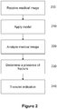
- An embodiment of the present invention provides a method of analyzing a medical image, the method comprises receiving a medical image; applying a model stored in a memory; analyzing the medical image based on the model; determining the medical image including a presence of fracture; and, transmitting an indication indicative of the determination.
- applying the model comprises receiving training data from a dataset, the training data including a plurality of training images, each training image includes diagnosis data; developing the model using the training data; and storing the model in a memory.
- the method further comprises identifying a portion of each training image, wherein the portion includes the diagnosis data; and developing the model using the training data and the portion identified.
- the method of analyzing the medical image disclosed herein is based on a model comprises augmenting the medical image.
- augmenting the medical image comprises at least one of zooming of the medical image; flipping the medical image horizontally; flipping the medical image vertically; or rotating the medical image.
- developing the model comprises using machine learning technique or deep neural network learning technique.
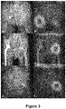
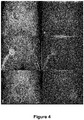
- the method further comprises identifying a lesion site.
- the processor is further configured to generate a heatmap to identify the lesion site.
- the fracture is a fracture in the hip or pelvic region and the medical image is frontal pelvic radiograph (PXR).
- PXR frontal pelvic radiograph
- PXR picture archiving and communication system
Landscapes
- Engineering & Computer Science (AREA)
- Health & Medical Sciences (AREA)
- Medical Informatics (AREA)
- Physics & Mathematics (AREA)
- General Health & Medical Sciences (AREA)
- Nuclear Medicine, Radiotherapy & Molecular Imaging (AREA)
- Radiology & Medical Imaging (AREA)
- Theoretical Computer Science (AREA)
- Life Sciences & Earth Sciences (AREA)
- General Physics & Mathematics (AREA)
- Computer Vision & Pattern Recognition (AREA)
- Public Health (AREA)
- Quality & Reliability (AREA)
- Optics & Photonics (AREA)
- Animal Behavior & Ethology (AREA)
- High Energy & Nuclear Physics (AREA)
- Pulmonology (AREA)
- Pathology (AREA)
- Biomedical Technology (AREA)
- Heart & Thoracic Surgery (AREA)
- Molecular Biology (AREA)
- Surgery (AREA)
- Biophysics (AREA)
- Geometry (AREA)
- Veterinary Medicine (AREA)
- Epidemiology (AREA)
- Primary Health Care (AREA)
- Image Analysis (AREA)
- Apparatus For Radiation Diagnosis (AREA)
- Measuring And Recording Apparatus For Diagnosis (AREA)
- Magnetic Resonance Imaging Apparatus (AREA)
- Medical Treatment And Welfare Office Work (AREA)
- Image Processing (AREA)
Applications Claiming Priority (1)
| Application Number | Priority Date | Filing Date | Title |
|---|---|---|---|
| US201862719664P | 2018-08-19 | 2018-08-19 |
Publications (1)
| Publication Number | Publication Date |
|---|---|
| EP3614337A1 true EP3614337A1 (en) | 2020-02-26 |
Family
ID=67659331
Family Applications (1)
| Application Number | Title | Priority Date | Filing Date |
|---|---|---|---|
| EP19192393.7A Withdrawn EP3614337A1 (en) | 2018-08-19 | 2019-08-19 | Method and system of analyzing medical images |
Country Status (5)
| Country | Link |
|---|---|
| US (1) | US11080852B2 (enExample) |
| EP (1) | EP3614337A1 (enExample) |
| JP (1) | JP7336309B2 (enExample) |
| SG (1) | SG10201907613PA (enExample) |
| TW (1) | TWI701680B (enExample) |
Families Citing this family (15)
| Publication number | Priority date | Publication date | Assignee | Title |
|---|---|---|---|---|
| KR102540998B1 (ko) * | 2019-01-18 | 2023-06-05 | 가톨릭대학교 산학협력단 | 영상 정복 기반 가상 내고정물 생성 방법 및 장치 |
| CN111325745B (zh) * | 2020-03-09 | 2023-08-25 | 北京深睿博联科技有限责任公司 | 骨折区域分析方法和装置、电子设备及可读存储介质 |
| JP2023531365A (ja) * | 2020-05-13 | 2023-07-24 | イオス・イメージング | 医用画像変換方法および関連付けられる医用画像3dモデルパーソナライズ方法 |
| JP7551370B2 (ja) * | 2020-07-15 | 2024-09-17 | キヤノンメディカルシステムズ株式会社 | 医用データ処理装置及び方法 |
| TWM616670U (zh) * | 2020-09-22 | 2021-09-11 | 網資科技股份有限公司 | 人工智慧輔助骨頭醫學影像判讀系統 |
| CN112329550A (zh) * | 2020-10-16 | 2021-02-05 | 中国科学院空间应用工程与技术中心 | 基于弱监督学习的受灾建筑快速定位评估方法及装置 |
| KR102672010B1 (ko) * | 2020-12-29 | 2024-06-04 | 고려대학교 산학협력단 | 인공지능을 이용한 안면부 골절 판독 장치 및 방법 |
| JP7687587B2 (ja) * | 2021-02-22 | 2025-06-03 | グローリー株式会社 | 画像処理装置、画像処理方法およびプログラム |
| TWI810680B (zh) * | 2021-10-18 | 2023-08-01 | 長庚醫療財團法人林口長庚紀念醫院 | 前後骨盆放射影像分析方法及系統 |
| KR102672531B1 (ko) * | 2021-11-09 | 2024-06-07 | 주식회사 피앤씨솔루션 | 딥러닝 기반의 포즈 추정을 활용한 의료 영상에서의 척추 위치 자동 추정 방법 및 장치 |
| JP7418018B2 (ja) * | 2021-11-12 | 2024-01-19 | iSurgery株式会社 | 診断支援装置、およびコンピュータプログラム |
| WO2023121510A1 (ru) * | 2021-12-20 | 2023-06-29 | Автономная некоммерческая организация высшего образования "Университет Иннополис" | Определение патологии органов грудной клетки на основе рентгеновских изображений |
| US20230222650A1 (en) * | 2022-01-11 | 2023-07-13 | Qatar Foundation For Education, Science And Community Development | Dianet: a deep learning based architecture to diagnose diabetes using retinal images only |
| CN114400089A (zh) * | 2022-01-20 | 2022-04-26 | 平安国际智慧城市科技股份有限公司 | 基于大数据的阅片数据分析方法、装置、设备及存储介质 |
| US20240112329A1 (en) * | 2022-10-04 | 2024-04-04 | HeHealth PTE Ltd. | Distinguishing a Disease State from a Non-Disease State in an Image |
Family Cites Families (13)
| Publication number | Priority date | Publication date | Assignee | Title |
|---|---|---|---|---|
| US7664298B2 (en) | 2003-03-25 | 2010-02-16 | Imaging Therapeutics, Inc. | Methods for the compensation of imaging technique in the processing of radiographic images |
| WO2005083635A1 (en) * | 2004-02-27 | 2005-09-09 | National University Of Singapore | Method and system for detection of bone fractures |
| US8538117B2 (en) * | 2009-04-07 | 2013-09-17 | Virginia Commonwealth University | Accurate pelvic fracture detection for X-ray and CT images |
| EP2741674A4 (en) * | 2011-08-12 | 2015-04-08 | Jointvue Llc | DEVICE AND METHODS FOR 3D ULTRASONOGRAPHY |
| US9480439B2 (en) | 2012-11-01 | 2016-11-01 | Virginia Commonwealth University | Segmentation and fracture detection in CT images |
| US10758198B2 (en) | 2014-02-25 | 2020-09-01 | DePuy Synthes Products, Inc. | Systems and methods for intra-operative image analysis |
| US10588589B2 (en) * | 2014-07-21 | 2020-03-17 | Zebra Medical Vision Ltd. | Systems and methods for prediction of osteoporotic fracture risk |
| CN113421652B (zh) | 2015-06-02 | 2024-06-28 | 推想医疗科技股份有限公司 | 对医疗数据进行分析的方法、训练模型的方法及分析仪 |
| US10275877B2 (en) | 2015-06-12 | 2019-04-30 | International Business Machines Corporation | Methods and systems for automatically determining diagnosis discrepancies for clinical images |
| US10127659B2 (en) | 2016-11-23 | 2018-11-13 | General Electric Company | Deep learning medical systems and methods for image acquisition |
| CN108309334B (zh) * | 2017-12-08 | 2023-05-30 | 李书纲 | 一种脊柱x线影像的数据处理方法 |
| CN108305248B (zh) | 2018-01-17 | 2020-05-29 | 慧影医疗科技(北京)有限公司 | 一种骨折识别模型的构建方法及应用 |
| CN108491770B (zh) * | 2018-03-08 | 2023-05-30 | 李书纲 | 一种基于骨折影像的数据处理方法 |
-
2019
- 2019-08-19 JP JP2019149904A patent/JP7336309B2/ja active Active
- 2019-08-19 US US16/544,479 patent/US11080852B2/en active Active
- 2019-08-19 EP EP19192393.7A patent/EP3614337A1/en not_active Withdrawn
- 2019-08-19 TW TW108129475A patent/TWI701680B/zh active
- 2019-08-19 SG SG10201907613PA patent/SG10201907613PA/en unknown
Non-Patent Citations (7)
| Title |
|---|
| BAR AMIR ET AL: "Compression fractures detection on CT", PROGRESS IN BIOMEDICAL OPTICS AND IMAGING, SPIE - INTERNATIONAL SOCIETY FOR OPTICAL ENGINEERING, BELLINGHAM, WA, US, vol. 10134, 3 March 2017 (2017-03-03), pages 1013440 - 1013440, XP060086687, ISSN: 1605-7422, ISBN: 978-1-5106-0027-0, DOI: 10.1117/12.2249635 * |
| CHENG CHI-TUNG ET AL: "Application of a deep learning algorithm for detection and visualization of hip fractures on plain pelvic radiographs", EUROPEAN RADIOLOGY, SPRINGER INTERNATIONAL, BERLIN, DE, vol. 29, no. 10, 1 April 2019 (2019-04-01), pages 5469 - 5477, XP036875428, ISSN: 0938-7994, [retrieved on 20190401], DOI: 10.1007/S00330-019-06167-Y * |
| MACIEJ A MAZUROWSKI ET AL: "Deep learning in radiology: an overview of the concepts and a survey of the state of the art", ARXIV.ORG, CORNELL UNIVERSITY LIBRARY, 201 OLIN LIBRARY CORNELL UNIVERSITY ITHACA, NY 14853, 10 February 2018 (2018-02-10), XP081218325 * |
| SELVARAJU RAMPRASAATH R ET AL: "Grad-CAM: Visual Explanations from Deep Networks via Gradient-Based Localization", 2017 IEEE INTERNATIONAL CONFERENCE ON COMPUTER VISION (ICCV), IEEE, 21 March 2017 (2017-03-21), pages 618 - 626, XP033282917, DOI: 10.1109/ICCV.2017.74 * |
| TOMITA NAOFUMI ET AL: "Deep neural networks for automatic detection of osteoporotic vertebral fractures on CT scans", COMPUTERS IN BIOLOGY AND MEDICINE, NEW YORK, NY, US, vol. 98, 8 May 2018 (2018-05-08), pages 8 - 15, XP085408028, ISSN: 0010-4825, DOI: 10.1016/J.COMPBIOMED.2018.05.011 * |
| URAKAWA TAKAAKI ET AL: "Detecting intertrochanteric hip fractures with orthopedist-level accuracy using a deep convolutional neural network", SKELETAL RADIOLOGY, SPRINGER, BERLIN, DE, vol. 48, no. 2, 28 June 2018 (2018-06-28), pages 239 - 244, XP036663366, ISSN: 0364-2348, [retrieved on 20180628], DOI: 10.1007/S00256-018-3016-3 * |
| WILLIAM GALE ET AL: "Detecting hip fractures with radiologist-level performance using deep neural networks", ARXIV.ORG, CORNELL UNIVERSITY LIBRARY, 201 OLIN LIBRARY CORNELL UNIVERSITY ITHACA, NY 14853, 17 November 2017 (2017-11-17), XP081289461 * |
Also Published As
| Publication number | Publication date |
|---|---|
| US20200058123A1 (en) | 2020-02-20 |
| TWI701680B (zh) | 2020-08-11 |
| SG10201907613PA (en) | 2020-03-30 |
| TW202015070A (zh) | 2020-04-16 |
| JP2020062378A (ja) | 2020-04-23 |
| JP7336309B2 (ja) | 2023-08-31 |
| US11080852B2 (en) | 2021-08-03 |
Similar Documents
| Publication | Publication Date | Title |
|---|---|---|
| US11080852B2 (en) | Method and system of analyzing medical images | |
| Cheng et al. | Application of a deep learning algorithm for detection and visualization of hip fractures on plain pelvic radiographs | |
| US10706545B2 (en) | Systems and methods for analysis of anatomical images | |
| Yang et al. | Diagnostic accuracy of deep learning in orthopaedic fractures: a systematic review and meta-analysis | |
| AU2004251359B2 (en) | Systems and methods for automated diagnosis and decision support for breast imaging | |
| EP3567525A1 (en) | Systems and methods for analysis of anatomical images each captured at a unique orientation | |
| AU2021396186A1 (en) | Methods of assessing lung disease in chest x-rays | |
| JP2020126598A (ja) | 人工知能の検出出力から疾患の進行を決定するシステムおよび方法 | |
| Sjogren et al. | Image segmentation and machine learning for detection of abdominal free fluid in focused assessment with sonography for trauma examinations: a pilot study | |
| Kang et al. | Prediction of bone mineral density in CT using deep learning with explainability | |
| Tariq et al. | Opportunistic screening for low bone density using abdominopelvic computed tomography scans | |
| ÖZİÇ et al. | Fully Automated Detection of Osteoporosis Stage on Panoramic Radiographs Using YOLOv5 Deep Learning Model and Designing a Graphical User Interface: MÜ ÖZİÇ et al. | |
| US20220277445A1 (en) | Artificial intelligence-based gastroscopic image diagnosis assisting system and method | |
| US11872069B2 (en) | Method for providing fracture-detection tool | |
| Sultana et al. | Efficient detection of knee osteoporosis using the swin transformer on x-ray images | |
| JP2021509977A (ja) | 医用画像評価のためのディープラーニングの応用 | |
| Velusamy et al. | Faster Region‐based Convolutional Neural Networks with You Only Look Once multi‐stage caries lesion from oral panoramic X‐ray images | |
| Lee et al. | Comparison of gray-scale inversion to improve detection of pulmonary nodules on chest X-rays between radiologists and a deep convolutional neural network | |
| Kumar et al. | Leveraging deep learning for accurate detection and precise localization of vertebral fractures in medical imaging | |
| Mahajan et al. | Audit of artificial intelligence algorithms and its impact in relieving shortage of specialist doctors | |
| Alike et al. | A two-step neural network-based guiding system for obtaining reliable radiographs for critical shoulder angle measurement | |
| Asghar et al. | CariesXplainer: Enhancing dental caries detection using Gradient-weighted Class Activation Mapping and transfer learning | |
| Nguyen et al. | Artificial intelligence system for predicting areal bone mineral density from plain X-rays | |
| Mousavinasab et al. | Deep learning for osteoporosis diagnosis using magnetic resonance images of lumbar vertebrae | |
| Chun et al. | Patch-Wise Approach with Vision Transformer for Detecting Implant Failure in Spinal Radiography |
Legal Events
| Date | Code | Title | Description |
|---|---|---|---|
| PUAI | Public reference made under article 153(3) epc to a published international application that has entered the european phase |
Free format text: ORIGINAL CODE: 0009012 |
|
| STAA | Information on the status of an ep patent application or granted ep patent |
Free format text: STATUS: THE APPLICATION HAS BEEN PUBLISHED |
|
| AK | Designated contracting states |
Kind code of ref document: A1 Designated state(s): AL AT BE BG CH CY CZ DE DK EE ES FI FR GB GR HR HU IE IS IT LI LT LU LV MC MK MT NL NO PL PT RO RS SE SI SK SM TR |
|
| AX | Request for extension of the european patent |
Extension state: BA ME |
|
| STAA | Information on the status of an ep patent application or granted ep patent |
Free format text: STATUS: REQUEST FOR EXAMINATION WAS MADE |
|
| 17P | Request for examination filed |
Effective date: 20200824 |
|
| RBV | Designated contracting states (corrected) |
Designated state(s): AL AT BE BG CH CY CZ DE DK EE ES FI FR GB GR HR HU IE IS IT LI LT LU LV MC MK MT NL NO PL PT RO RS SE SI SK SM TR |
|
| STAA | Information on the status of an ep patent application or granted ep patent |
Free format text: STATUS: EXAMINATION IS IN PROGRESS |
|
| 17Q | First examination report despatched |
Effective date: 20211005 |
|
| STAA | Information on the status of an ep patent application or granted ep patent |
Free format text: STATUS: THE APPLICATION IS DEEMED TO BE WITHDRAWN |
|
| 18D | Application deemed to be withdrawn |
Effective date: 20220216 |