EP3603546B1 - A kit for removing a tissue lesion - Google Patents
A kit for removing a tissue lesion Download PDFInfo
- Publication number
- EP3603546B1 EP3603546B1 EP19189139.9A EP19189139A EP3603546B1 EP 3603546 B1 EP3603546 B1 EP 3603546B1 EP 19189139 A EP19189139 A EP 19189139A EP 3603546 B1 EP3603546 B1 EP 3603546B1
- Authority
- EP
- European Patent Office
- Prior art keywords
- tissue
- kit
- coil
- electrode
- tube
- Prior art date
- Legal status (The legal status is an assumption and is not a legal conclusion. Google has not performed a legal analysis and makes no representation as to the accuracy of the status listed.)
- Active
Links
- 230000003902 lesion Effects 0.000 title claims description 46
- 238000002271 resection Methods 0.000 claims description 31
- 238000005520 cutting process Methods 0.000 claims description 26
- 230000007246 mechanism Effects 0.000 claims description 12
- 239000011295 pitch Substances 0.000 claims 6
- 210000001519 tissue Anatomy 0.000 description 97
- 210000004072 lung Anatomy 0.000 description 32
- 206010028980 Neoplasm Diseases 0.000 description 29
- 238000000034 method Methods 0.000 description 28
- 201000011510 cancer Diseases 0.000 description 21
- 210000004027 cell Anatomy 0.000 description 18
- 206010058467 Lung neoplasm malignant Diseases 0.000 description 9
- 201000005202 lung cancer Diseases 0.000 description 9
- 238000001356 surgical procedure Methods 0.000 description 9
- 208000020816 lung neoplasm Diseases 0.000 description 8
- 238000002266 amputation Methods 0.000 description 6
- 238000001574 biopsy Methods 0.000 description 6
- 238000002512 chemotherapy Methods 0.000 description 5
- 238000013188 needle biopsy Methods 0.000 description 5
- 230000037361 pathway Effects 0.000 description 5
- 241000208125 Nicotiana Species 0.000 description 4
- 235000002637 Nicotiana tabacum Nutrition 0.000 description 4
- 238000003745 diagnosis Methods 0.000 description 4
- 230000005855 radiation Effects 0.000 description 4
- 238000007789 sealing Methods 0.000 description 4
- 210000000115 thoracic cavity Anatomy 0.000 description 4
- 210000000779 thoracic wall Anatomy 0.000 description 4
- 238000013459 approach Methods 0.000 description 3
- 210000004204 blood vessel Anatomy 0.000 description 3
- 230000002068 genetic effect Effects 0.000 description 3
- 230000036541 health Effects 0.000 description 3
- 210000004224 pleura Anatomy 0.000 description 3
- 108090000623 proteins and genes Proteins 0.000 description 3
- 230000002159 abnormal effect Effects 0.000 description 2
- 230000004913 activation Effects 0.000 description 2
- 238000004873 anchoring Methods 0.000 description 2
- 230000008901 benefit Effects 0.000 description 2
- 230000005773 cancer-related death Effects 0.000 description 2
- 231100000504 carcinogenesis Toxicity 0.000 description 2
- 230000032823 cell division Effects 0.000 description 2
- 230000010261 cell growth Effects 0.000 description 2
- 230000034994 death Effects 0.000 description 2
- 231100000517 death Toxicity 0.000 description 2
- 201000010099 disease Diseases 0.000 description 2
- 208000037265 diseases, disorders, signs and symptoms Diseases 0.000 description 2
- 238000002224 dissection Methods 0.000 description 2
- 238000003384 imaging method Methods 0.000 description 2
- 238000003780 insertion Methods 0.000 description 2
- 230000037431 insertion Effects 0.000 description 2
- 230000004199 lung function Effects 0.000 description 2
- 230000003211 malignant effect Effects 0.000 description 2
- 210000000056 organ Anatomy 0.000 description 2
- 230000008520 organization Effects 0.000 description 2
- 230000000149 penetrating effect Effects 0.000 description 2
- 238000011084 recovery Methods 0.000 description 2
- 230000009467 reduction Effects 0.000 description 2
- 230000007480 spreading Effects 0.000 description 2
- 238000011144 upstream manufacturing Methods 0.000 description 2
- 230000033616 DNA repair Effects 0.000 description 1
- 208000026350 Inborn Genetic disease Diseases 0.000 description 1
- 208000004550 Postoperative Pain Diseases 0.000 description 1
- 108700020978 Proto-Oncogene Proteins 0.000 description 1
- 102000052575 Proto-Oncogene Human genes 0.000 description 1
- 206010056342 Pulmonary mass Diseases 0.000 description 1
- 108700025716 Tumor Suppressor Genes Proteins 0.000 description 1
- 102000044209 Tumor Suppressor Genes Human genes 0.000 description 1
- 238000002679 ablation Methods 0.000 description 1
- 230000005856 abnormality Effects 0.000 description 1
- 230000003213 activating effect Effects 0.000 description 1
- 238000003915 air pollution Methods 0.000 description 1
- 239000010425 asbestos Substances 0.000 description 1
- 239000008280 blood Substances 0.000 description 1
- 210000004369 blood Anatomy 0.000 description 1
- 239000003795 chemical substances by application Substances 0.000 description 1
- 238000004891 communication Methods 0.000 description 1
- 238000002591 computed tomography Methods 0.000 description 1
- 238000010276 construction Methods 0.000 description 1
- 239000007799 cork Substances 0.000 description 1
- 230000001419 dependent effect Effects 0.000 description 1
- 238000001514 detection method Methods 0.000 description 1
- 238000005516 engineering process Methods 0.000 description 1
- 230000007613 environmental effect Effects 0.000 description 1
- 230000006870 function Effects 0.000 description 1
- 208000016361 genetic disease Diseases 0.000 description 1
- 230000012010 growth Effects 0.000 description 1
- 238000013275 image-guided biopsy Methods 0.000 description 1
- 230000004807 localization Effects 0.000 description 1
- 230000007774 longterm Effects 0.000 description 1
- 201000005296 lung carcinoma Diseases 0.000 description 1
- 210000002751 lymph Anatomy 0.000 description 1
- 238000012986 modification Methods 0.000 description 1
- 230000004048 modification Effects 0.000 description 1
- HLXZNVUGXRDIFK-UHFFFAOYSA-N nickel titanium Chemical compound [Ti].[Ti].[Ti].[Ti].[Ti].[Ti].[Ti].[Ti].[Ti].[Ti].[Ti].[Ni].[Ni].[Ni].[Ni].[Ni].[Ni].[Ni].[Ni].[Ni].[Ni].[Ni].[Ni].[Ni].[Ni] HLXZNVUGXRDIFK-UHFFFAOYSA-N 0.000 description 1
- 229910001000 nickel titanium Inorganic materials 0.000 description 1
- 238000012856 packing Methods 0.000 description 1
- 230000002093 peripheral effect Effects 0.000 description 1
- 230000008569 process Effects 0.000 description 1
- 230000002062 proliferating effect Effects 0.000 description 1
- 229910052704 radon Inorganic materials 0.000 description 1
- SYUHGPGVQRZVTB-UHFFFAOYSA-N radon atom Chemical compound [Rn] SYUHGPGVQRZVTB-UHFFFAOYSA-N 0.000 description 1
- 229910052895 riebeckite Inorganic materials 0.000 description 1
- 238000005070 sampling Methods 0.000 description 1
- 230000035945 sensitivity Effects 0.000 description 1
- 239000012781 shape memory material Substances 0.000 description 1
- 239000000779 smoke Substances 0.000 description 1
- 230000000391 smoking effect Effects 0.000 description 1
- 206010041823 squamous cell carcinoma Diseases 0.000 description 1
- 208000017572 squamous cell neoplasm Diseases 0.000 description 1
- 239000000126 substance Substances 0.000 description 1
- 238000012360 testing method Methods 0.000 description 1
Images


Classifications
-
- A—HUMAN NECESSITIES
- A61—MEDICAL OR VETERINARY SCIENCE; HYGIENE
- A61B—DIAGNOSIS; SURGERY; IDENTIFICATION
- A61B18/00—Surgical instruments, devices or methods for transferring non-mechanical forms of energy to or from the body
- A61B18/04—Surgical instruments, devices or methods for transferring non-mechanical forms of energy to or from the body by heating
- A61B18/12—Surgical instruments, devices or methods for transferring non-mechanical forms of energy to or from the body by heating by passing a current through the tissue to be heated, e.g. high-frequency current
- A61B18/14—Probes or electrodes therefor
- A61B18/149—Probes or electrodes therefor bow shaped or with rotatable body at cantilever end, e.g. for resectoscopes, or coagulating rollers
-
- A—HUMAN NECESSITIES
- A61—MEDICAL OR VETERINARY SCIENCE; HYGIENE
- A61B—DIAGNOSIS; SURGERY; IDENTIFICATION
- A61B17/00—Surgical instruments, devices or methods, e.g. tourniquets
- A61B17/32—Surgical cutting instruments
- A61B17/3205—Excision instruments
- A61B17/32053—Punch like cutting instruments, e.g. using a cylindrical or oval knife
-
- A—HUMAN NECESSITIES
- A61—MEDICAL OR VETERINARY SCIENCE; HYGIENE
- A61B—DIAGNOSIS; SURGERY; IDENTIFICATION
- A61B18/00—Surgical instruments, devices or methods for transferring non-mechanical forms of energy to or from the body
- A61B18/04—Surgical instruments, devices or methods for transferring non-mechanical forms of energy to or from the body by heating
- A61B18/12—Surgical instruments, devices or methods for transferring non-mechanical forms of energy to or from the body by heating by passing a current through the tissue to be heated, e.g. high-frequency current
-
- A—HUMAN NECESSITIES
- A61—MEDICAL OR VETERINARY SCIENCE; HYGIENE
- A61B—DIAGNOSIS; SURGERY; IDENTIFICATION
- A61B10/00—Other methods or instruments for diagnosis, e.g. instruments for taking a cell sample, for biopsy, for vaccination diagnosis; Sex determination; Ovulation-period determination; Throat striking implements
- A61B10/02—Instruments for taking cell samples or for biopsy
- A61B10/0233—Pointed or sharp biopsy instruments
-
- A—HUMAN NECESSITIES
- A61—MEDICAL OR VETERINARY SCIENCE; HYGIENE
- A61B—DIAGNOSIS; SURGERY; IDENTIFICATION
- A61B17/00—Surgical instruments, devices or methods, e.g. tourniquets
- A61B17/32—Surgical cutting instruments
- A61B17/3205—Excision instruments
- A61B17/32056—Surgical snare instruments
-
- A—HUMAN NECESSITIES
- A61—MEDICAL OR VETERINARY SCIENCE; HYGIENE
- A61B—DIAGNOSIS; SURGERY; IDENTIFICATION
- A61B17/00—Surgical instruments, devices or methods, e.g. tourniquets
- A61B17/32—Surgical cutting instruments
- A61B17/3209—Incision instruments
-
- A—HUMAN NECESSITIES
- A61—MEDICAL OR VETERINARY SCIENCE; HYGIENE
- A61B—DIAGNOSIS; SURGERY; IDENTIFICATION
- A61B18/00—Surgical instruments, devices or methods for transferring non-mechanical forms of energy to or from the body
- A61B18/04—Surgical instruments, devices or methods for transferring non-mechanical forms of energy to or from the body by heating
- A61B18/12—Surgical instruments, devices or methods for transferring non-mechanical forms of energy to or from the body by heating by passing a current through the tissue to be heated, e.g. high-frequency current
- A61B18/14—Probes or electrodes therefor
-
- A—HUMAN NECESSITIES
- A61—MEDICAL OR VETERINARY SCIENCE; HYGIENE
- A61B—DIAGNOSIS; SURGERY; IDENTIFICATION
- A61B90/00—Instruments, implements or accessories specially adapted for surgery or diagnosis and not covered by any of the groups A61B1/00 - A61B50/00, e.g. for luxation treatment or for protecting wound edges
- A61B90/04—Protection of tissue around surgical sites against effects of non-mechanical surgery, e.g. laser surgery
-
- A—HUMAN NECESSITIES
- A61—MEDICAL OR VETERINARY SCIENCE; HYGIENE
- A61M—DEVICES FOR INTRODUCING MEDIA INTO, OR ONTO, THE BODY; DEVICES FOR TRANSDUCING BODY MEDIA OR FOR TAKING MEDIA FROM THE BODY; DEVICES FOR PRODUCING OR ENDING SLEEP OR STUPOR
- A61M29/00—Dilators with or without means for introducing media, e.g. remedies
- A61M29/02—Dilators made of swellable material
-
- A—HUMAN NECESSITIES
- A61—MEDICAL OR VETERINARY SCIENCE; HYGIENE
- A61B—DIAGNOSIS; SURGERY; IDENTIFICATION
- A61B17/00—Surgical instruments, devices or methods, e.g. tourniquets
- A61B17/12—Surgical instruments, devices or methods, e.g. tourniquets for ligaturing or otherwise compressing tubular parts of the body, e.g. blood vessels, umbilical cord
- A61B17/12009—Implements for ligaturing other than by clamps or clips, e.g. using a loop with a slip knot
- A61B17/12013—Implements for ligaturing other than by clamps or clips, e.g. using a loop with a slip knot for use in minimally invasive surgery, e.g. endoscopic surgery
-
- A—HUMAN NECESSITIES
- A61—MEDICAL OR VETERINARY SCIENCE; HYGIENE
- A61B—DIAGNOSIS; SURGERY; IDENTIFICATION
- A61B17/00—Surgical instruments, devices or methods, e.g. tourniquets
- A61B17/32—Surgical cutting instruments
- A61B17/320016—Endoscopic cutting instruments, e.g. arthroscopes, resectoscopes
-
- A—HUMAN NECESSITIES
- A61—MEDICAL OR VETERINARY SCIENCE; HYGIENE
- A61B—DIAGNOSIS; SURGERY; IDENTIFICATION
- A61B17/00—Surgical instruments, devices or methods, e.g. tourniquets
- A61B17/32—Surgical cutting instruments
- A61B17/3209—Incision instruments
- A61B17/32093—Incision instruments for skin incisions
-
- A—HUMAN NECESSITIES
- A61—MEDICAL OR VETERINARY SCIENCE; HYGIENE
- A61B—DIAGNOSIS; SURGERY; IDENTIFICATION
- A61B18/00—Surgical instruments, devices or methods for transferring non-mechanical forms of energy to or from the body
- A61B18/04—Surgical instruments, devices or methods for transferring non-mechanical forms of energy to or from the body by heating
- A61B18/12—Surgical instruments, devices or methods for transferring non-mechanical forms of energy to or from the body by heating by passing a current through the tissue to be heated, e.g. high-frequency current
- A61B18/14—Probes or electrodes therefor
- A61B18/1492—Probes or electrodes therefor having a flexible, catheter-like structure, e.g. for heart ablation
-
- A—HUMAN NECESSITIES
- A61—MEDICAL OR VETERINARY SCIENCE; HYGIENE
- A61B—DIAGNOSIS; SURGERY; IDENTIFICATION
- A61B17/00—Surgical instruments, devices or methods, e.g. tourniquets
- A61B2017/00743—Type of operation; Specification of treatment sites
- A61B2017/00809—Lung operations
-
- A—HUMAN NECESSITIES
- A61—MEDICAL OR VETERINARY SCIENCE; HYGIENE
- A61B—DIAGNOSIS; SURGERY; IDENTIFICATION
- A61B17/00—Surgical instruments, devices or methods, e.g. tourniquets
- A61B17/32—Surgical cutting instruments
- A61B2017/320044—Blunt dissectors
-
- A—HUMAN NECESSITIES
- A61—MEDICAL OR VETERINARY SCIENCE; HYGIENE
- A61B—DIAGNOSIS; SURGERY; IDENTIFICATION
- A61B17/00—Surgical instruments, devices or methods, e.g. tourniquets
- A61B17/32—Surgical cutting instruments
- A61B2017/320052—Guides for cutting instruments
-
- A—HUMAN NECESSITIES
- A61—MEDICAL OR VETERINARY SCIENCE; HYGIENE
- A61B—DIAGNOSIS; SURGERY; IDENTIFICATION
- A61B17/00—Surgical instruments, devices or methods, e.g. tourniquets
- A61B17/32—Surgical cutting instruments
- A61B2017/320064—Surgical cutting instruments with tissue or sample retaining means
-
- A—HUMAN NECESSITIES
- A61—MEDICAL OR VETERINARY SCIENCE; HYGIENE
- A61B—DIAGNOSIS; SURGERY; IDENTIFICATION
- A61B18/00—Surgical instruments, devices or methods for transferring non-mechanical forms of energy to or from the body
- A61B2018/00315—Surgical instruments, devices or methods for transferring non-mechanical forms of energy to or from the body for treatment of particular body parts
- A61B2018/00541—Lung or bronchi
-
- A—HUMAN NECESSITIES
- A61—MEDICAL OR VETERINARY SCIENCE; HYGIENE
- A61B—DIAGNOSIS; SURGERY; IDENTIFICATION
- A61B18/00—Surgical instruments, devices or methods for transferring non-mechanical forms of energy to or from the body
- A61B2018/00571—Surgical instruments, devices or methods for transferring non-mechanical forms of energy to or from the body for achieving a particular surgical effect
- A61B2018/00577—Ablation
-
- A—HUMAN NECESSITIES
- A61—MEDICAL OR VETERINARY SCIENCE; HYGIENE
- A61B—DIAGNOSIS; SURGERY; IDENTIFICATION
- A61B18/00—Surgical instruments, devices or methods for transferring non-mechanical forms of energy to or from the body
- A61B2018/00571—Surgical instruments, devices or methods for transferring non-mechanical forms of energy to or from the body for achieving a particular surgical effect
- A61B2018/00595—Cauterization
-
- A—HUMAN NECESSITIES
- A61—MEDICAL OR VETERINARY SCIENCE; HYGIENE
- A61B—DIAGNOSIS; SURGERY; IDENTIFICATION
- A61B18/00—Surgical instruments, devices or methods for transferring non-mechanical forms of energy to or from the body
- A61B18/04—Surgical instruments, devices or methods for transferring non-mechanical forms of energy to or from the body by heating
- A61B18/12—Surgical instruments, devices or methods for transferring non-mechanical forms of energy to or from the body by heating by passing a current through the tissue to be heated, e.g. high-frequency current
- A61B18/1206—Generators therefor
- A61B2018/1246—Generators therefor characterised by the output polarity
- A61B2018/126—Generators therefor characterised by the output polarity bipolar
-
- A—HUMAN NECESSITIES
- A61—MEDICAL OR VETERINARY SCIENCE; HYGIENE
- A61B—DIAGNOSIS; SURGERY; IDENTIFICATION
- A61B18/00—Surgical instruments, devices or methods for transferring non-mechanical forms of energy to or from the body
- A61B18/04—Surgical instruments, devices or methods for transferring non-mechanical forms of energy to or from the body by heating
- A61B18/12—Surgical instruments, devices or methods for transferring non-mechanical forms of energy to or from the body by heating by passing a current through the tissue to be heated, e.g. high-frequency current
- A61B18/14—Probes or electrodes therefor
- A61B2018/1405—Electrodes having a specific shape
- A61B2018/142—Electrodes having a specific shape at least partly surrounding the target, e.g. concave, curved or in the form of a cave
-
- A—HUMAN NECESSITIES
- A61—MEDICAL OR VETERINARY SCIENCE; HYGIENE
- A61B—DIAGNOSIS; SURGERY; IDENTIFICATION
- A61B18/00—Surgical instruments, devices or methods for transferring non-mechanical forms of energy to or from the body
- A61B18/04—Surgical instruments, devices or methods for transferring non-mechanical forms of energy to or from the body by heating
- A61B18/12—Surgical instruments, devices or methods for transferring non-mechanical forms of energy to or from the body by heating by passing a current through the tissue to be heated, e.g. high-frequency current
- A61B18/14—Probes or electrodes therefor
- A61B2018/1405—Electrodes having a specific shape
- A61B2018/1435—Spiral
-
- A—HUMAN NECESSITIES
- A61—MEDICAL OR VETERINARY SCIENCE; HYGIENE
- A61B—DIAGNOSIS; SURGERY; IDENTIFICATION
- A61B18/00—Surgical instruments, devices or methods for transferring non-mechanical forms of energy to or from the body
- A61B18/04—Surgical instruments, devices or methods for transferring non-mechanical forms of energy to or from the body by heating
- A61B18/12—Surgical instruments, devices or methods for transferring non-mechanical forms of energy to or from the body by heating by passing a current through the tissue to be heated, e.g. high-frequency current
- A61B18/14—Probes or electrodes therefor
- A61B2018/1467—Probes or electrodes therefor using more than two electrodes on a single probe
-
- A—HUMAN NECESSITIES
- A61—MEDICAL OR VETERINARY SCIENCE; HYGIENE
- A61B—DIAGNOSIS; SURGERY; IDENTIFICATION
- A61B18/00—Surgical instruments, devices or methods for transferring non-mechanical forms of energy to or from the body
- A61B18/04—Surgical instruments, devices or methods for transferring non-mechanical forms of energy to or from the body by heating
- A61B18/12—Surgical instruments, devices or methods for transferring non-mechanical forms of energy to or from the body by heating by passing a current through the tissue to be heated, e.g. high-frequency current
- A61B18/14—Probes or electrodes therefor
- A61B2018/1497—Electrodes covering only part of the probe circumference
Definitions
- the present invention relates to a kit for tissue resection. More particularly, the present invention relates to a kit for lesion removal.
- Cancer is not a single disease, but rather a collection of related diseases that can start essentially anywhere in the body.
- the body's cells begin to divide without stopping, proliferating and potentially spreading into surrounding tissues.
- cells grow and divide to form new cells as required by the body and when they become damaged or old, they die, and new cells replace the damaged or old cells; however, cancer interrupts this process.
- the cells become abnormal, and cells that should die do not and new cells form when they are not needed. These new cells can reproduce or proliferate without stopping and may form growths called tumors.
- Cancerous tumors are malignant, which means they can spread into or invade surrounding healthy tissue. In addition, cancer cells can break off and travel to remote areas in the body through blood or in the lymph system. Benign tumors, unlike malignant tumors, do not spread or invade surrounding tissue; however, they may grow large and cause damage. Both malignant and benign tumors may be removed or treated. Malignant tumors tend to grow back whereas benign tumors can grow back but are much less likely to do so.
- Cancer is a genetic disease in that it is caused by changes in the genes that control the ways that cells function, especially in how they grow and divide. Genetic changes that cause cancer may be inherited or they may arise over an individual's lifetime as a result of errors that occur as cells divide or because of damage to DNA caused by certain environmental exposure, for example, industrial/commercial chemicals and ultraviolet light.
- the genetic changes that may cause cancer tend to affect three types of genes; namely proto-oncogenes which are involved in normal cell growth and division, tumor suppressor genes which are also involved in controlling cell growth and division, and DNA repair genes which, as the name implies, are involved in repairing damaged DNA.
- the type of cancer may be named for the organ or tissue where the cancers arise, for example, lung cancer, or the type of cell that formed them, for example squamous cell cancer.
- Cancer is a leading cause of death both in the United States and world-wide. According to the World Health Organization, the number of new cancer cases will rise to twenty-five (25) million per year over the next two decades.
- Lung cancer is one of the most common cancers today. According to the World Cancer Report 2014 from the World Health Organization, lung cancer occurred in 14 million people and resulted in 8.8 million deaths world-wide, making it the most common cause of cancer-related death in men and the second most common cause of cancer-related death in women. Lung cancer or lung carcinoma is a malignant lung tumor that if left untreated can metastasize into neighboring tissues and organs. The majority of lung cancer is caused by long-term tobacco smoking; however, about 10 to 15 percent of lung cancer cases are not tobacco related. These non-tobacco cases are most often caused by a combination of genetic factors and exposure to certain environmental conditions, including radon gas, asbestos, second-hand tobacco smoke, other forms of air pollution, and other agents. The chance of surviving lung cancer as well as other forms of cancer depends on early detection and treatment.
- a biopsy is performed and sent for study. If it is determined that the lesion is cancerous, a second procedure may be performed to remove the cancer. If the biopsy reveals no cancer, it may be correct, or the biopsy did not pick the cancerous cells. Accordingly, there exists a need for removing the whole lesion in one single procedure so that an accurate diagnosis may be performed.
- US 2018140319 discloses apparatus and methods for tissue reduction where one embodiment of the tissue resection device may generally comprise an external clamping mechanism having at least two distally extending members shaped with an atraumatic distal end and defining a confined region between the distally extending members sized to receive a tissue region therebetween. Additionally, an elongate coring needle defining a lumen and having a proximal end attached to a handle and a distal end which defines a cutting mechanism may also be included, wherein the coring needle is translatable adjacent and/or relative to the external or internal clamping mechanism defined by the confined region.
- WO 2011094110 discloses a surgical instrument configured to deliver electrical energy to the tissue of a patient, comprising a first electrode comprising a distal portion configured to contact the tissue and a second electrode comprising a distal portion configured to be inserted into the tissue, wherein the distal portion of the second electrode at least partially encompasses the distal portion of the first electrode.
- DE 102016101915 discloses an incision instrument for performing incisions in a standardized manner in the opening of an operation field, comprising a handle element with a cutting blade receiving element formed at a distal end of the handle element, a cutting blade which is removably receivable in the cutting blade receiving element as well as a guide channel that extends in the longitudinal direction of the instrument for receiving a guide wire therein.
- the kit for performing lung lesion removal of the present invention overcomes the limitations associated with the prior art.
- the present invention relates to a kit for performing lung lesion removal.
- a lung needle biopsy is typically performed when an abnormality is found on an imaging test, for example, an X-ray or CAT scan.
- a fine needle is used to remove sample of lung tissue for examining under a microscope to determine the presence of abnormal cells.
- Tissue diagnosis is challenging in small ( ⁇ 6mm) and intermediate (6-12mm) nodules.
- CT guided biopsy of peripheral lesions either through the chest wall (80%) or by means of a bronchoscope (20%) yields only a .001-.002 cm2 of diagnostic tissue, and as such, cancer, when present, is only successfully identified in 60% of small and intermediate nodules.
- bronchoscopic techniques and technology continue to evolve, biopsy accuracy, specificity, and sensitivity will always be limited when dealing with small and intermediate nodules in the periphery of the lung.
- a second procedure may be performed to remove the lesion and then followed up with chemotherapy and/or radiation.
- the second procedure most likely involves lung surgery. These procedures are typically done through an incision between the ribs. There are a number of possible procedures depending on the state of the cancer.
- Video-assisted thoracic surgery is a less invasive procedure for certain types of lung cancer. It is performed through small incisions utilizing an endoscopic approach and is typically utilized for performing wedge resections of smaller lesions close to the surface of a lung. In a wedge resection, a portion of the lobe is removed. In a sleeve resection, a portion of a large airway is removed thereby preserving more lung function.
- surgeons perform an open thoracotomy or lobectomy to remove lung nodules that are 2-3 cm from the lung surface.
- a thoracotomy is an open approach surgery in which a portion of a lobe, a full lobe or an entire lung is removed.
- a pneumonectomy an entire lung is removed. This type of surgery is obviously the most aggressive.
- a lobectomy an entire section or lobe of a lung is removed and represents a less aggressive approach than removing the entire lung. All thoracoscopic lung surgeries require trained and experienced thoracic surgeons and the favorability of surgical outcomes track with operative experience.
- any of these types of lung surgery is a major operation with possible complications which depend on the extent of the surgery as well as the patient's overall health.
- the recovery may take from weeks to months.
- a thoracotomy spreading of the ribs is required, thereby increasing postoperative pain.
- video-assisted thoracic surgery is less invasive, there can still be a substantial recovery period.
- full treatment may require a system chemotherapy and/or radiation treatment.
- a fine needle biopsy may not prove to be totally diagnostic.
- the fine needle biopsy procedure involves guiding a needle in three-dimensional space under two-dimensional imaging. Accordingly, the doctor may miss the lesion, or even if he or she hits the correct target, the section of the lesion that is removed through the needle may not contain the cancerous cells or the cells necessary to assess the aggressiveness of the tumor.
- a needle biopsy removes enough tissue to create a smear on a slide.
- the device of the present invention is designed to remove the entire lesion, or a substantial portion of it, while minimizing the amount of healthy lung tissue removal. This offers a number of advantages. Firstly, the entire lesion may be examined for a more accurate diagnosis without confounding sampling error, loss of cell packing or gross architecture. Secondly, since the entire lesion is removed, a secondary procedure as described above may not be required. Thirdly, localized chemotherapy and/or energy-based tumor extirpation, such as radiation, may be introduced via the cavity created by the lesion removal.
- a kit for removing a tissue lesion is provided with an anchor, one or more incision blades, a tissue dilator, and a tissue resection mechanism.
- the tissue resection mechanism includes an outer tube having a helical coil disposed on a distal end where the coil includes a first electrode.
- a central tube is provided having a distal edge profile including one or more surface segments, at least one of surface segments includes a second electrode.
- the central tube is slidably disposed within the outer tube and is position such that the second electrode opposes at least a portion the first electrode.
- a cutting tube includes a cutting edge slidably disposed within the central tube and the cutting tube is configured to advance at least as far as one of the coil segments.
- the invention encompasses a kit for removing a tissue lesion is provided with one or more incision blades, a tissue dilator, and a tissue resection mechanism.
- the tissue resection mechanism includes an outer tube having a helical coil disposed on a distal end where the coil includes a first electrode.
- a central tube is provided having a distal edge profile including one or more surface segments, at least one of surface segments includes a second electrode.
- the central tube is slidably disposed within the outer tube and is position such that the second electrode opposes at least a portion the first electrode.
- a cutting tube includes a cutting edge slidably disposed within the central tube and the cutting tube is configured to advance at least as far as one of the coil segments.
- the invention encompasses a kit for removing a tissue lesion is provided with an anchor, one or more incision blades, and a tissue resection mechanism.
- the tissue resection mechanism includes an outer tube having a helical coil disposed on a distal end where the coil includes a first electrode.
- a central tube is provided having a distal edge profile including one or more surface segments, at least one of surface segments includes a second electrode.
- the central tube is slidably disposed within the outer tube and is position such that the second electrode opposes at least a portion the first electrode.
- a cutting tube includes a cutting edge slidably disposed within the central tube and the cutting tube is configured to advance at least as far as one of the coil segments.
- tissue resection device 1100 includes an outer tube 1105 is provided having a distal edge profile and having an inner diameter ID outer .
- a coil 1110 is attached to outer tube 1105 where the coil turns are spaced from and opposed to a distal end of outer tube 1105.
- Coil 1110 preferably has a slightly blunted tip 1115 to minimize the possibility that it will penetrate through a blood vessel while being sufficiently sharp to penetrate tissue such as pleura and parenchyma.
- coil 1110 may take the form of a helix having a constant or variable pitch. Coil 1110 may also have a variable cross-sectional geometry.
- An electrode 1130 is disposed on a surface or embedded within coil 1110.
- coil 1110 may include a plurality of contiguous coil segments, e.g., coil segments 1120 and 1125.
- Coil segment 1120 comprises a helical member having a pitch of zero, e.g., a generally planar open ring structure, having an inner diameter ID coil and an outer diameter OD coil .
- Coil segment 1125 comprises a helical structure of constant or variable pitch and constant or variable cross-sectional geometry.
- electrode 1130 may be disposed on a surface of or embedded in coil segment 1120.
- a central tube 1200 having a distal end with an edge profile comprising one or more surface segments and having an outer diameter OD central and an inner diameter ID central .
- an electrode 1205 is disposed on or embedded within at least one of the surface segments.
- Central tube 1200 is slidably disposed within outer tube 1105 and positioned such that electrode 1205 opposes and overlaps at least a portion of electrode 1130.
- the space between electrode 1205 and electrode 1130 is referred to as the tissue clamping zone.
- OD central > ID coil and OD coil > ID central .
- OD central is about equal to OD coil . Accordingly, Central tube 1200 may be advanced through the tissue clamping zone towards coil 1110 such that electrode 1205 abuts electrode 1130.
- a cutting tube 1300 is slidably disposed within central tube 1200.
- the distal end of cutting tube 1300 is provided with a knife edge to facilitate tissue cutting.
- the resection device 1100 may be inserted into tissue and outer tube 1105 may be advanced a predetermined distance towards a target.
- Coil segment 1125 allows the device to penetrate the tissue in a manner similar to a cork screw. As coil segment 1125 penetrates tissue, any vessel in its path is either moved to planar coil segment 1120 or pushed away from the coil 1100 for subsequent turns. Coil tip 1115 is made blunt enough to minimize chances that it will penetrate through a blood vessel while still sharp enough to penetrate certain tissue such as the lung pleura and parenchyma.
- Central tube 1200 may then be advanced a predetermined distance towards the target. Any vessels that are disposed in the tissue clamping zone will be clamped between electrode 1130 and electrode 1205.
- the vessels can then be sealed by the application of bipolar energy to electrode 1130 and electrode 1205.
- cutting tube 1300 is advanced to core the tissue to the depth that outer tube 1105 has reached. The sealing and cutting process can be repeated to create a core of desired size.
- the resection device may be further configured to dissect a target lesion and seal tissue proximate the dissection point.
- central tube 1200 is provided with a ligation snare 1230, first and second ligation electrodes 1215 and 1220, an amputation snare 1225 and a ligation snare 1230.
- the word "snare” refers to a flexible line, e.g., a string or a wire.
- the inner wall surface of central tube 1200 includes upper and lower circumferential grooved pathways 1212 and 1214 disposed proximate the distal end.
- the first and second ligation electrodes 1215 and 1220 are disposed on the inner wall of central tube 1200 such that lower circumferential groove 1214 is between them.
- Upper grooved pathway 1212 is disposed axially above ligation electrodes 1215 and 1220.
- Ligation snare 1230 is disposed in lower circumferential groove 1214 and extends through central tube 1200 and axially along the outer wall surface to a snare activation mechanism (not shown).
- Amputation snare 1225 is disposed in upper circumferential groove 1212 and extends through central tube 1200 and axially along the outer wall surface to a snare activation mechanism (not shown).
- the outer surface of central tube 1200 may be provided with a plurality of axially extending grooved pathways which receive amputation snare 1225, ligation snare 1230 and are in communication with upper and lower circumferential grooved pathways 1212 and 1214.
- electrode leads for ligation electrodes 1215 and 1220 may extend to an energy source via the axially extending grooved pathways.
- the resection device of this embodiment can detach and seal the tissue core.
- Cutting tube 1300 may be retracted to expose ligation snare 1230 which is preferably made of flexible line, e.g., suture.
- Ligation snare 1230 may be engaged to snag tissue and pull tissue against the inner wall surface between first and second ligation electrodes 1215 and 1220.
- Bipolar energy is then applied to first and second electrodes 1215 and 1220 to seal, i.e., cauterize, the tissue.
- cutting tube 1300 may be further retracted to expose amputation snare 1225 which may then be activated to sever the tissue core upstream from the point where the tissue was sealed (ligation point).
- amputation snare 1225 has a smaller diameter than that of ligation snare 1230.
- the smaller diameter facilitates tissue slicing. Accordingly, the resection device 1100 according to this embodiment both creates a tissue core and disengages the core from surrounding tissue.
- the resection device of the invention is provided with a single snare disposed between ligation electrodes which both ligates and cuts tissue.
- the single snare first pulls tissue against the inner wall surface of central tube 1200 between ligation electrodes 1215 and 1220. Bipolar energy is then applied to first and second electrodes 1215 and 1220 to seal, i.e., cauterize, the tissue. Once sealed, the snare is further pulled to sever the tissue core.
- ligation snare 1230 includes a set of knots 1235 and 1240 which tighten under load, shown, for example, in Figure 4 .
- Ligation is performed by retracting cutting tube 1300 to expose ligation snare 1210 and activating ligation snare 1230 which lassos tissue as ligation knot tightens. Once the tissue is lassoed, cutting tube 1300 may be further retracted to expose amputation snare 1225 which may then be activated to sever the tissue core upstream from the point where the point where the tissue was lassoed.
- the present disclosure also contemplates an exemplary method and system for using the resection device to remove tissue lesions, for example, lung lesions.
- the exemplary method generally comprises anchoring the lesion targeted for removal, creating a channel in the tissue leading to the target lesion, creating a tissue core which includes the anchored lesion, ligating the tissue core and sealing the surrounding tissue, and removing the tissue core including the target lesion from the channel.
- Anchoring may be performed by, any suitable structure for securing the device to the lung.
- a channel may be created to facilitate insertion of resection device 1100.
- the channel may be created by making an incision in the lung area and inserting a tissue dilator and port into the incision.
- a tissue core which includes the anchored lesion may be created.
- Resection device 1100 may be inserted into the channel and used to create the tissue core, to ligate the tissue core and to seal the tissue core and sever it from the surrounding tissue as described hereinabove. The tissue core may then be removed from the channel.
- a cavity port may be inserted in the channel to facilitate subsequent treatment of the target lesion site through chemotherapy and/or energy-based tumor extirpation such as radiation.
- the anchor depicted in Figure 5 is suitable for use in performing the exemplary method for removing tissue lesions described herein.
- the anchor comprises an outer tube 1422 having a sufficiently sharp edge to pierce the chest cavity tissue and lung without causing excess damage and an inner tube 1424 disposed within outer tube 1422.
- One or more tines or fingers 1426 formed from shape memory material, e.g., Nitinol, preformed are attached to the end of inner tube 1424.
- Outer tube 1422 is retractably disposed over inner tube 1424 such that when outer tube 1422 is retracted, tines 1426 assume their preform shape as shown.
- Outer tube 1422 is retracted after it has pierced the lung lesion thereby causing tines 1426 to engage the lung lesion.
- Other suitable anchors may include coils and suction-based structures.
- the incision blades depicted in Figure 6 are suitable for use in performing the exemplary method for removing tissue lesions described herein.
- anchor 1400 it is preferable to create a small cut or incision to facilitate insertion of chest wall tissue dilator.
- Incision blades 1605 are used to make a wider cut.
- Successive incision blades 1605 include a central aperture which allows them to be coaxially advanced along the anchor needle 1405 to create a wider cut in the chest wall, with each successive blade being larger than the previous blade, thereby increasing the width of the incision.
- the tissue dilator depicted in Figure 7 is suitable for use in performing the exemplary method for removing tissue lesions described herein.
- the tissue dilator may comprise any suitable device for creating a channel in organic tissue.
- the tissue dilator assembly includes a single cylindrical rod with rounded end 1510 or a cylindrical rod with rounded end and a rigid sleeve arrangement 1515. Successive tissue dilators are coaxially advanced along the anchor needle to create
- tissue resection device capable of penetrating lung tissue and creating a tissue core including a target lesion is suitable for use in performing the exemplary method for removing tissue lesions described herein.
- Tissue resection device 1100 described hereinbefore is preferred.
- tissue resection device 1100 Once tissue resection device 1100 is removed, a small channel in the lung exits where the target lesion was removed.
- This channel may be utilized to introduce an energy-based ablation device and/or localized chemotherapy depending on the results of the tissue diagnosis. Accordingly, the exemplary method and system of the present invention may not only be utilized to ensure an effective biopsy is performed but also complete removal of the lesion with minimal healthy lung tissue removal.
Description
- The present invention relates to a kit for tissue resection. More particularly, the present invention relates to a kit for lesion removal.
- Cancer is not a single disease, but rather a collection of related diseases that can start essentially anywhere in the body. Common amongst all types of cancer is that the body's cells begin to divide without stopping, proliferating and potentially spreading into surrounding tissues. In the normal course of events, cells grow and divide to form new cells as required by the body and when they become damaged or old, they die, and new cells replace the damaged or old cells; however, cancer interrupts this process. With cancer, the cells become abnormal, and cells that should die do not and new cells form when they are not needed. These new cells can reproduce or proliferate without stopping and may form growths called tumors.
- Cancerous tumors are malignant, which means they can spread into or invade surrounding healthy tissue. In addition, cancer cells can break off and travel to remote areas in the body through blood or in the lymph system. Benign tumors, unlike malignant tumors, do not spread or invade surrounding tissue; however, they may grow large and cause damage. Both malignant and benign tumors may be removed or treated. Malignant tumors tend to grow back whereas benign tumors can grow back but are much less likely to do so.
- Cancer is a genetic disease in that it is caused by changes in the genes that control the ways that cells function, especially in how they grow and divide. Genetic changes that cause cancer may be inherited or they may arise over an individual's lifetime as a result of errors that occur as cells divide or because of damage to DNA caused by certain environmental exposure, for example, industrial/commercial chemicals and ultraviolet light. The genetic changes that may cause cancer tend to affect three types of genes; namely proto-oncogenes which are involved in normal cell growth and division, tumor suppressor genes which are also involved in controlling cell growth and division, and DNA repair genes which, as the name implies, are involved in repairing damaged DNA.
- More than one-hundred distinct types of cancer have been identified. The type of cancer may be named for the organ or tissue where the cancers arise, for example, lung cancer, or the type of cell that formed them, for example squamous cell cancer. Cancer, unfortunately, is a leading cause of death both in the United States and world-wide. According to the World Health Organization, the number of new cancer cases will rise to twenty-five (25) million per year over the next two decades.
- Lung cancer is one of the most common cancers today. According to the World Cancer Report 2014 from the World Health Organization, lung cancer occurred in 14 million people and resulted in 8.8 million deaths world-wide, making it the most common cause of cancer-related death in men and the second most common cause of cancer-related death in women. Lung cancer or lung carcinoma is a malignant lung tumor that if left untreated can metastasize into neighboring tissues and organs. The majority of lung cancer is caused by long-term tobacco smoking; however, about 10 to 15 percent of lung cancer cases are not tobacco related. These non-tobacco cases are most often caused by a combination of genetic factors and exposure to certain environmental conditions, including radon gas, asbestos, second-hand tobacco smoke, other forms of air pollution, and other agents. The chance of surviving lung cancer as well as other forms of cancer depends on early detection and treatment.
- When a lesion is detected in the lungs, a biopsy is performed and sent for study. If it is determined that the lesion is cancerous, a second procedure may be performed to remove the cancer. If the biopsy reveals no cancer, it may be correct, or the biopsy did not pick the cancerous cells. Accordingly, there exists a need for removing the whole lesion in one single procedure so that an accurate diagnosis may be performed.
-
US 2018140319 discloses apparatus and methods for tissue reduction where one embodiment of the tissue resection device may generally comprise an external clamping mechanism having at least two distally extending members shaped with an atraumatic distal end and defining a confined region between the distally extending members sized to receive a tissue region therebetween. Additionally, an elongate coring needle defining a lumen and having a proximal end attached to a handle and a distal end which defines a cutting mechanism may also be included, wherein the coring needle is translatable adjacent and/or relative to the external or internal clamping mechanism defined by the confined region. -
WO 2011094110 discloses a surgical instrument configured to deliver electrical energy to the tissue of a patient, comprising a first electrode comprising a distal portion configured to contact the tissue and a second electrode comprising a distal portion configured to be inserted into the tissue, wherein the distal portion of the second electrode at least partially encompasses the distal portion of the first electrode. -
DE 102016101915 discloses an incision instrument for performing incisions in a standardized manner in the opening of an operation field, comprising a handle element with a cutting blade receiving element formed at a distal end of the handle element, a cutting blade which is removably receivable in the cutting blade receiving element as well as a guide channel that extends in the longitudinal direction of the instrument for receiving a guide wire therein. - The invention is defined by claim 1. Further embodiments are defined by the dependent claims.
- The kit for performing lung lesion removal of the present invention overcomes the limitations associated with the prior art.
- The present invention relates to a kit for performing lung lesion removal. A lung needle biopsy is typically performed when an abnormality is found on an imaging test, for example, an X-ray or CAT scan. In a lung needle biopsy, a fine needle is used to remove sample of lung tissue for examining under a microscope to determine the presence of abnormal cells. Tissue diagnosis is challenging in small (<6mm) and intermediate (6-12mm) nodules. CT guided biopsy of peripheral lesions, either through the chest wall (80%) or by means of a bronchoscope (20%) yields only a .001-.002 cm2 of diagnostic tissue, and as such, cancer, when present, is only successfully identified in 60% of small and intermediate nodules. Although bronchoscopic techniques and technology continue to evolve, biopsy accuracy, specificity, and sensitivity will always be limited when dealing with small and intermediate nodules in the periphery of the lung.
- If it is determined that the lesion is cancerous, a second procedure may be performed to remove the lesion and then followed up with chemotherapy and/or radiation. The second procedure most likely involves lung surgery. These procedures are typically done through an incision between the ribs. There are a number of possible procedures depending on the state of the cancer. Video-assisted thoracic surgery is a less invasive procedure for certain types of lung cancer. It is performed through small incisions utilizing an endoscopic approach and is typically utilized for performing wedge resections of smaller lesions close to the surface of a lung. In a wedge resection, a portion of the lobe is removed. In a sleeve resection, a portion of a large airway is removed thereby preserving more lung function.
- Nodules deeper than 2-3 cm from the lung surface, once identified as suspicious for cancer, are difficult to localize and excise using laparoscopic or robotic lung sparing technique despite pre-procedure image guided biopsy and localization. Thus, surgeons perform an open thoracotomy or lobectomy to remove lung nodules that are 2-3 cm from the lung surface. A thoracotomy is an open approach surgery in which a portion of a lobe, a full lobe or an entire lung is removed. In a pneumonectomy, an entire lung is removed. This type of surgery is obviously the most aggressive. In a lobectomy, an entire section or lobe of a lung is removed and represents a less aggressive approach than removing the entire lung. All thoracoscopic lung surgeries require trained and experienced thoracic surgeons and the favorability of surgical outcomes track with operative experience.
- Any of these types of lung surgery is a major operation with possible complications which depend on the extent of the surgery as well as the patient's overall health. In addition to the reduction in lung function associated with any of these procedures, the recovery may take from weeks to months. With a thoracotomy, spreading of the ribs is required, thereby increasing postoperative pain. Although video-assisted thoracic surgery is less invasive, there can still be a substantial recovery period. In addition, once the surgery is complete, full treatment may require a system chemotherapy and/or radiation treatment.
- As set forth above, a fine needle biopsy may not prove to be totally diagnostic. The fine needle biopsy procedure involves guiding a needle in three-dimensional space under two-dimensional imaging. Accordingly, the doctor may miss the lesion, or even if he or she hits the correct target, the section of the lesion that is removed through the needle may not contain the cancerous cells or the cells necessary to assess the aggressiveness of the tumor. A needle biopsy removes enough tissue to create a smear on a slide. The device of the present invention is designed to remove the entire lesion, or a substantial portion of it, while minimizing the amount of healthy lung tissue removal. This offers a number of advantages. Firstly, the entire lesion may be examined for a more accurate diagnosis without confounding sampling error, loss of cell packing or gross architecture. Secondly, since the entire lesion is removed, a secondary procedure as described above may not be required. Thirdly, localized chemotherapy and/or energy-based tumor extirpation, such as radiation, may be introduced via the cavity created by the lesion removal.
- In at least one embodiment, a kit for removing a tissue lesion is provided with an anchor, one or more incision blades, a tissue dilator, and a tissue resection mechanism. The tissue resection mechanism includes an outer tube having a helical coil disposed on a distal end where the coil includes a first electrode. A central tube is provided having a distal edge profile including one or more surface segments, at least one of surface segments includes a second electrode. The central tube is slidably disposed within the outer tube and is position such that the second electrode opposes at least a portion the first electrode. A cutting tube includes a cutting edge slidably disposed within the central tube and the cutting tube is configured to advance at least as far as one of the coil segments.
- In another embodiment the invention encompasses a kit for removing a tissue lesion is provided with one or more incision blades, a tissue dilator, and a tissue resection mechanism. The tissue resection mechanism includes an outer tube having a helical coil disposed on a distal end where the coil includes a first electrode. A central tube is provided having a distal edge profile including one or more surface segments, at least one of surface segments includes a second electrode. The central tube is slidably disposed within the outer tube and is position such that the second electrode opposes at least a portion the first electrode. A cutting tube includes a cutting edge slidably disposed within the central tube and the cutting tube is configured to advance at least as far as one of the coil segments.
- In still another embodiment the invention encompasses a kit for removing a tissue lesion is provided with an anchor, one or more incision blades, and a tissue resection mechanism. The tissue resection mechanism includes an outer tube having a helical coil disposed on a distal end where the coil includes a first electrode. A central tube is provided having a distal edge profile including one or more surface segments, at least one of surface segments includes a second electrode. The central tube is slidably disposed within the outer tube and is position such that the second electrode opposes at least a portion the first electrode. A cutting tube includes a cutting edge slidably disposed within the central tube and the cutting tube is configured to advance at least as far as one of the coil segments.
- The foregoing and other features and advantages of the invention will be apparent from the following, more particular description of preferred embodiments of the invention, as illustrated in the accompanying drawings.
-
Figure 1 depicts a tissue resection device in accordance with an embodiment of the invention. -
Figure 2 illustrates a sectional view of the tissue resection device ofFigure 1 . -
Figure 3 shows a sectional view of a tissue resection device in accordance with an embodiment of the invention. -
Figure 4 depicts a sectional view of a tissue resection device in accordance with an embodiment of the invention. -
Figure 5 illustrates an exemplary anchor that may be employed in a lesion removal exemplary method. -
Figure 6 shows a series of incision blades for use in a lesion removal exemplary method. -
Figure 7 displays tissue dilators suitable for use in a lesion removal exemplary method. - The resection device of the present invention comprises an energy-based arrangement capable of penetrating tissue towards a target lesion. In one embodiment depicted in
Figure 1 ,tissue resection device 1100 includes anouter tube 1105 is provided having a distal edge profile and having an inner diameter IDouter. Acoil 1110 is attached toouter tube 1105 where the coil turns are spaced from and opposed to a distal end ofouter tube 1105.Coil 1110 preferably has a slightly bluntedtip 1115 to minimize the possibility that it will penetrate through a blood vessel while being sufficiently sharp to penetrate tissue such as pleura and parenchyma. In some embodiments,coil 1110 may take the form of a helix having a constant or variable pitch.Coil 1110 may also have a variable cross-sectional geometry. Anelectrode 1130 is disposed on a surface or embedded withincoil 1110. - In some embodiments, as illustrated in
Figure 1 ,coil 1110 may include a plurality of contiguous coil segments, e.g.,coil segments Coil segment 1120 comprises a helical member having a pitch of zero, e.g., a generally planar open ring structure, having an inner diameter IDcoil and an outer diameter ODcoil.Coil segment 1125 comprises a helical structure of constant or variable pitch and constant or variable cross-sectional geometry. In this embodiment,electrode 1130 may be disposed on a surface of or embedded incoil segment 1120. - A
central tube 1200 is provided having a distal end with an edge profile comprising one or more surface segments and having an outer diameter ODcentral and an inner diameter IDcentral. As illustrated inFigure 2 , anelectrode 1205 is disposed on or embedded within at least one of the surface segments.Central tube 1200 is slidably disposed withinouter tube 1105 and positioned such thatelectrode 1205 opposes and overlaps at least a portion ofelectrode 1130. The space betweenelectrode 1205 andelectrode 1130 is referred to as the tissue clamping zone. In keeping with an aspect of the invention, ODcentral > IDcoil and ODcoil > IDcentral. In some embodiments, ODcentral is about equal to ODcoil. Accordingly,Central tube 1200 may be advanced through the tissue clamping zone towardscoil 1110 such thatelectrode 1205 abutselectrode 1130. - A cutting
tube 1300 is slidably disposed withincentral tube 1200. The distal end of cuttingtube 1300 is provided with a knife edge to facilitate tissue cutting. - To enable tissue resection, the
resection device 1100 may be inserted into tissue andouter tube 1105 may be advanced a predetermined distance towards a target.Coil segment 1125 allows the device to penetrate the tissue in a manner similar to a cork screw. Ascoil segment 1125 penetrates tissue, any vessel in its path is either moved toplanar coil segment 1120 or pushed away from thecoil 1100 for subsequent turns.Coil tip 1115 is made blunt enough to minimize chances that it will penetrate through a blood vessel while still sharp enough to penetrate certain tissue such as the lung pleura and parenchyma.Central tube 1200 may then be advanced a predetermined distance towards the target. Any vessels that are disposed in the tissue clamping zone will be clamped betweenelectrode 1130 andelectrode 1205. The vessels can then be sealed by the application of bipolar energy toelectrode 1130 andelectrode 1205. Once blood vessels are sealed, cuttingtube 1300 is advanced to core the tissue to the depth thatouter tube 1105 has reached. The sealing and cutting process can be repeated to create a core of desired size. - In keeping with an aspect of the invention, the resection device may be further configured to dissect a target lesion and seal tissue proximate the dissection point. To facilitate dissection and sealing, as illustrated in
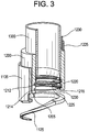
Figure 3 ,central tube 1200 is provided with aligation snare 1230, first andsecond ligation electrodes amputation snare 1225 and aligation snare 1230. As used herein, the word "snare" refers to a flexible line, e.g., a string or a wire. The inner wall surface ofcentral tube 1200 includes upper and lower circumferentialgrooved pathways second ligation electrodes central tube 1200 such that lowercircumferential groove 1214 is between them. Uppergrooved pathway 1212 is disposed axially aboveligation electrodes -
Ligation snare 1230 is disposed in lowercircumferential groove 1214 and extends throughcentral tube 1200 and axially along the outer wall surface to a snare activation mechanism (not shown).Amputation snare 1225 is disposed in uppercircumferential groove 1212 and extends throughcentral tube 1200 and axially along the outer wall surface to a snare activation mechanism (not shown). The outer surface ofcentral tube 1200 may be provided with a plurality of axially extending grooved pathways which receiveamputation snare 1225,ligation snare 1230 and are in communication with upper and lower circumferentialgrooved pathways ligation electrodes - In operation, the resection device of this embodiment can detach and seal the tissue core. Cutting
tube 1300 may be retracted to exposeligation snare 1230 which is preferably made of flexible line, e.g., suture.Ligation snare 1230 may be engaged to snag tissue and pull tissue against the inner wall surface between first andsecond ligation electrodes second electrodes tube 1300 may be further retracted to exposeamputation snare 1225 which may then be activated to sever the tissue core upstream from the point where the tissue was sealed (ligation point). In some embodiments,amputation snare 1225 has a smaller diameter than that ofligation snare 1230. The smaller diameter facilitates tissue slicing. Accordingly, theresection device 1100 according to this embodiment both creates a tissue core and disengages the core from surrounding tissue. - In an alternative embodiment, the resection device of the invention is provided with a single snare disposed between ligation electrodes which both ligates and cuts tissue. In this embodiment, the single snare first pulls tissue against the inner wall surface of
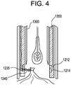
central tube 1200 betweenligation electrodes second electrodes - In yet another embodiment, cutting and sealing may be performed without employing electrodes. In this embodiment,
ligation snare 1230 includes a set ofknots Figure 4 . Ligation is performed by retractingcutting tube 1300 to expose ligation snare 1210 and activatingligation snare 1230 which lassos tissue as ligation knot tightens. Once the tissue is lassoed, cuttingtube 1300 may be further retracted to exposeamputation snare 1225 which may then be activated to sever the tissue core upstream from the point where the point where the tissue was lassoed. - The present disclosure also contemplates an exemplary method and system for using the resection device to remove tissue lesions, for example, lung lesions. The exemplary method generally comprises anchoring the lesion targeted for removal, creating a channel in the tissue leading to the target lesion, creating a tissue core which includes the anchored lesion, ligating the tissue core and sealing the surrounding tissue, and removing the tissue core including the target lesion from the channel.
- Anchoring may be performed by, any suitable structure for securing the device to the lung. Once the lesion is anchored, a channel may be created to facilitate insertion of
resection device 1100. The channel may be created by making an incision in the lung area and inserting a tissue dilator and port into the incision. A tissue core which includes the anchored lesion may be created.Resection device 1100 may be inserted into the channel and used to create the tissue core, to ligate the tissue core and to seal the tissue core and sever it from the surrounding tissue as described hereinabove. The tissue core may then be removed from the channel. - A cavity port may be inserted in the channel to facilitate subsequent treatment of the target lesion site through chemotherapy and/or energy-based tumor extirpation such as radiation.
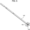
- The anchor depicted in
Figure 5 is suitable for use in performing the exemplary method for removing tissue lesions described herein. The anchor comprises anouter tube 1422 having a sufficiently sharp edge to pierce the chest cavity tissue and lung without causing excess damage and aninner tube 1424 disposed withinouter tube 1422. One or more tines or fingers 1426 formed from shape memory material, e.g., Nitinol, preformed are attached to the end ofinner tube 1424.Outer tube 1422 is retractably disposed overinner tube 1424 such that whenouter tube 1422 is retracted, tines 1426 assume their preform shape as shown.Outer tube 1422 is retracted after it has pierced the lung lesion thereby causing tines 1426 to engage the lung lesion. Other suitable anchors may include coils and suction-based structures. - The incision blades depicted in
Figure 6 are suitable for use in performing the exemplary method for removing tissue lesions described herein. Once anchor 1400 is set, it is preferable to create a small cut or incision to facilitate insertion of chest wall tissue dilator.Incision blades 1605 are used to make a wider cut.Successive incision blades 1605 include a central aperture which allows them to be coaxially advanced along the anchor needle 1405 to create a wider cut in the chest wall, with each successive blade being larger than the previous blade, thereby increasing the width of the incision. - The tissue dilator depicted in
Figure 7 is suitable for use in performing the exemplary method for removing tissue lesions described herein. The tissue dilator may comprise any suitable device for creating a channel in organic tissue. In one exemplary embodiment, the tissue dilator assembly includes a single cylindrical rod withrounded end 1510 or a cylindrical rod with rounded end and arigid sleeve arrangement 1515. Successive tissue dilators are coaxially advanced along the anchor needle to create - tissue tract or channel in the chest wall, with each successive dilator being larger than the previous dilator, thereby increasing the diameter of the channel. Once the final dilator with rigid sleeve is deployed, the inner rod 1505 is removed while leaving the rigid sleeve in the intercoastal space between ribs to create direct passage to the lung pleura.
- Any tissue resection device capable of penetrating lung tissue and creating a tissue core including a target lesion is suitable for use in performing the exemplary method for removing tissue lesions described herein.
Tissue resection device 1100 described hereinbefore is preferred. - Once
tissue resection device 1100 is removed, a small channel in the lung exits where the target lesion was removed. This channel may be utilized to introduce an energy-based ablation device and/or localized chemotherapy depending on the results of the tissue diagnosis. Accordingly, the exemplary method and system of the present invention may not only be utilized to ensure an effective biopsy is performed but also complete removal of the lesion with minimal healthy lung tissue removal. - Although shown and described is what is believed to be the most practical and preferred embodiments, it is apparent that departures from specific designs and methods described and shown will suggest themselves to those skilled in the art and may be used. For example, the systems, devices and exemplary methods described herein for removal of lesions from the lung. It will be appreciated by the skilled artisan that the devices and exemplary methods described herein are not limited to the lung and could be used for tissue resection and lesion removal in other areas of the body. The present invention is not restricted to the particular constructions described and illustrated, but should be constructed to cohere with all modifications that may fall within the scope of the appended claims.
Claims (13)
- A kit for removing a tissue lesion comprising:one or more incision blades (1605); anda tissue resection mechanism (1100) including,an outer tube (1105) having a helical coil (1110) disposed on a distal end, the coil including a first electrode (1130);a central tube (1200) having a distal edge profile including one or more surface segments, at least one of the surface segments including a second electrode (1205), said central tube being slidably disposed within said outer tube and being positioned such that the second electrode opposes at least a portion of the first electrode;a cutting tube (1300) including a cutting edge slidably disposed within said central tube, said cutting tube configured to advance at least as far as a segment of the coil.
- The kit of claim 1 where the coil includes first (1120) and second (1125) contiguous coil segments, the first coil segment including the first electrode.
- The kit of claim 2 wherein the first coil segment comprises a generally planar open ring.
- The kit of claim 2 or claim 3 wherein the first coil segment is helical and has a pitch of zero.
- The kit of any one of claims 2 to 4 wherein the second coil segment is helical and has a constant pitch.
- The kit of any one of claims 2 to 4 wherein the second coil segment is helical and has a variable pitch.
- The kit of claim 2 wherein the first coil segment is helical and has a first pitch and the second coil segment is helical and has a second pitch, at least one of the first and second pitches is variable.
- The kit of any one of claims 2 to 7 wherein the second coil segment includes a blunt tip (1115).
- The kit of any one of claims 2 to 8, wherein the first coil segment has an inner diameter and an outer diameter and said central tube includes an inner and outer diameter wherein the outer diameter of the central tube is greater than the inner diameter of the first coil segment and the outer diameter of the first coil segment is greater than the inner diameter of said central tube.
- The kit of any one of claims 2 to 9, wherein the first coil segment has an outer diameter and said central tube has an outer diameter that is about equal to the outer diameter of the first coil.
- The kit of any preceding claim wherein the first and second electrodes have surface profiles that are substantially matching.
- The kit of any preceding claim further comprising a tissue dilator.
- The kit of any preceding claim further comprising an anchor.
Priority Applications (1)
| Application Number | Priority Date | Filing Date | Title |
|---|---|---|---|
| PL19189139T PL3603546T3 (en) | 2018-07-31 | 2019-07-30 | A kit for removing a tissue lesion |
Applications Claiming Priority (6)
| Application Number | Priority Date | Filing Date | Title |
|---|---|---|---|
| US201862712545P | 2018-07-31 | 2018-07-31 | |
| US201862728170P | 2018-09-07 | 2018-09-07 | |
| US201862744797P | 2018-10-12 | 2018-10-12 | |
| US201862749302P | 2018-10-23 | 2018-10-23 | |
| US201862756234P | 2018-11-06 | 2018-11-06 | |
| US16/512,649 US20200038097A1 (en) | 2018-07-31 | 2019-07-16 | Kit for removing a tissue lesion |
Publications (2)
| Publication Number | Publication Date |
|---|---|
| EP3603546A1 EP3603546A1 (en) | 2020-02-05 |
| EP3603546B1 true EP3603546B1 (en) | 2021-03-24 |
Family
ID=67513400
Family Applications (1)
| Application Number | Title | Priority Date | Filing Date |
|---|---|---|---|
| EP19189139.9A Active EP3603546B1 (en) | 2018-07-31 | 2019-07-30 | A kit for removing a tissue lesion |
Country Status (7)
| Country | Link |
|---|---|
| US (1) | US20200038097A1 (en) |
| EP (1) | EP3603546B1 (en) |
| JP (1) | JP2020018854A (en) |
| CN (1) | CN110772312A (en) |
| BR (1) | BR102019015652A2 (en) |
| ES (1) | ES2874524T3 (en) |
| PL (1) | PL3603546T3 (en) |
Families Citing this family (3)
| Publication number | Priority date | Publication date | Assignee | Title |
|---|---|---|---|---|
| WO2021220220A1 (en) * | 2020-04-30 | 2021-11-04 | Ethicon, Inc. | Systems, devices, and methods for coring tissue |
| WO2022214896A1 (en) * | 2021-04-09 | 2022-10-13 | Ethicon, Inc. | Systems, devices, and methods for coring tissue |
| CN116019500A (en) | 2023-03-03 | 2023-04-28 | 陈麦林 | CT guided percutaneous puncture lung nodule cutter |
Family Cites Families (9)
| Publication number | Priority date | Publication date | Assignee | Title |
|---|---|---|---|---|
| US5843108A (en) * | 1997-10-23 | 1998-12-01 | Samuels; Shaun Laurence Wilkie | Over the wire scapel |
| US6733499B2 (en) * | 2002-02-28 | 2004-05-11 | Biosense Webster, Inc. | Catheter having circular ablation assembly |
| US6974467B1 (en) * | 2002-11-04 | 2005-12-13 | Gonzales Jr Antonio | Method and apparatus for making a precise surgical incision |
| US20040147917A1 (en) * | 2003-01-23 | 2004-07-29 | Mueller Richard L. | Device and method for treatment of breast tissue with electromagnetic radiation |
| US8317771B2 (en) * | 2007-07-11 | 2012-11-27 | Apollo Endosurgery, Inc. | Methods and systems for performing submucosal medical procedures |
| US20110190764A1 (en) * | 2010-01-29 | 2011-08-04 | Ethicon Endo-Surgery, Inc. | Surgical instrument comprising an electrode |
| US20140277039A1 (en) * | 2013-03-15 | 2014-09-18 | Acclarent, Inc. | Apparatus and method for treatment of ethmoid sinusitis |
| DE102016101915A1 (en) * | 2016-02-03 | 2017-08-03 | Aesculap Ag | Minimally invasive incision instrument with guided multiple cutting edge |
| EP3518783B1 (en) * | 2016-11-23 | 2023-05-24 | Corit Medical, LLC | Apparatus for tissue reduction |
-
2019
- 2019-07-16 US US16/512,649 patent/US20200038097A1/en not_active Abandoned
- 2019-07-30 PL PL19189139T patent/PL3603546T3/en unknown
- 2019-07-30 JP JP2019139537A patent/JP2020018854A/en active Pending
- 2019-07-30 EP EP19189139.9A patent/EP3603546B1/en active Active
- 2019-07-30 ES ES19189139T patent/ES2874524T3/en active Active
- 2019-07-30 BR BR102019015652-0A patent/BR102019015652A2/en not_active Application Discontinuation
- 2019-07-31 CN CN201910700729.2A patent/CN110772312A/en active Pending
Non-Patent Citations (1)
| Title |
|---|
| None * |
Also Published As
| Publication number | Publication date |
|---|---|
| ES2874524T3 (en) | 2021-11-05 |
| EP3603546A1 (en) | 2020-02-05 |
| US20200038097A1 (en) | 2020-02-06 |
| BR102019015652A2 (en) | 2020-02-18 |
| JP2020018854A (en) | 2020-02-06 |
| PL3603546T3 (en) | 2022-01-10 |
| CN110772312A (en) | 2020-02-11 |
Similar Documents
| Publication | Publication Date | Title |
|---|---|---|
| US20230380878A1 (en) | Tissue resection apparatus | |
| EP2923645B1 (en) | Devices and systems for obtaining a tissue sample using a biopsy tool | |
| EP3603546B1 (en) | A kit for removing a tissue lesion | |
| AU2011238490B2 (en) | Endoscopic ultrasound-guided biopsy needle | |
| KR20190112314A (en) | Minimally Invasive Method and Apparatus for Target Tissue Extraction | |
| EP2797518B1 (en) | Adjustable resection device | |
| US11723708B2 (en) | Method for removing a tissue lesion | |
| US20210219967A1 (en) | Systems, devices, and methods for coring tissue | |
| CA3177320A1 (en) | Systems, devices, and methods for coring tissue | |
| US10363087B2 (en) | Tissue resection device | |
| US20220047322A1 (en) | Coring and amputation devices, systems, and methods | |
| WO2022214896A1 (en) | Systems, devices, and methods for coring tissue | |
| CN108697412A (en) | System and method for improved tissue sampling | |
| BR102019015643A2 (en) | method to remove a tissue injury | |
| Tillson | Abdominal biopsy procedures: intestines and liver. | |
| IES85915Y1 (en) | Endoscopic ultrasound-guided biopsy needle | |
| IE20110141U1 (en) | Endoscopic ultrasound-guided biopsy needle |
Legal Events
| Date | Code | Title | Description |
|---|---|---|---|
| PUAI | Public reference made under article 153(3) epc to a published international application that has entered the european phase |
Free format text: ORIGINAL CODE: 0009012 |
|
| STAA | Information on the status of an ep patent application or granted ep patent |
Free format text: STATUS: THE APPLICATION HAS BEEN PUBLISHED |
|
| AK | Designated contracting states |
Kind code of ref document: A1 Designated state(s): AL AT BE BG CH CY CZ DE DK EE ES FI FR GB GR HR HU IE IS IT LI LT LU LV MC MK MT NL NO PL PT RO RS SE SI SK SM TR |
|
| AX | Request for extension of the european patent |
Extension state: BA ME |
|
| STAA | Information on the status of an ep patent application or granted ep patent |
Free format text: STATUS: REQUEST FOR EXAMINATION WAS MADE |
|
| 17P | Request for examination filed |
Effective date: 20200612 |
|
| RBV | Designated contracting states (corrected) |
Designated state(s): AL AT BE BG CH CY CZ DE DK EE ES FI FR GB GR HR HU IE IS IT LI LT LU LV MC MK MT NL NO PL PT RO RS SE SI SK SM TR |
|
| GRAP | Despatch of communication of intention to grant a patent |
Free format text: ORIGINAL CODE: EPIDOSNIGR1 |
|
| STAA | Information on the status of an ep patent application or granted ep patent |
Free format text: STATUS: GRANT OF PATENT IS INTENDED |
|
| INTG | Intention to grant announced |
Effective date: 20201019 |
|
| GRAJ | Information related to disapproval of communication of intention to grant by the applicant or resumption of examination proceedings by the epo deleted |
Free format text: ORIGINAL CODE: EPIDOSDIGR1 |
|
| STAA | Information on the status of an ep patent application or granted ep patent |
Free format text: STATUS: REQUEST FOR EXAMINATION WAS MADE |
|
| GRAP | Despatch of communication of intention to grant a patent |
Free format text: ORIGINAL CODE: EPIDOSNIGR1 |
|
| STAA | Information on the status of an ep patent application or granted ep patent |
Free format text: STATUS: GRANT OF PATENT IS INTENDED |
|
| INTC | Intention to grant announced (deleted) | ||
| INTG | Intention to grant announced |
Effective date: 20210113 |
|
| GRAS | Grant fee paid |
Free format text: ORIGINAL CODE: EPIDOSNIGR3 |
|
| GRAA | (expected) grant |
Free format text: ORIGINAL CODE: 0009210 |
|
| STAA | Information on the status of an ep patent application or granted ep patent |
Free format text: STATUS: THE PATENT HAS BEEN GRANTED |
|
| AK | Designated contracting states |
Kind code of ref document: B1 Designated state(s): AL AT BE BG CH CY CZ DE DK EE ES FI FR GB GR HR HU IE IS IT LI LT LU LV MC MK MT NL NO PL PT RO RS SE SI SK SM TR |
|
| REG | Reference to a national code |
Ref country code: GB Ref legal event code: FG4D |
|
| REG | Reference to a national code |
Ref country code: CH Ref legal event code: EP Ref country code: CH Ref legal event code: NV Representative=s name: E. BLUM AND CO. AG PATENT- UND MARKENANWAELTE V, CH |
|
| REG | Reference to a national code |
Ref country code: IE Ref legal event code: FG4D |
|
| REG | Reference to a national code |
Ref country code: DE Ref legal event code: R096 Ref document number: 602019003392 Country of ref document: DE Ref country code: AT Ref legal event code: REF Ref document number: 1373674 Country of ref document: AT Kind code of ref document: T Effective date: 20210415 |
|
| REG | Reference to a national code |
Ref country code: SE Ref legal event code: TRGR |
|
| REG | Reference to a national code |
Ref country code: NL Ref legal event code: FP |
|
| REG | Reference to a national code |
Ref country code: LT Ref legal event code: MG9D |
|
| PG25 | Lapsed in a contracting state [announced via postgrant information from national office to epo] |
Ref country code: HR Free format text: LAPSE BECAUSE OF FAILURE TO SUBMIT A TRANSLATION OF THE DESCRIPTION OR TO PAY THE FEE WITHIN THE PRESCRIBED TIME-LIMIT Effective date: 20210324 Ref country code: BG Free format text: LAPSE BECAUSE OF FAILURE TO SUBMIT A TRANSLATION OF THE DESCRIPTION OR TO PAY THE FEE WITHIN THE PRESCRIBED TIME-LIMIT Effective date: 20210624 Ref country code: FI Free format text: LAPSE BECAUSE OF FAILURE TO SUBMIT A TRANSLATION OF THE DESCRIPTION OR TO PAY THE FEE WITHIN THE PRESCRIBED TIME-LIMIT Effective date: 20210324 Ref country code: GR Free format text: LAPSE BECAUSE OF FAILURE TO SUBMIT A TRANSLATION OF THE DESCRIPTION OR TO PAY THE FEE WITHIN THE PRESCRIBED TIME-LIMIT Effective date: 20210625 Ref country code: NO Free format text: LAPSE BECAUSE OF FAILURE TO SUBMIT A TRANSLATION OF THE DESCRIPTION OR TO PAY THE FEE WITHIN THE PRESCRIBED TIME-LIMIT Effective date: 20210624 |
|
| PG25 | Lapsed in a contracting state [announced via postgrant information from national office to epo] |
Ref country code: RS Free format text: LAPSE BECAUSE OF FAILURE TO SUBMIT A TRANSLATION OF THE DESCRIPTION OR TO PAY THE FEE WITHIN THE PRESCRIBED TIME-LIMIT Effective date: 20210324 Ref country code: LV Free format text: LAPSE BECAUSE OF FAILURE TO SUBMIT A TRANSLATION OF THE DESCRIPTION OR TO PAY THE FEE WITHIN THE PRESCRIBED TIME-LIMIT Effective date: 20210324 |
|
| REG | Reference to a national code |
Ref country code: AT Ref legal event code: MK05 Ref document number: 1373674 Country of ref document: AT Kind code of ref document: T Effective date: 20210324 |
|
| PG25 | Lapsed in a contracting state [announced via postgrant information from national office to epo] |
Ref country code: AT Free format text: LAPSE BECAUSE OF FAILURE TO SUBMIT A TRANSLATION OF THE DESCRIPTION OR TO PAY THE FEE WITHIN THE PRESCRIBED TIME-LIMIT Effective date: 20210324 Ref country code: SM Free format text: LAPSE BECAUSE OF FAILURE TO SUBMIT A TRANSLATION OF THE DESCRIPTION OR TO PAY THE FEE WITHIN THE PRESCRIBED TIME-LIMIT Effective date: 20210324 Ref country code: LT Free format text: LAPSE BECAUSE OF FAILURE TO SUBMIT A TRANSLATION OF THE DESCRIPTION OR TO PAY THE FEE WITHIN THE PRESCRIBED TIME-LIMIT Effective date: 20210324 Ref country code: EE Free format text: LAPSE BECAUSE OF FAILURE TO SUBMIT A TRANSLATION OF THE DESCRIPTION OR TO PAY THE FEE WITHIN THE PRESCRIBED TIME-LIMIT Effective date: 20210324 Ref country code: CZ Free format text: LAPSE BECAUSE OF FAILURE TO SUBMIT A TRANSLATION OF THE DESCRIPTION OR TO PAY THE FEE WITHIN THE PRESCRIBED TIME-LIMIT Effective date: 20210324 |
|
| REG | Reference to a national code |
Ref country code: ES Ref legal event code: FG2A Ref document number: 2874524 Country of ref document: ES Kind code of ref document: T3 Effective date: 20211105 |
|
| PG25 | Lapsed in a contracting state [announced via postgrant information from national office to epo] |
Ref country code: IS Free format text: LAPSE BECAUSE OF FAILURE TO SUBMIT A TRANSLATION OF THE DESCRIPTION OR TO PAY THE FEE WITHIN THE PRESCRIBED TIME-LIMIT Effective date: 20210724 Ref country code: SK Free format text: LAPSE BECAUSE OF FAILURE TO SUBMIT A TRANSLATION OF THE DESCRIPTION OR TO PAY THE FEE WITHIN THE PRESCRIBED TIME-LIMIT Effective date: 20210324 Ref country code: RO Free format text: LAPSE BECAUSE OF FAILURE TO SUBMIT A TRANSLATION OF THE DESCRIPTION OR TO PAY THE FEE WITHIN THE PRESCRIBED TIME-LIMIT Effective date: 20210324 Ref country code: PT Free format text: LAPSE BECAUSE OF FAILURE TO SUBMIT A TRANSLATION OF THE DESCRIPTION OR TO PAY THE FEE WITHIN THE PRESCRIBED TIME-LIMIT Effective date: 20210726 |
|
| REG | Reference to a national code |
Ref country code: DE Ref legal event code: R097 Ref document number: 602019003392 Country of ref document: DE |
|
| PG25 | Lapsed in a contracting state [announced via postgrant information from national office to epo] |
Ref country code: DK Free format text: LAPSE BECAUSE OF FAILURE TO SUBMIT A TRANSLATION OF THE DESCRIPTION OR TO PAY THE FEE WITHIN THE PRESCRIBED TIME-LIMIT Effective date: 20210324 Ref country code: AL Free format text: LAPSE BECAUSE OF FAILURE TO SUBMIT A TRANSLATION OF THE DESCRIPTION OR TO PAY THE FEE WITHIN THE PRESCRIBED TIME-LIMIT Effective date: 20210324 |
|
| PLBE | No opposition filed within time limit |
Free format text: ORIGINAL CODE: 0009261 |
|
| STAA | Information on the status of an ep patent application or granted ep patent |
Free format text: STATUS: NO OPPOSITION FILED WITHIN TIME LIMIT |
|
| PG25 | Lapsed in a contracting state [announced via postgrant information from national office to epo] |
Ref country code: SI Free format text: LAPSE BECAUSE OF FAILURE TO SUBMIT A TRANSLATION OF THE DESCRIPTION OR TO PAY THE FEE WITHIN THE PRESCRIBED TIME-LIMIT Effective date: 20210324 |
|
| 26N | No opposition filed |
Effective date: 20220104 |
|
| PG25 | Lapsed in a contracting state [announced via postgrant information from national office to epo] |
Ref country code: MC Free format text: LAPSE BECAUSE OF FAILURE TO SUBMIT A TRANSLATION OF THE DESCRIPTION OR TO PAY THE FEE WITHIN THE PRESCRIBED TIME-LIMIT Effective date: 20210324 |
|
| REG | Reference to a national code |
Ref country code: BE Ref legal event code: MM Effective date: 20210731 |
|
| PG25 | Lapsed in a contracting state [announced via postgrant information from national office to epo] |
Ref country code: IS Free format text: LAPSE BECAUSE OF FAILURE TO SUBMIT A TRANSLATION OF THE DESCRIPTION OR TO PAY THE FEE WITHIN THE PRESCRIBED TIME-LIMIT Effective date: 20210724 Ref country code: LU Free format text: LAPSE BECAUSE OF NON-PAYMENT OF DUE FEES Effective date: 20210730 |
|
| PG25 | Lapsed in a contracting state [announced via postgrant information from national office to epo] |
Ref country code: IE Free format text: LAPSE BECAUSE OF NON-PAYMENT OF DUE FEES Effective date: 20210730 Ref country code: BE Free format text: LAPSE BECAUSE OF NON-PAYMENT OF DUE FEES Effective date: 20210731 |
|
| PGFP | Annual fee paid to national office [announced via postgrant information from national office to epo] |
Ref country code: SE Payment date: 20220615 Year of fee payment: 4 |
|
| PGFP | Annual fee paid to national office [announced via postgrant information from national office to epo] |
Ref country code: CH Payment date: 20220801 Year of fee payment: 4 |
|
| PG25 | Lapsed in a contracting state [announced via postgrant information from national office to epo] |
Ref country code: CY Free format text: LAPSE BECAUSE OF FAILURE TO SUBMIT A TRANSLATION OF THE DESCRIPTION OR TO PAY THE FEE WITHIN THE PRESCRIBED TIME-LIMIT Effective date: 20210324 |
|
| PG25 | Lapsed in a contracting state [announced via postgrant information from national office to epo] |
Ref country code: HU Free format text: LAPSE BECAUSE OF FAILURE TO SUBMIT A TRANSLATION OF THE DESCRIPTION OR TO PAY THE FEE WITHIN THE PRESCRIBED TIME-LIMIT; INVALID AB INITIO Effective date: 20190730 |
|
| PGFP | Annual fee paid to national office [announced via postgrant information from national office to epo] |
Ref country code: NL Payment date: 20230826 Year of fee payment: 5 |
|
| PGFP | Annual fee paid to national office [announced via postgrant information from national office to epo] |
Ref country code: IT Payment date: 20230822 Year of fee payment: 5 Ref country code: GB Payment date: 20230828 Year of fee payment: 5 Ref country code: ES Payment date: 20230901 Year of fee payment: 5 |
|
| PGFP | Annual fee paid to national office [announced via postgrant information from national office to epo] |
Ref country code: PL Payment date: 20230821 Year of fee payment: 5 Ref country code: FR Payment date: 20230825 Year of fee payment: 5 Ref country code: DE Payment date: 20230829 Year of fee payment: 5 |
|
| REG | Reference to a national code |
Ref country code: CH Ref legal event code: PL |
|
| REG | Reference to a national code |
Ref country code: SE Ref legal event code: EUG |