EP3589958B1 - Methods for analyzing intact proteins, antibodies, antibody subunits, and antibody drug conjugates - Google Patents
Methods for analyzing intact proteins, antibodies, antibody subunits, and antibody drug conjugates Download PDFInfo
- Publication number
- EP3589958B1 EP3589958B1 EP18713009.1A EP18713009A EP3589958B1 EP 3589958 B1 EP3589958 B1 EP 3589958B1 EP 18713009 A EP18713009 A EP 18713009A EP 3589958 B1 EP3589958 B1 EP 3589958B1
- Authority
- EP
- European Patent Office
- Prior art keywords
- sample
- dimension
- reagent
- exposing
- supercharging
- Prior art date
- Legal status (The legal status is an assumption and is not a legal conclusion. Google has not performed a legal analysis and makes no representation as to the accuracy of the status listed.)
- Active
Links
- 238000000034 method Methods 0.000 title claims description 74
- 229940049595 antibody-drug conjugate Drugs 0.000 title claims description 33
- 239000000611 antibody drug conjugate Substances 0.000 title claims description 30
- 102000004169 proteins and genes Human genes 0.000 title claims description 30
- 108090000623 proteins and genes Proteins 0.000 title claims description 30
- 239000003153 chemical reaction reagent Substances 0.000 claims description 50
- PEDCQBHIVMGVHV-UHFFFAOYSA-N Glycerine Chemical compound OCC(O)CO PEDCQBHIVMGVHV-UHFFFAOYSA-N 0.000 claims description 45
- RAXXELZNTBOGNW-UHFFFAOYSA-N imidazole Natural products C1=CNC=N1 RAXXELZNTBOGNW-UHFFFAOYSA-N 0.000 claims description 28
- 230000005526 G1 to G0 transition Effects 0.000 claims description 18
- 239000000203 mixture Substances 0.000 claims description 18
- CWNPOQFCIIFQDM-UHFFFAOYSA-N 3-nitrobenzyl alcohol Chemical compound OCC1=CC=CC([N+]([O-])=O)=C1 CWNPOQFCIIFQDM-UHFFFAOYSA-N 0.000 claims description 12
- 238000004611 spectroscopical analysis Methods 0.000 claims description 12
- 239000012634 fragment Substances 0.000 claims description 9
- ZHNUHDYFZUAESO-UHFFFAOYSA-N Formamide Chemical compound NC=O ZHNUHDYFZUAESO-UHFFFAOYSA-N 0.000 claims description 8
- XNWFRZJHXBZDAG-UHFFFAOYSA-N 2-METHOXYETHANOL Chemical compound COCCO XNWFRZJHXBZDAG-UHFFFAOYSA-N 0.000 claims description 4
- 125000002883 imidazolyl group Chemical group 0.000 claims description 2
- 239000000523 sample Substances 0.000 description 126
- 238000000132 electrospray ionisation Methods 0.000 description 29
- 230000035945 sensitivity Effects 0.000 description 29
- 235000018102 proteins Nutrition 0.000 description 28
- 238000000926 separation method Methods 0.000 description 28
- 150000002500 ions Chemical class 0.000 description 20
- 238000007792 addition Methods 0.000 description 17
- 238000009826 distribution Methods 0.000 description 15
- 210000004027 cell Anatomy 0.000 description 13
- 238000001514 detection method Methods 0.000 description 13
- 238000002474 experimental method Methods 0.000 description 13
- 238000004949 mass spectrometry Methods 0.000 description 12
- 238000001228 spectrum Methods 0.000 description 12
- 238000004458 analytical method Methods 0.000 description 11
- 210000004369 blood Anatomy 0.000 description 11
- 239000008280 blood Substances 0.000 description 11
- 239000012472 biological sample Substances 0.000 description 10
- 230000005284 excitation Effects 0.000 description 10
- 239000002245 particle Substances 0.000 description 10
- 125000001997 phenyl group Chemical group [H]C1=C([H])C([H])=C(*)C([H])=C1[H] 0.000 description 10
- WEVYAHXRMPXWCK-UHFFFAOYSA-N Acetonitrile Chemical compound CC#N WEVYAHXRMPXWCK-UHFFFAOYSA-N 0.000 description 9
- 239000012491 analyte Substances 0.000 description 9
- 150000001875 compounds Chemical class 0.000 description 9
- 239000000463 material Substances 0.000 description 9
- 230000008569 process Effects 0.000 description 9
- 230000005855 radiation Effects 0.000 description 9
- 239000000126 substance Substances 0.000 description 9
- 238000000862 absorption spectrum Methods 0.000 description 8
- 229940079593 drug Drugs 0.000 description 8
- 230000007246 mechanism Effects 0.000 description 8
- BDAGIHXWWSANSR-UHFFFAOYSA-N methanoic acid Natural products OC=O BDAGIHXWWSANSR-UHFFFAOYSA-N 0.000 description 8
- 230000008901 benefit Effects 0.000 description 7
- 239000003814 drug Substances 0.000 description 7
- 238000004128 high performance liquid chromatography Methods 0.000 description 7
- 230000006872 improvement Effects 0.000 description 7
- 238000004811 liquid chromatography Methods 0.000 description 7
- 239000012071 phase Substances 0.000 description 7
- 210000001519 tissue Anatomy 0.000 description 7
- 239000003643 water by type Substances 0.000 description 7
- -1 IgM Chemical compound 0.000 description 6
- 108060003951 Immunoglobulin Proteins 0.000 description 6
- 238000004847 absorption spectroscopy Methods 0.000 description 6
- 238000005516 engineering process Methods 0.000 description 6
- 102000018358 immunoglobulin Human genes 0.000 description 6
- 230000003595 spectral effect Effects 0.000 description 6
- 238000012546 transfer Methods 0.000 description 6
- 241000282414 Homo sapiens Species 0.000 description 5
- 238000010521 absorption reaction Methods 0.000 description 5
- 239000000427 antigen Substances 0.000 description 5
- 102000036639 antigens Human genes 0.000 description 5
- 108091007433 antigens Proteins 0.000 description 5
- 238000005259 measurement Methods 0.000 description 5
- 210000002381 plasma Anatomy 0.000 description 5
- 210000002966 serum Anatomy 0.000 description 5
- 239000002594 sorbent Substances 0.000 description 5
- 239000007921 spray Substances 0.000 description 5
- OSWFIVFLDKOXQC-UHFFFAOYSA-N 4-(3-methoxyphenyl)aniline Chemical compound COC1=CC=CC(C=2C=CC(N)=CC=2)=C1 OSWFIVFLDKOXQC-UHFFFAOYSA-N 0.000 description 4
- 210000001175 cerebrospinal fluid Anatomy 0.000 description 4
- 230000001419 dependent effect Effects 0.000 description 4
- 230000000694 effects Effects 0.000 description 4
- 238000010828 elution Methods 0.000 description 4
- 230000007613 environmental effect Effects 0.000 description 4
- 239000012530 fluid Substances 0.000 description 4
- 235000019253 formic acid Nutrition 0.000 description 4
- 238000011068 loading method Methods 0.000 description 4
- 230000003287 optical effect Effects 0.000 description 4
- 210000005105 peripheral blood lymphocyte Anatomy 0.000 description 4
- 239000000243 solution Substances 0.000 description 4
- 238000000672 surface-enhanced laser desorption--ionisation Methods 0.000 description 4
- 238000001195 ultra high performance liquid chromatography Methods 0.000 description 4
- 210000002700 urine Anatomy 0.000 description 4
- OKKJLVBELUTLKV-UHFFFAOYSA-N Methanol Chemical compound OC OKKJLVBELUTLKV-UHFFFAOYSA-N 0.000 description 3
- 238000013459 approach Methods 0.000 description 3
- 210000001124 body fluid Anatomy 0.000 description 3
- 150000001793 charged compounds Chemical class 0.000 description 3
- 230000003247 decreasing effect Effects 0.000 description 3
- 230000022811 deglycosylation Effects 0.000 description 3
- 201000010099 disease Diseases 0.000 description 3
- 208000037265 diseases, disorders, signs and symptoms Diseases 0.000 description 3
- 230000005670 electromagnetic radiation Effects 0.000 description 3
- 238000000695 excitation spectrum Methods 0.000 description 3
- 210000003608 fece Anatomy 0.000 description 3
- 210000004700 fetal blood Anatomy 0.000 description 3
- 238000001506 fluorescence spectroscopy Methods 0.000 description 3
- 230000010354 integration Effects 0.000 description 3
- 239000007788 liquid Substances 0.000 description 3
- 238000004895 liquid chromatography mass spectrometry Methods 0.000 description 3
- 238000000816 matrix-assisted laser desorption--ionisation Methods 0.000 description 3
- QSHDDOUJBYECFT-UHFFFAOYSA-N mercury Chemical compound [Hg] QSHDDOUJBYECFT-UHFFFAOYSA-N 0.000 description 3
- 230000004048 modification Effects 0.000 description 3
- 238000012986 modification Methods 0.000 description 3
- 238000012856 packing Methods 0.000 description 3
- 108090000765 processed proteins & peptides Proteins 0.000 description 3
- 238000004366 reverse phase liquid chromatography Methods 0.000 description 3
- 210000003296 saliva Anatomy 0.000 description 3
- 150000003384 small molecules Chemical class 0.000 description 3
- 239000002689 soil Substances 0.000 description 3
- 239000007787 solid Substances 0.000 description 3
- 238000000870 ultraviolet spectroscopy Methods 0.000 description 3
- XLYOFNOQVPJJNP-UHFFFAOYSA-N water Substances O XLYOFNOQVPJJNP-UHFFFAOYSA-N 0.000 description 3
- 101710102916 Ichor Proteins 0.000 description 2
- KFZMGEQAYNKOFK-UHFFFAOYSA-N Isopropanol Chemical compound CC(C)O KFZMGEQAYNKOFK-UHFFFAOYSA-N 0.000 description 2
- 241001529936 Murinae Species 0.000 description 2
- 241000283973 Oryctolagus cuniculus Species 0.000 description 2
- 102000000447 Peptide-N4-(N-acetyl-beta-glucosaminyl) Asparagine Amidase Human genes 0.000 description 2
- 108010055817 Peptide-N4-(N-acetyl-beta-glucosaminyl) Asparagine Amidase Proteins 0.000 description 2
- 239000008186 active pharmaceutical agent Substances 0.000 description 2
- 210000004381 amniotic fluid Anatomy 0.000 description 2
- 238000001574 biopsy Methods 0.000 description 2
- 239000010839 body fluid Substances 0.000 description 2
- 210000001185 bone marrow Anatomy 0.000 description 2
- 241000902900 cellular organisms Species 0.000 description 2
- 238000004587 chromatography analysis Methods 0.000 description 2
- 230000008878 coupling Effects 0.000 description 2
- 238000010168 coupling process Methods 0.000 description 2
- 238000005859 coupling reaction Methods 0.000 description 2
- 230000007423 decrease Effects 0.000 description 2
- 238000011033 desalting Methods 0.000 description 2
- 238000013461 design Methods 0.000 description 2
- 239000003651 drinking water Substances 0.000 description 2
- 239000003480 eluent Substances 0.000 description 2
- 238000004401 flow injection analysis Methods 0.000 description 2
- 238000002189 fluorescence spectrum Methods 0.000 description 2
- 230000002496 gastric effect Effects 0.000 description 2
- 230000013595 glycosylation Effects 0.000 description 2
- 238000006206 glycosylation reaction Methods 0.000 description 2
- 210000004209 hair Anatomy 0.000 description 2
- 229940022353 herceptin Drugs 0.000 description 2
- 235000020256 human milk Nutrition 0.000 description 2
- 210000004251 human milk Anatomy 0.000 description 2
- 238000010348 incorporation Methods 0.000 description 2
- 230000014759 maintenance of location Effects 0.000 description 2
- 210000001006 meconium Anatomy 0.000 description 2
- 210000000282 nail Anatomy 0.000 description 2
- 210000000056 organ Anatomy 0.000 description 2
- 244000052769 pathogen Species 0.000 description 2
- LOQGSOTUHASIHI-UHFFFAOYSA-N perfluoro-1,3-dimethylcyclohexane Chemical compound FC(F)(F)C1(F)C(F)(F)C(F)(F)C(F)(F)C(F)(C(F)(F)F)C1(F)F LOQGSOTUHASIHI-UHFFFAOYSA-N 0.000 description 2
- 210000002826 placenta Anatomy 0.000 description 2
- 230000005588 protonation Effects 0.000 description 2
- 230000009467 reduction Effects 0.000 description 2
- 238000006722 reduction reaction Methods 0.000 description 2
- 230000028327 secretion Effects 0.000 description 2
- 210000003491 skin Anatomy 0.000 description 2
- 239000002904 solvent Substances 0.000 description 2
- 241000894007 species Species 0.000 description 2
- 238000012360 testing method Methods 0.000 description 2
- 238000002834 transmittance Methods 0.000 description 2
- 238000000825 ultraviolet detection Methods 0.000 description 2
- 210000001113 umbilicus Anatomy 0.000 description 2
- 229910052724 xenon Inorganic materials 0.000 description 2
- FHNFHKCVQCLJFQ-UHFFFAOYSA-N xenon atom Chemical compound [Xe] FHNFHKCVQCLJFQ-UHFFFAOYSA-N 0.000 description 2
- 241000251468 Actinopterygii Species 0.000 description 1
- 108700023418 Amidases Proteins 0.000 description 1
- 102000052567 Anaphase-Promoting Complex-Cyclosome Apc1 Subunit Human genes 0.000 description 1
- 208000023275 Autoimmune disease Diseases 0.000 description 1
- 241000283690 Bos taurus Species 0.000 description 1
- 239000003341 Bronsted base Substances 0.000 description 1
- 241000283707 Capra Species 0.000 description 1
- 241000700199 Cavia porcellus Species 0.000 description 1
- BWGNESOTFCXPMA-UHFFFAOYSA-N Dihydrogen disulfide Chemical compound SS BWGNESOTFCXPMA-UHFFFAOYSA-N 0.000 description 1
- 241000283073 Equus caballus Species 0.000 description 1
- 241000282326 Felis catus Species 0.000 description 1
- 102000003886 Glycoproteins Human genes 0.000 description 1
- 108090000288 Glycoproteins Proteins 0.000 description 1
- 238000004566 IR spectroscopy Methods 0.000 description 1
- 102000008394 Immunoglobulin Fragments Human genes 0.000 description 1
- 108010021625 Immunoglobulin Fragments Proteins 0.000 description 1
- 241000124008 Mammalia Species 0.000 description 1
- 108010090665 Mannosyl-Glycoprotein Endo-beta-N-Acetylglucosaminidase Proteins 0.000 description 1
- 229910000661 Mercury cadmium telluride Inorganic materials 0.000 description 1
- 241001465754 Metazoa Species 0.000 description 1
- 241000699666 Mus <mouse, genus> Species 0.000 description 1
- 206010028980 Neoplasm Diseases 0.000 description 1
- 241001494479 Pecora Species 0.000 description 1
- 241000009328 Perro Species 0.000 description 1
- 206010036790 Productive cough Diseases 0.000 description 1
- 241000700159 Rattus Species 0.000 description 1
- 108091006463 SLC25A24 Proteins 0.000 description 1
- 241000282898 Sus scrofa Species 0.000 description 1
- 238000002835 absorbance Methods 0.000 description 1
- 239000002253 acid Substances 0.000 description 1
- 230000002378 acidificating effect Effects 0.000 description 1
- 150000007513 acids Chemical class 0.000 description 1
- 239000013543 active substance Substances 0.000 description 1
- 230000002776 aggregation Effects 0.000 description 1
- 238000004220 aggregation Methods 0.000 description 1
- 239000003570 air Substances 0.000 description 1
- 102000005922 amidase Human genes 0.000 description 1
- 125000000613 asparagine group Chemical group N[C@@H](CC(N)=O)C(=O)* 0.000 description 1
- 230000001363 autoimmune Effects 0.000 description 1
- 230000004888 barrier function Effects 0.000 description 1
- 239000011324 bead Substances 0.000 description 1
- 239000011230 binding agent Substances 0.000 description 1
- 238000011953 bioanalysis Methods 0.000 description 1
- 230000005540 biological transmission Effects 0.000 description 1
- 239000000090 biomarker Substances 0.000 description 1
- 230000015572 biosynthetic process Effects 0.000 description 1
- 238000010241 blood sampling Methods 0.000 description 1
- 235000012206 bottled water Nutrition 0.000 description 1
- MCMSPRNYOJJPIZ-UHFFFAOYSA-N cadmium;mercury;tellurium Chemical compound [Cd]=[Te]=[Hg] MCMSPRNYOJJPIZ-UHFFFAOYSA-N 0.000 description 1
- 201000011510 cancer Diseases 0.000 description 1
- 235000014633 carbohydrates Nutrition 0.000 description 1
- 150000001720 carbohydrates Chemical class 0.000 description 1
- 238000004113 cell culture Methods 0.000 description 1
- 239000000919 ceramic Substances 0.000 description 1
- 230000008859 change Effects 0.000 description 1
- 150000005829 chemical entities Chemical class 0.000 description 1
- 239000003795 chemical substances by application Substances 0.000 description 1
- 230000000295 complement effect Effects 0.000 description 1
- 230000001010 compromised effect Effects 0.000 description 1
- 239000000562 conjugate Substances 0.000 description 1
- 239000000470 constituent Substances 0.000 description 1
- 238000011109 contamination Methods 0.000 description 1
- 125000000151 cysteine group Chemical group N[C@@H](CS)C(=O)* 0.000 description 1
- 230000002939 deleterious effect Effects 0.000 description 1
- 230000000994 depressogenic effect Effects 0.000 description 1
- 238000004807 desolvation Methods 0.000 description 1
- 238000011161 development Methods 0.000 description 1
- 238000010586 diagram Methods 0.000 description 1
- 239000003085 diluting agent Substances 0.000 description 1
- 238000004141 dimensional analysis Methods 0.000 description 1
- 235000020188 drinking water Nutrition 0.000 description 1
- 238000009509 drug development Methods 0.000 description 1
- 230000009977 dual effect Effects 0.000 description 1
- 230000005684 electric field Effects 0.000 description 1
- 238000002330 electrospray ionisation mass spectrometry Methods 0.000 description 1
- 238000000295 emission spectrum Methods 0.000 description 1
- 238000003912 environmental pollution Methods 0.000 description 1
- 239000000835 fiber Substances 0.000 description 1
- 238000001914 filtration Methods 0.000 description 1
- 239000007789 gas Substances 0.000 description 1
- 239000003673 groundwater Substances 0.000 description 1
- 125000005597 hydrazone group Chemical group 0.000 description 1
- 210000000987 immune system Anatomy 0.000 description 1
- 229940127121 immunoconjugate Drugs 0.000 description 1
- 229940072221 immunoglobulins Drugs 0.000 description 1
- 238000000338 in vitro Methods 0.000 description 1
- 238000001727 in vivo Methods 0.000 description 1
- 238000001802 infusion Methods 0.000 description 1
- 239000004615 ingredient Substances 0.000 description 1
- 238000002347 injection Methods 0.000 description 1
- 239000007924 injection Substances 0.000 description 1
- 230000003993 interaction Effects 0.000 description 1
- 238000005040 ion trap Methods 0.000 description 1
- 238000002955 isolation Methods 0.000 description 1
- 229960004592 isopropanol Drugs 0.000 description 1
- 239000007791 liquid phase Substances 0.000 description 1
- 210000002751 lymph Anatomy 0.000 description 1
- 235000018977 lysine Nutrition 0.000 description 1
- 125000003588 lysine group Chemical class [H]N([H])C([H])([H])C([H])([H])C([H])([H])C([H])([H])C([H])(N([H])[H])C(*)=O 0.000 description 1
- 238000004519 manufacturing process Methods 0.000 description 1
- 238000001819 mass spectrum Methods 0.000 description 1
- 239000011159 matrix material Substances 0.000 description 1
- 238000010339 medical test Methods 0.000 description 1
- 229910052753 mercury Inorganic materials 0.000 description 1
- 238000002156 mixing Methods 0.000 description 1
- 239000003002 pH adjusting agent Substances 0.000 description 1
- 230000001717 pathogenic effect Effects 0.000 description 1
- 230000000704 physical effect Effects 0.000 description 1
- 230000010287 polarization Effects 0.000 description 1
- 230000004481 post-translational protein modification Effects 0.000 description 1
- 238000002360 preparation method Methods 0.000 description 1
- 238000002203 pretreatment Methods 0.000 description 1
- 150000003141 primary amines Chemical class 0.000 description 1
- 102000004196 processed proteins & peptides Human genes 0.000 description 1
- 238000012545 processing Methods 0.000 description 1
- 229940002612 prodrug Drugs 0.000 description 1
- 239000000651 prodrug Substances 0.000 description 1
- 238000007388 punch biopsy Methods 0.000 description 1
- 238000000746 purification Methods 0.000 description 1
- 238000011002 quantification Methods 0.000 description 1
- 238000004445 quantitative analysis Methods 0.000 description 1
- 238000011084 recovery Methods 0.000 description 1
- 238000006578 reductive coupling reaction Methods 0.000 description 1
- 230000002829 reductive effect Effects 0.000 description 1
- 238000001055 reflectance spectroscopy Methods 0.000 description 1
- 230000003252 repetitive effect Effects 0.000 description 1
- 230000004044 response Effects 0.000 description 1
- 230000000717 retained effect Effects 0.000 description 1
- 238000012216 screening Methods 0.000 description 1
- 239000013049 sediment Substances 0.000 description 1
- 239000004065 semiconductor Substances 0.000 description 1
- 239000010865 sewage Substances 0.000 description 1
- 238000000854 single-molecule fluorescence spectroscopy Methods 0.000 description 1
- 239000007790 solid phase Substances 0.000 description 1
- 238000012306 spectroscopic technique Methods 0.000 description 1
- 210000003802 sputum Anatomy 0.000 description 1
- 208000024794 sputum Diseases 0.000 description 1
- 229910001220 stainless steel Inorganic materials 0.000 description 1
- 239000010935 stainless steel Substances 0.000 description 1
- 239000000758 substrate Substances 0.000 description 1
- 239000013595 supernatant sample Substances 0.000 description 1
- 239000002352 surface water Substances 0.000 description 1
- 230000005469 synchrotron radiation Effects 0.000 description 1
- 229940124597 therapeutic agent Drugs 0.000 description 1
- 230000001052 transient effect Effects 0.000 description 1
- 210000004881 tumor cell Anatomy 0.000 description 1
- 239000002351 wastewater Substances 0.000 description 1
Images


Classifications
-
- G—PHYSICS
- G01—MEASURING; TESTING
- G01N—INVESTIGATING OR ANALYSING MATERIALS BY DETERMINING THEIR CHEMICAL OR PHYSICAL PROPERTIES
- G01N33/00—Investigating or analysing materials by specific methods not covered by groups G01N1/00 - G01N31/00
- G01N33/48—Biological material, e.g. blood, urine; Haemocytometers
- G01N33/50—Chemical analysis of biological material, e.g. blood, urine; Testing involving biospecific ligand binding methods; Immunological testing
- G01N33/53—Immunoassay; Biospecific binding assay; Materials therefor
- G01N33/543—Immunoassay; Biospecific binding assay; Materials therefor with an insoluble carrier for immobilising immunochemicals
- G01N33/54313—Immunoassay; Biospecific binding assay; Materials therefor with an insoluble carrier for immobilising immunochemicals the carrier being characterised by its particulate form
- G01N33/54346—Nanoparticles
-
- G—PHYSICS
- G01—MEASURING; TESTING
- G01N—INVESTIGATING OR ANALYSING MATERIALS BY DETERMINING THEIR CHEMICAL OR PHYSICAL PROPERTIES
- G01N33/00—Investigating or analysing materials by specific methods not covered by groups G01N1/00 - G01N31/00
- G01N33/48—Biological material, e.g. blood, urine; Haemocytometers
- G01N33/50—Chemical analysis of biological material, e.g. blood, urine; Testing involving biospecific ligand binding methods; Immunological testing
- G01N33/68—Chemical analysis of biological material, e.g. blood, urine; Testing involving biospecific ligand binding methods; Immunological testing involving proteins, peptides or amino acids
- G01N33/6803—General methods of protein analysis not limited to specific proteins or families of proteins
- G01N33/6848—Methods of protein analysis involving mass spectrometry
-
- G—PHYSICS
- G01—MEASURING; TESTING
- G01N—INVESTIGATING OR ANALYSING MATERIALS BY DETERMINING THEIR CHEMICAL OR PHYSICAL PROPERTIES
- G01N21/00—Investigating or analysing materials by the use of optical means, i.e. using sub-millimetre waves, infrared, visible or ultraviolet light
- G01N21/17—Systems in which incident light is modified in accordance with the properties of the material investigated
- G01N21/25—Colour; Spectral properties, i.e. comparison of effect of material on the light at two or more different wavelengths or wavelength bands
- G01N21/31—Investigating relative effect of material at wavelengths characteristic of specific elements or molecules, e.g. atomic absorption spectrometry
- G01N21/33—Investigating relative effect of material at wavelengths characteristic of specific elements or molecules, e.g. atomic absorption spectrometry using ultraviolet light
-
- G—PHYSICS
- G01—MEASURING; TESTING
- G01N—INVESTIGATING OR ANALYSING MATERIALS BY DETERMINING THEIR CHEMICAL OR PHYSICAL PROPERTIES
- G01N33/00—Investigating or analysing materials by specific methods not covered by groups G01N1/00 - G01N31/00
- G01N33/48—Biological material, e.g. blood, urine; Haemocytometers
- G01N33/50—Chemical analysis of biological material, e.g. blood, urine; Testing involving biospecific ligand binding methods; Immunological testing
- G01N33/68—Chemical analysis of biological material, e.g. blood, urine; Testing involving biospecific ligand binding methods; Immunological testing involving proteins, peptides or amino acids
- G01N33/6803—General methods of protein analysis not limited to specific proteins or families of proteins
- G01N33/6848—Methods of protein analysis involving mass spectrometry
- G01N33/6851—Methods of protein analysis involving laser desorption ionisation mass spectrometry
Definitions
- the present disclosure relates devices and methods for analyzing intact antibodies, bispecific antibodies, antibody subunits, antibody drug conjugate subunits, antibody drug conjugates, and intact proteins in a biological mixture. Only a method for analyzing the components of a biological mixture is claimed.
- ESI electrospray ionization
- LC-MS liquid chromatography coupled with mass spectrometry
- the current technologies can measure deglycosylated antibodies with concentrations that only extend down to 0.1 ⁇ g/mL ( ⁇ 60 nM) in a neat solution. This approximate three order of magnitude difference in sensitivity is mainly due to the challenges of ionizing intact proteins via electrospray, which is significantly more challenged due to the molecular weight differential between intact antibodies and small molecules.
- the molecular weight of a protein also has a significant impact on the width of the charge state distributions (CSDs). As molecular weight increases, so does the width of the CSD, which results in a reduction of the signal to noise (S:N) by splitting ion current over a larger number of ions ( Compton, P. D.; Zamdborg, L.; Thomas, P. M.; Kelleher, N.L. Anal. Chem. 2011, 83, 6868-6874 ). Accordingly, developing technologies that enable the narrowing of the CSD and increasing the charge state will further assist in improving sensitivity for the analysis of intact proteins.
- the present disclosure relates devices and methods for analyzing intact antibodies, antibody subunits, antibody drug conjugate subunits, antibody drug conjugates, and intact proteins in a biological mixture, which provide significantly improved sensitivity, linearity, and resolution as compared to conventional high flow analytical LC-MS.
- One aspect of the method comprises at least three steps.
- One step involves providing a sample comprising one or more of intact antibodies or fragments thereof, antibody drug conjugates or subunits thereof, or intact proteins.
- Another step involves exposing the sample to a first dimension comprising a trap.
- Another step involves exposing the sample to a second dimension comprising a stationary phase. Then the components of the sample are separated. Subsequently, a mass to charge ratio of each of the components in the sample is established.
- the method may also include another step wherein some separation occurs on the trap such that when the sample is loaded onto the trap, small ions are removed the sample ("desalting").
- This optional step occurs on the trap when the sample is eluted from the trap to the second dimension such that further separation occurs.
- the chemistry of the trap and the chemistry of the second dimension must be matched appropriately to provide optimal peak shape.
- a method which comprises at least four steps.
- One step involves providing a sample comprising one or more of intact antibodies or fragments thereof, antibody drug conjugates or subunits thereof, or intact proteins.
- Another step involves exposing the sample to a first dimension separation.
- Another step involves exposing the sample to a second dimension separation.
- Another step involves adding a supercharging or a decharging reagent to the sample using a post column addition (PCA) microflow device. Then the components of the sample are separated. Subsequently, a mass to charge ratio of each of the components in the sample is established.
- the microflow device comprises microfluidic based system.
- the microflow device can comprise a capillary based system.
- devices comprising at least four components.
- One component is a first dimension separator, such as, for example, a trap.
- Another component is a second dimension separator, such as, for example a stationary phase.
- Another component is a post column addition (PCA) microflow device.
- Another component is a mass spectrometer.
- the device further comprises an ultraviolet/visible (UV/Vis) spectrometer.
- the mass spectrometer is a quadrupole time-of-flight (QToF) mass spectrometer.
- the microflow device comprises microfluidic based system.
- the microflow device can comprise a capillary based system.
- the methods and devices provided herein enable isolation, purification, analysis, and detection of intact antibodies, antibody subunits, antibody drug conjugate subunits, antibody drug conjugates, and intact proteins in a biological mixture using a two dimensional microscale separation.
- the claimed invention is a method for analyzing the components of a biological mixture.
- the method includes (i) providing a sample comprising one or more of intact antibodies or fragments thereof, antibody drug conjugates or subunits thereof, or intact proteins; (ii) exposing the sample to a first dimension comprising a trap; (iii) exposing the sample to a second dimension comprising a stationary phase; (iv) separating the components of the sample; (v) establishing a mass to charge ratio of each of the components in the sample; and (iv) detecting each of the components in the sample prior to exposing the sample to the second dimension.
- UV and/or visible light spectroscopy can be used to detect each of the components in the sample.
- the detecting step can be performed between exposing the sample to the first dimension and the second dimension.
- the method includes adding a supercharging reagent to the sample.
- the supercharging reagent can be selected from the group consisting of formamide, methoxyethanol, glycerol, and m- nitrobenzyl alcohol (m-NBA).
- the supercharging reagent can be glycerol.
- the supercharging reagent is added to the sample after exposing the sample to the second dimension.
- the supercharging reagent can be added to the sample between exposing the sample to the second dimension and establishing a mass to charge ratio of each of the components in the sample.
- the supercharging reagent can be added to the sample using a microflow device.
- the method alternatively includes adding a decharging reagent to the sample.
- the decharging reagent can be imidazole.
- the decharging reagent is added to the sample after exposing the sample to the second dimension.
- the decharging reagent can be added to the sample between exposing the sample to the second dimension and establishing a mass to charge ratio of each of the components in the sample.
- the decharging reagent can be added to the sample using a microflow device.
- the microflow device can be a post column addition (PCA) microflow device.
- the microflow device can include a microfluidic based system.
- the microflow device can include a capillary based system.
- the method includes: (i) providing a sample comprising one or more of intact antibodies or fragments thereof, antibody drug conjugates or subunits thereof, or intact proteins; (ii) exposing the sample to a first dimension separation; (iii) exposing the sample to a second dimension separation; (iv) adding a supercharging or a decharging reagent to the sample using a post column addition (PCA) microflow device; (v) separating the components of the sample; and (vi) establishing a mass to charge ratio of each of the components in the sample.
- PCA post column addition
- the first dimension separation can include trapping a portion of the sample on a first stationary phase.
- the second dimension separation can include exposing the sample to a second stationary phase.
- the method can also include the step of detecting each of the components in the sample.
- UV and/or visible light spectroscopy can be used to detect each the components in the sample.
- the detecting step can be performed prior to exposing the sample to the second dimension.
- the detecting step can be performed between exposing the sample the first dimension and the second dimension.
- the supercharging reagent can be selected from the group consisting of formamide, methoxyethanol, glycerol, and m -nitrobenzyl alcohol ( m -NBA).
- the supercharging reagent can be glycerol.
- the decharging reagent can be imidazole.
- the microflow device can include a microfluidic based system.
- the microflow device can include a capillary based system.
- the sample can be a biological sample.
- the biological sample can be derived from a subject.
- the subject can be a human subject.
- the biological sample can be selected from the group consisting of blood or derived from blood, tissue, urine, saliva, lymph, and biopsy.
- the biological sample can be blood or derived from blood.
- the biological sample can be plasma or serum.
- the technology relates to a device that includes (i) a first dimension separator comprising a trap; a second dimension separator comprising a stationary phase; a post column addition (PCA) microflow device; and a mass spectrometer.
- a first dimension separator comprising a trap
- a second dimension separator comprising a stationary phase
- PCA post column addition
- the device can include an ultraviolet/visible (UV/Vis) spectrometer.
- UV/Vis ultraviolet/visible
- the mass spectrometer can be a quadrupole time-of-flight (QToF) mass spectrometer.
- QToF time-of-flight
- the microflow device can include a microfluidic based system.
- the microflow device can include a capillary based system.
- one or more advantages are achieved by incorporation of two distinct chromatographic dimensions, providing the capability of tuning selectively in each dimension and thereby allowing for control of refocusing functionality.
- one or more advantages is achieved or improved by pairing trapping chemistry with analytical chemistry to optimize refocusing.
- one or more advantages is achieved or improved by microscale integration of UV detection between the first and second dimension separation, which enables refocusing of analyte that has been broadened by UV detector and tubings prior to ESI spray.
- one or more advantages is achieved or enhanced by the addition of one or more supercharging and/or decharging reagents that retain glycoform resolution.
- antibody drug conjugates or "ADCs” are monoclonal antibodies (mAbs) attached to biologically active drugs by chemical linkers with labile bonds.
- Antibody is used herein in the broadest sense and specifically covers monoclonal antibodies, polyclonal antibodies, multispecific antibodies (e.g., bispecific antibodies), and antibody fragments. Antibodies may be murine, human, humanized, chimeric, or derived from other species. An antibody is a protein generated by the immune system that is capable of recognizing and binding to a specific antigen. ( Janeway, et al (2001) "Immunobiology", 5th Ed., Garland Publishing, New York ). A target antigen generally has numerous binding sites, also called epitopes, recognized by CDRs on multiple antibodies. Each antibody that specifically binds to a different epitope has a different structure. Thus, one antigen may have more than one corresponding antibody.
- Antibody also refers to a full-length immunoglobulin molecule or an immunologically active portion of a full-length immunoglobulin molecule, i.e., a molecule that contains an antigen binding site that immunospecifically binds an antigen of a target of interest or part thereof, such targets including but not limited to, cancer cell or cells that produce autoimmune antibodies associated with an autoimmune disease.
- the immunoglobulin disclosed herein can be of any type (e.g., IgG, IgE, IgM, IgD, and IgA), class ( e.g., IgG1, IgG2, IgG3, IgG4, IgA1 and IgA2) or subclass of immunoglobulin molecule.
- the immunoglobulins can be derived from any species. In one aspect, however, the immunoglobulin is of human, murine, or rabbit origin.
- linker unit refers to the direct or indirect linkage of the antibody to the drug. Attachment of a linker to a mAb can be accomplished in a variety of ways, such as through surface lysines, reductive-coupling to oxidized carbohydrates, and through cysteine residues liberated by reducing interchain disulfide linkages.
- a variety of ADC linkage systems are known in the art, including hydrazone-, disulfide- and peptide-based linkages.
- active pharmaceutical ingredient refers to is the ingredient in a pharmaceutical drug that is biologically active.
- pharmaceutical drug refers to any substance having biological or detectable activity, for example, therapeutic agents, detectable labels, binding agents, etc., and prodrugs, which are metabolized to an active agent in vivo.
- MS mass spectrometry
- MS refers to an analytical technique to identify compounds by their mass.
- MS refers to methods of filtering, detecting, and measuring ions based on their mass-to-charge ratio, or "m/z”.
- MS technology generally includes (1) ionizing the compounds to form charged compounds; and (2) detecting the molecular weight of the charged compounds and calculating a mass-to-charge ratio. The compounds may be ionized and detected by any suitable means.
- a “mass spectrometer” generally includes an ionizer and an ion detector.
- one or more molecules of interest are ionized, and the ions are subsequently introduced into a mass spectrometric instrument where, due to a combination of magnetic and electric fields, the ions follow a path in space that is dependent upon mass (“m”) and charge (“z").
- the term "ionization” or “ionizing” refers to the process of generating an analyte ion having a net electrical charge equal to one or more electron units. Negative ions are those having a net negative charge of one or more electron units, while positive ions are those having a net positive charge of one or more electron units.
- chromatography refers to a process in which a chemical mixture carried by a liquid or gas is separated into components as a result of differential distribution of the chemical entities as they flow around or over a stationary liquid or solid phase.
- liquid chromatography means a process of selective retardation of one or more components of a fluid solution as the fluid uniformly percolates through a column of a finely divided substance, or through capillary passageways. The retardation results from the distribution of the components of the mixture between one or more stationary phases and the bulk fluid, ( i.e., mobile phase), as this fluid moves relative to the stationary phase(s).
- liquid chromatography include reverse phase liquid chromatography (RPLC), high performance liquid chromatography (HPLC), ultra-high performance liquid chromatography (UHPLC), turbulent flow liquid chromatography (TFLC) (sometimes known as high turbulence liquid chromatography (HTLC) or high throughput liquid chromatography).
- HPLC high performance liquid chromatography
- HPLC also sometimes known as “high pressure liquid chromatography”
- UHPLC ultra high performance liquid chromatography
- LC/MS refers to a liquid chromatograph (LC) interfaced to a mass spectrometer.
- the disclosure provides devices for isolating, purifying, analyzing, and/or detecting, intact antibodies, antibody subunits, antibody drug conjugate subunits, antibody drug conjugates, and intact proteins in a biological mixture using a two dimensional microscale separation.
- the methods and devices described herein provide significantly improved sensitivity, linearity, and resolution as compared to conventional high flow analytical LC-MS.
- the LC-MS is a micro-flow LC-MS, which allow for greater sensitivity.
- devices comprising at least four components.
- One component is a first dimension separator comprising a trap.
- Another component is a second dimension separator comprising a stationary phase.
- Another component is a post column addition (PCA) microflow device.
- Another component is a mass spectrometer.
- the device further comprises an ultraviolet/visible (UV/Vis) spectrometer.
- the mass spectrometer is a quadrupole time-of-flight (QToF) mass spectrometer.
- the microflow device comprises microfluidic based system.
- the microflow device can comprise a capillary based system.
- the devices provided herein are designed to analyze antibodies, antibody drug conjugates, and antibody subunits using two dimensional LC coupled with microfluidic PCA and in-line coupled TUV-MS detection.
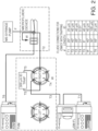
- the sample is collected in the sample manager and has a number of potential paths for analysis illustrated in FIG. 1 .
- the integration and adaptability of this system 100 incorporates a sample manager 110, pump 120, trapping valve manager 130, fiber optic coupled TUV flow cell 140, microfluidic LC tile 150, post-column microfluidic 160, and mass spectrometer 170.
- the sample can pass through each component in series along the flowpath illustrated by a straight line. Alternatively, the sample can take an alternative route. The dotted line in FIG.
- FIG. 1 represents alternative paths for the sample. It is possible to configure the instrument in a number of different configurations. An important factor is to minimize extra column volume.
- One system configuration in FIG. 1 addresses this factor by including a UV detector (TUV flow cell 140) after the first dimension separation (trap 130), but before the microfluidic LC tile 150 (second dimension separation).
- TUV flow cell 140 after the first dimension separation (trap 130), but before the microfluidic LC tile 150 (second dimension separation).
- the sample is cleaned up prior to UV detection, but then refocused on the second dimension for analysis by ESI-MS.
- post column additions of a supercharging or decharging reagent is added to the sample containing the antibody to add specific functionality.
- the invention relates to a method for analyzing the components of a biological mixture, the method comprising: (i) providing a sample comprising one or more of intact antibodies or fragments thereof, antibody drug conjugates or subunits thereof, or intact proteins; (ii) exposing the sample to a first dimension comprising a trap; (iii) exposing the sample to a second dimension comprising a stationary phase; (iv) separating the components of the sample; (v) establishing a mass to charge ratio of each of the components in the sample; and (vi) detecting each of the components in the sample prior to exposing the sample to the second dimension, wherein the method further comprises: (a) adding a supercharging reagent to the sample after exposing the sample to the second dimension; or (b) adding a decharging reagent to the sample after exposing the sample to the second dimension.
- the disclosure also provides methods for isolating, purifying, analyzing, and/or detecting, intact antibodies, antibody subunits, antibody drug conjugate subunits, antibody drug conjugates, and intact proteins in a biological mixture using a two dimensional microscale separation.
- the methods and devices described herein provide significantly improved sensitivity, linearity, and resolution as compared to conventional high flow analytical LC-MS.
- Microscale separation refers to separation systems that are miniaturized such that they significantly improve heat and mass transfer, expanding design choices and analysis capabilities. Microfabricated devices also benefit in terms of their readiness for system integration. This separation technique features reduced reagent consumption, improved performance and multifunctionality by utilizing interconnected network of channels, and inexpensive mass production. Microfluidic systems accommodate incorporation of various sample preparation steps and can be connected to such sophisticated detection systems, such as MS.
- One aspect of the method comprises at least three steps.
- One step involves providing a sample comprising one or more of intact antibodies or fragments thereof, antibody drug conjugates or subunits thereof, or intact proteins.
- Another step involves exposing the sample to a first dimension comprising a trap.
- Another step involves exposing the sample to a second dimension comprising a stationary phase. Then the components of the sample are separated. Subsequently, a mass to charge ratio of each of the components in the sample is established.
- Another aspect of the method comprises at least four steps.
- One step involves providing a sample comprising one or more of intact antibodies or fragments thereof, antibody drug conjugates or subunits thereof, or intact proteins.
- Another step involves exposing the sample to a first dimension separation.
- the first dimension separation comprises trapping a portion of the sample on a first stationary phase.
- Another step involves exposing the sample to a second dimension separation.
- the second dimension separation comprises exposing the sample to a second stationary phase.
- Another step involves adding a supercharging or a decharging reagent to the sample using a post column addition (PCA) microflow device. Then the components of the sample are separated. Subsequently, a mass to charge ratio of each of the components in the sample is established.
- PCA post column addition
- the methods disclosed herein further comprise the step of detecting each of the components in the sample.
- ultraviolet (UV) and/or visible light spectroscopy is used to detect each the components in the sample.
- the detecting step is performed prior to exposing the sample to the second dimension. In other aspects, the detecting step is performed between exposing the sample the first dimension and the second dimension.
- the methods disclosed herein utilize a microflow device.
- the microflow device comprises a microfluidic based system.
- the microflow device can comprise a capillary based system.
- Supercharging offers a unique route to further improve ionization efficiency and sensitivity beyond the capabilities of micro and nano-flow ESI alone ( Iavarone, A.T.; Jurchen, J.C.; Williams, E.R. Anal. Chem. 2001, 73, 1455-1460 ).
- a supercharging reagent can be added prior to the ESI tip (post column), whose physical properties include low volatility, weak Bronsted basicity, and capability of interacting with the analyte.
- a very weak Bronsted base has the effect of decreasing the population of carboxylate anions, while simultaneously increasing the protonation of primary amines located on the antibody ( Ogorzalek Loo, R.R.; Lakshmanan, R.; Loo, J.A. J. Am. Soc. Mass Spectrom., 2014, 25, 1675-1693 ). This increase in protonation of the weak acids present on the protein will shift the CSD to lower m/z, yielding a supercharged structure without
- Eq. 1 may be considered a ratio between a supercharging experiment and a baseline non-supercharging experiment, Eq. 1 can be reworked into Eq. 2 and 3 which yield a relationship between the charge shift ( ⁇ Charge) and the S:N enhancement ( ⁇ S:N). However, the change in signal to noise assumes
- the methods of the invention comprise in one alternative adding a supercharging reagent to the sample.
- a supercharging reagent include, but are not limited to formamide, methoxyethanol, glycerol, and m -nitrobenzyl alcohol ( m -NBA).
- the supercharging reagent is glycerol.
- pH modifications of a molecule that has acidic or basic functionalities can shift the charge state leading to higher mass-to-charge ratios, referred to as charge stripping or decharging ( Stephenson, J.L.; McLuckey, S. A. J. Mass Spectrometry 1998, 33, 664-672 ).
- the implications of pH modification are dependent upon the ionization mode, and the analyte of interest.
- Post column addition of pH modifiers can enable the greatest flexibility by separating the chromatographic performance from the ESI spray efficiency.
- the significance of shifting the overall charge state distribution to lower charge state and higher m/z has implications to providing better charge state resolution.
- the methods of the invention comprise in another alternative adding a decharging reagent to the sample.
- exemplary decharging reagents include, but are not limited to Perfluoro-1, 3 dimethylcyclohexane (PDCH).
- PDCH Perfluoro-1, 3 dimethylcyclohexane
- the decharging reagent is imidazole.
- Methods of the present technology include one or more detection or detecting steps.
- some embodiments utilize mass spectrometry for detection of the antibody-drug conjugate compound and the unconjugated drug compound in the sample.
- Mass spectrometry is used to establish a mass to charge ratio of each of the antibody-drug conjugate compound and the unconjugated drug compound in the sample.
- optical spectroscopy is used as the preferred detection technique.
- ultraviolet (UV) and/or visible spectroscopy or fluorescence spectroscopy is used.
- mass spectrometry systems capable of high mass accuracy, high sensitivity, and high resolution are known in the art and can be employed in the methods and devices of the invention.
- the mass analyzers of such mass spectrometers include, but are not limited to, quadrupole (Q), time of flight (TOF), ion trap, magnetic sector or FT-ICR or combinations thereof.
- the ion source of the mass spectrometer should yield mainly sample molecular ions, or pseudo-molecular ions, and certain characterizable fragment ions. Examples of such ion sources include atmospheric pressure ionization sources, e.g . electrospray ionization (ESI) and Matrix Assisted Laser Desorption Ionization (MALDI).
- ESI electrospray ionization
- MALDI Matrix Assisted Laser Desorption Ionization
- ESI and MALDI are the two most commonly employed methods to ionize proteins for mass spectrometric analysis.
- ESI and APC1 are the most commonly used ion source techniques for LC/MS ( Lee, M. "LC/MS Applications in Drug Development” (2002) J. Wiley & Sons, New York ).
- SELDI Surface Enhanced Laser Desorption Ionization
- U.S. Pat. No. 6,020,208 a surface-based ionization technique that allows for high-throughput mass spectrometry
- SELDI is used to analyze complex mixtures of proteins and other biomolecules.
- SELDI employs a chemically reactive surface such as a "protein chip" to interact with analytes, e.g ., proteins, in solution.
- analytes e.g ., proteins
- Such surfaces selectively interact with analytes and immobilize them thereon.
- the analytes of the invention can be partially purified on the chip and then quickly analyzed in the mass spectrometer. By providing different reactive moieties at different sites on a substrate surface, throughput may be increased.
- mass spectrometers can sample and record the whole mass spectrum simultaneously and with a frequency that allows enough spectra to be acquired for a plurality of constituents in the mixture to ensure that the mass spectrometric signal intensity or peak area is quantitatively representative. This will also ensure that the elution times observed for all the masses would not be modified or distorted by the mass analyzer and it would help ensure that quantitative measurements are not compromised by the need to measure abundances of transient signals.
- Absorption spectroscopy refers to optical spectroscopic techniques that measure the absorption of radiation, as a function of frequency or wavelength, due to its interaction with a sample.
- the sample absorbs energy, i.e., photons, from the radiating field.
- the intensity of the absorption varies as a function of frequency, and this variation is the absorption spectrum.
- Absorption spectroscopy is performed across the electromagnetic spectrum.
- Absorption spectroscopy is employed as an analytical chemistry tool to determine the presence of a particular substance in a sample and, in many cases, to quantify the amount of the substance present. Infrared and ultraviolet-visible spectroscopy are particularly common in analytical applications.
- the most straightforward approach to absorption spectroscopy is to generate radiation with a source, measure a reference spectrum of that radiation with a detector and then re-measure the sample spectrum after placing the material of interest in between the source and detector. The two measured spectra can then be combined to determine the material's absorption spectrum.
- the sample spectrum alone is not sufficient to determine the absorption spectrum because it will be affected by the experimental conditions-the spectrum of the source, the absorption spectra of other materials in between the source and detector and the wavelength dependent characteristics of the detector.
- the reference spectrum will be affected in the same way, though, by these experimental conditions and therefore the combination yields the absorption spectrum of the material alone.
- a wide variety of radiation sources can be employed in order to cover the electromagnetic spectrum.
- spectroscopy it is generally desirable for a source to cover a broad swath of wavelengths in order to measure a broad region of the absorption spectrum.
- Some sources inherently emit a broad spectrum. Examples of these include globars or other black body sources in the infrared, mercury lamps in the visible and ultraviolet and x-ray tubes.
- One recently developed, novel source of broad spectrum radiation is synchrotron radiation which covers all of these spectral regions.
- Other radiation sources generate a narrow spectrum but the emission wavelength can be tuned to cover a spectral range. Examples of these include klystrons in the microwave region and lasers across the infrared, visible and ultraviolet region (though not all lasers have tunable wavelengths).
- the detector employed to measure the radiation power will also depend on the wavelength range of interest. Most detectors are sensitive to a fairly broad spectral range and the sensor selected will often depend more on the sensitivity and noise requirements of a given measurement. Examples of detectors common in spectroscopy include heterodyne receivers in the microwave, bolometers in the millimeter-wave and infrared, mercury cadmium telluride and other cooled semiconductor detectors in the infrared, and photodiodes and photomultiplier tubes in the visible and ultraviolet.
- Ultraviolet/ visible spectroscopy refers to absorption spectroscopy or reflectance spectroscopy in the ultraviolet (UV) and/or visible electromagnetic spectral region.
- Ultraviolet (UV) electromagnetic radiation can have a wavelength ranging from 100 nm (30 PHz) to 380 nm (750 THz), shorter than that of visible light but longer than X-rays.
- the visible light is a type of electromagnetic radiation that is visible to the human eye.
- Visible electromagnetic radiation can have a wavelength ranging from about 390nm (430 THz) to about 700 nm (770 THz).
- the instrument used in ultraviolet-visible spectroscopy is called a UV/Vis spectrophotometer. It measures the intensity of light passing through a sample ( I ), and compares it to the intensity of light before it passes through the sample ( I o ).
- the ratio I / I o is called the transmittance, and is usually expressed as a percentage (%T).
- the absorbance, A is based on the transmittance:
- Fluorescence spectroscopy refers to a type of electromagnetic spectroscopy that analyzes fluorescence from a sample. It involves using a beam of light, usually ultraviolet light, that excites the electrons in molecules of certain compounds and causes them to emit light; typically, but not necessarily, visible light. A complementary technique is absorption spectroscopy. In the special case of single molecule fluorescence spectroscopy, intensity fluctuations from the emitted light are measured from either single fluorophores, or pairs of fluorophores.
- Various light sources may be used as excitation sources, including lasers, LED, and lamps; xenon arcs and mercury-vapor lamps in particular.
- a laser only emits light of high irradiance at a very narrow wavelength interval, typically under 0.01 nm, which makes an excitation monochromator or filter unnecessary.
- a mercury vapor lamp is a line lamp, meaning it emits light near peak wavelengths.
- a xenon arc has a continuous emission spectrum with nearly constant intensity in the range from 300-800 nm and a sufficient irradiance for measurements down to just above 200 nm.
- Filters and/or monochromators may be used in fluorimeters.
- a monochromator transmits light of an adjustable wavelength with an adjustable tolerance.
- the most common type of monochromator utilizes a diffraction grating, that is, collimated light illuminates a grating and exits with a different angle depending on the wavelength. The monochromator can then be adjusted to select which wavelengths to transmit.
- two polarization filters are necessary: One after the excitation monochromator or filter, and one before the emission monochromator or filter.
- the fluorescence is most often measured at a 90° angle relative to the excitation light.
- This geometry is used instead of placing the sensor at the line of the excitation light at a 180° angle in order to avoid interference of the transmitted excitation light.
- No monochromator is perfect and it will transmit some stray light, that is, light with other wavelengths than the targeted.
- An ideal monochromator would only transmit light in the specified range and have a high wavelength-independent transmission.
- the fluorescence can also be measured from the front, which is often done for turbid or opaque samples.
- the detector can either be single-channeled or multichanneled.
- the single-channeled detector can only detect the intensity of one wavelength at a time, while the multichanneled detects the intensity of all wavelengths simultaneously, making the emission monochromator or filter unnecessary.
- the different types of detectors have both advantages and disadvantages.
- the most versatile fluorimeters with dual monochromators and a continuous excitation light source can record both an excitation spectrum and a fluorescence spectrum.
- the wavelength of the excitation light is kept constant, preferably at a wavelength of high absorption, and the emission monochromator scans the spectrum.
- the wavelength passing though the emission filter or monochromator is kept constant and the excitation monochromator is scanning.
- the excitation spectrum generally is identical to the absorption spectrum as the fluorescence intensity is proportional to the absorption.
- a sample used in the methods described herein is a composition known or suspected to contain intact antibodies, bispecific antibodies, antibody subunits, antibody drug conjugate subunits, antibody drug conjugates, and/or intact proteins.
- Samples can include a solid, liquid, gas, mixture, material (e.g., of intermediary consistency, such as a, extract, cell, tissue, organisms) or a combination thereof.
- the sample is a biological sample.
- biological sample refers to a body fluid or a tissue of a living organism.
- Biological samples can include any sample that is derived from the body of a subject.
- the subject can be an animal, for example a mammal, for example a human.
- Other exemplary subjects include a mouse, rat, guinea-pig, rabbit, cat, dog, goat, sheep, pig, cow, or horse.
- the individual can be a patient, for example, an individual suffering from a disease or being suspected of suffering from a disease.
- a biological sample can be a bodily fluid or tissue, for example taken for the purpose of a scientific or medical test, such as for studying or diagnosing a disease (e.g., by detecting and/or identifying a pathogen or the presence of a biomarker).
- Bodily samples can also include cells, for example, pathogens or cells of the individual bodily sample (e.g., tumor cells).
- Such bodily samples can be obtained by known methods including tissue biopsy (e.g., punch biopsy) and by taking blood, bronchial aspirate, sputum, urine, feces, or other body fluids.
- Exemplary bodily samples include humor, whole blood, plasma, serum, umbilical cord blood (in particular, blood obtained by percutaneous umbilical cord blood sampling (PUBS)), cerebrospinal fluid (CSF), saliva, amniotic fluid, breast milk, secretion, ichor, urine, feces, meconium, skin, nail, hair, umbilicus, gastric contents, placenta, bone marrow, peripheral blood lymphocytes (PBL), and solid organ tissue extract.
- PUBS percutaneous umbilical cord blood sampling
- CSF cerebrospinal fluid
- saliva amniotic fluid
- breast milk secretion
- ichor secretion
- urine feces, meconium, skin, nail, hair, umbilicus, gastric contents, placenta, bone marrow, peripheral blood lymphocytes (PBL), and solid organ tissue extract.
- PBL peripheral blood lymphocytes
- the sample is a blood sample.
- the sample is a blood-derived sample, such as plasma or serum.
- the sample is a cell sample.
- the cell sample can contain material obtained or derived from a subject.
- the cell sample can contain cells from an in vitro or ex vivo cell culture.
- the sample is a cell supernatant sample.
- samples derived from other sources known or suspected to contain antibodies may also be used in the disclosed methods.
- samples include environmental samples, which may contain intact antibodies, antibody subunits, antibody drug conjugate subunits, antibody drug conjugates, and/or intact proteins due to, for example, the intentional or unintentional contamination of a given natural or manmade environment.
- Environmental samples can include any sample that is derived from the environment, such as the natural environment (e.g., seas, soils, air, and flora) or the manmade environment (e.g., canals, tunnels, buildings). Such environmental samples can be used to discover, monitor, study, control, mitigate, and avoid environmental pollution.
- Exemplary environmental samples include water (e.g., drinking water, river water, surface water, ground water, potable water, sewage, effluent, wastewater, or leachate), soil, air, sediment, biota (e.g., soil biota), flora, fauna (e.g., fish), and earth mass (e.g., excavated material).
- the sample is a biological sample selected from whole blood, plasma, serum, umbilical cord blood, cerebrospinal fluid (CSF), saliva, amniotic fluid, breast milk, secretion, ichor, urine, feces, meconium, skin, nail, hair, umbilicus, gastric contents, placenta, bone marrow, peripheral blood lymphocytes (PBL), and solid organ tissue extract.
- the sample is blood, plasma or serum.
- Example 1 Influence of Microflow, Supercharging, and Decharging on Whole Antibody Mass
- microfluidic LC device coupled to a QTOF capable of improving sensitivity and linearity for intact protein analysis while also tuning the charge state distributions (CSD) of whole antibodies is demonstrated in this example.
- the mechanism for sensitivity improvement using microflow ESI was demonstrated by shifting of the CSD to higher charge state, and narrowing of the overall CSD. Both of these aspects served to improve ion current of the most abundant charge state of antibodies and lead to improvement in sensitivity over high flow ESI.
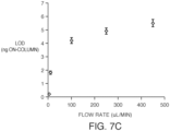
- improvements in sensitivity improvements in linear dynamic range for microflow ESI that results from a combination of lower limits of detection and narrower CSD were observed.
- Increasingly complex antibodies also were investigated for S:N comparisons relative to antibodies that were fully deglycosylated to demonstrate the deleterious effect of increasingly diverse CSD.
- Antibody IgG standards (C6472H9940N1698O2008552, Average MW: 145,329.7 Da, PN: 186006552) were purchased from Waters Cororation (Milford, MA).
- PNGase F peptide-N-glycosidase F
- PNGase F was purchased from New England Biolabs (Ipswitch, MA), and utilized standard deglycosylation procedure.
- PNGase F is an amidase which cleaves between the innermost GlcNac and asparagine residues from N-linked glycoproteins.
- Supercharging and decharging reagents m-NBA, glycerol, and imidazole were purchased from Sigma Aldrich (St. Louis, MO). These compounds were prepared in 50:50 MeOH:H2O with 0.1% formic acid.
- LC-MS grade mobile phases A (0.1% formic acid in water) and mobile phase B (0.1% formic acid in acetonitrile) were purchased from Sigma-Aldrich (St. Louis, MO).
- Antibodies were diluted in 3% acetonitrile and 0.1% formic acid, which proved to be a stable solution over a 48 hr period. Max recovery vials (Waters, Milford, MA PN: 18600327c) were used to dilute and prepare antibody samples for injection.
- Trapping columns were packed into 300 micron x 5 cm stainless steel column bodies.
- the trap geometry was optimized for coupling to a 150 micron microfluidic LC device.
- the 300 micron ID was optimized based on protein load capabilities and refocusing capacity.
- Mechanical frits with 2.0 micron porosity were used to pack against and seal the inlet of the packed columns.
- Two different sorbents were used for these traps including: a) bridged-ethyl-hybrid particle conjugated to a C4 (5 micron particle size, 300 A porosity) and b) TSKgel Phenyl-5PW particle conjugated with a phenyl functionality (20 micron particle size, 1000 A porosity).
- FIG. 2 illustrates the fluidic flow path for the trap coupled to the microfluidic LC device.
- FIG. 3A illustrates the complete high temperature cofired ceramic device integrated with high pressure flow connection ports, analytical LC column, post column addition, and a connector for the ESI emitter.
- FIG. 3B illustrates the mechanism that is used to connect the microfluidic device to the LC pump and autosampler.
- the high pressure flow connection ports are held in position, while the microfluidic device holder is depressed toward the high pressure connection ports to make a 10,000 psi seal. It is important to note here that all three ports are capable of withstanding the 10,000 psi pressure.
- PCA Emitter configuration is illustrated in FIG. 3C .
- the LC system comprised of an m-class binary solvent manager (BSM), a trap valve manager (TVM), an auxiliary solvent manager (ASM) and a sample manager (SM) from Waters, Milford, MA.
- BSM binary solvent manager
- TVM trap valve manager
- ASM auxiliary solvent manager
- SM sample manager
- FIG. 2 The components and fluidic flow path is illustrated in FIG. 2 .
- the LC gradient was linear starting at 3% mobile phase B (MPB) and increasing to 95% MPB in 3.5 min, followed by a 2.5 min hold at 95% and a 4 min requilibration. Strong needle wash was 50% acetonitrile, 25% iso-propanol, 25% H2O with 0.1% FA (V/V), weak needle wash was MPA.
- the MS system comprised of a Xevo G2-XS QTof with an ion Key source from Waters, Milford, MA.
- the MS was scanned at a rate of 5 Hz, from 500 m/z to 4,000 m/z. Since the antibody has a charge state distribution that ranges from 2000 m/z to 3200 m/z this was acceptable to capture both the CSD of the protein and lock mass (Glu-fibrinopeptide B, 785.84265 m/z, +2 charge state).
- the RF collision cell voltage was optimized at 600 V, with a gain value of 10, while the quad profile was set to automatic mode. Data processing was performed using MassLynx and MaxEnt 1 deconvolution software.
- first dimension is slightly less retentive than the microfluidic LC chemistry (second dimension) will enable analyte refocusing between dimensions.
- second dimension This refocusing between first and second dimension is illustrated in FIG. 4 .
- the process involves the analyte eluting from the trap, broadening via parabolic band broadening in the transfer capillary, and then refocusing on the second dimension.
- the extent of refocusing depends on the relative retentivity difference between the two stationary phases of each dimension.
- FIG. 5A and 5B The path length included the transfer tubing and an open tubular, unpacked, microfluidic device ( FIG. 5A ). Following this experiment the open tubular microfluidic device was replaced with a packed particle C4-BEH microfluidic device ( FIG. 5B).
- FIG. 5D demonstrates the refocusing between the phenyl trap coupled to the packed particle C4-BEH microfluidic device.
- FIG. 6 demonstrates the relative difference between the optimized C4 X-bridge trap ( FIG. 6B ) and the optimized phenyl trap ( FIG. 6A ).
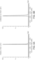
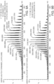
- FIGS. 7A and 7B demonstrate the charge state distribution that was obtained for a standard glycosylated IgG between these two conditions. Two general phenomena were present: 1) the charge state distribution was narrower, and 2) the average charge state was shifted to higher charge for microflow as compared to standard flow.
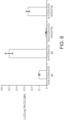
- FIG. 8 illustrates the difference in signal to noise for glycosylated and deglycosylated IgG standard, in addition to comparison between Herceptin that was glycosylated and deglycosylated.
- FIG. 3A In addition to the normal mechanisms of ESI spray described above, also investigated were supercharging and decharging of antibodies and antibody drug conjugates using the post column addition microfluidic device shown in FIG. 3A .
- This device enabled us to mix the LC eluent with the supercharging or decharging reagent within the emitter tip. Diffusional mixing within the emitter tip and spray droplets was sufficient to perform supercharging and decharging.
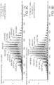
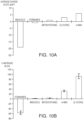
- FIGS. 9A-9D illustrate the optimal supercharging profiles obtained using a series of supercharging reagents. Glycoform resolution was retained in this experiment across all supercharging reagents, without any major adduct formation. The average charge state deviation from the antibody or antibody drug conjugate is shown in FIG. 10A .
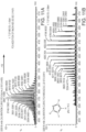
- FIGS. 11A and 11B illustrate the effect of decharging a standard IgG using imidazole PCA addition.
- the process shifted the CSD by -19 charges, which yielded an increase in charge state spacing by 50%.
- Such a process affords more spectral space to process complex mAb's and ADC where charge states could overlap.
- two antibodies were added, Silumab and the Waters standard IgG, which coeluted in the chromatograph.
- 11C, 11D , and 11E shows the ability of decharging to better resolve the charge states of two antibodies that are overlapping without decharging, and consequently resolved when decharging is present.
- the additional charge state spacing available with this decharging experiment enables baseline resolution between the CSD of both Silumab and the Waters standard IgG. This process further simplifies deconvolution and identification of zero charge masses.
- microfluidic device capable of performing supercharging and decharging experimentation in a robust configuration was demonstrated.
- Chemistry pairing between the phenyl trap and the C4 microfluidic demonstrated excellent refocusing capabilities, and yields modularity replacement between trap and microfluidic LC components.
- This type of sample "pre-treatment” and instrumental modularity should be an increasingly popular theme for microfluidic devices designed for repetitive analysis of 1,000's of samples.
- Microflow ESI was also demonstrated to shift the charge state distribution to higher charge and narrower distribution widths relative to high flow ESI. This characteristic of microflow is responsible for the 15X improvement in sensitivity over high flow ESI.
Landscapes
- Life Sciences & Earth Sciences (AREA)
- Health & Medical Sciences (AREA)
- Engineering & Computer Science (AREA)
- Molecular Biology (AREA)
- Physics & Mathematics (AREA)
- Immunology (AREA)
- Chemical & Material Sciences (AREA)
- Hematology (AREA)
- Urology & Nephrology (AREA)
- Biomedical Technology (AREA)
- Bioinformatics & Cheminformatics (AREA)
- Bioinformatics & Computational Biology (AREA)
- Spectroscopy & Molecular Physics (AREA)
- Analytical Chemistry (AREA)
- Pathology (AREA)
- General Physics & Mathematics (AREA)
- General Health & Medical Sciences (AREA)
- Biochemistry (AREA)
- Medicinal Chemistry (AREA)
- Cell Biology (AREA)
- Food Science & Technology (AREA)
- Microbiology (AREA)
- Biotechnology (AREA)
- Biophysics (AREA)
- Proteomics, Peptides & Aminoacids (AREA)
- Optics & Photonics (AREA)
- Nanotechnology (AREA)
- Other Investigation Or Analysis Of Materials By Electrical Means (AREA)
Description
- The present disclosure relates devices and methods for analyzing intact antibodies, bispecific antibodies, antibody subunits, antibody drug conjugate subunits, antibody drug conjugates, and intact proteins in a biological mixture. Only a method for analyzing the components of a biological mixture is claimed.
- One of the biggest barriers associated with intact antibody analysis is the relative ionization efficiency using electrospray ionization (ESI) mass spectrometry. Well ionizing peptides and small molecules can easily be measured down to 1 pg/mL (∼ 1 pM) using current technologies, such as liquid chromatography coupled with mass spectrometry (LC-MS). In comparison, the current technologies can measure deglycosylated antibodies with concentrations that only extend down to 0.1 µg/mL (~ 60 nM) in a neat solution. This approximate three order of magnitude difference in sensitivity is mainly due to the challenges of ionizing intact proteins via electrospray, which is significantly more challenged due to the molecular weight differential between intact antibodies and small molecules. The molecular weight of a protein also has a significant impact on the width of the charge state distributions (CSDs). As molecular weight increases, so does the width of the CSD, which results in a reduction of the signal to noise (S:N) by splitting ion current over a larger number of ions (Compton, P. D.; Zamdborg, L.; Thomas, P. M.; Kelleher, N.L. Anal. Chem. 2011, 83, 6868-6874). Accordingly, developing technologies that enable the narrowing of the CSD and increasing the charge state will further assist in improving sensitivity for the analysis of intact proteins.
- The following document is known: ERIN E CHAMBERS ET AL, "Practical applications of integrated microfluidics for peptide quantification", BIOANALYSIS, London, UK, (20150401), vol. 7, no. 7, doi:10.4155/bio.15.15, ISSN 1757-6180, pages 857 - 867, XP055475661 [Y] 1-19 * whole document, in particular abstract; p. 858, col. 1, bridging par. - p. 860, col. 2 * .
US2009/286258 discloses analysis of antibody-drug conjugates using capture by beads and reverse phase chromatography in combination with mass spectrometry. - The present disclosure relates devices and methods for analyzing intact antibodies, antibody subunits, antibody drug conjugate subunits, antibody drug conjugates, and intact proteins in a biological mixture, which provide significantly improved sensitivity, linearity, and resolution as compared to conventional high flow analytical LC-MS.
- Accordingly, provided herein are methods for analyzing the components of a biological mixture. One aspect of the method comprises at least three steps. One step involves providing a sample comprising one or more of intact antibodies or fragments thereof, antibody drug conjugates or subunits thereof, or intact proteins. Another step involves exposing the sample to a first dimension comprising a trap. Another step involves exposing the sample to a second dimension comprising a stationary phase. Then the components of the sample are separated. Subsequently, a mass to charge ratio of each of the components in the sample is established.
- Optionally, the method may also include another step wherein some separation occurs on the trap such that when the sample is loaded onto the trap, small ions are removed the sample ("desalting"). This optional step occurs on the trap when the sample is eluted from the trap to the second dimension such that further separation occurs. The chemistry of the trap and the chemistry of the second dimension must be matched appropriately to provide optimal peak shape.
- Also disclosed herein is a method which comprises at least four steps. One step involves providing a sample comprising one or more of intact antibodies or fragments thereof, antibody drug conjugates or subunits thereof, or intact proteins. Another step involves exposing the sample to a first dimension separation. Another step involves exposing the sample to a second dimension separation. Another step involves adding a supercharging or a decharging reagent to the sample using a post column addition (PCA) microflow device. Then the components of the sample are separated. Subsequently, a mass to charge ratio of each of the components in the sample is established. In some embodiments, the microflow device comprises microfluidic based system. Alternatively, the microflow device can comprise a capillary based system.
- Also provided herein are devices comprising at least four components. One component is a first dimension separator, such as, for example, a trap. Another component is a second dimension separator, such as, for example a stationary phase. Another component is a post column addition (PCA) microflow device. Another component is a mass spectrometer. In some aspects, the device further comprises an ultraviolet/visible (UV/Vis) spectrometer. In other aspects, the mass spectrometer is a quadrupole time-of-flight (QToF) mass spectrometer. In particular aspects, the microflow device comprises microfluidic based system. Alternatively, the microflow device can comprise a capillary based system.
- The methods and devices provided herein enable isolation, purification, analysis, and detection of intact antibodies, antibody subunits, antibody drug conjugate subunits, antibody drug conjugates, and intact proteins in a biological mixture using a two dimensional microscale separation.
- The claimed invention is a method for analyzing the components of a biological mixture. The method includes (i) providing a sample comprising one or more of intact antibodies or fragments thereof, antibody drug conjugates or subunits thereof, or intact proteins; (ii) exposing the sample to a first dimension comprising a trap; (iii) exposing the sample to a second dimension comprising a stationary phase; (iv) separating the components of the sample; (v) establishing a mass to charge ratio of each of the components in the sample; and (iv) detecting each of the components in the sample prior to exposing the sample to the second dimension.
- Ultraviolet (UV) and/or visible light spectroscopy can be used to detect each of the components in the sample. The detecting step can be performed between exposing the sample to the first dimension and the second dimension.
- The method includes adding a supercharging reagent to the sample. The supercharging reagent can be selected from the group consisting of formamide, methoxyethanol, glycerol, and m-nitrobenzyl alcohol (m-NBA). The supercharging reagent can be glycerol. The supercharging reagent is added to the sample after exposing the sample to the second dimension. The supercharging reagent can be added to the sample between exposing the sample to the second dimension and establishing a mass to charge ratio of each of the components in the sample. The supercharging reagent can be added to the sample using a microflow device.
- The method alternatively includes adding a decharging reagent to the sample. The decharging reagent can be imidazole. The decharging reagent is added to the sample after exposing the sample to the second dimension. The decharging reagent can be added to the sample between exposing the sample to the second dimension and establishing a mass to charge ratio of each of the components in the sample. The decharging reagent can be added to the sample using a microflow device.
- The microflow device can be a post column addition (PCA) microflow device. The microflow device can include a microfluidic based system. The microflow device can include a capillary based system.
- Also disclosed herein is a method for analyzing the components of a biological mixture. The method includes: (i) providing a sample comprising one or more of intact antibodies or fragments thereof, antibody drug conjugates or subunits thereof, or intact proteins; (ii) exposing the sample to a first dimension separation; (iii) exposing the sample to a second dimension separation; (iv) adding a supercharging or a decharging reagent to the sample using a post column addition (PCA) microflow device; (v) separating the components of the sample; and (vi) establishing a mass to charge ratio of each of the components in the sample.
- The first dimension separation can include trapping a portion of the sample on a first stationary phase. The second dimension separation can include exposing the sample to a second stationary phase.
- The method can also include the step of detecting each of the components in the sample.
- Ultraviolet (UV) and/or visible light spectroscopy can be used to detect each the components in the sample.
- The detecting step can be performed prior to exposing the sample to the second dimension. The detecting step can be performed between exposing the sample the first dimension and the second dimension.
- The supercharging reagent can be selected from the group consisting of formamide, methoxyethanol, glycerol, and m-nitrobenzyl alcohol (m-NBA). The supercharging reagent can be glycerol.
- The decharging reagent can be imidazole.
- The microflow device can include a microfluidic based system. The microflow device can include a capillary based system.
- The sample can be a biological sample. The biological sample can be derived from a subject. The subject can be a human subject. The biological sample can be selected from the group consisting of blood or derived from blood, tissue, urine, saliva, lymph, and biopsy. The biological sample can be blood or derived from blood. The biological sample can be plasma or serum.
- The technology relates to a device that includes (i) a first dimension separator comprising a trap; a second dimension separator comprising a stationary phase; a post column addition (PCA) microflow device; and a mass spectrometer.
- The device can include an ultraviolet/visible (UV/Vis) spectrometer.
- The mass spectrometer can be a quadrupole time-of-flight (QToF) mass spectrometer.
- The microflow device can include a microfluidic based system. The microflow device can include a capillary based system.
- The above aspects of the methods and devices described herein possess one or more advantages. For example, certain embodiments provide significantly improved sensitivity, linearity, and resolution as compared to conventional high flow analytical LC-MS. In some aspects, one or more advantages is achieved by incorporation of two distinct chromatographic dimensions, providing the capability of tuning selectively in each dimension and thereby allowing for control of refocusing functionality. In certain aspects, one or more advantages is achieved or improved by pairing trapping chemistry with analytical chemistry to optimize refocusing. In certain aspects, one or more advantages is achieved or improved by microscale integration of UV detection between the first and second dimension separation, which enables refocusing of analyte that has been broadened by UV detector and tubings prior to ESI spray. In some aspects, one or more advantages is achieved or enhanced by the addition of one or more supercharging and/or decharging reagents that retain glycoform resolution.
-
-
FIG. 1 shows an instrument flow diagram showing the components of the device disclosed herein. -
FIG. 2 is an illustration of the LC pump and trapping configuration for the 2D trap-wash-elute used in one t of the methods disclosed herein. -
FIG. 3 shows A:a microfluidic layout for the integrated microfluidic device; B: a clamping design that connects the outside fluidics to the microfluidic device shown in A; and C: an end on view of connection mechanism for emitter onto the end of the microfluidic device. -
FIG. 4 shows a mechanism of loading the trap, focusing the analyte at the head of the trap and consequently removing diluent and non-binding matrix components (top); and a mechanism of moving the analyte from the first dimension trap, through transfer capillary, and refocusing the analyte on the microfluidic LC (bottom). -
FIGS. 5A-5D show the packing configuration for the system. InFIG. 5A , the trap is packed, while the microfluidic device is not packed (open tubular) andFIG. 5C shows the corresponding chromatogram. InFIG. 5B , both trap and microfluidic LC are packed in this configuration andFIG. 5D shows the corresponding chromatogram. Separation is of IgG under neat conditions. -
FIG. 6 A: illustrates the peak shape between the phenyl and X-bridge C4 trap paired with a BEH-C4 analytical microfluidic device.FIG 6B and6C illustrate the dependency on peak width versus temperature for both the X-bridge C4 trap (6B) and the phenyl trap (6C). -
FIG. 7 shows A & B: dependency on the charge state distribution on flow rate of LC for low flow and high flow conditions; C: limits of detection as compared to flow rate for intact mAb; and D & E: linearity of quantitative analysis between 2.1 mm scale and microfluidic devices. -
FIG. 8 shows limits of detection and sensitivity for glycosylated and non-deglycosylated IgG and Herceptin using the microfluidic device. -
FIGS. 9A-9D show supercharging screening for IgG shows optimized conditions. -
FIG. 10 shows A: the average charge state shifts for the superchargers screened in this study; and B: increases in signal to noise for the supercharging and decharging experiments. -
FIG. 11 shows A and B: the effect of decharging mAb using imidazole added post column; and C, D, and E: the functionality of deconvoluting multiple antibodies using the improved charge state spacings. - Unless stated otherwise, the following terms and phrases as used herein are intended to have the following meanings:
As used herein "antibody drug conjugates: or "ADCs" are monoclonal antibodies (mAbs) attached to biologically active drugs by chemical linkers with labile bonds. - "Antibody" is used herein in the broadest sense and specifically covers monoclonal antibodies, polyclonal antibodies, multispecific antibodies (e.g., bispecific antibodies), and antibody fragments. Antibodies may be murine, human, humanized, chimeric, or derived from other species. An antibody is a protein generated by the immune system that is capable of recognizing and binding to a specific antigen. (Janeway, et al (2001) "Immunobiology", 5th Ed., Garland Publishing, New York). A target antigen generally has numerous binding sites, also called epitopes, recognized by CDRs on multiple antibodies. Each antibody that specifically binds to a different epitope has a different structure. Thus, one antigen may have more than one corresponding antibody.
- Antibody also refers to a full-length immunoglobulin molecule or an immunologically active portion of a full-length immunoglobulin molecule, i.e., a molecule that contains an antigen binding site that immunospecifically binds an antigen of a target of interest or part thereof, such targets including but not limited to, cancer cell or cells that produce autoimmune antibodies associated with an autoimmune disease. The immunoglobulin disclosed herein can be of any type (e.g., IgG, IgE, IgM, IgD, and IgA), class (e.g., IgG1, IgG2, IgG3, IgG4, IgA1 and IgA2) or subclass of immunoglobulin molecule. The immunoglobulins can be derived from any species. In one aspect, however, the immunoglobulin is of human, murine, or rabbit origin.
- The term "linker unit" refers to the direct or indirect linkage of the antibody to the drug. Attachment of a linker to a mAb can be accomplished in a variety of ways, such as through surface lysines, reductive-coupling to oxidized carbohydrates, and through cysteine residues liberated by reducing interchain disulfide linkages. A variety of ADC linkage systems are known in the art, including hydrazone-, disulfide- and peptide-based linkages.
- The term "active pharmaceutical ingredient" or "API" refers to is the ingredient in a pharmaceutical drug that is biologically active. The terms "pharmaceutical drug," "drug," and "payload" are used interchangeably throughout, and refer to any substance having biological or detectable activity, for example, therapeutic agents, detectable labels, binding agents, etc., and prodrugs, which are metabolized to an active agent in vivo.
- As used herein, the term "mass spectrometry" or "MS" refers to an analytical technique to identify compounds by their mass. MS refers to methods of filtering, detecting, and measuring ions based on their mass-to-charge ratio, or "m/z". MS technology generally includes (1) ionizing the compounds to form charged compounds; and (2) detecting the molecular weight of the charged compounds and calculating a mass-to-charge ratio. The compounds may be ionized and detected by any suitable means. A "mass spectrometer" generally includes an ionizer and an ion detector. In general, one or more molecules of interest are ionized, and the ions are subsequently introduced into a mass spectrometric instrument where, due to a combination of magnetic and electric fields, the ions follow a path in space that is dependent upon mass ("m") and charge ("z").
- As used herein, the term "ionization" or "ionizing" refers to the process of generating an analyte ion having a net electrical charge equal to one or more electron units. Negative ions are those having a net negative charge of one or more electron units, while positive ions are those having a net positive charge of one or more electron units.
- As used herein, the term "chromatography" refers to a process in which a chemical mixture carried by a liquid or gas is separated into components as a result of differential distribution of the chemical entities as they flow around or over a stationary liquid or solid phase.
- As used herein, the term "liquid chromatography" or "LC" means a process of selective retardation of one or more components of a fluid solution as the fluid uniformly percolates through a column of a finely divided substance, or through capillary passageways. The retardation results from the distribution of the components of the mixture between one or more stationary phases and the bulk fluid, (i.e., mobile phase), as this fluid moves relative to the stationary phase(s). Examples of "liquid chromatography" include reverse phase liquid chromatography (RPLC), high performance liquid chromatography (HPLC), ultra-high performance liquid chromatography (UHPLC), turbulent flow liquid chromatography (TFLC) (sometimes known as high turbulence liquid chromatography (HTLC) or high throughput liquid chromatography).
- As used herein, the term "high performance liquid chromatography" or "HPLC" (also sometimes known as "high pressure liquid chromatography") refers to liquid chromatography in which the degree of separation is increased by forcing the mobile phase under pressure through a stationary phase, typically a densely packed column. As used herein, the term "ultra high performance liquid chromatography" or "UHPLC" (sometimes known as "ultra high pressure liquid chromatography") refers to HPLC that occurs at much higher pressures than traditional HPLC techniques.
- The term "LC/MS" refers to a liquid chromatograph (LC) interfaced to a mass spectrometer.
- The disclosure provides devices for isolating, purifying, analyzing, and/or detecting, intact antibodies, antibody subunits, antibody drug conjugate subunits, antibody drug conjugates, and intact proteins in a biological mixture using a two dimensional microscale separation. The methods and devices described herein provide significantly improved sensitivity, linearity, and resolution as compared to conventional high flow analytical LC-MS. In some embodiments, the LC-MS is a micro-flow LC-MS, which allow for greater sensitivity.
- Accordingly, provided herein are devices comprising at least four components. One component is a first dimension separator comprising a trap. Another component is a second dimension separator comprising a stationary phase. Another component is a post column addition (PCA) microflow device. Another component is a mass spectrometer. In some aspects, the device further comprises an ultraviolet/visible (UV/Vis) spectrometer. In other aspects, the mass spectrometer is a quadrupole time-of-flight (QToF) mass spectrometer. In particular aspects, the microflow device comprises microfluidic based system. Alternatively, the microflow device can comprise a capillary based system.
- In one aspect, the devices provided herein are designed to analyze antibodies, antibody drug conjugates, and antibody subunits using two dimensional LC coupled with microfluidic PCA and in-line coupled TUV-MS detection. The sample is collected in the sample manager and has a number of potential paths for analysis illustrated in
FIG. 1 . The integration and adaptability of thissystem 100 incorporates asample manager 110, pump 120, trappingvalve manager 130, fiber optic coupledTUV flow cell 140,microfluidic LC tile 150, post-column microfluidic 160, andmass spectrometer 170. As illustrated inFIG. 1 , the sample can pass through each component in series along the flowpath illustrated by a straight line. Alternatively, the sample can take an alternative route. The dotted line inFIG. 1 represents alternative paths for the sample. It is possible to configure the instrument in a number of different configurations. An important factor is to minimize extra column volume. One system configuration inFIG. 1 addresses this factor by including a UV detector (TUV flow cell 140) after the first dimension separation (trap 130), but before the microfluidic LC tile 150 (second dimension separation). As a result, the sample is cleaned up prior to UV detection, but then refocused on the second dimension for analysis by ESI-MS. In some embodiments, following this process of refocusing and separation on a microfluidic device, post column additions of a supercharging or decharging reagent is added to the sample containing the antibody to add specific functionality. - The invention relates to a method for analyzing the components of a biological mixture, the method comprising: (i) providing a sample comprising one or more of intact antibodies or fragments thereof, antibody drug conjugates or subunits thereof, or intact proteins; (ii) exposing the sample to a first dimension comprising a trap; (iii) exposing the sample to a second dimension comprising a stationary phase; (iv) separating the components of the sample; (v) establishing a mass to charge ratio of each of the components in the sample; and (vi) detecting each of the components in the sample prior to exposing the sample to the second dimension, wherein the method further comprises: (a) adding a supercharging reagent to the sample after exposing the sample to the second dimension; or (b) adding a decharging reagent to the sample after exposing the sample to the second dimension.
- The disclosure also provides methods for isolating, purifying, analyzing, and/or detecting, intact antibodies, antibody subunits, antibody drug conjugate subunits, antibody drug conjugates, and intact proteins in a biological mixture using a two dimensional microscale separation. The methods and devices described herein provide significantly improved sensitivity, linearity, and resolution as compared to conventional high flow analytical LC-MS.
- Microscale separation refers to separation systems that are miniaturized such that they significantly improve heat and mass transfer, expanding design choices and analysis capabilities. Microfabricated devices also benefit in terms of their readiness for system integration. This separation technique features reduced reagent consumption, improved performance and multifunctionality by utilizing interconnected network of channels, and inexpensive mass production. Microfluidic systems accommodate incorporation of various sample preparation steps and can be connected to such sophisticated detection systems, such as MS.
- Accordingly, provided herein are methods for analyzing the components of a biological mixture. One aspect of the method comprises at least three steps. One step involves providing a sample comprising one or more of intact antibodies or fragments thereof, antibody drug conjugates or subunits thereof, or intact proteins. Another step involves exposing the sample to a first dimension comprising a trap. Another step involves exposing the sample to a second dimension comprising a stationary phase. Then the components of the sample are separated. Subsequently, a mass to charge ratio of each of the components in the sample is established.
- Another aspect of the method comprises at least four steps. One step involves providing a sample comprising one or more of intact antibodies or fragments thereof, antibody drug conjugates or subunits thereof, or intact proteins. Another step involves exposing the sample to a first dimension separation. In some aspects, the first dimension separation comprises trapping a portion of the sample on a first stationary phase. Another step involves exposing the sample to a second dimension separation. In some aspects, the second dimension separation comprises exposing the sample to a second stationary phase. Another step involves adding a supercharging or a decharging reagent to the sample using a post column addition (PCA) microflow device. Then the components of the sample are separated. Subsequently, a mass to charge ratio of each of the components in the sample is established.
- The methods disclosed herein further comprise the step of detecting each of the components in the sample. In some embodiments, ultraviolet (UV) and/or visible light spectroscopy is used to detect each the components in the sample. The detecting step is performed prior to exposing the sample to the second dimension. In other aspects, the detecting step is performed between exposing the sample the first dimension and the second dimension.
- In some aspects, the methods disclosed herein utilize a microflow device. In a particular embodiment, the microflow device comprises a microfluidic based system. Alternatively, the microflow device can comprise a capillary based system.
- Supercharging offers a unique route to further improve ionization efficiency and sensitivity beyond the capabilities of micro and nano-flow ESI alone (Iavarone, A.T.; Jurchen, J.C.; Williams, E.R. Anal. Chem. 2001, 73, 1455-1460). A supercharging reagent can be added prior to the ESI tip (post column), whose physical properties include low volatility, weak Bronsted basicity, and capability of interacting with the analyte. A very weak Bronsted base has the effect of decreasing the population of carboxylate anions, while simultaneously increasing the protonation of primary amines located on the antibody (Ogorzalek Loo, R.R.; Lakshmanan, R.; Loo, J.A. J. Am. Soc. Mass Spectrom., 2014, 25, 1675-1693). This increase in protonation of the weak acids present on the protein will shift the CSD to lower m/z, yielding a supercharged structure without structural modification.
- Alternative mechanisms have employed the relatively low volatility of supercharging agents. As the droplet evaporates mobile phase at a greater rate than the supercharging reagent the concentration of the supercharging reagent enriches significantly. This high concentration of supercharging reagent can serve as a charge trap by forming a supercharging reagent shell surrounding the antibody (Metwally, H.; McAllister, R.G.; Popa, V.; Konermann, L. Anal. Chem. 2016, 88, 5345-5354). The theoretical S:N gains using a supercharging reagent can be approimatded with
equation 1.
- Eq. 1 may be considered a ratio between a supercharging experiment and a baseline non-supercharging experiment, Eq. 1 can be reworked into Eq. 2 and 3 which yield a relationship between the charge shift (ΔCharge) and the S:N enhancement (ΔS:N). However, the change in signal to noise assumes
- equivalent chemical noise backgrounds between supercharging experiments, which is an important experimental variable.


- The methods of the invention comprise in one alternative adding a supercharging reagent to the sample. Exemplary supercharging reagents include, but are not limited to formamide, methoxyethanol, glycerol, and m-nitrobenzyl alcohol (m-NBA). In preferred embodiments, the supercharging reagent is glycerol.
- In addition to supercharging, pH modifications of a molecule that has acidic or basic functionalities can shift the charge state leading to higher mass-to-charge ratios, referred to as charge stripping or decharging (Stephenson, J.L.; McLuckey, S. A. J. Mass Spectrometry 1998, 33, 664-672). The implications of pH modification are dependent upon the ionization mode, and the analyte of interest. Post column addition of pH modifiers can enable the greatest flexibility by separating the chromatographic performance from the ESI spray efficiency. The significance of shifting the overall charge state distribution to lower charge state and higher m/z has implications to providing better charge state resolution. In cases of antibodies with multiple glycosylation sites, post translational modifications, and drug conjugates, the charge state spectra can be very complex. In many cases, deglycosylation is required, to further simplify proteins due to overlapping charge states. Shifting these high mass ions to lower charge state affords improved charge state spacing resolution and deconvolution outcomes. There are also limitations with this approach in the form of sensitivity decreases due to the lower charge of the antibody. However, some of the losses associated with decreased signal are made up for by reduction of the chemical noise that is present in the LC-MS separation de facto.
- The methods of the invention comprise in another alternative adding a decharging reagent to the sample. Exemplary decharging reagents include, but are not limited to Perfluoro-1, 3 dimethylcyclohexane (PDCH). In preferred embodiments, the decharging reagent is imidazole.
- Methods of the present technology include one or more detection or detecting steps. For example, some embodiments utilize mass spectrometry for detection of the antibody-drug conjugate compound and the unconjugated drug compound in the sample. Mass spectrometry is used to establish a mass to charge ratio of each of the antibody-drug conjugate compound and the unconjugated drug compound in the sample.
- In certain other aspects, optical spectroscopy is used as the preferred detection technique. In a particular embodiment, ultraviolet (UV) and/or visible spectroscopy or fluorescence spectroscopy is used.
- A variety of mass spectrometry systems capable of high mass accuracy, high sensitivity, and high resolution are known in the art and can be employed in the methods and devices of the invention. The mass analyzers of such mass spectrometers include, but are not limited to, quadrupole (Q), time of flight (TOF), ion trap, magnetic sector or FT-ICR or combinations thereof. The ion source of the mass spectrometer should yield mainly sample molecular ions, or pseudo-molecular ions, and certain characterizable fragment ions. Examples of such ion sources include atmospheric pressure ionization sources, e.g. electrospray ionization (ESI) and Matrix Assisted Laser Desorption Ionization (MALDI). ESI and MALDI are the two most commonly employed methods to ionize proteins for mass spectrometric analysis. ESI and APC1 are the most commonly used ion source techniques for LC/MS (Lee, M. "LC/MS Applications in Drug Development" (2002) J. Wiley & Sons, New York).
- Surface Enhanced Laser Desorption Ionization (SELDI) is an example of a surface-based ionization technique that allows for high-throughput mass spectrometry (
U.S. Pat. No. 6,020,208 ). Typically, SELDI is used to analyze complex mixtures of proteins and other biomolecules. SELDI employs a chemically reactive surface such as a "protein chip" to interact with analytes, e.g., proteins, in solution. Such surfaces selectively interact with analytes and immobilize them thereon. Thus, the analytes of the invention can be partially purified on the chip and then quickly analyzed in the mass spectrometer. By providing different reactive moieties at different sites on a substrate surface, throughput may be increased. - Commercially available mass spectrometers can sample and record the whole mass spectrum simultaneously and with a frequency that allows enough spectra to be acquired for a plurality of constituents in the mixture to ensure that the mass spectrometric signal intensity or peak area is quantitatively representative. This will also ensure that the elution times observed for all the masses would not be modified or distorted by the mass analyzer and it would help ensure that quantitative measurements are not compromised by the need to measure abundances of transient signals.
- A variety of optical spectroscopy systems capable of high accuracy, high sensitivity, and high resolution are known in the art and can be employed in the methods of the invention. Absorption spectroscopy refers to optical spectroscopic techniques that measure the absorption of radiation, as a function of frequency or wavelength, due to its interaction with a sample. The sample absorbs energy, i.e., photons, from the radiating field. The intensity of the absorption varies as a function of frequency, and this variation is the absorption spectrum. Absorption spectroscopy is performed across the electromagnetic spectrum.
- Absorption spectroscopy is employed as an analytical chemistry tool to determine the presence of a particular substance in a sample and, in many cases, to quantify the amount of the substance present. Infrared and ultraviolet-visible spectroscopy are particularly common in analytical applications.
- There are a wide range of experimental approaches for measuring absorption spectra. The most common arrangement is to direct a generated beam of radiation at a sample and detect the intensity of the radiation that passes through it. The transmitted energy can be used to calculate the absorption. The source, sample arrangement and detection technique vary significantly depending on the frequency range and the purpose of the experiment.
- The most straightforward approach to absorption spectroscopy is to generate radiation with a source, measure a reference spectrum of that radiation with a detector and then re-measure the sample spectrum after placing the material of interest in between the source and detector. The two measured spectra can then be combined to determine the material's absorption spectrum. The sample spectrum alone is not sufficient to determine the absorption spectrum because it will be affected by the experimental conditions-the spectrum of the source, the absorption spectra of other materials in between the source and detector and the wavelength dependent characteristics of the detector. The reference spectrum will be affected in the same way, though, by these experimental conditions and therefore the combination yields the absorption spectrum of the material alone.
- A wide variety of radiation sources can be employed in order to cover the electromagnetic spectrum. For spectroscopy, it is generally desirable for a source to cover a broad swath of wavelengths in order to measure a broad region of the absorption spectrum. Some sources inherently emit a broad spectrum. Examples of these include globars or other black body sources in the infrared, mercury lamps in the visible and ultraviolet and x-ray tubes. One recently developed, novel source of broad spectrum radiation is synchrotron radiation which covers all of these spectral regions. Other radiation sources generate a narrow spectrum but the emission wavelength can be tuned to cover a spectral range. Examples of these include klystrons in the microwave region and lasers across the infrared, visible and ultraviolet region (though not all lasers have tunable wavelengths).
- The detector employed to measure the radiation power will also depend on the wavelength range of interest. Most detectors are sensitive to a fairly broad spectral range and the sensor selected will often depend more on the sensitivity and noise requirements of a given measurement. Examples of detectors common in spectroscopy include heterodyne receivers in the microwave, bolometers in the millimeter-wave and infrared, mercury cadmium telluride and other cooled semiconductor detectors in the infrared, and photodiodes and photomultiplier tubes in the visible and ultraviolet.
- "Ultraviolet/ visible spectroscopy" refers to absorption spectroscopy or reflectance spectroscopy in the ultraviolet (UV) and/or visible electromagnetic spectral region. Ultraviolet (UV) electromagnetic radiation can have a wavelength ranging from 100 nm (30 PHz) to 380 nm (750 THz), shorter than that of visible light but longer than X-rays. The visible light is a type of electromagnetic radiation that is visible to the human eye. Visible electromagnetic radiation can have a wavelength ranging from about 390nm (430 THz) to about 700 nm (770 THz).
- The instrument used in ultraviolet-visible spectroscopy is called a UV/Vis spectrophotometer. It measures the intensity of light passing through a sample (I), and compares it to the intensity of light before it passes through the sample (Io ). The ratio I/Io is called the transmittance, and is usually expressed as a percentage (%T). The absorbance, A, is based on the transmittance:
- "Fluorescence spectroscopy" refers to a type of electromagnetic spectroscopy that analyzes fluorescence from a sample. It involves using a beam of light, usually ultraviolet light, that excites the electrons in molecules of certain compounds and causes them to emit light; typically, but not necessarily, visible light. A complementary technique is absorption spectroscopy. In the special case of single molecule fluorescence spectroscopy, intensity fluctuations from the emitted light are measured from either single fluorophores, or pairs of fluorophores.
- Two general types of instruments exist: (1) filter fluorometers that use filters to isolate the incident light and fluorescent light; and (2) spectrofluorometers that use a diffraction grating monochromators to isolate the incident light and fluorescent light. Both types use the following scheme: the light from an excitation source passes through a filter or monochromator, and strikes the sample. A proportion of the incident light is absorbed by the sample, and some of the molecules in the sample fluoresce. The fluorescent light is emitted in all directions. Some of this fluorescent light passes through a second filter or monochromator and reaches a detector, which is usually placed at 90° to the incident light beam to minimize the risk of transmitted or reflected incident light reaching the detector.
- Various light sources may be used as excitation sources, including lasers, LED, and lamps; xenon arcs and mercury-vapor lamps in particular. A laser only emits light of high irradiance at a very narrow wavelength interval, typically under 0.01 nm, which makes an excitation monochromator or filter unnecessary. A mercury vapor lamp is a line lamp, meaning it emits light near peak wavelengths. By contrast, a xenon arc has a continuous emission spectrum with nearly constant intensity in the range from 300-800 nm and a sufficient irradiance for measurements down to just above 200 nm.
- Filters and/or monochromators may be used in fluorimeters. A monochromator transmits light of an adjustable wavelength with an adjustable tolerance. The most common type of monochromator utilizes a diffraction grating, that is, collimated light illuminates a grating and exits with a different angle depending on the wavelength. The monochromator can then be adjusted to select which wavelengths to transmit. For allowing anisotropy measurements the addition of two polarization filters are necessary: One after the excitation monochromator or filter, and one before the emission monochromator or filter.
- As mentioned above, the fluorescence is most often measured at a 90° angle relative to the excitation light. This geometry is used instead of placing the sensor at the line of the excitation light at a 180° angle in order to avoid interference of the transmitted excitation light. No monochromator is perfect and it will transmit some stray light, that is, light with other wavelengths than the targeted. An ideal monochromator would only transmit light in the specified range and have a high wavelength-independent transmission. When measuring at a 90° angle, only the light scattered by the sample causes stray light. This results in a better signal-to-noise ratio, and lowers the detection limit by approximately a factor 10000, when compared to the 180° geometry. Furthermore, the fluorescence can also be measured from the front, which is often done for turbid or opaque samples.
- The detector can either be single-channeled or multichanneled. The single-channeled detector can only detect the intensity of one wavelength at a time, while the multichanneled detects the intensity of all wavelengths simultaneously, making the emission monochromator or filter unnecessary. The different types of detectors have both advantages and disadvantages.
- The most versatile fluorimeters with dual monochromators and a continuous excitation light source can record both an excitation spectrum and a fluorescence spectrum. When measuring fluorescence spectra, the wavelength of the excitation light is kept constant, preferably at a wavelength of high absorption, and the emission monochromator scans the spectrum. For measuring excitation spectra, the wavelength passing though the emission filter or monochromator is kept constant and the excitation monochromator is scanning. The excitation spectrum generally is identical to the absorption spectrum as the fluorescence intensity is proportional to the absorption.
- In general, a sample used in the methods described herein is a composition known or suspected to contain intact antibodies, bispecific antibodies, antibody subunits, antibody drug conjugate subunits, antibody drug conjugates, and/or intact proteins. Samples can include a solid, liquid, gas, mixture, material (e.g., of intermediary consistency, such as a, extract, cell, tissue, organisms) or a combination thereof. In various embodiments, the sample is a biological sample. The term "biological sample" refers to a body fluid or a tissue of a living organism.
- Biological samples can include any sample that is derived from the body of a subject. In this context, the subject can be an animal, for example a mammal, for example a human. Other exemplary subjects include a mouse, rat, guinea-pig, rabbit, cat, dog, goat, sheep, pig, cow, or horse. The individual can be a patient, for example, an individual suffering from a disease or being suspected of suffering from a disease. A biological sample can be a bodily fluid or tissue, for example taken for the purpose of a scientific or medical test, such as for studying or diagnosing a disease (e.g., by detecting and/or identifying a pathogen or the presence of a biomarker). Bodily samples can also include cells, for example, pathogens or cells of the individual bodily sample (e.g., tumor cells). Such bodily samples can be obtained by known methods including tissue biopsy (e.g., punch biopsy) and by taking blood, bronchial aspirate, sputum, urine, feces, or other body fluids. Exemplary bodily samples include humor, whole blood, plasma, serum, umbilical cord blood (in particular, blood obtained by percutaneous umbilical cord blood sampling (PUBS)), cerebrospinal fluid (CSF), saliva, amniotic fluid, breast milk, secretion, ichor, urine, feces, meconium, skin, nail, hair, umbilicus, gastric contents, placenta, bone marrow, peripheral blood lymphocytes (PBL), and solid organ tissue extract.
- In one embodiment, the sample is a blood sample. In another embodiment, the sample is a blood-derived sample, such as plasma or serum.
- In another embodiment, the sample is a cell sample. The cell sample can contain material obtained or derived from a subject. In other embodiments, the cell sample can contain cells from an in vitro or ex vivo cell culture. In other embodiments, the sample is a cell supernatant sample.
- While it is recognized that the majority of samples used in the methods described herein will be bodily samples, samples derived from other sources known or suspected to contain antibodies may also be used in the disclosed methods. Such other samples include environmental samples, which may contain intact antibodies, antibody subunits, antibody drug conjugate subunits, antibody drug conjugates, and/or intact proteins due to, for example, the intentional or unintentional contamination of a given natural or manmade environment.
- Environmental samples can include any sample that is derived from the environment, such as the natural environment (e.g., seas, soils, air, and flora) or the manmade environment (e.g., canals, tunnels, buildings). Such environmental samples can be used to discover, monitor, study, control, mitigate, and avoid environmental pollution. Exemplary environmental samples include water (e.g., drinking water, river water, surface water, ground water, potable water, sewage, effluent, wastewater, or leachate), soil, air, sediment, biota (e.g., soil biota), flora, fauna (e.g., fish), and earth mass (e.g., excavated material).
- In some embodiments, the sample is a biological sample selected from whole blood, plasma, serum, umbilical cord blood, cerebrospinal fluid (CSF), saliva, amniotic fluid, breast milk, secretion, ichor, urine, feces, meconium, skin, nail, hair, umbilicus, gastric contents, placenta, bone marrow, peripheral blood lymphocytes (PBL), and solid organ tissue extract. In an exemplary embodiment, the sample is blood, plasma or serum.
- An integrated microfluidic LC device coupled to a QTOF capable of improving sensitivity and linearity for intact protein analysis while also tuning the charge state distributions (CSD) of whole antibodies is demonstrated in this example. The mechanism for sensitivity improvement using microflow ESI was demonstrated by shifting of the CSD to higher charge state, and narrowing of the overall CSD. Both of these aspects served to improve ion current of the most abundant charge state of antibodies and lead to improvement in sensitivity over high flow ESI. In addition to improvements in sensitivity improvements in linear dynamic range for microflow ESI that results from a combination of lower limits of detection and narrower CSD were observed. Increasingly complex antibodies also were investigated for S:N comparisons relative to antibodies that were fully deglycosylated to demonstrate the deleterious effect of increasingly diverse CSD. In cases where the complexity of the antibody limited both sensitivity and spectral charge state resolution, supercharging and decharging mechanisms to further improve sensitivity and charge state spacing resolution were employed. An 89% increase in sensitivity using glycerol that was added post column, with retention of the glycoform resolution, was demonstrated. Additionally, a 51% increase in charge state resolution as imidazole was used to generate lower charge states for high-mass ions was demonstrated.
- Antibody IgG standards (C6472H9940N1698O2008552, Average MW: 145,329.7 Da, PN: 186006552) were purchased from Waters Cororation (Milford, MA). For deglycosylation of antibodies, peptide-N-glycosidase F (PNGase F) was purchased from New England Biolabs (Ipswitch, MA), and utilized standard deglycosylation procedure. PNGase F is an amidase which cleaves between the innermost GlcNac and asparagine residues from N-linked glycoproteins. Supercharging and decharging reagents, m-NBA, glycerol, and imidazole were purchased from Sigma Aldrich (St. Louis, MO). These compounds were prepared in 50:50 MeOH:H2O with 0.1% formic acid. LC-MS grade mobile phases A (0.1% formic acid in water) and mobile phase B (0.1% formic acid in acetonitrile) were purchased from Sigma-Aldrich (St. Louis, MO). Antibodies were diluted in 3% acetonitrile and 0.1% formic acid, which proved to be a stable solution over a 48 hr period. Max recovery vials (Waters, Milford, MA PN: 18600327c) were used to dilute and prepare antibody samples for injection.
- Trapping columns were packed into 300 micron x 5 cm stainless steel column bodies. The trap geometry was optimized for coupling to a 150 micron microfluidic LC device. The 300 micron ID was optimized based on protein load capabilities and refocusing capacity. Mechanical frits with 2.0 micron porosity were used to pack against and seal the inlet of the packed columns. Two different sorbents were used for these traps including: a) bridged-ethyl-hybrid particle conjugated to a C4 (5 micron particle size, 300 A porosity) and b) TSKgel Phenyl-5PW particle conjugated with a phenyl functionality (20 micron particle size, 1000 A porosity). Due to the large dimensions of the particle for these columns a relatively low packing pressure < 5000 psi was required to efficiently pack these traps. The traps were operated under a thermostated condition. Elution from the trap was performed at the same flow rate as chromatographic elution through the microfluidic device (3uL/min). Trap loading was performed at 0.1%B with a flow rate of 15 uL/min, performed for 1 min of loading time. Coupling of the trap to the microfluidic device was accomplished using a TVM.
FIG. 2 illustrates the fluidic flow path for the trap coupled to the microfluidic LC device. - Three different microfluidic devices were used for this study. These included: a) flow injection microfluidic device (unpacked, open tubular), b) LC separation microfluidic device, and c) LC separation with post column addition. The flow injection microfluidic device simply consists of an open manifold without any LC packings. This microfluidic device can be used for infusion experiments, or rapid method development for multi-dimensional analysis. The other two microfluidic devices either were integrated with and without post-column addition.
FIG. 3A illustrates the complete high temperature cofired ceramic device integrated with high pressure flow connection ports, analytical LC column, post column addition, and a connector for the ESI emitter.FIG. 3B illustrates the mechanism that is used to connect the microfluidic device to the LC pump and autosampler. The high pressure flow connection ports are held in position, while the microfluidic device holder is depressed toward the high pressure connection ports to make a 10,000 psi seal. It is important to note here that all three ports are capable of withstanding the 10,000 psi pressure. PCA Emitter configuration is illustrated inFIG. 3C . - The LC system comprised of an m-class binary solvent manager (BSM), a trap valve manager (TVM), an auxiliary solvent manager (ASM) and a sample manager (SM) from Waters, Milford, MA. The components and fluidic flow path is illustrated in
FIG. 2 . The LC gradient was linear starting at 3% mobile phase B (MPB) and increasing to 95% MPB in 3.5 min, followed by a 2.5 min hold at 95% and a 4 min requilibration. Strong needle wash was 50% acetonitrile, 25% iso-propanol, 25% H2O with 0.1% FA (V/V), weak needle wash was MPA. - The MS system comprised of a Xevo G2-XS QTof with an ion Key source from Waters, Milford, MA. For intact experiments the MS was scanned at a rate of 5 Hz, from 500 m/z to 4,000 m/z. Since the antibody has a charge state distribution that ranges from 2000 m/z to 3200 m/z this was acceptable to capture both the CSD of the protein and lock mass (Glu-fibrinopeptide B, 785.84265 m/z, +2 charge state). The RF collision cell voltage was optimized at 600 V, with a gain value of 10, while the quad profile was set to automatic mode. Data processing was performed using MassLynx and
MaxEnt 1 deconvolution software. - Correctly matching the sorbent in both dimensions of a multi-dimensional LC separation is a very important consideration for micro and nanoscale dimensions. Ensuring that the trap chemistry (first dimension) is slightly less retentive than the microfluidic LC chemistry (second dimension) will enable analyte refocusing between dimensions. This refocusing between first and second dimension is illustrated in
FIG. 4 . The process involves the analyte eluting from the trap, broadening via parabolic band broadening in the transfer capillary, and then refocusing on the second dimension. The extent of refocusing depends on the relative retentivity difference between the two stationary phases of each dimension. It was demonstrated the relative refocusing that occurs between a phenyl trap and a C4 BEH microfluidic device. The first experiment demonstrated the peak width that occurs from the phenyl trap itselfFIG. 5A and 5B . The path length included the transfer tubing and an open tubular, unpacked, microfluidic device (FIG. 5A ). Following this experiment the open tubular microfluidic device was replaced with a packed particle C4-BEH microfluidic device (FIG. 5B). FIG. 5D demonstrates the refocusing between the phenyl trap coupled to the packed particle C4-BEH microfluidic device. Peak width at 10% peak height was 2.38 +- 0.05 s (n= 5, 2%RSD) for the 2D system, as compared to 132 s (n= 5, 6% RSD) for the trap alone (FIG. 5C ). The trap load and elution were maintained equivalent in both configurations. Removing the trapping column and direct injecting onto the microfluidic device yields a peak width of 2.4 +- 0.08 s (n=5, 2% RSD), which was statistically equivalent to the trapping configuration, suggesting that precolumn band broadening in trapping and transfer is eliminated upon refocusing on the microfluidic device. - There are currently a limited number of chemistry sorbents that pair with C4 reversed phase sorbent. Many of the sorbents that have a lower retentivity of C4 are designed primarily for hydrophilic polar small molecules. The two chemistries that were identified as acceptable retentivity pairing with RP C4 were a HIC stationary phase particle (TSKgel Phenyl-5PW, TOSOH) and the X-Bridge C4 particle (Waters). These two traps paired with a BEH-C4 microfluidic device differed in the relative retentivity and peak shape.
FIG. 6 demonstrates the relative difference between the optimized C4 X-bridge trap (FIG. 6B ) and the optimized phenyl trap (FIG. 6A ). The lower retentivity of the phenyl desalting particle afforded greater refocusing capability, and resulted in longer retention time and lower band width. Since the X-Bridge C4 trap combined with the BEH C4 microfluidic LC had a much lower differential retentivity there was less refocusing, causing the peak to elute earlier in the gradient with greater peak width. This suggests that not all of the pre-column band broadening that occurs in both the trap and transfer tubings is eliminated upon refocusing. Testing the traps with respect to peak width versus temperature demonstrated that at elevated temperatures the phenyl trap generated greater breakthrough and peak width of the intact antibodies, affording to its lower retentivity shown inFIG. 6C . The opposite was true for increases in temperature for the higher retentivity X-bridge C4 trap, that yielded better peak shape due to improved refocusing between the two stationary phases. - The electrospray of intact proteins was analyzed at a series of different scales ranging from 150 micron ID with flow rates of 5 uL/min to 2.1 mm ID with flow rates of 600 uL/min. For identical MS inlet conditions two distinct charge state distributions were observed between the high flow rate 2.1 mm column combined with a desolvation gas ESI probe and the
low flow 150 micron column coupled to a purely ESI emitter.FIGS. 7A and 7B demonstrate the charge state distribution that was obtained for a standard glycosylated IgG between these two conditions. Two general phenomena were present: 1) the charge state distribution was narrower, and 2) the average charge state was shifted to higher charge for microflow as compared to standard flow. It is thought that since the distribution of droplet sizes is narrower in low flow ESI, that this is imparted to the charge state distributions of the antibodies. The shifting of the charge state distribution for antibodies at lower ID ESI emitters and lower flows is also thought to be a result of the higher charged droplets that are formed in electrospray. - The limits of detection were also improved for intact antibodies at low flow over conventional flow. Measurements of flow rate versus S:N for a standard IgG was performed for high and low flows and is shown in
FIG. 7C . The exponential increase in sensitivity as the scale of the chromatography is decreased, in combination with the decrease of flow rate is a result of the improved ionization efficiency present at low flow ESI. This ionization efficiency is a resultant of smaller droplet sizes and increased charge per droplet. - Due to improvements in sensitivity also observed was the extending of the linear dynamic range on 150 micron ID microfluidic, as compared to 2.1 mm standard flow. Although increases in sensitivity were observed, larger dynamic range for microscale ESI over conventional flow ESI was observed, illustrated in
FIGS. 7D and 7E . Without being bound to any one particular theory, greater linearity may be due, in part, to improved sensitivity that allows the dynamic range of the ESI and MS detector to reach a lower level, while high end response starts to deviate from linearity at similar loading on-column. - Charge splitting associated with ever more complex antibodies are also thought to heavily impact the resulting S:N ratios. For example, if a deglycosylated antibody is represented as having a signal of 100%, glycosylation will further split that ion current across the population of glycoforms. If four glycoforms of equal intensity were added, the theoretical split of the ion current will form 4 equivalent intensities that are 25% of the height relative to the deglycosylated antibody. A difference of 75-95% in S:N LOD was observed between glycosylated and deglycosylated IgG antibodies that have 4 major glycoforms.
FIG. 8 illustrates the difference in signal to noise for glycosylated and deglycosylated IgG standard, in addition to comparison between Herceptin that was glycosylated and deglycosylated. - In addition to the normal mechanisms of ESI spray described above, also investigated were supercharging and decharging of antibodies and antibody drug conjugates using the post column addition microfluidic device shown in
FIG. 3A . This device enabled us to mix the LC eluent with the supercharging or decharging reagent within the emitter tip. Diffusional mixing within the emitter tip and spray droplets was sufficient to perform supercharging and decharging.FIGS. 9A-9D illustrate the optimal supercharging profiles obtained using a series of supercharging reagents. Glycoform resolution was retained in this experiment across all supercharging reagents, without any major adduct formation. The average charge state deviation from the antibody or antibody drug conjugate is shown inFIG. 10A . Both m-NBA and glycerol provided the greatest shift in charge state, however glycerol provided the best signal enhancement, yielding 89% increase in S:N (FIG. 10B ). Given Eq. 3 (presented above), the increase in signal to noise for +6 charge shift state and +4 charge shift state for m-NBA and glycerol respectively was calculated. The theoretical enhancements for both m-NBA and glycerol, based on their supercharging capabilities alone, was calculated to be 75 and 84%, respectively. The difference between this theoretical enhancement in S:N and what was actually observed were due in part to variances between chemical noise present for these experiments. - In addition to supercharging it is possible to also decharge, or charge strip, the antibody in an attempt to improve charge state resolution. In this study imidazole was utilized to shift the charge state by increasing the pH of the LC eluent within the ESI emitter. FIGA. 11A and 11B illustrate the effect of decharging a standard IgG using imidazole PCA addition. The process shifted the CSD by -19 charges, which yielded an increase in charge state spacing by 50%. Such a process affords more spectral space to process complex mAb's and ADC where charge states could overlap. To further test this assumption, two antibodies were added, Silumab and the Waters standard IgG, which coeluted in the chromatograph.
FIGS. 11C, 11D , and11E shows the ability of decharging to better resolve the charge states of two antibodies that are overlapping without decharging, and consequently resolved when decharging is present. The additional charge state spacing available with this decharging experiment enables baseline resolution between the CSD of both Silumab and the Waters standard IgG. This process further simplifies deconvolution and identification of zero charge masses. - A two dimensional microfluidic device capable of performing supercharging and decharging experimentation in a robust configuration was demonstrated. Chemistry pairing between the phenyl trap and the C4 microfluidic demonstrated excellent refocusing capabilities, and yields modularity replacement between trap and microfluidic LC components. This type of sample "pre-treatment" and instrumental modularity should be an increasingly popular theme for microfluidic devices designed for repetitive analysis of 1,000's of samples. Microflow ESI was also demonstrated to shift the charge state distribution to higher charge and narrower distribution widths relative to high flow ESI. This characteristic of microflow is responsible for the 15X improvement in sensitivity over high flow ESI. The increases in sensitivity also afford increases in linearity over larger scale systems, where higher loads limit both chromatographic performance and ESI spray efficiency due to aggregation. Sensitivity is also dependent upon the complexity of the antibody, as glycans can further split charge states and broaden chromatographic performance which hinders sensitivity. Supercharging was demonstrated here to further improve sensitivity on top of what is already achievable using microflow ESI with glycerol added post column. Alternatively, decharging experiments using imidazole affords greater charge state spacings and yield improvements in analysis of complex antibodies that coelute in the LC.
Claims (13)
- A method for analyzing the components of a biological mixture, the method comprising:(i) providing a sample comprising one or more of intact antibodies or fragments thereof, antibody drug conjugates or subunits thereof, or intact proteins;(ii) exposing the sample to a first dimension comprising a trap;(iii) exposing the sample to a second dimension comprising a stationary phase;(iv) separating the components of the sample;(v) establishing a mass to charge ratio of each of the components in the sample; and(vi) detecting each of the components in the sample prior to exposing the sample to the second dimension,wherein the method further comprises:(a) adding a supercharging reagent to the sample after exposing the sample to the second dimension; or(b) adding a decharging reagent to the sample after exposing the sample to the second dimension.
- The method of claim 1, wherein ultraviolet (UV) and/or visible light spectroscopy is used to detect each the components in the sample.
- The method of claim 1, wherein the detecting step is performed between exposing the sample to the first dimension and the second dimension.
- The method of claim 1, wherein the method comprises adding the supercharging reagent to the sample.
- The method of claim 4, wherein the supercharging reagent is selected from the group consisting of formamide, methoxyethanol, glycerol, and m-nitrobenzyl alcohol (m-NBA).
- The method of claim 5, wherein the supercharging reagent is glycerol.
- The method of claim 4, wherein the supercharging reagent is added to the sample between exposing the sample to the second dimension and establishing a mass to charge ratio of each of the components in the sample.
- The method of claim 4, wherein the supercharging reagent is added to the sample using a microflow device.
- The method of claim 1, wherein the method comprises adding the decharging reagent to the sample.
- The method of claim 9, wherein the decharging reagent is imidazole.
- The method of claim 9, wherein the decharging reagent is added to the sample between exposing the sample to the second dimension and establishing a mass to charge ratio of each of the components in the sample.
- The method of claim 9, wherein the decharging reagent is added to the sample using a microflow device.
- The method of claim 8 or claim 12, wherein the microflow device is a post column addition (PCA) microflow device, optionally wherein the microflow device comprises a microfluidic based system or a capillary based system.
Applications Claiming Priority (2)
| Application Number | Priority Date | Filing Date | Title |
|---|---|---|---|
| US201762464682P | 2017-02-28 | 2017-02-28 | |
| PCT/IB2018/051245 WO2018158690A1 (en) | 2017-02-28 | 2018-02-27 | Devices and methods for analyzing intact proteins, antibodies, antibody subunits, and antibody drug conjugates |
Publications (2)
| Publication Number | Publication Date |
|---|---|
| EP3589958A1 EP3589958A1 (en) | 2020-01-08 |
| EP3589958B1 true EP3589958B1 (en) | 2024-05-15 |
Family
ID=61764057
Family Applications (1)
| Application Number | Title | Priority Date | Filing Date |
|---|---|---|---|
| EP18713009.1A Active EP3589958B1 (en) | 2017-02-28 | 2018-02-27 | Methods for analyzing intact proteins, antibodies, antibody subunits, and antibody drug conjugates |
Country Status (4)
| Country | Link |
|---|---|
| US (1) | US11092597B2 (en) |
| EP (1) | EP3589958B1 (en) |
| CN (1) | CN110352354B (en) |
| WO (1) | WO2018158690A1 (en) |
Families Citing this family (1)
| Publication number | Priority date | Publication date | Assignee | Title |
|---|---|---|---|---|
| EP4153721A1 (en) * | 2020-05-22 | 2023-03-29 | University of Maryland, Baltimore County | An apparatus comprising microfluidic protein-based sensors and methods of using same |
Citations (3)
| Publication number | Priority date | Publication date | Assignee | Title |
|---|---|---|---|---|
| US5726293A (en) * | 1992-10-02 | 1998-03-10 | The General Hospital Corporation | Affinity purification methods involving imidazole elution |
| US20050161399A1 (en) * | 2004-01-23 | 2005-07-28 | Amgen Inc. | LC/MS method of analyzing high molecular weight proteins |
| WO2015198252A1 (en) * | 2014-06-25 | 2015-12-30 | Agilent Technologies, Inc. | Secondary stage fluid separation device connectable with primary stage fluid separation device |
Family Cites Families (8)
| Publication number | Priority date | Publication date | Assignee | Title |
|---|---|---|---|---|
| US4567780A (en) * | 1984-03-12 | 1986-02-04 | American Hospital Supply Corporation | Hand-held pipette with disposable capillary |
| US6020208A (en) | 1994-05-27 | 2000-02-01 | Baylor College Of Medicine | Systems for surface-enhanced affinity capture for desorption and detection of analytes |
| US20050092910A1 (en) | 2001-12-08 | 2005-05-05 | Scott Geromanos | Method of mass spectrometry |
| WO2007002135A2 (en) * | 2005-06-21 | 2007-01-04 | Trace Bio Analytics, Llc | Non-signal imidazole reagents for mass spectrometry analysis of phosphomonoesters |
| WO2008033475A2 (en) * | 2006-09-15 | 2008-03-20 | Onconova Therapeutics, Inc. | Activated cytotoxic compounds for attachment to targeting molecules for the treatment of mammalian disease conditions |
| EP2277044B1 (en) * | 2008-05-13 | 2015-06-17 | Genentech, Inc. | Analysis of antibody drug conjugates by bead-based affinity capture and mass spectrometry |
| US9074236B2 (en) | 2012-05-01 | 2015-07-07 | Oxoid Limited | Apparatus and methods for microbial identification by mass spectrometry |
| WO2014121031A1 (en) | 2013-01-31 | 2014-08-07 | Excelimmune, Inc. | Characterization of antibody mixtures by mass spectrometry |
-
2018
- 2018-02-27 EP EP18713009.1A patent/EP3589958B1/en active Active
- 2018-02-27 US US15/905,935 patent/US11092597B2/en active Active
- 2018-02-27 WO PCT/IB2018/051245 patent/WO2018158690A1/en unknown
- 2018-02-27 CN CN201880014543.5A patent/CN110352354B/en active Active
Patent Citations (3)
| Publication number | Priority date | Publication date | Assignee | Title |
|---|---|---|---|---|
| US5726293A (en) * | 1992-10-02 | 1998-03-10 | The General Hospital Corporation | Affinity purification methods involving imidazole elution |
| US20050161399A1 (en) * | 2004-01-23 | 2005-07-28 | Amgen Inc. | LC/MS method of analyzing high molecular weight proteins |
| WO2015198252A1 (en) * | 2014-06-25 | 2015-12-30 | Agilent Technologies, Inc. | Secondary stage fluid separation device connectable with primary stage fluid separation device |
Also Published As
| Publication number | Publication date |
|---|---|
| EP3589958A1 (en) | 2020-01-08 |
| US11092597B2 (en) | 2021-08-17 |
| US20180246087A1 (en) | 2018-08-30 |
| CN110352354B (en) | 2024-05-10 |
| WO2018158690A1 (en) | 2018-09-07 |
| CN110352354A (en) | 2019-10-18 |
Similar Documents
| Publication | Publication Date | Title |
|---|---|---|
| Borsdorf et al. | Recent developments in ion mobility spectrometry | |
| Isailovic et al. | Profiling of human serum glycans associated with liver cancer and cirrhosis by IMS− MS | |
| US7767407B2 (en) | Elemental analysis of tagged biologically active materials | |
| Olivova et al. | Determination of N‐glycosylation sites and site heterogeneity in a monoclonal antibody by electrospray quadrupole ion‐mobility time‐of‐flight mass spectrometry | |
| Chouinard et al. | Improved sensitivity and separations for phosphopeptides using online liquid chromotography coupled with structures for lossless ion manipulations ion mobility–mass spectrometry | |
| Figueiredo et al. | Quantitation of drugs via molecularly imprinted polymer solid phase extraction and electrospray ionization mass spectrometry: benzodiazepines in human plasma | |
| JP6301362B2 (en) | C-peptide detection by mass spectrometry | |
| US9255906B2 (en) | Methods for the analysis of glycoproteins or glycopeptides by mass spectrometry | |
| Chindarkar et al. | Comparison of different time of flight-mass spectrometry modes for small molecule quantitative analysis | |
| US8455202B2 (en) | Affinity selector based recognition and quantification system and method for multiple analytes in a single analysis | |
| Thakur et al. | Bioanalytical strategies in drug discovery and development | |
| Rosati et al. | Tackling the increasing complexity of therapeutic monoclonal antibodies with mass spectrometry | |
| US20220073561A1 (en) | Multidimensional chromatography method for analysis of antibody-drug conjugates | |
| EP3589958B1 (en) | Methods for analyzing intact proteins, antibodies, antibody subunits, and antibody drug conjugates | |
| Zhang et al. | Bioanalytical strategies for developing highly sensitive liquid chromatography/tandem mass spectrometry based methods for the peptide GLP‐1 agonists in support of discovery PK/PD studies | |
| Lowe et al. | Rapid drug detection in oral samples by porous silicon assisted laser desorption/ionization mass spectrometry | |
| Roman et al. | Improving sensitivity and linear dynamic range of intact protein analysis using a robust and easy to use microfluidic device | |
| US10488375B2 (en) | System for detecting liquid analytes | |
| CN101191795A (en) | Reagent kit and method for detecting digestive system tumor biological mark group by immunomic mass spectrometry | |
| US20150105280A1 (en) | Selector based recognition and quantification system and method for multiple analytes in a single analysis | |
| Hsieh et al. | Ultra‐performance hydrophilic interaction liquid chromatography/tandem mass spectrometry for the determination of everolimus in mouse plasma | |
| US8497079B2 (en) | Glucagon detection and quantitation by mass spectrometry | |
| Ramagiri et al. | Macro‐to Micromolecular Quantitation of Proteins and Peptides by Mass Spectrometry | |
| Snell | Thesis submitted to the University of Strathclyde in fulfilment of the for the degree of Master of Philosophy | |
| Zheng et al. | Yehia M. Ibrahim, Kristin E. Burnum-Johnson, Daniel J. Orton, Matthew E. Monroe, Ronald J. Moore, Richard D. Smith, and Erin S. Baker1 |
Legal Events
| Date | Code | Title | Description |
|---|---|---|---|
| STAA | Information on the status of an ep patent application or granted ep patent |
Free format text: STATUS: UNKNOWN |
|
| STAA | Information on the status of an ep patent application or granted ep patent |
Free format text: STATUS: THE INTERNATIONAL PUBLICATION HAS BEEN MADE |
|
| PUAI | Public reference made under article 153(3) epc to a published international application that has entered the european phase |
Free format text: ORIGINAL CODE: 0009012 |
|
| STAA | Information on the status of an ep patent application or granted ep patent |
Free format text: STATUS: REQUEST FOR EXAMINATION WAS MADE |
|
| 17P | Request for examination filed |
Effective date: 20190903 |
|
| AK | Designated contracting states |
Kind code of ref document: A1 Designated state(s): AL AT BE BG CH CY CZ DE DK EE ES FI FR GB GR HR HU IE IS IT LI LT LU LV MC MK MT NL NO PL PT RO RS SE SI SK SM TR |
|
| AX | Request for extension of the european patent |
Extension state: BA ME |
|
| DAV | Request for validation of the european patent (deleted) | ||
| DAX | Request for extension of the european patent (deleted) | ||
| STAA | Information on the status of an ep patent application or granted ep patent |
Free format text: STATUS: EXAMINATION IS IN PROGRESS |
|
| 17Q | First examination report despatched |
Effective date: 20210315 |
|
| GRAP | Despatch of communication of intention to grant a patent |
Free format text: ORIGINAL CODE: EPIDOSNIGR1 |
|
| STAA | Information on the status of an ep patent application or granted ep patent |
Free format text: STATUS: GRANT OF PATENT IS INTENDED |
|
| INTG | Intention to grant announced |
Effective date: 20231211 |
|
| GRAS | Grant fee paid |
Free format text: ORIGINAL CODE: EPIDOSNIGR3 |
|
| GRAA | (expected) grant |
Free format text: ORIGINAL CODE: 0009210 |
|
| STAA | Information on the status of an ep patent application or granted ep patent |
Free format text: STATUS: THE PATENT HAS BEEN GRANTED |
|
| P01 | Opt-out of the competence of the unified patent court (upc) registered |
Effective date: 20240402 |
|
| AK | Designated contracting states |
Kind code of ref document: B1 Designated state(s): AL AT BE BG CH CY CZ DE DK EE ES FI FR GB GR HR HU IE IS IT LI LT LU LV MC MK MT NL NO PL PT RO RS SE SI SK SM TR |
|
| REG | Reference to a national code |
Ref country code: CH Ref legal event code: EP Ref country code: GB Ref legal event code: FG4D |
|
| REG | Reference to a national code |
Ref country code: DE Ref legal event code: R096 Ref document number: 602018069473 Country of ref document: DE |