EP3586755A1 - Apparatus and method for controlling medical equipment - Google Patents
Apparatus and method for controlling medical equipment Download PDFInfo
- Publication number
- EP3586755A1 EP3586755A1 EP19181561.2A EP19181561A EP3586755A1 EP 3586755 A1 EP3586755 A1 EP 3586755A1 EP 19181561 A EP19181561 A EP 19181561A EP 3586755 A1 EP3586755 A1 EP 3586755A1
- Authority
- EP
- European Patent Office
- Prior art keywords
- equipment
- patient positioning
- control system
- medical equipment
- user interface
- Prior art date
- Legal status (The legal status is an assumption and is not a legal conclusion. Google has not performed a legal analysis and makes no representation as to the accuracy of the status listed.)
- Granted
Links
Images



Classifications
-
- A—HUMAN NECESSITIES
- A61—MEDICAL OR VETERINARY SCIENCE; HYGIENE
- A61B—DIAGNOSIS; SURGERY; IDENTIFICATION
- A61B6/00—Apparatus or devices for radiation diagnosis; Apparatus or devices for radiation diagnosis combined with radiation therapy equipment
- A61B6/04—Positioning of patients; Tiltable beds or the like
- A61B6/0407—Supports, e.g. tables or beds, for the body or parts of the body
- A61B6/0435—Supports, e.g. tables or beds, for the body or parts of the body with means for imaging suspended breasts
-
- A—HUMAN NECESSITIES
- A61—MEDICAL OR VETERINARY SCIENCE; HYGIENE
- A61B—DIAGNOSIS; SURGERY; IDENTIFICATION
- A61B6/00—Apparatus or devices for radiation diagnosis; Apparatus or devices for radiation diagnosis combined with radiation therapy equipment
- A61B6/04—Positioning of patients; Tiltable beds or the like
- A61B6/0407—Supports, e.g. tables or beds, for the body or parts of the body
- A61B6/0414—Supports, e.g. tables or beds, for the body or parts of the body with compression means
-
- A—HUMAN NECESSITIES
- A61—MEDICAL OR VETERINARY SCIENCE; HYGIENE
- A61B—DIAGNOSIS; SURGERY; IDENTIFICATION
- A61B6/00—Apparatus or devices for radiation diagnosis; Apparatus or devices for radiation diagnosis combined with radiation therapy equipment
-
- A—HUMAN NECESSITIES
- A61—MEDICAL OR VETERINARY SCIENCE; HYGIENE
- A61B—DIAGNOSIS; SURGERY; IDENTIFICATION
- A61B6/00—Apparatus or devices for radiation diagnosis; Apparatus or devices for radiation diagnosis combined with radiation therapy equipment
- A61B6/04—Positioning of patients; Tiltable beds or the like
- A61B6/0407—Supports, e.g. tables or beds, for the body or parts of the body
-
- A—HUMAN NECESSITIES
- A61—MEDICAL OR VETERINARY SCIENCE; HYGIENE
- A61B—DIAGNOSIS; SURGERY; IDENTIFICATION
- A61B6/00—Apparatus or devices for radiation diagnosis; Apparatus or devices for radiation diagnosis combined with radiation therapy equipment
- A61B6/44—Constructional features of apparatus for radiation diagnosis
-
- A—HUMAN NECESSITIES
- A61—MEDICAL OR VETERINARY SCIENCE; HYGIENE
- A61B—DIAGNOSIS; SURGERY; IDENTIFICATION
- A61B6/00—Apparatus or devices for radiation diagnosis; Apparatus or devices for radiation diagnosis combined with radiation therapy equipment
- A61B6/46—Arrangements for interfacing with the operator or the patient
- A61B6/467—Arrangements for interfacing with the operator or the patient characterised by special input means
-
- A—HUMAN NECESSITIES
- A61—MEDICAL OR VETERINARY SCIENCE; HYGIENE
- A61B—DIAGNOSIS; SURGERY; IDENTIFICATION
- A61B6/00—Apparatus or devices for radiation diagnosis; Apparatus or devices for radiation diagnosis combined with radiation therapy equipment
- A61B6/50—Apparatus or devices for radiation diagnosis; Apparatus or devices for radiation diagnosis combined with radiation therapy equipment specially adapted for specific body parts; specially adapted for specific clinical applications
- A61B6/502—Apparatus or devices for radiation diagnosis; Apparatus or devices for radiation diagnosis combined with radiation therapy equipment specially adapted for specific body parts; specially adapted for specific clinical applications for diagnosis of breast, i.e. mammography
-
- A—HUMAN NECESSITIES
- A61—MEDICAL OR VETERINARY SCIENCE; HYGIENE
- A61B—DIAGNOSIS; SURGERY; IDENTIFICATION
- A61B6/00—Apparatus or devices for radiation diagnosis; Apparatus or devices for radiation diagnosis combined with radiation therapy equipment
- A61B6/54—Control of apparatus or devices for radiation diagnosis
-
- A—HUMAN NECESSITIES
- A61—MEDICAL OR VETERINARY SCIENCE; HYGIENE
- A61B—DIAGNOSIS; SURGERY; IDENTIFICATION
- A61B6/00—Apparatus or devices for radiation diagnosis; Apparatus or devices for radiation diagnosis combined with radiation therapy equipment
- A61B6/04—Positioning of patients; Tiltable beds or the like
- A61B6/0478—Chairs
-
- A—HUMAN NECESSITIES
- A61—MEDICAL OR VETERINARY SCIENCE; HYGIENE
- A61B—DIAGNOSIS; SURGERY; IDENTIFICATION
- A61B6/00—Apparatus or devices for radiation diagnosis; Apparatus or devices for radiation diagnosis combined with radiation therapy equipment
- A61B6/52—Devices using data or image processing specially adapted for radiation diagnosis
-
- A—HUMAN NECESSITIES
- A61—MEDICAL OR VETERINARY SCIENCE; HYGIENE
- A61B—DIAGNOSIS; SURGERY; IDENTIFICATION
- A61B8/00—Diagnosis using ultrasonic, sonic or infrasonic waves
- A61B8/40—Positioning of patients, e.g. means for holding or immobilising parts of the patient's body
- A61B8/406—Positioning of patients, e.g. means for holding or immobilising parts of the patient's body using means for diagnosing suspended breasts
Definitions
- Embodiments of the invention relate generally to control systems for medical equipment, and more specifically, to an apparatus and method for the unified control of mammography equipment.
- mammography equipment In the medical field, multiple pieces of mechanized equipment are often used in a medical procedure.
- mammography equipment is typically coupled to patient positioning equipment, e.g., a moveable patient support table, which ensures proper positioning and support of a patient.
- patient positioning equipment e.g., a moveable patient support table
- a practitioner must coordinate the movement of this equipment to obtain high quality mammographic images and/or biopsies.
- a practitioner must initiate movements of a mammographic gantry, as well as control breast compression, imaging parameters, and any other accessories employed during an examination.
- a practitioner In addition to controlling the mammography equipment, a practitioner must adjust the patient positioning equipment so that the patient is in the correct angle and orientation necessary for the mammography imaging system or associated accessories to interface with the patient and function properly.
- controls for mammography equipment are located on a gantry and/or main body of the equipment, as well as on one or more footswitch modules situated on the floor of the examination room. Given that such footswitches are on the floor, they may potentially contact or block the path of castors/wheels of the movable patent positioning system.
- controls on the various pieces of equipment utilized during a mammogram i.e., controls for the mammography equipment and the patient positioning system
- controls for the patient positioning system e.g., controls that raise/lower or change the angle of a patient
- controls for accessories are generally located on the accessories themselves, and, as mentioned, controls for the mammography equipment may be on the floor next to the equipment and on the gantry/body.
- an apparatus for controlling medical equipment includes patient positioning equipment, and a control system coupled to the patient positioning equipment.
- the control system is configured to control a function of the medical equipment.
- a system for controlling mammography equipment includes a mammography imaging system, a breast imaging table, and switch controls coupled to the breast imaging table.
- the switch controls are configured to control at least one function of the mammography equipment.
- a method of controlling medical equipment includes engaging a control system secured to patient positioning equipment, and controlling at least one function of the medical equipment via the control system.
- embodiments disclosed herein are described with respect to a mammography imaging system and procedure, it is to be understood that embodiments of the present invention may be applicable to other types of diagnostic procedures and "medical equipment” generally, i.e., devices used by practitioners to perform a medical procedure, e.g., an x-ray, CT, ultrasound machine, biopsy machine, endoscope, cardiac catheter, fluoroscope, dental equipment, etc. More specifically, practitioners in the art will readily recognize that embodiments may be suitable for use with various diagnostic procedures that involve medical equipment and a patient positioning system. As will be appreciated, embodiments may be utilized for procedures on animals generally, and are not limited to human procedures.
- the terms “substantially,” “generally,” and “about” indicate conditions within reasonably achievable manufacturing and assembly tolerances, relative to ideal desired conditions suitable for achieving the functional purpose of a component or assembly.
- the term "patient positioning equipment”, as used herein, refers to equipment that positions and/or supports a patient at the correct orientation necessary for the conduction of a procedure via medical equipment.
- the patient positioning equipment may be mechanized or manually configured and may include tables, chairs, support frames, kneelers, or other structures designed to position or support a patient during a medical procedure.
- the patient positioning equipment is a decubitus mammography table / Decubitus Breast Imaging (DBI) table.
- DI Decubitus Breast Imaging
- control system refers a system for controlling one or more functions of medical equipment and/or patient positioning equipment and accessory equipment, and is not limited to a specific type of controller or interface.
- the term includes, but is not limited to, switch controls, i.e., a set of positionable operated controls, e.g., footswitch controls such as pedals, buttons, rocker switches, etc., that may be configurable to control the functions of medical equipment, patient positioning equipment, and/or accessory device actions.
- Control systems further include, but are not limited to, a user interface, i.e., a surface configured to receive inputs from a practitioner that translate to machine actions. The surface may contain multiple programmable switches or buttons, a tactile display surface, a touch pad, a display, or combinations thereof.
- support structure refers to a structure configured to interface with and support the switch controls and/or the user interface on the patient positioning equipment. This can include a rigid structure as well as a flexible mounts, e.g., a gooseneck mount.
- coupled and “connect” mean to fasten or join as commonly used, and refers both to permanent affixation as well as selectively removable attachments.
- Typical connection/coupling devices/connectors/fasteners include fasteners, welds, clamps, straps, cables, zip ties, mating parts (e.g., post-socket, hinges, snap-fits, etc.), adhesives, friction fits, etc.
- the mammography imaging system 100 has a gantry 104 that contains side controls 106, front controls 108 above the compression paddle and breast support area 110, as well as switch controls 112, e.g., footswitch controls.
- Switch controls 112 are typically connected via a cable/wire 113 to mammography imaging system 100.
- the controls are also often mirrored on the opposite side of mammography imaging system 100 (not shown).
- other controls may be present on particular accessories placed either in the paddle/breast support area 110, or added on to or integrated with the gantry 104.
- Figure 1 further depicts patient positioning equipment 102, here a decubitus breast imaging table.
- the table 102 generally includes a support surface 114, e.g., a bed, mounted on a base 116, which is movable via wheels or casters 118.
- the support surface 114 may be rotatable/movable relative to the base 116.
- the table 102 has separate patient positioning controls 120 located on a side of the table 102 facing away from mammography imaging system 100.
- a practitioner In use, a practitioner must coordinate the functions of the mammography imaging system 100, the breast imaging table 102, and (optionally) any attached accessories.
- Functions of the mammography imaging system 100 include, but are not limited to, initiating movements of the mammographic gantry 104, including lift, rotation, and angulation.
- Further mammography imaging system functions include breast compression, imaging parameters (i.e., compression force, compression rate, field of view, lighting, radiation dosage, source angle), and, in in certain medical procedures, e.g., ultrasound-aided biopsies, needle movement, suction controls, and ultrasonic imaging parameters.
- the table 102 includes multiple moving elements which guide and support a seated or reclined patient into the correct angle or orientation necessary for the mammography imaging system 100 and/or associated accessories to properly interface with the patient.
- Functions of the table 102 include raising or lowering a portion of the support surface 114 to, by way of non-limiting examples, change an angle of the surface 114 or adjust table height. These functions are manipulated through the use of controls 120 located on the table 102.
- accessories and tools utilized for photographic imaging, volumetric measuring, tissue rigidity, and temperature mapping, may be employed during a mammographic examination.
- Accessory functions generally include powering the accessory and manipulating it to perform its intended task.
- the path of the movable patent positioning system may be limited and/or blocked.
- practitioners may have to reposition themselves multiple times during a procedure to access certain controls.
- the control system 200 includes switch controls 202, e.g., a footswitch, that are connected to the table 102 by a support structure, e.g., brace 204.
- the brace 204 includes a stepped arm portion 206 with a curved platform 208 configured to support auxiliary footswitch controls 200 ( Figure. 2 ).
- the arm portion 206 may connect to the table 102 through a variety of devices including threaded apertures in the arm that allow fasteners to pass through and into apertures in the base 116 of the table 102.
- the switch controls 202 may be positioned at either end of the table 102 and may be fixed or movably mounted.
- the support structure may be a structure other than the depicted brace 204, and the connection to the table 102 may be via a variety of attachment mechanisms.
- the support structure allows for the switch controls to be adjusted or moved, e.g., raised or lowered, relative to the base 116.
- the switch controls may be integrated into/connected directly to the base 116, such that they do not require a separate support structure.
- the switch controls 202 may be integrated with brace 204 to form a single movable piece.
- the patient support table 102 may support multiple attachment points capable of accepting brace 204, including both on the table base 116 and on the support surface 114.
- the control system 200 e.g., footswitch controls 202
- multiple control systems 200 may be connected to the base 204 and/or the support surface 114.
- the control system 200 may be projected onto the floor/ground via an interactive projection system.
- the control system 200 may be a projection of a footswitch with one or more sensors, e.g., laser and/or cameras, detecting which part of the projected footswitch an operator is attempting to activate/interact with.
- projecting the control system 200 onto the ground increases the ease of mirroring the control system 200 on opposed sides of the table 102.
- the control system 200 may communicate wirelessly (e.g., via wi-fi, Bluetooth, and/or a proprietary interface protocol) with the mammography imaging system 100 to control at least one function of the same. In other aspects, the control system 200 may be hardwired to the mammography imaging system 100.
- a practitioner/operator can manipulate the switch controls 202, e.g., footswitch, to control at least one function of the mammography imaging system 100, e.g., gantry lift, rotation, and angulation, while manipulating patient position controls 120 of the table 102 with one hand and supporting the patient with the other.
- the control system 200 e.g., footswitch controls 202
- the control system 200 can be used to control a function of the patient positioning equipment and/or an accessory, in addition to controlling the mammography imaging system 100.
- the control system 200 can include a user interface 300.
- the user interface 300 can be in addition to, or in lieu of, switch controls 202.
- the switch controls 202 may be used to manipulate the user interface 300.
- the user interface 300 may be a touch screen device such as a tablet.
- the user interface 300 may be utilized to control at least one function of the mammography imaging system 100.
- the user interface 300 may also be utilized to control the table 102 and/or any accessory equipment, as well as the machine 100.
- the user interface 300 may be configured as a unified control system capable of controlling all associated equipment, e.g., machine 100, table 102 and any attached accessories.
- the user interface 300 may also display data obtained from mammography imaging system, or other equipment utilized during a procedure.
- the user interface 300 is mounted to patient support table 204 via a support structure, such as, for example, a flexible mount 302, e.g., a bendable gooseneck rod, such that the user interface 300 can be adjusted relative to a practitioner and the associated equipment.
- a support structure such as, for example, a flexible mount 302, e.g., a bendable gooseneck rod, such that the user interface 300 can be adjusted relative to a practitioner and the associated equipment.
- support structure may be rigid, such as a bracket.
- a duplicate of the flexible mount 302 may also be attached to the medical equipment/mammography imaging system 100 to enable movement of user interface 300 from one piece of equipment to a second piece of equipment. For example, from patient support table 102 to mammography imaging system 100. Or, alternatively, the user interface 300 with support mount 302 may be moved from a first attachment point to a second attachment point. As will be appreciated, in embodiments, the table 102 may have a variety of attachment points for the support mount 302, on both the base 116 and support surface 114.
- cables 304 and 308 link the user interface 300 to switch controls 202, a power/communications interface 306, and mammography imaging system 100.
- Cables 304 and 308 may carry power or data either singly or simultaneously and may be composed of single or multiple conductive elements. In other embodiments, cables may be eliminated altogether with communications provided via wireless signal such as wi-fi or Bluetooth protocols or with a proprietary interface protocol.
- Interface 306 may perform the functions of power supply and communications regulation (i.e., command translation, analog to digital conversion, etc.) either singly or in tandem.
- the Interface 306 may include a rechargeable battery/power supply.
- Interface 306 further serves to receive commands from user interface 300 or auxiliary footswitch controls 200, interpret the commands, and output instructions triggering machine action in either the patient positioning equipment, the diagnostic equipment, or both.
- Power/communications interface 306 may include a stand-alone purpose-built device, a standard computer, or a computer containing additional hardware. Alternatively, the power/communications interface 306 may be integrated into the user interface 300, support mount 302, or brace 204.
- the power/communications interface 306 may be positioned in any location relative to the diagnostic and patient positioning equipment and other components of the modular control apparatus.
- a practitioner may engage switch controls 202 supported to initiate a movement of compression paddle, for example, after inputting target pressure into user interface 300 while supporting a patient on patient support table 114.
- the practitioner may, after a first procedure, move user interface 300 to a second location, in order to perform a second procedure.
- the switch controls 202 and/or user interface 300 controls can be configured such that when the patient positioning equipment, e.g., support table 114, is brought within a certain distance of the mammography imaging system, the controls are automatically activated to control functions of the mammography imaging system and/or accessories.
- the controls may also be user/practitioner activated.
- a control lock 400 may be disposed in the user interface 300 (as shown in Figure 4 ), on the switch controls 202, table 102, and/or other suitable position. As will be understood, the control lock 400 restricts control of the function(s) of the medical equipment 100 and/or patient positioning equipment 102 so as to prevent unwanted/undesired activation of the medical equipment 100 and/or patient positioning equipment 102 via the control system 200.
- the control lock 400 includes a button or moveable switch that toggles the control lock 400 between a "locked” state, in which the control system 200 is prevented from activating the medical equipment 100 and/or the patient positioning equipment 102, and an "unlocked” state, in which the control system 200 is allowed to activate the medical equipment 100 and/or the patient positioning equipment 102.
- the control lock 400 may be allowed to activate the medical equipment 100 and/or the patient positioning equipment 102 only while the button 400 is depressed.
- the control system 200 may be allowed to activate the medical equipment 100 and/or the patient positioning equipment 102 only for a predetermined amount of time after the button has been depressed.
- the control lock 400 reduces the need to move the control system 200 to a designated holding position when the patient positioning equipment 102 is moved.
- the medical equipment 100, the patient positioning equipment 102, and/or the control system 200 may include the necessary electronics, software, memory, storage, databases, firmware, logic/state machines, microprocessors, communication links, displays or other visual or audio user interfaces, printing devices, and any other input/output interfaces to perform the functions described herein and/or to achieve the results described herein.
- the medical equipment 100, the patient positioning equipment 102, and/or the control system 200 may include at least one processor and system memory / data storage structures, which may include random access memory (RAM) and read-only memory (ROM).
- the at least one processor may include one or more conventional microprocessors and one or more supplementary co-processors such as math co-processors or the like.
- the data storage structures discussed herein may include an appropriate combination of magnetic, optical and/or semiconductor memory, and may include, for example, RAM, ROM, flash drive, an optical disc such as a compact disc and/or a hard disk or drive.
- a software application that adapts a controller to perform the methods disclosed herein may be read into a main memory of the at least one processor from a computer-readable medium.
- the term "computer-readable medium”, as used herein, refers to any medium that provides or participates in providing instructions to the at least one processor of the medical equipment 100, the patient positioning equipment 102, and/or the control system 200 (or any other processor of a device described herein) for execution.
- Such a medium may take many forms, including but not limited to, non-volatile media and volatile media.
- Non-volatile media include, for example, optical, magnetic, or opto-magnetic disks, such as memory.
- Volatile media include dynamic random access memory (DRAM), which typically constitutes the main memory.
- DRAM dynamic random access memory
- Computer-readable media include, for example, a floppy disk, a flexible disk, hard disk, magnetic tape, any other magnetic medium, a CD-ROM, DVD, any other optical medium, a RAM, a PROM, an EPROM or EEPROM (electronically erasable programmable read-only memory), a FLASH-EEPROM, any other memory chip or cartridge, or any other medium from which a computer can read.
- an apparatus for controlling medical equipment includes patient positioning equipment, and a control system coupled to the patient positioning equipment.
- the control system is configured to control a function of the medical equipment.
- the control system includes switch controls connected to the patient positioning equipment via a support structure.
- the control system is configured to control a function of the patient positioning equipment and/or an accessory device, in addition to a function of the medical equipment.
- the medical equipment is a mammography imaging system.
- the patient positioning equipment is a breast imaging table.
- the breast imaging table is a Decubitus Breast Imaging (DBI) table.
- control system is configured to support wireless communication with at least one of the medical equipment, the patient support equipment, or an accessory device.
- the control system is a user interface.
- the user interface is configured to control a function of the patient positioning equipment and/or an accessory device, in addition to a function of the medical equipment.
- the user interface is configured to support wireless communication with at least one of the medical equipment, the patient support equipment or an accessory device.
- the user interface is a touch screen.
- the user interface is coupled to the patient positioning equipment via a flexible mount.
- the system includes a mammography imaging system, a breast imaging table, and switch controls coupled to the breast imaging table.
- the switch controls are configured to control at least one function of the mammography equipment.
- the switch controls are configured to control a function of the breast imaging table and/or an accessory device, in addition to a function of the mammography equipment.
- the system further includes a user interface coupled to the breast imaging table. The user interface is configured to control a function of the breast imaging table, a function of an accessory device, and/or a function of the mammography equipment.
- the method includes engaging a control system secured to patient positioning equipment, and controlling at least one function of the medical equipment via the control system.
- the medical equipment is a mammography imaging system and the patient positioning equipment is a breast imaging table.
- the control system includes switch controls.
- the control system includes a user interface.
- the control system is configured to control a function of the patient positioning equipment and/or an accessory device, in addition to a function of the medical equipment.
Landscapes
- Health & Medical Sciences (AREA)
- Life Sciences & Earth Sciences (AREA)
- Engineering & Computer Science (AREA)
- Medical Informatics (AREA)
- Radiology & Medical Imaging (AREA)
- Molecular Biology (AREA)
- Biophysics (AREA)
- Nuclear Medicine, Radiotherapy & Molecular Imaging (AREA)
- Optics & Photonics (AREA)
- Pathology (AREA)
- Physics & Mathematics (AREA)
- Biomedical Technology (AREA)
- Heart & Thoracic Surgery (AREA)
- High Energy & Nuclear Physics (AREA)
- Surgery (AREA)
- Animal Behavior & Ethology (AREA)
- General Health & Medical Sciences (AREA)
- Public Health (AREA)
- Veterinary Medicine (AREA)
- Dentistry (AREA)
- Oral & Maxillofacial Surgery (AREA)
- Human Computer Interaction (AREA)
- Apparatus For Radiation Diagnosis (AREA)
Abstract
Description
- Embodiments of the invention relate generally to control systems for medical equipment, and more specifically, to an apparatus and method for the unified control of mammography equipment.
- In the medical field, multiple pieces of mechanized equipment are often used in a medical procedure. For example, in the context of a mammogram, mammography equipment is typically coupled to patient positioning equipment, e.g., a moveable patient support table, which ensures proper positioning and support of a patient. As will be appreciated, during a mammogram, a practitioner must coordinate the movement of this equipment to obtain high quality mammographic images and/or biopsies.
- More specifically, a practitioner must initiate movements of a mammographic gantry, as well as control breast compression, imaging parameters, and any other accessories employed during an examination. In addition to controlling the mammography equipment, a practitioner must adjust the patient positioning equipment so that the patient is in the correct angle and orientation necessary for the mammography imaging system or associated accessories to interface with the patient and function properly.
- Typically, controls for mammography equipment are located on a gantry and/or main body of the equipment, as well as on one or more footswitch modules situated on the floor of the examination room. Given that such footswitches are on the floor, they may potentially contact or block the path of castors/wheels of the movable patent positioning system.
- Moreover, controls on the various pieces of equipment utilized during a mammogram, i.e., controls for the mammography equipment and the patient positioning system, may be in potentially inconvenient locations relative to one another. That is, controls for the patient positioning system, e.g., controls that raise/lower or change the angle of a patient, are typically located on the positioning system. Controls for accessories are generally located on the accessories themselves, and, as mentioned, controls for the mammography equipment may be on the floor next to the equipment and on the gantry/body.
- In addition, the various pieces of equipment are fabricated by different manufacturers and thus have no unified control system. As a result, the individual, equipment-specific controls must be utilized. To effectively use such controls, practitioners may have to reposition themselves multiple times during a procedure. Moreover, the practitioner often has both hands occupied, which potentially complicates effective equipment control.
- What is needed, therefore, is an improved system and method for controlling medical equipment.
- In an embodiment, an apparatus for controlling medical equipment is provided. The apparatus includes patient positioning equipment, and a control system coupled to the patient positioning equipment. The control system is configured to control a function of the medical equipment.
- In another embodiment, a system for controlling mammography equipment is provided. The system includes a mammography imaging system, a breast imaging table, and switch controls coupled to the breast imaging table. The switch controls are configured to control at least one function of the mammography equipment.
- In yet another embodiment, a method of controlling medical equipment is provided. The method includes engaging a control system secured to patient positioning equipment, and controlling at least one function of the medical equipment via the control system.
- Features and advantages of embodiments of the present invention will become apparent on reading the detailed description below with reference to the drawings, which are illustrative but non-limiting, wherein:
-
FIG. 1 illustrates an example of a common arrangement of a mammography imaging system and patient positioning equipment; -
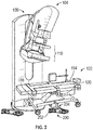
FIG. 2 illustrates an apparatus for controlling a mammography imaging system and patient positioning equipment according to an embodiment of the present invention; -
FIGS. 3A and 3B illustrate a support structure for use with an apparatus for controlling a mammography imaging system and patient positioning equipment according to an embodiment of the present invention; and -
FIG. 4 illustrates an apparatus for controlling a mammography imaging system and patient positioning equipment according to another embodiment of the present invention. - Reference will be made below in detail to exemplary embodiments of the invention, examples of which are illustrated in the accompanying drawings. Wherever possible, the same reference characters used throughout the drawings refer to the same or like parts, without duplicative description.
- While embodiments disclosed herein are described with respect to a mammography imaging system and procedure, it is to be understood that embodiments of the present invention may be applicable to other types of diagnostic procedures and "medical equipment" generally, i.e., devices used by practitioners to perform a medical procedure, e.g., an x-ray, CT, ultrasound machine, biopsy machine, endoscope, cardiac catheter, fluoroscope, dental equipment, etc. More specifically, practitioners in the art will readily recognize that embodiments may be suitable for use with various diagnostic procedures that involve medical equipment and a patient positioning system. As will be appreciated, embodiments may be utilized for procedures on animals generally, and are not limited to human procedures.
- As used herein, the terms "substantially," "generally," and "about" indicate conditions within reasonably achievable manufacturing and assembly tolerances, relative to ideal desired conditions suitable for achieving the functional purpose of a component or assembly.
- Moreover, the term "patient positioning equipment", as used herein, refers to equipment that positions and/or supports a patient at the correct orientation necessary for the conduction of a procedure via medical equipment. As will be appreciated, the patient positioning equipment may be mechanized or manually configured and may include tables, chairs, support frames, kneelers, or other structures designed to position or support a patient during a medical procedure. In some embodiments, the patient positioning equipment is a decubitus mammography table / Decubitus Breast Imaging (DBI) table.
- The term "control system", as used herein, refers a system for controlling one or more functions of medical equipment and/or patient positioning equipment and accessory equipment, and is not limited to a specific type of controller or interface. The term includes, but is not limited to, switch controls, i.e., a set of positionable operated controls, e.g., footswitch controls such as pedals, buttons, rocker switches, etc., that may be configurable to control the functions of medical equipment, patient positioning equipment, and/or accessory device actions. Control systems further include, but are not limited to, a user interface, i.e., a surface configured to receive inputs from a practitioner that translate to machine actions. The surface may contain multiple programmable switches or buttons, a tactile display surface, a touch pad, a display, or combinations thereof.
- As used herein, the term "support structure" refers to a structure configured to interface with and support the switch controls and/or the user interface on the patient positioning equipment. This can include a rigid structure as well as a flexible mounts, e.g., a gooseneck mount. Also, as used herein, the terms "coupled" and "connect" mean to fasten or join as commonly used, and refers both to permanent affixation as well as selectively removable attachments. Typical connection/coupling devices/connectors/fasteners include fasteners, welds, clamps, straps, cables, zip ties, mating parts (e.g., post-socket, hinges, snap-fits, etc.), adhesives, friction fits, etc.
- Turning now to
Figure 1 , an example of a common arrangement ofmedical equipment 100, here a mammography imaging system/machine, paired withpatient positioning equipment 102 is presented. Themammography imaging system 100 has agantry 104 that containsside controls 106,front controls 108 above the compression paddle andbreast support area 110, as well asswitch controls 112, e.g., footswitch controls.Switch controls 112 are typically connected via a cable/wire 113 tomammography imaging system 100. The controls are also often mirrored on the opposite side of mammography imaging system 100 (not shown). As will be appreciated, other controls (not shown) may be present on particular accessories placed either in the paddle/breast support area 110, or added on to or integrated with thegantry 104. - As mentioned above,
Figure 1 further depictspatient positioning equipment 102, here a decubitus breast imaging table. The table 102 generally includes asupport surface 114, e.g., a bed, mounted on abase 116, which is movable via wheels orcasters 118. Thesupport surface 114 may be rotatable/movable relative to thebase 116. The table 102 has separatepatient positioning controls 120 located on a side of the table 102 facing away frommammography imaging system 100. - In use, a practitioner must coordinate the functions of the
mammography imaging system 100, the breast imaging table 102, and (optionally) any attached accessories. Functions of themammography imaging system 100 include, but are not limited to, initiating movements of themammographic gantry 104, including lift, rotation, and angulation. Further mammography imaging system functions include breast compression, imaging parameters (i.e., compression force, compression rate, field of view, lighting, radiation dosage, source angle), and, in in certain medical procedures, e.g., ultrasound-aided biopsies, needle movement, suction controls, and ultrasonic imaging parameters. - The table 102 includes multiple moving elements which guide and support a seated or reclined patient into the correct angle or orientation necessary for the
mammography imaging system 100 and/or associated accessories to properly interface with the patient. Functions of the table 102 include raising or lowering a portion of thesupport surface 114 to, by way of non-limiting examples, change an angle of thesurface 114 or adjust table height. These functions are manipulated through the use ofcontrols 120 located on the table 102. - Moreover, accessories and tools, utilized for photographic imaging, volumetric measuring, tissue rigidity, and temperature mapping, may be employed during a mammographic examination. Accessory functions generally include powering the accessory and manipulating it to perform its intended task.
- As discussed above, due to the locations of the aforementioned controls, as well as the lack of a unified control system, the path of the movable patent positioning system may be limited and/or blocked. Moreover, practitioners may have to reposition themselves multiple times during a procedure to access certain controls.
- Turning now to
Figure 2 , an embodiment of the inventive apparatus and system for controlling medical equipment is shown. In the embodiment, thecontrol system 200 includes switch controls 202, e.g., a footswitch, that are connected to the table 102 by a support structure, e.g.,brace 204. Referring toFigures 3A and 3B , in embodiments, thebrace 204 includes a steppedarm portion 206 with acurved platform 208 configured to support auxiliary footswitch controls 200 (Figure. 2 ). Thearm portion 206 may connect to the table 102 through a variety of devices including threaded apertures in the arm that allow fasteners to pass through and into apertures in thebase 116 of the table 102. Although a mid-line installation next to patient position controls 120 is demonstrated, the switch controls 202 may be positioned at either end of the table 102 and may be fixed or movably mounted. - As will be appreciated, in certain embodiments, the support structure may be a structure other than the depicted
brace 204, and the connection to the table 102 may be via a variety of attachment mechanisms. In certain embodiments, the support structure allows for the switch controls to be adjusted or moved, e.g., raised or lowered, relative to thebase 116. In other embodiments, the switch controls may be integrated into/connected directly to thebase 116, such that they do not require a separate support structure. Moreover, the switch controls 202 may be integrated withbrace 204 to form a single movable piece. - The patient support table 102 may support multiple attachment points capable of accepting
brace 204, including both on thetable base 116 and on thesupport surface 114. In certain embodiments, thecontrol system 200, e.g., footswitch controls 202, may be mirrored on the opposite side of the table 102. In other embodiments,multiple control systems 200 may be connected to thebase 204 and/or thesupport surface 114. In embodiments, thecontrol system 200 may be projected onto the floor/ground via an interactive projection system. In other words, thecontrol system 200 may be a projection of a footswitch with one or more sensors, e.g., laser and/or cameras, detecting which part of the projected footswitch an operator is attempting to activate/interact with. As will be appreciated, projecting thecontrol system 200 onto the ground increases the ease of mirroring thecontrol system 200 on opposed sides of the table 102. - The
control system 200 may communicate wirelessly (e.g., via wi-fi, Bluetooth, and/or a proprietary interface protocol) with themammography imaging system 100 to control at least one function of the same. In other aspects, thecontrol system 200 may be hardwired to themammography imaging system 100. - In use, a practitioner/operator can manipulate the switch controls 202, e.g., footswitch, to control at least one function of the
mammography imaging system 100, e.g., gantry lift, rotation, and angulation, while manipulating patient position controls 120 of the table 102 with one hand and supporting the patient with the other. In embodiments, thecontrol system 200, e.g., footswitch controls 202, can be used to control a function of the patient positioning equipment and/or an accessory, in addition to controlling themammography imaging system 100. - Turning now to
Figure 4 , thecontrol system 200 can include auser interface 300. Theuser interface 300 can be in addition to, or in lieu of, switch controls 202. In embodiments, the switch controls 202 may be used to manipulate theuser interface 300. Theuser interface 300 may be a touch screen device such as a tablet. In embodiments, theuser interface 300 may be utilized to control at least one function of themammography imaging system 100. Theuser interface 300 may also be utilized to control the table 102 and/or any accessory equipment, as well as themachine 100. In embodiments that do not include switch controls 202, theuser interface 300 may be configured as a unified control system capable of controlling all associated equipment, e.g.,machine 100, table 102 and any attached accessories. In embodiments, theuser interface 300 may also display data obtained from mammography imaging system, or other equipment utilized during a procedure. - As depicted, the
user interface 300 is mounted to patient support table 204 via a support structure, such as, for example, aflexible mount 302, e.g., a bendable gooseneck rod, such that theuser interface 300 can be adjusted relative to a practitioner and the associated equipment. Alternatively, support structure may be rigid, such as a bracket. - A duplicate of the
flexible mount 302 may also be attached to the medical equipment/mammography imaging system 100 to enable movement ofuser interface 300 from one piece of equipment to a second piece of equipment. For example, from patient support table 102 tomammography imaging system 100. Or, alternatively, theuser interface 300 withsupport mount 302 may be moved from a first attachment point to a second attachment point. As will be appreciated, in embodiments, the table 102 may have a variety of attachment points for thesupport mount 302, on both thebase 116 andsupport surface 114. - In embodiments,
cables user interface 300 to switchcontrols 202, a power/communications interface 306, andmammography imaging system 100.Cables -
Interface 306 may perform the functions of power supply and communications regulation (i.e., command translation, analog to digital conversion, etc.) either singly or in tandem. In embodiments, theInterface 306 may include a rechargeable battery/power supply.Interface 306 further serves to receive commands fromuser interface 300 or auxiliary footswitch controls 200, interpret the commands, and output instructions triggering machine action in either the patient positioning equipment, the diagnostic equipment, or both. Power/communications interface 306 may include a stand-alone purpose-built device, a standard computer, or a computer containing additional hardware. Alternatively, the power/communications interface 306 may be integrated into theuser interface 300,support mount 302, orbrace 204. The power/communications interface 306 may be positioned in any location relative to the diagnostic and patient positioning equipment and other components of the modular control apparatus. - Thus, in use, a practitioner, in one instance, may engage switch controls 202 supported to initiate a movement of compression paddle, for example, after inputting target pressure into
user interface 300 while supporting a patient on patient support table 114. In another instance, the practitioner may, after a first procedure, moveuser interface 300 to a second location, in order to perform a second procedure. - In embodiments, the switch controls 202 and/or
user interface 300 controls can be configured such that when the patient positioning equipment, e.g., support table 114, is brought within a certain distance of the mammography imaging system, the controls are automatically activated to control functions of the mammography imaging system and/or accessories. As will be appreciated, the controls may also be user/practitioner activated. - Additionally, in embodiments, a
control lock 400 may be disposed in the user interface 300 (as shown inFigure 4 ), on the switch controls 202, table 102, and/or other suitable position. As will be understood, thecontrol lock 400 restricts control of the function(s) of themedical equipment 100 and/orpatient positioning equipment 102 so as to prevent unwanted/undesired activation of themedical equipment 100 and/orpatient positioning equipment 102 via thecontrol system 200. In embodiments, thecontrol lock 400 includes a button or moveable switch that toggles thecontrol lock 400 between a "locked" state, in which thecontrol system 200 is prevented from activating themedical equipment 100 and/or thepatient positioning equipment 102, and an "unlocked" state, in which thecontrol system 200 is allowed to activate themedical equipment 100 and/or thepatient positioning equipment 102. For example, in embodiments where thecontrol lock 400 has a button, thecontrol system 200 may be allowed to activate themedical equipment 100 and/or thepatient positioning equipment 102 only while thebutton 400 is depressed. In other embodiments, in which thecontrol lock 400 has a button, thecontrol system 200 may be allowed to activate themedical equipment 100 and/or thepatient positioning equipment 102 only for a predetermined amount of time after the button has been depressed. As will be appreciated, by restricting the ability of thecontrol system 200 to activate themedical equipment 100 and/or thepatient positioning equipment 102, thecontrol lock 400 reduces the need to move thecontrol system 200 to a designated holding position when thepatient positioning equipment 102 is moved. - It is also to be understood that the
medical equipment 100, thepatient positioning equipment 102, and/or thecontrol system 200 may include the necessary electronics, software, memory, storage, databases, firmware, logic/state machines, microprocessors, communication links, displays or other visual or audio user interfaces, printing devices, and any other input/output interfaces to perform the functions described herein and/or to achieve the results described herein. For example, themedical equipment 100, thepatient positioning equipment 102, and/or thecontrol system 200 may include at least one processor and system memory / data storage structures, which may include random access memory (RAM) and read-only memory (ROM). The at least one processor may include one or more conventional microprocessors and one or more supplementary co-processors such as math co-processors or the like. The data storage structures discussed herein may include an appropriate combination of magnetic, optical and/or semiconductor memory, and may include, for example, RAM, ROM, flash drive, an optical disc such as a compact disc and/or a hard disk or drive. - Additionally, a software application that adapts a controller to perform the methods disclosed herein may be read into a main memory of the at least one processor from a computer-readable medium. The term "computer-readable medium", as used herein, refers to any medium that provides or participates in providing instructions to the at least one processor of the
medical equipment 100, thepatient positioning equipment 102, and/or the control system 200 (or any other processor of a device described herein) for execution. Such a medium may take many forms, including but not limited to, non-volatile media and volatile media. Non-volatile media include, for example, optical, magnetic, or opto-magnetic disks, such as memory. Volatile media include dynamic random access memory (DRAM), which typically constitutes the main memory. Common forms of computer-readable media include, for example, a floppy disk, a flexible disk, hard disk, magnetic tape, any other magnetic medium, a CD-ROM, DVD, any other optical medium, a RAM, a PROM, an EPROM or EEPROM (electronically erasable programmable read-only memory), a FLASH-EEPROM, any other memory chip or cartridge, or any other medium from which a computer can read. - While in embodiments, the execution of sequences of instructions in the software application causes at least one processor to perform the methods/processes described herein, hard-wired circuitry may be used in place of, or in combination with, software instructions for implementation of the methods/processes of the present invention. Therefore, embodiments of the present invention are not limited to any specific combination of hardware and/or software.
- It is further to be understood that the above description is intended to be illustrative, and not restrictive. For example, the above-described embodiments (and/or aspects thereof) may be used in combination with each other. Additionally, many modifications may be made to adapt a particular situation or material to the teachings of the invention without departing from its scope.
- For example, in an embodiment, an apparatus for controlling medical equipment is provided. The apparatus includes patient positioning equipment, and a control system coupled to the patient positioning equipment. The control system is configured to control a function of the medical equipment. In certain embodiments, the control system includes switch controls connected to the patient positioning equipment via a support structure. In certain embodiments, the control system is configured to control a function of the patient positioning equipment and/or an accessory device, in addition to a function of the medical equipment. In certain embodiments, the medical equipment is a mammography imaging system. In certain embodiments, the patient positioning equipment is a breast imaging table. In certain embodiments, the breast imaging table is a Decubitus Breast Imaging (DBI) table. In certain embodiments, the control system is configured to support wireless communication with at least one of the medical equipment, the patient support equipment, or an accessory device. In certain embodiments, the control system is a user interface. In certain embodiments, the user interface is configured to control a function of the patient positioning equipment and/or an accessory device, in addition to a function of the medical equipment. In certain embodiments, the user interface is configured to support wireless communication with at least one of the medical equipment, the patient support equipment or an accessory device. In certain embodiments, the user interface is a touch screen. In certain embodiments, the user interface is coupled to the patient positioning equipment via a flexible mount.
- Other embodiments provide for a system a system for controlling mammography equipment. The system includes a mammography imaging system, a breast imaging table, and switch controls coupled to the breast imaging table. The switch controls are configured to control at least one function of the mammography equipment. In certain embodiments, the switch controls are configured to control a function of the breast imaging table and/or an accessory device, in addition to a function of the mammography equipment. In certain embodiments, the system further includes a user interface coupled to the breast imaging table. The user interface is configured to control a function of the breast imaging table, a function of an accessory device, and/or a function of the mammography equipment.
- Yet still other embodiments provide for a method of controlling medical equipment. The method includes engaging a control system secured to patient positioning equipment, and controlling at least one function of the medical equipment via the control system. In certain embodiments, the medical equipment is a mammography imaging system and the patient positioning equipment is a breast imaging table. In certain embodiments, the control system includes switch controls. In certain embodiments, the control system includes a user interface. In certain embodiments, the control system is configured to control a function of the patient positioning equipment and/or an accessory device, in addition to a function of the medical equipment.
- Finally, the written description uses examples to disclose the invention, including the best mode, and also to enable any person skilled in the art to practice the invention, including making and using any devices or systems and performing any incorporated methods. The patentable scope of the invention is defined by the claims, and may include other examples that occur to those skilled in the art. Such other examples are intended to be within the scope of the claims if they have structural elements that do not differ from the literal language of the claims, or if they include equivalent structural elements with insubstantial differences from the literal language of the claims.
- As used herein, an element or step recited in the singular and proceeded with the word "a" or "an" should be understood as not excluding plural of said elements or steps, unless such exclusion is explicitly stated. Furthermore, references to "one embodiment" of the present invention are not intended to be interpreted as excluding the existence of additional embodiments that also incorporate the recited features. Moreover, unless explicitly stated to the contrary, embodiments "comprising," "including," or "having" an element or a plurality of elements having a particular property may include additional such elements not having that property.
- Since certain changes may be made in the above-described invention, without departing from the spirit and scope of the invention herein involved, it is intended that all of the subject matter of the above description shown in the accompanying drawings shall be interpreted merely as examples illustrating the inventive concept herein and shall not be construed as limiting the invention.
Claims (15)
- An apparatus for controlling medical equipment 100 comprising:patient positioning equipment 102; anda control system 200 coupled to the patient positioning equipment 102;wherein the control system 200 is configured to control a function of the medical equipment 100.
- The apparatus of claim 1, wherein the control system 200 includes switch controls 202 connected to the patient positioning equipment 102 via a support structure 204.
- The apparatus of claim 1, wherein the control system 200 is configured to control a function of the patient positioning equipment 102 and/or an accessory device, in addition to a function of the medical equipment 100.
- The apparatus of claim 1, wherein the medical equipment 100 is a mammography imaging system.
- The apparatus of claim 1, wherein the patient positioning equipment 102 is a breast imaging table.
- The apparatus of claim 5, wherein the breast imaging table 102 is a Decubitus Breast Imaging (DBI) table.
- The apparatus of claim 1, wherein the control system 200 is configured to support wireless communication with at least one of the medical equipment 100, the patient support equipment 102, or an accessory device.
- The apparatus of claim 1, wherein the control system 200 is a user interface 300.
- The apparatus of claim 8, wherein the user interface 300 is configured to control a function of the patient positioning equipment 102 and/or an accessory device, in addition to a function of the medical equipment 100.
- The apparatus of claim 8, wherein the user interface 300 is configured to support wireless communication with at least one of the medical equipment 100, the patient support equipment 102 or an accessory device.
- The apparatus of claim 8, wherein the user interface 300 is a touch screen.
- The apparatus of claim 8, wherein the user interface 300 is coupled to the patient positioning equipment via a flexible mount 302.
- A method of controlling medical equipment 100 comprising:engaging a control system 200 secured to patient positioning equipment 102; andcontrolling at least one function of the medical equipment 100 via the control system 200.
- The method of claim 13, wherein the medical equipment 100 is a mammography imaging system and the patient positioning equipment 102 is a breast imaging table.
- The method of claim 13, wherein the control system 200 includes switch controls 202.
Applications Claiming Priority (1)
| Application Number | Priority Date | Filing Date | Title |
|---|---|---|---|
| US16/014,752 US10863952B2 (en) | 2018-06-21 | 2018-06-21 | Apparatus, system and method for controlling medical equipment |
Publications (3)
| Publication Number | Publication Date |
|---|---|
| EP3586755A1 true EP3586755A1 (en) | 2020-01-01 |
| EP3586755B1 EP3586755B1 (en) | 2025-04-30 |
| EP3586755B8 EP3586755B8 (en) | 2025-06-11 |
Family
ID=67001571
Family Applications (1)
| Application Number | Title | Priority Date | Filing Date |
|---|---|---|---|
| EP19181561.2A Active EP3586755B8 (en) | 2018-06-21 | 2019-06-20 | Apparatus and method for controlling medical equipment |
Country Status (4)
| Country | Link |
|---|---|
| US (1) | US10863952B2 (en) |
| EP (1) | EP3586755B8 (en) |
| JP (1) | JP7123862B2 (en) |
| CN (1) | CN110623683B (en) |
Cited By (1)
| Publication number | Priority date | Publication date | Assignee | Title |
|---|---|---|---|---|
| EP4137054A1 (en) * | 2021-08-20 | 2023-02-22 | Canon Medical Systems Corporation | X-ray imaging device, x-ray imaging system, and method of controlling x-ray imaging device |
Families Citing this family (4)
| Publication number | Priority date | Publication date | Assignee | Title |
|---|---|---|---|---|
| US12313495B2 (en) | 2020-02-13 | 2025-05-27 | Nsk Ltd. | State determination device for hub unit bearing, state determination method for hub unit bearing, program, and hub unit bearing |
| CN113768525B (en) * | 2021-10-11 | 2024-05-14 | 深圳圣诺医疗设备股份有限公司 | Motion component control system and method for mammary gland X-ray machine |
| JP2025083202A (en) * | 2023-11-20 | 2025-05-30 | 富士フイルム株式会社 | Radiation irradiation apparatus and radiography system |
| IT202400004312A1 (en) * | 2024-02-28 | 2025-08-28 | Metaltronica S P A | MULTIMODAL SYSTEM FOR OBTAINING BREAST IMAGES USING X-RAY AND ULTRASOUND TECHNIQUES |
Citations (2)
| Publication number | Priority date | Publication date | Assignee | Title |
|---|---|---|---|---|
| WO2009114365A2 (en) * | 2008-03-13 | 2009-09-17 | Mallinckrodt Inc. | Multi-function, foot-activated controller for imaging system |
| WO2015079118A1 (en) * | 2013-11-29 | 2015-06-04 | Planmed Oy | Improvement for mammography examinations |
Family Cites Families (46)
| Publication number | Priority date | Publication date | Assignee | Title |
|---|---|---|---|---|
| DE2134122C3 (en) * | 1971-07-08 | 1984-05-30 | Siemens AG, 1000 Berlin und 8000 München | X-ray machine for skull examinations |
| US4433690A (en) | 1981-07-20 | 1984-02-28 | Siemens Ag | Compact ultrasound apparatus for medical examination |
| JPH0616099B2 (en) * | 1989-02-07 | 1994-03-02 | 浜松ホトニクス株式会社 | Data correction device in CT device |
| EP0406455B1 (en) | 1989-07-03 | 1994-09-21 | Siemens Aktiengesellschaft | X-ray diagnostic apparatus for mammographies |
| US5078142A (en) * | 1989-11-21 | 1992-01-07 | Fischer Imaging Corporation | Precision mammographic needle biopsy system |
| US5415169A (en) * | 1989-11-21 | 1995-05-16 | Fischer Imaging Corporation | Motorized mammographic biopsy apparatus |
| US5289520A (en) * | 1991-11-27 | 1994-02-22 | Lorad Corporation | Stereotactic mammography imaging system with prone position examination table and CCD camera |
| JPH07303633A (en) * | 1994-05-11 | 1995-11-21 | Mitsubishi Electric Corp | X-ray mammography device |
| US5627869A (en) * | 1995-11-22 | 1997-05-06 | Thermotrex Corporation | Mammography apparatus with proportional collimation |
| US5590166A (en) | 1995-12-28 | 1996-12-31 | Instrumentarium Corporation | Mammography unit |
| DE59912639D1 (en) * | 1998-06-23 | 2006-02-23 | Siemens Ag | Mammography X-ray machine with a solid-state detector |
| WO2000015112A1 (en) * | 1998-09-17 | 2000-03-23 | Quanta Vision, Inc. | Reduced-angle mammography device and variants |
| US6480565B1 (en) * | 1999-11-18 | 2002-11-12 | University Of Rochester | Apparatus and method for cone beam volume computed tomography breast imaging |
| JP3893827B2 (en) * | 1999-11-26 | 2007-03-14 | コニカミノルタホールディングス株式会社 | X-ray imaging system |
| ES2281377T3 (en) * | 2000-07-07 | 2007-10-01 | Medical Positioning, Inc. | PATIENT SUPPORT APPARATUS AND METHOD FOR THE PERFORMANCE OF A BIOPSY OF BREAST IN THE DECUBE. |
| US6367104B1 (en) * | 2000-07-07 | 2002-04-09 | Medical Positioning, Inc. | Patient support apparatus and method for performing decubitus breast biopsy |
| US6832399B2 (en) * | 2000-11-14 | 2004-12-21 | Michael G. Falbo, Sr. | Breast biopsy bed |
| CA2326026A1 (en) * | 2000-11-16 | 2002-05-16 | Is2 Research Inc. | Scintillation camera for scintimamography |
| US7556602B2 (en) * | 2000-11-24 | 2009-07-07 | U-Systems, Inc. | Breast cancer screening with adjunctive ultrasound mammography |
| FR2818116B1 (en) * | 2000-12-19 | 2004-08-27 | Ge Med Sys Global Tech Co Llc | MAMMOGRAPHY APPARATUS |
| DE10156178A1 (en) * | 2001-11-15 | 2003-06-05 | Philips Intellectual Property | Mammography add-on for MR elastography |
| US6640364B1 (en) * | 2001-11-30 | 2003-11-04 | Ge Medical Systems Global Technololgy Company, Llc | Pedestal for use with patient transport system for multiple imaging systems |
| US6968223B2 (en) * | 2002-02-01 | 2005-11-22 | Ge Medical Systems Global Technology Company, Llc | System and method for wireless voice control of an interventional or diagnostic medical device |
| US7041109B2 (en) * | 2002-12-12 | 2006-05-09 | Kelsey, Inc. | Apparatus and method for interstitial laser therapy of small breast cancers and adjunctive therapy |
| WO2006030406A2 (en) * | 2004-09-15 | 2006-03-23 | Scientific Biopsy Ltd. | Breast cancer detection and biopsy |
| US9420991B2 (en) * | 2005-09-01 | 2016-08-23 | Shih-Ping Wang | Breast ultrasound scanning device |
| JP4597936B2 (en) * | 2005-10-06 | 2010-12-15 | 富士フイルム株式会社 | Breast imaging device |
| DE102006010196B4 (en) * | 2006-03-06 | 2012-06-28 | Siemens Ag | Operating device for controlling a medical device |
| EP2009465A4 (en) | 2006-03-31 | 2010-03-03 | Shimadzu Corp | Mammography system |
| US20080043905A1 (en) * | 2006-08-21 | 2008-02-21 | Bamdad Hassanpourgol | Portable Prone Stereotactic Mammography System for Biopsies, Image Guided Lumpectomies, and Radiation Treatment |
| JP2008119018A (en) * | 2006-11-08 | 2008-05-29 | Konica Minolta Medical & Graphic Inc | Radiation image radiographing system and radiation image detecting apparatus |
| US20080221478A1 (en) * | 2007-03-07 | 2008-09-11 | Ritchie Paul G | Integrated Imaging and Biopsy System with Integrated Control Interface |
| GB2465726A (en) * | 2007-08-23 | 2010-06-02 | Fischer Medical Technologies Inc | Improved computed tomography breast imaging and biopsy system |
| US8274376B2 (en) * | 2008-11-17 | 2012-09-25 | Canyon Ridge Resources, L.L.C. | System and method for wireless control of medical devices |
| US8159370B2 (en) * | 2008-11-17 | 2012-04-17 | Canyon Ridge Resources, Llc | System and method for control of medical equipment using multiple wireless devices |
| US8870772B2 (en) * | 2008-12-29 | 2014-10-28 | Perseus-Biomed Inc. | Method and system for tissue recognition |
| DE102009021023A1 (en) * | 2009-05-13 | 2010-11-18 | Siemens Aktiengesellschaft | Mammography procedure and mammography device |
| US20110073743A1 (en) * | 2009-05-13 | 2011-03-31 | Steven Shamie | Universal holder and flexible member for mounting, holding and adjustably positioning electronic products and accessories |
| US8565373B2 (en) * | 2009-12-29 | 2013-10-22 | General Electric Company | Method and system for automatically positioning a mammography imaging system |
| JP5693184B2 (en) * | 2010-11-29 | 2015-04-01 | キヤノン株式会社 | Biological information acquisition device |
| DE102011006353A1 (en) * | 2011-03-29 | 2012-10-04 | Siemens Aktiengesellschaft | mammography system |
| WO2014173946A1 (en) * | 2013-04-23 | 2014-10-30 | Face Recording And Measurements Ltd. | Database management system |
| JP6309376B2 (en) * | 2013-07-03 | 2018-04-11 | キヤノンメディカルシステムズ株式会社 | Medical information processing system, medical information processing program, and ultrasonic diagnostic apparatus |
| GB201313994D0 (en) * | 2013-08-05 | 2013-09-18 | Cirdan Imaging Ltd | A Mammographic Device |
| US9980703B2 (en) * | 2015-03-17 | 2018-05-29 | General Electric Company | Methods and systems for a display interface for diagnostic medical imaging |
| WO2017059134A1 (en) * | 2015-09-30 | 2017-04-06 | Devicor Medical Products, Inc. | Breast support compression pillow |
-
2018
- 2018-06-21 US US16/014,752 patent/US10863952B2/en active Active
-
2019
- 2019-06-20 EP EP19181561.2A patent/EP3586755B8/en active Active
- 2019-06-20 JP JP2019114216A patent/JP7123862B2/en active Active
- 2019-06-21 CN CN201910544568.2A patent/CN110623683B/en active Active
Patent Citations (2)
| Publication number | Priority date | Publication date | Assignee | Title |
|---|---|---|---|---|
| WO2009114365A2 (en) * | 2008-03-13 | 2009-09-17 | Mallinckrodt Inc. | Multi-function, foot-activated controller for imaging system |
| WO2015079118A1 (en) * | 2013-11-29 | 2015-06-04 | Planmed Oy | Improvement for mammography examinations |
Cited By (2)
| Publication number | Priority date | Publication date | Assignee | Title |
|---|---|---|---|---|
| EP4137054A1 (en) * | 2021-08-20 | 2023-02-22 | Canon Medical Systems Corporation | X-ray imaging device, x-ray imaging system, and method of controlling x-ray imaging device |
| US12458317B2 (en) | 2021-08-20 | 2025-11-04 | Canon Medical Systems Corporation | X-ray imaging device, X-ray imaging system, and method of controlling X-ray imaging device |
Also Published As
| Publication number | Publication date |
|---|---|
| US20190388040A1 (en) | 2019-12-26 |
| JP2020022734A (en) | 2020-02-13 |
| CN110623683B (en) | 2024-03-05 |
| EP3586755B1 (en) | 2025-04-30 |
| CN110623683A (en) | 2019-12-31 |
| JP7123862B2 (en) | 2022-08-23 |
| EP3586755B8 (en) | 2025-06-11 |
| US10863952B2 (en) | 2020-12-15 |
Similar Documents
| Publication | Publication Date | Title |
|---|---|---|
| EP3586755B1 (en) | Apparatus and method for controlling medical equipment | |
| US12295759B2 (en) | Horizontally-displaceable foam breast compression paddle | |
| EP3856036B1 (en) | Multi-screen imaging system for improved workflow | |
| US7591587B2 (en) | Method for controlling X-ray diagnostic apparatus | |
| US7331712B2 (en) | X-ray examination apparatus that is convertible among multiple examination configurations | |
| WO2006030436A2 (en) | Breast cancer detection and biopsy | |
| CN113940759B (en) | Puncture surgery console and puncture robot | |
| EP4527336A1 (en) | Operating system and method for ercp surgical robot | |
| US7151816B2 (en) | Imaging tomography apparatus having an attached patient support with a movable backrest | |
| CN112603539B (en) | Surgical navigation control system and master hand control device | |
| JP3860682B2 (en) | Medical treatment equipment | |
| JP2009153579A (en) | X-ray ct system and medical imaging system | |
| JPH08275940A (en) | X-ray fluoroscopic photographing stand | |
| JP3533303B2 (en) | Dental care equipment | |
| JP2025180178A (en) | X-ray diagnostic apparatus, control method for X-ray diagnostic apparatus, X-ray diagnostic system, and program | |
| JP2024172194A (en) | X-ray diagnostic device and X-ray diagnostic system | |
| JP2006334020A (en) | X-ray diagnostic apparatus control method | |
| JP2019198578A (en) | Medical image diagnostic apparatus and chair for use in medical imaging | |
| JP2003325594A (en) | Dental care equipment | |
| HK1152635A (en) | Dental care apparatus |
Legal Events
| Date | Code | Title | Description |
|---|---|---|---|
| PUAI | Public reference made under article 153(3) epc to a published international application that has entered the european phase |
Free format text: ORIGINAL CODE: 0009012 |
|
| STAA | Information on the status of an ep patent application or granted ep patent |
Free format text: STATUS: REQUEST FOR EXAMINATION WAS MADE |
|
| 17P | Request for examination filed |
Effective date: 20190620 |
|
| AK | Designated contracting states |
Kind code of ref document: A1 Designated state(s): AL AT BE BG CH CY CZ DE DK EE ES FI FR GB GR HR HU IE IS IT LI LT LU LV MC MK MT NL NO PL PT RO RS SE SI SK SM TR |
|
| AX | Request for extension of the european patent |
Extension state: BA ME |
|
| STAA | Information on the status of an ep patent application or granted ep patent |
Free format text: STATUS: EXAMINATION IS IN PROGRESS |
|
| 17Q | First examination report despatched |
Effective date: 20220720 |
|
| P01 | Opt-out of the competence of the unified patent court (upc) registered |
Effective date: 20230528 |
|
| GRAP | Despatch of communication of intention to grant a patent |
Free format text: ORIGINAL CODE: EPIDOSNIGR1 |
|
| STAA | Information on the status of an ep patent application or granted ep patent |
Free format text: STATUS: GRANT OF PATENT IS INTENDED |
|
| INTG | Intention to grant announced |
Effective date: 20240813 |
|
| GRAJ | Information related to disapproval of communication of intention to grant by the applicant or resumption of examination proceedings by the epo deleted |
Free format text: ORIGINAL CODE: EPIDOSDIGR1 |
|
| STAA | Information on the status of an ep patent application or granted ep patent |
Free format text: STATUS: EXAMINATION IS IN PROGRESS |
|
| GRAP | Despatch of communication of intention to grant a patent |
Free format text: ORIGINAL CODE: EPIDOSNIGR1 |
|
| STAA | Information on the status of an ep patent application or granted ep patent |
Free format text: STATUS: GRANT OF PATENT IS INTENDED |
|
| INTG | Intention to grant announced |
Effective date: 20241210 |
|
| RIC1 | Information provided on ipc code assigned before grant |
Ipc: A61B 8/00 20060101ALN20241129BHEP Ipc: A61B 6/50 20240101ALI20241129BHEP Ipc: A61B 6/46 20240101ALI20241129BHEP Ipc: A61B 6/00 20060101ALI20241129BHEP Ipc: A61B 6/04 20060101AFI20241129BHEP |
|
| GRAS | Grant fee paid |
Free format text: ORIGINAL CODE: EPIDOSNIGR3 |
|
| GRAA | (expected) grant |
Free format text: ORIGINAL CODE: 0009210 |
|
| STAA | Information on the status of an ep patent application or granted ep patent |
Free format text: STATUS: THE PATENT HAS BEEN GRANTED |
|
| REG | Reference to a national code |
Ref country code: DE Ref legal event code: R081 Ref document number: 602019069250 Country of ref document: DE Owner name: GE PRECISION HEALTHCARE LLC, WAUKESHA, US Free format text: FORMER OWNER: GENERAL ELECTRIC COMPANY, SCHENECTADY, NY, US |
|
| AK | Designated contracting states |
Kind code of ref document: B1 Designated state(s): AL AT BE BG CH CY CZ DE DK EE ES FI FR GB GR HR HU IE IS IT LI LT LU LV MC MK MT NL NO PL PT RO RS SE SI SK SM TR |
|
| REG | Reference to a national code |
Ref country code: CH Ref legal event code: EP Ref country code: GB Ref legal event code: FG4D |
|
| RAP2 | Party data changed (patent owner data changed or rights of a patent transferred) |
Owner name: GE PRECISION HEALTHCARE LLC |
|
| REG | Reference to a national code |
Ref country code: CH Ref legal event code: PK Free format text: BERICHTIGUNG B8 |
|
| REG | Reference to a national code |
Ref country code: IE Ref legal event code: FG4D |
|
| REG | Reference to a national code |
Ref country code: DE Ref legal event code: R096 Ref document number: 602019069250 Country of ref document: DE |
|
| REG | Reference to a national code |
Ref country code: NL Ref legal event code: FP |
|
| PGFP | Annual fee paid to national office [announced via postgrant information from national office to epo] |
Ref country code: NL Payment date: 20250520 Year of fee payment: 7 |
|
| PGFP | Annual fee paid to national office [announced via postgrant information from national office to epo] |
Ref country code: DE Payment date: 20250520 Year of fee payment: 7 |
|
| PGFP | Annual fee paid to national office [announced via postgrant information from national office to epo] |
Ref country code: GB Payment date: 20250520 Year of fee payment: 7 |
|
| PGFP | Annual fee paid to national office [announced via postgrant information from national office to epo] |
Ref country code: FR Payment date: 20250520 Year of fee payment: 7 |
|
| REG | Reference to a national code |
Ref country code: AT Ref legal event code: MK05 Ref document number: 1789280 Country of ref document: AT Kind code of ref document: T Effective date: 20250430 |
|
| PG25 | Lapsed in a contracting state [announced via postgrant information from national office to epo] |
Ref country code: FI Free format text: LAPSE BECAUSE OF FAILURE TO SUBMIT A TRANSLATION OF THE DESCRIPTION OR TO PAY THE FEE WITHIN THE PRESCRIBED TIME-LIMIT Effective date: 20250430 Ref country code: ES Free format text: LAPSE BECAUSE OF FAILURE TO SUBMIT A TRANSLATION OF THE DESCRIPTION OR TO PAY THE FEE WITHIN THE PRESCRIBED TIME-LIMIT Effective date: 20250430 Ref country code: PT Free format text: LAPSE BECAUSE OF FAILURE TO SUBMIT A TRANSLATION OF THE DESCRIPTION OR TO PAY THE FEE WITHIN THE PRESCRIBED TIME-LIMIT Effective date: 20250901 |
|
| REG | Reference to a national code |
Ref country code: LT Ref legal event code: MG9D |
|
| PG25 | Lapsed in a contracting state [announced via postgrant information from national office to epo] |
Ref country code: NO Free format text: LAPSE BECAUSE OF FAILURE TO SUBMIT A TRANSLATION OF THE DESCRIPTION OR TO PAY THE FEE WITHIN THE PRESCRIBED TIME-LIMIT Effective date: 20250730 Ref country code: GR Free format text: LAPSE BECAUSE OF FAILURE TO SUBMIT A TRANSLATION OF THE DESCRIPTION OR TO PAY THE FEE WITHIN THE PRESCRIBED TIME-LIMIT Effective date: 20250731 |
|
| PG25 | Lapsed in a contracting state [announced via postgrant information from national office to epo] |
Ref country code: PL Free format text: LAPSE BECAUSE OF FAILURE TO SUBMIT A TRANSLATION OF THE DESCRIPTION OR TO PAY THE FEE WITHIN THE PRESCRIBED TIME-LIMIT Effective date: 20250430 |
|
| PG25 | Lapsed in a contracting state [announced via postgrant information from national office to epo] |
Ref country code: BG Free format text: LAPSE BECAUSE OF FAILURE TO SUBMIT A TRANSLATION OF THE DESCRIPTION OR TO PAY THE FEE WITHIN THE PRESCRIBED TIME-LIMIT Effective date: 20250430 |
|
| PG25 | Lapsed in a contracting state [announced via postgrant information from national office to epo] |
Ref country code: HR Free format text: LAPSE BECAUSE OF FAILURE TO SUBMIT A TRANSLATION OF THE DESCRIPTION OR TO PAY THE FEE WITHIN THE PRESCRIBED TIME-LIMIT Effective date: 20250430 |
|
| PG25 | Lapsed in a contracting state [announced via postgrant information from national office to epo] |
Ref country code: AT Free format text: LAPSE BECAUSE OF FAILURE TO SUBMIT A TRANSLATION OF THE DESCRIPTION OR TO PAY THE FEE WITHIN THE PRESCRIBED TIME-LIMIT Effective date: 20250430 |
|
| PG25 | Lapsed in a contracting state [announced via postgrant information from national office to epo] |
Ref country code: RS Free format text: LAPSE BECAUSE OF FAILURE TO SUBMIT A TRANSLATION OF THE DESCRIPTION OR TO PAY THE FEE WITHIN THE PRESCRIBED TIME-LIMIT Effective date: 20250731 |
|
| PG25 | Lapsed in a contracting state [announced via postgrant information from national office to epo] |
Ref country code: IS Free format text: LAPSE BECAUSE OF FAILURE TO SUBMIT A TRANSLATION OF THE DESCRIPTION OR TO PAY THE FEE WITHIN THE PRESCRIBED TIME-LIMIT Effective date: 20250830 |
|
| PG25 | Lapsed in a contracting state [announced via postgrant information from national office to epo] |
Ref country code: LV Free format text: LAPSE BECAUSE OF FAILURE TO SUBMIT A TRANSLATION OF THE DESCRIPTION OR TO PAY THE FEE WITHIN THE PRESCRIBED TIME-LIMIT Effective date: 20250430 |
|
| PG25 | Lapsed in a contracting state [announced via postgrant information from national office to epo] |
Ref country code: DK Free format text: LAPSE BECAUSE OF FAILURE TO SUBMIT A TRANSLATION OF THE DESCRIPTION OR TO PAY THE FEE WITHIN THE PRESCRIBED TIME-LIMIT Effective date: 20250430 Ref country code: SM Free format text: LAPSE BECAUSE OF FAILURE TO SUBMIT A TRANSLATION OF THE DESCRIPTION OR TO PAY THE FEE WITHIN THE PRESCRIBED TIME-LIMIT Effective date: 20250430 |
|
| PG25 | Lapsed in a contracting state [announced via postgrant information from national office to epo] |
Ref country code: CZ Free format text: LAPSE BECAUSE OF FAILURE TO SUBMIT A TRANSLATION OF THE DESCRIPTION OR TO PAY THE FEE WITHIN THE PRESCRIBED TIME-LIMIT Effective date: 20250430 |
|
| PG25 | Lapsed in a contracting state [announced via postgrant information from national office to epo] |
Ref country code: SK Free format text: LAPSE BECAUSE OF FAILURE TO SUBMIT A TRANSLATION OF THE DESCRIPTION OR TO PAY THE FEE WITHIN THE PRESCRIBED TIME-LIMIT Effective date: 20250430 Ref country code: RO Free format text: LAPSE BECAUSE OF FAILURE TO SUBMIT A TRANSLATION OF THE DESCRIPTION OR TO PAY THE FEE WITHIN THE PRESCRIBED TIME-LIMIT Effective date: 20250430 |
|
| REG | Reference to a national code |
Ref country code: CH Ref legal event code: H13 Free format text: ST27 STATUS EVENT CODE: U-0-0-H10-H13 (AS PROVIDED BY THE NATIONAL OFFICE) Effective date: 20260127 |
|
| PG25 | Lapsed in a contracting state [announced via postgrant information from national office to epo] |
Ref country code: IT Free format text: LAPSE BECAUSE OF FAILURE TO SUBMIT A TRANSLATION OF THE DESCRIPTION OR TO PAY THE FEE WITHIN THE PRESCRIBED TIME-LIMIT Effective date: 20250430 |
|
| PG25 | Lapsed in a contracting state [announced via postgrant information from national office to epo] |
Ref country code: MC Free format text: LAPSE BECAUSE OF FAILURE TO SUBMIT A TRANSLATION OF THE DESCRIPTION OR TO PAY THE FEE WITHIN THE PRESCRIBED TIME-LIMIT Effective date: 20250430 |
|
| REG | Reference to a national code |
Ref country code: DE Ref legal event code: R097 Ref document number: 602019069250 Country of ref document: DE |
|
| PG25 | Lapsed in a contracting state [announced via postgrant information from national office to epo] |
Ref country code: LU Free format text: LAPSE BECAUSE OF NON-PAYMENT OF DUE FEES Effective date: 20250620 |
|
| REG | Reference to a national code |
Ref country code: BE Ref legal event code: MM Effective date: 20250630 |
|
| PLBE | No opposition filed within time limit |
Free format text: ORIGINAL CODE: 0009261 |
|
| STAA | Information on the status of an ep patent application or granted ep patent |
Free format text: STATUS: NO OPPOSITION FILED WITHIN TIME LIMIT |
|
| REG | Reference to a national code |
Ref country code: CH Ref legal event code: L10 Free format text: ST27 STATUS EVENT CODE: U-0-0-L10-L00 (AS PROVIDED BY THE NATIONAL OFFICE) Effective date: 20260311 |
|
| 26N | No opposition filed |
Effective date: 20260202 |
|
| PG25 | Lapsed in a contracting state [announced via postgrant information from national office to epo] |
Ref country code: IE Free format text: LAPSE BECAUSE OF NON-PAYMENT OF DUE FEES Effective date: 20250620 |
|
| PG25 | Lapsed in a contracting state [announced via postgrant information from national office to epo] |
Ref country code: BE Free format text: LAPSE BECAUSE OF NON-PAYMENT OF DUE FEES Effective date: 20250630 |
|
| PG25 | Lapsed in a contracting state [announced via postgrant information from national office to epo] |
Ref country code: CH Free format text: LAPSE BECAUSE OF NON-PAYMENT OF DUE FEES Effective date: 20250630 |