EP3580331B1 - Gamma delta t cells and a method of augmenting the tumoricidal activity of the same - Google Patents
Gamma delta t cells and a method of augmenting the tumoricidal activity of the same Download PDFInfo
- Publication number
- EP3580331B1 EP3580331B1 EP18750778.5A EP18750778A EP3580331B1 EP 3580331 B1 EP3580331 B1 EP 3580331B1 EP 18750778 A EP18750778 A EP 18750778A EP 3580331 B1 EP3580331 B1 EP 3580331B1
- Authority
- EP
- European Patent Office
- Prior art keywords
- cells
- receptor
- cell
- vγ9vδ2
- car
- Prior art date
- Legal status (The legal status is an assumption and is not a legal conclusion. Google has not performed a legal analysis and makes no representation as to the accuracy of the status listed.)
- Active
Links
Images


Classifications
-
- A—HUMAN NECESSITIES
- A61—MEDICAL OR VETERINARY SCIENCE; HYGIENE
- A61P—SPECIFIC THERAPEUTIC ACTIVITY OF CHEMICAL COMPOUNDS OR MEDICINAL PREPARATIONS
- A61P35/00—Antineoplastic agents
-
- C—CHEMISTRY; METALLURGY
- C07—ORGANIC CHEMISTRY
- C07K—PEPTIDES
- C07K14/00—Peptides having more than 20 amino acids; Gastrins; Somatostatins; Melanotropins; Derivatives thereof
- C07K14/435—Peptides having more than 20 amino acids; Gastrins; Somatostatins; Melanotropins; Derivatives thereof from animals; from humans
- C07K14/705—Receptors; Cell surface antigens; Cell surface determinants
- C07K14/70503—Immunoglobulin superfamily
- C07K14/7051—T-cell receptor (TcR)-CD3 complex
-
- A—HUMAN NECESSITIES
- A61—MEDICAL OR VETERINARY SCIENCE; HYGIENE
- A61K—PREPARATIONS FOR MEDICAL, DENTAL OR TOILETRY PURPOSES
- A61K40/00—Cellular immunotherapy
- A61K40/10—Cellular immunotherapy characterised by the cell type used
- A61K40/11—T-cells, e.g. tumour infiltrating lymphocytes [TIL] or regulatory T [Treg] cells; Lymphokine-activated killer [LAK] cells
-
- A—HUMAN NECESSITIES
- A61—MEDICAL OR VETERINARY SCIENCE; HYGIENE
- A61K—PREPARATIONS FOR MEDICAL, DENTAL OR TOILETRY PURPOSES
- A61K40/00—Cellular immunotherapy
- A61K40/30—Cellular immunotherapy characterised by the recombinant expression of specific molecules in the cells of the immune system
- A61K40/31—Chimeric antigen receptors [CAR]
-
- A—HUMAN NECESSITIES
- A61—MEDICAL OR VETERINARY SCIENCE; HYGIENE
- A61K—PREPARATIONS FOR MEDICAL, DENTAL OR TOILETRY PURPOSES
- A61K40/00—Cellular immunotherapy
- A61K40/40—Cellular immunotherapy characterised by antigens that are targeted or presented by cells of the immune system
- A61K40/41—Vertebrate antigens
- A61K40/42—Cancer antigens
- A61K40/4202—Receptors, cell surface antigens or cell surface determinants
- A61K40/4224—Molecules with a "CD" designation not provided for elsewhere
-
- C—CHEMISTRY; METALLURGY
- C07—ORGANIC CHEMISTRY
- C07K—PEPTIDES
- C07K14/00—Peptides having more than 20 amino acids; Gastrins; Somatostatins; Melanotropins; Derivatives thereof
- C07K14/435—Peptides having more than 20 amino acids; Gastrins; Somatostatins; Melanotropins; Derivatives thereof from animals; from humans
- C07K14/705—Receptors; Cell surface antigens; Cell surface determinants
- C07K14/70503—Immunoglobulin superfamily
- C07K14/70517—CD8
-
- C—CHEMISTRY; METALLURGY
- C07—ORGANIC CHEMISTRY
- C07K—PEPTIDES
- C07K14/00—Peptides having more than 20 amino acids; Gastrins; Somatostatins; Melanotropins; Derivatives thereof
- C07K14/435—Peptides having more than 20 amino acids; Gastrins; Somatostatins; Melanotropins; Derivatives thereof from animals; from humans
- C07K14/705—Receptors; Cell surface antigens; Cell surface determinants
- C07K14/7056—Lectin superfamily, e.g. CD23, CD72
-
- C—CHEMISTRY; METALLURGY
- C07—ORGANIC CHEMISTRY
- C07K—PEPTIDES
- C07K14/00—Peptides having more than 20 amino acids; Gastrins; Somatostatins; Melanotropins; Derivatives thereof
- C07K14/435—Peptides having more than 20 amino acids; Gastrins; Somatostatins; Melanotropins; Derivatives thereof from animals; from humans
- C07K14/705—Receptors; Cell surface antigens; Cell surface determinants
- C07K14/70596—Molecules with a "CD"-designation not provided for elsewhere
-
- C—CHEMISTRY; METALLURGY
- C07—ORGANIC CHEMISTRY
- C07K—PEPTIDES
- C07K16/00—Immunoglobulins [IG], e.g. monoclonal or polyclonal antibodies
- C07K16/18—Immunoglobulins [IG], e.g. monoclonal or polyclonal antibodies against material from animals or humans
- C07K16/28—Immunoglobulins [IG], e.g. monoclonal or polyclonal antibodies against material from animals or humans against receptors, cell surface antigens or cell surface determinants
- C07K16/2803—Immunoglobulins [IG], e.g. monoclonal or polyclonal antibodies against material from animals or humans against receptors, cell surface antigens or cell surface determinants against the immunoglobulin superfamily
- C07K16/2809—Immunoglobulins [IG], e.g. monoclonal or polyclonal antibodies against material from animals or humans against receptors, cell surface antigens or cell surface determinants against the immunoglobulin superfamily against the T-cell receptor (TcR)-CD3 complex
-
- C—CHEMISTRY; METALLURGY
- C12—BIOCHEMISTRY; BEER; SPIRITS; WINE; VINEGAR; MICROBIOLOGY; ENZYMOLOGY; MUTATION OR GENETIC ENGINEERING
- C12N—MICROORGANISMS OR ENZYMES; COMPOSITIONS THEREOF; PROPAGATING, PRESERVING, OR MAINTAINING MICROORGANISMS; MUTATION OR GENETIC ENGINEERING; CULTURE MEDIA
- C12N15/00—Mutation or genetic engineering; DNA or RNA concerning genetic engineering, vectors, e.g. plasmids, or their isolation, preparation or purification; Use of hosts therefor
- C12N15/09—Recombinant DNA-technology
- C12N15/63—Introduction of foreign genetic material using vectors; Vectors; Use of hosts therefor; Regulation of expression
- C12N15/79—Vectors or expression systems specially adapted for eukaryotic hosts
- C12N15/85—Vectors or expression systems specially adapted for eukaryotic hosts for animal cells
-
- C—CHEMISTRY; METALLURGY
- C12—BIOCHEMISTRY; BEER; SPIRITS; WINE; VINEGAR; MICROBIOLOGY; ENZYMOLOGY; MUTATION OR GENETIC ENGINEERING
- C12N—MICROORGANISMS OR ENZYMES; COMPOSITIONS THEREOF; PROPAGATING, PRESERVING, OR MAINTAINING MICROORGANISMS; MUTATION OR GENETIC ENGINEERING; CULTURE MEDIA
- C12N5/00—Undifferentiated human, animal or plant cells, e.g. cell lines; Tissues; Cultivation or maintenance thereof; Culture media therefor
- C12N5/06—Animal cells or tissues; Human cells or tissues
- C12N5/0602—Vertebrate cells
- C12N5/0634—Cells from the blood or the immune system
- C12N5/0636—T lymphocytes
-
- A—HUMAN NECESSITIES
- A61—MEDICAL OR VETERINARY SCIENCE; HYGIENE
- A61K—PREPARATIONS FOR MEDICAL, DENTAL OR TOILETRY PURPOSES
- A61K2239/00—Indexing codes associated with cellular immunotherapy of group A61K40/00
- A61K2239/31—Indexing codes associated with cellular immunotherapy of group A61K40/00 characterized by the route of administration
-
- A—HUMAN NECESSITIES
- A61—MEDICAL OR VETERINARY SCIENCE; HYGIENE
- A61K—PREPARATIONS FOR MEDICAL, DENTAL OR TOILETRY PURPOSES
- A61K2239/00—Indexing codes associated with cellular immunotherapy of group A61K40/00
- A61K2239/38—Indexing codes associated with cellular immunotherapy of group A61K40/00 characterised by the dose, timing or administration schedule
-
- A—HUMAN NECESSITIES
- A61—MEDICAL OR VETERINARY SCIENCE; HYGIENE
- A61K—PREPARATIONS FOR MEDICAL, DENTAL OR TOILETRY PURPOSES
- A61K2239/00—Indexing codes associated with cellular immunotherapy of group A61K40/00
- A61K2239/46—Indexing codes associated with cellular immunotherapy of group A61K40/00 characterised by the cancer treated
- A61K2239/59—Reproductive system, e.g. uterus, ovaries, cervix or testes
-
- C—CHEMISTRY; METALLURGY
- C07—ORGANIC CHEMISTRY
- C07K—PEPTIDES
- C07K2319/00—Fusion polypeptide
-
- C—CHEMISTRY; METALLURGY
- C07—ORGANIC CHEMISTRY
- C07K—PEPTIDES
- C07K2319/00—Fusion polypeptide
- C07K2319/01—Fusion polypeptide containing a localisation/targetting motif
- C07K2319/02—Fusion polypeptide containing a localisation/targetting motif containing a signal sequence
-
- C—CHEMISTRY; METALLURGY
- C07—ORGANIC CHEMISTRY
- C07K—PEPTIDES
- C07K2319/00—Fusion polypeptide
- C07K2319/01—Fusion polypeptide containing a localisation/targetting motif
- C07K2319/03—Fusion polypeptide containing a localisation/targetting motif containing a transmembrane segment
-
- C—CHEMISTRY; METALLURGY
- C12—BIOCHEMISTRY; BEER; SPIRITS; WINE; VINEGAR; MICROBIOLOGY; ENZYMOLOGY; MUTATION OR GENETIC ENGINEERING
- C12N—MICROORGANISMS OR ENZYMES; COMPOSITIONS THEREOF; PROPAGATING, PRESERVING, OR MAINTAINING MICROORGANISMS; MUTATION OR GENETIC ENGINEERING; CULTURE MEDIA
- C12N2501/00—Active agents used in cell culture processes, e.g. differentation
- C12N2501/998—Proteins not provided for elsewhere
-
- C—CHEMISTRY; METALLURGY
- C12—BIOCHEMISTRY; BEER; SPIRITS; WINE; VINEGAR; MICROBIOLOGY; ENZYMOLOGY; MUTATION OR GENETIC ENGINEERING
- C12N—MICROORGANISMS OR ENZYMES; COMPOSITIONS THEREOF; PROPAGATING, PRESERVING, OR MAINTAINING MICROORGANISMS; MUTATION OR GENETIC ENGINEERING; CULTURE MEDIA
- C12N2501/00—Active agents used in cell culture processes, e.g. differentation
- C12N2501/999—Small molecules not provided for elsewhere
-
- C—CHEMISTRY; METALLURGY
- C12—BIOCHEMISTRY; BEER; SPIRITS; WINE; VINEGAR; MICROBIOLOGY; ENZYMOLOGY; MUTATION OR GENETIC ENGINEERING
- C12N—MICROORGANISMS OR ENZYMES; COMPOSITIONS THEREOF; PROPAGATING, PRESERVING, OR MAINTAINING MICROORGANISMS; MUTATION OR GENETIC ENGINEERING; CULTURE MEDIA
- C12N2502/00—Coculture with; Conditioned medium produced by
- C12N2502/11—Coculture with; Conditioned medium produced by blood or immune system cells
- C12N2502/1121—Dendritic cells
-
- C—CHEMISTRY; METALLURGY
- C12—BIOCHEMISTRY; BEER; SPIRITS; WINE; VINEGAR; MICROBIOLOGY; ENZYMOLOGY; MUTATION OR GENETIC ENGINEERING
- C12N—MICROORGANISMS OR ENZYMES; COMPOSITIONS THEREOF; PROPAGATING, PRESERVING, OR MAINTAINING MICROORGANISMS; MUTATION OR GENETIC ENGINEERING; CULTURE MEDIA
- C12N2502/00—Coculture with; Conditioned medium produced by
- C12N2502/30—Coculture with; Conditioned medium produced by tumour cells
-
- C—CHEMISTRY; METALLURGY
- C12—BIOCHEMISTRY; BEER; SPIRITS; WINE; VINEGAR; MICROBIOLOGY; ENZYMOLOGY; MUTATION OR GENETIC ENGINEERING
- C12N—MICROORGANISMS OR ENZYMES; COMPOSITIONS THEREOF; PROPAGATING, PRESERVING, OR MAINTAINING MICROORGANISMS; MUTATION OR GENETIC ENGINEERING; CULTURE MEDIA
- C12N2510/00—Genetically modified cells
Definitions
- the present invention relates to gamma delta T cells and their uses thereof, and a method of generating gamma delta T cells. More particularly, the present invention relates to gamma delta T cells having improved tumoricidal activity and their uses thereof, and a method of generating gamma delta T cells having improved tumoricidal activity.
- Gamma delta ( ⁇ ) T cells are a minor population in the peripheral blood (0.5-10% of T cells in healthy adults) that express ⁇ heterodimer of T cell receptor (TCR) chains and play an important role in linking innate and adaptive immune responses.
- TCR T cell receptor
- the majority of peripheral blood ⁇ T cells express the variable-gene segments Vy9 and V ⁇ 2 (V ⁇ 9V ⁇ 2 T cells).
- V ⁇ 9V ⁇ 2 T cells In a non-major histocompatibility complex (MHC) restricted manner, ⁇ T cells can be activated by recognizing and interacting with a set of tumorassociated antigens, including phosphoantigens that are produced during metabolic dysregulation in tumour cells, lipids presented by CD1 family members, and cell stress markers.
- the activated ⁇ T cells release abundant inflammatory cytokines interferon (IFN)- ⁇ and tumour necrosis factor (TNF)- ⁇ , and use both perforin and granzyme B secretory pathway and death receptor (Fas/Fas-ligand, TRAIL/TRAIL-receptor) pathway to execute the killing of tumor cells.
- ⁇ T cells have been indicated to be able to kill many different types of tumor cell lines and tumor in vivo and in vitro, including leukemia, neuroblastoma and various carcinomas. As such, the development of ⁇ T cells that are suitable for cancer treatment is desirable.
- ⁇ T cells express the natural killer group 2D (NKG2D) receptor, killer-cell immunoglobulin-like receptors (KIRs), and many co-inhibitory receptors that can play either co-stimulatory or inhibitory roles to affect their tumoricidal activity.
- NSG2D natural killer group 2D
- KIRs killer-cell immunoglobulin-like receptors
- co-inhibitory receptors that can play either co-stimulatory or inhibitory roles to affect their tumoricidal activity.
- the balance between activating signals and inhibitory signals induced by their respective receptors has profound effects on the activation of ⁇ T cells.
- the NKG2D receptor is an activating receptor expressed by human natural killer (NK) cells, ⁇ T cells, CD8+ T cells, and NKT cells. This receptor can interact with eight stress-induced ligands belonging to two families: two MHC class I chain-related proteins MICA and MICB and six HCMV UL16-binding proteins (ULBP1-6).
- NK human natural killer
- MICA MHC class I chain-related proteins
- MICB MHCMV UL16-binding proteins
- ULBP1-6 HCMV UL16-binding proteins
- the NKG2D ligands are not usually expressed on healthy tissues but can be up-regulated upon DNA damage, infection and transformation of cells, thus being commonly detected on hematopoietic tumors and carcinomas. Because of the tumour-associated over-expression, the NKG2D ligands have been a favourable therapeutic target for anticancer strategies.
- the present invention seeks to address and/or ameliorate the problems in the prior art by providing a method of generating gamma delta T cells and augmenting and/or enhancing their functions.
- a method of generating ⁇ T cells having at least one down-regulated co-inhibitory receptor comprising the steps of: (a) culturing a population of cells comprising ⁇ T cells with a phosphoantigen to expand the ⁇ T cells; (b) culturing the expanded ⁇ T cells with artificial antigen-presenting cells expressing a Fc receptor, and an anti-CD3 antibody. and c) modifying the ⁇ T cells to express CAR, wherein the CAR comprises an extracellular antigen-binding domain of NKG2D.
- the Fc receptor is CD64.
- the phosphoantigen is zoledronic acid or a salt thereof. More preferably, the phosphoantigen is added in an amount of 5 ⁇ M.
- the population of cells are peripheral blood mononuclear cells.
- the method does not require an initial depletion step to enrich the peripheral blood mononuclear cells.
- the ⁇ T cells are of the V ⁇ 9V ⁇ 2 subtype.
- step (a) is carried out for 7 days. More preferably, step (b) is carried out for 10 days. Even more preferably, the method comprises passaging the cells every 2 to 3 days.
- the artificial antigen-presenting cells are K562 cells.
- the method further comprises irradiating the artificial antigen-presenting cells prior to step (b) using gamma irradiation.
- the anti-CD3 antibody is Muromonab-CD3.
- the co-inhibitory receptors are at least one co-inhibitory receptor selected from the group consisting of cytotoxic T lymphocyte (CTL)-associated antigen 4 (CTLA-4/CD152); programmed cell death protein 1 (PD-1/CD279); lymphocyte activation gene-3 (LAG-3); T cell immunoglobulin and immunoreceptor tyrosine-based inhibition motif (ITIM) domain (TIGIT); T-cell immunoglobulin and mucin-containing protein 3 (TIM3); and B and T lymphocyte attenuator (BTLA).
- CTL cytotoxic T lymphocyte
- CTLA-4/CD152 programmed cell death protein 1
- LAG-3 lymphocyte activation gene-3
- T cell immunoglobulin and immunoreceptor tyrosine-based inhibition motif (ITIM) domain T-cell immunoglobulin and mucin-containing protein 3 (TIM3)
- B and T lymphocyte attenuator BTLA
- step (b) Preferably, another portion of the phosphoantigen is added simultaneously with step (b).
- the method further comprises modifying the ⁇ T cells having at least one down-regulated co-inhibitory receptor to express a chimeric antigen receptor (CAR).
- the modified ⁇ T cells have at least one up-regulated activating receptor.
- modifying the ⁇ T cells comprises transfecting the ⁇ T cells with an mRNA vector encoding the CAR.
- transfecting the ⁇ T cells comprises RNA electroporation.
- the CAR comprises an extracellular antigen binding domain of natural killer group 2D (NKG2D).
- NKG2D natural killer group 2D
- the ⁇ T cells overexpresses NKG2D.
- the CAR comprises a signalling domain of CD3 zeta or DAP 12. More preferably, the CAR comprises an extracellular antigen binding domain of NKG2D and a signalling domain of DAP12.
- a ⁇ T cell having at least one down-regulated co-inhibitory receptor generated by a method according to an aspect of the present invention.
- a ⁇ T cell having at least one down-regulated co-inhibitory receptor and modified to express a chimeric antigen receptor (CAR), wherein the CAR comprises an extracellular antigen binding domain of NKG2D and a signalling domain of CD3 zeta or DAP 12.
- the signalling domain is DAP12.
- a therapeutically effective amount of ⁇ T cells for use in a method of treating cancer.
- the cancer is colorectal or ovarian cancer or any other cancer expressing NKG2D ligands.
- a ⁇ T cell according to an aspect of the present invention for use in treating cancer.
- a ⁇ T cell for use in the treatment of colorectal or ovarian cancer or any other cancer expressing NKG2D ligands.
- a therapeutically effective amount of ⁇ T cells having at least one down-regulated co-inhibitory receptor generated by a method comprising the steps of: (a) culturing a population of cells comprising ⁇ T cells with a phosphoantigen; (b) culturing the expanded ⁇ T cells with artificial antigen-presenting cells expressing a Fc receptor, and an anti-CD3 antibody for use in a method of treating a patient with cancer.
- said method comprises administering the ⁇ T cells having at least one down-regulated co-inhibitory receptor to the patient by intraperitoneal injection.
- ⁇ T cells for use in a method of treating cancer in a patient, the method comprising the steps of: (a) obtaining peripheral blood mononuclear cells (PBMCs) comprising ⁇ T cells; (b) culturing the PBMCs with a phosphoantigen to expand the ⁇ T cells; (c) culturing the PBMCs with the expanded ⁇ T cells, with an artificial antigen-presenting cells expressing a Fc receptor, and an anti-CD3 antibody against CD3 to generate ⁇ T cells with at least one down-regulated co-inhibitory receptor; and (d) administering the ⁇ T cells with at least one down-regulated co-inhibitory receptor to the patient.
- PBMCs peripheral blood mononuclear cells
- the Fc receptor is CD64.
- the phosphoantigen is zoledronic acid or a salt thereof.
- the phosphoantigen is added in an amount of 5 ⁇ M.
- the method does not require an initial depletion step to enrich the peripheral blood mononuclear cells.
- the ⁇ T cells are of the V ⁇ 9V ⁇ 2 subtype.
- step (b) is carried out for 7 days.
- step (c) is carried out for 10 days.
- the method comprises passaging the cells every 2 to 3 days.
- the artificial antigen-presenting cells are K562 cells.
- the method further comprises irradiating the artificial antigen-presenting cells prior to step (c) using gamma irradiation.
- the anti-CD3 antibody is Muromonab-CD3.
- the at least one co-inhibitory receptor is selected from the group consisting of cytotoxic T lymphocyte (CTL)-associated antigen 4 (CTLA-4/CD152); programmed cell death protein 1 (PD-1/CD279); lymphocyte activation gene-3 (LAG-3); T cell immunoglobulin and immunoreceptor tyrosine-based inhibition motif (ITIM) domain (TIGIT); T-cell immunoglobulin and mucin-containing protein 3 (TIM3); and B and T lymphocyte attenuator (BTLA).
- CTL cytotoxic T lymphocyte
- CTLA-4/CD152 programmed cell death protein 1
- LAG-3 lymphocyte activation gene-3
- ITIM T cell immunoglobulin and immunoreceptor tyrosine-based inhibition motif domain
- TAGIT T-cell immunoglobulin and mucin-containing protein 3
- BTLA B and T lymphocyte attenuator
- the method further comprises modifying the ⁇ T cells having at least one down-regulated co-inhibitory receptor to express a chimeric antigen receptor (CAR).
- CAR chimeric antigen receptor
- the modified ⁇ T cells have at least one up-regulated activating receptor.
- modifying the ⁇ T cells comprises transfecting the ⁇ T cells with an mRNA vector encoding the CAR.
- transfecting the ⁇ T cells comprises RNA electroporation.
- the CAR comprises an extracellular antigen binding domain of natural killer group 2D (NKG2D).
- the ⁇ T cells overexpresses NKG2D.
- the CAR comprises a signalling domain of CD3 zeta or DAP 12.
- the CAR comprises an extracellular antigen binding domain of NKG2D and a signalling domain of DAP12.
- the term "about” typically means +/- 5% of the stated value, more typically +/- 4% of the stated value, more typically +/- 3% of the stated value, more typically +/- 2% of the stated value, even more typically +/- 1% of the stated value, and even more typically +/- 0.5% of the stated value.
- the connotation “1E5", “1E7”, “1E8” and “1E10” refer to 1 ⁇ 10 ⁇ 5, 1 ⁇ 10 ⁇ 7, 1 ⁇ 10 ⁇ 8 and 1 ⁇ 10 ⁇ 10 respectively.
- the term “activating receptor” has ordinary meaning in the art, and would be understood to refer to a receptor which is capable of stimulating an activation signal upon binding of its corresponding ligand.
- the NKG2D receptor is an activating receptor.
- the term “augmenting” when used in the context of tumoricidal activity refers to improved tumor cell killing effects. For instance, improved tumor killing effects may be measured by an increase in the median survival time of a patient.
- the terms “co-inhibitory receptor” and “inhibitory receptor” have ordinary meaning in the art and would be understood to refer to receptors that are capable of stimulating an inhibitory or blocking signal upon binding of their corresponding ligands.
- the terms “co-inhibitory receptor” and “inhibitory receptor” are used interchangeably in the specification herein.
- co-stimulatory refers to a molecule that binds to a receptor on a T cell that is involved in the activation of the T cell.
- the term "down-regulation” or “down-regulated” when used in the context of receptors refers to a decrease in the expression of the receptors.
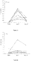
- a non-limiting example is illustrated in Fig. 2C , where the expression level of co-inhibitory receptor LAG-3 on V ⁇ 9V ⁇ 2 T cells after culturing said T cells with an anti-CD3 (OKT3 clone) antibody and gamma-irradiated CD64-expressing K562 aAPCs, is generally lower than the expression level of LAG-3 before the T cells are cultured with the anti-CD3 antibody and K562 aAPCs, but after expanding the V ⁇ 9V ⁇ 2 T cells in a sample of PBMCs using Zometa.
- Fc receptor or "FcR” is a receptor that binds to the Fc region of an immunoglobulin (Ig). Fc receptors are found on many-cells which participate in immune responses. Among the human FcRs that have been identified so far are those which recognize IgG (designated FcyR), IgE (FceRI), IgA (FcaR), and polymerized IgM/A (Fc ⁇ R). FcRs are found in the following cell types: FceRI (mast cells), FceRII (many leukocytes), FcaR (neutrophils), and Fc ⁇ R (glandular epithelium, hepatocytes). ( Hogg Immunol.
- the widely studied FcyRs are central in cellular immune defenses, and are responsible for stimulating the release of mediators of inflammation and hydrolytic enzymes involved in the pathogenesis of autoimmune disease. ( Unkeless, Annu. Rev. Immunol. 6: 251-87 (1988 )).
- the FcyRs provide a crucial link between effector cells and the lymphocytes that secrete Ig, since the macrophage/monocyte, polymorphonuclear leukocyte, and natural killer (NK) cell FcyRs confer an element of specific recognition mediated by IgG.
- Human leukocytes have at least three different receptors for IgG: hFc ⁇ RI (found on monocytes/macrophages), hFcyRII (on monocytes, neutrophils, eosinophils, platelets, possibly B cells, and the K562 cell line), and Fcylll (on NK cells, neutrophils, eosinophils, and macrophages).
- hFc ⁇ RI found on monocytes/macrophages
- hFcyRII on monocytes, neutrophils, eosinophils, platelets, possibly B cells, and the K562 cell line
- Fcylll on NK cells, neutrophils, eosinophils, and macrophages.
- the term “median survival time” refers to a parameter that is commonly used for evaluating therapeutic effects and is the time after which 50% of a patient group with a particular condition are still living and 50% have died.
- the median survival time of a patient group with a particular condition is 73 days after treatment with CAR-modified ⁇ T cells.
- MHC major histocompatibility complex
- Class I MHC, or MHC-I function mainly in antigen presentation to CD8 T lymphocytes.
- Class II MHC, or MHC-II function mainly in antigen presentation to CD4 T lymphocytes.
- the MHC gene located on chromosome 6, includes HLA genes divided into distinct regions including Class I genes that encode for a heavy chain polypeptide located in the HLA-A, B and C regions; and Class II genes including the D region which is subdivided in three main regions, DP, DQ and DR, each containing genes for a number of ⁇ and ⁇ chains.
- HLA human major histocompatibility complex
- HLA human major histocompatibility complex
- the IMGT/HLA Database is part of the international ImMunoGeneTics project.
- up-regulation or “up-regulated” when used in the context of receptors refers to an increase in the expression of the receptors.
- Figure 2C A non-limiting example is illustrated in Figure 2C , where the expression level of co-inhibitory receptor LAG-3 on V ⁇ 9V ⁇ 2 T cells after expanding the V ⁇ 9V ⁇ 2 T cells in a sample of PBMCs using Zometa, is higher than the expression level of LAG-3 before treatment with Zometa.
- treatment refers to both therapeutic treatment and prophylactic or preventative measures, wherein the object is to prevent or slow down (lessen) a disease, for example cancer.
- a disease for example cancer.
- Those in need of such treatment include those already with a disease as well as those prone to getting the disease or those in whom a disease is to be prevented.
- the term "therapeutically effective amount" of a compound will be an amount of active agent that is capable of preventing or at least slowing down (lessening) a disease, e.g. cancer.
- Dosages and administration of compounds, compositions and formulations of the present disclosure may be determined by one of ordinary skill in the art of clinical pharmacology or pharmacokinetics. See, for example, Mordenti and Rescigno, (1992) Pharmaceutical Research. 9:17-25 ; Morenti et al., (1991) Pharmaceutical Research. 8:1351-1359 ; and Mordenti and Chappell, "The use of interspecies scaling in toxicokinetics" in Toxicokinetics and New Drug Development, Yacobi et al.
- a typical dosage per infusion may range from about 1E5 cells to up to 1E10 cells of the patient's body weight or more per day, preferably about 1E3 cells/kg/infusion to 1E8 cells/kg/infusion.
- a polynucleotide is said to "encode" a polypeptide if, in its native state or when manipulated by methods well known to those skilled in the art, it can be transcribed and/or translated to produce the mRNA for and/or the polypeptide or a fragment thereof.
- Polynucleotides include but are not limited to DNA (e.g. cDNA) and RNA (e.g. mRNA).
- the anti-sense strand is the complement of such a nucleic acid, and the encoding sequence can be deduced therefrom.
- nucleic acid or fragment thereof is “substantially homologous" ("or substantially similar") to another if, when optimally aligned (with appropriate nucleotide insertions or deletions) with the other nucleic acid (or its complementary strand), there is nucleotide sequence identity in at least about 60% of the nucleotide bases, usually at least about 70%, more usually at least about 80%, preferably at least about 90%, and more preferably at least about 95-98% of the nucleotide bases.
- polypeptide refers to a polymer of amino acids and its equivalent and does not refer to a specific length of the product; thus, peptides, oligopeptides and proteins are included within the definition of a polypeptide. This term also does not refer to, or exclude modifications of the polypeptide, for example, glycosylations, acetylations, phosphorylations, and the like. Included within the definition are, for example, polypeptides containing one or more analogs of an amino acid (including, for example, natural amino acids, etc.), polypeptides with substituted linkages as well as other modifications known in the art, both naturally and non-naturally occurring.
- a polypeptide "fragment”, “portion” or “segment” is a stretch of amino acid residues of at least about five to seven contiguous amino acids, often at least about seven to nine contiguous amino acids, typically at least about nine to 13 contiguous amino acids and, most preferably, at least about 20 to 30 or more contiguous amino acids.
- a homologous sequence is taken to include an amino acid sequence which is at least 60, 70, 80 or 90% identical, preferably at least 95 or 98% identical at the amino acid level over at least 20, 50, 100, 200, 300 or 400 amino acids with the amino acid sequences set out in sequence listings.
- homology should typically be considered with respect to those regions of the sequence known to be essential for the function of the protein rather than non-essential.
- homology can also be considered in terms of similarity (i.e. amino acid residues having similar chemical properties/functions), in the context of the present disclosure it is also possible to express homology in terms of sequence identity.
- sequence identity i.e. amino acid residues having similar chemical properties/functions
- Homology comparisons can be conducted by eye, or more usually, with the aid of readily available sequence comparison programs. These commercially available computer programs can calculate % homology between two or more sequences.
- Percentage (%) homology may be calculated over contiguous sequences, i.e. one sequence is aligned with the other sequence and each amino acid in one sequence directly compared with the corresponding amino acid in the other sequence, one residue at a time. This is called an "ungapped" alignment. Typically, such ungapped alignments are performed only over a relatively short number of residues (for example less than 50 contiguous amino acids).
- the present disclosure provides for polyclonal and/or monoclonal antibodies and fragments thereof, and immunologic binding equivalents thereof, which are capable of specifically binding to a polypeptide target and fragments thereof.
- Such antibodies thus include for example, but are not limited to polyclonal, monoclonal, chimeric, single chain, Fab fragments, and a Fab expression library.
- a “monoclonal antibody” refers to an antibody having only one species of antibody combining site capable of immunoreacting with a particular antigen.
- a monoclonal antibody thus typically displays a single binding affinity for any antigen with which it immunoreacts.
- a monoclonal antibody may therefore contain an antibody molecule having a plurality of antibody combining sites, each immunospecific for a different antigen; e.g., a bi-specific (chimeric) monoclonal antibody.
- range format is merely for convenience and brevity and should not be construed as a limitation on the scope of the disclosed ranges. Accordingly, the description of a range should be considered to have specifically disclosed all the possible sub-ranges as well as individual numerical values within that range. For example, description of a range such as from 1 to 6 should be considered to have specifically disclosed sub-ranges such as from 1 to 3, from 1 to 4, from 1 to 5, from 2 to 4, from 2 to 6, from 3 to 6 etc., as well as individual numbers within that range, for example, 1, 2, 3, 4, 5, and 6. Ranges are not limited to integers, and can include decimal measurements. This applies regardless of the breadth of the range.
- a method of generating ⁇ T cells having at least one down-regulated co-inhibitory receptor comprising the steps of: (a) culturing a population of cells comprising ⁇ T cells with a phosphoantigen to expand the ⁇ T cells; and (b) culturing the expanded ⁇ T cells with artificial antigen-presenting cells expressing a Fc receptor, and an anti-CD3 antibody. Consequently, the expression of at least one co-stimulatory and/or inhibitory receptors of ⁇ T cells are manipulated, particularly by the manipulation of the expression of functional receptors expressed on the surface of ⁇ T cells, thereby facilitating the use of ⁇ T cells for treatment of cancer.
- the method of the present disclosure makes use of ⁇ TCRs that recognize cancer-associated antigens in an HLA (human leukocyte antigen)-independent manner.
- ⁇ TCRs do not recognize cancer-associated antigens in an HLA-independent manner.
- ⁇ T cells generated in the present disclosure may be used in the development of "off-the-shelf" therapeutics.
- ⁇ T cells are capable of infiltrating a range of human malignancies, a capacity required critically for them to interact with and kill cancer cells. These malignances include renal, bladder, ovarian, colorectal, breast and nasopharyngeal carcinomas.
- the method of the present disclosure is less time consuming, less complicated and at least as effective as prior art methods.
- methods of modifying ⁇ T cells such as CRISPR/Cas9, which makes use of a gene editing nuclease, are time consuming and complicated.
- the ⁇ T cells are V ⁇ 9V ⁇ 2 T cells, preferably pure V ⁇ 9V ⁇ 2 T cells.
- Certain earlier studies relate to expansion of polyclonal ⁇ T cells, not a single subtype of ⁇ T cells, meaning that the polyclonal ⁇ T cell population generated may contain a mixture of many subtypes of ⁇ T cells.
- Various problems may arise from the use of other methods in view of the following considerations:
- the method of the present disclosure relates to the manipulation of the expression of functional receptors expressed on the surface of V ⁇ 9V ⁇ 2 T cells.
- Inhibitory receptors can down-regulate immune responses to avoid excessive immune activation, providing a critical role in the maintenance of immune homeostasis.
- the co-inhibitory receptors expressed on ⁇ T cells include cytotoxic T lymphocyte (CTL)-associated antigen 4 (CTLA-4, CD152), programed cell death protein 1 (PD-1, CD279), lymphocyte activation gene-3 (LAG-3), T-cell immunoglobulin and mucin-containing protein (TIM-3), T cell immunoglobulin and immunoreceptor tyrosine-based inhibition motif (ITIM) domain (TIGIT), and B and T lymphocyte attenuator (BTLA).
- CTL cytotoxic T lymphocyte
- CTLA-4 programed cell death protein 1
- LAG-3 lymphocyte activation gene-3
- TIM-3 T-cell immunoglobulin and mucin-containing protein
- ITIM T cell immunoglobulin and immunoreceptor tyrosine-based inhibition motif
- TAGIT B and T lymphocyte attenuator
- the method of the present disclosure led to down-regulation or reduced expression of co-inhibitory receptors, thereby leading to improved tumor killing effects.
- the method of the present disclosure led to the reduced expression of at least one inhibitory receptor selected from the group consisting of CTLA-4, CD152, PD1, LAG-3, TIGIT, TIM3 and BTLA.
- the method of the present disclosure led to the reduced expression of two, three, four, five or six co-inhibitory receptors.
- the method of the present disclosure led to the reduced expression of six co-inhibitory receptors.
- ⁇ T cells express the natural killer group 2D (NKG2D) receptor, killer-cell immunoglobulin-like receptors (KIRs), and many co-inhibitory receptors that can play either co-stimulatory or inhibitory roles to affect their tumoricidal activity.
- the balance between activating signals and inhibitory signals induced by their respective receptors has profound effects on the activation of ⁇ T cells.
- the NKG2D receptor is an activating receptor expressed by human natural killer (NK) cells, ⁇ T cells, CD8+ T cells, and NKT cells. This receptor can interact with eight stress-induced ligands belonging to two families: two MHC class I chain-related proteins MICA and MICB and six HCMV UL16-binding proteins (ULBP1-6).
- the NKG2D ligands are not usually expressed on healthy tissues but can be up-regulated upon DNA damage, infection and transformation of cells, thus being commonly detected on hematopoietic tumors and carcinomas. Because of the tumour-associated over-expression, the NKG2D ligands have been a favourable therapeutic target for anticancer strategies.
- the method of the present disclosure led to the reduced expression of co-inhibitory receptors without obvious effects on NKG2D expression.
- the ⁇ T cells may be expanded from peripheral blood mononuclear cells (PBMCs), cord blood mononuclear cells (CBMCs) or tissue derived cells in a chemically defined culture medium which can include, but is not limited to, RPMI, TexMACS, IMDM, CTS OpTmizer or AIM-V media.
- PBMCs peripheral blood mononuclear cells
- CBMCs cord blood mononuclear cells
- tissue derived cells in a chemically defined culture medium which can include, but is not limited to, RPMI, TexMACS, IMDM, CTS OpTmizer or AIM-V media.
- the V ⁇ 9V ⁇ 2 T cells are expanded from PBMCs seeded in AIM-V media. Isolated PBMCs may be freshly isolated or cryopreserved prior to expansion in the culture medium.
- the PBMCs are seeded in AIM-V media at a density of 2E6.
- the method further comprises seeding the PB
- the cell culture medium may be supplemented with fetal calf serum (FCS), human AB serum, autologous plasma, human platelet lysate or a chemically defined serum replacement substitutes.
- FCS fetal calf serum
- human AB serum may be added in an amount of about 0.1 to about 20% (v/v), about 1 to about 10% (v/v), or about 1 to about 5% (v/v) to the culture solution.
- human AB serum is added in about 5% v/v.
- the method comprises passaging the cells every day, every 2 days, every 3 days or every 4 days, preferably every 2 to 3 days.
- step (a) further comprises adding the phosphoantigen into the sample of PBMCs.
- the population of cells comprising ⁇ T cells is cultured with the phosphoantigen to expand the ⁇ T cells for about 5 days to about 10 days, preferably about 7 days.
- the phosphoantigen is added in an amount of about 0.1 to about 10 ⁇ M, about 1 to about 10 ⁇ M or about 1 to about 5 ⁇ M.
- the phosphoantigen is zoledronic acid or zoledronate (Zometa) or salts thereof. The formula for zoledronate is as follows.
- the phosphoantigen may be a synthetic antigen such as isopentenyl pyrophosphate (IPP), phosphostim / bromohydrin pyrophosphate (BrHPP), (E)-4-Hydroxy-3-methyl-but-2-enyl pyrophosphate (HMBPP) or DMAPP.
- IPP isopentenyl pyrophosphate
- BrHPP phosphostim / bromohydrin pyrophosphate
- HMBPP 4-Hydroxy-3-methyl-but-2-enyl pyrophosphate
- DMAPP DMAPP
- Zometa is added in an amount of 5 ⁇ M.
- a small amount (1 ⁇ M or 5 ⁇ M) of Zometa leads to vigorous proliferation of V ⁇ 9V ⁇ 2 T cells in vitro in response to the phosphoantigen, Zometa, which is an FDA-approved, commercially available bisphosphonate drug that has been used to treat patients with postmenopausal osteoporosis.
- Zometa can inhibit farnesyl pyrophosphate synthase, an enzyme acting downstream of isopentenyl pyrophosphate (IPP) in the mevalonate pathway, thus profoundly increasing intracellular levels of IPP.
- IPP isopentenyl pyrophosphate
- the cytokine IL-2 is also added with the phosphoantigen.
- IL-2 is added in a concentration of about 50 IU/ml to about 1000 IU/ml, about 100 IU/ml to about 500 IU/m or about 200 IU/ml to about 400 IU/ml. In a preferred embodiment, IL-2 is added in a concentration of about 300 IU/ml.
- the method does not require an initial depletion step to enrich the sample of PBMCs.
- the method of the present disclosure leads to a savings in time and money because the PBMCs can be expanded without undergoing an initial depletion step.
- the V ⁇ 9V ⁇ 2 T cells are activated by using an artificial antigen-presenting cell (aAPC) and an antibody against CD3, homologous peptides, or a portion or a polypeptide fragment thereof.
- aAPC artificial antigen-presenting cell
- the expanded ⁇ T cells is cultured with artificial antigen-presenting cells expressing a Fc receptor, and an anti-CD3 antibody between about 10 days to about 21 days or longer, preferably between about 10 days to about 14 days.
- the anti-CD3 antibody may be a monoclonal and/or polyclonal antibody or fragments thereof, or immunologic binding equivalents thereof capable of binding CD3, homologous peptides, or a portion or a polypeptide fragment thereof.
- the inventors found that co-inhibitory receptors were up-regulated on V ⁇ 9V ⁇ 2 T cells when expanding V ⁇ 9V ⁇ 2 T cells in the sample of peripheral blood mononuclear cells using the phosphoantigen.
- activating the V ⁇ 9V ⁇ 2 T cells using the artificial antigen-presenting cell together with an antibody against CD3 led to down-regulation of the co-inhibitory receptor expression on V ⁇ 9V ⁇ 2 T cells.
- tumor cell killing effects as compared with V ⁇ 9V ⁇ 2 T cells collected after treatment with the phophoantigen.
- conformational change of the CD3 co-receptor on T cells and aAPCs advantageously affect the tumor killing activity of V ⁇ 9V ⁇ 2 T cells.
- the combination of expanding V ⁇ 9V ⁇ 2 T cells in the sample of PBMCs using a phosphoantigen and activating the V ⁇ 9V ⁇ 2 T cells using an artificial antigen-presenting cell together with an antibody against CD3 led to an unexpected technical advantage.
- the V ⁇ 9V ⁇ 2 T cells were effectively expanded and augmented to exhibit enhanced tumor cell killing activity.
- artificial antigen presenting cells are mimetic cells that are modelled after antigen presenting cells, and provide T cell stimulation signals.
- the aAPC is derived from a human erythroleukemia cell line, such as the K562 cell line.
- K562 cells expresses neither endogenous HLA class I and II molecules, nor co-stimulatory molecules such as CD86, CD83, 4-1BBL, OX40L, ICOSL (B7H2, B7RP1) or CD40L, thereby minimizing unintended allogeneic responses.
- K562 cells endogenously expresses a high level of CD32 but not CD16 or CD64.
- K562 cells also express adhesion molecules CD54 (ICAM-1) and CD58 (LFA-3), which enhances K562 cells interactions with T cells and improves T cell stimulation. Furthermore, K562 cells can be easily manipulated for stable expression of transgenes and have been found safe in clinical trials when used as feeder cells for T cell expansion.
- the aAPC expresses CD64 (SEQ ID NO: 1), homologous polypeptides, or a portion or a polypeptide fragment thereof.
- the method further comprises irradiating the aAPC prior to step (b) using gamma irradiation.
- the antibody against CD3 is Muromonal-CD3 (tradename OKT3; light chain SEQ ID NO: 3; heavy chain SEQ ID NO: 4).
- OKT3 tradename OKT3; light chain SEQ ID NO: 3; heavy chain SEQ ID NO: 4
- the antigen-binding sites of OKT3 can interact with CD3 on the ⁇ T cell surface to induce the conformational change of the co-receptor.
- the Fc-portion of OKT-3 can attach to CD64 expressed on the K562 cells because of the high affinity between the Fc-portion of OKT-3 for CD64. As such, down-regulation of the co-inhibitory receptors was observed, thereby leading to improved tumor killing effects.
- the method further comprises modifying the ⁇ T cells using a chimeric antigen receptor (CAR).
- CAR chimeric antigen receptor
- the method led to further enhanced tumor killing.
- treatment with CAR-modified ⁇ T cells resulted in the increase of the median survival time of tumor-bearing mice.
- the median survival time of such mice increased from 41 to 51 days in the control groups, to 73 days after treatment with CAR-modified V ⁇ 9V ⁇ 2 T cells ( Figure 6C ).
- the increase in median survival time is in the range of about 40% to about 80%, such as but not limited to at least about 40%, about 45%, about 50%, about 55%, about 60%, about 65%, about 70%, about 75%, or about 80%.
- Treatment with CAR-modified V ⁇ 9V ⁇ 2 T cells resulted in a prolonged survival time of more than 70 days in tumor-bearing mice ( Figure 6C ).
- the survival time was increased to about 80 days.
- modifying the ⁇ T cells using a CAR led to enhanced tumor killing because the median survival time of tumor-bearing mice increased by about 78% and about 43% compared to the PBS control group and the mGFP control respectively.
- the CAR comprises an extracellular antigen binding domain of NKG2D (SEQ ID NO: 5), homologous polypeptides, or a portion or a polypeptide fragment thereof.
- NKG2D SEQ ID NO: 5
- homologous polypeptides or a portion or a polypeptide fragment thereof.
- the NKG2D extracellular domain acts as the target recognition domain to bind the NKG2D ligands on tumor cells. Consequently, the interaction activates ⁇ cells through the intracellular signaling motif, thereby leading to enhanced tumor killing.
- the CAR comprises a signalling domain of CD3 zeta or DAP 12 (NKG2Dz or NKG2Dp, respectively; CD3 zeta SEQ ID NO: 6; DAP 12 SEQ ID NO: 7), homologous polypeptides, or a portion or a polypeptide fragment of CD3 zeta or DAP 12.
- the tumor-killing efficacy of the V ⁇ 9V ⁇ 2 T cells can be further enhanced.
- the tumor-killing efficacy of the V ⁇ 9V ⁇ 2 T cells is enhanced through the modification of the effector cells with gene transfer of NKG2Dz.
- the CAR comprises a transmembrane domain of CD8-a (CD8- ⁇ ) chain (SEQ ID NO: 8), homologous polypeptides, or a portion or a polypeptide fragment thereof.
- the CAR comprises an extracellular antigen binding domain of NKG2D and an intracellular signalling domain of DAP12, homologous polypeptides, or a portion or a polypeptide fragment of NKG2D or DAP 12.
- a ⁇ T cell having at least one down-regulated co-inhibitory receptor generated by a method according to an aspect of the present disclosure.
- a ⁇ T cell having at least one down-regulated co-inhibitory receptor and modified to express a chimeric antigen receptor (CAR), wherein the CAR comprises an extracellular antigen binding domain of NKG2D and an intracellular signalling domain of CD3 zeta or DAP 12.
- the signalling domain is DAP12.
- references to methods of treatment in this description are to be interpreted as references to the ⁇ T cells described herein for use in a method of treatment.
- a therapeutically effective amount of ⁇ T cells according to an aspect of the present disclsoure for use in a method of treating cancer comprising administering the ⁇ T cells.
- the cancer is colorectal or ovarian cancer or any other cancer expressing NKG2D ligands.
- a ⁇ T cell for use in treating cancer.
- the cancer is colorectal cancer or ovarian cancer or any other cancer expressing NKG2D ligands.
- ⁇ T cells having at least one down-regulated co-inhibitory receptor generated by a method comprising the steps of: (a) culturing a population of cells comprising ⁇ T cells with a phosphoantigen; (b) culturing the expanded ⁇ T cells with artificial antigen-presenting cells expressing a Fc receptor, and an anti-CD3 antibody for use in a method of treating a patient with cancer comprising: administering to the patient, a therapeutically effective amount of ⁇ T cells.
- said method comprises administering the ⁇ T cells having at least one down-regulated co-inhibitory receptor to the patient by intraperitoneal injection.
- ⁇ T cells for use in a method of treating cancer in a patient, the method comprising the steps of: (a) obtaining peripheral blood mononuclear cells (PBMCs) comprising ⁇ T cells; (b) culturing the PBMCs with a phosphoantigen to expand the ⁇ T cells; (c) culturing the PBMCs with the expanded ⁇ T cells, with an artificial antigen-presenting cells expressing a Fc receptor, and an anti-CD3 antibody against CD3 to generate ⁇ T cells with at least one down-regulated co-inhibitory receptor; and (d) administering the ⁇ T cells with at least one down-regulated co-inhibitory receptor to the patient.
- PBMCs peripheral blood mononuclear cells
- Human PBMC were isolated from fresh buffy coat of healthy donors by density gradient centrifugation using Ficoll-Paque (GE Healthcare, Milwaukee, WI).
- Human myelogenous leukemia cell line K562 (ATCC) and a K562-based aAPC cell line genetically engineered for stable expression of EGFP, CD86, CD64 and 4-1BBL (Du et al., 2016) were cultured in IMDM (Lonza Biotech, Basel, Switzerland) supplemented with 10% FBS.
- Other tumor cell lines used in the present disclosure were cultured in DMEM supplemented with 10% FBS.
- PBMCs were seeded at a density of 2E6 cells/ml in AIM-V (Life Technologies) supplemented with 5% human AB serum (Valley Biomedical, Winchester, VA).
- Zometa (5 ⁇ M, Sigma Aldrich, St. Louis, Missouri) and IL-2 (300 IU/ml) were added on day 0, followed by adding 50 ng/ml anti-CD3 antibody (OKT3 clone, eBioscience, San Diego, CA), gamma irradiated K562 aAPCs at a 100:1 ratio, 300 IU/ml IL-2, and 5 ⁇ M Zometa from day 7 onwards.
- a synthetic sequence containing the T7 promoter, 5'UTR, with Kozak sequence, a multiple cloning site, the GM-CSF signal peptide encoding sequence and the alpha-globin 3'UTR sequence was synthesized and inserted into pFastbac1 vector (Life Technologies) to construct the basal backbone vector pFBCMV-T7.
- the extracellular domain of human NKG2D was amplified by PCR from a PMBC cDNA library using the primers 5'-gcgcgcatgccttcaaccaagaagttcaaattcc-3' (forward primer with Sphl site (SEQ ID NO: 9)) and 5'-acgaagctagccacagtcctttgcatacagatgtacgtatttggag-3' (reverse primer with Nhel site (SEQ ID NO: 10)).
- NKG2D chimeric proteins were constructed by fusing NKG2D-ED to the CD8 ⁇ hinge and transmembrane region (CD8 H-TM, uniprot P01732 (SEQ ID NO: 8), amino acids 128-210) and CD3 ⁇ (SEQ ID NO: 6) or DAP12 (SEQ ID NO: 7) signalling moiety, and then subcloned into pFBCMV-T7 with EcoRI and Sall.
- the mGFP control vector was generated by replacing the NKG2D ED part of the NKG2Dz vector with GFP encoding sequence (the start codon removed) by Sphl and Nhel.
- PCR was performed using the above pFBCMV-T7 vectors as DNA templates, a forward primer CMV-F (5'-atccgctcgagtagttattaatagtaatcaattacggggtc-3') (SEQ ID NO: 11) and reverse primer T150-R (SEQ ID NO: 12).
- Capped mRNA was generated through in vitro transcription of the PCR DNA templates using the mMESSAGE mMACHINE T7 ULTRA transcription kit (Invitrogen, Carlsbad, CA) or the mScript TM RNA system (Epicentre, Madison, WI).
- V ⁇ 9V ⁇ 2 T cells were mixed with the generated mRNA molecules and electroporated in a 2-mm cuvette (Bio-Rad, Hercules, CA) using a NEPA21 electroporator (Nepagene, Chiba, Japan) with the following parameters: voltage 240 V, pulse length 4 ms, pulse once.
- the electroporated T cells were rested for 3 hours and stored at -80°C until use.
- Non-obese diabetic/severe combined immunodeficiency/IL-2Rycnull (NSG) mice (6-8 weeks old, female) were used in the present disclosure.
- Mice were inoculated via intraperitoneal (i.p.) injection of 1E7 SKOV3-Luc cells.
- tumor engraftment was confirmed by live bioluminescent imaging (BLI) monitored using an IVIS Spectrum Imaging platform with Living Image software (PerkinElmer). Mice with similar BLI signal intensity were randomly divided into different groups.
- 1E7 modified V ⁇ 9V ⁇ 2 T cells or PBS cells were i.p. injected into the tumor-bearing mice, twice a week for 3 weeks.
- mice Tumor progression was monitored by BLI. All luminescent signals and images were acquired and analysed with the Xenogen living image software v2.5. Behaviour and survival of the mice were monitored closely. Humane endpoints were used and mice were euthanized by cervical dislocation under sodium pentobarbital anaesthesia upon signs of severe distress such as swollen belly due to tumor ascites formation, seizures, tremors, laboured or difficulty in breathing, significant weight loss (>15% body weight), signs of emaciation (i.e. prominent skeletal structures), impaired ambulation, inability to remain upright or evidence of moribund condition. The survival curves were established based on the dates when mice were found dead or euthanized.
- V ⁇ 9V ⁇ 2 T cells were efficiently expanded by treating PBMCs using 5 ⁇ M of Zometa.
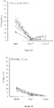
- the V ⁇ 9V ⁇ 2 T-cell population was amplified from -4% in PBMCs on day 0 to close to 90% by day 7, together with a concurrent increase of the NKG2D-positive cell population ( Fig. 1 ).
- ⁇ T cells can be regulated by the conformational change of the CD3 co-receptor on T cells and that K562-based aAPCs can be used to activate and propagate the proliferation of polyclonal ⁇ T cells
- an anti-CD3 (OKT3 clone) antibody and gamma-irradiated CD64-expressing K562 aAPCs, as well as another portion of Zometa were added to the culture and the cells were further cultured for another 10 days.
- Fc-portion of OKT-3 can attach to CD64 (the high affinity human IgG receptor FcyRI) expressed on K562 cells, whereas the antigen-binding sites of OKT3 can interact with CD3 on the ⁇ T cell surface to induce the conformational change of the co-receptor.
- V ⁇ 9V ⁇ 2 T cells obtained with the co-cultured method of the present disclosure, the inventors proceeded to test equipping V ⁇ 9V ⁇ 2 T cells by over-expressing an activating receptor on their surface.
- electroporation of mRNA encoding an activating receptor into the expanded V ⁇ 9V ⁇ 2 T cells was carried out.
- NKG2D chimeric proteins containing the extracellular domain of NKG2D fused with CD3 zeta (NKG2Dz) or DAP12 (NKG2Dp) were synthesized.
- An mRNA electroporation approach was adopted to facilitate fast evaluation of the two constructs.
- mRNA electroporation was optimized in V ⁇ 9V ⁇ 2 T cells using EGHP mRNA, which provided almost 99% transfection efficiency.
- an in vitro cytotoxicity assay was performed using the Delfia cytotoxicity kit. The same amount of mRNA for each chimeric protein was electroporated into V ⁇ 9V ⁇ 2 T cells.
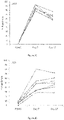
- the cancer cell killing efficacy of V ⁇ 9V ⁇ 2 T cells significantly increased 30 to 50% post NKG2Dp mRNA modification and 90% post NKG2Dz mRNA modification from less than 10% cytotoxicity observed for the mGFP control at an effector to target ratio of 20:1 ( Fig. 4 ).
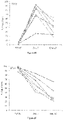
- Zometa treatment was used because Zometa can increase intracellular levels of IPP, thereby increasing the sensitivity of tumor cells to V ⁇ 9V ⁇ 2 T cells. Subsequently, the inventors tested whether tumor cell killing efficacy can be improved by using NKG2Dz-modified V ⁇ 9V ⁇ 2 T cells to kill Zometa pre-treated tumor cells at an effector to target ratio of 10:1 ( Fig. 5 ).
- V ⁇ 9V ⁇ 2 T cells or mGFP-modified V ⁇ 9V ⁇ 2 T cells did not kill the 4 tested tumor cell lines, namely, SW480 human colon carcinoma cells, HepG2 human hepatocarcinoma cells, U87 human glioblastoma cells and SKOV3 human ovarian cancer cells.
- NKG2Dz-modified V ⁇ 9V ⁇ 2 T cells were able to kill these tumor cells that were not pre-treated with Zometa, with the efficiency varying from 20 to 80%.
- the sensitivity of SW480, U87 and SKOV3 cells, but not HepG2 cells, towards V ⁇ 9V ⁇ 2 T cells were significantly increased by Zometa pre-treatment.
- Non-invasive whole-body bioluminescent imaging (BLI) of SKOV3-Luc cells were performed to monitor tumor growth.
- Fig. 6A shows images of mice in each group at 3 different time points post-tumor cell injection.
- the tumor burden in mice receiving the NKG2D- ⁇ T-Z treatment was significantly reduced relative to the initial tumor burdens (P ⁇ 0.001, Fig. 6B ).
- Significant reduction of the disease by the NKG2D- ⁇ T-Z treatment maintained for >2 weeks in 4 out of 6 treated mice.
Landscapes
- Health & Medical Sciences (AREA)
- Life Sciences & Earth Sciences (AREA)
- Chemical & Material Sciences (AREA)
- Organic Chemistry (AREA)
- Immunology (AREA)
- General Health & Medical Sciences (AREA)
- Genetics & Genomics (AREA)
- Zoology (AREA)
- Biochemistry (AREA)
- Engineering & Computer Science (AREA)
- Medicinal Chemistry (AREA)
- Biophysics (AREA)
- Molecular Biology (AREA)
- Proteomics, Peptides & Aminoacids (AREA)
- Biomedical Technology (AREA)
- Cell Biology (AREA)
- Animal Behavior & Ethology (AREA)
- Public Health (AREA)
- Veterinary Medicine (AREA)
- Gastroenterology & Hepatology (AREA)
- Toxicology (AREA)
- Biotechnology (AREA)
- Wood Science & Technology (AREA)
- Bioinformatics & Cheminformatics (AREA)
- Epidemiology (AREA)
- General Engineering & Computer Science (AREA)
- Microbiology (AREA)
- Pharmacology & Pharmacy (AREA)
- Hematology (AREA)
- Nuclear Medicine, Radiotherapy & Molecular Imaging (AREA)
- General Chemical & Material Sciences (AREA)
- Chemical Kinetics & Catalysis (AREA)
- Physics & Mathematics (AREA)
- Plant Pathology (AREA)
- Micro-Organisms Or Cultivation Processes Thereof (AREA)
- Medicines Containing Material From Animals Or Micro-Organisms (AREA)
- Medicines That Contain Protein Lipid Enzymes And Other Medicines (AREA)
- Developmental Biology & Embryology (AREA)
- Virology (AREA)
Description
- This application claims priority to
Singapore application No. 10201701009Q, filed 8 February 2017 - The present invention relates to gamma delta T cells and their uses thereof, and a method of generating gamma delta T cells. More particularly, the present invention relates to gamma delta T cells having improved tumoricidal activity and their uses thereof, and a method of generating gamma delta T cells having improved tumoricidal activity.
- The following discussion of the background to the invention is intended to facilitate understanding of the present invention.
- Gamma delta (γδ) T cells are a minor population in the peripheral blood (0.5-10% of T cells in healthy adults) that express γδ heterodimer of T cell receptor (TCR) chains and play an important role in linking innate and adaptive immune responses. The majority of peripheral blood γδ T cells express the variable-gene segments Vy9 and Vδ2 (Vγ9Vδ2 T cells). In a non-major histocompatibility complex (MHC) restricted manner, γδ T cells can be activated by recognizing and interacting with a set of tumorassociated antigens, including phosphoantigens that are produced during metabolic dysregulation in tumour cells, lipids presented by CD1 family members, and cell stress markers. The activated γδ T cells release abundant inflammatory cytokines interferon (IFN)-γ and tumour necrosis factor (TNF)-α, and use both perforin and granzyme B secretory pathway and death receptor (Fas/Fas-ligand, TRAIL/TRAIL-receptor) pathway to execute the killing of tumor cells. γδ T cells have been indicated to be able to kill many different types of tumor cell lines and tumor in vivo and in vitro, including leukemia, neuroblastoma and various carcinomas. As such, the development of γδ T cells that are suitable for cancer treatment is desirable.
- In addition to TCR, γδ T cells express the natural killer group 2D (NKG2D) receptor, killer-cell immunoglobulin-like receptors (KIRs), and many co-inhibitory receptors that can play either co-stimulatory or inhibitory roles to affect their tumoricidal activity. The balance between activating signals and inhibitory signals induced by their respective receptors has profound effects on the activation of γδ T cells.
- The NKG2D receptor is an activating receptor expressed by human natural killer (NK) cells, γδ T cells, CD8+ T cells, and NKT cells. This receptor can interact with eight stress-induced ligands belonging to two families: two MHC class I chain-related proteins MICA and MICB and six HCMV UL16-binding proteins (ULBP1-6). The NKG2D ligands are not usually expressed on healthy tissues but can be up-regulated upon DNA damage, infection and transformation of cells, thus being commonly detected on hematopoietic tumors and carcinomas. Because of the tumour-associated over-expression, the NKG2D ligands have been a favourable therapeutic target for anticancer strategies.
- Thus, there exists a need to develop γδ T cells and a method of augmenting the tumoricidal activity of γδ T cells to facilitate the use of γδ T cells for treatment of cancer.
- The present invention seeks to address and/or ameliorate the problems in the prior art by providing a method of generating gamma delta T cells and augmenting and/or enhancing their functions.
- In an aspect of the present invention, there is provided a method of generating γδ T cells having at least one down-regulated co-inhibitory receptor, the method comprising the steps of: (a) culturing a population of cells comprising γδ T cells with a phosphoantigen to expand the γδ T cells; (b) culturing the expanded γδ T cells with artificial antigen-presenting cells expressing a Fc receptor, and an anti-CD3 antibody. and c) modifying the γδ T cells to express CAR, wherein the CAR comprises an extracellular antigen-binding domain of NKG2D.
- Preferably, the Fc receptor is CD64.
- Preferably, the phosphoantigen is zoledronic acid or a salt thereof. More preferably, the phosphoantigen is added in an amount of 5 µM.
- Preferably, the population of cells are peripheral blood mononuclear cells.
- Preferably, the method does not require an initial depletion step to enrich the peripheral blood mononuclear cells.
- Preferably, the γδ T cells are of the Vγ9Vδ2 subtype.
- Preferably, step (a) is carried out for 7 days. More preferably, step (b) is carried out for 10 days. Even more preferably, the method comprises passaging the cells every 2 to 3 days.
- Preferably, the artificial antigen-presenting cells are K562 cells. Preferably, the method further comprises irradiating the artificial antigen-presenting cells prior to step (b) using gamma irradiation.
- Preferably, the anti-CD3 antibody is Muromonab-CD3.
- Preferably, the co-inhibitory receptors are at least one co-inhibitory receptor selected from the group consisting of cytotoxic T lymphocyte (CTL)-associated antigen 4 (CTLA-4/CD152); programmed cell death protein 1 (PD-1/CD279); lymphocyte activation gene-3 (LAG-3); T cell immunoglobulin and immunoreceptor tyrosine-based inhibition motif (ITIM) domain (TIGIT); T-cell immunoglobulin and mucin-containing protein 3 (TIM3); and B and T lymphocyte attenuator (BTLA).
- Preferably, another portion of the phosphoantigen is added simultaneously with step (b).
- The method further comprises modifying the γδ T cells having at least one down-regulated co-inhibitory receptor to express a chimeric antigen receptor (CAR). Preferably, the modified γδ T cells have at least one up-regulated activating receptor. More preferably, modifying the γδ T cells comprises transfecting the γδ T cells with an mRNA vector encoding the CAR. Preferably, transfecting the γδ T cells comprises RNA electroporation.
- The CAR comprises an extracellular antigen binding domain of natural killer group 2D (NKG2D). Preferably, the γδ T cells overexpresses NKG2D.
- Preferably, the CAR comprises a signalling domain of CD3 zeta or
DAP 12. More preferably, the CAR comprises an extracellular antigen binding domain of NKG2D and a signalling domain of DAP12. - In another aspect of the present invention, there is provided a γδ T cell having at least one down-regulated co-inhibitory receptor generated by a method according to an aspect of the present invention.
- In another aspect of the present invention, there is provided a γδ T cell having at least one down-regulated co-inhibitory receptor and modified to express a chimeric antigen receptor (CAR), wherein the CAR comprises an extracellular antigen binding domain of NKG2D and a signalling domain of CD3 zeta or
DAP 12. Preferably, the signalling domain is DAP12. - In another aspect of the present disclosure, not explicitly claimed, there is provided a therapeutically effective amount of γδ T cells according to an aspect of the present invention for use in a method of treating cancer. Preferably, the cancer is colorectal or ovarian cancer or any other cancer expressing NKG2D ligands.
- In another aspect of the present invention, there is provided a γδ T cell according to an aspect of the present invention for use in treating cancer.
- In another aspect of the present invention, there is provided a γδ T cell according to an aspect of the present invention for use in the treatment of colorectal or ovarian cancer or any other cancer expressing NKG2D ligands.
- In another aspect of the present disclosure, not explicitly claimed, there is provided a therapeutically effective amount of γδ T cells having at least one down-regulated co-inhibitory receptor generated by a method comprising the steps of: (a) culturing a population of cells comprising γδ T cells with a phosphoantigen; (b) culturing the expanded γδ T cells with artificial antigen-presenting cells expressing a Fc receptor, and an anti-CD3 antibody for use in a method of treating a patient with cancer. Preferably, said method comprises administering the γδ T cells having at least one down-regulated co-inhibitory receptor to the patient by intraperitoneal injection.
- In another aspect of the present disclosure, not explicitly claimed, there is provided γδ T cells for use in a method of treating cancer in a patient, the method comprising the steps of: (a) obtaining peripheral blood mononuclear cells (PBMCs) comprising γδ T cells; (b) culturing the PBMCs with a phosphoantigen to expand the γδ T cells; (c) culturing the PBMCs with the expanded γδ T cells, with an artificial antigen-presenting cells expressing a Fc receptor, and an anti-CD3 antibody against CD3 to generate γδ T cells with at least one down-regulated co-inhibitory receptor; and (d) administering the γδ T cells with at least one down-regulated co-inhibitory receptor to the patient.
- Preferably, the Fc receptor is CD64.
- Preferably, the phosphoantigen is zoledronic acid or a salt thereof.
- Preferably, the phosphoantigen is added in an amount of 5 µM.
- Preferably, the method does not require an initial depletion step to enrich the peripheral blood mononuclear cells.
- Preferably, the γδ T cells are of the Vγ9Vδ2 subtype.
- Preferably, step (b) is carried out for 7 days.
- Preferably, step (c) is carried out for 10 days.
- Preferably, the method comprises passaging the cells every 2 to 3 days.
- Preferably, the artificial antigen-presenting cells are K562 cells.
- Preferably, the method further comprises irradiating the artificial antigen-presenting cells prior to step (c) using gamma irradiation.
- Preferably, the anti-CD3 antibody is Muromonab-CD3.
- Preferably, the at least one co-inhibitory receptor is selected from the group consisting of cytotoxic T lymphocyte (CTL)-associated antigen 4 (CTLA-4/CD152); programmed cell death protein 1 (PD-1/CD279); lymphocyte activation gene-3 (LAG-3); T cell immunoglobulin and immunoreceptor tyrosine-based inhibition motif (ITIM) domain (TIGIT); T-cell immunoglobulin and mucin-containing protein 3 (TIM3); and B and T lymphocyte attenuator (BTLA).
- The method further comprises modifying the γδ T cells having at least one down-regulated co-inhibitory receptor to express a chimeric antigen receptor (CAR).
- Preferably, the modified γδ T cells have at least one up-regulated activating receptor.
- Preferably, modifying the γδ T cells comprises transfecting the γδ T cells with an mRNA vector encoding the CAR.
- Preferably, transfecting the γδ T cells comprises RNA electroporation.
- The CAR comprises an extracellular antigen binding domain of natural killer group 2D (NKG2D).
- Preferably, the γδ T cells overexpresses NKG2D.
- Preferably, the CAR comprises a signalling domain of CD3 zeta or
DAP 12. - Preferably, the CAR comprises an extracellular antigen binding domain of NKG2D and a signalling domain of DAP12.
- The invention will now be described, by way of example only, with reference to the accompanying drawings, in which:
-
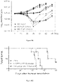
Figures 1A to 1D (collectively referred to as "Figure 1 ") show the characterization of the cells expanded with Zometa, OKT3 and gamma-irradiated K562 aAPCs. Human PBMCs were first expanded with Zometa for 7 days, followed by treatment with Zometa, OKT3 and gamma-irradiated K562 aAPCs for another 10 days. Frequencies of Vγ9Vδ2 T cells (Figure 1A ), NKG2D-positive cells (Figure 1B ), CD3-positive alpha beta T cells (Figure 1C ) and CD3-negative, CD56-positive NK cells (Figure 1D ) in PBMCs, onday 7 andday 17 analyzed by flow cytometry are shown. PBMC samples from 7 donors were tested and each sample is represented by one line. -
Figures 2A to 2F (collectively referred to as "Figure 2 ") show the expression of co-inhibitory receptors CTLA-4 (Figure 2A ), PD-1 (Figure 2B ), LAG-3 (Figure 2C ), TIGIT (Figure 2D ), TIM-3 (Figure 2E ) and BTLA (Figure 2F ) on PBMCs and the expanded cells. Human PBMCs were first expanded with Zometa for 7 days, followed by treatment with Zometa, OKT3 and gamma-irradiated K562 aAPCs for another 10 days. The expression frequencies of 6 co-inhibitory receptors on PBMCs and on the expanded cells collected onday 7 andday 17 analyzed by flow cytometry are shown. PBMC samples from 7 donors were tested and each sample is represented by one line. -
Figure 3 shows the expression of NKG2D chimeric proteins on Vγ9Vδ2 T cells. Flow cytometric analysis was performed with an anti-NKGD antibody or an isotype control antibody to detect the surface expression of NKG2D on Vγ9Vδ2 T cells or cells electroporated with mGFP, NKG2Dp or NKG2Dz mRNA 24 hours ago. -
Figure 4 shows tumor cell lysis induced by Vγ9Vδ2 T cells modified with NKG2D chimeric proteins. Delfia EuTDA cytotoxicity assay (3 hours EuTDA culturing) was used to assess tumor cell lysis efficiency. The cytotoxicity of Vγ9Vδ2 T cells against pCRC7 and SKOV3 tumor cells was observed after NKG2Dp- or NKG2Dz-mRNA electroporation, but not after mGFP electroporation. The results of one representative experiment out of three are shown. -
Figure 5 shows lysis of Zometa-treated tumor cells by Vγ9Vδ2 T cells modified with NKG2Dz. Target tumor cells tested were SW480 human colon carcinoma, HepG2 human hepatocarcinoma cells, U87 human glioblastoma cells and SKOV3 human ovarian cancer cells. Tumor cells were either pre-treated with 1 µM Zometa or without Zometa treatment. Effector used are indicated. Delfia EuTDA cytotoxicity assay (3 hours EuTDA culturing) was used to assess tumor cell lysis efficiency at an effector to target ratio of 10:1. The results of one representative experiment out of three are shown. -
Figure 6A to 6C show the treatment with Vγ9Vδ2 T cells electroporated with NKG2Dz mRNA results in reduction in disease burden and prolonged survival in mice with SKOV3-luc xenografts. NSG mice (n=6 per group) were i.p. injected with the SKOV3-luc human ovarian cancer cells (1E7 per mouse). The treatment started 7 days after tumor cell inoculation, twice a week for 3 weeks, 1E7 Vγ9Vδ2 T cells per injection. The mice were followed with serial weekly imaging to assess the tumor burden.Fig. 6A illustrates tumor burden images by bioluminescent imaging (BLI) ondays Fig. 6B illustrates the treatment results in reduction in SKOV3 xenografts. Tumor burden over time by BLI is shown. Each mouse is represented by one line.Fig. 6C illustrates survival curves. The treatment with Vγ9Vδ2 T cells electroporated with NKG2Dz mRNA plus Zometa resulted in significant survival advantages when compared with the PBS group or the treatment with the cells electroporated with mGFP mRNA plus Zometa. -
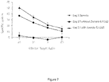
Figure 7 shows the cytotoxicity ofDay 7 andDay 17 Vγ9Vδ2 T cells against SKOV3 ovarian cancer cells. Vγ9Vδ2 T cells obtained after the co-culture with K562 cells (Day 17 with Zometa & K562) displayed improved tumor cell killing effects compared with Vγ9Vδ2 T cells collected after Zometa treatment for 7 days only (Day 7 Zometa). Cells collected afterDay 17 without Zometa and K562 cells were used as the control (Day 17 without Zometa & K562). - Particular embodiments of the present disclosure will now be described with reference to the accompany drawings. The terminology used herein is for the purpose of describing particular embodiments only and is not intended to limit the scope of the present disclosure. Additionally, unless defined otherwise, all technical and scientific terms used herein have the same meanings as commonly understood by one or ordinary skill in the art to which the present disclosure belongs.
- Throughout the specification, unless otherwise indicated to the contrary, the terms "comprising", "consisting of", and the like, are to be construed as non-exhaustive, or in other words, as meaning "including, but not limited to".
- Throughout the specification, unless the context requires otherwise, the word "comprise" or variations such as "comprises" or "comprising", will be understood to imply the inclusion of a stated integer or group of integers but not the exclusion of any other integer or group of integers.
- Throughout the specification, unless the context requires otherwise, the word "include" or variations such as "includes" or "including", will be understood to imply the inclusion of a stated integer or group of integers but not the exclusion of any other integer or group of integers.
- As used herein, the term "about" typically means +/- 5% of the stated value, more typically +/- 4% of the stated value, more typically +/- 3% of the stated value, more typically +/- 2% of the stated value, even more typically +/- 1% of the stated value, and even more typically +/- 0.5% of the stated value.
- As used herein, the connotation "1E5", "1E7", "1E8" and "1E10" refer to 1×10^5, 1×10^7, 1×10^8 and 1×10^10 respectively. As used herein, the term "activating receptor" has ordinary meaning in the art, and would be understood to refer to a receptor which is capable of stimulating an activation signal upon binding of its corresponding ligand. For instance, the NKG2D receptor is an activating receptor.
- As used herein, the term "augmenting" when used in the context of tumoricidal activity refers to improved tumor cell killing effects. For instance, improved tumor killing effects may be measured by an increase in the median survival time of a patient. As used herein, the terms "co-inhibitory receptor" and "inhibitory receptor" have ordinary meaning in the art and would be understood to refer to receptors that are capable of stimulating an inhibitory or blocking signal upon binding of their corresponding ligands. The terms "co-inhibitory receptor" and "inhibitory receptor" are used interchangeably in the specification herein.
- As used herein, the term "co-stimulatory" refers to a molecule that binds to a receptor on a T cell that is involved in the activation of the T cell.
- As used herein, the term "down-regulation" or "down-regulated" when used in the context of receptors refers to a decrease in the expression of the receptors. A non-limiting example is illustrated in
Fig. 2C , where the expression level of co-inhibitory receptor LAG-3 on Vγ9Vδ2 T cells after culturing said T cells with an anti-CD3 (OKT3 clone) antibody and gamma-irradiated CD64-expressing K562 aAPCs, is generally lower than the expression level of LAG-3 before the T cells are cultured with the anti-CD3 antibody and K562 aAPCs, but after expanding the Vγ9Vδ2 T cells in a sample of PBMCs using Zometa. - As used herein, the term "Fc receptor" or "FcR" is a receptor that binds to the Fc region of an immunoglobulin (Ig). Fc receptors are found on many-cells which participate in immune responses. Among the human FcRs that have been identified so far are those which recognize IgG (designated FcyR), IgE (FceRI), IgA (FcaR), and polymerized IgM/A (FcεµR). FcRs are found in the following cell types: FceRI (mast cells), FceRII (many leukocytes), FcaR (neutrophils), and FcµR (glandular epithelium, hepatocytes). (Hogg Immunol. Today 9: 185-86 (1988)). The widely studied FcyRs are central in cellular immune defenses, and are responsible for stimulating the release of mediators of inflammation and hydrolytic enzymes involved in the pathogenesis of autoimmune disease. (Unkeless, Annu. Rev. Immunol. 6: 251-87 (1988)). The FcyRs provide a crucial link between effector cells and the lymphocytes that secrete Ig, since the macrophage/monocyte, polymorphonuclear leukocyte, and natural killer (NK) cell FcyRs confer an element of specific recognition mediated by IgG. Human leukocytes have at least three different receptors for IgG: hFcµRI (found on monocytes/macrophages), hFcyRII (on monocytes, neutrophils, eosinophils, platelets, possibly B cells, and the K562 cell line), and Fcylll (on NK cells, neutrophils, eosinophils, and macrophages).
- As used herein, the term "median survival time" refers to a parameter that is commonly used for evaluating therapeutic effects and is the time after which 50% of a patient group with a particular condition are still living and 50% have died. For instance, the median survival time of a patient group with a particular condition, such as tumor-bearing mice, is 73 days after treatment with CAR-modified γδ T cells.
- As used herein, the term "major histocompatibility complex" or "MHC" is a specific cluster of genes, many of which encode evolutionarily related cell surface proteins involved in antigen presentation, which are among the most important determinants of histocompatibility. Class I MHC, or MHC-I, function mainly in antigen presentation to CD8 T lymphocytes. Class II MHC, or MHC-II, function mainly in antigen presentation to CD4 T lymphocytes. The MHC gene, located on chromosome 6, includes HLA genes divided into distinct regions including Class I genes that encode for a heavy chain polypeptide located in the HLA-A, B and C regions; and Class II genes including the D region which is subdivided in three main regions, DP, DQ and DR, each containing genes for a number of α and β chains. A complete listing of current HLA specificities, any of which may be used in the present immunovaccines, and corresponding sequences, both nucleotide and amino acid, can be located at the IMGT/HLA Database at "http://www.ebi.ac.uk/imgt/hla" which provides a database for sequences of the human major histocompatibility complex (HLA) and includes the official sequences for the WHO Nomenclature Committee For Factors of the HLA System. The IMGT/HLA Database is part of the international ImMunoGeneTics project.
- As used herein, the term "up-regulation" or "up-regulated" when used in the context of receptors refers to an increase in the expression of the receptors. A non-limiting example is illustrated in
Figure 2C , where the expression level of co-inhibitory receptor LAG-3 on Vγ9Vδ2 T cells after expanding the Vγ9Vδ2 T cells in a sample of PBMCs using Zometa, is higher than the expression level of LAG-3 before treatment with Zometa. - As used herein, the term "treatment", "treat" and "therapy", and synonyms thereof refer to both therapeutic treatment and prophylactic or preventative measures, wherein the object is to prevent or slow down (lessen) a disease, for example cancer. Those in need of such treatment include those already with a disease as well as those prone to getting the disease or those in whom a disease is to be prevented.
- As used herein, the term "therapeutically effective amount" of a compound will be an amount of active agent that is capable of preventing or at least slowing down (lessening) a disease, e.g. cancer. Dosages and administration of compounds, compositions and formulations of the present disclosure may be determined by one of ordinary skill in the art of clinical pharmacology or pharmacokinetics. See, for example, Mordenti and Rescigno, (1992) Pharmaceutical Research. 9:17-25; Morenti et al., (1991) Pharmaceutical Research. 8:1351-1359; and Mordenti and Chappell, "The use of interspecies scaling in toxicokinetics" in Toxicokinetics and New Drug Development, Yacobi et al. (eds) (Pergamon Press: NY, 1989), pp. 42-96. An effective amount of the active agent of the present disclosure to be employed therapeutically will depend, for example, upon the therapeutic objectives, the route of administration, and the condition of the patient. As used in the specification herein, the term "patient" includes humans and animals. Accordingly, it will be necessary for the therapist to titer the dosage and modify the route of administration as required to obtain the optimal therapeutic effect. A typical dosage per infusion may range from about 1E5 cells to up to 1E10 cells of the patient's body weight or more per day, preferably about 1E3 cells/kg/infusion to 1E8 cells/kg/infusion.
- As used herein, a polynucleotide is said to "encode" a polypeptide if, in its native state or when manipulated by methods well known to those skilled in the art, it can be transcribed and/or translated to produce the mRNA for and/or the polypeptide or a fragment thereof. Polynucleotides include but are not limited to DNA (e.g. cDNA) and RNA (e.g. mRNA). The anti-sense strand is the complement of such a nucleic acid, and the encoding sequence can be deduced therefrom.
- As used herein, a nucleic acid or fragment thereof is "substantially homologous" ("or substantially similar") to another if, when optimally aligned (with appropriate nucleotide insertions or deletions) with the other nucleic acid (or its complementary strand), there is nucleotide sequence identity in at least about 60% of the nucleotide bases, usually at least about 70%, more usually at least about 80%, preferably at least about 90%, and more preferably at least about 95-98% of the nucleotide bases.
- The term "polypeptide" refers to a polymer of amino acids and its equivalent and does not refer to a specific length of the product; thus, peptides, oligopeptides and proteins are included within the definition of a polypeptide. This term also does not refer to, or exclude modifications of the polypeptide, for example, glycosylations, acetylations, phosphorylations, and the like. Included within the definition are, for example, polypeptides containing one or more analogs of an amino acid (including, for example, natural amino acids, etc.), polypeptides with substituted linkages as well as other modifications known in the art, both naturally and non-naturally occurring.
- A polypeptide "fragment", "portion" or "segment" is a stretch of amino acid residues of at least about five to seven contiguous amino acids, often at least about seven to nine contiguous amino acids, typically at least about nine to 13 contiguous amino acids and, most preferably, at least about 20 to 30 or more contiguous amino acids.
- In the context of the present disclosure, a homologous sequence is taken to include an amino acid sequence which is at least 60, 70, 80 or 90% identical, preferably at least 95 or 98% identical at the amino acid level over at least 20, 50, 100, 200, 300 or 400 amino acids with the amino acid sequences set out in sequence listings. In particular, homology should typically be considered with respect to those regions of the sequence known to be essential for the function of the protein rather than non-essential.
- Although homology can also be considered in terms of similarity (i.e. amino acid residues having similar chemical properties/functions), in the context of the present disclosure it is also possible to express homology in terms of sequence identity. The terms "substantial homology" or "substantial identity", when referring to polypeptides, indicate that the polypeptide or protein in question exhibits at least about 70% identity with an entire naturallyoccurring protein or a portion thereof, usually at least about 80% identity, and preferably at least about 90 or 95% identity.
- Homology comparisons can be conducted by eye, or more usually, with the aid of readily available sequence comparison programs. These commercially available computer programs can calculate % homology between two or more sequences.
- Percentage (%) homology may be calculated over contiguous sequences, i.e. one sequence is aligned with the other sequence and each amino acid in one sequence directly compared with the corresponding amino acid in the other sequence, one residue at a time. This is called an "ungapped" alignment. Typically, such ungapped alignments are performed only over a relatively short number of residues (for example less than 50 contiguous amino acids).
- Although this is a very simple and consistent method, it fails to take into consideration that, for example, in an otherwise identical pair of sequences, one insertion or deletion will cause the following amino acid residues to be put out of alignment, thus potentially resulting in a large reduction in % homology when a global alignment is performed. Consequently, most sequence comparison methods are designed to produce optimal alignments that take into consideration possible insertions and deletions without penalising unduly the overall homology score. This is achieved by inserting "gaps" in the sequence alignment to try to maximise local homology.
- However, these more complex methods assign "gap penalties" to each gap that occurs in the alignment so that, for the same number of identical amino acids, a sequence alignment with as few gaps as possible - reflecting higher relatedness between the two compared sequences - will achieve a higher score than one with many gaps. "Affine gap costs" are typically used that charge a relatively high cost for the existence of a gap and a smaller penalty for each subsequent residue in the gap. This is the most commonly used gap scoring system. High gap penalties will of course produce optimised alignments with fewer gaps. Most alignment programs allow the gap penalties to be modified. However, it is preferred to use the default values when using such software for sequence comparisons. For example when using the GCG Wisconsin Bestfit package (see below) the default gap penalty for amino acid sequences is -12 for a gap and -4 for each extension.
- The present disclosure provides for polyclonal and/or monoclonal antibodies and fragments thereof, and immunologic binding equivalents thereof, which are capable of specifically binding to a polypeptide target and fragments thereof. Such antibodies thus include for example, but are not limited to polyclonal, monoclonal, chimeric, single chain, Fab fragments, and a Fab expression library.
- A "monoclonal antibody" refers to an antibody having only one species of antibody combining site capable of immunoreacting with a particular antigen. A monoclonal antibody thus typically displays a single binding affinity for any antigen with which it immunoreacts. A monoclonal antibody may therefore contain an antibody molecule having a plurality of antibody combining sites, each immunospecific for a different antigen; e.g., a bi-specific (chimeric) monoclonal antibody.
- Throughout this disclosure, certain embodiments may be disclosed in a range format. It should be understood that the description in range format is merely for convenience and brevity and should not be construed as a limitation on the scope of the disclosed ranges. Accordingly, the description of a range should be considered to have specifically disclosed all the possible sub-ranges as well as individual numerical values within that range. For example, description of a range such as from 1 to 6 should be considered to have specifically disclosed sub-ranges such as from 1 to 3, from 1 to 4, from 1 to 5, from 2 to 4, from 2 to 6, from 3 to 6 etc., as well as individual numbers within that range, for example, 1, 2, 3, 4, 5, and 6. Ranges are not limited to integers, and can include decimal measurements. This applies regardless of the breadth of the range.
- In an aspect of the present disclosure, there is provided a method of generating γδ T cells having at least one down-regulated co-inhibitory receptor, the method comprising the steps of: (a) culturing a population of cells comprising γδ T cells with a phosphoantigen to expand the γδ T cells; and (b) culturing the expanded γδ T cells with artificial antigen-presenting cells expressing a Fc receptor, and an anti-CD3 antibody. Consequently, the expression of at least one co-stimulatory and/or inhibitory receptors of γδ T cells are manipulated, particularly by the manipulation of the expression of functional receptors expressed on the surface of γδ T cells, thereby facilitating the use of γδ T cells for treatment of cancer. Advantageously, there may be up-regulated expression of activating receptors and down-regulated expression of co-inhibitory receptors in the γδ T cells. Preferably, there is an up-regulation of the expression of at least one co-inhibitory receptor during the step of culturing the population of cells comprising γδ T cells with a phosphoantigen to expand the γδ T cells; and there is a down-regulation of the expression of at least one co-inhibitory receptor during the step of culturing the expanded γδ T cells with artificial antigen-presenting cells expressing a Fc receptor, and an anti-CD3 antibody.
- Advantageously, the method of the present disclosure makes use of γδ TCRs that recognize cancer-associated antigens in an HLA (human leukocyte antigen)-independent manner. In contrast, αβ TCRs do not recognize cancer-associated antigens in an HLA-independent manner. As such, γδ T cells generated in the present disclosure may be used in the development of "off-the-shelf" therapeutics. Importantly, for cancer treatment, γδ T cells are capable of infiltrating a range of human malignancies, a capacity required critically for them to interact with and kill cancer cells. These malignances include renal, bladder, ovarian, colorectal, breast and nasopharyngeal carcinomas.
- More advantageously, the method of the present disclosure is less time consuming, less complicated and at least as effective as prior art methods. For instance, methods of modifying γδ T cells such as CRISPR/Cas9, which makes use of a gene editing nuclease, are time consuming and complicated.
- In various embodiments, the γδ T cells are Vγ9Vδ2 T cells, preferably pure Vγ9Vδ2 T cells. Certain earlier studies relate to expansion of polyclonal γδ T cells, not a single subtype of γδ T cells, meaning that the polyclonal γδ T cell population generated may contain a mixture of many subtypes of γδ T cells. Various problems may arise from the use of other methods in view of the following considerations:
- 1) the benefits of using γδ T cells expressing polyclonal repertoire of γ and δ TCR chains have yet to be thoroughly investigated since very little is known about human γδ T cells expressing TCR γδ alleles other than Vδ1 and Vδ2;
- 2) emerging pathogenic roles of γδ T cells in cancer progression have been reported, which are mainly attributed to IL17+ γδ T cell subsets; and
- 3) since adult human peripheral blood Vγ9Vδ2 T cells distinctly express Th1 signature and 50-80% produce IFNγ but only <5% produce IL17, the present disclosure preferably relates to the expansion of Vγ9Vδ2 T cells, particularly pure Vγ9Vδ2 T cells. The Vγ9Vδ2 T subpopulation of γδ T cells have been reported to be highly cytotoxic to a wide range of tumour cells.
- In a preferred embodiment, the method of the present disclosure relates to the manipulation of the expression of functional receptors expressed on the surface of Vγ9Vδ2 T cells. Advantageously, there may be up-regulated expression of activating receptors and down-regulated expression of co-inhibitory receptors in the Vγ9Vδ2 T cells. Preferably, there is an up-regulation of the expression of at least one co-inhibitory receptor during the step of culturing the population of cells comprising Vγ9Vδ2 T cells with a phosphoantigen to expand the Vγ9Vδ2 T cells; and there is a down-regulation of the expression of at least one co-inhibitory receptor during the step of culturing the expanded Vγ9Vδ2 T cells with artificial antigen-presenting cells expressing a Fc receptor, and an anti-CD3 antibody.
- Inhibitory receptors (or co-inhibitory receptors), also named as immune checkpoint receptors, can down-regulate immune responses to avoid excessive immune activation, providing a critical role in the maintenance of immune homeostasis. The co-inhibitory receptors expressed on γδ T cells include cytotoxic T lymphocyte (CTL)-associated antigen 4 (CTLA-4, CD152), programed cell death protein 1 (PD-1, CD279), lymphocyte activation gene-3 (LAG-3), T-cell immunoglobulin and mucin-containing protein (TIM-3), T cell immunoglobulin and immunoreceptor tyrosine-based inhibition motif (ITIM) domain (TIGIT), and B and T lymphocyte attenuator (BTLA). Advantageously, the method of the present disclosure led to down-regulation or reduced expression of co-inhibitory receptors, thereby leading to improved tumor killing effects. In particular, the method of the present disclosure led to the reduced expression of at least one inhibitory receptor selected from the group consisting of CTLA-4, CD152, PD1, LAG-3, TIGIT, TIM3 and BTLA. In a preferred embodiment, the method of the present disclosure led to the reduced expression of two, three, four, five or six co-inhibitory receptors. In a more preferred embodiment, the method of the present disclosure led to the reduced expression of six co-inhibitory receptors.
- In addition to TCR, γδ T cells express the natural killer group 2D (NKG2D) receptor, killer-cell immunoglobulin-like receptors (KIRs), and many co-inhibitory receptors that can play either co-stimulatory or inhibitory roles to affect their tumoricidal activity. The balance between activating signals and inhibitory signals induced by their respective receptors has profound effects on the activation of γδ T cells. The NKG2D receptor is an activating receptor expressed by human natural killer (NK) cells, γδ T cells, CD8+ T cells, and NKT cells. This receptor can interact with eight stress-induced ligands belonging to two families: two MHC class I chain-related proteins MICA and MICB and six HCMV UL16-binding proteins (ULBP1-6). The NKG2D ligands are not usually expressed on healthy tissues but can be up-regulated upon DNA damage, infection and transformation of cells, thus being commonly detected on hematopoietic tumors and carcinomas. Because of the tumour-associated over-expression, the NKG2D ligands have been a favourable therapeutic target for anticancer strategies. Advantageously, the method of the present disclosure led to the reduced expression of co-inhibitory receptors without obvious effects on NKG2D expression.
- In various embodiments, the γδ T cells, in particular, Vγ9Vδ2 T cells, may be expanded from peripheral blood mononuclear cells (PBMCs), cord blood mononuclear cells (CBMCs) or tissue derived cells in a chemically defined culture medium which can include, but is not limited to, RPMI, TexMACS, IMDM, CTS OpTmizer or AIM-V media. In a preferred embodiment, the Vγ9Vδ2 T cells are expanded from PBMCs seeded in AIM-V media. Isolated PBMCs may be freshly isolated or cryopreserved prior to expansion in the culture medium. In a more preferred embodiment, the PBMCs are seeded in AIM-V media at a density of 2E6. As such, in various embodiments, the method further comprises seeding the PBMCs in a serum-free lymphocyte-activation medium, such as AIM-V media.
- In various embodiments, the cell culture medium may be supplemented with fetal calf serum (FCS), human AB serum, autologous plasma, human platelet lysate or a chemically defined serum replacement substitutes. In various embodiments, the serum / plasma / substitute may be added in an amount of about 0.1 to about 20% (v/v), about 1 to about 10% (v/v), or about 1 to about 5% (v/v) to the culture solution. In a preferred embodiment, human AB serum is added in about 5% v/v.
- In various embodiments, the method comprises passaging the cells every day, every 2 days, every 3 days or every 4 days, preferably every 2 to 3 days.
- Because of their infrequent nature, Vγ9Vδ2 T cells need to be expanded from PBMCs for adoptive T cell therapy. In various embodiments, step (a) further comprises adding the phosphoantigen into the sample of PBMCs. In various embodiments, the population of cells comprising γδ T cells is cultured with the phosphoantigen to expand the γδ T cells for about 5 days to about 10 days, preferably about 7 days. In various embodiments, the phosphoantigen is added in an amount of about 0.1 to about 10 µM, about 1 to about 10 µM or about 1 to about 5 µM. In a preferred embodiment, the phosphoantigen is zoledronic acid or zoledronate (Zometa) or salts thereof. The formula for zoledronate is as follows.

- The phosphoantigen may be a synthetic antigen such as isopentenyl pyrophosphate (IPP), phosphostim / bromohydrin pyrophosphate (BrHPP), (E)-4-Hydroxy-3-methyl-but-2-enyl pyrophosphate (HMBPP) or DMAPP.
- In a more preferred embodiment, Zometa is added in an amount of 5 µM. Advantageously, a small amount (1 µM or 5 µM) of Zometa leads to vigorous proliferation of Vγ9Vδ2 T cells in vitro in response to the phosphoantigen, Zometa, which is an FDA-approved, commercially available bisphosphonate drug that has been used to treat patients with postmenopausal osteoporosis. Zometa can inhibit farnesyl pyrophosphate synthase, an enzyme acting downstream of isopentenyl pyrophosphate (IPP) in the mevalonate pathway, thus profoundly increasing intracellular levels of IPP.
- In various embodiments, the cytokine IL-2 is also added with the phosphoantigen. In various embodiments, IL-2 is added in a concentration of about 50 IU/ml to about 1000 IU/ml, about 100 IU/ml to about 500 IU/m or about 200 IU/ml to about 400 IU/ml. In a preferred embodiment, IL-2 is added in a concentration of about 300 IU/ml.
- In various embodiments, the method does not require an initial depletion step to enrich the sample of PBMCs. As such, the method of the present disclosure leads to a savings in time and money because the PBMCs can be expanded without undergoing an initial depletion step.
- In various embodiments, the Vγ9Vδ2 T cells are activated by using an artificial antigen-presenting cell (aAPC) and an antibody against CD3, homologous peptides, or a portion or a polypeptide fragment thereof. In various embodiments, the expanded γδ T cells is cultured with artificial antigen-presenting cells expressing a Fc receptor, and an anti-CD3 antibody between about 10 days to about 21 days or longer, preferably between about 10 days to about 14 days. The anti-CD3 antibody may be a monoclonal and/or polyclonal antibody or fragments thereof, or immunologic binding equivalents thereof capable of binding CD3, homologous peptides, or a portion or a polypeptide fragment thereof. Surprisingly, the inventors found that co-inhibitory receptors were up-regulated on Vγ9Vδ2 T cells when expanding Vγ9Vδ2 T cells in the sample of peripheral blood mononuclear cells using the phosphoantigen. Advantageously, activating the Vγ9Vδ2 T cells using the artificial antigen-presenting cell together with an antibody against CD3 led to down-regulation of the co-inhibitory receptor expression on Vγ9Vδ2 T cells. In addition, there was improved tumor cell killing effects as compared with Vγ9Vδ2 T cells collected after treatment with the phophoantigen. Without being bound by theory, it is believed that conformational change of the CD3 co-receptor on T cells and aAPCs, in particular K562-based aAPCs, advantageously affect the tumor killing activity of Vγ9Vδ2 T cells. As such, the combination of expanding Vγ9Vδ2 T cells in the sample of PBMCs using a phosphoantigen and activating the Vγ9Vδ2 T cells using an artificial antigen-presenting cell together with an antibody against CD3 led to an unexpected technical advantage. In particular, the Vγ9Vδ2 T cells were effectively expanded and augmented to exhibit enhanced tumor cell killing activity.
- As used herein, artificial antigen presenting cells (aAPCs) are mimetic cells that are modelled after antigen presenting cells, and provide T cell stimulation signals. In various embodiments, the aAPC is derived from a human erythroleukemia cell line, such as the K562 cell line. Advantageously, K562 cells expresses neither endogenous HLA class I and II molecules, nor co-stimulatory molecules such as CD86, CD83, 4-1BBL, OX40L, ICOSL (B7H2, B7RP1) or CD40L, thereby minimizing unintended allogeneic responses. Among three human IgG Fc receptors, K562 cells endogenously expresses a high level of CD32 but not CD16 or CD64. K562 cells also express adhesion molecules CD54 (ICAM-1) and CD58 (LFA-3), which enhances K562 cells interactions with T cells and improves T cell stimulation. Furthermore, K562 cells can be easily manipulated for stable expression of transgenes and have been found safe in clinical trials when used as feeder cells for T cell expansion. In a preferred embodiment, the aAPC expresses CD64 (SEQ ID NO: 1), homologous polypeptides, or a portion or a polypeptide fragment thereof.
- In various embodiments, the method further comprises irradiating the aAPC prior to step (b) using gamma irradiation.
- In various embodiments, the antibody against CD3 (SEQ ID NO: 2) is Muromonal-CD3 (tradename OKT3; light chain SEQ ID NO: 3; heavy chain SEQ ID NO: 4). Without being bound by theory, it is believed that the antigen-binding sites of OKT3 can interact with CD3 on the γδ T cell surface to induce the conformational change of the co-receptor. Advantageously, the Fc-portion of OKT-3 can attach to CD64 expressed on the K562 cells because of the high affinity between the Fc-portion of OKT-3 for CD64. As such, down-regulation of the co-inhibitory receptors was observed, thereby leading to improved tumor killing effects.
- The method further comprises modifying the γδ T cells using a chimeric antigen receptor (CAR). Advantageously, the method led to further enhanced tumor killing. In various embodiments, treatment with CAR-modified γδ T cells resulted in the increase of the median survival time of tumor-bearing mice. The median survival time of such mice increased from 41 to 51 days in the control groups, to 73 days after treatment with CAR-modified Vγ9Vδ2 T cells (
Figure 6C ). Preferably, the increase in median survival time is in the range of about 40% to about 80%, such as but not limited to at least about 40%, about 45%, about 50%, about 55%, about 60%, about 65%, about 70%, about 75%, or about 80%. Treatment with CAR-modified Vγ9Vδ2 T cells resulted in a prolonged survival time of more than 70 days in tumor-bearing mice (Figure 6C ). In various embodiments, the survival time was increased to about 80 days. As such, modifying the γδ T cells using a CAR led to enhanced tumor killing because the median survival time of tumor-bearing mice increased by about 78% and about 43% compared to the PBS control group and the mGFP control respectively. - The CAR comprises an extracellular antigen binding domain of NKG2D (SEQ ID NO: 5), homologous polypeptides, or a portion or a polypeptide fragment thereof. As such, γδ T cells with an up-regulated NKG2D function are obtained. The NKG2D extracellular domain acts as the target recognition domain to bind the NKG2D ligands on tumor cells. Consequently, the interaction activates γδ cells through the intracellular signaling motif, thereby leading to enhanced tumor killing.
- In various embodiments, the CAR comprises a signalling domain of CD3 zeta or DAP 12 (NKG2Dz or NKG2Dp, respectively; CD3 zeta SEQ ID NO: 6;
DAP 12 SEQ ID NO: 7), homologous polypeptides, or a portion or a polypeptide fragment of CD3 zeta orDAP 12. Advantageously, the tumor-killing efficacy of the Vγ9Vδ2 T cells can be further enhanced. In particular, the tumor-killing efficacy of the Vγ9Vδ2 T cells is enhanced through the modification of the effector cells with gene transfer of NKG2Dz. - In various embodiments, the CAR comprises a transmembrane domain of CD8-a (CD8-β) chain (SEQ ID NO: 8), homologous polypeptides, or a portion or a polypeptide fragment thereof.
- In a preferred embodiment, the CAR comprises an extracellular antigen binding domain of NKG2D and an intracellular signalling domain of DAP12, homologous polypeptides, or a portion or a polypeptide fragment of NKG2D or
DAP 12. - In another aspect of the present disclosure, there is provided a γδ T cell having at least one down-regulated co-inhibitory receptor generated by a method according to an aspect of the present disclosure.
- In another aspect of the present disclosure, there is provided a γδ T cell having at least one down-regulated co-inhibitory receptor and modified to express a chimeric antigen receptor (CAR), wherein the CAR comprises an extracellular antigen binding domain of NKG2D and an intracellular signalling domain of CD3 zeta or
DAP 12. Preferably the signalling domain is DAP12. - References to methods of treatment in this description are to be interpreted as references to the γδ T cells described herein for use in a method of treatment. In another aspect of the present disclosure, there is provided a therapeutically effective amount of γδ T cells according to an aspect of the present disclsoure for use in a method of treating cancer comprising administering the γδ T cells. Preferably, the cancer is colorectal or ovarian cancer or any other cancer expressing NKG2D ligands.
- In another aspect of the present disclosure, there is provided a γδ T cell according to an aspect of the present disclosure for use in treating cancer. Preferably, the cancer is colorectal cancer or ovarian cancer or any other cancer expressing NKG2D ligands.
- In another aspect of the present disclosure, there is provided γδ T cells having at least one down-regulated co-inhibitory receptor generated by a method comprising the steps of: (a) culturing a population of cells comprising γδ T cells with a phosphoantigen; (b) culturing the expanded γδ T cells with artificial antigen-presenting cells expressing a Fc receptor, and an anti-CD3 antibody for use in a method of treating a patient with cancer comprising: administering to the patient, a therapeutically effective amount of γδ T cells. Preferably, said method comprises administering the γδ T cells having at least one down-regulated co-inhibitory receptor to the patient by intraperitoneal injection.
- In another aspect of the present disclosure, there is provided γδ T cells for use in a method of treating cancer in a patient, the method comprising the steps of: (a) obtaining peripheral blood mononuclear cells (PBMCs) comprising γδ T cells; (b) culturing the PBMCs with a phosphoantigen to expand the γδ T cells; (c) culturing the PBMCs with the expanded γδ T cells, with an artificial antigen-presenting cells expressing a Fc receptor, and an anti-CD3 antibody against CD3 to generate γδ T cells with at least one down-regulated co-inhibitory receptor; and (d) administering the γδ T cells with at least one down-regulated co-inhibitory receptor to the patient.
- Other aspects of the invention will become apparent to those of ordinary skill in the art upon review of the following description of specific embodiments of the invention in conjunction with the accompanying figures.
- Human PBMC were isolated from fresh buffy coat of healthy donors by density gradient centrifugation using Ficoll-Paque (GE Healthcare, Milwaukee, WI). Human myelogenous leukemia cell line K562 (ATCC) and a K562-based aAPC cell line genetically engineered for stable expression of EGFP, CD86, CD64 and 4-1BBL (Du et al., 2016) were cultured in IMDM (Lonza Biotech, Basel, Switzerland) supplemented with 10% FBS. Other tumor cell lines used in the present disclosure were cultured in DMEM supplemented with 10% FBS.
- For Vγ9Vδ2 T cell activation and expansion, PBMCs were seeded at a density of 2E6 cells/ml in AIM-V (Life Technologies) supplemented with 5% human AB serum (Valley Biomedical, Winchester, VA). Zometa (5µM, Sigma Aldrich, St. Louis, Missouri) and IL-2 (300 IU/ml) were added on
day 0, followed by adding 50 ng/ml anti-CD3 antibody (OKT3 clone, eBioscience, San Diego, CA), gamma irradiated K562 aAPCs at a 100:1 ratio, 300 IU/ml IL-2, and 5µM Zometa fromday 7 onwards. 5µM Zometa and 300 IU/ml IL-2 onday 0 and the anti-CD3/CD28-dynabead method (Life Technologies). The culture medium was refreshed every 2 to 3 days. In other words, the cells were passaged every 2 to 3 days. - To generate NKG2D mRNA CAR vectors, a synthetic sequence containing the T7 promoter, 5'UTR, with Kozak sequence, a multiple cloning site, the GM-CSF signal peptide encoding sequence and the alpha-globin 3'UTR sequence was synthesized and inserted into pFastbac1 vector (Life Technologies) to construct the basal backbone vector pFBCMV-T7. The extracellular domain of human NKG2D (NKG2D-ED, uniprot P26718-1, amino acids 83-216) was amplified by PCR from a PMBC cDNA library using the primers 5'-gcgcgcatgccttcaaccaagaagttcaaattcc-3' (forward primer with Sphl site (SEQ ID NO: 9)) and 5'-acgaagctagccacagtcctttgcatacagatgtacgtatttggag-3' (reverse primer with Nhel site (SEQ ID NO: 10)). NKG2D chimeric proteins were constructed by fusing NKG2D-ED to the CD8α hinge and transmembrane region (CD8 H-TM, uniprot P01732 (SEQ ID NO: 8), amino acids 128-210) and CD3ζ (SEQ ID NO: 6) or DAP12 (SEQ ID NO: 7) signalling moiety, and then subcloned into pFBCMV-T7 with EcoRI and Sall. The mGFP control vector was generated by replacing the NKG2D ED part of the NKG2Dz vector with GFP encoding sequence (the start codon removed) by Sphl and Nhel.
- To generate mRNA molecules encoding the NKG2D chimeric proteins, PCR was performed using the above pFBCMV-T7 vectors as DNA templates, a forward primer CMV-F (5'-atccgctcgagtagttattaatagtaatcaattacggggtc-3') (SEQ ID NO: 11) and reverse primer T150-R (SEQ ID NO: 12). Capped mRNA was generated through in vitro transcription of the PCR DNA templates using the mMESSAGE mMACHINE T7 ULTRA transcription kit (Invitrogen, Carlsbad, CA) or the mScript™ RNA system (Epicentre, Madison, WI). For mRNA transfection, Vγ9Vδ2 T cells were mixed with the generated mRNA molecules and electroporated in a 2-mm cuvette (Bio-Rad, Hercules, CA) using a NEPA21 electroporator (Nepagene, Chiba, Japan) with the following parameters: voltage 240 V,
pulse length 4 ms, pulse once. The electroporated T cells were rested for 3 hours and stored at -80°C until use. - The cytolytic activity of CAR-modified Vγ9Vδ2 T cells was examined with a non-radioactive method (DELFIA EuTDA Cytotoxicity Reagents kit, PerkinElmer, MA). Time-resolved fluorescence was measured in Victor3 multilabel plate counter (PerkinElmer). The effector to target (E:T) ratios used ranged from 20:1 to 2.5:1. Control groups were set up to measure spontaneous release (only target cells added), maximum release (target cells added with 10 µl lysis buffer), and medium background (no cell added). Killing efficacy was calculated by using the following formula:

- Non-obese diabetic/severe combined immunodeficiency/IL-2Rycnull (NSG) mice (6-8 weeks old, female) were used in the present disclosure. Mice were inoculated via intraperitoneal (i.p.) injection of 1E7 SKOV3-Luc cells. On
day 7 post-tumor inoculation, tumor engraftment was confirmed by live bioluminescent imaging (BLI) monitored using an IVIS Spectrum Imaging platform with Living Image software (PerkinElmer). Mice with similar BLI signal intensity were randomly divided into different groups. For treatment, 1E7 modified Vγ9Vδ2 T cells or PBS cells were i.p. injected into the tumor-bearing mice, twice a week for 3 weeks. Tumor progression was monitored by BLI. All luminescent signals and images were acquired and analysed with the Xenogen living image software v2.5. Behaviour and survival of the mice were monitored closely. Humane endpoints were used and mice were euthanized by cervical dislocation under sodium pentobarbital anaesthesia upon signs of severe distress such as swollen belly due to tumor ascites formation, seizures, tremors, laboured or difficulty in breathing, significant weight loss (>15% body weight), signs of emaciation (i.e. prominent skeletal structures), impaired ambulation, inability to remain upright or evidence of moribund condition. The survival curves were established based on the dates when mice were found dead or euthanized. - Data are presented as mean ± standard deviation (SD). All statistics were performed using GraphPad Prim 5.0 (San Diego, CA). P values <0.05 were considered significant.
- Vγ9Vδ2 T cells were efficiently expanded by treating PBMCs using 5 µM of Zometa. The Vγ9Vδ2 T-cell population was amplified from -4% in PBMCs on
day 0 to close to 90% byday 7, together with a concurrent increase of the NKG2D-positive cell population (Fig. 1 ). - On the other hand, the decrease in the proportion of αβ T cells to less than 10% and NK cells to less than 3% was observed on
day 7. In view of the important roles of co-inhibitory receptors in cancer therapy, the expression of six of them, namely, CTLA-4, PD1, LAG3, TIGIT, TIM3, and BTLA, on the Zometa-expanded Vγ9Vδ2 T cells was examined using flow cytometry (Fig. 2 ). The PMBCs did not express detectable levels of the receptors, except BTLA. Surprisingly, 7 days after Zometa treatment, a significant increase of co-inhibitory receptor-expressing cells on all 7 samples was observed for 4 out of the 6 co-inhibitory receptors examined, including CTLA-4, LAG3, TIGIT and TIM3. A significant increase of PD1-positive cells was observed in one sample and a decrease was observed in another sample, whereas the remaining samples showed either minor increase or no change in the frequency of PD1-positive cells. BTLA expression was reduced after Zometa treatment for 7 days. - Given the findings that the tumor killing activity of γδ T cells can be regulated by the conformational change of the CD3 co-receptor on T cells and that K562-based aAPCs can be used to activate and propagate the proliferation of polyclonal γδ T cells, the inventors tested whether the combined use of the two treatments can affect the expression of co-inhibitory receptors on γδ T cells.
- After the initial treatment of PBMCs with Zometa for 7 days, an anti-CD3 (OKT3 clone) antibody and gamma-irradiated CD64-expressing K562 aAPCs, as well as another portion of Zometa, were added to the culture and the cells were further cultured for another 10 days. Without being bound by theory, it is believed that the Fc-portion of OKT-3 can attach to CD64 (the high affinity human IgG receptor FcyRI) expressed on K562 cells, whereas the antigen-binding sites of OKT3 can interact with CD3 on the γδ T cell surface to induce the conformational change of the co-receptor. This cell-cell interaction between CD3+ γδ T cells and CD64+ K562 aAPCs provides co-stimulatory signal-2, leading to γδ T cell activation and proliferation, likely without triggering their anergy or apoptosis. Advantageously, numerical expansion of T cells were observed after this combined treatment for 10 days and Vγ9Vδ2 T cells were expanded more than 1000-fold during the co-culture period. Importantly, down-regulation of all 6 examined co-inhibitory receptors on γδ T cells was observed, without obvious effects on NKG2D expression (
Fig. 1 and2 ). In addition, Vγ9Vδ2 T cells obtained after the co-culture displayed improved tumor cell killing effects compared with Vγ9Vδ2 T cells collected after Zometa treatment for 7 days only (Figure 7 ). - To further improve the tumor cell killing activity of Vγ9Vδ2 T cells obtained with the co-cultured method of the present disclosure, the inventors proceeded to test equipping Vγ9Vδ2 T cells by over-expressing an activating receptor on their surface. In particular, electroporation of mRNA encoding an activating receptor into the expanded Vγ9Vδ2 T cells was carried out.
- Two NKG2D chimeric proteins containing the extracellular domain of NKG2D fused with CD3 zeta (NKG2Dz) or DAP12 (NKG2Dp) were synthesized. An mRNA electroporation approach was adopted to facilitate fast evaluation of the two constructs. mRNA electroporation was optimized in Vγ9Vδ2 T cells using EGHP mRNA, which provided almost 99% transfection efficiency. While the electroporation of Vγ9Vδ2 T cells with EGFP fused with CD3 zeta (mGFP), a control structure with EGFP to replace the extracellular domain of NKG2D, did not increase NKG2D expression, the increased NKG2D expression was observed after the electroporation of Vγ9Vδ2 T cells with NKG2Dz or NKG2Dp (
Fig. 3 ). To determine the cytotoxicity of Vγ9Vδ2 T cells after genetic modification with the NKG2D chimeric proteins, an in vitro cytotoxicity assay was performed using the Delfia cytotoxicity kit. The same amount of mRNA for each chimeric protein was electroporated into Vγ9Vδ2 T cells. With pCRC7 human colorectal cancer cells and SKOV3 human ovarian cancer cells as the targets, the cancer cell killing efficacy of Vγ9Vδ2 T cells significantly increased 30 to 50% post NKG2Dp mRNA modification and 90% post NKG2Dz mRNA modification from less than 10% cytotoxicity observed for the mGFP control at an effector to target ratio of 20:1 (Fig. 4 ). - Zometa treatment was used because Zometa can increase intracellular levels of IPP, thereby increasing the sensitivity of tumor cells to Vγ9Vδ2 T cells. Subsequently, the inventors tested whether tumor cell killing efficacy can be improved by using NKG2Dz-modified Vγ9Vδ2 T cells to kill Zometa pre-treated tumor cells at an effector to target ratio of 10:1 (
Fig. 5 ). - It was found that without Zometa pre-treatment of tumor cells, Vγ9Vδ2 T cells or mGFP-modified Vγ9Vδ2 T cells did not kill the 4 tested tumor cell lines, namely, SW480 human colon carcinoma cells, HepG2 human hepatocarcinoma cells, U87 human glioblastoma cells and SKOV3 human ovarian cancer cells. However, NKG2Dz-modified Vγ9Vδ2 T cells were able to kill these tumor cells that were not pre-treated with Zometa, with the efficiency varying from 20 to 80%. The sensitivity of SW480, U87 and SKOV3 cells, but not HepG2 cells, towards Vγ9Vδ2 T cells were significantly increased by Zometa pre-treatment. Moreover, consistent tumor cell killing efficiencies across the 4 tested tumor cell lines were achieved when NKG2Dz-modified Vγ9Vδ2 T cells were used to kill Zometa pre-treated tumor cells. These findings indicate that while Vγ9Vδ2 T cells plus Zometa pre-treatment were able to effectively kill some types of tumor cells, the tumor-killing efficacy can be further enhanced through the modification of the effector cells with gene transfer of NKG2Dz.
- To demonstrate the in vivo tumor killing effect of the NKG2Dz-modified Vγ9Vδ2 T cells, an ovarian cancer mouse model was established by inoculating via intraperitoneal (i.p.) injection of 1E7 SKOV3-Luc cells into NSG mice. 7 days post-tumor cell injection, the tumor-bearing mice were randomized into three groups (n=6) and given three different treatments intraperitoneally twice a week for 3 weeks: PBS was used as a negative control, Vγ9Vδ2 T cells (1E7 per dose) transfected with the control gene mGFP plus Zometa i.p. injection before cell injection (γδT-Z) and the NKG2Dz-modified Vγ9Vδ2 T cells (1E7 per dose) plus Zometa (NKG2D-γδ-T-Z or CAR- Vγ9Vδ2 T-cell + Zometa). Non-invasive whole-body bioluminescent imaging (BLI) of SKOV3-Luc cells were performed to monitor tumor growth.
-
Fig. 6A shows images of mice in each group at 3 different time points post-tumor cell injection. The BLI intensities, indicative of tumor burden and distribution, demonstrated the obvious inhibitory effect of the NKG2D-γδT-Z treatment on tumor growth as compared with the PBS group and the γδT-Z group (Fig. 6B ). Atday 28, while established tumors continued to grow in PBS-treated mice or remained stable in mice treated with γδT-Z, the tumor burden in mice receiving the NKG2D- γδT-Z treatment was significantly reduced relative to the initial tumor burdens (P < 0.001,Fig. 6B ). Significant reduction of the disease by the NKG2D- γδT-Z treatment maintained for >2 weeks in 4 out of 6 treated mice. Attributed to the inhibitory effects of Vγ9Vδ2 T cells, survival of the tumor-bearing mice in the two groups receiving Vγ9Vδ2 T cell treatment was significantly prolonged (Fig. 6C ). At day 48, while all mice in the PBS control group had died, 50% of the mice treated with γδT-Z and 100% of the mice treated with NKG2D-γδT-Z were still alive. The median survival times were 45 to 50 days in the two control groups, but more than 70 days in the treated group. The difference was statistically significant (p= 0.0019) in logrank test). The treatment of NKG2Dz-modified Vγ9Vδ2 T cells together with Zometa resulted in significant survival advantages in tumor-bearing mice. - It should be further appreciated by the person skilled in the art that variations and combinations of features described above, not being alternatives or substitutes, may be combined to form yet further embodiments falling within the intended scope of the invention.
-
- 1. Deniger DC, Maiti SN, Mi T, Switzer KC, Ramachandran V, Hurton LV, Ang S, Olivares S, Rabinovich BA, Huls MH, Lee DA, Bast RC Jr, Champlin RE, Copper LJ. Activating and Propagating Polyclonal Gamma Delta T Cells with Broad Specificity for Malignancies. Clin Cancer Res. 2014 Nov 15; 20 (22): 5708-19.
- 2. Dopfer EP et al. The CD3 Conformational Change in the γδ T Cell Receptor is not Triggered by Antigens but can be Enforced to Enhance Tumor Killing. Cell Rep 2014; 7: 1704-1715.
- 3. Du SH, Li Z, Chen C, Tan WK, Chi Z, Kwang TW, Xu XH, Wang S. Co-Expansion of Cytokine-Induced Killer Cells and Vγ9Vδ2 T Cells for CAR T-Cell Therapy. PLoS One. 2016 Sep 6; 11 (9): e0161820.
- 4. Kabelitz D. Human γδ T Cells: From a Neglected Lymphocyte Population to Cellular Immunotherapy: A Personal Reflection of 30 years of γδ T Cell Research. Clin Immunol. 2016 .
-
- <110> AGENCY FOR SCIENCE, TECHNOLOGY AND RESEARCH
- <120> GAMMA DELTA T CELLS AND A METHOD OF GENERATING THE SAME
- <130> AY/IG/WL/re/2018.P00970
- <150>
Singapore Patent Application Number 10201701009Q
<151> 2017-02-08 - <160> 12
- <170> PatentIn version 3.5
- <210> 1
<211> 374
<212> PRT
<213> Homo sapiens - <400> 1


- <210> 2
<211> 171
<212> PRT
<213> Homo sapiens - <400> 2


- <210> 3
<211> 213
<212> PRT
<213> Mus musculus - <400> 3


- <210> 4
<211> 450
<212> PRT
<213> Mus musculus - <400> 4


- <210> 5
<211> 144
<212> PRT
<213> Homo sapiens - <400> 5


- <210> 6
<211> 164
<212> PRT
<213> Homo sapiens - <400> 6


- <210> 7
<211> 113
<212> PRT
<213> Homo sapiens - <400> 7

- <210> 8
<211> 21
<212> PRT
<213> Homo sapiens - <400> 8

- <210> 9
<211> 34
<212> DNA
<213> Artificial Sequence - <220>
<223> Forward primer with SphI site - <400> 9
gcgcgcatgc cttcaaccaa gaagttcaaa ttcc 34 - <210> 10
<211> 46
<212> DNA
<213> Artificial Sequence - <220>
<223> Reverse primer with NheI site - <400> 10
acgaagctag ccacagtcct ttgcatacag atgtacgtat ttggag 46 - <210> 11
<211> 41
<212> DNA
<213> Artificial Sequence - <220>
<223> Forward primer CMV-F - <400> 11
atccgctcga gtagttatta atagtaatca attacggggt c 41 - <210> 12
<211> 150
<212> DNA
<213> Artificial Sequence - <220>
<223> Reverse Primer T150-R - <400> 12

Claims (15)
- A method of generating γδ T cells having at least one down-regulated co-inhibitory receptor, the method comprising the steps of:(a) culturing a population of cells comprising γδ T cells with a phosphoantigen to expand the γδ T cells;(b) culturing the expanded γδ T cells with artificial antigen-presenting cells expressing a Fc receptor, and an anti-CD3 antibody; and(c) modifying the γδ T cells to express a chimeric antigen receptor (CAR) wherein the CAR comprises an extracellular antigen binding domain of natural killer group 2D (NKG2D).
- The method of claim 1, wherein the Fc receptor is CD64.
- The method of claim 1 or 2, wherein the phosphoantigen is zoledronic acid or a salt thereof, wherein preferably the phosphoantigen is added in an amount of 5 µM, wherein preferably another portion of the phosphoantigen is added simultaneously with step (b).
- The method of any one of the preceding claims, wherein the population of cells are peripheral blood mononuclear cells, wherein preferably the method does not require an initial depletion step to enrich the peripheral blood mononuclear cells.
- The method of any one of the preceding claims, wherein the γδ T cells are of the Vγ9Vδ2 subtype.
- The method of any one of the preceding claims, wherein step (a) is carried out for 7 days and/or wherein step (b) is carried out for 10 days or more; optionally wherein step (a) and/or (b) comprises passaging the cells every 2 to 3 days.
- The method of any one of the preceding claims, wherein the artificial antigen-presenting cells are K562 cells, and/or wherein the anti-CD3 antibody is Muromonab-CD3.
- The method of any one of the preceding claims, further comprising irradiating the artificial antigen-presenting cells prior to step (b) using gamma irradiation, and/or wherein modifying the γδ T cells to express a CAR comprises transfecting the γδ T cells with an mRNA vector encoding the CAR, and/or wherein transfecting the γδ T cells to express a CAR preferably comprises RNA electroporation.
- The method of any one of the preceding claims, wherein the at least one co-inhibitory receptor is selected from the group consisting of cytotoxic T lymphocyte (CTL)-associated antigen 4 (CTLA-4/CD152); programmed cell death protein 1 (PD-1/CD279); lymphocyte activation gene-3 (LAG-3); T cell immunoglobulin and immunoreceptor tyrosine-based inhibition motif (ITIM) domain (TIGIT); T-cell immunoglobulin and mucin-containing protein 3 (TIM3); and B and T lymphocyte attenuator (BTLA).
- The method of any one of the preceding claims, wherein the modified γδ T cells have at least one up-regulated activating receptor.
- The method of any one of the preceding claims, wherein the CAR further comprises
a signalling domain of CD3 zeta or DAP 12. - The method any one of the preceding claims, wherein the γδ T cells overexpresses NKG2D CAR.
- A γδ T cell having at least one down-regulated co-inhibitory receptor generated by the method of any one of claims 1 to 12.
- A γδ T cell having at least one down-regulated co-inhibitory receptor and modified to express a chimeric antigen receptor (CAR), wherein the CAR comprises an extracellular antigen binding domain of NKG2D and a signalling domain of CD3 zeta or DAP 12, wherein the signalling domain is preferably DAP12.
- A γδ T cell of claim 13 or 14 for use in a method of treating cancer, wherein preferably the cancer is colorectal or ovarian cancer or any other cancer expressing NKG2D ligands.
Applications Claiming Priority (2)
| Application Number | Priority Date | Filing Date | Title |
|---|---|---|---|
| SG10201701009Q | 2017-02-08 | ||
| PCT/SG2018/050055 WO2018147805A1 (en) | 2017-02-08 | 2018-02-08 | Gamma delta t cells and a method of augmenting the tumoricidal activity of the same |
Publications (3)
| Publication Number | Publication Date |
|---|---|
| EP3580331A1 EP3580331A1 (en) | 2019-12-18 |
| EP3580331A4 EP3580331A4 (en) | 2020-10-14 |
| EP3580331B1 true EP3580331B1 (en) | 2022-06-01 |
Family
ID=63108345
Family Applications (1)
| Application Number | Title | Priority Date | Filing Date |
|---|---|---|---|
| EP18750778.5A Active EP3580331B1 (en) | 2017-02-08 | 2018-02-08 | Gamma delta t cells and a method of augmenting the tumoricidal activity of the same |
Country Status (6)
| Country | Link |
|---|---|
| US (1) | US11629333B2 (en) |
| EP (1) | EP3580331B1 (en) |
| CN (1) | CN110494558B (en) |
| MY (1) | MY200528A (en) |
| SG (2) | SG10202107136XA (en) |
| WO (1) | WO2018147805A1 (en) |
Families Citing this family (12)
| Publication number | Priority date | Publication date | Assignee | Title |
|---|---|---|---|---|
| CN109777778B (en) * | 2017-11-14 | 2023-07-18 | 中国科学院广州生物医药与健康研究院 | A Genetically Modified γδT Cell |
| CN109234236A (en) * | 2018-09-29 | 2019-01-18 | 吉林大学第医院 | Preparation method of chimeric antigen receptor gamma delta T cell |
| KR20210070330A (en) * | 2018-10-01 | 2021-06-14 | 아디셋 바이오, 인크. | Compositions and methods relating to engineered and non-engineered γδ-T cells for the treatment of hematologic tumors |
| AU2019354395A1 (en) * | 2018-10-01 | 2021-05-06 | Adicet Therapeutics, Inc. | Compositions and methods regarding engineered and non-engineered γδ -T cells for treatment of solid tumors |
| CN109517793B (en) * | 2018-11-30 | 2022-05-10 | 广州长峰生物技术有限公司 | Establishment method for NK cell and gamma delta T cell co-culture |
| US20220154144A1 (en) * | 2019-04-26 | 2022-05-19 | H. Lee Moffitt Cancer Center And Research Institute, Inc. | Artificial antigen presenting cells comprising ligands for nkg2d for expanding immune cells for immunotherapy |
| CN110438077B (en) * | 2019-08-26 | 2021-02-26 | 北京致仁生物科技有限公司 | Method for simultaneously culturing NK and gamma delta T cells |
| SG10201908256RA (en) * | 2019-09-06 | 2021-04-29 | Nat Univ Singapore | T cell modified with a synthetic receptor containing a single ITAM signaling motif |
| IL295891A (en) * | 2020-02-24 | 2022-10-01 | Immatics Us Inc | Methods for expanding t cells for the treatment of cancer and related malignancies |
| CN111363046A (en) * | 2020-03-11 | 2020-07-03 | 深圳宾德生物技术有限公司 | Chimeric antigen receptor targeting NKG2D, chimeric antigen receptor T cell, and preparation method and application thereof |
| CN111632135A (en) * | 2020-05-09 | 2020-09-08 | 深圳宾德生物技术有限公司 | Application of chimeric antigen receptor T cell targeting NKG2D in treatment of prostate cancer and medicine for treating prostate cancer |
| CN112626028B (en) * | 2020-12-21 | 2023-11-21 | 广东昭泰细胞生物科技有限公司 | Engineering cell for activating NK-like cells and preparation method and application thereof |
Family Cites Families (6)
| Publication number | Priority date | Publication date | Assignee | Title |
|---|---|---|---|---|
| KR20160068960A (en) * | 2013-10-25 | 2016-06-15 | 보드 오브 리전츠, 더 유니버시티 오브 텍사스 시스템 | Polyclonal gamma delta t cells for immunotherapy |
| GB201506423D0 (en) | 2015-04-15 | 2015-05-27 | Tc Biopharm Ltd | Gamma delta T cells and uses thereof |
| IL297773B2 (en) * | 2014-11-17 | 2024-07-01 | Adicet Bio Inc | Transgenic gamma delta T-cells |
| CN105112370B (en) * | 2015-08-25 | 2019-02-05 | 杭州优善生物科技有限公司 | A kind of method and its application of stimulated in vitro peripheral blood gamma delta T cells high efficiently multiplying |
| CN105624107B (en) | 2015-09-21 | 2020-06-12 | 杭州优善生物科技有限公司 | Amplification method of multiple lymphocyte subsets and application thereof |
| AU2017261804A1 (en) | 2016-05-12 | 2018-11-29 | Adicet Therapeutics, Inc. | Methods for selective expansion of γδ T-cell populations and compositions thereof |
-
2018
- 2018-02-08 EP EP18750778.5A patent/EP3580331B1/en active Active
- 2018-02-08 WO PCT/SG2018/050055 patent/WO2018147805A1/en not_active Ceased
- 2018-02-08 MY MYPI2019004561A patent/MY200528A/en unknown
- 2018-02-08 SG SG10202107136XA patent/SG10202107136XA/en unknown
- 2018-02-08 US US16/484,427 patent/US11629333B2/en active Active
- 2018-02-08 CN CN201880023646.8A patent/CN110494558B/en active Active
- 2018-02-08 SG SG11201907319QA patent/SG11201907319QA/en unknown
Also Published As
| Publication number | Publication date |
|---|---|
| SG10202107136XA (en) | 2021-07-29 |
| US11629333B2 (en) | 2023-04-18 |
| CN110494558A (en) | 2019-11-22 |
| CN110494558B (en) | 2023-07-07 |
| SG11201907319QA (en) | 2019-09-27 |
| EP3580331A4 (en) | 2020-10-14 |
| US20200032207A1 (en) | 2020-01-30 |
| MY200528A (en) | 2023-12-30 |
| WO2018147805A1 (en) | 2018-08-16 |
| EP3580331A1 (en) | 2019-12-18 |
Similar Documents
| Publication | Publication Date | Title |
|---|---|---|
| EP3580331B1 (en) | Gamma delta t cells and a method of augmenting the tumoricidal activity of the same | |
| US11407805B2 (en) | Modified monocytes/macrophage expressing chimeric antigen receptors and uses thereof | |
| JP7026161B2 (en) | Immunomodulatory fusion proteins and their use | |
| ES2963718T3 (en) | Antigen-presenting capacity of CAR-T cells enhanced by co-introduction of co-stimulatory molecules | |
| JP2021078514A (en) | COMPOSITIONS OF CHIMERIC ANTIBODY RECEPTORS (CARs) TARGETING HEMATOLOGIC MALIGNANCIES, AND METHODS OF USE THEREOF | |
| KR102830462B1 (en) | Immune-competent cells expressing cell surface molecules, IL-7, and CCL19, that specifically recognize human mesothelin | |
| CN108137670A (en) | NY-ESO-1 specific TCRs and methods of use thereof | |
| BR112020018173A2 (en) | compositions and methods for reprogramming tcr using fusion proteins | |
| US20220089718A1 (en) | Chimeric antigen receptors with modified linker domains and uses thereof | |
| BR112021014360A2 (en) | RECEPTORS THAT PROVIDE DIRECTED CO-STIMULATION FOR ADOPTIVE CELL THERAPY | |
| KR20200079507A (en) | Primary histocompatibility complex-based chimeric receptor and its use for treating autoimmune diseases | |
| EP3907280A1 (en) | Immune effector cell targeting gpc3 and application thereof | |
| CN115916963A (en) | Ex vivo use of leukemia-derived modified cells to enhance the efficacy of adoptive cell therapy | |
| EP4378952A1 (en) | Universal t cell and application thereof | |
| US20250115916A1 (en) | Modulation of suv39h1 expression by rnas | |
| CA2992742C (en) | Modified monocytes/macrophage expressing chimeric antigen receptors and uses thereof |
Legal Events
| Date | Code | Title | Description |
|---|---|---|---|
| STAA | Information on the status of an ep patent application or granted ep patent |
Free format text: STATUS: THE INTERNATIONAL PUBLICATION HAS BEEN MADE |
|
| PUAI | Public reference made under article 153(3) epc to a published international application that has entered the european phase |
Free format text: ORIGINAL CODE: 0009012 |
|
| STAA | Information on the status of an ep patent application or granted ep patent |
Free format text: STATUS: REQUEST FOR EXAMINATION WAS MADE |
|
| 17P | Request for examination filed |
Effective date: 20190904 |
|
| AK | Designated contracting states |
Kind code of ref document: A1 Designated state(s): AL AT BE BG CH CY CZ DE DK EE ES FI FR GB GR HR HU IE IS IT LI LT LU LV MC MK MT NL NO PL PT RO RS SE SI SK SM TR |
|
| AX | Request for extension of the european patent |
Extension state: BA ME |
|
| DAV | Request for validation of the european patent (deleted) | ||
| DAX | Request for extension of the european patent (deleted) | ||
| A4 | Supplementary search report drawn up and despatched |
Effective date: 20200911 |
|
| RIC1 | Information provided on ipc code assigned before grant |
Ipc: A61K 35/17 20150101ALI20200907BHEP Ipc: C07K 14/725 20060101ALI20200907BHEP Ipc: C07K 16/28 20060101ALI20200907BHEP Ipc: A61P 35/00 20060101ALI20200907BHEP Ipc: A61K 39/00 20060101ALI20200907BHEP Ipc: C12N 5/0783 20100101AFI20200907BHEP Ipc: C12N 15/00 20060101ALI20200907BHEP |
|
| STAA | Information on the status of an ep patent application or granted ep patent |
Free format text: STATUS: EXAMINATION IS IN PROGRESS |
|
| 17Q | First examination report despatched |
Effective date: 20210628 |
|
| REG | Reference to a national code |
Ref country code: DE Ref legal event code: R079 Ref document number: 602018036220 Country of ref document: DE Free format text: PREVIOUS MAIN CLASS: C12N0005078300 Ipc: A61K0035170000 |
|
| GRAP | Despatch of communication of intention to grant a patent |
Free format text: ORIGINAL CODE: EPIDOSNIGR1 |
|
| STAA | Information on the status of an ep patent application or granted ep patent |
Free format text: STATUS: GRANT OF PATENT IS INTENDED |
|
| RIC1 | Information provided on ipc code assigned before grant |
Ipc: C07K 14/725 20060101ALI20220128BHEP Ipc: A61K 39/00 20060101ALI20220128BHEP Ipc: A61P 35/00 20060101ALI20220128BHEP Ipc: C07K 14/705 20060101ALI20220128BHEP Ipc: C12N 5/0783 20100101ALI20220128BHEP Ipc: C07K 16/28 20060101ALI20220128BHEP Ipc: A61K 35/17 20150101AFI20220128BHEP |
|
| INTG | Intention to grant announced |
Effective date: 20220215 |
|
| GRAS | Grant fee paid |
Free format text: ORIGINAL CODE: EPIDOSNIGR3 |
|
| GRAA | (expected) grant |
Free format text: ORIGINAL CODE: 0009210 |
|
| STAA | Information on the status of an ep patent application or granted ep patent |
Free format text: STATUS: THE PATENT HAS BEEN GRANTED |
|
| AK | Designated contracting states |
Kind code of ref document: B1 Designated state(s): AL AT BE BG CH CY CZ DE DK EE ES FI FR GB GR HR HU IE IS IT LI LT LU LV MC MK MT NL NO PL PT RO RS SE SI SK SM TR |
|
| REG | Reference to a national code |
Ref country code: GB Ref legal event code: FG4D |
|
| REG | Reference to a national code |
Ref country code: AT Ref legal event code: REF Ref document number: 1494930 Country of ref document: AT Kind code of ref document: T Effective date: 20220615 Ref country code: CH Ref legal event code: EP Ref country code: DE Ref legal event code: R096 Ref document number: 602018036220 Country of ref document: DE |
|
| REG | Reference to a national code |
Ref country code: IE Ref legal event code: FG4D |
|
| REG | Reference to a national code |
Ref country code: LT Ref legal event code: MG9D |
|
| REG | Reference to a national code |
Ref country code: NL Ref legal event code: MP Effective date: 20220601 |
|
| PG25 | Lapsed in a contracting state [announced via postgrant information from national office to epo] |
Ref country code: SE Free format text: LAPSE BECAUSE OF FAILURE TO SUBMIT A TRANSLATION OF THE DESCRIPTION OR TO PAY THE FEE WITHIN THE PRESCRIBED TIME-LIMIT Effective date: 20220601 Ref country code: NO Free format text: LAPSE BECAUSE OF FAILURE TO SUBMIT A TRANSLATION OF THE DESCRIPTION OR TO PAY THE FEE WITHIN THE PRESCRIBED TIME-LIMIT Effective date: 20220901 Ref country code: LT Free format text: LAPSE BECAUSE OF FAILURE TO SUBMIT A TRANSLATION OF THE DESCRIPTION OR TO PAY THE FEE WITHIN THE PRESCRIBED TIME-LIMIT Effective date: 20220601 Ref country code: HR Free format text: LAPSE BECAUSE OF FAILURE TO SUBMIT A TRANSLATION OF THE DESCRIPTION OR TO PAY THE FEE WITHIN THE PRESCRIBED TIME-LIMIT Effective date: 20220601 Ref country code: GR Free format text: LAPSE BECAUSE OF FAILURE TO SUBMIT A TRANSLATION OF THE DESCRIPTION OR TO PAY THE FEE WITHIN THE PRESCRIBED TIME-LIMIT Effective date: 20220902 Ref country code: FI Free format text: LAPSE BECAUSE OF FAILURE TO SUBMIT A TRANSLATION OF THE DESCRIPTION OR TO PAY THE FEE WITHIN THE PRESCRIBED TIME-LIMIT Effective date: 20220601 Ref country code: ES Free format text: LAPSE BECAUSE OF FAILURE TO SUBMIT A TRANSLATION OF THE DESCRIPTION OR TO PAY THE FEE WITHIN THE PRESCRIBED TIME-LIMIT Effective date: 20220601 Ref country code: BG Free format text: LAPSE BECAUSE OF FAILURE TO SUBMIT A TRANSLATION OF THE DESCRIPTION OR TO PAY THE FEE WITHIN THE PRESCRIBED TIME-LIMIT Effective date: 20220901 |
|
| REG | Reference to a national code |
Ref country code: AT Ref legal event code: MK05 Ref document number: 1494930 Country of ref document: AT Kind code of ref document: T Effective date: 20220601 |
|
| PG25 | Lapsed in a contracting state [announced via postgrant information from national office to epo] |
Ref country code: RS Free format text: LAPSE BECAUSE OF FAILURE TO SUBMIT A TRANSLATION OF THE DESCRIPTION OR TO PAY THE FEE WITHIN THE PRESCRIBED TIME-LIMIT Effective date: 20220601 Ref country code: PL Free format text: LAPSE BECAUSE OF FAILURE TO SUBMIT A TRANSLATION OF THE DESCRIPTION OR TO PAY THE FEE WITHIN THE PRESCRIBED TIME-LIMIT Effective date: 20220601 Ref country code: LV Free format text: LAPSE BECAUSE OF FAILURE TO SUBMIT A TRANSLATION OF THE DESCRIPTION OR TO PAY THE FEE WITHIN THE PRESCRIBED TIME-LIMIT Effective date: 20220601 |
|
| PG25 | Lapsed in a contracting state [announced via postgrant information from national office to epo] |
Ref country code: NL Free format text: LAPSE BECAUSE OF FAILURE TO SUBMIT A TRANSLATION OF THE DESCRIPTION OR TO PAY THE FEE WITHIN THE PRESCRIBED TIME-LIMIT Effective date: 20220601 |
|
| PG25 | Lapsed in a contracting state [announced via postgrant information from national office to epo] |
Ref country code: SM Free format text: LAPSE BECAUSE OF FAILURE TO SUBMIT A TRANSLATION OF THE DESCRIPTION OR TO PAY THE FEE WITHIN THE PRESCRIBED TIME-LIMIT Effective date: 20220601 Ref country code: SK Free format text: LAPSE BECAUSE OF FAILURE TO SUBMIT A TRANSLATION OF THE DESCRIPTION OR TO PAY THE FEE WITHIN THE PRESCRIBED TIME-LIMIT Effective date: 20220601 Ref country code: RO Free format text: LAPSE BECAUSE OF FAILURE TO SUBMIT A TRANSLATION OF THE DESCRIPTION OR TO PAY THE FEE WITHIN THE PRESCRIBED TIME-LIMIT Effective date: 20220601 Ref country code: PT Free format text: LAPSE BECAUSE OF FAILURE TO SUBMIT A TRANSLATION OF THE DESCRIPTION OR TO PAY THE FEE WITHIN THE PRESCRIBED TIME-LIMIT Effective date: 20221003 Ref country code: EE Free format text: LAPSE BECAUSE OF FAILURE TO SUBMIT A TRANSLATION OF THE DESCRIPTION OR TO PAY THE FEE WITHIN THE PRESCRIBED TIME-LIMIT Effective date: 20220601 Ref country code: CZ Free format text: LAPSE BECAUSE OF FAILURE TO SUBMIT A TRANSLATION OF THE DESCRIPTION OR TO PAY THE FEE WITHIN THE PRESCRIBED TIME-LIMIT Effective date: 20220601 Ref country code: AT Free format text: LAPSE BECAUSE OF FAILURE TO SUBMIT A TRANSLATION OF THE DESCRIPTION OR TO PAY THE FEE WITHIN THE PRESCRIBED TIME-LIMIT Effective date: 20220601 |
|
| PG25 | Lapsed in a contracting state [announced via postgrant information from national office to epo] |
Ref country code: IS Free format text: LAPSE BECAUSE OF FAILURE TO SUBMIT A TRANSLATION OF THE DESCRIPTION OR TO PAY THE FEE WITHIN THE PRESCRIBED TIME-LIMIT Effective date: 20221001 |
|
| REG | Reference to a national code |
Ref country code: DE Ref legal event code: R097 Ref document number: 602018036220 Country of ref document: DE |
|
| PG25 | Lapsed in a contracting state [announced via postgrant information from national office to epo] |
Ref country code: AL Free format text: LAPSE BECAUSE OF FAILURE TO SUBMIT A TRANSLATION OF THE DESCRIPTION OR TO PAY THE FEE WITHIN THE PRESCRIBED TIME-LIMIT Effective date: 20220601 |
|
| PLBE | No opposition filed within time limit |
Free format text: ORIGINAL CODE: 0009261 |
|
| STAA | Information on the status of an ep patent application or granted ep patent |
Free format text: STATUS: NO OPPOSITION FILED WITHIN TIME LIMIT |
|
| PG25 | Lapsed in a contracting state [announced via postgrant information from national office to epo] |
Ref country code: DK Free format text: LAPSE BECAUSE OF FAILURE TO SUBMIT A TRANSLATION OF THE DESCRIPTION OR TO PAY THE FEE WITHIN THE PRESCRIBED TIME-LIMIT Effective date: 20220601 |
|
| 26N | No opposition filed |
Effective date: 20230302 |
|
| PG25 | Lapsed in a contracting state [announced via postgrant information from national office to epo] |
Ref country code: SI Free format text: LAPSE BECAUSE OF FAILURE TO SUBMIT A TRANSLATION OF THE DESCRIPTION OR TO PAY THE FEE WITHIN THE PRESCRIBED TIME-LIMIT Effective date: 20220601 |
|
| PG25 | Lapsed in a contracting state [announced via postgrant information from national office to epo] |
Ref country code: MC Free format text: LAPSE BECAUSE OF FAILURE TO SUBMIT A TRANSLATION OF THE DESCRIPTION OR TO PAY THE FEE WITHIN THE PRESCRIBED TIME-LIMIT Effective date: 20220601 |
|
| REG | Reference to a national code |
Ref country code: CH Ref legal event code: PL |
|
| REG | Reference to a national code |
Ref country code: BE Ref legal event code: MM Effective date: 20230228 |
|
| PG25 | Lapsed in a contracting state [announced via postgrant information from national office to epo] |
Ref country code: LU Free format text: LAPSE BECAUSE OF NON-PAYMENT OF DUE FEES Effective date: 20230208 Ref country code: LI Free format text: LAPSE BECAUSE OF NON-PAYMENT OF DUE FEES Effective date: 20230228 Ref country code: CH Free format text: LAPSE BECAUSE OF NON-PAYMENT OF DUE FEES Effective date: 20230228 |
|
| REG | Reference to a national code |
Ref country code: IE Ref legal event code: MM4A |
|
| PG25 | Lapsed in a contracting state [announced via postgrant information from national office to epo] |
Ref country code: IT Free format text: LAPSE BECAUSE OF FAILURE TO SUBMIT A TRANSLATION OF THE DESCRIPTION OR TO PAY THE FEE WITHIN THE PRESCRIBED TIME-LIMIT Effective date: 20220601 Ref country code: IE Free format text: LAPSE BECAUSE OF NON-PAYMENT OF DUE FEES Effective date: 20230208 |
|
| PG25 | Lapsed in a contracting state [announced via postgrant information from national office to epo] |
Ref country code: BE Free format text: LAPSE BECAUSE OF NON-PAYMENT OF DUE FEES Effective date: 20230228 |
|
| PG25 | Lapsed in a contracting state [announced via postgrant information from national office to epo] |
Ref country code: BG Free format text: LAPSE BECAUSE OF FAILURE TO SUBMIT A TRANSLATION OF THE DESCRIPTION OR TO PAY THE FEE WITHIN THE PRESCRIBED TIME-LIMIT Effective date: 20220601 |
|
| PG25 | Lapsed in a contracting state [announced via postgrant information from national office to epo] |
Ref country code: BG Free format text: LAPSE BECAUSE OF FAILURE TO SUBMIT A TRANSLATION OF THE DESCRIPTION OR TO PAY THE FEE WITHIN THE PRESCRIBED TIME-LIMIT Effective date: 20220601 |
|
| PGFP | Annual fee paid to national office [announced via postgrant information from national office to epo] |
Ref country code: DE Payment date: 20250218 Year of fee payment: 8 |
|
| PGFP | Annual fee paid to national office [announced via postgrant information from national office to epo] |
Ref country code: FR Payment date: 20250219 Year of fee payment: 8 |
|
| PGFP | Annual fee paid to national office [announced via postgrant information from national office to epo] |
Ref country code: GB Payment date: 20250219 Year of fee payment: 8 |
|
| PG25 | Lapsed in a contracting state [announced via postgrant information from national office to epo] |
Ref country code: CY Free format text: LAPSE BECAUSE OF FAILURE TO SUBMIT A TRANSLATION OF THE DESCRIPTION OR TO PAY THE FEE WITHIN THE PRESCRIBED TIME-LIMIT; INVALID AB INITIO Effective date: 20180208 |
|
| PG25 | Lapsed in a contracting state [announced via postgrant information from national office to epo] |
Ref country code: HU Free format text: LAPSE BECAUSE OF FAILURE TO SUBMIT A TRANSLATION OF THE DESCRIPTION OR TO PAY THE FEE WITHIN THE PRESCRIBED TIME-LIMIT; INVALID AB INITIO Effective date: 20180208 |
|
| PG25 | Lapsed in a contracting state [announced via postgrant information from national office to epo] |
Ref country code: TR Free format text: LAPSE BECAUSE OF FAILURE TO SUBMIT A TRANSLATION OF THE DESCRIPTION OR TO PAY THE FEE WITHIN THE PRESCRIBED TIME-LIMIT Effective date: 20220601 |