EP3575792B1 - Method of assessing risk of pml - Google Patents
Method of assessing risk of pml Download PDFInfo
- Publication number
- EP3575792B1 EP3575792B1 EP19169482.7A EP19169482A EP3575792B1 EP 3575792 B1 EP3575792 B1 EP 3575792B1 EP 19169482 A EP19169482 A EP 19169482A EP 3575792 B1 EP3575792 B1 EP 3575792B1
- Authority
- EP
- European Patent Office
- Prior art keywords
- patient
- pml
- jcv
- therapy
- vla
- Prior art date
- Legal status (The legal status is an assumption and is not a legal conclusion. Google has not performed a legal analysis and makes no representation as to the accuracy of the status listed.)
- Active
Links
- 238000000034 method Methods 0.000 title claims description 104
- 206010036807 progressive multifocal leukoencephalopathy Diseases 0.000 claims description 326
- 238000002560 therapeutic procedure Methods 0.000 claims description 211
- 238000011282 treatment Methods 0.000 claims description 129
- 229960005027 natalizumab Drugs 0.000 claims description 123
- 229960003444 immunosuppressant agent Drugs 0.000 claims description 94
- 239000003018 immunosuppressive agent Substances 0.000 claims description 94
- 230000001861 immunosuppressant effect Effects 0.000 claims description 87
- 239000000758 substrate Substances 0.000 claims description 78
- 230000005764 inhibitory process Effects 0.000 claims description 63
- 201000006417 multiple sclerosis Diseases 0.000 claims description 60
- 210000002966 serum Anatomy 0.000 claims description 36
- 239000002245 particle Substances 0.000 claims description 30
- 239000011541 reaction mixture Substances 0.000 claims description 29
- 239000000203 mixture Substances 0.000 claims description 13
- 239000013642 negative control Substances 0.000 claims description 11
- 239000013641 positive control Substances 0.000 claims description 11
- 238000011534 incubation Methods 0.000 claims description 9
- CMSMOCZEIVJLDB-UHFFFAOYSA-N Cyclophosphamide Chemical compound ClCCN(CCCl)P1(=O)NCCCO1 CMSMOCZEIVJLDB-UHFFFAOYSA-N 0.000 claims description 7
- 238000012286 ELISA Assay Methods 0.000 claims description 7
- LMEKQMALGUDUQG-UHFFFAOYSA-N azathioprine Chemical compound CN1C=NC([N+]([O-])=O)=C1SC1=NC=NC2=C1NC=N2 LMEKQMALGUDUQG-UHFFFAOYSA-N 0.000 claims description 7
- KKZJGLLVHKMTCM-UHFFFAOYSA-N mitoxantrone Chemical group O=C1C2=C(O)C=CC(O)=C2C(=O)C2=C1C(NCCNCCO)=CC=C2NCCNCCO KKZJGLLVHKMTCM-UHFFFAOYSA-N 0.000 claims description 7
- FBOZXECLQNJBKD-ZDUSSCGKSA-N L-methotrexate Chemical compound C=1N=C2N=C(N)N=C(N)C2=NC=1CN(C)C1=CC=C(C(=O)N[C@@H](CCC(O)=O)C(O)=O)C=C1 FBOZXECLQNJBKD-ZDUSSCGKSA-N 0.000 claims description 6
- 229960002170 azathioprine Drugs 0.000 claims description 6
- 229960004397 cyclophosphamide Drugs 0.000 claims description 6
- 229960000485 methotrexate Drugs 0.000 claims description 6
- 229960001156 mitoxantrone Drugs 0.000 claims description 6
- 229960004866 mycophenolate mofetil Drugs 0.000 claims description 6
- RTGDFNSFWBGLEC-SYZQJQIISA-N mycophenolate mofetil Chemical compound COC1=C(C)C=2COC(=O)C=2C(O)=C1C\C=C(/C)CCC(=O)OCCN1CCOCC1 RTGDFNSFWBGLEC-SYZQJQIISA-N 0.000 claims description 6
- 230000003287 optical effect Effects 0.000 claims description 6
- HPNSFSBZBAHARI-UHFFFAOYSA-N micophenolic acid Natural products OC1=C(CC=C(C)CCC(O)=O)C(OC)=C(C)C2=C1C(=O)OC2 HPNSFSBZBAHARI-UHFFFAOYSA-N 0.000 claims description 5
- 229940014456 mycophenolate Drugs 0.000 claims description 5
- HPNSFSBZBAHARI-RUDMXATFSA-N mycophenolic acid Chemical compound OC1=C(C\C=C(/C)CCC(O)=O)C(OC)=C(C)C2=C1C(=O)OC2 HPNSFSBZBAHARI-RUDMXATFSA-N 0.000 claims description 5
- 241000700605 Viruses Species 0.000 claims description 4
- 238000003556 assay Methods 0.000 description 137
- 241000701460 JC polyomavirus Species 0.000 description 134
- 239000000523 sample Substances 0.000 description 119
- 101710132601 Capsid protein Proteins 0.000 description 71
- 101710197658 Capsid protein VP1 Proteins 0.000 description 71
- 101710118046 RNA-directed RNA polymerase Proteins 0.000 description 71
- 101710108545 Viral protein 1 Proteins 0.000 description 71
- 238000012360 testing method Methods 0.000 description 40
- 239000003153 chemical reaction reagent Substances 0.000 description 34
- 238000003745 diagnosis Methods 0.000 description 33
- 238000001514 detection method Methods 0.000 description 32
- 238000009739 binding Methods 0.000 description 30
- 239000000243 solution Substances 0.000 description 30
- 230000027455 binding Effects 0.000 description 29
- 239000012472 biological sample Substances 0.000 description 28
- 239000003795 chemical substances by application Substances 0.000 description 28
- 101710081079 Minor spike protein H Proteins 0.000 description 26
- 108010008212 Integrin alpha4beta1 Proteins 0.000 description 24
- 238000004458 analytical method Methods 0.000 description 24
- 239000000463 material Substances 0.000 description 21
- 101000805768 Banna virus (strain Indonesia/JKT-6423/1980) mRNA (guanine-N(7))-methyltransferase Proteins 0.000 description 20
- 238000012544 monitoring process Methods 0.000 description 20
- 108090000765 processed proteins & peptides Proteins 0.000 description 19
- 108090000623 proteins and genes Proteins 0.000 description 18
- 102000004169 proteins and genes Human genes 0.000 description 18
- 210000002381 plasma Anatomy 0.000 description 17
- 102000004196 processed proteins & peptides Human genes 0.000 description 17
- 235000018102 proteins Nutrition 0.000 description 17
- 101000686790 Chaetoceros protobacilladnavirus 2 Replication-associated protein Proteins 0.000 description 16
- 101000864475 Chlamydia phage 1 Internal scaffolding protein VP3 Proteins 0.000 description 16
- 101000803553 Eumenes pomiformis Venom peptide 3 Proteins 0.000 description 16
- 101000583961 Halorubrum pleomorphic virus 1 Matrix protein Proteins 0.000 description 16
- 238000010790 dilution Methods 0.000 description 16
- 239000012895 dilution Substances 0.000 description 16
- 238000002360 preparation method Methods 0.000 description 16
- 239000000872 buffer Substances 0.000 description 15
- 210000004027 cell Anatomy 0.000 description 15
- 230000000694 effects Effects 0.000 description 15
- 108020004707 nucleic acids Proteins 0.000 description 15
- 102000039446 nucleic acids Human genes 0.000 description 15
- 150000007523 nucleic acids Chemical class 0.000 description 15
- 229940079023 tysabri Drugs 0.000 description 15
- 239000012634 fragment Substances 0.000 description 14
- 229920001184 polypeptide Polymers 0.000 description 14
- 241000701447 unidentified baculovirus Species 0.000 description 14
- 238000002965 ELISA Methods 0.000 description 13
- 239000000427 antigen Substances 0.000 description 13
- 102000036639 antigens Human genes 0.000 description 13
- 108091007433 antigens Proteins 0.000 description 13
- 238000011161 development Methods 0.000 description 13
- 108010001336 Horseradish Peroxidase Proteins 0.000 description 12
- 239000005557 antagonist Substances 0.000 description 12
- 101000611183 Homo sapiens Tumor necrosis factor Proteins 0.000 description 11
- 230000008859 change Effects 0.000 description 11
- 239000003446 ligand Substances 0.000 description 11
- 238000005259 measurement Methods 0.000 description 11
- 238000012502 risk assessment Methods 0.000 description 11
- 102100040247 Tumor necrosis factor Human genes 0.000 description 10
- 230000007423 decrease Effects 0.000 description 10
- 238000011156 evaluation Methods 0.000 description 10
- 230000000977 initiatory effect Effects 0.000 description 10
- 239000012071 phase Substances 0.000 description 10
- 108020004414 DNA Proteins 0.000 description 9
- 102000004190 Enzymes Human genes 0.000 description 9
- 108090000790 Enzymes Proteins 0.000 description 9
- 108010072051 Glatiramer Acetate Proteins 0.000 description 9
- -1 e.g. Substances 0.000 description 9
- 108010050904 Interferons Proteins 0.000 description 8
- 102000014150 Interferons Human genes 0.000 description 8
- 238000004422 calculation algorithm Methods 0.000 description 8
- 238000002809 confirmatory assay Methods 0.000 description 8
- LOKCTEFSRHRXRJ-UHFFFAOYSA-I dipotassium trisodium dihydrogen phosphate hydrogen phosphate dichloride Chemical compound P(=O)(O)(O)[O-].[K+].P(=O)(O)([O-])[O-].[Na+].[Na+].[Cl-].[K+].[Cl-].[Na+] LOKCTEFSRHRXRJ-UHFFFAOYSA-I 0.000 description 8
- 239000002953 phosphate buffered saline Substances 0.000 description 8
- 238000010206 sensitivity analysis Methods 0.000 description 8
- 239000000126 substance Substances 0.000 description 8
- FHEAIOHRHQGZPC-KIWGSFCNSA-N acetic acid;(2s)-2-amino-3-(4-hydroxyphenyl)propanoic acid;(2s)-2-aminopentanedioic acid;(2s)-2-aminopropanoic acid;(2s)-2,6-diaminohexanoic acid Chemical compound CC(O)=O.C[C@H](N)C(O)=O.NCCCC[C@H](N)C(O)=O.OC(=O)[C@@H](N)CCC(O)=O.OC(=O)[C@@H](N)CC1=CC=C(O)C=C1 FHEAIOHRHQGZPC-KIWGSFCNSA-N 0.000 description 7
- 238000004113 cell culture Methods 0.000 description 7
- 238000006243 chemical reaction Methods 0.000 description 7
- 229960003776 glatiramer acetate Drugs 0.000 description 7
- 239000002955 immunomodulating agent Substances 0.000 description 7
- 229940079322 interferon Drugs 0.000 description 7
- 239000000178 monomer Substances 0.000 description 7
- 239000013610 patient sample Substances 0.000 description 7
- 238000000746 purification Methods 0.000 description 7
- 150000003384 small molecules Chemical class 0.000 description 7
- VREFGVBLTWBCJP-UHFFFAOYSA-N alprazolam Chemical compound C12=CC(Cl)=CC=C2N2C(C)=NN=C2CN=C1C1=CC=CC=C1 VREFGVBLTWBCJP-UHFFFAOYSA-N 0.000 description 6
- 238000012790 confirmation Methods 0.000 description 6
- 239000003246 corticosteroid Substances 0.000 description 6
- 238000009472 formulation Methods 0.000 description 6
- 229940121354 immunomodulator Drugs 0.000 description 6
- 239000003112 inhibitor Substances 0.000 description 6
- 229960004641 rituximab Drugs 0.000 description 6
- 238000013517 stratification Methods 0.000 description 6
- 208000024891 symptom Diseases 0.000 description 6
- 239000012491 analyte Substances 0.000 description 5
- 210000000234 capsid Anatomy 0.000 description 5
- 230000002584 immunomodulator Effects 0.000 description 5
- 208000015181 infectious disease Diseases 0.000 description 5
- 238000011002 quantification Methods 0.000 description 5
- 230000009257 reactivity Effects 0.000 description 5
- MHAJPDPJQMAIIY-UHFFFAOYSA-N Hydrogen peroxide Chemical compound OO MHAJPDPJQMAIIY-UHFFFAOYSA-N 0.000 description 4
- 102100026878 Interleukin-2 receptor subunit alpha Human genes 0.000 description 4
- 241001505332 Polyomavirus sp. Species 0.000 description 4
- PXIPVTKHYLBLMZ-UHFFFAOYSA-N Sodium azide Chemical compound [Na+].[N-]=[N+]=[N-] PXIPVTKHYLBLMZ-UHFFFAOYSA-N 0.000 description 4
- QAOWNCQODCNURD-UHFFFAOYSA-N Sulfuric acid Chemical compound OS(O)(=O)=O QAOWNCQODCNURD-UHFFFAOYSA-N 0.000 description 4
- 101150024766 VP1 gene Proteins 0.000 description 4
- 101150093578 VP2 gene Proteins 0.000 description 4
- 239000005018 casein Substances 0.000 description 4
- BECPQYXYKAMYBN-UHFFFAOYSA-N casein, tech. Chemical compound NCCCCC(C(O)=O)N=C(O)C(CC(O)=O)N=C(O)C(CCC(O)=N)N=C(O)C(CC(C)C)N=C(O)C(CCC(O)=O)N=C(O)C(CC(O)=O)N=C(O)C(CCC(O)=O)N=C(O)C(C(C)O)N=C(O)C(CCC(O)=N)N=C(O)C(CCC(O)=N)N=C(O)C(CCC(O)=N)N=C(O)C(CCC(O)=O)N=C(O)C(CCC(O)=O)N=C(O)C(COP(O)(O)=O)N=C(O)C(CCC(O)=N)N=C(O)C(N)CC1=CC=CC=C1 BECPQYXYKAMYBN-UHFFFAOYSA-N 0.000 description 4
- 235000021240 caseins Nutrition 0.000 description 4
- 238000005119 centrifugation Methods 0.000 description 4
- 239000011248 coating agent Substances 0.000 description 4
- 238000000576 coating method Methods 0.000 description 4
- 102000037865 fusion proteins Human genes 0.000 description 4
- 108020001507 fusion proteins Proteins 0.000 description 4
- 230000008569 process Effects 0.000 description 4
- 238000005070 sampling Methods 0.000 description 4
- 230000035945 sensitivity Effects 0.000 description 4
- 239000007787 solid Substances 0.000 description 4
- 230000001131 transforming effect Effects 0.000 description 4
- 239000011534 wash buffer Substances 0.000 description 4
- VHRSUDSXCMQTMA-PJHHCJLFSA-N 6alpha-methylprednisolone Chemical compound C([C@@]12C)=CC(=O)C=C1[C@@H](C)C[C@@H]1[C@@H]2[C@@H](O)C[C@]2(C)[C@@](O)(C(=O)CO)CC[C@H]21 VHRSUDSXCMQTMA-PJHHCJLFSA-N 0.000 description 3
- 108091003079 Bovine Serum Albumin Proteins 0.000 description 3
- 208000011231 Crohn disease Diseases 0.000 description 3
- 102000004127 Cytokines Human genes 0.000 description 3
- 108090000695 Cytokines Proteins 0.000 description 3
- 102000016359 Fibronectins Human genes 0.000 description 3
- 108010067306 Fibronectins Proteins 0.000 description 3
- 229940121710 HMGCoA reductase inhibitor Drugs 0.000 description 3
- 108010005716 Interferon beta-1a Proteins 0.000 description 3
- 108090000467 Interferon-beta Proteins 0.000 description 3
- 102000003996 Interferon-beta Human genes 0.000 description 3
- 229930006000 Sucrose Natural products 0.000 description 3
- CZMRCDWAGMRECN-UGDNZRGBSA-N Sucrose Chemical compound O[C@H]1[C@H](O)[C@@H](CO)O[C@@]1(CO)O[C@@H]1[C@H](O)[C@@H](O)[C@H](O)[C@@H](CO)O1 CZMRCDWAGMRECN-UGDNZRGBSA-N 0.000 description 3
- 101150036700 VP3 gene Proteins 0.000 description 3
- 206010003246 arthritis Diseases 0.000 description 3
- 229940098773 bovine serum albumin Drugs 0.000 description 3
- 239000003814 drug Substances 0.000 description 3
- 229960000284 efalizumab Drugs 0.000 description 3
- 238000001914 filtration Methods 0.000 description 3
- 230000002519 immonomodulatory effect Effects 0.000 description 3
- 238000003018 immunoassay Methods 0.000 description 3
- 230000001506 immunosuppresive effect Effects 0.000 description 3
- 108010044426 integrins Proteins 0.000 description 3
- 102000006495 integrins Human genes 0.000 description 3
- 230000003993 interaction Effects 0.000 description 3
- 150000002894 organic compounds Chemical class 0.000 description 3
- 102000013415 peroxidase activity proteins Human genes 0.000 description 3
- 108040007629 peroxidase activity proteins Proteins 0.000 description 3
- 238000011533 pre-incubation Methods 0.000 description 3
- 239000003755 preservative agent Substances 0.000 description 3
- 238000003908 quality control method Methods 0.000 description 3
- 238000007423 screening assay Methods 0.000 description 3
- 238000012163 sequencing technique Methods 0.000 description 3
- 230000011664 signaling Effects 0.000 description 3
- 239000005720 sucrose Substances 0.000 description 3
- 230000000153 supplemental effect Effects 0.000 description 3
- 238000010200 validation analysis Methods 0.000 description 3
- YRNWIFYIFSBPAU-UHFFFAOYSA-N 4-[4-(dimethylamino)phenyl]-n,n-dimethylaniline Chemical compound C1=CC(N(C)C)=CC=C1C1=CC=C(N(C)C)C=C1 YRNWIFYIFSBPAU-UHFFFAOYSA-N 0.000 description 2
- 239000005541 ACE inhibitor Substances 0.000 description 2
- 208000030507 AIDS Diseases 0.000 description 2
- 101710192393 Attachment protein G3P Proteins 0.000 description 2
- 102100031111 Disintegrin and metalloproteinase domain-containing protein 17 Human genes 0.000 description 2
- 241000283074 Equus asinus Species 0.000 description 2
- UGJMXCAKCUNAIE-UHFFFAOYSA-N Gabapentin Chemical compound OC(=O)CC1(CN)CCCCC1 UGJMXCAKCUNAIE-UHFFFAOYSA-N 0.000 description 2
- WHUUTDBJXJRKMK-UHFFFAOYSA-N Glutamic acid Natural products OC(=O)C(N)CCC(O)=O WHUUTDBJXJRKMK-UHFFFAOYSA-N 0.000 description 2
- 101001057504 Homo sapiens Interferon-stimulated gene 20 kDa protein Proteins 0.000 description 2
- 101001055144 Homo sapiens Interleukin-2 receptor subunit alpha Proteins 0.000 description 2
- 241000725303 Human immunodeficiency virus Species 0.000 description 2
- 241000829111 Human polyomavirus 1 Species 0.000 description 2
- 102000008394 Immunoglobulin Fragments Human genes 0.000 description 2
- 108010021625 Immunoglobulin Fragments Proteins 0.000 description 2
- QNAYBMKLOCPYGJ-REOHCLBHSA-N L-alanine Chemical compound C[C@H](N)C(O)=O QNAYBMKLOCPYGJ-REOHCLBHSA-N 0.000 description 2
- OUYCCCASQSFEME-QMMMGPOBSA-N L-tyrosine Chemical compound OC(=O)[C@@H](N)CC1=CC=C(O)C=C1 OUYCCCASQSFEME-QMMMGPOBSA-N 0.000 description 2
- KDXKERNSBIXSRK-UHFFFAOYSA-N Lysine Natural products NCCCCC(N)C(O)=O KDXKERNSBIXSRK-UHFFFAOYSA-N 0.000 description 2
- 239000004472 Lysine Substances 0.000 description 2
- 239000004793 Polystyrene Substances 0.000 description 2
- 108010067390 Viral Proteins Proteins 0.000 description 2
- 239000002253 acid Substances 0.000 description 2
- 230000002411 adverse Effects 0.000 description 2
- 235000004279 alanine Nutrition 0.000 description 2
- 235000001014 amino acid Nutrition 0.000 description 2
- 150000001413 amino acids Chemical class 0.000 description 2
- 238000009175 antibody therapy Methods 0.000 description 2
- 239000002246 antineoplastic agent Substances 0.000 description 2
- 238000002820 assay format Methods 0.000 description 2
- 229940003504 avonex Drugs 0.000 description 2
- 150000001540 azides Chemical class 0.000 description 2
- 239000011230 binding agent Substances 0.000 description 2
- 239000012620 biological material Substances 0.000 description 2
- 230000015572 biosynthetic process Effects 0.000 description 2
- 210000004369 blood Anatomy 0.000 description 2
- 239000008280 blood Substances 0.000 description 2
- 238000004587 chromatography analysis Methods 0.000 description 2
- MYSWGUAQZAJSOK-UHFFFAOYSA-N ciprofloxacin Chemical compound C12=CC(N3CCNCC3)=C(F)C=C2C(=O)C(C(=O)O)=CN1C1CC1 MYSWGUAQZAJSOK-UHFFFAOYSA-N 0.000 description 2
- 230000009918 complex formation Effects 0.000 description 2
- 150000001875 compounds Chemical class 0.000 description 2
- 230000002596 correlated effect Effects 0.000 description 2
- 230000001186 cumulative effect Effects 0.000 description 2
- 238000011026 diafiltration Methods 0.000 description 2
- 238000010586 diagram Methods 0.000 description 2
- 239000003085 diluting agent Substances 0.000 description 2
- 208000037265 diseases, disorders, signs and symptoms Diseases 0.000 description 2
- 229940079593 drug Drugs 0.000 description 2
- 235000013922 glutamic acid Nutrition 0.000 description 2
- 239000004220 glutamic acid Substances 0.000 description 2
- 102000057041 human TNF Human genes 0.000 description 2
- 229960001388 interferon-beta Drugs 0.000 description 2
- 238000004255 ion exchange chromatography Methods 0.000 description 2
- 235000018977 lysine Nutrition 0.000 description 2
- 210000004962 mammalian cell Anatomy 0.000 description 2
- 229960004584 methylprednisolone Drugs 0.000 description 2
- 238000002156 mixing Methods 0.000 description 2
- 238000007481 next generation sequencing Methods 0.000 description 2
- 229940021182 non-steroidal anti-inflammatory drug Drugs 0.000 description 2
- XQYZDYMELSJDRZ-UHFFFAOYSA-N papaverine Chemical compound C1=C(OC)C(OC)=CC=C1CC1=NC=CC2=CC(OC)=C(OC)C=C12 XQYZDYMELSJDRZ-UHFFFAOYSA-N 0.000 description 2
- 229920000136 polysorbate Polymers 0.000 description 2
- 229920002223 polystyrene Polymers 0.000 description 2
- 230000002335 preservative effect Effects 0.000 description 2
- 238000002810 primary assay Methods 0.000 description 2
- 239000000047 product Substances 0.000 description 2
- 102000005962 receptors Human genes 0.000 description 2
- 108020003175 receptors Proteins 0.000 description 2
- 230000009467 reduction Effects 0.000 description 2
- 239000013074 reference sample Substances 0.000 description 2
- 206010039073 rheumatoid arthritis Diseases 0.000 description 2
- 238000007789 sealing Methods 0.000 description 2
- BNRNXUUZRGQAQC-UHFFFAOYSA-N sildenafil Chemical compound CCCC1=NN(C)C(C(N2)=O)=C1N=C2C(C(=CC=1)OCC)=CC=1S(=O)(=O)N1CCN(C)CC1 BNRNXUUZRGQAQC-UHFFFAOYSA-N 0.000 description 2
- DUYSYHSSBDVJSM-KRWOKUGFSA-N sphingosine 1-phosphate Chemical compound CCCCCCCCCCCCC\C=C\[C@@H](O)[C@@H](N)COP(O)(O)=O DUYSYHSSBDVJSM-KRWOKUGFSA-N 0.000 description 2
- 239000007858 starting material Substances 0.000 description 2
- 238000007619 statistical method Methods 0.000 description 2
- 238000012066 statistical methodology Methods 0.000 description 2
- 239000004094 surface-active agent Substances 0.000 description 2
- 201000000596 systemic lupus erythematosus Diseases 0.000 description 2
- 239000002447 tumor necrosis factor alpha converting enzyme inhibitor Substances 0.000 description 2
- 235000002374 tyrosine Nutrition 0.000 description 2
- OUYCCCASQSFEME-UHFFFAOYSA-N tyrosine Natural products OC(=O)C(N)CC1=CC=C(O)C=C1 OUYCCCASQSFEME-UHFFFAOYSA-N 0.000 description 2
- 238000000108 ultra-filtration Methods 0.000 description 2
- 238000005199 ultracentrifugation Methods 0.000 description 2
- 239000013026 undiluted sample Substances 0.000 description 2
- 210000002700 urine Anatomy 0.000 description 2
- 230000003612 virological effect Effects 0.000 description 2
- AHOUBRCZNHFOSL-YOEHRIQHSA-N (+)-Casbol Chemical compound C1=CC(F)=CC=C1[C@H]1[C@H](COC=2C=C3OCOC3=CC=2)CNCC1 AHOUBRCZNHFOSL-YOEHRIQHSA-N 0.000 description 1
- YLOCGHYTXIINAI-XKUOMLDTSA-N (2s)-2-amino-3-(4-hydroxyphenyl)propanoic acid;(2s)-2-aminopentanedioic acid;(2s)-2-aminopropanoic acid;(2s)-2,6-diaminohexanoic acid Chemical compound C[C@H](N)C(O)=O.NCCCC[C@H](N)C(O)=O.OC(=O)[C@@H](N)CCC(O)=O.OC(=O)[C@@H](N)CC1=CC=C(O)C=C1 YLOCGHYTXIINAI-XKUOMLDTSA-N 0.000 description 1
- GMVPRGQOIOIIMI-UHFFFAOYSA-N (8R,11R,12R,13E,15S)-11,15-Dihydroxy-9-oxo-13-prostenoic acid Natural products CCCCCC(O)C=CC1C(O)CC(=O)C1CCCCCCC(O)=O GMVPRGQOIOIIMI-UHFFFAOYSA-N 0.000 description 1
- RTHCYVBBDHJXIQ-MRXNPFEDSA-N (R)-fluoxetine Chemical compound O([C@H](CCNC)C=1C=CC=CC=1)C1=CC=C(C(F)(F)F)C=C1 RTHCYVBBDHJXIQ-MRXNPFEDSA-N 0.000 description 1
- HNSDLXPSAYFUHK-UHFFFAOYSA-N 1,4-bis(2-ethylhexyl) sulfosuccinate Chemical compound CCCCC(CC)COC(=O)CC(S(O)(=O)=O)C(=O)OCC(CC)CCCC HNSDLXPSAYFUHK-UHFFFAOYSA-N 0.000 description 1
- OZOMQRBLCMDCEG-CHHVJCJISA-N 1-[(z)-[5-(4-nitrophenyl)furan-2-yl]methylideneamino]imidazolidine-2,4-dione Chemical compound C1=CC([N+](=O)[O-])=CC=C1C(O1)=CC=C1\C=N/N1C(=O)NC(=O)C1 OZOMQRBLCMDCEG-CHHVJCJISA-N 0.000 description 1
- YFGHCGITMMYXAQ-UHFFFAOYSA-N 2-[(diphenylmethyl)sulfinyl]acetamide Chemical compound C=1C=CC=CC=1C(S(=O)CC(=O)N)C1=CC=CC=C1 YFGHCGITMMYXAQ-UHFFFAOYSA-N 0.000 description 1
- UAIUNKRWKOVEES-UHFFFAOYSA-N 3,3',5,5'-tetramethylbenzidine Chemical compound CC1=C(N)C(C)=CC(C=2C=C(C)C(N)=C(C)C=2)=C1 UAIUNKRWKOVEES-UHFFFAOYSA-N 0.000 description 1
- NUKYPUAOHBNCPY-UHFFFAOYSA-N 4-aminopyridine Chemical compound NC1=CC=NC=C1 NUKYPUAOHBNCPY-UHFFFAOYSA-N 0.000 description 1
- 229930008281 A03AD01 - Papaverine Natural products 0.000 description 1
- 108091007505 ADAM17 Proteins 0.000 description 1
- KHOITXIGCFIULA-UHFFFAOYSA-N Alophen Chemical compound C1=CC(OC(=O)C)=CC=C1C(C=1N=CC=CC=1)C1=CC=C(OC(C)=O)C=C1 KHOITXIGCFIULA-UHFFFAOYSA-N 0.000 description 1
- 208000023275 Autoimmune disease Diseases 0.000 description 1
- KPYSYYIEGFHWSV-UHFFFAOYSA-N Baclofen Chemical compound OC(=O)CC(CN)C1=CC=C(Cl)C=C1 KPYSYYIEGFHWSV-UHFFFAOYSA-N 0.000 description 1
- BTBUEUYNUDRHOZ-UHFFFAOYSA-N Borate Chemical compound [O-]B([O-])[O-] BTBUEUYNUDRHOZ-UHFFFAOYSA-N 0.000 description 1
- 102000017420 CD3 protein, epsilon/gamma/delta subunit Human genes 0.000 description 1
- 108050005493 CD3 protein, epsilon/gamma/delta subunit Proteins 0.000 description 1
- 101150013553 CD40 gene Proteins 0.000 description 1
- 102100035904 Caspase-1 Human genes 0.000 description 1
- 108090000426 Caspase-1 Proteins 0.000 description 1
- 206010051290 Central nervous system lesion Diseases 0.000 description 1
- 241001227713 Chiron Species 0.000 description 1
- 108020004638 Circular DNA Proteins 0.000 description 1
- PTOAARAWEBMLNO-KVQBGUIXSA-N Cladribine Chemical compound C1=NC=2C(N)=NC(Cl)=NC=2N1[C@H]1C[C@H](O)[C@@H](CO)O1 PTOAARAWEBMLNO-KVQBGUIXSA-N 0.000 description 1
- 206010009900 Colitis ulcerative Diseases 0.000 description 1
- 208000035473 Communicable disease Diseases 0.000 description 1
- PMATZTZNYRCHOR-CGLBZJNRSA-N Cyclosporin A Chemical compound CC[C@@H]1NC(=O)[C@H]([C@H](O)[C@H](C)C\C=C\C)N(C)C(=O)[C@H](C(C)C)N(C)C(=O)[C@H](CC(C)C)N(C)C(=O)[C@H](CC(C)C)N(C)C(=O)[C@@H](C)NC(=O)[C@H](C)NC(=O)[C@H](CC(C)C)N(C)C(=O)[C@H](C(C)C)NC(=O)[C@H](CC(C)C)N(C)C(=O)CN(C)C1=O PMATZTZNYRCHOR-CGLBZJNRSA-N 0.000 description 1
- 229930105110 Cyclosporin A Natural products 0.000 description 1
- 108010036949 Cyclosporine Proteins 0.000 description 1
- 241000701022 Cytomegalovirus Species 0.000 description 1
- 102100039498 Cytotoxic T-lymphocyte protein 4 Human genes 0.000 description 1
- 108010000437 Deamino Arginine Vasopressin Proteins 0.000 description 1
- XIQVNETUBQGFHX-UHFFFAOYSA-N Ditropan Chemical compound C=1C=CC=CC=1C(O)(C(=O)OCC#CCN(CC)CC)C1CCCCC1 XIQVNETUBQGFHX-UHFFFAOYSA-N 0.000 description 1
- 102100025137 Early activation antigen CD69 Human genes 0.000 description 1
- 238000000729 Fisher's exact test Methods 0.000 description 1
- VZCYOOQTPOCHFL-OWOJBTEDSA-N Fumaric acid Chemical class OC(=O)\C=C\C(O)=O VZCYOOQTPOCHFL-OWOJBTEDSA-N 0.000 description 1
- 108010017213 Granulocyte-Macrophage Colony-Stimulating Factor Proteins 0.000 description 1
- 102100039620 Granulocyte-macrophage colony-stimulating factor Human genes 0.000 description 1
- 208000002250 Hematologic Neoplasms Diseases 0.000 description 1
- 208000007514 Herpes zoster Diseases 0.000 description 1
- 241000238631 Hexapoda Species 0.000 description 1
- 101710121996 Hexon protein p72 Proteins 0.000 description 1
- 101000889276 Homo sapiens Cytotoxic T-lymphocyte protein 4 Proteins 0.000 description 1
- 101000777461 Homo sapiens Disintegrin and metalloproteinase domain-containing protein 17 Proteins 0.000 description 1
- 101000934374 Homo sapiens Early activation antigen CD69 Proteins 0.000 description 1
- 101001054334 Homo sapiens Interferon beta Proteins 0.000 description 1
- 101000962483 Homo sapiens Max dimerization protein 1 Proteins 0.000 description 1
- 101000957106 Homo sapiens Mitotic spindle assembly checkpoint protein MAD1 Proteins 0.000 description 1
- 101000738771 Homo sapiens Receptor-type tyrosine-protein phosphatase C Proteins 0.000 description 1
- 101000934346 Homo sapiens T-cell surface antigen CD2 Proteins 0.000 description 1
- 101000716102 Homo sapiens T-cell surface glycoprotein CD4 Proteins 0.000 description 1
- 101000946843 Homo sapiens T-cell surface glycoprotein CD8 alpha chain Proteins 0.000 description 1
- 101000914514 Homo sapiens T-cell-specific surface glycoprotein CD28 Proteins 0.000 description 1
- 101000914484 Homo sapiens T-lymphocyte activation antigen CD80 Proteins 0.000 description 1
- 101000800116 Homo sapiens Thy-1 membrane glycoprotein Proteins 0.000 description 1
- 101000851376 Homo sapiens Tumor necrosis factor receptor superfamily member 8 Proteins 0.000 description 1
- 241000701024 Human betaherpesvirus 5 Species 0.000 description 1
- 101000807236 Human cytomegalovirus (strain AD169) Membrane glycoprotein US3 Proteins 0.000 description 1
- 206010061598 Immunodeficiency Diseases 0.000 description 1
- 108060003951 Immunoglobulin Proteins 0.000 description 1
- 208000022559 Inflammatory bowel disease Diseases 0.000 description 1
- 108010041012 Integrin alpha4 Proteins 0.000 description 1
- 102000001617 Interferon Receptors Human genes 0.000 description 1
- 108010054267 Interferon Receptors Proteins 0.000 description 1
- 108010005714 Interferon beta-1b Proteins 0.000 description 1
- 108010002352 Interleukin-1 Proteins 0.000 description 1
- 108010065805 Interleukin-12 Proteins 0.000 description 1
- 108010002350 Interleukin-2 Proteins 0.000 description 1
- 108090000978 Interleukin-4 Proteins 0.000 description 1
- 102000004388 Interleukin-4 Human genes 0.000 description 1
- 108090001005 Interleukin-6 Proteins 0.000 description 1
- 108010002586 Interleukin-7 Proteins 0.000 description 1
- 108090001007 Interleukin-8 Proteins 0.000 description 1
- DHUZAAUGHUHIDS-ONEGZZNKSA-N Isomyristicin Chemical compound COC1=CC(\C=C\C)=CC2=C1OCO2 DHUZAAUGHUHIDS-ONEGZZNKSA-N 0.000 description 1
- UETNIIAIRMUTSM-UHFFFAOYSA-N Jacareubin Natural products CC1(C)OC2=CC3Oc4c(O)c(O)ccc4C(=O)C3C(=C2C=C1)O UETNIIAIRMUTSM-UHFFFAOYSA-N 0.000 description 1
- WHUUTDBJXJRKMK-VKHMYHEASA-N L-glutamic acid Chemical compound OC(=O)[C@@H](N)CCC(O)=O WHUUTDBJXJRKMK-VKHMYHEASA-N 0.000 description 1
- KDXKERNSBIXSRK-YFKPBYRVSA-N L-lysine Chemical compound NCCCC[C@H](N)C(O)=O KDXKERNSBIXSRK-YFKPBYRVSA-N 0.000 description 1
- 206010025323 Lymphomas Diseases 0.000 description 1
- 208000030289 Lymphoproliferative disease Diseases 0.000 description 1
- 101710125418 Major capsid protein Proteins 0.000 description 1
- OCJYIGYOJCODJL-UHFFFAOYSA-N Meclizine Chemical compound CC1=CC=CC(CN2CCN(CC2)C(C=2C=CC=CC=2)C=2C=CC(Cl)=CC=2)=C1 OCJYIGYOJCODJL-UHFFFAOYSA-N 0.000 description 1
- 101710157639 Minor capsid protein Proteins 0.000 description 1
- 102100038828 Mitotic spindle assembly checkpoint protein MAD1 Human genes 0.000 description 1
- 208000034578 Multiple myelomas Diseases 0.000 description 1
- 101000590284 Mus musculus 26S proteasome non-ATPase regulatory subunit 14 Proteins 0.000 description 1
- PHVGLTMQBUFIQQ-UHFFFAOYSA-N Nortryptiline Chemical compound C1CC2=CC=CC=C2C(=CCCNC)C2=CC=CC=C21 PHVGLTMQBUFIQQ-UHFFFAOYSA-N 0.000 description 1
- 238000012408 PCR amplification Methods 0.000 description 1
- 238000002944 PCR assay Methods 0.000 description 1
- AHOUBRCZNHFOSL-UHFFFAOYSA-N Paroxetine hydrochloride Natural products C1=CC(F)=CC=C1C1C(COC=2C=C3OCOC3=CC=2)CNCC1 AHOUBRCZNHFOSL-UHFFFAOYSA-N 0.000 description 1
- QPFYXYFORQJZEC-FOCLMDBBSA-N Phenazopyridine Chemical compound NC1=NC(N)=CC=C1\N=N\C1=CC=CC=C1 QPFYXYFORQJZEC-FOCLMDBBSA-N 0.000 description 1
- CXOFVDLJLONNDW-UHFFFAOYSA-N Phenytoin Chemical compound N1C(=O)NC(=O)C1(C=1C=CC=CC=1)C1=CC=CC=C1 CXOFVDLJLONNDW-UHFFFAOYSA-N 0.000 description 1
- 244000134552 Plantago ovata Species 0.000 description 1
- 235000003421 Plantago ovata Nutrition 0.000 description 1
- 206010035226 Plasma cell myeloma Diseases 0.000 description 1
- 101710136297 Protein VP2 Proteins 0.000 description 1
- 239000009223 Psyllium Substances 0.000 description 1
- 102100037422 Receptor-type tyrosine-protein phosphatase C Human genes 0.000 description 1
- 102100025237 T-cell surface antigen CD2 Human genes 0.000 description 1
- 102100036011 T-cell surface glycoprotein CD4 Human genes 0.000 description 1
- 102100034922 T-cell surface glycoprotein CD8 alpha chain Human genes 0.000 description 1
- 102100027213 T-cell-specific surface glycoprotein CD28 Human genes 0.000 description 1
- 210000001744 T-lymphocyte Anatomy 0.000 description 1
- 102100027222 T-lymphocyte activation antigen CD80 Human genes 0.000 description 1
- QJJXYPPXXYFBGM-LFZNUXCKSA-N Tacrolimus Chemical compound C1C[C@@H](O)[C@H](OC)C[C@@H]1\C=C(/C)[C@@H]1[C@H](C)[C@@H](O)CC(=O)[C@H](CC=C)/C=C(C)/C[C@H](C)C[C@H](OC)[C@H]([C@H](C[C@H]2C)OC)O[C@@]2(O)C(=O)C(=O)N2CCCC[C@H]2C(=O)O1 QJJXYPPXXYFBGM-LFZNUXCKSA-N 0.000 description 1
- 102100033523 Thy-1 membrane glycoprotein Human genes 0.000 description 1
- 108060008683 Tumor Necrosis Factor Receptor Proteins 0.000 description 1
- 102100040245 Tumor necrosis factor receptor superfamily member 5 Human genes 0.000 description 1
- 102100036857 Tumor necrosis factor receptor superfamily member 8 Human genes 0.000 description 1
- 201000006704 Ulcerative Colitis Diseases 0.000 description 1
- SECKRCOLJRRGGV-UHFFFAOYSA-N Vardenafil Chemical compound CCCC1=NC(C)=C(C(N=2)=O)N1NC=2C(C(=CC=1)OCC)=CC=1S(=O)(=O)N1CCN(CC)CC1 SECKRCOLJRRGGV-UHFFFAOYSA-N 0.000 description 1
- 230000009471 action Effects 0.000 description 1
- 239000000464 adrenergic agent Substances 0.000 description 1
- 239000000556 agonist Substances 0.000 description 1
- 229960000548 alemtuzumab Drugs 0.000 description 1
- 229960000711 alprostadil Drugs 0.000 description 1
- 229960003805 amantadine Drugs 0.000 description 1
- DKNWSYNQZKUICI-UHFFFAOYSA-N amantadine Chemical compound C1C(C2)CC3CC2CC1(N)C3 DKNWSYNQZKUICI-UHFFFAOYSA-N 0.000 description 1
- 229960000836 amitriptyline Drugs 0.000 description 1
- KRMDCWKBEZIMAB-UHFFFAOYSA-N amitriptyline Chemical compound C1CC2=CC=CC=C2C(=CCCN(C)C)C2=CC=CC=C21 KRMDCWKBEZIMAB-UHFFFAOYSA-N 0.000 description 1
- 230000003321 amplification Effects 0.000 description 1
- 239000003708 ampul Substances 0.000 description 1
- 238000013103 analytical ultracentrifugation Methods 0.000 description 1
- 229940044094 angiotensin-converting-enzyme inhibitor Drugs 0.000 description 1
- 230000003110 anti-inflammatory effect Effects 0.000 description 1
- 230000000118 anti-neoplastic effect Effects 0.000 description 1
- 239000003146 anticoagulant agent Substances 0.000 description 1
- 229940034982 antineoplastic agent Drugs 0.000 description 1
- 229960004676 antithrombotic agent Drugs 0.000 description 1
- 239000012131 assay buffer Substances 0.000 description 1
- 229960000794 baclofen Drugs 0.000 description 1
- 239000011324 bead Substances 0.000 description 1
- 229940021459 betaseron Drugs 0.000 description 1
- 230000001588 bifunctional effect Effects 0.000 description 1
- 238000002306 biochemical method Methods 0.000 description 1
- 229960000503 bisacodyl Drugs 0.000 description 1
- 230000000903 blocking effect Effects 0.000 description 1
- 210000004556 brain Anatomy 0.000 description 1
- SNPPWIUOZRMYNY-UHFFFAOYSA-N bupropion Chemical compound CC(C)(C)NC(C)C(=O)C1=CC=CC(Cl)=C1 SNPPWIUOZRMYNY-UHFFFAOYSA-N 0.000 description 1
- 229960001058 bupropion Drugs 0.000 description 1
- FFGPTBGBLSHEPO-UHFFFAOYSA-N carbamazepine Chemical compound C1=CC2=CC=CC=C2N(C(=O)N)C2=CC=CC=C21 FFGPTBGBLSHEPO-UHFFFAOYSA-N 0.000 description 1
- 229960000623 carbamazepine Drugs 0.000 description 1
- 229940082638 cardiac stimulant phosphodiesterase inhibitors Drugs 0.000 description 1
- 230000001364 causal effect Effects 0.000 description 1
- 210000003169 central nervous system Anatomy 0.000 description 1
- 210000001175 cerebrospinal fluid Anatomy 0.000 description 1
- 229960003115 certolizumab pegol Drugs 0.000 description 1
- 238000012512 characterization method Methods 0.000 description 1
- 238000009388 chemical precipitation Methods 0.000 description 1
- 239000007795 chemical reaction product Substances 0.000 description 1
- BFPSDSIWYFKGBC-UHFFFAOYSA-N chlorotrianisene Chemical compound C1=CC(OC)=CC=C1C(Cl)=C(C=1C=CC(OC)=CC=1)C1=CC=C(OC)C=C1 BFPSDSIWYFKGBC-UHFFFAOYSA-N 0.000 description 1
- 235000019365 chlortetracycline Nutrition 0.000 description 1
- 239000003593 chromogenic compound Substances 0.000 description 1
- 229960003405 ciprofloxacin Drugs 0.000 description 1
- 229960002436 cladribine Drugs 0.000 description 1
- DGBIGWXXNGSACT-UHFFFAOYSA-N clonazepam Chemical compound C12=CC([N+](=O)[O-])=CC=C2NC(=O)CN=C1C1=CC=CC=C1Cl DGBIGWXXNGSACT-UHFFFAOYSA-N 0.000 description 1
- 229960003120 clonazepam Drugs 0.000 description 1
- 238000002648 combination therapy Methods 0.000 description 1
- 230000002860 competitive effect Effects 0.000 description 1
- 239000004074 complement inhibitor Substances 0.000 description 1
- 229940038717 copaxone Drugs 0.000 description 1
- 229920001577 copolymer Polymers 0.000 description 1
- 108010057085 cytokine receptors Proteins 0.000 description 1
- 102000003675 cytokine receptors Human genes 0.000 description 1
- 229940127089 cytotoxic agent Drugs 0.000 description 1
- 229960002806 daclizumab Drugs 0.000 description 1
- 229960001987 dantrolene Drugs 0.000 description 1
- 238000013480 data collection Methods 0.000 description 1
- 238000000432 density-gradient centrifugation Methods 0.000 description 1
- 230000001419 dependent effect Effects 0.000 description 1
- 230000008021 deposition Effects 0.000 description 1
- 229960004281 desmopressin Drugs 0.000 description 1
- NFLWUMRGJYTJIN-NXBWRCJVSA-N desmopressin Chemical compound C([C@H]1C(=O)N[C@H](C(N[C@@H](CC(N)=O)C(=O)N[C@@H](CSSCCC(=O)N[C@@H](CC=2C=CC(O)=CC=2)C(=O)N1)C(=O)N1[C@@H](CCC1)C(=O)N[C@@H](CCCNC(N)=N)C(=O)NCC(N)=O)=O)CCC(=O)N)C1=CC=CC=C1 NFLWUMRGJYTJIN-NXBWRCJVSA-N 0.000 description 1
- 229960003957 dexamethasone Drugs 0.000 description 1
- UREBDLICKHMUKA-CXSFZGCWSA-N dexamethasone Chemical compound C1CC2=CC(=O)C=C[C@]2(C)[C@]2(F)[C@@H]1[C@@H]1C[C@@H](C)[C@@](C(=O)CO)(O)[C@@]1(C)C[C@@H]2O UREBDLICKHMUKA-CXSFZGCWSA-N 0.000 description 1
- AAOVKJBEBIDNHE-UHFFFAOYSA-N diazepam Chemical compound N=1CC(=O)N(C)C2=CC=C(Cl)C=C2C=1C1=CC=CC=C1 AAOVKJBEBIDNHE-UHFFFAOYSA-N 0.000 description 1
- 229960003529 diazepam Drugs 0.000 description 1
- LDCRTTXIJACKKU-ONEGZZNKSA-N dimethyl fumarate Chemical compound COC(=O)\C=C\C(=O)OC LDCRTTXIJACKKU-ONEGZZNKSA-N 0.000 description 1
- 229960004419 dimethyl fumarate Drugs 0.000 description 1
- 201000010099 disease Diseases 0.000 description 1
- 208000035475 disorder Diseases 0.000 description 1
- 229940018602 docusate Drugs 0.000 description 1
- 238000001493 electron microscopy Methods 0.000 description 1
- 238000001962 electrophoresis Methods 0.000 description 1
- 230000002255 enzymatic effect Effects 0.000 description 1
- 229960004979 fampridine Drugs 0.000 description 1
- 229960002464 fluoxetine Drugs 0.000 description 1
- 239000011888 foil Substances 0.000 description 1
- 229960002870 gabapentin Drugs 0.000 description 1
- 229940042385 glatiramer Drugs 0.000 description 1
- 239000003102 growth factor Substances 0.000 description 1
- 235000010299 hexamethylene tetramine Nutrition 0.000 description 1
- VKYKSIONXSXAKP-UHFFFAOYSA-N hexamethylenetetramine Chemical compound C1N(C2)CN3CN1CN2C3 VKYKSIONXSXAKP-UHFFFAOYSA-N 0.000 description 1
- 238000009396 hybridization Methods 0.000 description 1
- 239000002471 hydroxymethylglutaryl coenzyme A reductase inhibitor Substances 0.000 description 1
- 229960000930 hydroxyzine Drugs 0.000 description 1
- ZQDWXGKKHFNSQK-UHFFFAOYSA-N hydroxyzine Chemical compound C1CN(CCOCCO)CCN1C(C=1C=CC(Cl)=CC=1)C1=CC=CC=C1 ZQDWXGKKHFNSQK-UHFFFAOYSA-N 0.000 description 1
- BCGWQEUPMDMJNV-UHFFFAOYSA-N imipramine Chemical compound C1CC2=CC=CC=C2N(CCCN(C)C)C2=CC=CC=C21 BCGWQEUPMDMJNV-UHFFFAOYSA-N 0.000 description 1
- 229960004801 imipramine Drugs 0.000 description 1
- 230000036737 immune function Effects 0.000 description 1
- 102000018358 immunoglobulin Human genes 0.000 description 1
- 238000002650 immunosuppressive therapy Methods 0.000 description 1
- 229940073062 imuran Drugs 0.000 description 1
- 238000000338 in vitro Methods 0.000 description 1
- 238000001802 infusion Methods 0.000 description 1
- 229940047124 interferons Drugs 0.000 description 1
- 238000001990 intravenous administration Methods 0.000 description 1
- 238000011835 investigation Methods 0.000 description 1
- 238000005342 ion exchange Methods 0.000 description 1
- 229960003350 isoniazid Drugs 0.000 description 1
- QRXWMOHMRWLFEY-UHFFFAOYSA-N isoniazide Chemical compound NNC(=O)C1=CC=NC=C1 QRXWMOHMRWLFEY-UHFFFAOYSA-N 0.000 description 1
- 229940043355 kinase inhibitor Drugs 0.000 description 1
- 238000002372 labelling Methods 0.000 description 1
- 229960000681 leflunomide Drugs 0.000 description 1
- VHOGYURTWQBHIL-UHFFFAOYSA-N leflunomide Chemical compound O1N=CC(C(=O)NC=2C=CC(=CC=2)C(F)(F)F)=C1C VHOGYURTWQBHIL-UHFFFAOYSA-N 0.000 description 1
- 108020001756 ligand binding domains Proteins 0.000 description 1
- 230000007774 longterm Effects 0.000 description 1
- 239000006166 lysate Substances 0.000 description 1
- 230000002934 lysing effect Effects 0.000 description 1
- 238000002595 magnetic resonance imaging Methods 0.000 description 1
- 238000013507 mapping Methods 0.000 description 1
- 239000003550 marker Substances 0.000 description 1
- 238000004949 mass spectrometry Methods 0.000 description 1
- 239000011159 matrix material Substances 0.000 description 1
- 229960001474 meclozine Drugs 0.000 description 1
- GLVAUDGFNGKCSF-UHFFFAOYSA-N mercaptopurine Chemical class S=C1NC=NC2=C1NC=N2 GLVAUDGFNGKCSF-UHFFFAOYSA-N 0.000 description 1
- 239000003475 metalloproteinase inhibitor Substances 0.000 description 1
- 229960004011 methenamine Drugs 0.000 description 1
- 235000013336 milk Nutrition 0.000 description 1
- 239000008267 milk Substances 0.000 description 1
- 210000004080 milk Anatomy 0.000 description 1
- 230000003278 mimic effect Effects 0.000 description 1
- ZAHQPTJLOCWVPG-UHFFFAOYSA-N mitoxantrone dihydrochloride Chemical compound Cl.Cl.O=C1C2=C(O)C=CC(O)=C2C(=O)C2=C1C(NCCNCCO)=CC=C2NCCNCCO ZAHQPTJLOCWVPG-UHFFFAOYSA-N 0.000 description 1
- 229960001165 modafinil Drugs 0.000 description 1
- 230000035772 mutation Effects 0.000 description 1
- KDGKTJGPFXIBEB-UHFFFAOYSA-N n-hydroxyformamide Chemical compound ONC=O KDGKTJGPFXIBEB-UHFFFAOYSA-N 0.000 description 1
- 230000000508 neurotrophic effect Effects 0.000 description 1
- 230000007935 neutral effect Effects 0.000 description 1
- NXFQHRVNIOXGAQ-YCRREMRBSA-N nitrofurantoin Chemical compound O1C([N+](=O)[O-])=CC=C1\C=N\N1C(=O)NC(=O)C1 NXFQHRVNIOXGAQ-YCRREMRBSA-N 0.000 description 1
- 229960000564 nitrofurantoin Drugs 0.000 description 1
- 238000010606 normalization Methods 0.000 description 1
- 229960001158 nortriptyline Drugs 0.000 description 1
- 238000003199 nucleic acid amplification method Methods 0.000 description 1
- 210000004248 oligodendroglia Anatomy 0.000 description 1
- 229920001542 oligosaccharide Polymers 0.000 description 1
- 150000002482 oligosaccharides Chemical class 0.000 description 1
- 238000005457 optimization Methods 0.000 description 1
- 229940046781 other immunosuppressants in atc Drugs 0.000 description 1
- 229960005434 oxybutynin Drugs 0.000 description 1
- 229960001789 papaverine Drugs 0.000 description 1
- 229960002296 paroxetine Drugs 0.000 description 1
- 230000008506 pathogenesis Effects 0.000 description 1
- NRNCYVBFPDDJNE-UHFFFAOYSA-N pemoline Chemical compound O1C(N)=NC(=O)C1C1=CC=CC=C1 NRNCYVBFPDDJNE-UHFFFAOYSA-N 0.000 description 1
- 229960000761 pemoline Drugs 0.000 description 1
- 229960001181 phenazopyridine Drugs 0.000 description 1
- 229960002036 phenytoin Drugs 0.000 description 1
- 239000002571 phosphodiesterase inhibitor Substances 0.000 description 1
- 239000003757 phosphotransferase inhibitor Substances 0.000 description 1
- 230000035479 physiological effects, processes and functions Effects 0.000 description 1
- 238000002616 plasmapheresis Methods 0.000 description 1
- 239000004033 plastic Substances 0.000 description 1
- 238000002264 polyacrylamide gel electrophoresis Methods 0.000 description 1
- 230000003334 potential effect Effects 0.000 description 1
- 239000000843 powder Substances 0.000 description 1
- 238000001556 precipitation Methods 0.000 description 1
- 229960004618 prednisone Drugs 0.000 description 1
- XOFYZVNMUHMLCC-ZPOLXVRWSA-N prednisone Chemical compound O=C1C=C[C@]2(C)[C@H]3C(=O)C[C@](C)([C@@](CC4)(O)C(=O)CO)[C@@H]4[C@@H]3CCC2=C1 XOFYZVNMUHMLCC-ZPOLXVRWSA-N 0.000 description 1
- 238000012545 processing Methods 0.000 description 1
- 238000004393 prognosis Methods 0.000 description 1
- 230000000770 proinflammatory effect Effects 0.000 description 1
- 229960005439 propantheline bromide Drugs 0.000 description 1
- GMVPRGQOIOIIMI-DWKJAMRDSA-N prostaglandin E1 Chemical compound CCCCC[C@H](O)\C=C\[C@H]1[C@H](O)CC(=O)[C@@H]1CCCCCCC(O)=O GMVPRGQOIOIIMI-DWKJAMRDSA-N 0.000 description 1
- 229940070687 psyllium Drugs 0.000 description 1
- 239000003379 purinergic P1 receptor agonist Substances 0.000 description 1
- 230000002285 radioactive effect Effects 0.000 description 1
- ZAHRKKWIAAJSAO-UHFFFAOYSA-N rapamycin Natural products COCC(O)C(=C/C(C)C(=O)CC(OC(=O)C1CCCCN1C(=O)C(=O)C2(O)OC(CC(OC)C(=CC=CC=CC(C)CC(C)C(=O)C)C)CCC2C)C(C)CC3CCC(O)C(C3)OC)C ZAHRKKWIAAJSAO-UHFFFAOYSA-N 0.000 description 1
- 239000000376 reactant Substances 0.000 description 1
- 230000007420 reactivation Effects 0.000 description 1
- 229940038850 rebif Drugs 0.000 description 1
- 238000013163 risk stratification algorithm Methods 0.000 description 1
- 231100000279 safety data Toxicity 0.000 description 1
- 238000012216 screening Methods 0.000 description 1
- 238000002805 secondary assay Methods 0.000 description 1
- 238000004062 sedimentation Methods 0.000 description 1
- 230000000405 serological effect Effects 0.000 description 1
- VGKDLMBJGBXTGI-SJCJKPOMSA-N sertraline Chemical compound C1([C@@H]2CC[C@@H](C3=CC=CC=C32)NC)=CC=C(Cl)C(Cl)=C1 VGKDLMBJGBXTGI-SJCJKPOMSA-N 0.000 description 1
- 229960002073 sertraline Drugs 0.000 description 1
- 238000010008 shearing Methods 0.000 description 1
- 229960003310 sildenafil Drugs 0.000 description 1
- 238000004088 simulation Methods 0.000 description 1
- 238000009097 single-agent therapy Methods 0.000 description 1
- 229960002930 sirolimus Drugs 0.000 description 1
- QFJCIRLUMZQUOT-HPLJOQBZSA-N sirolimus Chemical compound C1C[C@@H](O)[C@H](OC)C[C@@H]1C[C@@H](C)[C@H]1OC(=O)[C@@H]2CCCCN2C(=O)C(=O)[C@](O)(O2)[C@H](C)CC[C@H]2C[C@H](OC)/C(C)=C/C=C/C=C/[C@@H](C)C[C@@H](C)C(=O)[C@H](OC)[C@H](O)/C(C)=C/[C@@H](C)C(=O)C1 QFJCIRLUMZQUOT-HPLJOQBZSA-N 0.000 description 1
- 238000001542 size-exclusion chromatography Methods 0.000 description 1
- 239000007790 solid phase Substances 0.000 description 1
- 239000002904 solvent Substances 0.000 description 1
- 238000001179 sorption measurement Methods 0.000 description 1
- VIDRYROWYFWGSY-UHFFFAOYSA-N sotalol hydrochloride Chemical compound Cl.CC(C)NCC(O)C1=CC=C(NS(C)(=O)=O)C=C1 VIDRYROWYFWGSY-UHFFFAOYSA-N 0.000 description 1
- 241000894007 species Species 0.000 description 1
- 230000009870 specific binding Effects 0.000 description 1
- 239000011550 stock solution Substances 0.000 description 1
- 235000000346 sugar Nutrition 0.000 description 1
- 150000008163 sugars Chemical class 0.000 description 1
- 229960005404 sulfamethoxazole Drugs 0.000 description 1
- NCEXYHBECQHGNR-QZQOTICOSA-N sulfasalazine Chemical compound C1=C(O)C(C(=O)O)=CC(\N=N\C=2C=CC(=CC=2)S(=O)(=O)NC=2N=CC=CC=2)=C1 NCEXYHBECQHGNR-QZQOTICOSA-N 0.000 description 1
- 229960001940 sulfasalazine Drugs 0.000 description 1
- NCEXYHBECQHGNR-UHFFFAOYSA-N sulfasalazine Natural products C1=C(O)C(C(=O)O)=CC(N=NC=2C=CC(=CC=2)S(=O)(=O)NC=2N=CC=CC=2)=C1 NCEXYHBECQHGNR-UHFFFAOYSA-N 0.000 description 1
- JLKIGFTWXXRPMT-UHFFFAOYSA-N sulphamethoxazole Chemical compound O1C(C)=CC(NS(=O)(=O)C=2C=CC(N)=CC=2)=N1 JLKIGFTWXXRPMT-UHFFFAOYSA-N 0.000 description 1
- 208000011580 syndromic disease Diseases 0.000 description 1
- 238000010189 synthetic method Methods 0.000 description 1
- QJJXYPPXXYFBGM-SHYZHZOCSA-N tacrolimus Natural products CO[C@H]1C[C@H](CC[C@@H]1O)C=C(C)[C@H]2OC(=O)[C@H]3CCCCN3C(=O)C(=O)[C@@]4(O)O[C@@H]([C@H](C[C@H]4C)OC)[C@@H](C[C@H](C)CC(=C[C@@H](CC=C)C(=O)C[C@H](O)[C@H]2C)C)OC QJJXYPPXXYFBGM-SHYZHZOCSA-N 0.000 description 1
- 230000001225 therapeutic effect Effects 0.000 description 1
- XFYDIVBRZNQMJC-UHFFFAOYSA-N tizanidine Chemical compound ClC=1C=CC2=NSN=C2C=1NC1=NCCN1 XFYDIVBRZNQMJC-UHFFFAOYSA-N 0.000 description 1
- 229960000488 tizanidine Drugs 0.000 description 1
- OOGJQPCLVADCPB-HXUWFJFHSA-N tolterodine Chemical compound C1([C@@H](CCN(C(C)C)C(C)C)C=2C(=CC=C(C)C=2)O)=CC=CC=C1 OOGJQPCLVADCPB-HXUWFJFHSA-N 0.000 description 1
- 229960004045 tolterodine Drugs 0.000 description 1
- 230000009466 transformation Effects 0.000 description 1
- 102000003298 tumor necrosis factor receptor Human genes 0.000 description 1
- BDIAUFOIMFAIPU-UHFFFAOYSA-N valepotriate Natural products CC(C)CC(=O)OC1C=C(C(=COC2OC(=O)CC(C)C)COC(C)=O)C2C11CO1 BDIAUFOIMFAIPU-UHFFFAOYSA-N 0.000 description 1
- 229960002381 vardenafil Drugs 0.000 description 1
- PNVNVHUZROJLTJ-UHFFFAOYSA-N venlafaxine Chemical compound C1=CC(OC)=CC=C1C(CN(C)C)C1(O)CCCCC1 PNVNVHUZROJLTJ-UHFFFAOYSA-N 0.000 description 1
- 229960004688 venlafaxine Drugs 0.000 description 1
- 210000000605 viral structure Anatomy 0.000 description 1
- 238000001429 visible spectrum Methods 0.000 description 1
- 230000001755 vocal effect Effects 0.000 description 1
- 238000005406 washing Methods 0.000 description 1
- 230000003442 weekly effect Effects 0.000 description 1
Images


Classifications
-
- G—PHYSICS
- G01—MEASURING; TESTING
- G01N—INVESTIGATING OR ANALYSING MATERIALS BY DETERMINING THEIR CHEMICAL OR PHYSICAL PROPERTIES
- G01N33/00—Investigating or analysing materials by specific methods not covered by groups G01N1/00 - G01N31/00
- G01N33/48—Biological material, e.g. blood, urine; Haemocytometers
- G01N33/50—Chemical analysis of biological material, e.g. blood, urine; Testing involving biospecific ligand binding methods; Immunological testing
- G01N33/53—Immunoassay; Biospecific binding assay; Materials therefor
- G01N33/569—Immunoassay; Biospecific binding assay; Materials therefor for microorganisms, e.g. protozoa, bacteria, viruses
- G01N33/56983—Viruses
-
- A—HUMAN NECESSITIES
- A61—MEDICAL OR VETERINARY SCIENCE; HYGIENE
- A61P—SPECIFIC THERAPEUTIC ACTIVITY OF CHEMICAL COMPOUNDS OR MEDICINAL PREPARATIONS
- A61P1/00—Drugs for disorders of the alimentary tract or the digestive system
- A61P1/04—Drugs for disorders of the alimentary tract or the digestive system for ulcers, gastritis or reflux esophagitis, e.g. antacids, inhibitors of acid secretion, mucosal protectants
-
- A—HUMAN NECESSITIES
- A61—MEDICAL OR VETERINARY SCIENCE; HYGIENE
- A61P—SPECIFIC THERAPEUTIC ACTIVITY OF CHEMICAL COMPOUNDS OR MEDICINAL PREPARATIONS
- A61P25/00—Drugs for disorders of the nervous system
-
- C—CHEMISTRY; METALLURGY
- C12—BIOCHEMISTRY; BEER; SPIRITS; WINE; VINEGAR; MICROBIOLOGY; ENZYMOLOGY; MUTATION OR GENETIC ENGINEERING
- C12N—MICROORGANISMS OR ENZYMES; COMPOSITIONS THEREOF; PROPAGATING, PRESERVING, OR MAINTAINING MICROORGANISMS; MUTATION OR GENETIC ENGINEERING; CULTURE MEDIA
- C12N2710/00—MICROORGANISMS OR ENZYMES; COMPOSITIONS THEREOF; PROPAGATING, PRESERVING, OR MAINTAINING MICROORGANISMS; MUTATION OR GENETIC ENGINEERING; CULTURE MEDIA dsDNA viruses
- C12N2710/00011—Details
- C12N2710/22011—Polyomaviridae, e.g. polyoma, SV40, JC
-
- G—PHYSICS
- G01—MEASURING; TESTING
- G01N—INVESTIGATING OR ANALYSING MATERIALS BY DETERMINING THEIR CHEMICAL OR PHYSICAL PROPERTIES
- G01N2333/00—Assays involving biological materials from specific organisms or of a specific nature
- G01N2333/005—Assays involving biological materials from specific organisms or of a specific nature from viruses
- G01N2333/01—DNA viruses
- G01N2333/025—Papovaviridae, e.g. papillomavirus, polyomavirus, SV40, BK virus, JC virus
-
- G—PHYSICS
- G01—MEASURING; TESTING
- G01N—INVESTIGATING OR ANALYSING MATERIALS BY DETERMINING THEIR CHEMICAL OR PHYSICAL PROPERTIES
- G01N2469/00—Immunoassays for the detection of microorganisms
- G01N2469/20—Detection of antibodies in sample from host which are directed against antigens from microorganisms
-
- G—PHYSICS
- G01—MEASURING; TESTING
- G01N—INVESTIGATING OR ANALYSING MATERIALS BY DETERMINING THEIR CHEMICAL OR PHYSICAL PROPERTIES
- G01N2800/00—Detection or diagnosis of diseases
- G01N2800/28—Neurological disorders
-
- G—PHYSICS
- G01—MEASURING; TESTING
- G01N—INVESTIGATING OR ANALYSING MATERIALS BY DETERMINING THEIR CHEMICAL OR PHYSICAL PROPERTIES
- G01N2800/00—Detection or diagnosis of diseases
- G01N2800/28—Neurological disorders
- G01N2800/2814—Dementia; Cognitive disorders
-
- G—PHYSICS
- G01—MEASURING; TESTING
- G01N—INVESTIGATING OR ANALYSING MATERIALS BY DETERMINING THEIR CHEMICAL OR PHYSICAL PROPERTIES
- G01N2800/00—Detection or diagnosis of diseases
- G01N2800/50—Determining the risk of developing a disease
-
- G—PHYSICS
- G01—MEASURING; TESTING
- G01N—INVESTIGATING OR ANALYSING MATERIALS BY DETERMINING THEIR CHEMICAL OR PHYSICAL PROPERTIES
- G01N2800/00—Detection or diagnosis of diseases
- G01N2800/52—Predicting or monitoring the response to treatment, e.g. for selection of therapy based on assay results in personalised medicine; Prognosis
Definitions
- the invention relates to methods of assessing a patient's risk of developing Progressive multifocal leukoencephalopathy (PML).
- PML Progressive multifocal leukoencephalopathy
- the anti-VLA-4 (Very Late Antigen 4) antibody therapeutic natalizumab is indicated to treat relapsing forms of multiple sclerosis (MS) and moderate-to-severe Crohn's Disease. Natalizumab treatment, however, is associated with an increased risk of progressive multifocal leukoencephalopathy (PML), an opportunistic brain infection caused by the JC virus (JCV). PML occurs primarily in immunocompromised individuals and in patients receiving certain immunomodulatory therapies, including natalizumab. PML is hypothesized to be the result of a complex interaction between host and viral factors, leading to reactivation and mutation of latent archetype JCV to a neurotrophic form which can infect oligodendrocytes in the central nervous system.
- PML occurs primarily in immunocompromised individuals and in patients receiving certain immunomodulatory therapies, including natalizumab.
- PML is hypothesized to be the result of a complex interaction between host and viral factors, leading to reactiv
- WO 2011/085369 A1 relates to methods and reagents for analyzing samples for the presence of JC virus antibodies, such as a method that includes obtaining a biological sample from a subject, contacting the sample with highly purified viral-like particles (HPVLPs) under conditions suitable for binding of a JCV antibody in the sample to an HPVLP, and detecting the level of JCV antibody binding in the sample to HPVLP.
- HPVLPs highly purified viral-like particles
- the invention relates, to an optimized analytically validated, sensitive assay for detecting the presence of JCV antibodies in serum or plasma.
- the invention provides a method of evaluating a patient's risk of developing Progressive Multifocal Leukoencephalopathy (PML), the method comprising:
- the method may further comprise:
- the method further comprises: (d) forming a third reaction mixture containing a third aliquot under conditions where anti-JCV antibodies in the sample are not bound by HPVLP or other antigen, and detecting the level of anti-JCV antibody in the third reaction mixture, such as by detecting anti-JCV antibody capable of binding with a substrate on which is disposed HPVLP, e.g., a high signal-to-noise HPVLP substrate.
- the inhibition or % inhibition can be calculated as a function of the degree that incubation with soluble-phase HPVLP (step (c)) reduces the amount of unbound anti-JCV antibody, as compared to the result in step (d).
- the method comprises steps (a) and (b), and optionally, providing the results to another entity, e.g., a healthcare provider.
- another entity e.g., a healthcare provider.
- the method comprises steps (a), (b), and (c), and optionally, providing the results to another entity, e.g., a healthcare provider.
- another entity e.g., a healthcare provider.
- the method comprises steps (a), (b), (c), and (d), and optionally, providing the results to another entity, e.g., a healthcare provider.
- another entity e.g., a healthcare provider.
- Methods described herein use optimized levels and amounts of reagents, allowing for improved performance.
- 20 ngs to 60 ngs, 30 ngs to 50 ngs, 20 ngs to 40 ngs, 35 ngs to 45 ngs of HPVLP are disposed on said substrate.
- about 20 ngs, 30 ngs, 40 ngs, 50 ngs or 60 ngs of HPVLP are disposed on said substrate.
- a multi-substrate device e.g., a multi-well plate, e.g., a polystyrene multi-well plate, will have an amount of HPVLP specified herein on each of a plurality of substrates.
- a typical substrate is the interior of a well on a multi-well plate.
- the ratio of ⁇ l of sample (this refers to undiluted sample, or the amount of sample in a dilution, so 100 ⁇ l of a 1 ⁇ l :100 ⁇ l dilution would be 1 ⁇ l of sample), to ngs of HPVLP disposed on the substrate in the first reaction is: between 1: 100 and 1:20; 1:80 and 1:30; 1:60 and 1:20; 1:20 and 1: 60; 1:30 and 1:50.
- the ratio of ⁇ l of sample, to ngs of HPVLP disposed on the substrate is about: 1:30, 1:40, or 1:50.
- the ratio of ⁇ l of sample, to ngs of HPVLP disposed on the substrate is about: (0.08 to 1.2): 30, (0.08 to 1.2): 40, or (0.08 to 1.2): 50.
- the sample e.g., serum
- the sample for the first reaction is diluted, such as by about 100-fold, in buffer, for example, prior to contact with the substrate on which is disposed HPVLP, e.g., a high signal-to-noise HPVLP substrate.
- detection is with an enzyme labeled antibody, e.g., an enzyme labeled IgG, such as an HRP (Horseradish Peroxidase) labeled IgG.
- an enzyme labeled antibody e.g., an enzyme labeled IgG, such as an HRP (Horseradish Peroxidase) labeled IgG.
- the detection reagent e.g., an HRP labeled IgG
- the detection reagent is added at a concentration of at least 0.01 ⁇ g/mL, 0.02 ⁇ g/mL, 0.03 ⁇ g/mL, 0.04 ⁇ g/mL, 0.05 ⁇ g/mL, 0.06 ⁇ g/mL, or 0.08 ⁇ g/ml.
- the detection reagent is provided at 10x to 100x excess over antibody bound to the substrate.
- the detection reagent is provided, in an amount that gives equal to or more than 10x, 20x, 50x, 75x or 100x) excess as compared to the antibody bound to the substrate.
- solution-phase HPVLP in (c) is present at 2x to 100x excess particles over anti-JCV antibody in the second reaction mixture or sample.
- excess of particles over the anti-JCV antibody in the second reaction mixture or sample is equal to or greater than 2x, 4x, 5x, 10x, 15x, 20x, 40x, 50x, 70x, 80x, 100x or 110x.
- a multi-substrate device e.g., a multi-well plate, e.g., a polystyrene multi-well plate, will have an amount of HPVLP specified herein on each of a plurality of substrates.
- a typical substrate is the interior of a well on a multi-well plate.
- the sample is contacted with the soluble-phase HPVLP and then unbound anti-JVC antibody is allowed to bind to a HPVLP disposed on a substrate.
- the sample is in simultaneous contact with the soluble-phase HPVLP and HPVLP disposed on a substrate.
- the ratio of ⁇ l sample (this refers to undiluted sample, or the amount of sample in a dilution, so 100 ⁇ l of a 1 ⁇ l :100 ⁇ l dilution would be 1 ⁇ l of sample), to ngs of HPVLP disposed on the substrate is: between 1: 100 and 1:20; 1:80 and 1:30; 1:60 and 1:20; 1:20 and 1: 60; 1:30 and 1:50.
- the ratio of ⁇ l of sample, to ngs of HPVLP disposed on the substrate is about: 1:30, 1:40, or 1:50.
- the ratio of ⁇ l of sample, to ngs of HPVLP disposed on the substrate is about: (0.08 to 1.2): 30, (0.08 to 1.2): 40, or (0.08 to 1.2): 50.
- the sample e.g., serum
- the substrate on which is disposed HPVLP e.g., a high signal-to-noise HPVLP substrate.
- detection is with an enzyme labeled antibody, e.g., an enzyme labeled IgG, such as an HRP labeled IgG.
- the detection reagent e.g., an HRP labeled IgG
- the detection reagent is added at a concentration of at least 0.01 ⁇ g/mL, 0.02 ⁇ g/mL, 0.03 ⁇ g/mL, 0.04 ⁇ g/mL, 0.05 ⁇ g/mL, 0.06 ⁇ g/mL, or 0.08 ⁇ g/ml.
- the detection reagent is provided at 10x to 100x excess over antibody bound to the substrate.
- the detection reagent is provided, in an amount that gives equal to or more than 10x, 20x, 50x, 75x or 100x) excess as compared to the antibody bound to the substrate.
- steps (c) and/or (d) are performed.
- steps (c) and (d) can be performed.
- the serum or plasma sample is diluted, such as by an amount equal to or greater than about 50, 100, or 150 fold, in, e.g., buffer, prior to forming said second reaction mixture.
- the sample is diluted, such as by an amount equal to or greater than about 50-fold, 100-fold, or 150-fold, in, e.g., buffer, prior to forming said third reaction mixture.
- detection of one or both of the second and third reaction mixture is with an enzyme labeled antibody, e.g., an enzyme labeled IgG, e.g., an HRP labeled IgG.
- Detection of one or both of the second and third reaction mixtures can be with an HRP labeled IgG, added at a concentration of at least 0.01, 0.02, 0.03, 0.04, 0.05, 0.06, or 0.08 ⁇ g/ml.
- the detection reagent is provided at 10x to 100x ( e.g., 10x, 20x, 50x, 75x or 100x) excess as compared to the antibody bound to the substrate.
- evaluating the level of anti-JCV antibody in a sample further includes a cut off calibrator, having an index value of 1.
- the method includes determining the amount that binding to said soluble phase HPVLP particles inhibits or reduces binding to substrate disposed HPVLP particles as compared with binding to substrate disposed HPVLP particles in said first aliquot.
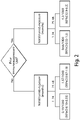
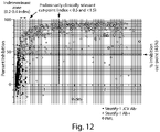
- the results of the first step of the two-step assay are typically expressed as a normalized OD (nOD, or "index") value.
- the results of the second step of the two-step assay are typically expressed as "percent inhibition.”
- the nOD is OD 450 .
- said inhibition is less than or equal to a predetermined value, e.g., 45 %, and said sample is classified as negative.
- said inhibition is greater than a predetermined value, e.g., 45 % and said sample is classified as positive.
- a cut-off calibrator (CO) is adjusted to have a reactivity index (nOD) of about 1.0, and a positive control (PC) is adjusted to have a reactivity index of about 1.3.
- the CO and PC solutions are made by mixing a serum positive for JCV antibodies with a serum that is negative for JCV antibodies.
- the negative control which can be, for example, a bottle of anti-JCV antibody-negative sera
- the index (nOD) target is about 0.1.
- a HPVLP can be chromatographically purified prior to use in an assay featured in the invention.
- the sample is a serum sample diluted 1:101 prior to forming the first reaction mixture comprising a first aliquot of the sample and the substrate on which is disposed HPVLPs.
- the secondary detection reagent e.g., an anti-human IgG
- a detectable agent such as a peroxidase, such as HRP.
- the secondary detection reagent can be anti-human IgG, wherein the anti-human IgG is conjugated to HRP.
- the detection reagent solution containing IgG-HRP is used at 0.04 ⁇ g/mL. For example, a 0.8 mg/mL stock solution of IgG-HRP is diluted 1:15,000, 1:20,000, 1:30000 or more, prior to use in the assay to detect the level of anti-JCV antibody bound to HPVLP.
- the concentration of the secondary detection reagent is adjusted for new lots to match signal to previous lot and the incubation time with the conjugate is only 30 min.
- TMB tetramethylbenzidine
- hydrogen peroxide in buffer are incubated with the reaction mix containing the HRP IgG mixture bound to anti-JCV antibody for 20 minutes, ⁇ 2 minutes.
- a decrease in the detected level in the secondary assay sample compared to the sample that was not preincubated indicates the sample is positive for anti-JCV antibody, and a change in the detected level below a specified percentage indicates that there is no JCV-specific antibody present in the sample.
- the sample can be determined to have an index value (i.e., nOD value) >0.2 and ⁇ 0.4 (the "indeterminant zone") after the first step of the assay, which is the formation of a first reaction mixture comprising a first aliquot of sample and a substrate on which is disposed HPVLP, e.g., a high signal-to-noise HPVLP substrate, and detecting the level of anti-JCV antibody bound to said substrate on which is disposed HPVLP, e.g., a high signal-to-noise HPVLP substrate.
- nOD value index value
- a second aliquot of the sample can then be tested in the second step of the assay, which comprises formation of a second mixture between the second aliquot and a solution-phase HPVLP prior to detecting unbound anti-JCV antibody in the second mixture by contacting the second mixture with a substrate on which is disposed HPVLP, e.g., a high signal-to-noise HPVLP substrate.
- a substrate on which is disposed HPVLP e.g., a high signal-to-noise HPVLP substrate.
- the sample is determined to have an index value ⁇ 0.2 after the first step of the assay, then the sample is determined to be anti-JCV antibody negative.
- a sample determined to be anti-JCV antibody negative may not be evaluated using the second step of the assay.
- sample determined to have an index value > 0.4 after the first step of the assay then the sample determined to be anti-JCV antibody positive.
- a sample determined to be anti-JCV antibody positive may not be evaluated using the second step or the assay.
- the methods disclosed herein are based at least in part on the discovery that anti-JCV antibody titer and other characteristics such as affinity/avidity can be indicators of a patient's risk of developing Progressive Multifocal Leukoencephalopathy (PML).
- PML Progressive Multifocal Leukoencephalopathy
- the invention features, a method of evaluating a patient's risk of developing PML, comprising acquiring knowledge of a JC Virus (JCV) antibody titer (e.g. , determined as described herein and expressed as normalized optical density (nOD) or index) as defined in the claims.
- JCV JC Virus
- nOD normalized optical density
- an anti-JCV antibody titer or percent inhibition is determined in a biological sample from a patient, such as a blood (serum or plasma), or CSF sample.
- the patient is determined to be at a lower risk of developing PML, and if the titer and/or percent inhibition, or a function of both values is determined to be at or above the pre-determined level the patient is determined to be at a higher risk of developing PML.
- the subject has multiple sclerosis, e.g. , a multiple sclerosis patient that is already receiving therapy with an anti-VLA-4 antibody, e.g. , natalizumab.
- multiple sclerosis e.g. , a multiple sclerosis patient that is already receiving therapy with an anti-VLA-4 antibody, e.g. , natalizumab.
- the patient can be determined to be at a higher risk of developing PML, and the patient can be identified as someone who should receive an alternative therapy, e.g., the patient should stop receiving anti-VLA-4 antibody therapy, e.g., natalizumab, and, e.g., receive an alternative therapy, e.g., an alternative approved multiple sclerosis (MS) therapy such as Avonex ® .
- the patient can be determined to be at a higher risk of developing PML, and the patient can be administered an anti-VLA-4 antibody therapy, e.g ., natalizumab.
- the patient can be determined to be at a higher risk of developing PML based upon anti-JCV antibody titer or percent inhibition, and the patient can be identified as someone who should receive additional testing to determine risk of developing PML.
- the percent inhibition of anti-JCV antibodies can be measured, for example, by: (i) contacting a biological sample from the subject with HPVLPs in a solution under conditions suitable for binding of an anti-JCV antibody in the sample to an HPVLP; (ii) separating the JCV antibodies bound to HPVLP from the solution to create a secondary sample; (iii) contacting the secondary sample with HPVLP under the same conditions as (i); and (iv) detecting the level of anti-JCV antibody binding to HPVLP in the secondary sample.
- Anti-JCV antibody titer can be measured by a commercial platform, such as a VIDAS ® assay (bioMérieux), or another alternative platform, such as a solution-phase method or a lateral flow method.
- a commercial platform such as a VIDAS ® assay (bioMérieux)
- another alternative platform such as a solution-phase method or a lateral flow method.
- the assay then further includes: (iv) contacting a portion of the biological sample from the subject with HPVLP in a solution prior to step (i) and where the HPVLP of step (i) is attached to a solid substrate, such as to provide a secondary sample; (v) contacting the secondary sample with HPVLP under the same conditions as (i); (vi) detecting the level of anti-JCV antibody binding to HPVLP in the secondary sample; and (vii) comparing the detected level of anti-JCV antibody in the secondary sample to the level of binding in the biological sample when incubated with the solution without HPVLP.
- a decrease in the detected level in the sample pre-incubated with HPVLP compared to the solution-incubated sample indicates that the sample is positive for an anti-JCV antibody, and no change in the detected level indicates that anti-JCV antibody is not present above background levels in the sample.
- the patient can be determined to be at higher risk for PML.
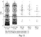
- the patient can be determined to have a lower risk of PML if the anti-JCV antibody titer as indicated by index value or nOD is determined to be ⁇ 0.5, the patient can be determined to have a higher risk if the anti-JCV antibody titer as indicated by index value or nOD is determined to be > 0.5 and ⁇ 1.5. The patient is determined to have an even higher risk if the anti-JCV antibody titer as indicated by index value or nOD is determined to be > 1.5.
- the patient can be determined to be at lower risk for PML.
- An anti-JCV antibody assay can be reevaluated for effectiveness at a predetermined interval, such as every 6 months or every year.
- a collection of samples e.g. , 30, 40 or 50 serum samples and 30, 40, or 50 plasma samples are provided such as for evaluation by the current optimized method and a preceding earlier-generation method.
- the concordance between the results is assessed and if the concordance is found to be greater than, e.g., 90% or 95%, the performance of the assay can be determined to be acceptable.
- a panel of samples e.g ., containing 90, 100, 150 or more samples, with known anti-JCV antibody status can be utilized to assess consistency of assay performance over time.
- the concordance between the results is assessed and if the concordance is found to be greater than, for example, 90% or 95%, the performance of the assay can be determined to be acceptable.
- the panel of samples is patient sera available in sufficient volume to create a sample bank.
- An entity e.g., a healthcare provider, can acquire information resulting from an anti-JCV antibody assay described herein, and responsive to the information, administer a treatment described herein to the patient, e.g., a MS patient.
- a JCV assay described herein can be performed on a patient, and then the patient can be treated, e.g., the MS patient can be treated, based on the results of the assay.
- the anti-JCV antibody titer or percent inhibition in a patient can be reevaluated at regular intervals, such as every 3 months, every 6 months, or every 12 months or at longer intervals or more frequently.
- An observed increase in antibody titer or percent inhibition can indicate an increase in the patient's risk of developing PML.
- an increase of 2 fold or 3 fold in antibody titer (nOD or index) can indicate an increased risk of PML.
- a patient receiving an anti-VLA-4 therapy such as a natalizumab, may stop therapy with the anti-VLA-4 therapy, and optionally begin therapy with an alternative agent, e.g., an immunosuppressant other than an anti-VLA-4 therapy, or other than natalizumab.
- An increase in titer may present differently in patients having a high baseline titer (e.g., at a more narrow range in range of titer) than in patients having a low baseline titer.
- a patient receiving an anti-VLA-4 antibody e.g. , natalizumab
- can be monitored e.g ., every five, six, seven, eight, nine, ten, eleven, twelve, fifteen, twenty, thirty, forty months, for anti-JCV antibody titer and/or percent inhibition.
- a patient is not re-evaluated for the presence of JCV antibodies, or for anti-JCV antibody titer or percent inhibition within one or two or three weeks after having received plasmapheresis. In another embodiment, a patient is not re-evaluated for the presence of JCV antibodies, or for anti-JCV antibody titer or percent inhibition within one or two or three weeks after having received intravenous immunoglobulin (IVIG) treatment.
- IVIG intravenous immunoglobulin
- the measure of anti-JCV antibody titer is in terms of nOD or an index value.
- Evaluation of a patient as described herein can be conducted prior to administration of an anti-VLA-4 therapy, or after the patient has begun an anti-VLA-4 therapy.
- the patient can be monitored at regular intervals, e.g. , every 3 months, every 6 months, every year, or more or less frequently, for a decrease in anti-JCV antibody titer or a decrease in percent inhibition of JCV antibodies.
- a decrease in anti-JCV antibody titer or a decrease in percent inhibition of JCV antibodies can indicate that the patient has a lowered risk of developing PML.
- the patient may not tested for JCV status again.
- the patient can stop therapy with an anti-VLA-4 therapy such as natalizumab, and not be tested again for anti-JCV antibody status.
- a method of evaluating a patient as described herein, such as to determine an anti-JCV antibody titer or percent inhibition can further include assessing other measures of risk predictors.
- a method of evaluating a patient can further include: (a) determining if the patient has received extended treatment with an anti-VLA-4 therapy (e.g., longer than 24 months); or (b) determining if the patient has received a specified non-anti-VLA-4 immunosuppressant therapy (e.g., mitoxantrone or other therapies in the last 2, 3, 5 years or ever in the patient's life).
- an anti-VLA-4 therapy e.g., longer than 24 months
- a specified non-anti-VLA-4 immunosuppressant therapy e.g., mitoxantrone or other therapies in the last 2, 3, 5 years or ever in the patient's life.
- the relative risk of PML for a patient who has an anti-JCV antibody titer or percent inhibition above a pre-determined level but has no specified prior immunosuppressant use and has not had an extended treatment with an anti-VLA-4 therapy is less than the relative risk of a patient who has an anti-JCV antibody titer or percent inhibition below a pre-determined level and has specified prior immunosuppressant use or an extended treatment with an anti-VLA-4 therapy, which is less than the relative risk of a patient who has an anti-JCV antibody titer or percent inhibition above a pre-determined level and has specified prior immunosuppressant use and extended treatment with an anti-VLA-4 therapy.
- factors to be included in the stratification model are the patient's age or gender.
- Method described herein can incorporate one or more factors into the evaluation of the patient.
- the method can include, for example, acquiring or determining a JC Virus (JCV) antibody titer and percent inhibition in a biological sample from the patient, e.g. , by a method described herein. If the antibody titer or percent inhibition is determined to be below a pre-determined level, then the patient can be classified as being suitable for treatment with a first category of therapy, such as an anti-VLA-4 therapy, e.g., natalizumab. If the antibody titer or percent inhibition is determined to be at or above the pre-determined level the patient is classified as being suitable for a second category of therapy, e.g ., interferon, glatiramer acetate or a corticosteroid.
- a second category of therapy e.g ., interferon, glatiramer acetate or a corticosteroid.
- Acquiring an anti-JCV antibody titer and percent inhibition in a sample of a patient may include removing a biological sample from the patient's body or analyzing a sample from the patient.
- the method of evaluation may also include administering a therapy, such as from the first category (e.g., natalizumab) or the second category (e.g., interferon, glatiramer acetate or a corticosteroid), to the patient.
- a therapy such as from the first category (e.g., natalizumab) or the second category (e.g., interferon, glatiramer acetate or a corticosteroid), to the patient.
- the patient is determined to have a higher risk of PML if, (i) the anti-JCV antibody titer as indicated by index value or nOD is determined to be > 1.5 and the percent inhibition value is determined to be > 70%,
- methods of evaluating a patient can incorporate more than one consideration or factor.
- methods of evaluating a patient can further include:
- a patient who has an anti-JCV antibody titer or percent inhibition above a pre-determined level but has no specified prior immunosuppressant use and has not had an extended treatment with an anti-VLA-4 therapy is classified as having less risk of developing PML than the relative risk of a patient who has an anti-JCV antibody titer or percent inhibition below a pre-determined level and has specified prior immunosuppressant use or an extended treatment with an anti-VLA-4 therapy, which is less than the relative risk of a patient who has an anti-JCV antibody titer or a percent inhibition above a pre-determined level and has specified prior immunosuppressant use and extended treatment with an anti-VLA-4 therapy.
- the patient has previously received an anti-VLA-4 therapy.
- the method includes administering an anti-VLA-4 therapy, e.g., natalizumab to the patient.
- the patient is classified as a candidate for anti-VLA-4 therapy, and the patient is further administered the anti-VLA-4 therapy.
- patients who have received an anti-VLA-4 therapy such as natalizumab for 24 months or less, who have not previously received an immunosuppressant therapy (other than anti-VLA-4 therapy), and who test negative for exposure to JCV (e.g., negative for JCV antibodies) typically have the lowest risk for developing PML.
- patients who received anti-VLA-4 therapy for longer than 24 months, who have previously received an immunosuppressant therapy (other than an anti-VLA-4 therapy), and who test positive for exposure to JCV e.g ., positive for JCV antibodies
- patients who received anti-VLA-4 therapy for longer than 24 months, who have previously received an immunosuppressant therapy (other than an anti-VLA-4 therapy), and who test positive for exposure to JCV e.g ., positive for JCV antibodies typically have the highest risk for developing PML.
- a patient's risk level for PML can be assessed by evaluating one, or any two or all three of the identified risk factors. For example, a patient, e.g., a patient with multiple sclerosis (MS) who tests negative for anti-JCV antibody titer can be determined to be at a lower risk for PML.
- MS multiple sclerosis
- a patient at a lower risk for PML can have a risk of less than about 0.2/1000 patients, e.g ., ⁇ 0.11/1000.
- a patient e.g., a patient with MS, who has received an anti-VLA-4 therapy, such as natalizumab, for 24 months or less (e.g., for 23 months, 22 months, 20 months, 15 months, 12 months, 6 months, 1 month or less), and who has not previously received an immunosuppressant therapy can be determined to be at a lower risk for PML.
- the patient can be determined to have a risk of PML of about 0.54/1000 patients.
- the patient can accordingly be determined to be a candidate to receive further treatment with an anti-VLA-4 therapy, such as natalizumab.
- a patient who has received an anti-VLA-4 therapy such as natalizumab, for longer than 24 months, such as for about 25 to 48 months or more (e.g., 26, 28, 30, 36, 40, or 48 months or more), and who has not previously received an immunosuppressant therapy can be determined to be at, or classified as having, a higher risk for PML.
- a patient at a higher risk of PML can have a risk of ⁇ about 3.7/1000 patients, e.g ., about 1.37/1000 patients.
- the patient can accordingly be determined to be a candidate to receive further treatment with an anti-VLA-4 therapy, such as natalizumab.
- a patient who has received an anti-VLA-4 therapy such as natalizumab, for 24 months or less (e.g., for 24 months, 22 months, 20 months, 15 months, 12 months, 6 months, 1 month or less), and who is determined to be negative for anti-JCV antibodies, or JCV nucleic acid, can be determined to be at, or classified as having, a lower risk for PML.
- the patient can be determined to be at a risk of ⁇ 0.2/1000 patients.
- the patient can accordingly be determined to be a candidate to receive further treatment with an anti-VLA-4 therapy, such as natalizumab.
- a patient who has not received prior treatment with an immunosuppressant (other than an anti-VLA-4 therapy), and who is determined to be negative for JCV, can be determined to be at a lower risk for PML, e.g., ⁇ 0.2/1000 patients.
- the patient can accordingly be determined to be a candidate to receive further treatment with an anti-VLA-4 therapy, such as natalizumab.
- a patient who has received an anti-VLA-4 therapy such as natalizumab, for longer than 24 months (e.g ., for 25 months, 26 months, 28 months, 30 months, 35 months, 38 months, 40 months, 48 months or longer), and who has previously received an immunosuppressant therapy other than an anti-VLA-4 therapy can be determined to be at a higher risk for PML.
- a patient at a higher risk for PML can have a risk of about 0.37/1000 or greater, e.g., about 4.3/1000 patients.
- the patient can accordingly be determined not to be a candidate to receive further treatment with an anti-VLA-4 therapy, such as natalizumab, or can be determined to be a candidate to receive treatment with an anti-VLA-4 therapy accompanied by more frequent monitoring.
- an anti-VLA-4 therapy such as natalizumab
- a patient at higher risk for PML who receives therapy with an anti-VLA-4 therapy can receive more frequent monitoring for development of PML, then a patient at lower risk of PML.
- a patient who has received an anti-VLA-4 therapy such as natalizumab, for 24 months or less (e.g., for 24 months, 22 months, 20 months, 15 months, 12 months, 6 months, 1 month or less), and who has previously received an immunosuppressant therapy other than an anti-VLA-4 therapy can be determined to be at a higher risk for PML.
- the patient can be determined to have a risk of PML of 0.66/1000 patients.
- the patient can accordingly be determined not to be a candidate to receive further treatment with an anti-VLA-4 therapy, such as natalizumab, or can be determined to be a candidate to receive treatment with an anti-VLA-4 therapy accompanied by more frequent monitoring.
- a patient at higher risk for PML who receives therapy with an anti-VLA-4 therapy can receive more frequent monitoring for development of PML, then a patient at lower risk of PML.
- a patient who has received an anti-VLA-4 therapy such as natalizumab, for longer than 24 months (e.g ., for 25 to 48 months, such as 26, 30, 36, 42 or 48 months or longer), and who is determined to be positive for JCV, can be determined to be at a higher risk for PML.
- the patient can accordingly be determined not to be a candidate to receive further treatment with an anti-VLA-4 therapy, or can be determined to be a candidate to receive treatment with an anti-VLA-4 therapy accompanied by more frequent monitoring.
- a patient who has received an anti-VLA-4 therapy such as natalizumab, for longer than 24 months (e.g ., for 25 to 48 months, such as 26, 30, 36, 42 or 48 months or longer), and who has not received prior treatment with an immunosuppressant (other than an anti-VLA-4 therapy), and who is determined to be positive for JCV, and is determined to be at a higher risk for PML.
- an anti-VLA-4 therapy such as natalizumab
- an immunosuppressant other than an anti-VLA-4 therapy
- the patient can be determined to have a risk of PML of 4/1000 patients.
- the patient can accordingly be determined not to be a candidate to receive further treatment with an anti-VLA-4 therapy, or can be determined to be a candidate to receive treatment with an anti-VLA-4 therapy accompanied by more frequent monitoring.
- a patient e.g., an MS patient, who has received prior treatment with an immunosuppressant other than an anti-VLA-4 therapy, and who is determined to be positive for anti-JCV antibodies, or JCV nucleic acid, can be determined to be at a higher risk for PML.
- the patient can accordingly be determined not to be a candidate to received further treatment with an anti-VLA-4 therapy, or can be determined to be a candidate to receive treatment with an anti-VLA-4 therapy accompanied by more frequent monitoring.
- the patient can be determined to have a risk of 9.8/1000 patients.
- the patient can accordingly be determined not to be a candidate to received further treatment with an anti-VLA-4 therapy, or can be determined to be a candidate to receive treatment with an anti-VLA-4 therapy accompanied by more frequent monitoring.
- a patient who has received an anti-VLA-4 therapy such as natalizumab, for 24 months or less (e.g., for 24 months, 22 months, 20 months, 15 months, 12 months, 6 months, 1 month or less), and who has received prior treatment with an immunosuppressant other than an anti-VLA-4 therapy, and who is determined to be positive for JCV, can be determined to be at a higher risk for PML.
- the patient can be determined to have a risk of PML of 4.5/1000 patients.
- the patient can accordingly be determined not to be a candidate to receive further treatment with an anti-VLA-4 therapy, or can be determined to be a candidate to receive treatment with an anti-VLA-4 therapy accompanied by more frequent monitoring.
- a patient who has received an anti-VLA-4 therapy such as natalizumab, for 24 months or less (e.g., for 24 months, 22 months, 20 months, 15 months, 12 months, 6 months, 1 month or less), and who has not received prior treatment with an immunosuppressant (other than an anti-VLA-4 therapy), and who is determined to be positive for JCV, can be determined to be at a higher risk for PML.
- the patient can be determined to have a risk of PML of 0.35/1000 patients.
- the patient can accordingly be determined not to be a candidate to receive further treatment with an anti-VLA-4 therapy, or can be determined to be a candidate to receive treatment with an anti-VLA-4 therapy accompanied by more frequent monitoring.
- a patient determined to have a lower risk of developing PML can be determined to have a risk of ⁇ about 0.54/1000 patients, e.g ., ⁇ 0.25/1000, ⁇ 0.2/1000, 0.19/1000, ⁇ 0.15/1000, ⁇ 0.11/1000, ⁇ 0.1/1000, e.g., 0.3/1000, 0.25/1000, 0.2/1000, 0.19/1000, 0.15/1000, 0.11/1000, or 0.1/1000 or lower.
- a patient determined to have a higher risk of PML can be determined to have a risk of about 0.54/1000 or greater, e.g., about 0.55/1000, about 0.60/1000, about 0.66/1000, about 1.2/1000, about 1.37/1000, about 2.0/1000, about 2.5/1000, about 3.0/1000, about 4.3/1000, about 5.0/1000, about 7.8/1000, about 8.0/1000, or higher.
- a patient determined to have a higher risk of PML can be determined to have a risk of 0.3/1000, 0.35/1000, 0.5/1000, 0.66/1000, 1.2/1000, 1.37/1000, 2.0/1000, 2.5/1000, 3.0/1000, 4.3/1000, 5.0/1000, 7.8/1000, 8.0/1000 or higher.
- a patient who has received prior treatment with an anti-VLA-4 therapy for longer than 24 months, and who has not received prior therapy with an immunosuppressant other than anti-VLA-4 therapy, and who is determined to be JCV negative, can be determined to be at lower risk of developing PML, and therefore a suitable candidate to receive further treatment with an anti-VLA-4 therapy, such as natalizumab.
- the risk assessment can include a recommendation to monitor the patient more frequently for the development of adverse symptoms, such as symptoms that may indicate the development of PML.
- Enhanced monitoring of patients for the development of PML can include increased frequency of tests to identify the presence of JCV, e.g ., increased testing by anti-JCV antibody assays or nucleic acid-based assays.
- Enhanced monitoring can also include MRI scans to identify brain lesions due to PML.
- a patient who has anti-JCV antibodies at less than a preselected criterion can have an undetectable level of anti-JCV antibodies.
- the patient may have previously received an anti-VLA-4 therapy, and the patient may not have previously received an anti-VLA-4 therapy.
- the patient can be classified as a candidate for anti-VLA-4 therapy, and an anti-VLA-4 therapy, e.g. , natalizumab, can be administered to the patient.
- an anti-VLA-4 therapy e.g. , natalizumab
- a patient classified as a candidate for anti-VLA-4 therapy is determined to have a lower risk for developing PML, e.g., a risk of less than about 0.2/1000 patients, e.g., 0.3/1000 patients, or 0.2/1000 patients or 0.19/1000 patients or 0.11/1000 patients.
- a patient having a lower risk of PML can have a risk of ⁇ 0.2/1000.
- a patient not classified as a candidate for anti-VLA-4 therapy, or determined to be a candidate for anti-VLA-4 therapy with enhanced monitoring for development of PML is determined to have a higher risk for developing PML, e.g., a risk of greater than or equal to about 0.3.7/1000 patients.
- a patient determined to have a higher risk of PML can have a risk of 0.37/1000, 0.35/1000, 0.66/1000, 1.2/1000, 1.37/1000, 2.5/1000, 4.3/1000, or 7.8/1000 patients.
- a prior immunosuppressant exposure classification is one of the following:
- a prior VLA-4 therapy exposure classification is one of the following:
- a treatment suitability classification can be selected from one of:
- a positive treatment suitability classification correlates with a lower risk of developing PML
- a negative treatment suitability classification correlates with a higher risk of developing PML.
- a lower risk of developing PML typically corresponds to a risk less than 0.2/1000 patients
- a higher risk of developing PML corresponds to a risk of ⁇ 0.37/1000.
- the patient is assigned a low exposure classification, and a negative JCV status classification
- the patient is assigned a positive treatment suitability classification, e.g., a modified positive treatment suitability classification that advises or requires monitoring for development of PML.
- the patient can be assigned a positive treatment suitability classification, e.g., a modified positive treatment suitability classification that advises or requires monitoring for development of PML.
- a positive treatment suitability classification e.g., a modified positive treatment suitability classification that advises or requires monitoring for development of PML.
- the patient is assigned a low exposure classification, a negative prior immunosuppressant exposure classification, and a negative JCV antibody classification, the patient is assigned a positive treatment suitability classification.
- the patient can be further administered an anti-VLA-4 therapy, e.g., natalizumab.
- an anti-VLA-4 therapy e.g., natalizumab.
- the assay for the presence of anti-JCV antibodies is an ELISA assay.
- An assay for JCV nucleic acid can be, e.g., a PCR assay or a Next Generation Sequencing (NGS) method.
- a patient determined to be at lower risk for PML can further be administered an anti-VLA-4 therapy, such as natalizumab.
- a patient determined to be at higher risk for PML can further be administered an alternative to an anti-VLA-4 therapy, such as an interferon, glatiramer acetate, a corticosteroid or a TNF agonist.
- a patient determined to be at higher risk for PML can be further administered an anti-VLA-4 therapy, and can be required to receive an increased frequency of testing for PML, and where the patient is initially determined to be JCV negative, can also be required to received an increased frequency of testing for JCV.
- Treatment can include e.g ., determining the patient's prior exposure to an anti-VLA-4 therapy, and determining whether the patient previously received treatment with an immunosuppressant.
- the patient's status for JCV can also be determined.
- the patient is determined to have received the anti-VLA-4 therapy for 24 months or less, and not to have previously received treatment with an immunosuppressant, then the patient is determined to be at lower risk for PML, and the patient is administered the anti-VLA-4 therapy. If the patient is determined to have received natalizumab for longer than 24 months ( e.g ., 25 months or longer), and not to have previously received treatment with an immunosuppressant, then the patient is determined to be at higher risk for PML, and the patient is administered an alternative to anti-VLA-4 therapy, e.g., an interferon, a corticosteroid, a statin or a TNF antagonist.
- an alternative to anti-VLA-4 therapy e.g., an interferon, a corticosteroid, a statin or a TNF antagonist.
- Determining the patient's prior exposure to an anti-VLA-4 therapy or an immunosuppressant can include asking the patient or a caregiver, e.g., a physician, nurse, parent or other caregiver. In some cases, determining the patient's prior exposure can include accessing the information in a database, e.g., a database of medical records.
- a method of determining a patient's risk for PML includes determining the patient's previous exposure to an anti-VLA-4 therapy, and determining whether the patient previously received treatment with an immunosuppressant.
- the patient's anti-JCV antibody status may also be determined. If the patient is determined to have received an anti-VLA-4 therapy for 24 months or less, and not to have previously received treatment with an immunosuppressant, then the patient is determined to be at lower risk for PML. If the patient is determined to have received anti-VLA-4 therapy for longer than 24 months, and not to have previously received treatment with an immunosuppressant, then the patient is determined to be at higher risk for PML.
- a patient determined to be at lower risk for PML may further be administered an anti-VLA-4 therapy, e.g., natalizumab.
- an anti-VLA-4 therapy e.g., natalizumab.
- a patient determined to be at higher risk for PML may further be administered an alternative to anti-VLA-4 therapy, e.g., an interferon, a corticosteroid, a statin or a TNF antagonist.
- the patient's JCV status can be also determined, and if the patient is determined to be JCV negative, then the patient is determined to be at a lower risk for PML than if the patient was determined to be JCV positive.
- the invention is based, at least in part, on the discovery of new and improved methods of assessing the risk of a patient for PML that include assessing anti-JCV antibody titers or percent inhibition.
- the invention is based at least in part on the discovery that anti-JCV antibody titer and percent antibody inhibition can be an indicator of a patient's risk of developing Progressive Multifocal Leukoencephalopathy (PML).
- PML Progressive Multifocal Leukoencephalopathy
- Applicants have also developed an optimized assay for determining anti-JCV antibody titer levels in a biological sample, and a method for assaying the antibodies qualitatively by determining percent inhibition values, and using this information to determine the risk of a patient for developing PML.
- the assay includes: (a) forming a first reaction mixture comprising a first aliquot of a sample and a substrate on which is disposed HPVLPs, where, the VLP particles are present at an amount of 0.04 ⁇ g, and a concentration of 0.4 ⁇ g/mL; b) detecting the level of anti-JCV antibody bound to HPVLP disposed on the substrate, such as by detecting a labeled secondary detection reagent, e.g., an enzyme labeled anti-IgG antibody, bound to anti-JCV antibody bound to said substrate; (c) forming a second reaction mixture comprising a second aliquot of sample with solution-phase HPVLP provided at a concentration of, e.g., 0.4 ⁇ g/mL, and a second aliquot of sample provided at, e.g., a 1:100 or 1:101, dilution; (d) forming a third reaction mixture comprising a negative control solution containing no HPVLP, and a third aliquot of
- a patient has a lower risk of developing PML if, (i) the anti-JCV antibody titer as indicated by index value or nOD is determined to be ⁇ 0.5, or (ii) the anti-JCV antibody titer as indicated by index value or nOD is determined to be > 0.5 and ⁇ 3.0, and the percent inhibition is determined to be less than or equal to 70%.
- the patient has a higher risk of PML if, (i) the anti-JCV antibody titer as indicated by index value or nOD is determined to be > 3 and the percent inhibition value is determined to be > 70%, or (ii) the patient showed an increase in index, nOD or titer by 2-fold from a previous test.
- a patient can be monitored at regular intervals, such as every 6 months or every 12 months for a change in anti-JCV antibody titer or percent inhibition. If the results of this later assay indicate that the patient still has an anti-JCV antibody titer of nOD less than 0.5, and a percent inhibition of ⁇ 70%, then the patient can be determined to still be at a lower risk for developing PML. If a later assay indicates that the patient's antibody titer is increased by 2 to 3 fold from the initial assay, then the patient can be determined to be at increased or higher risk for developing PML. Applicants observed patients diagnosed with PML tend to demonstrate an increase in antibody titer and nOD by 2 to 3 fold in the six months prior to diagnosis.
- a patient has a higher risk of PML if, (i) the anti-JCV antibody titer as indicated by index value or nOD is determined to be > 3 and the percent inhibition value is determined to be > 70%, or (ii) the patient showed an increase in index, nOD or titer by 2-fold from a previous test.
- a patient satisfying these criteria can, optionally, be determined not to be a candidate to receive therapy with an anti-VLA-4 therapy, such as an anti-VLA-4 antibody, e.g., natalizumab, or the patient can further be assessed for other risk factors of developing PML.
- These risk factors include whether or not the patient has previously received an anti-VLA-4 therapy, such as natalizumab, and for how long the patient has received the therapy; and whether and for how long the patient has previously received an immunosuppressant therapy other than an anti-VLA-4 therapy.
- a patient's risk of PML is a combination of each of these factors.
- Antibody titer is herein measured by "nOD” or "index.”
- nOD is the normalized optical density value in a test, such as an ELISA test, for anti-JCV antibody detection.
- patients who received an anti-VLA-4 therapy such as natalizumab, for 24 months or less, and who have not previously received an immunosuppressant therapy, are at lower risk for developing PML, than patients who do not meet these two criteria.
- patients who have the lowest risk are those who meet these two criteria, and who are also JCV negative, e.g., patients who do not test positive for anti-JCV antibodies or JCV nucleic acid, e.g., JCV DNA.
- each of these three risk factors ((i) the amount of time the patient has previously received an anti-VLA-4 therapy; (ii) whether or not a patient has previously received treatment with an immunosuppressant other than an anti-VLA-4 therapy; and (iii) JCV status) independently contribute to a patient's risk of PML.
- the inventions described herein can be used in general for patients treated with a VLA-4 inhibitor.
- the ability to identify subpopulations of patients at distinctly different PML risks allows for better characterization of risk than previous methods ( i.e., overall PML risk) and should assist healthcare professionals and patients in making more informed benefit-risk treatment decisions.
- the risk criteria described herein directed to anti-JCV antibody titer (e.g., as measured by nOD or index level) and percent inhibition can be considered in combination with the risk factors described in the prior co-owned provisional applications.
- the methods for determining PML risk can require acquiring one, two or all three of a JCV classification for a patient (e.g., anti-JCV antibody titer, such as measured by nOD or index level and percent inhibition), prior anti-VLA-4 therapy history for the patient, and prior immunosuppressant therapy history (other than anti-VLA-4 therapy) for the patient. Responsive to these classifications, a patient can be assigned a treatment suitability classification. Patients who are determined to have low risk of developing PML can be assigned a positive treatment classification, and patients who are determined to have a higher relative risk of developing PML can be assigned a negative treatment classification.
- a JCV classification for a patient e.g., anti-JCV antibody titer, such as measured by nOD or index level and percent inhibition
- prior anti-VLA-4 therapy history for the patient. Responsive to these classifications, a patient can be assigned a treatment suitability classification. Patients who are determined to have low risk of developing PML can be assigned a
- a patient who receives a positive treatment classification can receive a recommendation for further treatment or for initiating treatment with an anti-VLA-4 therapy.
- a patient receiving a negative treatment classification may receive a recommendation to terminate treatment with an anti-VLA-4, a recommendation to initiate treatment with a non-anti-VLA-4 therapy, a recommendation for continuing or initiating anti-VLA4 therapy with increased surveillance for signs and symptoms of PML.
- a recommendation for further treatment with an anti-VLA-4 therapy may be accompanied with further instructions or requirements that the patient receive additional or enhanced monitoring, such as if one or more factors indicate that the patient may be at an increased risk of PML, e.g., prior treatment with an anti-VLA-4 therapy for longer than 24 months, e.g., 25 months or longer, or prior treatment with an immunosuppressant other than an anti-VLA-4 therapy.
- a patient can be determined to have previously received an anti-VLA-4 therapy or an immunosuppressant therapy other than an anti-VLA-4 therapy through self-reporting by the patient, or through information (verbal or written) provided by a parent, physician, physician's assistant, nurse or other healthcare provider.
- the information can also be obtained through a database, such as a medical database or a clinical trials database.
- Prior immunosuppressant therapies other than anti-VLA-4 therapy, that will be indicative of an increased risk of PML can include prior treatment with antineoplastics, immunosuppressants or immunomodulators, such as one or more beta-interferon or glatiramer acetate.
- immunosuppressants include, e.g., mitoxantrone, methotrexate, azathioprine, cyclophosphamide, and mycophenolate, anti-CD20 therapy (e.g., rituximab), an anti-CD11a therapy (e.g., efalizumab), or mycophenolate mofetil.
- prior treatment with other immunosuppressant therapies as described below will also be predicted to increase a patient's risk of PML following further administration of an anti-VLA-4 therapy.
- a determination of prior immunosuppressant use is a specified use which can be any prior use of an immunosuppressant that is not a VLA-4 inhibitor (e.g., an anti-VLA-4 antibody) or prior use within a specified period of time, for example, within the previous 1, 2, 3, 5, or 10 years prior to the evaluation of PML risk.
- JCV antibodies are detected by an ELISA assay.
- JCV antibodies can be detected by the method described in International Application Number PCT/US2011/20832 , which utilizes HPVLPs under conditions suitable for binding of an anti-JCV antibody for detecting the level of anti-JCV antibody binding in a biological sample.
- Methods of determining JCV status also include methods of determining anti-JCV antibody titer and percent inhibition. Detection of anti-JCV antibody titer and percent inhibition typically include a two-step antibody detection assay as described in International Application Number PCT/US2011/20832 .
- JCV positive a biological sample from a patient
- JCV antibodies proteins, peptides, or nucleic acids
- a positive JCV classification corresponds to the presence of JCV antibodies in the biological sample, e.g., JCV antibodies that are equal to or greater than a preselected criterion.
- the preselected criterion is typically a qualitative value, e.g., a "detectable" amount of antibody according to a particular assay, e.g., an immunoassay.
- the methods described herein for determining PML risk can be useful for any human subject, including a subject considering treatment with an immunomodulator, for example an anti-VLA-4 therapy (e.g., natalizumab), an anti-CD20 therapy (e.g., rituximab), an anti-CD11a therapy (e.g., efalizumab), or mycophenolate mofetil; in a subject currently being treated with an immunomodulator; or a subject that has ceased treatment with an immunomodulator.
- an immunomodulator for example an anti-VLA-4 therapy (e.g., natalizumab), an anti-CD20 therapy (e.g., rituximab), an anti-CD11a therapy (e.g., efalizumab), or mycophenolate mofetil
- an immunomodulator for example an anti-VLA-4 therapy (e.g., natalizumab), an anti-CD20 therapy (e.g., rituxima
- the method may be useful to others who may be susceptible to PML such as individuals having lymphoproliferative disorders, such as multiple myeloma or a lymphoma; individuals infected with human immunodeficiency virus (HIV), or having acquired immune deficiency syndrome (AIDS), hematologic malignancies, or an autoimmune disease such as systemic lupus erythematosus (SLE), an inflammatory bowel disease, such as Crohn's Disease (CD) or ulcerative colitis, multiple sclerosis (MS) or arthritis, e.g., rheumatoid arthritis (RA).
- the risk-assessment method may also be useful to subjects receiving immunosuppressive or immunomodulatory therapies, such as transplant patients.
- Exemplary immunosuppressive or immunomodulatory therapies include natalizumab, rituximab, efalizumab, and mycophenolate mofetil.
- the method can be useful for assessing risk in a subject having a disorder, or being treated with a drug, disclosed in Piccinni et al. "Stronger association of drug-induced progressive multifocal leukoencephalopathy (PML) with biological immunomodulating agents" Eur. J. Clin. Pharmacol. 66:199-206, 2010 .
- PML drug-induced progressive multifocal leukoencephalopathy
- an “HPVLP” is a highly purified VLP ("virus-like particle") consisting predominantly of the VP1 protein.
- An “HPVLP” featured in the invention is composed mainly of the major capsid protein "VP1,” which can be a naturally-occurring VP1 or a recombinant VP1, from the polyomavirus, JCV).
- VP1, major capsid protein
- JCV JC Virus
- An HPVLP can be composed of, e.g., at least one pentameric subunit, more than one pentameric subunit, up to seventy-two pentameric subunits or more of VP1.
- An HPVLP of the invention can bind antibodies against naturally-occurring, intact JC virus.
- an HPVLP includes a second, and optionally a third, polypeptide that is a minor capsid protein of JC virus, e.g., at least one VP2 or VP3 polypeptide.
- the VP2 or VP3 can be recombinant or naturally-occurring or naturally-derived polypeptides.
- Such "highly purified” particles contain more than one VP1 pentamer, e.g., at least 5, 10, 20, 30, 40, 50, 60, 70, 72 VP1 pentamers, or less than 100 VP1 pentamers.
- Such highly purified particles can be obtained, for example, by a method that involves double filtration.
- a highly purified preparation of VLPs is obtained by purifying the particles at least twice by centrifugation, e.g., through a sucrose cushion.
- HPVLPs are prepared using chromatographic methods.
- an HPVLP preparation can be identified by its activity in an ELISA assay using defined control samples. In some cases, such control samples are negative controls and/or control samples containing low levels of JCV antibodies.
- a "high signal-to-noise HPVLP substrate” is a substrate on which is disposed HPVLP. It can be used to evaluate the level of free (that is unbound to antigen or other target, e.g., HPVLP, in a sample.
- the concentration of HPVLP on the substrate is such that, when measuring the amount of anti-JCV antibody present, it provides for a signal-to-noise ratio of 10 to 30, 15 to 30, 15 to 25, 18 to 22.
- the signal-to-noise ratio is at least 10, 15, 18 or 20.
- signal-to-noise ratio is about 10, 15, 18 or 20.
- the signal-to noise ratio can be determined with a sample, e.g., a calibration control, that gives an optical density of 1.0.
- the HPVLP is provided on said substrate at a concentration which results from lyophilizing 0.5 ml, 0.8 ml, 1.0 ml, 1.2 ml, or 1.5 ml of 0.4 ⁇ g/ml of HPVLP in a well of a 96 well plate.
- the HPVLP is provided on said substrate at a concentration which results from lyophilizing 1.0 ml of 0.4 ⁇ g/ml of HPVLP in a well of a 96 well plate, which as used herein, is equivalent to 30 ng to 50 ng (e.g., 40 ng) HPVLP per well.
- the HPVLP is provided on said substrate at a concentration which results from lyophilizing 0.05 ml to 0.35 mL or 0.1 ml to 0.2 ml of 0.4 ⁇ g/ml of HPVLP in a well of a 96 well plate.
- the amount of HPVLP disposed on the substrate, or the conditions under which deposition is achieved, can vary as long as the desired signal-to-noise ratio is obtained.
- a signal-to-noise ratio is computed by comparing the optical density value of the negative control to the calibrator control to determine the dynamic range of the signal intensity in the assay.
- the sample is diluted about 100 fold and the cut off for negative score is a reduction that is less than or equal to 45 % and, the cutoff for a positive score is greater than 45 %.
- the dilution is other than 100 fold but is less than 200 fold.
- the dilution is between 50- and 150-fold, 75- and 125-fold, 85- and 115-fold.
- the dilution is less than 150-fold, 125-fold, 100-fold, or 75-fold.
- the cutoff, or other parameters are adjusted such that a sample would receive the same score (positive or negative) as it would if the dilution was 100-fold and the cut off for negative is less than 45% and the cut off for positive is greater than or equal to 45%.
- Anti-JCV Antibody Detection Assay are conducted by adding a biological sample to a substrate that has been coated with an HPVLP and detected using methods known in the art.
- a solid base platform is used such as a microtiter plate (for example, a 96 well plate); although other formats known in the art can be used.
- the biological sample is diluted prior to use in an assay.
- the assay format is an enzyme-linked immunoassay (ELISA).
- the method typically includes coating the substrate with capture antigen such as HPVLP, incubating sample containing binding antibodies directed to capture reagent, washing to remove non-specifically bound species, and detecting the bound immune complexes, e.g., by a chromogenic or chemiluminescent assay.
- Chromogenic substrates produce a colored end product, which can be detected and measured visually or with the use of a spectrophotometer.
- Chemiluminescent substrates produce light, which can be measured using a luminometer.
- Coating a plate with HPVLP generally includes incubating the solid substrate (such as wells of a microtiter plate) with a solution of HPVLP at a suitable concentration (e.g., 0.4 ⁇ g/ml), either overnight or for a specified number of hours.
- the HPVLP can include VP1 as the only JCV viral component, or the HPVLP can be a heterologous particle, that contains at least one of VP2 or VP3 per particle or at least one each of VP2 and VP3 per particle.
- the wells of the plate are washed.
- the substrate is then "coated" with a nonspecific protein that is antigenically neutral with regard to the samples to be tested.
- Suitable coating materials are known in the art and include bovine serum albumin (BSA), casein, sugars or solutions of milk powder. Plates may then be dried and stored for a longer period of time, such as 1 days, 1 month or 1 year prior to proceeding to the next step of the assay.
- BSA bovine serum albumin
- the sample or reference is incubated on the prepared substrate under conditions effective to permit complex formation (HPVLP/JCV antibody), thus forming a bound complex.
- Detection of the bound complex is performed using a labeled antibody that can bind to human antibody.
- the labeled antibody can detect human IgG or human IgG and IgM.
- the assay can be performed using secondary or tertiary detection methods.
- a reference sample can be of the same biological material (e.g ., plasma, serum, urine, or CSF) isolated from an individual known to be infected with JC virus based on the presence of JCV DNA in urine of the individual (uropositive).
- a reference sample is used to establish the assay cut-point such that the false negative rate of the assay is not greater than 1%-3%.
- Under conditions effective to permit complex formation generally means conditions in which the reagents have been diluted to reduce background and provide readouts of results that lie within a specified range.
- Diluents can include, in non-limiting examples, solutions that include BSA, phosphate buffered saline (PBS), or PBS containing Tween.
- Suitable conditions also include conditions that are at a temperature and/or for a period of time sufficient to allow effective binding. Incubations are typically from about one to two hours or one to four hours, at temperatures of approximately 25°C to 27°C, or may be overnight at about 4°C. However, those in the art will understand that other conditions may be suitable.
- wash solutions include diluent buffer (e.g., PBS or PBS/Tween) or borate buffer.
- the detection of antibody bound to HPVLP is performed using methods well known in the art. In general, such methods are based on the detection of a label or marker, such as a radioactive, fluorescent, biological or enzymatic tag.
- a label or marker such as a radioactive, fluorescent, biological or enzymatic tag.
- U.S. patents concerning the use of such labels include, for example, U.S. Pat. Nos. 3,817,837 ; 3,850,752 ; 3,939,350 ; 3,996,345 ; 4,277,437 ; 4,275,149 and 4,366,241 .
- the detection of anti-JCV antibody binding is detected using a secondary antibody that is labeled. In general, the secondary antibody is specific for detecting human IgG. Quantification is achieved by measuring the degree of color generated, e.g., using a visible spectra spectrophotometer.
- the assay is performed in a medical office, such as by a healthcare provider, e.g., a doctor, a nurse or a technician, working in a facility where the biological sample is obtained from a patient.
- a healthcare provider e.g., a doctor, a nurse or a technician
- the biological sample obtained from a patient is transported to another facility, e.g., to a third party facility, where the assay is performed.
- the results of the assay can be reported back to the healthcare provider, such as through a form, which can be submitted by mail or electronically ( e.g., through facsimile or e-mail) or through an on-line database.
- the results of the assay (including the screening assay and, optionally, a confirmatory assay) can be stored in a database and can be accessed by a healthcare provider, such as through the worldwide web.
- a secondary test also referred to herein as a "confirmatory assay" of the sample is employed.
- a secondary test also referred to herein as a "confirmatory assay" of the sample is employed.
- two aliquots of a biological sample are used. The first is prepared prior to use in the assay by preincubating the sample in the presence of assay buffer in solution for a period of time (e.g., for 30 minutes, one hour, or longer such as overnight at 4°C).
- the second aliquot is prepared prior to use in the assay by preincubating the sample in the presence of HPVLP in solution for a period of time (e.g., for 30 minutes, or one hour or longer). The two aliquots are then used in the HPVLP assay as described herein, and the assignment of the sample to anti-JCV antibody positive or antibody negative is made. If the assay results for the aliquot incubated with HPVLP indicate a value of ⁇ 45% inhibition (i.e., the "cut-point"), then the sample is interpreted to be negative for the presence of JCV-specific antibodies. If the assay results indicate a value of ⁇ 45% inhibition, then the sample is interpreted to have JCV-specific antibodies and therefore as antibody positive.
- An assay featured in the invention that utilizes a secondary test is also referred to herein as a "two-step test” or a "two-step assay.”
- An earlier version of the two step assay is described in co-owned International Application No. PCT/US2011/020832 .
- any suitable solid substrate can be used for the HPVLP assay format.
- the substrate is a microtiter plate (e.g., a 96-well plate) a slide, a bead, or a column.
- the substrate can be suitable for chromogenic or chemiluminescent detection methods, or solution based methods such as proximal ligation.
- Cut-point The invention provides methods of analysis that employ "cut-points" to reduce false negative and false positive rates.
- the cut-points are established based on data from the HPVLP assays (e.g., to detect JCV antibodies in a biological sample), averaged, for example, between duplicate test samples and multiple replicates (for example, at least two, at least four, or at least eight replicates of control samples). Cut-points can also be determined statistically using large panels of non-PML and PML samples.
- results from initial HPVLP screening assays e.g., ELISA assays
- results from initial HPVLP screening assays will cause a test sample to be classified as having or not having JCV-specific antibodies, or, if the sample does not fall under one of these two classifications, then the sample will be subjected to a supplemental confirmation assay.
- samples that produce a result in an HPVLP ELISA assay featured in the invention less than an established level e.g., an nOD 450 ⁇ 0.2
- samples that provide a result in the ELISA greater than an established level e.g., an nOD 450 > 0.4
- samples that do not clearly fall into one of these classifications e.g., 0.2 ⁇ OD 450 ⁇ 0.4
- the confirmatory assay requires a pre-incubation step, where the test sample is pre-incubated with buffer (or other suitable solution) control or with HPVLPs (in buffer or other suitable solution) to pre-adsorb JCV-specific antibodies prior to analysis in an HPVLP ELISA, as described in further detail below.
- buffer or other suitable solution
- HPVLPs in buffer or other suitable solution
- the sample is interpreted to be negative for the presence of JCV-specific antibodies. If the results show a ⁇ 45% reduction in reaction compared to buffer control in the primary assay after pre-incubation with HPVLP then the sample is interpreted to contain JCV specific antibodies.
- only the confirmatory assay is performed.
- VP1 for use in producing HPVLPs can be generated using methods known in the art and can be either naturally-occurring VP1 or recombinantly produced VP1, e.g., a VP1 from a JC virus.
- the VP1 used is VP1 from a MAD1 strain of JCV.
- the VP1 used in the assay comprises VP1 from more than one JCV strain, for example, from one or more of strains 1A, 1B, 2A, 2B, 3, 4, and 7.
- the VP1 for use in the assays described herein is then further purified through standard biochemical methods including density-gradient/ultracentrifugation methods, or a series of chemical precipitation steps, concentration/diafiltration and ion-exchange chromatography.
- the purification methods typically include a step to remove smaller proteins including monomer VP1 polypeptides, or pentamer VP1. The removal of these smaller particles can be done in, for example, in one step or in two steps (e.g., a first filtration step to remove VP1 monomers, and then a second filtration step to remove pentamer VP1 particles).
- biochemical purification methods are known to those in the art. Examples 1 and 7 provide two different methods of JCV VP1-VLP purification.
- HPVLP preparation does not contain significant amounts of VP1 monomer (e.g ., has been purified to remove monomers).
- An HPVLP preparation according to another aspect of the present invention does not contain significant amounts of VP1 molecules in configurations the size of a VP1 pentamer, or smaller (including monomer).
- the HPVLP can be prepared from recombinant VP1 or naturally-occurring VP1 ( e.g., isolated from virus or virus capsid).
- additional JCV components such as one or both of the minor coat proteins from JC virus, e.g., VP2 or VP3, are included in the HPVLP particle or are associated with the substrate.
- recombinantly expressed VP1 may not assemble into pentamers or HPVLPs that resemble naturally-occurring viral capsids, for example, recombinantly expressed VP1 may assemble into tubes or other non-spherical geometries. Accordingly, the invention relates to methods of producing HPVLPs that are substantially spherical in geometry.
- the invention includes HPVLP preparations where at least about 10%, about 15%, about 20%, about 25%, about 50%, about 60%, about 65%, about 70%, about 80%, about 90%, about 95%, or about 99% of the HPVLPs in the preparation resemble the naturally-occurring JCV capsid (e.g., are in an icosahedral or substantially spherical configuration).
- an HPVLP preparation contains at least 10%, at least 15%, at least 20%, at least 50%, at least 60%, at least 70%, at least 80%, at least 90%, at least 95%, or at least 99% of the HPVLPs in the preparation resemble the naturally-occurring JCV capsid.
- Such methods can include expressing viral proteins under conditions that result in such a preparation and/or isolating and purifying expressed viral proteins as described herein to produce such a preparation.
- HPVLPs can be made, for example, by transforming a baculovirus with a vector expressing a VP1 gene, such as a VP1 gene from a JC virus.
- the baculovirus is used to infect a cell culture, such as an insect cell culture (e.g., SF9 cells) or a mammalian cell culture, and the cells express the VP1 protein.
- HPVLPs are isolated by lysing the cells, and purifying the particles through a series of centrifugation and ultrafiltration steps. In general, the purification is performed using methods such as sucrose cushion sedimentation, isopycnic ultracentrifugation and extensive ultrafiltration or other methods known to those in the art.
- the purification will include twice centrifuging the particles through a sucrose cushion.
- cells are lysed, and particles are isolated by a series of precipitation and concentration/diafiltration steps with a final ion-exchange step.
- the HPVLPs are purified by chromatographic methods, and without centrifugation steps.
- Purity can be assessed using any suitable techniques known in the art, for example, analytical ultracentrifugation, electron microscopy, PAGE analysis, mass spectrometry, protein concentration, or activity in an ELISA with control sera. Insufficiently purified VLPs result in a high background yielding falsely high anti-JCV antibody levels or calculated exposure rates.
- the HPVLPs contain VP1 as the sole JC virus protein.
- the HPVLPs are heterogeneous particles, and therefore include VP1 protein, and at least one of the minor coat proteins of JC virus, e.g., VP2 or VP3.
- the HPVLP includes VP1, VP2 and VP3 proteins.
- An HPVLP that includes VP1 and VP2 can be produced using methods known in the art, for example, by transforming a baculovirus with a nucleic acid including a VP1 and a VP2 gene, such as under the control of the same or different promoters. A cell culture is infected with the baculovirus, and the cells express VP1 and VP2, and HPVLPs form which include both types of proteins.
- the VP1 and VP2 genes are on different DNA molecules, the DNA molecules are transformed into different baculoviruses and the baculoviruses are used to transfect cells in the same culture.
- the cells express the VP1 and VP2 proteins, and HPVLPs form which include both types of protein.
- a heterogeneous HPVLP will include, e.g, one or two VP2 polypeptides for every five VP1 polypeptides.
- an HPVLP will contain more VP1 polypeptides than VP2 polypeptides, as is the case in naturally-occurring JC virus.
- An HPVLP that includes both VP1 and VP3 or both VP1 and VP2 molecules can be produced, for example, by transforming a baculovirus with a nucleic acid including a VP1 and a VP3 gene or a VP1 and VP2 gene, respectively, under the control of the same or different promoters.
- a cell culture is infected with the baculovirus, and the cells express VP1 and VP3 or VP1 and VP2, and HPVLPs form which include both types of proteins.
- the VP1 and VP3 or VP1 and VP2 genes are on different DNA molecules, the DNA molecules are transformed into different baculoviruses, and the baculoviruses are used to transfect cells in the same culture.
- the cells express the VP1 and VP3 proteins or VP1 and VP2 genes, respectively, and HPVLPs form which include both types of protein.
- HPVLP particles can be isolated from such preparations using methods known in the art such as those used to isolate JCV capsids.
- a VP1 pentamer that is in a heterogeneous HPVLP will include, e.g, five VP1 polypeptides and one VP3 polypeptide and/or one VP2 polypeptide, depending on whether a VP3 gene or VP2 gene was used to make the constructs. There will typically be more VP1 polypeptides than VP3 or VP2 polypeptides in an HPVLP.
- the VP2 or VP3 is from a polyoma virus that is not a JC virus, e.g., a BK virus polypeptide.
- An HPVLP that includes all three of VP1 and VP2 and VP3 molecules can be produced by transforming a baculovirus with a nucleic acid (e.g., a circular DNA, e.g., ⁇ 5.5 kb) including a VP1, VP2 and VP3 gene, such as under the control of the same or different promoters.
- a cell culture such as a mammalian cell culture, is infected with the baculovirus, and the cells express VP1, VP2 and VP3 proteins.
- HPVLPs consequently form which include all three types of proteins.
- the VP1, and either or both of the VP2 and VP3 genes are on different DNA molecules, the DNA molecules are transformed into the same or different baculovirus, and the baculovirus are used to infect cells in the same or separate cultures.
- the cells express the VP1, VP2 and VP3 proteins, and HPVLPs form which include both types of protein.
- a heterogeneous HPVLP can include, e.g, five VP1 polypeptides and one each of VP2 and VP3 polypeptides, although the ratios may vary within a preparation. There will typically be more VP1 polypeptides than VP2 and VP3 polypeptides in an HPVLP.
- the HPVLP will be greater in size than a VP1 pentamer.
- greater in size it is meant that the mass of protein contained in an HPVLP particle is greater than a pentamer containing solely VP1.
- the method of preparing a solution of HPVLP can include removing from the solution particles (e.g., VP1 monomers or small VP1 containing particles) that are the size of a VP1 pentamer or smaller. Methods such as centrifugation and size-exclusion chromatography can be used to perform this purification step. In some embodiments, other methods known in the art, e.g., ion exchange chromatography, can be used in the preparation of HPVLPs that are larger than a VP1 pentamer.
- solution particles e.g., VP1 monomers or small VP1 containing particles
- Methods such as centrifugation and size-exclusion chromatography can be used to perform this purification step.
- other methods known in the art e.g., ion exchange chromatography, can be used in the preparation of HPVLPs that are larger than a VP1 pentamer.
- an HPVLP preparation suitable for use in an assay will contain at least 20% HPVLPs, at least 25% HPVLPs, at least 40% HPVLPs, at least 60% HPVLPs, at least 65% HPVLPs, at least 70% HPVLPs, at least 80% HPVLPs, at least 85% HPVLPs, at least 90% HPVLPs, at least 95% HPVLPs, or at least 99% HPVPLs compared to non-HLVLP particles ( e.g., by percent of pentamers compared to VP1 monomers and aggregates containing fewer than five VP1 molecules).
- methods of evaluating or analyzing a subject or biological sample from a subject include one or more of performing the analysis of the sample, requesting analysis of the sample, requesting results from analysis of the sample, or receiving the results from analysis of the sample.
- determination (or determining), analysis or evaluation (or evaluating) can include one or both of performing the underlying method or receiving data from another who has performed the underlying method.
- the analysis or evaluation requires a transformation of material, e.g., biological material or assay components.
- a biological sample can be evaluated for the presence of anti-JCV antibodies, anti-JCV antibody titer and percent inhibition of JCV antibodies.
- the evaluation can be performed before or after or at the same time the patient is receiving treatment, such as for MS.
- the evaluation is based, at least in part, on analysis of a sample from the subject.
- the presence of anti-JCV antibodies can be determined by contact with a specific binding agent, e.g., a JCV protein, such as VP1.
- the binding agent can be a JCV protein, e.g., VP1 in the form of a particle, e.g., a HPVLP.
- an assay to detect the presence of anti-JCV antibodies is a two-step assay, such as described herein.
- the assay utilizes HPVLPs under conditions suitable for binding an anti-JCV antibody.
- the assay is capable of detecting any isotype of anti-JCV antibody (including IgG, IgM, IgA, and IgE).
- the assay is also highly sensitive and can detect anti-JCV antibodies at a concentration of, for example, 2.0 ⁇ g/mL or less, e.g.
- the sample can be analyzed for the level of JCV nucleic acid present in the sample.
- nucleic acids can be isolated from the sample and used for PCR amplification or a Next-Generation (Nex-Gen) Sequencing technique.
- a crude lysate of the biological sample is subject to an amplification method, such as PCR, and the amplified product is analyzed by one or more of electrophoresis, restriction fragment mapping, hybridization or sequencing to identify whether JCV DNA or RNA is present in the sample and how much is in the sample.
- an amplification method such as PCR
- the amplified product is analyzed by one or more of electrophoresis, restriction fragment mapping, hybridization or sequencing to identify whether JCV DNA or RNA is present in the sample and how much is in the sample.
- the biological sample can be removed from the patient and analyzed.
- the patient sample can be stored prior to testing for JCV antibodies
- the patient sample e.g., the patient sample containing JCV antibodies can be stored for 1-21 days, e.g., 1-14 days or 1-7 days or longer (e.g., one day, two days, three days, five days, seven days, ten days, 14 days, 21 days or longer); for one to six weeks, e.g., one to three weeks or one to two weeks or longer (e.g., up to one week, up to two weeks, up to three weeks, up to six weeks, or longer); or for one to six months, e.g., one to three months or one to two months or longer (e.g., up to one month, up to two months, up to three months, up to six months or longer).
- the sample can be stored, for example, frozen (e.g., at -80°C to -20°C), at 2-8°C, at ambient temperature (18°C-25°C) or warmer, e.g., at 37°C.
- the term "acquire” or “acquiring” refers to obtaining possession of a physical entity, or a value, e.g., a numerical value, by “directly acquiring” or “indirectly acquiring” the physical entity or value, e.g., the status of a patient, such as prior exposure to anti-VLA-4 therapy or other immunosuppressants, or JCV status.
- Directly acquiring means performing a process (e.g., performing a synthetic or analytical method) to obtain the physical entity or value.
- “Indirectly acquiring” refers to receiving the physical entity or value from another party or source (e.g., a third party laboratory that directly acquired the physical entity or value).
- Directly acquiring a physical entity includes performing a process that includes a physical change in a physical substance, e.g., a starting material.
- exemplary changes include making a physical entity from two or more starting materials, shearing or fragmenting a substance, separating or purifying a substance, combining two or more separate entities into a mixture, performing a chemical reaction that includes breaking or forming a covalent or non-covalent bond.
- Directly acquiring a value includes performing a process that includes a physical change in a sample or another substance, e.g., performing an analytical process which includes a physical change in a substance, e.g., a sample, analyte, or reagent (sometimes referred to herein as "physical analysis"), performing an analytical method, e.g., a method which includes one or more of the following: separating or purifying a substance, e.g., an analyte, or a fragment or other derivative thereof, from another substance; combining an analyte, or fragment or other derivative thereof, with another substance, e.g., a buffer, solvent, or reactant; or changing the structure of an analyte, or a fragment or other derivative thereof, e.g., by breaking or forming a covalent or non covalent bond, between a first and a second atom of the analyte; or by changing the structure of a reagent, or a fragment or other derivative thereof
- At least one or both of determining a patient's status includes one or more of analyzing a sample, requesting analysis of the sample, requesting results from analysis of the sample, or receiving the results from analysis of the sample.
- analysis can include one or both of performing the underlying method (e.g., an immunoassay) or receiving data from another who has performed the underlying method.)
- An anti-VLA-4 therapy is a molecule, e.g., a small molecule compound or protein biologic (e.g., an antibody or fragment thereof, such as an antigen-binding fragment thereof) that blocks VLA-4 activity.
- the molecule that is the anti-VLA-4 therapy is a VLA-4 antagonist.
- a VLA-4 antagonist includes any compound that inhibits a VLA-4 integrin from binding a ligand and/or receptor.
- An anti-VLA-4 therapy can be an antibody ( e.g., natalizumab (TYSABRI ® )) or fragment thereof, or a soluble form of a ligand.
- Soluble forms of the ligand proteins for ⁇ 4 integrins include soluble VCAM-I or fibronectin peptides, VCAM-I fusion proteins, or bifunctional VCAM-I/Ig fusion proteins.
- a soluble form of a VLA-4 ligand or a fragment thereof may be administered to bind to VLA-4, and in some instances, compete for a VLA-4 binding site on cells, thereby leading to effects similar to the administration of antagonists such as anti-VLA-4 antibodies.
- antagonists such as anti-VLA-4 antibodies.
- soluble VLA-4 integrin mutants that bind VLA-4 ligand but do not elicit integrin-dependent signaling are suitable for use in the described methods. Such mutants can act as competitive inhibitors of wild type integrin protein and are considered "antagonists.”
- Other suitable antagonists are "small molecules.”
- Small molecules are agents that mimic the action of peptides to disrupt VLA-4/ligand interactions by, for instance, binding VLA-4 and blocking interaction with a VLA-4 ligand (e.g., VCAM-I or fibronectin), or by binding a VLA-4 ligand and preventing the ligand from interacting with VLA-4.
- VLA-4 ligand e.g., VCAM-I or fibronectin
- One exemplary small molecule is an oligosaccharide that mimics the binding domain of a VLA-4 ligand (e.g., fibronectin or VCAM-I) and binds the ligand-binding domain of VLA-4.
- a “small molecule” may be chemical compound, e.g., an organic compound, or a small peptide, or a larger peptide-containing organic compound or non-peptidic organic compound.
- a “small molecule” is not intended to encompass an antibody or antibody fragment. Although the molecular weight of small molecules is generally less than 2000 Daltons, this figure is not intended as an absolute upper limit on molecular weight.
- the anti-VLA-4 therapy e.g., natalizumab
- a second agent can be administered instead of the anti-VLA-4 therapy, such as when a patient is determined to be at higher risk for PML.
- Non-limiting examples of second agents for treating multiple sclerosis in combination with the anti-VLA-4 therapy, or alternative agents for use instead of the anti-VLA-4 therapy include: fumaric acid salts, such as dimethyl fumarate; Sphingosine 1-phosphate (S1P)-antagonists, such as the SIB-blocking antibody Sphingomab; interferons, such as human interferon beta-la (e.g., AVONEX ® or Rebif ® )) and interferon P-1b (BETASERON ® human interferon ⁇ substituted at position 17; Berlex/Chiron); glatiramer acetate (also termed Copolymer 1, Cop-1; COPAXONE ® Teva Pharmaceutical Industries, Inc.); an antibody or a fragment thereof (such as an antigen-binding fragment thereof), such as an anti-CD20 antibody, e.g., Rituxan ® (rituximab), or an antibody or fragment thereof that competes with or binds an overlapping
- Glatiramer acetate is a protein formed from a random chain of amino acids (glutamic acid, lysine, alanine and tyrosine (hence GLATiramer)). Glatiramer acetate can be synthesized in solution from these amino acids at a ratio of approximately 5 parts alanine to 3 parts lysine, 1.5 parts glutamic acid and 1 part tyrosine using N-carboxyamino acid anhydrides.
- Additional second agents include antibodies or antagonists of other human cytokines or growth factors, for example, TNF, LT, IL-1, IL-2, IL-6, IL-7, IL-8, IL-12 IL-15, IL-16, IL-18, EMAP-11, GM-CSF, FGF, and PDGF.
- Still other exemplary second agents include antibodies to cell surface molecules such as CD2, CD3, CD4, CD8, CD25, CD28, CD30, CD40, CD45, CD69, CD80, CD86, CD90 or their ligands.
- daclizubmab is an anti-CD25 antibody that may ameliorate multiple sclerosis.
- Still other exemplary antibodies include antibodies that provide an activity of an agent described herein, such as an antibody that engages an interferon receptor, e.g., an interferon beta receptor.
- an agent described herein such as an antibody that engages an interferon receptor, e.g., an interferon beta receptor.
- the second agent includes an antibody, it binds to a target protein other than VLA-4 or other than an ⁇ 4 integrin, or at least an epitope on VLA-4 other than one recognized by natalizumab.
- Still other additional exemplary second agents include: FK506, rapamycin, mycophenolate mofetil, leflunomide, non-steroidal anti-inflammatory drugs (NSAIDs), for example, phosphodiesterase inhibitors, adenosine agonists, antithrombotic agents, complement inhibitors, adrenergic agents, agents that interfere with signaling by proinflammatory cytokines as described herein, IL- 1 ⁇ converting enzyme inhibitors (e.g., Vx740), anti-P7s, PSGL, TACE inhibitors, T-cell signaling inhibitors such as kinase inhibitors, metalloproteinase inhibitors, sulfasalazine, azathloprine, 6-mercaptopurines, angiotensin converting enzyme inhibitors, soluble cytokine receptors and derivatives thereof, as described herein, anti-inflammatory cytokines (e.g. IL-4, IL-10, IL-13 and TGF).
- NSAIDs non-steroidal anti
- a second agent may be used to treat one or more symptoms or side effects of MS.
- agents include, e.g., amantadine, baclofen, papaverine, meclizine, hydroxyzine, sulfamethoxazole, ciprofloxacin, docusate, pemoline, dantrolene, desmopressin, dexamethasone, tolterodine, phenytoin, oxybutynin, bisacodyl, venlafaxine, amitriptyline, methenamine, clonazepam, isoniazid, vardenafil, nitrofurantoin, psyllium hydrophilic mucilloid, alprostadil, gabapentin, nortriptyline, paroxetine, propantheline bromide, modafinil, fluoxetine, phenazopyridine, methylprednisolone, carbamazepine, imipramine, diazep
- TNF antagonists include chimeric, humanized, human or in vitro generated antibodies (or antigen-binding fragments thereof) to TNF (e.g., human TNF ⁇ ), such as D2E7, (human TNF ⁇ antibody, U.S. Patent No.
- TNF ⁇ converting enzyme (TACE) inhibitors e.g. , an alpha-sulfonyl hydroxamic acid derivative, WO 01/55112 , and N-hydroxyformamide TACE inhibitor GW 3333, -005, or -022
- TNF-bp/s-TNFR soluble TNF binding protein
- the anti-VLA-4 therapy and the second agent are provided as a co-formulation, and the co-formulation is administered to the subject. It is further possible, e.g., at least 24 hours before or after administering the co-formulation, to administer separately one dose of the anti-VLA-4 therapy formulation and then one dose of a formulation containing the second agent.
- the anti-VLA-4 therapy and the second agent are provided as separate formulations, and the step of administering includes sequentially administering the anti-VLA-4 therapy and the second agent. The sequential administrations can be provided on the same day ( e.g. , within one hour of one another or at least 3, 6, or 12 hours apart) or on different days.
- the anti-VLA-4 therapy and the second agent each can be administered as a plurality of doses separately in time.
- the anti-VLA-4 therapy and the second agent are typically each administered according to a regimen.
- the regimen for one or both may have a regular periodicity.
- the regimen for the anti-VLA-4 therapy can have a different periodicity from the regimen for the second agent, e.g., one can be administered more frequently than the other.
- one of the anti-VLA-4 therapy and the second agent is administered once weekly and the other once monthly.
- one of the anti-VLA-4 therapy and the second agent is administered continuously, e.g., over a period of more than 30 minutes but less than 1, 2, 4, or 12 hours, and the other is administered as a bolus.
- the anti-VLA-4 therapy and the second agent can be administered by any appropriate method, e.g., subcutaneously, intramuscularly, or intravenously.
- Each of the anti-VLA-4 therapy and the second agent can be administered at the same dose as each is prescribed for monotherapy.
- the anti-VLA-4 therapy can be administered at a dosage that is equal to or less than an amount required for efficacy if administered alone.
- the second agent can be administered at a dosage that is equal to or less than an amount required for efficacy if administered alone.
- kits Reagents for performing an anti-JCV antibody assay can be provided in the form of a kit. Except for the patient sample, some or all materials required for the assay can be provided in the kit.
- a kit can include for example, a substrate, such as a plate with wells coated with JCV antigen substrate, e.g., HPVLP.
- the plate can be for example a 6-well plate, a 12-well plate, a 24-well plate, a 48-well plate, a 96-well plate or a 384 well plate.
- the plates provided in a kit can be pre-coated with JCV VLP antigen, such as at 0.4 ⁇ g/mL.
- the kit can include materials and reagents for use with high-throughput systems such as SPR (Solid Phase Receptacle) tips for use with bioMerieux systems.
- the kit can also include JCV antigen, e.g., HPVLP lyophilized or in solution, such as for use with the confirmation step of the assay.
- the kit can include a JCV cut-off calibrator, an anti-JCV antibody positive control and a JCV negative control, which are samples of sera, such as human sera. Solutions containing JCV antigen and sera can include a preservative, such as sodium azide, e.g., 0.05%, 0.1%, 1.5%, and 2% sodium azide.
- a kit can include one or more reagents for detecting a complex containing anti-JCV antibodies bound to antigen, such as HPVLP. Reagents for detecting the complex include, for example, a JCV conjugate, a casein sample, a detectable reagent, such as TMB (tetramethylbenzidine), a wash buffer, and a stop reagent.
- the JCV substrate can be, for example, an anti-human antibody, such as an enzyme-conjugated anti-human antibody.
- the JCV conjugate can be an affinity-purified and peroxidase-conjugated donkey anti-human antibody.
- the casein solution can contain casein, a surfactant and a non-azide preservative in buffer ( e . g ., phosphate buffered saline (PBS)).
- PBS phosphate buffered saline
- the TMB substrate solution can include TMB and hydrogen peroxide in buffer.
- the kit can include a wash buffer, and the wash buffer can contain, for example, surfactant in PBS with non-azide preservatives.
- the stop reagent can be, for example, an acid, such as sulfuric acid ( e.g., 1 M sulfuric acid).
- the solutions provided in the kit can be provided at concentrated levels such that dilution is required before use.
- the HPVLP for use in solution binding to anti-JCV antibody in a biological sample can be provided as a concentration of 2 mg/mL, 1.5 mg/mL, 1 mg/mL, 0.5 mg/mL, for use at, for example, 10 ⁇ g/mL, 5 ⁇ g/mL, 1 ⁇ g/mL, 0.8 ⁇ g/mL, 0.4 ⁇ g/mL, 0.2 ⁇ g/mL
- the wash buffer for example, can be provided at 10x concentration.
- the JCV substrate (such as an affinity-purified and peroxidase-conjugated donkey anti-human antibody) can be provided at, for example, 1 mg/mL, 0.8 mg/mL or 0.6 mg/mL, for dilution by, e.g., 1:40,000, 1:30,000, 1:20,000 or 1:20,000 prior to use in an anti-JCV antibody detection assay.
- Materials for sealing the reaction mixes can also be included in the kit.
- the results of the risk-assessment analysis can be reported, such as to a treatment center, or a healthcare provider, or an insurance provider.
- the results of the risk-assessment can be stored in a database.
- Informational material can be provided for performing and interpreting the risk assessment. The informational material can provide guidance as to where to report the results of the assessment, such as to a treatment center or healthcare provider or database provider.
- the informational material can be provided in a kit or a packet, and can include forms for reporting the results of the assessment, including each prong of the assessment (information regarding prior treatment with anti-VLA-4 therapies, prior treatment with immunosuppressants, and JCV status), and address and contact information regarding where to send such forms or other related information; or a URL (Uniform Resource Locator) address for reporting the results in an online database or an online application (e.g ., an "app").
- the informational material can include guidance regarding whether a patient should receive treatment with an anti-VLA-4 therapy, depending on the patient's risk of PML according to the results of the risk assessment.
- the kit or packet may also include instructions and items for the collection or transport of a patient sample to a healthcare provider, or for receiving a sample from a healthcare provider, or for performing the evaluative methods described herein.
- a kit or packet featured in the invention can include one or more of a swab or scraper, or a vessel ( e.g ., a cup, a test tube, an ampoule, or a bag) for collecting, and storing and transporting a biological sample.
- the kit or packet may also contain supplies for performing an immunoassay or a sequencing assay for detection of JCV antibodies or nucleic acids, respectively.
- a kit can include one or more containers for the reagents required for an assay, e.g., a JCV-detection assay.
- the reagents can be provided in a concentration suitable for use in the assay or with instructions for dilution for use in the assay.
- the kit can contain separate containers, dividers or compartments for the assay components, and the informational material.
- the assay components can be contained in a bottle or vial, and the informational material can be contained in a plastic sleeve or packet.
- the separate elements of the kit can be are contained within a single, undivided container.
- an assay reagent is contained in a bottle or vial that has attached thereto the informational material in the form of a label.
- the kit can include a plurality (e.g., a pack) of individual containers, each containing one or more unit forms (e.g. , for use with one assay) of an assay component.
- the kit includes a plurality of ampoules, foil packets, or blister packs, each containing a single unit of assay reagent for use in a screening or confirmatory assay.
- the containers of the kits can be air tight and/or waterproof.
- the container can be labeled for use.
- the informational material of a kit or packet is not limited in its form.
- the informational material e.g., instructions
- the informational material can also be provided in other formats, such as computer readable material, video recording, or audio recording.
- the informational material of the kit can be contact information, e.g., a physical address, email address, website, or telephone number, where a user of the kit or packet can obtain substantive information about how to find the information required for the risk assessment analysis, e.g., where and how to identify prior treatments administered to a subject, and how to perform an assay to determine the JCV status of a patient.
- the informational material can also be provided in any combination of formats.
- a biological sample can be provided to an assay provider, e.g., a service provider (such as a third party facility) or a healthcare provider, who evaluates the sample in an assay and provides a read out.
- an assay provider receives a biological sample from a subject, such as a plasma, blood or serum sample, and evaluates the sample using an assay described herein, and determines that the sample contains JCV antibodies or nucleic acid.
- the assay provider e.g., a service provider or healthcare provider, can further determine, e.g., by contacting a healthcare provider or a database service provider, the amount of prior anti-VLA-4 therapy that a patient has received or whether a patient has previously received treatment with an immunomodulator.
- the assay provider can further determine that the subject is not a candidate to receive treatment with an anti-VLA-4 therapy, such as natalizumab, or that the subject is a candidate to receive treatment with an immunomodulator, or that the subject may be a candidate who should have enhanced monitoring as compared to a subject who is determined to have a negative JCV status (e.g., who tests negative for JCV nucleic acid or anti-JCV antibodies).
- a candidate who has received prior treatment with an anti-VLA-4 therapy for 24 months or less, and who has not received prior therapy with an immunosuppressant, but who is determined to be JCV positive can be selected as a candidate to receive further anti-VLA-4 therapy, but with a recommendation to monitor the patient more frequently for the development of adverse symptoms, such as symptoms that may indicate the development of PML.
- the assay provider can perform an assessment for PML risk as described herein and determines that subject is a candidate to receive treatment with an anti-VLA-4 therapy, such as natalizumab.
- the assay provider can inform a healthcare provider that the subject is a candidate for treatment with the anti-VLA-4 therapy, and the candidate is administered the anti-VLA-4 therapy.
- the assay provider may determine that a patient is at a lower risk for PML and subsequently inform the healthcare provider of the determination of the lower risk and that the subject is a candidate for treatment with the anti-VLA-4 therapy.
- the assay provider determines that a patient is at a higher risk for PML and subsequently informs a healthcare provider of the determination of the higher risk, and recommends that the patient is a candidate for treatment with the anti-VLA-4 therapy, but that the patient should undergo increased testing for PML and, optionally, JCV status.
- the assay provider can inform the healthcare provider that the patient is at higher risk of PML and therefore the patient should receive an alternative to anti-VLA-4 therapy, or the patient is a candidate to receive anti-VLA-4 therapy with increased testing for PML and, optionally, JCV status.
- the assay provider can provide the results of the risk assessment, and optionally, conclusions regarding one or more of diagnosis, prognosis, or appropriate therapy options to, for example, a healthcare provider, or patient, or an insurance company, in any suitable format, such as by mail or electronically, or through an online database.
- the information collected and provided by the assay provider can be stored in a database.
- a healthcare provider or insurance provider or another entity can recommend, e.g., to the patient or a second healthcare provider, that a patient undergo a risk assessment for PML as described herein.
- PML risk stratification tools are useful as one component in making individual benefit-risk treatment decisions for patients taking or considering taking a VLA4 inhibitor or other therapeutics known to increase risk of developing PML. Quantification of a patient's PML risk can be used, for example, in benefit-risk analysis.
- Example 1 PML risk in MS patients was quantified for the first time using the two established risk factors and anti-JCV antibody status as determined by a unique, two-step VP1 VLP-based ELISA.
- TYGRIS Global Observational Program in Safety
- PML incidence since natalizumab market reintroduction were calculated based on natalizumab post marketing exposure through February 28, 2011, and the number of confirmed PML cases as of May 2011.
- PML incidence for each time period was calculated using the number of patients that developed PML during that time period divided by the number of patients ever exposed to natalizumab for that amount of time.
- Immunosuppressant treatment histories of natalizumab-treated MS patients that developed PML in the post-marketing setting and clinical trials were obtained from the Biogen Idec global natalizumab safety database as of November 2, 2010, and compared to the data obtained from TYGRIS. This cut-off date for prior immunosuppressant use history was chosen (versus March 4, 2011 for all other data) as it is expected that the inclusion of this as a risk factor in labeling (December 2010 in the EU) (Package Insert. TYSABRI ® (natalizumab). Biogen Idec. Weston, MA July 2010; Summary of Product Characteristics. TYSABRI ® (natalizumab). Biogen Idec. Weston, MA, December 13, 2010) would be a confounding factor.
- the overall prevalence of anti-JCV antibodies in the general MS population was determined using a unique two-step VP1 VLP-based ELISA as previously described ( Gorelik et al., Ann. Neurology, 2010 ).
- the prevalence of anti-JCV antibodies in MS patients with PML where pre-PML serum or plasma samples were available prior to diagnosis was also determined using this assay and compared to the overall prevalence in the general MS population.
- the incidence of PML in anti-JCV antibody positive and negative patients was estimated using the overall global incidence of PML, and the incidence after 25-48 infusions (the time point after which the increase in PML incidence was most pronounced in this analysis), the anti-JCV antibody prevalence in the general MS population, and the number of MS patients with PML who had pre-PML samples available that tested anti-JCV antibody positive prior to diagnosis.
- a one-sided Fisher's exact test was used to compare the estimated incidence of PML in anti-JCV antibody positive and anti-JCV antibody negative patients. To provide a conservative estimate of PML incidence in anti-JCV antibody negative patients, a sensitivity analysis was performed to assess the impact of a hypothetical anti-JCV antibody negative PML case on this estimate. Additionally, a sensitivity analysis was performed to assess the statistical certainty of this estimate by varying the number of anti-JCV antibody positive PML cases.
- Risk factor algorithms were developed to estimate PML incidence in patients with and without certain risk factors for natalizumab associated PML and anti-JCV antibody serostatus. These algorithms estimated PML risk by prior immunosuppressant use (yes or no), natalizumab treatment duration (1-24 months and 25-48 months), and anti-JCV antibody status. These risk algorithms were based upon PML incidence data by natalizumab treatment duration (1-24 or 25-48 months) and estimates of prior immunosuppressant use in natalizumab-treated patients from TYGRIS and in those with PML.
- the estimated overall prevalence of anti-JCV antibodies in the general MS population was used to impute the incidence of PML associated with serostatus for the three-factor risk algorithm, assuming that all confirmed cases of natalizumab-associated PML were anti-JCV antibody positive prior to diagnosis.
- a sensitivity analysis was performed to quantify the effect of varying the estimates used to develop this three factor algorithm based upon the highest and lowest values observed.
- the most common prior immunosuppressants used in both the natalizumab-treated PML population and in TYGRIS included mitoxantrone, methotrexate, cyclophosphamide, azathioprine, and mycophenolate with no specific pattern being observed in the type of immunosuppressant, duration of use, or wash-out period between discontinuation of the immunosuppressant and initiation of natalizumab (Table 1). Table 1.
- One or more pre-PML samples were obtained from 25 natalizumab-treated MS patients 6.5-187 months prior to PML diagnosis. As shown in Supplemental Table 1, these 25 patients had clinical characteristics that were similar to the 102 confirmed PML cases worldwide, indicating no obvious selection bias. All patients for whom multiple pre-PML samples were available tested anti-JCV antibody positive at all time points, including those samples collected prior to initiation of natalizumab treatment. The 100% (25 out of 25) anti-JCV antibody positive prevalence in MS natalizumab-treated PML patients prior to PML diagnosis was significantly different from the expected 55% prevalence observed in the general MS population (p ⁇ 0 ⁇ 0001), demonstrating the ability of anti-JCV antibody status to serve as an additional PML risk stratification tool. Supplemental Table 1.
- MS onset date was unknown for 5 patients in the anti-JCV antibody positive group and 38 patients in all PML group.
- Prior immunosuppressant use status was unknown for 1 patient in anti-JCV antibody positive group and 9 patients in the overall PML group.
- a combined, quantitative PML risk algorithm was developed for natalizumab-treated MS patients based on natalizumab treatment duration, prior immunosuppressant use, and anti-JCV antibody status ( FIG. 3 ). Because JCV exposure is a requirement for PML, patients who are anti-JCV antibody negative represented the lowest risk sub-group in the PML risk stratification algorithm, with an estimated risk of ⁇ 0.11 per 1000 (95% CI: 0-0 ⁇ 59), based on the conservative estimate determined in the sensitivity analysis. Conversely, the highest risk group consists of those patients that are anti-JCV antibody positive, with prior immunosuppressant use, and who have been treated with natalizumab for 25-28 months.
- PML risk was highest in patients who possessed all three risk factors (natalizumab treatment for 25-48 months, prior immunosuppressant use, and anti-JCV antibody positive status) with an estimated incidence of 7.8 cases per 1000 patients (95% CI: 5 ⁇ 2-11 ⁇ 3).
- Example 2 The anti-JCV antibody ELISA was validated at clinical laboratories to demonstrate the robustness of the method.
- ELISA enzyme-linked immunosorbent assay
- the key attributes of the assay include both direct binding and in-solution competition components; use of well characterized preparations of JC virus-like particles (VLP); inclusion of appropriate quality control (QC) samples; statistical determination of assay cut points using a large number of longitudinally collected clinical samples; normalization of the signal of the assay; and detection of several isotypes of anti-JCV antibodies (including IgG, IgM, IgA, and IgE).
- the anti-JCV antibody ELISA was validated at three clinical laboratories in order to demonstrate the robustness of the method.
- Analytical validation was performed by evaluation of intra- and inter-assay precision, analytical specificity and sensitivity, matrix interference, robustness, and reagent stability.
- Stability of anti-JCV antibodies in serum and plasma samples was demonstrated by using assay QC samples prepared from pooled human sera as well as serum and plasma samples from individual donors.
- Anti-JCV antibodies were shown to be stable in serum or plasma through 6 freeze/thaw cycles, and for 14 days when stored both at ambient (18-25°C) temperature and at 2-8°C. Additionally, stability of anti-JCV antibodies in whole blood stored at 2-8°C, at ambient temperatures (18-25°C), or at 37°C for 7 days, 7 days, and 3 days, respectively, prior to processing was also shown using both serum and plasma collection tubes. Stability of the JC VLP was shown through 4 freeze/thaw cycles and for 18 months at 2-8°C.
- the analytical validation demonstrated that the assay is sensitive, specific, and precise.
- the assay sensitivity was estimated at 1.7 ng/mL using a humanized anti-JCV monoclonal antibody control, and was estimated at 1.25 ⁇ g/mL using a purified polyclonal antibody from anti-JCV antibody positive sera.
- the sensitivity to detect JCV infection was estimated to be 97.5%.
- the average inter- and intra-assay precision was approximately 6.4% and 12.2% for the screening step and 2.6% and 5.3% for the confirmation step. Results obtained for plasma and serum were highly congruent, and assay robustness was demonstrated by the highly concordant results generated by 3 laboratories testing a panel of 100 blinded samples.
- Example 3 A refined two-step JCV assay (the Gen2 Assay) provides more accurate results than the original assay (Gen1Assay).
- the two-step anti-JCV antibody assay was modified following optimization rounds.
- the new assay differs from the first in at least the follow ways:
- Anti-JCV antibody status can be used to categorize a patient's risk for PML.
- anti-JCV antibody positive patients could be further stratified for the risk of developing PML based on anti-JCV antibody titers (nOD or index) and anti-JCV antibody avidity/affinity (% inhibition).
- This hypothesis was derived from the observation that patients having an anti-JCV antibody titer and % inhibition below a predetermined level ("a clinical cut-point") are at lower risk for developing PML compared to the overall anti-JCV antibody positive population.
- anti-JCV antibody titers and percent inhibition could be made prior to initiation of TYSABRI ® (natalizumab) or when patients are already on TYSABRI ® .
- pre-existing data from two different anti-JCV antibody assays (“Generation I” and “Generation II") was collected and analyzed.
- the pre-existing data included anti-JCV antibody titer information expressed as "nOD" or "index.”
- FIGs. 5A-10B Exemplary nOD and antibody titers are provided in FIGs. 5A-10B .
- Patient data is summarized in Table 7.
- FIG. 11 A graph of the statistical analysis is shown in FIG. 11 .
- the aim of the below study was to assess anti-JCV-antibody titer changes prior to and after initiation of treatment with natalizumab.
- the anti-JCV antibody assay ( Gorelik et al., Ann. Neurol. 2010 ) was applied to samples of Swedish MS patients treated with natalizumab, including five PML positive patients. Normalized OD (nOD) values of the anti-JCV antibody assay were studied before and during treatment with natalizumab. Positive samples were diluted in 1:3 dilution steps to determine titer levels. A proportion of the same patients was also tested for antibodies towards a nuclear human cytomegalovirus (CMV) antigen ( Schmitz et al., J. Clin. Microbiol. 1977 ), and antibodies towards the recombinant varicella-zoster (VZV) glycoprotein E antigen ( Thomsson, J. Virol.
- CMV nuclear human cytomegalovirus
- VZV varicella-zoster glycoprotein E antigen
- Table 8 Patients tested for anti-JCV antibodies. Characteristics Female Male All Patients (n, %) 603 (70%) 258 (30%) 861 Age (median, range) 37 (13-60) 36 (12-63) 36 (12-63) Time between paired sampling (median, range) 12 (1-38) 12 (1-36) 12 (1-38) Patients with time between paired sampling 1 to 8 month (n, % all patients) 244 (28%) Patients with time between paired sampling 8 to 18 month (n, % all patients) 296 (34%) Patients with time between paired sampling >18 month (n, % all patients) 321 (37%)
- nOD anti-JCV antibody levels
- Example 6 Use of a clinical cut-off distinct from an analytical cut-off to delineate high and low risk groups among anti-JCV antibody positive patients.
- Results from the Stratify I study were used to determine a clinical cut-off distinct from an analytical cut-off to delineate high and low risk groups among anti-JCV antibody positive patients. Thus, a patient's risk of PML would be initially based on baseline anti-JCV antibody titer levels.
- the Generation II anti-JCV antibody assay was used in this study.
- 17% of anti-JCV antibody positive patients had titers (index) below the lowest titer (index) observed for samples from PML patients collected >6 months prior to PML diagnosis, suggesting that those patients may have lower risk for developing PML (like anti-JCV antibody negative patients).
- 50% of anti-JCV antibody positive patients had titers (index) below index 1.5, compared to only 13% PML patients from whom samples were collected >6 months prior to PML diagnosis had index ⁇ 1.5.
- Patients having an nOD ⁇ 0.5 (109/1044 (10.4% of total samples) or 109/549 (20% of anti-JCV antibody positive patients)) were determined to be in the lowest PML risk group (potentially as low as anti-JCV antibody negative patients), as no PML patients had an index ⁇ 0.5.
- Patients having index >0.5 but ⁇ 1.5 were determined to be in the lower risk zone, as 50% of non-PML anti-JCV antibody positive patients and only 13% of PML patients, respectively, had samples in this zone. Additionally, only 4% of PML patients who were not known to receive prior immunosuppressive therapies, had samples with index ⁇ 1.5 ( FIG. 13 ).
- IA immune-adsorption
- PLEX plasma exchange
- Example 7 Proposed statistical methodology for assigning stratified risks to Multiple Sclerosis (MS) patients undergoing TYSABRI ® treatment who have already tested positive for anti-JCV antibodies in the refined two-step anti-JCV assay described herein.
- Strategy-1 the more conservative of the two methods, will be a refinement of one of the nonparametric bivariate tolerance regions provided in the attached report.
- Strategy-2 whose statistical methodology is sketched below, should assign a higher proportion of future anti-JCV sero-positives to low risk of developing PML compared with Strategy-1.
- Strategy-2 devises a lower simultaneous tolerance region around a fitted equation that measures %-inhibition vs. index for a PML patient sample.
- Strategy-1 constructs a low risk region based on the %-inhibition/index measurements from the two-step anti-JCV assay in STRATA ( Ann. Neurol., 68:295-303, 2010 ) and STRATIFY-I ( Ann. Neurol., 70:742-750, 2011 ) patients (almost all of which are assumed to have very low risk of developing PML)
- Strategy-2 constructs a high risk region based on measurements collected from MS patients prior to their dates of PML diagnosis.
- %-inhibition and index will be first statistically modeled for the combined set of STRATIFY-1/STRATA/PML samples.
- the fitted equation to %-inhibition vs. index will distinguish between PML and STRATIFY-1/STRATA samples.
- a lower simultaneous 95% or 99% tolerance region will then be constructed around the fitted equation constrained for PML samples.
- Future anti-JCV sero-positives with %-inhibition/index measurements falling inside this tolerance region will be assigned higher risk of developing PML; this tolerance region should guarantee that at least 95% (or 99%) of samples from PML patients prior to their diagnosis will be assigned to higher risk. Note that future samples with index measurements > 2.5 will automatically be assigned to higher risk of developing PML.
- ⁇ ij 's are assumed normally and independently distributed with mean zero and variance ⁇ 2 ; ⁇ i 's are assumed normally and independently distributed with mean zero and variance ⁇ p 2 ; ⁇ ij 's and ⁇ i 's are assumed independent.
- X + and W are fixed effects, while ⁇ i and ⁇ ij are random effects.
- a preliminary estimate of x 0 was 1.77, but that will be refined.
- a lower 95 or 99% simultaneous tolerance region around the fitted equation will be constructed (refs. 1-5). Lower boundaries of the tolerance region as a function of index level will then be back transformed into %-inhibition. Future samples whose %-inhibition/index measurements either fall into the tolerance region or have index measurements > 2.5 will be judged at higher risk of developing PML.
- Table 10 provides estimated percentages of anti-JCV antibody positives that will be classified as lower risk based on different nODs.
- Table 10. Estimated percentages of anti-JCV antibody positives that will be classified as lower risk based on different nODs. Index Measurement Rule for Assignment to Lower PML Risk Empirical Percentage of non-PML STRATIFY-1 Sero-positives Assigned to Lower Risk of Developing PML Estimated Percentage of Future non-PML Sero-positives Assigned to Lower PML Risk with 95% Confidence Estimated Proportion of Pre-PML Sero-positives misclassified at Lower PML Risk (based on a fitted Weibull to 39 independent patient samples collected prior to diagnosis) ⁇ Estimated Percentage of Future pre-PML Sero-positives Misclassified at Lower PML Risk (with 95% Confidence certainty) Empirical Percentage of In-house collection of pre-PML Sero-positives misclassified at lower PML risk ⁇ ⁇ 0.40 11.1% (66/59
Description
- The invention relates to methods of assessing a patient's risk of developing Progressive multifocal leukoencephalopathy (PML).
- The anti-VLA-4 (Very Late Antigen 4) antibody therapeutic natalizumab is indicated to treat relapsing forms of multiple sclerosis (MS) and moderate-to-severe Crohn's Disease. Natalizumab treatment, however, is associated with an increased risk of progressive multifocal leukoencephalopathy (PML), an opportunistic brain infection caused by the JC virus (JCV). PML occurs primarily in immunocompromised individuals and in patients receiving certain immunomodulatory therapies, including natalizumab. PML is hypothesized to be the result of a complex interaction between host and viral factors, leading to reactivation and mutation of latent archetype JCV to a neurotrophic form which can infect oligodendrocytes in the central nervous system.
- Sandrock et al, 2011, Neurology, 76 (9, suppl 4), A248) relates to the risk stratification for PML in MS patients and the role of prior immunosuppressant use, natalizumab treatment duration and anti-JCV antibody status. Gorelik et al, 2010, Ann Neurol, 68(3), 295-303 relates to anti-JCV antibodies and implications for PML risk stratification. Subramanyam et al, 2011, Neurology, 76 (9, suppl 4), A636-A637 relates to the detection of anti-JCV antibodies prior to and after PML diagnosis in natalizumab treated MS patients. Warnke et al, 2010, March Neurol, (6788), 923-930 relates to the causal factors of natalizumab and PML. Wright et al, 1993, Rev Sci Tech; 12(2):435-50 relates to the standardisation and validation of enzyme-linked immunosorbent assay techniques for the detection of antibody in infectious disease diagnosis.
WO 2011/085369 A1 relates to methods and reagents for analyzing samples for the presence of JC virus antibodies, such as a method that includes obtaining a biological sample from a subject, contacting the sample with highly purified viral-like particles (HPVLPs) under conditions suitable for binding of a JCV antibody in the sample to an HPVLP, and detecting the level of JCV antibody binding in the sample to HPVLP. The pathogenesis of JCV infection is described, for example, in the book Polyomaviruses and Human Diseases, edited by Nasimul Ahsan, ISBN: 0-387-29233-0, Chapter 19, pages 266-273. - The invention relates, to an optimized analytically validated, sensitive assay for detecting the presence of JCV antibodies in serum or plasma.
- Accordingly, the invention provides a method of evaluating a patient's risk of developing Progressive Multifocal Leukoencephalopathy (PML), the method comprising:
- i) determining, in a serum or plasma sample of the patient, an anti-JC Virus (JCV) antibody titer, wherein the anti-JCV antibody titer is determined by an ELISA assay comprising the following steps:
- (a) forming a reaction mixture comprising an aliquot of sample and a substrate on which is disposed Highly Purified Viral-Like Particles (HPVLPs), and
- (b) detecting the level of anti-JCV antibody bound to said substrate on which is disposed HPVLPs;
and - ii) determining the patient to be at high risk of developing PML if the anti-JCV antibody index value is determined to be > 1.5.
- The method may further comprise:
- (c) forming a second reaction mixture containing a second aliquot of sample and solution-phase HPVLP, and detecting the level of unbound anti-JCV antibody in said second reaction mixture, such as by detecting anti-JCV antibody capable of binding with a substrate on which is disposed HPVLP, e.g., a high signal-to-noise HPVLP substrate (as is discussed herein, the method can comprise classifying, or assigning, to the sample, a value indicative of the degree to which incubation with the soluble-phase HPVLP reduces the level of unbound anti-JCV antibody in the second reaction mixture, which value is sometimes referred to herein as inhibition, % inhibition, or the like. This value can be used to evaluate the sample or a patient),
- thereby evaluating the level of anti-JCV antibody in a sample.
- In an embodiment the method further comprises:
(d) forming a third reaction mixture containing a third aliquot under conditions where anti-JCV antibodies in the sample are not bound by HPVLP or other antigen, and detecting the level of anti-JCV antibody in the third reaction mixture, such as by detecting anti-JCV antibody capable of binding with a substrate on which is disposed HPVLP, e.g., a high signal-to-noise HPVLP substrate. The inhibition or % inhibition can be calculated as a function of the degree that incubation with soluble-phase HPVLP (step (c)) reduces the amount of unbound anti-JCV antibody, as compared to the result in step (d). - In an embodiment the method comprises steps (a) and (b), and optionally, providing the results to another entity, e.g., a healthcare provider.
- In an embodiment the method comprises steps (a), (b), and (c), and optionally, providing the results to another entity, e.g., a healthcare provider.
- In an embodiment the method comprises steps (a), (b), (c), and (d), and optionally, providing the results to another entity, e.g., a healthcare provider.
- Methods described herein use optimized levels and amounts of reagents, allowing for improved performance. Thus, in an embodiment, for the first reaction mixture, 20 ngs to 60 ngs, 30 ngs to 50 ngs, 20 ngs to 40 ngs, 35 ngs to 45 ngs of HPVLP are disposed on said substrate. In an embodiment about 20 ngs, 30 ngs, 40 ngs, 50 ngs or 60 ngs of HPVLP are disposed on said substrate. Typically, a multi-substrate device, e.g., a multi-well plate, e.g., a polystyrene multi-well plate, will have an amount of HPVLP specified herein on each of a plurality of substrates. A typical substrate is the interior of a well on a multi-well plate.
- Methods described herein use optimized ratios of reagents and sample, allowing for improved performance. In an embodiment the ratio of µl of sample (this refers to undiluted sample, or the amount of sample in a dilution, so 100 µl of a 1 µl :100 µl dilution would be 1 µl of sample), to ngs of HPVLP disposed on the substrate in the first reaction is: between 1: 100 and 1:20; 1:80 and 1:30; 1:60 and 1:20; 1:20 and 1: 60; 1:30 and 1:50. In an embodiment the ratio of µl of sample, to ngs of HPVLP disposed on the substrate is about: 1:30, 1:40, or 1:50. In an embodiment the ratio of µl of sample, to ngs of HPVLP disposed on the substrate is about: (0.08 to 1.2): 30, (0.08 to 1.2): 40, or (0.08 to 1.2): 50.
- In one embodiment, the sample, e.g., serum, for the first reaction is diluted, such as by about 100-fold, in buffer, for example, prior to contact with the substrate on which is disposed HPVLP, e.g., a high signal-to-noise HPVLP substrate. In one embodiment, detection is with an enzyme labeled antibody, e.g., an enzyme labeled IgG, such as an HRP (Horseradish Peroxidase) labeled IgG. In another embodiment, the detection reagent, e.g., an HRP labeled IgG, is added at a concentration of at least 0.01 µg/mL, 0.02 µg/mL, 0.03 µg/mL, 0.04 µg/mL, 0.05 µg/mL, 0.06 µg/mL, or 0.08 µg/ml. In one embodiment, the detection reagent is provided at 10x to 100x excess over antibody bound to the substrate. In an embodiment the detection reagent is provided, in an amount that gives equal to or more than 10x, 20x, 50x, 75x or 100x) excess as compared to the antibody bound to the substrate.
- In an embodiment the solution-phase HPVLP in (c) is present at 2x to 100x excess particles over anti-JCV antibody in the second reaction mixture or sample. In an embodiment the excess of particles over the anti-JCV antibody in the second reaction mixture or sample is equal to or greater than 2x, 4x, 5x, 10x, 15x, 20x, 40x, 50x, 70x, 80x, 100x or 110x.
- In an embodiment, for the second reaction mixture, 20 ngs to 60 ngs, 30 ngs to 50 ngs, 20 ngs to 40 ngs, 35 ngs to 45 ngs of HPVLP are disposed on said substrate. In an embodiment about 20 ngs, 30 ngs, 40 ngs, 50 ngs or 60 ngs of HPVLP are disposed on said substrate. Typically, a multi-substrate device, e.g., a multi-well plate, e.g., a polystyrene multi-well plate, will have an amount of HPVLP specified herein on each of a plurality of substrates. A typical substrate is the interior of a well on a multi-well plate.
- In an embodiment, for the second reaction mixture, the sample is contacted with the soluble-phase HPVLP and then unbound anti-JVC antibody is allowed to bind to a HPVLP disposed on a substrate. In an embodiment, for the second reaction mixture, the sample is in simultaneous contact with the soluble-phase HPVLP and HPVLP disposed on a substrate.
- In an embodiment the ratio of µl sample (this refers to undiluted sample, or the amount of sample in a dilution, so 100 µl of a 1 µl :100 µl dilution would be 1 µl of sample), to ngs of HPVLP disposed on the substrate is: between 1: 100 and 1:20; 1:80 and 1:30; 1:60 and 1:20; 1:20 and 1: 60; 1:30 and 1:50. In an embodiment the ratio of µl of sample, to ngs of HPVLP disposed on the substrate is about: 1:30, 1:40, or 1:50. In an embodiment the ratio of µl of sample, to ngs of HPVLP disposed on the substrate is about: (0.08 to 1.2): 30, (0.08 to 1.2): 40, or (0.08 to 1.2): 50.
- In one embodiment, the sample, e.g., serum, is diluted, such as by about 100-fold, in, for example, buffer, prior to contact with the substrate on which is disposed HPVLP, e.g., a high signal-to-noise HPVLP substrate. In one embodiment, detection is with an enzyme labeled antibody, e.g., an enzyme labeled IgG, such as an HRP labeled IgG. In another embodiment, the detection reagent, e.g., an HRP labeled IgG, is added at a concentration of at least 0.01 µg/mL, 0.02 µg/mL, 0.03 µg/mL, 0.04 µg/mL, 0.05 µg/mL, 0.06 µg/mL, or 0.08 µg/ml. In one embodiment, the detection reagent is provided at 10x to 100x excess over antibody bound to the substrate. In an embodiment the detection reagent is provided, in an amount that gives equal to or more than 10x, 20x, 50x, 75x or 100x) excess as compared to the antibody bound to the substrate.
- In one embodiment, responsive to the level of anti-JCV antibodies detected in step (b), steps (c) and/or (d) are performed.
- Responsive to the level of anti-JCV antibodies detected in step (b), e.g., the index level (nOD) is >0.2 and is < 0.4, then steps (c) and (d) can be performed.
- In one embodiment, the serum or plasma sample is diluted, such as by an amount equal to or greater than about 50, 100, or 150 fold, in, e.g., buffer, prior to forming said second reaction mixture. In another embodiment, the sample, is diluted, such as by an amount equal to or greater than about 50-fold, 100-fold, or 150-fold, in, e.g., buffer, prior to forming said third reaction mixture. In another embodiment, detection of one or both of the second and third reaction mixture is with an enzyme labeled antibody, e.g., an enzyme labeled IgG, e.g., an HRP labeled IgG.
- Detection of one or both of the second and third reaction mixtures can be with an HRP labeled IgG, added at a concentration of at least 0.01, 0.02, 0.03, 0.04, 0.05, 0.06, or 0.08 µg/ml. In one embodiment, the detection reagent is provided at 10x to 100x (e.g., 10x, 20x, 50x, 75x or 100x) excess as compared to the antibody bound to the substrate.
- In the invention, evaluating the level of anti-JCV antibody in a sample further includes a cut off calibrator, having an index value of 1.
- In an embodiment the method includes determining the amount that binding to said soluble phase HPVLP particles inhibits or reduces binding to substrate disposed HPVLP particles as compared with binding to substrate disposed HPVLP particles in said first aliquot. The results of the first step of the two-step assay (steps (a) and (b) above), are typically expressed as a normalized OD (nOD, or "index") value. The results of the second step of the two-step assay (steps (c) and optionally (d) above), are typically expressed as "percent inhibition." In an embodiment the nOD is OD450. In an embodiment said inhibition is less than or equal to a predetermined value, e.g., 45 %, and said sample is classified as negative.
- In an embodiment said inhibition is greater than a predetermined value, e.g., 45 % and said sample is classified as positive.
- According to the invention, a cut-off calibrator (CO) is adjusted to have a reactivity index (nOD) of about 1.0, and a positive control (PC) is adjusted to have a reactivity index of about 1.3. The CO and PC solutions are made by mixing a serum positive for JCV antibodies with a serum that is negative for JCV antibodies. For the negative control (NC), which can be, for example, a bottle of anti-JCV antibody-negative sera, the index (nOD) target is about 0.1.
- A HPVLP can be chromatographically purified prior to use in an assay featured in the invention. In one embodiment the sample is a serum sample diluted 1:101 prior to forming the first reaction mixture comprising a first aliquot of the sample and the substrate on which is disposed HPVLPs.
- In another embodiment, the secondary detection reagent (e.g., an anti-human IgG) is conjugated to a detectable agent, such as a peroxidase, such as HRP. In one embodiment, the secondary detection reagent can be anti-human IgG, wherein the anti-human IgG is conjugated to HRP. In another embodiment, the detection reagent solution containing IgG-HRP is used at 0.04 µg/mL. For example, a 0.8 mg/mL stock solution of IgG-HRP is diluted 1:15,000, 1:20,000, 1:30000 or more, prior to use in the assay to detect the level of anti-JCV antibody bound to HPVLP. In another embodiment, the concentration of the secondary detection reagent is adjusted for new lots to match signal to previous lot and the incubation time with the conjugate is only 30 min. In one embodiment, TMB (tetramethylbenzidine) and hydrogen peroxide in buffer are incubated with the reaction mix containing the HRP IgG mixture bound to anti-JCV antibody for 20 minutes, ± 2 minutes.
- A decrease in the detected level in the secondary assay sample compared to the sample that was not preincubated indicates the sample is positive for anti-JCV antibody, and a change in the detected level below a specified percentage indicates that there is no JCV-specific antibody present in the sample.
- The sample can be determined to have an index value (i.e., nOD value) >0.2 and <0.4 (the "indeterminant zone") after the first step of the assay, which is the formation of a first reaction mixture comprising a first aliquot of sample and a substrate on which is disposed HPVLP, e.g., a high signal-to-noise HPVLP substrate, and detecting the level of anti-JCV antibody bound to said substrate on which is disposed HPVLP, e.g., a high signal-to-noise HPVLP substrate. A second aliquot of the sample can then be tested in the second step of the assay, which comprises formation of a second mixture between the second aliquot and a solution-phase HPVLP prior to detecting unbound anti-JCV antibody in the second mixture by contacting the second mixture with a substrate on which is disposed HPVLP, e.g., a high signal-to-noise HPVLP substrate.
- If the sample is determined to have an index value <0.2 after the first step of the assay, then the sample is determined to be anti-JCV antibody negative. A sample determined to be anti-JCV antibody negative may not be evaluated using the second step of the assay.
- If the sample is determined to have an index value > 0.4 after the first step of the assay, then the sample determined to be anti-JCV antibody positive. A sample determined to be anti-JCV antibody positive may not be evaluated using the second step or the assay.
- The methods disclosed herein are based at least in part on the discovery that anti-JCV antibody titer and other characteristics such as affinity/avidity can be indicators of a patient's risk of developing Progressive Multifocal Leukoencephalopathy (PML).
- Accordingly, the invention features, a method of evaluating a patient's risk of developing PML, comprising acquiring knowledge of a JC Virus (JCV) antibody titer (e.g., determined as described herein and expressed as normalized optical density (nOD) or index) as defined in the claims.
- In one embodiment, an anti-JCV antibody titer or percent inhibition is determined in a biological sample from a patient, such as a blood (serum or plasma), or CSF sample.
- If the titer or/and percent inhibition, or a function of both values is determined to be below a pre-determined level, the patient is determined to be at a lower risk of developing PML, and if the titer and/or percent inhibition, or a function of both values is determined to be at or above the pre-determined level the patient is determined to be at a higher risk of developing PML.
- In one embodiment, the subject has multiple sclerosis, e.g., a multiple sclerosis patient that is already receiving therapy with an anti-VLA-4 antibody, e.g., natalizumab.
- The patient can be determined to be at a higher risk of developing PML, and the patient can be identified as someone who should receive an alternative therapy, e.g., the patient should stop receiving anti-VLA-4 antibody therapy, e.g., natalizumab, and, e.g., receive an alternative therapy, e.g., an alternative approved multiple sclerosis (MS) therapy such as Avonex®. The patient can be determined to be at a higher risk of developing PML, and the patient can be administered an anti-VLA-4 antibody therapy, e.g., natalizumab.
- The patient can be determined to be at a higher risk of developing PML based upon anti-JCV antibody titer or percent inhibition, and the patient can be identified as someone who should receive additional testing to determine risk of developing PML.
- The percent inhibition of anti-JCV antibodies can be measured, for example, by: (i) contacting a biological sample from the subject with HPVLPs in a solution under conditions suitable for binding of an anti-JCV antibody in the sample to an HPVLP; (ii) separating the JCV antibodies bound to HPVLP from the solution to create a secondary sample; (iii) contacting the secondary sample with HPVLP under the same conditions as (i); and (iv) detecting the level of anti-JCV antibody binding to HPVLP in the secondary sample.
- Anti-JCV antibody titer can be measured by a commercial platform, such as a VIDAS® assay (bioMérieux), or another alternative platform, such as a solution-phase method or a lateral flow method.
- If the assay indicates that the biological sample does not contain JCV antibodies, the assay then further includes: (iv) contacting a portion of the biological sample from the subject with HPVLP in a solution prior to step (i) and where the HPVLP of step (i) is attached to a solid substrate, such as to provide a secondary sample; (v) contacting the secondary sample with HPVLP under the same conditions as (i); (vi) detecting the level of anti-JCV antibody binding to HPVLP in the secondary sample; and (vii) comparing the detected level of anti-JCV antibody in the secondary sample to the level of binding in the biological sample when incubated with the solution without HPVLP. A decrease in the detected level in the sample pre-incubated with HPVLP compared to the solution-incubated sample indicates that the sample is positive for an anti-JCV antibody, and no change in the detected level indicates that anti-JCV antibody is not present above background levels in the sample.
- If the assay indicates that the biological sample contains JCV antibodies, the patient can be determined to be at higher risk for PML.
- The patient can be determined to have a lower risk of PML if the anti-JCV antibody titer as indicated by index value or nOD is determined to be < 0.5, the patient can be determined to have a higher risk if the anti-JCV antibody titer as indicated by index value or nOD is determined to be > 0.5 and <1.5. The patient is determined to have an even higher risk if the anti-JCV antibody titer as indicated by index value or nOD is determined to be > 1.5.
- If the assay indicates that the biological sample does not contain JCV antibodies above a background level, the patient can be determined to be at lower risk for PML.
- An anti-JCV antibody assay can be reevaluated for effectiveness at a predetermined interval, such as every 6 months or every year. In one exemplary proficiency assay, a collection of samples, e.g., 30, 40 or 50 serum samples and 30, 40, or 50 plasma samples are provided such as for evaluation by the current optimized method and a preceding earlier-generation method. The concordance between the results is assessed and if the concordance is found to be greater than, e.g., 90% or 95%, the performance of the assay can be determined to be acceptable. A panel of samples, e.g., containing 90, 100, 150 or more samples, with known anti-JCV antibody status can be utilized to assess consistency of assay performance over time. The concordance between the results is assessed and if the concordance is found to be greater than, for example, 90% or 95%, the performance of the assay can be determined to be acceptable. The panel of samples is patient sera available in sufficient volume to create a sample bank.
- An entity, e.g., a healthcare provider, can acquire information resulting from an anti-JCV antibody assay described herein, and responsive to the information, administer a treatment described herein to the patient, e.g., a MS patient.
- A JCV assay described herein can be performed on a patient, and then the patient can be treated, e.g., the MS patient can be treated, based on the results of the assay.
- The anti-JCV antibody titer or percent inhibition in a patient can be reevaluated at regular intervals, such as every 3 months, every 6 months, or every 12 months or at longer intervals or more frequently. An observed increase in antibody titer or percent inhibition can indicate an increase in the patient's risk of developing PML. For example, an increase of 2 fold or 3 fold in antibody titer (nOD or index) can indicate an increased risk of PML. A patient receiving an anti-VLA-4 therapy, such as a natalizumab, may stop therapy with the anti-VLA-4 therapy, and optionally begin therapy with an alternative agent, e.g., an immunosuppressant other than an anti-VLA-4 therapy, or other than natalizumab. An increase in titer may present differently in patients having a high baseline titer (e.g., at a more narrow range in range of titer) than in patients having a low baseline titer.
- In one embodiment, a patient receiving an anti-VLA-4 antibody, e.g., natalizumab, can be monitored, e.g., every five, six, seven, eight, nine, ten, eleven, twelve, fifteen, twenty, thirty, forty months, for anti-JCV antibody titer and/or percent inhibition.
- In one embodiment, a patient is not re-evaluated for the presence of JCV antibodies, or for anti-JCV antibody titer or percent inhibition within one or two or three weeks after having received plasmapheresis. In another embodiment, a patient is not re-evaluated for the presence of JCV antibodies, or for anti-JCV antibody titer or percent inhibition within one or two or three weeks after having received intravenous immunoglobulin (IVIG) treatment.
- The measure of anti-JCV antibody titer is in terms of nOD or an index value.
- Evaluation of a patient as described herein can be conducted prior to administration of an anti-VLA-4 therapy, or after the patient has begun an anti-VLA-4 therapy.
- The patient can be monitored at regular intervals, e.g., every 3 months, every 6 months, every year, or more or less frequently, for a decrease in anti-JCV antibody titer or a decrease in percent inhibition of JCV antibodies. A decrease in anti-JCV antibody titer or a decrease in percent inhibition of JCV antibodies can indicate that the patient has a lowered risk of developing PML.
- After a patient is determined to be at a higher risk of PML, then the patient may not tested for JCV status again. For example, the patient can stop therapy with an anti-VLA-4 therapy such as natalizumab, and not be tested again for anti-JCV antibody status.
- In one embodiment, a method of evaluating a patient as described herein, such as to determine an anti-JCV antibody titer or percent inhibition, can further include assessing other measures of risk predictors. For example, a method of evaluating a patient can further include: (a) determining if the patient has received extended treatment with an anti-VLA-4 therapy (e.g., longer than 24 months); or (b) determining if the patient has received a specified non-anti-VLA-4 immunosuppressant therapy (e.g., mitoxantrone or other therapies in the last 2, 3, 5 years or ever in the patient's life). The relative risk of PML for a patient who has an anti-JCV antibody titer or percent inhibition above a pre-determined level but has no specified prior immunosuppressant use and has not had an extended treatment with an anti-VLA-4 therapy is less than the relative risk of a patient who has an anti-JCV antibody titer or percent inhibition below a pre-determined level and has specified prior immunosuppressant use or an extended treatment with an anti-VLA-4 therapy, which is less than the relative risk of a patient who has an anti-JCV antibody titer or percent inhibition above a pre-determined level and has specified prior immunosuppressant use and extended treatment with an anti-VLA-4 therapy.
- In some embodiment, factors to be included in the stratification model are the patient's age or gender.
- Method described herein can incorporate one or more factors into the evaluation of the patient.
- The method can include, for example, acquiring or determining a JC Virus (JCV) antibody titer and percent inhibition in a biological sample from the patient, e.g., by a method described herein. If the antibody titer or percent inhibition is determined to be below a pre-determined level, then the patient can be classified as being suitable for treatment with a first category of therapy, such as an anti-VLA-4 therapy, e.g., natalizumab. If the antibody titer or percent inhibition is determined to be at or above the pre-determined level the patient is classified as being suitable for a second category of therapy, e.g., interferon, glatiramer acetate or a corticosteroid. Acquiring an anti-JCV antibody titer and percent inhibition in a sample of a patient may include removing a biological sample from the patient's body or analyzing a sample from the patient. The method of evaluation may also include administering a therapy, such as from the first category (e.g., natalizumab) or the second category (e.g., interferon, glatiramer acetate or a corticosteroid), to the patient.
- The patient is determined to have a higher risk of PML if, (i) the anti-JCV antibody titer as indicated by index value or nOD is determined to be > 1.5 and the percent inhibition value is determined to be > 70%,
- As discussed above, methods of evaluating a patient can incorporate more than one consideration or factor. Thus, methods of evaluating a patient can further include:
- (aa) determining if the patient has received extended treatment with an anti-VLA-4 therapy (e.g., longer than 24 months) and providing a prior anti-VLA-4 therapy exposure classification; or
- (bb) determining if the patient has received a specified non-anti-VLA-4 immunosuppressant therapy (e.g., in the last 2, 3, 5 years or ever in the patient's life), and providing a prior immunosuppressive exposure classification.
- Typically, a patient who has an anti-JCV antibody titer or percent inhibition above a pre-determined level but has no specified prior immunosuppressant use and has not had an extended treatment with an anti-VLA-4 therapy is classified as having less risk of developing PML than the relative risk of a patient who has an anti-JCV antibody titer or percent inhibition below a pre-determined level and has specified prior immunosuppressant use or an extended treatment with an anti-VLA-4 therapy, which is less than the relative risk of a patient who has an anti-JCV antibody titer or a percent inhibition above a pre-determined level and has specified prior immunosuppressant use and extended treatment with an anti-VLA-4 therapy.
- In one embodiment, the patient has previously received an anti-VLA-4 therapy. In another embodiment, the method includes administering an anti-VLA-4 therapy, e.g., natalizumab to the patient.
- In one embodiment, the patient is classified as a candidate for anti-VLA-4 therapy, and the patient is further administered the anti-VLA-4 therapy.
- Patients who have received an anti-VLA-4 therapy, such as natalizumab for 24 months or less, who have not previously received an immunosuppressant therapy (other than anti-VLA-4 therapy), and who test negative for exposure to JCV (e.g., negative for JCV antibodies) typically have the lowest risk for developing PML. Conversely, patients who received anti-VLA-4 therapy for longer than 24 months, who have previously received an immunosuppressant therapy (other than an anti-VLA-4 therapy), and who test positive for exposure to JCV (e.g., positive for JCV antibodies) typically have the highest risk for developing PML.
- A patient's risk level for PML can be assessed by evaluating one, or any two or all three of the identified risk factors. For example, a patient, e.g., a patient with multiple sclerosis (MS) who tests negative for anti-JCV antibody titer can be determined to be at a lower risk for PML. A patient at a lower risk for PML can have a risk of less than about 0.2/1000 patients, e.g., ≤0.11/1000.
- A patient, e.g., a patient with MS, who has received an anti-VLA-4 therapy, such as natalizumab, for 24 months or less (e.g., for 23 months, 22 months, 20 months, 15 months, 12 months, 6 months, 1 month or less), and who has not previously received an immunosuppressant therapy can be determined to be at a lower risk for PML. For example, the patient can be determined to have a risk of PML of about 0.54/1000 patients. The patient can accordingly be determined to be a candidate to receive further treatment with an anti-VLA-4 therapy, such as natalizumab.
- A patient who has received an anti-VLA-4 therapy, such as natalizumab, for longer than 24 months, such as for about 25 to 48 months or more (e.g., 26, 28, 30, 36, 40, or 48 months or more), and who has not previously received an immunosuppressant therapy can be determined to be at, or classified as having, a higher risk for PML. A patient at a higher risk of PML can have a risk of ≥ about 3.7/1000 patients, e.g., about 1.37/1000 patients. The patient can accordingly be determined to be a candidate to receive further treatment with an anti-VLA-4 therapy, such as natalizumab.
- A patient who has received an anti-VLA-4 therapy, such as natalizumab, for 24 months or less (e.g., for 24 months, 22 months, 20 months, 15 months, 12 months, 6 months, 1 month or less), and who is determined to be negative for anti-JCV antibodies, or JCV nucleic acid, can be determined to be at, or classified as having, a lower risk for PML. For example, the patient can be determined to be at a risk of ≤0.2/1000 patients. The patient can accordingly be determined to be a candidate to receive further treatment with an anti-VLA-4 therapy, such as natalizumab.
- A patient who has not received prior treatment with an immunosuppressant (other than an anti-VLA-4 therapy), and who is determined to be negative for JCV, can be determined to be at a lower risk for PML, e.g., ≤0.2/1000 patients. The patient can accordingly be determined to be a candidate to receive further treatment with an anti-VLA-4 therapy, such as natalizumab.
- A patient who has received an anti-VLA-4 therapy, such as natalizumab, for longer than 24 months (e.g., for 25 months, 26 months, 28 months, 30 months, 35 months, 38 months, 40 months, 48 months or longer), and who has previously received an immunosuppressant therapy other than an anti-VLA-4 therapy can be determined to be at a higher risk for PML. A patient at a higher risk for PML can have a risk of about 0.37/1000 or greater, e.g., about 4.3/1000 patients. The patient can accordingly be determined not to be a candidate to receive further treatment with an anti-VLA-4 therapy, such as natalizumab, or can be determined to be a candidate to receive treatment with an anti-VLA-4 therapy accompanied by more frequent monitoring. For example, a patient at higher risk for PML who receives therapy with an anti-VLA-4 therapy can receive more frequent monitoring for development of PML, then a patient at lower risk of PML.
- A patient who has received an anti-VLA-4 therapy, such as natalizumab, for 24 months or less (e.g., for 24 months, 22 months, 20 months, 15 months, 12 months, 6 months, 1 month or less), and who has previously received an immunosuppressant therapy other than an anti-VLA-4 therapy can be determined to be at a higher risk for PML. For example, the patient can be determined to have a risk of PML of 0.66/1000 patients. The patient can accordingly be determined not to be a candidate to receive further treatment with an anti-VLA-4 therapy, such as natalizumab, or can be determined to be a candidate to receive treatment with an anti-VLA-4 therapy accompanied by more frequent monitoring. For example, a patient at higher risk for PML who receives therapy with an anti-VLA-4 therapy can receive more frequent monitoring for development of PML, then a patient at lower risk of PML.
- A patient who has received an anti-VLA-4 therapy, such as natalizumab, for longer than 24 months (e.g., for 25 to 48 months, such as 26, 30, 36, 42 or 48 months or longer), and who is determined to be positive for JCV, can be determined to be at a higher risk for PML. The patient can accordingly be determined not to be a candidate to receive further treatment with an anti-VLA-4 therapy, or can be determined to be a candidate to receive treatment with an anti-VLA-4 therapy accompanied by more frequent monitoring.
- In an embodiment, a patient who has received an anti-VLA-4 therapy, such as natalizumab, for longer than 24 months (e.g., for 25 to 48 months, such as 26, 30, 36, 42 or 48 months or longer), and who has not received prior treatment with an immunosuppressant (other than an anti-VLA-4 therapy), and who is determined to be positive for JCV, and is determined to be at a higher risk for PML. For example, the patient can be determined to have a risk of PML of 4/1000 patients. The patient can accordingly be determined not to be a candidate to receive further treatment with an anti-VLA-4 therapy, or can be determined to be a candidate to receive treatment with an anti-VLA-4 therapy accompanied by more frequent monitoring.
- A patient, e.g., an MS patient, who has received prior treatment with an immunosuppressant other than an anti-VLA-4 therapy, and who is determined to be positive for anti-JCV antibodies, or JCV nucleic acid, can be determined to be at a higher risk for PML. The patient can accordingly be determined not to be a candidate to received further treatment with an anti-VLA-4 therapy, or can be determined to be a candidate to receive treatment with an anti-VLA-4 therapy accompanied by more frequent monitoring.
- A patient who has received prior treatment with an immunosuppressant other than an anti-VLA-4 therapy, and who is determined to be positive for anti-JCV antibodies, or JCV nucleic acid, and who has received an anti-VLA-4 therapy, such as natalizumab, for longer than 24 months (e.g., for 25 to 48 months, such as 26, 30, 36, 42 or 48 months or longer) can be determined to be at a higher risk for PML. For example, the patient can be determined to have a risk of 9.8/1000 patients. The patient can accordingly be determined not to be a candidate to received further treatment with an anti-VLA-4 therapy, or can be determined to be a candidate to receive treatment with an anti-VLA-4 therapy accompanied by more frequent monitoring.
- A patient who has received an anti-VLA-4 therapy, such as natalizumab, for 24 months or less (e.g., for 24 months, 22 months, 20 months, 15 months, 12 months, 6 months, 1 month or less), and who has received prior treatment with an immunosuppressant other than an anti-VLA-4 therapy, and who is determined to be positive for JCV, can be determined to be at a higher risk for PML. For example, the patient can be determined to have a risk of PML of 4.5/1000 patients. The patient can accordingly be determined not to be a candidate to receive further treatment with an anti-VLA-4 therapy, or can be determined to be a candidate to receive treatment with an anti-VLA-4 therapy accompanied by more frequent monitoring.
- A patient who has received an anti-VLA-4 therapy, such as natalizumab, for 24 months or less (e.g., for 24 months, 22 months, 20 months, 15 months, 12 months, 6 months, 1 month or less), and who has not received prior treatment with an immunosuppressant (other than an anti-VLA-4 therapy), and who is determined to be positive for JCV, can be determined to be at a higher risk for PML. For example, the patient can be determined to have a risk of PML of 0.35/1000 patients. The patient can accordingly be determined not to be a candidate to receive further treatment with an anti-VLA-4 therapy, or can be determined to be a candidate to receive treatment with an anti-VLA-4 therapy accompanied by more frequent monitoring.
- A patient determined to have a lower risk of developing PML can be determined to have a risk of ≤ about 0.54/1000 patients, e.g., ≤ 0.25/1000, ≤ 0.2/1000, 0.19/1000, ≤ 0.15/1000, ≤ 0.11/1000, ≤ 0.1/1000, e.g., 0.3/1000, 0.25/1000, 0.2/1000, 0.19/1000, 0.15/1000, 0.11/1000, or 0.1/1000 or lower. A patient determined to have a higher risk of PML can be determined to have a risk of about 0.54/1000 or greater, e.g., about 0.55/1000, about 0.60/1000, about 0.66/1000, about 1.2/1000, about 1.37/1000, about 2.0/1000, about 2.5/1000, about 3.0/1000, about 4.3/1000, about 5.0/1000, about 7.8/1000, about 8.0/1000, or higher. For example, a patient determined to have a higher risk of PML can be determined to have a risk of 0.3/1000, 0.35/1000, 0.5/1000, 0.66/1000, 1.2/1000, 1.37/1000, 2.0/1000, 2.5/1000, 3.0/1000, 4.3/1000, 5.0/1000, 7.8/1000, 8.0/1000 or higher.
- A patient who has received prior treatment with an anti-VLA-4 therapy for longer than 24 months, and who has not received prior therapy with an immunosuppressant other than anti-VLA-4 therapy, and who is determined to be JCV negative, can be determined to be at lower risk of developing PML, and therefore a suitable candidate to receive further treatment with an anti-VLA-4 therapy, such as natalizumab. However, due to having received anti-VLA-4 therapy for longer than 24 months, the risk assessment can include a recommendation to monitor the patient more frequently for the development of adverse symptoms, such as symptoms that may indicate the development of PML.
- Enhanced monitoring of patients for the development of PML can include increased frequency of tests to identify the presence of JCV, e.g., increased testing by anti-JCV antibody assays or nucleic acid-based assays. Enhanced monitoring can also include MRI scans to identify brain lesions due to PML.
- A patient who has anti-JCV antibodies at less than a preselected criterion can have an undetectable level of anti-JCV antibodies. The patient may have previously received an anti-VLA-4 therapy, and the patient may not have previously received an anti-VLA-4 therapy.
- The patient can be classified as a candidate for anti-VLA-4 therapy, and an anti-VLA-4 therapy, e.g., natalizumab, can be administered to the patient.
- If the patient is classified as a candidate for anti-VLA-4 therapy, the patient can be further administered an anti-VLA-4 therapy. A patient classified as a candidate for anti-VLA-4 therapy is determined to have a lower risk for developing PML, e.g., a risk of less than about 0.2/1000 patients, e.g., 0.3/1000 patients, or 0.2/1000 patients or 0.19/1000 patients or 0.11/1000 patients. For example, a patient having a lower risk of PML can have a risk of ≤0.2/1000.
- A patient not classified as a candidate for anti-VLA-4 therapy, or determined to be a candidate for anti-VLA-4 therapy with enhanced monitoring for development of PML, is determined to have a higher risk for developing PML, e.g., a risk of greater than or equal to about 0.3.7/1000 patients. For example, a patient determined to have a higher risk of PML can have a risk of 0.37/1000, 0.35/1000, 0.66/1000, 1.2/1000, 1.37/1000, 2.5/1000, 4.3/1000, or 7.8/1000 patients.
- A prior immunosuppressant exposure classification, if selected, is one of the following:
- a positive prior immunosuppressant exposure classification that corresponds to having received a non-anti-VLA-4 immunosuppressant therapy within a preselected time period, e.g., within 1, 3, or 5 years, or in the patient's lifetime; and
- a negative prior immunosuppressant exposure classification that corresponds to being free of a non-anti-VLA-4 immunosuppressant therapy for a preselected time period, e.g., within 1, 3, or 5 years, or in the patient's lifetime.
- A prior VLA-4 therapy exposure classification, if selected, is one of the following:
- a positive prior VLA-4 therapy exposure classification that corresponds to having received an anti-VLA-4 therapy for more than a preselected period of time, e.g., as much or more than 1, 2, 3, or 5 years; and
- a negative prior VLA-4 therapy exposure classification that corresponds to having received an anti-VLA-4 therapy for less than a preselected period of time, e.g., less than 6 months, 1, 2, 3, or 5 years
- A treatment suitability classification can be selected from one of:
- a positive treatment suitability classification that is correlated with suitability of the patient for anti-VLA-4 treatment (the positive treatment suitability classification can be further subdivided into positive treatment suitability classifications that are accompanied by various warnings or requirements for monitoring, such as increased monitoring for development of PML); and
- a negative treatment suitability classification that is correlated with unsuitability of the patient for anti-VLA-4 treatment, or suitability of the patient for anti-VLA-4 treatment, accompanied by various warnings or requirements for increased monitoring, such for development of PML.
- A positive treatment suitability classification correlates with a lower risk of developing PML, and a negative treatment suitability classification correlates with a higher risk of developing PML. A lower risk of developing PML typically corresponds to a risk less than 0.2/1000 patients, and a higher risk of developing PML corresponds to a risk of ≥0.37/1000.
- If the patient is assigned a low exposure classification, and a negative JCV status classification, the patient is assigned a positive treatment suitability classification, e.g., a modified positive treatment suitability classification that advises or requires monitoring for development of PML.
- If the patient is assigned a negative prior immunosuppressant exposure classification, and a negative anti-JCV antibody status classification, the patient can be assigned a positive treatment suitability classification, e.g., a modified positive treatment suitability classification that advises or requires monitoring for development of PML.
- If the patient is assigned a low exposure classification, a negative prior immunosuppressant exposure classification, and a negative JCV antibody classification, the patient is assigned a positive treatment suitability classification.
- If the patient is assigned a positive treatment suitability classification, the patient can be further administered an anti-VLA-4 therapy, e.g., natalizumab.
- The assay for the presence of anti-JCV antibodies is an ELISA assay. An assay for JCV nucleic acid can be, e.g., a PCR assay or a Next Generation Sequencing (NGS) method.
- A patient determined to be at lower risk for PML can further be administered an anti-VLA-4 therapy, such as natalizumab. A patient determined to be at higher risk for PML can further be administered an alternative to an anti-VLA-4 therapy, such as an interferon, glatiramer acetate, a corticosteroid or a TNF agonist. A patient determined to be at higher risk for PML can be further administered an anti-VLA-4 therapy, and can be required to receive an increased frequency of testing for PML, and where the patient is initially determined to be JCV negative, can also be required to received an increased frequency of testing for JCV.
- Treatment can include e.g., determining the patient's prior exposure to an anti-VLA-4 therapy, and determining whether the patient previously received treatment with an immunosuppressant. Optionally, the patient's status for JCV can also be determined.
- If the patient is determined to have received the anti-VLA-4 therapy for 24 months or less, and not to have previously received treatment with an immunosuppressant, then the patient is determined to be at lower risk for PML, and the patient is administered the anti-VLA-4 therapy. If the patient is determined to have received natalizumab for longer than 24 months (e.g., 25 months or longer), and not to have previously received treatment with an immunosuppressant, then the patient is determined to be at higher risk for PML, and the patient is administered an alternative to anti-VLA-4 therapy, e.g., an interferon, a corticosteroid, a statin or a TNF antagonist.
- Determining the patient's prior exposure to an anti-VLA-4 therapy or an immunosuppressant can include asking the patient or a caregiver, e.g., a physician, nurse, parent or other caregiver. In some cases, determining the patient's prior exposure can include accessing the information in a database, e.g., a database of medical records.
- A method of determining a patient's risk for PML includes determining the patient's previous exposure to an anti-VLA-4 therapy, and determining whether the patient previously received treatment with an immunosuppressant. Optionally, the patient's anti-JCV antibody status may also be determined. If the patient is determined to have received an anti-VLA-4 therapy for 24 months or less, and not to have previously received treatment with an immunosuppressant, then the patient is determined to be at lower risk for PML. If the patient is determined to have received anti-VLA-4 therapy for longer than 24 months, and not to have previously received treatment with an immunosuppressant, then the patient is determined to be at higher risk for PML. A patient determined to be at lower risk for PML may further be administered an anti-VLA-4 therapy, e.g., natalizumab. Conversely, a patient determined to be at higher risk for PML may further be administered an alternative to anti-VLA-4 therapy, e.g., an interferon, a corticosteroid, a statin or a TNF antagonist.
- The patient's JCV status can be also determined, and if the patient is determined to be JCV negative, then the patient is determined to be at a lower risk for PML than if the patient was determined to be JCV positive.
- Unless otherwise defined, all technical and scientific terms used herein have the same meaning as commonly understood by one of ordinary skill in the art to which this invention belongs. Although methods and materials similar or equivalent to those described herein can be used in the practice or testing of the invention, suitable methods and materials are described below. In case of conflict, the present specification, including definitions, will control. In addition, the materials, methods, and examples are illustrative only and not intended to be limiting.
- The details of one or more embodiments of the invention are set forth in the accompanying drawings and the description below. Other features, objects, and advantages of the invention will be apparent from the description and drawings, and from the claims.
-
-
FIGs. 1A and 1B are graphs depicting natalizumab-associated PML incidence by cumulative treatment duration (FIG. 1A ) and by 12-month treatment interval duration (FIG. 1B ). -
FIG. 2 is a schematic diagram depicting the approximate incidence of PML stratified by prior immunosuppressant use and natalizumab treatment duration. -
FIG. 3 is a schematic diagram depicting the approximate incidence of PML stratified by anti-JCV antibody sero status, prior immunosuppressant use, and natalizumab treatment duration. -
FIGs. 4A and 4B are graphs depicting sensitivity analyses of PML incidence estimates in anti-JCV antibody positive patients, stratified by prior immunosuppressant use (yes or no) and natalizumab treatment duration (1-24 months (FIG. 4A ) or 25-48 months (FIG. 4B )). Base= Base case scenario. -
FIGs. 5A and 5B are graphs depicting nODs and titers, respectively, ofpatient 1. -
FIGs. 6A and 6B are graphs depicting nODs and titers, respectively, ofpatient 2. -
FIGs. 7A and 7B are graphs depicting nODs and titers, respectively, ofpatient 3. -
FIGs. 8A and 8B are graphs depicting nODs and titers, respectively, ofpatient 4. -
FIGs. 9A and 9B are graphs depicting nODs and titers, respectively, ofpatient 5. -
FIGs. 9C and 9D are graphs depicting nODs and titers, respectively, ofpatient 6. -
FIGs. 10A and 10B are graphs depicting nODs and titers, respectively, ofpatient 7. -
FIG. 11 is a scatter plot depicting index (x axis) and percent inhibition (y axis) data collected for a group of MS patients. -
FIG. 12 is a scatter plot depicting index (x axis) and percent inhibition (y axis) data collected for a group of MS patients. -
FIG. 13 is a scatter plot depicting index values determined for a group of MS patients. - The invention is based, at least in part, on the discovery of new and improved methods of assessing the risk of a patient for PML that include assessing anti-JCV antibody titers or percent inhibition. The invention is based at least in part on the discovery that anti-JCV antibody titer and percent antibody inhibition can be an indicator of a patient's risk of developing Progressive Multifocal Leukoencephalopathy (PML).
- Applicants have also developed an optimized assay for determining anti-JCV antibody titer levels in a biological sample, and a method for assaying the antibodies qualitatively by determining percent inhibition values, and using this information to determine the risk of a patient for developing PML. The assay includes: (a) forming a first reaction mixture comprising a first aliquot of a sample and a substrate on which is disposed HPVLPs, where, the VLP particles are present at an amount of 0.04µg, and a concentration of 0.4 µg/mL; b) detecting the level of anti-JCV antibody bound to HPVLP disposed on the substrate, such as by detecting a labeled secondary detection reagent, e.g., an enzyme labeled anti-IgG antibody, bound to anti-JCV antibody bound to said substrate; (c) forming a second reaction mixture comprising a second aliquot of sample with solution-phase HPVLP provided at a concentration of, e.g., 0.4 µg/mL, and a second aliquot of sample provided at, e.g., a 1:100 or 1:101, dilution; (d) forming a third reaction mixture comprising a negative control solution containing no HPVLP, and a third aliquot of sample diluted, e.g., 1:100 or 1:101, or 1:110 in the negative control solution; (e) detecting the level of unbound anti-JCV antibody in the second and third reaction mixtures, such as by detecting JCV capable of binding a substrate on which is disposed HPVLPs, where said HPVLP is present; (f) providing a first value, which corresponds to the level of anti-JCV antibody binding to HPVLP disposed on substrate in the first aliquot of sample, and a second value, which corresponds to the level of unbound anti-JCV antibody in the second reaction mixture, e.g., the level anti-JCV antibody that binds to HPVLP disposed on a substrate from said second reaction mixture; and (g) optionally, comparing the first and second antibody levels.
- Applicants have also discovered that a patient has a lower risk of developing PML if, (i) the anti-JCV antibody titer as indicated by index value or nOD is determined to be < 0.5, or (ii) the anti-JCV antibody titer as indicated by index value or nOD is determined to be > 0.5 and < 3.0, and the percent inhibition is determined to be less than or equal to 70%. The patient has a higher risk of PML if, (i) the anti-JCV antibody titer as indicated by index value or nOD is determined to be > 3 and the percent inhibition value is determined to be > 70%, or (ii) the patient showed an increase in index, nOD or titer by 2-fold from a previous test.
- A patient can be monitored at regular intervals, such as every 6 months or every 12 months for a change in anti-JCV antibody titer or percent inhibition. If the results of this later assay indicate that the patient still has an anti-JCV antibody titer of nOD less than 0.5, and a percent inhibition of <70%, then the patient can be determined to still be at a lower risk for developing PML. If a later assay indicates that the patient's antibody titer is increased by 2 to 3 fold from the initial assay, then the patient can be determined to be at increased or higher risk for developing PML. Applicants observed patients diagnosed with PML tend to demonstrate an increase in antibody titer and nOD by 2 to 3 fold in the six months prior to diagnosis.
- A patient has a higher risk of PML if, (i) the anti-JCV antibody titer as indicated by index value or nOD is determined to be > 3 and the percent inhibition value is determined to be > 70%, or (ii) the patient showed an increase in index, nOD or titer by 2-fold from a previous test.
- A patient satisfying these criteria can, optionally, be determined not to be a candidate to receive therapy with an anti-VLA-4 therapy, such as an anti-VLA-4 antibody, e.g., natalizumab, or the patient can further be assessed for other risk factors of developing PML. These risk factors include whether or not the patient has previously received an anti-VLA-4 therapy, such as natalizumab, and for how long the patient has received the therapy; and whether and for how long the patient has previously received an immunosuppressant therapy other than an anti-VLA-4 therapy. A patient's risk of PML is a combination of each of these factors.
- Antibody titer is herein measured by "nOD" or "index." "nOD" is the normalized optical density value in a test, such as an ELISA test, for anti-JCV antibody detection.
- Applicants previously discovered that patients who received an anti-VLA-4 therapy, such as natalizumab, for 24 months or less, and who have not previously received an immunosuppressant therapy, are at lower risk for developing PML, than patients who do not meet these two criteria. Further, patients who have the lowest risk are those who meet these two criteria, and who are also JCV negative, e.g., patients who do not test positive for anti-JCV antibodies or JCV nucleic acid, e.g., JCV DNA. It was previously unknown that each of these three risk factors ((i) the amount of time the patient has previously received an anti-VLA-4 therapy; (ii) whether or not a patient has previously received treatment with an immunosuppressant other than an anti-VLA-4 therapy; and (iii) JCV status) independently contribute to a patient's risk of PML. The inventions described herein can be used in general for patients treated with a VLA-4 inhibitor. The ability to identify subpopulations of patients at distinctly different PML risks allows for better characterization of risk than previous methods (i.e., overall PML risk) and should assist healthcare professionals and patients in making more informed benefit-risk treatment decisions. These risk assessment criteria are described in co-owned
U.S. provisional applications 61/491,810, filed May 31, 2011 61/508584, filed July 15, 2011 - The methods for determining PML risk can require acquiring one, two or all three of a JCV classification for a patient (e.g., anti-JCV antibody titer, such as measured by nOD or index level and percent inhibition), prior anti-VLA-4 therapy history for the patient, and prior immunosuppressant therapy history (other than anti-VLA-4 therapy) for the patient. Responsive to these classifications, a patient can be assigned a treatment suitability classification. Patients who are determined to have low risk of developing PML can be assigned a positive treatment classification, and patients who are determined to have a higher relative risk of developing PML can be assigned a negative treatment classification. A patient who receives a positive treatment classification can receive a recommendation for further treatment or for initiating treatment with an anti-VLA-4 therapy. A patient receiving a negative treatment classification may receive a recommendation to terminate treatment with an anti-VLA-4, a recommendation to initiate treatment with a non-anti-VLA-4 therapy, a recommendation for continuing or initiating anti-VLA4 therapy with increased surveillance for signs and symptoms of PML.
- A recommendation for further treatment with an anti-VLA-4 therapy may be accompanied with further instructions or requirements that the patient receive additional or enhanced monitoring, such as if one or more factors indicate that the patient may be at an increased risk of PML, e.g., prior treatment with an anti-VLA-4 therapy for longer than 24 months, e.g., 25 months or longer, or prior treatment with an immunosuppressant other than an anti-VLA-4 therapy.
- A patient can be determined to have previously received an anti-VLA-4 therapy or an immunosuppressant therapy other than an anti-VLA-4 therapy through self-reporting by the patient, or through information (verbal or written) provided by a parent, physician, physician's assistant, nurse or other healthcare provider. The information can also be obtained through a database, such as a medical database or a clinical trials database.
- Prior immunosuppressant therapies, other than anti-VLA-4 therapy, that will be indicative of an increased risk of PML can include prior treatment with antineoplastics, immunosuppressants or immunomodulators, such as one or more beta-interferon or glatiramer acetate. Exemplary immunosuppressants include, e.g., mitoxantrone, methotrexate, azathioprine, cyclophosphamide, and mycophenolate, anti-CD20 therapy (e.g., rituximab), an anti-CD11a therapy (e.g., efalizumab), or mycophenolate mofetil. Prior treatment with other immunosuppressant therapies as described below will also be predicted to increase a patient's risk of PML following further administration of an anti-VLA-4 therapy. In general, a determination of prior immunosuppressant use is a specified use which can be any prior use of an immunosuppressant that is not a VLA-4 inhibitor (e.g., an anti-VLA-4 antibody) or prior use within a specified period of time, for example, within the previous 1, 2, 3, 5, or 10 years prior to the evaluation of PML risk.
- JCV antibodies are detected by an ELISA assay. In one embodiment, JCV antibodies can be detected by the method described in International Application Number
PCT/US2011/20832 , which utilizes HPVLPs under conditions suitable for binding of an anti-JCV antibody for detecting the level of anti-JCV antibody binding in a biological sample. Methods of determining JCV status also include methods of determining anti-JCV antibody titer and percent inhibition. Detection of anti-JCV antibody titer and percent inhibition typically include a two-step antibody detection assay as described in International Application NumberPCT/US2011/20832 . - If the presence of JCV is identified in a biological sample from a patient, e.g., JCV antibodies, proteins, peptides, or nucleic acids, the patient is determined to be "JCV positive." A positive JCV classification corresponds to the presence of JCV antibodies in the biological sample, e.g., JCV antibodies that are equal to or greater than a preselected criterion. The preselected criterion is typically a qualitative value, e.g., a "detectable" amount of antibody according to a particular assay, e.g., an immunoassay.
- The methods described herein for determining PML risk can be useful for any human subject, including a subject considering treatment with an immunomodulator, for example an anti-VLA-4 therapy (e.g., natalizumab), an anti-CD20 therapy (e.g., rituximab), an anti-CD11a therapy (e.g., efalizumab), or mycophenolate mofetil; in a subject currently being treated with an immunomodulator; or a subject that has ceased treatment with an immunomodulator. The method may be useful to others who may be susceptible to PML such as individuals having lymphoproliferative disorders, such as multiple myeloma or a lymphoma; individuals infected with human immunodeficiency virus (HIV), or having acquired immune deficiency syndrome (AIDS), hematologic malignancies, or an autoimmune disease such as systemic lupus erythematosus (SLE), an inflammatory bowel disease, such as Crohn's Disease (CD) or ulcerative colitis, multiple sclerosis (MS) or arthritis, e.g., rheumatoid arthritis (RA). The risk-assessment method may also be useful to subjects receiving immunosuppressive or immunomodulatory therapies, such as transplant patients. Exemplary immunosuppressive or immunomodulatory therapies include natalizumab, rituximab, efalizumab, and mycophenolate mofetil. The method can be useful for assessing risk in a subject having a disorder, or being treated with a drug, disclosed in Piccinni et al. "Stronger association of drug-induced progressive multifocal leukoencephalopathy (PML) with biological immunomodulating agents" Eur. J. Clin. Pharmacol. 66:199-206, 2010.
- As used herein, an "HPVLP" is a highly purified VLP ("virus-like particle") consisting predominantly of the VP1 protein. An "HPVLP" featured in the invention is composed mainly of the major capsid protein "VP1," which can be a naturally-occurring VP1 or a recombinant VP1, from the polyomavirus, JC Virus (JCV). An HPVLP can be composed of, e.g., at least one pentameric subunit, more than one pentameric subunit, up to seventy-two pentameric subunits or more of VP1. An HPVLP of the invention can bind antibodies against naturally-occurring, intact JC virus. In some embodiments, an HPVLP includes a second, and optionally a third, polypeptide that is a minor capsid protein of JC virus, e.g., at least one VP2 or VP3 polypeptide. The VP2 or VP3 can be recombinant or naturally-occurring or naturally-derived polypeptides.
- Such "highly purified" particles contain more than one VP1 pentamer, e.g., at least 5, 10, 20, 30, 40, 50, 60, 70, 72 VP1 pentamers, or less than 100 VP1 pentamers. Such highly purified particles can be obtained, for example, by a method that involves double filtration. For example, in one embodiment, a highly purified preparation of VLPs is obtained by purifying the particles at least twice by centrifugation, e.g., through a sucrose cushion. In other embodiments, HPVLPs are prepared using chromatographic methods. In general, an HPVLP preparation can be identified by its activity in an ELISA assay using defined control samples. In some cases, such control samples are negative controls and/or control samples containing low levels of JCV antibodies.
- As used herein, a "high signal-to-noise HPVLP substrate" is a substrate on which is disposed HPVLP. It can be used to evaluate the level of free (that is unbound to antigen or other target, e.g., HPVLP, in a sample. The concentration of HPVLP on the substrate is such that, when measuring the amount of anti-JCV antibody present, it provides for a signal-to-noise ratio of 10 to 30, 15 to 30, 15 to 25, 18 to 22. In embodiment the signal-to-noise ratio is at least 10, 15, 18 or 20. In embodiments signal-to-noise ratio is about 10, 15, 18 or 20. The signal-to noise ratio can be determined with a sample, e.g., a calibration control, that gives an optical density of 1.0. In an embodiment the HPVLP is provided on said substrate at a concentration which results from lyophilizing 0.5 ml, 0.8 ml, 1.0 ml, 1.2 ml, or 1.5 ml of 0.4 µg/ml of HPVLP in a well of a 96 well plate. In an embodiment the HPVLP is provided on said substrate at a concentration which results from lyophilizing 1.0 ml of 0.4µg/ml of HPVLP in a well of a 96 well plate, which as used herein, is equivalent to 30 ng to 50 ng (e.g., 40 ng) HPVLP per well. In an embodiment the HPVLP is provided on said substrate at a concentration which results from lyophilizing 0.05 ml to 0.35 mL or 0.1 ml to 0.2 ml of 0.4 µg/ml of HPVLP in a well of a 96 well plate. The amount of HPVLP disposed on the substrate, or the conditions under which deposition is achieved, can vary as long as the desired signal-to-noise ratio is obtained.
- A signal-to-noise ratio is computed by comparing the optical density value of the negative control to the calibrator control to determine the dynamic range of the signal intensity in the assay.
- In an embodiment the sample is diluted about 100 fold and the cut off for negative score is a reduction that is less than or equal to 45 % and, the cutoff for a positive score is greater than 45 %. In embodiments the dilution is other than 100 fold but is less than 200 fold. For example, the dilution is between 50- and 150-fold, 75- and 125-fold, 85- and 115-fold. In embodiments, the dilution is less than 150-fold, 125-fold, 100-fold, or 75-fold. In embodiments where the dilution is other than 100-fold (e.g., 200-fold 400-fold, 500-fold, 800-fold, up to >1,000,000-fold, the cutoff, or other parameters, are adjusted such that a sample would receive the same score (positive or negative) as it would if the dilution was 100-fold and the cut off for negative is less than 45% and the cut off for positive is greater than or equal to 45%.
- Anti-JCV Antibody Detection Assay. Assays are conducted by adding a biological sample to a substrate that has been coated with an HPVLP and detected using methods known in the art. In general, a solid base platform is used such as a microtiter plate (for example, a 96 well plate); although other formats known in the art can be used. In some embodiments, the biological sample is diluted prior to use in an assay.
- The assay format is an enzyme-linked immunoassay (ELISA). Broadly, the method typically includes coating the substrate with capture antigen such as HPVLP, incubating sample containing binding antibodies directed to capture reagent, washing to remove non-specifically bound species, and detecting the bound immune complexes, e.g., by a chromogenic or chemiluminescent assay. Chromogenic substrates produce a colored end product, which can be detected and measured visually or with the use of a spectrophotometer. Chemiluminescent substrates produce light, which can be measured using a luminometer.
- Coating a plate with HPVLP generally includes incubating the solid substrate (such as wells of a microtiter plate) with a solution of HPVLP at a suitable concentration (e.g., 0.4 µg/ml), either overnight or for a specified number of hours. The HPVLP can include VP1 as the only JCV viral component, or the HPVLP can be a heterologous particle, that contains at least one of VP2 or VP3 per particle or at least one each of VP2 and VP3 per particle. After coating with the HPVLP, the wells of the plate are washed. The substrate is then "coated" with a nonspecific protein that is antigenically neutral with regard to the samples to be tested. Suitable coating materials are known in the art and include bovine serum albumin (BSA), casein, sugars or solutions of milk powder. Plates may then be dried and stored for a longer period of time, such as 1 days, 1 month or 1 year prior to proceeding to the next step of the assay.
- The sample or reference is incubated on the prepared substrate under conditions effective to permit complex formation (HPVLP/JCV antibody), thus forming a bound complex. Detection of the bound complex is performed using a labeled antibody that can bind to human antibody. In general, the labeled antibody can detect human IgG or human IgG and IgM. In some cases, the assay can be performed using secondary or tertiary detection methods.
- A reference sample can be of the same biological material (e.g., plasma, serum, urine, or CSF) isolated from an individual known to be infected with JC virus based on the presence of JCV DNA in urine of the individual (uropositive). A reference sample is used to establish the assay cut-point such that the false negative rate of the assay is not greater than 1%-3%.
- "Under conditions effective to permit complex formation" generally means conditions in which the reagents have been diluted to reduce background and provide readouts of results that lie within a specified range. Diluents can include, in non-limiting examples, solutions that include BSA, phosphate buffered saline (PBS), or PBS containing Tween.
- "Suitable" conditions also include conditions that are at a temperature and/or for a period of time sufficient to allow effective binding. Incubations are typically from about one to two hours or one to four hours, at temperatures of approximately 25°C to 27°C, or may be overnight at about 4°C. However, those in the art will understand that other conditions may be suitable.
- In general, one or more washes are conducted between the incubations of the assay. Appropriate wash solutions include diluent buffer (e.g., PBS or PBS/Tween) or borate buffer.
- In general, the detection of antibody bound to HPVLP is performed using methods well known in the art. In general, such methods are based on the detection of a label or marker, such as a radioactive, fluorescent, biological or enzymatic tag. U.S. patents concerning the use of such labels include, for example,
U.S. Pat. Nos. 3,817,837 ;3,850,752 ;3,939,350 ;3,996,345 ;4,277,437 ;4,275,149 and4,366,241 . In general, the detection of anti-JCV antibody binding is detected using a secondary antibody that is labeled. In general, the secondary antibody is specific for detecting human IgG. Quantification is achieved by measuring the degree of color generated, e.g., using a visible spectra spectrophotometer. - In one embodiment, the assay is performed in a medical office, such as by a healthcare provider, e.g., a doctor, a nurse or a technician, working in a facility where the biological sample is obtained from a patient. In another embodiment, the biological sample obtained from a patient is transported to another facility, e.g., to a third party facility, where the assay is performed. In this latter case, the results of the assay can be reported back to the healthcare provider, such as through a form, which can be submitted by mail or electronically (e.g., through facsimile or e-mail) or through an on-line database. In one embodiment, the results of the assay (including the screening assay and, optionally, a confirmatory assay) can be stored in a database and can be accessed by a healthcare provider, such as through the worldwide web.
- Secondary Test. In some cases, for example, when the level of anti-JCV antibody in a sample falls into a designated "equivocal zone" or "indeterminate zone," e.g., where it is determined that there is limited certainty regarding the presence or absence of anti-JCV antibody (such as when the nOD value is determined to be >0.2 and <0.4), a secondary test (also referred to herein as a "confirmatory assay") of the sample is employed. For the secondary test, two aliquots of a biological sample are used. The first is prepared prior to use in the assay by preincubating the sample in the presence of assay buffer in solution for a period of time (e.g., for 30 minutes, one hour, or longer such as overnight at 4°C). The second aliquot is prepared prior to use in the assay by preincubating the sample in the presence of HPVLP in solution for a period of time (e.g., for 30 minutes, or one hour or longer). The two aliquots are then used in the HPVLP assay as described herein, and the assignment of the sample to anti-JCV antibody positive or antibody negative is made. If the assay results for the aliquot incubated with HPVLP indicate a value of <45% inhibition (i.e., the "cut-point"), then the sample is interpreted to be negative for the presence of JCV-specific antibodies. If the assay results indicate a value of ≥45% inhibition, then the sample is interpreted to have JCV-specific antibodies and therefore as antibody positive.
- An assay featured in the invention that utilizes a secondary test is also referred to herein as a "two-step test" or a "two-step assay." An earlier version of the two step assay is described in co-owned International Application No.
PCT/US2011/020832 . - Substrates and Solution Based Methods. Any suitable solid substrate can be used for the HPVLP assay format. In some embodiments, the substrate is a microtiter plate (e.g., a 96-well plate) a slide, a bead, or a column. The substrate can be suitable for chromogenic or chemiluminescent detection methods, or solution based methods such as proximal ligation.
- Cut-point. The invention provides methods of analysis that employ "cut-points" to reduce false negative and false positive rates. The cut-points are established based on data from the HPVLP assays (e.g., to detect JCV antibodies in a biological sample), averaged, for example, between duplicate test samples and multiple replicates (for example, at least two, at least four, or at least eight replicates of control samples). Cut-points can also be determined statistically using large panels of non-PML and PML samples.
- In one version of an assay according to the present invention, results from initial HPVLP screening assays, e.g., ELISA assays, will cause a test sample to be classified as having or not having JCV-specific antibodies, or, if the sample does not fall under one of these two classifications, then the sample will be subjected to a supplemental confirmation assay. For example, samples that produce a result in an HPVLP ELISA assay featured in the invention less than an established level (e.g., an nOD450 < 0.2) will be classified as lacking JCV-specific antibodies, and samples that provide a result in the ELISA greater than an established level (e.g., an nOD450 > 0.4) will be classified as positive for JCV-specific antibodies. Samples that do not clearly fall into one of these classifications (e.g., 0.2 < OD450 < 0.4) can be tested in a confirmatory assay.
- In one embodiment, the confirmatory assay requires a pre-incubation step, where the test sample is pre-incubated with buffer (or other suitable solution) control or with HPVLPs (in buffer or other suitable solution) to pre-adsorb JCV-specific antibodies prior to analysis in an HPVLP ELISA, as described in further detail below. After pre-incubation with HPVLP if the reaction in the primary assay decreases by less than 45% compared to buffer control, then the sample is interpreted to be negative for the presence of JCV-specific antibodies. If the results show a ≥45% reduction in reaction compared to buffer control in the primary assay after pre-incubation with HPVLP then the sample is interpreted to contain JCV specific antibodies. In some embodiments, only the confirmatory assay is performed.
- VP1. The use of HPVLPs in an assay for JCV antibodies can improve the accuracy of the assay and is useful in an assay suitable for analytic and diagnostic purposes. VP1 for use in producing HPVLPs can be generated using methods known in the art and can be either naturally-occurring VP1 or recombinantly produced VP1, e.g., a VP1 from a JC virus. In general, the VP1 used is VP1 from a MAD1 strain of JCV. In some embodiments, the VP1 used in the assay comprises VP1 from more than one JCV strain, for example, from one or more of
strains - An HPVLP preparation (HPVLPs) according to one aspect of the present invention does not contain significant amounts of VP1 monomer (e.g., has been purified to remove monomers). An HPVLP preparation according to another aspect of the present invention does not contain significant amounts of VP1 molecules in configurations the size of a VP1 pentamer, or smaller (including monomer). The HPVLP can be prepared from recombinant VP1 or naturally-occurring VP1 (e.g., isolated from virus or virus capsid). In some embodiments, additional JCV components, such as one or both of the minor coat proteins from JC virus, e.g., VP2 or VP3, are included in the HPVLP particle or are associated with the substrate.
- In some cases, recombinantly expressed VP1 may not assemble into pentamers or HPVLPs that resemble naturally-occurring viral capsids, for example, recombinantly expressed VP1 may assemble into tubes or other non-spherical geometries. Accordingly, the invention relates to methods of producing HPVLPs that are substantially spherical in geometry. The invention includes HPVLP preparations where at least about 10%, about 15%, about 20%, about 25%, about 50%, about 60%, about 65%, about 70%, about 80%, about 90%, about 95%, or about 99% of the HPVLPs in the preparation resemble the naturally-occurring JCV capsid (e.g., are in an icosahedral or substantially spherical configuration). In some embodiments, an HPVLP preparation contains at least 10%, at least 15%, at least 20%, at least 50%, at least 60%, at least 70%, at least 80%, at least 90%, at least 95%, or at least 99% of the HPVLPs in the preparation resemble the naturally-occurring JCV capsid. Such methods can include expressing viral proteins under conditions that result in such a preparation and/or isolating and purifying expressed viral proteins as described herein to produce such a preparation.
- Methods of Making HPVLPs. HPVLPs can be made, for example, by transforming a baculovirus with a vector expressing a VP1 gene, such as a VP1 gene from a JC virus. The baculovirus is used to infect a cell culture, such as an insect cell culture (e.g., SF9 cells) or a mammalian cell culture, and the cells express the VP1 protein. HPVLPs are isolated by lysing the cells, and purifying the particles through a series of centrifugation and ultrafiltration steps. In general, the purification is performed using methods such as sucrose cushion sedimentation, isopycnic ultracentrifugation and extensive ultrafiltration or other methods known to those in the art. In certain embodiments, the purification will include twice centrifuging the particles through a sucrose cushion. In an alternative purification method, cells are lysed, and particles are isolated by a series of precipitation and concentration/diafiltration steps with a final ion-exchange step. In yet another alternative method, the HPVLPs are purified by chromatographic methods, and without centrifugation steps.
- Purity can be assessed using any suitable techniques known in the art, for example, analytical ultracentrifugation, electron microscopy, PAGE analysis, mass spectrometry, protein concentration, or activity in an ELISA with control sera. Insufficiently purified VLPs result in a high background yielding falsely high anti-JCV antibody levels or calculated exposure rates.
- In some embodiments, the HPVLPs contain VP1 as the sole JC virus protein.
- In some embodiments, the HPVLPs are heterogeneous particles, and therefore include VP1 protein, and at least one of the minor coat proteins of JC virus, e.g., VP2 or VP3. In another embodiment, the HPVLP includes VP1, VP2 and VP3 proteins. An HPVLP that includes VP1 and VP2 can be produced using methods known in the art, for example, by transforming a baculovirus with a nucleic acid including a VP1 and a VP2 gene, such as under the control of the same or different promoters. A cell culture is infected with the baculovirus, and the cells express VP1 and VP2, and HPVLPs form which include both types of proteins. In one embodiment, the VP1 and VP2 genes are on different DNA molecules, the DNA molecules are transformed into different baculoviruses and the baculoviruses are used to transfect cells in the same culture. The cells express the VP1 and VP2 proteins, and HPVLPs form which include both types of protein. In some cases, a heterogeneous HPVLP will include, e.g, one or two VP2 polypeptides for every five VP1 polypeptides. In general, an HPVLP will contain more VP1 polypeptides than VP2 polypeptides, as is the case in naturally-occurring JC virus.
- An HPVLP that includes both VP1 and VP3 or both VP1 and VP2 molecules can be produced, for example, by transforming a baculovirus with a nucleic acid including a VP1 and a VP3 gene or a VP1 and VP2 gene, respectively, under the control of the same or different promoters. A cell culture is infected with the baculovirus, and the cells express VP1 and VP3 or VP1 and VP2, and HPVLPs form which include both types of proteins. In some embodiments, the VP1 and VP3 or VP1 and VP2 genes are on different DNA molecules, the DNA molecules are transformed into different baculoviruses, and the baculoviruses are used to transfect cells in the same culture. The cells express the VP1 and VP3 proteins or VP1 and VP2 genes, respectively, and HPVLPs form which include both types of protein. HPVLP particles can be isolated from such preparations using methods known in the art such as those used to isolate JCV capsids.
- Typically, a VP1 pentamer that is in a heterogeneous HPVLP will include, e.g, five VP1 polypeptides and one VP3 polypeptide and/or one VP2 polypeptide, depending on whether a VP3 gene or VP2 gene was used to make the constructs. There will typically be more VP1 polypeptides than VP3 or VP2 polypeptides in an HPVLP. In some embodiments, the VP2 or VP3 is from a polyoma virus that is not a JC virus, e.g., a BK virus polypeptide.
- An HPVLP that includes all three of VP1 and VP2 and VP3 molecules can be produced by transforming a baculovirus with a nucleic acid (e.g., a circular DNA, e.g., < 5.5 kb) including a VP1, VP2 and VP3 gene, such as under the control of the same or different promoters. A cell culture, such as a mammalian cell culture, is infected with the baculovirus, and the cells express VP1, VP2 and VP3 proteins. HPVLPs consequently form which include all three types of proteins. In one embodiment, the VP1, and either or both of the VP2 and VP3 genes are on different DNA molecules, the DNA molecules are transformed into the same or different baculovirus, and the baculovirus are used to infect cells in the same or separate cultures. The cells express the VP1, VP2 and VP3 proteins, and HPVLPs form which include both types of protein. A heterogeneous HPVLP can include, e.g, five VP1 polypeptides and one each of VP2 and VP3 polypeptides, although the ratios may vary within a preparation. There will typically be more VP1 polypeptides than VP2 and VP3 polypeptides in an HPVLP.
- In some embodiments, the HPVLP will be greater in size than a VP1 pentamer. By greater in size, it is meant that the mass of protein contained in an HPVLP particle is greater than a pentamer containing solely VP1.
- In other embodiments, the method of preparing a solution of HPVLP can include removing from the solution particles (e.g., VP1 monomers or small VP1 containing particles) that are the size of a VP1 pentamer or smaller. Methods such as centrifugation and size-exclusion chromatography can be used to perform this purification step. In some embodiments, other methods known in the art, e.g., ion exchange chromatography, can be used in the preparation of HPVLPs that are larger than a VP1 pentamer. In general, an HPVLP preparation suitable for use in an assay will contain at least 20% HPVLPs, at least 25% HPVLPs, at least 40% HPVLPs, at least 60% HPVLPs, at least 65% HPVLPs, at least 70% HPVLPs, at least 80% HPVLPs, at least 85% HPVLPs, at least 90% HPVLPs, at least 95% HPVLPs, or at least 99% HPVPLs compared to non-HLVLP particles (e.g., by percent of pentamers compared to VP1 monomers and aggregates containing fewer than five VP1 molecules).
- Methods of Evaluating Samples and/or Subjects. As used herein, methods of evaluating or analyzing a subject or biological sample from a subject include one or more of performing the analysis of the sample, requesting analysis of the sample, requesting results from analysis of the sample, or receiving the results from analysis of the sample. (Generally herein, determination (or determining), analysis or evaluation (or evaluating) can include one or both of performing the underlying method or receiving data from another who has performed the underlying method.)
- The analysis or evaluation requires a transformation of material, e.g., biological material or assay components. For example, a biological sample can be evaluated for the presence of anti-JCV antibodies, anti-JCV antibody titer and percent inhibition of JCV antibodies. The evaluation can be performed before or after or at the same time the patient is receiving treatment, such as for MS. The evaluation is based, at least in part, on analysis of a sample from the subject. The presence of anti-JCV antibodies can be determined by contact with a specific binding agent, e.g., a JCV protein, such as VP1. The binding agent can be a JCV protein, e.g., VP1 in the form of a particle, e.g., a HPVLP.
- In one embodiment, an assay to detect the presence of anti-JCV antibodies is a two-step assay, such as described herein. The assay utilizes HPVLPs under conditions suitable for binding an anti-JCV antibody. The assay is capable of detecting any isotype of anti-JCV antibody (including IgG, IgM, IgA, and IgE). The assay is also highly sensitive and can detect anti-JCV antibodies at a concentration of, for example, 2.0 µg/mL or less, e.g. 1.5 µg/mL or less, 1.25 µg/mL or less, 1.0 µg/mL or less, 0.5 µg/mL or less, 50 ng/mL or less, 10 ng/mL or less, 5 ng/mL or less, 1.7 ng/mL or less, or 1 ng/mL or less.
- The sample can be analyzed for the level of JCV nucleic acid present in the sample. For example, nucleic acids can be isolated from the sample and used for PCR amplification or a Next-Generation (Nex-Gen) Sequencing technique.
- For example, a crude lysate of the biological sample is subject to an amplification method, such as PCR, and the amplified product is analyzed by one or more of electrophoresis, restriction fragment mapping, hybridization or sequencing to identify whether JCV DNA or RNA is present in the sample and how much is in the sample.
- The biological sample can be removed from the patient and analyzed.
- In some embodiments, the patient sample, can be stored prior to testing for JCV antibodies The patient sample, e.g., the patient sample containing JCV antibodies can be stored for 1-21 days, e.g., 1-14 days or 1-7 days or longer (e.g., one day, two days, three days, five days, seven days, ten days, 14 days, 21 days or longer); for one to six weeks, e.g., one to three weeks or one to two weeks or longer (e.g., up to one week, up to two weeks, up to three weeks, up to six weeks, or longer); or for one to six months, e.g., one to three months or one to two months or longer (e.g., up to one month, up to two months, up to three months, up to six months or longer). The sample can be stored, for example, frozen (e.g., at -80°C to -20°C), at 2-8°C, at ambient temperature (18°C-25°C) or warmer, e.g., at 37°C.
- As used herein, the term "acquire" or "acquiring" refers to obtaining possession of a physical entity, or a value, e.g., a numerical value, by "directly acquiring" or "indirectly acquiring" the physical entity or value, e.g., the status of a patient, such as prior exposure to anti-VLA-4 therapy or other immunosuppressants, or JCV status. "Directly acquiring" means performing a process (e.g., performing a synthetic or analytical method) to obtain the physical entity or value. "Indirectly acquiring" refers to receiving the physical entity or value from another party or source (e.g., a third party laboratory that directly acquired the physical entity or value). Directly acquiring a physical entity includes performing a process that includes a physical change in a physical substance, e.g., a starting material. Exemplary changes include making a physical entity from two or more starting materials, shearing or fragmenting a substance, separating or purifying a substance, combining two or more separate entities into a mixture, performing a chemical reaction that includes breaking or forming a covalent or non-covalent bond. Directly acquiring a value includes performing a process that includes a physical change in a sample or another substance, e.g., performing an analytical process which includes a physical change in a substance, e.g., a sample, analyte, or reagent (sometimes referred to herein as "physical analysis"), performing an analytical method, e.g., a method which includes one or more of the following: separating or purifying a substance, e.g., an analyte, or a fragment or other derivative thereof, from another substance; combining an analyte, or fragment or other derivative thereof, with another substance, e.g., a buffer, solvent, or reactant; or changing the structure of an analyte, or a fragment or other derivative thereof, e.g., by breaking or forming a covalent or non covalent bond, between a first and a second atom of the analyte; or by changing the structure of a reagent, or a fragment or other derivative thereof, e.g., by breaking or forming a covalent or non covalent bond, between a first and a second atom of the reagent.
- At least one or both of determining a patient's status (e.g., JCV status), or an activity level, and determining if the status has a preselected relationship with a reference criterion, includes one or more of analyzing a sample, requesting analysis of the sample, requesting results from analysis of the sample, or receiving the results from analysis of the sample. (Generally, analysis can include one or both of performing the underlying method (e.g., an immunoassay) or receiving data from another who has performed the underlying method.)
- Anti-VLA-4 therapy. An anti-VLA-4 therapy is a molecule, e.g., a small molecule compound or protein biologic (e.g., an antibody or fragment thereof, such as an antigen-binding fragment thereof) that blocks VLA-4 activity. The molecule that is the anti-VLA-4 therapy is a VLA-4 antagonist. A VLA-4 antagonist includes any compound that inhibits a VLA-4 integrin from binding a ligand and/or receptor. An anti-VLA-4 therapy can be an antibody (e.g., natalizumab (TYSABRI®)) or fragment thereof, or a soluble form of a ligand. Soluble forms of the ligand proteins for α4 integrins include soluble VCAM-I or fibronectin peptides, VCAM-I fusion proteins, or bifunctional VCAM-I/Ig fusion proteins. For example, a soluble form of a VLA-4 ligand or a fragment thereof may be administered to bind to VLA-4, and in some instances, compete for a VLA-4 binding site on cells, thereby leading to effects similar to the administration of antagonists such as anti-VLA-4 antibodies. For example, soluble VLA-4 integrin mutants that bind VLA-4 ligand but do not elicit integrin-dependent signaling are suitable for use in the described methods. Such mutants can act as competitive inhibitors of wild type integrin protein and are considered "antagonists." Other suitable antagonists are "small molecules."
- "Small molecules" are agents that mimic the action of peptides to disrupt VLA-4/ligand interactions by, for instance, binding VLA-4 and blocking interaction with a VLA-4 ligand (e.g., VCAM-I or fibronectin), or by binding a VLA-4 ligand and preventing the ligand from interacting with VLA-4. One exemplary small molecule is an oligosaccharide that mimics the binding domain of a VLA-4 ligand (e.g., fibronectin or VCAM-I) and binds the ligand-binding domain of VLA-4. (See, Devlin et al., Science 249: 400-406 (1990); Scott and Smith, Science 249:386-390 (1990); and
U.S. Pat. No. 4,833,092 (Geysen .) - A "small molecule" may be chemical compound, e.g., an organic compound, or a small peptide, or a larger peptide-containing organic compound or non-peptidic organic compound. A "small molecule" is not intended to encompass an antibody or antibody fragment. Although the molecular weight of small molecules is generally less than 2000 Daltons, this figure is not intended as an absolute upper limit on molecular weight.
- Combination Therapy or Alternatives to Anti-VLA-4 Therapy. The anti-VLA-4 therapy, e.g., natalizumab, can be administered with a second agent, or an alternative therapy can be administered instead of the anti-VLA-4 therapy, such as when a patient is determined to be at higher risk for PML.
- Non-limiting examples of second agents for treating multiple sclerosis in combination with the anti-VLA-4 therapy, or alternative agents for use instead of the anti-VLA-4 therapy, include: fumaric acid salts, such as dimethyl fumarate; Sphingosine 1-phosphate (S1P)-antagonists, such as the SIB-blocking antibody Sphingomab; interferons, such as human interferon beta-la (e.g., AVONEX® or Rebif®)) and interferon P-1b (BETASERON® human interferon β substituted at position 17; Berlex/Chiron); glatiramer acetate (also termed
Copolymer 1, Cop-1; COPAXONE® Teva Pharmaceutical Industries, Inc.); an antibody or a fragment thereof (such as an antigen-binding fragment thereof), such as an anti-CD20 antibody, e.g., Rituxan® (rituximab), or an antibody or fragment thereof that competes with or binds an overlapping epitope with rituximab; mixtoxantrone (NOVANTRONE®, Lederle); a chemotherapeutic agent, such as clabribine (LEUSTATIN®), azathioprine (IMURAN®), cyclophosphamide (CYTOXAN®), cyclosporine-A, methotrexate, 4-aminopyridine, and tizanidine; a corticosteroid, such as methylprednisolone (MEDRONE®, Pfizer), or prednisone; CTLA4 Ig; alemtuzumab (MabCAMPATH®) or daclizumab (an antibody that binds CD25); statins; and TNF antagonists. - Glatiramer acetate is a protein formed from a random chain of amino acids (glutamic acid, lysine, alanine and tyrosine (hence GLATiramer)). Glatiramer acetate can be synthesized in solution from these amino acids at a ratio of approximately 5 parts alanine to 3 parts lysine, 1.5 parts glutamic acid and 1 part tyrosine using N-carboxyamino acid anhydrides.
- Additional second agents, or agents for use in place of the anti-VLA-4 therapy, include antibodies or antagonists of other human cytokines or growth factors, for example, TNF, LT, IL-1, IL-2, IL-6, IL-7, IL-8, IL-12 IL-15, IL-16, IL-18, EMAP-11, GM-CSF, FGF, and PDGF. Still other exemplary second agents include antibodies to cell surface molecules such as CD2, CD3, CD4, CD8, CD25, CD28, CD30, CD40, CD45, CD69, CD80, CD86, CD90 or their ligands. For example, daclizubmab is an anti-CD25 antibody that may ameliorate multiple sclerosis.
- Still other exemplary antibodies include antibodies that provide an activity of an agent described herein, such as an antibody that engages an interferon receptor, e.g., an interferon beta receptor. Typically, in implementations in which the second agent includes an antibody, it binds to a target protein other than VLA-4 or other than an α4 integrin, or at least an epitope on VLA-4 other than one recognized by natalizumab.
- Still other additional exemplary second agents include: FK506, rapamycin, mycophenolate mofetil, leflunomide, non-steroidal anti-inflammatory drugs (NSAIDs), for example, phosphodiesterase inhibitors, adenosine agonists, antithrombotic agents, complement inhibitors, adrenergic agents, agents that interfere with signaling by proinflammatory cytokines as described herein, IL- 1β converting enzyme inhibitors (e.g., Vx740), anti-P7s, PSGL, TACE inhibitors, T-cell signaling inhibitors such as kinase inhibitors, metalloproteinase inhibitors, sulfasalazine, azathloprine, 6-mercaptopurines, angiotensin converting enzyme inhibitors, soluble cytokine receptors and derivatives thereof, as described herein, anti-inflammatory cytokines (e.g. IL-4, IL-10, IL-13 and TGF).
- A second agent may be used to treat one or more symptoms or side effects of MS. Such agents include, e.g., amantadine, baclofen, papaverine, meclizine, hydroxyzine, sulfamethoxazole, ciprofloxacin, docusate, pemoline, dantrolene, desmopressin, dexamethasone, tolterodine, phenytoin, oxybutynin, bisacodyl, venlafaxine, amitriptyline, methenamine, clonazepam, isoniazid, vardenafil, nitrofurantoin, psyllium hydrophilic mucilloid, alprostadil, gabapentin, nortriptyline, paroxetine, propantheline bromide, modafinil, fluoxetine, phenazopyridine, methylprednisolone, carbamazepine, imipramine, diazepam, sildenafil, bupropion, and sertraline. Many second agents that are small molecules have a molecular weight between 150 and 5000 Daltons.
- Examples of TNF antagonists include chimeric, humanized, human or in vitro generated antibodies (or antigen-binding fragments thereof) to TNF (e.g., human TNF α), such as D2E7, (human TNFα antibody,
U.S. Patent No. 6,258,562 ; BASF), CDP-571/CDP-870/BAY-10-3356 (humanized anti-TNFα antibody; Celltech/Pharmacia), cA2 (chimeric anti-TNFα antibody; REMICADE™, Centocor); anti-TNF antibody fragments (e.g., CPD870); soluble fragments of the TNF receptors, e.g., p55 or p75 human TNF receptors or derivatives thereof, e.g., 75 kd TNFR-IgG (75 kD TNF receptor-IgG fusion protein, ENBREL™; Immunex; see, e.g., Arthritis & Rheumatism 37:S295, 1994; J. Invest. Med. 44:235A, 1996), p55 kdTNFR-IgG (55 kD TNF receptor-IgG fusion protein (LENERCEPT™)); enzyme antagonists, e.g., TNFα converting enzyme (TACE) inhibitors (e.g., an alpha-sulfonyl hydroxamic acid derivative,WO 01/55112 - In one implementation, the anti-VLA-4 therapy and the second agent are provided as a co-formulation, and the co-formulation is administered to the subject. It is further possible, e.g., at least 24 hours before or after administering the co-formulation, to administer separately one dose of the anti-VLA-4 therapy formulation and then one dose of a formulation containing the second agent. In another implementation, the anti-VLA-4 therapy and the second agent are provided as separate formulations, and the step of administering includes sequentially administering the anti-VLA-4 therapy and the second agent. The sequential administrations can be provided on the same day (e.g., within one hour of one another or at least 3, 6, or 12 hours apart) or on different days.
- The anti-VLA-4 therapy and the second agent each can be administered as a plurality of doses separately in time. The anti-VLA-4 therapy and the second agent are typically each administered according to a regimen. The regimen for one or both may have a regular periodicity. The regimen for the anti-VLA-4 therapy can have a different periodicity from the regimen for the second agent, e.g., one can be administered more frequently than the other. In one implementation, one of the anti-VLA-4 therapy and the second agent is administered once weekly and the other once monthly. In another implementation, one of the anti-VLA-4 therapy and the second agent is administered continuously, e.g., over a period of more than 30 minutes but less than 1, 2, 4, or 12 hours, and the other is administered as a bolus. The anti-VLA-4 therapy and the second agent can be administered by any appropriate method, e.g., subcutaneously, intramuscularly, or intravenously.
- Each of the anti-VLA-4 therapy and the second agent can be administered at the same dose as each is prescribed for monotherapy. The anti-VLA-4 therapy can be administered at a dosage that is equal to or less than an amount required for efficacy if administered alone. Likewise, the second agent can be administered at a dosage that is equal to or less than an amount required for efficacy if administered alone.
- Kits. Reagents for performing an anti-JCV antibody assay can be provided in the form of a kit. Except for the patient sample, some or all materials required for the assay can be provided in the kit. A kit can include for example, a substrate, such as a plate with wells coated with JCV antigen substrate, e.g., HPVLP. The plate can be for example a 6-well plate, a 12-well plate, a 24-well plate, a 48-well plate, a 96-well plate or a 384 well plate. The plates provided in a kit can be pre-coated with JCV VLP antigen, such as at 0.4 µg/mL. The kit can include materials and reagents for use with high-throughput systems such as SPR (Solid Phase Receptacle) tips for use with bioMerieux systems.
- The kit can also include JCV antigen, e.g., HPVLP lyophilized or in solution, such as for use with the confirmation step of the assay. The kit can include a JCV cut-off calibrator, an anti-JCV antibody positive control and a JCV negative control, which are samples of sera, such as human sera. Solutions containing JCV antigen and sera can include a preservative, such as sodium azide, e.g., 0.05%, 0.1%, 1.5%, and 2% sodium azide. A kit can include one or more reagents for detecting a complex containing anti-JCV antibodies bound to antigen, such as HPVLP. Reagents for detecting the complex include, for example, a JCV conjugate, a casein sample, a detectable reagent, such as TMB (tetramethylbenzidine), a wash buffer, and a stop reagent.
- The JCV substrate can be, for example, an anti-human antibody, such as an enzyme-conjugated anti-human antibody. The JCV conjugate can be an affinity-purified and peroxidase-conjugated donkey anti-human antibody. The casein solution can contain casein, a surfactant and a non-azide preservative in buffer (e.g., phosphate buffered saline (PBS)). The TMB substrate solution can include TMB and hydrogen peroxide in buffer. The kit can include a wash buffer, and the wash buffer can contain, for example, surfactant in PBS with non-azide preservatives. The stop reagent can be, for example, an acid, such as sulfuric acid (e.g., 1 M sulfuric acid).
- The solutions provided in the kit can be provided at concentrated levels such that dilution is required before use. The HPVLP for use in solution binding to anti-JCV antibody in a biological sample, such as in the confirmation step of the two-step assay, can be provided as a concentration of 2 mg/mL, 1.5 mg/mL, 1 mg/mL, 0.5 mg/mL, for use at, for example, 10 µg/mL, 5 µg/mL, 1 µg/mL, 0.8 µg/mL, 0.4 µg/mL, 0.2 µg/mL The wash buffer, for example, can be provided at 10x concentration. The JCV substrate (such as an affinity-purified and peroxidase-conjugated donkey anti-human antibody) can be provided at, for example, 1 mg/mL, 0.8 mg/mL or 0.6 mg/mL, for dilution by, e.g., 1:40,000, 1:30,000, 1:20,000 or 1:20,000 prior to use in an anti-JCV antibody detection assay.
- Materials for sealing the reaction mixes, such as sealing tape, can also be included in the kit.
- Reporting of results. The results of the risk-assessment analysis can be reported, such as to a treatment center, or a healthcare provider, or an insurance provider. The results of the risk-assessment can be stored in a database. Informational material can be provided for performing and interpreting the risk assessment. The informational material can provide guidance as to where to report the results of the assessment, such as to a treatment center or healthcare provider or database provider. The informational material can be provided in a kit or a packet, and can include forms for reporting the results of the assessment, including each prong of the assessment (information regarding prior treatment with anti-VLA-4 therapies, prior treatment with immunosuppressants, and JCV status), and address and contact information regarding where to send such forms or other related information; or a URL (Uniform Resource Locator) address for reporting the results in an online database or an online application (e.g., an "app"). The informational material can include guidance regarding whether a patient should receive treatment with an anti-VLA-4 therapy, depending on the patient's risk of PML according to the results of the risk assessment.
- The kit or packet may also include instructions and items for the collection or transport of a patient sample to a healthcare provider, or for receiving a sample from a healthcare provider, or for performing the evaluative methods described herein. For example, besides instructional information, a kit or packet featured in the invention can include one or more of a swab or scraper, or a vessel (e.g., a cup, a test tube, an ampoule, or a bag) for collecting, and storing and transporting a biological sample. The kit or packet may also contain supplies for performing an immunoassay or a sequencing assay for detection of JCV antibodies or nucleic acids, respectively.
- A kit can include one or more containers for the reagents required for an assay, e.g., a JCV-detection assay. The reagents can be provided in a concentration suitable for use in the assay or with instructions for dilution for use in the assay. The kit can contain separate containers, dividers or compartments for the assay components, and the informational material. For example, the assay components can be contained in a bottle or vial, and the informational material can be contained in a plastic sleeve or packet. The separate elements of the kit can be are contained within a single, undivided container. For example, an assay reagent is contained in a bottle or vial that has attached thereto the informational material in the form of a label. The kit can include a plurality (e.g., a pack) of individual containers, each containing one or more unit forms (e.g., for use with one assay) of an assay component. For example, the kit includes a plurality of ampoules, foil packets, or blister packs, each containing a single unit of assay reagent for use in a screening or confirmatory assay. The containers of the kits can be air tight and/or waterproof. The container can be labeled for use.
- The informational material of a kit or packet is not limited in its form. In many cases, the informational material, e.g., instructions, is provided in printed matter, e.g., a printed text, drawing, and/or photograph, e.g., a label or printed sheet. However, the informational material can also be provided in other formats, such as computer readable material, video recording, or audio recording. The informational material of the kit can be contact information, e.g., a physical address, email address, website, or telephone number, where a user of the kit or packet can obtain substantive information about how to find the information required for the risk assessment analysis, e.g., where and how to identify prior treatments administered to a subject, and how to perform an assay to determine the JCV status of a patient. The informational material can also be provided in any combination of formats.
- A biological sample can be provided to an assay provider, e.g., a service provider (such as a third party facility) or a healthcare provider, who evaluates the sample in an assay and provides a read out. For example, an assay provider receives a biological sample from a subject, such as a plasma, blood or serum sample, and evaluates the sample using an assay described herein, and determines that the sample contains JCV antibodies or nucleic acid. The assay provider, e.g., a service provider or healthcare provider, can further determine, e.g., by contacting a healthcare provider or a database service provider, the amount of prior anti-VLA-4 therapy that a patient has received or whether a patient has previously received treatment with an immunomodulator. The assay provider can further determine that the subject is not a candidate to receive treatment with an anti-VLA-4 therapy, such as natalizumab, or that the subject is a candidate to receive treatment with an immunomodulator, or that the subject may be a candidate who should have enhanced monitoring as compared to a subject who is determined to have a negative JCV status (e.g., who tests negative for JCV nucleic acid or anti-JCV antibodies). For example, a candidate who has received prior treatment with an anti-VLA-4 therapy for 24 months or less, and who has not received prior therapy with an immunosuppressant, but who is determined to be JCV positive, can be selected as a candidate to receive further anti-VLA-4 therapy, but with a recommendation to monitor the patient more frequently for the development of adverse symptoms, such as symptoms that may indicate the development of PML. The assay provider can perform an assessment for PML risk as described herein and determines that subject is a candidate to receive treatment with an anti-VLA-4 therapy, such as natalizumab. The assay provider can inform a healthcare provider that the subject is a candidate for treatment with the anti-VLA-4 therapy, and the candidate is administered the anti-VLA-4 therapy. For example, the assay provider may determine that a patient is at a lower risk for PML and subsequently inform the healthcare provider of the determination of the lower risk and that the subject is a candidate for treatment with the anti-VLA-4 therapy.
- In another example, the assay provider determines that a patient is at a higher risk for PML and subsequently informs a healthcare provider of the determination of the higher risk, and recommends that the patient is a candidate for treatment with the anti-VLA-4 therapy, but that the patient should undergo increased testing for PML and, optionally, JCV status. The assay provider can inform the healthcare provider that the patient is at higher risk of PML and therefore the patient should receive an alternative to anti-VLA-4 therapy, or the patient is a candidate to receive anti-VLA-4 therapy with increased testing for PML and, optionally, JCV status.
- The assay provider can provide the results of the risk assessment, and optionally, conclusions regarding one or more of diagnosis, prognosis, or appropriate therapy options to, for example, a healthcare provider, or patient, or an insurance company, in any suitable format, such as by mail or electronically, or through an online database. The information collected and provided by the assay provider can be stored in a database. A healthcare provider or insurance provider or another entity can recommend, e.g., to the patient or a second healthcare provider, that a patient undergo a risk assessment for PML as described herein.
- PML risk stratification tools are useful as one component in making individual benefit-risk treatment decisions for patients taking or considering taking a VLA4 inhibitor or other therapeutics known to increase risk of developing PML. Quantification of a patient's PML risk can be used, for example, in benefit-risk analysis.
- Headings, e.g., (a), (b), (i) etc, are presented merely for ease of reading the specification and claims. The use of headings in the specification or claims does not require the steps or elements be performed in alphabetical or numerical order or the order in which they are presented.
- The invention is further illustrated by the following examples, which should not be construed as further limiting.
- Due to the infrequent occurrence of PML, data on natalizumab-treated PML patients were collected from several sources including post-marketing data from the Biogen Idec global natalizumab safety database and clinical trials as of March 4, 2011. Prior immunosuppressant use data were not available for all patients exposed to natalizumab; therefore, the proportion of patients with and without prior immunosuppressant use in the TYSABRI® Global Observational Program in Safety (TYGRIS; NCT00477113, NCT00483847) was used as an estimate for the overall natalizumab-treated population. TYGRIS is an observational cohort study designed to obtain long term safety data in natalizumab-treated MS patients in a clinical practice setting. Assessment of anti-JCV antibody prevalence in the general MS population was based on a single baseline plasma or serum sample collected from patients from four sources, including ongoing or completed natalizumab clinical studies (AFFIRM (Polman et al., N. Engl. J. Med. 354:899-910, 2006; STRATIFY-1 (NCT01070823), TYGRIS-US, and an independent MS registry in Sweden (available on the internet at msreg.net/cms/sv/home, accessed February 3, 2011)). A clinical plan was developed for wide-scale collection of serum and plasma samples obtained prior to PML diagnosis, including both clinical trial and post-marketing cases.
- Estimates of PML incidence since natalizumab market reintroduction were calculated based on natalizumab post marketing exposure through February 28, 2011, and the number of confirmed PML cases as of May 2011. PML incidence for each time period (cumulative duration or 12-month treatment interval) was calculated using the number of patients that developed PML during that time period divided by the number of patients ever exposed to natalizumab for that amount of time.
- Immunosuppressant treatment histories of natalizumab-treated MS patients that developed PML in the post-marketing setting and clinical trials were obtained from the Biogen Idec global natalizumab safety database as of November 2, 2010, and compared to the data obtained from TYGRIS. This cut-off date for prior immunosuppressant use history was chosen (versus March 4, 2011 for all other data) as it is expected that the inclusion of this as a risk factor in labeling (December 2010 in the EU) (Package Insert. TYSABRI® (natalizumab). Biogen Idec. Weston, MA July 2010; Summary of Product Characteristics. TYSABRI® (natalizumab). Biogen Idec. Weston, MA, December 13, 2010) would be a confounding factor.
- The overall prevalence of anti-JCV antibodies in the general MS population was determined using a unique two-step VP1 VLP-based ELISA as previously described (Gorelik et al., Ann. Neurology, 2010). The prevalence of anti-JCV antibodies in MS patients with PML where pre-PML serum or plasma samples were available prior to diagnosis was also determined using this assay and compared to the overall prevalence in the general MS population.
- The incidence of PML in anti-JCV antibody positive and negative patients was estimated using the overall global incidence of PML, and the incidence after 25-48 infusions (the time point after which the increase in PML incidence was most pronounced in this analysis), the anti-JCV antibody prevalence in the general MS population, and the number of MS patients with PML who had pre-PML samples available that tested anti-JCV antibody positive prior to diagnosis. A one-sided Fisher's exact test was used to compare the estimated incidence of PML in anti-JCV antibody positive and anti-JCV antibody negative patients. To provide a conservative estimate of PML incidence in anti-JCV antibody negative patients, a sensitivity analysis was performed to assess the impact of a hypothetical anti-JCV antibody negative PML case on this estimate. Additionally, a sensitivity analysis was performed to assess the statistical certainty of this estimate by varying the number of anti-JCV antibody positive PML cases.
- Risk factor algorithms were developed to estimate PML incidence in patients with and without certain risk factors for natalizumab associated PML and anti-JCV antibody serostatus. These algorithms estimated PML risk by prior immunosuppressant use (yes or no), natalizumab treatment duration (1-24 months and 25-48 months), and anti-JCV antibody status. These risk algorithms were based upon PML incidence data by natalizumab treatment duration (1-24 or 25-48 months) and estimates of prior immunosuppressant use in natalizumab-treated patients from TYGRIS and in those with PML. In addition, the estimated overall prevalence of anti-JCV antibodies in the general MS population was used to impute the incidence of PML associated with serostatus for the three-factor risk algorithm, assuming that all confirmed cases of natalizumab-associated PML were anti-JCV antibody positive prior to diagnosis. A sensitivity analysis was performed to quantify the effect of varying the estimates used to develop this three factor algorithm based upon the highest and lowest values observed.
- On a worldwide basis, 102 confirmed cases of PML were identified as of March 4, 2011. Overall, PML risk increased with increasing treatment duration (
FIG. 1A ), with the greatest increase in risk occurring after two years of therapy, peaking at 1.68 cases per 1000 patients in year three (FIG. 1B ). Data beyond four year were limited. - Prior immunosuppressant use was more common in natalizumab treated patients who developed PML compared to patients enrolled in TYGRIS, which represented the overall patient population receiving natalizumab. Forty-five percent of natalizumab-treated PML patients had received one or more immunosuppressant therapies prior to initiating treatment with natalizumab, compared with 20.3% in natalizumab-treated patients (13.9% in the US and 23.6% in the EU) from TYGRIS. The most common prior immunosuppressants used in both the natalizumab-treated PML population and in TYGRIS included mitoxantrone, methotrexate, cyclophosphamide, azathioprine, and mycophenolate with no specific pattern being observed in the type of immunosuppressant, duration of use, or wash-out period between discontinuation of the immunosuppressant and initiation of natalizumab (Table 1).
Table 1. Prior immunosuppressant use history in natalizumab-associated PML patients and patients enrolled in TYGRIS. Characteristics All post-marketing confirmed PML cases with prior immunosuppressant use (N = 32) TYGRIS patients with prior immunosuppressant use (N=792) Prior immunosuppressants Mitoxantrone 18 (56%) 344 (43%) Methotrexate 5 (16%) 45 (6%) Azathioprine 5 (16%) 133 (17%) Cyclophosphamide 6 (19%) 71 (9%) Mycophenolate 4 (13%) 48 (6%) Other 3 (9%) 201 (25%) Duration of Prior immunosuppressant use 0.03-204 months <1-24 months Range 30·6 months 10·1 months Mean Wash-out period Range 2-93 months <1-24 months Mean 24·7 months 8·5 months - When patients were stratified by natalizumab treatment duration (1-24 or 25-48 months) and prior immunosuppressant use (yes or no), four distinct subgroups of patients were identified with respect to incidence of PML (
FIG. 2 ). Three of these subgroups had an estimated PML incidence of less than or approximately equal to 1 per 1000. PML risk was lowest in patients who were treated with natalizumab for 1-24 months, and who had not received prior immunosuppressant therapy, 0.19 per 1000 (95% CI: 0·10-0·33). The fourth subgroup, including patients who had both of these risk factors for PML, had the highest risk, with an estimated PML incidence of 4·3 per 1000 (95% CI: 2·9-6·2). - 5,896 patients from AFFIRM, TYGRIS-US, STRATIFY-1, and the Swedish MS Registry had a baseline sample available for anti-JCV antibody testing. Demographics, including natalizumab treatment duration and prior immunosuppressant use history, where available, were similar between these data sources (Table 2). The overall anti-JCV antibody prevalence in the general MS population assessed in this study was 55% (95% CI: 54-56%).
Table 2. Anti-JCV antibody prevalence and demographic data of general MS population. AFFIRM (N=823) TYGRIS-US (N=1480) STRATIFY-1 (N=1096) Swedish MS Patients (N=2497) Anti-JCV Antibody Prevalence 54·6% (51·1-58·0) 47·6% (45·0-50·1) 56·0% (53·0-59·0) 59·0% (57·0-60·9) Age (years) Range 18-50 18-75 12-75 12-75 Mean 35.9 44.3 44.4 37.5 Median 36 44 45 37 Gender (%) Male 30·6% 24·1% 24·3% 28·1% Female 69-4% 75·9% 75·7% 71·9% Geography North America and EU/Rest of World US and Canada US Sweden Prior immunosuppressant Use (%) Yes 3·6% 8·8% 3·8% NA No 96-4% 91·2% 96·2% NA - In the TYGRIS-US dataset, 1451 of the 1480 patients had age, gender and prior immunosuppressant information available. In the STRATIFY-1 dataset, 988 of 1096 patients had prior immunosuppressant information available. In the Swedish MS dataset, 2464 of 2497 patients had age information available and 2494 of 2497 patients had gender information available. Prior immunosuppressant use was not available (NA) in the Swedish MS patients.
- One or more pre-PML samples were obtained from 25 natalizumab-treated MS patients 6.5-187 months prior to PML diagnosis. As shown in Supplemental Table 1, these 25 patients had clinical characteristics that were similar to the 102 confirmed PML cases worldwide, indicating no obvious selection bias. All patients for whom multiple pre-PML samples were available tested anti-JCV antibody positive at all time points, including those samples collected prior to initiation of natalizumab treatment. The 100% (25 out of 25) anti-JCV antibody positive prevalence in MS natalizumab-treated PML patients prior to PML diagnosis was significantly different from the expected 55% prevalence observed in the general MS population (p<0·0001), demonstrating the ability of anti-JCV antibody status to serve as an additional PML risk stratification tool.
Supplemental Table 1. Clinical characteristics of 25 MS PML patients with Pre-PML samples compared to all 102 post-marketing PML cases. Characteristics MS patients with pre-PML samples - all tested positive for anti-JCV antibody (N = 25) All post-marketing confirmed PML cases (N = 102) Geographic distribution US 4(16%) 42(41%) Europe/ROW 21 (84%) 60 (59%) Age at diagnosis Range 27 - 55 23 - 67 Mean 40·7 44·6 Median 41 44 Gender Male 8 (32%) 32 (31%) Female 17 (68%) 70 (69%) Duration of MS at diagnosis (years) Range 1·5-21 1·5-23 Mean 12·2 11·9 Median 12·3 11·1 TYSABRI® exposure (month) Range 17-51 12-52 Mean 33·0 30·8 Median 32 30 Prior immunosuppressants Yes 9(38%) 39 (42%) No 15 (63%) 54 (58%) - MS onset date was unknown for 5 patients in the anti-JCV antibody positive group and 38 patients in all PML group. Prior immunosuppressant use status was unknown for 1 patient in anti-JCV antibody positive group and 9 patients in the overall PML group.
- The incidence of PML in patients who were anti-JCV antibody positive was estimated to be almost 2-fold that of the overall natalizumab-treated population (Table 4). To estimate the overall incidence of PML by anti-JCV antibody status, the following method was used: Based on the 25 natalizumab-treated MS patients with pre-PML samples available, it was estimated that the 25 MS patients with PML came from approximately 20276 patients receiving natalizumab treatment, based upon the overall rate of PML, 1.23 per 1000 patients (
FIG. 1 ). Assuming that 55% of these 20276 patients were anti-JCV antibody positive (i.e., 11152 patients) and 45% were anti-JCV antibody negative (i.e., 9124 patients), the incidence of PML in anti-JCV antibody positive patients was estimated to be 2.24 cases per 1000 patients treated (=1000×25/11152), 95% CI: 1.45-3.31, similar to the rate estimated in the literature (Tyler, Ann. Neurol. 68:271-274, 2010). Conversely, the estimated incidence of PML in anti-JCV antibody negative patients was 0 cases per 1000 patients (95% CI: 0-0·40), significantly different from the estimated incidence in anti-JCV antibody positive patients, p<0·0001.Table 4. Estimated incidence of PML by anti-JCV antibody status based on 25 cases of PML that were anti-JCV antibody positive prior to the onset of PML. Number of PML Cases Total Patients Treated Incidence per 1000 patients (95% CI) Overall PML incidence in natalizumab-treated MS patients Anti-JCV Antibody Positive 25 11152 2·24 (1·45, 3·31) Anti-JCV Antibody Negative 0 9124 0(0, 0·40) Total 25 20276 1·23 (0·80, 1·82) P-value <0·0001 RR (95% CI) ∞ (6·44, ∞) PML incidence after 25-48 months of natalizumab therapy Anti-JCV Antibody Positive 18 4533 3.97 (2·36, 6·27) Anti-JCV Antibody Negative 0 3709 0 (0, 0·99) Total 18 8242 2·18 (1·30, 3·45) P-value <0·0001 RR (95% CI) ∞ (5·63, ∞) Sensitivity analysis: assumption of 1 anti-JCV antibody negative hypothetical PML patient Anti-JCV Antibody Positive 25 11598 2·16 (1·40, 3·18) Anti-JCV Antibody Negative 1 9489 0·11 (0·00, 0·59) Total 26 21087 1·23 (0·81, 1·81) P-value <0·0001 RR (95% CI) 20·5 (3·35, 842) - The effect of natalizumab treatment duration on this estimate was assessed in an analysis using the PML incidence after 25-48 months of natalizumab (the duration of therapy after which the increase in incidence was most pronounced). The risk of PML after 25-48 months of therapy in anti-JCV antibody positive patients was 3.97 per 1000 (95% CI: 2.36-6·27) and in anti-JCV antibody negative patients was 0 per 1,000 (95% CI: 0-0.99), Table 3.
- At the time of this writing, the incidence of PML in anti-JCV antibody negative patients could not be fully ascertained because no PML case has tested anti-JCV antibody negative prior to diagnosis. Therefore, to estimate the incidence of PML in anti-JCV antibody negative patients, a sensitivity analysis was performed assuming a hypothetical case of PML had occurred in an anti-JCV antibody negative patient, thus allowing for determination of a conservative estimate for which the rate is likely lower. This analysis demonstrated an estimated incidence of PML in anti-JCV antibody negative patients of at least 20-fold lower than in anti-JCV antibody positive patients, p<0·0001 (Table 3).
- Sensitivity analysis of the effect of increasing the number of anti-JCV antibody positive PML patients with Pre-PML samples available that tested anti-JCV antibody positive demonstrated that statistical certainty regarding the increased risk of PML in anti-JCV antibody positive patients was not improved beyond 25 available Pre-PML samples (Table 5).
Table 5. Effect of increasing anti-JCV antibody positive PML case numbers on statistical certainty of PML incidence estimations in anti-JCV antibody positive patients. Number of anti-JCV antibody positive PML cases that tested prior to PML diagnosis PML Incidence per 1000 Patients 1-sided p-value: Anti-JCV antibody negative Anti-JCV antibody positive Incidence 95 % CI Incidence 95 % CI 10 0, 1·01 1·08, 4·12 p=0025 25 0 0, 0·40 2·24 1·45, 3·31 P<0·0001 30 0, 0·34 1·51, 3·20 40 0, 0·25 1·60, 3·05 50 0, 0·20 1·66, 2·95 60 0, 0·17 1·71, 2·88 - A combined, quantitative PML risk algorithm was developed for natalizumab-treated MS patients based on natalizumab treatment duration, prior immunosuppressant use, and anti-JCV antibody status (
FIG. 3 ). Because JCV exposure is a requirement for PML, patients who are anti-JCV antibody negative represented the lowest risk sub-group in the PML risk stratification algorithm, with an estimated risk of ≤ 0.11 per 1000 (95% CI: 0-0·59), based on the conservative estimate determined in the sensitivity analysis. Conversely, the highest risk group consists of those patients that are anti-JCV antibody positive, with prior immunosuppressant use, and who have been treated with natalizumab for 25-28 months. This algorithm assumed that all 102 confirmed cases of PML were anti-JCV antibody positive prior to PML diagnosis. For the higher risk sub-group (patients who had all three risk factors) the estimated PML risk was approximately 7.8 per 1000 (95% CI: 5·2-11·3). For patients who were anti-JCV antibody positive with no prior immunosuppressant use, PML risk was consistent with risk in the overall natalizumab-treated population at similar time points (FIGs. 1A and 1B ). Sensitivity analysis of the effect of varying the estimates used to develop this algorithm resulted in minimum and maximum values that were generally consistent with the original estimates seen in the base case scenario (seeFIGs. 4A and 4B ). The risk in anti-JCV antibody negative patients was determined by assuming a hypothetical case of PML had occurred in an anti-JCV antibody negative patient. - This analysis varied anti-JCV antibody prevalence in the general MS population from 48% (as seen in TYGRIS-US) to 59% (as seen in the independent Swedish Registry), prior immunosuppressant use in the natalizumab-treated MS population from 14-24% (based upon US and EU estimates in TYGRIS, respectively), and natalizumab 25-48 month exposure estimates from 35-45% (based upon the current estimate of 40% and an approximate 10% increase over the past year). Plots represent point estimates and 95% confidence intervals for each scenario (base case, minimum, and maximum). In general, the base case scenario was relatively consistent with the minimum and maximum estimates.
- Since JCV infection is required for PML development, the lowest risk of PML was determined to be in patients who were anti-JCV antibody negative, ≤ 0·11 cases per 1000 natalizumab-treated patients (95% CI 0-0.59), irrespective of other risk factors, at least 20-fold lower than in anti-JCV antibody positive patients, p<0.0001. Although there have been no cases of PML in anti-JCV antibody negative patients to date, the true risk of developing PML cannot be zero because the anti-JCV antibody assay has an estimated analytical false-negative rate between 2.5-3.2%.1, 19 Conversely, PML risk was highest in patients who possessed all three risk factors (natalizumab treatment for 25-48 months, prior immunosuppressant use, and anti-JCV antibody positive status) with an estimated incidence of 7.8 cases per 1000 patients (95% CI: 5·2-11·3).
- A novel, 2-step enzyme-linked immunosorbent assay (ELISA) that detects anti-JCV antibodies in human serum or plasma was recently described (see
PCT/US2011/020832 ). The key attributes of the assay include both direct binding and in-solution competition components; use of well characterized preparations of JC virus-like particles (VLP); inclusion of appropriate quality control (QC) samples; statistical determination of assay cut points using a large number of longitudinally collected clinical samples; normalization of the signal of the assay; and detection of several isotypes of anti-JCV antibodies (including IgG, IgM, IgA, and IgE). - The anti-JCV antibody ELISA was validated at three clinical laboratories in order to demonstrate the robustness of the method. Analytical validation was performed by evaluation of intra- and inter-assay precision, analytical specificity and sensitivity, matrix interference, robustness, and reagent stability.
- Stability of anti-JCV antibodies in serum and plasma samples was demonstrated by using assay QC samples prepared from pooled human sera as well as serum and plasma samples from individual donors. Anti-JCV antibodies were shown to be stable in serum or plasma through 6 freeze/thaw cycles, and for 14 days when stored both at ambient (18-25°C) temperature and at 2-8°C. Additionally, stability of anti-JCV antibodies in whole blood stored at 2-8°C, at ambient temperatures (18-25°C), or at 37°C for 7 days, 7 days, and 3 days, respectively, prior to processing was also shown using both serum and plasma collection tubes. Stability of the JC VLP was shown through 4 freeze/thaw cycles and for 18 months at 2-8°C.
- The analytical validation demonstrated that the assay is sensitive, specific, and precise. The assay sensitivity was estimated at 1.7 ng/mL using a humanized anti-JCV monoclonal antibody control, and was estimated at 1.25 µg/mL using a purified polyclonal antibody from anti-JCV antibody positive sera. The sensitivity to detect JCV infection was estimated to be 97.5%. The specificity of the assay to discriminate JCV specific antibodies from antibodies directed to BK virus, a related polyomavirus, was also demonstrated. The average inter- and intra-assay precision was approximately 6.4% and 12.2% for the screening step and 2.6% and 5.3% for the confirmation step. Results obtained for plasma and serum were highly congruent, and assay robustness was demonstrated by the highly concordant results generated by 3 laboratories testing a panel of 100 blinded samples.
- The two-step anti-JCV antibody assay was modified following optimization rounds. The new assay differs from the first in at least the follow ways:
- HPVLP is used at a substrate concentration of 0.4 µg/mL on plates in the first step, and in solution in the confirmatory assay, as opposed to 1 µg/mL used in the Gen1 assay;
- Patient serum is diluted 1:101 prior to applying to HPVLP on plates in the first step of the assay, or to HPVLP in solution in the confirmatory assay, as opposed to 1:200 in the Gen1 assay;
- The secondary reagent (anti-human IgG) conjugated to HRP is typically diluted 1:20,000 (but may have to be readjusted for new lots to match signal to previous lot), and the incubation time with the conjugate is only 30 min. In the Gen1 assay, the same reagent was diluted 1:80,000 and incubation time was 60 min;
- the binding reaction is assayed by incubating the HRP substrate TMB for 20 minutes ± 2 minutes, whereas in Gen1, the TMB incubation was for 20 minutes ± 5 minutes;
- In the confirmation assay, 10 µl of sample is added to 1 mL of confirmation buffer (1:101 dilution), and the reaction proceeds for 10 to 20 minutes. In the Gen1 assay, a 2x concentration of sample (1:100 dilution) and HPVLP (2 µg/mL) was mixed in equal proportion and then incubated for 60 minutes.
- The cut-off calibrator (CO) is adjusted to have a reactivity index of about nOD 1.0, and a positive control (PC) is adjusted to have a reactivity index of about nOD 1.3). The CO and PC are made by mixing an anti-JCV antibody positive serum and an anti-JCV antibody negative serum. For the negative control (NC), which is typically bottle negative sera, the reactivity index target is about 0.1;. Qualitatively, the controls come from different pools of human serum, but from an assay target concentration, they are similar to the
Gen 1 control levels. - The JCV Gen2 clinical agreement study results are summarized in Table 6 below. All Gen1 testing was performed at a Focus Diagnostics reference laboratory (Cypress, CA), and the testing sites for the Gen2 assay included Denver (n: overall= 275; on TYSABRI®= 149; Naïve= 126); New York (n: overall= 275; on TYSABRI®= 136; Naïve= 139); and Focus Diagnostics (n= overall 262; on TYSABRI®= 95; Naïve= 167). Percent agreement is expressed as (Gen2/Gen1), lower and upper bound of the 96% confidence interval (95% Cl: LB to UB).
Table 6 . JCV Gen2 Clinical Agreement Study Results Overall Patients on TYSABRI® Naïve Patients Testing Site Negative % Agreement (NPA) Positive % Agreement (PPA) Negative % Agreement (NPA) Positive % Agreement (PPA) Negative % Agreement (NPA) Positive % Agreement (PPA) Denver 86.5% (115/133) 97.2% (138/142) 85.7% (60/70) 94.9%(75/79) 87.3% (55/63) 100% (63/63) 95% 95% 95% 95% 95% 95% CI:79.6 to 91.3% CI:93 to 98.9% CI:75.7 to 92.1% CI:87.7 to 98% CI:76.9 to 93.4% CI:94.3 to 100% New York 88.8% (119/134) 100% (141/141) 89.5% (51/57) 100%(79/79) 88.3% (68/77) 100% (62/62) 95% 95% 95% 95% 95% 95% CI:82.4 to 93.1% CI:97.3 to 100% CI:78.9 to 95.1% CI:95.4 to 100% CI:79.3 to 93.7% CI:94.2 to 100% Focus Diagnostics 90.2% (101/112) 100% (150/150) 87.2% (34/39) 100%(56/56) 91.8% (67/73) 100% (94/94) 95% 95% 95% 95% 95% 95% CI:83.3 to 94.4% CI:97.5 to 100% CI:73.3 to 94.4% CI:93.6 to 100% CI:83.2 to 96.2% CI:96.1 to 100% - We hypothesized that anti-JCV antibody positive patients could be further stratified for the risk of developing PML based on anti-JCV antibody titers (nOD or index) and anti-JCV antibody avidity/affinity (% inhibition). This hypothesis was derived from the observation that patients having an anti-JCV antibody titer and % inhibition below a predetermined level ("a clinical cut-point") are at lower risk for developing PML compared to the overall anti-JCV antibody positive population. To determine the relative risk of PML suggested by the status of antibody titer and percent inhibition, anti-JCV antibody titers and percent inhibition could be made prior to initiation of TYSABRI® (natalizumab) or when patients are already on TYSABRI®.
- To determine the relative risk of PML suggested by the status of antibody titer and percent inhibition in combination with other risk factors, pre-existing data from two different anti-JCV antibody assays ("Generation I" and "Generation II") was collected and analyzed. The pre-existing data included anti-JCV antibody titer information expressed as "nOD" or "index."
- In the Generation I assay, 22% (77/356) of anti-JCV antibody positive patients had nODs > 1.0 (C-1801), and 34% (13/38) of anti-JCV antibody positive PML patients had nODs >1.0 (C-1801). Thus ~1.5 fold more PML patients have an nOD > 1.0 as compared to non-PML patients. This translates to a 2-3-fold risk ratio associated with an nOD>1.0.
- 6% of TYSABRI® non-PML patients were also observed to have >2 fold change in anti-JCV antibody titer (nOD) for longitudinal samples collected over >2 years (C-1801). However, the majority of PML patients with longitudinal samples collected at informative timepoints (>1 year before PML diagnosis, and within 6 month of PML diagnosis and at PML diagnosis) demonstrated a >2 fold increase in anti-JCV antibody titer (nOD). This suggests that patients who do not exhibit a significant change in JCV titer over time are at a lower risk of developing PML.
- Exemplary nOD and antibody titers are provided in
FIGs. 5A-10B . Patient data is summarized in Table 7. - A graph of the statistical analysis is shown in
FIG. 11 . - Statistical analysis indicated that for percent inhibition, ~17% of antibody-positive samples are less than 0.502, and ∼0% of PML samples are less than 0.502. ~30% of antibody-positive samples are less than 70% inhibition, and ∼0% of PML samples (with index <3.0) are less than 70% inhibition.
Table 7. anti-JCV antibody titers and nOD measurements in patients. Patient Diagnosis nODs or Index Titers 1 09Oct09 Ratio: Ratio: 1.077/0.258= ~4 fold between first and second test 5400/600= 9 fold between first and second test 2 16Feb05 Consistently high ( >1.0) Consistently high (16200) 3 08Oct09 Ratio: 0.385/0.129= ~3 fold between first and second test Ratio: 600/200= 3 between first and second test (increased) (increased) 4 16Feb10 Ratio: 0.628/0.309= ~2 fold between first and second test Ratio: 1800/600= 3 between first and second test (increased) (increased) 5 14Jun09 No increase No increase 6 Unknown No increase No increase 7 11Jul08 Ratio: 0.639/0.226= ~2.8 between first and second test Ratio: 1800/200= 9 between first and second test (increased) (increased) - The aim of the below study was to assess anti-JCV-antibody titer changes prior to and after initiation of treatment with natalizumab.
- The anti-JCV antibody assay (Gorelik et al., Ann. Neurol. 2010) was applied to samples of Swedish MS patients treated with natalizumab, including five PML positive patients. Normalized OD (nOD) values of the anti-JCV antibody assay were studied before and during treatment with natalizumab. Positive samples were diluted in 1:3 dilution steps to determine titer levels. A proportion of the same patients was also tested for antibodies towards a nuclear human cytomegalovirus (CMV) antigen (Schmitz et al., J. Clin. Microbiol. 1977), and antibodies towards the recombinant varicella-zoster (VZV) glycoprotein E antigen (Thomsson, J. Virol. Methods 2011).
Table 8. Patients tested for anti-JCV antibodies. Characteristics Female Male All Patients (n, %) 603 (70%) 258 (30%) 861 Age (median, range) 37 (13-60) 36 (12-63) 36 (12-63) Time between paired sampling (median, range) 12 (1-38) 12 (1-36) 12 (1-38) Patients with time between paired sampling 1 to 8 month (n, % all patients) 244 (28%) Patients with time between paired sampling 8 to 18 month (n, % all patients) 296 (34%) Patients with time between paired sampling >18 month (n, % all patients) 321 (37%) - After initiation of natalizumab treatment, the anti-JCV antibody levels remained relatively stable with a mild decline of nOD levels observed in the anti-JCV positive patients. The apparent decline in anti-JCV antibody levels (nOD) was observed when patients were on natalizumab treatment (n=471), but not during the preceding interferon beta therapy (n=210). This indicates a potential effect of natalizumab therapy on the anti-JCV antibody levels, without significantly affecting the serological status and the seropositivity rate (pre: 56%; post 55%).
- After initiation of natalizumab treatment, the anti-VZV (OD) (n=715), but not the anti-CMV (n=502) antibody levels declined slightly.
- For the 5 patients who developed PML, the observed change in levels of anti-JCV antibodies (nOD) in serum is summarized in the below Table 9. Levels of anti-JCV antibodies (nOD) in serum increased at the time of PML diagnosis compared with the baseline values.
Table 9. Change in nOD during natalizumab treatment PML Patient Months treated with natalizumab prior to PML diagnosis ΔnOD between time of initiation of natalizumab treatment and time of PML diagnosis 1 -25 0.348 2 -29 0.284 3 -34 0.190 4 -49 0.932 5 -25 0.175 - From this study we concluded that the therapy with natalizumab may lead to a mild decrease in anti-JCV antibody levels (nOD) without affecting the JCV seropositivity rate. Notably, only 5% of the anti-JCV positive reference population demonstrated a change in nOD values (ΔnOD) above 0.151 (95%-percentile), while this was observed in all the 5 Swedish cases of PML at time of diagnosis compared with the baseline values. Thus, the investigation of an increase in anti-JCV antibody levels during natalizumab therapy, prior to PML diagnosis, in the context of PML risk stratification is warranted.
- Results from the Stratify I study were used to determine a clinical cut-off distinct from an analytical cut-off to delineate high and low risk groups among anti-JCV antibody positive patients. Thus, a patient's risk of PML would be initially based on baseline anti-JCV antibody titer levels. The Generation II anti-JCV antibody assay was used in this study.
- TYSABRI® non-PML patients (Stratify I, n= 1044) and PML patients (> 6 months prior to PML diagnosis (n= 38) were evaluated (
FIG. 12 ). In the Generation II assay, 17% of anti-JCV antibody positive patients had titers (index) below the lowest titer (index) observed for samples from PML patients collected >6 months prior to PML diagnosis, suggesting that those patients may have lower risk for developing PML (like anti-JCV antibody negative patients). Additionally 50% of anti-JCV antibody positive patients had titers (index) below index 1.5, compared to only 13% PML patients from whom samples were collected >6 months prior to PML diagnosis had index <1.5. Also, only 4.4% of the PML patients not known to previously receive immunosuppressant agents had samples with index <1.5, suggesting that those patients may have lower risk for developing PML compared to patient5s with high anti-JCV antibody titer (nOD or index). - Patients having an nOD < 0.5 (109/1044 (10.4% of total samples) or 109/549 (20% of anti-JCV antibody positive patients)) were determined to be in the lowest PML risk group (potentially as low as anti-JCV antibody negative patients), as no PML patients had an index<0.5. Patients having index >0.5 but <1.5 were determined to be in the lower risk zone, as 50% of non-PML anti-JCV antibody positive patients and only 13% of PML patients, respectively, had samples in this zone. Additionally, only 4% of PML patients who were not known to receive prior immunosuppressive therapies, had samples with index <1.5 (
FIG. 13 ). Patients having an index >1.5 (271/549 (50%) of anti-JCV antibody positive population) were determined to be at higher risk for PML. Forty seven percent of patients were anti-JCV antibody negative. - Post-PML diagnosis, patients are subjected to immune-adsorption (IA) or plasma exchange (PLEX) to remove circulating natalizumab and to restore immune function. The anti-JCV antibody levels are rapidly restored to pre-procedure levels in these patients.
- For the STRATIFY-II (American Academy of Neurology (AAN) Meeting, April 21-28, 2012, abstract S041.002) study, two alternative strategies (denoted as
Strategies 1 and 2) for assigning PML risk to anti-JCV sero-positives will be evaluated. Strategy-1, the more conservative of the two methods, will be a refinement of one of the nonparametric bivariate tolerance regions provided in the attached report. Strategy-2, whose statistical methodology is sketched below, should assign a higher proportion of future anti-JCV sero-positives to low risk of developing PML compared with Strategy-1. - Strategy-2 devises a lower simultaneous tolerance region around a fitted equation that measures %-inhibition vs. index for a PML patient sample. Whereas Strategy-1 constructs a low risk region based on the %-inhibition/index measurements from the two-step anti-JCV assay in STRATA (Ann. Neurol., 68:295-303, 2010) and STRATIFY-I (Ann. Neurol., 70:742-750, 2011) patients (almost all of which are assumed to have very low risk of developing PML), Strategy-2 constructs a high risk region based on measurements collected from MS patients prior to their dates of PML diagnosis. Though our limited collection of PML samples may not be representative of the entire universe of Tysabri treated MS patients infected with PML, Strategy-2 assumes that the %-inhibition vs. index relationship in these samples is representative of the PML universe prior to diagnosis. This assumption was statistically supported by anti-JCV PML data showing a %-inhibition vs. index relationship parallel to that of the STRATIFY-1+STRATA. It is this parallelism that exploited by Strategy-2 to model the PML %-inhibition vs. index relationship.
- The relationship between %-inhibition and index will be first statistically modeled for the combined set of STRATIFY-1/STRATA/PML samples. The fitted equation to %-inhibition vs. index will distinguish between PML and STRATIFY-1/STRATA samples. A lower simultaneous 95% or 99% tolerance region will then be constructed around the fitted equation constrained for PML samples. Future anti-JCV sero-positives with %-inhibition/index measurements falling inside this tolerance region will be assigned higher risk of developing PML; this tolerance region should guarantee that at least 95% (or 99%) of samples from PML patients prior to their diagnosis will be assigned to higher risk. Note that future samples with index measurements > 2.5 will automatically be assigned to higher risk of developing PML.
- Statistical Details. The following mixed model (or some refinement of it) will be first fit using the SAS MIXED procedure to a combined set of Gen-2 anti-JCV STRATIFY-1 + STRATA + PML sample %-inhibition/index measurements.

- Y (or log-ratio)= -log e {1- %inhibition/100};
- X + = index if index< x 0
= x 0 for x0 ≤ index ≤ 2.5; - W=0 if the sample was collected from a STRATIFY-1 or STRATA patient
=1 if the sample was collected from a PML patient prior to diagnosis; - πi =random effect for patient i;
- εij =random assay measurement+longitudinal error for jth time point of ith patient;
- εij 's are assumed normally and independently distributed with mean zero and variance σ2; πi 's are assumed normally and independently distributed with mean zero and
variance 
- The below Table 10 provides estimated percentages of anti-JCV antibody positives that will be classified as lower risk based on different nODs.
Table 10. Estimated percentages of anti-JCV antibody positives that will be classified as lower risk based on different nODs. Index Measurement Rule for Assignment to Lower PML Risk Empirical Percentage of non-PML STRATIFY-1 Sero-positives Assigned to Lower Risk of Developing PML Estimated Percentage of Future non-PML Sero-positives Assigned to Lower PML Risk with 95% Confidence Estimated Proportion of Pre-PML Sero-positives misclassified at Lower PML Risk (based on a fitted Weibull to 39 independent patient samples collected prior to diagnosis)∗∗ Estimated Percentage of Future pre-PML Sero-positives Misclassified at Lower PML Risk (with 95% Confidence certainty) Empirical Percentage of In-house collection of pre-PML Sero-positives misclassified at lower PML risk ∗ ≤0.40 11.1% (66/595) ≥9.2% 0.4% ≤1.2% 0% (0/153) ≤0.50 16.0% (95/595) ≥13.7% 0.8% ≤2.0% 0.65% (1/153) ≤0.65 22.7% (135/595) ≥20.0% 1.7% ≤3.6% 4.58% (7/153) ≤0.70 25.2% (205/595) ≥22.5% 2.0% ≤4.2% 4.58% (7/153) ≤0.75 25.7% (153/595) ≥22.9% 2.4% ≤5.0% 8.50% (13/153) ≤1.00 34.5% (205/595) ≥31.4% 5.4% ≤9.6% 11.76% (18/153) ≤1.25 41.0% (244/595) ≥37.8% 9.7% ≤15.7% 15.69% (24/153) ≤1.50 46.9% (279/595) ≥43.6% 15.6% ≤22.2%% 22.22% (34/153) ∗Biased estimates of pre-PML population due to multiple and unequal numbers of measurements within patient donors.
∗∗ Estimated fit based on average of 1000 simulations where 1 time point per pre-PML patient was randomly selected.
Claims (9)
- A method of evaluating a patient's risk of developing Progressive Multifocal Leukoencephalopathy (PML), the method comprising:i) determining, in a serum or plasma sample of the patient, an anti-JC Virus (JCV) antibody titer, wherein the anti-JCV antibody titer is determined by an ELISA assay comprising the following steps:(a) forming a reaction mixture comprising an aliquot of sample and a substrate on which is disposed Highly Purified Viral-Like Particles (HPVLPs), and(b) detecting the level of anti-JCV antibody bound to said substrate on which is disposed HPVLPs;wherein the anti-JCV antibody titer is expressed as an index value, wherein the index value is determined by normalizing an optical density (OD) value of the sample to a cut-off calibrator adjusted to have an nOD of 1, and a positive control is adjusted to have an nOD of 1.3; wherein the cut-off calibrator and positive control comprise a mixture of serum positive for anti-JCV antibodies and serum negative for anti-JCV antibodies, and wherein a negative control comprises anti-JCV antibody negative serum and has an nOD of 0.1;
andii) determining the patient to be at high risk of developing PML if the anti-JCV antibody index value is determined to be > 1.5. - The method of claim 1, whereinthe anti-JCV antibody titer is expressed as an index value for a first reaction mixture comprising a first aliquot of the serum or plasma sample of the patient and a substrate on which is disposed HPVLP; andin a second step, a % inhibition indicative of a degree to which incubation with soluble-phase HPVLP reduces a level of unbound anti-JCV antibody that binds to HPVLP disposed on a substrate as compared to the first reaction mixture, is determined in a second reaction mixture comprising a second aliquot of the serum or plasma sample of the patient and a substrate on which is disposed HPVLP; anddetermining the patient to be at high risk of developing PML if the anti-JCV antibody index value is determined to be > 1.5 and % inhibition is determined to be > 70%.
- The method according to claim 1 or claim 2, wherein the anti-JCV antibody titer or % inhibition is determined prior to an administration of natalizumab.
- The method according to any one of claims 1 to 3, wherein the anti-JCV antibody titer or % inhibition is determined after the patient has initiated a treatment with natalizumab.
- The method according to any one of claims 1 to 4, further comprising:(a) determining if the patient has received treatment with natalizumab for longer than 24 months; or(b) determining if the patient has received a non-anti-VLA-4 immunosuppressant therapy, wherein the non-anti-VLA-4 immunosuppressant therapy is selected from mitoxantrone, methotrexate, azathioprine, cyclophosphamide, mycophenolate, anti-CD20 therapy, anti-CD11a therapy, and mycophenolate mofetil.
- The method according to any one of claims 1 to 5, wherein the anti-JCV antibody titer or % inhibition is retested at 6 month or 12 month intervals.
- The method according to claim 6, wherein an increase in anti-JCV antibody titer or % inhibition indicates an increase in the patient's risk of developing PML.
- The method according to any one of claims 1 to 7, wherein the patient has multiple sclerosis.
- The method according to any one of claims 1 to 8, wherein the patient determined to be at high risk of developing PML is determined to be at higher risk of developing PML if the patient has received natalizumab for longer than 24 months and has not previously received a non-anti-VLA-4 immunosuppressant therapy, wherein the non-anti-VLA-4 immunosuppressant therapy is selected from mitoxantrone, methotrexate, azathioprine, cyclophosphamide, mycophenolate, anti-CD20 therapy, anti-CD11a therapy, and mycophenolate mofetil.
Priority Applications (4)
| Application Number | Priority Date | Filing Date | Title |
|---|---|---|---|
| HRP20230186TT HRP20230186T1 (en) | 2011-05-31 | 2012-05-31 | Method of assessing risk of pml |
| RS20230142A RS63989B1 (en) | 2011-05-31 | 2012-05-31 | Method of assessing risk of pml |
| SI201232024T SI3575792T1 (en) | 2011-05-31 | 2012-05-31 | Method of assessing risk of pml |
| EP22209669.5A EP4187248A1 (en) | 2011-05-31 | 2012-05-31 | Method of assessing risk of pml |
Applications Claiming Priority (6)
| Application Number | Priority Date | Filing Date | Title |
|---|---|---|---|
| US201161491810P | 2011-05-31 | 2011-05-31 | |
| US201161508584P | 2011-07-15 | 2011-07-15 | |
| US201161550257P | 2011-10-21 | 2011-10-21 | |
| US201261636588P | 2012-04-20 | 2012-04-20 | |
| EP12792375.3A EP2715352B1 (en) | 2011-05-31 | 2012-05-31 | Method of assessing risk of pml |
| PCT/US2012/040283 WO2012166971A2 (en) | 2011-05-31 | 2012-05-31 | Method of assessing risk of pml |
Related Parent Applications (1)
| Application Number | Title | Priority Date | Filing Date |
|---|---|---|---|
| EP12792375.3A Division EP2715352B1 (en) | 2011-05-31 | 2012-05-31 | Method of assessing risk of pml |
Related Child Applications (1)
| Application Number | Title | Priority Date | Filing Date |
|---|---|---|---|
| EP22209669.5A Division EP4187248A1 (en) | 2011-05-31 | 2012-05-31 | Method of assessing risk of pml |
Publications (2)
| Publication Number | Publication Date |
|---|---|
| EP3575792A1 EP3575792A1 (en) | 2019-12-04 |
| EP3575792B1 true EP3575792B1 (en) | 2022-11-30 |
Family
ID=47260338
Family Applications (3)
| Application Number | Title | Priority Date | Filing Date |
|---|---|---|---|
| EP22209669.5A Pending EP4187248A1 (en) | 2011-05-31 | 2012-05-31 | Method of assessing risk of pml |
| EP12792375.3A Revoked EP2715352B1 (en) | 2011-05-31 | 2012-05-31 | Method of assessing risk of pml |
| EP19169482.7A Active EP3575792B1 (en) | 2011-05-31 | 2012-05-31 | Method of assessing risk of pml |
Family Applications Before (2)
| Application Number | Title | Priority Date | Filing Date |
|---|---|---|---|
| EP22209669.5A Pending EP4187248A1 (en) | 2011-05-31 | 2012-05-31 | Method of assessing risk of pml |
| EP12792375.3A Revoked EP2715352B1 (en) | 2011-05-31 | 2012-05-31 | Method of assessing risk of pml |
Country Status (19)
| Country | Link |
|---|---|
| US (2) | US20190227064A1 (en) |
| EP (3) | EP4187248A1 (en) |
| JP (5) | JP6243838B2 (en) |
| KR (3) | KR102039751B1 (en) |
| CN (1) | CN103635803A (en) |
| AU (1) | AU2012262122B2 (en) |
| CA (1) | CA2836490A1 (en) |
| CY (1) | CY1121981T1 (en) |
| DK (2) | DK2715352T3 (en) |
| ES (2) | ES2939482T3 (en) |
| FI (1) | FI3575792T3 (en) |
| HR (2) | HRP20230186T1 (en) |
| HU (2) | HUE061244T2 (en) |
| LT (2) | LT2715352T (en) |
| PL (2) | PL3575792T3 (en) |
| PT (2) | PT3575792T (en) |
| RS (2) | RS58967B1 (en) |
| SI (2) | SI3575792T1 (en) |
| WO (1) | WO2012166971A2 (en) |
Families Citing this family (13)
| Publication number | Priority date | Publication date | Assignee | Title |
|---|---|---|---|---|
| JP2009528359A (en) | 2006-02-28 | 2009-08-06 | エラン ファーマシューティカルズ,インコーポレイテッド | Methods of treating inflammatory and autoimmune diseases with natalizumab |
| KR20080104343A (en) | 2006-03-03 | 2008-12-02 | 엘란 파마슈티칼스, 인크. | Methods of treating inflammatory and autoimmune diseases with natalizumab |
| EP3349010A1 (en) | 2009-10-11 | 2018-07-18 | Biogen MA Inc. | Anti-vla-4 related assays |
| HUE060312T2 (en) | 2010-01-11 | 2023-02-28 | Biogen Ma Inc | Assay for jc virus antibodies |
| US11287423B2 (en) | 2010-01-11 | 2022-03-29 | Biogen Ma Inc. | Assay for JC virus antibodies |
| FI3575792T3 (en) | 2011-05-31 | 2023-03-17 | Method of assessing risk of pml | |
| EP3004334A4 (en) | 2013-05-28 | 2016-12-21 | Biogen Ma Inc | Method of assessing risk of pml |
| US20160223565A1 (en) * | 2013-09-18 | 2016-08-04 | The Johns Hopkins University | Bk virus serology assessment of progressive multifocal leukoencephalopathy (pml) risk |
| US10955422B2 (en) | 2014-02-27 | 2021-03-23 | Biogen Ma, Inc. | Method of assessing risk of PML |
| MA40985A (en) | 2014-11-17 | 2017-09-26 | Biogen Ma Inc | MULTIPLE SCLEROSIS TREATMENT METHODS |
| KR20200088350A (en) | 2017-10-26 | 2020-07-22 | 바이오젠 엠에이 인코포레이티드 | Natalizumab's long-term dosing regimen |
| KR20230107644A (en) | 2020-11-14 | 2023-07-17 | 바이오젠 엠에이 인코포레이티드 | Phase 2 subcutaneous dosing regimen for anti-VLA-4 antibodies |
| EP4323407A1 (en) | 2021-04-13 | 2024-02-21 | Biogen MA Inc. | Compositions and methods for treating chronic active white matter lesions / radiologically isolated syndrome |
Citations (2)
| Publication number | Priority date | Publication date | Assignee | Title |
|---|---|---|---|---|
| WO2011085369A1 (en) * | 2010-01-11 | 2011-07-14 | Biogen Idec Ma Inc. | Assay for jc virus antibodies |
| WO2014193804A1 (en) | 2013-05-28 | 2014-12-04 | Biogen Idec Ma Inc. | Method of assessing risk of pml |
Family Cites Families (19)
| Publication number | Priority date | Publication date | Assignee | Title |
|---|---|---|---|---|
| NL154598B (en) | 1970-11-10 | 1977-09-15 | Organon Nv | PROCEDURE FOR DETERMINING AND DETERMINING LOW MOLECULAR COMPOUNDS AND PROTEINS THAT CAN SPECIFICALLY BIND THESE COMPOUNDS AND TEST PACKAGING. |
| US3817837A (en) | 1971-05-14 | 1974-06-18 | Syva Corp | Enzyme amplification assay |
| US3939350A (en) | 1974-04-29 | 1976-02-17 | Board Of Trustees Of The Leland Stanford Junior University | Fluorescent immunoassay employing total reflection for activation |
| US3996345A (en) | 1974-08-12 | 1976-12-07 | Syva Company | Fluorescence quenching with immunological pairs in immunoassays |
| US4275149A (en) | 1978-11-24 | 1981-06-23 | Syva Company | Macromolecular environment control in specific receptor assays |
| US4277437A (en) | 1978-04-05 | 1981-07-07 | Syva Company | Kit for carrying out chemically induced fluorescence immunoassay |
| US4366241A (en) | 1980-08-07 | 1982-12-28 | Syva Company | Concentrating zone method in heterogeneous immunoassays |
| NZ215865A (en) | 1985-04-22 | 1988-10-28 | Commw Serum Lab Commission | Method of determining the active site of a receptor-binding analogue |
| DE19543553B4 (en) * | 1995-11-22 | 2009-04-09 | Deutsches Primatenzentrum Gmbh | VP antigens of the JC virus |
| RO123028B1 (en) | 1996-02-09 | 2010-07-30 | Abbott Laboratories (Bermuda) Ltd. | Human antibodies that bind human tnf alpha |
| CA2398561A1 (en) | 2000-01-27 | 2001-08-02 | American Cyanamid Company | Method for preparing alpha-sulfonyl hydroxamic acid derivatives |
| ES2387317T3 (en) * | 2005-03-03 | 2012-09-20 | Seedlings Life Science Ventures, Llc. | Risk control procedure for patients undergoing treatment with natalizumab |
| JP2009500040A (en) * | 2005-07-08 | 2009-01-08 | フイラデルフイア・ヘルス・アンド・エデユケーシヨン・コーポレーシヨン・デイー/ビー/エイ ドレクセルユニバーシテイカレツジオブメデイシン | Methods for monitoring neuro-inflammatory destruction of neurons and methods for treating diseases with inflammatory components associated with phospholipase A2 |
| JP2009528359A (en) * | 2006-02-28 | 2009-08-06 | エラン ファーマシューティカルズ,インコーポレイテッド | Methods of treating inflammatory and autoimmune diseases with natalizumab |
| US20110184747A1 (en) * | 2006-08-09 | 2011-07-28 | Carmen Bozic | Method for distribution of a drug |
| TWI415845B (en) * | 2006-10-03 | 2013-11-21 | Arena Pharm Inc | Pyrazole derivatives as modulators of the 5-ht2a serotonin receptor useful for the treatment of disorders related thereto |
| FR2919804B1 (en) * | 2007-08-08 | 2010-08-27 | Erytech Pharma | COMPOSITION AND ANTI-TUMOR THERAPEUTIC VACCINE |
| AU2009311588B2 (en) * | 2008-11-07 | 2016-07-28 | Adaptive Biotechnologies Corp. | Methods of monitoring conditions by sequence analysis |
| FI3575792T3 (en) | 2011-05-31 | 2023-03-17 | Method of assessing risk of pml |
-
2012
- 2012-05-31 FI FIEP19169482.7T patent/FI3575792T3/en active
- 2012-05-31 PL PL19169482.7T patent/PL3575792T3/en unknown
- 2012-05-31 EP EP22209669.5A patent/EP4187248A1/en active Pending
- 2012-05-31 SI SI201232024T patent/SI3575792T1/en unknown
- 2012-05-31 EP EP12792375.3A patent/EP2715352B1/en not_active Revoked
- 2012-05-31 DK DK12792375.3T patent/DK2715352T3/en active
- 2012-05-31 JP JP2014513712A patent/JP6243838B2/en active Active
- 2012-05-31 ES ES19169482T patent/ES2939482T3/en active Active
- 2012-05-31 EP EP19169482.7A patent/EP3575792B1/en active Active
- 2012-05-31 DK DK19169482.7T patent/DK3575792T3/en active
- 2012-05-31 SI SI201231596T patent/SI2715352T1/en unknown
- 2012-05-31 RS RS20190690A patent/RS58967B1/en unknown
- 2012-05-31 RS RS20230142A patent/RS63989B1/en unknown
- 2012-05-31 PL PL12792375T patent/PL2715352T3/en unknown
- 2012-05-31 AU AU2012262122A patent/AU2012262122B2/en active Active
- 2012-05-31 KR KR1020137031996A patent/KR102039751B1/en active IP Right Grant
- 2012-05-31 WO PCT/US2012/040283 patent/WO2012166971A2/en active Application Filing
- 2012-05-31 HR HRP20230186TT patent/HRP20230186T1/en unknown
- 2012-05-31 HU HUE19169482A patent/HUE061244T2/en unknown
- 2012-05-31 ES ES12792375T patent/ES2729945T3/en active Active
- 2012-05-31 PT PT191694827T patent/PT3575792T/en unknown
- 2012-05-31 KR KR1020217026042A patent/KR102473834B1/en active IP Right Grant
- 2012-05-31 KR KR1020197031074A patent/KR102292563B1/en active IP Right Grant
- 2012-05-31 CA CA2836490A patent/CA2836490A1/en active Pending
- 2012-05-31 CN CN201280026045.5A patent/CN103635803A/en active Pending
- 2012-05-31 PT PT12792375T patent/PT2715352T/en unknown
- 2012-05-31 LT LTEP12792375.3T patent/LT2715352T/en unknown
- 2012-05-31 HU HUE12792375A patent/HUE043339T2/en unknown
- 2012-05-31 LT LTEP19169482.7T patent/LT3575792T/en unknown
-
2017
- 2017-07-26 JP JP2017144730A patent/JP6663396B2/en active Active
-
2018
- 2018-12-05 US US16/211,154 patent/US20190227064A1/en not_active Abandoned
-
2019
- 2019-05-09 HR HRP20190860TT patent/HRP20190860T1/en unknown
- 2019-06-03 CY CY20191100584T patent/CY1121981T1/en unknown
-
2020
- 2020-02-14 JP JP2020023062A patent/JP7128221B2/en active Active
-
2021
- 2021-08-03 US US17/393,335 patent/US20220011310A1/en active Pending
- 2021-10-13 JP JP2021168084A patent/JP7353338B2/en active Active
-
2023
- 2023-09-19 JP JP2023151467A patent/JP2023169307A/en active Pending
Patent Citations (2)
| Publication number | Priority date | Publication date | Assignee | Title |
|---|---|---|---|---|
| WO2011085369A1 (en) * | 2010-01-11 | 2011-07-14 | Biogen Idec Ma Inc. | Assay for jc virus antibodies |
| WO2014193804A1 (en) | 2013-05-28 | 2014-12-04 | Biogen Idec Ma Inc. | Method of assessing risk of pml |
Non-Patent Citations (23)
| Title |
|---|
| AHSAN NASIMUL: "Polyomaviruses and Human Diseases", vol. 577, 1 January 2006, SPRINGER NEW YORK, New York, NY, ISBN: 978-0-387-32957-4, ISSN: 0065-2598, article VISCIDI RAPHAEL P., CLAYMAN BARBARA: "Serological Cross Reactivity between Polyomavirus Capsids", pages: 73 - 84, XP093130481, DOI: 10.1007/0-387-32957-9_5 |
| ANONYMOUS: "StratifyJCV DxSelect", FOCUS DIAGNOSTICS, 24 November 2015 (2015-11-24), pages 1 - 15, XP093130536 |
| BERGER JOSEPH R., HOUFF SIDNEY A., MAJOR EUGENE O.: "Monoclonal antibodies and progressive multifocal leukoencephalopathy", MABS, vol. 1, no. 6, 1 November 2009 (2009-11-01), US , pages 583 - 589, XP093130366, ISSN: 1942-0862, DOI: 10.4161/mabs.1.6.9884 |
| CINQUE PAOLA, KORALNIK IGOR J., GEREVINI SIMONETTA, MIRO JOSE M., PRICE RICHARD W.: "Progressive Multifocal Leukoencephalopathy Complicating HIV-1 Infection", THE LANCET INFECTIOUS DISEASES, vol. 9, no. 10, 1 October 2009 (2009-10-01), AMSTERDAM, NL , pages 625 - 636, XP093130255, ISSN: 1473-3099, DOI: 10.1016/S1473-3099(09)70226-9 |
| DAVID WILD: "The Immunoassay Handbook Third Edition", 1 January 2005, ELSEVIER LTD, ISBN: 0-08-044526-8, article LIDDELL ERYL: "Antibodies", pages: 144 - 145, XP093130345 |
| DAVID WILD: "The Immunoassay Handbook Third Edition", 1 January 2005, ELSEVIER LTD., ISBN: 0-08-044526-8, article ALLERGY: "Yman Lars", pages: 695 - 699, XP093130359 |
| DAVID WILD: "The Immunoassay Handbook Third Edition", 1 January 2005, ELSEVIER LTD., ISBN: 0-08-044526-8, article CHRIS DAVIES: "Concepts", pages: 103 - 103, XP093130332 |
| DAVID WILD: "The Immunoassay Handbook Third Edition", 1 January 2005, ELSEVIER LTD., ISBN: 0-08-044526-8, article DAVIES CHRIS: "Introduction to Immunoassay Principles", pages: 3 - 25, XP093130311 |
| DAVID WILD: "The Immunoassay Handbook Third Edition", 1 January 2005, ELSEVIER LTD., ISBN: 0-08-044526-8, article RHYS JOHN, CAROLE A. SPENCER, NIC CHRISTOFIDES , DAVID WILD: "Thyroid", pages: 1 - 4, XP093130356 |
| FOX ROBERT J, RICHARD A. RUDICK: "Risk stratification and patient counseling for natalizumab in multiple sclerosis", NEUROLOGY, vol. 78, no. 6, 7 February 2012 (2012-02-07), pages 436 - 437, XP093130513, DOI: 10.1212/WNL.0b013e318245d2d0 |
| GARY BLOOMGREN, SANDRA RICHMAN, CHRISTOPHE HOTERMANS, MEENA SUBRAMANYAM, SUSAN GOELZ, AMY NATARAJAN, SOPHIA LEE, TATIANA PLAVINA, : "Risk of Natalizumab-Associated Progressive Multifocal Leukoencephalopathy", NEW ENGLAND JOURNAL OF MEDICINE, vol. 366, no. 20, 17 May 2012 (2012-05-17), pages 1870 - 1880, XP055049527, ISSN: 0028-4793, DOI: 10.1056/NEJMoa1107829 |
| HOU JEAN ET AL: "JC virus can infect human immune and nervous system progenitor cells: implications for pathogenesis.", ADVANCES IN EXPERIMENTAL MEDICINE AND BIOLOGY 2006, vol. 577, 2006, pages 266 - 273, ISSN: 0065-2598 * |
| HUNT DAVID, GIOVANNONI GAVIN: "Natalizumab-associated progressive multifocal leucoencephalopathy: a practical approach to risk profiling and monitoring", PRACTICAL NEUROLOGY, vol. 12, no. 1, 1 February 2012 (2012-02-01), London, UK , pages 25 - 35, XP093130521, ISSN: 1474-7758, DOI: 10.1136/practneurol-2011-000092 |
| KANNEL WILLIAM B, WILSON PETER W.F, NAM BYUNG-H.O, D’AGOSTINO RALPH B: "Risk stratification of obesity as a coronary risk factor", AMERICAN JOURNAL OF CARDIOLOGY, vol. 90, no. 7, 1 October 2002 (2002-10-01), US , pages 697 - 701, XP093130295, ISSN: 0002-9149, DOI: 10.1016/S0002-9149(02)02592-4 |
| KENNETH L. TYLER: "Progressive multifocal leukoencephalopathy: Can we reduce risk in patients receiving biological immunomodulatory therapies?", ANNALS OF NEUROLOGY, vol. 68, no. 3, 31 August 2010 (2010-08-31), Boston , US , pages 271 - 274, XP071639310, ISSN: 0364-5134, DOI: 10.1002/ana.22185 |
| LEE P , PLAVINA T, CASTRO A, BERMAN M, JAISWAL D, RIVAS S, SCHLAIN B, SUBRAMANYAM M: "An enhanced ELISA for detection of anti-JC virus antibodies in human serum and plasma to support progressive multifocal leukoencephalopathy risk stratification", 22ND ANNUAL MEETING OF THE EUROPEAN NEUROLOGICAL SOCIETY, 1 January 2012 (2012-01-01), pages 1 - 1, XP093130496 |
| LEONID GORELIK, MICHAELA LERNER, SARAH BIXLER, MARY CROSSMAN, BRIAN SCHLAIN, KENNETH SIMON, AMY PACE, ANNE CHEUNG, LING LING CHEN,: "Anti-JC virus antibodies: Implications for PML Risk Stratification", ANNALS OF NEUROLOGY, vol. 68, no. 3, 1 September 2010 (2010-09-01), pages 295 - 303, XP055136721, ISSN: 03645134, DOI: 10.1002/ana.22128 |
| NASIMUL AHSAN: "Polyomaviruses and human diseases", 1 January 2006, SPRINGER , ISBN: 978-0-387-29233-5, article KHALILI KAMEL, GORDON JENNIFER, WHITE MARTYN K: "The Polyomavirus, J CV, and Its Involvement in Human Disease", pages: 274 - 287, XP093130488, DOI: 10.1007/0-387-32957-9_20 |
| PETER LEE, PLAVINA TATIANA, CASTRO ALBERT, BERMAN MELISSA, JAISWAL DIPESHKUMAR, RIVAS SUZANNE, SCHLAIN BRIAN, SUBRAMANYAM MEENA: "A second-generation ELISA (STRATIFY JCV™ DxSelect™) for detection of JC virus antibodies in human serum and plasma to support progressive multifocal leukoencephalopathy risk stratification", JOURNAL OF CLINICAL VIROLOGY, vol. 57, no. 2, 1 June 2013 (2013-06-01), NL , pages 141 - 146, XP055555106, ISSN: 1386-6532, DOI: 10.1016/j.jcv.2013.02.002 |
| TAN CHEN S, KORALNIK IGOR J: "Beyond progressive multifocal leukoencephalopathy: expanded pathogenesis of JC virus infection in the central nervous system", THE LANCET NEUROLOGY, vol. 9, no. 4, 1 April 2010 (2010-04-01), AMSTERDAM, NL, pages 425 - 437, XP093130473, ISSN: 1474-4422, DOI: 10.1016/S1474-4422(10)70040-5 |
| TATIANA PLAVINA, MEENA SUBRAMANYAM, GARY BLOOMGREN, SANDRA RICHMAN, AMY PACE, SOPHIA LEE, BRIAN SCHLAIN, DENISE CAMPAGNOLO, SHIBES: "Anti-JC virus antibody levels in serum or plasma further define risk of natalizumab-associated progressive multifocal leukoencephalopathy : Anti-JCV Antibody and PML Risk", ANNALS OF NEUROLOGY, vol. 76, no. 6, 1 December 2014 (2014-12-01), Boston , US , pages 802 - 812, XP055538158, ISSN: 0364-5134, DOI: 10.1002/ana.24286 |
| WARNHE CLEMENS, MENGE TIL, HARTUNG HANS-PETER, RACKE MICHAEL K, CRAVENS PETRA D, BENNETT JEFFREY L, FROHMAN EUIOT M, GREENBERG BEN: "Natalizumab and Progressive Multifocal Leukoencephalopathy What Are the Causal Factors and Can It Be Avoided?", ARCH NEURAL, vol. 67, no. 8, 1 August 2010 (2010-08-01), pages 923 - 930, XP093130267 |
| WRIGHT P F ET AL: "Standardisation and validation of enzyme-linked immunosorbent assay techniques for the detection of antibody in infectious disease diagnosis.", REVUE SCIENTIFIQUE ET TECHNIQUE (INTERNATIONAL OFFICE OF EPIZOOTICS) JUN 1993, vol. 12, no. 2, June 1993 (1993-06-01), pages 435 - 450, ISSN: 0253-1933 * |
Also Published As
Similar Documents
| Publication | Publication Date | Title |
|---|---|---|
| US20220011310A1 (en) | Method of assessing risk of pml | |
| US11280794B2 (en) | Method of assessing risk of PML | |
| KR101877576B1 (en) | Assay for jc virus antibodies | |
| EP3110976B1 (en) | Method of assessing risk of pml | |
| US20140322164A1 (en) | Method of assessing risk of pml |
Legal Events
| Date | Code | Title | Description |
|---|---|---|---|
| PUAI | Public reference made under article 153(3) epc to a published international application that has entered the european phase |
Free format text: ORIGINAL CODE: 0009012 |
|
| STAA | Information on the status of an ep patent application or granted ep patent |
Free format text: STATUS: THE APPLICATION HAS BEEN PUBLISHED |
|
| AC | Divisional application: reference to earlier application |
Ref document number: 2715352 Country of ref document: EP Kind code of ref document: P |
|
| AK | Designated contracting states |
Kind code of ref document: A1 Designated state(s): AL AT BE BG CH CY CZ DE DK EE ES FI FR GB GR HR HU IE IS IT LI LT LU LV MC MK MT NL NO PL PT RO RS SE SI SK SM TR |
|
| STAA | Information on the status of an ep patent application or granted ep patent |
Free format text: STATUS: REQUEST FOR EXAMINATION WAS MADE |
|
| 17P | Request for examination filed |
Effective date: 20200604 |
|
| RBV | Designated contracting states (corrected) |
Designated state(s): AL AT BE BG CH CY CZ DE DK EE ES FI FR GB GR HR HU IE IS IT LI LT LU LV MC MK MT NL NO PL PT RO RS SE SI SK SM TR |
|
| REG | Reference to a national code |
Ref country code: HK Ref legal event code: DE Ref document number: 40018298 Country of ref document: HK |
|
| TPAC | Observations filed by third parties |
Free format text: ORIGINAL CODE: EPIDOSNTIPA |
|
| STAA | Information on the status of an ep patent application or granted ep patent |
Free format text: STATUS: EXAMINATION IS IN PROGRESS |
|
| 17Q | First examination report despatched |
Effective date: 20201217 |
|
| TPAC | Observations filed by third parties |
Free format text: ORIGINAL CODE: EPIDOSNTIPA |
|
| TPAC | Observations filed by third parties |
Free format text: ORIGINAL CODE: EPIDOSNTIPA |
|
| GRAP | Despatch of communication of intention to grant a patent |
Free format text: ORIGINAL CODE: EPIDOSNIGR1 |
|
| STAA | Information on the status of an ep patent application or granted ep patent |
Free format text: STATUS: GRANT OF PATENT IS INTENDED |
|
| RIC1 | Information provided on ipc code assigned before grant |
Ipc: A61P 25/00 20060101ALI20220913BHEP Ipc: A61P 1/04 20060101ALI20220913BHEP Ipc: G01N 33/569 20060101AFI20220913BHEP |
|
| GRAS | Grant fee paid |
Free format text: ORIGINAL CODE: EPIDOSNIGR3 |
|
| GRAA | (expected) grant |
Free format text: ORIGINAL CODE: 0009210 |
|
| STAA | Information on the status of an ep patent application or granted ep patent |
Free format text: STATUS: THE PATENT HAS BEEN GRANTED |
|
| INTG | Intention to grant announced |
Effective date: 20221014 |
|
| RIN1 | Information on inventor provided before grant (corrected) |
Inventor name: LEE, SOPHIA Inventor name: BOZIC, CARMEN Inventor name: BLOOMGREN, GARY, LEWIS Inventor name: PLAVINA, TATIANA Inventor name: SUBRAMANYAM, MEENA |
|
| AC | Divisional application: reference to earlier application |
Ref document number: 2715352 Country of ref document: EP Kind code of ref document: P |
|
| AK | Designated contracting states |
Kind code of ref document: B1 Designated state(s): AL AT BE BG CH CY CZ DE DK EE ES FI FR GB GR HR HU IE IS IT LI LT LU LV MC MK MT NL NO PL PT RO RS SE SI SK SM TR |
|
| REG | Reference to a national code |
Ref country code: CH Ref legal event code: EP Ref country code: GB Ref legal event code: FG4D |
|
| REG | Reference to a national code |
Ref country code: DE Ref legal event code: R026 Ref document number: 602012079076 Country of ref document: DE |
|
| PLBI | Opposition filed |
Free format text: ORIGINAL CODE: 0009260 |
|
| REG | Reference to a national code |
Ref country code: AT Ref legal event code: REF Ref document number: 1535046 Country of ref document: AT Kind code of ref document: T Effective date: 20221215 |
|
| REG | Reference to a national code |
Ref country code: IE Ref legal event code: FG4D |
|
| REG | Reference to a national code |
Ref country code: DE Ref legal event code: R096 Ref document number: 602012079076 Country of ref document: DE |
|
| REG | Reference to a national code |
Ref country code: FI Ref legal event code: MDE Opponent name: POLPHARMA BIOLOGICS S.A. |
|
| 26 | Opposition filed |
Opponent name: POLPHARMA BIOLOGICS S.A. Effective date: 20221201 |
|
| REG | Reference to a national code |
Ref country code: NL Ref legal event code: FP |
|
| REG | Reference to a national code |
Ref country code: DK Ref legal event code: T3 Effective date: 20230223 Ref country code: NO Ref legal event code: T2 Effective date: 20221130 |
|
| REG | Reference to a national code |
Ref country code: PT Ref legal event code: SC4A Ref document number: 3575792 Country of ref document: PT Date of ref document: 20230228 Kind code of ref document: T Free format text: AVAILABILITY OF NATIONAL TRANSLATION Effective date: 20230222 |
|
| REG | Reference to a national code |
Ref country code: SE Ref legal event code: TRGR |
|
| REG | Reference to a national code |
Ref country code: HR Ref legal event code: T1PR Ref document number: P20230186 Country of ref document: HR |
|
| REG | Reference to a national code |
Ref country code: SK Ref legal event code: T3 Ref document number: E 41267 Country of ref document: SK |
|
| REG | Reference to a national code |
Ref country code: ES Ref legal event code: FG2A Ref document number: 2939482 Country of ref document: ES Kind code of ref document: T3 Effective date: 20230424 |
|
| REG | Reference to a national code |
Ref country code: GR Ref legal event code: EP Ref document number: 20230400303 Country of ref document: GR Effective date: 20230410 |
|
| REG | Reference to a national code |
Ref country code: EE Ref legal event code: FG4A Ref document number: E023218 Country of ref document: EE Effective date: 20230222 |
|
| REG | Reference to a national code |
Ref country code: HR Ref legal event code: ODRP Ref document number: P20230186 Country of ref document: HR Payment date: 20230511 Year of fee payment: 12 |
|
| P01 | Opt-out of the competence of the unified patent court (upc) registered |
Effective date: 20230502 |
|
| REG | Reference to a national code |
Ref country code: GB Ref legal event code: S75Z Free format text: APPLICATION OPEN FOR OPPOSITION; PATENT NUMBER:EP3575792 PATENT COURT ACTION NUMBER: HP-2021-000031 TITLE OF PATENT: METHOD OF ASSESSING RISK OF PLM INTERNATIONAL CLASSIFICATION: G01N NAME OF PROPRIETOR: BIOGEN MA INC. PROPRIETOR'S ADDRESS FOR SERVICE: POWELL GILBERT LLP 85 FLEET STREET LONDON EC4Y 1AE UNITED KINGDOM THESE AMENDMENTS MAY BE VIEWED ON OUR WEBSITE AND HAVE BEEN OFFERED ON AN UNCONDITIONAL BASIS. Ref country code: GB Ref legal event code: S72Z Free format text: CLAIM LODGED; CLAIM FOR REVOCATION LODGED AT THE PATENTS COURT ON 01 MARCH 2023 (HP-2021-000031) |
|
| REG | Reference to a national code |
Ref country code: HU Ref legal event code: AG4A Ref document number: E061244 Country of ref document: HU |
|
| PGFP | Annual fee paid to national office [announced via postgrant information from national office to epo] |
Ref country code: NL Payment date: 20230419 Year of fee payment: 12 Ref country code: LU Payment date: 20230419 Year of fee payment: 12 |
|
| PGFP | Annual fee paid to national office [announced via postgrant information from national office to epo] |
Ref country code: RS Payment date: 20230523 Year of fee payment: 12 Ref country code: RO Payment date: 20230509 Year of fee payment: 12 Ref country code: PT Payment date: 20230420 Year of fee payment: 12 Ref country code: NO Payment date: 20230420 Year of fee payment: 12 Ref country code: MC Payment date: 20230424 Year of fee payment: 12 Ref country code: LT Payment date: 20230420 Year of fee payment: 12 Ref country code: IT Payment date: 20230420 Year of fee payment: 12 Ref country code: IE Payment date: 20230420 Year of fee payment: 12 Ref country code: FR Payment date: 20230420 Year of fee payment: 12 Ref country code: ES Payment date: 20230601 Year of fee payment: 12 Ref country code: EE Payment date: 20230420 Year of fee payment: 12 Ref country code: DK Payment date: 20230419 Year of fee payment: 12 Ref country code: DE Payment date: 20230419 Year of fee payment: 12 Ref country code: CZ Payment date: 20230421 Year of fee payment: 12 Ref country code: CH Payment date: 20230602 Year of fee payment: 12 Ref country code: BG Payment date: 20230428 Year of fee payment: 12 |
|
| PG25 | Lapsed in a contracting state [announced via postgrant information from national office to epo] |
Ref country code: AL Free format text: LAPSE BECAUSE OF FAILURE TO SUBMIT A TRANSLATION OF THE DESCRIPTION OR TO PAY THE FEE WITHIN THE PRESCRIBED TIME-LIMIT Effective date: 20221130 |
|
| PGFP | Annual fee paid to national office [announced via postgrant information from national office to epo] |
Ref country code: TR Payment date: 20230428 Year of fee payment: 12 Ref country code: SK Payment date: 20230424 Year of fee payment: 12 Ref country code: SI Payment date: 20230515 Year of fee payment: 12 Ref country code: SE Payment date: 20230419 Year of fee payment: 12 Ref country code: PL Payment date: 20230419 Year of fee payment: 12 Ref country code: LV Payment date: 20230424 Year of fee payment: 12 Ref country code: IS Payment date: 20230422 Year of fee payment: 12 Ref country code: HU Payment date: 20230426 Year of fee payment: 12 Ref country code: HR Payment date: 20230511 Year of fee payment: 12 Ref country code: GR Payment date: 20230420 Year of fee payment: 12 Ref country code: FI Payment date: 20230419 Year of fee payment: 12 Ref country code: AT Payment date: 20230420 Year of fee payment: 12 |
|
| PLAB | Opposition data, opponent's data or that of the opponent's representative modified |
Free format text: ORIGINAL CODE: 0009299OPPO |
|
| PLBI | Opposition filed |
Free format text: ORIGINAL CODE: 0009260 |
|
| PLAX | Notice of opposition and request to file observation + time limit sent |
Free format text: ORIGINAL CODE: EPIDOSNOBS2 |
|
| PGFP | Annual fee paid to national office [announced via postgrant information from national office to epo] |
Ref country code: BE Payment date: 20230419 Year of fee payment: 12 |
|
| 26 | Opposition filed |
Opponent name: SANDOZ AG Effective date: 20230830 |
|
| R26 | Opposition filed (corrected) |
Opponent name: POLPHARMA BIOLOGICS S.A. Effective date: 20221201 |
|
| PGFP | Annual fee paid to national office [announced via postgrant information from national office to epo] |
Ref country code: SM Payment date: 20230731 Year of fee payment: 12 Ref country code: MT Payment date: 20230503 Year of fee payment: 12 Ref country code: GB Payment date: 20230420 Year of fee payment: 12 Ref country code: CY Payment date: 20230420 Year of fee payment: 12 |
|
| PGFP | Annual fee paid to national office [announced via postgrant information from national office to epo] |
Ref country code: AL Payment date: 20230530 Year of fee payment: 12 |
|
| PLBB | Reply of patent proprietor to notice(s) of opposition received |
Free format text: ORIGINAL CODE: EPIDOSNOBS3 |
|
| PGFP | Annual fee paid to national office [announced via postgrant information from national office to epo] |
Ref country code: MK Payment date: 20230425 Year of fee payment: 12 |
|
| PLAB | Opposition data, opponent's data or that of the opponent's representative modified |
Free format text: ORIGINAL CODE: 0009299OPPO |
|
| R26 | Opposition filed (corrected) |
Opponent name: SANDOZ AG Effective date: 20230830 Opponent name: POLPHARMA BIOLOGICS S.A. Effective date: 20221201 |