EP3574955A1 - Dispositif de détermination d'erreur de dose - Google Patents
Dispositif de détermination d'erreur de dose Download PDFInfo
- Publication number
- EP3574955A1 EP3574955A1 EP18174501.9A EP18174501A EP3574955A1 EP 3574955 A1 EP3574955 A1 EP 3574955A1 EP 18174501 A EP18174501 A EP 18174501A EP 3574955 A1 EP3574955 A1 EP 3574955A1
- Authority
- EP
- European Patent Office
- Prior art keywords
- dose
- error
- interest
- value
- region
- Prior art date
- Legal status (The legal status is an assumption and is not a legal conclusion. Google has not performed a legal analysis and makes no representation as to the accuracy of the status listed.)
- Withdrawn
Links
Images


Classifications
-
- A—HUMAN NECESSITIES
- A61—MEDICAL OR VETERINARY SCIENCE; HYGIENE
- A61N—ELECTROTHERAPY; MAGNETOTHERAPY; RADIATION THERAPY; ULTRASOUND THERAPY
- A61N5/00—Radiation therapy
- A61N5/10—X-ray therapy; Gamma-ray therapy; Particle-irradiation therapy
- A61N5/103—Treatment planning systems
- A61N5/1038—Treatment planning systems taking into account previously administered plans applied to the same patient, i.e. adaptive radiotherapy
-
- A—HUMAN NECESSITIES
- A61—MEDICAL OR VETERINARY SCIENCE; HYGIENE
- A61N—ELECTROTHERAPY; MAGNETOTHERAPY; RADIATION THERAPY; ULTRASOUND THERAPY
- A61N5/00—Radiation therapy
- A61N5/10—X-ray therapy; Gamma-ray therapy; Particle-irradiation therapy
- A61N5/103—Treatment planning systems
-
- A—HUMAN NECESSITIES
- A61—MEDICAL OR VETERINARY SCIENCE; HYGIENE
- A61N—ELECTROTHERAPY; MAGNETOTHERAPY; RADIATION THERAPY; ULTRASOUND THERAPY
- A61N5/00—Radiation therapy
- A61N5/10—X-ray therapy; Gamma-ray therapy; Particle-irradiation therapy
- A61N5/1048—Monitoring, verifying, controlling systems and methods
- A61N5/1075—Monitoring, verifying, controlling systems and methods for testing, calibrating, or quality assurance of the radiation treatment apparatus
-
- G—PHYSICS
- G16—INFORMATION AND COMMUNICATION TECHNOLOGY [ICT] SPECIALLY ADAPTED FOR SPECIFIC APPLICATION FIELDS
- G16H—HEALTHCARE INFORMATICS, i.e. INFORMATION AND COMMUNICATION TECHNOLOGY [ICT] SPECIALLY ADAPTED FOR THE HANDLING OR PROCESSING OF MEDICAL OR HEALTHCARE DATA
- G16H20/00—ICT specially adapted for therapies or health-improving plans, e.g. for handling prescriptions, for steering therapy or for monitoring patient compliance
- G16H20/40—ICT specially adapted for therapies or health-improving plans, e.g. for handling prescriptions, for steering therapy or for monitoring patient compliance relating to mechanical, radiation or invasive therapies, e.g. surgery, laser therapy, dialysis or acupuncture
-
- G—PHYSICS
- G16—INFORMATION AND COMMUNICATION TECHNOLOGY [ICT] SPECIALLY ADAPTED FOR SPECIFIC APPLICATION FIELDS
- G16H—HEALTHCARE INFORMATICS, i.e. INFORMATION AND COMMUNICATION TECHNOLOGY [ICT] SPECIALLY ADAPTED FOR THE HANDLING OR PROCESSING OF MEDICAL OR HEALTHCARE DATA
- G16H30/00—ICT specially adapted for the handling or processing of medical images
- G16H30/40—ICT specially adapted for the handling or processing of medical images for processing medical images, e.g. editing
-
- A—HUMAN NECESSITIES
- A61—MEDICAL OR VETERINARY SCIENCE; HYGIENE
- A61N—ELECTROTHERAPY; MAGNETOTHERAPY; RADIATION THERAPY; ULTRASOUND THERAPY
- A61N5/00—Radiation therapy
- A61N5/10—X-ray therapy; Gamma-ray therapy; Particle-irradiation therapy
- A61N5/103—Treatment planning systems
- A61N2005/1041—Treatment planning systems using a library of previously administered radiation treatment applied to other patients
Definitions
- the present invention relates to a dose error determination device for forecasting radiation dose error for a body volume comprising one or more regions of interest subject to radiation treatment based on a dose plan.
- DIR deformable image registration
- a treatment plan is calculated based on dosimetric constraints for each ROI such as, for example, minimum therapeutic dose level for the tumor, and maximum tolerated dose levels for organs at risk.
- Treatment plan evaluation on the current anatomy recalculates the dose parameters and updates a scorecard.
- the scorecard indicates whether the current values of the dosimetric parameters fulfill the dosimetric constraints.
- the recalculated dose parameters and the updated score card information are used to estimate the need for treatment adaptation.
- a clinical DIR algorithm is applied to these test image sets. From these tests, the software tool generates spatial and dose uncertainty estimates for each image voxel based on a Student's t distribution. In the reported study, four commercially available DIR algorithms were used to deform a dose distribution associated with a virtual pelvic phantom image set, and the software tool was used to generate dose uncertainty estimates for each deformation.
- a dose error determination device for forecasting radiation dose error for a body volume comprising one or more regions of interest subject to radiation treatment based on a dose plan.
- the dose error determination device comprises:
- the dose error determination device of the present invention is based on the recognition that the described known solution by Kirby et al. for determining DIR-related dose error is based on the assumptions that artificially produced registration errors are of the same magnitude as the real unknown registration error of the tested algorithm and that the artificially produced registration errors are statistically independent and Gaussian distributed. These assumptions are normally not applicable to real patient data in clinical applications. For example, the method by Kirby et al. could produce errors of lower magnitude than the real errors, thus misleading the user on the correctness of the tested algorithm.
- the present invention recognizes that the solution proposed by Kirby et al. does not allow a user investigating the robustness of dose volume parameters for spatial errors of a given magnitude.
- Kirby et al. requires the implementation and the use of several deformable registration algorithms, which can be time consuming and is therefore not feasible for real clinical applications.
- the dose error determination device of the first aspect of the present invention is for forecasting radiation dose error for a body volume comprising one or more regions of interest subject to radiation treatment based on a dose plan.
- the device has a dose plan unit providing dose plan data indicative of dose values associated with voxels of a current image of the body volume comprising voxels associated with the region of interest.
- an input unit user input indicative of a value of a spatial-error quantity corresponding to an amount of a spatial error of voxel coordinates in the current image associated with corresponding features of the body volume
- the dose error determination device provides a respective value of a dose-error quantity associated with the respective region of interest, using a calculation rule representing a positive correlation of the dose-error quantity with determined dose variation data.
- the dose variation data is indicative of a rate of change of the dose values in a respective modified region of interest that in comparison with the respective region of interest is expanded in volume by an amount corresponding with the value of the spatial-error quantity.
- Such dose variation data is generated by the dose-error determining unit using the dose plan data for the respective region of interest and a value of the spatial-error quantity provided by user-input via the input unit of the dose error determination device.
- the input value of the spatial-error quantity is used by the dose-error determining unit.
- the dose error determination device thus takes account of the recognition that sources of said spatial error of voxel coordinates are manifold. They include, for instance, but are not limited to, missing voxels, extra voxels, or displacement of the ROI's within the body volume. Other examples of spatial errors are caused by a low resolution of the current image, yet other examples by an incorrect spatial definition of the regions of interest. Thus, among the manifold causes for spatial error, the error component contributed by the deformable image registration may have less relevance.
- the dose-error determining unit therefore receives the dose plan data from the dose plan unit and the value of the spatial-error quantity by user input via the input unit and is configured to generate the dose variation data and the dose-error quantity for one or more given regions of interest.
- the dose variation data is indicative of a rate of change of the dose value in a respective modified region of interest which, in comparison with the respective region of interest, is expanded in volume by an amount corresponding to the value of the spatial-error quantity.
- the determination of the dose-error quantity is performed using a calculation rule that represents a positive correlation of the dose-error quantity with the determined dose variation data.
- a positive correlation means that, in accordance with the calculation rule used by the dose-error determining unit, for any increase in value of the dose variation data a resulting value of the dose error-quantity also increases, and that for any decrease in value of the dose variation data, the resulting dose error-quantity also decreases.
- the dose error determination device also includes an output unit which receives the respective value of the dose-error quantity associated to one of the one or more regions of interest and provides an output indicative thereof.
- an output indicative of a value of the dose-error quantity that is associated to a respective region of interest can be therefore used by the user to efficiently estimate or forecast a potential dose-error in dependence on the input value of the spatial-error quantity, which allows efficiently estimating the dose error irrespective of particular causes for spatial error.
- the value of the spatial-error quantity is provided as a number indicative of an amount of a spatial error of voxel coordinates that has been determined by the user based on differences between positions of the regions of interest in a current image and in an initial image of the body volume.
- radiation dose plans are generated based on the initial image of the body volume obtained by imaging methods having capable of providing high resolution images such as, for example, computed tomography (CT).
- CT computed tomography
- the current patient anatomy can differ from the patient anatomy at the time when the treatment plan was generated, for instance, due to a size increase or to a size reduction of a given region of interest.
- the current patient anatomy may be imaged by using another imaging technique, including, but not limited to, magnetic resonance, ultrasound, or cone beam computed tomography. Differences in image resolution between imaging methods may also result in differences in the patient anatomy between the initial image on which the treatment plan is based and the current image of the body volume.
- the present embodiment thus requires a particularly low processing expenditure for determining the value of the spatial-error quantity.
- the value of the spatial-error quantity should preferably be indicative of a maximum spatial error of voxel coordinates, i.e. of a maximal spatial distance between a given point or feature in a region of interest on the initial image and the respective point or feature on the current image.
- the dose error determination device additionally comprises a statistics unit that stores statistical population data.
- the statistical population data associates statistical values associated with the spatial-error quantity to respective regions of interest. The association is based on a statistical population of previous measurements.
- the input unit is configured to request and receive user input indicative of at least one region of interest and, in response to receiving such user input, to access the statistics unit and to ascertain the statistical values associated with the spatial-error quantity for those regions of interest indicated by the user input, and to subsequently provide the request for user input indicative of the value of the spatial-error quantity in association with an output indicative of the ascertained respective statistical value.
- the statistics unit and the corresponding input unit of this particular embodiment may be included in any of the embodiments discussed in the following.
- the dose-error determining unit is configured to determine, for each voxel of the modified region of interest, local dose variation data representing a product of an amount of a local rate of change of the current dose values associated with the given voxel and of the value of the spatial-error quantity, and to determine the dose variation data associated with the region of interest as a maximum amount of the product within the modified region of interest.
- the dose variation data provided by the dose-determining unit is the maximum value of the determined local dose variation data.
- the local dose variation data is obtained, in a particular embodiment, by performing a high order discretization of a continuous Nabla operator that defines the spatial variation of the dose values on a computational grid.
- the dose-error determining unit is additionally configured to identify a maximum dose value among the dose plan data, and, upon determining that the identified maximum dose value is smaller in amount than the determined maximum of the product within the modified region of interest, the dose-error determining unit is configured to provide to the output unit the identified maximum dose value as the respective value of the dose-error quantity associated with the respective region of interest. This offers a realistic output since the maximum possible output value is limited to the highest dose value of the dose plan.
- Another embodiment in accordance with the first aspect of the invention comprises a dose-error determining unit that is additionally or alternatively configured to determine, as the dose variation quantity, a maximum dose value and a minimum dose value among those dose values corresponding to voxels located within a distance given by the spatial-error quantity with respect to any voxel associated with each region of interest.
- the maximum dose value and the minimum dose value provide can advantageously be used by a user to estimate worst-case scenario dose error.
- the input unit is additionally configured to request and receive dose-tolerance user input indicative of a tolerance interval of the dose-error quantity associated with a given region of interest
- the dose-error determining unit is further configured to receive the tolerance interval, and to determine, additionally using the tolerance interval, a relation between the determined value of the dose-error quantity and the tolerance interval associated with the given region of interest.
- the output unit is configured to provide the output as additionally being indicative of the determined relation. This embodiment enables the user to assign tolerance intervals of the dose-error quantity to one or more regions of interest.
- the output provided by the output unit takes into account the determined relation.
- the dose determination device comprises a dose-objective input interface for receiving dose-objective data for the region of interest.
- the dose-objective data is indicative of desired values of dose-objective quantities associated with the previous dose plan statistics unit.
- the dose-error determining unit is additionally configured, using the dose-plan data and the determined value of the dose-error quantity for the region of interest, to generate dose-forecast data for the region of interest indicative of forecast values of the dose-objective quantities.
- the output unit of this embodiment is configured to provide the output as additionally being indicative of the dose-objective data and of the dose-forecast data.
- the dose determination device of the first aspect comprises a ROI modelling unit.
- the ROI modelling unit is configured to generate, in response to a selection instruction received via the input unit, second instance region of interest data.
- the second instance region of interest data is data associated with a given one of the one or more regions of interest by modifying, in accordance with a selected one of a set of one or more respective uncertainty models defining spatial coordinate transformation instructions for transforming the region of interest only in regard to its spatial voxel coordinates, thus providing a representation of a transformed region of interest.
- the dose-error determining unit is additionally configured to determine and provide an expected value of the dose-error quantity associated with the transformed region of interest.
- the input unit is additionally configured to request and receive user input indicative of the uncertainty model to be used by the ROI modelling unit.
- the output unit is additionally configured to receive two-dimensional image data representing an image of the respective region of interest, and to generate and provide the output of the dose-error quantity in the form of a graphical overlay onto the image of the respective region of interest.
- the output unit is alternatively or additionally configured to associate a color-code value to the received value of the dose-error quantity, in accordance with a predetermined color code assigning different colors to different predetermined value intervals of the dose-error quantity. It is also configured to generate and provide the output as additionally being indicative of the color assigned to the respective value of the dose-error quantity
- the dose plan unit is additionally configured to provide a plurality of dose plans, each comprising respective dose plan data associated with alternative dose plans for the region of interest.
- the dose-error determining unit is further configured to provide plan-robustness data indicative of a respective plan-robustness value for each dose plan and in dependence of the received value of the spatial-error quantity, the plan-robustness value being determined in accordance with a predetermined rule based on the determined values of the dose-error quantities associated with the one or more regions of interest.
- the output unit of this embodiment is further configured to receive the plan-robustness data and to provide an output indicative thereof.
- different dose plans are compared in terms of robustness to spatial errors of a given ROI by the dose determining unit.
- Such dose determining unit is advantageously configured to use a plurality of values of the spatial-error quantity and to determine, for each of the values of the spatial error quantity and for each of the dose plans provided a respective value of the dose error quantity.
- One particular embodiment provides the value of the dose error quantity as a standard deviation value of a fractional ROI volume that is expected to receive a radiation dose higher than a threshold dose value.
- other embodiment outputs the respective value of the dose error quantity as a width value of a dose volume histogram for a given radiation dose value.
- the respective width values of the different dose plans at the given dose value can be output as numerical quantities.
- a second aspect of the present invention is a dose error determination method for forecasting radiation dose error for a body volume comprising one or more regions of interest subject to radiation treatment based on a dose plan.
- the dose error determination method comprises:
- the method of the second aspect of the invention shares the advantages of the dose error determination device of the first aspect of the invention.
- Embodiments of the method of the second aspect of the invention have additional method features corresponding to the additional features of embodiments of the dose error determination device described hereinabove.
- a computer program which comprises instructions which, when the program is executed by a computer, cause the computer to carry out the method of the second aspect of the invention or one of its embodiments.
- the dose error determination device of claim 1 the dose error determination method of claim 12 and the computer program of claim 13 have corresponding embodiments.
- a preferred embodiment of the present invention can also be formed by any combination of the dependent claims or above embodiments with the respective independent claim.
- the statistics of the dose delivered to an ROI is summarized and presented to the user in a graphical form, e.g., dose volume histogram, or informative table containing dose parameters.
- the ROI is represented by a set of voxels. For each voxel, the value of the dose at that voxel is interpolated from a 3D dose volume.
- the uncertainty (due to spatial errors) in estimating the dose at an ROI voxel with coordinates ( x i ,y i ,z i ) can be quantified by the difference between the dose d at the position ( x i ,y i ,z i ) and the dose d at the "true" voxel position ( x ⁇ i , ⁇ i , ⁇ i ) .
- the magnitude of the error vector is denoted by r.
- determining the "true" voxel position requires manual editing to determine ( x ⁇ i , ⁇ i , ⁇ i ) or, equivalently, it requires the error vector. In clinical practice, however, the true voxel position or the error vector, are not available.
- the embodiments of the dose error determination device and method disclosed herein allow calculating in an efficient way the impact of the magnitude of the contour mapping errors on dose parameters.
- the proposed solution consists of several components, which will be described in more abstract form in the following and elucidated further below more specifically when the description turns to the enclosed drawings:
- an upper bound for the dose difference d ( x i ,y i ,z i ) - d ( x ⁇ i , ⁇ i , ⁇ i ) can be established by using the mean value theorem.
- the mean value theorem for the case of functions with three variables f and with real values, i.e., f : D ⁇ R , where D is a subset of , states that if D is an open and connected set and f is continuous and differentiable on D then, for any two points ( x i , y i ,z i ) and ( x ⁇ i , ⁇ i , ⁇ i ) such that the line segment L uniting ( x i , y i ,z i ) and ( x ⁇ i , y î , ⁇ i ) belongs to D, there is a point ( a,b,c ) ⁇ L such that f x i y i z i ⁇ f x ⁇ i y ⁇ i z ⁇ i ⁇ f a b c ⁇ x i y i z i ⁇ x ⁇ i
- inequality (5) holds not only for spatial errors that can be described as displaced voxel position (e.g., from the true position ( x ⁇ i , ⁇ i , ⁇ i ) to position ( x i , y i ,z i ) but also for spatial errors that result in missing or extra voxels up to a distance r from the boundary of the "true" ROI.
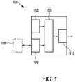
- Fig. 1 shows a schematic representation of an embodiment of a dose error determination device 100 for forecasting radiation dose error for a body volume comprising one or more regions of interest subject to radiation treatment based on a dose plan.
- the explanation of the dose error determination device will be done with further reference to Figs. 2A and 2B which show a first image 200 and a second image 201 of the same body volume.
- the images show a respective region of interest 204, 205, which is associated to a different set of voxels.
- Each voxel is additionally associated to respective dose value d xy in accordance with a dose plan.
- the dose error determination device comprises a dose plan unit 102 configured to provide dose plan data indicative of dose values associated with voxels of a current image of the body volume 200 comprising voxels (d 33 , d 34 , d 43 , d 44 ) associated with the region of interest (ROI) 204.
- the dose error determination device 100 also comprises an input unit 104, that is configured to request and receive user input indicative of a value of a spatial-error quantity corresponding to an amount of a spatial error of voxel coordinates in the current image associated with corresponding features of the body volume.
- a use who has access to the first image 200 and to the second image 201 of the body volume identifies that between the ROI 204 and the ROI 500, there is a spatial error or misalignment.
- the user then can manually input using the input unit the value of the value of a spatial-error quantity, which is r in the case of Figs. 2A and 2B , as a numerical value.
- some dose error determination devices comprise a statistics unit 108 that stores statistical population data associating, based on a statistical population of previous measurements, statistical values associated with the spatial-error quantity to respective regions of interest.
- the input unit is in these particular dose error determination devices configured to request and receive user input indicative of at least one region of interest and, in response to receiving such user input, to access the statistics unit and to ascertain the statistical values associated with the spatial-error quantity for those regions of interest indicated by the user input, and to subsequently provide the request for user input indicative of the value of the spatial-error quantity in association with an output indicative of the ascertained respective statistical value.
- the dose error determination device comprises a statistics unit or not, it comprises a dose-error determining unit 106 that is configured to receive the dose plan data and the value of the spatial error quantity r and to generate dose variation data indicative of a rate of change of the dose values d xy in a respective modified region of interest 207 that in comparison with the corresponding region of interest 205 is expanded in volume by an amount corresponding with the value of the spatial-error quantity r.
- the dose-error determining unit is also configured to determine and provide a respective value of a dose-error quantity associated to the region of interest. This is done by using a calculation rule that represents a positive correlation of the dose-error quantity with the determined dose variation data.
- This calculation rule involves in some particular dose-determination unit a multiplication operation between the dose variation data for each voxel with the value of the spatial-error quantity.
- the error determination unit is configured to determine, for each voxel of the modified region of interest, local dose variation data representing a product of an amount of a local rate of change of the current dose values associated with the given voxel and of the value of the spatial-error quantity, and to determine the dose variation data associated with the region of interest as a maximum amount of the product within the modified region of interest.
- the dose-error determining unit is further configured to determine as the dose variation quantity a maximum dose value and a minimum dose value among those dose values corresponding to voxels located within a distance given by the spatial-error quantity with respect to any voxel associated with each region of interest.
- the dose error determination device also comprises an output unit 110 which receives from the dose-error determination unit the respective value of the dose-error quantity of the one or more ROIs and is configured to provide an output indicative thereof.
- the dose error determination unit is used in combination with a display unit that is configured to receive from the output unit 110 the output indicative of the dose error quantity of the one or more ROIs.
- the display unit may comprise a graphical user interface configured to display the data received from the output unit in a way which enables the user to focus on the most critical ROIs in dependence on the dose plan and the determined dose-error quantities.
- the graphical user interface may be configured to displays the output data in different ways.
- an exemplary graphical user interface that can be used in combination with any of the dose error determination devices in accordance with the invention is configured to display the output data in the form of an informative table, listing the one or more ROIs in a predetermined order, such as in an increasing or decreasing order of the associated dose-error quantity.
- An example of an informative table displayed at the displayed unit is shown in Fig. 3 .
- the figure shows an informative table presenting a list of ROIs A, B, C and D together with their respective determined dose-error quantities V DEQ for a given value of the spatial error quantity, which is this particular case is 3mm.
- a user can select how the list is to be sorted, i.e. in an increasing or a decreasing order of the determined dose-error quantities, which are indicative of the maximum dose change expected at the ROI for the given value of the spatial error quantity.
- the user might decide to skip manual editing for the ROIs for which, for example, the estimated impact of contour inaccuracies given by the value of the spatial error quantity on the dose parameters is small.
- the user could choose to manually review the ROIs for which small contour mapping errors result in large dose changes, as given by the associated value of the dose-error quantity.
- the value of the dose-error quantity is a measure of sensitivity for all voxels of a given ROI, and not only for the voxels with maximum dose.
- the user can provide thresholds or ranges for accepted values of maximum dose changes (i.e. values of dose-error quantities).
- a predetermined color coding such as for instance a traffic light color coding with red yellow and green colors.
- Some dose error determination devices are suitably configured to determine the values of dose-error quantity for one or more ROIs and for a plurality of values of the spatial-error quantity. These dose error determination devices can be advantageously used in combination with a display unit configured to generate a sensitivity map, such as the one shown in Fig. 4 .
- the sensitivity map of Fig. 4 includes a listing of ROIs, A-G, in a vertical axis and a sequence of values of the spatial-error quantity, in this case a magnitude of spatial error due to inaccuracies in a deformable image registration, in a horizontal axis.
- the dose error determination device determines the for each ROI and for each value of the spatial error quantity the corresponding value of the dose-error quantity.
- the display unit is then advantageously configured to display the output data as a sensitivity map using a predetermined color coding, wherein in color represents a range of the values of dose error quantity V DEQ .
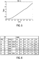
- Another example of the display unit can be alternatively or additionally configured to display the output data as a graph plotting, for one or more ROIs, the value of the dose-error quantity, for example in a vertical axis, versus the value of the spatial error quantity in the horizontal axis, as it is exemplarily shown in Fig. 5 .
- Dose error determination devices that are configured to determine as the dose variation quantity a maximum dose value and a minimum dose value among those dose values corresponding to voxels located within a distance given by the spatial-error quantity with respect to any voxel associated with each region of interest can be advantageously used in combination with a display unit configured to display an upper and a lower bound dose volume histogram (DVH) for a given ROI and a given value of the spatial-error quantity.
- upper and lower bounds of dose value parameters inferred from the dose plan such as a mean dose value for a given ROI or a value of a fractional volume indicative of that volume fraction for which an estimated received radiation is higher than a predetermined radiation threshold, can be provided also in the form of an informative table.
- Some display units are also configured to display an interactive graph that allows the user to interactively inspect all values of the dose-error quantity for a specific value of the spatial-error quantity.
- a display unit can be configured to provide the value of the dose-error quantity for a given value of the spatial-error quantity, or alternatively or additionally, a maximum value of tolerated spatial-error quantity such that the value of the spatial-error quantity satisfies a predetermined constrain, such as a maximum allowed value thereof.
- These displays units are advantageously used with dose error determination devices that include a dose-objective input interface for receiving dose-objective data for the region of interest indicative of desired values of dose-objective quantities associated with the dose plan.
- the dose-error determining unit is additionally configured, using the dose-plan data and the determined value of the dose-error quantity for the region of interest, to generate dose-forecast data for the region of interest indicative of forecast values of the dose-objective quantities; and the output unit is configured to provide the output as additionally being indicative of the dose-objective data and of the dose-forecast data.
- FIG. 6 Another example of a graphical user interface of a display unit that can be used in combination with a dose error determination device in accordance with the present invention is shown in Fig. 6 .
- the table shown in Fig. 6 includes a first column with the list of the ROIs, A-F. It also shows the objectives O for each ROI.
- the objectives include the information pertaining to a type of value of dose-error quantity (e.g. 1 being a maximum value of DVH, 2 being a mean dose value, 3 being a percentage of the maximum DVH), to the desired dose value D in accordance with the dose plan and to a volume value V, either in terms of absolute volume or relative volume.
- the list of objectives is may be automatically imported from a treatment plan in Pinnacle, or the user could manually fill in the objectives.
- the graphical user interface will indicate whether for a certain range of DIR errors, also given in the table of Fig. 6 as r(cm), the objectives are met (ok), or not met (no).
- the user could decide to skip review of ROIs that are robust to a large range of DIR errors and instead review the ROIs that are sensitive to DIR uncertainties.
- the names of ROIs that are relatively more sensitive to spatial errors could be highlighted by, for instance displaying them in a larger font, or displaying them in a different font color, or intermittently displaying them with a blinking pattern, etc.)
- Yet another display unit is advantageously configured to enable a robustness evaluation of the dose parameters against values of spatial-error quantities such as DIR uncertainties by applying a user-selected model of uncertainties by means of a dedicated graphical user interface.
- a model of uncertainty is a parametric model that modifies the physical coordinates of the ROI's voxels, thus producing a second instance of the ROI.
- uncertainty models can be provided, such as for example, worst case scenario DVH, rotations around the three axis, translations, shrinking or expanding the ROI with margins, combination of translations with rotations, random perturbations of the ROI surface, parametric models of errors learned from previous data using principal component analysis, or any combination of the previously mentioned models.
- the user could select the desired uncertainty model from a drop-down menu.
- the user can input parameter values or select a range of values using for instance a slider.
- the value of the dose-error quantity can be calculated for each selected uncertainty model and value(s) of spatial-error quantity.
- the results of the robustness evaluation will be presented, for example, in the form of a figure displaying the dose volume histograms corresponding to the selected uncertainty model, and baseline DVH (e.g., before applying the DIR error).
- the user can also be presented with a table summarizing, for instance, dosimetric objectives for each selected ROI, or baseline value of the objective or, the objective value for the selected value of DIR error, or any combination thereof.
- a traffic light system can for example be used to notify the user whether the objective for the selected DIR error value is met (green), equal (yellow), or not met (red).
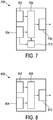
- the dose error determination device 700 comprises a ROI modelling unit 712, which is configured to generate, in response to a selection instruction received via the input unit 704, second instance region of interest data associated with a given one of the one or more regions of interest by modifying, in accordance with a selected one of a set of one or more respective uncertainty models defining spatial coordinate transformation instructions for transforming the region of interest only in regard to its spatial voxel coordinates, thus providing a representation of a transformed region of interest.
- a ROI modelling unit 712 which is configured to generate, in response to a selection instruction received via the input unit 704, second instance region of interest data associated with a given one of the one or more regions of interest by modifying, in accordance with a selected one of a set of one or more respective uncertainty models defining spatial coordinate transformation instructions for transforming the region of interest only in regard to its spatial voxel coordinates, thus providing a representation of a transformed region of interest.
- the dose-error determining unit 706 is additionally configured to determine and provide an expected value of the dose-error quantity associated with the transformed region of interest, and the input unit 704 is additionally configured to request and receive user input indicative of the uncertainty model to be used by the ROI modelling unit.
- Yet another display unit is advantageously configured to offer the user a sensitivity tool configured to analyze plan robustness to spatial errors, such as, but not limited to, ROI contouring errors or geometric image artifacts.
- This display unit can advantageously be used to compare rival radiation plans for a patient and thus assist in a choice of radiation plan. This analysis can be performed manually by the expert or in automated mode. Furthermore, the results of such sensitivity analysis could be taken as an input in a further optimization process to generate plans robust to contouring errors.
- two different radiation plans A and B for a given patient P can be compared in terms of robustness to spatial errors of an ROI in the following way: for each plan A and B, a plurality of values of the spatial-error quantity are applied and the corresponding value of the dose-error quantity associated to plan A and plan B is calculated.
- the value of the dose-error quantity is in a particular example a standard deviation of a fractional ROI volume receiving dose higher than a threshold dose value v.
- it is indicative of the width of the DVH band a certain value of the dose
- the width of the DVH bands for two plans can be shown to the user as numerical quantities or displayed in a graphical way.
- the dose error determination device 800 comprises a dose plan unit 802 that is configured to provide a plurality of dose plans, each comprising respective dose plan data associated with alternative dose plans for the region of interest.
- the dose-error determination unit 806 is further configured to provide plan-robustness data indicative of a respective plan-robustness value for each dose plan and in dependence of the received value of the spatial-error quantity, the plan-robustness value being determined in accordance with a predetermined rule based on the determined values of the dose-error quantities associated with the one or more regions of interest.
- the output unit 810 is further configured to receive the plan-robustness data and to provide an output indicative thereof.
- a dose error determination device for forecasting radiation dose error for a body volume comprising one or more regions of interest subject to radiation treatment based on a dose plan in presented.
- the dose error determination device comprises a dose-error determining unit configured to generate, using dose plan data for the respective region of interest and an input value of the spatial-error quantity, dose variation data indicative of a rate of change of the dose values in a respective modified region of interest that in comparison with the respective region of interest is expanded in volume by an amount corresponding with the value of the spatial-error quantity and to determine and provide a respective value of a dose-error quantity associated with the respective region of interest, using a calculation rule representing a positive correlation of the dose-error quantity with the determined dose variation data.
Priority Applications (6)
| Application Number | Priority Date | Filing Date | Title |
|---|---|---|---|
| EP18174501.9A EP3574955A1 (fr) | 2018-05-28 | 2018-05-28 | Dispositif de détermination d'erreur de dose |
| CN201980035808.4A CN112203720A (zh) | 2018-05-28 | 2019-05-17 | 剂量误差确定设备 |
| JP2020563534A JP6949256B2 (ja) | 2018-05-28 | 2019-05-17 | 線量誤差決定装置 |
| US17/057,376 US20210196981A1 (en) | 2018-05-28 | 2019-05-17 | Dose error determination device |
| PCT/EP2019/062753 WO2019228824A1 (fr) | 2018-05-28 | 2019-05-17 | Dispositif de détermination d'erreur de dose |
| EP19723808.2A EP3801760B1 (fr) | 2018-05-28 | 2019-05-17 | Dispositif de détermination d'erreur de dose |
Applications Claiming Priority (1)
| Application Number | Priority Date | Filing Date | Title |
|---|---|---|---|
| EP18174501.9A EP3574955A1 (fr) | 2018-05-28 | 2018-05-28 | Dispositif de détermination d'erreur de dose |
Publications (1)
| Publication Number | Publication Date |
|---|---|
| EP3574955A1 true EP3574955A1 (fr) | 2019-12-04 |
Family
ID=62620624
Family Applications (2)
| Application Number | Title | Priority Date | Filing Date |
|---|---|---|---|
| EP18174501.9A Withdrawn EP3574955A1 (fr) | 2018-05-28 | 2018-05-28 | Dispositif de détermination d'erreur de dose |
| EP19723808.2A Active EP3801760B1 (fr) | 2018-05-28 | 2019-05-17 | Dispositif de détermination d'erreur de dose |
Family Applications After (1)
| Application Number | Title | Priority Date | Filing Date |
|---|---|---|---|
| EP19723808.2A Active EP3801760B1 (fr) | 2018-05-28 | 2019-05-17 | Dispositif de détermination d'erreur de dose |
Country Status (5)
| Country | Link |
|---|---|
| US (1) | US20210196981A1 (fr) |
| EP (2) | EP3574955A1 (fr) |
| JP (1) | JP6949256B2 (fr) |
| CN (1) | CN112203720A (fr) |
| WO (1) | WO2019228824A1 (fr) |
Citations (5)
| Publication number | Priority date | Publication date | Assignee | Title |
|---|---|---|---|---|
| US20140163302A1 (en) * | 2012-12-07 | 2014-06-12 | Emory University | Methods, systems and computer readable storage media storing instructions for image-guided treatment planning and assessment |
| EP2808057A1 (fr) * | 2013-05-31 | 2014-12-03 | RaySearch Laboratories AB | Procédé et système de planification de traitement radiothérapeutique robuste |
| US20160059039A1 (en) * | 2014-09-02 | 2016-03-03 | Mayo Foundation For Medical Education And Research | System and method for robust intensity-modulated proton therapy planning |
| US20160140300A1 (en) * | 2013-06-12 | 2016-05-19 | University Health Network | Method and system for automated quality assurance and automated treatment planning in radiation therapy |
| US20160166855A1 (en) * | 2013-07-31 | 2016-06-16 | Koninklijke Philips N.V. | Automation of therapy planning |
Family Cites Families (5)
| Publication number | Priority date | Publication date | Assignee | Title |
|---|---|---|---|---|
| FR2839894A1 (fr) * | 2002-05-21 | 2003-11-28 | Chabunda Christophe Mwanza | Procedes, appareils de cyclotherapie image-guidee et mode d'obtention d'images scanographiques diagnostiques instantanees pour la planification et la dosimetrie en ligne |
| JP6285434B2 (ja) * | 2012-07-27 | 2018-02-28 | コーニンクレッカ フィリップス エヌ ヴェKoninklijke Philips N.V. | 線量変形誤差計算方法およびシステム |
| WO2014167461A1 (fr) * | 2013-04-11 | 2014-10-16 | Koninklijke Philips N.V. | Optimisation d'isodose |
| US9089696B2 (en) * | 2013-11-07 | 2015-07-28 | Varian Medical Systems International Ag | Time-resolved pre-treatment portal dosimetry systems, devices, and methods |
| US9987504B2 (en) * | 2015-04-02 | 2018-06-05 | Varian Medical Systems International Ag | Portal dosimetry systems, devices, and methods |
-
2018
- 2018-05-28 EP EP18174501.9A patent/EP3574955A1/fr not_active Withdrawn
-
2019
- 2019-05-17 WO PCT/EP2019/062753 patent/WO2019228824A1/fr unknown
- 2019-05-17 EP EP19723808.2A patent/EP3801760B1/fr active Active
- 2019-05-17 US US17/057,376 patent/US20210196981A1/en active Pending
- 2019-05-17 JP JP2020563534A patent/JP6949256B2/ja active Active
- 2019-05-17 CN CN201980035808.4A patent/CN112203720A/zh active Pending
Patent Citations (5)
| Publication number | Priority date | Publication date | Assignee | Title |
|---|---|---|---|---|
| US20140163302A1 (en) * | 2012-12-07 | 2014-06-12 | Emory University | Methods, systems and computer readable storage media storing instructions for image-guided treatment planning and assessment |
| EP2808057A1 (fr) * | 2013-05-31 | 2014-12-03 | RaySearch Laboratories AB | Procédé et système de planification de traitement radiothérapeutique robuste |
| US20160140300A1 (en) * | 2013-06-12 | 2016-05-19 | University Health Network | Method and system for automated quality assurance and automated treatment planning in radiation therapy |
| US20160166855A1 (en) * | 2013-07-31 | 2016-06-16 | Koninklijke Philips N.V. | Automation of therapy planning |
| US20160059039A1 (en) * | 2014-09-02 | 2016-03-03 | Mayo Foundation For Medical Education And Research | System and method for robust intensity-modulated proton therapy planning |
Non-Patent Citations (2)
| Title |
|---|
| NEIL KIRBY ET AL.: "An automated deformable image registration evaluation of confidence tool", PHYSICS IN MEDICINE AND BIOLOGY, vol. 61, 2016, pages N203 - N214, XP020303406, DOI: doi:10.1088/0031-9155/61/8/N203 |
| NEIL KIRBY ET AL: "An automated deformable image registration evaluation of confidence tool", PHYSICS IN MEDICINE AND BIOLOGY, INSTITUTE OF PHYSICS PUBLISHING, BRISTOL GB, vol. 61, no. 8, 30 March 2016 (2016-03-30), XP020303406, ISSN: 0031-9155, [retrieved on 20160330], DOI: 10.1088/0031-9155/61/8/N203 * |
Also Published As
| Publication number | Publication date |
|---|---|
| CN112203720A (zh) | 2021-01-08 |
| EP3801760B1 (fr) | 2022-01-12 |
| JP2021517059A (ja) | 2021-07-15 |
| US20210196981A1 (en) | 2021-07-01 |
| WO2019228824A1 (fr) | 2019-12-05 |
| JP6949256B2 (ja) | 2021-10-13 |
| EP3801760A1 (fr) | 2021-04-14 |
Similar Documents
| Publication | Publication Date | Title |
|---|---|---|
| US11915424B2 (en) | Evaluating quality of segmentation of an image into different types of tissue for planning treatment using tumor treating fields (TTFields) | |
| US10186031B2 (en) | Shape similarity measure for body tissue | |
| Chen et al. | Automated contouring error detection based on supervised geometric attribute distribution models for radiation therapy: a general strategy | |
| US20210042700A1 (en) | Index computation device, prediction system, progress prediction evaluation method, and program | |
| CN111932552A (zh) | 一种主动脉建模的方法及装置 | |
| CN115409879A (zh) | 图像配准的数据处理方法及装置、存储介质、电子设备 | |
| CN107567638A (zh) | 对解剖结构的基于模型的分割 | |
| EP3801760B1 (fr) | Dispositif de détermination d'erreur de dose | |
| EP3270308B9 (fr) | Procédé pour fournir un paramètre secondaire, système d'aide à la décision, support lisible par ordinateur et produit-programme informatique | |
| EP3438932B1 (fr) | Contournage intelligent d'anatomie utilisant des points de clic d'utilisateur structurés | |
| US9390549B2 (en) | Shape data generation method and apparatus | |
| US20210390707A1 (en) | Predicting correctness of algorithmic segmentation | |
| Park et al. | Composite radiation dose representation using fuzzy set theory | |
| CN112912924B (zh) | 预测算法分割的正确性 | |
| US20220054861A1 (en) | Automated qualitative description of anatomical changes in radiotherapy | |
| Chaves-de-Plaza et al. | Inclusion Depth for Contour Ensembles | |
| WO2023057281A1 (fr) | Système et procédé d'assurance qualité de segmentation d'image médicale | |
| Santos et al. | Some recurrent concepts of quality evaluation in medical visualization |
Legal Events
| Date | Code | Title | Description |
|---|---|---|---|
| PUAI | Public reference made under article 153(3) epc to a published international application that has entered the european phase |
Free format text: ORIGINAL CODE: 0009012 |
|
| AK | Designated contracting states |
Kind code of ref document: A1 Designated state(s): AL AT BE BG CH CY CZ DE DK EE ES FI FR GB GR HR HU IE IS IT LI LT LU LV MC MK MT NL NO PL PT RO RS SE SI SK SM TR |
|
| AX | Request for extension of the european patent |
Extension state: BA ME |
|
| RAP1 | Party data changed (applicant data changed or rights of an application transferred) |
Owner name: KONINKLIJKE PHILIPS N.V. |
|
| STAA | Information on the status of an ep patent application or granted ep patent |
Free format text: STATUS: THE APPLICATION IS DEEMED TO BE WITHDRAWN |
|
| 18D | Application deemed to be withdrawn |
Effective date: 20200605 |