EP3568144B1 - Probiotic compositions and uses thereof - Google Patents
Probiotic compositions and uses thereof Download PDFInfo
- Publication number
- EP3568144B1 EP3568144B1 EP18707627.8A EP18707627A EP3568144B1 EP 3568144 B1 EP3568144 B1 EP 3568144B1 EP 18707627 A EP18707627 A EP 18707627A EP 3568144 B1 EP3568144 B1 EP 3568144B1
- Authority
- EP
- European Patent Office
- Prior art keywords
- cells
- mean
- placebo
- value
- probiotic
- Prior art date
- Legal status (The legal status is an assumption and is not a legal conclusion. Google has not performed a legal analysis and makes no representation as to the accuracy of the status listed.)
- Active
Links
- 239000006041 probiotic Substances 0.000 title claims description 55
- 235000018291 probiotics Nutrition 0.000 title claims description 55
- 230000000529 probiotic effect Effects 0.000 title claims description 53
- 239000000203 mixture Substances 0.000 title claims description 28
- 208000015943 Coeliac disease Diseases 0.000 claims description 81
- 230000005784 autoimmunity Effects 0.000 claims description 19
- 241000186605 Lactobacillus paracasei Species 0.000 claims description 14
- 240000006024 Lactobacillus plantarum Species 0.000 claims description 13
- 235000013305 food Nutrition 0.000 claims description 8
- 235000013965 Lactobacillus plantarum Nutrition 0.000 claims description 6
- 229940072205 lactobacillus plantarum Drugs 0.000 claims description 6
- 239000000843 powder Substances 0.000 claims description 6
- 230000002265 prevention Effects 0.000 claims description 5
- 238000002360 preparation method Methods 0.000 claims description 3
- 239000002775 capsule Substances 0.000 claims description 2
- 239000011785 micronutrient Substances 0.000 claims description 2
- 235000013369 micronutrients Nutrition 0.000 claims description 2
- 239000000546 pharmaceutical excipient Substances 0.000 claims description 2
- 239000003826 tablet Substances 0.000 claims 1
- 210000004027 cell Anatomy 0.000 description 68
- 239000000902 placebo Substances 0.000 description 68
- 229940068196 placebo Drugs 0.000 description 68
- 241000186660 Lactobacillus Species 0.000 description 55
- 101001057504 Homo sapiens Interferon-stimulated gene 20 kDa protein Proteins 0.000 description 36
- 101001055144 Homo sapiens Interleukin-2 receptor subunit alpha Proteins 0.000 description 36
- 102100026878 Interleukin-2 receptor subunit alpha Human genes 0.000 description 36
- 210000004698 lymphocyte Anatomy 0.000 description 29
- MHMNJMPURVTYEJ-UHFFFAOYSA-N fluorescein-5-isothiocyanate Chemical compound O1C(=O)C2=CC(N=C=S)=CC=C2C21C1=CC=C(O)C=C1OC1=CC(O)=CC=C21 MHMNJMPURVTYEJ-UHFFFAOYSA-N 0.000 description 23
- 108010004729 Phycoerythrin Proteins 0.000 description 22
- 210000003819 peripheral blood mononuclear cell Anatomy 0.000 description 21
- 210000002443 helper t lymphocyte Anatomy 0.000 description 18
- 102100033467 L-selectin Human genes 0.000 description 17
- 229940039696 lactobacillus Drugs 0.000 description 16
- 108700039882 Protein Glutamine gamma Glutamyltransferase 2 Proteins 0.000 description 15
- 102100038095 Protein-glutamine gamma-glutamyltransferase 2 Human genes 0.000 description 15
- 230000007423 decrease Effects 0.000 description 14
- 102000054766 genetic haplotypes Human genes 0.000 description 14
- 235000006171 gluten free diet Nutrition 0.000 description 13
- 235000020884 gluten-free diet Nutrition 0.000 description 13
- 210000001744 T-lymphocyte Anatomy 0.000 description 12
- 230000001900 immune effect Effects 0.000 description 12
- 238000000034 method Methods 0.000 description 12
- 230000000694 effects Effects 0.000 description 11
- 210000000822 natural killer cell Anatomy 0.000 description 11
- 238000004458 analytical method Methods 0.000 description 10
- 102100022718 Atypical chemokine receptor 2 Human genes 0.000 description 9
- 108010061711 Gliadin Proteins 0.000 description 9
- 101000678892 Homo sapiens Atypical chemokine receptor 2 Proteins 0.000 description 9
- 101000716070 Homo sapiens C-C chemokine receptor type 9 Proteins 0.000 description 9
- 210000001151 cytotoxic T lymphocyte Anatomy 0.000 description 9
- 239000000047 product Substances 0.000 description 9
- 208000024891 symptom Diseases 0.000 description 9
- 102100031585 ADP-ribosyl cyclase/cyclic ADP-ribose hydrolase 1 Human genes 0.000 description 8
- 108700028369 Alleles Proteins 0.000 description 8
- 108010068370 Glutens Proteins 0.000 description 8
- 101000777636 Homo sapiens ADP-ribosyl cyclase/cyclic ADP-ribose hydrolase 1 Proteins 0.000 description 8
- 210000004369 blood Anatomy 0.000 description 8
- 239000008280 blood Substances 0.000 description 8
- 230000008859 change Effects 0.000 description 8
- 238000000684 flow cytometry Methods 0.000 description 8
- 230000002068 genetic effect Effects 0.000 description 8
- 210000003289 regulatory T cell Anatomy 0.000 description 8
- 238000010186 staining Methods 0.000 description 8
- 241000186000 Bifidobacterium Species 0.000 description 7
- 230000004913 activation Effects 0.000 description 7
- 230000003247 decreasing effect Effects 0.000 description 7
- 238000003745 diagnosis Methods 0.000 description 7
- 235000021312 gluten Nutrition 0.000 description 7
- 230000000968 intestinal effect Effects 0.000 description 7
- 238000012163 sequencing technique Methods 0.000 description 7
- 239000011782 vitamin Substances 0.000 description 7
- 229930003231 vitamin Natural products 0.000 description 7
- 229940088594 vitamin Drugs 0.000 description 7
- 102100033016 Integrin beta-7 Human genes 0.000 description 6
- XEEYBQQBJWHFJM-UHFFFAOYSA-N Iron Chemical compound [Fe] XEEYBQQBJWHFJM-UHFFFAOYSA-N 0.000 description 6
- HCHKCACWOHOZIP-UHFFFAOYSA-N Zinc Chemical compound [Zn] HCHKCACWOHOZIP-UHFFFAOYSA-N 0.000 description 6
- 238000001574 biopsy Methods 0.000 description 6
- 108010021315 integrin beta7 Proteins 0.000 description 6
- 239000002953 phosphate buffered saline Substances 0.000 description 6
- 108090000623 proteins and genes Proteins 0.000 description 6
- 238000012360 testing method Methods 0.000 description 6
- 235000013343 vitamin Nutrition 0.000 description 6
- 210000001266 CD8-positive T-lymphocyte Anatomy 0.000 description 5
- 206010067584 Type 1 diabetes mellitus Diseases 0.000 description 5
- 206010012601 diabetes mellitus Diseases 0.000 description 5
- 208000037265 diseases, disorders, signs and symptoms Diseases 0.000 description 5
- 229940079593 drug Drugs 0.000 description 5
- 239000003814 drug Substances 0.000 description 5
- 230000008823 permeabilization Effects 0.000 description 5
- 238000003653 radioligand binding assay Methods 0.000 description 5
- 230000000405 serological effect Effects 0.000 description 5
- 210000002966 serum Anatomy 0.000 description 5
- HZWWPUTXBJEENE-UHFFFAOYSA-N 5-amino-2-[[1-[5-amino-2-[[1-[2-amino-3-(4-hydroxyphenyl)propanoyl]pyrrolidine-2-carbonyl]amino]-5-oxopentanoyl]pyrrolidine-2-carbonyl]amino]-5-oxopentanoic acid Chemical compound C1CCC(C(=O)NC(CCC(N)=O)C(=O)N2C(CCC2)C(=O)NC(CCC(N)=O)C(O)=O)N1C(=O)C(N)CC1=CC=C(O)C=C1 HZWWPUTXBJEENE-UHFFFAOYSA-N 0.000 description 4
- 101100284398 Bos taurus BoLA-DQB gene Proteins 0.000 description 4
- 102210047476 DQA1*02:01 Human genes 0.000 description 4
- 102210047410 DQA1*05:05 Human genes 0.000 description 4
- 102210047212 DQB1*02:02 Human genes 0.000 description 4
- 102210047287 DQB1*03:02 Human genes 0.000 description 4
- 101000581981 Homo sapiens Neural cell adhesion molecule 1 Proteins 0.000 description 4
- 208000002720 Malnutrition Diseases 0.000 description 4
- 238000000585 Mann–Whitney U test Methods 0.000 description 4
- 102100027347 Neural cell adhesion molecule 1 Human genes 0.000 description 4
- 238000003556 assay Methods 0.000 description 4
- 235000005911 diet Nutrition 0.000 description 4
- 201000010099 disease Diseases 0.000 description 4
- 238000001943 fluorescence-activated cell sorting Methods 0.000 description 4
- 229910052500 inorganic mineral Inorganic materials 0.000 description 4
- 239000007788 liquid Substances 0.000 description 4
- 230000001071 malnutrition Effects 0.000 description 4
- 235000000824 malnutrition Nutrition 0.000 description 4
- 239000003550 marker Substances 0.000 description 4
- 239000011707 mineral Substances 0.000 description 4
- 235000010755 mineral Nutrition 0.000 description 4
- 230000035764 nutrition Effects 0.000 description 4
- 235000016709 nutrition Nutrition 0.000 description 4
- 208000015380 nutritional deficiency disease Diseases 0.000 description 4
- 108090000765 processed proteins & peptides Proteins 0.000 description 4
- 239000007787 solid Substances 0.000 description 4
- 239000006228 supernatant Substances 0.000 description 4
- 150000003722 vitamin derivatives Chemical class 0.000 description 4
- XLYOFNOQVPJJNP-UHFFFAOYSA-N water Substances O XLYOFNOQVPJJNP-UHFFFAOYSA-N 0.000 description 4
- 241000894006 Bacteria Species 0.000 description 3
- VTYYLEPIZMXCLO-UHFFFAOYSA-L Calcium carbonate Chemical compound [Ca+2].[O-]C([O-])=O VTYYLEPIZMXCLO-UHFFFAOYSA-L 0.000 description 3
- 206010061218 Inflammation Diseases 0.000 description 3
- 229920002774 Maltodextrin Polymers 0.000 description 3
- 239000005913 Maltodextrin Substances 0.000 description 3
- 241000736262 Microbiota Species 0.000 description 3
- 206010061291 Mineral deficiency Diseases 0.000 description 3
- 238000011529 RT qPCR Methods 0.000 description 3
- 239000011324 bead Substances 0.000 description 3
- 230000000903 blocking effect Effects 0.000 description 3
- 230000029087 digestion Effects 0.000 description 3
- 238000010790 dilution Methods 0.000 description 3
- 239000012895 dilution Substances 0.000 description 3
- 238000009826 distribution Methods 0.000 description 3
- 235000013350 formula milk Nutrition 0.000 description 3
- 210000001035 gastrointestinal tract Anatomy 0.000 description 3
- 235000020256 human milk Nutrition 0.000 description 3
- 210000004251 human milk Anatomy 0.000 description 3
- 230000028993 immune response Effects 0.000 description 3
- 238000011534 incubation Methods 0.000 description 3
- 230000004054 inflammatory process Effects 0.000 description 3
- 229910052742 iron Inorganic materials 0.000 description 3
- 229960003284 iron Drugs 0.000 description 3
- 210000000265 leukocyte Anatomy 0.000 description 3
- 229940035034 maltodextrin Drugs 0.000 description 3
- 239000000523 sample Substances 0.000 description 3
- 230000001225 therapeutic effect Effects 0.000 description 3
- 238000003260 vortexing Methods 0.000 description 3
- 235000016804 zinc Nutrition 0.000 description 3
- 239000011701 zinc Substances 0.000 description 3
- 229910052725 zinc Inorganic materials 0.000 description 3
- 208000023275 Autoimmune disease Diseases 0.000 description 2
- 208000000412 Avitaminosis Diseases 0.000 description 2
- 102100024222 B-lymphocyte antigen CD19 Human genes 0.000 description 2
- 241001608472 Bifidobacterium longum Species 0.000 description 2
- 241000186015 Bifidobacterium longum subsp. infantis Species 0.000 description 2
- 102210047285 DQA1*05:01 Human genes 0.000 description 2
- 102210010092 DQB1*03:01 Human genes 0.000 description 2
- 206010012735 Diarrhoea Diseases 0.000 description 2
- 238000000729 Fisher's exact test Methods 0.000 description 2
- WSFSSNUMVMOOMR-UHFFFAOYSA-N Formaldehyde Chemical compound O=C WSFSSNUMVMOOMR-UHFFFAOYSA-N 0.000 description 2
- 101000980825 Homo sapiens B-lymphocyte antigen CD19 Proteins 0.000 description 2
- 101000917858 Homo sapiens Low affinity immunoglobulin gamma Fc region receptor III-A Proteins 0.000 description 2
- 101000917839 Homo sapiens Low affinity immunoglobulin gamma Fc region receptor III-B Proteins 0.000 description 2
- 101000738771 Homo sapiens Receptor-type tyrosine-protein phosphatase C Proteins 0.000 description 2
- 108010092694 L-Selectin Proteins 0.000 description 2
- 102000016551 L-selectin Human genes 0.000 description 2
- 241000218588 Lactobacillus rhamnosus Species 0.000 description 2
- 102100029185 Low affinity immunoglobulin gamma Fc region receptor III-B Human genes 0.000 description 2
- FYYHWMGAXLPEAU-UHFFFAOYSA-N Magnesium Chemical compound [Mg] FYYHWMGAXLPEAU-UHFFFAOYSA-N 0.000 description 2
- 229920000168 Microcrystalline cellulose Polymers 0.000 description 2
- 208000001132 Osteoporosis Diseases 0.000 description 2
- 239000012980 RPMI-1640 medium Substances 0.000 description 2
- 102100037422 Receptor-type tyrosine-protein phosphatase C Human genes 0.000 description 2
- 239000006146 Roswell Park Memorial Institute medium Substances 0.000 description 2
- 229930003316 Vitamin D Natural products 0.000 description 2
- QYSXJUFSXHHAJI-XFEUOLMDSA-N Vitamin D3 Natural products C1(/[C@@H]2CC[C@@H]([C@]2(CCC1)C)[C@H](C)CCCC(C)C)=C/C=C1\C[C@@H](O)CCC1=C QYSXJUFSXHHAJI-XFEUOLMDSA-N 0.000 description 2
- 238000001793 Wilcoxon signed-rank test Methods 0.000 description 2
- 206010000059 abdominal discomfort Diseases 0.000 description 2
- 208000019790 abdominal distention Diseases 0.000 description 2
- 208000007502 anemia Diseases 0.000 description 2
- 239000003125 aqueous solvent Substances 0.000 description 2
- 210000003719 b-lymphocyte Anatomy 0.000 description 2
- 230000033228 biological regulation Effects 0.000 description 2
- 239000000872 buffer Substances 0.000 description 2
- 239000002771 cell marker Substances 0.000 description 2
- 230000001086 cytosolic effect Effects 0.000 description 2
- 238000013461 design Methods 0.000 description 2
- 238000011161 development Methods 0.000 description 2
- 238000002405 diagnostic procedure Methods 0.000 description 2
- 230000037213 diet Effects 0.000 description 2
- 230000000378 dietary effect Effects 0.000 description 2
- 235000015872 dietary supplement Nutrition 0.000 description 2
- 239000003085 diluting agent Substances 0.000 description 2
- 239000012636 effector Substances 0.000 description 2
- 230000007613 environmental effect Effects 0.000 description 2
- OVBPIULPVIDEAO-LBPRGKRZSA-N folic acid Chemical compound C=1N=C2NC(N)=NC(=O)C2=NC=1CNC1=CC=C(C(=O)N[C@@H](CCC(O)=O)C(O)=O)C=C1 OVBPIULPVIDEAO-LBPRGKRZSA-N 0.000 description 2
- 230000037406 food intake Effects 0.000 description 2
- 239000008098 formaldehyde solution Substances 0.000 description 2
- 238000009472 formulation Methods 0.000 description 2
- 238000012252 genetic analysis Methods 0.000 description 2
- 244000005709 gut microbiome Species 0.000 description 2
- 210000004347 intestinal mucosa Anatomy 0.000 description 2
- 210000005024 intraepithelial lymphocyte Anatomy 0.000 description 2
- 238000011835 investigation Methods 0.000 description 2
- 235000001055 magnesium Nutrition 0.000 description 2
- 239000011777 magnesium Substances 0.000 description 2
- 229910052749 magnesium Inorganic materials 0.000 description 2
- 229940091250 magnesium supplement Drugs 0.000 description 2
- 239000000463 material Substances 0.000 description 2
- 235000019813 microcrystalline cellulose Nutrition 0.000 description 2
- 239000008108 microcrystalline cellulose Substances 0.000 description 2
- 229940016286 microcrystalline cellulose Drugs 0.000 description 2
- 235000013336 milk Nutrition 0.000 description 2
- 239000008267 milk Substances 0.000 description 2
- 210000004080 milk Anatomy 0.000 description 2
- 238000002156 mixing Methods 0.000 description 2
- 230000003287 optical effect Effects 0.000 description 2
- 230000002093 peripheral effect Effects 0.000 description 2
- 230000007414 peripheral immune response Effects 0.000 description 2
- 230000002085 persistent effect Effects 0.000 description 2
- 102000004196 processed proteins & peptides Human genes 0.000 description 2
- 230000000770 proinflammatory effect Effects 0.000 description 2
- 235000018102 proteins Nutrition 0.000 description 2
- 102000004169 proteins and genes Human genes 0.000 description 2
- 238000003753 real-time PCR Methods 0.000 description 2
- 230000009467 reduction Effects 0.000 description 2
- 238000012552 review Methods 0.000 description 2
- 238000012216 screening Methods 0.000 description 2
- 238000007619 statistical method Methods 0.000 description 2
- 239000013589 supplement Substances 0.000 description 2
- 230000003827 upregulation Effects 0.000 description 2
- 235000019166 vitamin D Nutrition 0.000 description 2
- 239000011710 vitamin D Substances 0.000 description 2
- 150000003710 vitamin D derivatives Chemical class 0.000 description 2
- 229940046008 vitamin d Drugs 0.000 description 2
- 208000016261 weight loss Diseases 0.000 description 2
- 230000004580 weight loss Effects 0.000 description 2
- MZOFCQQQCNRIBI-VMXHOPILSA-N (3s)-4-[[(2s)-1-[[(2s)-1-[[(1s)-1-carboxy-2-hydroxyethyl]amino]-4-methyl-1-oxopentan-2-yl]amino]-5-(diaminomethylideneamino)-1-oxopentan-2-yl]amino]-3-[[2-[[(2s)-2,6-diaminohexanoyl]amino]acetyl]amino]-4-oxobutanoic acid Chemical compound OC[C@@H](C(O)=O)NC(=O)[C@H](CC(C)C)NC(=O)[C@H](CCCN=C(N)N)NC(=O)[C@H](CC(O)=O)NC(=O)CNC(=O)[C@@H](N)CCCCN MZOFCQQQCNRIBI-VMXHOPILSA-N 0.000 description 1
- 229920000936 Agarose Polymers 0.000 description 1
- 206010003694 Atrophy Diseases 0.000 description 1
- 241000283690 Bos taurus Species 0.000 description 1
- 101100495352 Candida albicans CDR4 gene Proteins 0.000 description 1
- 241000283707 Capra Species 0.000 description 1
- 102000011632 Caseins Human genes 0.000 description 1
- 108010076119 Caseins Proteins 0.000 description 1
- 102000016289 Cell Adhesion Molecules Human genes 0.000 description 1
- 108010067225 Cell Adhesion Molecules Proteins 0.000 description 1
- 102000009410 Chemokine receptor Human genes 0.000 description 1
- 108050000299 Chemokine receptor Proteins 0.000 description 1
- 229920002261 Corn starch Polymers 0.000 description 1
- 102000004127 Cytokines Human genes 0.000 description 1
- 108090000695 Cytokines Proteins 0.000 description 1
- 244000024675 Eruca sativa Species 0.000 description 1
- 235000014755 Eruca sativa Nutrition 0.000 description 1
- 102100036241 HLA class II histocompatibility antigen, DQ beta 1 chain Human genes 0.000 description 1
- 102100040485 HLA class II histocompatibility antigen, DRB1 beta chain Human genes 0.000 description 1
- 108010062347 HLA-DQ Antigens Proteins 0.000 description 1
- 108010047762 HLA-DQ8 antigen Proteins 0.000 description 1
- 108010086786 HLA-DQA1 antigen Proteins 0.000 description 1
- 108010065026 HLA-DQB1 antigen Proteins 0.000 description 1
- 108010039343 HLA-DRB1 Chains Proteins 0.000 description 1
- 101000666171 Homo sapiens Protein-glutamine gamma-glutamyltransferase 2 Proteins 0.000 description 1
- 101001100327 Homo sapiens RNA-binding protein 45 Proteins 0.000 description 1
- 240000005979 Hordeum vulgare Species 0.000 description 1
- 235000007340 Hordeum vulgare Nutrition 0.000 description 1
- 206010021135 Hypovitaminosis Diseases 0.000 description 1
- 208000007924 IgA Deficiency Diseases 0.000 description 1
- 102100037850 Interferon gamma Human genes 0.000 description 1
- 108010074328 Interferon-gamma Proteins 0.000 description 1
- 108010032774 Interleukin-2 Receptor alpha Subunit Proteins 0.000 description 1
- 102000007351 Interleukin-2 Receptor alpha Subunit Human genes 0.000 description 1
- 102100030703 Interleukin-22 Human genes 0.000 description 1
- 229920001202 Inulin Polymers 0.000 description 1
- ZDXPYRJPNDTMRX-VKHMYHEASA-N L-glutamine Chemical compound OC(=O)[C@@H](N)CCC(N)=O ZDXPYRJPNDTMRX-VKHMYHEASA-N 0.000 description 1
- 229930182816 L-glutamine Natural products 0.000 description 1
- 240000001046 Lactobacillus acidophilus Species 0.000 description 1
- 240000001929 Lactobacillus brevis Species 0.000 description 1
- 244000199885 Lactobacillus bulgaricus Species 0.000 description 1
- 244000199866 Lactobacillus casei Species 0.000 description 1
- 241000218492 Lactobacillus crispatus Species 0.000 description 1
- 241000186840 Lactobacillus fermentum Species 0.000 description 1
- 241001468157 Lactobacillus johnsonii Species 0.000 description 1
- 241000186604 Lactobacillus reuteri Species 0.000 description 1
- 241000186869 Lactobacillus salivarius Species 0.000 description 1
- 206010028116 Mucosal inflammation Diseases 0.000 description 1
- OVBPIULPVIDEAO-UHFFFAOYSA-N N-Pteroyl-L-glutaminsaeure Natural products C=1N=C2NC(N)=NC(=O)C2=NC=1CNC1=CC=C(C(=O)NC(CCC(O)=O)C(O)=O)C=C1 OVBPIULPVIDEAO-UHFFFAOYSA-N 0.000 description 1
- 108091034117 Oligonucleotide Proteins 0.000 description 1
- 238000012408 PCR amplification Methods 0.000 description 1
- 239000001888 Peptone Substances 0.000 description 1
- 108010080698 Peptones Proteins 0.000 description 1
- 102100038823 RNA-binding protein 45 Human genes 0.000 description 1
- 240000004808 Saccharomyces cerevisiae Species 0.000 description 1
- 241000209056 Secale Species 0.000 description 1
- 235000007238 Secale cereale Nutrition 0.000 description 1
- 206010039915 Selective IgA immunodeficiency Diseases 0.000 description 1
- 229920002684 Sepharose Polymers 0.000 description 1
- 208000032023 Signs and Symptoms Diseases 0.000 description 1
- 229920002472 Starch Polymers 0.000 description 1
- 244000057717 Streptococcus lactis Species 0.000 description 1
- 238000000692 Student's t-test Methods 0.000 description 1
- 241000209140 Triticum Species 0.000 description 1
- 235000021307 Triticum Nutrition 0.000 description 1
- 108060008682 Tumor Necrosis Factor Proteins 0.000 description 1
- 102000000852 Tumor Necrosis Factor-alpha Human genes 0.000 description 1
- 206010047627 Vitamin deficiencies Diseases 0.000 description 1
- 239000005862 Whey Substances 0.000 description 1
- 102000007544 Whey Proteins Human genes 0.000 description 1
- 108010046377 Whey Proteins Proteins 0.000 description 1
- UYRDHEJRPVSJFM-VSWVFQEASA-N [(1s,3r)-3-hydroxy-4-[(3e,5e,7e,9e,11z)-11-[4-[(e)-2-[(1r,3s,6s)-3-hydroxy-1,5,5-trimethyl-7-oxabicyclo[4.1.0]heptan-6-yl]ethenyl]-5-oxofuran-2-ylidene]-3,10-dimethylundeca-1,3,5,7,9-pentaenylidene]-3,5,5-trimethylcyclohexyl] acetate Chemical compound C[C@@]1(O)C[C@@H](OC(=O)C)CC(C)(C)C1=C=C\C(C)=C\C=C\C=C\C=C(/C)\C=C/1C=C(\C=C\[C@]23[C@@](O2)(C)C[C@@H](O)CC3(C)C)C(=O)O\1 UYRDHEJRPVSJFM-VSWVFQEASA-N 0.000 description 1
- JLCPHMBAVCMARE-UHFFFAOYSA-N [3-[[3-[[3-[[3-[[3-[[3-[[3-[[3-[[3-[[3-[[3-[[5-(2-amino-6-oxo-1H-purin-9-yl)-3-[[3-[[3-[[3-[[3-[[3-[[5-(2-amino-6-oxo-1H-purin-9-yl)-3-[[5-(2-amino-6-oxo-1H-purin-9-yl)-3-hydroxyoxolan-2-yl]methoxy-hydroxyphosphoryl]oxyoxolan-2-yl]methoxy-hydroxyphosphoryl]oxy-5-(5-methyl-2,4-dioxopyrimidin-1-yl)oxolan-2-yl]methoxy-hydroxyphosphoryl]oxy-5-(6-aminopurin-9-yl)oxolan-2-yl]methoxy-hydroxyphosphoryl]oxy-5-(6-aminopurin-9-yl)oxolan-2-yl]methoxy-hydroxyphosphoryl]oxy-5-(6-aminopurin-9-yl)oxolan-2-yl]methoxy-hydroxyphosphoryl]oxy-5-(6-aminopurin-9-yl)oxolan-2-yl]methoxy-hydroxyphosphoryl]oxyoxolan-2-yl]methoxy-hydroxyphosphoryl]oxy-5-(5-methyl-2,4-dioxopyrimidin-1-yl)oxolan-2-yl]methoxy-hydroxyphosphoryl]oxy-5-(4-amino-2-oxopyrimidin-1-yl)oxolan-2-yl]methoxy-hydroxyphosphoryl]oxy-5-(5-methyl-2,4-dioxopyrimidin-1-yl)oxolan-2-yl]methoxy-hydroxyphosphoryl]oxy-5-(5-methyl-2,4-dioxopyrimidin-1-yl)oxolan-2-yl]methoxy-hydroxyphosphoryl]oxy-5-(6-aminopurin-9-yl)oxolan-2-yl]methoxy-hydroxyphosphoryl]oxy-5-(6-aminopurin-9-yl)oxolan-2-yl]methoxy-hydroxyphosphoryl]oxy-5-(4-amino-2-oxopyrimidin-1-yl)oxolan-2-yl]methoxy-hydroxyphosphoryl]oxy-5-(4-amino-2-oxopyrimidin-1-yl)oxolan-2-yl]methoxy-hydroxyphosphoryl]oxy-5-(4-amino-2-oxopyrimidin-1-yl)oxolan-2-yl]methoxy-hydroxyphosphoryl]oxy-5-(6-aminopurin-9-yl)oxolan-2-yl]methoxy-hydroxyphosphoryl]oxy-5-(4-amino-2-oxopyrimidin-1-yl)oxolan-2-yl]methyl [5-(6-aminopurin-9-yl)-2-(hydroxymethyl)oxolan-3-yl] hydrogen phosphate Polymers Cc1cn(C2CC(OP(O)(=O)OCC3OC(CC3OP(O)(=O)OCC3OC(CC3O)n3cnc4c3nc(N)[nH]c4=O)n3cnc4c3nc(N)[nH]c4=O)C(COP(O)(=O)OC3CC(OC3COP(O)(=O)OC3CC(OC3COP(O)(=O)OC3CC(OC3COP(O)(=O)OC3CC(OC3COP(O)(=O)OC3CC(OC3COP(O)(=O)OC3CC(OC3COP(O)(=O)OC3CC(OC3COP(O)(=O)OC3CC(OC3COP(O)(=O)OC3CC(OC3COP(O)(=O)OC3CC(OC3COP(O)(=O)OC3CC(OC3COP(O)(=O)OC3CC(OC3COP(O)(=O)OC3CC(OC3COP(O)(=O)OC3CC(OC3COP(O)(=O)OC3CC(OC3COP(O)(=O)OC3CC(OC3COP(O)(=O)OC3CC(OC3CO)n3cnc4c(N)ncnc34)n3ccc(N)nc3=O)n3cnc4c(N)ncnc34)n3ccc(N)nc3=O)n3ccc(N)nc3=O)n3ccc(N)nc3=O)n3cnc4c(N)ncnc34)n3cnc4c(N)ncnc34)n3cc(C)c(=O)[nH]c3=O)n3cc(C)c(=O)[nH]c3=O)n3ccc(N)nc3=O)n3cc(C)c(=O)[nH]c3=O)n3cnc4c3nc(N)[nH]c4=O)n3cnc4c(N)ncnc34)n3cnc4c(N)ncnc34)n3cnc4c(N)ncnc34)n3cnc4c(N)ncnc34)O2)c(=O)[nH]c1=O JLCPHMBAVCMARE-UHFFFAOYSA-N 0.000 description 1
- 206010060926 abdominal symptom Diseases 0.000 description 1
- 230000033289 adaptive immune response Effects 0.000 description 1
- 108010004469 allophycocyanin Proteins 0.000 description 1
- 239000000427 antigen Substances 0.000 description 1
- 210000000612 antigen-presenting cell Anatomy 0.000 description 1
- 108091007433 antigens Proteins 0.000 description 1
- 102000036639 antigens Human genes 0.000 description 1
- 238000000149 argon plasma sintering Methods 0.000 description 1
- 230000037444 atrophy Effects 0.000 description 1
- 230000001363 autoimmune Effects 0.000 description 1
- 230000006472 autoimmune response Effects 0.000 description 1
- 235000008452 baby food Nutrition 0.000 description 1
- 210000004082 barrier epithelial cell Anatomy 0.000 description 1
- 230000009286 beneficial effect Effects 0.000 description 1
- 235000013361 beverage Nutrition 0.000 description 1
- 229940004120 bifidobacterium infantis Drugs 0.000 description 1
- 229940009291 bifidobacterium longum Drugs 0.000 description 1
- 230000015572 biosynthetic process Effects 0.000 description 1
- 239000000969 carrier Substances 0.000 description 1
- 230000001413 cellular effect Effects 0.000 description 1
- 238000006243 chemical reaction Methods 0.000 description 1
- 229930002875 chlorophyll Natural products 0.000 description 1
- 235000019804 chlorophyll Nutrition 0.000 description 1
- ATNHDLDRLWWWCB-AENOIHSZSA-M chlorophyll a Chemical compound C1([C@@H](C(=O)OC)C(=O)C2=C3C)=C2N2C3=CC(C(CC)=C3C)=[N+]4C3=CC3=C(C=C)C(C)=C5N3[Mg-2]42[N+]2=C1[C@@H](CCC(=O)OC\C=C(/C)CCC[C@H](C)CCC[C@H](C)CCCC(C)C)[C@H](C)C2=C5 ATNHDLDRLWWWCB-AENOIHSZSA-M 0.000 description 1
- 230000001684 chronic effect Effects 0.000 description 1
- 238000004140 cleaning Methods 0.000 description 1
- 230000001332 colony forming effect Effects 0.000 description 1
- 230000000052 comparative effect Effects 0.000 description 1
- 239000002299 complementary DNA Substances 0.000 description 1
- 239000012141 concentrate Substances 0.000 description 1
- 239000008120 corn starch Substances 0.000 description 1
- 229940099112 cornstarch Drugs 0.000 description 1
- 231100000433 cytotoxic Toxicity 0.000 description 1
- 230000001472 cytotoxic effect Effects 0.000 description 1
- 230000006378 damage Effects 0.000 description 1
- 230000007812 deficiency Effects 0.000 description 1
- 230000002950 deficient Effects 0.000 description 1
- 210000004443 dendritic cell Anatomy 0.000 description 1
- 238000000432 density-gradient centrifugation Methods 0.000 description 1
- 238000001514 detection method Methods 0.000 description 1
- 239000005546 dideoxynucleotide Substances 0.000 description 1
- LOKCTEFSRHRXRJ-UHFFFAOYSA-I dipotassium trisodium dihydrogen phosphate hydrogen phosphate dichloride Chemical compound P(=O)(O)(O)[O-].[K+].P(=O)(O)([O-])[O-].[Na+].[Na+].[Cl-].[K+].[Cl-].[Na+] LOKCTEFSRHRXRJ-UHFFFAOYSA-I 0.000 description 1
- 230000009266 disease activity Effects 0.000 description 1
- BFMYDTVEBKDAKJ-UHFFFAOYSA-L disodium;(2',7'-dibromo-3',6'-dioxido-3-oxospiro[2-benzofuran-1,9'-xanthene]-4'-yl)mercury;hydrate Chemical compound O.[Na+].[Na+].O1C(=O)C2=CC=CC=C2C21C1=CC(Br)=C([O-])C([Hg])=C1OC1=C2C=C(Br)C([O-])=C1 BFMYDTVEBKDAKJ-UHFFFAOYSA-L 0.000 description 1
- 208000035475 disorder Diseases 0.000 description 1
- 238000004090 dissolution Methods 0.000 description 1
- 239000012153 distilled water Substances 0.000 description 1
- 230000003828 downregulation Effects 0.000 description 1
- 230000002183 duodenal effect Effects 0.000 description 1
- 235000013399 edible fruits Nutrition 0.000 description 1
- 210000002889 endothelial cell Anatomy 0.000 description 1
- 230000004890 epithelial barrier function Effects 0.000 description 1
- 238000011156 evaluation Methods 0.000 description 1
- 230000002550 fecal effect Effects 0.000 description 1
- ZNOLGFHPUIJIMJ-UHFFFAOYSA-N fenitrothion Chemical compound COP(=S)(OC)OC1=CC=C([N+]([O-])=O)C(C)=C1 ZNOLGFHPUIJIMJ-UHFFFAOYSA-N 0.000 description 1
- 238000000855 fermentation Methods 0.000 description 1
- 230000004151 fermentation Effects 0.000 description 1
- 235000021001 fermented dairy product Nutrition 0.000 description 1
- 210000004700 fetal blood Anatomy 0.000 description 1
- 235000019152 folic acid Nutrition 0.000 description 1
- 239000011724 folic acid Substances 0.000 description 1
- 229960000304 folic acid Drugs 0.000 description 1
- 238000007672 fourth generation sequencing Methods 0.000 description 1
- 235000015203 fruit juice Nutrition 0.000 description 1
- 230000002496 gastric effect Effects 0.000 description 1
- 238000003205 genotyping method Methods 0.000 description 1
- 150000004676 glycans Chemical class 0.000 description 1
- PCHJSUWPFVWCPO-UHFFFAOYSA-N gold Chemical compound [Au] PCHJSUWPFVWCPO-UHFFFAOYSA-N 0.000 description 1
- 244000000058 gram-negative pathogen Species 0.000 description 1
- 210000005205 gut mucosa Anatomy 0.000 description 1
- 230000008935 histological improvement Effects 0.000 description 1
- 235000012171 hot beverage Nutrition 0.000 description 1
- 235000021268 hot food Nutrition 0.000 description 1
- 238000009396 hybridization Methods 0.000 description 1
- 201000007156 immunoglobulin alpha deficiency Diseases 0.000 description 1
- 231100001039 immunological change Toxicity 0.000 description 1
- 238000012744 immunostaining Methods 0.000 description 1
- 230000006872 improvement Effects 0.000 description 1
- 238000000338 in vitro Methods 0.000 description 1
- 235000021125 infant nutrition Nutrition 0.000 description 1
- 230000002757 inflammatory effect Effects 0.000 description 1
- 230000028709 inflammatory response Effects 0.000 description 1
- 239000004615 ingredient Substances 0.000 description 1
- 210000005007 innate immune system Anatomy 0.000 description 1
- 230000003993 interaction Effects 0.000 description 1
- 108010074108 interleukin-21 Proteins 0.000 description 1
- 230000016507 interphase Effects 0.000 description 1
- 210000002490 intestinal epithelial cell Anatomy 0.000 description 1
- 230000003870 intestinal permeability Effects 0.000 description 1
- 230000003834 intracellular effect Effects 0.000 description 1
- 238000010212 intracellular staining Methods 0.000 description 1
- JYJIGFIDKWBXDU-MNNPPOADSA-N inulin Chemical compound O[C@H]1[C@H](O)[C@@H](CO)O[C@@]1(CO)OC[C@]1(OC[C@]2(OC[C@]3(OC[C@]4(OC[C@]5(OC[C@]6(OC[C@]7(OC[C@]8(OC[C@]9(OC[C@]%10(OC[C@]%11(OC[C@]%12(OC[C@]%13(OC[C@]%14(OC[C@]%15(OC[C@]%16(OC[C@]%17(OC[C@]%18(OC[C@]%19(OC[C@]%20(OC[C@]%21(OC[C@]%22(OC[C@]%23(OC[C@]%24(OC[C@]%25(OC[C@]%26(OC[C@]%27(OC[C@]%28(OC[C@]%29(OC[C@]%30(OC[C@]%31(OC[C@]%32(OC[C@]%33(OC[C@]%34(OC[C@]%35(OC[C@]%36(O[C@@H]%37[C@@H]([C@@H](O)[C@H](O)[C@@H](CO)O%37)O)[C@H]([C@H](O)[C@@H](CO)O%36)O)[C@H]([C@H](O)[C@@H](CO)O%35)O)[C@H]([C@H](O)[C@@H](CO)O%34)O)[C@H]([C@H](O)[C@@H](CO)O%33)O)[C@H]([C@H](O)[C@@H](CO)O%32)O)[C@H]([C@H](O)[C@@H](CO)O%31)O)[C@H]([C@H](O)[C@@H](CO)O%30)O)[C@H]([C@H](O)[C@@H](CO)O%29)O)[C@H]([C@H](O)[C@@H](CO)O%28)O)[C@H]([C@H](O)[C@@H](CO)O%27)O)[C@H]([C@H](O)[C@@H](CO)O%26)O)[C@H]([C@H](O)[C@@H](CO)O%25)O)[C@H]([C@H](O)[C@@H](CO)O%24)O)[C@H]([C@H](O)[C@@H](CO)O%23)O)[C@H]([C@H](O)[C@@H](CO)O%22)O)[C@H]([C@H](O)[C@@H](CO)O%21)O)[C@H]([C@H](O)[C@@H](CO)O%20)O)[C@H]([C@H](O)[C@@H](CO)O%19)O)[C@H]([C@H](O)[C@@H](CO)O%18)O)[C@H]([C@H](O)[C@@H](CO)O%17)O)[C@H]([C@H](O)[C@@H](CO)O%16)O)[C@H]([C@H](O)[C@@H](CO)O%15)O)[C@H]([C@H](O)[C@@H](CO)O%14)O)[C@H]([C@H](O)[C@@H](CO)O%13)O)[C@H]([C@H](O)[C@@H](CO)O%12)O)[C@H]([C@H](O)[C@@H](CO)O%11)O)[C@H]([C@H](O)[C@@H](CO)O%10)O)[C@H]([C@H](O)[C@@H](CO)O9)O)[C@H]([C@H](O)[C@@H](CO)O8)O)[C@H]([C@H](O)[C@@H](CO)O7)O)[C@H]([C@H](O)[C@@H](CO)O6)O)[C@H]([C@H](O)[C@@H](CO)O5)O)[C@H]([C@H](O)[C@@H](CO)O4)O)[C@H]([C@H](O)[C@@H](CO)O3)O)[C@H]([C@H](O)[C@@H](CO)O2)O)[C@@H](O)[C@H](O)[C@@H](CO)O1 JYJIGFIDKWBXDU-MNNPPOADSA-N 0.000 description 1
- 229940029339 inulin Drugs 0.000 description 1
- 150000002500 ions Chemical class 0.000 description 1
- 239000012669 liquid formulation Substances 0.000 description 1
- 238000002794 lymphocyte assay Methods 0.000 description 1
- 210000000207 lymphocyte subset Anatomy 0.000 description 1
- 210000003563 lymphoid tissue Anatomy 0.000 description 1
- 239000006166 lysate Substances 0.000 description 1
- 210000002540 macrophage Anatomy 0.000 description 1
- 230000008774 maternal effect Effects 0.000 description 1
- 235000012054 meals Nutrition 0.000 description 1
- 230000007246 mechanism Effects 0.000 description 1
- 230000001404 mediated effect Effects 0.000 description 1
- 238000011880 melting curve analysis Methods 0.000 description 1
- 210000003071 memory t lymphocyte Anatomy 0.000 description 1
- 239000002207 metabolite Substances 0.000 description 1
- 244000005700 microbiome Species 0.000 description 1
- 235000020801 mineral status Nutrition 0.000 description 1
- 238000012986 modification Methods 0.000 description 1
- 230000004048 modification Effects 0.000 description 1
- 210000005087 mononuclear cell Anatomy 0.000 description 1
- 210000004400 mucous membrane Anatomy 0.000 description 1
- 210000000581 natural killer T-cell Anatomy 0.000 description 1
- 235000021590 normal diet Nutrition 0.000 description 1
- 238000010606 normalization Methods 0.000 description 1
- 230000031787 nutrient reservoir activity Effects 0.000 description 1
- 235000015097 nutrients Nutrition 0.000 description 1
- 238000004806 packaging method and process Methods 0.000 description 1
- 239000002245 particle Substances 0.000 description 1
- 230000008506 pathogenesis Effects 0.000 description 1
- 230000007170 pathology Effects 0.000 description 1
- 230000007310 pathophysiology Effects 0.000 description 1
- 235000019319 peptone Nutrition 0.000 description 1
- UTIQDNPUHSAVDN-UHFFFAOYSA-N peridinin Natural products CC(=O)OC1CC(C)(C)C(=C=CC(=CC=CC=CC=C2/OC(=O)C(=C2)C=CC34OC3(C)CC(O)CC4(C)C)C)C(C)(O)C1 UTIQDNPUHSAVDN-UHFFFAOYSA-N 0.000 description 1
- 239000008194 pharmaceutical composition Substances 0.000 description 1
- 235000021135 plant-based food Nutrition 0.000 description 1
- 229920001282 polysaccharide Polymers 0.000 description 1
- 239000005017 polysaccharide Substances 0.000 description 1
- 229920001592 potato starch Polymers 0.000 description 1
- 229940116317 potato starch Drugs 0.000 description 1
- 230000003449 preventive effect Effects 0.000 description 1
- 230000008569 process Effects 0.000 description 1
- 238000012175 pyrosequencing Methods 0.000 description 1
- 230000009257 reactivity Effects 0.000 description 1
- 102000005962 receptors Human genes 0.000 description 1
- 108020003175 receptors Proteins 0.000 description 1
- 230000007115 recruitment Effects 0.000 description 1
- 238000007634 remodeling Methods 0.000 description 1
- 230000004044 response Effects 0.000 description 1
- 210000001995 reticulocyte Anatomy 0.000 description 1
- 238000007480 sanger sequencing Methods 0.000 description 1
- 208000029138 selective IgA deficiency disease Diseases 0.000 description 1
- 239000004065 semiconductor Substances 0.000 description 1
- 230000035945 sensitivity Effects 0.000 description 1
- 238000000926 separation method Methods 0.000 description 1
- 238000007841 sequencing by ligation Methods 0.000 description 1
- 230000011664 signaling Effects 0.000 description 1
- 239000000243 solution Substances 0.000 description 1
- 235000014347 soups Nutrition 0.000 description 1
- 241000894007 species Species 0.000 description 1
- 230000004936 stimulating effect Effects 0.000 description 1
- 230000000638 stimulation Effects 0.000 description 1
- 150000005846 sugar alcohols Chemical class 0.000 description 1
- 230000009469 supplementation Effects 0.000 description 1
- 239000000725 suspension Substances 0.000 description 1
- 238000003786 synthesis reaction Methods 0.000 description 1
- 230000009897 systematic effect Effects 0.000 description 1
- 238000012353 t test Methods 0.000 description 1
- 210000001519 tissue Anatomy 0.000 description 1
- 231100000331 toxic Toxicity 0.000 description 1
- 230000002588 toxic effect Effects 0.000 description 1
- 238000013518 transcription Methods 0.000 description 1
- 230000035897 transcription Effects 0.000 description 1
- 238000013519 translation Methods 0.000 description 1
- 239000011715 vitamin B12 Substances 0.000 description 1
- 239000011726 vitamin B6 Substances 0.000 description 1
- 208000030401 vitamin deficiency disease Diseases 0.000 description 1
- 235000020812 vitamin status Nutrition 0.000 description 1
- 238000005406 washing Methods 0.000 description 1
- 235000013618 yogurt Nutrition 0.000 description 1
Images
Classifications
-
- A—HUMAN NECESSITIES
- A61—MEDICAL OR VETERINARY SCIENCE; HYGIENE
- A61K—PREPARATIONS FOR MEDICAL, DENTAL OR TOILETRY PURPOSES
- A61K35/00—Medicinal preparations containing materials or reaction products thereof with undetermined constitution
- A61K35/66—Microorganisms or materials therefrom
- A61K35/74—Bacteria
- A61K35/741—Probiotics
- A61K35/744—Lactic acid bacteria, e.g. enterococci, pediococci, lactococci, streptococci or leuconostocs
- A61K35/747—Lactobacilli, e.g. L. acidophilus or L. brevis
-
- A—HUMAN NECESSITIES
- A61—MEDICAL OR VETERINARY SCIENCE; HYGIENE
- A61K—PREPARATIONS FOR MEDICAL, DENTAL OR TOILETRY PURPOSES
- A61K35/00—Medicinal preparations containing materials or reaction products thereof with undetermined constitution
- A61K35/66—Microorganisms or materials therefrom
- A61K35/74—Bacteria
-
- A—HUMAN NECESSITIES
- A61—MEDICAL OR VETERINARY SCIENCE; HYGIENE
- A61K—PREPARATIONS FOR MEDICAL, DENTAL OR TOILETRY PURPOSES
- A61K9/00—Medicinal preparations characterised by special physical form
- A61K9/0012—Galenical forms characterised by the site of application
- A61K9/0053—Mouth and digestive tract, i.e. intraoral and peroral administration
-
- A—HUMAN NECESSITIES
- A61—MEDICAL OR VETERINARY SCIENCE; HYGIENE
- A61K—PREPARATIONS FOR MEDICAL, DENTAL OR TOILETRY PURPOSES
- A61K9/00—Medicinal preparations characterised by special physical form
- A61K9/14—Particulate form, e.g. powders, Processes for size reducing of pure drugs or the resulting products, Pure drug nanoparticles
- A61K9/19—Particulate form, e.g. powders, Processes for size reducing of pure drugs or the resulting products, Pure drug nanoparticles lyophilised, i.e. freeze-dried, solutions or dispersions
-
- A—HUMAN NECESSITIES
- A61—MEDICAL OR VETERINARY SCIENCE; HYGIENE
- A61K—PREPARATIONS FOR MEDICAL, DENTAL OR TOILETRY PURPOSES
- A61K9/00—Medicinal preparations characterised by special physical form
- A61K9/20—Pills, tablets, discs, rods
-
- A—HUMAN NECESSITIES
- A61—MEDICAL OR VETERINARY SCIENCE; HYGIENE
- A61K—PREPARATIONS FOR MEDICAL, DENTAL OR TOILETRY PURPOSES
- A61K9/00—Medicinal preparations characterised by special physical form
- A61K9/48—Preparations in capsules, e.g. of gelatin, of chocolate
-
- A—HUMAN NECESSITIES
- A61—MEDICAL OR VETERINARY SCIENCE; HYGIENE
- A61P—SPECIFIC THERAPEUTIC ACTIVITY OF CHEMICAL COMPOUNDS OR MEDICINAL PREPARATIONS
- A61P37/00—Drugs for immunological or allergic disorders
Definitions
- the present invention relates to a defined combination of probiotic strains of lactobacilli for use in the prevention and/or treatment in a subject of celiac disease autoimmunity (CDA), or celiac disease (CD).
- CDA celiac disease autoimmunity
- CD celiac disease
- the invention also provides compositions for such uses and methods of preventing and/or treating CDA and/or CD involving administering an effective amount of said probiotic strain to a subject.
- Celiac disease is a chronic immune-mediated disorder affecting the intestinal mucosa of the small bowel. It is caused by intolerance to gluten, the major storage protein found in wheat, rye and barley ( Schuppan D, et al. Gastroenterology. 2009;137(6):1912-33 ).
- Classical symptoms and clinical signs of celiac disease include abdominal discomfort, distention and diarrhoea, followed by signs of malnutrition (e.g., weight loss, anemia and osteoporosis).
- a significant portion of patients lack symptoms and are diagnosed through screening ( Ludvigsson JF, et al. Journal of Internal Medicine. 2011;269(6):560-71 ).
- treatment consists of a life-long gluten free diet (GFD).
- CD pathophysiology of CD is not completely understood but is proposed to be T-cell driven. Following the digestion of gluten proteins in the small bowel, the resulting gliadin peptides somehow cross the epithelial barrier and are presented by antigen-presenting cells on MHC-II-structures, enabling the activation of gliadin-specific CD4+ T-helper (T H ) 1 cells and CD8+ cytotoxic T (Tc) cells in the lamina intestinal.
- T H CD4+ T-helper
- Tc cytotoxic T
- IELs intra-epithelial lymphocytes
- CD shares these genetic risk traits with several other autoimmune disorders, most importantly type 1 diabetes (T1D), which shares its major susceptibility genes in the HLA-DQB1 and HLA-DRB1 loci as well as several non-HLA loci ( Smyth DJ, et al., New England Journal of Medicine. 2008;359(26):2767-77 ). Nevertheless, genetics alone cannot explain CD, since only a small minority of those carrying these haplotypes develop the disease.
- tissue transglutaminase autoantibodies While intestinal biopsy was previously considered the gold standard for the diagnosis of CD, several serological markers have been discovered of which tissue transglutaminase autoantibodies (tTGA) are currently the most common in clinical practice due to its high diagnostic sensitivity and specificity (van der Windt DA, et al., Diagnostic testing for celiac disease among patients with abdominal symptoms: a systematic review. Jama. 2010;303(17):1738-46 ).
- the revised guidelines from European Society for Paediatric Gastroenterology, Hepatology and Nutrition (ESPGHAN) in 2012 Husby S, et al., ESPGHAN guidelines for the diagnosis of coeliac disease. Journal of Pediatric Gastroenterology and Nutrition. 2012;54(1):136-60 ) suggest that significantly elevated tTGA levels, if followed by appropriate further testing, may eliminate the need for biopsy to confirm the diagnosis. In the majority of CD patients, tTGA levels decrease after introduction of GFD.
- CDA celiac disease autoimmunity
- CD celiac disease
- CDA celiac disease autoimmunity
- CD celiac disease
- Lactobacillus species The following probiotic strains of a Lactobacillus species are disclosed L. paracasei; L. plantarum; L. acidophilus; L. rhamnosus; L. casei; L. reuteri; L. brevis; L. crispatus; L. bulgaricus; L. fermentum; L. salivarius; L. johnsonii; and L. lactis
- Lactobacilli have been deposited under the Budapest Treaty by Probi A.B., Sölvegatan 41, Lund 22370, Sweden, as follows:- Species Strain Accession No. Date of deposit Depositor Depositary Lactobacillus plantarum HEAL 9 DSM 15312 27/11/2002 Probi DSMZ HEAL 19 DSM 15313 27/11/2002 Probi DSMZ HEAL 99 DSM 15316 27/11/2002 Probi DSMZ 299 DSM 6595 02/07/1991 Probi DSMZ 299v DSM 9843 16/03/1995 Probi DSMZ GOS42 DSM 32131 02/03/2015 Probi DSMZ Lactobacillus paracasei 8700:2 DSM 13434 06/04/2000 Probi DSMZ O2A DSM 13432 06/04/2000 Probi DSMZ Lactobacillus rhamnosus 271 DSM 6594 02/07/1991 Probi DSMZ
- Lactobacillus paracasei 8700:2 (DSM 13434) is used in combination with probiotic strain Lactobacillus plantarum HEAL 9 (DSM 15312).
- compositions may comprise the specified probiotic strain or strains of Lactobacilli , but preferably they consist of the specified strains without another effective amount of any other probiotic strains of Lactobacilli or other micro-organisms.
- compositions and formulations are provided.
- the probiotic strains of the invention are preferably freeze-dried.
- the probiotic strains of the invention may be provided together with a suitable carrier, diluent or excipient as a solid or liquid formulation, which may be a pharmaceutical formulation in on embodiment.
- a suitable liquid carrier examples include water and other aqueous solvents.
- a suitable solid carrier examples include maltodextrin, inulin, potato starch, corn starch or other vegetable starch, microcrystalline cellulose (MCC), and sugar alcohols.
- the composition may be a dry fermented or non-fermented composition.
- fermentation takes place in the gastrointestinal tract after ingestion of the composition by a subject.
- the probiotic strain(s) of the invention may be mixed with a liquid or solid carrier before administration to a subject.
- the subject may mix the strain(s) with a carrier consisting of water or some other aqueous solvent, or a drink prior to intake.
- the probiotic strains may be mixed with a carrier consisting of one or more foods.
- Preferred foods are gluten free products such as fermented or non-fermented dairy products such as yoghurts, fruit juices; beverages, soups, plant based foods such as soy products, dry food bars, baby food, infant nutrition, infant formula, breast milk replacements from birth.
- Infant or baby formula milk is a particularly preferred carrier for the probiotic of Lactobacilli strain(s) of the invention. It may be in a dry powder form for mixing with water before feeding it to babies as a ready-to-feed liquid form. It is normally made from cows' milk and contains whey and casein protein.
- the probiotic strain(s) of the invention may also be provided in a composition together with one or more ingredients of known dietary supplements, for example, micronutrients such as vitamins and minerals.
- the vitamin(s) and/or mineral(s) are selected from one or more of the vitamins: A, B 6 , B 12 , D; and/or the minerals: iron, zinc, magnesium.
- the probiotic strain(s) compositions the invention are provided in the form of a capsule or tablet or a powder for oral administration.
- Stick packs are a popular type of single-portion/single dose packaging used in the food industry and the pharmaceutical sector (see www.selo.com/packaging-machines/stick-packs/ ). They are very convenient for consumers to use and by containing a predetermined amount of the probiotic compositions of the invention, ensure the correct dose is taken to achieve a desired preventative and/or therapeutic effect according to the invention.
- the probiotic strain(s) of the invention are administered to a subject in a daily amount of from 1 ⁇ 10 6 to 1 ⁇ 10 14 colony forming units (CFU), preferably from 1 ⁇ 10 9 to 1 ⁇ 10 11 CFU and most preferably 1 ⁇ 10 10 CFU.
- the daily amount of CFU is preferably administered in a single dose or serving.
- the subject is a human.
- the human subject is a child.
- the child is less than 18, 17, 16, 15, 14, 13, 12, 11, 10, 9, 8, 7, 6, 5, 4, 3, 2, or 1 years old.
- the compositions of the invention are for administration from birth and especially from weaning, i.e. the point at which the baby ceases to be fed entirely by breast milk.
- the subject lacks one or more of the symptoms of CD but is at an increased risk of developing CD.
- the subject is identified as being at an increased risk of CD by the presence of one or more serological, immunological and/or genetic risk factors.
- Increased risk of developing celiac disease can be determined by immunological serotyping and/or genetic analysis. Immunological serotyping is used to identify the presence of the DQ2 serotype marker and/or the DQ8 serotype marker.
- Genetic analysis of genomic DNA can be performed by sequence-specific PCR using sequence-specific primers (PCR-SSP) or by gene sequencing. Sequence-specific PCR methods include: PCR with sequence-specific primers (PCR-SSP) ( Sacchetti L, et al., Rapid Identification of HLA DQA1*0501, DQB1*0201, and DRB1*04 Alleles in Celiac Disease by a PCR-Based Methodology. Clin Chem.
- Gene sequencing methods include Sanger sequencing with chain-terminating dideoxynucleotides, pyrosequencing, sequencing by synthesis (Illumina sequencing), sequencing by ligation (SOLiD sequencing), nanopore sequencing (MinION), Ion Torrent semiconductor sequencing and single-molecule real-time sequencing (Pacific Biosciences).
- DQ2.5 cis haplotype ( DQA1 * 05 / DQ89 * 02 ); DQ2.2 cis haplotype ( DQA1 * 02:01 / DQB1 * 02:02 ); DQ2.5 trans haplotype, e.g.
- DQ2.2 cis haplotype ( DQA1 * 02:01 / DQB1 * 02:02 ) with DQ7.5 cis haplotype ( DQA1 * 05:05 / DQB1 * 03:01 ); DQ8 cis haplotype ( DQA1 * 03 / DQB1 * 03:02 ); DQA1 * 05:01 allele; DQA1 * 05:05 allele; DQB1 * 03:02 allele.
- Immunological markers for CDA and/or CD include: tissue transglutaminase antibody level, including by radioligand binding assays ( Agardh et al., Using radioligand-binding assays to measure tissue transglutaminase autoantibodies in young children. Acta paediatrica (Oslo, Norway : 1992). 2004;93(8): 1046-51 , Agardh et al., Prediction of silent celiac disease at diagnosis of childhood type 1 diabetes by tissue transglutaminase autoantibodies and HLA. Pediatric diabetes.
- IgA Endomysial antibody (EMA) level including in the absence of a deficiency in total serum IgA; deaminated gliadin peptide (DGP IgA and IgG) levels.
- EMA Endomysial antibody
- the one or more risk factor(s) is HLA-DQ2 and/or HLA-DQ8.
- the one or more immunological risk factors is persistent tissue transglutaminase (tTGA) positivity, i.e. a tTGA positive test on two or more consecutive occasions.
- tTGA persistent tissue transglutaminase
- tTG positivity can be shown by the presence of tTG autoantibodies, preferably IgA-tTGA and/or IgG-tTGA, as described herein.
- T1D-associated autoantibodies constitute a serological marker for increased risk of developing CD and as such can be used to identify preferred subjects for preventative and/or therapeutic treatment according to the uses and methods of the present invention.
- Radioligand binding assays were used to assess tTGA as previously described ( ( Agardh D, et al., Pediatric Diabetes. 2001 ;2(2):58-65 ) ( Agardh D et al., Using radioligand-binding assays to measure tissue transglutaminase autoantibodies in young children. Acta paediatrica (Oslo, Norway: 1992). 2004;93(8):1046-51 ).
- tTG human tissue transglutaminase
- TNT SP6 Coupled Reticulocyte Lysate System Promega, Madison, Wl, USA
- 35S-methionine Perkin Elmer, Waltham, MA, USA
- IgG-tTGA 35S-tTG was diluted and added to human serum and incubated overnight at 4°C.
- Protein A sepharose (PAS) (Invitrogen, Thermo Fisher Scientific, Carlsbad, CA, USA) was used to separate free and antibody-bound 35S-tTG by binding IgG in serum.
- PAS Protein A sepharose
- the levels of TGA were expressed as U/mL calculated from standard curves containing approximately 2, 4, 8, 16, 31, 63, 125, 250, 500 and 1000 U/mL of respective IgA-tTGA and IgG-tTGA.
- the cut off level for positive values of IgG-tTG and IgA-tTG was set at >4.0 U/mL, which represented the 99 th percentile of 398 adult blood donors ( Agardh D, et al., Acta paediatrica. 2004;93(8):1046-51 ).
- the participants were tested for total IgA levels as part of their original studies; however, no such condition was detected among the participants.
- the lactobacilli culture was prepared by Probi AB, Lund, Sweden.
- Active product consisted of freeze-dried L. paracasei 8700:2 (DSM 13434) in combination with L. plantarum HEAL 9 (DSM 15312) with maltodextrin (Glucidex IT-19, Roquette, France) in the form of a powder containing a combined 1 ⁇ 10 10 CFU, with each strain being represented equally.
- Placebo consisted of powdered maltodextrin and yeast peptone (HYP-A, BioSpringer, France) to adjust colour and taste, so that the two test products (probiotic and placebo) were identical in appearance and taste.
- PBMCs mononuclear cells
- PBMCs Peripheral blood mononuclear cells from the cohorts in Example 1 were separated from whole blood by a density gradient centrifugation (1800 G) using a hydrophilic polysaccharide (BD Vac ® CPT TM Cell Preparation Tube NC FICOLL TM 4mL, Cat No 362760 Becton Dickinson, NJ, USA). Cells were separated within 2-24 hours after blood sample collection. The aspirated interphase mononuclear layer was washed three times with RPMI-1640 Medium with L-Glutamine (GIBCO no 21875034, Thermo Fisher Scientific, Gothenburg, Sweden).
- RPMI-1640 Medium with L-Glutamine
- the cells were counted in an Abbott CELL_DYN Ruby to a final concentration of 1-4 ⁇ 10 6 /mL Lymphocytes in RPMI-1640 (i.e. the volume of the PBMC suspension was adjusted so that there were 1-4 ⁇ 10 6 lymphocytes per mL).
- PBMCs were stained with monoclonal antibodies directed to surface and intracellular structures.
- the monoclonal antibodies (Table 2) were pre-labelled with four fluorochromes, fluorescein isothiocyanate (FITC), Phycoerythrin (PE), Peridinin chlorophyll protein (PerCP) and Allophycocyanin (APC).
- Stained cells were analysed using a four-colour FACSCalibur ® instrument (BD Biosciences, CA, USA) by passing via a laser beam.
- the FACSCalibur ® defines cell populations and count the cells by their characteristic features of size, granularity and fluorescence intensity.
- Optical detectors amplify and convert the light signals to electrical data signals.
- Direct Staining Tube Mixture Panel Label Mab prep in 100 ⁇ L Dilution Number of lymphocytes acquired, collected and gated 1 Isotype control FITC/PE/PerCP 5 ⁇ L 1/20 3000 APC 2,5 ⁇ L 1/40 2 CD3 FITC 5 ⁇ L 1/20 10000 CD16+56 PE 5 ⁇ L 1/5 CD19 PerCP 20 ⁇ L 3 CD3 FITC 5 ⁇ L 1/20 10000 CD45RA PE 0,4 ⁇ L 1/250 CD4 PerCP 5 ⁇ L 1/20 CD45RO APC 20 ⁇ L 1/5 4 CD3 FITC 5 ⁇ L 1/20 10000 CD45RA PE 0,4 ⁇ L 1/250 CD8 PerCP 2,5 ⁇ L 1/40 CD45RO APC 20 ⁇ L 115 5 CD3 FITC 5 ⁇ L 1/20 10000 CD8 PE 0,4 ⁇ L 1/250 CD4 PerCP 5 ⁇ L 1/20 CD62L APC 2,5 ⁇ L 1/40 6 CD25 FITC 10 ⁇ L 1/10 100000 CD45RA PE 0,4
- PBMCs were divided into two tubes: tube A for blocking of PBMCs at about 2-4 ⁇ 10 6 lymphocytes/ml in RPMI; tube B for PBMCs for staining without blocking, diluted to about 1-2 ⁇ 10 6 lymphocytes/ml by addition of RPMI.
- PBMCs in tube A were blocked by incubating with normal human IgG (1/34 dilution) for 15 minutes at 2-8°C protected from light.
- Antibody mixtures according to tubes 1-12 of the table above were prepared in FACS tubes (BD Falcon, VWR cat no 352052). Blocked PBMCs from tube A were added to tubes 9-11 and non-blocked PBMCs from tube B were added to tubes 1-8 and 12. FACS tubes 1-12 were vortexed gently and incubated for 30 minutes at 2-8°C protected from light. Stained PBMCs were washed by adding 2 ml cold phosphate buffered saline (PBS, pH 7.4, Gibco cat no 10010-015, Fisher Scientific, Gothenburg, Sweden), vortexing gently, centrifuging (10 minutes, 400 ⁇ g ), and discarding the supernatant.
- PBS cold phosphate buffered saline
- PBMCs were fixed by resuspension in 200 ⁇ l cold PBS-1% formaldehyde solution (3 parts PBS, pH 7.4, Gibco cat no 10010-015 to 1 part 4% formaldehyde solution, Apoteket cat no 34 24 36) and incubation at 2-8°C overnight protected from light.
- FACS tube 13 (see table above) was prepared with 100 ⁇ l non-blocked PBMCs from tube B.
- FACS tube 14 (see table above) was prepared for surface staining with 20 ⁇ l of the CD4 FITC / CD25 APC cocktail and 100 ⁇ l blocked PBMCs from tube A, vortexed gently, then incubated for 30 minutes at 2-8°C protected from light.
- PBMCs in tubes 13 and 14 were washed by adding 2 ml cold PBS, vortexing gently, centrifuging (5 minutes, 400 ⁇ g ), and discarding the supernatant.
- PBMCs in tubes 13 and 14 were fixed by resuspension in 1ml Fixation/Permeabilization Solution (one part Fixation/Permeabilization 4X Concentrate and three parts Fixation/Permeabilization Diluent) and incubation at 2-8°C overnight protected from light.
- Fixation/Permeabilization Solution one part Fixation/Permeabilization 4X Concentrate and three parts Fixation/Permeabilization Diluent
- PBMCs (tubes 13 and 14) were washed and permeabilized by centrifuging (10 minutes, 400 ⁇ g ) discarding the supernatant, resuspending in 2 ml 1X Permeabilization Buffer (one part 10X Permeabilization Buffer and nine parts distilled water), vortexing gently, repeating the preceding four steps again, and finally centrifuging again (10 minutes, 400 ⁇ g ).
- Permeabilized PBMCs in tube 14 were blocked with 2 ⁇ l normal rat serum by incubating for 15 minutes at 2-8°C protected from light.
- Intracellular staining was performed by adding 20 ⁇ l rat IgG2a isotype control PE to tube 13 and 20 ⁇ l anti-human FoxP3 PE to tube 14, followed by incubation for 30 minutes at 2-8°C protected from light. Stained PBMCs were washed with 2 ml PBS, gently vortexed, centrifuged (10 minutes, 400 ⁇ g ), and the supernatant was discarded before resuspending the cells in 200 ⁇ l PBS prior to flow cytometry analysis.
- the Becton Dickinson FACSCalibur ® can detect 6 different parameters of a single particle or cell in an optical system.
- the instrument converts light scattering and fluorescence intensity into digital pulses.
- Forward Scatter (FSC) provides a measure of size
- Side Scatter (SSC) provides a measure of cytoplasmic granularity.
- the instrument has four-color fluorescence detectors: the blue (488nm) laser detects FL1, FL2 and FL3 and an additional red diode laser (635nm) detects FL4.
- BD no 340486 3-color CaliBRITE Beads
- BD no 340487 CaliBRITE APC Beads
- AutoCOMPTM Software was performed for setting photomultiplier tube (PMT) voltages and adjusting and optimizing the fluorescence compensation of the detectors (FL1; FL2, FL3 and FL4).
- the CaliBRITE beads are of exact size and are labelled with exact amount of fluorochromes simulating unstained and stained leucocytes.
- AutoCOMP TM Software generates a CalibFile. All of the monoclonal antibodies were pre-titrated with the Calib File instrument setting. CellQuest Pro Software was used to run the BD FACSCalibur. 3000-10000 acquired, collected and gated lymphocytes were measured for each tube according to the table above.
- Isotype-matched control antibodies IgG2/IgG1/CD4 isotype (FITC, PE and PerCP-Cy 5.5) and IgG1 isotype (APC), BD biosciences, CA USA
- FITC cytoplasmic granularity
- APC FITC positive intensity
- New dot plots were displayed from the lymphocyte gate with different two-parameter combinations (FL1, FL2, FL3 and FL4) of the stained monoclonal antibodies of interest and the characteristic subsets were then identified. Quadrants were set in the two parameter dot plots from the isotype control, negative population. A subset is reported as a percentage of a quadrant population in the lymphocyte gate. Non-stained negative cells in lymphocyte gate were subtracted.
- leucocytes as used herein means PBMCs (i.e. the total population of isolated cells).
- CD4+ (Th) or CD8+ (Tc) cells were gated from the lymphocyte gate. From these gates, na ⁇ ve cells were gated as CD3+CD4/CD8+CD45RA+CD45RO- and memory cells as CD3+CD4/CD8+CD45RA-CD45RO+.
- Activated and differentiated effector and memory cells were gated as CD3+CD4/8+CD62L+, CD4+CD25+CD45RA+CD45RO+, CD4+CD25+CCR4+CD45RO, CD4+CD25+CCR4+CD62L+, CD8+CD45RA+CCR9+ ⁇ 7+, CD3+CD4+ICD8+ ⁇ 7+CCR9+, CD4+CD38+ ⁇ 7+CD62-.
- B cells were defined as CD3-CD19+ lymphocytes. From the lymphocyte gate, CD4+ cells were gated, followed by gating for CD25+ cells. This population was then examined for the expression of FoxP3+ cells.
- CD4+CD25+ gate From the CD4+CD25+ gate, the percent with the highest CD25 expression, CD4+CD25 high was determined. The CD4+CD25 high lymphocyte population was then examined further for the expression of FoxP3. NK cells were gated from the lymphocyte gate. From this gate NK cells were gated as CD3-CD16+/CD56+ cells.
- the lymphocyte assay region contained >70% lymphocytes.
- the intra-assay analysis of CD3+ T-cells showed a covariance of 3% and the inter-assay analysis of CD3+ T-cells showed a covariance of 5%.
- the study outcome was coeliac autoimmunity assessed as changes in serum levels of tTG autoantibodies (one child was excluded due to insufficient volumes) and changes in peripheral immune response of B cells, NK cells, and subpopulations of regulatory T cells after 6 months. Comparison between groups of binary variables was done by means of Fisher's exact test. Comparison between groups on continuous and ordered categorical data is done by the Wilcoxon Rank Sum test, e.g. Wilcoxon rank-sum test was used to compare tTGA levels between the probiotic-treated group with the placebo group at 0, 3 and 6 months. To compare the changes in tTGA levels over time within each group, Wilcoxon signed rank test for continuous variables was used.
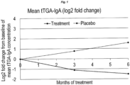
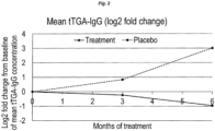
- HLA-type Probiotic group n (%) Placebo group n (%) p-value DR3-DQ2/DR4-DQ8 10 (25.0) 16 (42.1) 0.112 DR4-DQ8/DR4-DQ8 10 (25.0) 7 (18.4) 0.488 DR4-DQ8/DR8/DQ4 4 (10.0) 2 (5.2) 0.439 DR3-DQ2/DR3-DQ2 15 (37.5) 13 (34.2) 0.766 DR4/DR1 1 (2.5) 0 (0.0). 0.333 Table 4: Tissue transglutaminase autoantibody (tTGA) levels, IgA and IgG. p -values are comparisons between treatment and placebo at each visit.
- tTGA Tissue transglutaminase autoantibody
- Visit 1 Visit 2 (approx. 3 mo.) Visit 3 (approx. 6. mo.) tTGA-IgA, mean (median) Treatment 158.60 (4.71) 72.84 (3.07) 55.41 (2.69) Placebo 12.96 (4.38) 21.98 (4.93) 38.23 (3.72) p -value 1 0.8912 0.3013 0.6545 tTGA-IgG, mean (median) Treatment 166.98 (1.57) 141.74(1.64) 86.35 (1.61) Placebo 8.00 (1.60) 14.26 (1.56) 64.33 (1.36) p -value 1 0.8480 0.9373 0.5428 1 Wilcoxon ranked sum test, 2-sided
- Results - treatment with L. paracasei and L. plantarum strains decreases the proportion of natural killer T cells, counteracts the increased proportion of natural killer cells and reduces CD62L expression in cytotoxic T cells
- Flow cytometry analysis was used to examine any changes in the size and activation status of white blood cell populations at baseline and at each of the follow-up visits, in both study groups.
- CD62L is the cell adhesion molecule L-selectin, involved in lymphocyte interactions with endothelial cells, assisting entry into secondary lymphoid tissues.
- the presence of CD62L on the cell surface indicates a naive state (CD62L high ) whereas when the cells have been activated they release CD62L from their surface (CD62L low ).
- Table 5 NK-T cells CD3+CD56+ (% NK-T cells of lymphocytes)
- Group Statistics V0 V1 V2 V1-V0 V2-V0 Lactobacilli N 33 39 38 32 31 Missing 13 7 8 14 15 Min 0.10 0.10 0.10 -1.54 -3.67 Median 0.44 0.45 0.44 -0.09 -0.16 Max 4.74 3.27 1.60 1.57 1.00 Mean 0.77 0.67 0.51 -0.12 -0.29 Std 0.85 0.62 0.34 0.66 0.81 P-value NA NA NA 0.2082 0.0297 Placebo N 32 34 34 30 30 Missing 12 10 10 14 14 Min 0.12 0.22 0.19 -0.95 -0.94 Median 0.46 0.49 0.58 0.02 0.18 Max 1.26 1.30 2.35 0.97 1.
- T helper cells identified as CD3+CD4+, with sub-populations of CD4+ T helper cells differing between the two study groups.
- Table 18 shows that Lactobacillus treatment prevented an increase in the proportion of activated T helper cells by the second follow-up visit, where activation is identified by CD25, namely the interleukin-2 receptor alpha chain.
- Table 19 further shows that Lactobacillus treatment by the second follow-up visit, prevented an increase in the proportion of Tregulatory cells, identified as CD4+ cells with high CD25 positivity (CD25 high ).
- CD4+CD45RA+RO- (% CD45RA+CD45RO- of CD3+CD4+ cells or naive T-helper cells as % of lymphocytes)
- Group Statistics V0 V1 V2 V1-V0 V2-V0 Lactobacilli N 32 39 38 31 30 Missing 14 7 8 15 16 Min 38.93 48.45 49.57 -16.37 -14.55 Median 68.60 66.79 65.01 1.78 -0.59 Max 80.51 83.83 78.37 26.60 17.89 Mean 65.25 66.88 65.68 1.75 -0.08 Std 9.63 8.00 7.95 8.17 6.92 P-value NA NA NA NA 0.3105 0.7457 Placebo N 31 32 34 27 29 Missing 13 12 10 17 15 Min 42.72 46.64 47.24 -19.50 -28.78 Median 71.52 68.41 68.87 -1.72 -4.03 Max 85.28 82.13 82.39 12.27 19.53 Mean 69
- CD4+CD25+Foxp3+ T-cells in the placebo group which remained unchanged in the treatment group, could be explained by the downregulation effects of the two Lactobacillus strains on activated CD4+ cells.
- the observed reduction in CD3+CD4+ cells in the placebo group may be considered to be secondary to the compartmentalization of gluten-sensitive lymphocytes within the intestinal mucosa.
- CCR4 is an important chemokine receptor for recruitment of T-cells to the sight of inflammation and it is highly expressed on differentiated regulatory T cells ( lellem A, et al., Eur J Immunol. 2003;33(6):1488-96 ).
- the increases of CD4+CD25 high CD45RO+CCR4+ cells and CD4+CD25+Foxp3+ cells in the placebo group indicate an attempt to extinguish an ongoing intestinal inflammation and the immune response to dietary gluten antigens as previously described ( Frisullo G, et al., Human Immunol. 2009;70(6):430-5 ; Tiittanen M, et al., Clin Exp Immunol. 2008;152(3):498-507 ).
- the third finding of particular relevance was the peripheral changes in NK cells over time in the placebo group with ongoing coeliac autoimmunity, which was not observed in children that received probiotics.
- the population of NK and NK-T cells has been found to decrease in both tissue and in periphery in active coeliac disease ( Dunne MR, et al., PLoS ONE. 2013;8(10):e76008 ). This is in line with the findings of our study, where we found that NK cells increased in the probiotic group, but not in the placebo group. This further supports the importance of NK cells in coeliac disease and that the probiotic supplement may have a direct or indirect stimulatory effect on NK cells mirrored as a reduced autoimmune response in the periphery.
- IgA-tTGA and IgG-tTGA levels are valid diagnostic tests in CD
- the current clinical recommendations advocate the use of IgA-tTGA in children with normal total IgA levels, due to a higher specificity and clinical relevance (Husby S, et al., supra).
- the exemplary probiotic Lactobacilli used in this clinical study Lactobacillus plantarum HEAL9 and Lactobacillus paracasei 8700:2, showed suppressing effects on CD autoimmunity in children on a gluten-containing diet. This indicated that Lactobacillus strains can prevent and/or delay CD autoimmunity in 'at HLA-risk' individuals, suggesting a possible preventive application of probiotic Lactobacilli in CD.
- probiotic Lactobacilli species may delay or prevent the development of ongoing CD autoimmunity in children at genetic risk for CD. It indicates that the probiotic Lactobacillus strains of the invention can be used to delay and/or prevent progression from CDA to CD.
Landscapes
- Health & Medical Sciences (AREA)
- Life Sciences & Earth Sciences (AREA)
- Veterinary Medicine (AREA)
- Chemical & Material Sciences (AREA)
- Medicinal Chemistry (AREA)
- Pharmacology & Pharmacy (AREA)
- Animal Behavior & Ethology (AREA)
- General Health & Medical Sciences (AREA)
- Public Health (AREA)
- Epidemiology (AREA)
- Microbiology (AREA)
- Mycology (AREA)
- Molecular Biology (AREA)
- Engineering & Computer Science (AREA)
- Bioinformatics & Cheminformatics (AREA)
- Chemical Kinetics & Catalysis (AREA)
- Immunology (AREA)
- General Chemical & Material Sciences (AREA)
- Nuclear Medicine, Radiotherapy & Molecular Imaging (AREA)
- Organic Chemistry (AREA)
- Physiology (AREA)
- Nutrition Science (AREA)
- Medicines Containing Material From Animals Or Micro-Organisms (AREA)
- Medicinal Preparation (AREA)
- Micro-Organisms Or Cultivation Processes Thereof (AREA)
- Coloring Foods And Improving Nutritive Qualities (AREA)
Description
- The present invention relates to a defined combination of probiotic strains of lactobacilli for use in the prevention and/or treatment in a subject of celiac disease autoimmunity (CDA), or celiac disease (CD).
- The invention also provides compositions for such uses and methods of preventing and/or treating CDA and/or CD involving administering an effective amount of said probiotic strain to a subject.
- Celiac disease (CD) is a chronic immune-mediated disorder affecting the intestinal mucosa of the small bowel. It is caused by intolerance to gluten, the major storage protein found in wheat, rye and barley (Schuppan D, et al. Gastroenterology. 2009;137(6):1912-33). Classical symptoms and clinical signs of celiac disease include abdominal discomfort, distention and diarrhoea, followed by signs of malnutrition (e.g., weight loss, anemia and osteoporosis). However, a significant portion of patients lack symptoms and are diagnosed through screening (Ludvigsson JF, et al. Journal of Internal Medicine. 2011;269(6):560-71). Currently, treatment consists of a life-long gluten free diet (GFD).
- The pathophysiology of CD is not completely understood but is proposed to be T-cell driven. Following the digestion of gluten proteins in the small bowel, the resulting gliadin peptides somehow cross the epithelial barrier and are presented by antigen-presenting cells on MHC-II-structures, enabling the activation of gliadin-specific CD4+ T-helper (TH) 1 cells and CD8+ cytotoxic T (Tc) cells in the lamina propria. This results in upregulation of several cytokines, IFN-γ, TNF-α and IL-21 in particular, of which the former two causes the typical mucosal remodeling and villous atrophy through activation of myelofibroblasts while the latter is likely involved in maintaining the activity of the CD4+ cells (Schuppan D, et al., supra). In the last decade, concomitant, direct stimulation of the innate immune system by gliadins has been shown to be an additional important factor in the development of disease. This is currently attributed to upregulation of IL-15 signaling in dendritic cells and macrophages, causing mucosal damage through activation of intra-epithelial lymphocytes (IELs) (Londei M, et al., Molecular Immunology. 2005;42(8):913-8).
- The global prevalence of CD is roughly estimated at 1%, but varies greatly between ethnic groups and geographic location. Sweden ranks amongst the most heavily afflicted nations with an estimated prevalence of 1.5-3% (Ludvigsson JF, et al., supra). It is yet to be fully determined what processes help induce this dysfunctional reaction to the gliadin structures, and why disease prevalence varies between populations. There is a clear genetic component in CD as evidenced by the fact that almost all celiac patients are carriers of the DR3-DQ2 and/or the DR4-DQ8, haplotypes (Sollid LM, et al. The Journal of Experimental Medicine. 1989;169(1):345-50). In addition, a number of other less influential genes have also been found to affect the risk of disease, most of them related to the activation of the adaptive immune response (Hunt KA, et al., Nature Genetics. 2008;40(4):395-402). CD shares these genetic risk traits with several other autoimmune disorders, most importantly
type 1 diabetes (T1D), which shares its major susceptibility genes in the HLA-DQB1 and HLA-DRB1 loci as well as several non-HLA loci (Smyth DJ, et al., New England Journal of Medicine. 2008;359(26):2767-77). Nevertheless, genetics alone cannot explain CD, since only a small minority of those carrying these haplotypes develop the disease. The rapidly increasing incidence observed in many countries during the last decades also point to environmental factors contributing to the pathogenesis in some way. Important areas of investigation include infant feeding practices, breast milk feeding, (and variations or disturbances in the gut microbiota (De Palma G, et al., Advance: Bifidobacteria and Gram-negative bacteria differentially influence immune responses in the proinflammatory milieu of celiac disease. Journal of Leukocyte Biology. 2010;87(5):765-78). - While intestinal biopsy was previously considered the gold standard for the diagnosis of CD, several serological markers have been discovered of which tissue transglutaminase autoantibodies (tTGA) are currently the most common in clinical practice due to its high diagnostic sensitivity and specificity (van der Windt DA, et al., Diagnostic testing for celiac disease among patients with abdominal symptoms: a systematic review. Jama. 2010;303(17):1738-46). Furthermore, the revised guidelines from European Society for Paediatric Gastroenterology, Hepatology and Nutrition (ESPGHAN) in 2012 (Husby S, et al., ESPGHAN guidelines for the diagnosis of coeliac disease. Journal of Pediatric Gastroenterology and Nutrition. 2012;54(1):136-60) suggest that significantly elevated tTGA levels, if followed by appropriate further testing, may eliminate the need for biopsy to confirm the diagnosis. In the majority of CD patients, tTGA levels decrease after introduction of GFD.
- In children, such a decrease is also highly predictive of histological improvement in the gut mucosa (Bannister EG, et al., American Journal of Gastroenterology. 2014;109(9):1478-83), indicating that tTGA may be used not only as a binary diagnostic tool but also as a marker for disease activity and dietary compliance. However, this brings the question of how to manage asymptomatic patients who are found to have persistently elevated tTGA levels, so called CD autoimmunity (CDA), or more widely referred to as potential CD if confirmed with normal intestinal biopsy features. These children are at increased risk of developing CD (Liu E, et al., The New England Journal of Medicine. 2014;371(1):42-9), with no treatment options currently available to reduce or eliminate that risk other than a GFD.
- Earlier studies have shown that the microbiota of patients with active CD is composed to a greater degree of gram-negative pathogens compared to healthy controls and symptom-free patients (Nadal I, et al., Imbalance in the composition of the duodenal microbiota of children with coeliac disease. Journal of Medical Microbiology. 2007;56(Pt12):1669-74). Accordingly, later studies have suggested that such a microbiota potentiates a higher degree of inflammatory reaction in response to gliadins (De Palma G, et al., Advance: Bifidobacteria and Gram-negative bacteria differentially influence immune responses in the proinflammatory milieu of celiac disease. Journal of Leukocyte Biology. 2010;87(5):765-78) and conversely, that certain Bifidobacterium strains influence the digestion of gliadins and reduces their immunological potential (Laparra JM, et al., Bifidobacteria inhibit the inflammatory response induced by gliadins in intestinal epithelial cells via modifications of toxic peptide generation during digestion. Journal of Cellular Biochemistry. 2010;109(4):801-7).
- Some recently published studies have examined the effect of administering specific Bifidobacterium strains in the context of already clinically manifested CD. Olivares et al. (Olivares M, et al., The British Journal of Nutrition. 2014;112(1):30-40) randomized 36 children recently diagnosed with CD to treatment with either daily consumption of Bifidobacterium longum CECT 7347 or placebo for 3 months, in addition to GFD. Due to introduction of GFD, the probiotic effect on immunologic parameters is difficult to appreciate; the study did not examine tTGA levels, but did find a significant decrease in total levels of mature T cells in the treatment group compared to placebo. In addition, they showed that children in the probiotic-treated group achieved a greater height percentage gain compare with controls.
- Also, Smecuol et al. Journal of Clinical Gastroenterology. 2013;47(2):139-47) treated 22 adult CD patients currently not on GFD with Bifidobacterium infantis or placebo daily for 3 weeks and evaluated intestinal permeability, immunological parameters, and changes in symptoms. Participants in the probiotic-treated group reported improvement of gastrointestinal symptoms compared to controls. However, unlike B. longum CECT 7347, B. infantis (NLS) failed to influence inflammatory markers and the gut microbiota and host-related defence mechanisms.
- Whilst both of the above studies with specific Bifidobacterium strains suggest some beneficial role for those Bifidobacterium strains in CD, they also highlight the need for further studies. Further search has its focus on the role of metabolites of lactobacilli ( SARNO MARCO ET AL: "Lactobacillus paracasei CBA L74 interferes with gliadin peptides entrance in Caco-2 cells",INTERNATIONAL JOURNAL OF FOOD SCIENCES AND NUTRI, CARFAX PUBLISHING LTD, GB, vol. 65, no. 8, 1 December 2014 (2014-12-01), pages 953-959).
- Thus, despite significant investigations of CDA and CD, currently the only available treatment for CD is a gluten free diet (GFD). Hence, there is a need for compositions and methods for preventing and/or treating celiac disease autoimmunity (CDA), or for preventing and/or treating celiac disease (CD).
- According to the invention there is a specific combination of two Lactobacillus species for use in a subject for the prevention and/or treatment of celiac disease autoimmunity (CDA), or for the prevention and/or treatment of celiac disease (CD).
- By "use for the prevention and/or treatment" we mean a use which gives rise to an effect in a subject of preventing, delaying, reducing the severity and/or removing one or more symptoms and/or other markers associated with CDA or CD. The scope of the invention is defined by the appended claims.
- The following probiotic strains of a Lactobacillus species are disclosed L. paracasei; L. plantarum; L. acidophilus; L. rhamnosus; L. casei; L. reuteri; L. brevis; L. crispatus; L. bulgaricus; L. fermentum; L. salivarius; L. johnsonii; and L. lactis
- Lactobacilli have been deposited under the Budapest Treaty by Probi A.B., Sölvegatan 41, Lund 22370, Sweden, as follows:-
Species Strain Accession No. Date of deposit Depositor Depositary Lactobacillus plantarum HEAL 9 DSM 15312 27/11/2002 Probi DSMZ HEAL 19 DSM 15313 27/11/2002 Probi DSMZ HEAL 99 DSM 15316 27/11/2002 Probi DSMZ 299 DSM 6595 02/07/1991 Probi DSMZ 299v DSM 9843 16/03/1995 Probi DSMZ GOS42 DSM 32131 02/09/2015 Probi DSMZ Lactobacillus paracasei 8700:2 DSM 13434 06/04/2000 Probi DSMZ O2A DSM 13432 06/04/2000 Probi DSMZ Lactobacillus rhamnosus 271 DSM 6594 02/07/1991 Probi DSMZ - According to the claims Lactobacillus paracasei 8700:2 (DSM 13434) is used in combination with probiotic strain Lactobacillus plantarum HEAL 9 (DSM 15312).
- The compositions may comprise the specified probiotic strain or strains of Lactobacilli, but preferably they consist of the specified strains without another effective amount of any other probiotic strains of Lactobacilli or other micro-organisms.
- The probiotic strains of the invention are preferably freeze-dried.
- The probiotic strains of the invention may be provided together with a suitable carrier, diluent or excipient as a solid or liquid formulation, which may be a pharmaceutical formulation in on embodiment.
- Examples of a suitable liquid carrier include water and other aqueous solvents.
- Examples of a suitable solid carrier include maltodextrin, inulin, potato starch, corn starch or other vegetable starch, microcrystalline cellulose (MCC), and sugar alcohols.
- The composition may be a dry fermented or non-fermented composition. In the case of a dry non-fermented composition, fermentation takes place in the gastrointestinal tract after ingestion of the composition by a subject.
- In use, the probiotic strain(s) of the invention may be mixed with a liquid or solid carrier before administration to a subject. For example, the subject may mix the strain(s) with a carrier consisting of water or some other aqueous solvent, or a drink prior to intake. Similarly, the probiotic strains may be mixed with a carrier consisting of one or more foods. Preferred foods are gluten free products such as fermented or non-fermented dairy products such as yoghurts, fruit juices; beverages, soups, plant based foods such as soy products, dry food bars, baby food, infant nutrition, infant formula, breast milk replacements from birth.
- Infant or baby formula milk is a particularly preferred carrier for the probiotic of Lactobacilli strain(s) of the invention. It may be in a dry powder form for mixing with water before feeding it to babies as a ready-to-feed liquid form. It is normally made from cows' milk and contains whey and casein protein.
- The probiotic strain(s) of the invention may also be provided in a composition together with one or more ingredients of known dietary supplements, for example, micronutrients such as vitamins and minerals.
- Classical symptoms and clinical signs of celiac disease include abdominal discomfort, distention and diarrhea, followed by signs of malnutrition (e.g., weight loss, anemia and osteoporosis). Considering the risks involved with malnutrition, it has been published that children newly diagnosed with celiac disease were deficient in vitamin D, zinc and iron (Erdem et al., Vitamin and mineral deficiency in children newly diagnosed with celiac disease. Turk J Med Sci. 2015;45(4):833-6 2015). Similarly, adults with newly diagnosed celiac disease and untreated CD patients were also found to have values below the limits of reference for vitamins B6 and B12, vitamin D, folic acid, zinc, magnesium and iron (Wierdsma et al., Vitamin and mineral deficiencies are highly prevalent in newly diagnosed celiac disease patients. Nutrients. 2013 Sep 30;5(10):3975-92. doi: 10.3390/nu5103975, Caruso et al., Appropriate nutrient supplementation in celiac disease 2013 Ann Med. 2013 Dec;45(8):522-31. doi: 10.3109107853890.2013.849383. Review, Schøsler et al., Symptoms and findings in adult-onset celiac disease in a historical Danish patient cohort Scandinavian Journal of Gastroenterology Vol. 51, ISS. 3,2016). In most of the cases the removal of gluten from the diet, once a person has been diagnosed with CD or CDA, restores the "healthy" histology of the gut and leads to normalization of the vitamin and mineral status. However, resolution of mucosal inflammation by adapting to a gluten free diet is not always enough to counteract mineral deficiency (Caruso et al. 2013 supra). Hence, supplementation of people diagnosed with CD/CDA or at risk of developing CD/CDA with vitamins and/or minerals may be used together with the probiotic Lactobacilli according to the invention. Preferably, the vitamin(s) and/or mineral(s) are selected from one or more of the vitamins: A, B6, B12, D; and/or the minerals: iron, zinc, magnesium.
- Preferably, the probiotic strain(s) compositions the invention are provided in the form of a capsule or tablet or a powder for oral administration. Stick packs are a popular type of single-portion/single dose packaging used in the food industry and the pharmaceutical sector (see www.selo.com/packaging-machines/stick-packs/). They are very convenient for consumers to use and by containing a predetermined amount of the probiotic compositions of the invention, ensure the correct dose is taken to achieve a desired preventative and/or therapeutic effect according to the invention.
- Preferably, in use the probiotic strain(s) of the invention are administered to a subject in a daily amount of from 1 × 10⁶ to 1 × 10¹⁴ colony forming units (CFU), preferably from 1 × 10⁹ to 1 × 10¹¹ CFU and most preferably 1 × 10¹⁰ CFU. The daily amount of CFU is preferably administered in a single dose or serving.
- Preferably, the subject is a human. Advantageously, the human subject is a child. Preferably the child is less than 18, 17, 16, 15, 14, 13, 12, 11, 10, 9, 8, 7, 6, 5, 4, 3, 2, or 1 years old. Advantageously, the compositions of the invention are for administration from birth and especially from weaning, i.e. the point at which the baby ceases to be fed entirely by breast milk.
- Preferably the subject lacks one or more of the symptoms of CD but is at an increased risk of developing CD.
- Ideally, the subject is identified as being at an increased risk of CD by the presence of one or more serological, immunological and/or genetic risk factors.
- A variety of methods for detecting the presence of serological, immunological and/or genetic risk factors are well known to skilled persons; but examples of particularly suitable methods are provided herein for convenience.
-
- Serotyping
- ∘ DQ2 positive
- ∘ DQ8 positive
- Genetic testing
- ∘ DQ2.5 cis haplotype (DQA9*05/DQB1*02)
- ∘ DQ2.2 cis haplotype (DQA1*02:01/DQB1*02:02)
- ∘ DQ2.5 trans haplotype, e.g. DQ2.2 cis haplotype (DQA1*02:01/DQB1*02:02) with DQ7.5 cis haplotype (DQA1*05:05/DQB1*03:01)
- ∘ DQ8 cis haplotype (DQA1*03/DQB1*03:02)
- ∘ DQA1*05:01 allele
- ∘ DQA1*05:05 allele
- ∘ DQB1*03:02 allele
- Increased risk of developing celiac disease can be determined by immunological serotyping and/or genetic analysis. Immunological serotyping is used to identify the presence of the DQ2 serotype marker and/or the DQ8 serotype marker. Genetic analysis of genomic DNA can be performed by sequence-specific PCR using sequence-specific primers (PCR-SSP) or by gene sequencing. Sequence-specific PCR methods include: PCR with sequence-specific primers (PCR-SSP) (Sacchetti L, et al., Rapid Identification of HLA DQA1*0501, DQB1*0201, and DRB1*04 Alleles in Celiac Disease by a PCR-Based Methodology. Clin Chem. 1997 Nov;43(11):2204-6.); quantitative real-time PCR (qPCR) with sequence-specific primers and TaqMan probes (Reinton et al., A one-step real-time PCR assay for detection of DQA1*05, DQB1*02 and DQB1*0302 to aid diagnosis of celiac disease. J Immunol Methods. 2006 Oct 20;316(1-2):125-32. Epub 2006 Sep 18.); qPCR with sequence-specific primers and melting curve analysis (Selleski et al., Simplifying celiac disease predisposing HLA-DQ alleles determination by the real time PCR method. Arq Gastroenterol. 2015 Apr-Jun;52(2):143-6. doi: 10.1590/S0004-28032015000200013.); PCR amplification followed by hybridization with sequence-specific oligonucleotides Gene sequencing methods include Sanger sequencing with chain-terminating dideoxynucleotides, pyrosequencing, sequencing by synthesis (Illumina sequencing), sequencing by ligation (SOLiD sequencing), nanopore sequencing (MinION), Ion Torrent semiconductor sequencing and single-molecule real-time sequencing (Pacific Biosciences).
- Genetic markers corresponding to DQ2 or DQ8 serotypes include: DQ2.5 cis haplotype (DQA1*05/DQ89*02); DQ2.2 cis haplotype (DQA1*02:01/DQB1*02:02); DQ2.5 trans haplotype, e.g. DQ2.2 cis haplotype (DQA1*02:01/DQB1*02:02) with DQ7.5 cis haplotype (DQA1*05:05/DQB1*03:01); DQ8 cis haplotype (DQA1*03/DQB1*03:02); DQA1*05:01 allele; DQA1*05:05 allele; DQB1*03:02 allele.
- Immunological markers for CDA and/or CD include: tissue transglutaminase antibody level, including by radioligand binding assays (Agardh et al., Using radioligand-binding assays to measure tissue transglutaminase autoantibodies in young children. Acta paediatrica (Oslo, Norway : 1992). 2004;93(8): 1046-51, Agardh et al., Prediction of silent celiac disease at diagnosis of .); IgA Endomysial antibody (EMA) level, including in the absence of a deficiency in total serum IgA; deaminated gliadin peptide (DGP IgA and IgG) levels.
- Preferably, the one or more risk factor(s) is HLA-DQ2 and/or HLA-DQ8.
- Preferably, the one or more immunological risk factors is persistent tissue transglutaminase (tTGA) positivity, i.e. a tTGA positive test on two or more consecutive occasions.
- tTG positivity can be shown by the presence of tTG autoantibodies, preferably IgA-tTGA and/or IgG-tTGA, as described herein.
-
-
Fig 1 : shows the change in mean tTGA-lgA level over time in the L. paracasei + L. plantarum strain treatment group compared to the placebo group. -
Fig 2 : shows the change in mean tTGA-IgG level over time in the L. paracasei + L. plantarum treatment group compared to placebo. - The following materials, methods and examples embody aspects of the present invention.
- Participants were recruited from the CiPiS and Swedish participants of TEDDY study. The TEDDY study protocol has been thoroughly described elsewhere (The Environmental Determinants of Diabetes in the Young (TEDDY) study: study design. Pediatric Diabetes. 2007;8(5):286-98). In short, between 2004 and 2010, newborns at these centers were HLA genotyped and considered eligible if carrying one of the following genotypes: DR3-DQ2/DR4-DQ8, DR4-DQ8/DR4-DQ8, DR4-DQ8/DR8, DR3- DQ2/DR3-DQ2, DR4-DQ8/DR4b, DR4-DQ8/DR1, DR4-DQ8/DR13, DR4-DQ8/DR9, or DR3-DQ2/DR9. At the Swedish center, a total of 48,140 children were screened of whom 3,723 were found to be HLA-eligible. Of these, written informed consent to study participation was provided by parents or primary caretakers in a total of 2,525 cases. Within the TEDDY protocol blood samples are collected every 6 months and children are annually screened for tTGA starting at 2 years of age. Upon showing tTGA positivity, earlier blood samples are analyzed retrospectively to determine the time of seroconversion. Participants testing positive in 2 consecutive blood samples are considered persistently tTGA positive, and ruled in for CDA. All definitive results are based on laboratory analysis performed in the European reference laboratory at Southmead Hospital, Bristol, United Kingdom. Further evaluation concerning CD is outside the scope of the TEDDY protocol. In Sweden, the decision to perform an intestinal biopsy is made on a case-by-case basis depending on tTGA levels, degree of symptoms and signs of malnutrition.
- In the CiPiS study, children born in Skåne between 2000 and 2004 were screened using HLA genotyping of cord blood and a questionnaire concerning maternal factors. Children carrying DQ2 and/or DQ8 were considered at HLA risk. A total of 6202 children at risk were invited, as well as 7654 children lacking HLA risk as controls. tTGA screening was performed at
age 3 in 1620/6206 (26.1%) of participants in the HLA-risk group and 1815/7654 (23.7%) of controls, and at age 9 in 1910/5870 (32.5%) of participants with HLA-risk and 2176 of 7072 (30.6%) of controls. If tested tTGA positive, participants were re-tested at least 3 months later to confirm persistent tTGA positivity, whereupon they were referred to the local pediatric clinic for an intestinal biopsy. Participants were continuously invited from these 2 cohorts between March 2012 and August 2015. Inclusion criteria were as follows: - tTGA positivity in 2 consecutive samples, <30 U/ml using the Bristol tTGA assay.
- No diagnosis of celiac disease.
- Negative in all diabetes-associated autoantibodies screened for in the TEDDY protocol (GADA, IAA, IA-2A, ZnT8A).
- Not participating in the TEDDY study through a long-distance protocol.
- Children positive in T1D-associated autoantibodies were excluded from the present study to minimize influences on the natural history of islet autoimmunity. However, it will be appreciated that T1D-associated autoantibodies constitute a serological marker for increased risk of developing CD and as such can be used to identify preferred subjects for preventative and/or therapeutic treatment according to the uses and methods of the present invention.
- Children matching the inclusion criteria were invited for an initial meeting, then scheduled for follow-up visits approximately 3 and 6 months later. Participants were randomized at a 1:1 ratio to treatment or control group. At the initial meeting and first follow-up visit, each group was provided with a powdered formulation containing Lactobacilli, or a placebo, respectively, and instructed to consume the product daily by ingestion of the powder after dissolution in 100 ml of cold liquid or after mixing with fruit/food, in association with a meal once daily, for a total period of 6 months, to halt consumption of any other food products containing probiotics, and to store the sachets refrigerated (2-8°C). The parents were also instructed not to add the powder to hot drinks or hot food. At every visit, 10 ml venous blood and a fecal sample was collected. Allocation to the treatment or control group was blinded to participants, clinicians and lab personnel.
- Radioligand binding assays (RBA) were used to assess tTGA as previously described ( (Agardh D, et al., Pediatric Diabetes. 2001 ;2(2):58-65) (Agardh D et al., Using radioligand-binding assays to measure tissue transglutaminase autoantibodies in young children. Acta paediatrica (Oslo, Norway: 1992). 2004;93(8):1046-51). In short, human tissue transglutaminase (tTG) was synthesized by in vitro transcription and translation of cDNA using the TNT SP6 Coupled Reticulocyte Lysate System (Promega, Madison, Wl, USA) in the presence of 35S-methionine (Perkin Elmer, Waltham, MA, USA). Both IgG-tTGA and IgA-tTGA were analyzed. For IgG-tTGA analysis, 35S-tTG was diluted and added to human serum and incubated overnight at 4°C. Protein A sepharose (PAS) (Invitrogen, Thermo Fisher Scientific, Carlsbad, CA, USA) was used to separate free and antibody-bound 35S-tTG by binding IgG in serum. PAS and immunoprecipitated 35S-tTG-serum were added to a 96 well MultiScreenHTS DV Filter Plate (Merck Life Science, Darmstadt, Germany) and incubated on a plate shaker, followed by washing. OptiPhase Supermix scintillation cocktail (Perkin Elmer) was added and reactivity was measured in a MicroBeta Counter TriLux (Perkin Elmer). IgA-tTGA analysis was performed similarly, except goat anti-human IgA-agarose (Merck Life Science) was used instead of PAS. The levels of TGA were expressed as U/mL calculated from standard curves containing approximately 2, 4, 8, 16, 31, 63, 125, 250, 500 and 1000 U/mL of respective IgA-tTGA and IgG-tTGA. The cut off level for positive values of IgG-tTG and IgA-tTG was set at >4.0 U/mL, which represented the 99th percentile of 398 adult blood donors (Agardh D, et al., Acta paediatrica. 2004;93(8):1046-51). When selective IgA deficiency was suspected the participants were tested for total IgA levels as part of their original studies; however, no such condition was detected among the participants.
- The lactobacilli culture was prepared by Probi AB, Lund, Sweden. Active product consisted of freeze-dried L. paracasei 8700:2 (DSM 13434) in combination with L. plantarum HEAL 9 (DSM 15312) with maltodextrin (Glucidex IT-19, Roquette, France) in the form of a powder containing a combined 1 × 10¹⁰ CFU, with each strain being represented equally. Placebo consisted of powdered maltodextrin and yeast peptone (HYP-A, BioSpringer, France) to adjust colour and taste, so that the two test products (probiotic and placebo) were identical in appearance and taste.. Participants were instructed to store the product in the refrigerator, and consume a one gram sachet of product every morning. It will be appreciated that it is the total CFU of Lactobacillus rather than the concentration (i.e. mass or number of Lactobacillus per unit of mass or volume of the composition) that is relevant to the efficacy of the product.
- Peripheral blood mononuclear cells (PBMCs) from the cohorts in Example 1 were separated from whole blood by a density gradient centrifugation (1800 G) using a hydrophilic polysaccharide (BD Vac ® CPT™ Cell Preparation Tube NC FICOLL™ 4mL, Cat No 362760 Becton Dickinson, NJ, USA). Cells were separated within 2-24 hours after blood sample collection. The aspirated interphase mononuclear layer was washed three times with RPMI-1640 Medium with L-Glutamine (GIBCO no 21875034, Thermo Fisher Scientific, Gothenburg, Sweden). The cells were counted in an Abbott CELL_DYN Ruby to a final concentration of 1-4 × 10⁶ /mL Lymphocytes in RPMI-1640 (i.e. the volume of the PBMC suspension was adjusted so that there were 1-4 × 10⁶ lymphocytes per mL).
- Following counting, PBMCs were stained with monoclonal antibodies directed to surface and intracellular structures. The monoclonal antibodies (Table 2) were pre-labelled with four fluorochromes, fluorescein isothiocyanate (FITC), Phycoerythrin (PE), Peridinin chlorophyll protein (PerCP) and Allophycocyanin (APC). Stained cells were analysed using a four-colour FACSCalibur® instrument (BD Biosciences, CA, USA) by passing via a laser beam. The FACSCalibur® defines cell populations and count the cells by their characteristic features of size, granularity and fluorescence intensity. Optical detectors amplify and convert the light signals to electrical data signals. The data were acquired and analyzed in CellQuestPro® software (BD Biosciences).
Table 1: Antibodies used in flow cytometry analysis. Antibody & fluorochrome Supplier Catalogue No CCR9 APC R&D Systems, Inc., Abingdon, UK FAB17991A CD45RO APC BD Biosciences, CA USA 559865 CD62L APC BD Biosciences, CA USA 559772 IgG1 isotype APC BD Biosciences, CA USA 555751 CD25 FITC BD Biosciences, CA USA 555431 CD3 FITC BD Biosciences, CA USA 555332 CD38 FITC BD Biosciences, CA USA 340909 CCR4 PE BD Biosciences, CA USA 551120 CD8 PE BD Biosciences, CA USA 345773 CD45RA PE BD Biosciences, CA USA 555489 Integrin beta7 PE BD Biosciences, CA USA 555945 CD19 PerCP BD Biosciences, CA USA 345778 CD4 PerCP BD Biosciences, CA USA 345770 CD8 PerCP BD Biosciences, CA USA 345774 CD3 FITC / CD16 + CD56 PE BD Biosciences, CA USA 342403 IgG2a isotype FITC / IgG1 isotype PE / CD4 PerCP-Cy 5.5 BD Biosciences, CA USA 340965 Human IgG Isotype Control Fisher Scientific, Gothenburg, Sweden 12000C eBioscience™ Human Regulatory T Cell Staining Kit (CD4, CD25, FoxP3) AH diagnostics AB, Solna, Sweden 88-8999-40 Table 2: Antibody combinations and concentrations used in flow cytometry analysis. Direct Staining Tube Mixture Panel Label Mab prep in 100µL Dilution Number of lymphocytes acquired, collected and gated 1 Isotype control FITC/PE/PerCP 5 µL 1/20 3000 APC 2,5 µL 1/40 2 CD3 FITC 5 µL 1/20 10000 CD16+56 PE 5 µL 1/5 CD19 PerCP 20 µL 3 CD3 FITC 5 µL 1/20 10000 CD45RA PE 0,4 µL 1/250 CD4 PerCP 5 µL 1/20 CD45RO APC 20 µL 1/5 4 CD3 FITC 5 µL 1/20 10000 CD45RA PE 0,4 µL 1/250 CD8 PerCP 2,5 µL 1/40 CD45RO APC 20 µL 115 5 CD3 FITC 5 µL 1/20 10000 CD8 PE 0,4 µL 1/250 CD4 PerCP 5 µL 1/20 CD62L APC 2,5 µL 1/40 6 CD25 FITC 10 µL 1/10 100000 CD45RA PE 0,4 µL 1/250 CD4 PerCP 5 µL 1/20 CD45RO APC 20 µL 1/5 7 CD25 FITC 10 µL 1/10 50000 CCR4 PE 2,5 µL 1/40 CD4 PerCP 5 µL 1/20 CD45RO APC 20 µL 1/5 8 CD25 FITC 10 µL 1/10 50000 CCR4 PE 2,5 µL 1/40 CD4 PerCP 5 µL 1/20 CD62L APC 2,5 µL 1/40 9 CD45 RA FITC 10 µL 1/10 50000 Integrin β7 PE 10 µL 1/10 CD8 PerCP 5 µL 1/20 CCR9 APC 5 µL 1/20 10 CD3 FITC 5 µL 1/20 50000 Integrin β7 PE 10 µL 1/10 CD4 PerCP 5 µL 1/20 CCR9 APC 5 µL 1/20 11 CD3 FITC 5 µL 1/20 50000 Integrin β7 PE 10 µL 1/10 CD8 PerCP 5 µL 1/20 CCR9 APC 5 µL 1/20 12 CD38 FITC 10 µL 1/10 50000 Integrin β7 PE 10 µL 1/10 CD4 PerCP 5 µL 1/20 CD62L APC 2,5 µL 1/40 Human Regulatory T Cell Staining Kit Tube Mixture Panel Label Mab prep in 100µL Dilution Number of cells to acquire 13 Isotype control IgG2a PE 20 µL 1/5 3000 14 CD4/CD25 FITC/APC 20 µL mix 1/20 & 1/5 50000 FoxP3 PE 20 µL 1/5 - PBMCs were divided into two tubes: tube A for blocking of PBMCs at about 2-4 × 10⁶ lymphocytes/ml in RPMI; tube B for PBMCs for staining without blocking, diluted to about 1-2 × 10⁶ lymphocytes/ml by addition of RPMI. PBMCs in tube A were blocked by incubating with normal human IgG (1/34 dilution) for 15 minutes at 2-8°C protected from light.
- Antibody mixtures according to tubes 1-12 of the table above were prepared in FACS tubes (BD Falcon, VWR cat no 352052). Blocked PBMCs from tube A were added to tubes 9-11 and non-blocked PBMCs from tube B were added to tubes 1-8 and 12. FACS tubes 1-12 were vortexed gently and incubated for 30 minutes at 2-8°C protected from light. Stained PBMCs were washed by adding 2 ml cold phosphate buffered saline (PBS, pH 7.4, Gibco cat no 10010-015, Fisher Scientific, Gothenburg, Sweden), vortexing gently, centrifuging (10 minutes, 400 × g), and discarding the supernatant. Stained PBMCs were fixed by resuspension in 200 µl cold PBS-1% formaldehyde solution (3 parts PBS, pH 7.4, Gibco cat no 10010-015 to 1
part 4% formaldehyde solution, Apoteket cat no 34 24 36) and incubation at 2-8°C overnight protected from light. - FACS tube 13 (see table above) was prepared with 100 µl non-blocked PBMCs from tube B. FACS tube 14 (see table above) was prepared for surface staining with 20 µl of the CD4 FITC / CD25 APC cocktail and 100 µl blocked PBMCs from tube A, vortexed gently, then incubated for 30 minutes at 2-8°C protected from light. PBMCs in tubes 13 and 14 were washed by adding 2 ml cold PBS, vortexing gently, centrifuging (5 minutes, 400 × g), and discarding the supernatant. PBMCs in tubes 13 and 14 were fixed by resuspension in 1ml Fixation/Permeabilization Solution (one part Fixation/Permeabilization 4X Concentrate and three parts Fixation/Permeabilization Diluent) and incubation at 2-8°C overnight protected from light.
- Fixed PBMCs (tubes 13 and 14) were washed and permeabilized by centrifuging (10 minutes, 400 × g) discarding the supernatant, resuspending in 2 ml 1X Permeabilization Buffer (one part 10X Permeabilization Buffer and nine parts distilled water), vortexing gently, repeating the preceding four steps again, and finally centrifuging again (10 minutes, 400 × g). Permeabilized PBMCs in tube 14 were blocked with 2 µl normal rat serum by incubating for 15 minutes at 2-8°C protected from light. Intracellular staining was performed by adding 20 µl rat IgG2a isotype control PE to tube 13 and 20 µl anti-human FoxP3 PE to tube 14, followed by incubation for 30 minutes at 2-8°C protected from light. Stained PBMCs were washed with 2 ml PBS, gently vortexed, centrifuged (10 minutes, 400 × g), and the supernatant was discarded before resuspending the cells in 200 µl PBS prior to flow cytometry analysis.
- The Becton Dickinson FACSCalibur® can detect 6 different parameters of a single particle or cell in an optical system. The instrument converts light scattering and fluorescence intensity into digital pulses. Forward Scatter (FSC) provides a measure of size and Side Scatter (SSC) provides a measure of cytoplasmic granularity. The instrument has four-color fluorescence detectors: the blue (488nm) laser detects FL1, FL2 and FL3 and an additional red diode laser (635nm) detects FL4.
- After regular system cleaning of the instrument a calibration check with 3-color CaliBRITE Beads (BD no 340486) and CaliBRITE APC Beads (BD no 340487) and AutoCOMPTM Software was performed for setting photomultiplier tube (PMT) voltages and adjusting and optimizing the fluorescence compensation of the detectors (FL1; FL2, FL3 and FL4). The CaliBRITE beads are of exact size and are labelled with exact amount of fluorochromes simulating unstained and stained leucocytes. AutoCOMP™ Software generates a CalibFile. All of the monoclonal antibodies were pre-titrated with the Calib File instrument setting. CellQuest Pro Software was used to run the BD FACSCalibur. 3000-10000 acquired, collected and gated lymphocytes were measured for each tube according to the table above.
- Isotype-matched control antibodies (IgG2/IgG1/CD4 isotype (FITC, PE and PerCP-Cy 5.5) and IgG1 isotype (APC), BD biosciences, CA USA) were used to set the dot plot quadrant and calculate the percent of lymphocyte populations through subtraction of contaminating non-lymphocytes. Lymphocytes were identified and gated in a dot plot displaying side scatter (cytoplasmic granularity) and FL1 parameter (FITC positive intensity) CD3+ T-cells. A multicolour back gate of lymphocytes was then shown in a FSC and SSC dot plot. A region was set around these identified lymphocytes (lymphocyte gate). New dot plots were displayed from the lymphocyte gate with different two-parameter combinations (FL1, FL2, FL3 and FL4) of the stained monoclonal antibodies of interest and the characteristic subsets were then identified. Quadrants were set in the two parameter dot plots from the isotype control, negative population. A subset is reported as a percentage of a quadrant population in the lymphocyte gate. Non-stained negative cells in lymphocyte gate were subtracted. The term "leucocytes" as used herein means PBMCs (i.e. the total population of isolated cells).
- Specifically, subgroups of T cells, CD4+ (Th) or CD8+ (Tc) cells were gated from the lymphocyte gate. From these gates, naïve cells were gated as CD3+CD4/CD8+CD45RA+CD45RO- and memory cells as CD3+CD4/CD8+CD45RA-CD45RO+. Activated and differentiated effector and memory cells were gated as CD3+CD4/8+CD62L+, CD4+CD25+CD45RA+CD45RO+, CD4+CD25+CCR4+CD45RO, CD4+CD25+CCR4+CD62L+, CD8+CD45RA+CCR9+ β7+, CD3+CD4+ICD8+ β7+CCR9+, CD4+CD38+ β7+CD62-. B cells were defined as CD3-CD19+ lymphocytes. From the lymphocyte gate, CD4+ cells were gated, followed by gating for CD25+ cells. This population was then examined for the expression of FoxP3+ cells. From the CD4+CD25+ gate, the percent with the highest CD25 expression, CD4+CD25high was determined. The CD4+CD25high lymphocyte population was then examined further for the expression of FoxP3. NK cells were gated from the lymphocyte gate. From this gate NK cells were gated as CD3-CD16+/CD56+ cells.
- The lymphocyte assay region contained >70% lymphocytes. The intra-assay analysis of CD3+ T-cells showed a covariance of 3% and the inter-assay analysis of CD3+ T-cells showed a covariance of 5%.
- The study outcome was coeliac autoimmunity assessed as changes in serum levels of tTG autoantibodies (one child was excluded due to insufficient volumes) and changes in peripheral immune response of B cells, NK cells, and subpopulations of regulatory T cells after 6 months. Comparison between groups of binary variables was done by means of Fisher's exact test. Comparison between groups on continuous and ordered categorical data is done by the Wilcoxon Rank Sum test, e.g. Wilcoxon rank-sum test was used to compare tTGA levels between the probiotic-treated group with the placebo group at 0, 3 and 6 months. To compare the changes in tTGA levels over time within each group, Wilcoxon signed rank test for continuous variables was used. Levels of IgA- tTGA and IgG-tTGA were analyzed as separate data sets. Likewise, Wilcoxon rank-sum test was applied for the comparison between the probiotic-treated group and placebo, with regard to changes measured from baseline to 3 and 6 months for the parameters analysed by flow cytometry. Wilcoxon signed rank test was used for measuring the differences from baseline at 3 and 6 months within each group. A t-test was used to compare HLA distribution between groups.
- Missing data were not imputed, i.e. the analyses are on observed cases. To avoid the effect of drop-outs and to restrict the comparison of the treatment to those children who adhered perfectly to the clinical trial instructions, intention-to-treat analysis and per-protocol-analysis were performed. All reported p-values are 2-sided and were not adjusted for multiplicity (i.e. nominal). A p-value of < 0.05 was considered of statistical significance. Statistical analyses were performed in StatXact version 10.1 (Cytel, Cambridge, MA, USA).
- A total of 118 children fulfilled the inclusion criteria and were invited to study participation. Of those, 90 children and their caretakers agreed to study participation: 1 from the CiPiS study and the remaining 89 from the TEDDY study. Twelve (13%) of these left the study after the initial visit. One child was excluded due to blood samples being of insufficient volumes to perform tTGA analysis. In total, 77 (87%) children were included in the final data set; 40 (52%) in the probiotic-treated group and 37 (48%) in the placebo group, respectively. Baseline characteristics of these groups are shown in Table 3 and HLA distribution in Table 3A. Mean and median study duration was 188 and 190 days, respectively (Q1: 176.5 days, Q3: 203 days, spread 153 - 237 days).
Table 3: Baseline characteristics at start of study (first visit). Treatment n = 40 Placebo n = 37 p-value Age, years, mean 4.85 4.42 0.2841 Sex, n 0.1722 Boys 22 14 Girls 18 23 Weight, kg, mean 22.0 20.8 0.1821 Length, cm, mean 114.0 110.8 0.2431 1Wilcoxon Rank Sum test, 2-sided; 2Fishers Exact test, 2-sided Table 3A: HLA distribution of the study population. HLA-type Probiotic group n (%) Placebo group n (%) p-value DR3-DQ2/DR4-DQ8 10 (25.0) 16 (42.1) 0.112 DR4-DQ8/DR4-DQ8 10 (25.0) 7 (18.4) 0.488 DR4-DQ8/DR8/DQ4 4 (10.0) 2 (5.2) 0.439 DR3-DQ2/DR3-DQ2 15 (37.5) 13 (34.2) 0.766 DR4/DR1 1 (2.5) 0 (0.0). 0.333 Table 4: Tissue transglutaminase autoantibody (tTGA) levels, IgA and IgG. p-values are comparisons between treatment and placebo at each visit. Visit 1 Visit 2 (approx. 3 mo.) Visit 3 (approx. 6. mo.) tTGA-IgA, mean (median) Treatment 158.60 (4.71) 72.84 (3.07) 55.41 (2.69) Placebo 12.96 (4.38) 21.98 (4.93) 38.23 (3.72) p-value1 0.8912 0.3013 0.6545 tTGA-IgG, mean (median) Treatment 166.98 (1.57) 141.74(1.64) 86.35 (1.61) Placebo 8.00 (1.60) 14.26 (1.56) 64.33 (1.36) p-value1 0.8480 0.9373 0.5428 1Wilcoxon ranked sum test, 2-sided - After 3 months, levels of tTG-IgG decreased a mean 29.4±513 U/mL in the probiotic group and increased in the placebo group a mean 6.3±48 U/mL as compared to baseline levels (p=0.046 and p=0.034, respectively), but no significant difference in IgA-tTG levels between the groups was observed after 3 months. After 6 months, levels decreased for both IgA-tTG (mean decrease 107.0±855 U/mL; p=0.013) and IgG-tTG (mean decrease 84.7±748 U/mL; p=0.062) in the probiotic group, whereas the opposite was true for the placebo group, which showed increased levels for both IgA-tTG (mean increase 25.0±161 SO U/mL; p=0.043) and IgG-tTG (mean increase 56.2±349 U/mL; p=0.008) compared to baseline.
- Flow cytometry analysis was used to examine any changes in the size and activation status of white blood cell populations at baseline and at each of the follow-up visits, in both study groups.
- Table 5 shows that the percentage of natural killer T (NK-T) cells, identified as CD3+ CD56+ cells, decreased with Lactobacillus treatment and the reduction was statistically significant by the second follow-up visit within the treatment group (p=0.0297) and between treatment and placebo groups (p=0.0079). Table 6 shows that the percentage of natural killer (NK) cells, identified as CD3-CD56+ increased statistically significant by the 6-months visit in the placebo group as compared to the probiotic group (p=0.0381).
- Table 7 shows the number of cytotoxic T cells (CD3+CD8+), which were increased by a mean of 1.74% (p=0.017) in the placebo group at the first follow-up visit (3 months) only. Table 7A shows the proportion of gated TH cells (CD3+CD4+), which by the second follow-up visit (6 months) had decreased by a mean of 5.55% (p=0.039) in the placebo group, whereas no significant change was observed for the probiotic group.
- Further to Table 7, the expression of CD62L on the surface of CD3+CD8+ cells in Tables 8 and 9 shows a statistical trend towards more CD62Llow cytotoxic T cells (p=0.0815) and fewer CD62Lhigh cytotoxic T cells (p=0.0729) with probiotic treatment compared to placebo at the second follow-up visit. CD62L is the cell adhesion molecule L-selectin, involved in lymphocyte interactions with endothelial cells, assisting entry into secondary lymphoid tissues. The presence of CD62L on the cell surface indicates a naive state (CD62Lhigh) whereas when the cells have been activated they release CD62L from their surface (CD62Llow). Table 9A also shows fewer CD3+CD8+CD62Llow cells (mean 0.86% decrease, p=0.014) in the placebo group at the second follow-up visit, whereas no significant change was observed for the probiotic group.
- Further to Table 7A, Table 9B shows a trend to a decrease in CD3+CD4+CD62Llow cells in the placebo group (mean 5.55% decrease, p=0.039), whereas no significant change was observed for the probiotic group.
Table 5: NK-T cells CD3+CD56+ (% NK-T cells of lymphocytes) Group Statistics V0 V1 V2 V1-V0 V2-V0 Lactobacilli N 33 39 38 32 31 Missing 13 7 8 14 15 Min 0.10 0.10 0.10 -1.54 -3.67 Median 0.44 0.45 0.44 -0.09 -0.16 Max 4.74 3.27 1.60 1.57 1.00 Mean 0.77 0.67 0.51 -0.12 -0.29 Std 0.85 0.62 0.34 0.66 0.81 P-value NA NA NA 0.2082 0.0297 Placebo N 32 34 34 30 30 Missing 12 10 10 14 14 Min 0.12 0.22 0.19 -0.95 -0.94 Median 0.46 0.49 0.58 0.02 0.18 Max 1.26 1.30 2.35 0.97 1.72 Mean 0.55 0.59 0.69 0.04 0.13 Std 0.30 0.33 0.40 0.41 0.52 P-value NA NA NA 0.6815 0.1082 P-value 0.4590 0.8238 0.0202 0.2316 0.0079 Table 6: NK cells CD3-CD56+ (% NK cells of lymphocytes) Group Statistics V0 V1 V2 V1-V0 V2-V0 Lactobacilli N 33 39 38 32 31 Missing 13 7 8 14 15 Min 1.11 3.14 1.68 -26.45 -34.27 Median 8.79 7.63 7.07 0.18 -1.61 Max 43.71 21.95 15.14 8.62 10.74 Mean 10.69 8.33 7.76 -2.01 -2.86 Std 8.99 4.55 3.50 7.54 9.81 P-value NA NA NA 0.4559 0.2639 Placebo N 32 34 34 30 30 Missing 12 10 10 14 14 Min 2.37 1.99 3.08 -11.55 -6.78 Median 7.18 7.11 7.65 1.80 2.18 Max 26.01 27.77 47.42 9.55 21.41 Mean 8.33 8.44 10.62 0.66 2.68 Std 5.52 5.02 8.32 4.29 6.55 P-value NA NA NA 0.1347 0.0606 P-value 0.2930 0.8842 0.2172 0.2194 0.0381 Table 7: CD3+CD8+ (% cytotoxic T cells of CD3+ lymphocytes) Group Statistics V0 V1 V2 V1-V0 V2-V0 Lactobacilli N 32 39 38 31 30 Missing 14 7 8 15 16 Min 19.23 20.42 24.06 -9.73 -4.15 Median 33.62 33.22 34.56 0.93 1.14 Max 42.13 45.21 44.05 13.63 15.40 Mean 32.51 33.50 34.08 1.24 2.10 Std 6.22 6.04 5.67 4.47 4.94 P-value NA NA NA 0.1122 0.0732 Placebo N 31 32 34 27 29 Missing 13 12 10 17 15 Min 17.62 17.33 17.47 -4.17 -14.04 Median 32.39 33.08 31.86 1.12 0.31 Max 47.94 50.53 47.49 9.93 16.86 Mean 32.10 33.53 33.15 1.74 1.01 Std 7.70 8.53 7.37 3.36 6.35 P-value NA NA NA 0.0171 0.6295 P-value 0.6181 0.9679 0.4206 0.4643 0.4136 Table 7A: CD3+ CD4+ (% T-helper cells of CD3+ leucocytes) Group Statistics V0 V1 V2 V1-V0 V2-V0 Lactobacilli N 32 39 38 31 30 Missing 14 7 8 15 16 Min 2.13 1.21 4.73 -30.81 -38.10 Median 14.95 13.71 11.75 -1.34 -3.01 Max 48.34 31.08 29.82 19.02 17.09 Mean 17.04 13.84 14.63 -2.47 -3.44 Std 9.78 7.36 6.79 12.40 12.63 P-value NA NA NA 0.3176 0.2286 Placebo N 31 32 34 27 29 Missing 13 12 10 17 15 Min 1.06 6.40 0.93 -32.01 -31.93 Median 14.04 13.30 13.35 0.04 -4.07 Max 50.33 35.42 29.26 11.53 14.57 Mean 19.01 15.61 14.20 -4.43 -5.55 Std 12.82 8.00 7.13 11.98 12.61 P-value NA NA NA 0.2508 0.0386 P-value 0.8244 0.4243 0.8073 0.8649 0.6679 Table 8: CD3+CD8+CD62Llow (% CD62Llow of CD3+CD8+ cells or cytotoxic T cells with low expression of CD62L as % of lymphocytes) Group Statistics V0 V1 V2 V1-V0 V2-V0 Lactobacilli N 31 39 38 30 29 Missing 15 7 8 16 17 Min 12.93 11.02 12.99 -30.81 -22.56 Median 30.01 25.78 28.38 0.12 2.86 Max 61.80 70.98 90.33 43.51 60.32 Mean 32.53 32.12 33.72 0.89 3.66 Std 13.05 15.49 18.31 17.39 16.52 P-value NA NA NA 1.0000 0.3495 Placebo N 31 32 34 27 29 Missing 13 12 10 17 15 Min 8.59 10.38 13.17 -25.80 -21.86 Median 33.01 28.55 25.92 -2.69 -3.39 Max 55.58 63.70 68.32 55.02 34.12 Mean 32.15 31.07 28.73 -0.63 -3.25 Std 10.91 14.24 13.44 15.51 14.66 P-value NA NA NA 0.5619 0.1622 P-value 1.0000 0.7983 0.3736 0.7692 0.0815 Table 9: CD3+CD8+ CD62Lhigh (% CD62Lhigh of CD3+CD8+ cells or % cytotoxic T cells with high expression of CD62L) Group Statistics V0 V1 V2 V1-V0 V2-V0 Lactobacilli N 31 39 38 30 29 Missing 15 7 8 16 17 Min 38.20 28.91 7.83 -43.45 -61.92 Median 69.75 73.68 71.19 -0.19 -2.72 Max 87.07 88.73 86.80 31.37 22.56 Mean 67.25 67.68 65.74 -0.87 -4.07 Std 13.13 15.42 18.30 17.46 16.37 P-value NA NA NA 1.0000 0.2867 Placebo N 31 32 34 27 29 Missing 13 12 10 17 15 Min 44.42 35.40 31.27 -56.01 -34.20 Median 66.93 71.24 73.51 3.18 3.26 Max 91.41 89.62 86.56 25.80 21.86 Mean 67.62 68.63 71.03 0.56 3.24 Std 10.91 14.31 13.40 15.51 14.65 P-value NA NA NA 0.4846 0.1689 P-value 0.9554 0.8206 0.3001 0.7212 0.0729 Table 9A: CD3+CD8+ CD62Llow (% CD62Llow of CD3+CD8+ cells or cytotoxic T cells with low expression of CD62L as % of leucocytes) Group Statistics V0 V1 V2 V1-V0 V2-V0 Lactobacilli N 31 39 38 30 29 Missing 15 7 8 16 17 Min 0.62 0.14 0.57 -3.63 -5.08 Median 2.53 2.11 2.43 -0.37 -0.10 Max 10.30 14.95 7.67 12.94 4.87 Mean 2.91 2.54 2.75 0.01 -0.01 Std 2.05 2.37 1.61 2.84 1.93 P-value NA NA NA 0.2917 0.7922 Placebo N 31 32 34 27 29 Missing 13 12 10 17 15 Min 0.20 0.63 0.23 -5.70 -4.99 Median 2.70 2.26 2.02 -0.31 -0.85 Max 8.68 7.48 7.66 4.58 4.78 Mean 2.98 2.62 2.24 -0.45 -0.86 Std 2.03 1.58 1.56 1.89 2.09 P-value NA NA NA 0.2087 0.0143 P-value 0.7241 0.5793 0.1456 0.8460 0.0879 Table 9B: CD3+CD4+ CD62Llow (% CD62Llow of CD3+CD4+ cells or T-helper cells with low expression of CD62L as % of leucocytes) Group Statistics V0 V1 V2 V1-V0 V2-V0 Lactobacilli N 31 39 38 30 29 Missing 15 7 8 16 17 Min 0.35 0.31 0.63 -5.97 -5.16 Median 2.42 1.74 1.85 -0.47 -0.67 Max 7.69 17.53 10.62 15.99 5.43 Mean 2.83 2.45 2.59 -0.30 -0.18 Std 1.80 2.92 2.05 3.64 2.44 P-value NA NA NA 0.1446 0.3330 Placebo N 31 32 34 27 29 Missing 13 12 10 17 15 Min 0.27 0.61 0.13 -4.93 -6.83 Median 2.56 2.19 2.06 -0.23 -0.83 Max 10.62 5.69 6.82 3.67 2.86 Mean 3.25 2.41 2.31 -0.86 -0.91 Std 2.20 1.38 1.54 1.98 2.15 P-value NA NA NA 0.0702 0.0506 P-value 0.4414 0.2857 0.6680 0.8336 0.4559 - Further flow cytometry analysis focused on T helper cells, identified as CD3+CD4+, with sub-populations of CD4+ T helper cells differing between the two study groups.
- Table 10 shows that the population of naïve TH cells (CD4+CD45RA+CD45RO-) at the second follow-up visit (6 months) had decreased by a mean of 4.73% (p=0.002) in the placebo group. Table 10 also shows that Lactobacillus treatment prevented this decrease in the proportion of cells with the naïve T cell marker CD45RA+/RO- compared to placebo (p=0.0532 for V1-V0; p=0.0217 for V2-V0). Conversely, Table 11 shows that the population of memory TH cells (CD4+CD45RA-CD45RO+) increased by a mean of 3.07% (p=0.003) in the placebo group by the second follow-up visit (6 months). Table 11 also shows that Lactobacillus treatment prevented this increase in the proportion of cells with the memory T cell marker CD45RA-IRO+ compared to placebo (p=0.0650 for V1-V0; p=0.0198 for V2-V0).
- Table 10A shows that the population of naïve Tc cells (CD8+CD45RA+CD45RO-) at the second follow-up visit (6 months) had decreased by a mean of 2.17% (p=0.030) in the placebo group, whereas no significant change was observed for the probiotic group.
- Lactobacillus treatment also prevented an increase in the percentage of memory (CD45RO+) T-helper cells, compared to placebo (Tables 12-15), and prevented an increase in the percentage of memory TH cells expressing CCR4 (CD4+CD45RO+CCR4+) (placebo group mean increase 7.63%, p=0.003; comparative p=0.0110; Table 15A).
- Lactobacillus treatment prevents a decrease in CD4+ CD38+ CD62L- T helper cells, expressing the CD38 ectoenzyme and glycoprotein cell adhesion molecule but not L-selectin (CD62L), with a trend at the second follow-up visit (Table 16, p=0.0753). Treatment also prevents a decrease in CD4+ CCR9+ β7+ T helper cells observed at the second follow-up visit (Table 17, p=0.0382). The expression of β7 (from the gut homing receptor α4β7) on the surface of T cells is regarded as characteristic of intestinal homing cells.
- Table 18 shows that Lactobacillus treatment prevented an increase in the proportion of activated T helper cells by the second follow-up visit, where activation is identified by CD25, namely the interleukin-2 receptor alpha chain. Table 18A shows that the percentage of naïve CD4+CD25+CD45RA+ cells decreased by a mean of 5.72% (p=0.0179) in the placebo group by the second follow-up visit (6 months), whereas no significant change was observed for the probiotic group.
- Table 19 further shows that Lactobacillus treatment by the second follow-up visit, prevented an increase in the proportion of Tregulatory cells, identified as CD4+ cells with high CD25 positivity (CD25high).
- Table 20 shows a trend to an increase in the percentage of CD4+CD25+FoxP3+ cells in the placebo group by the second follow-up visit (mean increase of 0.32%, p=0.0521), and a significant effect overall of the probiotic treatment compared to placebo (p=0.0275).
Table 10: CD4+CD45RA+RO- (% CD45RA+CD45RO- of CD3+CD4+ cells or naive T-helper cells as % of lymphocytes) Group Statistics V0 V1 V2 V1-V0 V2-V0 Lactobacilli N 32 39 38 31 30 Missing 14 7 8 15 16 Min 38.93 48.45 49.57 -16.37 -14.55 Median 68.60 66.79 65.01 1.78 -0.59 Max 80.51 83.83 78.37 26.60 17.89 Mean 65.25 66.88 65.68 1.75 -0.08 Std 9.63 8.00 7.95 8.17 6.92 P-value NA NA NA 0.3105 0.7457 Placebo N 31 32 34 27 29 Missing 13 12 10 17 15 Min 42.72 46.64 47.24 -19.50 -28.78 Median 71.52 68.41 68.87 -1.72 -4.03 Max 85.28 82.13 82.39 12.27 19.53 Mean 69.70 67.91 65.87 -2.79 -4.73 Std 9.62 9.12 9.00 7.67 8.80 P-value NA NA NA 0.0692 0.0017 P-value 0.0420 0.4672 0.7901 0.0532 0.0217 Table 10A: CD8+ CD45RA+RO- (% CD45RA+CD45RO- of CD3+CD8+ cells or naive cytotoxic T cells as % of leucocytes) Group Statistics V0 V1 V2 V1-V0 V2-V0 Lactobacilli N 32 39 38 31 30 Missing 14 7 8 15 16 Min 0.85 0.52 1.96 -9.82 -9.82 Median 5.23 5.39 5.79 -0.12 -0.26 Max 14.25 17.78 15.00 10.20 7.09 Mean 6.29 6.06 6.16 0.10 -0.58 Std 3.61 3.62 3.07 5.13 4.34 P-value NA NA NA 0.9655 0.4016 Placebo N 31 32 34 27 29 Missing 13 12 10 17 15 Min 0.94 2.03 0.59 -20.68 -20.47 Median 5.57 4.67 4.70 -1.24 -1.71 Max 23.89 15.68 14.23 6.07 9.28 Mean 7.27 5.94 5.44 -1.88 -2.17 Std 5.07 3.64 3.35 4.99 5.38 P-value NA NA NA 0.0731 0.0308 P-value 0.6745 0.719 0.2647 0.2156 0.1951 Table 11: CD4+CD45RA-RO+ (%CD45RA-CD45RO+ of CD3+CD4+ cells or % memory T-helper cells) Group Statistics V0 V1 V2 V1-V0 V2-V0 Lactobacilli N 32 39 38 31 30 Missing 14 7 8 15 16 Min 5.36 1.96 9.45 -15.41 -10.50 Median 18.46 17.46 18.40 -0.25 0.05 Max 43.39 27.98 35.53 5.00 9.07 Mean 19.61 17.00 18.72 -1.95 -0.34 Std 8.58 5.71 6.20 6.05 5.42 P-value NA NA NA 0.2336 0.8513 Placebo N 31 32 34 27 29 Missing 13 12 10 17 15 Min 0.84 0.28 0.31 -6.51 -5.91 Median 14.89 14.47 17.85 0.23 3.82 Max 30.44 40.92 30.94 17.82 11.81 Mean 15.69 16.37 17.76 1.53 3.07 Std 6.03 7.51 7.24 5.21 4.82 P-value NA NA NA 0.2220 0.0032 P-value 0.0749 0.4463 0.6040 0.0650 0.0198 Table 12: CD4+CD25+CD45RO+ (% CD45RO+ of CD3+CD4+CD25+ cells or % memory cells of T-helper cells expressing CD25) Group Statistics V0 V1 V2 V1-V0 V2-V0 Lactobacilli N 32 39 38 31 30 Missing 14 7 8 15 16 Min 4.30 20.03 29.09 -54.63 -34.75 Median 49.80 48.33 49.82 0.54 4.67 Max 79.94 81.02 75.62 38.33 22.71 Mean 50.68 49.47 50.48 0.51 0.94 Std 17.76 14.48 11.69 15.44 12.57 P-value NA NA NA 0.5814 0.4161 Placebo N 32 33 33 29 29 Missing 12 11 11 15 15 Min 1.36 4.51 21.47 -25.54 -26.73 Median 44.76 42.96 48.32 2.13 12.56 Max 66.91 76.11 79.29 45.63 60.72 Mean 41.87 44.75 52.11 3.18 11.75 Std 15.19 15.84 15.63 14.01 17.00 P-value NA NA NA 0.2770 0.0006 P-value 0.0536 0.2195 0.7440 0.7690 0.0027 Table 13: CD4+CD25+CCR4+ CD45RO+ (% CD45RO+ of CD4+CD25+CCR4+ cells) (% memory cells of the T-helper cells expressing both CD25 and CCR4) Group Statistics V0 V1 V2 V1-V0 V2-V0 Lactobacilli N 32 39 38 31 30 Missing 14 7 8 15 16 Min 0.00 50.16 48.73 -16.72 -30.75 Median 70.63 74.93 72.53 3.15 2.40 Max 91.41 91.89 92.78 64.64 20.14 Mean 69.16 73.65 72.40 4.13 1.96 Std 16.42 9.60 11.23 14.97 11.94 P-value NA NA NA 0.1406 0.2367 Placebo N 32 33 33 29 29 Missing 12 11 11 15 15 Min 7.09 47.80 42.75 -23.37 -10.71 Median 68.36 72.43 75.77 2.30 6.65 Max 91.72 87.69 97.06 53.46 57.05 Mean 67.18 71.72 75.98 4.06 9.38 Std 15.39 9.70 11.15 12.95 13.82 P-value NA NA NA 0.0760 0.0007 P-value 0.4407 0.4513 0.1512 0.9181 0.0825 Table 14: CD4+CD25high CD45RO+ (% CD45RO+ of CD4+CD25high cells or % memory cells of the T-helper cells with high expression of CD25) Group Statistics V0 V1 V2 V1-V0 V2-V0 Lactobacilli N 32 39 38 31 30 Missing 14 7 8 15 16 Min 3.80 21.34 25.97 -53.86 -39.60 Median 45.89 48.07 46.22 -0.95 -0.41 Max 82.58 84.63 71.63 39.87 30.38 Mean 48.43 46.67 47.65 -0.12 -0.29 Std 18.10 15.41 12.85 15.80 15.81 P-value NA NA NA 0.7905 0.8872 Placebo N 32 32 33 28 29 Missing 12 12 11 16 15 Min 1.82 14.37 19.27 -35.46 -41.68 Median 41.13 40.76 44.30 0.45 7.78 Max 73.40 77.40 93.43 44.43 63.66 Mean 41.22 42.91 49.31 1.53 9.35 Std 17.19 16.16 18.32 16.54 21.65 P-value NA NA NA 0.6456 0.0121 P-value 0.1316 0.2188 0.9315 0.7891 0.0456 Table 15: CD4+CD25high CCR4+ CD45RO+ (% CD45RO+ of CD4+CD25highCCR4+ cells or % memory T-helper cells expressing CCR4 and high levels of CD25) Group Statistics V0 V1 V2 V1-V0 V2-V0 Lactobacilli N 32 39 38 31 30 Missing 14 7 8 15 16 Min 0.00 52.12 55.22 -19.05 -20.84 Median 75.68 79.65 78.81 0.47 3.97 Max 97.30 92.41 95.63 70.53 28.18 Mean 73.10 77.16 77.04 3.32 1.84 Std 17.86 9.84 10.22 16.78 11.65 P-value NA NA NA 0.5136 0.3259 Placebo N 32 32 33 28 29 Missing 12 12 11 16 15 Min 9.93 45.90 34.09 -21.33 -22.60 Median 73.76 77.93 82.87 3.82 7.88 Max 94.87 89.46 98.37 64.49 66.47 Mean 71.24 75.47 81.30 4.20 10.67 Std 17.52 10.28 12.33 16.12 17.87 P-value NA NA NA 0.2183 0.0011 P-value 0.6383 0.5311 0.0492 0.9790 0.0452 Table 15A: CD4+ CCR4+ CD45RO+ (% memory T-helper cells expressing CDR4 on their surface) Group Statistics V0 V1 V2 V1-V0 V2-V0 Lactobacilli N 32 39 38 31 30 Missing 14 7 8 15 16 Min 1.71 44.95 38.12 -33.51 -27.79 Median 65.03 70.17 66.43 1.27 0.50 Max 90.03 90.63 86.20 64.29 17.88 Mean 64.95 68.80 66.81 2.74 -0.32 Std 15.29 9.60 10.60 15.27 10.20 P-value NA NA NA 0.1461 1.0000 Placebo N 32 33 34 29 30 Missing 12 11 10 15 14 Min 6.23 39.83 40.82 -19.87 -23.38 Median 65.73 68.45 69.99 3.22 5.86 Max 84.23 90.04 90.64 33.60 41.56 Mean 62.97 67.78 70.12 4.63 7.63 Std 15.11 10.42 11.82 12.77 13.92 P-value NA NA NA 0.0920 0.0026 P-value 0.7338 0.7553 0.1900 0.5179 0.0110 Table 16: CD4+CD62Llow CD38+ (% CD62Llow of CD38+ CD4+ or % of T-helper cells expressing CD38 and low levels of CD62L) Group Statistics V0 V1 V2 V1-V0 V2-V0 Lactobacilli N 30 39 38 29 28 Missing 16 7 8 17 18 Min 1.30 1.42 0.94 -8.68 -6.11 Median 3.90 3.86 3.57 -0.15 0.64 Max 10.53 20.90 23.76 7.98 19.44 Mean 4.89 5.32 5.58 0.17 1.03 Std 3.03 4.01 4.45 3.87 5.00 P-value NA NA NA 0.6740 0.5670 Placebo N 31 32 33 27 28 Missing 13 12 11 17 16 Min 1.08 1.03 1.33 -10.31 -12.08 Median 5.23 3.41 3.97 -1.11 -1.32 Max 15.50 20.62 20.32 14.44 17.54 Mean 6.52 5.50 5.31 -0.89 -1.29 Std 4.12 4.88 3.90 5.04 5.50 P-value NA NA NA 0.1111 0.0735 P-value 0.1292 0.4923 0.9202 0.1856 0.0753 Table 17: CD4+CCR9+ β7+ (% of CD4+ cells positive for CCR9 and integrin β7) Group Statistics V0 V1 V2 V1-V0 V2-V0 Lactobacilli N 32 39 38 31 30 Missing 14 7 8 15 16 Min 0.72 0.41 0.52 -4.42 -2.67 Median 1.34 1.26 1.46 -0.13 0.21 Max 5.96 5.62 5.40 1.40 4.32 Mean 1.68 1.44 1.71 -0.41 0.06 Std 1.11 0.88 0.94 1.12 1.25 P-value NA NA NA 0.0703 0.6227 Placebo N 32 31 34 27 30 Missing 12 13 10 17 14 Min 0.60 0.59 0.37 -7.10 -5.40 Median 1.65 1.61 1.42 -0.03 -0.33 Max 8.71 2.68 3.31 1.24 1.79 Mean 2.07 1.62 1.61 -0.46 -0.57 Std 1.62 0.63 0.74 1.75 1.36 P-value NA NA NA 0.5342 0.0237 P-value 0.2378 0.0848 0.7900 0.4932 0.0382 Table 18: CD4+CD25+ (% CD25+ of CD3+CD4+ cells or % of T-helper cells positive for CD25) Group Statistics V0 V1 V2 V1-V0 V2-V0 Lactobacilli N 32 39 38 31 30 Missing 14 7 8 15 16 Min 4.78 5.25 5.54 -8.36 -6.83 Median 8.82 9.85 8.83 0.13 -0.10 Max 19.55 15.51 19.82 7.03 8.12 Mean 9.80 9.84 9.57 0.16 -0.12 Std 3.65 2.21 2.99 3.44 4.21 P-value NA NA NA 0.7151 0.8078 Placebo N 32 33 33 29 29 Missing 12 11 11 15 15 Min 3.03 4.53 4.70 -5.89 -6.79 Median 9.15 9.89 9.88 1.15 1.37 Max 20.66 15.39 18.29 5.76 12.74 Mean 9.76 10.02 10.60 0.66 1.60 Std 3.76 2.69 3.83 3.06 3.74 P-value NA NA NA 0.1828 0.0121 P-value 0.9628 0.9217 0.3423 0.4124 0.0971 Table 18A: CD4+ CD25+ total CD45RA+ (% naive CD4+ cells expressing CD25 on their surface) Group Statistics V0 V1 V2 V1-V0 V2-V0 Lactobacilli N 31 39 38 30 29 Missing 15 7 8 16 17 Min 3.05 11.46 10.09 -23.80 -21.93 Median 28.76 35.65 30.19 0.78 -0.80 Max 57.29 56.58 59.50 32.86 18.99 Mean 31.03 33.89 31.67 1.48 -1.18 Std 14.08 11.63 11.48 11.96 9.86 P-value NA NA NA 0.6554 0.5221 Placebo N 32 33 33 29 29 Missing 12 11 11 15 15 Min 19.96 15.04 12.36 -33.69 -32.45 Median 39.71 35.61 35.89 -3.86 -3.76 Max 72.86 65.34 73.61 19.31 10.99 Mean 38.69 37.03 33.75 -3.38 -5.72 Std 12.72 14.35 13.59 11.28 11.09 P-value NA NA NA 0.1373 0.0179 P-value 0.0316 0.4770 0.6121 0.1244 0.1930 Table 19: CD4+CD25high (% of CD4+ cells highly positive for CD25, regarded as T regulatory cells). Group Statistics V0 V1 V2 V1-V0 V2-V0 Lactobacilli N 32 39 38 31 30 Missing 14 7 8 15 16 Min 1.83 2.12 2.22 -4.60 -4.14 Median 3.93 4.52 4.30 0.52 0.08 Max 9.31 7.11 8.58 3.91 3.23 Mean 4.17 4.36 4.22 0.29 0.04 Std 1.62 1.10 1.33 1.66 1.98 P-value NA NA NA 0.2260 0.8670 Placebo N 32 33 33 29 29 Missing 12 11 11 15 15 Min 1.25 2.03 1.56 -3.54 -3.35 Median 4.04 4.38 4.63 0.38 1.06 Max 8.99 7.52 8.26 3.64 5.39 Mean 4.21 4.49 4.76 0.47 0.90 Std 1.63 1.37 1.71 1.54 1.82 P-value NA NA NA 0.0846 0.0081 P-value 0.8493 0.8332 0.1720 0.7831 0.1026 Table 20: CD4+CD25+Foxp3+ (T-regulatory cells as % of leucocytes) Group Statistics V0 V1 V2 V1-V0 V2-V0 Lactobacilli N 31 39 38 30 29 Missing 15 7 8 16 17 Min 0.22 0.18 0.17 -1.78 -2.11 Median 1.62 1.74 1.50 0.04 -0.15 Max 3.58 2.88 3.22 2.10 1.30 Mean 1.55 1.67 1.45 0.11 -0.20 Std 0.79 0.66 0.64 0.86 0.87 P-value NA NA NA 0.4995 0.3276 Placebo N 29 33 34 26 27 Missing 15 11 10 18 17 Min 0.29 0.83 0.08 -0.84 -1.71 Median 1.57 1.78 1.83 0.14 0.31 Max 2.75 3.43 3.78 1.32 2.32 Mean 1.54 1.74 1.82 0.19 0.32 Std 0.64 0.54 0.85 0.64 0.91 P-value NA NA NA 0.1711 0.0521 P-value 0.8515 0.9351 0.0326 0.6866 0.0275 - The major finding of importance in this study was the retardation of increase in tTG autoantibody levels by use of Lactobacillus strains in children with ongoing coeliac autoimmunity on a normal diet (
Figures 1 and2 ). This gives credible evidence of a dampening effect of probiotic supplements on early active coeliac autoimmunity never previously observed before. Only a gluten-free diet has been shown to reduce levels of tTG autoantibody efficiently over time (Agardh D, et al., Acta paediatrica. 2004;93(8):1046-51). The effect of Lactobacillus on coeliac autoimmunity was further supported by the consistent changes in the peripheral immune response involved in the regulation of T cells, which was only observed in the children in the placebo group. Interestingly, the differences in most lymphocyte subsets found in the placebo group were similar to what is found in patients with active coeliac disease. - The increase of CD4+CD25+Foxp3+ T-cells in the placebo group, which remained unchanged in the treatment group, could be explained by the downregulation effects of the two Lactobacillus strains on activated CD4+ cells. The observed reduction in CD3+CD4+ cells in the placebo group may be considered to be secondary to the compartmentalization of gluten-sensitive lymphocytes within the intestinal mucosa. Furthermore, naïve TH cells expressing CD45RA were reduced meanwhile the percentage of effector and memory TH cells expressing CD45RO was higher in the placebo group, which has previously been observed in untreated celiac disease patients and explained by higher percentages of circulating CD45+ αβTcR+ and γδTcR+ lymphocytes activated by gluten (Kerttula TO, et al., Clin Exp Immunol. 1998;111(3):536-40). This explanation is further strengthened by the finding of an increased percentage of CD45RO+ cells also expressing CCR4 in the placebo group, suggesting a re-circulation of primed regulatory T-cells. CCR4 is an important chemokine receptor for recruitment of T-cells to the sight of inflammation and it is highly expressed on differentiated regulatory T cells (lellem A, et al., Eur J Immunol. 2003;33(6):1488-96). The increases of CD4+CD25highCD45RO+CCR4+ cells and CD4+CD25+Foxp3+ cells in the placebo group indicate an attempt to extinguish an ongoing intestinal inflammation and the immune response to dietary gluten antigens as previously described (Frisullo G, et al., Human Immunol. 2009;70(6):430-5; Tiittanen M, et al., Clin Exp Immunol. 2008;152(3):498-507).
- The third finding of particular relevance was the peripheral changes in NK cells over time in the placebo group with ongoing coeliac autoimmunity, which was not observed in children that received probiotics. The population of NK and NK-T cells has been found to decrease in both tissue and in periphery in active coeliac disease (Dunne MR, et al., PLoS ONE. 2013;8(10):e76008). This is in line with the findings of our study, where we found that NK cells increased in the probiotic group, but not in the placebo group. This further supports the importance of NK cells in coeliac disease and that the probiotic supplement may have a direct or indirect stimulatory effect on NK cells mirrored as a reduced autoimmune response in the periphery.
- To summarise, by comparing immune profiles between the groups, we were able to identify a distinct suppressing effect on coeliac autoimmunity mirrored as a decrease in levels of tTG autoantibodies. This observation was further strengthened by the consistent changes of peripheral changes in the proportions of T lymphocytes involved in autoimmune regulation and NK cells notably seen in children with ongoing coeliac autoimmunity that received placebo. This novel finding offers a potential new field of therapeutic intervention with probiotic bacteria in human autoimmune disease.
- We chose to study the immunological changes associated with CD pathology without the influence of GFD. Treatment with a GFD causes a rapid change in several immunological markers such as tTGA in CD patients. (Midhagen G, et al., Journal of Internal Medicine. 2004;256(6):519-24) and (Agardh D, et al., Clinical and Experimental Immunology. 2006;144(1):67-75). Consequently, we utilized a low cut-off limit of <30 U/ml for study participation, in accordance to current diagnostic guidelines stating that significantly elevated tTGA levels can be sufficient evidence to make a diagnosis of CD without an intestinal biopsy (Husby S, et al., Journal of Pediatric Gastroenterology and Nutrition. 2012;54(1):136-60 The fact that a majority of our participants entered the study with low initial tTGA levels as an early sign of ongoing autoimmunity was therefore expected and an inherent component of our population.
- While both IgA-tTGA and IgG-tTGA levels are valid diagnostic tests in CD, the current clinical recommendations advocate the use of IgA-tTGA in children with normal total IgA levels, due to a higher specificity and clinical relevance (Husby S, et al., supra).
- In this study, changes in IgA-tTGA levels in the probiotic Lactobacilli-treated group were reduced more significantly than the placebo group. Indeed, this was reflected by two children treated with Lactobacilli that progressed to high levels from invitation to visit 0 that significantly reduced their IgA-tTGA levels after 3 and 6 months, indicating that probiotics may have had an effect in some children with CD autoimmunity (data not shown).
- The exemplary probiotic Lactobacilli used in this clinical study, Lactobacillus plantarum HEAL9 and Lactobacillus paracasei 8700:2, showed suppressing effects on CD autoimmunity in children on a gluten-containing diet. This indicated that Lactobacillus strains can prevent and/or delay CD autoimmunity in 'at HLA-risk' individuals, suggesting a possible preventive application of probiotic Lactobacilli in CD.
- To our knowledge, this is the first interventional study to evaluate and show that probiotic Lactobacilli species may delay or prevent the development of ongoing CD autoimmunity in children at genetic risk for CD. It indicates that the probiotic Lactobacillus strains of the invention can be used to delay and/or prevent progression from CDA to CD.
Claims (6)
- Probiotic strain Lactobacillus paracasei 8700:2 (DSM 13434) in combination with probiotic strain Lactobacillus plantarum HEAL 9 (DSM 15312) for use in the prevention and/or treatment of celiac disease autoimmunity (CDA) or celiac disease (CD).
- The probiotic strains for use as claimed in Claim 1, wherein the probiotic strains are administered in an amount of from 1 × 10⁶ to 1 × 10¹² CFU perdaily dose.
- A composition containing probiotic strain Lactobacillus paracasei 8700:2 (DSM 13434) in combination with probiotic strain Lactobacillus plantarum HEAL 9 (DSM 15312) for use as claimed in any of Claims 1 or 2 together with a suitable excipient or carrier.
- A composition for use as claimed in Claim 3 wherein the carrier is a food and/or a micronutrient.
- A composition for use as claimed in Claim 3 or 4 wherein the composition is provided as a capsule, tablet or powder for oral administration.
- Probiotic strain Lactobacillus paracasei 8700:2 (DSM 13434) in combination with Probiotic strain Lactobacillus plantarum HEAL 9 (DSM 15312) for use as claimed in any of Claims 1-2, or composition for use according to claims 3-5 in the form of a freeze-dried preparation.
Applications Claiming Priority (3)
| Application Number | Priority Date | Filing Date | Title |
|---|---|---|---|
| GBGB1700542.2A GB201700542D0 (en) | 2017-01-12 | 2017-01-12 | Probiotic compositions and uses thereof |
| GBGB1709731.2A GB201709731D0 (en) | 2017-06-19 | 2017-06-19 | Probiotic compositions and uses thereof |
| PCT/EP2018/050789 WO2018130667A1 (en) | 2017-01-12 | 2018-01-12 | Probiotic compositions and uses thereof |
Publications (2)
| Publication Number | Publication Date |
|---|---|
| EP3568144A1 EP3568144A1 (en) | 2019-11-20 |
| EP3568144B1 true EP3568144B1 (en) | 2024-05-08 |
Family
ID=61386800
Family Applications (1)
| Application Number | Title | Priority Date | Filing Date |
|---|---|---|---|
| EP18707627.8A Active EP3568144B1 (en) | 2017-01-12 | 2018-01-12 | Probiotic compositions and uses thereof |
Country Status (14)
| Country | Link |
|---|---|
| US (1) | US11166993B2 (en) |
| EP (1) | EP3568144B1 (en) |
| JP (1) | JP7089523B2 (en) |
| KR (1) | KR102500496B1 (en) |
| CN (1) | CN110177561B (en) |
| AU (1) | AU2018207946A1 (en) |
| BR (1) | BR112019014418A2 (en) |
| CA (1) | CA3049624A1 (en) |
| CL (1) | CL2019001938A1 (en) |
| MX (1) | MX2019008292A (en) |
| MY (1) | MY195253A (en) |
| PH (1) | PH12019501629A1 (en) |
| RU (1) | RU2763311C2 (en) |
| WO (1) | WO2018130667A1 (en) |
Family Cites Families (11)
| Publication number | Priority date | Publication date | Assignee | Title |
|---|---|---|---|---|
| WO2004076615A2 (en) * | 2003-02-27 | 2004-09-10 | Bioneer A/S | Immunomodulating probiotic compounds |
| WO2006097949A1 (en) * | 2005-03-16 | 2006-09-21 | Actial Farmacêutica, Lda. | Mixture of at least 6 species of lactic acid bacteria and/or bifidobacteria in the manufacture of sourdough |
| ZA200802935B (en) * | 2005-10-06 | 2009-11-25 | Brobi Ab | Use of lactobacillus for treatment of autoimmune diseases |
| HUE030542T2 (en) * | 2005-10-06 | 2017-05-29 | Probi Ab | Use of lactobacillus for treatment of virus infections |
| WO2007108764A2 (en) | 2006-03-17 | 2007-09-27 | Probac Ab | Thermostable lactobacillus strains |
| WO2007108763A1 (en) * | 2006-03-17 | 2007-09-27 | Probac Ab | Use of lactobacillus strains for promoting immunotolerance in autoimmune disease |
| AU2009321481B2 (en) * | 2008-11-30 | 2014-08-21 | Immusant, Inc. | Compositions and methods for treatment of celiac disease |
| EP2581461B1 (en) * | 2011-10-14 | 2014-09-24 | GenMont Biotech Incorporation | Composition and use of lactobacillus paracasei strain GMNL-133 in treating atopic dermatitis or other allergic diseases |
| WO2013192163A1 (en) | 2012-06-18 | 2013-12-27 | H.J. Heinz Company | Gluten-related disorders |
| DK2981274T3 (en) * | 2013-04-03 | 2020-07-06 | Probi Ab | Probiotic strains for use in the treatment or prevention of osteoporosis |
| ES2764210T3 (en) * | 2014-11-25 | 2020-06-02 | Dsm Ip Assets Bv | Lactobacillus to use as a probiotic |
-
2018
- 2018-01-12 US US16/477,259 patent/US11166993B2/en active Active
- 2018-01-12 CA CA3049624A patent/CA3049624A1/en active Pending
- 2018-01-12 JP JP2019538207A patent/JP7089523B2/en active Active
- 2018-01-12 MY MYPI2019003890A patent/MY195253A/en unknown
- 2018-01-12 BR BR112019014418A patent/BR112019014418A2/en unknown
- 2018-01-12 AU AU2018207946A patent/AU2018207946A1/en active Pending
- 2018-01-12 MX MX2019008292A patent/MX2019008292A/en unknown
- 2018-01-12 WO PCT/EP2018/050789 patent/WO2018130667A1/en active Application Filing
- 2018-01-12 EP EP18707627.8A patent/EP3568144B1/en active Active
- 2018-01-12 KR KR1020197022803A patent/KR102500496B1/en active IP Right Grant
- 2018-01-12 CN CN201880006818.0A patent/CN110177561B/en active Active
- 2018-01-12 RU RU2019121145A patent/RU2763311C2/en active
-
2019
- 2019-07-11 CL CL2019001938A patent/CL2019001938A1/en unknown
- 2019-07-12 PH PH12019501629A patent/PH12019501629A1/en unknown
Also Published As
| Publication number | Publication date |
|---|---|
| US11166993B2 (en) | 2021-11-09 |
| BR112019014418A2 (en) | 2020-04-14 |
| RU2019121145A (en) | 2021-02-12 |
| RU2763311C2 (en) | 2021-12-28 |
| MX2019008292A (en) | 2019-09-16 |
| US20190358275A1 (en) | 2019-11-28 |
| PH12019501629A1 (en) | 2020-06-01 |
| JP2020513780A (en) | 2020-05-21 |
| RU2019121145A3 (en) | 2021-04-26 |
| EP3568144A1 (en) | 2019-11-20 |
| KR102500496B1 (en) | 2023-02-16 |
| MY195253A (en) | 2023-01-11 |
| CL2019001938A1 (en) | 2019-09-27 |
| CN110177561B (en) | 2023-09-12 |
| CN110177561A (en) | 2019-08-27 |
| JP7089523B2 (en) | 2022-06-22 |
| KR20190104568A (en) | 2019-09-10 |
| WO2018130667A1 (en) | 2018-07-19 |
| AU2018207946A1 (en) | 2019-07-25 |
| CA3049624A1 (en) | 2018-07-19 |
Similar Documents
| Publication | Publication Date | Title |
|---|---|---|
| Hornsby et al. | Vitamin D supplementation during pregnancy: Effect on the neonatal immune system in a randomized controlled trial | |
| Miyazawa et al. | Heat-killed Lactobacillus gasseri can enhance immunity in the elderly in a double-blind, placebo-controlled clinical study | |
| JP6182628B2 (en) | Compositions comprising probiotic bacteria for use in the treatment of immune diseases | |
| Marcial et al. | Lactobacillus johnsonii N6. 2 modulates the host immune responses: a double-blind, randomized trial in healthy adults | |
| EP3539548B1 (en) | Composition for inducing proliferation or accumulation of regulatory cells | |
| Hirose et al. | Daily intake of heat-killed Lactobacillus plantarum L-137 augments acquired immunity in healthy adults | |
| Sugimura et al. | Effects of oral intake of plasmacytoid dendritic cells-stimulative lactic acid bacterial strain on pathogenesis of influenza-like illness and immunological response to influenza virus | |
| De Palma et al. | Effects of a gluten-free diet on gut microbiota and immune function in healthy adult human subjects | |
| DK1951272T3 (en) | Use of lactobacillus for treatment of virus infections | |
| Przemska-Kosicka et al. | Age-related changes in the natural killer cell response to seasonal influenza vaccination are not influenced by a synbiotic: A randomised controlled trial | |
| Sugimura et al. | Immunomodulatory effect of Lactococcus lactis JCM5805 on human plasmacytoid dendritic cells | |
| WO2016209806A1 (en) | Induction of lamina propria regulatory t cells | |
| JP7273824B2 (en) | Serpin production | |
| EP3568144B1 (en) | Probiotic compositions and uses thereof | |
| Dons’koi et al. | HCMV seropositivity is associated with specific proinflammatory immune phenotype in women with implantation failure | |
| Talayev et al. | Cytokine Profile of CCR6+ T-Helpers Isolated from the Blood of Patients with Peptic Ulcer Associated with Helicobacter pylori Infection | |
| Nehra et al. | Celiac disease and its therapy: current approaches and new advances | |
| Tochio et al. | Changes in the Intestinal Microbiota and Systemic Immune Responses by Dietary 1-Kestose Supplementation in Healthy Dogs. | |
| Stein et al. | Celiac disease | |
| EP3994163A1 (en) | Serpin production | |
| Ramón | Scientific Design of a Dairy Product aimed at Celiac Patients | |
| Pozo-Rubio et al. | Pozo-Rubio T et al. Br J Nutr. 2011 Oct; 106 (8): 1216-23 |
Legal Events
| Date | Code | Title | Description |
|---|---|---|---|
| STAA | Information on the status of an ep patent application or granted ep patent |
Free format text: STATUS: UNKNOWN |
|
| STAA | Information on the status of an ep patent application or granted ep patent |
Free format text: STATUS: THE INTERNATIONAL PUBLICATION HAS BEEN MADE |
|
| PUAI | Public reference made under article 153(3) epc to a published international application that has entered the european phase |
Free format text: ORIGINAL CODE: 0009012 |
|
| STAA | Information on the status of an ep patent application or granted ep patent |
Free format text: STATUS: REQUEST FOR EXAMINATION WAS MADE |
|
| 17P | Request for examination filed |
Effective date: 20190812 |
|
| AK | Designated contracting states |
Kind code of ref document: A1 Designated state(s): AL AT BE BG CH CY CZ DE DK EE ES FI FR GB GR HR HU IE IS IT LI LT LU LV MC MK MT NL NO PL PT RO RS SE SI SK SM TR |
|
| AX | Request for extension of the european patent |
Extension state: BA ME |
|
| DAV | Request for validation of the european patent (deleted) | ||
| RAX | Requested extension states of the european patent have changed |
Extension state: BA Payment date: 20190812 |
|
| STAA | Information on the status of an ep patent application or granted ep patent |
Free format text: STATUS: EXAMINATION IS IN PROGRESS |
|
| 17Q | First examination report despatched |
Effective date: 20230310 |
|
| GRAP | Despatch of communication of intention to grant a patent |
Free format text: ORIGINAL CODE: EPIDOSNIGR1 |
|
| STAA | Information on the status of an ep patent application or granted ep patent |
Free format text: STATUS: GRANT OF PATENT IS INTENDED |
|
| RAP3 | Party data changed (applicant data changed or rights of an application transferred) |
Owner name: PROBI AB |
|
| INTG | Intention to grant announced |
Effective date: 20231205 |
|
| GRAS | Grant fee paid |
Free format text: ORIGINAL CODE: EPIDOSNIGR3 |
|
| GRAA | (expected) grant |
Free format text: ORIGINAL CODE: 0009210 |
|
| STAA | Information on the status of an ep patent application or granted ep patent |
Free format text: STATUS: THE PATENT HAS BEEN GRANTED |
|
| AK | Designated contracting states |
Kind code of ref document: B1 Designated state(s): AL AT BE BG CH CY CZ DE DK EE ES FI FR GB GR HR HU IE IS IT LI LT LU LV MC MK MT NL NO PL PT RO RS SE SI SK SM TR |
|
| REG | Reference to a national code |
Ref country code: GB Ref legal event code: FG4D |
|
| REG | Reference to a national code |
Ref country code: CH Ref legal event code: EP |
|
| REG | Reference to a national code |
Ref country code: DE Ref legal event code: R096 Ref document number: 602018069235 Country of ref document: DE |