EP3559272B1 - Methods and systems for associating physical and genetic properties of biological particles - Google Patents
Methods and systems for associating physical and genetic properties of biological particles Download PDFInfo
- Publication number
- EP3559272B1 EP3559272B1 EP18836715.5A EP18836715A EP3559272B1 EP 3559272 B1 EP3559272 B1 EP 3559272B1 EP 18836715 A EP18836715 A EP 18836715A EP 3559272 B1 EP3559272 B1 EP 3559272B1
- Authority
- EP
- European Patent Office
- Prior art keywords
- nucleic acid
- particle
- biological particle
- bead
- barcode
- Prior art date
- Legal status (The legal status is an assumption and is not a legal conclusion. Google has not performed a legal analysis and makes no representation as to the accuracy of the status listed.)
- Active
Links
- 239000002245 particle Substances 0.000 title claims description 588
- 238000000034 method Methods 0.000 title claims description 69
- 230000000704 physical effect Effects 0.000 title claims description 61
- 230000002068 genetic effect Effects 0.000 title description 12
- 239000011324 bead Substances 0.000 claims description 413
- 150000007523 nucleic acids Chemical class 0.000 claims description 377
- 238000005192 partition Methods 0.000 claims description 349
- 102000039446 nucleic acids Human genes 0.000 claims description 334
- 108020004707 nucleic acids Proteins 0.000 claims description 334
- 230000003287 optical effect Effects 0.000 claims description 168
- 210000004027 cell Anatomy 0.000 claims description 143
- 108091028043 Nucleic acid sequence Proteins 0.000 claims description 46
- 238000000638 solvent extraction Methods 0.000 claims description 46
- 108020004414 DNA Proteins 0.000 claims description 38
- 102000053602 DNA Human genes 0.000 claims description 38
- 239000011159 matrix material Substances 0.000 claims description 29
- 239000007850 fluorescent dye Substances 0.000 claims description 28
- 238000003384 imaging method Methods 0.000 claims description 28
- 238000012545 processing Methods 0.000 claims description 21
- 102000004169 proteins and genes Human genes 0.000 claims description 21
- 108090000623 proteins and genes Proteins 0.000 claims description 21
- 210000003463 organelle Anatomy 0.000 claims description 13
- 239000002105 nanoparticle Substances 0.000 claims description 11
- 229920002477 rna polymer Polymers 0.000 claims description 9
- 108010077544 Chromatin Proteins 0.000 claims description 7
- 210000003483 chromatin Anatomy 0.000 claims description 7
- 238000002835 absorbance Methods 0.000 claims description 6
- 239000011859 microparticle Substances 0.000 claims description 5
- 206010034972 Photosensitivity reaction Diseases 0.000 claims description 4
- 230000029142 excretion Effects 0.000 claims description 4
- 230000036211 photosensitivity Effects 0.000 claims description 4
- 238000002310 reflectometry Methods 0.000 claims description 4
- 238000002834 transmittance Methods 0.000 claims description 4
- 230000001973 epigenetic effect Effects 0.000 claims description 3
- 230000004807 localization Effects 0.000 claims description 3
- 239000003550 marker Substances 0.000 claims description 3
- 230000028327 secretion Effects 0.000 claims description 3
- 239000012530 fluid Substances 0.000 description 191
- 239000000499 gel Substances 0.000 description 85
- 229920000642 polymer Polymers 0.000 description 70
- 241000894007 species Species 0.000 description 70
- 239000003153 chemical reaction reagent Substances 0.000 description 55
- 108091034117 Oligonucleotide Proteins 0.000 description 51
- 238000012163 sequencing technique Methods 0.000 description 45
- 239000002243 precursor Substances 0.000 description 36
- 239000000523 sample Substances 0.000 description 36
- 239000003094 microcapsule Substances 0.000 description 35
- -1 such as a biopsy Substances 0.000 description 32
- 108020003224 Small Nucleolar RNA Proteins 0.000 description 29
- 102000042773 Small Nucleolar RNA Human genes 0.000 description 29
- 239000003638 chemical reducing agent Substances 0.000 description 29
- 239000000126 substance Substances 0.000 description 28
- JLCPHMBAVCMARE-UHFFFAOYSA-N [3-[[3-[[3-[[3-[[3-[[3-[[3-[[3-[[3-[[3-[[3-[[5-(2-amino-6-oxo-1H-purin-9-yl)-3-[[3-[[3-[[3-[[3-[[3-[[5-(2-amino-6-oxo-1H-purin-9-yl)-3-[[5-(2-amino-6-oxo-1H-purin-9-yl)-3-hydroxyoxolan-2-yl]methoxy-hydroxyphosphoryl]oxyoxolan-2-yl]methoxy-hydroxyphosphoryl]oxy-5-(5-methyl-2,4-dioxopyrimidin-1-yl)oxolan-2-yl]methoxy-hydroxyphosphoryl]oxy-5-(6-aminopurin-9-yl)oxolan-2-yl]methoxy-hydroxyphosphoryl]oxy-5-(6-aminopurin-9-yl)oxolan-2-yl]methoxy-hydroxyphosphoryl]oxy-5-(6-aminopurin-9-yl)oxolan-2-yl]methoxy-hydroxyphosphoryl]oxy-5-(6-aminopurin-9-yl)oxolan-2-yl]methoxy-hydroxyphosphoryl]oxyoxolan-2-yl]methoxy-hydroxyphosphoryl]oxy-5-(5-methyl-2,4-dioxopyrimidin-1-yl)oxolan-2-yl]methoxy-hydroxyphosphoryl]oxy-5-(4-amino-2-oxopyrimidin-1-yl)oxolan-2-yl]methoxy-hydroxyphosphoryl]oxy-5-(5-methyl-2,4-dioxopyrimidin-1-yl)oxolan-2-yl]methoxy-hydroxyphosphoryl]oxy-5-(5-methyl-2,4-dioxopyrimidin-1-yl)oxolan-2-yl]methoxy-hydroxyphosphoryl]oxy-5-(6-aminopurin-9-yl)oxolan-2-yl]methoxy-hydroxyphosphoryl]oxy-5-(6-aminopurin-9-yl)oxolan-2-yl]methoxy-hydroxyphosphoryl]oxy-5-(4-amino-2-oxopyrimidin-1-yl)oxolan-2-yl]methoxy-hydroxyphosphoryl]oxy-5-(4-amino-2-oxopyrimidin-1-yl)oxolan-2-yl]methoxy-hydroxyphosphoryl]oxy-5-(4-amino-2-oxopyrimidin-1-yl)oxolan-2-yl]methoxy-hydroxyphosphoryl]oxy-5-(6-aminopurin-9-yl)oxolan-2-yl]methoxy-hydroxyphosphoryl]oxy-5-(4-amino-2-oxopyrimidin-1-yl)oxolan-2-yl]methyl [5-(6-aminopurin-9-yl)-2-(hydroxymethyl)oxolan-3-yl] hydrogen phosphate Polymers Cc1cn(C2CC(OP(O)(=O)OCC3OC(CC3OP(O)(=O)OCC3OC(CC3O)n3cnc4c3nc(N)[nH]c4=O)n3cnc4c3nc(N)[nH]c4=O)C(COP(O)(=O)OC3CC(OC3COP(O)(=O)OC3CC(OC3COP(O)(=O)OC3CC(OC3COP(O)(=O)OC3CC(OC3COP(O)(=O)OC3CC(OC3COP(O)(=O)OC3CC(OC3COP(O)(=O)OC3CC(OC3COP(O)(=O)OC3CC(OC3COP(O)(=O)OC3CC(OC3COP(O)(=O)OC3CC(OC3COP(O)(=O)OC3CC(OC3COP(O)(=O)OC3CC(OC3COP(O)(=O)OC3CC(OC3COP(O)(=O)OC3CC(OC3COP(O)(=O)OC3CC(OC3COP(O)(=O)OC3CC(OC3COP(O)(=O)OC3CC(OC3CO)n3cnc4c(N)ncnc34)n3ccc(N)nc3=O)n3cnc4c(N)ncnc34)n3ccc(N)nc3=O)n3ccc(N)nc3=O)n3ccc(N)nc3=O)n3cnc4c(N)ncnc34)n3cnc4c(N)ncnc34)n3cc(C)c(=O)[nH]c3=O)n3cc(C)c(=O)[nH]c3=O)n3ccc(N)nc3=O)n3cc(C)c(=O)[nH]c3=O)n3cnc4c3nc(N)[nH]c4=O)n3cnc4c(N)ncnc34)n3cnc4c(N)ncnc34)n3cnc4c(N)ncnc34)n3cnc4c(N)ncnc34)O2)c(=O)[nH]c1=O JLCPHMBAVCMARE-UHFFFAOYSA-N 0.000 description 26
- 239000000463 material Substances 0.000 description 26
- 230000015556 catabolic process Effects 0.000 description 24
- 238000006731 degradation reaction Methods 0.000 description 24
- 239000003795 chemical substances by application Substances 0.000 description 22
- 239000012836 macromolecular constituent Substances 0.000 description 22
- 238000006243 chemical reaction Methods 0.000 description 20
- 230000015654 memory Effects 0.000 description 20
- BWGNESOTFCXPMA-UHFFFAOYSA-N Dihydrogen disulfide Chemical compound SS BWGNESOTFCXPMA-UHFFFAOYSA-N 0.000 description 19
- 239000002773 nucleotide Substances 0.000 description 19
- 125000003729 nucleotide group Chemical group 0.000 description 19
- 229920002401 polyacrylamide Polymers 0.000 description 19
- 238000006116 polymerization reaction Methods 0.000 description 19
- VHJLVAABSRFDPM-QWWZWVQMSA-N dithiothreitol Chemical compound SC[C@@H](O)[C@H](O)CS VHJLVAABSRFDPM-QWWZWVQMSA-N 0.000 description 18
- 238000003860 storage Methods 0.000 description 18
- 230000009089 cytolysis Effects 0.000 description 17
- 230000003321 amplification Effects 0.000 description 16
- 239000002585 base Substances 0.000 description 16
- 238000003199 nucleic acid amplification method Methods 0.000 description 16
- 239000012071 phase Substances 0.000 description 15
- 239000012472 biological sample Substances 0.000 description 14
- 230000001965 increasing effect Effects 0.000 description 14
- 239000000178 monomer Substances 0.000 description 14
- 239000004971 Cross linker Substances 0.000 description 13
- OOTFVKOQINZBBF-UHFFFAOYSA-N cystamine Chemical compound CCSSCCN OOTFVKOQINZBBF-UHFFFAOYSA-N 0.000 description 13
- 239000007787 solid Substances 0.000 description 13
- 230000015572 biosynthetic process Effects 0.000 description 12
- 230000000875 corresponding effect Effects 0.000 description 12
- 239000000203 mixture Substances 0.000 description 12
- 239000011148 porous material Substances 0.000 description 12
- 102000004190 Enzymes Human genes 0.000 description 11
- 108090000790 Enzymes Proteins 0.000 description 11
- 238000004891 communication Methods 0.000 description 11
- 238000004132 cross linking Methods 0.000 description 11
- 229940099500 cystamine Drugs 0.000 description 11
- 229940088598 enzyme Drugs 0.000 description 11
- 230000008569 process Effects 0.000 description 11
- 239000000839 emulsion Substances 0.000 description 10
- 238000005538 encapsulation Methods 0.000 description 10
- 230000008859 change Effects 0.000 description 9
- 238000003776 cleavage reaction Methods 0.000 description 9
- 102000040430 polynucleotide Human genes 0.000 description 9
- 108091033319 polynucleotide Proteins 0.000 description 9
- 239000002157 polynucleotide Substances 0.000 description 9
- 230000007017 scission Effects 0.000 description 9
- 150000003573 thiols Chemical class 0.000 description 9
- 238000011144 upstream manufacturing Methods 0.000 description 9
- 239000012491 analyte Substances 0.000 description 8
- 238000012512 characterization method Methods 0.000 description 8
- 239000002299 complementary DNA Substances 0.000 description 8
- 230000000593 degrading effect Effects 0.000 description 8
- 230000003204 osmotic effect Effects 0.000 description 8
- 238000004458 analytical method Methods 0.000 description 7
- 238000001514 detection method Methods 0.000 description 7
- 125000000524 functional group Chemical group 0.000 description 7
- 238000002360 preparation method Methods 0.000 description 7
- 230000002441 reversible effect Effects 0.000 description 7
- 239000000243 solution Substances 0.000 description 7
- 230000032258 transport Effects 0.000 description 7
- LMDZBCPBFSXMTL-UHFFFAOYSA-N 1-ethyl-3-(3-dimethylaminopropyl)carbodiimide Chemical compound CCN=C=NCCCN(C)C LMDZBCPBFSXMTL-UHFFFAOYSA-N 0.000 description 6
- 102100031780 Endonuclease Human genes 0.000 description 6
- HEMHJVSKTPXQMS-UHFFFAOYSA-M Sodium hydroxide Chemical compound [OH-].[Na+] HEMHJVSKTPXQMS-UHFFFAOYSA-M 0.000 description 6
- 230000004913 activation Effects 0.000 description 6
- 230000032823 cell division Effects 0.000 description 6
- 230000001413 cellular effect Effects 0.000 description 6
- 230000000295 complement effect Effects 0.000 description 6
- 239000000470 constituent Substances 0.000 description 6
- 230000001276 controlling effect Effects 0.000 description 6
- 229940079919 digestives enzyme preparation Drugs 0.000 description 6
- 238000009826 distribution Methods 0.000 description 6
- 108020004999 messenger RNA Proteins 0.000 description 6
- 125000000896 monocarboxylic acid group Chemical group 0.000 description 6
- 108020004418 ribosomal RNA Proteins 0.000 description 6
- 230000008961 swelling Effects 0.000 description 6
- 210000001519 tissue Anatomy 0.000 description 6
- 108010092799 RNA-directed DNA polymerase Proteins 0.000 description 5
- PZBFGYYEXUXCOF-UHFFFAOYSA-N TCEP Chemical compound OC(=O)CCP(CCC(O)=O)CCC(O)=O PZBFGYYEXUXCOF-UHFFFAOYSA-N 0.000 description 5
- 108020004566 Transfer RNA Proteins 0.000 description 5
- 238000013459 approach Methods 0.000 description 5
- 238000004581 coalescence Methods 0.000 description 5
- 125000001153 fluoro group Chemical group F* 0.000 description 5
- 238000007306 functionalization reaction Methods 0.000 description 5
- 238000009396 hybridization Methods 0.000 description 5
- 230000002401 inhibitory effect Effects 0.000 description 5
- 230000003993 interaction Effects 0.000 description 5
- 229920002521 macromolecule Polymers 0.000 description 5
- 230000007246 mechanism Effects 0.000 description 5
- 238000000386 microscopy Methods 0.000 description 5
- 238000003752 polymerase chain reaction Methods 0.000 description 5
- 108090000765 processed proteins & peptides Proteins 0.000 description 5
- 239000002096 quantum dot Substances 0.000 description 5
- 230000009467 reduction Effects 0.000 description 5
- 239000000725 suspension Substances 0.000 description 5
- 125000003396 thiol group Chemical group [H]S* 0.000 description 5
- VGLCUHJZKWYDPC-BYPYZUCNSA-N (2s)-2-aminobutane-1,4-dithiol Chemical compound SC[C@@H](N)CCS VGLCUHJZKWYDPC-BYPYZUCNSA-N 0.000 description 4
- HRPVXLWXLXDGHG-UHFFFAOYSA-N Acrylamide Chemical group NC(=O)C=C HRPVXLWXLXDGHG-UHFFFAOYSA-N 0.000 description 4
- 229920001661 Chitosan Polymers 0.000 description 4
- UQSXHKLRYXJYBZ-UHFFFAOYSA-N Iron oxide Chemical compound [Fe]=O UQSXHKLRYXJYBZ-UHFFFAOYSA-N 0.000 description 4
- 239000013283 Janus particle Substances 0.000 description 4
- 102000003960 Ligases Human genes 0.000 description 4
- 108090000364 Ligases Proteins 0.000 description 4
- 102000018697 Membrane Proteins Human genes 0.000 description 4
- 108010052285 Membrane Proteins Proteins 0.000 description 4
- 241001465754 Metazoa Species 0.000 description 4
- VYPSYNLAJGMNEJ-UHFFFAOYSA-N Silicium dioxide Chemical compound O=[Si]=O VYPSYNLAJGMNEJ-UHFFFAOYSA-N 0.000 description 4
- 235000010443 alginic acid Nutrition 0.000 description 4
- 229920000615 alginic acid Polymers 0.000 description 4
- 239000007900 aqueous suspension Substances 0.000 description 4
- 239000000872 buffer Substances 0.000 description 4
- 238000001816 cooling Methods 0.000 description 4
- 230000000694 effects Effects 0.000 description 4
- 150000002148 esters Chemical class 0.000 description 4
- 239000012634 fragment Substances 0.000 description 4
- 238000010438 heat treatment Methods 0.000 description 4
- 239000000017 hydrogel Substances 0.000 description 4
- 238000011068 loading method Methods 0.000 description 4
- 238000004519 manufacturing process Methods 0.000 description 4
- 229920005615 natural polymer Polymers 0.000 description 4
- 108091008146 restriction endonucleases Proteins 0.000 description 4
- 239000004055 small Interfering RNA Substances 0.000 description 4
- 239000002904 solvent Substances 0.000 description 4
- 230000000087 stabilizing effect Effects 0.000 description 4
- 238000002560 therapeutic procedure Methods 0.000 description 4
- FWBHETKCLVMNFS-UHFFFAOYSA-N 4',6-Diamino-2-phenylindol Chemical compound C1=CC(C(=N)N)=CC=C1C1=CC2=CC=C(C(N)=N)C=C2N1 FWBHETKCLVMNFS-UHFFFAOYSA-N 0.000 description 3
- BMTZEAOGFDXDAD-UHFFFAOYSA-M 4-(4,6-dimethoxy-1,3,5-triazin-2-yl)-4-methylmorpholin-4-ium;chloride Chemical compound [Cl-].COC1=NC(OC)=NC([N+]2(C)CCOCC2)=N1 BMTZEAOGFDXDAD-UHFFFAOYSA-M 0.000 description 3
- FHVDTGUDJYJELY-UHFFFAOYSA-N 6-{[2-carboxy-4,5-dihydroxy-6-(phosphanyloxy)oxan-3-yl]oxy}-4,5-dihydroxy-3-phosphanyloxane-2-carboxylic acid Chemical compound O1C(C(O)=O)C(P)C(O)C(O)C1OC1C(C(O)=O)OC(OP)C(O)C1O FHVDTGUDJYJELY-UHFFFAOYSA-N 0.000 description 3
- 229920000936 Agarose Polymers 0.000 description 3
- 108091032955 Bacterial small RNA Proteins 0.000 description 3
- 241000196324 Embryophyta Species 0.000 description 3
- 108010067770 Endopeptidase K Proteins 0.000 description 3
- 239000002202 Polyethylene glycol Substances 0.000 description 3
- 102000008579 Transposases Human genes 0.000 description 3
- 108010020764 Transposases Proteins 0.000 description 3
- 239000002253 acid Substances 0.000 description 3
- 229940072056 alginate Drugs 0.000 description 3
- 125000003277 amino group Chemical group 0.000 description 3
- 239000008346 aqueous phase Substances 0.000 description 3
- 238000003556 assay Methods 0.000 description 3
- 230000005540 biological transmission Effects 0.000 description 3
- 238000000339 bright-field microscopy Methods 0.000 description 3
- 239000003086 colorant Substances 0.000 description 3
- 239000003431 cross linking reagent Substances 0.000 description 3
- 238000013500 data storage Methods 0.000 description 3
- 201000010099 disease Diseases 0.000 description 3
- 208000037265 diseases, disorders, signs and symptoms Diseases 0.000 description 3
- 238000005516 engineering process Methods 0.000 description 3
- 230000002255 enzymatic effect Effects 0.000 description 3
- 238000000799 fluorescence microscopy Methods 0.000 description 3
- 230000005484 gravity Effects 0.000 description 3
- 239000003112 inhibitor Substances 0.000 description 3
- 239000003999 initiator Substances 0.000 description 3
- 150000002500 ions Chemical class 0.000 description 3
- 230000000670 limiting effect Effects 0.000 description 3
- 150000002632 lipids Chemical class 0.000 description 3
- 239000002184 metal Substances 0.000 description 3
- 229910052751 metal Inorganic materials 0.000 description 3
- UNEXJVCWJSHFNN-UHFFFAOYSA-N n,n,n',n'-tetraethylmethanediamine Chemical compound CCN(CC)CN(CC)CC UNEXJVCWJSHFNN-UHFFFAOYSA-N 0.000 description 3
- 239000004417 polycarbonate Substances 0.000 description 3
- 229920001223 polyethylene glycol Polymers 0.000 description 3
- 239000012704 polymeric precursor Substances 0.000 description 3
- 229920001184 polypeptide Polymers 0.000 description 3
- 102000004196 processed proteins & peptides Human genes 0.000 description 3
- 238000005086 pumping Methods 0.000 description 3
- 230000001105 regulatory effect Effects 0.000 description 3
- 230000004044 response Effects 0.000 description 3
- 229920001059 synthetic polymer Polymers 0.000 description 3
- 238000012546 transfer Methods 0.000 description 3
- NZGSNQJCTOMELT-UHFFFAOYSA-N 3,5-dimethylorsellinic acid Chemical compound CC1=C(C)C(C(O)=O)=C(O)C(C)=C1O NZGSNQJCTOMELT-UHFFFAOYSA-N 0.000 description 2
- 108091026890 Coding region Proteins 0.000 description 2
- 102000008186 Collagen Human genes 0.000 description 2
- 108010035532 Collagen Proteins 0.000 description 2
- 108010008286 DNA nucleotidylexotransferase Proteins 0.000 description 2
- 108010014303 DNA-directed DNA polymerase Proteins 0.000 description 2
- 102000016928 DNA-directed DNA polymerase Human genes 0.000 description 2
- 102100029764 DNA-directed DNA/RNA polymerase mu Human genes 0.000 description 2
- 102000016911 Deoxyribonucleases Human genes 0.000 description 2
- 108010053770 Deoxyribonucleases Proteins 0.000 description 2
- 108010042407 Endonucleases Proteins 0.000 description 2
- 108010010803 Gelatin Proteins 0.000 description 2
- 108010043121 Green Fluorescent Proteins Proteins 0.000 description 2
- 102000004144 Green Fluorescent Proteins Human genes 0.000 description 2
- 229920000569 Gum karaya Polymers 0.000 description 2
- 241000124008 Mammalia Species 0.000 description 2
- 108700011259 MicroRNAs Proteins 0.000 description 2
- 229920001730 Moisture cure polyurethane Polymers 0.000 description 2
- NQTADLQHYWFPDB-UHFFFAOYSA-N N-Hydroxysuccinimide Chemical compound ON1C(=O)CCC1=O NQTADLQHYWFPDB-UHFFFAOYSA-N 0.000 description 2
- 206010028980 Neoplasm Diseases 0.000 description 2
- 101710163270 Nuclease Proteins 0.000 description 2
- 108010047956 Nucleosomes Proteins 0.000 description 2
- 108091007412 Piwi-interacting RNA Proteins 0.000 description 2
- 239000004698 Polyethylene Substances 0.000 description 2
- 239000004793 Polystyrene Substances 0.000 description 2
- 238000012300 Sequence Analysis Methods 0.000 description 2
- BQCADISMDOOEFD-UHFFFAOYSA-N Silver Chemical compound [Ag] BQCADISMDOOEFD-UHFFFAOYSA-N 0.000 description 2
- 108020004459 Small interfering RNA Proteins 0.000 description 2
- DBMJMQXJHONAFJ-UHFFFAOYSA-M Sodium laurylsulphate Chemical compound [Na+].CCCCCCCCCCCCOS([O-])(=O)=O DBMJMQXJHONAFJ-UHFFFAOYSA-M 0.000 description 2
- 125000003275 alpha amino acid group Chemical group 0.000 description 2
- 230000004075 alteration Effects 0.000 description 2
- 230000004888 barrier function Effects 0.000 description 2
- 230000008901 benefit Effects 0.000 description 2
- 230000004993 binary fission Effects 0.000 description 2
- 238000001574 biopsy Methods 0.000 description 2
- 210000004369 blood Anatomy 0.000 description 2
- 239000008280 blood Substances 0.000 description 2
- 201000011510 cancer Diseases 0.000 description 2
- 239000002775 capsule Substances 0.000 description 2
- 150000001720 carbohydrates Chemical class 0.000 description 2
- 235000014633 carbohydrates Nutrition 0.000 description 2
- 239000013592 cell lysate Substances 0.000 description 2
- 210000000170 cell membrane Anatomy 0.000 description 2
- 239000002738 chelating agent Substances 0.000 description 2
- 239000013626 chemical specie Substances 0.000 description 2
- 210000000349 chromosome Anatomy 0.000 description 2
- 239000011248 coating agent Substances 0.000 description 2
- 238000000576 coating method Methods 0.000 description 2
- 229920001436 collagen Polymers 0.000 description 2
- 230000008602 contraction Effects 0.000 description 2
- 229920001577 copolymer Polymers 0.000 description 2
- 230000001419 dependent effect Effects 0.000 description 2
- 238000009792 diffusion process Methods 0.000 description 2
- 238000004090 dissolution Methods 0.000 description 2
- 230000005684 electric field Effects 0.000 description 2
- 230000007613 environmental effect Effects 0.000 description 2
- 230000009969 flowable effect Effects 0.000 description 2
- 229920000159 gelatin Polymers 0.000 description 2
- 239000008273 gelatin Substances 0.000 description 2
- 229940014259 gelatin Drugs 0.000 description 2
- 235000019322 gelatine Nutrition 0.000 description 2
- 235000011852 gelatine desserts Nutrition 0.000 description 2
- PCHJSUWPFVWCPO-UHFFFAOYSA-N gold Chemical compound [Au] PCHJSUWPFVWCPO-UHFFFAOYSA-N 0.000 description 2
- 229910052737 gold Inorganic materials 0.000 description 2
- 239000010931 gold Substances 0.000 description 2
- 239000005090 green fluorescent protein Substances 0.000 description 2
- 230000000415 inactivating effect Effects 0.000 description 2
- 125000005647 linker group Chemical group 0.000 description 2
- 239000007791 liquid phase Substances 0.000 description 2
- 238000002844 melting Methods 0.000 description 2
- 230000008018 melting Effects 0.000 description 2
- 239000012528 membrane Substances 0.000 description 2
- 150000002739 metals Chemical class 0.000 description 2
- 230000031864 metaphase Effects 0.000 description 2
- 239000002679 microRNA Substances 0.000 description 2
- DJVKJGIZQFBFGS-UHFFFAOYSA-N n-[2-[2-(prop-2-enoylamino)ethyldisulfanyl]ethyl]prop-2-enamide Chemical compound C=CC(=O)NCCSSCCNC(=O)C=C DJVKJGIZQFBFGS-UHFFFAOYSA-N 0.000 description 2
- 238000007899 nucleic acid hybridization Methods 0.000 description 2
- 210000001623 nucleosome Anatomy 0.000 description 2
- 210000004940 nucleus Anatomy 0.000 description 2
- 230000003647 oxidation Effects 0.000 description 2
- 238000007254 oxidation reaction Methods 0.000 description 2
- 230000002093 peripheral effect Effects 0.000 description 2
- 238000002135 phase contrast microscopy Methods 0.000 description 2
- 230000010287 polarization Effects 0.000 description 2
- 229920003229 poly(methyl methacrylate) Polymers 0.000 description 2
- 229920000573 polyethylene Polymers 0.000 description 2
- 229920000139 polyethylene terephthalate Polymers 0.000 description 2
- 239000005020 polyethylene terephthalate Substances 0.000 description 2
- 229920006324 polyoxymethylene Polymers 0.000 description 2
- 229920001282 polysaccharide Polymers 0.000 description 2
- 239000005017 polysaccharide Substances 0.000 description 2
- 150000004804 polysaccharides Chemical class 0.000 description 2
- 229920002223 polystyrene Polymers 0.000 description 2
- 229920002635 polyurethane Polymers 0.000 description 2
- 239000004814 polyurethane Substances 0.000 description 2
- 229920002689 polyvinyl acetate Polymers 0.000 description 2
- 239000011118 polyvinyl acetate Substances 0.000 description 2
- 229920002451 polyvinyl alcohol Polymers 0.000 description 2
- 238000011176 pooling Methods 0.000 description 2
- 230000037452 priming Effects 0.000 description 2
- 230000031877 prophase Effects 0.000 description 2
- 238000011002 quantification Methods 0.000 description 2
- 230000005855 radiation Effects 0.000 description 2
- 230000002829 reductive effect Effects 0.000 description 2
- 210000003296 saliva Anatomy 0.000 description 2
- 238000000926 separation method Methods 0.000 description 2
- 210000002966 serum Anatomy 0.000 description 2
- 239000000377 silicon dioxide Substances 0.000 description 2
- 229910052709 silver Inorganic materials 0.000 description 2
- 239000004332 silver Substances 0.000 description 2
- JQWHASGSAFIOCM-UHFFFAOYSA-M sodium periodate Chemical compound [Na+].[O-]I(=O)(=O)=O JQWHASGSAFIOCM-UHFFFAOYSA-M 0.000 description 2
- 230000003595 spectral effect Effects 0.000 description 2
- 239000004094 surface-active agent Substances 0.000 description 2
- 210000002700 urine Anatomy 0.000 description 2
- 238000001429 visible spectrum Methods 0.000 description 2
- DGVVWUTYPXICAM-UHFFFAOYSA-N β‐Mercaptoethanol Chemical compound OCCS DGVVWUTYPXICAM-UHFFFAOYSA-N 0.000 description 2
- KIUKXJAPPMFGSW-DNGZLQJQSA-N (2S,3S,4S,5R,6R)-6-[(2S,3R,4R,5S,6R)-3-Acetamido-2-[(2S,3S,4R,5R,6R)-6-[(2R,3R,4R,5S,6R)-3-acetamido-2,5-dihydroxy-6-(hydroxymethyl)oxan-4-yl]oxy-2-carboxy-4,5-dihydroxyoxan-3-yl]oxy-5-hydroxy-6-(hydroxymethyl)oxan-4-yl]oxy-3,4,5-trihydroxyoxane-2-carboxylic acid Chemical compound CC(=O)N[C@H]1[C@H](O)O[C@H](CO)[C@@H](O)[C@@H]1O[C@H]1[C@H](O)[C@@H](O)[C@H](O[C@H]2[C@@H]([C@@H](O[C@H]3[C@@H]([C@@H](O)[C@H](O)[C@H](O3)C(O)=O)O)[C@H](O)[C@@H](CO)O2)NC(C)=O)[C@@H](C(O)=O)O1 KIUKXJAPPMFGSW-DNGZLQJQSA-N 0.000 description 1
- 102000040650 (ribonucleotides)n+m Human genes 0.000 description 1
- 108091032973 (ribonucleotides)n+m Proteins 0.000 description 1
- VGONTNSXDCQUGY-RRKCRQDMSA-N 2'-deoxyinosine Chemical compound C1[C@H](O)[C@@H](CO)O[C@H]1N1C(N=CNC2=O)=C2N=C1 VGONTNSXDCQUGY-RRKCRQDMSA-N 0.000 description 1
- SMZOUWXMTYCWNB-UHFFFAOYSA-N 2-(2-methoxy-5-methylphenyl)ethanamine Chemical compound COC1=CC=C(C)C=C1CCN SMZOUWXMTYCWNB-UHFFFAOYSA-N 0.000 description 1
- NIXOWILDQLNWCW-UHFFFAOYSA-N 2-Propenoic acid Natural products OC(=O)C=C NIXOWILDQLNWCW-UHFFFAOYSA-N 0.000 description 1
- MWBWWFOAEOYUST-UHFFFAOYSA-N 2-aminopurine Chemical compound NC1=NC=C2N=CNC2=N1 MWBWWFOAEOYUST-UHFFFAOYSA-N 0.000 description 1
- NZBKIOJQXNGENQ-UHFFFAOYSA-N 4-(4,6-dimethoxy-1,3,5-triazin-2-yl)-4-methylmorpholin-4-ium Chemical class COC1=NC(OC)=NC([N+]2(C)CCOCC2)=N1 NZBKIOJQXNGENQ-UHFFFAOYSA-N 0.000 description 1
- LRSASMSXMSNRBT-UHFFFAOYSA-N 5-methylcytosine Chemical compound CC1=CNC(=O)N=C1N LRSASMSXMSNRBT-UHFFFAOYSA-N 0.000 description 1
- 108020004565 5.8S Ribosomal RNA Proteins 0.000 description 1
- 108020005075 5S Ribosomal RNA Proteins 0.000 description 1
- MSSXOMSJDRHRMC-UHFFFAOYSA-N 9H-purine-2,6-diamine Chemical compound NC1=NC(N)=C2NC=NC2=N1 MSSXOMSJDRHRMC-UHFFFAOYSA-N 0.000 description 1
- 229920001817 Agar Polymers 0.000 description 1
- 239000012099 Alexa Fluor family Substances 0.000 description 1
- 239000004382 Amylase Substances 0.000 description 1
- 108010065511 Amylases Proteins 0.000 description 1
- 102000013142 Amylases Human genes 0.000 description 1
- 229920000945 Amylopectin Polymers 0.000 description 1
- 229920000856 Amylose Polymers 0.000 description 1
- 241000271566 Aves Species 0.000 description 1
- 241000894006 Bacteria Species 0.000 description 1
- 108091028075 Circular RNA Proteins 0.000 description 1
- RYGMFSIKBFXOCR-UHFFFAOYSA-N Copper Chemical compound [Cu] RYGMFSIKBFXOCR-UHFFFAOYSA-N 0.000 description 1
- 108091008102 DNA aptamers Proteins 0.000 description 1
- 102000007260 Deoxyribonuclease I Human genes 0.000 description 1
- 108010008532 Deoxyribonuclease I Proteins 0.000 description 1
- 229920002307 Dextran Polymers 0.000 description 1
- 238000005698 Diels-Alder reaction Methods 0.000 description 1
- KCXVZYZYPLLWCC-UHFFFAOYSA-N EDTA Chemical compound OC(=O)CN(CC(O)=O)CCN(CC(O)=O)CC(O)=O KCXVZYZYPLLWCC-UHFFFAOYSA-N 0.000 description 1
- 102000016942 Elastin Human genes 0.000 description 1
- 108010014258 Elastin Proteins 0.000 description 1
- 102000004533 Endonucleases Human genes 0.000 description 1
- 240000004181 Eucalyptus cladocalyx Species 0.000 description 1
- 102000009123 Fibrin Human genes 0.000 description 1
- 108010073385 Fibrin Proteins 0.000 description 1
- BWGVNKXGVNDBDI-UHFFFAOYSA-N Fibrin monomer Chemical compound CNC(=O)CNC(=O)CN BWGVNKXGVNDBDI-UHFFFAOYSA-N 0.000 description 1
- SXRSQZLOMIGNAQ-UHFFFAOYSA-N Glutaraldehyde Chemical compound O=CCCCC=O SXRSQZLOMIGNAQ-UHFFFAOYSA-N 0.000 description 1
- 229920002907 Guar gum Polymers 0.000 description 1
- AVXURJPOCDRRFD-UHFFFAOYSA-N Hydroxylamine Chemical compound ON AVXURJPOCDRRFD-UHFFFAOYSA-N 0.000 description 1
- 235000010643 Leucaena leucocephala Nutrition 0.000 description 1
- 240000007472 Leucaena leucocephala Species 0.000 description 1
- 108090000988 Lysostaphin Proteins 0.000 description 1
- 108010053229 Lysyl endopeptidase Proteins 0.000 description 1
- 238000006845 Michael addition reaction Methods 0.000 description 1
- 241001430197 Mollicutes Species 0.000 description 1
- 239000012807 PCR reagent Substances 0.000 description 1
- 108091005804 Peptidases Proteins 0.000 description 1
- 244000134552 Plantago ovata Species 0.000 description 1
- 235000003421 Plantago ovata Nutrition 0.000 description 1
- 229920003171 Poly (ethylene oxide) Polymers 0.000 description 1
- 239000005062 Polybutadiene Substances 0.000 description 1
- 229920002367 Polyisobutene Polymers 0.000 description 1
- 239000004743 Polypropylene Substances 0.000 description 1
- 229920001213 Polysorbate 20 Polymers 0.000 description 1
- 239000004372 Polyvinyl alcohol Substances 0.000 description 1
- 241000288906 Primates Species 0.000 description 1
- 206010036790 Productive cough Diseases 0.000 description 1
- 239000004365 Protease Substances 0.000 description 1
- 238000011529 RT qPCR Methods 0.000 description 1
- 229920000297 Rayon Polymers 0.000 description 1
- 102100037486 Reverse transcriptase/ribonuclease H Human genes 0.000 description 1
- 102000006382 Ribonucleases Human genes 0.000 description 1
- 108010083644 Ribonucleases Proteins 0.000 description 1
- 241000283984 Rodentia Species 0.000 description 1
- 240000004808 Saccharomyces cerevisiae Species 0.000 description 1
- 229920001800 Shellac Polymers 0.000 description 1
- 108020004682 Single-Stranded DNA Proteins 0.000 description 1
- 229920002334 Spandex Polymers 0.000 description 1
- 229920002472 Starch Polymers 0.000 description 1
- 241000251539 Vertebrata <Metazoa> Species 0.000 description 1
- 241000700605 Viruses Species 0.000 description 1
- 150000007513 acids Chemical class 0.000 description 1
- 150000001252 acrylic acid derivatives Chemical class 0.000 description 1
- 229920006397 acrylic thermoplastic Polymers 0.000 description 1
- 230000009471 action Effects 0.000 description 1
- 239000013543 active substance Substances 0.000 description 1
- 239000000443 aerosol Substances 0.000 description 1
- 239000008272 agar Substances 0.000 description 1
- 229940023476 agar Drugs 0.000 description 1
- 235000010419 agar Nutrition 0.000 description 1
- 230000002776 aggregation Effects 0.000 description 1
- 238000004220 aggregation Methods 0.000 description 1
- 239000000783 alginic acid Substances 0.000 description 1
- 229960001126 alginic acid Drugs 0.000 description 1
- 150000004781 alginic acids Chemical class 0.000 description 1
- WQZGKKKJIJFFOK-DVKNGEFBSA-N alpha-D-glucose Chemical compound OC[C@H]1O[C@H](O)[C@H](O)[C@@H](O)[C@@H]1O WQZGKKKJIJFFOK-DVKNGEFBSA-N 0.000 description 1
- 235000019418 amylase Nutrition 0.000 description 1
- 210000004102 animal cell Anatomy 0.000 description 1
- 238000000137 annealing Methods 0.000 description 1
- 239000007864 aqueous solution Substances 0.000 description 1
- 230000001580 bacterial effect Effects 0.000 description 1
- 239000003637 basic solution Substances 0.000 description 1
- 238000010923 batch production Methods 0.000 description 1
- 230000009286 beneficial effect Effects 0.000 description 1
- 230000000975 bioactive effect Effects 0.000 description 1
- 238000001369 bisulfite sequencing Methods 0.000 description 1
- 229920001400 block copolymer Polymers 0.000 description 1
- 239000011203 carbon fibre reinforced carbon Substances 0.000 description 1
- 150000001732 carboxylic acid derivatives Chemical class 0.000 description 1
- 125000002843 carboxylic acid group Chemical group 0.000 description 1
- 150000001735 carboxylic acids Chemical class 0.000 description 1
- 235000010418 carrageenan Nutrition 0.000 description 1
- 239000000679 carrageenan Substances 0.000 description 1
- 229920001525 carrageenan Polymers 0.000 description 1
- 229940113118 carrageenan Drugs 0.000 description 1
- 230000003833 cell viability Effects 0.000 description 1
- 210000002421 cell wall Anatomy 0.000 description 1
- 230000036755 cellular response Effects 0.000 description 1
- 239000001913 cellulose Substances 0.000 description 1
- 229920002678 cellulose Polymers 0.000 description 1
- 239000000919 ceramic Substances 0.000 description 1
- 125000003636 chemical group Chemical group 0.000 description 1
- 238000001311 chemical methods and process Methods 0.000 description 1
- 229940045110 chitosan Drugs 0.000 description 1
- 229960005188 collagen Drugs 0.000 description 1
- 239000002131 composite material Substances 0.000 description 1
- 238000012790 confirmation Methods 0.000 description 1
- 238000011109 contamination Methods 0.000 description 1
- 238000013270 controlled release Methods 0.000 description 1
- 238000007334 copolymerization reaction Methods 0.000 description 1
- 230000002596 correlated effect Effects 0.000 description 1
- 239000011557 critical solution Substances 0.000 description 1
- 229920006037 cross link polymer Polymers 0.000 description 1
- APQPRKLAWCIJEK-UHFFFAOYSA-N cystamine Chemical compound NCCSSCCN APQPRKLAWCIJEK-UHFFFAOYSA-N 0.000 description 1
- 238000000151 deposition Methods 0.000 description 1
- VGONTNSXDCQUGY-UHFFFAOYSA-N desoxyinosine Natural products C1C(O)C(CO)OC1N1C(NC=NC2=O)=C2N=C1 VGONTNSXDCQUGY-UHFFFAOYSA-N 0.000 description 1
- 230000029087 digestion Effects 0.000 description 1
- 238000007847 digital PCR Methods 0.000 description 1
- 150000002009 diols Chemical group 0.000 description 1
- KPUWHANPEXNPJT-UHFFFAOYSA-N disiloxane Chemical compound [SiH3]O[SiH3] KPUWHANPEXNPJT-UHFFFAOYSA-N 0.000 description 1
- 125000002228 disulfide group Chemical group 0.000 description 1
- 239000000975 dye Substances 0.000 description 1
- 229920002549 elastin Polymers 0.000 description 1
- 229920001971 elastomer Polymers 0.000 description 1
- 230000005670 electromagnetic radiation Effects 0.000 description 1
- 238000004520 electroporation Methods 0.000 description 1
- 238000006911 enzymatic reaction Methods 0.000 description 1
- 238000007419 epigenetic assay Methods 0.000 description 1
- 210000003527 eukaryotic cell Anatomy 0.000 description 1
- 238000011156 evaluation Methods 0.000 description 1
- 230000002349 favourable effect Effects 0.000 description 1
- 239000000835 fiber Substances 0.000 description 1
- 229950003499 fibrin Drugs 0.000 description 1
- 235000013305 food Nutrition 0.000 description 1
- 230000002538 fungal effect Effects 0.000 description 1
- 238000011331 genomic analysis Methods 0.000 description 1
- 239000002241 glass-ceramic Substances 0.000 description 1
- 239000008187 granular material Substances 0.000 description 1
- 239000000665 guar gum Substances 0.000 description 1
- 235000010417 guar gum Nutrition 0.000 description 1
- 229960002154 guar gum Drugs 0.000 description 1
- 229920002674 hyaluronan Polymers 0.000 description 1
- 229960003160 hyaluronic acid Drugs 0.000 description 1
- 230000003301 hydrolyzing effect Effects 0.000 description 1
- 230000005660 hydrophilic surface Effects 0.000 description 1
- 230000005661 hydrophobic surface Effects 0.000 description 1
- 125000002887 hydroxy group Chemical group [H]O* 0.000 description 1
- 230000001976 improved effect Effects 0.000 description 1
- 238000010348 incorporation Methods 0.000 description 1
- 238000011534 incubation Methods 0.000 description 1
- 230000001939 inductive effect Effects 0.000 description 1
- 238000001764 infiltration Methods 0.000 description 1
- 230000008595 infiltration Effects 0.000 description 1
- 230000000977 initiatory effect Effects 0.000 description 1
- 229910010272 inorganic material Inorganic materials 0.000 description 1
- 239000011147 inorganic material Substances 0.000 description 1
- JDNTWHVOXJZDSN-UHFFFAOYSA-N iodoacetic acid Chemical compound OC(=O)CI JDNTWHVOXJZDSN-UHFFFAOYSA-N 0.000 description 1
- 239000002563 ionic surfactant Substances 0.000 description 1
- 230000002427 irreversible effect Effects 0.000 description 1
- 238000011901 isothermal amplification Methods 0.000 description 1
- 235000010494 karaya gum Nutrition 0.000 description 1
- 108010074304 kitalase Proteins 0.000 description 1
- 239000002502 liposome Substances 0.000 description 1
- 230000002934 lysing effect Effects 0.000 description 1
- 235000010335 lysozyme Nutrition 0.000 description 1
- 108010056929 lyticase Proteins 0.000 description 1
- 238000007726 management method Methods 0.000 description 1
- 238000013507 mapping Methods 0.000 description 1
- 230000001404 mediated effect Effects 0.000 description 1
- 229910021645 metal ion Inorganic materials 0.000 description 1
- FQPSGWSUVKBHSU-UHFFFAOYSA-N methacrylamide Chemical compound CC(=C)C(N)=O FQPSGWSUVKBHSU-UHFFFAOYSA-N 0.000 description 1
- 239000000693 micelle Substances 0.000 description 1
- 244000005700 microbiome Species 0.000 description 1
- 238000006011 modification reaction Methods 0.000 description 1
- HFGVZFZKVOBHAQ-UHFFFAOYSA-N n-[2-(2-aminoethyldisulfanyl)ethyl]-n-prop-2-enoylprop-2-enamide Chemical compound NCCSSCCN(C(=O)C=C)C(=O)C=C HFGVZFZKVOBHAQ-UHFFFAOYSA-N 0.000 description 1
- 239000002736 nonionic surfactant Substances 0.000 description 1
- 239000002777 nucleoside Substances 0.000 description 1
- 229920001778 nylon Polymers 0.000 description 1
- 239000012188 paraffin wax Substances 0.000 description 1
- 230000036961 partial effect Effects 0.000 description 1
- 230000035699 permeability Effects 0.000 description 1
- 150000004713 phosphodiesters Chemical class 0.000 description 1
- 238000000016 photochemical curing Methods 0.000 description 1
- 230000010399 physical interaction Effects 0.000 description 1
- 239000000049 pigment Substances 0.000 description 1
- 210000002381 plasma Anatomy 0.000 description 1
- 229920002493 poly(chlorotrifluoroethylene) Polymers 0.000 description 1
- 239000005014 poly(hydroxyalkanoate) Substances 0.000 description 1
- 229920000747 poly(lactic acid) Polymers 0.000 description 1
- 229920000058 polyacrylate Polymers 0.000 description 1
- 229920002239 polyacrylonitrile Polymers 0.000 description 1
- 229920002857 polybutadiene Polymers 0.000 description 1
- 229920000515 polycarbonate Polymers 0.000 description 1
- 229920000671 polyethylene glycol diacrylate Polymers 0.000 description 1
- 229920000903 polyhydroxyalkanoate Polymers 0.000 description 1
- 239000004626 polylactic acid Substances 0.000 description 1
- 239000002861 polymer material Substances 0.000 description 1
- 239000003505 polymerization initiator Substances 0.000 description 1
- 239000004926 polymethyl methacrylate Substances 0.000 description 1
- 239000000256 polyoxyethylene sorbitan monolaurate Substances 0.000 description 1
- 235000010486 polyoxyethylene sorbitan monolaurate Nutrition 0.000 description 1
- 229920001155 polypropylene Polymers 0.000 description 1
- 229920001296 polysiloxane Polymers 0.000 description 1
- 229920001343 polytetrafluoroethylene Polymers 0.000 description 1
- 229920000915 polyvinyl chloride Polymers 0.000 description 1
- 239000004800 polyvinyl chloride Substances 0.000 description 1
- 229920002620 polyvinyl fluoride Polymers 0.000 description 1
- 230000002028 premature Effects 0.000 description 1
- 238000011112 process operation Methods 0.000 description 1
- 210000001236 prokaryotic cell Anatomy 0.000 description 1
- 235000019419 proteases Nutrition 0.000 description 1
- 238000000746 purification Methods 0.000 description 1
- 239000011541 reaction mixture Substances 0.000 description 1
- 238000003753 real-time PCR Methods 0.000 description 1
- 238000010839 reverse transcription Methods 0.000 description 1
- 239000005060 rubber Substances 0.000 description 1
- 238000009781 safety test method Methods 0.000 description 1
- 150000003839 salts Chemical class 0.000 description 1
- 238000005464 sample preparation method Methods 0.000 description 1
- 239000004065 semiconductor Substances 0.000 description 1
- 238000010008 shearing Methods 0.000 description 1
- 235000013874 shellac Nutrition 0.000 description 1
- ZLGIYFNHBLSMPS-ATJNOEHPSA-N shellac Chemical compound OCCCCCC(O)C(O)CCCCCCCC(O)=O.C1C23[C@H](C(O)=O)CCC2[C@](C)(CO)[C@@H]1C(C(O)=O)=C[C@@H]3O ZLGIYFNHBLSMPS-ATJNOEHPSA-N 0.000 description 1
- 239000004208 shellac Substances 0.000 description 1
- 229940113147 shellac Drugs 0.000 description 1
- 239000010454 slate Substances 0.000 description 1
- 239000007790 solid phase Substances 0.000 description 1
- 239000004759 spandex Substances 0.000 description 1
- 210000003802 sputum Anatomy 0.000 description 1
- 208000024794 sputum Diseases 0.000 description 1
- 239000008107 starch Substances 0.000 description 1
- 235000019698 starch Nutrition 0.000 description 1
- 239000007858 starting material Substances 0.000 description 1
- 238000006467 substitution reaction Methods 0.000 description 1
- 239000000758 substrate Substances 0.000 description 1
- 235000000346 sugar Nutrition 0.000 description 1
- 150000008163 sugars Chemical class 0.000 description 1
- 150000003457 sulfones Chemical class 0.000 description 1
- 238000010557 suspension polymerization reaction Methods 0.000 description 1
- 238000003786 synthesis reaction Methods 0.000 description 1
- 230000002194 synthesizing effect Effects 0.000 description 1
- 229920002994 synthetic fiber Polymers 0.000 description 1
- 230000008685 targeting Effects 0.000 description 1
- ISXSCDLOGDJUNJ-UHFFFAOYSA-N tert-butyl prop-2-enoate Chemical compound CC(C)(C)OC(=O)C=C ISXSCDLOGDJUNJ-UHFFFAOYSA-N 0.000 description 1
- 238000012360 testing method Methods 0.000 description 1
- 230000034005 thiol-disulfide exchange Effects 0.000 description 1
- 239000001226 triphosphate Substances 0.000 description 1
- 235000011178 triphosphate Nutrition 0.000 description 1
- 210000004881 tumor cell Anatomy 0.000 description 1
- 238000007039 two-step reaction Methods 0.000 description 1
- 239000003981 vehicle Substances 0.000 description 1
- 239000007762 w/o emulsion Substances 0.000 description 1
- 238000005406 washing Methods 0.000 description 1
- XLYOFNOQVPJJNP-UHFFFAOYSA-N water Substances O XLYOFNOQVPJJNP-UHFFFAOYSA-N 0.000 description 1
- 229920001285 xanthan gum Polymers 0.000 description 1
- 239000000230 xanthan gum Substances 0.000 description 1
- 235000010493 xanthan gum Nutrition 0.000 description 1
- 229940082509 xanthan gum Drugs 0.000 description 1
- UHVMMEOXYDMDKI-JKYCWFKZSA-L zinc;1-(5-cyanopyridin-2-yl)-3-[(1s,2s)-2-(6-fluoro-2-hydroxy-3-propanoylphenyl)cyclopropyl]urea;diacetate Chemical compound [Zn+2].CC([O-])=O.CC([O-])=O.CCC(=O)C1=CC=C(F)C([C@H]2[C@H](C2)NC(=O)NC=2N=CC(=CC=2)C#N)=C1O UHVMMEOXYDMDKI-JKYCWFKZSA-L 0.000 description 1
Images


Classifications
-
- C—CHEMISTRY; METALLURGY
- C12—BIOCHEMISTRY; BEER; SPIRITS; WINE; VINEGAR; MICROBIOLOGY; ENZYMOLOGY; MUTATION OR GENETIC ENGINEERING
- C12Q—MEASURING OR TESTING PROCESSES INVOLVING ENZYMES, NUCLEIC ACIDS OR MICROORGANISMS; COMPOSITIONS OR TEST PAPERS THEREFOR; PROCESSES OF PREPARING SUCH COMPOSITIONS; CONDITION-RESPONSIVE CONTROL IN MICROBIOLOGICAL OR ENZYMOLOGICAL PROCESSES
- C12Q1/00—Measuring or testing processes involving enzymes, nucleic acids or microorganisms; Compositions therefor; Processes of preparing such compositions
- C12Q1/68—Measuring or testing processes involving enzymes, nucleic acids or microorganisms; Compositions therefor; Processes of preparing such compositions involving nucleic acids
- C12Q1/6806—Preparing nucleic acids for analysis, e.g. for polymerase chain reaction [PCR] assay
-
- G—PHYSICS
- G16—INFORMATION AND COMMUNICATION TECHNOLOGY [ICT] SPECIALLY ADAPTED FOR SPECIFIC APPLICATION FIELDS
- G16B—BIOINFORMATICS, i.e. INFORMATION AND COMMUNICATION TECHNOLOGY [ICT] SPECIALLY ADAPTED FOR GENETIC OR PROTEIN-RELATED DATA PROCESSING IN COMPUTATIONAL MOLECULAR BIOLOGY
- G16B20/00—ICT specially adapted for functional genomics or proteomics, e.g. genotype-phenotype associations
-
- G—PHYSICS
- G16—INFORMATION AND COMMUNICATION TECHNOLOGY [ICT] SPECIALLY ADAPTED FOR SPECIFIC APPLICATION FIELDS
- G16B—BIOINFORMATICS, i.e. INFORMATION AND COMMUNICATION TECHNOLOGY [ICT] SPECIALLY ADAPTED FOR GENETIC OR PROTEIN-RELATED DATA PROCESSING IN COMPUTATIONAL MOLECULAR BIOLOGY
- G16B35/00—ICT specially adapted for in silico combinatorial libraries of nucleic acids, proteins or peptides
- G16B35/20—Screening of libraries
Definitions
- Samples may be processed for various purposes, such as identification of a type of sample moiety within the sample.
- the sample may be a biological sample.
- the biological samples may be processed for various purposes, such as detection of a disease (e.g., cancer) or identification of a particular species.
- PCR polymerase chain reaction
- Biological samples may be processed within various reaction environments, such as partitions.
- Partitions may be wells or droplets.
- Droplets or wells may be employed to process biological samples in a manner that enables the biological samples to be partitioned and processed separately.
- droplets may be fluidically isolated from other droplets, enabling accurate control of respective environments in the droplets.
- Biological samples in partitions may be subjected to various processes, such as chemical processes or physical processes. Samples in partitions may be subjected to heating or cooling, or chemical reactions, such as to yield species that may be qualitatively or quantitatively processed.
- Biological samples may be processed wherein the sample information (e.g., phenotypic information) may be lost using existing methods. Such sample processing may not be useful for analyzing cell-to-cell variations in the sample. Further, existing methods can suffer from inefficient sample preparation methods, such as time-consuming procedures that may include multiple steps.
- sample information e.g., phenotypic information
- WO 2016/138496 describes methods of determining spatial locations of a plurality of single cells using synthetic particles comprising stochastic barcodes and optical labels.
- the present invention provides a method for processing a nucleic acid molecule of a biological particle, comprising: (a) providing (i) a particle comprising a plurality of nucleic acid barcode molecules and one or more optical barcodes, wherein a nucleic acid barcode molecule of said plurality of nucleic acid barcode molecules comprises a barcode sequence, and (ii) a biological particle comprising said nucleic acid molecule; (b) partitioning (i) the particle comprising a plurality of nucleic acid barcode molecules and one or more optical barcodes and (ii) the biological particle comprising said nucleic acid molecule into a partition of a plurality of partitions; (c) prior to (b) or subsequent to (b), generating a first data set comprising data indicative of one or more physical properties of said biological particle stored in computer memory, which data is generated upon sensing said biological particle; (d) using said nucleic acid barcode molecule of said plurality of nucleic acid barcode molecules and said nucleic acid
- said one or more physical properties of said biological particle includes a size of said biological particle, a shape of said biological particle, a surface marker on said biological particle, an inclusion in said biological particle, a structure of an organelle in said biological particle, a number of organelles in said biological particle, a secretion or excretion with respect to said biological particle, or a localization of an organelle in said biological particle.
- said biological particle comprises a plurality of nucleic acid molecules.
- said plurality of nucleic acid molecules comprises a plurality of ribonucleic acid molecules.
- said plurality of nucleic acid molecules comprises a plurality of deoxyribonucleic acid molecules.
- said nucleic acid molecule is a ribonucleic acid molecule. In some embodiments, said nucleic acid molecule is a deoxyribonucleic acid molecule.
- said one or more physical properties comprises phenotypic information of said biological particle.
- the method further comprises optically detecting said particle to generate a third data set comprising data indicative of one or more physical properties of said particle or said plurality of nucleic acid barcode molecules.
- said one or more physical properties of said particle comprises an optical property of said particle.
- said physical property of said particle comprises a size, a shape, a circularity, a hardness, or a symmetry of said particle or a component thereof.
- said optical property of said particle comprises an absorbance, a birefringence, a color, a fluorescence characteristic, a luminosity, a photosensitivity, a reflectivity, a refractive index, a scattering, or a transmittance of said particle or a component thereof.
- nucleic acid barcode molecules of said plurality of nucleic acid barcode molecules comprise barcode sequences, which barcode sequences are identical.
- said plurality of partitions is a plurality of droplets. In some embodiments, said plurality of partitions is a plurality of wells.
- said plurality of nucleic acid barcode molecules of said particle comprise said one or more optical barcodes.
- said one or more optical barcodes comprises a fluorescent dye, a nanoparticle, a microparticle, or any combination thereof.
- said one or more optical barcodes has an associated optical intensity or frequency that is distinct with respect to other optical barcodes of said plurality of partitions.
- (c)-(f) are repeated for an additional partition among said plurality of partitions, wherein said additional partition comprises (i) an additional particle comprising an additional plurality of nucleic acid barcode molecules and one or more additional optical barcodes, wherein a nucleic acid barcode molecule of said additional plurality of nucleic acid barcode molecules comprises an additional barcode sequence different than said barcode sequence, and (ii) an additional biological particle comprising an additional nucleic acid molecule, wherein said partition and said additional partition yield optical signals at different intensities or frequencies upon sensing.
- said nanoparticle comprises a quantum dot.
- said nanoparticle comprises a Janus particle.
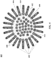
- said plurality of nucleic acid barcode molecules is configured in a geometric structure.
- said geometric structure is a nucleic acid origami.
- said one or more physical properties of said biological particle are identified by imaging said partition using (i) bright field microscopy, (ii) fluorescence microscopy, (iii) phase contrast microscopy, (iv) multispectral microscopy, or (v) polarization microscopy.
- said particle is a bead. In some embodiments, said bead is a gel bead. In some embodiments, said plurality of nucleic acid barcode molecules is releasably attached to said gel bead. In some embodiments, said gel bead comprises a polyacrylamide polymer.
- said one or more physical properties are one or more optical properties.
- step (c) occurs subsequent to (b) and comprises imaging said partition to optically detect said biological particle, thereby identifying said one or more optical properties of said biological particle.
- step (c) occurs prior to (b) and comprises imaging said biological particle to identify said one or more optical properties of said biological particle.
- said biological particle is imaged prior to providing said biological particle in said partition.
- (c)-(f) are repeated for additional partitions among said plurality of partitions, wherein said additional partitions each comprise (i) an additional plurality of nucleic acid barcode molecules, wherein a nucleic acid barcode molecule of said additional plurality of nucleic acid barcode molecules comprises an additional barcode sequence different than said barcode sequence, and (ii) an additional biological particle comprising an additional nucleic acid molecule, and wherein said partition and said additional partitions comprise a combination of particles comprising molecular barcodes and particles comprising optical barcodes, which combination is different across said partition and said additional partitions.
- (c)-(f) are repeated for additional partitions among said plurality of partitions, wherein said additional partitions each comprise (i) an additional plurality of nucleic acid barcode molecules and one or more additional optical barcodes, wherein a nucleic acid barcode molecule of said additional plurality of nucleic acid barcode molecules comprises an additional barcode sequence different than said barcode sequence, and (ii) an additional biological particle comprising an additional nucleic acid molecule, which additional partitions comprises at least 1,000 partitions.
- said additional partitions comprise at least 10,000 partitions.
- said additional partitions comprise at least 100,000 partitions.
- said additional partitions comprise a plurality of particles comprising nucleic acid barcode molecules comprising at least 1,000 barcode sequences, which at least 1,000 barcode sequences are different across said partition and said additional partitions. In some embodiments, said additional partitions comprises a plurality of particles comprising nucleic acid barcode molecules comprising at least 10,000 barcode sequences, which at least 10,000 barcode sequences are different across said partition and said additional partitions. In some embodiments, said additional partitions comprises a plurality of particles comprising nucleic acid barcode molecules comprising at least 100,000 barcode sequences, which at least 100,000 barcode sequences are different across said partition and said additional partitions.
- said biological particle is a cell.
- said biological particle comprises a cell, or one or more components thereof, in a matrix.
- said matrix is a polymeric matrix.
- said matrix is a gel matrix.
- the method further comprises associating a protein of said biological particle with said one or more physical properties of said biological particle with said nucleic acid sequence.
- said protein is a cell surface protein.
- the method further comprises associating one or more ribonucleic acid sequences or one or more deoxyribonucleic acid (DNA) sequences of said biological particle with said one or more physical properties.
- said one or more DNA sequences comprise epigenetic information.
- said one or more DNA sequences comprise chromatin information.
- barcode generally refers to a label, or identifier, that conveys or is capable of conveying information about an analyte.
- a barcode can be part of an analyte.
- a barcode can be independent of an analyte.
- a barcode can be a tag attached to an analyte (e.g., nucleic acid molecule) or a combination of the tag in addition to an endogenous characteristic of the analyte (e.g., size of the analyte or end sequence(s)).
- a barcode may be unique. Barcodes can have a variety of different formats.
- barcodes can include: polynucleotide barcodes; random nucleic acid and/or amino acid sequences; and synthetic nucleic acid and/or amino acid sequences.
- a barcode can be attached to an analyte in a reversible or irreversible manner.
- a barcode can be added to, for example, a fragment of a deoxyribonucleic acid (DNA) or ribonucleic acid (RNA) sample before, during, and/or after sequencing of the sample. Barcodes can allow for identification and/or quantification of individual sequencing-reads.
- real time can refer to a response time of less than about 1 second, a tenth of a second, a hundredth of a second, a millisecond, or less.
- the response time may be greater than 1 second.
- real time can refer to simultaneous or substantially simultaneous processing, detection or identification.
- subject generally refers to an animal, such as a mammal (e.g., human) or avian (e.g., bird), or other organism, such as a plant.
- the subject can be a vertebrate, a mammal, a rodent (e.g., a mouse), a primate, a simian or a human. Animals may include, but are not limited to, farm animals, sport animals, and pets.
- a subject can be a healthy or asymptomatic individual, an individual that has or is suspected of having a disease (e.g., cancer) or a pre-disposition to the disease, and/or an individual that is in need of therapy or suspected of needing therapy.
- a subject can be a patient.
- genomic information generally refers to genomic information from a subject, which may be, for example, at least a portion or an entirety of a subject's hereditary information.
- a genome can be encoded either in DNA or in RNA.
- a genome can comprise coding regions (e.g., that code for proteins) as well as non-coding regions.
- a genome can include the sequence of all chromosomes together in an organism.
- the human genome ordinarily has a total of 46 chromosomes. The sequence of all of these together may constitute a human genome.
- adaptor(s) can be used synonymously.
- An adaptor or tag can be coupled to a polynucleotide sequence to be “tagged” by any approach, including ligation, hybridization, or other approaches.
- sequence of nucleotide bases in one or more polynucleotides can be, for example, nucleic acid molecules such as deoxyribonucleic acid (DNA) or ribonucleic acid (RNA), including variants or derivatives thereof (e.g., single stranded DNA). Sequencing can be performed by various systems currently available, such as, without limitation, a sequencing system by Illumina ® , Pacific Biosciences (PacBio ® ), Oxford Nanopore ® , or Life Technologies (Ion Torrent ® ).
- sequencing may be performed using nucleic acid amplification, polymerase chain reaction (PCR) (e.g., digital PCR, quantitative PCR, or real time PCR), or isothermal amplification.
- PCR polymerase chain reaction
- Such systems may provide a plurality of raw genetic data corresponding to the genetic information of a subject (e.g., human), as generated by the systems from a sample provided by the subject.
- sequencing reads also "reads" herein).
- a read may include a string of nucleic acid bases corresponding to a sequence of a nucleic acid molecule that has been sequenced.
- systems and methods provided herein may be used with proteomic information.
- the term "bead,” as used herein, generally refers to a particle.
- the bead may be a solid or semi-solid particle.
- the bead may be a gel bead.
- the gel bead may include a polymer matrix (e.g., matrix formed by polymerization or cross-linking).
- the polymer matrix may include one or more polymers (e.g., polymers having different functional groups or repeat units).
- Cross-linking can be via covalent, ionic, or inductive, interactions, or physical entanglement.
- the bead may be a macromolecule.
- the bead may be formed of nucleic acid molecules bound together.
- the bead may be formed via covalent or non-covalent assembly of molecules (e.g., macromolecules), such as monomers or polymers. Such polymers or monomers may be natural or synthetic. Such polymers or monomers may be or include, for example, nucleic acid molecules (e.g., DNA or RNA).
- the bead may be formed of a polymeric material.
- the bead may be magnetic or non-magnetic.
- the bead may be rigid.
- the bead may be flexible and/or compressible.
- the bead may be disruptable or dissolvable.
- the bead may be a solid particle (e.g., a metal-based particle including but not limited to iron oxide, gold or silver) covered with a coating comprising one or more polymers. Such coating may be disruptable or dissolvable.
- sample generally refers to a biological sample of a subject.
- the biological sample may comprise any number of macromolecules, for example, cellular macromolecules.
- the biological sample may be a nucleic acid sample or protein sample.
- the biological sample may also be a carbohydrate sample or a lipid sample.
- the biological sample may be derived from another sample.
- the sample may be a tissue sample, such as a biopsy, core biopsy, needle aspirate, or fine needle aspirate.
- the sample may be a fluid sample, such as a blood sample, urine sample, or saliva sample.
- the sample may be a skin sample.
- the sample may be a cheek swab.
- the sample may be a plasma or serum sample.
- the sample may be a cell-free or cell free sample.
- a cell-free sample may include extracellular polynucleotides.
- Extracellular polynucleotides may be isolated from a bodily sample that may be selected from the group consisting of blood, plasma, serum, urine, saliva, mucosal excretions, sputum, stool and tears.
- biological particle generally refers to a discrete biological system derived from a biological sample.
- the biological particle may be a virus.
- the biological particle may be a cell or derivative of a cell.
- the biological particle may be an organelle.
- the biological particle may be a rare cell from a population of cells.
- the biological particle may be any type of cell, including without limitation prokaryotic cells, eukaryotic cells, bacterial, fungal, plant, mammalian, or other animal cell type, mycoplasmas, normal tissue cells, tumor cells, or any other cell type, whether derived from single cell or multicellular organisms.
- the biological particle may be or may include a matrix (e.g., a gel or polymer matrix) comprising a cell or one or more constituents from a cell (e.g., cell bead), such as DNA, RNA, organelles, proteins, or any combination thereof, from the cell.
- the biological particle may be obtained from a tissue of a subject.
- the biological particle may be a hardened cell. Such hardened cell may or may not include a cell wall or cell membrane.
- the biological particle may include one or more constituents of a cell, but may not include other constituents of the cell. An example of such constituents is a nucleus or an organelle.
- a cell may be a live cell.
- the live cell may be capable of being cultured, for example, being cultured when enclosed in a gel or polymer matrix, or cultured when comprising a gel or polymer matrix.
- the biological particle may be a fixed cell or a population of fixed cells, such as a tissue.
- the biological particle may be FFPE cells or tissues.
- the biological particle may be obtained from an FFPE sample by laser capturing the cells.
- a cell may split into two or more daughter cells, such as by binary fission, for example.
- the cell can be at any stage of cell division (e.g., prophase, metaphase).
- One or more physical properties of the biological particle can include a number of daughter cells, stage of cell division, and/or type of cell division.
- the macromolecular constituent may comprise a nucleic acid.
- the macromolecular constituent may comprise DNA.
- the macromolecular constituent may comprise RNA.
- the RNA may be coding or non-coding.
- the RNA may be messenger RNA (mRNA), ribosomal RNA (rRNA) or transfer RNA (tRNA), for example.
- the RNA may be a transcript.
- the RNA may small RNA that are less than 200 nucleic acid bases in length, or large RNA that are greater than 200 nucleic acid bases in length.
- Small RNAs mainly include 5.8S ribosomal RNA (rRNA), 5S rRNA, transfer RNA (tRNA), microRNA (miRNA), small interfering RNA (siRNA), small nucleolar RNA (snoRNAs), Piwi-interacting RNA (piRNA), tRNA-derived small RNA (tsRNA) and small rDNA-derived RNA (srRNA).
- the RNA may be double-stranded RNA or single-stranded RNA.
- the RNA may be circular RNA.
- the macromolecular constituent may comprise a protein.
- the macromolecular constituent may comprise a peptide.
- the macromolecular constituent may comprise a polypeptide.
- molecular tag generally refers to a molecule capable of binding to a macromolecular constituent.
- the molecular tag may bind to the macromolecular constituent with high affinity.
- the molecular tag may bind to the macromolecular constituent with high specificity.
- the molecular tag may comprise a nucleotide sequence.
- the molecular tag may comprise a nucleic acid sequence.
- the nucleic acid sequence may be at least a portion or an entirety of the molecular tag.
- the molecular tag may be a nucleic acid molecule or may be part of a nucleic acid molecule.
- the molecular tag may be an oligonucleotide or a polypeptide.
- the molecular tag may comprise a DNA aptamer.
- the molecular tag may be or comprise a primer.
- the molecular tag may be, or comprise, a protein.
- the molecular tag may comprise a polypeptide.
- the molecular tag may be a barcode.
- partition refers to a space or volume that may be suitable to contain one or more species or conduct one or more reactions.
- the partition may isolate space or volume from another space or volume.
- the partition may be a droplet or well, for example.
- the droplet may be a first phase (e.g., aqueous phase) in a second phase (e.g., oil) immiscible with the first phase.
- the droplet may be a first phase in a second phase that does not phase separate from the first phase, such as, for example, a capsule or liposome in an aqueous phase.
- the present disclosure provides methods and systems for obtaining genetic and/or proteomic information from a biological particle (e.g., a cell) and associating such information with one or more physical properties of the biological particle, such as size, shape, or density, or any other phenotypic property of the biological particle. This may permit genetic and/or proteomic information from the biological particle to be linked to the one or more physical properties of the biological particle.
- a biological particle e.g., a cell
- the present invention provides a method for processing a nucleic acid molecule of a biological particle, comprising: (a) providing (i) a particle comprising a plurality of nucleic acid barcode molecules and one or more optical barcodes, wherein a nucleic acid barcode molecule of said plurality of nucleic acid barcode molecules comprises a barcode sequence, and (ii) a biological particle comprising said nucleic acid molecule; (b) partitioning the (i) particle comprising a plurality of nucleic acid barcode molecules and one or more optical barcodes and the (ii) biological particle comprising said nucleic acid molecule into a partition of a plurality of partitions; (c) prior to (b) or subsequent to (b), generating a first data set comprising data indicative of one or more physical properties of said biological particle stored in computer memory, which data is generated upon sensing said biological particle; (d) using said nucleic acid barcode molecule of said plurality of nucleic acid barcode molecules and said nucleic acid
- a given nucleic acid barcode molecule of the plurality of nucleic acid barcode molecules may be used to barcode the nucleic acid molecule of the biological particle (e.g., included within or derived from the biological particle), to generate a barcoded nucleic acid molecule.
- a second data set comprising data identifying a nucleic acid sequence of the barcoded nucleic acid molecule or a derivative thereof may then be generated (e.g., using nucleic acid sequencing).
- the first data set and the second data set may then be used to associate (e.g., electronically associate) the one or more physical properties of the biological particle with the nucleic acid sequence.
- the biological particle can be a cell.
- the biological particle can be a gel or polymer matrix comprising the cell, a derivative of the cell, or one or more constituents of the cell.
- the biological particle is a cell.
- the cell may split into two or more daughter cells, such as by binary fission, for example.
- the cell can be at any stage of cell division (e.g., prophase, metaphase).
- the one or more physical properties of the cell can include a number of daughter cells, stage of cell division, and/or type of cell division.
- the biological particle may comprise a plurality of nucleic acid molecules. Some or all of the nucleic acid molecules may be disposed in the interior of the cell. Alternatively, the nucleic acid molecules may be disposed external or partially external to the cell.
- the cell may be lysed or permeabilized to provide access to one or more nucleic acid molecules within the cell.
- One or more nucleic acid molecules of or associated with the biological particle may be associated with the one or more physical properties of the biological particle. For example, at least 1, 10, 100, 1,000, 10,000, 100,000 or 1,000,000 nucleic acid molecules from or associated with the biological particle may be associated with the one or more physical properties of the biological particle.
- One or more nucleic acid molecules may be conjugated to one or more antibodies coupled to one or more proteins coupled to a surface of the biological particle (e.g., cell). Identifying a sequence of such one or more nucleic acid molecules may permit identifying the one or more antibodies and the one or more proteins.
- the one or more proteins from the biological particle may subsequently be associated with the one or more physical properties of the biological particle.
- proteomic information may be used in association with genetic information from the biological particle, such as DNA sequence information, transcriptome information (i.e., sequences of transcripts), or both.
- a cell surface protein of a cell can be identified using one or more antibodies.
- the cell surface protein can be associated with one or more physical properties of the cell (e.g., a shape of the cell) with a nucleic acid molecule.
- the one or more physical properties can be characterized by imaging the cell.
- the nucleic acid molecule of a derivative thereof of the cell can be sequenced to obtain a nucleic acid sequence.
- the nucleic acid sequence can be associated with the cell surface protein, in turn, with the one or more physical properties of the cell (e.g., a shape of the cell).
- RNA ribonucleic acid
- DNA deoxyribonucleic acid
- the one or more DNA sequences can comprise epigenetic information.
- the one or more DNA sequences can include methylated DNA (e.g., 5-methylcytosine) as indicated by an epigenetic assay (e.g., bisulfite sequencing).
- the one or more DNA sequences can include chromatin information, such as a lower nucleosome occupancy resulting in a highly accessible chromatin or "open" chromatin. The open chromatin can be as assessed by subjecting the one or more DNA sequences to an enzymatic treatment (e.g., DNase I, transposase), releasing a segment of nucleic acid molecules between two nucleosomes in an open chromatin.
- an enzymatic treatment e.g., DNase I, transposase
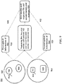
- a partition e.g., droplet
- the partition can comprise a particle (e.g., bead) and a biological particle (e.g., cell).
- the partition can be distinguished from other partitions in order to uniquely identify the partition.
- the partition can be characterized by, for example, imaging the partition to obtain optical information of the particle, a plurality of nucleic acid barcode molecules attached to the particle and/or the biological particle.
- the partition may be distinguished from other partitions by the particle included in the partition.
- the particle includes one or more optical barcodes and optionally other characteristics that may be used to distinguish the partition from other partitions.
- a lookup table (LUT) can be used to associate the plurality of nucleic acid barcode molecules to the particle.
- the optical information of the partition can permit associating the particle (e.g., gel bead) with the biological particle (e.g., cell) in the partition (e.g., droplet).
- the association of the particle with the biological particle can further permit associating a nucleic acid sequence of a nucleic acid molecule of the biological particle to one or more physical properties (e.g., a color of a cell) of the biological particle.
- the partition e.g., droplet
- the partition can include a particle (e.g., bead) having a first color and a biological particle (e.g., cell) having a second color or other optical characteristic, such as a shape or morphology of the biological particle, which first color and second color or characteristic can be associated with one another.
- the particle can have a plurality of nucleic acid barcode molecules attached thereto.
- the plurality of nucleic acid barcode molecules can comprise barcode sequences.
- the plurality of nucleic acid molecules attached to a given particle can have the same barcode sequence.
- the LUT can be used to associate a feature (e.g., an optical barcode, such as a color and/or intensity) of the particle with the barcode sequence.
- the feature may derive from the particle or an optical tag associated with the particle.
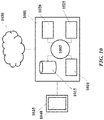
- the partition e.g., droplet
- the partition can be imaged to obtain optical information of the partition, including, for example, the feature (e.g., color and/or intensity) of the particle or the optical tag associated with the particle, and optical information of the biological particle in the partition.
- an image can include optical information in the visible spectrum, non-visible spectrum, or both.
- multiple images may be obtained of a partition across various optical frequencies.
- the biological particle can comprise a nucleic acid molecule which can be barcoded with a barcode sequence of a nucleic acid barcode molecule of the particle in the partition to provide a barcoded nucleic acid molecule.
- the barcoded nucleic acid molecule can be sequenced to obtain a nucleic acid sequence.
- the nucleic acid sequence can comprise genetic information of the biological particle.
- the nucleic acid sequence may comprise the barcode sequence, or a complement thereof.
- the barcode sequence, or complement thereof, of the nucleic acid sequence can be electronically associated with the feature (e.g., color and/or intensity) of the particle using the LUT to identify the particle.
- the LUT may be used to look up the barcode sequence to identify the feature (e.g., color and/or intensity), which may subsequently be used to identify an image having such feature, which may identify the partition comprising the particle.
- Identification of the particle can be useful in identifying the partition. This may in turn permit identification of the biological particle by using the optical information of the partition. This may permit genotypic information from a biological particle (e.g., cell) to be associated with phenotypic information of the biological particle.
- an image of a partition comprising a biological particle (e.g., cell or cell bead) and a particle (e.g., bead) comprising nucleic acid barcode molecules is captured.
- the nucleic acid barcode molecules include the same barcode sequence.
- the image identifies the particle as having a color (e.g., a green color) and also captures a shape of the biological particle (e.g., cell).
- the nucleic acid barcode molecules are used to barcode a nucleic acid molecule (e.g., an RNA molecule) from the biological particle (e.g., cell) to generate a sequencing library that is subsequently sequenced to yield a plurality of sequencing reads.
- the some or all of the plurality of sequencing reads include the barcode sequence.
- an LUT is used to associate the color of the particle from the captured image to the barcode sequence, which permits sequencing reads generated from the sequencing library to be identified.
- the sequencing reads are then associated with the shape of the biological particle.
- one or more optical properties of the partition can be used to distinguish the individual partition from other partitions.
- the optical properties of the partition such as color of a partitioning fluid (e.g., aqueous fluid) can be used to distinguish the partition from other partitions.
- the partitions can have different colors associated therewith to be able to distinguish from the partitions from one another.
- the partitions can include droplets having different colors associated therewith.
- the partition can be any container or a vessel, such as a well, microwell, tube, nanoarrays or other containers.
- the plurality of partitions can include at least 1,000 partitions, at least 10,000 partitions or at least 100,000 partitions. In such cases, at least a portion of the plurality of partitions can have one or more unique optical properties.
- one or more optical properties of the partition (e.g., droplet) and/or components (e.g., particle or biological particle) thereof may be used to identify whether the partition includes a particle and/or biological particle or not (e.g., whether the partition is occupied or unoccupied).
- Non-limiting examples of optical properties of a partition or a component thereof can include absorbance, birefringence, color, fluorescence, luminosity, photosensitivity, reflectivity, refractive index, scattering, or transmittance.
- Such optical properties may be associated with the partition, particle, or biological particle, such as associated with a signal directly from the partition, particle, or biological particle, or from one or more moieties coupled to the partition, particle, or biological particle.
- the plurality of partitions can comprise a first partition and a second partition.
- the first partition and the second partition can yield optical signals at different intensities and/or frequencies.
- the first and second partitions can include one or more optical materials such as one or more fluorescent dyes, nanoparticles, and/or microparticles that can provide a distinct optical signal.
- the one or more optical materials may be associated with the partition (e.g., a partitioning fluid), the particle (e.g., nucleic acid barcode molecules of the particle), or the biological particle.
- the partitioning fluid e.g., aqueous fluid
- the partitioning fluid can include a fluorescent dye.
- the partitioning fluid can include nanoparticles, such as quantum dots, to provide enhanced spectral resolution in order to distinguish between the partitions.
- the one or more optical materials can be introduced prior to or subsequent to generating partitions.
- a fluorescent dye can be injected into the partitioning fluid after generating partitions (e.g., droplets).
- a fluorescent dye can be included in the partitioning fluid before generating partitions (e.g., droplets).
- the particles can have different colors to be able to distinguish from one another.
- the particles can have different refractive indices based on the composition of the particles.
- the particles may be beads such as gel beads. Gel beads may comprise a polyacrylamide polymer. Gel beads may have different birefringence values based on degree of polymerization, chain length, or monomer chemistry.
- the particle can include (e.g., encapsulate or have attached thereto) a plurality of nucleic acid molecules, which can be nucleic acid barcode molecules.
- optical properties of the nucleic acid barcode molecules can be used for optical detection of the particles. For example, the absorbance of light by the nucleic acid barcode molecules can be used to distinguish the particles from one another.
- the optical barcodes can be on interior and/or exterior surfaces of the particles.
- Optical barcodes may comprise optical codes (e.g., one or more characteristic frequencies and/or intensities).
- Non-limiting examples of the optical barcodes can include fluorescent dyes, nanoparticles, and/or microparticles.
- the particles can include a plurality of different fluorescent dyes. Fluorescent dyes may be attached to the surface of the particles and/or may be incorporated into the particles for use as optical barcodes or components thereof. Fluorescent dyes may be attached to a surface of particles, incorporated within particles, and/or attached to nucleic acid barcode molecules coupled to particles.
- Different intensities of the different fluorescent dyes may be used to increase the number of optical barcodes (e.g., optical codes) that may be used. For example, if N is the number of fluorescent dyes (e.g., between 2 and 10 fluorescent dyes, such as 4 fluorescent dyes) and M is the possible intensities for the dyes (e.g., between 2 and 50 intensities, such as 20 intensities), then M N are the possible distinct optical barcodes. In one example, 4 fluorescent dyes with 20 possible intensities will be used to generate 160,000 distinct optical barcodes.
- nanoparticles such as quantum dots or Janus particles, can be used as optical barcodes or components thereof.
- the quantum dots can be attached to nucleic acid barcode molecules of the particles.
- Optical barcodes of particles may provide enhanced spectral resolution in order to distinguish between particles with unique nucleic acid barcode molecules (e.g., particles comprising unique barcode sequences).
- a first particle comprises a first optical barcode and nucleic acid barcode molecules each having a first barcode sequence.
- a second particle comprises a second optical barcode and nucleic acid barcode molecules each having a second barcode sequence.
- the first optical barcode and second optical barcode may be different (e.g., provided by two different fluorescent dyes or the same fluorescent dye at two different intensities).
- the first and second barcode sequences may be different nucleic acid sequences.
- the particles may be imaged to sense the first and second optical barcodes and the first and second optical barcodes may then be used to associate the first and second optical barcodes with the first and second barcode sequences, respectively.
- Optical barcodes may be included while generating the particles.
- the particles can be beads (e.g., gel beads).
- Gel beads can comprise polyacrylamide polymers.
- Optical barcodes can be included in the polymer structure, or attached at the pre-polymer or monomer stage in bead production.
- the beads may comprise moieties that can attach to one or more optical barcodes (e.g., at a surface of a bead and/or within a bead).
- optical barcodes can be loaded into the beads with one or more reagents.
- reagents and optical barcodes can be loaded into the beads by diffusion of the reagents (e.g., a solution of reagents comprising the optical barcodes).
- optical barcodes can be included while preparing nucleic acid barcode molecules.
- nucleic acid barcode molecules can be prepared by synthesizing molecules comprising barcode sequences (e.g., using a split pool or combinatorial approach).
- Optical barcodes may be attached to nucleic acid barcode molecules prior to attaching the nucleic acid barcode molecules to a particle.
- optical barcodes can be included after attaching nucleic acid barcode molecules to a particle.
- optical barcodes can be attached to nucleic acid barcode molecules coupled to the particle.
- Nucleic acid barcode molecules or sequences thereof can be releasably attached to the particle.
- Optical barcodes can be releasably or non-releasably attached to the particle.
- Optical properties of a biological particles can be used for optical detection of the biological particles.
- the optical detection of the biological particles can provide one or more physical properties (e.g., optical information) of the biological particle.
- the one or more physical properties of the biological particle can include phenotypic information.
- the phenotypic information of the biological particle can include one or more members selected from the group consisting of a size of the biological particle, a shape of the biological particle, a surface marker on the biological particle, an inclusion in the biological particle, a structure of an organelle in the biological particle, number of organelles in the biological particle, secretion or excretion with respect to the biological particle, a localization of an organelle in the biological particle, a circularity of the biological particle, a density of the biological particle, symmetry of the biological particle, and hardness of the biological particle.
- Optical properties comprising or correlated with phenotypic information can be used to characterize the biological particle.
- absorbance of an inclusion in the cell can be used to distinguish biological particles from one another.
- a biological particle can include one or more components of a cell in a matrix (e.g., polymeric matrix or a gel matrix).
- optical properties of the matrix can be used for optical detection of a biological particle (e.g., cell) or components thereof included in the matrix.
- phenotypic information of the biological particle e.g., cell
- Biological particles can be made optically detectable by including optical materials (e.g., optical barcodes or labels) in the biological particles, on surfaces of the biological particles, or both in and on surfaces of the biological particles.
- Optical materials can be on the interior and or exterior surfaces (e.g., cell membranes) of biological particles.
- biological particles e.g., cells
- the components thereof e.g., nucleic acid molecule
- nucleic acid molecules such as DNA molecules
- DAPI 4',6-diamidino-2-phenylindole
- optical materials can be attached to antibodies.
- a biological particle or components thereof can be optically detected using optical materials (e.g., fluorescent dyes such as Alexa Fluor) conjugated to an antibody against a protein.
- biological particles can be engineered to express one or more optical materials.
- a biological particle e.g., cell
- GFP Green Fluorescent Protein
- One or more physical properties of a partition can be used to distinguish a partition from other partitions.
- the physical properties can include size, shape, circularity, hardness, volume, or symmetry of the partition and/or components thereof.
- physical properties of a partition can be used to distinguish the partition from other partitions.
- the volume of the partitioning fluid e.g., aqueous fluid
- the shape and/or the symmetry of the partition (which may reflect characteristics of the particle and/or biological particle included therein) can be used to distinguish the partition from other partitions.
- Partitions can be made physically detectable by generating partitions with unique physical properties.
- the plurality of partitions can include at least 1,000 partitions, at least 10,000 partitions or at least 100,000 partitions. In such cases, at least a portion of the partitions can have one or more unique physical properties.
- the partitions can be generated with different volumes.
- the volume of partitioning fluids e.g., aqueous fluid
- the particles can be beads, such as gel beads.
- the particles can be biological particles.
- the particles can include (e.g., encapsulate or have attached thereto) nucleic acid molecules, which can be nucleic acid barcode molecules.
- the nucleic acid barcode molecules can include barcode sequences (e.g., nucleic acid barcode sequences).
- the barcode sequences can be the same across a plurality of the nucleic acid barcode molecules (e.g., a plurality of nucleic acid barcode molecules coupled to a given bead).
- Non-limiting examples of physical properties of particles can include size, shape, circularity, density, symmetry, and hardness.
- particles can be of different sizes. Different sizes of particles can be obtained by using microfluidic channel networks configured to provide specific sized particles (e.g., based on channel sizes, flow rates, etc). In some examples, particles can have different hardness values that can be obtained by varying the concentration of polymer used to generate the particles.
- a nucleic acid barcode molecule attached to a particle e.g., bead
- a nucleic acid origami such as a deoxyribonucleic acid (DNA) origami can be used to generate an optically detectable nucleic acid barcode molecule.
- a nucleic acid molecule or a plurality of nucleic acid molecules, can be folded to create two-and/or three-dimensional geometric shapes. The different geometric shapes can then be further optically detected.
- special types of nanoparticles with more than one distinct physical property can be used to make the particles physically distinguishable.
- Janus particles with both hydrophilic and hydrophobic surfaces can be used to provide unique physical property.
- physical properties of particles can be characterized prior to and/or subsequent to partitioning.
- a partition can be characterized using one or more physical and/or optical properties of the partitions and/or components thereof (e.g., particles, nucleic acid barcode molecules of particles, and/or biological particles) by imaging the partition.
- the partition can be imaged using an imaging unit.
- the imaging unit can be configured to image the partition based on an expected property (e.g., optical property) of the partition to be characterized.
- an imaging unit can be configured to excite and subsequently detect an emission wavelength of a partition comprising one or more fluorescent dyes.
- the imaging unit can be configured to perform bright field microscopy, fluorescence microscopy, phase contrast microscopy, multispectral microscopy, or polarization microscopy.
- the imaging unit can be configured to include an optical sensor based on the microscopy performed.
- the imaging unit can be configured to perform bright field microscopy to determine the physical property of the partition (e.g., shape and/or size of the partition, particle and the biological particle). In other examples, the imaging unit can be configured to perform fluorescence microscopy to image the partition (e.g., fluorescently-labelled partition, particle and/or biological particle).
- microfluidic chips can include additional features for imaging the individual partitions without interference. For example, the microfluidic chip can include features, such as rails, asymmetric turns, herringbone mixers, to facilitate the imaging of the partitions without interference.
- the microfluidic channel structure 800 can include the channel segments 801, 802, 804, 806, and 808 communicating at a channel junction 810.
- the channel segment 801 can transport an aqueous fluid or a first liquid phase that includes a plurality of beads 812 (e.g., with nucleic acid barcode molecule), and reagent 814 along the channel segment 801.
- the plurality of beads 812 can be releasably attached to a plurality of nucleic acid barcode molecules with barcode sequences and/or additional sequences (e.g., R1 and R2).
- the plurality of beads 812 may be sourced from a suspension of beads.
- the channel segment 801 may be connected to a reservoir comprising an aqueous suspension of beads.
- the reagent 814 can include a lysis reagent, PCR reagents, ligase etc.
- the channel segment 802 may transport the aqueous fluid that includes a plurality of biological particles 816 along the channel segment 802 into junction 810.
- the plurality of biological particles 816 may be sourced from a suspension of biological particles.
- the plurality of biological particles 816 can be cells or derivate thereof, such as macromolecular constituents.
- the plurality of biological particles 816 can be a sample obtained from formalin-fixed paraffin embedded tissues.
- a second fluid or a second liquid phase 818 that is immiscible with the aqueous fluid can be delivered to the junction 810 via channel segment 804.
- the aqueous fluid e.g., oil
- the aqueous fluid can be partitioned as discrete droplets 816 in the second fluid 814 and flow away from the junction 810 along channel segment 808.
- the channel segment 808 may deliver the discrete droplets to an outlet reservoir fluidly coupled to the channel segment 808, where they may be harvested.
- An individual droplet 820 may include a biological particle, a particle and reagents. The individual droplet 820 can be imaged with an imaging unit 822.
- the imaging unit 822 can be communicatively coupled to the microfluidic channel structure 800.
- the imaging unit 822 can be configured to image the individual droplet as the droplet flows through the channel segment 808.
- the imaging unit 822 can provide optical information of the droplet to a controller 824.
- the controller 822 can be operatively coupled to the imaging unit 822.
- the controller 824 can be configured to direct fluid flow in the channel segment 808.
- the controller 824 can be configured to communicate the optical information to a processor 826 for further processing.
- other sensors may be used to detect one or more physical properties of the individual droplet 820 and/or constituents of the individual droplet 820.
- the partition can be imaged prior to injecting the particle in the partition.
- the biological particle e.g., cell, cell bead or microcapsule
- the biological particle can be imaged prior to and subsequent to partitioning into the partition (e.g., droplet).
- the partition can be imaged after injecting the particle in the partition.
- the partition comprising the biological particle and the particle can be imaged.
- optical information can be generated.
- the optical information can include optical information of the partition and/or particle.
- the optical information can include optical information of the particle (e.g., bead).
- the optical information can include phenotypic information of the biological particle (e.g., cell).
- the optical information can include the optical information of the particle and the phenotypic information of the biological particle.
- the biological particle can comprise a nucleic acid molecule.
- the biological particle can comprise a plurality of nucleic acid molecules.
- the nucleic acid molecule(s) can comprise ribonucleic acid (RNA).
- the nucleic acid molecule(s) can comprise deoxyribonucleic acid (DNA).
- the biological particle can be a single biological particle, such as a cell or a polymer matrix (e.g., cell bead) as disclosed elsewhere herein.
- the biological particle can be a plurality of biological particles, such as a plurality of cells.
- the biological particle can comprise a live or fixed cell, such as FFPE cells.
- the biological particle can be any cellular derivative or combination thereof, such as DNA, RNA, nucleus, etc.
- the biological particle can be encapsulated in a gel matrix to preserve the particle.
- the biological particle can be encapsulated prior to and/or subsequent to partitioning into a partition.
- the partition can be any container or a vessel, such as a well, microwell, tube, nanoarrays or other containers.
- the partition can be flowable within fluid streams, such as a microcapsule having an inner fluid core surrounded by an outer barrier.
- the partition can be a droplet of aqueous fluid within a non-aqueous phase, such as an oil phase. Partitioning one or more materials (e.g., a particle and a biological particle) into a partition can be performed by any of the methods disclosed herein.
- the particle can comprise a plurality of nucleic acid barcode molecules.
- the plurality of nucleic acid barcode molecules can comprise barcode sequences (e.g., nucleic acid barcode sequences). In some cases, the barcodes sequences can be the same across a plurality of nucleic acid barcode molecules.
- the plurality of partitions can comprise a plurality of particles comprising a plurality of nucleic acid barcode molecules.
- the plurality of nucleic acid barcode molecules can comprise at least 1,000 barcode sequences which at least 1,000 barcode sequences, at least 10,000 barcode sequences or at least 100,000 barcode sequences can be different across the plurality of partitions. In some cases, the nucleic acid barcode molecule can comprise additional sequences.
- the additional sequences can include a primer binding site, such as a sequencing primer site (e.g., R1 or R2), flow cell binding sequences (e.g., P5, P7).
- a primer binding site can comprise sequences for sequencing primers to hybridize with a nucleic acid in a sequencing reaction.
- a primer binding site can comprise sequences for primers to hybridize with a nucleic acid in amplification or other extension reactions.
- the additional sequences can be useful in downstream assays, such as a sequencing assay. The additional sequences can be selected based on the assay used.
- a give nucleic acid barcode molecule or portion thereof of a plurality of the nucleic acid molecules coupled to a particle can barcode the nucleic acid molecule from the biological particle to generate a barcoded nucleic acid molecule.
- the given nucleic acid barcode molecule can attach to the nucleic acid molecule by annealing, extension and amplification reaction and/or ligation reactions.
- Extension and amplification reagents such as DNA polymerase, nucleoside triphosphates, and buffers with co-factors (e.g. Mg 2+ ), can be co-partitioned with biological particles and beads.
- the nucleic acid barcode molecule can be attached at either one or both ends of the segment to yield a barcoded nucleic acid molecule.
- a nucleic acid hybridization and extension process may take place within a partition and additional extension or amplification reactions may take place outside a partition (e.g., upon pooling materials from a plurality of such partitions).
- Barcoded nucleic acid molecules or derivatives thereof e.g., barcoded nucleic acid molecules to which one or more functional sequences have been added, or from which one or more features have been removed
- Barcoded nucleic acid molecules or derivatives thereof e.g., barcoded nucleic acid molecules to which one or more functional sequences have been added, or from which one or more features have been removed
- Processing with pooling may be implemented using barcode sequences.
- barcoded nucleic acid molecules of a given partition may have the same barcode sequence, which barcode sequence is different from barcode sequences of other partitions.
- barcoded nucleic acid molecules of different partitions can be processed separately for subsequent analysis (e.g., sequencing).
- Sequencing of barcoded nucleic acid molecules can provide sequencing reads comprising nucleic acid sequences.
- Such nucleic acid sequences may comprise the barcode sequences of the barcoded nucleic acid molecules, or complements thereof.
- a plurality of sequencing reads corresponding to a given partition e.g., a given barcoded nucleic acid molecule or collection thereof
- a subset of the plurality of sequencing reads comprises the barcode sequence of the barcoded nucleic acid molecule or a complement thereof.
- the nucleic acid sequence may comprise a sequence corresponding to the nucleic acid barcode molecules of a particle in a partition and/or a sequence corresponding to the nucleic acid molecule of a biological particle in the partition.
- a barcode sequence in a nucleic acid sequence can enable electronic association of physical and/or optical information of the biological particle (e.g., cell) from which a nucleic acid molecule (and thus barcoded nucleic acid molecule) was derived with the nucleic acid sequence.
- a barcode sequence in a nucleic acid sequence can enable the identification of the particle from which the barcode sequence of the nucleic acid barcode molecule was derived.
- the identification of the particle can enable identification of the partition by electronically associating physical and/or optical information of the particle with the partition.
- the identification of the partition can enable identification of the biological particle in the partition by electronically associating the optical information of the biological particle (e.g., cell) with the partition.
- the identification of the biological particle can enable associating any phenotypic information (e.g., cell size, shape, etc.) associated with the biological particle with the nucleic acid sequence.
- the electronic association of the phenotypic information of the biological particle with the nucleic acid sequence may be useful in assaying cell response to a therapy.
- the response to the therapy may include changes in phenotypic information of the cell (e.g., number of organelles).
- the changes in phenotypic information of the cell can be associated with the nucleic acid sequence of the nucleic acid molecule of the cell.
- a plurality of partitions can include a first partition 902 and a second partition 904.
- a first partition 902 can include a first biological particle (e.g., cell) 906 and a first particle (e.g., gel bead) 908.
- a second partition 904 can include a second biological particle (e.g., cell) 910 and a second particle (e.g., gel bead) 912.
- the first partition 902 and the second partition 904 can each have a unique physical and/or optical property for differentiating from other partitions.
- the unique physical and/or optical property may be a property of the partition itself (e.g., a volume or other feature of the partition) and/or a property of a component of the partition, such as a particle or biological particle of the partition.
- the first particle 908 can be a round bead and the second particle 912 can be a square bead.
- the first and second particles can comprise a plurality of nucleic acid barcode molecules coupled thereto.
- the plurality of nucleic acid barcode molecules can comprise barcode sequences.
- the barcode sequences of nucleic acid barcode molecules coupled to a given particle can be the same (e.g., nucleic acid barcode molecules coupled to the first particle each comprise the same barcode sequence and nucleic acid barcode molecules coupled to the second particle each comprise the same barcode sequence), while the barcode sequences associated with the first particle may be different from the barcode sequences associated with the second particle.
- the first particle can have a first nucleic acid barcode molecule comprising a first barcode sequence 914 and the second particle can have a second nucleic acid barcode molecule comprising a second barcode sequence 916.
- the first barcode sequence of the first nucleic acid barcode molecule 914 can be coupled to the first particle 908 before and/or after partitioning the particle.
- the second barcode sequence of the second nucleic acid barcode molecule 916 can be coupled to the second particle 912 before and/or after partitioning the particle.
- a lookup table LUT
- LUT can be used to associate the shape, color, or a combination of shape and color (or other optical properties) of the particle (e.g., round or square bead) with the barcode sequence (e.g., first or second barcode sequence).
- the first particle 908 with the round bead can be associated with the first barcode sequence of the first nucleic acid barcode molecule 914.
- the second particle 912 with the square bead can be associated with the second barcode sequence of the second nucleic acid barcode molecule 916.
- color may be used to differentiate the first particle 908 and the second particle 912.
- the first and second biological particles can have different phenotypic properties, such as a circular shape of the cell and a square shape of the cell, respectively.
- the first and the second partitions can be imaged to obtain a first optical characterization and a second optical characterization of the partitions in operations 918 and 920, respectively.
- the first and second optical characterizations can include the physical characterization of the particles and the phenotypic information of the biological particles.
- the biological particles e.g., first biological particle
- the nucleic acid molecule of the first biological particle 906 can be barcoded using the first barcode sequence of the first nucleic acid barcode molecule 914 to generate a first barcoded nucleic acid molecule 922.
- the nucleic acid molecule of the second biological particle 910 can be barcoded using the second barcode sequence of the second nucleic acid barcode molecule 916 to generate a second barcoded nucleic acid molecule 924.
- the first and second barcoded nucleic acid molecules can be sequenced (e.g., together or separately) to generate a first nucleic acid sequence 926 and a second nucleic acid sequence 928, respectively, corresponding to the nucleic acid molecules of the first and second biological particles and/or the first and second barcode sequences.
- a barcode sequence in a nucleic acid sequence can enable identification of the partition from which the barcode sequence derives (e.g., the particle from which the barcode sequence derives) by electronically associating the nucleic acid sequence with physical and/or optical information of the partition.
- the electronic association can permit associating nucleic acid sequence with phenotypic information (e.g., shape of cell) of the biological particle (e.g., cell).
- the nucleic acid sequence can comprise genetic information of the biological particle.
- the first barcode sequence of the first barcode nucleic acid molecule 914 in the first nucleic acid sequence 926 can be used to identify the first particle 908 using a LUT.
- Identifying the first particle 908 can permit identifying of the first biological particle 906 by using the first optical characterization 918. Further, the phenotypic information of the first biological particle 906 (e.g., circular cell) can be associated with the first nucleic acid sequence 926, in turn to genetic information of the biological particle. Similarly, in operation 932, the second barcode sequence of the second nucleic acid barcode molecule 916 in the second nucleic acid sequence can be used to identify the second particle 912 using a LUT. Identifying the second particle 912 can permit identifying of the second biological particle 910 by using the second optical characterization 920. Further, the phenotypic information of the second biological particle 910 (e.g., square shape of cell) can be associated with the second nucleic acid sequence 928, in turn to genetic information of the biological particle.
- the phenotypic information of the second biological particle 910 e.g., square shape of cell
- a kit which may find use in the present invention may comprise a plurality of particles (e.g., as described herein).
- a kit may comprise a plurality of particles, which plurality of particles comprise (i) a plurality of nucleic acid barcode molecules coupled thereto, wherein the plurality of barcode molecules comprise a plurality of barcode sequences, and (ii) a plurality of optical barcodes, wherein the plurality of optical barcodes comprise a plurality of optical codes.
- Each particle of the plurality of particles may comprise (i) a subset of the plurality of nucleic acid barcode molecules coupled thereto and (ii) a subset of the plurality of optical barcodes.
- Barcode sequences of subsets of the plurality of nucleic acid barcode molecules differ across particles of the plurality of particles, and optical codes of subsets of the plurality of optical barcodes differ across the particles.
- Each subset of the subsets of the plurality of optical barcodes may comprise a single optical barcode or two or more optical barcodes.
- the plurality of optical barcodes may confer optical properties to the plurality of particles. Such optical properties may be selected from the group consisting of, for example, an absorbance, a birefringence, a color, a fluorescence characteristic, a luminosity, a photosensitivity, a reflectivity, a refractive index, a scattering, or a transmittance of the particle or a component thereof.
- optical barcodes may comprise one or more fluorescent dyes, nanoparticles, microparticles, or any combination thereof.
- optical barcodes may comprise a plurality of nanoparticles, such as a plurality of quantum dots or Janus particles.
- optical barcodes may comprise a plurality of fluorescent dyes, such as between 2-10 fluorescent dyes, such as 4 fluorescent dyes.
- a plurality of fluorescent dyes may comprise fluorescent dyes having the same emission wavelength and different intensities, such as between 2 and 50 different intensities, such as 20 different intensities.
- Each subset of the subsets of the plurality of optical barcodes may comprise different optical codes.
- a plurality of optical barcodes may have associated optical intensities or frequencies that are distinct with respect to one another.
- a plurality of optical barcodes may comprise at least 1,000 different optical codes, such as at least 10,000 different optical codes or at least 100,000 different optical codes.
- nucleic acid barcode molecules within a given subset of the subsets of the plurality of nucleic acid barcode molecules may comprise identical barcode sequences.
- a plurality of barcode sequences may comprise at least 1,000 different barcode sequences, such as at least 10,000 different barcode sequences or at least 100,000 different barcode sequences.
- a plurality of nucleic acid barcode molecules may comprise a plurality of functional sequences (e.g., starter sequences, sequencing adapters, priming sequences, random N-mers, capture sequences, etc.).
- Functional sequences may be configured to interact with one or more target molecules, such as one or more different types of target molecules (e.g., RNA molecules and DNA molecules).
- a plurality of nucleic acid barcode molecules attached to a given particle may each comprise an N-mer sequence (e.g., a random N-mer sequence), which N-mer sequences are configured to interact with a plurality of DNA molecules.
- a plurality of nucleic acid barcode molecules attached to a given particle may each comprise a poly(T) sequence, which poly(T) sequences are configured to interact with a plurality of RNA molecules (e.g., messenger RNA (mRNA) molecules).
- Functional sequences may be configured to capture target molecules. For example, functional sequences may be configured to hybridize to target molecules to facilitate nucleic acid extension reactions.
- the plurality of particles of a kit may comprise at least 10,000 particles, such as at least 100,000 particles.
- the plurality of particles may comprise a plurality of beads, such as a plurality of gel beads.
- the plurality of gel beads may comprise polyacrylamide polymers.
- the plurality of gel beads may each comprise a plurality of nucleic acid barcode molecules releasably coupled thereto.
- a first bead may comprise a first plurality of nucleic acid barcode molecules releasably coupled thereto
- a second bead may comprise a second plurality of nucleic acid barcode molecules releasably coupled thereto.
- Optical barcodes e.g., optical materials such as fluorescent dyes
- first particles of the plurality of particles are coupled to second particles of the plurality of particles, wherein the first particles comprise nucleic acid barcode molecules of the plurality of nucleic acid barcode molecules and the second particles comprise optical barcodes of the plurality of optical barcodes.
- the methods can be useful for electronically associating optical information of the cell with a nucleic acid sequence of a nucleic acid molecule from the cell.
- the systems and methods described herein provide for the compartmentalization, depositing, or partitioning of macromolecular constituent contents of individual biological particles into discrete compartments or partitions (referred to interchangeably herein as partitions), where each partition maintains separation of its own contents from the contents of other partitions.
- the partition can be a droplet in an emulsion.
- a partition may comprise one or more other partitions.
- a partition of the present disclosure may comprise biological particles and/or macromolecular constituents thereof.
- a partition may comprise one or more gel beads.
- a partition may comprise one or more cell beads.
- a partition may include a single gel bead, a single cell bead, or both a single cell bead and single gel bead.
- a cell bead can be a biological particle and/or one or more of its macromolecular constituents encased inside of a gel or polymer matrix, such as via polymerization of a droplet containing the biological particle and precursors capable of being polymerized or gelled.
- Unique identifiers such as barcodes
- a microcapsule e.g., bead
- Microfluidic channel networks e.g., on a chip
- Alternative mechanisms may also be employed in the partitioning of individual biological particles, including porous membranes through which aqueous mixtures of cells are extruded into non-aqueous fluids.
- the partitions can be flowable within fluid streams.
- the partitions may comprise, for example, micro-vesicles that have an outer barrier surrounding an inner fluid center or core.
- the partitions may comprise a porous matrix that is capable of entraining and/or retaining materials within its matrix.
- the partitions can comprise droplets of aqueous fluid within a non-aqueous continuous phase (e.g., oil phase).
- the partitions can comprise droplets of a first phase within a second phase, wherein the first and second phases are immiscible.
- a variety of different vessels are described in, for example, U.S. Patent Application Publication No. 2014/0155295 .
- Emulsion systems for creating stable droplets in non-aqueous or oil continuous phases are described in, for example, U.S. Patent Application Publication No. 2010/0105112 .
- allocating individual biological particles to discrete partitions may in one non-limiting example be accomplished by introducing a flowing stream of biological particles in an aqueous fluid into a flowing stream of a non-aqueous fluid, such that droplets are generated at the junction of the two streams.
- the occupancy of the resulting partitions e.g., number of biological particles per partition
- the relative flow rates of the immiscible fluids can be selected such that, on average, the partitions may contain less than one biological particle per partition in order to ensure that those partitions that are occupied are primarily singly occupied.
- partitions among a plurality of partitions may contain at most one biological particle (e.g., bead, cell or cellular material).
- the relative flow rates of the fluids can be selected such that a majority of partitions are occupied, for example, allowing for only a small percentage of unoccupied partitions.
- the flows and channel architectures can be controlled as to ensure a given number of singly occupied partitions, less than a certain level of unoccupied partitions and/or less than a certain level of multiply occupied partitions.
- FIG. 1 shows an example of a microfluidic channel structure 100 for partitioning individual biological particles.
- the channel structure 100 can include channel segments 102, 104, 106 and 108 communicating at a channel junction 110.
- a first aqueous fluid 112 that includes suspended biological particles (or cells) 114 may be transported along channel segment 102 into junction 110, while a second fluid 116 that is immiscible with the aqueous fluid 112 is delivered to the junction 110 from each of channel segments 104 and 106 to create discrete droplets 118, 120 of the first aqueous fluid 112 flowing into channel segment 108, and flowing away from junction 110.
- the channel segment 108 may be fluidically coupled to an outlet reservoir where the discrete droplets can be stored and/or harvested.
- a discrete droplet generated may include an individual biological particle 114 (such as droplets 118).
- a discrete droplet generated may include more than one individual biological particle 114 (not shown in FIG. 1 ).
- a discrete droplet may contain no biological particle 114 (such as droplet 120).
- Each discrete partition may maintain separation of its own contents (e.g., individual biological particle 114) from the contents of other partitions.
- the second fluid 116 can comprise an oil, such as a fluorinated oil, that includes a fluorosurfactant for stabilizing the resulting droplets, for example, inhibiting subsequent coalescence of the resulting droplets 118, 120.
- an oil such as a fluorinated oil
- fluorosurfactant for stabilizing the resulting droplets, for example, inhibiting subsequent coalescence of the resulting droplets 118, 120.
- particularly useful partitioning fluids and fluorosurfactants are described, for example, in U.S. Patent Application Publication No. 2010/0105112 .
- the channel segments described herein may be coupled to any of a variety of different fluid sources or receiving components, including reservoirs, tubing, manifolds, or fluidic components of other systems.
- the microfluidic channel structure 100 may have other geometries.
- a microfluidic channel structure can have more than one channel junction.
- a microfluidic channel structure can have 2, 3, 4, or 5 channel segments each carrying biological particles, cell beads, and/or gel beads that meet at a channel junction.
- Fluid may be directed flow along one or more channels or reservoirs via one or more fluid flow units.
- a fluid flow unit can comprise compressors (e.g., providing positive pressure), pumps (e.g., providing negative pressure), actuators, and the like to control flow of the fluid. Fluid may also or otherwise be controlled via applied pressure differentials, centrifugal force, electrokinetic pumping, vacuum, capillary or gravity flow, or the like.
- the generated droplets may comprise two subsets of droplets: (1) occupied droplets 118, containing one or more biological particles 114, and (2) unoccupied droplets 120, not containing any biological particles 114.
- Occupied droplets 118 may comprise singly occupied droplets (having one biological particle) and multiply occupied droplets (having more than one biological particle).
- the majority of occupied partitions can include no more than one biological particle per occupied partition and some of the generated partitions can be unoccupied (of any biological particle). In some cases, though, some of the occupied partitions may include more than one biological particle.
- the partitioning process may be controlled such that fewer than about 25% of the occupied partitions contain more than one biological particle, and in many cases, fewer than about 20% of the occupied partitions have more than one biological particle, while in some cases, fewer than about 10% or even fewer than about 5% of the occupied partitions include more than one biological particle per partition.
- the Poissonian distribution may expectedly increase the number of partitions that include multiple biological particles. As such, where singly occupied partitions are to be obtained, at most about 95%, 90%, 85%, 80%, 75%, 70%, 65%, 60%, 55%, 50%, 45%, 40%, 35%, 30%, 25%, 20%, 15%, 10%, 5% or less of the generated partitions can be unoccupied.
- the flow of one or more of the biological particles (e.g., in channel segment 102), or other fluids directed into the partitioning junction (e.g., in channel segments 104, 106 ) can be controlled such that, in many cases, no more than about 50% of the generated partitions, no more than about 25% of the generated partitions, or no more than about 10% of the generated partitions are unoccupied.
- These flows can be controlled so as to present a non-Poissonian distribution of single-occupied partitions while providing lower levels of unoccupied partitions.
- the above noted ranges of unoccupied partitions can be achieved while still providing any of the single occupancy rates described above.
- the use of the systems and methods described herein can create resulting partitions that have multiple occupancy rates of less than about 25%, less than about 20%, less than about 15%, less than about 10%, and in many cases, less than about 5%, while having unoccupied partitions of less than about 50%, less than about 40%, less than about 30%, less than about 20%, less than about 10%, less than about 5%, or less.
- the above-described occupancy rates are also applicable to partitions that include both biological particles and additional reagents, including, but not limited to, microcapsules carrying barcoded nucleic acid molecules (e.g., oligonucleotides) (described in relation to FIG. 2 ).
- the occupied partitions e.g., at least about 10%, 20%, 30%, 40%, 50%, 60%, 70%, 80%, 90%, 95%, or 99% of the occupied partitions
- biological particles may be encapsulated within a microcapsule that comprises an outer shell, layer or porous matrix in which is entrained one or more individual biological particles or small groups of biological particles.
- the microcapsule may include other reagents. Encapsulation of biological particles may be performed by a variety of processes. Such processes combine an aqueous fluid containing the biological particles with a polymeric precursor material that may be capable of being formed into a gel or other solid or semi-solid matrix upon application of a particular stimulus to the polymer precursor.
- Such stimuli can include, for example, thermal stimuli (either heating or cooling), photo-stimuli (e.g., through photo-curing), chemical stimuli (e.g., through crosslinking, polymerization initiation of the precursor (e.g., through added initiators), or the like, and/or a combination of the above.
- microcapsules comprising biological particles may be performed by a variety of methods.
- air knife droplet or aerosol generators may be used to dispense droplets of precursor fluids into gelling solutions in order to form microcapsules that include individual biological particles or small groups of biological particles.
- membrane based encapsulation systems may be used to generate microcapsules comprising encapsulated biological particles as described herein.
- Microfluidic systems of the present disclosure such as that shown in FIG. 1 , may be readily used in encapsulating cells as described herein. In particular, and with reference to FIG.
- non-aqueous fluid 116 may also include an initiator (not shown) to cause polymerization and/or crosslinking of the polymer precursor to form the microcapsule that includes the entrained biological particles.
- initiator not shown
- polymer precursor/initiator pairs include those described in U.S. Patent Application Publication No. 2014/0378345 .
- the activation agent may comprise a cross-linking agent, or a chemical that activates a cross-linking agent within the formed droplets.
- the activation agent may comprise a polymerization initiator.
- the polymer precursor comprises a mixture of acrylamide monomer with a N,N'-bis-(acryloyl)cystamine (BAC) comonomer
- an agent such as tetraethylmethylenediamine (TEMED) may be provided within the second fluid streams 116 in channel segments 104 and 106, which can initiate the copolymerization of the acrylamide and BAC into a cross-linked polymer network, or hydrogel.
- TEMED tetraethylmethylenediamine
- the TEMED may diffuse from the second fluid 116 into the aqueous fluid 112 comprising the linear polyacrylamide, which will activate the crosslinking of the polyacrylamide within the droplets 118, 120, resulting in the formation of gel (e.g., hydrogel) microcapsules, as solid or semi-solid beads or particles entraining the cells 114.
- gel e.g., hydrogel
- other 'activatable' encapsulation compositions may also be employed in the context of the methods and compositions described herein.
- alginate droplets followed by exposure to divalent metal ions (e.g., Ca 2+ ions), can be used as an encapsulation process using the described processes.
- divalent metal ions e.g., Ca 2+ ions
- agarose droplets may also be transformed into capsules through temperature based gelling (e.g., upon cooling, etc.).
- encapsulated biological particles can be selectively releasable from the microcapsule, such as through passage of time or upon application of a particular stimulus, that degrades the microcapsule sufficiently to allow the biological particles (e.g., cell), or its other contents to be released from the microcapsule, such as into a partition (e.g., droplet).
- a partition e.g., droplet
- degradation of the microcapsule may be accomplished through the introduction of an appropriate reducing agent, such as DTT or the like, to cleave disulfide bonds that cross-link the polymer matrix. See, for example, U.S. Patent Application Publication No. 2014/0378345 .
- the biological particle can be subjected to other conditions sufficient to polymerize or gel the precursors.
- the conditions sufficient to polymerize or gel the precursors may comprise exposure to heating, cooling, electromagnetic radiation, and/or light.
- the conditions sufficient to polymerize or gel the precursors may comprise any conditions sufficient to polymerize or gel the precursors.
- a polymer or gel may be formed around the biological particle.
- the polymer or gel may be diffusively permeable to chemical or biochemical reagents.
- the polymer or gel may be diffusively impermeable to macromolecular constituents of the biological particle.
- the polymer or gel may act to allow the biological particle to be subjected to chemical or biochemical operations while spatially confining the macromolecular constituents to a region of the droplet defined by the polymer or gel.
- the polymer or gel may include one or more of disulfide cross-linked polyacrylamide, agarose, alginate, polyvinyl alcohol, polyethylene glycol (PEG)-diacrylate, PEG-acrylate, PEG-thiol, PEG-azide, PEG-alkyne, other acrylates, chitosan, hyaluronic acid, collagen, fibrin, gelatin, or elastin.
- the polymer or gel may comprise any other polymer or gel.
- the polymer or gel may be functionalized to bind to targeted analytes, such as nucleic acids, proteins, carbohydrates, lipids or other analytes.
- the polymer or gel may be polymerized or gelled via a passive mechanism.
- the polymer or gel may be stable in alkaline conditions or at elevated temperature.
- the polymer or gel may have mechanical properties similar to the mechanical properties of the bead. For instance, the polymer or gel may be of a similar size to the bead.
- the polymer or gel may have a mechanical strength (e.g. tensile strength) similar to that of the bead.
- the polymer or gel may be of a lower density than an oil.
- the polymer or gel may be of a density that is roughly similar to that of a buffer.
- the polymer or gel may have a tunable pore size.
- the pore size may be chosen to, for instance, retain denatured nucleic acids.
- the pore size may be chosen to maintain diffusive permeability to exogenous chemicals such as sodium hydroxide (NaOH) and/or endogenous chemicals such as inhibitors.
- the polymer or gel may be biocompatible.
- the polymer or gel may maintain or enhance cell viability.
- the polymer or gel may be biochemically compatible.
- the polymer or gel may be polymerized and/or depolymerized thermally, chemically, enzymatically, and/or optically.
- the polymer may comprise poly(acrylamide-co-acrylic acid) crosslinked with disulfide linkages.
- the preparation of the polymer may comprise a two-step reaction.
- poly(acrylamide-co-acrylic acid) may be exposed to an acylating agent to convert carboxylic acids to esters.
- the poly(acrylamide-co-acrylic acid) may be exposed to 4-(4,6-dimethoxy-1,3,5-triazin-2-yl)-4-methylmorpholinium chloride (DMTMM).
- DTMM 4-(4,6-dimethoxy-1,3,5-triazin-2-yl)-4-methylmorpholinium chloride
- the polyacrylamide-co-acrylic acid may be exposed to other salts of 4-(4,6-dimethoxy-1,3,5-triazin-2-yl)-4-methylmorpholinium.
- the ester formed in the first step may be exposed to a disulfide crosslinking agent.
- the ester may be exposed to cystamine (2,2'-dithiobis(ethylamine)).
- the biological particle may be surrounded by polyacrylamide strands linked together by disulfide bridges.
- the biological particle may be encased inside of or comprise a gel or matrix (e.g., polymer matrix) to form a "cell bead.”
- a cell bead can contain biological particles (e.g., a cell) or macromolecular constituents (e.g., RNA, DNA, proteins, etc.) of biological particles.
- a cell bead may include a single cell or multiple cells, or a derivative of the single cell or multiple cells. For example after lysing and washing the cells, inhibitory components from cell lysates can be washed away and the macromolecular constituents can be bound as cell beads.
- Systems and methods disclosed herein can be applicable to both cell beads (and/or droplets or other partitions) containing biological particles and cell beads (and/or droplets or other partitions) containing macromolecular constituents of biological particles.
- Encapsulated biological particles can provide certain potential advantages of being more storable and more portable than droplet-based partitioned biological particles. Furthermore, in some cases, it may be desirable to allow biological particles to incubate for a select period of time before analysis, such as in order to characterize changes in such biological particles over time, either in the presence or absence of different stimuli.
- encapsulation may allow for longer incubation than partitioning in emulsion droplets, although in some cases, droplet partitioned biological particles may also be incubated for different periods of time, e.g., at least 10 seconds, at least 30 seconds, at least 1 minute, at least 5 minutes, at least 10 minutes, at least 30 minutes, at least 1 hour, at least 2 hours, at least 5 hours, or at least 10 hours or more.
- the encapsulation of biological particles may constitute the partitioning of the biological particles into which other reagents are co-partitioned.
- encapsulated biological particles may be readily deposited into other partitions (e.g., droplets) as described above.
- a partition may comprise one or more unique identifiers, such as barcodes.
- Barcodes may be previously, subsequently or concurrently delivered to the partitions that hold the compartmentalized or partitioned biological particle. For example, barcodes may be injected into droplets previous to, subsequent to, or concurrently with droplet generation. The delivery of the barcodes to a particular partition allows for the later attribution of the characteristics of the individual biological particle to the particular partition.
- Barcodes may be delivered, for example on a nucleic acid molecule (e.g., an oligonucleotide), to a partition via any suitable mechanism. Barcoded nucleic acid molecules can be delivered to a partition via a microcapsule.
- a microcapsule in some instances, can comprise a bead. Beads are described in further detail below.
- barcoded nucleic acid molecules can be initially associated with the microcapsule and then released from the microcapsule. Release of the barcoded nucleic acid molecules can be passive (e.g., by diffusion out of the microcapsule). In addition or alternatively, release from the microcapsule can be upon application of a stimulus which allows the barcoded nucleic acid molecules to dissociate or to be released from the microcapsule. Such stimulus may disrupt the microcapsule, an interaction that couples the barcoded nucleic acid molecules to or within the microcapsule, or both.
- Such stimulus can include, for example, a thermal stimulus, photo-stimulus, chemical stimulus (e.g., change in pH or use of a reducing agent(s)), a mechanical stimulus, a radiation stimulus; a biological stimulus (e.g., enzyme), or any combination thereof.
- chemical stimulus e.g., change in pH or use of a reducing agent(s)
- mechanical stimulus e.g., change in pH or use of a reducing agent(s)
- a radiation stimulus e.g., a radiation stimulus
- a biological stimulus e.g., enzyme
- FIG. 2 shows an example of a microfluidic channel structure 200 for delivering barcode carrying beads to droplets.
- the channel structure 200 can include channel segments 201, 202, 204, 206 and 208 communicating at a channel junction 210.
- the channel segment 201 may transport an aqueous fluid 212 that includes a plurality of beads 214 (e.g., with nucleic acid molecules, oligonucleotides, molecular tags) along the channel segment 201 into junction 210.
- the plurality of beads 214 may be sourced from a suspension of beads.
- the channel segment 201 may be connected to a reservoir comprising an aqueous suspension of beads 214.
- the channel segment 202 may transport the aqueous fluid 212 that includes a plurality of biological particles 216 along the channel segment 202 into junction 210.
- the plurality of biological particles 216 may be sourced from a suspension of biological particles.
- the channel segment 202 may be connected to a reservoir comprising an aqueous suspension of biological particles 216.
- the aqueous fluid 212 in either the first channel segment 201 or the second channel segment 202, or in both segments can include one or more reagents, as further described below.
- a second fluid 218 that is immiscible with the aqueous fluid 212 e.g., oil
- the aqueous fluid 212 can be partitioned as discrete droplets 220 in the second fluid 218 and flow away from the junction 210 along channel segment 208.
- the channel segment 208 may deliver the discrete droplets to an outlet reservoir fluidly coupled to the channel segment 208, where they may be harvested.
- the channel segments 201 and 202 may meet at another junction upstream of the junction 210.
- beads and biological particles may form a mixture that is directed along another channel to the junction 210 to yield droplets 220.
- the mixture may provide the beads and biological particles in an alternating fashion, such that, for example, a droplet comprises a single bead and a single biological particle.
- Beads, biological particles and droplets may flow along channels at substantially regular flow profiles (e.g., at regular flow rates). Such regular flow profiles may permit a droplet to include a single bead and a single biological particle. Such regular flow profiles may permit the droplets to have an occupancy (e.g., droplets having beads and biological particles) greater than 5%, 10%, 20%, 30%, 40%, 50%, 60%, 70%, 80%, 90%, or 95%.
- occupancy e.g., droplets having beads and biological particles
- the second fluid 218 can comprise an oil, such as a fluorinated oil, that includes a fluorosurfactant for stabilizing the resulting droplets, for example, inhibiting subsequent coalescence of the resulting droplets 220.
- an oil such as a fluorinated oil, that includes a fluorosurfactant for stabilizing the resulting droplets, for example, inhibiting subsequent coalescence of the resulting droplets 220.
- a discrete droplet that is generated may include an individual biological particle 216.
- a discrete droplet that is generated may include a barcode or other reagent carrying bead 214.
- a discrete droplet generated may include both an individual biological particle and a barcode carrying bead, such as droplets 220.
- a discrete droplet may include more than one individual biological particle or no biological particle.
- a discrete droplet may include more than one bead or no bead.
- a discrete droplet may be unoccupied (e.g., no beads, no biological particles).
- a discrete droplet partitioning a biological particle and a barcode carrying bead may effectively allow the attribution of the barcode to macromolecular constituents of the biological particle within the partition.
- the contents of a partition may remain discrete from the contents of other partitions.
- the channel segments described herein may be coupled to any of a variety of different fluid sources or receiving components, including reservoirs, tubing, manifolds, or fluidic components of other systems.
- the microfluidic channel structure 200 may have other geometries.
- a microfluidic channel structure can have more than one channel junctions.
- a microfluidic channel structure can have 2, 3, 4, or 5 channel segments each carrying beads that meet at a channel junction.
- Fluid may be directed flow along one or more channels or reservoirs via one or more fluid flow units.
- a fluid flow unit can comprise compressors (e.g., providing positive pressure), pumps (e.g., providing negative pressure), actuators, and the like to control flow of the fluid. Fluid may also or otherwise be controlled via applied pressure differentials, centrifugal force, electrokinetic pumping, vacuum, capillary or gravity flow, or the like.
- a bead may be porous, non-porous, solid, semi-solid, semi-fluidic, fluidic, and/or a combination thereof. In some instances, a bead may be dissolvable, disruptable, and/or degradable. In some cases, a bead may not be degradable. In some cases, the bead may be a gel bead. A gel bead may be a hydrogel bead. A gel bead may be formed from molecular precursors, such as a polymeric or monomeric species. A semi-solid bead may be a liposomal bead. Solid beads may comprise metals including iron oxide, gold, and silver. In some cases, the bead may be a silica bead. In some cases, the bead can be rigid. In other cases, the bead may be flexible and/or compressible.
- a bead may be of any suitable shape.
- bead shapes include, but are not limited to, spherical, non-spherical, oval, oblong, amorphous, circular, cylindrical, and variations thereof.
- Beads may be of uniform size or heterogeneous size.
- the diameter of a bead may be at least about 1micrometers ( ⁇ m), 5 ⁇ m, 10 ⁇ m, 20 ⁇ m, 30 ⁇ m, 40 ⁇ m, 50 ⁇ m, 60 ⁇ m, 70 ⁇ m, 80 ⁇ m, 90 ⁇ m, 100 ⁇ m, 250 ⁇ m, 500 ⁇ m, 1mm, or greater.
- a bead may have a diameter of less than about 1 ⁇ m, 5 ⁇ m, 10 ⁇ m, 20 ⁇ m, 30 ⁇ m, 40 ⁇ m, 50 ⁇ m, 60 ⁇ m, 70 ⁇ m, 80 ⁇ m, 90 ⁇ m, 100 ⁇ m, 250 ⁇ m, 500 ⁇ m, 1mm, or less.
- a bead may have a diameter in the range of about 40-75 ⁇ m, 30-75 ⁇ m, 20-75 ⁇ m, 40-85 ⁇ m, 40-95 ⁇ m, 20-100 ⁇ m, 10-100 ⁇ m, 1-100 ⁇ m, 20-250 ⁇ m, or 20-500 ⁇ m.
- beads can be provided as a population or plurality of beads having a relatively monodisperse size distribution. Where it may be desirable to provide relatively consistent amounts of reagents within partitions, maintaining relatively consistent bead characteristics, such as size, can contribute to the overall consistency.
- the beads described herein may have size distributions that have a coefficient of variation in their cross-sectional dimensions of less than 50%, less than 40%, less than 30%, less than 20%, and in some cases less than 15%, less than 10%, less than 5%, or less.
- a bead may comprise natural and/or synthetic materials.
- a bead can comprise a natural polymer, a synthetic polymer or both natural and synthetic polymers.
- natural polymers include proteins and sugars such as deoxyribonucleic acid, rubber, cellulose, starch (e.g., amylose, amylopectin), proteins, enzymes, polysaccharides, silks, polyhydroxyalkanoates, chitosan, dextran, collagen, carrageenan, ispaghula, acacia, agar, gelatin, shellac, sterculia gum, xanthan gum, Corn sugar gum, guar gum, gum karaya, agarose, alginic acid, alginate, or natural polymers thereof.
- proteins and sugars such as deoxyribonucleic acid, rubber, cellulose, starch (e.g., amylose, amylopectin), proteins, enzymes, polysaccharides, silks, polyhydroxyalkano
- Examples of synthetic polymers include acrylics, nylons, silicones, spandex, viscose rayon, polycarboxylic acids, polyvinyl acetate, polyacrylamide, polyacrylate, polyethylene glycol, polyurethanes, polylactic acid, silica, polystyrene, polyacrylonitrile, polybutadiene, polycarbonate, polyethylene, polyethylene terephthalate, poly(chlorotrifluoroethylene), poly(ethylene oxide), poly(ethylene terephthalate), polyethylene, polyisobutylene, poly(methyl methacrylate), poly(oxymethylene), polyformaldehyde, polypropylene, polystyrene, poly(tetrafluoroethylene), poly(vinyl acetate), poly(vinyl alcohol), poly(vinyl chloride), poly(vinylidene dichloride), poly(vinylidene difluoride), poly(vinyl fluoride) and/or combinations (e.g., co-polymers) thereof.
- the bead may contain molecular precursors (e.g., monomers or polymers), which may form a polymer network via polymerization of the molecular precursors.
- a precursor may be an already polymerized species capable of undergoing further polymerization via, for example, a chemical cross-linkage.
- a precursor can comprise one or more of an acrylamide or a methacrylamide monomer, oligomer, or polymer.
- the bead may comprise prepolymers, which are oligomers capable of further polymerization.
- polyurethane beads may be prepared using prepolymers.
- the bead may contain individual polymers that may be further polymerized together.
- beads may be generated via polymerization of different precursors, such that they comprise mixed polymers, co-polymers, and/or block co-polymers.
- the bead may comprise covalent or ionic bonds between polymeric precursors (e.g., monomers, oligomers, linear polymers), nucleic acid molecules (e.g., oligonucleotides), primers, and other entities.
- the covalent bonds can be carbon-carbon bonds or thioether bonds.
- Cross-linking may be permanent or reversible, depending upon the particular cross-linker used. Reversible cross-linking may allow for the polymer to linearize or dissociate under appropriate conditions. In some cases, reversible cross-linking may also allow for reversible attachment of a material bound to the surface of a bead. In some cases, a cross-linker may form disulfide linkages. In some cases, the chemical cross-linker forming disulfide linkages may be cystamine or a modified cystamine.
- disulfide linkages can be formed between molecular precursor units (e.g., monomers, oligomers, or linear polymers) or precursors incorporated into a bead and nucleic acid molecules (e.g., oligonucleotides).
- Cystamine is an organic agent comprising a disulfide bond that may be used as a crosslinker agent between individual monomeric or polymeric precursors of a bead.
- Polyacrylamide may be polymerized in the presence of cystamine or a species comprising cystamine (e.g., a modified cystamine) to generate polyacrylamide gel beads comprising disulfide linkages (e.g., chemically degradable beads comprising chemically-reducible cross-linkers).
- the disulfide linkages may permit the bead to be degraded (or dissolved) upon exposure of the bead to a reducing agent.
- chitosan a linear polysaccharide polymer
- glutaraldehyde via hydrophilic chains to form a bead.
- Crosslinking of chitosan polymers may be achieved by chemical reactions that are initiated by heat, pressure, change in pH, and/or radiation.
- a bead may comprise an acrydite moiety, which in certain aspects may be used to attach one or more nucleic acid molecules (e.g., barcode sequence, barcoded nucleic acid molecule, barcoded oligonucleotide, primer, or other oligonucleotide) to the bead.
- an acrydite moiety can refer to an acrydite analogue generated from the reaction of acrydite with one or more species, such as, the reaction of acrydite with other monomers and cross-linkers during a polymerization reaction.
- Acrydite moieties may be modified to form chemical bonds with a species to be attached, such as a nucleic acid molecule (e.g., barcode sequence, barcoded nucleic acid molecule, barcoded oligonucleotide, primer, or other oligonucleotide).
- Acrydite moieties may be modified with thiol groups capable of forming a disulfide bond or may be modified with groups already comprising a disulfide bond. The thiol or disulfide (via disulfide exchange) may be used as an anchor point for a species to be attached or another part of the acrydite moiety may be used for attachment.
- attachment can be reversible, such that when the disulfide bond is broken (e.g., in the presence of a reducing agent), the attached species is released from the bead.
- an acrydite moiety can comprise a reactive hydroxyl group that may be used for attachment.
- nucleic acid molecules e.g., oligonucleotides
- Functionalization of beads for attachment of nucleic acid molecules may be achieved through a wide range of different approaches, including activation of chemical groups within a polymer, incorporation of active or activatable functional groups in the polymer structure, or attachment at the pre-polymer or monomer stage in bead production.
- precursors e.g., monomers, cross-linkers
- precursors that are polymerized to form a bead may comprise acrydite moieties, such that when a bead is generated, the bead also comprises acrydite moieties.
- the acrydite moieties can be attached to a nucleic acid molecule (e.g., oligonucleotide), which may include a priming sequence (e.g., a primer for amplifying target nucleic acids, random primer, primer sequence for messenger RNA) and/or a one or more barcode sequences.
- the one more barcode sequences may include sequences that are the same for all nucleic acid molecules coupled to a given bead and/or sequences that are different across all nucleic acid molecules coupled to the given bead.
- the nucleic acid molecule may be incorporated into the bead.
- the nucleic acid molecule can comprise a functional sequence, for example, for attachment to a sequencing flow cell, such as, for example, a P5 sequence for Illumina ® sequencing.
- the nucleic acid molecule or derivative thereof e.g., oligonucleotide or polynucleotide generated from the nucleic acid molecule
- can comprise another functional sequence such as, for example, a P7 sequence for attachment to a sequencing flow cell for Illumina sequencing.
- the nucleic acid molecule can comprise a barcode sequence.
- the primer can further comprise a unique molecular identifier (UMI).
- UMI unique molecular identifier
- the primer can comprise an R1 primer sequence for Illumina sequencing.
- the primer can comprise an R2 primer sequence for Illumina sequencing.
- nucleic acid molecules e.g., oligonucleotides, polynucleotides, etc.
- uses thereof as may be used with compositions, devices, methods and systems of the present disclosure, are provided in U.S. Patent Pub. Nos. 2014/0378345 and 2015/0376609 .
- precursors comprising a functional group that is reactive or capable of being activated such that it becomes reactive can be polymerized with other precursors to generate gel beads comprising the activated or activatable functional group.
- the functional group may then be used to attach additional species (e.g., disulfide linkers, primers, other oligonucleotides, etc.) to the gel beads.
- additional species e.g., disulfide linkers, primers, other oligonucleotides, etc.
- some precursors comprising a carboxylic acid (COOH) group can co-polymerize with other precursors to form a gel bead that also comprises a COOH functional group.
- acrylic acid a species comprising free COOH groups
- acrylamide acrylamide
- bis(acryloyl)cystamine can be co-polymerized together to generate a gel bead comprising free COOH groups.
- the COOH groups of the gel bead can be activated (e.g., via 1-Ethyl-3-(3-dimethylaminopropyl)carbodiimide (EDC) and N-Hydroxysuccinimide (NHS) or 4-(4,6-Dimethoxy-1,3,5-triazin-2-yl)-4-methylmorpholinium chloride (DMTMM)) such that they are reactive (e.g., reactive to amine functional groups where EDC/NHS or DMTMM are used for activation).
- EDC 1-Ethyl-3-(3-dimethylaminopropyl)carbodiimide
- NHS N-Hydroxysuccinimide
- DTMM 4-(4,6-Dimethoxy
- the activated COOH groups can then react with an appropriate species (e.g., a species comprising an amine functional group where the carboxylic acid groups are activated to be reactive with an amine functional group) comprising a moiety to be linked to the bead.
- an appropriate species e.g., a species comprising an amine functional group where the carboxylic acid groups are activated to be reactive with an amine functional group
- Beads comprising disulfide linkages in their polymeric network may be functionalized with additional species via reduction of some of the disulfide linkages to free thiols.
- the disulfide linkages may be reduced via, for example, the action of a reducing agent (e.g., DTT, TCEP, etc.) to generate free thiol groups, without dissolution of the bead.
- Free thiols of the beads can then react with free thiols of a species or a species comprising another disulfide bond (e.g., via thiol-disulfide exchange) such that the species can be linked to the beads (e.g., via a generated disulfide bond).
- free thiols of the beads may react with any other suitable group.
- free thiols of the beads may react with species comprising an acrydite moiety.
- the free thiol groups of the beads can react with the acrydite via Michael addition chemistry, such that the species comprising the acrydite is linked to the bead.
- uncontrolled reactions can be prevented by inclusion of a thiol capping agent such as N-ethylmalieamide or iodoacetate.
- Activation of disulfide linkages within a bead can be controlled such that only a small number of disulfide linkages are activated. Control may be exerted, for example, by controlling the concentration of a reducing agent used to generate free thiol groups and/or concentration of reagents used to form disulfide bonds in bead polymerization.
- a low concentration e.g., molecules of reducing agent:gel bead ratios of less than or equal to about 1:100,000,000,000, less than or equal to about 1:10,000,000,000, less than or equal to about 1:1,000,000,000, less than or equal to about 1:100,000,000, less than or equal to about 1:10,000,000, less than or equal to about 1:1,000,000, less than or equal to about 1:100,000, less than or equal to about 1:10,000
- reducing agent may be used for reduction.
- Controlling the number of disulfide linkages that are reduced to free thiols may be useful in ensuring bead structural integrity during functionalization.
- optically-active agents such as fluorescent dyes may be coupled to beads via free thiol groups of the beads and used to quantify the number of free thiols present in a bead and/or track a bead.
- addition of moieties to a gel bead after gel bead formation may be advantageous.
- addition of an oligonucleotide (e.g., barcoded oligonucleotide) after gel bead formation may avoid loss of the species during chain transfer termination that can occur during polymerization.
- smaller precursors e.g., monomers or cross linkers that do not comprise side chain groups and linked moieties
- functionalization after gel bead synthesis can minimize exposure of species (e.g., oligonucleotides) to be loaded with potentially damaging agents (e.g., free radicals) and/or chemical environments.
- the generated gel may possess an upper critical solution temperature (UCST) that can permit temperature driven swelling and collapse of a bead.
- UCT upper critical solution temperature
- Such functionality may aid in oligonucleotide (e.g., a primer) infiltration into the bead during subsequent functionalization of the bead with the oligonucleotide.
- Post-production functionalization may also be useful in controlling loading ratios of species in beads, such that, for example, the variability in loading ratio is minimized.
- Species loading may also be performed in a batch process such that a plurality of beads can be functionalized with the species in a single batch.
- a bead injected or otherwise introduced into a partition may comprise releasably, cleavably, or reversibly attached barcodes.
- a bead injected or otherwise introduced into a partition may comprise activatable barcodes.
- a bead injected or otherwise introduced into a partition may be degradable, disruptable, or dissolvable beads.
- Barcodes can be releasably, cleavably or reversibly attached to the beads such that barcodes can be released or be releasable through cleavage of a linkage between the barcode molecule and the bead, or released through degradation of the underlying bead itself, allowing the barcodes to be accessed or be accessible by other reagents, or both.
- cleavage may be achieved through reduction of di-sulfide bonds, use of restriction enzymes, photo-activated cleavage, or cleavage via other types of stimuli (e.g., chemical, thermal, pH, enzymatic, etc.) and/or reactions, such as described elsewhere herein.
- Releasable barcodes may sometimes be referred to as being activatable, in that they are available for reaction once released.
- an activatable barcode may be activated by releasing the barcode from a bead (or other suitable type of partition described herein).
- Other activatable configurations are also envisioned in the context of the described methods and systems.
- the beads may be degradable, disruptable, or dissolvable spontaneously or upon exposure to one or more stimuli (e.g., temperature changes, pH changes, exposure to particular chemical species or phase, exposure to light, reducing agent, etc.).
- a bead may be dissolvable, such that material components of the beads are solubilized when exposed to a particular chemical species or an environmental change, such as a change temperature or a change in pH.
- a gel bead can be degraded or dissolved at elevated temperature and/or in basic conditions.
- a bead may be thermally degradable such that when the bead is exposed to an appropriate change in temperature (e.g., heat), the bead degrades.
- a species e.g., a nucleic acid molecule, e.g., barcoded oligonucleotide
- a species e.g., a nucleic acid molecule, e.g., barcoded oligonucleotide
- the degradation of a bead may refer to the disassociation of a bound or entrained species from a bead, both with and without structurally degrading the physical bead itself.
- the degradation of the bead may involve cleavage of a cleavable linkage via one or more species and/or methods described elsewhere herein.
- entrained species may be released from beads through osmotic pressure differences due to, for example, changing chemical environments.
- alteration of bead pore sizes due to osmotic pressure differences can generally occur without structural degradation of the bead itself.
- an increase in pore size due to osmotic swelling of a bead can permit the release of entrained species within the bead.
- osmotic shrinking of a bead may cause a bead to better retain an entrained species due to pore size contraction.
- a degradable bead may be introduced into a partition, such as a droplet of an emulsion or a well, such that the bead degrades within the partition and any associated species (e.g., oligonucleotides) are released within the droplet when the appropriate stimulus is applied.
- the free species e.g., oligonucleotides, nucleic acid molecules
- a polyacrylamide bead comprising cystamine and linked, via a disulfide bond, to a barcode sequence, may be combined with a reducing agent within a droplet of a water-in-oil emulsion.
- the reducing agent can break the various disulfide bonds, resulting in bead degradation and release of the barcode sequence into the aqueous, inner environment of the droplet.
- heating of a droplet comprising a bead-bound barcode sequence in basic solution may also result in bead degradation and release of the attached barcode sequence into the aqueous, inner environment of the droplet.
- any suitable number of molecular tag molecules can be associated with a bead such that, upon release from the bead, the molecular tag molecules (e.g., primer, e.g., barcoded oligonucleotide) are present in the partition at a pre-defined concentration.
- the pre-defined concentration may be selected to facilitate certain reactions for generating a sequencing library, e.g., amplification, within the partition.
- the pre-defined concentration of the primer can be limited by the process of producing nucleic acid molecule (e.g., oligonucleotide) bearing beads.
- beads can be non-covalently loaded with one or more reagents.
- the beads can be non-covalently loaded by, for instance, subjecting the beads to conditions sufficient to swell the beads, allowing sufficient time for the reagents to diffuse into the interiors of the beads, and subjecting the beads to conditions sufficient to de-swell the beads.
- the swelling of the beads may be accomplished, for instance, by placing the beads in a thermodynamically favorable solvent, subjecting the beads to a higher or lower temperature, subjecting the beads to a higher or lower ion concentration, and/or subjecting the beads to an electric field.
- the swelling of the beads may be accomplished by various swelling methods.
- the de-swelling of the beads may be accomplished, for instance, by transferring the beads in a thermodynamically unfavorable solvent, subjecting the beads to lower or high temperatures, subjecting the beads to a lower or higher ion concentration, and/or removing an electric field.
- the de-swelling of the beads may be accomplished by various de-swelling methods. Transferring the beads may cause pores in the bead to shrink. The shrinking may then hinder reagents within the beads from diffusing out of the interiors of the beads. The hindrance may be due to steric interactions between the reagents and the interiors of the beads.
- the transfer may be accomplished microfluidically. For instance, the transfer may be achieved by moving the beads from one co-flowing solvent stream to a different co-flowing solvent stream.
- the swellability and/or pore size of the beads may be adjusted by changing the polymer composition of the bead.
- an acrydite moiety linked to a precursor, another species linked to a precursor, or a precursor itself can comprise a labile bond, such as chemically, thermally, or photo-sensitive bond e.g., disulfide bond, UV sensitive bond, or the like.
- a labile bond such as chemically, thermally, or photo-sensitive bond e.g., disulfide bond, UV sensitive bond, or the like.
- the bead may also comprise the labile bond.
- the labile bond may be, for example, useful in reversibly linking (e.g., covalently linking) species (e.g., barcodes, primers, etc.) to a bead.
- a thermally labile bond may include a nucleic acid hybridization based attachment, e.g., where an oligonucleotide is hybridized to a complementary sequence that is attached to the bead, such that thermal melting of the hybrid releases the oligonucleotide, e.g., a barcode containing sequence, from the bead or microcapsule.
- a nucleic acid hybridization based attachment e.g., where an oligonucleotide is hybridized to a complementary sequence that is attached to the bead, such that thermal melting of the hybrid releases the oligonucleotide, e.g., a barcode containing sequence, from the bead or microcapsule.
- labile bonds may result in the generation of a bead capable of responding to varied stimuli.
- Each type of labile bond may be sensitive to an associated stimulus (e.g., chemical stimulus, light, temperature, enzymatic, etc.) such that release of species attached to a bead via each labile bond may be controlled by the application of the appropriate stimulus.
- an associated stimulus e.g., chemical stimulus, light, temperature, enzymatic, etc.
- Such functionality may be useful in controlled release of species from a gel bead.
- another species comprising a labile bond may be linked to a gel bead after gel bead formation via, for example, an activated functional group of the gel bead as described above.
- barcodes that are releasably, cleavably or reversibly attached to the beads described herein include barcodes that are released or releasable through cleavage of a linkage between the barcode molecule and the bead, or that are released through degradation of the underlying bead itself, allowing the barcodes to be accessed or accessible by other reagents, or both.
- the barcodes that are releasable as described herein may sometimes be referred to as being activatable, in that they are available for reaction once released.
- an activatable barcode may be activated by releasing the barcode from a bead (or other suitable type of partition described herein).
- Other activatable configurations are also envisioned in the context of the described methods and systems.
- labile bonds that may be coupled to a precursor or bead include an ester linkage (e.g., cleavable with an acid, a base, or hydroxylamine), a vicinal diol linkage (e.g., cleavable via sodium periodate), a Diels-Alder linkage (e.g., cleavable via heat), a sulfone linkage (e.g., cleavable via a base), a silyl ether linkage (e.g., cleavable via an acid), a glycosidic linkage (e.g., cleavable via an amylase), a peptide linkage (e.g., cleavable via a protease), or a phosphodiester linkage (e.g., cleavable via a nuclease (
- Species may be encapsulated in beads during bead generation (e.g., during polymerization of precursors). Such species may or may not participate in polymerization. Such species may be entered into polymerization reaction mixtures such that generated beads comprise the species upon bead formation. In some cases, such species may be added to the gel beads after formation.
- Such species may include, for example, nucleic acid molecules (e.g., oligonucleotides), reagents for a nucleic acid amplification reaction (e.g., primers, polymerases, dNTPs, co-factors (e.g., ionic co-factors), buffers) including those described herein, reagents for enzymatic reactions (e.g., enzymes, co-factors, substrates, buffers), reagents for nucleic acid modification reactions such as polymerization, ligation, or digestion, and/or reagents for template preparation (e.g., tagmentation) for one or more sequencing platforms (e.g., Nextera ® for Illumina ® ).
- nucleic acid molecules e.g., oligonucleotides
- reagents for a nucleic acid amplification reaction e.g., primers, polymerases, dNTPs, co-factors (e.g.
- Such species may include one or more enzymes described herein, including without limitation, polymerase, reverse transcriptase, restriction enzymes (e.g., endonuclease), transposase, ligase, proteinase K, DNAse, etc.
- Such species may include one or more reagents described elsewhere herein (e.g., lysis agents, inhibitors, inactivating agents, chelating agents, stimulus). Trapping of such species may be controlled by the polymer network density generated during polymerization of precursors, control of ionic charge within the gel bead (e.g., via ionic species linked to polymerized species), or by the release of other species.
- Encapsulated species may be released from a bead upon bead degradation and/or by application of a stimulus capable of releasing the species from the bead.
- species may be partitioned in a partition (e.g., droplet) during or subsequent to partition formation.
- a partition e.g., droplet
- Such species may include, without limitation, the abovementioned species that may also be encapsulated in a bead.
- a degradable bead may comprise one or more species with a labile bond such that, when the bead/species is exposed to the appropriate stimuli, the bond is broken and the bead degrades.
- the labile bond may be a chemical bond (e.g., covalent bond, ionic bond) or may be another type of physical interaction (e.g., van der Waals interactions, dipole-dipole interactions, etc.).
- a crosslinker used to generate a bead may comprise a labile bond.
- the labile bond can be broken and the bead degraded. For example, upon exposure of a polyacrylamide gel bead comprising cystamine crosslinkers to a reducing agent, the disulfide bonds of the cystamine can be broken and the bead degraded.
- a degradable bead may be useful in more quickly releasing an attached species (e.g., a nucleic acid molecule, a barcode sequence, a primer, etc) from the bead when the appropriate stimulus is applied to the bead as compared to a bead that does not degrade.
- an attached species e.g., a nucleic acid molecule, a barcode sequence, a primer, etc
- the species may have greater mobility and accessibility to other species in solution upon degradation of the bead.
- a species may also be attached to a degradable bead via a degradable linker (e.g., disulfide linker).
- the degradable linker may respond to the same stimuli as the degradable bead or the two degradable species may respond to different stimuli.
- a barcode sequence may be attached, via a disulfide bond, to a polyacrylamide bead comprising cystamine.
- the bead Upon exposure of the barcoded-bead to a reducing agent, the bead degrades and the barcode sequence is released upon breakage of both the disulfide linkage between the barcode sequence and the bead and the disulfide linkages of the cystamine in the bead.
- degradation may refer to the disassociation of a bound or entrained species from a bead, both with and without structurally degrading the physical bead itself.
- entrained species may be released from beads through osmotic pressure differences due to, for example, changing chemical environments.
- alteration of bead pore sizes due to osmotic pressure differences can generally occur without structural degradation of the bead itself.
- an increase in pore size due to osmotic swelling of a bead can permit the release of entrained species within the bead.
- osmotic shrinking of a bead may cause a bead to better retain an entrained species due to pore size contraction.
- degradable beads it may be beneficial to avoid exposing such beads to the stimulus or stimuli that cause such degradation prior to a given time, in order to, for example, avoid premature bead degradation and issues that arise from such degradation, including for example poor flow characteristics and aggregation.
- beads comprise reducible cross-linking groups, such as disulfide groups
- reducing agents e.g., DTT or other disulfide cleaving reagents.
- treatment to the beads described herein will, in some cases be provided free of reducing agents, such as DTT.
- reducing agent free (or DTT free) enzyme preparations in treating the beads described herein.
- enzymes include, e.g., polymerase enzyme preparations, reverse transcriptase enzyme preparations, ligase enzyme preparations, as well as many other enzyme preparations that may be used to treat the beads described herein.
- the terms "reducing agent free” or "DTT free” preparations can refer to a preparation having less than about 1/10th, less than about 1/50th, or even less than about 1/100th of the lower ranges for such materials used in degrading the beads.
- the reducing agent free preparation can have less than about 0.01 millimolar (mM), 0.005 mM, 0.001 mM DTT, 0.0005 mM DTT, or even less than about 0.0001 mM DTT. In many cases, the amount of DTT can be undetectable.
- Numerous chemical triggers may be used to trigger the degradation of beads. Examples of these chemical changes may include, but are not limited to pH-mediated changes to the integrity of a component within the bead, degradation of a component of a bead via cleavage of cross-linked bonds, and depolymerization of a component of a bead.
- a bead may be formed from materials that comprise degradable chemical crosslinkers, such as BAC or cystamine. Degradation of such degradable crosslinkers may be accomplished through a number of mechanisms.
- a bead may be contacted with a chemical degrading agent that may induce oxidation, reduction or other chemical changes.
- a chemical degrading agent may be a reducing agent, such as dithiothreitol (DTT).
- reducing agents may include ⁇ -mercaptoethanol, (2S)-2-amino-1,4-dimercaptobutane (dithiobutylamine or DTBA), tris(2-carboxyethyl) phosphine (TCEP), or combinations thereof.
- a reducing agent may degrade the disulfide bonds formed between gel precursors forming the bead, and thus, degrade the bead.
- a change in pH of a solution such as an increase in pH, may trigger degradation of a bead.
- exposure to an aqueous solution, such as water may trigger hydrolytic degradation, and thus degradation of the bead.
- Beads may also be induced to release their contents upon the application of a thermal stimulus.
- a change in temperature can cause a variety of changes to a bead. For example, heat can cause a solid bead to liquefy. A change in heat may cause melting of a bead such that a portion of the bead degrades. In other cases, heat may increase the internal pressure of the bead components such that the bead ruptures or explodes. Heat may also act upon heat-sensitive polymers used as materials to construct beads.
- any suitable agent may degrade beads.
- changes in temperature or pH may be used to degrade thermo-sensitive or pH-sensitive bonds within beads.
- chemical degrading agents may be used to degrade chemical bonds within beads by oxidation, reduction or other chemical changes.
- a chemical degrading agent may be a reducing agent, such as DTT, wherein DTT may degrade the disulfide bonds formed between a crosslinker and gel precursors, thus degrading the bead.
- a reducing agent may be added to degrade the bead, which may or may not cause the bead to release its contents.
- reducing agents may include dithiothreitol (DTT), ⁇ -mercaptoethanol, (2S)-2-amino-1,4-dimercaptobutane (dithiobutylamine or DTBA), tris(2-carboxyethyl) phosphine (TCEP), or combinations thereof.
- the reducing agent may be present at a concentration of about 0.1mM, 0.5mM, 1mM, 5mM, 10mM.
- the reducing agent may be present at a concentration of at least about 0.1mM, 0.5mM, 1mM, 5mM, 10mM, or greater than 10 mM.
- the reducing agent may be present at concentration of at most about 10mM, 5mM, 1mM, 0.5mM, 0.1mM, or less.
- any suitable number of molecular tag molecules can be associated with a bead such that, upon release from the bead, the molecular tag molecules (e.g., primer, e.g., barcoded oligonucleotide) are present in the partition at a pre-defined concentration.
- the pre-defined concentration may be selected to facilitate certain reactions for generating a sequencing library, e.g., amplification, within the partition.
- the pre-defined concentration of the primer can be limited by the process of producing oligonucleotide bearing beads.
- FIG. 1 and FIG. 2 have been described in terms of providing substantially singly occupied partitions, above, in certain cases, it may be desirable to provide multiply occupied partitions, e.g., containing two, three, four or more cells and/or microcapsules (e.g., beads) comprising barcoded nucleic acid molecules (e.g., oligonucleotides) within a single partition.
- multiply occupied partitions e.g., containing two, three, four or more cells and/or microcapsules (e.g., beads) comprising barcoded nucleic acid molecules (e.g., oligonucleotides) within a single partition.
- the flow characteristics of the biological particle and/or bead containing fluids and partitioning fluids may be controlled to provide for such multiply occupied partitions.
- the flow parameters may be controlled to provide a given occupancy rate at greater than about 50% of the partitions, greater than about 75%, and in some cases greater than about 80%, 90%, 95%,
- additional microcapsules can be used to deliver additional reagents to a partition.
- the flow and frequency of the different beads into the channel or junction may be controlled to provide for a certain ratio of microcapsules from each source, while ensuring a given pairing or combination of such beads into a partition with a given number of biological particles (e.g., one biological particle and one bead per partition).
- the partitions described herein may comprise small volumes, for example, less than about 10 microliters ( ⁇ L), 5 ⁇ L, 1 ⁇ L, 900 picoliters (pL), 800 pL, 700 pL, 600 pL, 500 pL, 400pL, 300 pL, 200 pL, 100pL, 50 pL, 20 pL, 10 pL, 1 pL, 500 nanoliters (nL), 100 nL, 50 nL, or less.
- the droplets may have overall volumes that are less than about 1000 pL, 900 pL, 800 pL, 700 pL, 600 pL, 500 pL, 400pL, 300 pL, 200 pL, 100pL, 50 pL, 20 pL, 10 pL, 1 pL, or less.
- sample fluid volume e.g., including co-partitioned biological particles and/or beads
- the sample fluid volume within the partitions may be less than about 90% of the above described volumes, less than about 80%, less than about 70%, less than about 60%, less than about 50%, less than about 40%, less than about 30%, less than about 20%, or less than about 10% of the above described volumes.
- partitioning species may generate a population or plurality of partitions.
- any suitable number of partitions can be generated or otherwise provided. For example, at least about 1,000 partitions, at least about 5,000 partitions, at least about 10,000 partitions, at least about 50,000 partitions, at least about 100,000 partitions, at least about 500,000 partitions, at least about 1,000,000 partitions, at least about 5,000,000 partitions at least about 10,000,000 partitions, at least about 50,000,000 partitions, at least about 100,000,000 partitions, at least about 500,000,000 partitions, at least about 1,000,000,000 partitions, or more partitions can be generated or otherwise provided.
- the plurality of partitions may comprise both unoccupied partitions (e.g., empty partitions) and occupied partitions.
- biological particles may be partitioned along with lysis reagents in order to release the contents of the biological particles within the partition.
- the lysis agents can be contacted with the biological particle suspension concurrently with, or immediately prior to, the introduction of the biological particles into the partitioning junction/droplet generation zone (e.g., junction 210), such as through an additional channel or channels upstream of the channel junction.
- biological particles may be partitioned along with other reagents, as will be described further below.
- FIG. 3 shows an example of a microfluidic channel structure 300 for co-partitioning biological particles and reagents.
- the channel structure 300 can include channel segments 301, 302, 304, 306 and 308.
- Channel segments 301 and 302 communicate at a first channel junction 309.
- Channel segments 302, 304, 306, and 308 communicate at a second channel junction 310.
- the channel segment 301 may transport an aqueous fluid 312 that includes a plurality of biological particles 314 along the channel segment 301 into the second junction 310.
- channel segment 301 may transport beads (e.g., gel beads).
- the beads may comprise barcode molecules.
- the channel segment 301 may be connected to a reservoir comprising an aqueous suspension of biological particles 314. Upstream of, and immediately prior to reaching, the second junction 310, the channel segment 301 may meet the channel segment 302 at the first junction 309.
- the channel segment 302 may transport a plurality of reagents 315 (e.g., lysis agents) suspended in the aqueous fluid 312 along the channel segment 302 into the first junction 309.
- the channel segment 302 may be connected to a reservoir comprising the reagents 315.
- the aqueous fluid 312 in the channel segment 301 can carry both the biological particles 314 and the reagents 315 towards the second junction 310.
- the aqueous fluid 312 in the channel segment 301 can include one or more reagents, which can be the same or different reagents as the reagents 315.
- a second fluid 316 that is immiscible with the aqueous fluid 312 e.g., oil
- the aqueous fluid 312 can be partitioned as discrete droplets 318 in the second fluid 316 and flow away from the second junction 310 along channel segment 308.
- the channel segment 308 may deliver the discrete droplets 318 to an outlet reservoir fluidly coupled to the channel segment 308, where they may be harvested.
- the second fluid 316 can comprise an oil, such as a fluorinated oil, that includes a fluorosurfactant for stabilizing the resulting droplets, for example, inhibiting subsequent coalescence of the resulting droplets 318.
- an oil such as a fluorinated oil, that includes a fluorosurfactant for stabilizing the resulting droplets, for example, inhibiting subsequent coalescence of the resulting droplets 318.
- a discrete droplet generated may include an individual biological particle 314 and/or one or more reagents 315.
- a discrete droplet generated may include a barcode carrying bead (not shown), such as via other microfluidics structures described elsewhere herein.
- a discrete droplet may be unoccupied (e.g., no reagents, no biological particles).
- the lysis reagents can facilitate the release of the contents of the biological particles within the partition.
- the contents released in a partition may remain discrete from the contents of other partitions.
- the channel segments described herein may be coupled to any of a variety of different fluid sources or receiving components, including reservoirs, tubing, manifolds, or fluidic components of other systems.
- the microfluidic channel structure 300 may have other geometries.
- a microfluidic channel structure can have more than two channel junctions.
- a microfluidic channel structure can have 2, 3, 4, 5 channel segments or more each carrying the same or different types of beads, reagents, and/or biological particles that meet at a channel junction.
- Fluid flow in each channel segment may be controlled to control the partitioning of the different elements into droplets. Fluid flow may be directed along one or more channels or reservoirs via one or more fluid flow units.
- a fluid flow unit can comprise compressors (e.g., providing positive pressure), pumps (e.g., providing negative pressure), actuators, and the like to control flow of the fluid. Fluid may also or otherwise be controlled via applied pressure differentials, centrifugal force, electrokinetic pumping, vacuum, capillary or gravity flow, or the like.
- lysis agents include bioactive reagents, such as lysis enzymes that are used for lysis of different cell types, e.g., gram positive or negative bacteria, plants, yeast, mammalian, etc., such as lysozymes, achromopeptidase, lysostaphin, labiase, kitalase, lyticase, and a variety of other lysis enzymes available from, e.g., Sigma-Aldrich, Inc. (St Louis, MO), as well as other commercially available lysis enzymes.
- Other lysis agents may additionally or alternatively be co-partitioned with the biological particles to cause the release of the biological particles's contents into the partitions.
- surfactant-based lysis solutions may be used to lyse cells, although these may be less desirable for emulsion based systems where the surfactants can interfere with stable emulsions.
- lysis solutions may include non-ionic surfactants such as, for example, TritonX-100 and Tween 20.
- lysis solutions may include ionic surfactants such as, for example, sarcosyl and sodium dodecyl sulfate (SDS).
- Electroporation, thermal, acoustic or mechanical cellular disruption may also be used in certain cases, e.g., non-emulsion based partitioning such as encapsulation of biological particles that may be in addition to or in place of droplet partitioning, where any pore size of the encapsulate is sufficiently small to retain nucleic acid fragments of a given size, following cellular disruption.
- non-emulsion based partitioning such as encapsulation of biological particles that may be in addition to or in place of droplet partitioning, where any pore size of the encapsulate is sufficiently small to retain nucleic acid fragments of a given size, following cellular disruption.
- reagents can also be co-partitioned with the biological particles, including, for example, DNase and RNase inactivating agents or inhibitors, such as proteinase K, chelating agents, such as EDTA, and other reagents employed in removing or otherwise reducing negative activity or impact of different cell lysate components on subsequent processing of nucleic acids.
- DNase and RNase inactivating agents or inhibitors such as proteinase K
- chelating agents such as EDTA
- the biological particles may be exposed to an appropriate stimulus to release the biological particles or their contents from a co-partitioned microcapsule.
- a chemical stimulus may be co-partitioned along with an encapsulated biological particle to allow for the degradation of the microcapsule and release of the cell or its contents into the larger partition.
- this stimulus may be the same as the stimulus described elsewhere herein for release of nucleic acid molecules (e.g., oligonucleotides) from their respective microcapsule (e.g., bead).
- this may be a different and non-overlapping stimulus, in order to allow an encapsulated biological particle to be released into a partition at a different time from the release of nucleic acid molecules into the same partition.
- Additional reagents may also be co-partitioned with the biological particles, such as endonucleases to fragment a biological particle's DNA, DNA polymerase enzymes and dNTPs used to amplify the biological particle's nucleic acid fragments and to attach the barcode molecular tags to the amplified fragments.
- Other enzymes may be co-partitioned, including without limitation, polymerase, transposase, ligase, proteinase K, DNAse, etc.
- Additional reagents may also include reverse transcriptase enzymes, including enzymes with terminal transferase activity, primers and oligonucleotides, and switch oligonucleotides (also referred to herein as "switch oligos" or "template switching oligonucleotides”) which can be used for template switching.
- reverse transcriptase enzymes including enzymes with terminal transferase activity
- primers and oligonucleotides include primers and oligonucleotides, and switch oligonucleotides (also referred to herein as "switch oligos" or "template switching oligonucleotides”) which can be used for template switching.
- switch oligonucleotides also referred to herein as "switch oligos” or "template switching oligonucleotides” which can be used for template switching.
- template switching can be used to increase the length of a cDNA.
- template switching can
- cDNA can be generated from reverse transcription of a template, e.g., cellular mRNA, where a reverse transcriptase with terminal transferase activity can add additional nucleotides, e.g., polyC, to the cDNA in a template independent manner.
- Switch oligos can include sequences complementary to the additional nucleotides, e.g., polyG.
- the additional nucleotides (e.g., polyC) on the cDNA can hybridize to the additional nucleotides (e.g., polyG) on the switch oligo, whereby the switch oligo can be used by the reverse transcriptase as template to further extend the cDNA.
- Template switching oligonucleotides may comprise a hybridization region and a template region.
- the hybridization region can comprise any sequence capable of hybridizing to the target.
- the hybridization region comprises a series of G bases to complement the overhanging C bases at the 3' end of a cDNA molecule.
- the series of G bases may comprise 1 G base, 2 G bases, 3 G bases, 4 G bases, 5 G bases or more than 5 G bases.
- the template sequence can comprise any sequence to be incorporated into the cDNA.
- the template region comprises at least 1 (e.g., at least 2, 3, 4, 5 or more) tag sequences and/or functional sequences.
- Switch oligos may comprise deoxyribonucleic acids; ribonucleic acids; modified nucleic acids including 2-Aminopurine, 2,6-Diaminopurine (2-Amino-dA), inverted dT, 5-Methyl dC, 2'-deoxyInosine, Super T (5-hydroxybutynl-2'-deoxyuridine), Super G (8-aza-7-deazaguanosine), locked nucleic acids (LNAs), unlocked nucleic acids (UNAs, e.g., UNA-A, UNA-U, UNA-C, UNA-G), Iso-dG, Iso-dC, 2' Fluoro bases (e.g., Fluoro C, Fluoro U, Fluoro A, and Fluoro G), or any combination.
- the length of a switch oligo may be at least about 2, 3, 4, 5, 6, 7, 8, 9, 10, 11, 12, 13, 14, 15, 16, 17, 18, 19, 20, 21, 22, 23, 24, 25, 26, 27, 28, 29, 30, 31, 32, 33, 34, 35, 36, 37, 38, 39, 40, 41, 42, 43, 44, 45, 46, 47, 48, 49, 50, 51, 52, 53, 54, 55, 56, 57, 58, 59, 60, 61, 62, 63, 64, 65, 66, 67, 68, 69, 70, 71, 72, 73, 74, 75, 76, 77, 78, 79, 80, 81, 82, 83, 84, 85, 86, 87, 88, 89, 90, 91, 92, 93, 94, 95, 96, 97, 98, 99, 100, 101, 102, 103, 104, 105, 106, 107, 108, 109, 110, 111, 112, 113,
- the length of a switch oligo may be at most about 2, 3, 4, 5, 6, 7, 8, 9, 10, 11, 12, 13, 14, 15, 16, 17, 18, 19, 20, 21, 22, 23, 24, 25, 26, 27, 28, 29, 30, 31, 32, 33, 34, 35, 36, 37, 38, 39, 40, 41, 42, 43, 44, 45, 46, 47, 48, 49, 50, 51, 52, 53, 54, 55, 56, 57, 58, 59, 60, 61, 62, 63, 64, 65, 66, 67, 68, 69, 70, 71, 72, 73, 74, 75, 76, 77, 78, 79, 80, 81, 82, 83, 84, 85, 86, 87, 88, 89, 90, 91, 92, 93, 94, 95, 96, 97, 98, 99, 100, 101, 102, 103, 104, 105, 106, 107, 108, 109, 110, 111, 112, 113,
- the macromolecular components e.g., macromolecular constituents of biological particles, such as RNA, DNA, or proteins
- the macromolecular component contents of individual biological particles can be provided with unique identifiers such that, upon characterization of those macromolecular components they may be attributed as having been derived from the same biological particle or particles.
- unique identifiers such that, upon characterization of those macromolecular components they may be attributed as having been derived from the same biological particle or particles.
- the ability to attribute characteristics to individual biological particles or groups of biological particles is provided by the assignment of unique identifiers specifically to an individual biological particle or groups of biological particles.
- Unique identifiers e.g., in the form of nucleic acid barcodes can be assigned or associated with individual biological particles or populations of biological particle, in order to tag or label the biological particle's macromolecular components (and as a result, its characteristics) with the unique identifiers. These unique identifiers can then be used to attribute the biological particle's components and characteristics to an individual biological particle or group of biological particles.
- this is performed by co-partitioning the individual biological particle or groups of biological particles with the unique identifiers, such as described above (with reference to FIG. 2 ).
- the unique identifiers are provided in the form of nucleic acid molecules (e.g., oligonucleotides) that comprise nucleic acid barcode sequences that may be attached to or otherwise associated with the nucleic acid contents of individual biological particle, or to other components of the biological particle, and particularly to fragments of those nucleic acids.
- the nucleic acid molecules are partitioned such that as between nucleic acid molecules in a given partition, the nucleic acid barcode sequences contained therein are the same, but as between different partitions, the nucleic acid molecule can, and do have differing barcode sequences, or at least represent a large number of different barcode sequences across all of the partitions in a given analysis.
- a single nucleic acid barcode sequence may be associated with a given partition, although in some cases, two or more different barcode sequences may be present.
- the nucleic acid barcode sequences can include from about 6 to about 20 or more nucleotides within the sequence of the nucleic acid molecules (e.g., oligonucleotides).
- the length of a barcode sequence may be about 6, 7, 8, 9, 10, 11, 12, 13, 14, 15, 16, 17, 18, 19, 20 nucleotides or longer.
- the length of a barcode sequence may be at least about 6, 7, 8, 9, 10, 11, 12, 13, 14, 15, 16, 17, 18, 19, 20 nucleotides or longer.
- the length of a barcode sequence may be at most about 6, 7, 8, 9, 10, 11, 12, 13, 14, 15, 16, 17, 18, 19, 20 nucleotides or shorter.
- nucleotides may be completely contiguous, i.e., in a single stretch of adjacent nucleotides, or they may be separated into two or more separate subsequences that are separated by 1 or more nucleotides.
- separated barcode subsequences can be from about 4 to about 16 nucleotides in length.
- the barcode subsequence may be about 4, 5, 6, 7, 8, 9, 10, 11, 12, 13, 14, 15, 16 nucleotides or longer.
- the barcode subsequence may be at least about 4, 5, 6, 7, 8, 9, 10, 11, 12, 13, 14, 15, 16 nucleotides or longer.
- the barcode subsequence may be at most about 4, 5, 6, 7, 8, 9, 10, 11, 12, 13, 14, 15, 16 nucleotides or shorter.
- the co-partitioned nucleic acid molecules can also comprise other functional sequences useful in the processing of the nucleic acids from the co-partitioned biological particles.
- sequences include, e.g., targeted or random/universal amplification primer sequences for amplifying the genomic DNA from the individual biological particles within the partitions while attaching the associated barcode sequences, sequencing primers or primer recognition sites, hybridization or probing sequences, e.g., for identification of presence of the sequences or for pulling down barcoded nucleic acids, or any of a number of other potential functional sequences.
- oligonucleotides may also be employed, including, e.g., coalescence of two or more droplets, where one droplet contains oligonucleotides, or microdispensing of oligonucleotides into partitions, e.g., droplets within microfluidic systems.
- microcapsules such as beads
- barcoded nucleic acid molecules e.g., barcoded oligonucleotides
- hydrogel beads e.g., comprising polyacrylamide polymer matrices
- hydrogel beads are used as a solid support and delivery vehicle for the nucleic acid molecules into the partitions, as they are capable of carrying large numbers of nucleic acid molecules, and may be configured to release those nucleic acid molecules upon exposure to a particular stimulus, as described elsewhere herein.
- the population of beads provides a diverse barcode sequence library that includes at least about 1,000 different barcode sequences, at least about 5,000 different barcode sequences, at least about 10,000 different barcode sequences, at least about 50,000 different barcode sequences, at least about 100,000 different barcode sequences, at least about 1,000,000 different barcode sequences, at least about 5,000,000 different barcode sequences, or at least about 10,000,000 different barcode sequences, or more.
- each bead can be provided with large numbers of nucleic acid (e.g., oligonucleotide) molecules attached.
- the number of molecules of nucleic acid molecules including the barcode sequence on an individual bead can be at least about 1,000 nucleic acid molecules, at least about 5,000 nucleic acid molecules, at least about 10,000 nucleic acid molecules, at least about 50,000 nucleic acid molecules, at least about 100,000 nucleic acid molecules, at least about 500,000 nucleic acids, at least about 1,000,000 nucleic acid molecules, at least about 5,000,000 nucleic acid molecules, at least about 10,000,000 nucleic acid molecules, at least about 50,000,000 nucleic acid molecules, at least about 100,000,000 nucleic acid molecules, at least about 250,000,000 nucleic acid molecules and in some cases at least about 1 billion nucleic acid molecules, or more.
- Nucleic acid molecules of a given bead can include identical (or common) barcode sequences, different barcode sequences, or a combination of both. Nucleic acid molecules of a given bead can include multiple sets of nucleic acid molecules. Nucleic acid molecules of a given set can include identical barcode sequences. The identical barcode sequences can be different from barcode sequences of nucleic acid molecules of another set.
- the resulting population of partitions can also include a diverse barcode library that includes at least about 1,000 different barcode sequences, at least about 5,000 different barcode sequences, at least about 10,000 different barcode sequences, at least at least about 50,000 different barcode sequences, at least about 100,000 different barcode sequences, at least about 1,000,000 different barcode sequences, at least about 5,000,000 different barcode sequences, or at least about 10,000,000 different barcode sequences.
- each partition of the population can include at least about 1,000 nucleic acid molecules, at least about 5,000 nucleic acid molecules, at least about 10,000 nucleic acid molecules, at least about 50,000 nucleic acid molecules, at least about 100,000 nucleic acid molecules, at least about 500,000 nucleic acids, at least about 1,000,000 nucleic acid molecules, at least about 5,000,000 nucleic acid molecules, at least about 10,000,000 nucleic acid molecules, at least about 50,000,000 nucleic acid molecules, at least about 100,000,000 nucleic acid molecules, at least about 250,000,000 nucleic acid molecules and in some cases at least about 1 billion nucleic acid molecules.
- a mixed, but known set of barcode sequences may provide greater assurance of identification in the subsequent processing, e.g., by providing a stronger address or attribution of the barcodes to a given partition, as a duplicate or independent confirmation of the output from a given partition.
- the nucleic acid molecules are releasable from the beads upon the application of a particular stimulus to the beads.
- the stimulus may be a photo-stimulus, e.g., through cleavage of a photo-labile linkage that releases the nucleic acid molecules.
- a thermal stimulus may be used, where elevation of the temperature of the beads environment will result in cleavage of a linkage or other release of the nucleic acid molecules form the beads.
- a chemical stimulus can be used that cleaves a linkage of the nucleic acid molecules to the beads, or otherwise results in release of the nucleic acid molecules from the beads.
- such compositions include the polyacrylamide matrices described above for encapsulation of biological particles, and may be degraded for release of the attached nucleic acid molecules through exposure to a reducing agent, such as DTT.
- Droplet size may be controlled by adjusting certain geometric features in channel architecture (e.g., microfluidics channel architecture). For example, an expansion angle, width, and/or length of a channel may be adjusted to control droplet size.
- channel architecture e.g., microfluidics channel architecture
- FIG. 4 shows an example of a microfluidic channel structure for the controlled partitioning of beads into discrete droplets.
- a channel structure 400 can include a channel segment 402 communicating at a channel junction 406 (or intersection) with a reservoir 404.
- the reservoir 404 can be a chamber. Any reference to "reservoir,” as used herein, can also refer to a "chamber.”
- an aqueous fluid 408 that includes suspended beads 412 may be transported along the channel segment 402 into the junction 406 to meet a second fluid 410 that is immiscible with the aqueous fluid 408 in the reservoir 404 to create droplets 416, 418 of the aqueous fluid 408 flowing into the reservoir 404.
- droplets can form based on factors such as the hydrodynamic forces at the junction 406, flow rates of the two fluids 408, 410, fluid properties, and certain geometric parameters (e.g., w, h 0 , ⁇ , etc.) of the channel structure 400.
- a plurality of droplets can be collected in the reservoir 404 by continuously injecting the aqueous fluid 408 from the channel segment 402 through the junction 406.
- a discrete droplet generated may include a bead (e.g., as in occupied droplets 416 ). Alternatively, a discrete droplet generated may include more than one bead. Alternatively, a discrete droplet generated may not include any beads (e.g., as in unoccupied droplet 418 ). In some instances, a discrete droplet generated may contain one or more biological particles, as described elsewhere herein. In some instances, a discrete droplet generated may comprise one or more reagents, as described elsewhere herein.
- the aqueous fluid 408 can have a substantially uniform concentration or frequency of beads 412.
- the beads 412 can be introduced into the channel segment 402 from a separate channel (not shown in FIG. 4 ).
- the frequency of beads 412 in the channel segment 402 may be controlled by controlling the frequency in which the beads 412 are introduced into the channel segment 402 and/or the relative flow rates of the fluids in the channel segment 402 and the separate channel.
- the beads can be introduced into the channel segment 402 from a plurality of different channels, and the frequency controlled accordingly.
- the aqueous fluid 408 in the channel segment 402 can comprise biological particles (e.g., described with reference to FIGS. 1 and 2 ). In some instances, the aqueous fluid 408 can have a substantially uniform concentration or frequency of biological particles.
- the biological particles can be introduced into the channel segment 402 from a separate channel. The frequency or concentration of the biological particles in the aqueous fluid 408 in the channel segment 402 may be controlled by controlling the frequency in which the biological particles are introduced into the channel segment 402 and/or the relative flow rates of the fluids in the channel segment 402 and the separate channel.
- the biological particles can be introduced into the channel segment 402 from a plurality of different channels, and the frequency controlled accordingly.
- a first separate channel can introduce beads and a second separate channel can introduce biological particles into the channel segment 402. The first separate channel introducing the beads may be upstream or downstream of the second separate channel introducing the biological particles.
- the second fluid 410 can comprise an oil, such as a fluorinated oil, that includes a fluorosurfactant for stabilizing the resulting droplets, for example, inhibiting subsequent coalescence of the resulting droplets.
- an oil such as a fluorinated oil, that includes a fluorosurfactant for stabilizing the resulting droplets, for example, inhibiting subsequent coalescence of the resulting droplets.
- the second fluid 410 may not be subjected to and/or directed to any flow in or out of the reservoir 404.
- the second fluid 410 may be substantially stationary in the reservoir 404.
- the second fluid 410 may be subjected to flow within the reservoir 404, but not in or out of the reservoir 404, such as via application of pressure to the reservoir 404 and/or as affected by the incoming flow of the aqueous fluid 408 at the junction 406.
- the second fluid 410 may be subjected and/or directed to flow in or out of the reservoir 404.
- the reservoir 404 can be a channel directing the second fluid 410 from upstream to downstream, transporting the generated droplets.
- the channel structure 400 at or near the junction 406 may have certain geometric features that at least partly determine the sizes of the droplets formed by the channel structure 400.
- the channel segment 402 can have a height, h 0 and width, w , at or near the junction 406.
- the channel segment 402 can comprise a rectangular cross-section that leads to a reservoir 404 having a wider cross-section (such as in width or diameter).
- the cross-section of the channel segment 402 can be other shapes, such as a circular shape, trapezoidal shape, polygonal shape, or any other shapes.
- the top and bottom walls of the reservoir 404 at or near the junction 406 can be inclined at an expansion angle, ⁇ .
- the expansion angle, ⁇ allows the tongue (portion of the aqueous fluid 408 leaving channel segment 402 at junction 406 and entering the reservoir 404 before droplet formation) to increase in depth and facilitate decrease in curvature of the intermediately formed droplet.
- Droplet size may decrease with increasing expansion angle.
- the resulting droplet radius, R d may be predicted by the following equation for the aforementioned geometric parameters of h 0 , w, and ⁇ : R d ⁇ 0.44 1 + 2.2 tan ⁇ w k 0 h 0 tan ⁇
- the predicted droplet size is 121 ⁇ m.
- the predicted droplet size is 123 ⁇ m.
- the predicted droplet size is 124 ⁇ m.
- the expansion angle, ⁇ may be between a range of from about 0.5° to about 4°, from about 0.1° to about 10°, or from about 0° to about 90°.
- the expansion angle can be at least about 0.01°, 0.1°, 0.2°, 0.3°, 0.4°, 0.5°, 0.6°, 0.7°, 0.8°, 0.9°, 1°, 2°, 3°, 4°, 5°, 6°, 7°, 8°, 9°, 10°, 15°, 20°, 25°, 30°, 35°, 40°, 45°, 50°, 55°, 60°, 65°, 70°, 75°, 80°, 85°, or higher.
- the expansion angle can be at most about 89°, 88°, 87°, 86°, 85°, 84°, 83°, 82°, 81°, 80°, 75°, 70°, 65°, 60°, 55°, 50°, 45°, 40°, 35°, 30°, 25°, 20°, 15°, 10°, 9°, 8°, 7°, 6°, 5°, 4°, 3°, 2°, 1°, 0.1°, 0.01°, or less.
- the width, w can be between a range of from about 100 micrometers ( ⁇ m) to about 500 ⁇ m.
- the width, w can be between a range of from about 10 ⁇ m to about 200 ⁇ m. Alternatively, the width can be less than about 10 ⁇ m. Alternatively, the width can be greater than about 500 ⁇ m.
- the flow rate of the aqueous fluid 408 entering the junction 406 can be between about 0.04 microliters ( ⁇ L)/minute (min) and about 40 ⁇ L/min. In some instances, the flow rate of the aqueous fluid 408 entering the junction 406 can be between about 0.01 microliters ( ⁇ L)/minute (min) and about 100 ⁇ L/min. Alternatively, the flow rate of the aqueous fluid 408 entering the junction 406 can be less than about 0.01 ⁇ L/min.
- the flow rate of the aqueous fluid 408 entering the junction 406 can be greater than about 40 ⁇ L/min, such as 45 ⁇ L/min, 50 ⁇ L/min, 55 ⁇ L/min, 60 ⁇ L/min, 65 ⁇ L/min, 70 ⁇ L/min, 75 ⁇ L/min, 80 ⁇ L/min, 85 ⁇ L/min, 90 ⁇ L/min, 95 ⁇ L/min, 100 ⁇ L/min, 110 ⁇ L/min , 120 ⁇ L/min , 130 ⁇ L/min , 140 ⁇ L/min , 150 ⁇ L/min, or greater.
- the droplet radius may not be dependent on the flow rate of the aqueous fluid 408 entering the junction 406.
- At least about 50% of the droplets generated can have uniform size. In some instances, at least about 55%, 60%, 65%, 70%, 75%, 80%, 85%, 90%, 95%, 96%, 97%, 98%, 99%, or greater of the droplets generated can have uniform size. Alternatively, less than about 50% of the droplets generated can have uniform size.
- the throughput of droplet generation can be increased by increasing the points of generation, such as increasing the number of junctions (e.g., junction 406 ) between aqueous fluid 408 channel segments (e.g., channel segment 402 ) and the reservoir 404.
- the throughput of droplet generation can be increased by increasing the flow rate of the aqueous fluid 408 in the channel segment 402.
- FIG. 5 shows an example of a microfluidic channel structure for increased droplet generation throughput.
- a microfluidic channel structure 500 can comprise a plurality of channel segments 502 and a reservoir 504. Each of the plurality of channel segments 502 may be in fluid communication with the reservoir 504.
- the channel structure 500 can comprise a plurality of channel junctions 506 between the plurality of channel segments 502 and the reservoir 504. Each channel junction can be a point of droplet generation.
- the channel segment 402 from the channel structure 400 in FIG. 4 and any description to the components thereof may correspond to a given channel segment of the plurality of channel segments 502 in channel structure 500 and any description to the corresponding components thereof.
- the reservoir 404 from the channel structure 400 and any description to the components thereof may correspond to the reservoir 504 from the channel structure 500 and any description to the corresponding components thereof.
- Each channel segment of the plurality of channel segments 502 may comprise an aqueous fluid 508 that includes suspended beads 512.
- the reservoir 504 may comprise a second fluid 510 that is immiscible with the aqueous fluid 508.
- the second fluid 510 may not be subjected to and/or directed to any flow in or out of the reservoir 504.
- the second fluid 510 may be substantially stationary in the reservoir 504.
- the second fluid 510 may be subjected to flow within the reservoir 504, but not in or out of the reservoir 504, such as via application of pressure to the reservoir 504 and/or as affected by the incoming flow of the aqueous fluid 508 at the junctions.
- the second fluid 510 may be subjected and/or directed to flow in or out of the reservoir 504.
- the reservoir 504 can be a channel directing the second fluid 510 from upstream to downstream, transporting the generated droplets.
- the aqueous fluid 508 that includes suspended beads 512 may be transported along the plurality of channel segments 502 into the plurality of junctions 506 to meet the second fluid 510 in the reservoir 504 to create droplets 516, 518.
- a droplet may form from each channel segment at each corresponding junction with the reservoir 504.
- droplets can form based on factors such as the hydrodynamic forces at the junction, flow rates of the two fluids 508, 510, fluid properties, and certain geometric parameters (e.g., w, h 0 , ⁇ , etc.) of the channel structure 500, as described elsewhere herein.
- a plurality of droplets can be collected in the reservoir 504 by continuously injecting the aqueous fluid 508 from the plurality of channel segments 502 through the plurality of junctions 506.
- Throughput may significantly increase with the parallel channel configuration of channel structure 500.
- a channel structure having five inlet channel segments comprising the aqueous fluid 508 may generate droplets five times as frequently than a channel structure having one inlet channel segment, provided that the fluid flow rate in the channel segments are substantially the same.
- the fluid flow rate in the different inlet channel segments may or may not be substantially the same.
- a channel structure may have as many parallel channel segments as is practical and allowed for the size of the reservoir.
- the channel structure may have at least about 2, 3, 4, 5, 6, 7, 8, 9, 10, 20, 30, 40, 50, 60, 70, 80, 90, 100, 150, 500, 250, 300, 350, 400, 450, 500, 600, 700, 800, 900, 1000, 1500, 5000 or more parallel or substantially parallel channel segments.
- the geometric parameters, w, h 0 , and ⁇ may or may not be uniform for each of the channel segments in the plurality of channel segments 502.
- each channel segment may have the same or different widths at or near its respective channel junction with the reservoir 504.
- each channel segment may have the same or different height at or near its respective channel junction with the reservoir 504.
- the reservoir 504 may have the same or different expansion angle at the different channel junctions with the plurality of channel segments 502.
- At least about 50% of the droplets generated can have uniform size. In some instances, at least about 55%, 60%, 65%, 70%, 75%, 80%, 85%, 90%, 95%, 96%, 97%, 98%, 99%, or greater of the droplets generated can have uniform size. Alternatively, less than about 50% of the droplets generated can have uniform size.
- FIG. 6 shows another example of a microfluidic channel structure for increased droplet generation throughput.
- a microfluidic channel structure 600 can comprise a plurality of channel segments 602 arranged generally circularly around the perimeter of a reservoir 604. Each of the plurality of channel segments 602 may be in fluid communication with the reservoir 604.
- the channel structure 600 can comprise a plurality of channel junctions 606 between the plurality of channel segments 602 and the reservoir 604. Each channel junction can be a point of droplet generation.
- the channel segment 402 from the channel structure 400 in FIG. 2 and any description to the components thereof may correspond to a given channel segment of the plurality of channel segments 602 in channel structure 600 and any description to the corresponding components thereof.
- the reservoir 404 from the channel structure 400 and any description to the components thereof may correspond to the reservoir 604 from the channel structure 600 and any description to the corresponding components thereof.
- Each channel segment of the plurality of channel segments 602 may comprise an aqueous fluid 608 that includes suspended beads 612.
- the reservoir 604 may comprise a second fluid 610 that is immiscible with the aqueous fluid 608.
- the second fluid 610 may not be subjected to and/or directed to any flow in or out of the reservoir 604.
- the second fluid 610 may be substantially stationary in the reservoir 604.
- the second fluid 610 may be subjected to flow within the reservoir 604, but not in or out of the reservoir 604, such as via application of pressure to the reservoir 604 and/or as affected by the incoming flow of the aqueous fluid 608 at the junctions.
- the second fluid 610 may be subjected and/or directed to flow in or out of the reservoir 604.
- the reservoir 604 can be a channel directing the second fluid 610 from upstream to downstream, transporting the generated droplets.
- the aqueous fluid 608 that includes suspended beads 612 may be transported along the plurality of channel segments 602 into the plurality of junctions 606 to meet the second fluid 610 in the reservoir 604 to create a plurality of droplets 616.
- a droplet may form from each channel segment at each corresponding junction with the reservoir 604.
- droplets can form based on factors such as the hydrodynamic forces at the junction, flow rates of the two fluids 608, 610, fluid properties, and certain geometric parameters (e.g., widths and heights of the channel segments 602, expansion angle of the reservoir 604, etc.) of the channel structure 600, as described elsewhere herein.
- a plurality of droplets can be collected in the reservoir 604 by continuously injecting the aqueous fluid 608 from the plurality of channel segments 602 through the plurality of junctions 606. Throughput may significantly increase with the substantially parallel channel configuration of the channel structure 600.
- a channel structure may have as many substantially parallel channel segments as is practical and allowed for by the size of the reservoir.
- the channel structure may have at least about 2, 3, 4, 5, 6, 7, 8, 9, 10, 20, 30, 40, 50, 60, 70, 80, 90, 100, 150, 200, 250, 300, 350, 400, 450, 500, 600, 700, 800, 900, 1000, 1500, 5000 or more parallel or substantially parallel channel segments.
- the plurality of channel segments may be substantially evenly spaced apart, for example, around an edge or perimeter of the reservoir. Alternatively, the spacing of the plurality of channel segments may be uneven.
- the reservoir 604 may have an expansion angle, ⁇ (not shown in FIG. 6 ) at or near each channel junction.
- Each channel segment of the plurality of channel segments 602 may have a width, w , and a height, h 0 , at or near the channel junction.
- the geometric parameters, w , h 0 , and ⁇ may or may not be uniform for each of the channel segments in the plurality of channel segments 602.
- each channel segment may have the same or different widths at or near its respective channel junction with the reservoir 604.
- each channel segment may have the same or different height at or near its respective channel junction with the reservoir 604.
- the reservoir 604 may have the same or different expansion angle at the different channel junctions with the plurality of channel segments 602.
- a circular reservoir (as shown in FIG. 6 ) may have a conical, dome-like, or hemispherical ceiling (e.g., top wall) to provide the same or substantially same expansion angle for each channel segments 602 at or near the plurality of channel junctions 606.
- the geometric parameters are uniform, beneficially, resulting droplet size may be controlled to be uniform even with the increased throughput.
- the geometric parameters for the plurality of channel segments 602 may be varied accordingly.
- At least about 50% of the droplets generated can have uniform size. In some instances, at least about 55%, 60%, 65%, 70%, 75%, 80%, 85%, 90%, 95%, 96%, 97%, 98%, 99%, or greater of the droplets generated can have uniform size. Alternatively, less than about 50% of the droplets generated can have uniform size.
- the beads and/or biological particle injected into the droplets may or may not have uniform size.
- FIG. 7A shows a cross-section view of another example of a microfluidic channel structure with a geometric feature for controlled partitioning.
- a channel structure 700 can include a channel segment 702 communicating at a channel junction 706 (or intersection) with a reservoir 704.
- the channel structure 700 and one or more of its components can correspond to the channel structure 100 and one or more of its components.
- FIG. 7B shows a perspective view of the channel structure 700 of FIG. 7A .
- An aqueous fluid 712 comprising a plurality of particles 716 may be transported along the channel segment 702 into the junction 706 to meet a second fluid 714 (e.g., oil, etc.) that is immiscible with the aqueous fluid 712 in the reservoir 704 to create droplets 720 of the aqueous fluid 712 flowing into the reservoir 704.
- a second fluid 714 e.g., oil, etc.
- droplets can form based on factors such as the hydrodynamic forces at the junction 706, relative flow rates of the two fluids 712, 714, fluid properties, and certain geometric parameters (e.g., ⁇ h, etc.) of the channel structure 700.
- a plurality of droplets can be collected in the reservoir 704 by continuously injecting the aqueous fluid 712 from the channel segment 702 at the junction 706.
- a discrete droplet generated may comprise one or more particles of the plurality of particles 716.
- a particle may be any particle, such as a bead, cell bead, gel bead, biological particle, macromolecular constituents of biological particle, or other particles.
- a discrete droplet generated may not include any particles.
- the aqueous fluid 712 can have a substantially uniform concentration or frequency of particles 716.
- the particles 716 e.g., beads
- the frequency of particles 716 in the channel segment 702 may be controlled by controlling the frequency in which the particles 716 are introduced into the channel segment 702 and/or the relative flow rates of the fluids in the channel segment 702 and the separate channel.
- the particles 716 can be introduced into the channel segment 702 from a plurality of different channels, and the frequency controlled accordingly.
- different particles may be introduced via separate channels.
- a first separate channel can introduce beads and a second separate channel can introduce biological particles into the channel segment 702.
- the first separate channel introducing the beads may be upstream or downstream of the second separate channel introducing the biological particles.
- the second fluid 714 may not be subjected to and/or directed to any flow in or out of the reservoir 704.
- the second fluid 714 may be substantially stationary in the reservoir 704.
- the second fluid 714 may be subjected to flow within the reservoir 704, but not in or out of the reservoir 704, such as via application of pressure to the reservoir 704 and/or as affected by the incoming flow of the aqueous fluid 712 at the junction 706.
- the second fluid 714 may be subjected and/or directed to flow in or out of the reservoir 704.
- the reservoir 704 can be a channel directing the second fluid 714 from upstream to downstream, transporting the generated droplets.
- the channel structure 700 at or near the junction 706 may have certain geometric features that at least partly determine the sizes and/or shapes of the droplets formed by the channel structure 700.
- the channel segment 702 can have a first cross-section height, h 1
- the reservoir 704 can have a second cross-section height, h 2 .
- the first cross-section height, h 1 , and the second cross-section height, h 2 may be different, such that at the junction 706, there is a height difference of ⁇ h.
- the second cross-section height, h 2 may be greater than the first cross-section height, h 1 .
- the reservoir may thereafter gradually increase in cross-section height, for example, the more distant it is from the junction 706.
- the cross-section height of the reservoir may increase in accordance with expansion angle, ⁇ , at or near the junction 706.
- the height difference, ⁇ h, and/or expansion angle, ⁇ can allow the tongue (portion of the aqueous fluid 712 leaving channel segment 702 at junction 706 and entering the reservoir 704 before droplet formation) to increase in depth and facilitate decrease in curvature of the intermediately formed droplet.
- droplet size may decrease with increasing height difference and/or increasing expansion angle.
- the height difference, ⁇ h can be at least about 1 ⁇ m.
- the height difference can be at least about 1, 2, 3, 4, 5, 6, 7, 8, 9, 10, 11, 12, 13, 14, 15, 16, 17, 18, 19, 20, 25, 30, 35, 40, 45, 50, 60, 70, 80, 90, 100, 200, 300, 400, 500 ⁇ m or more.
- the height difference can be at most about 500, 400, 300, 200, 100, 90, 80, 70, 60, 50, 45, 40, 35, 30, 25, 20, 19, 18, 17, 16, 15, 14, 13, 12, 11, 10, 9, 8, 7, 6, 5, 4, 3, 2, 1 ⁇ m or less.
- the expansion angle, ⁇ may be between a range of from about 0.5° to about 4°, from about 0.1° to about 10°, or from about 0° to about 90°.
- the expansion angle can be at least about 0.01°, 0.1°, 0.2°, 0.3°, 0.4°, 0.5°, 0.6°, 0.7°, 0.8°, 0.9°, 1°, 2°, 3°, 4°, 5°, 6°, 7°, 8°, 9°, 10°, 15°, 20°, 25°, 30°, 35°, 40°, 45°, 50°, 55°, 60°, 65°, 70°, 75°, 80°, 85°, or higher.
- the expansion angle can be at most about 89°, 88°, 87°, 86°, 85°, 84°, 83°, 82°, 81°, 80°, 75°, 70°, 65°, 60°, 55°, 50°, 45°, 40°, 35°, 30°, 25°, 20°, 15°, 10°, 9°, 8°, 7°, 6°, 5°, 4°, 3°, 2°, 1°, 0.1°, 0.01°, or less.
- the flow rate of the aqueous fluid 712 entering the junction 706 can be between about 0.04 microliters ( ⁇ L)/minute (min) and about 40 ⁇ L/min. In some instances, the flow rate of the aqueous fluid 712 entering the junction 706 can be between about 0.01 microliters ( ⁇ L)/minute (min) and about 100 ⁇ L/min. Alternatively, the flow rate of the aqueous fluid 712 entering the junction 706 can be less than about 0.01 ⁇ L/min.
- the flow rate of the aqueous fluid 712 entering the junction 706 can be greater than about 40 ⁇ L/min, such as 45 ⁇ L/min, 50 ⁇ L/min, 55 ⁇ L/min, 60 ⁇ L/min, 65 ⁇ L/min, 70 ⁇ L/min, 75 ⁇ L/min, 80 ⁇ L/min, 85 ⁇ L/min, 90 ⁇ L/min, 95 ⁇ L/min, 100 ⁇ L/min, 110 ⁇ L/min , 120 ⁇ L/min , 130 ⁇ L/min , 140 ⁇ L/min , 150 ⁇ L/min, or greater.
- the droplet radius may not be dependent on the flow rate of the aqueous fluid 712 entering the junction 706.
- the second fluid 714 may be stationary, or substantially stationary, in the reservoir 704. Alternatively, the second fluid 714 may be flowing, such as at the above flow rates described for the aqueous fluid 712.
- At least about 50% of the droplets generated can have uniform size. In some instances, at least about 55%, 60%, 65%, 70%, 75%, 80%, 85%, 90%, 95%, 96%, 97%, 98%, 99%, or greater of the droplets generated can have uniform size. Alternatively, less than about 50% of the droplets generated can have uniform size.
- FIGs. 7A and 7B illustrate the height difference, ⁇ h, being abrupt at the junction 706 (e.g., a step increase)
- the height difference may increase gradually (e.g., from about 0 ⁇ m to a maximum height difference).
- the height difference may decrease gradually (e.g., taper) from a maximum height difference.
- a gradual increase or decrease in height difference may refer to a continuous incremental increase or decrease in height difference, wherein an angle between any one differential segment of a height profile and an immediately adjacent differential segment of the height profile is greater than 90°.
- a bottom wall of the channel and a bottom wall of the reservoir can meet at an angle greater than 90°.
- a top wall (e.g., ceiling) of the channel and a top wall (e.g., ceiling) of the reservoir can meet an angle greater than 90°.
- a gradual increase or decrease may be linear or non-linear (e.g., exponential, sinusoidal, etc.).
- the height difference may variably increase and/or decrease linearly or non-linearly. While FIGs. 7A and 7B illustrate the expanding reservoir cross-section height as linear (e.g., constant expansion angle, ⁇ ), the cross-section height may expand non-linearly.
- the reservoir may be defined at least partially by a dome-like (e.g., hemispherical) shape having variable expansion angles.
- the cross-section height may expand in any shape.
- the channel networks can be fluidly coupled to appropriate fluidic components.
- the inlet channel segments are fluidly coupled to appropriate sources of the materials they are to deliver to a channel junction.
- These sources may include any of a variety of different fluidic components, from simple reservoirs defined in or connected to a body structure of a microfluidic device, to fluid conduits that deliver fluids from off-device sources, manifolds, fluid flow units (e.g., actuators, pumps, compressors) or the like.
- the outlet channel segment e.g., channel segment 208, reservoir 604, etc.
- this may be a reservoir defined in the body of a microfluidic device, or it may be a fluidic conduit for delivering the partitioned cells to a subsequent process operation, instrument or component.
- the methods and systems described herein may be used to greatly increase the efficiency of single cell applications and/or other applications receiving droplet-based input.
- subsequent operations that can be performed can include generation of amplification products, purification (e.g., via solid phase reversible immobilization (SPRI)), further processing (e.g., shearing, ligation of functional sequences, and subsequent amplification (e.g., via PCR)).
- SPRI solid phase reversible immobilization
- further processing e.g., shearing, ligation of functional sequences
- subsequent amplification e.g., via PCR
- Additional reagents that may be co-partitioned along with the barcode bearing bead may include oligonucleotides to block ribosomal RNA (rRNA) and nucleases to digest genomic DNA from cells. Alternatively, rRNA removal agents may be applied during additional processing operations.
- the configuration of the constructs generated by such a method can help minimize (or avoid) sequencing of the poly-T sequence during sequencing and/or sequence the 5' end of a polynucleotide sequence.
- the amplification products for example, first amplification products and/or second amplification products, may be subject to sequencing for sequence analysis. In some cases, amplification may be performed using the Partial Hairpin Amplification for Sequencing (PHASE) method.
- a variety of applications require the evaluation of the presence and quantification of different biological particle or organism types within a population of biological particles, including, for example, microbiome analysis and characterization, environmental testing, food safety testing, epidemiological analysis, e.g., in tracing contamination or the like.
- FIG. 10 shows a computer system 1001 that is programmed or otherwise configured to (i) control a microfluidics system (e.g., fluid flow), (ii) sort occupied droplets from unoccupied droplets, (iii) polymerize droplets, (iv) image individual droplets to obtain optical information, (v) perform sequencing applications, (vi) generate and maintain a library of barcoded nucleic acid molecules, (vii) analyze sequencing data, (viii) mapping sequencing data to a reference genome, and (ix) electronically associate sequencing data to the individual droplet.
- a microfluidics system e.g., fluid flow
- the computer system 1001 can regulate various aspects of the present disclosure, such as, for example, regulating polymerization application units for generating particles (e.g., beads) with different physical and/or optical properties, regulating fluid flow rate in one or more channels, regulating fluid flow to enable an imaging unit to image individual droplets flowing through the channel and storing optical information of the droplets for further processing.
- the computer system 1001 can be an electronic device of a user or a computer system that is remotely located with respect to the electronic device.
- the electronic device can be a mobile electronic device.
- the computer system 1001 includes a central processing unit (CPU, also "processor” and “computer processor” herein) 1005, which can be a single core or multi core processor, or a plurality of processors for parallel processing.
- the computer system 1001 also includes memory or memory location 1010 (e.g., random-access memory, read-only memory, flash memory), electronic storage unit 1015 (e.g., hard disk), communication interface 1020 (e.g., network adapter) for communicating with one or more other systems, and peripheral devices 1025, such as cache, other memory, data storage and/or electronic display adapters.
- the memory 1010, storage unit 1015, interface 1020 and peripheral devices 1025 are in communication with the CPU 1005 through a communication bus (solid lines), such as a motherboard.
- the storage unit 1015 can be a data storage unit (or data repository) for storing data.
- the computer system 1001 can be operatively coupled to a computer network ("network") 1030 with the aid of the communication interface 1020.
- the network 1030 can be the Internet, an internet and/or extranet, or an intranet and/or extranet that is in communication with the Internet.
- the network 1030 in some cases is a telecommunication and/or data network.
- the network 1030 can include one or more computer servers, which can enable distributed computing, such as cloud computing.
- the network 1030, in some cases with the aid of the computer system 1001, can implement a peer-to-peer network, which may enable devices coupled to the computer system 1001 to behave as a client or a server.
- the CPU 1005 can execute a sequence of machine-readable instructions, which can be embodied in a program or software.
- the instructions may be stored in a memory location, such as the memory 1010.
- the instructions can be directed to the CPU 1005, which can subsequently program or otherwise configure the CPU 1005 to implement methods of the present disclosure. Examples of operations performed by the CPU 1005 can include fetch, decode, execute, and writeback.
- the CPU 1005 can be part of a circuit, such as an integrated circuit.
- a circuit such as an integrated circuit.
- One or more other components of the system 1001 can be included in the circuit.
- the circuit is an application specific integrated circuit (ASIC).
- the storage unit 1015 can store files, such as drivers, libraries and saved programs.
- the storage unit 1015 can store user data, e.g., user preferences and user programs.
- the computer system 1001 in some cases can include one or more additional data storage units that are external to the computer system 1001, such as located on a remote server that is in communication with the computer system 1001 through an intranet or the Internet.
- the computer system 1001 can communicate with one or more remote computer systems through the network 1030.
- the computer system 1001 can communicate with a remote computer system of a user (e.g., operator).
- remote computer systems include personal computers (e.g., portable PC), slate or tablet PC's (e.g., Apple ® iPad, Samsung ® Galaxy Tab), telephones, Smart phones (e.g., Apple ® iPhone, Android-enabled device, Blackberry ® ), or personal digital assistants.
- the user can access the computer system 1001 via the network 1030.
- Methods as described herein can be implemented by way of machine (e.g., computer processor) executable code stored on an electronic storage location of the computer system 1001, such as, for example, on the memory 1010 or electronic storage unit 1015.
- the machine executable or machine readable code can be provided in the form of software.
- the code can be executed by the processor 1005.
- the code can be retrieved from the storage unit 1015 and stored on the memory 1010 for ready access by the processor 1005.
- the electronic storage unit 1015 can be precluded, and machine-executable instructions are stored on memory 1010.
- the code can be pre-compiled and configured for use with a machine having a processor adapted to execute the code, or can be compiled during runtime.
- the code can be supplied in a programming language that can be selected to enable the code to execute in a pre-compiled or as-compiled fashion.
- aspects of the systems and methods provided herein can be embodied in programming.
- Various aspects of the technology may be thought of as “products” or “articles of manufacture” typically in the form of machine (or processor) executable code and/or associated data that is carried on or embodied in a type of machine readable medium.
- Machine-executable code can be stored on an electronic storage unit, such as memory (e.g., read-only memory, random-access memory, flash memory) or a hard disk.
- “Storage” type media can include any or all of the tangible memory of the computers, processors or the like, or associated modules thereof, such as various semiconductor memories, tape drives, disk drives and the like, which may provide non-transitory storage at any time for the software programming.
- All or portions of the software may at times be communicated through the Internet or various other telecommunication networks. Such communications, for example, may enable loading of the software from one computer or processor into another, for example, from a management server or host computer into the computer platform of an application server.
- another type of media that may bear the software elements includes optical, electrical and electromagnetic waves, such as used across physical interfaces between local devices, through wired and optical landline networks and over various air-links.
- the physical elements that carry such waves, such as wired or wireless links, optical links or the like, also may be considered as media bearing the software.
- terms such as computer or machine "readable medium” refer to any medium that participates in providing instructions to a processor for execution.
- a machine readable medium such as computer-executable code
- a tangible storage medium such as computer-executable code
- Non-volatile storage media include, for example, optical or magnetic disks, such as any of the storage devices in any computer(s) or the like, such as may be used to implement the databases, etc. shown in the drawings.
- Volatile storage media include dynamic memory, such as main memory of such a computer platform.
- Tangible transmission media include coaxial cables; copper wire and fiber optics, including the wires that comprise a bus within a computer system.
- Carrier-wave transmission media may take the form of electric or electromagnetic signals, or acoustic or light waves such as those generated during radio frequency (RF) and infrared (IR) data communications.
- RF radio frequency
- IR infrared
- Common forms of computer-readable media therefore include for example: a floppy disk, a flexible disk, hard disk, magnetic tape, any other magnetic medium, a CD-ROM, DVD or DVD-ROM, any other optical medium, punch cards paper tape, any other physical storage medium with patterns of holes, a RAM, a ROM, a PROM and EPROM, a FLASH-EPROM, any other memory chip or cartridge, a carrier wave transporting data or instructions, cables or links transporting such a carrier wave, or any other medium from which a computer may read programming code and/or data.
- Many of these forms of computer readable media may be involved in carrying one or more sequences of one or more instructions to a processor for execution.
- the computer system 1001 can include or be in communication with an electronic display 1035 that comprises a user interface (UI) 1040 for providing, for example, optical information of droplets, phenotypic information of biological particles (e.g., cell) results of sequencing analysis and association of sequence data to the optical information.
- UI user interface
- Examples of UIs include, without limitation, a graphical user interface (GUI) and web-based user interface.
- Methods and systems of the present disclosure can be implemented by way of one or more algorithms.
- An algorithm can be implemented by way of software upon execution by the central processing unit 1005.
- the algorithm can, for example, obtain optical information of droplets, perform sequencing, perform sequence analysis, associate sequence data to the optical information, etc.
- Devices, systems, compositions and methods of the present disclosure may be used for various applications, such as, for example, processing a single analyte (e.g., RNA, DNA, or protein) or multiple analytes (e.g., DNA and RNA, DNA and protein, RNA and protein, or RNA, DNA and protein) form a single cell.
- a biological particle e.g., a cell or cell bead
- a partition e.g., droplet
- multiple analytes from the biological particle are processed for subsequent processing.
- the multiple analytes may be from the single cell. This may enable, for example, simultaneous proteomic, transcriptomic and genomic analysis of the cell.
Description
- Samples may be processed for various purposes, such as identification of a type of sample moiety within the sample. The sample may be a biological sample. The biological samples may be processed for various purposes, such as detection of a disease (e.g., cancer) or identification of a particular species. There are various approaches for processing samples, such as polymerase chain reaction (PCR) and sequencing.
- Biological samples may be processed within various reaction environments, such as partitions. Partitions may be wells or droplets. Droplets or wells may be employed to process biological samples in a manner that enables the biological samples to be partitioned and processed separately. For example, such droplets may be fluidically isolated from other droplets, enabling accurate control of respective environments in the droplets.
- Biological samples in partitions may be subjected to various processes, such as chemical processes or physical processes. Samples in partitions may be subjected to heating or cooling, or chemical reactions, such as to yield species that may be qualitatively or quantitatively processed.
- Biological samples may be processed wherein the sample information (e.g., phenotypic information) may be lost using existing methods. Such sample processing may not be useful for analyzing cell-to-cell variations in the sample. Further, existing methods can suffer from inefficient sample preparation methods, such as time-consuming procedures that may include multiple steps.
-
WO 2016/138496 describes methods of determining spatial locations of a plurality of single cells using synthetic particles comprising stochastic barcodes and optical labels. - Recognized herein is a need for the improved methods for sample preparation for analyzing cell-to-cell variation (e.g., phenotypic variation and sequence variation) in a biological sample.
- The present invention provides a method for processing a nucleic acid molecule of a biological particle, comprising: (a) providing (i) a particle comprising a plurality of nucleic acid barcode molecules and one or more optical barcodes, wherein a nucleic acid barcode molecule of said plurality of nucleic acid barcode molecules comprises a barcode sequence, and (ii) a biological particle comprising said nucleic acid molecule; (b) partitioning (i) the particle comprising a plurality of nucleic acid barcode molecules and one or more optical barcodes and (ii) the biological particle comprising said nucleic acid molecule into a partition of a plurality of partitions; (c) prior to (b) or subsequent to (b), generating a first data set comprising data indicative of one or more physical properties of said biological particle stored in computer memory, which data is generated upon sensing said biological particle; (d) using said nucleic acid barcode molecule of said plurality of nucleic acid barcode molecules and said nucleic acid molecule of said biological particle to generate a barcoded nucleic acid molecule; (e) generating a second data set comprising data identifying a nucleic acid sequence of said barcoded nucleic acid molecule or a derivative thereof; and (f) using said first data set and said second data set to associate said one or more physical properties of said biological particle with said nucleic acid sequence.
- In some embodiments, said one or more physical properties of said biological particle includes a size of said biological particle, a shape of said biological particle, a surface marker on said biological particle, an inclusion in said biological particle, a structure of an organelle in said biological particle, a number of organelles in said biological particle, a secretion or excretion with respect to said biological particle, or a localization of an organelle in said biological particle.
- In some embodiments, said biological particle comprises a plurality of nucleic acid molecules. In some embodiments, said plurality of nucleic acid molecules comprises a plurality of ribonucleic acid molecules. In some embodiments, said plurality of nucleic acid molecules comprises a plurality of deoxyribonucleic acid molecules.
- In some embodiments, said nucleic acid molecule is a ribonucleic acid molecule. In some embodiments, said nucleic acid molecule is a deoxyribonucleic acid molecule.
- In some embodiments, said one or more physical properties comprises phenotypic information of said biological particle.
- In some embodiments, the method further comprises optically detecting said particle to generate a third data set comprising data indicative of one or more physical properties of said particle or said plurality of nucleic acid barcode molecules. In some embodiments, said one or more physical properties of said particle comprises an optical property of said particle. In some embodiments, said physical property of said particle comprises a size, a shape, a circularity, a hardness, or a symmetry of said particle or a component thereof. In some embodiments, said optical property of said particle comprises an absorbance, a birefringence, a color, a fluorescence characteristic, a luminosity, a photosensitivity, a reflectivity, a refractive index, a scattering, or a transmittance of said particle or a component thereof.
- In some embodiments, nucleic acid barcode molecules of said plurality of nucleic acid barcode molecules comprise barcode sequences, which barcode sequences are identical.
- In some embodiments, said plurality of partitions is a plurality of droplets. In some embodiments, said plurality of partitions is a plurality of wells.
- In some embodiments, said plurality of nucleic acid barcode molecules of said particle comprise said one or more optical barcodes. In some embodiments, said one or more optical barcodes comprises a fluorescent dye, a nanoparticle, a microparticle, or any combination thereof. In some embodiments, said one or more optical barcodes has an associated optical intensity or frequency that is distinct with respect to other optical barcodes of said plurality of partitions. In some embodiments, (c)-(f) are repeated for an additional partition among said plurality of partitions, wherein said additional partition comprises (i) an additional particle comprising an additional plurality of nucleic acid barcode molecules and one or more additional optical barcodes, wherein a nucleic acid barcode molecule of said additional plurality of nucleic acid barcode molecules comprises an additional barcode sequence different than said barcode sequence, and (ii) an additional biological particle comprising an additional nucleic acid molecule, wherein said partition and said additional partition yield optical signals at different intensities or frequencies upon sensing. In some embodiments, said nanoparticle comprises a quantum dot. In some embodiments, said nanoparticle comprises a Janus particle.
- In some embodiments, said plurality of nucleic acid barcode molecules is configured in a geometric structure. In some embodiments, said geometric structure is a nucleic acid origami.
- In some embodiments, said one or more physical properties of said biological particle are identified by imaging said partition using (i) bright field microscopy, (ii) fluorescence microscopy, (iii) phase contrast microscopy, (iv) multispectral microscopy, or (v) polarization microscopy.
- In some embodiments, said particle is a bead. In some embodiments, said bead is a gel bead. In some embodiments, said plurality of nucleic acid barcode molecules is releasably attached to said gel bead. In some embodiments, said gel bead comprises a polyacrylamide polymer.
- In some embodiments, said one or more physical properties are one or more optical properties. In some embodiments, step (c) occurs subsequent to (b) and comprises imaging said partition to optically detect said biological particle, thereby identifying said one or more optical properties of said biological particle. In some embodiments, step (c) occurs prior to (b) and comprises imaging said biological particle to identify said one or more optical properties of said biological particle. In some embodiments, said biological particle is imaged prior to providing said biological particle in said partition.
- In some embodiments, (c)-(f) are repeated for additional partitions among said plurality of partitions, wherein said additional partitions each comprise (i) an additional plurality of nucleic acid barcode molecules, wherein a nucleic acid barcode molecule of said additional plurality of nucleic acid barcode molecules comprises an additional barcode sequence different than said barcode sequence, and (ii) an additional biological particle comprising an additional nucleic acid molecule, and wherein said partition and said additional partitions comprise a combination of particles comprising molecular barcodes and particles comprising optical barcodes, which combination is different across said partition and said additional partitions.
- In some embodiments, (c)-(f) are repeated for additional partitions among said plurality of partitions, wherein said additional partitions each comprise (i) an additional plurality of nucleic acid barcode molecules and one or more additional optical barcodes, wherein a nucleic acid barcode molecule of said additional plurality of nucleic acid barcode molecules comprises an additional barcode sequence different than said barcode sequence, and (ii) an additional biological particle comprising an additional nucleic acid molecule, which additional partitions comprises at least 1,000 partitions. In some embodiments, said additional partitions comprise at least 10,000 partitions. In some embodiments, said additional partitions comprise at least 100,000 partitions. In some embodiments, said additional partitions comprise a plurality of particles comprising nucleic acid barcode molecules comprising at least 1,000 barcode sequences, which at least 1,000 barcode sequences are different across said partition and said additional partitions. In some embodiments, said additional partitions comprises a plurality of particles comprising nucleic acid barcode molecules comprising at least 10,000 barcode sequences, which at least 10,000 barcode sequences are different across said partition and said additional partitions. In some embodiments, said additional partitions comprises a plurality of particles comprising nucleic acid barcode molecules comprising at least 100,000 barcode sequences, which at least 100,000 barcode sequences are different across said partition and said additional partitions.
- In some embodiments, said biological particle is a cell.
- In some embodiments, said biological particle comprises a cell, or one or more components thereof, in a matrix. In some embodiments, said matrix is a polymeric matrix. In some embodiments, said matrix is a gel matrix.
- In some embodiments, the method further comprises associating a protein of said biological particle with said one or more physical properties of said biological particle with said nucleic acid sequence. In some embodiments, said protein is a cell surface protein.
- In some embodiments, the method further comprises associating one or more ribonucleic acid sequences or one or more deoxyribonucleic acid (DNA) sequences of said biological particle with said one or more physical properties. In some embodiments, said one or more DNA sequences comprise epigenetic information. In some embodiments, said one or more DNA sequences comprise chromatin information.
- The novel features of the invention are set forth with particularity in the appended claims. A better understanding of the features and advantages of the present invention will be obtained by reference to the following detailed description that sets forth illustrative embodiments, in which the principles of the invention are utilized, and the accompanying drawings (also "Figure" and "FIG." herein), of which:
-
FIG. 1 shows an example of a microfluidic channel structure for partitioning individual biological particles. -
FIG. 2 shows an example of a microfluidic channel structure for delivering barcode carrying beads to droplets. -
FIG. 3 shows an example of a microfluidic channel structure for co-partitioning biological particles and reagents. -
FIG. 4 shows an example of a microfluidic channel structure for the controlled partitioning of beads into discrete droplets. -
FIG. 5 shows an example of a microfluidic channel structure for increased droplet generation throughput. -
FIG. 6 shows another example of a microfluidic channel structure for increased droplet generation throughput. -
FIG. 7A shows a cross-section view of another example of a microfluidic channel structure with a geometric feature for controlled partitioning.FIG. 7B shows a perspective view of the channel structure ofFIG. 7A . -
FIG. 8 shows an example of a microfluidic channel structure for optical detection of partitions. -
FIG. 9 shows an example of a procedure for electronically associating genetic and phenotypic information of biological particles. -
FIG. 10 shows a computer system that is programmed or otherwise configured to implement methods provided herein. - While various embodiments of the invention have been shown and described herein, it will be obvious to those skilled in the art that such embodiments are provided by way of example only. Numerous variations, changes, and substitutions may occur to those skilled in the art without departing from the invention. It should be understood that various alternatives to the embodiments of the invention described herein may be employed.
- Where values are described as ranges, it will be understood that such disclosure includes the disclosure of all possible sub-ranges within such ranges, as well as specific numerical values that fall within such ranges irrespective of whether a specific numerical value or specific sub-range is expressly stated.
- The term "barcode," as used herein, generally refers to a label, or identifier, that conveys or is capable of conveying information about an analyte. A barcode can be part of an analyte. A barcode can be independent of an analyte. A barcode can be a tag attached to an analyte (e.g., nucleic acid molecule) or a combination of the tag in addition to an endogenous characteristic of the analyte (e.g., size of the analyte or end sequence(s)). A barcode may be unique. Barcodes can have a variety of different formats. For example, barcodes can include: polynucleotide barcodes; random nucleic acid and/or amino acid sequences; and synthetic nucleic acid and/or amino acid sequences. A barcode can be attached to an analyte in a reversible or irreversible manner. A barcode can be added to, for example, a fragment of a deoxyribonucleic acid (DNA) or ribonucleic acid (RNA) sample before, during, and/or after sequencing of the sample. Barcodes can allow for identification and/or quantification of individual sequencing-reads.
- The term "real time," as used herein, can refer to a response time of less than about 1 second, a tenth of a second, a hundredth of a second, a millisecond, or less. The response time may be greater than 1 second. In some instances, real time can refer to simultaneous or substantially simultaneous processing, detection or identification.
- The term "subject," as used herein, generally refers to an animal, such as a mammal (e.g., human) or avian (e.g., bird), or other organism, such as a plant. The subject can be a vertebrate, a mammal, a rodent (e.g., a mouse), a primate, a simian or a human. Animals may include, but are not limited to, farm animals, sport animals, and pets. A subject can be a healthy or asymptomatic individual, an individual that has or is suspected of having a disease (e.g., cancer) or a pre-disposition to the disease, and/or an individual that is in need of therapy or suspected of needing therapy. A subject can be a patient.
- The term "genome," as used herein, generally refers to genomic information from a subject, which may be, for example, at least a portion or an entirety of a subject's hereditary information. A genome can be encoded either in DNA or in RNA. A genome can comprise coding regions (e.g., that code for proteins) as well as non-coding regions. A genome can include the sequence of all chromosomes together in an organism. For example, the human genome ordinarily has a total of 46 chromosomes. The sequence of all of these together may constitute a human genome.
- The terms "adaptor(s)", "adapter(s)" and "tag(s)" may be used synonymously. An adaptor or tag can be coupled to a polynucleotide sequence to be "tagged" by any approach, including ligation, hybridization, or other approaches.
- The term "sequencing," as used herein, generally refers to methods and technologies for determining the sequence of nucleotide bases in one or more polynucleotides. The polynucleotides can be, for example, nucleic acid molecules such as deoxyribonucleic acid (DNA) or ribonucleic acid (RNA), including variants or derivatives thereof (e.g., single stranded DNA). Sequencing can be performed by various systems currently available, such as, without limitation, a sequencing system by Illumina®, Pacific Biosciences (PacBio®), Oxford Nanopore®, or Life Technologies (Ion Torrent®). Alternatively or in addition, sequencing may be performed using nucleic acid amplification, polymerase chain reaction (PCR) (e.g., digital PCR, quantitative PCR, or real time PCR), or isothermal amplification. Such systems may provide a plurality of raw genetic data corresponding to the genetic information of a subject (e.g., human), as generated by the systems from a sample provided by the subject. In some examples, such systems provide sequencing reads (also "reads" herein). A read may include a string of nucleic acid bases corresponding to a sequence of a nucleic acid molecule that has been sequenced. In some situations, systems and methods provided herein may be used with proteomic information.
- The term "bead," as used herein, generally refers to a particle. The bead may be a solid or semi-solid particle. The bead may be a gel bead. The gel bead may include a polymer matrix (e.g., matrix formed by polymerization or cross-linking). The polymer matrix may include one or more polymers (e.g., polymers having different functional groups or repeat units). Cross-linking can be via covalent, ionic, or inductive, interactions, or physical entanglement. The bead may be a macromolecule. The bead may be formed of nucleic acid molecules bound together. The bead may be formed via covalent or non-covalent assembly of molecules (e.g., macromolecules), such as monomers or polymers. Such polymers or monomers may be natural or synthetic. Such polymers or monomers may be or include, for example, nucleic acid molecules (e.g., DNA or RNA). The bead may be formed of a polymeric material. The bead may be magnetic or non-magnetic. The bead may be rigid. The bead may be flexible and/or compressible. The bead may be disruptable or dissolvable. The bead may be a solid particle (e.g., a metal-based particle including but not limited to iron oxide, gold or silver) covered with a coating comprising one or more polymers. Such coating may be disruptable or dissolvable.
- The term "sample," as used herein, generally refers to a biological sample of a subject. The biological sample may comprise any number of macromolecules, for example, cellular macromolecules. The biological sample may be a nucleic acid sample or protein sample. The biological sample may also be a carbohydrate sample or a lipid sample. The biological sample may be derived from another sample. The sample may be a tissue sample, such as a biopsy, core biopsy, needle aspirate, or fine needle aspirate. The sample may be a fluid sample, such as a blood sample, urine sample, or saliva sample. The sample may be a skin sample. The sample may be a cheek swab. The sample may be a plasma or serum sample. The sample may be a cell-free or cell free sample. A cell-free sample may include extracellular polynucleotides. Extracellular polynucleotides may be isolated from a bodily sample that may be selected from the group consisting of blood, plasma, serum, urine, saliva, mucosal excretions, sputum, stool and tears.
- The term "biological particle," as used herein, generally refers to a discrete biological system derived from a biological sample. The biological particle may be a virus. The biological particle may be a cell or derivative of a cell. The biological particle may be an organelle. The biological particle may be a rare cell from a population of cells. The biological particle may be any type of cell, including without limitation prokaryotic cells, eukaryotic cells, bacterial, fungal, plant, mammalian, or other animal cell type, mycoplasmas, normal tissue cells, tumor cells, or any other cell type, whether derived from single cell or multicellular organisms. The biological particle may be or may include a matrix (e.g., a gel or polymer matrix) comprising a cell or one or more constituents from a cell (e.g., cell bead), such as DNA, RNA, organelles, proteins, or any combination thereof, from the cell. The biological particle may be obtained from a tissue of a subject. The biological particle may be a hardened cell. Such hardened cell may or may not include a cell wall or cell membrane. The biological particle may include one or more constituents of a cell, but may not include other constituents of the cell. An example of such constituents is a nucleus or an organelle. A cell may be a live cell. The live cell may be capable of being cultured, for example, being cultured when enclosed in a gel or polymer matrix, or cultured when comprising a gel or polymer matrix.
- The biological particle may be a fixed cell or a population of fixed cells, such as a tissue. The biological particle may be FFPE cells or tissues. The biological particle may be obtained from an FFPE sample by laser capturing the cells.
- A cell may split into two or more daughter cells, such as by binary fission, for example. The cell can be at any stage of cell division (e.g., prophase, metaphase). One or more physical properties of the biological particle can include a number of daughter cells, stage of cell division, and/or type of cell division.
- The term "macromolecular constituent," as used herein, generally refers to a macromolecule contained within or from a biological particle. The macromolecular constituent may comprise a nucleic acid. The macromolecular constituent may comprise DNA. The macromolecular constituent may comprise RNA. The RNA may be coding or non-coding. The RNA may be messenger RNA (mRNA), ribosomal RNA (rRNA) or transfer RNA (tRNA), for example. The RNA may be a transcript. The RNA may small RNA that are less than 200 nucleic acid bases in length, or large RNA that are greater than 200 nucleic acid bases in length. Small RNAs mainly include 5.8S ribosomal RNA (rRNA), 5S rRNA, transfer RNA (tRNA), microRNA (miRNA), small interfering RNA (siRNA), small nucleolar RNA (snoRNAs), Piwi-interacting RNA (piRNA), tRNA-derived small RNA (tsRNA) and small rDNA-derived RNA (srRNA). The RNA may be double-stranded RNA or single-stranded RNA. The RNA may be circular RNA. The macromolecular constituent may comprise a protein. The macromolecular constituent may comprise a peptide. The macromolecular constituent may comprise a polypeptide.
- The term "molecular tag," as used herein, generally refers to a molecule capable of binding to a macromolecular constituent. The molecular tag may bind to the macromolecular constituent with high affinity. The molecular tag may bind to the macromolecular constituent with high specificity. The molecular tag may comprise a nucleotide sequence. The molecular tag may comprise a nucleic acid sequence. The nucleic acid sequence may be at least a portion or an entirety of the molecular tag. The molecular tag may be a nucleic acid molecule or may be part of a nucleic acid molecule. The molecular tag may be an oligonucleotide or a polypeptide. The molecular tag may comprise a DNA aptamer. The molecular tag may be or comprise a primer. The molecular tag may be, or comprise, a protein. The molecular tag may comprise a polypeptide. The molecular tag may be a barcode.
- The term "partition," as used herein, generally, refers to a space or volume that may be suitable to contain one or more species or conduct one or more reactions. The partition may isolate space or volume from another space or volume. The partition may be a droplet or well, for example. The droplet may be a first phase (e.g., aqueous phase) in a second phase (e.g., oil) immiscible with the first phase. The droplet may be a first phase in a second phase that does not phase separate from the first phase, such as, for example, a capsule or liposome in an aqueous phase.
- The present disclosure provides methods and systems for obtaining genetic and/or proteomic information from a biological particle (e.g., a cell) and associating such information with one or more physical properties of the biological particle, such as size, shape, or density, or any other phenotypic property of the biological particle. This may permit genetic and/or proteomic information from the biological particle to be linked to the one or more physical properties of the biological particle.
- The present invention provides a method for processing a nucleic acid molecule of a biological particle, comprising: (a) providing (i) a particle comprising a plurality of nucleic acid barcode molecules and one or more optical barcodes, wherein a nucleic acid barcode molecule of said plurality of nucleic acid barcode molecules comprises a barcode sequence, and (ii) a biological particle comprising said nucleic acid molecule; (b) partitioning the (i) particle comprising a plurality of nucleic acid barcode molecules and one or more optical barcodes and the (ii) biological particle comprising said nucleic acid molecule into a partition of a plurality of partitions; (c) prior to (b) or subsequent to (b), generating a first data set comprising data indicative of one or more physical properties of said biological particle stored in computer memory, which data is generated upon sensing said biological particle; (d) using said nucleic acid barcode molecule of said plurality of nucleic acid barcode molecules and said nucleic acid molecule of said biological particle to generate a barcoded nucleic acid molecule; (e) generating a second data set comprising data identifying a nucleic acid sequence of said barcoded nucleic acid molecule or a derivative thereof; and (f)using said first data set and said second data set to associate said one or more physical properties of said biological particle with said nucleic acid sequence. The one or more physical properties may include, for example, a size, shape, or density of the biological particle. The sensing of the one or more physical properties may generate optical information, such as by optically detecting the biological particle and/or the partition.
- A given nucleic acid barcode molecule of the plurality of nucleic acid barcode molecules may be used to barcode the nucleic acid molecule of the biological particle (e.g., included within or derived from the biological particle), to generate a barcoded nucleic acid molecule. A second data set comprising data identifying a nucleic acid sequence of the barcoded nucleic acid molecule or a derivative thereof may then be generated (e.g., using nucleic acid sequencing). The first data set and the second data set may then be used to associate (e.g., electronically associate) the one or more physical properties of the biological particle with the nucleic acid sequence.
- The biological particle can be a cell. As an alternative, the biological particle can be a gel or polymer matrix comprising the cell, a derivative of the cell, or one or more constituents of the cell.
- In some examples, the biological particle is a cell. The cell may split into two or more daughter cells, such as by binary fission, for example. The cell can be at any stage of cell division (e.g., prophase, metaphase). The one or more physical properties of the cell can include a number of daughter cells, stage of cell division, and/or type of cell division. The biological particle may comprise a plurality of nucleic acid molecules. Some or all of the nucleic acid molecules may be disposed in the interior of the cell. Alternatively, the nucleic acid molecules may be disposed external or partially external to the cell. The cell may be lysed or permeabilized to provide access to one or more nucleic acid molecules within the cell.
- One or more nucleic acid molecules of or associated with the biological particle may be associated with the one or more physical properties of the biological particle. For example, at least 1, 10, 100, 1,000, 10,000, 100,000 or 1,000,000 nucleic acid molecules from or associated with the biological particle may be associated with the one or more physical properties of the biological particle.
- One or more nucleic acid molecules may be conjugated to one or more antibodies coupled to one or more proteins coupled to a surface of the biological particle (e.g., cell). Identifying a sequence of such one or more nucleic acid molecules may permit identifying the one or more antibodies and the one or more proteins. The one or more proteins from the biological particle may subsequently be associated with the one or more physical properties of the biological particle. Such proteomic information may be used in association with genetic information from the biological particle, such as DNA sequence information, transcriptome information (i.e., sequences of transcripts), or both. For example, a cell surface protein of a cell can be identified using one or more antibodies. The cell surface protein can be associated with one or more physical properties of the cell (e.g., a shape of the cell) with a nucleic acid molecule. The one or more physical properties can be characterized by imaging the cell. The nucleic acid molecule of a derivative thereof of the cell can be sequenced to obtain a nucleic acid sequence. The nucleic acid sequence can be associated with the cell surface protein, in turn, with the one or more physical properties of the cell (e.g., a shape of the cell). In some cases, one or more ribonucleic acid (RNA) sequences and/or one or more deoxyribonucleic acid (DNA) sequences of a biological particle (e.g., cell) can be associated with one or more physical properties. In some cases, the one or more DNA sequences can comprise epigenetic information. For example, the one or more DNA sequences can include methylated DNA (e.g., 5-methylcytosine) as indicated by an epigenetic assay (e.g., bisulfite sequencing). In some cases, the one or more DNA sequences can include chromatin information, such as a lower nucleosome occupancy resulting in a highly accessible chromatin or "open" chromatin. The open chromatin can be as assessed by subjecting the one or more DNA sequences to an enzymatic treatment (e.g., DNase I, transposase), releasing a segment of nucleic acid molecules between two nucleosomes in an open chromatin.
- A partition (e.g., droplet) can comprise a particle (e.g., bead) and a biological particle (e.g., cell). The partition can be distinguished from other partitions in order to uniquely identify the partition. The partition can be characterized by, for example, imaging the partition to obtain optical information of the particle, a plurality of nucleic acid barcode molecules attached to the particle and/or the biological particle. In an example, the partition may be distinguished from other partitions by the particle included in the partition. In particular the particle includes one or more optical barcodes and optionally other characteristics that may be used to distinguish the partition from other partitions. A lookup table (LUT) can be used to associate the plurality of nucleic acid barcode molecules to the particle. The optical information of the partition can permit associating the particle (e.g., gel bead) with the biological particle (e.g., cell) in the partition (e.g., droplet). The association of the particle with the biological particle can further permit associating a nucleic acid sequence of a nucleic acid molecule of the biological particle to one or more physical properties (e.g., a color of a cell) of the biological particle. For example, the partition (e.g., droplet) can include a particle (e.g., bead) having a first color and a biological particle (e.g., cell) having a second color or other optical characteristic, such as a shape or morphology of the biological particle, which first color and second color or characteristic can be associated with one another. The particle can have a plurality of nucleic acid barcode molecules attached thereto. The plurality of nucleic acid barcode molecules can comprise barcode sequences. The plurality of nucleic acid molecules attached to a given particle can have the same barcode sequence.
- In some examples, the LUT can be used to associate a feature (e.g., an optical barcode, such as a color and/or intensity) of the particle with the barcode sequence. The feature may derive from the particle or an optical tag associated with the particle. The partition (e.g., droplet) can be imaged to obtain optical information of the partition, including, for example, the feature (e.g., color and/or intensity) of the particle or the optical tag associated with the particle, and optical information of the biological particle in the partition. For example, an image can include optical information in the visible spectrum, non-visible spectrum, or both. For example, multiple images may be obtained of a partition across various optical frequencies.
- The biological particle can comprise a nucleic acid molecule which can be barcoded with a barcode sequence of a nucleic acid barcode molecule of the particle in the partition to provide a barcoded nucleic acid molecule. The barcoded nucleic acid molecule can be sequenced to obtain a nucleic acid sequence. The nucleic acid sequence can comprise genetic information of the biological particle. The nucleic acid sequence may comprise the barcode sequence, or a complement thereof. The barcode sequence, or complement thereof, of the nucleic acid sequence can be electronically associated with the feature (e.g., color and/or intensity) of the particle using the LUT to identify the particle. Alternatively, the LUT may be used to look up the barcode sequence to identify the feature (e.g., color and/or intensity), which may subsequently be used to identify an image having such feature, which may identify the partition comprising the particle. Identification of the particle can be useful in identifying the partition. This may in turn permit identification of the biological particle by using the optical information of the partition. This may permit genotypic information from a biological particle (e.g., cell) to be associated with phenotypic information of the biological particle.
- In an example, an image of a partition (e.g., droplet) comprising a biological particle (e.g., cell or cell bead) and a particle (e.g., bead) comprising nucleic acid barcode molecules is captured. The nucleic acid barcode molecules include the same barcode sequence. The image identifies the particle as having a color (e.g., a green color) and also captures a shape of the biological particle (e.g., cell). The nucleic acid barcode molecules are used to barcode a nucleic acid molecule (e.g., an RNA molecule) from the biological particle (e.g., cell) to generate a sequencing library that is subsequently sequenced to yield a plurality of sequencing reads. The some or all of the plurality of sequencing reads include the barcode sequence. Next, an LUT is used to associate the color of the particle from the captured image to the barcode sequence, which permits sequencing reads generated from the sequencing library to be identified. The sequencing reads are then associated with the shape of the biological particle.
- In some cases, one or more optical properties of the partition (e.g., droplet) and/or components (e.g., particle or biological particle) thereof can be used to distinguish the individual partition from other partitions. In some cases, the optical properties of the partition, such as color of a partitioning fluid (e.g., aqueous fluid) can be used to distinguish the partition from other partitions. For example, the partitions can have different colors associated therewith to be able to distinguish from the partitions from one another. In some cases, the partitions can include droplets having different colors associated therewith. The partition can be any container or a vessel, such as a well, microwell, tube, nanoarrays or other containers. In some cases, the plurality of partitions can include at least 1,000 partitions, at least 10,000 partitions or at least 100,000 partitions. In such cases, at least a portion of the plurality of partitions can have one or more unique optical properties. In an example, one or more optical properties of the partition (e.g., droplet) and/or components (e.g., particle or biological particle) thereof may be used to identify whether the partition includes a particle and/or biological particle or not (e.g., whether the partition is occupied or unoccupied).
- Non-limiting examples of optical properties of a partition or a component thereof (e.g., particle or biological particle) can include absorbance, birefringence, color, fluorescence, luminosity, photosensitivity, reflectivity, refractive index, scattering, or transmittance. Such optical properties may be associated with the partition, particle, or biological particle, such as associated with a signal directly from the partition, particle, or biological particle, or from one or more moieties coupled to the partition, particle, or biological particle.
- In an example, the plurality of partitions can comprise a first partition and a second partition. The first partition and the second partition can yield optical signals at different intensities and/or frequencies. For example, the first and second partitions can include one or more optical materials such as one or more fluorescent dyes, nanoparticles, and/or microparticles that can provide a distinct optical signal. The one or more optical materials may be associated with the partition (e.g., a partitioning fluid), the particle (e.g., nucleic acid barcode molecules of the particle), or the biological particle. In some examples, the partitioning fluid (e.g., aqueous fluid) can include a fluorescent dye. In some examples, the partitioning fluid can include nanoparticles, such as quantum dots, to provide enhanced spectral resolution in order to distinguish between the partitions. In some cases, the one or more optical materials can be introduced prior to or subsequent to generating partitions. For example, a fluorescent dye can be injected into the partitioning fluid after generating partitions (e.g., droplets). In some examples, a fluorescent dye can be included in the partitioning fluid before generating partitions (e.g., droplets).
- In another example, the particles can have different colors to be able to distinguish from one another. In some examples, the particles can have different refractive indices based on the composition of the particles. The particles may be beads such as gel beads. Gel beads may comprise a polyacrylamide polymer. Gel beads may have different birefringence values based on degree of polymerization, chain length, or monomer chemistry. The particle can include (e.g., encapsulate or have attached thereto) a plurality of nucleic acid molecules, which can be nucleic acid barcode molecules. In some cases, optical properties of the nucleic acid barcode molecules can be used for optical detection of the particles. For example, the absorbance of light by the nucleic acid barcode molecules can be used to distinguish the particles from one another.
- In some embodiments, the optical barcodes can be on interior and/or exterior surfaces of the particles. Optical barcodes may comprise optical codes (e.g., one or more characteristic frequencies and/or intensities). Non-limiting examples of the optical barcodes can include fluorescent dyes, nanoparticles, and/or microparticles. For example, the particles can include a plurality of different fluorescent dyes. Fluorescent dyes may be attached to the surface of the particles and/or may be incorporated into the particles for use as optical barcodes or components thereof. Fluorescent dyes may be attached to a surface of particles, incorporated within particles, and/or attached to nucleic acid barcode molecules coupled to particles. Different intensities of the different fluorescent dyes may be used to increase the number of optical barcodes (e.g., optical codes) that may be used. For example, if N is the number of fluorescent dyes (e.g., between 2 and 10 fluorescent dyes, such as 4 fluorescent dyes) and M is the possible intensities for the dyes (e.g., between 2 and 50 intensities, such as 20 intensities), then MN are the possible distinct optical barcodes. In one example, 4 fluorescent dyes with 20 possible intensities will be used to generate 160,000 distinct optical barcodes. In some examples, nanoparticles, such as quantum dots or Janus particles, can be used as optical barcodes or components thereof. In some examples, the quantum dots can be attached to nucleic acid barcode molecules of the particles. Optical barcodes (e.g., optical codes) of particles may provide enhanced spectral resolution in order to distinguish between particles with unique nucleic acid barcode molecules (e.g., particles comprising unique barcode sequences). In an example, a first particle comprises a first optical barcode and nucleic acid barcode molecules each having a first barcode sequence. A second particle comprises a second optical barcode and nucleic acid barcode molecules each having a second barcode sequence. The first optical barcode and second optical barcode may be different (e.g., provided by two different fluorescent dyes or the same fluorescent dye at two different intensities). The first and second barcode sequences may be different nucleic acid sequences. The particles may be imaged to sense the first and second optical barcodes and the first and second optical barcodes may then be used to associate the first and second optical barcodes with the first and second barcode sequences, respectively.
- Optical barcodes may be included while generating the particles. For example, the particles can be beads (e.g., gel beads). Gel beads can comprise polyacrylamide polymers. Optical barcodes can be included in the polymer structure, or attached at the pre-polymer or monomer stage in bead production. In some cases, the beads may comprise moieties that can attach to one or more optical barcodes (e.g., at a surface of a bead and/or within a bead). In some cases, optical barcodes can be loaded into the beads with one or more reagents. For example, reagents and optical barcodes can be loaded into the beads by diffusion of the reagents (e.g., a solution of reagents comprising the optical barcodes). In some cases, optical barcodes can be included while preparing nucleic acid barcode molecules. For example, nucleic acid barcode molecules can be prepared by synthesizing molecules comprising barcode sequences (e.g., using a split pool or combinatorial approach). Optical barcodes may be attached to nucleic acid barcode molecules prior to attaching the nucleic acid barcode molecules to a particle. In some cases, optical barcodes can be included after attaching nucleic acid barcode molecules to a particle. For example, optical barcodes can be attached to nucleic acid barcode molecules coupled to the particle. Nucleic acid barcode molecules or sequences thereof can be releasably attached to the particle. Optical barcodes can be releasably or non-releasably attached to the particle.
- Optical properties of a biological particles (e.g., cell) can be used for optical detection of the biological particles. The optical detection of the biological particles can provide one or more physical properties (e.g., optical information) of the biological particle. The one or more physical properties of the biological particle can include phenotypic information. The phenotypic information of the biological particle can include one or more members selected from the group consisting of a size of the biological particle, a shape of the biological particle, a surface marker on the biological particle, an inclusion in the biological particle, a structure of an organelle in the biological particle, number of organelles in the biological particle, secretion or excretion with respect to the biological particle, a localization of an organelle in the biological particle, a circularity of the biological particle, a density of the biological particle, symmetry of the biological particle, and hardness of the biological particle. Optical properties comprising or correlated with phenotypic information can be used to characterize the biological particle. For example, absorbance of an inclusion in the cell (e.g., pigment granules) can be used to distinguish biological particles from one another. In some cases, a biological particle can include one or more components of a cell in a matrix (e.g., polymeric matrix or a gel matrix). In such cases, optical properties of the matrix can be used for optical detection of a biological particle (e.g., cell) or components thereof included in the matrix. In some cases, phenotypic information of the biological particle (e.g., cell) can be preserved by encapsulating (e.g., in microcapsule) the biological particle prior to partitioning.
- Biological particles can be made optically detectable by including optical materials (e.g., optical barcodes or labels) in the biological particles, on surfaces of the biological particles, or both in and on surfaces of the biological particles. Optical materials can be on the interior and or exterior surfaces (e.g., cell membranes) of biological particles. For example, biological particles (e.g., cells) or the components thereof (e.g., nucleic acid molecule) can be labelled with fluorescent dyes. In some examples, nucleic acid molecules, such as DNA molecules, can be fluorescently labelled using 4',6-diamidino-2-phenylindole (DAPI). The fluorescence from binding of DAPI to DNA can be imaged to optically detect the biological particle. In some cases, optical materials can be attached to antibodies. For example, a biological particle or components thereof can be optically detected using optical materials (e.g., fluorescent dyes such as Alexa Fluor) conjugated to an antibody against a protein. In some cases, biological particles can be engineered to express one or more optical materials. For example, a biological particle (e.g., cell) can be engineered to express Green Fluorescent Protein (GFP).
- One or more physical properties of a partition (e.g., droplet) and/or components (e.g., particle and/or biological particle) thereof can be used to distinguish a partition from other partitions. Non-limiting examples of the physical properties can include size, shape, circularity, hardness, volume, or symmetry of the partition and/or components thereof. In some cases, physical properties of a partition can be used to distinguish the partition from other partitions. For examples, the volume of the partitioning fluid (e.g., aqueous fluid) can be used to distinguish the partition from other partitions. In other examples, the shape and/or the symmetry of the partition (which may reflect characteristics of the particle and/or biological particle included therein) can be used to distinguish the partition from other partitions. Partitions can be made physically detectable by generating partitions with unique physical properties. In some cases, the plurality of partitions can include at least 1,000 partitions, at least 10,000 partitions or at least 100,000 partitions. In such cases, at least a portion of the partitions can have one or more unique physical properties. For example, the partitions can be generated with different volumes. In some cases, the volume of partitioning fluids (e.g., aqueous fluid) can be different in the plurality of partitions.
- Physical properties of particles (e.g., beads) can be used to characterize the particles (e.g., within partitions). The particles can be beads, such as gel beads. The particles can be biological particles. The particles can include (e.g., encapsulate or have attached thereto) nucleic acid molecules, which can be nucleic acid barcode molecules. The nucleic acid barcode molecules can include barcode sequences (e.g., nucleic acid barcode sequences). The barcode sequences can be the same across a plurality of the nucleic acid barcode molecules (e.g., a plurality of nucleic acid barcode molecules coupled to a given bead). Non-limiting examples of physical properties of particles can include size, shape, circularity, density, symmetry, and hardness. For example, particles can be of different sizes. Different sizes of particles can be obtained by using microfluidic channel networks configured to provide specific sized particles (e.g., based on channel sizes, flow rates, etc). In some examples, particles can have different hardness values that can be obtained by varying the concentration of polymer used to generate the particles. In some cases, a nucleic acid barcode molecule attached to a particle (e.g., bead) can be made optically detectable using a physical property of the nucleic acid molecule. For example, a nucleic acid origami, such as a deoxyribonucleic acid (DNA) origami can be used to generate an optically detectable nucleic acid barcode molecule. For example, a nucleic acid molecule, or a plurality of nucleic acid molecules, can be folded to create two-and/or three-dimensional geometric shapes. The different geometric shapes can then be further optically detected. In some examples, special types of nanoparticles with more than one distinct physical property can be used to make the particles physically distinguishable. In an example, Janus particles with both hydrophilic and hydrophobic surfaces can be used to provide unique physical property. In some cases, physical properties of particles can be characterized prior to and/or subsequent to partitioning.
- A partition can be characterized using one or more physical and/or optical properties of the partitions and/or components thereof (e.g., particles, nucleic acid barcode molecules of particles, and/or biological particles) by imaging the partition. The partition can be imaged using an imaging unit. The imaging unit can be configured to image the partition based on an expected property (e.g., optical property) of the partition to be characterized. For example, an imaging unit can be configured to excite and subsequently detect an emission wavelength of a partition comprising one or more fluorescent dyes. The imaging unit can be configured to perform bright field microscopy, fluorescence microscopy, phase contrast microscopy, multispectral microscopy, or polarization microscopy. The imaging unit can be configured to include an optical sensor based on the microscopy performed. In some examples, the imaging unit can be configured to perform bright field microscopy to determine the physical property of the partition (e.g., shape and/or size of the partition, particle and the biological particle). In other examples, the imaging unit can be configured to perform fluorescence microscopy to image the partition (e.g., fluorescently-labelled partition, particle and/or biological particle). In some cases, microfluidic chips can include additional features for imaging the individual partitions without interference. For example, the microfluidic chip can include features, such as rails, asymmetric turns, herringbone mixers, to facilitate the imaging of the partitions without interference.
- As illustrated in
FIG.8 , themicrofluidic channel structure 800 can include thechannel segments channel junction 810. In operation, thechannel segment 801 can transport an aqueous fluid or a first liquid phase that includes a plurality of beads 812 (e.g., with nucleic acid barcode molecule), andreagent 814 along thechannel segment 801. The plurality ofbeads 812 can be releasably attached to a plurality of nucleic acid barcode molecules with barcode sequences and/or additional sequences (e.g., R1 and R2). The plurality ofbeads 812 may be sourced from a suspension of beads. For example, thechannel segment 801 may be connected to a reservoir comprising an aqueous suspension of beads. Thereagent 814 can include a lysis reagent, PCR reagents, ligase etc. Thechannel segment 802 may transport the aqueous fluid that includes a plurality ofbiological particles 816 along thechannel segment 802 intojunction 810. The plurality ofbiological particles 816 may be sourced from a suspension of biological particles. The plurality ofbiological particles 816 can be cells or derivate thereof, such as macromolecular constituents. The plurality ofbiological particles 816 can be a sample obtained from formalin-fixed paraffin embedded tissues. A second fluid or a secondliquid phase 818 that is immiscible with the aqueous fluid (e.g., oil) can be delivered to thejunction 810 viachannel segment 804. Upon meeting of the aqueous fluid from each ofchannel segments second fluid 818 from thechannel segment 804 at thechannel junction 810, the aqueous fluid can be partitioned asdiscrete droplets 816 in thesecond fluid 814 and flow away from thejunction 810 alongchannel segment 808. Thechannel segment 808 may deliver the discrete droplets to an outlet reservoir fluidly coupled to thechannel segment 808, where they may be harvested. Anindividual droplet 820 may include a biological particle, a particle and reagents. Theindividual droplet 820 can be imaged with animaging unit 822. Theimaging unit 822 can be communicatively coupled to themicrofluidic channel structure 800. Theimaging unit 822 can be configured to image the individual droplet as the droplet flows through thechannel segment 808. Theimaging unit 822 can provide optical information of the droplet to acontroller 824. Thecontroller 822 can be operatively coupled to theimaging unit 822. Thecontroller 824 can be configured to direct fluid flow in thechannel segment 808. Thecontroller 824 can be configured to communicate the optical information to aprocessor 826 for further processing. As an alternative to theimaging unit 822 or in addition to theimaging unit 822, other sensors may be used to detect one or more physical properties of theindividual droplet 820 and/or constituents of theindividual droplet 820. - In some cases, more than one property of and/or from the partition can be assessed. For example, size of the particle and optical barcodes on the particle can be imaged using the imaging unit. In some cases, the partition can be imaged prior to injecting the particle in the partition. For example, the biological particle (e.g., cell, cell bead or microcapsule) can be imaged prior to injecting the particle (e.g., bead). The biological particle can be imaged prior to and subsequent to partitioning into the partition (e.g., droplet). In another example, the partition can be imaged after injecting the particle in the partition. For example, the partition comprising the biological particle and the particle can be imaged. Upon imaging the partition, optical information can be generated. The optical information can include optical information of the partition and/or particle. In some cases, the optical information can include optical information of the particle (e.g., bead). The optical information can include phenotypic information of the biological particle (e.g., cell). In some cases, the optical information can include the optical information of the particle and the phenotypic information of the biological particle.
- Upon imaging, the individual partitions can carry out reactions to generate a barcoded nucleic acid molecule. The biological particle can comprise a nucleic acid molecule. The biological particle can comprise a plurality of nucleic acid molecules. The nucleic acid molecule(s) can comprise ribonucleic acid (RNA). The nucleic acid molecule(s) can comprise deoxyribonucleic acid (DNA). The biological particle can be a single biological particle, such as a cell or a polymer matrix (e.g., cell bead) as disclosed elsewhere herein. The biological particle can be a plurality of biological particles, such as a plurality of cells. The biological particle can comprise a live or fixed cell, such as FFPE cells. The biological particle can be any cellular derivative or combination thereof, such as DNA, RNA, nucleus, etc. The biological particle can be encapsulated in a gel matrix to preserve the particle.
- The biological particle can be encapsulated prior to and/or subsequent to partitioning into a partition. The partition can be any container or a vessel, such as a well, microwell, tube, nanoarrays or other containers. The partition can be flowable within fluid streams, such as a microcapsule having an inner fluid core surrounded by an outer barrier. The partition can be a droplet of aqueous fluid within a non-aqueous phase, such as an oil phase. Partitioning one or more materials (e.g., a particle and a biological particle) into a partition can be performed by any of the methods disclosed herein.
- The particle can comprise a plurality of nucleic acid barcode molecules. The plurality of nucleic acid barcode molecules can comprise barcode sequences (e.g., nucleic acid barcode sequences). In some cases, the barcodes sequences can be the same across a plurality of nucleic acid barcode molecules. The plurality of partitions can comprise a plurality of particles comprising a plurality of nucleic acid barcode molecules. The plurality of nucleic acid barcode molecules can comprise at least 1,000 barcode sequences which at least 1,000 barcode sequences, at least 10,000 barcode sequences or at least 100,000 barcode sequences can be different across the plurality of partitions. In some cases, the nucleic acid barcode molecule can comprise additional sequences. The additional sequences can include a primer binding site, such as a sequencing primer site (e.g., R1 or R2), flow cell binding sequences (e.g., P5, P7). A primer binding site can comprise sequences for sequencing primers to hybridize with a nucleic acid in a sequencing reaction. A primer binding site can comprise sequences for primers to hybridize with a nucleic acid in amplification or other extension reactions. The additional sequences can be useful in downstream assays, such as a sequencing assay. The additional sequences can be selected based on the assay used.
- Once released from the bead, a give nucleic acid barcode molecule or portion thereof of a plurality of the nucleic acid molecules coupled to a particle can barcode the nucleic acid molecule from the biological particle to generate a barcoded nucleic acid molecule. The given nucleic acid barcode molecule can attach to the nucleic acid molecule by annealing, extension and amplification reaction and/or ligation reactions. Extension and amplification reagents, such as DNA polymerase, nucleoside triphosphates, and buffers with co-factors (e.g. Mg2+), can be co-partitioned with biological particles and beads. The nucleic acid barcode molecule can be attached at either one or both ends of the segment to yield a barcoded nucleic acid molecule. Alternatively or in addition, a nucleic acid hybridization and extension process may take place within a partition and additional extension or amplification reactions may take place outside a partition (e.g., upon pooling materials from a plurality of such partitions).
- Barcoded nucleic acid molecules or derivatives thereof (e.g., barcoded nucleic acid molecules to which one or more functional sequences have been added, or from which one or more features have been removed) of different partitions can be pooled and processed together for subsequent analysis such as sequencing on high throughput sequencers. Processing with pooling may be implemented using barcode sequences. For example, barcoded nucleic acid molecules of a given partition may have the same barcode sequence, which barcode sequence is different from barcode sequences of other partitions. Alternatively, barcoded nucleic acid molecules of different partitions can be processed separately for subsequent analysis (e.g., sequencing). Sequencing of barcoded nucleic acid molecules (e.g., in a pooled mixture or separately) can provide sequencing reads comprising nucleic acid sequences. Such nucleic acid sequences may comprise the barcode sequences of the barcoded nucleic acid molecules, or complements thereof. For example, a plurality of sequencing reads corresponding to a given partition (e.g., a given barcoded nucleic acid molecule or collection thereof) may be generated, in which a subset of the plurality of sequencing reads comprises the barcode sequence of the barcoded nucleic acid molecule or a complement thereof. The nucleic acid sequence may comprise a sequence corresponding to the nucleic acid barcode molecules of a particle in a partition and/or a sequence corresponding to the nucleic acid molecule of a biological particle in the partition.
- A barcode sequence in a nucleic acid sequence can enable electronic association of physical and/or optical information of the biological particle (e.g., cell) from which a nucleic acid molecule (and thus barcoded nucleic acid molecule) was derived with the nucleic acid sequence. For example, a barcode sequence in a nucleic acid sequence can enable the identification of the particle from which the barcode sequence of the nucleic acid barcode molecule was derived. The identification of the particle can enable identification of the partition by electronically associating physical and/or optical information of the particle with the partition. The identification of the partition can enable identification of the biological particle in the partition by electronically associating the optical information of the biological particle (e.g., cell) with the partition. The identification of the biological particle can enable associating any phenotypic information (e.g., cell size, shape, etc.) associated with the biological particle with the nucleic acid sequence. In some cases, the electronic association of the phenotypic information of the biological particle with the nucleic acid sequence may be useful in assaying cell response to a therapy. For example, the response to the therapy may include changes in phenotypic information of the cell (e.g., number of organelles). The changes in phenotypic information of the cell can be associated with the nucleic acid sequence of the nucleic acid molecule of the cell.
- As illustrated in
FIG. 9 , a plurality of partitions can include afirst partition 902 and asecond partition 904. Afirst partition 902 can include a first biological particle (e.g., cell) 906 and a first particle (e.g., gel bead) 908. Asecond partition 904 can include a second biological particle (e.g., cell) 910 and a second particle (e.g., gel bead) 912. Thefirst partition 902 and thesecond partition 904 can each have a unique physical and/or optical property for differentiating from other partitions. The unique physical and/or optical property may be a property of the partition itself (e.g., a volume or other feature of the partition) and/or a property of a component of the partition, such as a particle or biological particle of the partition. For example, thefirst particle 908 can be a round bead and thesecond particle 912 can be a square bead. The first and second particles can comprise a plurality of nucleic acid barcode molecules coupled thereto. The plurality of nucleic acid barcode molecules can comprise barcode sequences. The barcode sequences of nucleic acid barcode molecules coupled to a given particle (e.g., the first particle) can be the same (e.g., nucleic acid barcode molecules coupled to the first particle each comprise the same barcode sequence and nucleic acid barcode molecules coupled to the second particle each comprise the same barcode sequence), while the barcode sequences associated with the first particle may be different from the barcode sequences associated with the second particle. For example, the first particle can have a first nucleic acid barcode molecule comprising afirst barcode sequence 914 and the second particle can have a second nucleic acid barcode molecule comprising asecond barcode sequence 916. The first barcode sequence of the first nucleicacid barcode molecule 914 can be coupled to thefirst particle 908 before and/or after partitioning the particle. The second barcode sequence of the second nucleicacid barcode molecule 916 can be coupled to thesecond particle 912 before and/or after partitioning the particle. A lookup table (LUT) can be used to associate the shape, color, or a combination of shape and color (or other optical properties) of the particle (e.g., round or square bead) with the barcode sequence (e.g., first or second barcode sequence). For example, thefirst particle 908 with the round bead can be associated with the first barcode sequence of the first nucleicacid barcode molecule 914. Thesecond particle 912 with the square bead can be associated with the second barcode sequence of the second nucleicacid barcode molecule 916. Alternatively or in addition to, color may be used to differentiate thefirst particle 908 and thesecond particle 912. - With continued reference to
FIG. 9 , the first and second biological particles can have different phenotypic properties, such as a circular shape of the cell and a square shape of the cell, respectively. The first and the second partitions can be imaged to obtain a first optical characterization and a second optical characterization of the partitions inoperations biological particle 906 can be barcoded using the first barcode sequence of the first nucleicacid barcode molecule 914 to generate a first barcoded nucleic acid molecule 922. The nucleic acid molecule of the secondbiological particle 910 can be barcoded using the second barcode sequence of the second nucleicacid barcode molecule 916 to generate a second barcoded nucleic acid molecule 924. The first and second barcoded nucleic acid molecules can be sequenced (e.g., together or separately) to generate a first nucleic acid sequence 926 and a second nucleic acid sequence 928, respectively, corresponding to the nucleic acid molecules of the first and second biological particles and/or the first and second barcode sequences. - With continued reference to
FIG. 9 , a barcode sequence in a nucleic acid sequence can enable identification of the partition from which the barcode sequence derives (e.g., the particle from which the barcode sequence derives) by electronically associating the nucleic acid sequence with physical and/or optical information of the partition. The electronic association can permit associating nucleic acid sequence with phenotypic information (e.g., shape of cell) of the biological particle (e.g., cell). The nucleic acid sequence can comprise genetic information of the biological particle. For example, inoperation 930, the first barcode sequence of the first barcodenucleic acid molecule 914 in the first nucleic acid sequence 926 can be used to identify thefirst particle 908 using a LUT. Identifying thefirst particle 908 can permit identifying of the firstbiological particle 906 by using the firstoptical characterization 918. Further, the phenotypic information of the first biological particle 906 (e.g., circular cell) can be associated with the first nucleic acid sequence 926, in turn to genetic information of the biological particle. Similarly, in operation 932, the second barcode sequence of the second nucleicacid barcode molecule 916 in the second nucleic acid sequence can be used to identify thesecond particle 912 using a LUT. Identifying thesecond particle 912 can permit identifying of the secondbiological particle 910 by using the secondoptical characterization 920. Further, the phenotypic information of the second biological particle 910 (e.g., square shape of cell) can be associated with the second nucleic acid sequence 928, in turn to genetic information of the biological particle. - A kit which may find use in the present invention may comprise a plurality of particles (e.g., as described herein). Such a kit may comprise a plurality of particles, which plurality of particles comprise (i) a plurality of nucleic acid barcode molecules coupled thereto, wherein the plurality of barcode molecules comprise a plurality of barcode sequences, and (ii) a plurality of optical barcodes, wherein the plurality of optical barcodes comprise a plurality of optical codes. Each particle of the plurality of particles may comprise (i) a subset of the plurality of nucleic acid barcode molecules coupled thereto and (ii) a subset of the plurality of optical barcodes. Barcode sequences of subsets of the plurality of nucleic acid barcode molecules differ across particles of the plurality of particles, and optical codes of subsets of the plurality of optical barcodes differ across the particles. Each subset of the subsets of the plurality of optical barcodes may comprise a single optical barcode or two or more optical barcodes. The plurality of optical barcodes may confer optical properties to the plurality of particles. Such optical properties may be selected from the group consisting of, for example, an absorbance, a birefringence, a color, a fluorescence characteristic, a luminosity, a photosensitivity, a reflectivity, a refractive index, a scattering, or a transmittance of the particle or a component thereof.
- As described elsewhere herein, optical barcodes may comprise one or more fluorescent dyes, nanoparticles, microparticles, or any combination thereof. For example, optical barcodes may comprise a plurality of nanoparticles, such as a plurality of quantum dots or Janus particles. In another example, optical barcodes may comprise a plurality of fluorescent dyes, such as between 2-10 fluorescent dyes, such as 4 fluorescent dyes. A plurality of fluorescent dyes may comprise fluorescent dyes having the same emission wavelength and different intensities, such as between 2 and 50 different intensities, such as 20 different intensities. Each subset of the subsets of the plurality of optical barcodes may comprise different optical codes. A plurality of optical barcodes may have associated optical intensities or frequencies that are distinct with respect to one another. A plurality of optical barcodes may comprise at least 1,000 different optical codes, such as at least 10,000 different optical codes or at least 100,000 different optical codes.
- As described elsewhere herein, nucleic acid barcode molecules within a given subset of the subsets of the plurality of nucleic acid barcode molecules may comprise identical barcode sequences. A plurality of barcode sequences may comprise at least 1,000 different barcode sequences, such as at least 10,000 different barcode sequences or at least 100,000 different barcode sequences. A plurality of nucleic acid barcode molecules may comprise a plurality of functional sequences (e.g., starter sequences, sequencing adapters, priming sequences, random N-mers, capture sequences, etc.). Functional sequences may be configured to interact with one or more target molecules, such as one or more different types of target molecules (e.g., RNA molecules and DNA molecules). In an example, a plurality of nucleic acid barcode molecules attached to a given particle may each comprise an N-mer sequence (e.g., a random N-mer sequence), which N-mer sequences are configured to interact with a plurality of DNA molecules. In another example, a plurality of nucleic acid barcode molecules attached to a given particle may each comprise a poly(T) sequence, which poly(T) sequences are configured to interact with a plurality of RNA molecules (e.g., messenger RNA (mRNA) molecules). Functional sequences may be configured to capture target molecules. For example, functional sequences may be configured to hybridize to target molecules to facilitate nucleic acid extension reactions.
- The plurality of particles of a kit may comprise at least 10,000 particles, such as at least 100,000 particles. The plurality of particles may comprise a plurality of beads, such as a plurality of gel beads. The plurality of gel beads may comprise polyacrylamide polymers. The plurality of gel beads may each comprise a plurality of nucleic acid barcode molecules releasably coupled thereto. For example, a first bead may comprise a first plurality of nucleic acid barcode molecules releasably coupled thereto, and a second bead may comprise a second plurality of nucleic acid barcode molecules releasably coupled thereto. Optical barcodes (e.g., optical materials such as fluorescent dyes) may be included on surfaces of particles and/or within particles. In an example, first particles of the plurality of particles are coupled to second particles of the plurality of particles, wherein the first particles comprise nucleic acid barcode molecules of the plurality of nucleic acid barcode molecules and the second particles comprise optical barcodes of the plurality of optical barcodes.
- Provided herein are methods and systems for processing a nucleic acid molecule from a cell. The methods can be useful for electronically associating optical information of the cell with a nucleic acid sequence of a nucleic acid molecule from the cell.
- In an aspect, the systems and methods described herein provide for the compartmentalization, depositing, or partitioning of macromolecular constituent contents of individual biological particles into discrete compartments or partitions (referred to interchangeably herein as partitions), where each partition maintains separation of its own contents from the contents of other partitions. The partition can be a droplet in an emulsion. A partition may comprise one or more other partitions.
- A partition of the present disclosure may comprise biological particles and/or macromolecular constituents thereof. A partition may comprise one or more gel beads. A partition may comprise one or more cell beads. A partition may include a single gel bead, a single cell bead, or both a single cell bead and single gel bead. A cell bead can be a biological particle and/or one or more of its macromolecular constituents encased inside of a gel or polymer matrix, such as via polymerization of a droplet containing the biological particle and precursors capable of being polymerized or gelled. Unique identifiers, such as barcodes, may be injected into the droplets previous to, subsequent to, or concurrently with droplet generation, such as via a microcapsule (e.g., bead), as described further below. Microfluidic channel networks (e.g., on a chip) can be utilized to generate partitions as described herein. Alternative mechanisms may also be employed in the partitioning of individual biological particles, including porous membranes through which aqueous mixtures of cells are extruded into non-aqueous fluids.
- The partitions can be flowable within fluid streams. The partitions may comprise, for example, micro-vesicles that have an outer barrier surrounding an inner fluid center or core. In some cases, the partitions may comprise a porous matrix that is capable of entraining and/or retaining materials within its matrix. The partitions can comprise droplets of aqueous fluid within a non-aqueous continuous phase (e.g., oil phase). The partitions can comprise droplets of a first phase within a second phase, wherein the first and second phases are immiscible. A variety of different vessels are described in, for example,
U.S. Patent Application Publication No. 2014/0155295 . Emulsion systems for creating stable droplets in non-aqueous or oil continuous phases are described in, for example,U.S. Patent Application Publication No. 2010/0105112 . - In the case of droplets in an emulsion, allocating individual biological particles to discrete partitions may in one non-limiting example be accomplished by introducing a flowing stream of biological particles in an aqueous fluid into a flowing stream of a non-aqueous fluid, such that droplets are generated at the junction of the two streams. By providing the aqueous stream at a certain concentration and/or flow rate of biological particles, the occupancy of the resulting partitions (e.g., number of biological particles per partition) can be controlled. Where single biological particle partitions are used, the relative flow rates of the immiscible fluids can be selected such that, on average, the partitions may contain less than one biological particle per partition in order to ensure that those partitions that are occupied are primarily singly occupied. In some cases, partitions among a plurality of partitions may contain at most one biological particle (e.g., bead, cell or cellular material). In some embodiments, the relative flow rates of the fluids can be selected such that a majority of partitions are occupied, for example, allowing for only a small percentage of unoccupied partitions. The flows and channel architectures can be controlled as to ensure a given number of singly occupied partitions, less than a certain level of unoccupied partitions and/or less than a certain level of multiply occupied partitions.
-
FIG. 1 shows an example of amicrofluidic channel structure 100 for partitioning individual biological particles. Thechannel structure 100 can includechannel segments channel junction 110. In operation, a firstaqueous fluid 112 that includes suspended biological particles (or cells) 114 may be transported alongchannel segment 102 intojunction 110, while asecond fluid 116 that is immiscible with theaqueous fluid 112 is delivered to thejunction 110 from each ofchannel segments discrete droplets aqueous fluid 112 flowing intochannel segment 108, and flowing away fromjunction 110. Thechannel segment 108 may be fluidically coupled to an outlet reservoir where the discrete droplets can be stored and/or harvested. A discrete droplet generated may include an individual biological particle 114 (such as droplets 118). A discrete droplet generated may include more than one individual biological particle 114 (not shown inFIG. 1 ). A discrete droplet may contain no biological particle 114 (such as droplet 120). Each discrete partition may maintain separation of its own contents (e.g., individual biological particle 114) from the contents of other partitions. - The
second fluid 116 can comprise an oil, such as a fluorinated oil, that includes a fluorosurfactant for stabilizing the resulting droplets, for example, inhibiting subsequent coalescence of the resultingdroplets U.S. Patent Application Publication No. 2010/0105112 . - As will be appreciated, the channel segments described herein may be coupled to any of a variety of different fluid sources or receiving components, including reservoirs, tubing, manifolds, or fluidic components of other systems. As will be appreciated, the
microfluidic channel structure 100 may have other geometries. For example, a microfluidic channel structure can have more than one channel junction. For example, a microfluidic channel structure can have 2, 3, 4, or 5 channel segments each carrying biological particles, cell beads, and/or gel beads that meet at a channel junction. Fluid may be directed flow along one or more channels or reservoirs via one or more fluid flow units. A fluid flow unit can comprise compressors (e.g., providing positive pressure), pumps (e.g., providing negative pressure), actuators, and the like to control flow of the fluid. Fluid may also or otherwise be controlled via applied pressure differentials, centrifugal force, electrokinetic pumping, vacuum, capillary or gravity flow, or the like. - The generated droplets may comprise two subsets of droplets: (1) occupied
droplets 118, containing one or morebiological particles 114, and (2)unoccupied droplets 120, not containing anybiological particles 114.Occupied droplets 118 may comprise singly occupied droplets (having one biological particle) and multiply occupied droplets (having more than one biological particle). As described elsewhere herein, in some cases, the majority of occupied partitions can include no more than one biological particle per occupied partition and some of the generated partitions can be unoccupied (of any biological particle). In some cases, though, some of the occupied partitions may include more than one biological particle. In some cases, the partitioning process may be controlled such that fewer than about 25% of the occupied partitions contain more than one biological particle, and in many cases, fewer than about 20% of the occupied partitions have more than one biological particle, while in some cases, fewer than about 10% or even fewer than about 5% of the occupied partitions include more than one biological particle per partition. - In some cases, it may be desirable to minimize the creation of excessive numbers of empty partitions, such as to reduce costs and/or increase efficiency. While this minimization may be achieved by providing a sufficient number of biological particles (e.g., biological particles 114) at the
partitioning junction 110, such as to ensure that at least one biological particle is encapsulated in a partition, the Poissonian distribution may expectedly increase the number of partitions that include multiple biological particles. As such, where singly occupied partitions are to be obtained, at most about 95%, 90%, 85%, 80%, 75%, 70%, 65%, 60%, 55%, 50%, 45%, 40%, 35%, 30%, 25%, 20%, 15%, 10%, 5% or less of the generated partitions can be unoccupied. - In some cases, the flow of one or more of the biological particles (e.g., in channel segment 102), or other fluids directed into the partitioning junction (e.g., in
channel segments 104, 106) can be controlled such that, in many cases, no more than about 50% of the generated partitions, no more than about 25% of the generated partitions, or no more than about 10% of the generated partitions are unoccupied. These flows can be controlled so as to present a non-Poissonian distribution of single-occupied partitions while providing lower levels of unoccupied partitions. The above noted ranges of unoccupied partitions can be achieved while still providing any of the single occupancy rates described above. For example, in many cases, the use of the systems and methods described herein can create resulting partitions that have multiple occupancy rates of less than about 25%, less than about 20%, less than about 15%, less than about 10%, and in many cases, less than about 5%, while having unoccupied partitions of less than about 50%, less than about 40%, less than about 30%, less than about 20%, less than about 10%, less than about 5%, or less. - As will be appreciated, the above-described occupancy rates are also applicable to partitions that include both biological particles and additional reagents, including, but not limited to, microcapsules carrying barcoded nucleic acid molecules (e.g., oligonucleotides) (described in relation to
FIG. 2 ). The occupied partitions (e.g., at least about 10%, 20%, 30%, 40%, 50%, 60%, 70%, 80%, 90%, 95%, or 99% of the occupied partitions) can include both a microcapsule (e.g., bead) comprising barcoded nucleic acid molecules and a biological particle. - In another aspect, in addition to or as an alternative to droplet based partitioning, biological particles may be encapsulated within a microcapsule that comprises an outer shell, layer or porous matrix in which is entrained one or more individual biological particles or small groups of biological particles. The microcapsule may include other reagents. Encapsulation of biological particles may be performed by a variety of processes. Such processes combine an aqueous fluid containing the biological particles with a polymeric precursor material that may be capable of being formed into a gel or other solid or semi-solid matrix upon application of a particular stimulus to the polymer precursor. Such stimuli can include, for example, thermal stimuli (either heating or cooling), photo-stimuli (e.g., through photo-curing), chemical stimuli (e.g., through crosslinking, polymerization initiation of the precursor (e.g., through added initiators), or the like, and/or a combination of the above.
- Preparation of microcapsules comprising biological particles may be performed by a variety of methods. For example, air knife droplet or aerosol generators may be used to dispense droplets of precursor fluids into gelling solutions in order to form microcapsules that include individual biological particles or small groups of biological particles. Likewise, membrane based encapsulation systems may be used to generate microcapsules comprising encapsulated biological particles as described herein. Microfluidic systems of the present disclosure, such as that shown in
FIG. 1 , may be readily used in encapsulating cells as described herein. In particular, and with reference toFIG. 1 , theaqueous fluid 112 comprising (i) thebiological particles 114 and (ii) the polymer precursor material (not shown) is flowed intochannel junction 110, where it is partitioned intodroplets non-aqueous fluid 116. In the case of encapsulation methods,non-aqueous fluid 116 may also include an initiator (not shown) to cause polymerization and/or crosslinking of the polymer precursor to form the microcapsule that includes the entrained biological particles. Examples of polymer precursor/initiator pairs include those described inU.S. Patent Application Publication No. 2014/0378345 . - For example, in the case where the polymer precursor material comprises a linear polymer material, such as a linear polyacrylamide, PEG, or other linear polymeric material, the activation agent may comprise a cross-linking agent, or a chemical that activates a cross-linking agent within the formed droplets. Likewise, for polymer precursors that comprise polymerizable monomers, the activation agent may comprise a polymerization initiator. For example, in certain cases, where the polymer precursor comprises a mixture of acrylamide monomer with a N,N'-bis-(acryloyl)cystamine (BAC) comonomer, an agent such as tetraethylmethylenediamine (TEMED) may be provided within the second fluid streams 116 in
channel segments - Upon contact of the
second fluid stream 116 with the firstfluid stream 112 atjunction 110, during formation of droplets, the TEMED may diffuse from thesecond fluid 116 into theaqueous fluid 112 comprising the linear polyacrylamide, which will activate the crosslinking of the polyacrylamide within thedroplets cells 114. Although described in terms of polyacrylamide encapsulation, other 'activatable' encapsulation compositions may also be employed in the context of the methods and compositions described herein. For example, formation of alginate droplets followed by exposure to divalent metal ions (e.g., Ca2+ ions), can be used as an encapsulation process using the described processes. Likewise, agarose droplets may also be transformed into capsules through temperature based gelling (e.g., upon cooling, etc.). - In some cases, encapsulated biological particles can be selectively releasable from the microcapsule, such as through passage of time or upon application of a particular stimulus, that degrades the microcapsule sufficiently to allow the biological particles (e.g., cell), or its other contents to be released from the microcapsule, such as into a partition (e.g., droplet). For example, in the case of the polyacrylamide polymer described above, degradation of the microcapsule may be accomplished through the introduction of an appropriate reducing agent, such as DTT or the like, to cleave disulfide bonds that cross-link the polymer matrix. See, for example,
U.S. Patent Application Publication No. 2014/0378345 . - The biological particle can be subjected to other conditions sufficient to polymerize or gel the precursors. The conditions sufficient to polymerize or gel the precursors may comprise exposure to heating, cooling, electromagnetic radiation, and/or light. The conditions sufficient to polymerize or gel the precursors may comprise any conditions sufficient to polymerize or gel the precursors. Following polymerization or gelling, a polymer or gel may be formed around the biological particle. The polymer or gel may be diffusively permeable to chemical or biochemical reagents. The polymer or gel may be diffusively impermeable to macromolecular constituents of the biological particle. In this manner, the polymer or gel may act to allow the biological particle to be subjected to chemical or biochemical operations while spatially confining the macromolecular constituents to a region of the droplet defined by the polymer or gel. The polymer or gel may include one or more of disulfide cross-linked polyacrylamide, agarose, alginate, polyvinyl alcohol, polyethylene glycol (PEG)-diacrylate, PEG-acrylate, PEG-thiol, PEG-azide, PEG-alkyne, other acrylates, chitosan, hyaluronic acid, collagen, fibrin, gelatin, or elastin. The polymer or gel may comprise any other polymer or gel.
- The polymer or gel may be functionalized to bind to targeted analytes, such as nucleic acids, proteins, carbohydrates, lipids or other analytes. The polymer or gel may be polymerized or gelled via a passive mechanism. The polymer or gel may be stable in alkaline conditions or at elevated temperature. The polymer or gel may have mechanical properties similar to the mechanical properties of the bead. For instance, the polymer or gel may be of a similar size to the bead. The polymer or gel may have a mechanical strength (e.g. tensile strength) similar to that of the bead. The polymer or gel may be of a lower density than an oil. The polymer or gel may be of a density that is roughly similar to that of a buffer. The polymer or gel may have a tunable pore size. The pore size may be chosen to, for instance, retain denatured nucleic acids. The pore size may be chosen to maintain diffusive permeability to exogenous chemicals such as sodium hydroxide (NaOH) and/or endogenous chemicals such as inhibitors. The polymer or gel may be biocompatible. The polymer or gel may maintain or enhance cell viability. The polymer or gel may be biochemically compatible. The polymer or gel may be polymerized and/or depolymerized thermally, chemically, enzymatically, and/or optically.
- The polymer may comprise poly(acrylamide-co-acrylic acid) crosslinked with disulfide linkages. The preparation of the polymer may comprise a two-step reaction. In the first activation step, poly(acrylamide-co-acrylic acid) may be exposed to an acylating agent to convert carboxylic acids to esters. For instance, the poly(acrylamide-co-acrylic acid) may be exposed to 4-(4,6-dimethoxy-1,3,5-triazin-2-yl)-4-methylmorpholinium chloride (DMTMM). The polyacrylamide-co-acrylic acid may be exposed to other salts of 4-(4,6-dimethoxy-1,3,5-triazin-2-yl)-4-methylmorpholinium. In the second cross-linking step, the ester formed in the first step may be exposed to a disulfide crosslinking agent. For instance, the ester may be exposed to cystamine (2,2'-dithiobis(ethylamine)). Following the two steps, the biological particle may be surrounded by polyacrylamide strands linked together by disulfide bridges. In this manner, the biological particle may be encased inside of or comprise a gel or matrix (e.g., polymer matrix) to form a "cell bead." A cell bead can contain biological particles (e.g., a cell) or macromolecular constituents (e.g., RNA, DNA, proteins, etc.) of biological particles. A cell bead may include a single cell or multiple cells, or a derivative of the single cell or multiple cells. For example after lysing and washing the cells, inhibitory components from cell lysates can be washed away and the macromolecular constituents can be bound as cell beads. Systems and methods disclosed herein can be applicable to both cell beads (and/or droplets or other partitions) containing biological particles and cell beads (and/or droplets or other partitions) containing macromolecular constituents of biological particles.
- Encapsulated biological particles can provide certain potential advantages of being more storable and more portable than droplet-based partitioned biological particles. Furthermore, in some cases, it may be desirable to allow biological particles to incubate for a select period of time before analysis, such as in order to characterize changes in such biological particles over time, either in the presence or absence of different stimuli. In such cases, encapsulation may allow for longer incubation than partitioning in emulsion droplets, although in some cases, droplet partitioned biological particles may also be incubated for different periods of time, e.g., at least 10 seconds, at least 30 seconds, at least 1 minute, at least 5 minutes, at least 10 minutes, at least 30 minutes, at least 1 hour, at least 2 hours, at least 5 hours, or at least 10 hours or more. The encapsulation of biological particles may constitute the partitioning of the biological particles into which other reagents are co-partitioned. Alternatively or in addition, encapsulated biological particles may be readily deposited into other partitions (e.g., droplets) as described above.
- A partition may comprise one or more unique identifiers, such as barcodes. Barcodes may be previously, subsequently or concurrently delivered to the partitions that hold the compartmentalized or partitioned biological particle. For example, barcodes may be injected into droplets previous to, subsequent to, or concurrently with droplet generation. The delivery of the barcodes to a particular partition allows for the later attribution of the characteristics of the individual biological particle to the particular partition. Barcodes may be delivered, for example on a nucleic acid molecule (e.g., an oligonucleotide), to a partition via any suitable mechanism. Barcoded nucleic acid molecules can be delivered to a partition via a microcapsule. A microcapsule, in some instances, can comprise a bead. Beads are described in further detail below.
- In some cases, barcoded nucleic acid molecules can be initially associated with the microcapsule and then released from the microcapsule. Release of the barcoded nucleic acid molecules can be passive (e.g., by diffusion out of the microcapsule). In addition or alternatively, release from the microcapsule can be upon application of a stimulus which allows the barcoded nucleic acid molecules to dissociate or to be released from the microcapsule. Such stimulus may disrupt the microcapsule, an interaction that couples the barcoded nucleic acid molecules to or within the microcapsule, or both. Such stimulus can include, for example, a thermal stimulus, photo-stimulus, chemical stimulus (e.g., change in pH or use of a reducing agent(s)), a mechanical stimulus, a radiation stimulus; a biological stimulus (e.g., enzyme), or any combination thereof.
-
FIG. 2 shows an example of amicrofluidic channel structure 200 for delivering barcode carrying beads to droplets. Thechannel structure 200 can includechannel segments channel junction 210. In operation, thechannel segment 201 may transport anaqueous fluid 212 that includes a plurality of beads 214 (e.g., with nucleic acid molecules, oligonucleotides, molecular tags) along thechannel segment 201 intojunction 210. The plurality ofbeads 214 may be sourced from a suspension of beads. For example, thechannel segment 201 may be connected to a reservoir comprising an aqueous suspension ofbeads 214. Thechannel segment 202 may transport theaqueous fluid 212 that includes a plurality ofbiological particles 216 along thechannel segment 202 intojunction 210. The plurality ofbiological particles 216 may be sourced from a suspension of biological particles. For example, thechannel segment 202 may be connected to a reservoir comprising an aqueous suspension ofbiological particles 216. In some instances, theaqueous fluid 212 in either thefirst channel segment 201 or thesecond channel segment 202, or in both segments, can include one or more reagents, as further described below. Asecond fluid 218 that is immiscible with the aqueous fluid 212 (e.g., oil) can be delivered to thejunction 210 from each ofchannel segments aqueous fluid 212 from each ofchannel segments second fluid 218 from each ofchannel segments channel junction 210, theaqueous fluid 212 can be partitioned asdiscrete droplets 220 in thesecond fluid 218 and flow away from thejunction 210 alongchannel segment 208. Thechannel segment 208 may deliver the discrete droplets to an outlet reservoir fluidly coupled to thechannel segment 208, where they may be harvested. - As an alternative, the
channel segments junction 210. At such junction, beads and biological particles may form a mixture that is directed along another channel to thejunction 210 to yielddroplets 220. The mixture may provide the beads and biological particles in an alternating fashion, such that, for example, a droplet comprises a single bead and a single biological particle. - Beads, biological particles and droplets may flow along channels at substantially regular flow profiles (e.g., at regular flow rates). Such regular flow profiles may permit a droplet to include a single bead and a single biological particle. Such regular flow profiles may permit the droplets to have an occupancy (e.g., droplets having beads and biological particles) greater than 5%, 10%, 20%, 30%, 40%, 50%, 60%, 70%, 80%, 90%, or 95%. Such regular flow profiles and devices that may be used to provide such regular flow profiles are provided in, for example,
U.S. Patent Publication No. 2015/0292988 . - The
second fluid 218 can comprise an oil, such as a fluorinated oil, that includes a fluorosurfactant for stabilizing the resulting droplets, for example, inhibiting subsequent coalescence of the resultingdroplets 220. - A discrete droplet that is generated may include an individual
biological particle 216. A discrete droplet that is generated may include a barcode or otherreagent carrying bead 214. A discrete droplet generated may include both an individual biological particle and a barcode carrying bead, such asdroplets 220. In some instances, a discrete droplet may include more than one individual biological particle or no biological particle. In some instances, a discrete droplet may include more than one bead or no bead. A discrete droplet may be unoccupied (e.g., no beads, no biological particles). - Beneficially, a discrete droplet partitioning a biological particle and a barcode carrying bead may effectively allow the attribution of the barcode to macromolecular constituents of the biological particle within the partition. The contents of a partition may remain discrete from the contents of other partitions.
- As will be appreciated, the channel segments described herein may be coupled to any of a variety of different fluid sources or receiving components, including reservoirs, tubing, manifolds, or fluidic components of other systems. As will be appreciated, the
microfluidic channel structure 200 may have other geometries. For example, a microfluidic channel structure can have more than one channel junctions. For example, a microfluidic channel structure can have 2, 3, 4, or 5 channel segments each carrying beads that meet at a channel junction. Fluid may be directed flow along one or more channels or reservoirs via one or more fluid flow units. A fluid flow unit can comprise compressors (e.g., providing positive pressure), pumps (e.g., providing negative pressure), actuators, and the like to control flow of the fluid. Fluid may also or otherwise be controlled via applied pressure differentials, centrifugal force, electrokinetic pumping, vacuum, capillary or gravity flow, or the like. - A bead may be porous, non-porous, solid, semi-solid, semi-fluidic, fluidic, and/or a combination thereof. In some instances, a bead may be dissolvable, disruptable, and/or degradable. In some cases, a bead may not be degradable. In some cases, the bead may be a gel bead. A gel bead may be a hydrogel bead. A gel bead may be formed from molecular precursors, such as a polymeric or monomeric species. A semi-solid bead may be a liposomal bead. Solid beads may comprise metals including iron oxide, gold, and silver. In some cases, the bead may be a silica bead. In some cases, the bead can be rigid. In other cases, the bead may be flexible and/or compressible.
- A bead may be of any suitable shape. Examples of bead shapes include, but are not limited to, spherical, non-spherical, oval, oblong, amorphous, circular, cylindrical, and variations thereof.
- Beads may be of uniform size or heterogeneous size. In some cases, the diameter of a bead may be at least about 1micrometers (µm), 5µm, 10µm, 20µm, 30µm, 40µm, 50µm, 60µm, 70µm, 80µm, 90µm, 100µm, 250µm, 500µm, 1mm, or greater. In some cases, a bead may have a diameter of less than about 1µm, 5µm, 10µm, 20µm, 30µm, 40µm, 50µm, 60µm, 70µm, 80µm, 90µm, 100µm, 250µm, 500µm, 1mm, or less. In some cases, a bead may have a diameter in the range of about 40-75µm, 30-75µm, 20-75µm, 40-85µm, 40-95µm, 20-100µm, 10-100µm, 1-100µm, 20-250µm, or 20-500µm.
- In certain aspects, beads can be provided as a population or plurality of beads having a relatively monodisperse size distribution. Where it may be desirable to provide relatively consistent amounts of reagents within partitions, maintaining relatively consistent bead characteristics, such as size, can contribute to the overall consistency. In particular, the beads described herein may have size distributions that have a coefficient of variation in their cross-sectional dimensions of less than 50%, less than 40%, less than 30%, less than 20%, and in some cases less than 15%, less than 10%, less than 5%, or less.
- A bead may comprise natural and/or synthetic materials. For example, a bead can comprise a natural polymer, a synthetic polymer or both natural and synthetic polymers. Examples of natural polymers include proteins and sugars such as deoxyribonucleic acid, rubber, cellulose, starch (e.g., amylose, amylopectin), proteins, enzymes, polysaccharides, silks, polyhydroxyalkanoates, chitosan, dextran, collagen, carrageenan, ispaghula, acacia, agar, gelatin, shellac, sterculia gum, xanthan gum, Corn sugar gum, guar gum, gum karaya, agarose, alginic acid, alginate, or natural polymers thereof. Examples of synthetic polymers include acrylics, nylons, silicones, spandex, viscose rayon, polycarboxylic acids, polyvinyl acetate, polyacrylamide, polyacrylate, polyethylene glycol, polyurethanes, polylactic acid, silica, polystyrene, polyacrylonitrile, polybutadiene, polycarbonate, polyethylene, polyethylene terephthalate, poly(chlorotrifluoroethylene), poly(ethylene oxide), poly(ethylene terephthalate), polyethylene, polyisobutylene, poly(methyl methacrylate), poly(oxymethylene), polyformaldehyde, polypropylene, polystyrene, poly(tetrafluoroethylene), poly(vinyl acetate), poly(vinyl alcohol), poly(vinyl chloride), poly(vinylidene dichloride), poly(vinylidene difluoride), poly(vinyl fluoride) and/or combinations (e.g., co-polymers) thereof. Beads may also be formed from materials other than polymers, including lipids, micelles, ceramics, glass-ceramics, material composites, metals, other inorganic materials, and others.
- In some instances, the bead may contain molecular precursors (e.g., monomers or polymers), which may form a polymer network via polymerization of the molecular precursors. In some cases, a precursor may be an already polymerized species capable of undergoing further polymerization via, for example, a chemical cross-linkage. In some cases, a precursor can comprise one or more of an acrylamide or a methacrylamide monomer, oligomer, or polymer. In some cases, the bead may comprise prepolymers, which are oligomers capable of further polymerization. For example, polyurethane beads may be prepared using prepolymers. In some cases, the bead may contain individual polymers that may be further polymerized together. In some cases, beads may be generated via polymerization of different precursors, such that they comprise mixed polymers, co-polymers, and/or block co-polymers. In some cases, the bead may comprise covalent or ionic bonds between polymeric precursors (e.g., monomers, oligomers, linear polymers), nucleic acid molecules (e.g., oligonucleotides), primers, and other entities. In some cases, the covalent bonds can be carbon-carbon bonds or thioether bonds.
- Cross-linking may be permanent or reversible, depending upon the particular cross-linker used. Reversible cross-linking may allow for the polymer to linearize or dissociate under appropriate conditions. In some cases, reversible cross-linking may also allow for reversible attachment of a material bound to the surface of a bead. In some cases, a cross-linker may form disulfide linkages. In some cases, the chemical cross-linker forming disulfide linkages may be cystamine or a modified cystamine.
- In some cases, disulfide linkages can be formed between molecular precursor units (e.g., monomers, oligomers, or linear polymers) or precursors incorporated into a bead and nucleic acid molecules (e.g., oligonucleotides). Cystamine (including modified cystamines), for example, is an organic agent comprising a disulfide bond that may be used as a crosslinker agent between individual monomeric or polymeric precursors of a bead. Polyacrylamide may be polymerized in the presence of cystamine or a species comprising cystamine (e.g., a modified cystamine) to generate polyacrylamide gel beads comprising disulfide linkages (e.g., chemically degradable beads comprising chemically-reducible cross-linkers). The disulfide linkages may permit the bead to be degraded (or dissolved) upon exposure of the bead to a reducing agent.
- In some cases, chitosan, a linear polysaccharide polymer, may be crosslinked with glutaraldehyde via hydrophilic chains to form a bead. Crosslinking of chitosan polymers may be achieved by chemical reactions that are initiated by heat, pressure, change in pH, and/or radiation.
- In some cases, a bead may comprise an acrydite moiety, which in certain aspects may be used to attach one or more nucleic acid molecules (e.g., barcode sequence, barcoded nucleic acid molecule, barcoded oligonucleotide, primer, or other oligonucleotide) to the bead. In some cases, an acrydite moiety can refer to an acrydite analogue generated from the reaction of acrydite with one or more species, such as, the reaction of acrydite with other monomers and cross-linkers during a polymerization reaction. Acrydite moieties may be modified to form chemical bonds with a species to be attached, such as a nucleic acid molecule (e.g., barcode sequence, barcoded nucleic acid molecule, barcoded oligonucleotide, primer, or other oligonucleotide). Acrydite moieties may be modified with thiol groups capable of forming a disulfide bond or may be modified with groups already comprising a disulfide bond. The thiol or disulfide (via disulfide exchange) may be used as an anchor point for a species to be attached or another part of the acrydite moiety may be used for attachment. In some cases, attachment can be reversible, such that when the disulfide bond is broken (e.g., in the presence of a reducing agent), the attached species is released from the bead. In other cases, an acrydite moiety can comprise a reactive hydroxyl group that may be used for attachment.
- Functionalization of beads for attachment of nucleic acid molecules (e.g., oligonucleotides) may be achieved through a wide range of different approaches, including activation of chemical groups within a polymer, incorporation of active or activatable functional groups in the polymer structure, or attachment at the pre-polymer or monomer stage in bead production.
- For example, precursors (e.g., monomers, cross-linkers) that are polymerized to form a bead may comprise acrydite moieties, such that when a bead is generated, the bead also comprises acrydite moieties. The acrydite moieties can be attached to a nucleic acid molecule (e.g., oligonucleotide), which may include a priming sequence (e.g., a primer for amplifying target nucleic acids, random primer, primer sequence for messenger RNA) and/or a one or more barcode sequences. The one more barcode sequences may include sequences that are the same for all nucleic acid molecules coupled to a given bead and/or sequences that are different across all nucleic acid molecules coupled to the given bead. The nucleic acid molecule may be incorporated into the bead.
- In some cases, the nucleic acid molecule can comprise a functional sequence, for example, for attachment to a sequencing flow cell, such as, for example, a P5 sequence for Illumina® sequencing. In some cases, the nucleic acid molecule or derivative thereof (e.g., oligonucleotide or polynucleotide generated from the nucleic acid molecule) can comprise another functional sequence, such as, for example, a P7 sequence for attachment to a sequencing flow cell for Illumina sequencing. In some cases, the nucleic acid molecule can comprise a barcode sequence. In some cases, the primer can further comprise a unique molecular identifier (UMI). In some cases, the primer can comprise an R1 primer sequence for Illumina sequencing. In some cases, the primer can comprise an R2 primer sequence for Illumina sequencing. Examples of such nucleic acid molecules (e.g., oligonucleotides, polynucleotides, etc.) and uses thereof, as may be used with compositions, devices, methods and systems of the present disclosure, are provided in
U.S. Patent Pub. Nos. 2014/0378345 and2015/0376609 . - In some cases, precursors comprising a functional group that is reactive or capable of being activated such that it becomes reactive can be polymerized with other precursors to generate gel beads comprising the activated or activatable functional group. The functional group may then be used to attach additional species (e.g., disulfide linkers, primers, other oligonucleotides, etc.) to the gel beads. For example, some precursors comprising a carboxylic acid (COOH) group can co-polymerize with other precursors to form a gel bead that also comprises a COOH functional group. In some cases, acrylic acid (a species comprising free COOH groups), acrylamide, and bis(acryloyl)cystamine can be co-polymerized together to generate a gel bead comprising free COOH groups. The COOH groups of the gel bead can be activated (e.g., via 1-Ethyl-3-(3-dimethylaminopropyl)carbodiimide (EDC) and N-Hydroxysuccinimide (NHS) or 4-(4,6-Dimethoxy-1,3,5-triazin-2-yl)-4-methylmorpholinium chloride (DMTMM)) such that they are reactive (e.g., reactive to amine functional groups where EDC/NHS or DMTMM are used for activation). The activated COOH groups can then react with an appropriate species (e.g., a species comprising an amine functional group where the carboxylic acid groups are activated to be reactive with an amine functional group) comprising a moiety to be linked to the bead.
- Beads comprising disulfide linkages in their polymeric network may be functionalized with additional species via reduction of some of the disulfide linkages to free thiols. The disulfide linkages may be reduced via, for example, the action of a reducing agent (e.g., DTT, TCEP, etc.) to generate free thiol groups, without dissolution of the bead. Free thiols of the beads can then react with free thiols of a species or a species comprising another disulfide bond (e.g., via thiol-disulfide exchange) such that the species can be linked to the beads (e.g., via a generated disulfide bond). In some cases, free thiols of the beads may react with any other suitable group. For example, free thiols of the beads may react with species comprising an acrydite moiety. The free thiol groups of the beads can react with the acrydite via Michael addition chemistry, such that the species comprising the acrydite is linked to the bead. In some cases, uncontrolled reactions can be prevented by inclusion of a thiol capping agent such as N-ethylmalieamide or iodoacetate.
- Activation of disulfide linkages within a bead can be controlled such that only a small number of disulfide linkages are activated. Control may be exerted, for example, by controlling the concentration of a reducing agent used to generate free thiol groups and/or concentration of reagents used to form disulfide bonds in bead polymerization. In some cases, a low concentration (e.g., molecules of reducing agent:gel bead ratios of less than or equal to about 1:100,000,000,000, less than or equal to about 1:10,000,000,000, less than or equal to about 1:1,000,000,000, less than or equal to about 1:100,000,000, less than or equal to about 1:10,000,000, less than or equal to about 1:1,000,000, less than or equal to about 1:100,000, less than or equal to about 1:10,000) of reducing agent may be used for reduction. Controlling the number of disulfide linkages that are reduced to free thiols may be useful in ensuring bead structural integrity during functionalization. In some cases, optically-active agents, such as fluorescent dyes may be coupled to beads via free thiol groups of the beads and used to quantify the number of free thiols present in a bead and/or track a bead.
- In some cases, addition of moieties to a gel bead after gel bead formation may be advantageous. For example, addition of an oligonucleotide (e.g., barcoded oligonucleotide) after gel bead formation may avoid loss of the species during chain transfer termination that can occur during polymerization. Moreover, smaller precursors (e.g., monomers or cross linkers that do not comprise side chain groups and linked moieties) may be used for polymerization and can be minimally hindered from growing chain ends due to viscous effects. In some cases, functionalization after gel bead synthesis can minimize exposure of species (e.g., oligonucleotides) to be loaded with potentially damaging agents (e.g., free radicals) and/or chemical environments. In some cases, the generated gel may possess an upper critical solution temperature (UCST) that can permit temperature driven swelling and collapse of a bead. Such functionality may aid in oligonucleotide (e.g., a primer) infiltration into the bead during subsequent functionalization of the bead with the oligonucleotide. Post-production functionalization may also be useful in controlling loading ratios of species in beads, such that, for example, the variability in loading ratio is minimized. Species loading may also be performed in a batch process such that a plurality of beads can be functionalized with the species in a single batch.
- A bead injected or otherwise introduced into a partition may comprise releasably, cleavably, or reversibly attached barcodes. A bead injected or otherwise introduced into a partition may comprise activatable barcodes. A bead injected or otherwise introduced into a partition may be degradable, disruptable, or dissolvable beads.
- Barcodes can be releasably, cleavably or reversibly attached to the beads such that barcodes can be released or be releasable through cleavage of a linkage between the barcode molecule and the bead, or released through degradation of the underlying bead itself, allowing the barcodes to be accessed or be accessible by other reagents, or both. In non-limiting examples, cleavage may be achieved through reduction of di-sulfide bonds, use of restriction enzymes, photo-activated cleavage, or cleavage via other types of stimuli (e.g., chemical, thermal, pH, enzymatic, etc.) and/or reactions, such as described elsewhere herein. Releasable barcodes may sometimes be referred to as being activatable, in that they are available for reaction once released. Thus, for example, an activatable barcode may be activated by releasing the barcode from a bead (or other suitable type of partition described herein). Other activatable configurations are also envisioned in the context of the described methods and systems.
- In addition to, or as an alternative to the cleavable linkages between the beads and the associated molecules, such as barcode containing nucleic acid molecules (e.g., barcoded oligonucleotides), the beads may be degradable, disruptable, or dissolvable spontaneously or upon exposure to one or more stimuli (e.g., temperature changes, pH changes, exposure to particular chemical species or phase, exposure to light, reducing agent, etc.). In some cases, a bead may be dissolvable, such that material components of the beads are solubilized when exposed to a particular chemical species or an environmental change, such as a change temperature or a change in pH. In some cases, a gel bead can be degraded or dissolved at elevated temperature and/or in basic conditions. In some cases, a bead may be thermally degradable such that when the bead is exposed to an appropriate change in temperature (e.g., heat), the bead degrades. Degradation or dissolution of a bead bound to a species (e.g., a nucleic acid molecule, e.g., barcoded oligonucleotide) may result in release of the species from the bead.
- As will be appreciated from the above disclosure, the degradation of a bead may refer to the disassociation of a bound or entrained species from a bead, both with and without structurally degrading the physical bead itself. For example, the degradation of the bead may involve cleavage of a cleavable linkage via one or more species and/or methods described elsewhere herein. In another example, entrained species may be released from beads through osmotic pressure differences due to, for example, changing chemical environments. By way of example, alteration of bead pore sizes due to osmotic pressure differences can generally occur without structural degradation of the bead itself. In some cases, an increase in pore size due to osmotic swelling of a bead can permit the release of entrained species within the bead. In other cases, osmotic shrinking of a bead may cause a bead to better retain an entrained species due to pore size contraction.
- A degradable bead may be introduced into a partition, such as a droplet of an emulsion or a well, such that the bead degrades within the partition and any associated species (e.g., oligonucleotides) are released within the droplet when the appropriate stimulus is applied. The free species (e.g., oligonucleotides, nucleic acid molecules) may interact with other reagents contained in the partition. For example, a polyacrylamide bead comprising cystamine and linked, via a disulfide bond, to a barcode sequence, may be combined with a reducing agent within a droplet of a water-in-oil emulsion. Within the droplet, the reducing agent can break the various disulfide bonds, resulting in bead degradation and release of the barcode sequence into the aqueous, inner environment of the droplet. In another example, heating of a droplet comprising a bead-bound barcode sequence in basic solution may also result in bead degradation and release of the attached barcode sequence into the aqueous, inner environment of the droplet.
- Any suitable number of molecular tag molecules (e.g., primer, barcoded oligonucleotide) can be associated with a bead such that, upon release from the bead, the molecular tag molecules (e.g., primer, e.g., barcoded oligonucleotide) are present in the partition at a pre-defined concentration. Such pre-defined concentration may be selected to facilitate certain reactions for generating a sequencing library, e.g., amplification, within the partition. In some cases, the pre-defined concentration of the primer can be limited by the process of producing nucleic acid molecule (e.g., oligonucleotide) bearing beads.
- In some cases, beads can be non-covalently loaded with one or more reagents. The beads can be non-covalently loaded by, for instance, subjecting the beads to conditions sufficient to swell the beads, allowing sufficient time for the reagents to diffuse into the interiors of the beads, and subjecting the beads to conditions sufficient to de-swell the beads. The swelling of the beads may be accomplished, for instance, by placing the beads in a thermodynamically favorable solvent, subjecting the beads to a higher or lower temperature, subjecting the beads to a higher or lower ion concentration, and/or subjecting the beads to an electric field. The swelling of the beads may be accomplished by various swelling methods. The de-swelling of the beads may be accomplished, for instance, by transferring the beads in a thermodynamically unfavorable solvent, subjecting the beads to lower or high temperatures, subjecting the beads to a lower or higher ion concentration, and/or removing an electric field. The de-swelling of the beads may be accomplished by various de-swelling methods. Transferring the beads may cause pores in the bead to shrink. The shrinking may then hinder reagents within the beads from diffusing out of the interiors of the beads. The hindrance may be due to steric interactions between the reagents and the interiors of the beads. The transfer may be accomplished microfluidically. For instance, the transfer may be achieved by moving the beads from one co-flowing solvent stream to a different co-flowing solvent stream. The swellability and/or pore size of the beads may be adjusted by changing the polymer composition of the bead.
- In some cases, an acrydite moiety linked to a precursor, another species linked to a precursor, or a precursor itself can comprise a labile bond, such as chemically, thermally, or photo-sensitive bond e.g., disulfide bond, UV sensitive bond, or the like. Once acrydite moieties or other moieties comprising a labile bond are incorporated into a bead, the bead may also comprise the labile bond. The labile bond may be, for example, useful in reversibly linking (e.g., covalently linking) species (e.g., barcodes, primers, etc.) to a bead. In some cases, a thermally labile bond may include a nucleic acid hybridization based attachment, e.g., where an oligonucleotide is hybridized to a complementary sequence that is attached to the bead, such that thermal melting of the hybrid releases the oligonucleotide, e.g., a barcode containing sequence, from the bead or microcapsule.
- The addition of multiple types of labile bonds to a gel bead may result in the generation of a bead capable of responding to varied stimuli. Each type of labile bond may be sensitive to an associated stimulus (e.g., chemical stimulus, light, temperature, enzymatic, etc.) such that release of species attached to a bead via each labile bond may be controlled by the application of the appropriate stimulus. Such functionality may be useful in controlled release of species from a gel bead. In some cases, another species comprising a labile bond may be linked to a gel bead after gel bead formation via, for example, an activated functional group of the gel bead as described above. As will be appreciated, barcodes that are releasably, cleavably or reversibly attached to the beads described herein include barcodes that are released or releasable through cleavage of a linkage between the barcode molecule and the bead, or that are released through degradation of the underlying bead itself, allowing the barcodes to be accessed or accessible by other reagents, or both.
- The barcodes that are releasable as described herein may sometimes be referred to as being activatable, in that they are available for reaction once released. Thus, for example, an activatable barcode may be activated by releasing the barcode from a bead (or other suitable type of partition described herein). Other activatable configurations are also envisioned in the context of the described methods and systems.
- In addition to thermally cleavable bonds, disulfide bonds and UV sensitive bonds, other non-limiting examples of labile bonds that may be coupled to a precursor or bead include an ester linkage (e.g., cleavable with an acid, a base, or hydroxylamine), a vicinal diol linkage (e.g., cleavable via sodium periodate), a Diels-Alder linkage (e.g., cleavable via heat), a sulfone linkage (e.g., cleavable via a base), a silyl ether linkage (e.g., cleavable via an acid), a glycosidic linkage (e.g., cleavable via an amylase), a peptide linkage (e.g., cleavable via a protease), or a phosphodiester linkage (e.g., cleavable via a nuclease (e.g., DNAase)). A bond may be cleavable via other nucleic acid molecule targeting enzymes, such as restriction enzymes (e.g., restriction endonucleases), as described further below.
- Species may be encapsulated in beads during bead generation (e.g., during polymerization of precursors). Such species may or may not participate in polymerization. Such species may be entered into polymerization reaction mixtures such that generated beads comprise the species upon bead formation. In some cases, such species may be added to the gel beads after formation. Such species may include, for example, nucleic acid molecules (e.g., oligonucleotides), reagents for a nucleic acid amplification reaction (e.g., primers, polymerases, dNTPs, co-factors (e.g., ionic co-factors), buffers) including those described herein, reagents for enzymatic reactions (e.g., enzymes, co-factors, substrates, buffers), reagents for nucleic acid modification reactions such as polymerization, ligation, or digestion, and/or reagents for template preparation (e.g., tagmentation) for one or more sequencing platforms (e.g., Nextera® for Illumina®). Such species may include one or more enzymes described herein, including without limitation, polymerase, reverse transcriptase, restriction enzymes (e.g., endonuclease), transposase, ligase, proteinase K, DNAse, etc. Such species may include one or more reagents described elsewhere herein (e.g., lysis agents, inhibitors, inactivating agents, chelating agents, stimulus). Trapping of such species may be controlled by the polymer network density generated during polymerization of precursors, control of ionic charge within the gel bead (e.g., via ionic species linked to polymerized species), or by the release of other species. Encapsulated species may be released from a bead upon bead degradation and/or by application of a stimulus capable of releasing the species from the bead. Alternatively or in addition, species may be partitioned in a partition (e.g., droplet) during or subsequent to partition formation. Such species may include, without limitation, the abovementioned species that may also be encapsulated in a bead.
- A degradable bead may comprise one or more species with a labile bond such that, when the bead/species is exposed to the appropriate stimuli, the bond is broken and the bead degrades. The labile bond may be a chemical bond (e.g., covalent bond, ionic bond) or may be another type of physical interaction (e.g., van der Waals interactions, dipole-dipole interactions, etc.). In some cases, a crosslinker used to generate a bead may comprise a labile bond. Upon exposure to the appropriate conditions, the labile bond can be broken and the bead degraded. For example, upon exposure of a polyacrylamide gel bead comprising cystamine crosslinkers to a reducing agent, the disulfide bonds of the cystamine can be broken and the bead degraded.
- A degradable bead may be useful in more quickly releasing an attached species (e.g., a nucleic acid molecule, a barcode sequence, a primer, etc) from the bead when the appropriate stimulus is applied to the bead as compared to a bead that does not degrade. For example, for a species bound to an inner surface of a porous bead or in the case of an encapsulated species, the species may have greater mobility and accessibility to other species in solution upon degradation of the bead. In some cases, a species may also be attached to a degradable bead via a degradable linker (e.g., disulfide linker). The degradable linker may respond to the same stimuli as the degradable bead or the two degradable species may respond to different stimuli. For example, a barcode sequence may be attached, via a disulfide bond, to a polyacrylamide bead comprising cystamine. Upon exposure of the barcoded-bead to a reducing agent, the bead degrades and the barcode sequence is released upon breakage of both the disulfide linkage between the barcode sequence and the bead and the disulfide linkages of the cystamine in the bead.
- As will be appreciated from the above disclosure, while referred to as degradation of a bead, in many instances as noted above, that degradation may refer to the disassociation of a bound or entrained species from a bead, both with and without structurally degrading the physical bead itself. For example, entrained species may be released from beads through osmotic pressure differences due to, for example, changing chemical environments. By way of example, alteration of bead pore sizes due to osmotic pressure differences can generally occur without structural degradation of the bead itself. In some cases, an increase in pore size due to osmotic swelling of a bead can permit the release of entrained species within the bead. In other cases, osmotic shrinking of a bead may cause a bead to better retain an entrained species due to pore size contraction.
- Where degradable beads are provided, it may be beneficial to avoid exposing such beads to the stimulus or stimuli that cause such degradation prior to a given time, in order to, for example, avoid premature bead degradation and issues that arise from such degradation, including for example poor flow characteristics and aggregation. By way of example, where beads comprise reducible cross-linking groups, such as disulfide groups, it will be desirable to avoid contacting such beads with reducing agents, e.g., DTT or other disulfide cleaving reagents. In such cases, treatment to the beads described herein will, in some cases be provided free of reducing agents, such as DTT. Because reducing agents are often provided in commercial enzyme preparations, it may be desirable to provide reducing agent free (or DTT free) enzyme preparations in treating the beads described herein. Examples of such enzymes include, e.g., polymerase enzyme preparations, reverse transcriptase enzyme preparations, ligase enzyme preparations, as well as many other enzyme preparations that may be used to treat the beads described herein. The terms "reducing agent free" or "DTT free" preparations can refer to a preparation having less than about 1/10th, less than about 1/50th, or even less than about 1/100th of the lower ranges for such materials used in degrading the beads. For example, for DTT, the reducing agent free preparation can have less than about 0.01 millimolar (mM), 0.005 mM, 0.001 mM DTT, 0.0005 mM DTT, or even less than about 0.0001 mM DTT. In many cases, the amount of DTT can be undetectable.
- Numerous chemical triggers may be used to trigger the degradation of beads. Examples of these chemical changes may include, but are not limited to pH-mediated changes to the integrity of a component within the bead, degradation of a component of a bead via cleavage of cross-linked bonds, and depolymerization of a component of a bead.
- In some embodiments, a bead may be formed from materials that comprise degradable chemical crosslinkers, such as BAC or cystamine. Degradation of such degradable crosslinkers may be accomplished through a number of mechanisms. In some examples, a bead may be contacted with a chemical degrading agent that may induce oxidation, reduction or other chemical changes. For example, a chemical degrading agent may be a reducing agent, such as dithiothreitol (DTT). Additional examples of reducing agents may include β-mercaptoethanol, (2S)-2-amino-1,4-dimercaptobutane (dithiobutylamine or DTBA), tris(2-carboxyethyl) phosphine (TCEP), or combinations thereof. A reducing agent may degrade the disulfide bonds formed between gel precursors forming the bead, and thus, degrade the bead. In other cases, a change in pH of a solution, such as an increase in pH, may trigger degradation of a bead. In other cases, exposure to an aqueous solution, such as water, may trigger hydrolytic degradation, and thus degradation of the bead.
- Beads may also be induced to release their contents upon the application of a thermal stimulus. A change in temperature can cause a variety of changes to a bead. For example, heat can cause a solid bead to liquefy. A change in heat may cause melting of a bead such that a portion of the bead degrades. In other cases, heat may increase the internal pressure of the bead components such that the bead ruptures or explodes. Heat may also act upon heat-sensitive polymers used as materials to construct beads.
- Any suitable agent may degrade beads. In some embodiments, changes in temperature or pH may be used to degrade thermo-sensitive or pH-sensitive bonds within beads. In some embodiments, chemical degrading agents may be used to degrade chemical bonds within beads by oxidation, reduction or other chemical changes. For example, a chemical degrading agent may be a reducing agent, such as DTT, wherein DTT may degrade the disulfide bonds formed between a crosslinker and gel precursors, thus degrading the bead. In some embodiments, a reducing agent may be added to degrade the bead, which may or may not cause the bead to release its contents. Examples of reducing agents may include dithiothreitol (DTT), β-mercaptoethanol, (2S)-2-amino-1,4-dimercaptobutane (dithiobutylamine or DTBA), tris(2-carboxyethyl) phosphine (TCEP), or combinations thereof. The reducing agent may be present at a concentration of about 0.1mM, 0.5mM, 1mM, 5mM, 10mM. The reducing agent may be present at a concentration of at least about 0.1mM, 0.5mM, 1mM, 5mM, 10mM, or greater than 10 mM. The reducing agent may be present at concentration of at most about 10mM, 5mM, 1mM, 0.5mM, 0.1mM, or less.
- Any suitable number of molecular tag molecules (e.g., primer, barcoded oligonucleotide) can be associated with a bead such that, upon release from the bead, the molecular tag molecules (e.g., primer, e.g., barcoded oligonucleotide) are present in the partition at a pre-defined concentration. Such pre-defined concentration may be selected to facilitate certain reactions for generating a sequencing library, e.g., amplification, within the partition. In some cases, the pre-defined concentration of the primer can be limited by the process of producing oligonucleotide bearing beads.
- Although
FIG. 1 andFIG. 2 have been described in terms of providing substantially singly occupied partitions, above, in certain cases, it may be desirable to provide multiply occupied partitions, e.g., containing two, three, four or more cells and/or microcapsules (e.g., beads) comprising barcoded nucleic acid molecules (e.g., oligonucleotides) within a single partition. Accordingly, as noted above, the flow characteristics of the biological particle and/or bead containing fluids and partitioning fluids may be controlled to provide for such multiply occupied partitions. In particular, the flow parameters may be controlled to provide a given occupancy rate at greater than about 50% of the partitions, greater than about 75%, and in some cases greater than about 80%, 90%, 95%, or higher. - In some cases, additional microcapsules can be used to deliver additional reagents to a partition. In such cases, it may be advantageous to introduce different beads into a common channel or droplet generation junction, from different bead sources (e.g., containing different associated reagents) through different channel inlets into such common channel or droplet generation junction (e.g., junction 210). In such cases, the flow and frequency of the different beads into the channel or junction may be controlled to provide for a certain ratio of microcapsules from each source, while ensuring a given pairing or combination of such beads into a partition with a given number of biological particles (e.g., one biological particle and one bead per partition).
- The partitions described herein may comprise small volumes, for example, less than about 10 microliters (µL), 5µL, 1µL, 900 picoliters (pL), 800 pL, 700 pL, 600 pL, 500 pL, 400pL, 300 pL, 200 pL, 100pL, 50 pL, 20 pL, 10 pL, 1 pL, 500 nanoliters (nL), 100 nL, 50 nL, or less.
- For example, in the case of droplet based partitions, the droplets may have overall volumes that are less than about 1000 pL, 900 pL, 800 pL, 700 pL, 600 pL, 500 pL, 400pL, 300 pL, 200 pL, 100pL, 50 pL, 20 pL, 10 pL, 1 pL, or less. Where co-partitioned with microcapsules, it will be appreciated that the sample fluid volume, e.g., including co-partitioned biological particles and/or beads, within the partitions may be less than about 90% of the above described volumes, less than about 80%, less than about 70%, less than about 60%, less than about 50%, less than about 40%, less than about 30%, less than about 20%, or less than about 10% of the above described volumes.
- As is described elsewhere herein, partitioning species may generate a population or plurality of partitions. In such cases, any suitable number of partitions can be generated or otherwise provided. For example, at least about 1,000 partitions, at least about 5,000 partitions, at least about 10,000 partitions, at least about 50,000 partitions, at least about 100,000 partitions, at least about 500,000 partitions, at least about 1,000,000 partitions, at least about 5,000,000 partitions at least about 10,000,000 partitions, at least about 50,000,000 partitions, at least about 100,000,000 partitions, at least about 500,000,000 partitions, at least about 1,000,000,000 partitions, or more partitions can be generated or otherwise provided. Moreover, the plurality of partitions may comprise both unoccupied partitions (e.g., empty partitions) and occupied partitions.
- In accordance with certain aspects, biological particles may be partitioned along with lysis reagents in order to release the contents of the biological particles within the partition. In such cases, the lysis agents can be contacted with the biological particle suspension concurrently with, or immediately prior to, the introduction of the biological particles into the partitioning junction/droplet generation zone (e.g., junction 210), such as through an additional channel or channels upstream of the channel junction. In accordance with other aspects, additionally or alternatively, biological particles may be partitioned along with other reagents, as will be described further below.
-
FIG. 3 shows an example of amicrofluidic channel structure 300 for co-partitioning biological particles and reagents. Thechannel structure 300 can includechannel segments Channel segments first channel junction 309.Channel segments second channel junction 310. - In an example operation, the
channel segment 301 may transport anaqueous fluid 312 that includes a plurality ofbiological particles 314 along thechannel segment 301 into thesecond junction 310. As an alternative or in addition to,channel segment 301 may transport beads (e.g., gel beads). The beads may comprise barcode molecules. - For example, the
channel segment 301 may be connected to a reservoir comprising an aqueous suspension ofbiological particles 314. Upstream of, and immediately prior to reaching, thesecond junction 310, thechannel segment 301 may meet thechannel segment 302 at thefirst junction 309. Thechannel segment 302 may transport a plurality of reagents 315 (e.g., lysis agents) suspended in theaqueous fluid 312 along thechannel segment 302 into thefirst junction 309. For example, thechannel segment 302 may be connected to a reservoir comprising thereagents 315. After thefirst junction 309, theaqueous fluid 312 in thechannel segment 301 can carry both thebiological particles 314 and thereagents 315 towards thesecond junction 310. In some instances, theaqueous fluid 312 in thechannel segment 301 can include one or more reagents, which can be the same or different reagents as thereagents 315. Asecond fluid 316 that is immiscible with the aqueous fluid 312 (e.g., oil) can be delivered to thesecond junction 310 from each ofchannel segments aqueous fluid 312 from thechannel segment 301 and thesecond fluid 316 from each ofchannel segments second channel junction 310, theaqueous fluid 312 can be partitioned asdiscrete droplets 318 in thesecond fluid 316 and flow away from thesecond junction 310 alongchannel segment 308. Thechannel segment 308 may deliver thediscrete droplets 318 to an outlet reservoir fluidly coupled to thechannel segment 308, where they may be harvested. - The
second fluid 316 can comprise an oil, such as a fluorinated oil, that includes a fluorosurfactant for stabilizing the resulting droplets, for example, inhibiting subsequent coalescence of the resultingdroplets 318. - A discrete droplet generated may include an individual
biological particle 314 and/or one ormore reagents 315. In some instances, a discrete droplet generated may include a barcode carrying bead (not shown), such as via other microfluidics structures described elsewhere herein. In some instances, a discrete droplet may be unoccupied (e.g., no reagents, no biological particles). - Beneficially, when lysis reagents and biological particles are co-partitioned, the lysis reagents can facilitate the release of the contents of the biological particles within the partition. The contents released in a partition may remain discrete from the contents of other partitions.
- As will be appreciated, the channel segments described herein may be coupled to any of a variety of different fluid sources or receiving components, including reservoirs, tubing, manifolds, or fluidic components of other systems. As will be appreciated, the
microfluidic channel structure 300 may have other geometries. For example, a microfluidic channel structure can have more than two channel junctions. For example, a microfluidic channel structure can have 2, 3, 4, 5 channel segments or more each carrying the same or different types of beads, reagents, and/or biological particles that meet at a channel junction. Fluid flow in each channel segment may be controlled to control the partitioning of the different elements into droplets. Fluid flow may be directed along one or more channels or reservoirs via one or more fluid flow units. A fluid flow unit can comprise compressors (e.g., providing positive pressure), pumps (e.g., providing negative pressure), actuators, and the like to control flow of the fluid. Fluid may also or otherwise be controlled via applied pressure differentials, centrifugal force, electrokinetic pumping, vacuum, capillary or gravity flow, or the like. - Examples of lysis agents include bioactive reagents, such as lysis enzymes that are used for lysis of different cell types, e.g., gram positive or negative bacteria, plants, yeast, mammalian, etc., such as lysozymes, achromopeptidase, lysostaphin, labiase, kitalase, lyticase, and a variety of other lysis enzymes available from, e.g., Sigma-Aldrich, Inc. (St Louis, MO), as well as other commercially available lysis enzymes. Other lysis agents may additionally or alternatively be co-partitioned with the biological particles to cause the release of the biological particles's contents into the partitions. For example, in some cases, surfactant-based lysis solutions may be used to lyse cells, although these may be less desirable for emulsion based systems where the surfactants can interfere with stable emulsions. In some cases, lysis solutions may include non-ionic surfactants such as, for example, TritonX-100 and Tween 20. In some cases, lysis solutions may include ionic surfactants such as, for example, sarcosyl and sodium dodecyl sulfate (SDS). Electroporation, thermal, acoustic or mechanical cellular disruption may also be used in certain cases, e.g., non-emulsion based partitioning such as encapsulation of biological particles that may be in addition to or in place of droplet partitioning, where any pore size of the encapsulate is sufficiently small to retain nucleic acid fragments of a given size, following cellular disruption.
- In addition to the lysis agents co-partitioned with the biological particles described above, other reagents can also be co-partitioned with the biological particles, including, for example, DNase and RNase inactivating agents or inhibitors, such as proteinase K, chelating agents, such as EDTA, and other reagents employed in removing or otherwise reducing negative activity or impact of different cell lysate components on subsequent processing of nucleic acids. In addition, in the case of encapsulated biological particles, the biological particles may be exposed to an appropriate stimulus to release the biological particles or their contents from a co-partitioned microcapsule. For example, in some cases, a chemical stimulus may be co-partitioned along with an encapsulated biological particle to allow for the degradation of the microcapsule and release of the cell or its contents into the larger partition. In some cases, this stimulus may be the same as the stimulus described elsewhere herein for release of nucleic acid molecules (e.g., oligonucleotides) from their respective microcapsule (e.g., bead). In alternative aspects, this may be a different and non-overlapping stimulus, in order to allow an encapsulated biological particle to be released into a partition at a different time from the release of nucleic acid molecules into the same partition.
- Additional reagents may also be co-partitioned with the biological particles, such as endonucleases to fragment a biological particle's DNA, DNA polymerase enzymes and dNTPs used to amplify the biological particle's nucleic acid fragments and to attach the barcode molecular tags to the amplified fragments. Other enzymes may be co-partitioned, including without limitation, polymerase, transposase, ligase, proteinase K, DNAse, etc. Additional reagents may also include reverse transcriptase enzymes, including enzymes with terminal transferase activity, primers and oligonucleotides, and switch oligonucleotides (also referred to herein as "switch oligos" or "template switching oligonucleotides") which can be used for template switching. In some cases, template switching can be used to increase the length of a cDNA. In some cases, template switching can be used to append a predefined nucleic acid sequence to the cDNA. In an example of template switching, cDNA can be generated from reverse transcription of a template, e.g., cellular mRNA, where a reverse transcriptase with terminal transferase activity can add additional nucleotides, e.g., polyC, to the cDNA in a template independent manner. Switch oligos can include sequences complementary to the additional nucleotides, e.g., polyG. The additional nucleotides (e.g., polyC) on the cDNA can hybridize to the additional nucleotides (e.g., polyG) on the switch oligo, whereby the switch oligo can be used by the reverse transcriptase as template to further extend the cDNA. Template switching oligonucleotides may comprise a hybridization region and a template region. The hybridization region can comprise any sequence capable of hybridizing to the target. In some cases, as previously described, the hybridization region comprises a series of G bases to complement the overhanging C bases at the 3' end of a cDNA molecule. The series of G bases may comprise 1 G base, 2 G bases, 3 G bases, 4 G bases, 5 G bases or more than 5 G bases. The template sequence can comprise any sequence to be incorporated into the cDNA. In some cases, the template region comprises at least 1 (e.g., at least 2, 3, 4, 5 or more) tag sequences and/or functional sequences. Switch oligos may comprise deoxyribonucleic acids; ribonucleic acids; modified nucleic acids including 2-Aminopurine, 2,6-Diaminopurine (2-Amino-dA), inverted dT, 5-Methyl dC, 2'-deoxyInosine, Super T (5-hydroxybutynl-2'-deoxyuridine), Super G (8-aza-7-deazaguanosine), locked nucleic acids (LNAs), unlocked nucleic acids (UNAs, e.g., UNA-A, UNA-U, UNA-C, UNA-G), Iso-dG, Iso-dC, 2' Fluoro bases (e.g., Fluoro C, Fluoro U, Fluoro A, and Fluoro G), or any combination.
- In some cases, the length of a switch oligo may be at least about 2, 3, 4, 5, 6, 7, 8, 9, 10, 11, 12, 13, 14, 15, 16, 17, 18, 19, 20, 21, 22, 23, 24, 25, 26, 27, 28, 29, 30, 31, 32, 33, 34, 35, 36, 37, 38, 39, 40, 41, 42, 43, 44, 45, 46, 47, 48, 49, 50, 51, 52, 53, 54, 55, 56, 57, 58, 59, 60, 61, 62, 63, 64, 65, 66, 67, 68, 69, 70, 71, 72, 73, 74, 75, 76, 77, 78, 79, 80, 81, 82, 83, 84, 85, 86, 87, 88, 89, 90, 91, 92, 93, 94, 95, 96, 97, 98, 99, 100, 101, 102, 103, 104, 105, 106, 107, 108, 109, 110, 111, 112, 113, 114, 115, 116, 117, 118, 119, 120, 121, 122, 123, 124, 125, 126, 127, 128, 129, 130, 131, 132, 133, 134, 135, 136, 137, 138, 139, 140, 141, 142, 143, 144, 145, 146, 147, 148, 149, 150, 151, 152, 153, 154, 155, 156, 157, 158, 159, 160, 161, 162, 163, 164, 165, 166, 167, 168, 169, 170, 171, 172, 173, 174, 175, 176, 177, 178, 179, 180, 181, 182, 183, 184, 185, 186, 187, 188, 189, 190, 191, 192, 193, 194, 195, 196, 197 , 198, 199, 200, 201, 202, 203, 204, 205, 206, 207, 208, 209, 210, 211, 212, 213, 214, 215, 216, 217, 218, 219, 220, 221, 222, 223, 224, 225, 226, 227, 228, 229, 230, 231, 232, 233, 234, 235, 236, 237, 238, 239, 240, 241, 242, 243, 244, 245, 246, 247, 248, 249 or 250 nucleotides or longer.
- In some cases, the length of a switch oligo may be at most about 2, 3, 4, 5, 6, 7, 8, 9, 10, 11, 12, 13, 14, 15, 16, 17, 18, 19, 20, 21, 22, 23, 24, 25, 26, 27, 28, 29, 30, 31, 32, 33, 34, 35, 36, 37, 38, 39, 40, 41, 42, 43, 44, 45, 46, 47, 48, 49, 50, 51, 52, 53, 54, 55, 56, 57, 58, 59, 60, 61, 62, 63, 64, 65, 66, 67, 68, 69, 70, 71, 72, 73, 74, 75, 76, 77, 78, 79, 80, 81, 82, 83, 84, 85, 86, 87, 88, 89, 90, 91, 92, 93, 94, 95, 96, 97, 98, 99, 100, 101, 102, 103, 104, 105, 106, 107, 108, 109, 110, 111, 112, 113, 114, 115, 116, 117, 118, 119, 120, 121, 122, 123, 124, 125, 126, 127, 128, 129, 130, 131, 132, 133, 134, 135, 136, 137, 138, 139, 140, 141, 142, 143, 144, 145, 146, 147, 148, 149, 150, 151, 152, 153, 154, 155, 156, 157, 158, 159, 160, 161, 162, 163, 164, 165, 166, 167, 168, 169, 170, 171, 172, 173, 174, 175, 176, 177, 178, 179, 180, 181, 182, 183, 184, 185, 186, 187, 188, 189, 190, 191, 192, 193, 194, 195, 196, 197 , 198, 199, 200, 201, 202, 203, 204, 205, 206, 207, 208, 209, 210, 211, 212, 213, 214, 215, 216, 217, 218, 219, 220, 221, 222, 223, 224, 225, 226, 227, 228, 229, 230, 231, 232, 233, 234, 235, 236, 237, 238, 239, 240, 241, 242, 243, 244, 245, 246, 247, 248, 249 or 250 nucleotides.
- Once the contents of the cells are released into their respective partitions, the macromolecular components (e.g., macromolecular constituents of biological particles, such as RNA, DNA, or proteins) contained therein may be further processed within the partitions. In accordance with the methods and systems described herein, the macromolecular component contents of individual biological particles can be provided with unique identifiers such that, upon characterization of those macromolecular components they may be attributed as having been derived from the same biological particle or particles. The ability to attribute characteristics to individual biological particles or groups of biological particles is provided by the assignment of unique identifiers specifically to an individual biological particle or groups of biological particles. Unique identifiers, e.g., in the form of nucleic acid barcodes can be assigned or associated with individual biological particles or populations of biological particle, in order to tag or label the biological particle's macromolecular components (and as a result, its characteristics) with the unique identifiers. These unique identifiers can then be used to attribute the biological particle's components and characteristics to an individual biological particle or group of biological particles.
- In some aspects, this is performed by co-partitioning the individual biological particle or groups of biological particles with the unique identifiers, such as described above (with reference to
FIG. 2 ). In some aspects, the unique identifiers are provided in the form of nucleic acid molecules (e.g., oligonucleotides) that comprise nucleic acid barcode sequences that may be attached to or otherwise associated with the nucleic acid contents of individual biological particle, or to other components of the biological particle, and particularly to fragments of those nucleic acids. The nucleic acid molecules are partitioned such that as between nucleic acid molecules in a given partition, the nucleic acid barcode sequences contained therein are the same, but as between different partitions, the nucleic acid molecule can, and do have differing barcode sequences, or at least represent a large number of different barcode sequences across all of the partitions in a given analysis. In some aspects, a single nucleic acid barcode sequence may be associated with a given partition, although in some cases, two or more different barcode sequences may be present. - The nucleic acid barcode sequences can include from about 6 to about 20 or more nucleotides within the sequence of the nucleic acid molecules (e.g., oligonucleotides). In some cases, the length of a barcode sequence may be about 6, 7, 8, 9, 10, 11, 12, 13, 14, 15, 16, 17, 18, 19, 20 nucleotides or longer. In some cases, the length of a barcode sequence may be at least about 6, 7, 8, 9, 10, 11, 12, 13, 14, 15, 16, 17, 18, 19, 20 nucleotides or longer. In some cases, the length of a barcode sequence may be at most about 6, 7, 8, 9, 10, 11, 12, 13, 14, 15, 16, 17, 18, 19, 20 nucleotides or shorter. These nucleotides may be completely contiguous, i.e., in a single stretch of adjacent nucleotides, or they may be separated into two or more separate subsequences that are separated by 1 or more nucleotides. In some cases, separated barcode subsequences can be from about 4 to about 16 nucleotides in length. In some cases, the barcode subsequence may be about 4, 5, 6, 7, 8, 9, 10, 11, 12, 13, 14, 15, 16 nucleotides or longer. In some cases, the barcode subsequence may be at least about 4, 5, 6, 7, 8, 9, 10, 11, 12, 13, 14, 15, 16 nucleotides or longer. In some cases, the barcode subsequence may be at most about 4, 5, 6, 7, 8, 9, 10, 11, 12, 13, 14, 15, 16 nucleotides or shorter.
- The co-partitioned nucleic acid molecules can also comprise other functional sequences useful in the processing of the nucleic acids from the co-partitioned biological particles. These sequences include, e.g., targeted or random/universal amplification primer sequences for amplifying the genomic DNA from the individual biological particles within the partitions while attaching the associated barcode sequences, sequencing primers or primer recognition sites, hybridization or probing sequences, e.g., for identification of presence of the sequences or for pulling down barcoded nucleic acids, or any of a number of other potential functional sequences. Other mechanisms of co-partitioning oligonucleotides may also be employed, including, e.g., coalescence of two or more droplets, where one droplet contains oligonucleotides, or microdispensing of oligonucleotides into partitions, e.g., droplets within microfluidic systems.
- In an example, microcapsules, such as beads, are provided that each include large numbers of the above described barcoded nucleic acid molecules (e.g., barcoded oligonucleotides) releasably attached to the beads, where all of the nucleic acid molecules attached to a particular bead will include the same nucleic acid barcode sequence, but where a large number of diverse barcode sequences are represented across the population of beads used. In some embodiments, hydrogel beads, e.g., comprising polyacrylamide polymer matrices, are used as a solid support and delivery vehicle for the nucleic acid molecules into the partitions, as they are capable of carrying large numbers of nucleic acid molecules, and may be configured to release those nucleic acid molecules upon exposure to a particular stimulus, as described elsewhere herein. In some cases, the population of beads provides a diverse barcode sequence library that includes at least about 1,000 different barcode sequences, at least about 5,000 different barcode sequences, at least about 10,000 different barcode sequences, at least about 50,000 different barcode sequences, at least about 100,000 different barcode sequences, at least about 1,000,000 different barcode sequences, at least about 5,000,000 different barcode sequences, or at least about 10,000,000 different barcode sequences, or more. Additionally, each bead can be provided with large numbers of nucleic acid (e.g., oligonucleotide) molecules attached. In particular, the number of molecules of nucleic acid molecules including the barcode sequence on an individual bead can be at least about 1,000 nucleic acid molecules, at least about 5,000 nucleic acid molecules, at least about 10,000 nucleic acid molecules, at least about 50,000 nucleic acid molecules, at least about 100,000 nucleic acid molecules, at least about 500,000 nucleic acids, at least about 1,000,000 nucleic acid molecules, at least about 5,000,000 nucleic acid molecules, at least about 10,000,000 nucleic acid molecules, at least about 50,000,000 nucleic acid molecules, at least about 100,000,000 nucleic acid molecules, at least about 250,000,000 nucleic acid molecules and in some cases at least about 1 billion nucleic acid molecules, or more. Nucleic acid molecules of a given bead can include identical (or common) barcode sequences, different barcode sequences, or a combination of both. Nucleic acid molecules of a given bead can include multiple sets of nucleic acid molecules. Nucleic acid molecules of a given set can include identical barcode sequences. The identical barcode sequences can be different from barcode sequences of nucleic acid molecules of another set.
- Moreover, when the population of beads is partitioned, the resulting population of partitions can also include a diverse barcode library that includes at least about 1,000 different barcode sequences, at least about 5,000 different barcode sequences, at least about 10,000 different barcode sequences, at least at least about 50,000 different barcode sequences, at least about 100,000 different barcode sequences, at least about 1,000,000 different barcode sequences, at least about 5,000,000 different barcode sequences, or at least about 10,000,000 different barcode sequences. Additionally, each partition of the population can include at least about 1,000 nucleic acid molecules, at least about 5,000 nucleic acid molecules, at least about 10,000 nucleic acid molecules, at least about 50,000 nucleic acid molecules, at least about 100,000 nucleic acid molecules, at least about 500,000 nucleic acids, at least about 1,000,000 nucleic acid molecules, at least about 5,000,000 nucleic acid molecules, at least about 10,000,000 nucleic acid molecules, at least about 50,000,000 nucleic acid molecules, at least about 100,000,000 nucleic acid molecules, at least about 250,000,000 nucleic acid molecules and in some cases at least about 1 billion nucleic acid molecules.
- In some cases, it may be desirable to incorporate multiple different barcodes within a given partition, either attached to a single or multiple beads within the partition. For example, in some cases, a mixed, but known set of barcode sequences may provide greater assurance of identification in the subsequent processing, e.g., by providing a stronger address or attribution of the barcodes to a given partition, as a duplicate or independent confirmation of the output from a given partition.
- The nucleic acid molecules (e.g., oligonucleotides) are releasable from the beads upon the application of a particular stimulus to the beads. In some cases, the stimulus may be a photo-stimulus, e.g., through cleavage of a photo-labile linkage that releases the nucleic acid molecules. In other cases, a thermal stimulus may be used, where elevation of the temperature of the beads environment will result in cleavage of a linkage or other release of the nucleic acid molecules form the beads. In still other cases, a chemical stimulus can be used that cleaves a linkage of the nucleic acid molecules to the beads, or otherwise results in release of the nucleic acid molecules from the beads. In one case, such compositions include the polyacrylamide matrices described above for encapsulation of biological particles, and may be degraded for release of the attached nucleic acid molecules through exposure to a reducing agent, such as DTT.
- In some aspects, provided are systems and methods for controlled partitioning. Droplet size may be controlled by adjusting certain geometric features in channel architecture (e.g., microfluidics channel architecture). For example, an expansion angle, width, and/or length of a channel may be adjusted to control droplet size.
-
FIG. 4 shows an example of a microfluidic channel structure for the controlled partitioning of beads into discrete droplets. Achannel structure 400 can include achannel segment 402 communicating at a channel junction 406 (or intersection) with areservoir 404. Thereservoir 404 can be a chamber. Any reference to "reservoir," as used herein, can also refer to a "chamber." In operation, anaqueous fluid 408 that includes suspendedbeads 412 may be transported along thechannel segment 402 into thejunction 406 to meet asecond fluid 410 that is immiscible with theaqueous fluid 408 in thereservoir 404 to createdroplets aqueous fluid 408 flowing into thereservoir 404. At thejunction 406 where theaqueous fluid 408 and thesecond fluid 410 meet, droplets can form based on factors such as the hydrodynamic forces at thejunction 406, flow rates of the twofluids channel structure 400. A plurality of droplets can be collected in thereservoir 404 by continuously injecting theaqueous fluid 408 from thechannel segment 402 through thejunction 406. - A discrete droplet generated may include a bead (e.g., as in occupied droplets 416). Alternatively, a discrete droplet generated may include more than one bead. Alternatively, a discrete droplet generated may not include any beads (e.g., as in unoccupied droplet 418). In some instances, a discrete droplet generated may contain one or more biological particles, as described elsewhere herein. In some instances, a discrete droplet generated may comprise one or more reagents, as described elsewhere herein.
- In some instances, the
aqueous fluid 408 can have a substantially uniform concentration or frequency ofbeads 412. Thebeads 412 can be introduced into thechannel segment 402 from a separate channel (not shown inFIG. 4 ). The frequency ofbeads 412 in thechannel segment 402 may be controlled by controlling the frequency in which thebeads 412 are introduced into thechannel segment 402 and/or the relative flow rates of the fluids in thechannel segment 402 and the separate channel. In some instances, the beads can be introduced into thechannel segment 402 from a plurality of different channels, and the frequency controlled accordingly. - In some instances, the
aqueous fluid 408 in thechannel segment 402 can comprise biological particles (e.g., described with reference toFIGS. 1 and2 ). In some instances, theaqueous fluid 408 can have a substantially uniform concentration or frequency of biological particles. As with the beads, the biological particles can be introduced into thechannel segment 402 from a separate channel. The frequency or concentration of the biological particles in theaqueous fluid 408 in thechannel segment 402 may be controlled by controlling the frequency in which the biological particles are introduced into thechannel segment 402 and/or the relative flow rates of the fluids in thechannel segment 402 and the separate channel. In some instances, the biological particles can be introduced into thechannel segment 402 from a plurality of different channels, and the frequency controlled accordingly. In some instances, a first separate channel can introduce beads and a second separate channel can introduce biological particles into thechannel segment 402. The first separate channel introducing the beads may be upstream or downstream of the second separate channel introducing the biological particles. - The
second fluid 410 can comprise an oil, such as a fluorinated oil, that includes a fluorosurfactant for stabilizing the resulting droplets, for example, inhibiting subsequent coalescence of the resulting droplets. - In some instances, the
second fluid 410 may not be subjected to and/or directed to any flow in or out of thereservoir 404. For example, thesecond fluid 410 may be substantially stationary in thereservoir 404. In some instances, thesecond fluid 410 may be subjected to flow within thereservoir 404, but not in or out of thereservoir 404, such as via application of pressure to thereservoir 404 and/or as affected by the incoming flow of theaqueous fluid 408 at thejunction 406. Alternatively, thesecond fluid 410 may be subjected and/or directed to flow in or out of thereservoir 404. For example, thereservoir 404 can be a channel directing thesecond fluid 410 from upstream to downstream, transporting the generated droplets. - The
channel structure 400 at or near thejunction 406 may have certain geometric features that at least partly determine the sizes of the droplets formed by thechannel structure 400. Thechannel segment 402 can have a height, h0 and width, w, at or near thejunction 406. By way of example, thechannel segment 402 can comprise a rectangular cross-section that leads to areservoir 404 having a wider cross-section (such as in width or diameter). Alternatively, the cross-section of thechannel segment 402 can be other shapes, such as a circular shape, trapezoidal shape, polygonal shape, or any other shapes. The top and bottom walls of thereservoir 404 at or near thejunction 406 can be inclined at an expansion angle, α. The expansion angle, α, allows the tongue (portion of theaqueous fluid 408 leavingchannel segment 402 atjunction 406 and entering thereservoir 404 before droplet formation) to increase in depth and facilitate decrease in curvature of the intermediately formed droplet. Droplet size may decrease with increasing expansion angle. The resulting droplet radius, Rd , may be predicted by the following equation for the aforementioned geometric parameters of h0, w, and α:
- By way of example, for a channel structure with w = 21 µm, h = 21 µm, and α = 3°, the predicted droplet size is 121 µm. In another example, for a channel structure with w = 25 µm, h = 25 µm, and α = 5°, the predicted droplet size is 123 µm. In another example, for a channel structure with w = 28 µm, h = 28 µm, and α = 7°, the predicted droplet size is 124 µm.
- In some instances, the expansion angle, α, may be between a range of from about 0.5° to about 4°, from about 0.1° to about 10°, or from about 0° to about 90°. For example, the expansion angle can be at least about 0.01°, 0.1°, 0.2°, 0.3°, 0.4°, 0.5°, 0.6°, 0.7°, 0.8°, 0.9°, 1°, 2°, 3°, 4°, 5°, 6°, 7°, 8°, 9°, 10°, 15°, 20°, 25°, 30°, 35°, 40°, 45°, 50°, 55°, 60°, 65°, 70°, 75°, 80°, 85°, or higher. In some instances, the expansion angle can be at most about 89°, 88°, 87°, 86°, 85°, 84°, 83°, 82°, 81°, 80°, 75°, 70°, 65°, 60°, 55°, 50°, 45°, 40°, 35°, 30°, 25°, 20°, 15°, 10°, 9°, 8°, 7°, 6°, 5°, 4°, 3°, 2°, 1°, 0.1°, 0.01°, or less. In some instances, the width, w, can be between a range of from about 100 micrometers (µm) to about 500 µm. In some instances, the width, w, can be between a range of from about 10 µm to about 200 µm. Alternatively, the width can be less than about 10 µm. Alternatively, the width can be greater than about 500 µm. In some instances, the flow rate of the
aqueous fluid 408 entering thejunction 406 can be between about 0.04 microliters (µL)/minute (min) and about 40 µL/min. In some instances, the flow rate of theaqueous fluid 408 entering thejunction 406 can be between about 0.01 microliters (µL)/minute (min) and about 100 µL/min. Alternatively, the flow rate of theaqueous fluid 408 entering thejunction 406 can be less than about 0.01 µL/min. Alternatively, the flow rate of theaqueous fluid 408 entering thejunction 406 can be greater than about 40 µL/min, such as 45 µL/min, 50 µL/min, 55 µL/min, 60 µL/min, 65 µL/min, 70 µL/min, 75 µL/min, 80 µL/min, 85 µL/min, 90 µL/min, 95 µL/min, 100 µL/min, 110 µL/min , 120 µL/min , 130 µL/min , 140 µL/min , 150 µL/min, or greater. At lower flow rates, such as flow rates of about less than or equal to 10 microliters/minute, the droplet radius may not be dependent on the flow rate of theaqueous fluid 408 entering thejunction 406. - In some instances, at least about 50% of the droplets generated can have uniform size. In some instances, at least about 55%, 60%, 65%, 70%, 75%, 80%, 85%, 90%, 95%, 96%, 97%, 98%, 99%, or greater of the droplets generated can have uniform size. Alternatively, less than about 50% of the droplets generated can have uniform size.
- The throughput of droplet generation can be increased by increasing the points of generation, such as increasing the number of junctions (e.g., junction 406) between aqueous fluid 408 channel segments (e.g., channel segment 402) and the
reservoir 404. Alternatively or in addition, the throughput of droplet generation can be increased by increasing the flow rate of theaqueous fluid 408 in thechannel segment 402. -
FIG. 5 shows an example of a microfluidic channel structure for increased droplet generation throughput. Amicrofluidic channel structure 500 can comprise a plurality ofchannel segments 502 and areservoir 504. Each of the plurality ofchannel segments 502 may be in fluid communication with thereservoir 504. Thechannel structure 500 can comprise a plurality ofchannel junctions 506 between the plurality ofchannel segments 502 and thereservoir 504. Each channel junction can be a point of droplet generation. Thechannel segment 402 from thechannel structure 400 inFIG. 4 and any description to the components thereof may correspond to a given channel segment of the plurality ofchannel segments 502 inchannel structure 500 and any description to the corresponding components thereof. Thereservoir 404 from thechannel structure 400 and any description to the components thereof may correspond to thereservoir 504 from thechannel structure 500 and any description to the corresponding components thereof. - Each channel segment of the plurality of
channel segments 502 may comprise anaqueous fluid 508 that includes suspendedbeads 512. Thereservoir 504 may comprise asecond fluid 510 that is immiscible with theaqueous fluid 508. In some instances, thesecond fluid 510 may not be subjected to and/or directed to any flow in or out of thereservoir 504. For example, thesecond fluid 510 may be substantially stationary in thereservoir 504. In some instances, thesecond fluid 510 may be subjected to flow within thereservoir 504, but not in or out of thereservoir 504, such as via application of pressure to thereservoir 504 and/or as affected by the incoming flow of theaqueous fluid 508 at the junctions. Alternatively, thesecond fluid 510 may be subjected and/or directed to flow in or out of thereservoir 504. For example, thereservoir 504 can be a channel directing thesecond fluid 510 from upstream to downstream, transporting the generated droplets. - In operation, the
aqueous fluid 508 that includes suspendedbeads 512 may be transported along the plurality ofchannel segments 502 into the plurality ofjunctions 506 to meet thesecond fluid 510 in thereservoir 504 to createdroplets reservoir 504. At the junction where theaqueous fluid 508 and thesecond fluid 510 meet, droplets can form based on factors such as the hydrodynamic forces at the junction, flow rates of the twofluids channel structure 500, as described elsewhere herein. A plurality of droplets can be collected in thereservoir 504 by continuously injecting theaqueous fluid 508 from the plurality ofchannel segments 502 through the plurality ofjunctions 506. Throughput may significantly increase with the parallel channel configuration ofchannel structure 500. For example, a channel structure having five inlet channel segments comprising theaqueous fluid 508 may generate droplets five times as frequently than a channel structure having one inlet channel segment, provided that the fluid flow rate in the channel segments are substantially the same. The fluid flow rate in the different inlet channel segments may or may not be substantially the same. A channel structure may have as many parallel channel segments as is practical and allowed for the size of the reservoir. For example, the channel structure may have at least about 2, 3, 4, 5, 6, 7, 8, 9, 10, 20, 30, 40, 50, 60, 70, 80, 90, 100, 150, 500, 250, 300, 350, 400, 450, 500, 600, 700, 800, 900, 1000, 1500, 5000 or more parallel or substantially parallel channel segments. - The geometric parameters, w, h0, and α, may or may not be uniform for each of the channel segments in the plurality of
channel segments 502. For example, each channel segment may have the same or different widths at or near its respective channel junction with thereservoir 504. For example, each channel segment may have the same or different height at or near its respective channel junction with thereservoir 504. In another example, thereservoir 504 may have the same or different expansion angle at the different channel junctions with the plurality ofchannel segments 502. When the geometric parameters are uniform, beneficially, droplet size may also be controlled to be uniform even with the increased throughput. In some instances, when it is desirable to have a different distribution of droplet sizes, the geometric parameters for the plurality ofchannel segments 502 may be varied accordingly. - In some instances, at least about 50% of the droplets generated can have uniform size. In some instances, at least about 55%, 60%, 65%, 70%, 75%, 80%, 85%, 90%, 95%, 96%, 97%, 98%, 99%, or greater of the droplets generated can have uniform size. Alternatively, less than about 50% of the droplets generated can have uniform size.
-
FIG. 6 shows another example of a microfluidic channel structure for increased droplet generation throughput. Amicrofluidic channel structure 600 can comprise a plurality ofchannel segments 602 arranged generally circularly around the perimeter of areservoir 604. Each of the plurality ofchannel segments 602 may be in fluid communication with thereservoir 604. Thechannel structure 600 can comprise a plurality ofchannel junctions 606 between the plurality ofchannel segments 602 and thereservoir 604. Each channel junction can be a point of droplet generation. Thechannel segment 402 from thechannel structure 400 inFIG. 2 and any description to the components thereof may correspond to a given channel segment of the plurality ofchannel segments 602 inchannel structure 600 and any description to the corresponding components thereof. Thereservoir 404 from thechannel structure 400 and any description to the components thereof may correspond to thereservoir 604 from thechannel structure 600 and any description to the corresponding components thereof. - Each channel segment of the plurality of
channel segments 602 may comprise anaqueous fluid 608 that includes suspendedbeads 612. Thereservoir 604 may comprise asecond fluid 610 that is immiscible with theaqueous fluid 608. In some instances, thesecond fluid 610 may not be subjected to and/or directed to any flow in or out of thereservoir 604. For example, thesecond fluid 610 may be substantially stationary in thereservoir 604. In some instances, thesecond fluid 610 may be subjected to flow within thereservoir 604, but not in or out of thereservoir 604, such as via application of pressure to thereservoir 604 and/or as affected by the incoming flow of theaqueous fluid 608 at the junctions. Alternatively, thesecond fluid 610 may be subjected and/or directed to flow in or out of thereservoir 604. For example, thereservoir 604 can be a channel directing thesecond fluid 610 from upstream to downstream, transporting the generated droplets. - In operation, the
aqueous fluid 608 that includes suspendedbeads 612 may be transported along the plurality ofchannel segments 602 into the plurality ofjunctions 606 to meet thesecond fluid 610 in thereservoir 604 to create a plurality ofdroplets 616. A droplet may form from each channel segment at each corresponding junction with thereservoir 604. At the junction where theaqueous fluid 608 and thesecond fluid 610 meet, droplets can form based on factors such as the hydrodynamic forces at the junction, flow rates of the twofluids channel segments 602, expansion angle of thereservoir 604, etc.) of thechannel structure 600, as described elsewhere herein. A plurality of droplets can be collected in thereservoir 604 by continuously injecting theaqueous fluid 608 from the plurality ofchannel segments 602 through the plurality ofjunctions 606. Throughput may significantly increase with the substantially parallel channel configuration of thechannel structure 600. A channel structure may have as many substantially parallel channel segments as is practical and allowed for by the size of the reservoir. For example, the channel structure may have at least about 2, 3, 4, 5, 6, 7, 8, 9, 10, 20, 30, 40, 50, 60, 70, 80, 90, 100, 150, 200, 250, 300, 350, 400, 450, 500, 600, 700, 800, 900, 1000, 1500, 5000 or more parallel or substantially parallel channel segments. The plurality of channel segments may be substantially evenly spaced apart, for example, around an edge or perimeter of the reservoir. Alternatively, the spacing of the plurality of channel segments may be uneven. - The
reservoir 604 may have an expansion angle, α (not shown inFIG. 6 ) at or near each channel junction. Each channel segment of the plurality ofchannel segments 602 may have a width, w, and a height, h0, at or near the channel junction. The geometric parameters, w, h0, and α, may or may not be uniform for each of the channel segments in the plurality ofchannel segments 602. For example, each channel segment may have the same or different widths at or near its respective channel junction with thereservoir 604. For example, each channel segment may have the same or different height at or near its respective channel junction with thereservoir 604. - The
reservoir 604 may have the same or different expansion angle at the different channel junctions with the plurality ofchannel segments 602. For example, a circular reservoir (as shown inFIG. 6 ) may have a conical, dome-like, or hemispherical ceiling (e.g., top wall) to provide the same or substantially same expansion angle for eachchannel segments 602 at or near the plurality ofchannel junctions 606. When the geometric parameters are uniform, beneficially, resulting droplet size may be controlled to be uniform even with the increased throughput. In some instances, when it is desirable to have a different distribution of droplet sizes, the geometric parameters for the plurality ofchannel segments 602 may be varied accordingly. - In some instances, at least about 50% of the droplets generated can have uniform size. In some instances, at least about 55%, 60%, 65%, 70%, 75%, 80%, 85%, 90%, 95%, 96%, 97%, 98%, 99%, or greater of the droplets generated can have uniform size. Alternatively, less than about 50% of the droplets generated can have uniform size. The beads and/or biological particle injected into the droplets may or may not have uniform size.
-
FIG. 7A shows a cross-section view of another example of a microfluidic channel structure with a geometric feature for controlled partitioning. Achannel structure 700 can include achannel segment 702 communicating at a channel junction 706 (or intersection) with areservoir 704. In some instances, thechannel structure 700 and one or more of its components can correspond to thechannel structure 100 and one or more of its components.FIG. 7B shows a perspective view of thechannel structure 700 ofFIG. 7A . - An
aqueous fluid 712 comprising a plurality ofparticles 716 may be transported along thechannel segment 702 into thejunction 706 to meet a second fluid 714 (e.g., oil, etc.) that is immiscible with theaqueous fluid 712 in thereservoir 704 to createdroplets 720 of theaqueous fluid 712 flowing into thereservoir 704. At thejunction 706 where theaqueous fluid 712 and thesecond fluid 714 meet, droplets can form based on factors such as the hydrodynamic forces at thejunction 706, relative flow rates of the twofluids channel structure 700. A plurality of droplets can be collected in thereservoir 704 by continuously injecting theaqueous fluid 712 from thechannel segment 702 at thejunction 706. - A discrete droplet generated may comprise one or more particles of the plurality of
particles 716. As described elsewhere herein, a particle may be any particle, such as a bead, cell bead, gel bead, biological particle, macromolecular constituents of biological particle, or other particles. Alternatively, a discrete droplet generated may not include any particles. - In some instances, the
aqueous fluid 712 can have a substantially uniform concentration or frequency ofparticles 716. As described elsewhere herein (e.g., with reference toFIG. 4 ), the particles 716 (e.g., beads) can be introduced into thechannel segment 702 from a separate channel (not shown inFIG. 7 ). The frequency ofparticles 716 in thechannel segment 702 may be controlled by controlling the frequency in which theparticles 716 are introduced into thechannel segment 702 and/or the relative flow rates of the fluids in thechannel segment 702 and the separate channel. In some instances, theparticles 716 can be introduced into thechannel segment 702 from a plurality of different channels, and the frequency controlled accordingly. In some instances, different particles may be introduced via separate channels. For example, a first separate channel can introduce beads and a second separate channel can introduce biological particles into thechannel segment 702. The first separate channel introducing the beads may be upstream or downstream of the second separate channel introducing the biological particles. - In some instances, the
second fluid 714 may not be subjected to and/or directed to any flow in or out of thereservoir 704. For example, thesecond fluid 714 may be substantially stationary in thereservoir 704. In some instances, thesecond fluid 714 may be subjected to flow within thereservoir 704, but not in or out of thereservoir 704, such as via application of pressure to thereservoir 704 and/or as affected by the incoming flow of theaqueous fluid 712 at thejunction 706. Alternatively, thesecond fluid 714 may be subjected and/or directed to flow in or out of thereservoir 704. For example, thereservoir 704 can be a channel directing thesecond fluid 714 from upstream to downstream, transporting the generated droplets. - The
channel structure 700 at or near thejunction 706 may have certain geometric features that at least partly determine the sizes and/or shapes of the droplets formed by thechannel structure 700. Thechannel segment 702 can have a first cross-section height, h1 , and thereservoir 704 can have a second cross-section height, h2. The first cross-section height, h1, and the second cross-section height, h2 , may be different, such that at thejunction 706, there is a height difference of Δh. The second cross-section height, h2 , may be greater than the first cross-section height, h1. In some instances, the reservoir may thereafter gradually increase in cross-section height, for example, the more distant it is from thejunction 706. In some instances, the cross-section height of the reservoir may increase in accordance with expansion angle, β, at or near thejunction 706. The height difference, Δh, and/or expansion angle, β, can allow the tongue (portion of theaqueous fluid 712 leavingchannel segment 702 atjunction 706 and entering thereservoir 704 before droplet formation) to increase in depth and facilitate decrease in curvature of the intermediately formed droplet. For example, droplet size may decrease with increasing height difference and/or increasing expansion angle. - The height difference, Δh, can be at least about 1 µm. Alternatively, the height difference can be at least about 1, 2, 3, 4, 5, 6, 7, 8, 9, 10, 11, 12, 13, 14, 15, 16, 17, 18, 19, 20, 25, 30, 35, 40, 45, 50, 60, 70, 80, 90, 100, 200, 300, 400, 500 µm or more. Alternatively, the height difference can be at most about 500, 400, 300, 200, 100, 90, 80, 70, 60, 50, 45, 40, 35, 30, 25, 20, 19, 18, 17, 16, 15, 14, 13, 12, 11, 10, 9, 8, 7, 6, 5, 4, 3, 2, 1 µm or less. In some instances, the expansion angle, β, may be between a range of from about 0.5° to about 4°, from about 0.1° to about 10°, or from about 0° to about 90°. For example, the expansion angle can be at least about 0.01°, 0.1°, 0.2°, 0.3°, 0.4°, 0.5°, 0.6°, 0.7°, 0.8°, 0.9°, 1°, 2°, 3°, 4°, 5°, 6°, 7°, 8°, 9°, 10°, 15°, 20°, 25°, 30°, 35°, 40°, 45°, 50°, 55°, 60°, 65°, 70°, 75°, 80°, 85°, or higher. In some instances, the expansion angle can be at most about 89°, 88°, 87°, 86°, 85°, 84°, 83°, 82°, 81°, 80°, 75°, 70°, 65°, 60°, 55°, 50°, 45°, 40°, 35°, 30°, 25°, 20°, 15°, 10°, 9°, 8°, 7°, 6°, 5°, 4°, 3°, 2°, 1°, 0.1°, 0.01°, or less.
- In some instances, the flow rate of the
aqueous fluid 712 entering thejunction 706 can be between about 0.04 microliters (µL)/minute (min) and about 40 µL/min. In some instances, the flow rate of theaqueous fluid 712 entering thejunction 706 can be between about 0.01 microliters (µL)/minute (min) and about 100 µL/min. Alternatively, the flow rate of theaqueous fluid 712 entering thejunction 706 can be less than about 0.01 µL/min. Alternatively, the flow rate of theaqueous fluid 712 entering thejunction 706 can be greater than about 40 µL/min, such as 45 µL/min, 50 µL/min, 55 µL/min, 60 µL/min, 65 µL/min, 70 µL/min, 75 µL/min, 80 µL/min, 85 µL/min, 90 µL/min, 95 µL/min, 100 µL/min, 110 µL/min , 120 µL/min , 130 µL/min , 140 µL/min , 150 µL/min, or greater. At lower flow rates, such as flow rates of about less than or equal to 10 microliters/minute, the droplet radius may not be dependent on the flow rate of theaqueous fluid 712 entering thejunction 706. Thesecond fluid 714 may be stationary, or substantially stationary, in thereservoir 704. Alternatively, thesecond fluid 714 may be flowing, such as at the above flow rates described for theaqueous fluid 712. - In some instances, at least about 50% of the droplets generated can have uniform size. In some instances, at least about 55%, 60%, 65%, 70%, 75%, 80%, 85%, 90%, 95%, 96%, 97%, 98%, 99%, or greater of the droplets generated can have uniform size. Alternatively, less than about 50% of the droplets generated can have uniform size.
- While
FIGs. 7A and 7B illustrate the height difference, Δh, being abrupt at the junction 706 (e.g., a step increase), the height difference may increase gradually (e.g., from about 0 µm to a maximum height difference). Alternatively, the height difference may decrease gradually (e.g., taper) from a maximum height difference. A gradual increase or decrease in height difference, as used herein, may refer to a continuous incremental increase or decrease in height difference, wherein an angle between any one differential segment of a height profile and an immediately adjacent differential segment of the height profile is greater than 90°. For example, at thejunction 706, a bottom wall of the channel and a bottom wall of the reservoir can meet at an angle greater than 90°. Alternatively or in addition, a top wall (e.g., ceiling) of the channel and a top wall (e.g., ceiling) of the reservoir can meet an angle greater than 90°. A gradual increase or decrease may be linear or non-linear (e.g., exponential, sinusoidal, etc.). Alternatively or in addition, the height difference may variably increase and/or decrease linearly or non-linearly. WhileFIGs. 7A and 7B illustrate the expanding reservoir cross-section height as linear (e.g., constant expansion angle, β), the cross-section height may expand non-linearly. For example, the reservoir may be defined at least partially by a dome-like (e.g., hemispherical) shape having variable expansion angles. The cross-section height may expand in any shape. - The channel networks, e.g., as described above or elsewhere herein, can be fluidly coupled to appropriate fluidic components. For example, the inlet channel segments are fluidly coupled to appropriate sources of the materials they are to deliver to a channel junction. These sources may include any of a variety of different fluidic components, from simple reservoirs defined in or connected to a body structure of a microfluidic device, to fluid conduits that deliver fluids from off-device sources, manifolds, fluid flow units (e.g., actuators, pumps, compressors) or the like. Likewise, the outlet channel segment (e.g.,
channel segment 208,reservoir 604, etc.) may be fluidly coupled to a receiving vessel or conduit for the partitioned cells for subsequent processing. Again, this may be a reservoir defined in the body of a microfluidic device, or it may be a fluidic conduit for delivering the partitioned cells to a subsequent process operation, instrument or component. - The methods and systems described herein may be used to greatly increase the efficiency of single cell applications and/or other applications receiving droplet-based input. For example, following the sorting of occupied cells and/or appropriately-sized cells, subsequent operations that can be performed can include generation of amplification products, purification (e.g., via solid phase reversible immobilization (SPRI)), further processing (e.g., shearing, ligation of functional sequences, and subsequent amplification (e.g., via PCR)). These operations may occur in bulk (e.g., outside the partition). In the case where a partition is a droplet in an emulsion, the emulsion can be broken and the contents of the droplet pooled for additional operations. Additional reagents that may be co-partitioned along with the barcode bearing bead may include oligonucleotides to block ribosomal RNA (rRNA) and nucleases to digest genomic DNA from cells. Alternatively, rRNA removal agents may be applied during additional processing operations. The configuration of the constructs generated by such a method can help minimize (or avoid) sequencing of the poly-T sequence during sequencing and/or sequence the 5' end of a polynucleotide sequence. The amplification products, for example, first amplification products and/or second amplification products, may be subject to sequencing for sequence analysis. In some cases, amplification may be performed using the Partial Hairpin Amplification for Sequencing (PHASE) method.
- A variety of applications require the evaluation of the presence and quantification of different biological particle or organism types within a population of biological particles, including, for example, microbiome analysis and characterization, environmental testing, food safety testing, epidemiological analysis, e.g., in tracing contamination or the like.
- The present disclosure provides computer systems that are programmed to implement methods of the disclosure.
FIG. 10 shows acomputer system 1001 that is programmed or otherwise configured to (i) control a microfluidics system (e.g., fluid flow), (ii) sort occupied droplets from unoccupied droplets, (iii) polymerize droplets, (iv) image individual droplets to obtain optical information, (v) perform sequencing applications, (vi) generate and maintain a library of barcoded nucleic acid molecules, (vii) analyze sequencing data, (viii) mapping sequencing data to a reference genome, and (ix) electronically associate sequencing data to the individual droplet.. Thecomputer system 1001 can regulate various aspects of the present disclosure, such as, for example, regulating polymerization application units for generating particles (e.g., beads) with different physical and/or optical properties, regulating fluid flow rate in one or more channels, regulating fluid flow to enable an imaging unit to image individual droplets flowing through the channel and storing optical information of the droplets for further processing. Thecomputer system 1001 can be an electronic device of a user or a computer system that is remotely located with respect to the electronic device. The electronic device can be a mobile electronic device. - The
computer system 1001 includes a central processing unit (CPU, also "processor" and "computer processor" herein) 1005, which can be a single core or multi core processor, or a plurality of processors for parallel processing. Thecomputer system 1001 also includes memory or memory location 1010 (e.g., random-access memory, read-only memory, flash memory), electronic storage unit 1015 (e.g., hard disk), communication interface 1020 (e.g., network adapter) for communicating with one or more other systems, andperipheral devices 1025, such as cache, other memory, data storage and/or electronic display adapters. Thememory 1010,storage unit 1015,interface 1020 andperipheral devices 1025 are in communication with theCPU 1005 through a communication bus (solid lines), such as a motherboard. Thestorage unit 1015 can be a data storage unit (or data repository) for storing data. Thecomputer system 1001 can be operatively coupled to a computer network ("network") 1030 with the aid of thecommunication interface 1020. Thenetwork 1030 can be the Internet, an internet and/or extranet, or an intranet and/or extranet that is in communication with the Internet. Thenetwork 1030 in some cases is a telecommunication and/or data network. Thenetwork 1030 can include one or more computer servers, which can enable distributed computing, such as cloud computing. Thenetwork 1030, in some cases with the aid of thecomputer system 1001, can implement a peer-to-peer network, which may enable devices coupled to thecomputer system 1001 to behave as a client or a server. - The
CPU 1005 can execute a sequence of machine-readable instructions, which can be embodied in a program or software. The instructions may be stored in a memory location, such as thememory 1010. The instructions can be directed to theCPU 1005, which can subsequently program or otherwise configure theCPU 1005 to implement methods of the present disclosure. Examples of operations performed by theCPU 1005 can include fetch, decode, execute, and writeback. - The
CPU 1005 can be part of a circuit, such as an integrated circuit. One or more other components of thesystem 1001 can be included in the circuit. In some cases, the circuit is an application specific integrated circuit (ASIC). - The
storage unit 1015 can store files, such as drivers, libraries and saved programs. Thestorage unit 1015 can store user data, e.g., user preferences and user programs. Thecomputer system 1001 in some cases can include one or more additional data storage units that are external to thecomputer system 1001, such as located on a remote server that is in communication with thecomputer system 1001 through an intranet or the Internet. - The
computer system 1001 can communicate with one or more remote computer systems through thenetwork 1030. For instance, thecomputer system 1001 can communicate with a remote computer system of a user (e.g., operator). Examples of remote computer systems include personal computers (e.g., portable PC), slate or tablet PC's (e.g., Apple® iPad, Samsung® Galaxy Tab), telephones, Smart phones (e.g., Apple® iPhone, Android-enabled device, Blackberry®), or personal digital assistants. The user can access thecomputer system 1001 via thenetwork 1030. - Methods as described herein can be implemented by way of machine (e.g., computer processor) executable code stored on an electronic storage location of the
computer system 1001, such as, for example, on thememory 1010 orelectronic storage unit 1015. The machine executable or machine readable code can be provided in the form of software. During use, the code can be executed by theprocessor 1005. In some cases, the code can be retrieved from thestorage unit 1015 and stored on thememory 1010 for ready access by theprocessor 1005. In some situations, theelectronic storage unit 1015 can be precluded, and machine-executable instructions are stored onmemory 1010. - The code can be pre-compiled and configured for use with a machine having a processor adapted to execute the code, or can be compiled during runtime. The code can be supplied in a programming language that can be selected to enable the code to execute in a pre-compiled or as-compiled fashion.
- Aspects of the systems and methods provided herein, such as the
computer system 1001, can be embodied in programming. Various aspects of the technology may be thought of as "products" or "articles of manufacture" typically in the form of machine (or processor) executable code and/or associated data that is carried on or embodied in a type of machine readable medium. Machine-executable code can be stored on an electronic storage unit, such as memory (e.g., read-only memory, random-access memory, flash memory) or a hard disk. "Storage" type media can include any or all of the tangible memory of the computers, processors or the like, or associated modules thereof, such as various semiconductor memories, tape drives, disk drives and the like, which may provide non-transitory storage at any time for the software programming. All or portions of the software may at times be communicated through the Internet or various other telecommunication networks. Such communications, for example, may enable loading of the software from one computer or processor into another, for example, from a management server or host computer into the computer platform of an application server. Thus, another type of media that may bear the software elements includes optical, electrical and electromagnetic waves, such as used across physical interfaces between local devices, through wired and optical landline networks and over various air-links. The physical elements that carry such waves, such as wired or wireless links, optical links or the like, also may be considered as media bearing the software. As used herein, unless restricted to non-transitory, tangible "storage" media, terms such as computer or machine "readable medium" refer to any medium that participates in providing instructions to a processor for execution. - Hence, a machine readable medium, such as computer-executable code, may take many forms, including but not limited to, a tangible storage medium, a carrier wave medium or physical transmission medium. Non-volatile storage media include, for example, optical or magnetic disks, such as any of the storage devices in any computer(s) or the like, such as may be used to implement the databases, etc. shown in the drawings. Volatile storage media include dynamic memory, such as main memory of such a computer platform. Tangible transmission media include coaxial cables; copper wire and fiber optics, including the wires that comprise a bus within a computer system. Carrier-wave transmission media may take the form of electric or electromagnetic signals, or acoustic or light waves such as those generated during radio frequency (RF) and infrared (IR) data communications. Common forms of computer-readable media therefore include for example: a floppy disk, a flexible disk, hard disk, magnetic tape, any other magnetic medium, a CD-ROM, DVD or DVD-ROM, any other optical medium, punch cards paper tape, any other physical storage medium with patterns of holes, a RAM, a ROM, a PROM and EPROM, a FLASH-EPROM, any other memory chip or cartridge, a carrier wave transporting data or instructions, cables or links transporting such a carrier wave, or any other medium from which a computer may read programming code and/or data. Many of these forms of computer readable media may be involved in carrying one or more sequences of one or more instructions to a processor for execution.
- The
computer system 1001 can include or be in communication with anelectronic display 1035 that comprises a user interface (UI) 1040 for providing, for example, optical information of droplets, phenotypic information of biological particles (e.g., cell) results of sequencing analysis and association of sequence data to the optical information. Examples of UIs include, without limitation, a graphical user interface (GUI) and web-based user interface. - Methods and systems of the present disclosure can be implemented by way of one or more algorithms. An algorithm can be implemented by way of software upon execution by the
central processing unit 1005. The algorithm can, for example, obtain optical information of droplets, perform sequencing, perform sequence analysis, associate sequence data to the optical information, etc. - Devices, systems, compositions and methods of the present disclosure may be used for various applications, such as, for example, processing a single analyte (e.g., RNA, DNA, or protein) or multiple analytes (e.g., DNA and RNA, DNA and protein, RNA and protein, or RNA, DNA and protein) form a single cell. For example, a biological particle (e.g., a cell or cell bead) is partitioned in a partition (e.g., droplet), and multiple analytes from the biological particle are processed for subsequent processing. The multiple analytes may be from the single cell. This may enable, for example, simultaneous proteomic, transcriptomic and genomic analysis of the cell.
Claims (15)
- A method for processing a nucleic acid molecule of a biological particle, comprising:(a) providing (i) a particle comprising a plurality of nucleic acid barcode molecules and one or more optical barcodes, wherein a nucleic acid barcode molecule of said plurality of nucleic acid barcode molecules comprises a barcode sequence, and (ii) a biological particle comprising said nucleic acid molecule;(b) partitioning (i) the particle comprising a plurality of nucleic acid barcode molecules and one or more optical barcodes, and (ii) the biological particle comprising said nucleic acid molecule into a partition of a plurality of partitions;(c) prior to (b) or subsequent to (b), generating a first data set comprising data indicative of one or more physical properties of said biological particle stored in computer memory, which data is generated upon sensing said biological particle;(d) using said nucleic acid barcode molecule of said plurality of nucleic acid barcode molecules and said nucleic acid molecule of said biological particle to generate a barcoded nucleic acid molecule;(e) generating a second data set comprising data identifying a nucleic acid sequence of said barcoded nucleic acid molecule or a derivative thereof; and(f) using said first data set and said second data set to associate said one or more physical properties of said biological particle with said nucleic acid sequence.
- The method of claim 1, wherein said one or more physical properties of said biological particle includes a size of said biological particle, a shape of said biological particle, a surface marker on said biological particle, an inclusion in said biological particle, a structure of an organelle in said biological particle, a number of organelles in said biological particle, a secretion or excretion with respect to said biological particle, or a localization of an organelle in said biological particle.
- The method of claim 1, wherein said one or more physical properties comprises phenotypic information of said biological particle.
- The method of claim 1, further comprising optically detecting said particle to generate a third data set comprising data indicative of one or more physical properties of said particle or said plurality of nucleic acid barcode molecules, optionally wherein said one or more physical properties of said particle comprises (i) an optical property of said particle, optionally wherein said optical property of said particle comprises an absorbance, a birefringence, a color, a fluorescence characteristic, a luminosity, a photosensitivity, a reflectivity, a refractive index, a scattering, or a transmittance of said particle or a component thereof; or (ii) a size, a shape, a circularity, a hardness, or a symmetry of said particle or a component thereof.
- The method of claim 1, wherein step (c) occurs subsequent to (b) and comprises imaging said partition to optically detect said biological particle, thereby identifying said one or more physical properties of said biological particle, wherein said one or more physical properties comprise one or more optical properties.
- The method of claim 1, wherein step (c) occurs prior to (b) and comprises imaging said biological particle to identify said one or more physical properties of said biological particle, wherein said one or more physical properties comprise one or more optical properties.
- The method of claim 1, wherein nucleic acid barcode molecules of said plurality of nucleic acid barcode molecules comprise barcode sequences, which barcode sequences are identical.
- The method of claim 1, wherein said plurality of partitions is a plurality of physical partitions, optionally wherein said plurality of physical partitions is a plurality of droplets.
- The method of claim 1, wherein said one or more optical barcodes has an associated optical intensity or frequency that is distinct with respect to other optical barcodes of said plurality of partitions.
- The method of claim 1, wherein said one or more optical barcodes comprises a fluorescent dye, a nanoparticle, a microparticle, or any combination thereof.
- The method of claim 1, wherein (c)-(f) are repeated for an additional partition among said plurality of partitions, wherein said additional partition comprises (i) an additional particle comprising an additional plurality of nucleic acid barcode molecules and one or more additional optical barcodes, wherein a nucleic acid barcode molecule of said additional plurality of nucleic acid barcode molecules comprises an additional barcode sequence different than said barcode sequence, and (ii) an additional biological particle comprising an additional nucleic acid molecule, wherein said partition and said additional partition yield optical signals at different intensities or frequencies upon sensing.
- The method of claim 1, wherein said particle is a bead, optionally wherein said plurality of nucleic acid barcode molecules is releasably attached to said bead.
- The method of claim 1, wherein (c)-(f) are repeated for additional partitions among said plurality of partitions, wherein said additional partitions each comprise (i) an additional plurality of nucleic acid barcode molecules, wherein a nucleic acid barcode molecule of said additional plurality of nucleic acid barcode molecules comprises an additional barcode sequence different than said barcode sequence, and (ii) an additional biological particle comprising an additional nucleic acid molecule, and wherein said partition and said additional partitions comprise a combination of particles comprising molecular barcodes and particles comprising optical barcodes, which combination is different across said partition and said additional partitions.
- The method of claim 1, wherein said biological particle comprises a cell or one or more components thereof, optionally wherein said cell, or said one or more components thereof, are included in a matrix.
- The method of claim 1, further comprising (i) associating a protein of said biological particle with said one or more physical properties of said biological particle with said nucleic acid sequence, or (ii) associating one or more ribonucleic acid sequences or one or more deoxyribonucleic acid (DNA) sequences of said biological particle with said one or more physical properties, wherein said one or more DNA sequences comprise epigenetic or chromatin information.
Priority Applications (1)
| Application Number | Priority Date | Filing Date | Title |
|---|---|---|---|
| EP22186351.7A EP4212629A1 (en) | 2017-11-17 | 2018-11-16 | Methods and systems for associating physical and genetic properties of biological particles |
Applications Claiming Priority (2)
| Application Number | Priority Date | Filing Date | Title |
|---|---|---|---|
| US201762587634P | 2017-11-17 | 2017-11-17 | |
| PCT/US2018/061629 WO2019099908A1 (en) | 2017-11-17 | 2018-11-16 | Methods and systems for associating physical and genetic properties of biological particles |
Related Child Applications (2)
| Application Number | Title | Priority Date | Filing Date |
|---|---|---|---|
| EP22186351.7A Division-Into EP4212629A1 (en) | 2017-11-17 | 2018-11-16 | Methods and systems for associating physical and genetic properties of biological particles |
| EP22186351.7A Division EP4212629A1 (en) | 2017-11-17 | 2018-11-16 | Methods and systems for associating physical and genetic properties of biological particles |
Publications (2)
| Publication Number | Publication Date |
|---|---|
| EP3559272A1 EP3559272A1 (en) | 2019-10-30 |
| EP3559272B1 true EP3559272B1 (en) | 2022-08-31 |
Family
ID=65041876
Family Applications (2)
| Application Number | Title | Priority Date | Filing Date |
|---|---|---|---|
| EP22186351.7A Pending EP4212629A1 (en) | 2017-11-17 | 2018-11-16 | Methods and systems for associating physical and genetic properties of biological particles |
| EP18836715.5A Active EP3559272B1 (en) | 2017-11-17 | 2018-11-16 | Methods and systems for associating physical and genetic properties of biological particles |
Family Applications Before (1)
| Application Number | Title | Priority Date | Filing Date |
|---|---|---|---|
| EP22186351.7A Pending EP4212629A1 (en) | 2017-11-17 | 2018-11-16 | Methods and systems for associating physical and genetic properties of biological particles |
Country Status (5)
| Country | Link |
|---|---|
| EP (2) | EP4212629A1 (en) |
| CN (2) | CN110770352B (en) |
| DK (1) | DK3559272T3 (en) |
| SG (1) | SG11201906717SA (en) |
| WO (1) | WO2019099908A1 (en) |
Families Citing this family (10)
| Publication number | Priority date | Publication date | Assignee | Title |
|---|---|---|---|---|
| EP2882872B1 (en) | 2012-08-13 | 2021-10-06 | The Regents of The University of California | Methods and systems for detecting biological components |
| WO2015200717A2 (en) | 2014-06-27 | 2015-12-30 | The Regents Of The University Of California | Pcr-activated sorting (pas) |
| EP3209419A4 (en) | 2014-10-22 | 2018-10-03 | The Regents of The University of California | High definition microdroplet printer |
| AU2016215304B2 (en) | 2015-02-04 | 2022-01-27 | The Regents Of The University Of California | Sequencing of nucleic acids via barcoding in discrete entities |
| WO2018031691A1 (en) | 2016-08-10 | 2018-02-15 | The Regents Of The University Of California | Combined multiple-displacement amplification and pcr in an emulsion microdroplet |
| AU2017382905A1 (en) | 2016-12-21 | 2019-07-04 | The Regents Of The University Of California | Single cell genomic sequencing using hydrogel based droplets |
| EP3675998A4 (en) * | 2017-09-29 | 2021-03-31 | The Regents of The University of California | Method of generating monodisperse emulsions |
| US10501739B2 (en) | 2017-10-18 | 2019-12-10 | Mission Bio, Inc. | Method, systems and apparatus for single cell analysis |
| WO2020185791A1 (en) | 2019-03-11 | 2020-09-17 | 10X Genomics, Inc. | Systems and methods for processing optically tagged beads |
| JP2023502924A (en) * | 2019-11-15 | 2023-01-26 | ミルテニー バイオテック ベー.フェー. ウント コー. カー・ゲー | Colored and barcoded beads for single-cell indexing |
Family Cites Families (9)
| Publication number | Priority date | Publication date | Assignee | Title |
|---|---|---|---|---|
| WO2008021123A1 (en) | 2006-08-07 | 2008-02-21 | President And Fellows Of Harvard College | Fluorocarbon emulsion stabilizing surfactants |
| CN113528634A (en) | 2012-08-14 | 2021-10-22 | 10X基因组学有限公司 | Microcapsule compositions and methods |
| US20140378345A1 (en) | 2012-08-14 | 2014-12-25 | 10X Technologies, Inc. | Compositions and methods for sample processing |
| DK3039158T3 (en) * | 2013-08-28 | 2019-03-04 | Becton Dickinson Co | MASSIVE PARALLEL SINGLE CELL CELL ANALYSIS |
| CN106413896B (en) | 2014-04-10 | 2019-07-05 | 10X基因组学有限公司 | For encapsulating and dividing fluid means, system and method and its application of reagent |
| EP3161160B1 (en) | 2014-06-26 | 2021-10-13 | 10X Genomics, Inc. | Methods of analyzing nucleic acids from individual cells or cell populations |
| US9727810B2 (en) * | 2015-02-27 | 2017-08-08 | Cellular Research, Inc. | Spatially addressable molecular barcoding |
| EP3271713B1 (en) * | 2015-03-18 | 2021-05-05 | The Broad Institute, Inc. | Massively parallel on-chip coalescence of microemulsions |
| EP3371309B1 (en) * | 2015-11-04 | 2023-07-05 | Atreca, Inc. | Combinatorial sets of nucleic acid barcodes for analysis of nucleic acids associated with single cells |
-
2018
- 2018-11-16 EP EP22186351.7A patent/EP4212629A1/en active Pending
- 2018-11-16 EP EP18836715.5A patent/EP3559272B1/en active Active
- 2018-11-16 SG SG11201906717SA patent/SG11201906717SA/en unknown
- 2018-11-16 WO PCT/US2018/061629 patent/WO2019099908A1/en unknown
- 2018-11-16 CN CN201880017886.7A patent/CN110770352B/en active Active
- 2018-11-16 DK DK18836715.5T patent/DK3559272T3/en active
- 2018-11-16 CN CN202311574737.XA patent/CN117604074A/en active Pending
Also Published As
| Publication number | Publication date |
|---|---|
| CN110770352A (en) | 2020-02-07 |
| WO2019099908A1 (en) | 2019-05-23 |
| EP3559272A1 (en) | 2019-10-30 |
| SG11201906717SA (en) | 2019-08-27 |
| DK3559272T3 (en) | 2022-10-10 |
| CN117604074A (en) | 2024-02-27 |
| EP4212629A1 (en) | 2023-07-19 |
| CN110770352B (en) | 2023-12-15 |
Similar Documents
| Publication | Publication Date | Title |
|---|---|---|
| US10829815B2 (en) | Methods and systems for associating physical and genetic properties of biological particles | |
| US20200378961A1 (en) | Systems and methods for single cell processing | |
| US11365438B2 (en) | Systems and methods for nucleic acid preparation and analysis | |
| US20240052397A1 (en) | Methods and systems for nucleic acid preparation and chromatin analysis | |
| US11584954B2 (en) | Methods and systems for sample preparation and analysis | |
| US11155881B2 (en) | Systems and methods for quality control in single cell processing | |
| US10927370B2 (en) | Single cell analysis of transposase accessible chromatin | |
| US20220025435A1 (en) | Methods and systems for enrichment of barcodes | |
| US11459607B1 (en) | Systems and methods for processing-nucleic acid molecules from a single cell using sequential co-partitioning and composite barcodes | |
| US20190330694A1 (en) | Systems and methods for processing nucleic acid molecules from one or more cells | |
| EP3559272B1 (en) | Methods and systems for associating physical and genetic properties of biological particles | |
| US20190345636A1 (en) | Methods and systems for molecular library generation | |
| EP3954782A1 (en) | Functionalized gel beads | |
| WO2019148042A1 (en) | Compositions and methods for sample processing | |
| WO2019084328A1 (en) | Methods for preparing nucleic acid molecules | |
| US11845983B1 (en) | Methods and systems for multiplexing of droplet based assays |
Legal Events
| Date | Code | Title | Description |
|---|---|---|---|
| STAA | Information on the status of an ep patent application or granted ep patent |
Free format text: STATUS: UNKNOWN |
|
| STAA | Information on the status of an ep patent application or granted ep patent |
Free format text: STATUS: THE INTERNATIONAL PUBLICATION HAS BEEN MADE |
|
| PUAI | Public reference made under article 153(3) epc to a published international application that has entered the european phase |
Free format text: ORIGINAL CODE: 0009012 |
|
| STAA | Information on the status of an ep patent application or granted ep patent |
Free format text: STATUS: REQUEST FOR EXAMINATION WAS MADE |
|
| 17P | Request for examination filed |
Effective date: 20190722 |
|
| AK | Designated contracting states |
Kind code of ref document: A1 Designated state(s): AL AT BE BG CH CY CZ DE DK EE ES FI FR GB GR HR HU IE IS IT LI LT LU LV MC MK MT NL NO PL PT RO RS SE SI SK SM TR |
|
| AX | Request for extension of the european patent |
Extension state: BA ME |
|
| RAP1 | Party data changed (applicant data changed or rights of an application transferred) |
Owner name: 10X GENOMICS, INC. |
|
| STAA | Information on the status of an ep patent application or granted ep patent |
Free format text: STATUS: EXAMINATION IS IN PROGRESS |
|
| 17Q | First examination report despatched |
Effective date: 20201019 |
|
| DAV | Request for validation of the european patent (deleted) | ||
| DAX | Request for extension of the european patent (deleted) | ||
| GRAP | Despatch of communication of intention to grant a patent |
Free format text: ORIGINAL CODE: EPIDOSNIGR1 |
|
| STAA | Information on the status of an ep patent application or granted ep patent |
Free format text: STATUS: GRANT OF PATENT IS INTENDED |
|
| GRAJ | Information related to disapproval of communication of intention to grant by the applicant or resumption of examination proceedings by the epo deleted |
Free format text: ORIGINAL CODE: EPIDOSDIGR1 |
|
| STAA | Information on the status of an ep patent application or granted ep patent |
Free format text: STATUS: EXAMINATION IS IN PROGRESS |
|
| GRAP | Despatch of communication of intention to grant a patent |
Free format text: ORIGINAL CODE: EPIDOSNIGR1 |
|
| STAA | Information on the status of an ep patent application or granted ep patent |
Free format text: STATUS: GRANT OF PATENT IS INTENDED |
|
| INTG | Intention to grant announced |
Effective date: 20210826 |
|
| INTC | Intention to grant announced (deleted) | ||
| INTG | Intention to grant announced |
Effective date: 20210922 |
|
| GRAJ | Information related to disapproval of communication of intention to grant by the applicant or resumption of examination proceedings by the epo deleted |
Free format text: ORIGINAL CODE: EPIDOSDIGR1 |
|
| STAA | Information on the status of an ep patent application or granted ep patent |
Free format text: STATUS: EXAMINATION IS IN PROGRESS |
|
| INTC | Intention to grant announced (deleted) | ||
| GRAP | Despatch of communication of intention to grant a patent |
Free format text: ORIGINAL CODE: EPIDOSNIGR1 |
|
| STAA | Information on the status of an ep patent application or granted ep patent |
Free format text: STATUS: GRANT OF PATENT IS INTENDED |
|
| INTG | Intention to grant announced |
Effective date: 20220317 |
|
| GRAS | Grant fee paid |
Free format text: ORIGINAL CODE: EPIDOSNIGR3 |
|
| GRAA | (expected) grant |
Free format text: ORIGINAL CODE: 0009210 |
|
| STAA | Information on the status of an ep patent application or granted ep patent |
Free format text: STATUS: THE PATENT HAS BEEN GRANTED |
|
| AK | Designated contracting states |
Kind code of ref document: B1 Designated state(s): AL AT BE BG CH CY CZ DE DK EE ES FI FR GB GR HR HU IE IS IT LI LT LU LV MC MK MT NL NO PL PT RO RS SE SI SK SM TR |
|
| REG | Reference to a national code |
Ref country code: CH Ref legal event code: EP Ref country code: GB Ref legal event code: FG4D |
|
| REG | Reference to a national code |
Ref country code: AT Ref legal event code: REF Ref document number: 1515334 Country of ref document: AT Kind code of ref document: T Effective date: 20220915 Ref country code: DE Ref legal event code: R096 Ref document number: 602018040124 Country of ref document: DE |
|
| REG | Reference to a national code |
Ref country code: IE Ref legal event code: FG4D |
|
| REG | Reference to a national code |
Ref country code: DK Ref legal event code: T3 Effective date: 20221007 |
|
| REG | Reference to a national code |
Ref country code: SE Ref legal event code: TRGR |
|
| REG | Reference to a national code |
Ref country code: NL Ref legal event code: FP |
|
| REG | Reference to a national code |
Ref country code: LT Ref legal event code: MG9D |
|
| PG25 | Lapsed in a contracting state [announced via postgrant information from national office to epo] |
Ref country code: RS Free format text: LAPSE BECAUSE OF FAILURE TO SUBMIT A TRANSLATION OF THE DESCRIPTION OR TO PAY THE FEE WITHIN THE PRESCRIBED TIME-LIMIT Effective date: 20220831 Ref country code: NO Free format text: LAPSE BECAUSE OF FAILURE TO SUBMIT A TRANSLATION OF THE DESCRIPTION OR TO PAY THE FEE WITHIN THE PRESCRIBED TIME-LIMIT Effective date: 20221130 Ref country code: LV Free format text: LAPSE BECAUSE OF FAILURE TO SUBMIT A TRANSLATION OF THE DESCRIPTION OR TO PAY THE FEE WITHIN THE PRESCRIBED TIME-LIMIT Effective date: 20220831 Ref country code: LT Free format text: LAPSE BECAUSE OF FAILURE TO SUBMIT A TRANSLATION OF THE DESCRIPTION OR TO PAY THE FEE WITHIN THE PRESCRIBED TIME-LIMIT Effective date: 20220831 Ref country code: FI Free format text: LAPSE BECAUSE OF FAILURE TO SUBMIT A TRANSLATION OF THE DESCRIPTION OR TO PAY THE FEE WITHIN THE PRESCRIBED TIME-LIMIT Effective date: 20220831 |
|
| REG | Reference to a national code |
Ref country code: AT Ref legal event code: MK05 Ref document number: 1515334 Country of ref document: AT Kind code of ref document: T Effective date: 20220831 |
|
| PG25 | Lapsed in a contracting state [announced via postgrant information from national office to epo] |
Ref country code: PL Free format text: LAPSE BECAUSE OF FAILURE TO SUBMIT A TRANSLATION OF THE DESCRIPTION OR TO PAY THE FEE WITHIN THE PRESCRIBED TIME-LIMIT Effective date: 20220831 Ref country code: IS Free format text: LAPSE BECAUSE OF FAILURE TO SUBMIT A TRANSLATION OF THE DESCRIPTION OR TO PAY THE FEE WITHIN THE PRESCRIBED TIME-LIMIT Effective date: 20221231 Ref country code: HR Free format text: LAPSE BECAUSE OF FAILURE TO SUBMIT A TRANSLATION OF THE DESCRIPTION OR TO PAY THE FEE WITHIN THE PRESCRIBED TIME-LIMIT Effective date: 20220831 Ref country code: GR Free format text: LAPSE BECAUSE OF FAILURE TO SUBMIT A TRANSLATION OF THE DESCRIPTION OR TO PAY THE FEE WITHIN THE PRESCRIBED TIME-LIMIT Effective date: 20221201 |
|
| PG25 | Lapsed in a contracting state [announced via postgrant information from national office to epo] |
Ref country code: SM Free format text: LAPSE BECAUSE OF FAILURE TO SUBMIT A TRANSLATION OF THE DESCRIPTION OR TO PAY THE FEE WITHIN THE PRESCRIBED TIME-LIMIT Effective date: 20220831 Ref country code: RO Free format text: LAPSE BECAUSE OF FAILURE TO SUBMIT A TRANSLATION OF THE DESCRIPTION OR TO PAY THE FEE WITHIN THE PRESCRIBED TIME-LIMIT Effective date: 20220831 Ref country code: PT Free format text: LAPSE BECAUSE OF FAILURE TO SUBMIT A TRANSLATION OF THE DESCRIPTION OR TO PAY THE FEE WITHIN THE PRESCRIBED TIME-LIMIT Effective date: 20230102 Ref country code: ES Free format text: LAPSE BECAUSE OF FAILURE TO SUBMIT A TRANSLATION OF THE DESCRIPTION OR TO PAY THE FEE WITHIN THE PRESCRIBED TIME-LIMIT Effective date: 20220831 Ref country code: CZ Free format text: LAPSE BECAUSE OF FAILURE TO SUBMIT A TRANSLATION OF THE DESCRIPTION OR TO PAY THE FEE WITHIN THE PRESCRIBED TIME-LIMIT Effective date: 20220831 Ref country code: AT Free format text: LAPSE BECAUSE OF FAILURE TO SUBMIT A TRANSLATION OF THE DESCRIPTION OR TO PAY THE FEE WITHIN THE PRESCRIBED TIME-LIMIT Effective date: 20220831 |
|
| PG25 | Lapsed in a contracting state [announced via postgrant information from national office to epo] |
Ref country code: SK Free format text: LAPSE BECAUSE OF FAILURE TO SUBMIT A TRANSLATION OF THE DESCRIPTION OR TO PAY THE FEE WITHIN THE PRESCRIBED TIME-LIMIT Effective date: 20220831 Ref country code: EE Free format text: LAPSE BECAUSE OF FAILURE TO SUBMIT A TRANSLATION OF THE DESCRIPTION OR TO PAY THE FEE WITHIN THE PRESCRIBED TIME-LIMIT Effective date: 20220831 |
|
| REG | Reference to a national code |
Ref country code: DE Ref legal event code: R097 Ref document number: 602018040124 Country of ref document: DE |
|
| PG25 | Lapsed in a contracting state [announced via postgrant information from national office to epo] |
Ref country code: MC Free format text: LAPSE BECAUSE OF FAILURE TO SUBMIT A TRANSLATION OF THE DESCRIPTION OR TO PAY THE FEE WITHIN THE PRESCRIBED TIME-LIMIT Effective date: 20220831 Ref country code: AL Free format text: LAPSE BECAUSE OF FAILURE TO SUBMIT A TRANSLATION OF THE DESCRIPTION OR TO PAY THE FEE WITHIN THE PRESCRIBED TIME-LIMIT Effective date: 20220831 |
|
| PLBE | No opposition filed within time limit |
Free format text: ORIGINAL CODE: 0009261 |
|
| STAA | Information on the status of an ep patent application or granted ep patent |
Free format text: STATUS: NO OPPOSITION FILED WITHIN TIME LIMIT |
|
| REG | Reference to a national code |
Ref country code: BE Ref legal event code: MM Effective date: 20221130 |
|
| 26N | No opposition filed |
Effective date: 20230601 |
|
| PG25 | Lapsed in a contracting state [announced via postgrant information from national office to epo] |
Ref country code: SI Free format text: LAPSE BECAUSE OF FAILURE TO SUBMIT A TRANSLATION OF THE DESCRIPTION OR TO PAY THE FEE WITHIN THE PRESCRIBED TIME-LIMIT Effective date: 20220831 Ref country code: LU Free format text: LAPSE BECAUSE OF NON-PAYMENT OF DUE FEES Effective date: 20221116 |
|
| PG25 | Lapsed in a contracting state [announced via postgrant information from national office to epo] |
Ref country code: IE Free format text: LAPSE BECAUSE OF NON-PAYMENT OF DUE FEES Effective date: 20221116 |
|
| PG25 | Lapsed in a contracting state [announced via postgrant information from national office to epo] |
Ref country code: BE Free format text: LAPSE BECAUSE OF NON-PAYMENT OF DUE FEES Effective date: 20221130 |
|
| PGFP | Annual fee paid to national office [announced via postgrant information from national office to epo] |
Ref country code: NL Payment date: 20231126 Year of fee payment: 6 |
|
| PGFP | Annual fee paid to national office [announced via postgrant information from national office to epo] |
Ref country code: GB Payment date: 20231127 Year of fee payment: 6 |
|
| PGFP | Annual fee paid to national office [announced via postgrant information from national office to epo] |
Ref country code: SE Payment date: 20231127 Year of fee payment: 6 Ref country code: FR Payment date: 20231127 Year of fee payment: 6 Ref country code: DK Payment date: 20231127 Year of fee payment: 6 Ref country code: DE Payment date: 20231129 Year of fee payment: 6 Ref country code: CH Payment date: 20231202 Year of fee payment: 6 |
|
| PG25 | Lapsed in a contracting state [announced via postgrant information from national office to epo] |
Ref country code: HU Free format text: LAPSE BECAUSE OF FAILURE TO SUBMIT A TRANSLATION OF THE DESCRIPTION OR TO PAY THE FEE WITHIN THE PRESCRIBED TIME-LIMIT; INVALID AB INITIO Effective date: 20181116 |