EP3559259B1 - Dekonvolution und detektion seltener dna in plasma - Google Patents
Dekonvolution und detektion seltener dna in plasma Download PDFInfo
- Publication number
- EP3559259B1 EP3559259B1 EP17882359.7A EP17882359A EP3559259B1 EP 3559259 B1 EP3559259 B1 EP 3559259B1 EP 17882359 A EP17882359 A EP 17882359A EP 3559259 B1 EP3559259 B1 EP 3559259B1
- Authority
- EP
- European Patent Office
- Prior art keywords
- vessel
- methylation
- tissue
- cancer
- origin
- Prior art date
- Legal status (The legal status is an assumption and is not a legal conclusion. Google has not performed a legal analysis and makes no representation as to the accuracy of the status listed.)
- Active
Links
Images






Classifications
-
- C—CHEMISTRY; METALLURGY
- C12—BIOCHEMISTRY; BEER; SPIRITS; WINE; VINEGAR; MICROBIOLOGY; ENZYMOLOGY; MUTATION OR GENETIC ENGINEERING
- C12Q—MEASURING OR TESTING PROCESSES INVOLVING ENZYMES, NUCLEIC ACIDS OR MICROORGANISMS; COMPOSITIONS OR TEST PAPERS THEREFOR; PROCESSES OF PREPARING SUCH COMPOSITIONS; CONDITION-RESPONSIVE CONTROL IN MICROBIOLOGICAL OR ENZYMOLOGICAL PROCESSES
- C12Q1/00—Measuring or testing processes involving enzymes, nucleic acids or microorganisms; Compositions therefor; Processes of preparing such compositions
- C12Q1/68—Measuring or testing processes involving enzymes, nucleic acids or microorganisms; Compositions therefor; Processes of preparing such compositions involving nucleic acids
- C12Q1/6876—Nucleic acid products used in the analysis of nucleic acids, e.g. primers or probes
- C12Q1/6883—Nucleic acid products used in the analysis of nucleic acids, e.g. primers or probes for diseases caused by alterations of genetic material
- C12Q1/6886—Nucleic acid products used in the analysis of nucleic acids, e.g. primers or probes for diseases caused by alterations of genetic material for cancer
-
- C—CHEMISTRY; METALLURGY
- C12—BIOCHEMISTRY; BEER; SPIRITS; WINE; VINEGAR; MICROBIOLOGY; ENZYMOLOGY; MUTATION OR GENETIC ENGINEERING
- C12Q—MEASURING OR TESTING PROCESSES INVOLVING ENZYMES, NUCLEIC ACIDS OR MICROORGANISMS; COMPOSITIONS OR TEST PAPERS THEREFOR; PROCESSES OF PREPARING SUCH COMPOSITIONS; CONDITION-RESPONSIVE CONTROL IN MICROBIOLOGICAL OR ENZYMOLOGICAL PROCESSES
- C12Q1/00—Measuring or testing processes involving enzymes, nucleic acids or microorganisms; Compositions therefor; Processes of preparing such compositions
- C12Q1/68—Measuring or testing processes involving enzymes, nucleic acids or microorganisms; Compositions therefor; Processes of preparing such compositions involving nucleic acids
- C12Q1/6813—Hybridisation assays
- C12Q1/6827—Hybridisation assays for detection of mutation or polymorphism
-
- G—PHYSICS
- G16—INFORMATION AND COMMUNICATION TECHNOLOGY [ICT] SPECIALLY ADAPTED FOR SPECIFIC APPLICATION FIELDS
- G16B—BIOINFORMATICS, i.e. INFORMATION AND COMMUNICATION TECHNOLOGY [ICT] SPECIALLY ADAPTED FOR GENETIC OR PROTEIN-RELATED DATA PROCESSING IN COMPUTATIONAL MOLECULAR BIOLOGY
- G16B20/00—ICT specially adapted for functional genomics or proteomics, e.g. genotype-phenotype associations
- G16B20/10—Ploidy or copy number detection
-
- C—CHEMISTRY; METALLURGY
- C12—BIOCHEMISTRY; BEER; SPIRITS; WINE; VINEGAR; MICROBIOLOGY; ENZYMOLOGY; MUTATION OR GENETIC ENGINEERING
- C12Q—MEASURING OR TESTING PROCESSES INVOLVING ENZYMES, NUCLEIC ACIDS OR MICROORGANISMS; COMPOSITIONS OR TEST PAPERS THEREFOR; PROCESSES OF PREPARING SUCH COMPOSITIONS; CONDITION-RESPONSIVE CONTROL IN MICROBIOLOGICAL OR ENZYMOLOGICAL PROCESSES
- C12Q2600/00—Oligonucleotides characterized by their use
- C12Q2600/118—Prognosis of disease development
-
- C—CHEMISTRY; METALLURGY
- C12—BIOCHEMISTRY; BEER; SPIRITS; WINE; VINEGAR; MICROBIOLOGY; ENZYMOLOGY; MUTATION OR GENETIC ENGINEERING
- C12Q—MEASURING OR TESTING PROCESSES INVOLVING ENZYMES, NUCLEIC ACIDS OR MICROORGANISMS; COMPOSITIONS OR TEST PAPERS THEREFOR; PROCESSES OF PREPARING SUCH COMPOSITIONS; CONDITION-RESPONSIVE CONTROL IN MICROBIOLOGICAL OR ENZYMOLOGICAL PROCESSES
- C12Q2600/00—Oligonucleotides characterized by their use
- C12Q2600/154—Methylation markers
-
- C—CHEMISTRY; METALLURGY
- C12—BIOCHEMISTRY; BEER; SPIRITS; WINE; VINEGAR; MICROBIOLOGY; ENZYMOLOGY; MUTATION OR GENETIC ENGINEERING
- C12Q—MEASURING OR TESTING PROCESSES INVOLVING ENZYMES, NUCLEIC ACIDS OR MICROORGANISMS; COMPOSITIONS OR TEST PAPERS THEREFOR; PROCESSES OF PREPARING SUCH COMPOSITIONS; CONDITION-RESPONSIVE CONTROL IN MICROBIOLOGICAL OR ENZYMOLOGICAL PROCESSES
- C12Q2600/00—Oligonucleotides characterized by their use
- C12Q2600/172—Haplotypes
Definitions
- CpG methylation in mammalian genomes is a relatively stable epigenetic modification, which can be transmitted across cell division (Wigler et al. (1981)) through DNMT1, and dynamically established, or removed by DNMT3 A/B and TET proteins. Due to the processivity of some of these enzymes, physically adjacent CpG sites on the same DNA molecules can share similar methylation status, although discordant CpG methylation has also been observed, especially in cancer cells.
- the theoretical framework of linkage disequilibrium (Slatkin (2008)), which was developed to model the coordinated segregration of adjacent genetic variants on human chromosomes among human populations, can be applied to the analysis of CpG co-methylation in cell populations.
- DNA methylation is cell-type specific, and the pattern can be harnessed for deconvoluting the relative cell composition of heterogeneous samples, such as different white blood cells in whole blood (Houseman et al. (2016)), fetal components in maternal cell-free DNA (Sun et al. (2015)), or circulating tumor DNA in plasma (Sun et al. (2015)).
- heterogeneous samples such as different white blood cells in whole blood (Houseman et al. (2016)), fetal components in maternal cell-free DNA (Sun et al. (2015)), or circulating tumor DNA in plasma (Sun et al. (2015)).
- Most of these recent efforts rely on the methylation level of individual CpG sites, and are fundamentally limited by the technical noise and sensitivity in measuring single CpG methylation.
- Lehmann-Werman et al. demonstrated a superior sensitivity with multi-CpG haplotypes in detecting tissue-specific signatures in circulating DNA (Lehmann-Werman et al.
- Some embodiments described herein provide accurate tissue-of-origin mapping based on comparing the patterns and abundance of methylation haplotypes against a reference set of human reference tissues and provide accurate quantitative estimation of the cancer DNA fraction.
- nucleic acid as used herein is understood to represent one or more nucleic acids.
- the terms “a” (or “an”), “one or more,” and “at least one” can be used interchangeably herein.
- Methylation Haplotype Block MHL
- Methylation Haplotype Load MHL
- Unmethylated Haplotype Load uMHL
- Group Specific Index GPI
- Circulating cell-free DNA cfDNA
- Reduced Representation Bisulfite Sequencing RRBS
- single-cell Reduced Representation Bisulfite Sequencing scRRBS
- Whole Genome Bisulfite Sequencing WGBS
- the Cancer Genome Atlas project TCGA
- the Encyclopedia of DNA Elements ENCODE
- Gene Expression Omnibus GEO
- Lung Cancer LC
- Colorectal or colon cancer CCRC
- cancer associated High Methylation Haplotype caHMH
- tissue-specific Methylation Haplotype Block regions tsMHB: Colorectal or colon cancer tissue (CCT); colorectal or colon cancer plasma (CCP); lung cancer tissue (LCT); lung cancer plasma (LCP); normal plasma (NP).
- Amplification refers to any known procedure for obtaining multiple copies of a target nucleic acid or its complement, or fragments thereof. The multiple copies may be referred to as amplicons or amplification products. Amplification, in the context of fragments, refers to production of an amplified nucleic acid that contains less than the complete target nucleic acid or its complement, e.g., produced by using an amplification oligonucleotide that hybridizes to, and initiates polymerization from, an internal position of the target nucleic acid.
- amplification methods include, for example, replicase-mediated amplification, polymerase chain reaction (PCR), reverse transcription polymerase chain reaction (RT-PCR), ligase chain reaction (LCR), strand-displacement amplification (SDA), and transcription-mediated or transcription-associated amplification.
- PCR polymerase chain reaction
- RT-PCR reverse transcription polymerase chain reaction
- LCR ligase chain reaction
- SDA strand-displacement amplification
- transcription-mediated or transcription-associated amplification include, for example, replicase-mediated amplification, polymerase chain reaction (PCR), reverse transcription polymerase chain reaction (RT-PCR), ligase chain reaction (LCR), strand-displacement amplification (SDA), and transcription-mediated or transcription-associated amplification.
- PCR polymerase chain reaction
- RT-PCR reverse transcription polymerase chain reaction
- LCR ligase chain reaction
- SDA strand-displacement amplification
- Complementary means that a contiguous nucleic acid base sequence is capable of hybridizing to another base sequence by standard base pairing (hydrogen bonding) between a series of complementary bases.
- Complementary sequences may be completely complementary (i.e. no mismatches in the nucleic acid duplex) at each position in an oligomer sequence relative to its target sequence by using standard base pairing (e.g., G:C, A:T or A:U pairing) or sequences may contain one or more positions that are not complementary by base pairing (e.g., there exists at least one mismatch or unmatched base in the nucleic acid duplex), but such sequences are sufficiently complementary because the entire oligomer sequence is capable of specifically hybridizing with its target sequence in appropriate hybridization conditions ( i.e. partially complementary).
- Contiguous bases in an oligomer are typically at least 80%, preferably at least 90%, and more preferably completely complementary to the intended target sequence.
- a primer that is configured to generate a specified amplicon from a target nucleic acid has a nucleic acid sequence that hybridizes to the target nucleic acid or a region thereof and can be used in an amplification reaction to generate the amplicon.
- an oligonucleotide that is configured to specifically hybridize to a target nucleic acid or a region thereof has a nucleic acid sequence that specifically hybridizes to the referenced sequence under stringent hybridization conditions.
- oligonucleotide is designed to have a nucleic acid sequence that can hybridize with a target nucleic acid or region thereof.
- the oligonucleotide is designed to function as a component of an assay for amplification and detection of a target nucleic acid (or a region thereof) in a sample, and therefore is designed to hybridize with a target nucleic acid (or a region thereof) in the presence of other nucleic acids that may be found in testing samples.
- “Fragment” refers to a piece of contiguous nucleic acid that contains fewer nucleotides than the complete nucleic acid.
- Hybridization or “annealing” refer to the base-pairing interaction of one nucleic acid with another nucleic acid (typically an antiparallel nucleic acid) that results in formation of a duplex or other higher-ordered structure (i.e. a hybridization complex).
- the primary interaction between the antiparallel nucleic acid molecules is typically base specific, e.g., A/T and G/C. It is not a requirement that two nucleic acids have 100% complementarity over their full length to achieve hybridization. Nucleic acids hybridize due to a variety of well characterized physio-chemical forces, such as hydrogen bonding, solvent exclusion, base stacking and the like.
- Nucleic acid or “nucleic acid molecule” refers to a multimeric compound comprising two or more covalently bonded nucleosides or nucleoside analogs having nitrogenous heterocyclic bases, or base analogs, where the nucleosides are linked together by phosphodiester bonds or other linkages to form a polynucleotide.
- Nucleic acids include RNA, DNA, or chimeric DNA-RNA polymers or oligonucleotides, and analogs thereof.
- a nucleic acid backbone can be made up of a variety of linkages, including one or more of sugar-phosphodiester linkages, peptide-nucleic acid bonds, phosphorothioate linkages, methylphosphonate linkages, or combinations thereof.
- Nucleic acids can include "locked nucleic acids” (LNA), in which one or more nucleotide monomers have a bicyclic furanose unit locked in an RNA mimicking sugar conformation, which enhances hybridization affinity toward complementary sequences in single-stranded RNA (ssRNA), single-stranded DNA (ssDNA), or double-stranded DNA (dsDNA).
- Nucleic acids can include modified bases to alter the function or behavior of the nucleic acid (e.g., addition of a 3'-terminal dideoxynucleotide to block additional nucleotides from being added to the nucleic acid). Synthetic methods for making nucleic acids in vitro are well known in the art although nucleic acids can be purified from natural sources using routine techniques. Nucleic acids can be single-stranded or double-stranded.
- Primer refers to an enzymatically extendable oligonucleotide, generally with a defined sequence that is designed to hybridize in an antiparallel manner with a complementary, primer-specific portion of a target nucleic acid.
- a primer can initiate the polymerization of nucleotides in a template-dependent manner to yield a nucleic acid that is complementary to the target nucleic acid when placed under suitable nucleic acid synthesis conditions (e.g. a primer annealed to a target can be extended in the presence of nucleotides and a DNA/RNA polymerase at a suitable temperature and pH). Suitable reaction conditions and reagents are known to those of ordinary skill in the art.
- a primer is typically single stranded for maximum efficiency in amplification, but may alternatively be double stranded. If double stranded, the primer is generally first treated to separate its strands before being used to prepare extension products.
- the primer generally is sufficiently long to prime the synthesis of extension products in the presence of the inducing agent (e.g. polymerase). Specific length and sequence will be dependent on the complexity of the required DNA or RNA targets, as well as on the conditions of primer use such as temperature and ionic strength.
- the primer is about 5-100 nucleotides.
- a primer can be, e.g., 5, 8, 9, 10, 11, 12, 13, 14, 15, 16, 17, 18, 19, 20, 21, 22, 23, 24, 25, 26, 27, 28, 29, 30, 31, 32, 33, 34, 35, 40, 45, 50, 55, 60, 65, 70, 75, 80, 85, 90, 95 or 100 nucleotides in length.
- a primer does not need to have 100% complementarity with its template for primer elongation to occur; primers with less than 100% complementarity can be sufficient for hybridization and polymerase elongation to occur.
- a primer can be labeled if desired.
- the label used on a primer can be any suitable label, and can be detected by, for example, spectroscopic, photochemical, biochemical, immunochemical, chemical, or other detection means.
- a labeled primer therefore refers to an oligomer that hybridizes specifically to a target sequence in a nucleic acid, or in an amplified nucleic acid, under conditions that promote hybridization to allow selective detection of the target sequence.
- Sample preparation refers to any steps or methods that prepare a sample for subsequent sequencing, amplification, and/or detection of target nucleic acids present in the sample.
- Sample preparation may include any known method of concentrating components, such as nucleic acids, from a larger sample volume.
- Sample preparation may include physical disruption and/or chemical lysis of cellular components to release intracellular components into a substantially aqueous or organic phase and removal of debris, such as by using filtration, centrifugation or adsorption.
- Sample preparation may include use of a nucleic acid oligonucleotide that selectively or non-specifically captures a target nucleic acid and separates it from other sample components.
- Sequencing refers to any known procedure, method, or technology for determining the precise order of the nucleosides or nucleoside analogs of a target nucleic acid molecule, or its complement, or fragments thereof. Sequencing, in the context of fragments, refers to determining the precise order of nucleosides or nucleotides within a nucleic acid molecule that contains less bases than the complete target nucleic acid molecule e.g., determined by sequencing amplicons produced by using an amplification oligonucleotide that hybridizes to, and initiates polymerization from, an internal position of the target nucleic acid.
- Known sequencing methods include, for example, whole-genome sequencing as well as targeted sequencing wherein only subset of genes or regions of the genome are isolated and sequenced.
- Some embodiments described herein allow a quantitative deconvolution of biological samples (for example, human plasma) that contain mixed DNA molecules, based on comparing the patterns and abundance of methylation haplotypes against a reference set of human tissues. Detection and quantification of low-abundant cancer DNA in plasma, and simultaneous mapping to the tissue of origin has been successfully demonstrated. With the high sensitivity and the capability of detecting rare species in heterogeneous samples. Some embodiments described herein are, for example, suitable for non-invasive detection of tumor or other organ damage (diabetes, stroke etc.) in a subject's plasma, urine, stool, or cerebrospinal fluid, and detection of fetal aneuploidy in maternal blood at a very early stage of pregnancy.
- Some embodiments described herein provide methods of creating a database of patient specific methylation haplotypes and unmethylated haplotypes. Some embodiments described herein provide methods of preparing data of comparative methylation haplotypes and unmethylated haplotypes. Some embodiments described herein include further steps for methods of treatment of diagnosed diseases or conditions associated with methylation haplotypes and unmethylated haplotypes.
- Some embodiments described herein utilize the co-methylation status of multiple adjacent CpG sites in single DNA molecules rather than the methylation status of individual loci and or their local average. Some embodiments described herein suppress the stochastic noise from the low coverage methylation data and therefore have high sensitivity and specificity in both detecting circulating tumor DNA and mapping the tissue-of-origin of the tumor. Additionally, some embodiments described herein provide a set of MHL based biomarkers and a statistical approach to the detection.
- Some embodiments described herein relate to a set of markers for cancer detection and tissue-of-origin mapping based on an exhaustive search across the whole genome for methylation haplotype blocks (MHB) that have a higher level of methylation haplotype load (MHL) or higher level of unmethylated haplotype load (uMHL). Some embodiments described herein relate to a statistical approach that takes the information of these markers for the tissue-of-origin mapping and cancer detection. Compared with existing prediction models (such as tree-based: random forest), some embodiments described herein are suitable for sparse methylation sequencing data, which is typical in the clinical setting due to the low amounts of cell-free DNA that can be extracted from patients.
- Some embodiments described herein are also applicable to detection of fetal aneuploidy in maternal blood.
- TISSUE OF ORIGIN test from Cancer Genetics Inc. which is an invasive assay that uses tissue biopsies from patients, as compared to non-invasive liquid biopsies that allow far less discomfort for the patients.
- Human peripheral blood contains low levels of DNA molecules from other tissues or cell types, such as circulating cancer stem cells, cell-free DNA from apoptotic cancer cells in cancer patients, or fetal DNA in pregnant women.
- DNA methylation signals along with the DNA molecules are released into the blood simultaneously and these methylation signals are tissue-specific and can be applied to identify the tissue source of the DNA fragment.
- Methylation haplotypes provide high sensitivity detection of DNA molecules from DNA samples ( WO2015/116837 and PCT/US2015/013562 )
- DNA molecules are extracted from plasma samples, from for example cancer patients or healthy individuals, and the DNA methylation status of cell-free DNA molecules are assayed by bisulfite methylation sequencing (reduced representation bisulfite sequencing (RRBS), Meissner et al. (2005)).
- RRBS reduced representation bisulfite sequencing
- alternative technologies such as BSPP (Diep et al. (2012)), micro-droplet PCR ( Komori et al. (2011)), Selector probes (Johansson et al. (2011)), or MeDiP (Papageorgiou et al. (2011)) can also potentially be used with some differences in the requirement of input materials and/or cost.
- Other methods for determining the methylation status of cell-free DNA may also be used.
- bisulfite sequencing reads are used as the input for the proposed analytical framework for detection of cancer and their tissue-of-origin.
- Methylation haplotypes and their abundance may be derived from the raw sequencing reads.
- Each haplotype represents the combination of binary methylation status (methylated or unmethylated) at multiple CpG sites of one sequencing read.
- a computational pipeline (implemented with Perl) was developed to deal with methylation haplotype and further derived metrics, such as methylation haplotype (successive methylation allele combination), methylation haplotype block (linkage disequilibrium regions), highly methylated haplotype (HMH), and methylation/unmethylated haplotype load (MHL/uMHL) (weighted methylation status by haplotype length).
- methylation haplotype successive methylation allele combination
- methylation haplotype block linkage disequilibrium regions
- HMH highly methylated haplotype
- MHL/uMHL methylation/unmethylated haplotype load
- methylation haplotype block (MHB) was defined to describe genomic regions in which the methylation status of CpG pairs within are in linkage disequilibrium and could be taken as co-methylation regions.
- WGBS Whole Genome Bisulfite Sequencing
- MHL methylation haplotype load
- uMHL unmethylated haplotype load
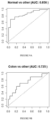
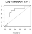
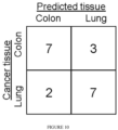
- a MHL matrix for a set of human tissues (61 published samples/data sets) was built encompassing all the 147,888 identified MHBs in the entire human genome. From this matrix, tissue-specific MHBs were identified. After including additional training data subsets comprising plasma from cancer patients and healthy controls, sets of 154 MHBs for binary prediction of tissue-of-origin between colon, lung, and normal (no solid tissue) and 295 MHBs for cancer types classification (colon versus lung) non-invasively in the blood were identified (Table 5(a)-(b)). The invention demonstrated binary classification AUCs of 0.856, 0.725, and 0.751 in independent test sets from normal, colon cancer, and lung cancer plasma respectively Figures 9(a)-9(c). ). Distinguishing between colon cancer and lung cancer plasma achieved classification accuracies of 70% and 78% for test sets comprising colon cancer and lung cancer plasma respectively using cancer type classification ( Fig. 10 ).
- the disclosed method was also applicable to tracking metastasized secondary tumors.
- Adjacent CpG sites in mammalian genomes tend to be co-methylated due to the processivity of enzymes responsible for adding or removing the methyl group.
- Yet discordant methylation patterns have also been observed, and found to be related to stochastic or uncoordinated molecular processes. The invention focused on a systematic search and investigation of regions in the human genome that exhibit highly coordinated methylation.
- methylation haplotype blocks based on 53 sets of whole genome bisulfite sequencing (WGBS) data, including 43 published sets from human adult tissues, ESC, and in vitro differentiated cell lines, as well as 10 sets from human adult tissues generated in this invention.
- the MHBs were further validated with 101 sets of RRBS ENCODE data, and 1,274 sets of Illumina450k methylation array data from TCGA tumor and normal samples.
- MHBs are enriched in, but only partially overlap with, several well-known genomic features, including CpG islands, promoters, enhancers and VMRs.
- MHL methylation haplotype load
- methylation haplotype blocks (MHBs) ( Fig. 1(b) ), using a r 2 cutoff of 0.5.
- cutoff values such as 0.3 or 0.7, for partitioning of the human genome into genetic haplotype blocks, resulted in only minor quantitative differences in the block size and number without affecting the global pattern (data not shown).
- WGBS Whole Genome Bisulfite Sequencing
- the uncovered CpG sites were either in regions with low mappability, or CpG sparse regions where there are too few sites within Illumina read pairs for deriving informative haplotypes.
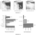
- 147,888 MHBs with an average size of 95bp and minimum 3 CpGs per block were identified, representing ⁇ 0.5% of the human genome that tends to be tightly co-regulated on the epigenetic status at the level of single DNA molecules (Fig. 11(a)-11(b) ).
- the majority of CpG sites within the same MHBs are near perfectly coupled (r 2 ⁇ 1.0) regardless of the sample type.
- Methylation LD was found to extend further along the DNA in stem cells and progenitors, compared with normal adult tissue, both in the fraction of tightly coupled CpG pairs (94.8% versus 91.2%, P-value ⁇ 2.6 ⁇ 10 -16 ), and the over-representation of partially coupled CpG pairs that are over 100 bp apart while the linkage was slightly decayed in primary cancer data sets (87.8%, mixture of CRC and LC), which was validated by another independent WGBS data set from kidney cancer (Chen et al. (2016)) ( Fig. 1(c) ). Gene Ontology analysis show cancer loss of linkage regions was significantly associated with number of cancer related pathways and functions.
- Illumina methylation arrays only report average CpG methylation of all DNA molecules in a sample, preventing a methylation linkage disequilibrium analysis. Therefore, the invention calculated the pairwise correlation coefficient of adjacent CpG methylation levels across different sample sets for block partitioning. Note that the presence of such correlated methylation blocks is a necessary but not sufficient condition for MHBs ( Fig. 12(a) ). Nonetheless, the absence of correlated methylation blocks in these data sets would invalidate the pattern of MHBs. 23,517 and 2,212 correlated methylation blocks from ENCODE RRBS and TCGA HM450K array data, respectively, were identified, among which 8,920 and 1,258 have significant overlaps with WGBS-defined MHBs.
- modest depletion in LAD was observed (Guelen et al. (2008)) and LOCK regions (Wen et al. (2009)) (46% and 37% of the expected values), and modest enrichment in TAD (Dixon et al. (2012)).
- VMR variable methylation regions
- MHBs likely capture the local coherent epigenetic signatures that are directly or indirectly coupled with transcriptional regulation.
- methylation haplotype load MHL
- uMHL unmethylated haplotype load
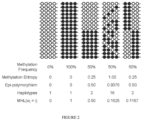
- MHL is capable of distinguishing blocks that have the same average methylation but various degrees of coordinated methylation ( Fig. 2 ).
- MHL and uMHL are bounded between 0 and 1, which allows for direct comparison of different regions across many data sets without normalization.
- the invention addressed whether treating MHBs as individual genomic elements and performing quantitative analysis based on MHL and uMHL would provide an advantage over previous approaches using the weighted average methylation in genomic windows.
- the invention sought to identify tissue specific MHBs from a collection of human solid tissues WGBS datasets based on the MHL and uMHL.
- a group specific index for each MHB was computed using either MHL or uMHL for every tissue type and then each MHB was assigned to one or more tissues based on their respective GSI. If no other tissue type have a GSI that is at least 80% of the maximum GSI, then only the tissue type with the maximum GSI is reported.
- the top tissue specific MHL or uMHL regions were selected using average MHL or uMHL values in blood cells of less than 0.05, and average non-blood cell values greater than 0.4. Using a maximum GSI of greater than 0.6 as a threshold resulted in the identification of 1,290 and 15,377 tissue specific MHL and uMHL regions respectively.
- GSI value a maximum GSI of greater than 0.6 as a threshold resulted in the identification of 1,290 and 15,377 tissue specific MHL and uMHL regions respectively.
- To identify the most informative MHL or uMHL marker regions for cancer detection or tissue mapping the top 10% of the identified MHL or uMHL regions determined by GSI value are reported in Tables 1(a) and 1(b). The tissue specific regions using MHL and uMHL were visualized using heatmaps ( Fig. 3(a)-3(b) ).
- the human adult tissues used in this invention have various degrees of similarity amongst each other.
- the invention hypothesized that this is primarily defined by their developmental lineage, and that the related MHBs might reveal epigenetic insights related to germ layer speciation.
- All the data sets based on the three germ layers were grouped, and searched for MHBs that have differential MHL.
- MHBs that have differential MHL.
- 114 ectoderm-specific MHBs 99 hyper- and 15 hypo-methylated
- 75 endoderm specific MHBs 58 hyper- and 17 hypo-methylated
- 31 mesoderm specific MHBs (9 hyper- and 22 hypo-methylated) were identified.
- the invention speculated that some of these MHBs might capture binding events of transcription factors (TF) specific to developmental germ-layers.
- hypo-MHL regions shared by mesoderm and endoderm might have regulatory functions in the fate commitment towards multiple tissues, whereas ectoderm specific hyper-MHL regions might induce the ectoderm development by suppressing the path towards the immune lineage.
- Methylation-haplotype based analysis of circulating cell-free DNA in cancer patients and healthy donors A unique aspect of methylation haplotype analysis is that the pattern of co-methylation, especially within MHBs, is robust for capturing low-frequency alleles among a heterogeneous population of molecules or cells, in the presence of biological noise or technical variability (i.e. incomplete bisulfite conversion or sequencing errors).
- the invention focused on the methylation haplotype analysis of cell-free DNA from healthy donors and cancer patients, of which various low fractions of DNA molecules were released from tumor cells and potentially carry epigenetic signatures different from blood.
- the invention sought to detect the presence of tumor specific signatures in the plasma samples, using the methylation haplotypes identified in the reference tumor tissues and in normal samples used as the negative controls. For five lung cancer plasma samples and five colorectal cancer plasma samples, matched primary tumor tissues were also obtained, and generated RRBS data (30 million reads per sample) from 100ng of tumor genomic DNA.
- the invention focused on MHBs with low MHL (i.e. genomic regions that have low or no methylation) in the blood, and determined whether cancer-associated highly methylated haplotypes (caHMH) can be detected.
- the invention provides in some embodiments that such haplotypes are present only in the tumor tissues and the matched plasma from the same patient, but not in whole blood or any other non-cancer samples.
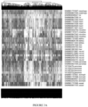
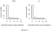
- caHMHs were found to be individual specific while few caHMHs were present in at least 53% (16/30) and 62% (18/29) cancer plasma samples for CRC and LC ( Fig. 13 ). Improving the sampling depth, by either using more input cfDNA or reducing sample loss during analysis, will likely increase the number of caHMHs commonly observed in multiple patients.
- the tumor load in cancer plasma samples was quantified, using non-negative decomposition with quadratic programming, on the RRBS data from primary cancer biopsies (LC and CRC) and the WGBS data from 10 normal tissues.
- the invention sought to use the information from normal human tissues, primary tumor biopsies, and cancer cell lines to improve the detection of cfDNA.
- the invention selected a subset of MHBs that show high MHL in primary cancer biopsies and low MHL in normal control plasma.
- MHBs with a high GSI score tended to be methylated in fewer tissue types.
- 20 colon cancer plasma, 20 lung cancer plasma, and 30 normal plasma samples were randomly sampled to create training data sets from the RRBS plasma data sets. The remaining (10 colon, 10 lung, and 39 normal) were held out as test data sets.
- An ensemble MARS (Multivariate adaptive regression splines) model was generated using the training data sets (Friedman, (1991)).
- the first classifier was an ensemble model with 100 MARS models with 154 MHB features (Table 5(a)).
- the scores from the resulting ensemble are averaged to compute a prediction score.
- the invention was able to obtain a colon vs non-colon AUC of 0.725, a lung vs non-lung AUC of 0.751, and a normal vs abnormal AUC of 0.856 ( Fig. 9(a)-9(c) ).
- these AUC values were obtained using an initial marker set identified from an independent training set of normal WGBS tissue samples only; no cancer tissue samples were used in the initial feature selection, suggesting that plasma samples can be segregated using only tissue specific markers.
- the invention focused on colon and lung cancer plasma.
- the top MHBs features that were assigned to either colon, lung, or both tissues ranked by GSI were selected.
- An ensemble of 500 MARS models identifying 295 unique tissue specific features was made (Table 5(b)).
- PLSDA partial least squares discriminant analysis
- the classification using this model was assessed using independent test data sets and the accuracy for classifying colon cancer plasma to colon tissue was found to be 70% (7/10) and the accuracy for classifying lung cancer plasma to lung tissue was 78% (7/9) ( Fig. 10 ).
- linkage disequilibrium In this invention a well-established concept in population genetics, linkage disequilibrium (LD), was extended to the analysis of co-methylated CpG patterns. While the mathematical representations are identical, there are two key differences. First, traditional linkage disequilibrium (LD) was defined for human individuals in a population, whereas in this invention the analysis was performed on the diploid genome of individual cells in a heterogeneous cell population. Second, linkage disequilibrium in human populations depend on the mutation rate, frequency of meiotic recombination, effective population size and demographic history. The LD level typically decays over the range of hundreds of kilobases to megabases.
- CpG co-methylation depends on DNA methytransferases and demethylases, which tend to have lower processivity, and, in the case of hemi-methyltransferases, much lower fidelity compared with DNA polymerases (Williams et al. (2011)). Therefore, methylation LD decays over much shorter distances ranging from tens to hundreds of bases, with the exception of imprinting regions. Even if longer-read sequencing methods were used no radical change of the block-like pattern presented in this work is expected, which is supported by a recent study (Saito et al. (2015)). Nonetheless, these short and punctuated blocks capture discrete entities of epigenetic regulation in individual cells widespread in the human genome. Such a phenomenon can be harnessed to improve the robustness and sensitivity of DNA methylation analysis, such as the deconvolution of data from heterogeneous samples including circulating cell-free DNA.
- Epigenetic abnormalities tend to be more widespread across the genome (compared with somatic mutations), and hence enabling the integration of the sparse coverage across many loci to achieve very accurate prediction by direct counting of methylated haplotypes within the appropriate tissue-specific features.
- WGBS whole genome bisulfite
- RRBS reduced representation bisulfite sequencing
- the DNA fragments were adapted to barcoded methylated adaptors (Illumina).
- WGBS the adapted DNA were converted using the EZ DNA Methylation Lightning kit (Zymo Research) and amplified for 10 cycles using iQ SYBR Green Supermix (BioRad).
- RRBS the adapted DNA were converted using the MethylCode TM Bisulfite Conversion kit (Thermo Fisher Scientific) and amplified using the PfuTurboCx polymerase (Agilent) for 12-14 cycles.
- Libraries were pooled and size selected using 6% TBE polyacrylamide gels. Libraries were sequenced using the Illumina HiSeq platform for paired-end 100-111 cycles, the Illumina MiSeq platform for paired-end 75 cycles, and the GAIIx (WGBS only) for single-end 36 cycles.
- Methylation haplotype blocks (MHB) .
- the human genome was separated into non-overlapping "sequenceable and mappable" segments using a set of generated WGBS data from 10 tissues from a 25-year adult male individual. Mapped reads from WGBS data sets were converted into methylation haplotypes in each segment. Methylation linkage disequilibrium was calculated on the combined methylation haplotypes.
- Each segment was partitioned into methylation haplotype blocks (MHBs).
- MHBs were defined as the genomic region in which the r 2 value of two adjacent CpG sites is no less than 0.5. MHB regions inferred by WGBS data sets were also validated by bulk data of methylation level.
- Methylation haplotype load (MHL) .
- Unmethylated haplotype load UMH
- P(MH i ) or P(UMH i ) is the fraction of fully successive methylated or unmethylated haplotype with i loci respectively.
- w i is the weight for i-locus haplotype.
- w i i was applied. Quantile normalization, standardization (scale) as well as the batch effect elimination (Johnson et al. (2007)) were applied.
- MHL(j) denotes the average of MHL of j th group.
- MHL max denotes the average of MHL of highest methylated group.
- the deconvolution references were constructed from normal human solid tissues, WBC, colorectal cancer tissues (CCT), and lung cancer tissues (LCT). Tissue specific MHB regions for normal human tissues were selected for brain, colon, esophagus, heart, intestine, kidney, liver, lung, and stomach using candidate features for deconvolution based on non-negative decomposition with quadratic programming (Sun et al. (2015); Houseman et al. (2012); Gong et al. (2013)). Raw MHL signals were logit transformed before deconvolution analysis on plasma samples. Samples with less than 30% whole blood content from deconvolution analysis were considered to have failed due to poor library complexity.
- Tumor load estimation using tumor specific MHBs Pruning and K-nearest neighbors (KNN) imputation was performed on the MHL matrix with only the RRBS plasma and RRBS tumor tissue samples removing samples with low coverage and imputed missing values. Thus 30 colon cancer plasma, 29 lung cancer plasma, 69 normal plasma, 4 colon cancer tissues, and 5 lung cancer tissues remained. The 69 healthy normal plasma samples were split into "training" and “test” sets; with 46 samples set aside for feature selection and training while the remaining 23 samples were used as a completely independent data set to test the quantitation. Tumor specific methylation haplotype blocks were identified by 2-tailed t-test with a False Discovery Rate (FDR) of 0.001 and a minimum difference cutoff of 0.3.
- FDR False Discovery Rate
- Two sets of markers were identified separately for colon cancer and lung cancer.
- 20 sets of simulations in which mixed sequencing reads from cancer tissue samples and normal plasma samples at a 1:5, 1:10, 1:20, 1:100, and 0:1 ratio were generated (totaling 100 simulated data sets for each cancer tissue).
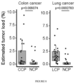
- the average MHL value for these regions was computed for each region and a linear regression model was generated using these values and the known cancer tissue proportions for each cancer tissue.
- the model was applied to the average MHL value in these regions for test sets which included 30 colon cancer plasma, 29 lung cancer plasma, and 23 normal plasma samples.
- the estimated tumor loads for normal versus colon cancer and for normal versus lung cancer were compared using the colon cancer and lung cancer markers respectively.
- Training WGBS data were collected to generate an MHL matrix and the top 15,000 MHBs by GSI were selected. These MHBs were clustered using K-means into 50 marker groups with the WGBS data matrix. Each cluster should have similar methylation patterns across each tissue type.
- 20 colon cancer plasma, 20 lung cancer plasma, and 30 normal plasma samples were randomly selected to create a training data set from the RRBS plasma samples. The remaining samples (10 colon, 10 lung, and 39 normal) were held out as a test data set.
- an ensemble MARS (Multivariate Adaptive Regression Splines) model (Friedman, (1991)) implemented in the Earth R package (https://cran.r-project.org/web/packages/earth/index.html) was trained on the training data set using features from the top 15,000 MHBs ranked by the Group Specific Index (GSI).
- the ensemble MARS model was used to perform classification on the test plasma data set using the binary classifier.
- the features selected using MARS were used in a PLSDA (partial least squares discriminant analysis) to distinguish colon versus lung.
- WGBS and RRBS data are available at the Gene Expression Omnibus (GEO) under accession GSE79279.
Landscapes
- Chemical & Material Sciences (AREA)
- Life Sciences & Earth Sciences (AREA)
- Health & Medical Sciences (AREA)
- Organic Chemistry (AREA)
- Proteomics, Peptides & Aminoacids (AREA)
- Engineering & Computer Science (AREA)
- Zoology (AREA)
- Analytical Chemistry (AREA)
- Wood Science & Technology (AREA)
- Genetics & Genomics (AREA)
- Physics & Mathematics (AREA)
- Bioinformatics & Cheminformatics (AREA)
- Immunology (AREA)
- General Health & Medical Sciences (AREA)
- Biotechnology (AREA)
- Molecular Biology (AREA)
- Biophysics (AREA)
- Pathology (AREA)
- General Engineering & Computer Science (AREA)
- Biochemistry (AREA)
- Microbiology (AREA)
- Hospice & Palliative Care (AREA)
- Oncology (AREA)
- Bioinformatics & Computational Biology (AREA)
- Evolutionary Biology (AREA)
- Medical Informatics (AREA)
- Spectroscopy & Molecular Physics (AREA)
- Theoretical Computer Science (AREA)
- Measuring Or Testing Involving Enzymes Or Micro-Organisms (AREA)
- Pharmaceuticals Containing Other Organic And Inorganic Compounds (AREA)
Claims (16)
- Verfahren zum Nachweis des Vorhandenseins einer oder mehrerer Nukleinsäuren, die auf einen Krankheitszustand, ein Ursprungsgewebe, eine Ursprungskeimschicht oder ein Ursprungsorgan hinweisen, in einem Gemisch von Nukleinsäuren, umfassend:Durchführen einer Methylierungsanalyse bei einer Probe, die eine Vielzahl von Nukleinsäuren umfasst, durch Identifizieren von Methylierungs-Haplotyp-Blöcken, wobei die Methylierungs-Haplotyp-Blöcke eine Vielzahl von Methylierungsstellen umfassen, für die der Methylierungsstatus koordiniert ist;Bestimmen eines Methylierungshaplotyp-Loads (MHL), wobei der MHL den normalisierten Anteil von methylierten Haplotypen bei unterschiedlichen Längen umfasst;Berechnen eines gruppenspezifischen Index (GSI) unter Verwendung des MHL; undBestimmen auf der Grundlage des GSI und des MHL, ob die Probe eine Vielzahl von Methylierungs-Haplotyp-Blöcken enthält, die auf das Vorhandensein einer oder mehrerer Nukleinsäuren hindeuten, die auf einen Krankheitszustand, ein Ursprungsgewebe, eine Ursprungskeimschicht, ein Ursprungsorgan oder eine beliebige Kombination davon hinweisen.
- Verfahren nach Anspruch 1, wobei die Methylierungsanalyse an zellfreier DNA durchgeführt wird.
- Verfahren nach einem der Ansprüche 1 und 2, wobei die Methylierungsanalyse an zellfreier DNA in einer Blutprobe durchgeführt wird.
- Verfahren nach einem der Ansprüche 1 bis 3, wobei die Vielzahl von Methylierungs-Haplotyp-Blöcken mindestens 2, mindestens 3, mindestens 4, mindestens 5, mindestens 10, mindestens 20, mindestens 40, mindestens 50, mindestens 100, mindestens 200, mindestens 300, mindestens 400, mindestens 500 oder mehr als 500 Methylierungs-Haplotyp-Blöcke umfasst.
- Verfahren nach einem der Ansprüche 1-4, wobei der Krankheitszustand ein Tumor ist.
- Verfahren nach Anspruch 5, ferner umfassend das Bestimmen, ob die Probe eine Vielzahl von Methylierungs-Haplotyp-Blöcken enthält, die auf das Vorhandensein einer oder mehrerer Nukleinsäuren hindeuten, die auf ein normales Gewebe oder ein normales Organ hinweisen, das dem Ursprungsgewebe oder -organ des Tumors entspricht.
- Verfahren nach einem der Ansprüche 1 bis 6, wobei der Krankheitszustand fetale Aneuploidie ist.
- Verfahren nach einem der Ansprüche 1-7, wobei die Probe eine Blutprobe ist.
- Verfahren nach einem der Ansprüche 1-8, ferner umfassend die Quantifizierung des Anteils der einen oder mehreren Nukleinsäuren in der Probe, die auf einen Krankheitszustand, ein Ursprungsgewebe, ein Ursprungsorgan oder eine beliebige Kombination davon hinweisen.
- Verfahren nach einem der Ansprüche 1-9, wobei die Methylierungsanalyse unter Verwendung einer Technik durchgeführt wird, die aus der Gruppe ausgewählt ist, die aus Bisulfit-Methylierungsanalyse, Bisulfit-Sequenzierung mit reduzierter Darstellung (RRBS), WGBS, BSPP, Mikrotröpfchen-PCR, auf Selektorsonden basierenden Verfahren und MeDiP besteht.
- Verfahren nach einem der Ansprüche 1 bis 10, ferner umfassend das Bestimmen eines unmethylierten Haplotyp-Loads (uMHL) für jeden Methylierungs-Haplotyp-Block, wobei der uMHL den normalisierten Anteil der unmethylierten Haplotypen bei unterschiedlichen Längen umfasst.
- Verfahren nach einem der Ansprüche 1-11, wobei die Methylierungs-Haplotyp-Blöcke eine durchschnittliche Größe von 95 bp haben.
- Verfahren nach einem der Ansprüche 1-12, wobei die Methylierungs-Haplotyp-Blöcke ein Minimum von 3 CpG pro Block aufweisen.
- Verfahren nach einem der Ansprüche 1-13, ferner umfassend die Verwendung einer quantitativen Dekonvolution zum Nachweis von Krebs-DNA.
- Verfahren nach Anspruch 6, ferner umfassend das Quantifizieren des Anteils der Vielzahl von Methylierungs-Haplotyp-Blöcken, die auf das Vorhandensein eines Tumors in der Probe hinweisen, und das Quantifizieren des Anteils der Vielzahl von Methylierungs-Haplotyp-Blöcken, die auf das Vorhandensein einer oder mehrerer Nukleinsäuren hindeuten, die auf ein normales Gewebe oder ein normales Organ hinweisen, das dem Ursprungsgewebe oder -organ des Tumors in der Probe entspricht.
- Verfahren zur Identifizierung von Krebs oder einem Ursprungsgewebe in einer Probe, die eine Vielzahl von Nukleinsäuren umfasst, umfassend:Bestimmen von Methylierungs-Haplotypen in einer Vielzahl von Nukleinsäuresegmenten;Kombinieren der Methylierungs-Haplotypen und Berechnen des Methylierungs-Linkage-Ungleichgewichts an den kombinierten Methylierungs-Haplotypen;Aufteilen jedes Segments in eine Vielzahl von Methylierungs-Haplotyp-Blöcken, wobei die Methylierungs-Haplotyp-Blöcke eine Vielzahl von Methylierungsstellen umfassen, für die der Methylierungsstatus koordiniert ist;Berechnen des gruppenspezifischen Index (GSI) auf der Basis des normalisierten Anteils der methylierten Haplotypen bei unterschiedlichen Längen der Methylierungs-Haplotyp-Blöcke; und Bestimmen auf der Basis des GSI, ob die Probe Krebs oder das Ursprungsgewebe umfasst.
Applications Claiming Priority (3)
| Application Number | Priority Date | Filing Date | Title |
|---|---|---|---|
| US201662437512P | 2016-12-21 | 2016-12-21 | |
| US201662438401P | 2016-12-22 | 2016-12-22 | |
| PCT/US2017/067871 WO2018119216A1 (en) | 2016-12-21 | 2017-12-21 | Deconvolution and detection of rare dna in plasma |
Publications (4)
| Publication Number | Publication Date |
|---|---|
| EP3559259A1 EP3559259A1 (de) | 2019-10-30 |
| EP3559259A4 EP3559259A4 (de) | 2020-08-26 |
| EP3559259C0 EP3559259C0 (de) | 2025-02-05 |
| EP3559259B1 true EP3559259B1 (de) | 2025-02-05 |
Family
ID=62627223
Family Applications (1)
| Application Number | Title | Priority Date | Filing Date |
|---|---|---|---|
| EP17882359.7A Active EP3559259B1 (de) | 2016-12-21 | 2017-12-21 | Dekonvolution und detektion seltener dna in plasma |
Country Status (6)
| Country | Link |
|---|---|
| US (1) | US12509731B2 (de) |
| EP (1) | EP3559259B1 (de) |
| CN (1) | CN110168108A (de) |
| CA (1) | CA3047421A1 (de) |
| ES (1) | ES3022457T3 (de) |
| WO (1) | WO2018119216A1 (de) |
Families Citing this family (18)
| Publication number | Priority date | Publication date | Assignee | Title |
|---|---|---|---|---|
| CN111417731A (zh) | 2017-05-12 | 2020-07-14 | 哈佛学院校长同事会 | 通用早期癌症诊断 |
| CN111989407B (zh) | 2018-03-13 | 2024-10-29 | 格里尔公司 | 异常的片段检测及分类 |
| CA3302676A1 (en) | 2018-04-02 | 2026-05-13 | Grail, Inc. | Methylation markers and targeted methylation probe panels |
| EP3856903A4 (de) | 2018-09-27 | 2022-07-27 | Grail, LLC | Methylierungsmarker und gezieltes methylierungssondenpaneel |
| AU2019384154B2 (en) | 2018-11-21 | 2025-11-20 | Agilent Technologies, Inc. | Methods for targeted nucleic acid library formation |
| AU2019403273A1 (en) * | 2018-12-19 | 2021-08-05 | Grail, Llc | Cancer tissue source of origin prediction with multi-tier analysis of small variants in cell-free dna samples |
| EP3899953A1 (de) | 2018-12-21 | 2021-10-27 | Grail, Inc. | Ursprungsquellendekonvolution auf basis von methylierungsfragmenten in zellfreien dna-proben |
| WO2021072786A1 (zh) * | 2019-10-16 | 2021-04-22 | 厦门信道生物技术有限公司 | 人结直肠癌相关基因特定区域甲基化的引物组及试剂以及试剂盒及其应用 |
| JP7874822B2 (ja) * | 2019-10-18 | 2026-06-17 | ワシントン ユニバーシティ | 細胞状態を測定するための方法及びシステム |
| CN111833965B (zh) * | 2019-11-08 | 2024-06-04 | 中国科学院北京基因组研究所(国家生物信息中心) | 一种尿沉渣基因组dna的分类方法、装置和用途 |
| GB2603051B (en) | 2020-01-30 | 2023-04-26 | Prognomiq Inc | Lung biomarkers and methods of use thereof |
| CN115103909A (zh) | 2020-01-31 | 2022-09-23 | 艾维达生物医学公司 | 用于靶向核酸捕获的系统和方法 |
| WO2022133315A1 (en) * | 2020-12-17 | 2022-06-23 | President And Fellows Of Harvard College | Methods of cancer detection using extraembryonically methylated cpg islands |
| AU2022249074A1 (en) | 2021-03-31 | 2023-09-21 | PrognomIQ, Inc. | Multi-omic assessment |
| CN113604553B (zh) * | 2021-07-27 | 2023-08-15 | 中南大学湘雅二医院 | 成人still’s病生物标志物、诊断试剂及其应用 |
| WO2023028270A1 (en) * | 2021-08-25 | 2023-03-02 | Xgenomes Corp. | Random epigenomic sampling |
| WO2023039479A1 (en) | 2021-09-10 | 2023-03-16 | PrognomIQ, Inc. | Direct classification of raw biomolecule measurement data |
| JP2024534992A (ja) | 2021-09-13 | 2024-09-26 | プログノミック インコーポレイテッド | 生体分子の増強された検出および定量 |
Family Cites Families (10)
| Publication number | Priority date | Publication date | Assignee | Title |
|---|---|---|---|---|
| WO2004065617A2 (en) * | 2003-01-17 | 2004-08-05 | The Trustees Of Boston University | Haplotype analysis |
| US10927415B2 (en) * | 2008-11-26 | 2021-02-23 | The Johns Hopkins University | Methods for identifying cancer risk |
| US8877442B2 (en) | 2010-12-07 | 2014-11-04 | The Board Of Trustees Of The Leland Stanford Junior University | Non-invasive determination of fetal inheritance of parental haplotypes at the genome-wide scale |
| US10041124B2 (en) * | 2013-01-14 | 2018-08-07 | King Abdullah University Of Science And Technology | Methylation biomarkers for ovarian cancer |
| US10570455B2 (en) * | 2013-05-10 | 2020-02-25 | University Of Southern California | DNA methylation biomarkers for bladder cancer |
| US20160340740A1 (en) * | 2014-01-30 | 2016-11-24 | The Regents Of The University Of California | Methylation haplotyping for non-invasive diagnosis (monod) |
| EP2915883A1 (de) * | 2014-03-07 | 2015-09-09 | Ruprecht-Karls-Universität Heidelberg | Nichtinvasives Assay-Verfahren zur Früherkennung von Krebs |
| TWI758011B (zh) | 2014-07-18 | 2022-03-11 | 香港中文大學 | Dna混合物中之組織甲基化模式分析 |
| CN104328183A (zh) * | 2014-10-30 | 2015-02-04 | 东南大学 | 基于高通量测序的基因组单倍型甲基化检测方法 |
| DK3256605T3 (da) * | 2015-02-10 | 2022-03-14 | Univ Hong Kong Chinese | Påvisning af mutationer til cancerscreening og føtal analyse |
-
2017
- 2017-12-21 EP EP17882359.7A patent/EP3559259B1/de active Active
- 2017-12-21 CA CA3047421A patent/CA3047421A1/en active Pending
- 2017-12-21 WO PCT/US2017/067871 patent/WO2018119216A1/en not_active Ceased
- 2017-12-21 US US16/470,586 patent/US12509731B2/en active Active
- 2017-12-21 ES ES17882359T patent/ES3022457T3/es active Active
- 2017-12-21 CN CN201780082929.5A patent/CN110168108A/zh active Pending
Non-Patent Citations (3)
| Title |
|---|
| BUCKLEY PATRICK G. ET AL: "Genome-wide DNA methylation analysis of neuroblastic tumors reveals clinically relevant epigenetic events and large-scale epigenomic alterations localized to telomeric regions : Methylation analysis of neuroblastoma", INTERNATIONAL JOURNAL OF CANCER, vol. 128, no. 10, 28 July 2010 (2010-07-28), US, pages 2296 - 2305, XP055842115, ISSN: 0020-7136, Retrieved from the Internet <URL:https://api.wiley.com/onlinelibrary/tdm/v1/articles/10.1002%2Fijc.25584> DOI: 10.1002/ijc.25584 * |
| ECKHARDT FLORIAN ET AL: "DNA methylation profiling of human chromosomes 6, 20 and 22", NATURE GENETICS, vol. 38, no. 12, 1 December 2006 (2006-12-01), New York, pages 1378 - 1385, XP055829961, ISSN: 1061-4036, Retrieved from the Internet <URL:https://www.ncbi.nlm.nih.gov/pmc/articles/PMC3082778/pdf/ukmss-32232.pdf> DOI: 10.1038/ng1909 * |
| XU JING ET AL: "EWAS: epigenome-wide association studies software 1.0 - identifying the association between combinations of methylation levels and diseases", SCIENTIFIC REPORTS, vol. 6, no. 1, 28 November 2016 (2016-11-28), XP055842189, Retrieved from the Internet <URL:http://www.nature.com/articles/srep37951> DOI: 10.1038/srep37951 * |
Also Published As
| Publication number | Publication date |
|---|---|
| EP3559259C0 (de) | 2025-02-05 |
| WO2018119216A1 (en) | 2018-06-28 |
| US20200087731A1 (en) | 2020-03-19 |
| US12509731B2 (en) | 2025-12-30 |
| CA3047421A1 (en) | 2018-06-28 |
| CN110168108A (zh) | 2019-08-23 |
| ES3022457T3 (en) | 2025-05-28 |
| EP3559259A4 (de) | 2020-08-26 |
| EP3559259A1 (de) | 2019-10-30 |
Similar Documents
| Publication | Publication Date | Title |
|---|---|---|
| US12509731B2 (en) | Deconvolution and detection of rare DNA in plasma | |
| AU2022201026B2 (en) | Non-invasive determination of methylome of fetus or tumor from plasma | |
| US11274347B2 (en) | Non-invasive determination of type of cancer | |
| CN105431552B (zh) | 多组学标记在预测糖尿病中的用途 | |
| WO2019008148A9 (en) | Enrichment of targeted genomic regions for multiplexed parallel analysis | |
| HK40075963B (en) | Non-invasive determination of methylome of tumor from plasma | |
| HK40075963A (en) | Non-invasive determination of methylome of tumor from plasma | |
| HK40055436A (en) | Non-invasive determination of methylome of tumor from plasma | |
| HK40013800B (en) | Non-invasive determination of methylome of tumor from plasma | |
| HK40013800A (en) | Non-invasive determination of methylome of tumor from plasma | |
| HK1257868B (en) | Non-invasive determination of methylome of tumor from plasma | |
| HK1206797B (en) | Non-invasive determination of methylome of fetus or tumor from plasma |
Legal Events
| Date | Code | Title | Description |
|---|---|---|---|
| STAA | Information on the status of an ep patent application or granted ep patent |
Free format text: STATUS: THE INTERNATIONAL PUBLICATION HAS BEEN MADE |
|
| PUAI | Public reference made under article 153(3) epc to a published international application that has entered the european phase |
Free format text: ORIGINAL CODE: 0009012 |
|
| STAA | Information on the status of an ep patent application or granted ep patent |
Free format text: STATUS: REQUEST FOR EXAMINATION WAS MADE |
|
| 17P | Request for examination filed |
Effective date: 20190711 |
|
| AK | Designated contracting states |
Kind code of ref document: A1 Designated state(s): AL AT BE BG CH CY CZ DE DK EE ES FI FR GB GR HR HU IE IS IT LI LT LU LV MC MK MT NL NO PL PT RO RS SE SI SK SM TR |
|
| AX | Request for extension of the european patent |
Extension state: BA ME |
|
| DAV | Request for validation of the european patent (deleted) | ||
| DAX | Request for extension of the european patent (deleted) | ||
| REG | Reference to a national code |
Ref country code: DE Ref legal event code: R079 Free format text: PREVIOUS MAIN CLASS: C12Q0001680000 Ipc: C12Q0001688600 |
|
| A4 | Supplementary search report drawn up and despatched |
Effective date: 20200724 |
|
| RIC1 | Information provided on ipc code assigned before grant |
Ipc: G01N 27/62 20060101ALI20200720BHEP Ipc: C12Q 1/6886 20180101AFI20200720BHEP |
|
| STAA | Information on the status of an ep patent application or granted ep patent |
Free format text: STATUS: EXAMINATION IS IN PROGRESS |
|
| 17Q | First examination report despatched |
Effective date: 20210923 |
|
| GRAP | Despatch of communication of intention to grant a patent |
Free format text: ORIGINAL CODE: EPIDOSNIGR1 |
|
| STAA | Information on the status of an ep patent application or granted ep patent |
Free format text: STATUS: GRANT OF PATENT IS INTENDED |
|
| INTG | Intention to grant announced |
Effective date: 20240829 |
|
| GRAS | Grant fee paid |
Free format text: ORIGINAL CODE: EPIDOSNIGR3 |
|
| GRAA | (expected) grant |
Free format text: ORIGINAL CODE: 0009210 |
|
| STAA | Information on the status of an ep patent application or granted ep patent |
Free format text: STATUS: THE PATENT HAS BEEN GRANTED |
|
| AK | Designated contracting states |
Kind code of ref document: B1 Designated state(s): AL AT BE BG CH CY CZ DE DK EE ES FI FR GB GR HR HU IE IS IT LI LT LU LV MC MK MT NL NO PL PT RO RS SE SI SK SM TR |
|
| REG | Reference to a national code |
Ref country code: GB Ref legal event code: FG4D |
|
| REG | Reference to a national code |
Ref country code: CH Ref legal event code: EP |
|
| REG | Reference to a national code |
Ref country code: IE Ref legal event code: FG4D |
|
| REG | Reference to a national code |
Ref country code: DE Ref legal event code: R096 Ref document number: 602017087657 Country of ref document: DE |
|
| U01 | Request for unitary effect filed |
Effective date: 20250305 |
|
| U07 | Unitary effect registered |
Designated state(s): AT BE BG DE DK EE FI FR IT LT LU LV MT NL PT RO SE SI Effective date: 20250415 |
|
| REG | Reference to a national code |
Ref country code: ES Ref legal event code: FG2A Ref document number: 3022457 Country of ref document: ES Kind code of ref document: T3 Effective date: 20250528 |
|
| PG25 | Lapsed in a contracting state [announced via postgrant information from national office to epo] |
Ref country code: RS Free format text: LAPSE BECAUSE OF FAILURE TO SUBMIT A TRANSLATION OF THE DESCRIPTION OR TO PAY THE FEE WITHIN THE PRESCRIBED TIME-LIMIT Effective date: 20250505 |
|
| PG25 | Lapsed in a contracting state [announced via postgrant information from national office to epo] |
Ref country code: PL Free format text: LAPSE BECAUSE OF FAILURE TO SUBMIT A TRANSLATION OF THE DESCRIPTION OR TO PAY THE FEE WITHIN THE PRESCRIBED TIME-LIMIT Effective date: 20250205 |
|
| PG25 | Lapsed in a contracting state [announced via postgrant information from national office to epo] |
Ref country code: NO Free format text: LAPSE BECAUSE OF FAILURE TO SUBMIT A TRANSLATION OF THE DESCRIPTION OR TO PAY THE FEE WITHIN THE PRESCRIBED TIME-LIMIT Effective date: 20250505 Ref country code: IS Free format text: LAPSE BECAUSE OF FAILURE TO SUBMIT A TRANSLATION OF THE DESCRIPTION OR TO PAY THE FEE WITHIN THE PRESCRIBED TIME-LIMIT Effective date: 20250605 |
|
| PG25 | Lapsed in a contracting state [announced via postgrant information from national office to epo] |
Ref country code: HR Free format text: LAPSE BECAUSE OF FAILURE TO SUBMIT A TRANSLATION OF THE DESCRIPTION OR TO PAY THE FEE WITHIN THE PRESCRIBED TIME-LIMIT Effective date: 20250205 |
|
| PG25 | Lapsed in a contracting state [announced via postgrant information from national office to epo] |
Ref country code: GR Free format text: LAPSE BECAUSE OF FAILURE TO SUBMIT A TRANSLATION OF THE DESCRIPTION OR TO PAY THE FEE WITHIN THE PRESCRIBED TIME-LIMIT Effective date: 20250506 |
|
| PG25 | Lapsed in a contracting state [announced via postgrant information from national office to epo] |
Ref country code: SM Free format text: LAPSE BECAUSE OF FAILURE TO SUBMIT A TRANSLATION OF THE DESCRIPTION OR TO PAY THE FEE WITHIN THE PRESCRIBED TIME-LIMIT Effective date: 20250205 |
|
| PG25 | Lapsed in a contracting state [announced via postgrant information from national office to epo] |
Ref country code: CZ Free format text: LAPSE BECAUSE OF FAILURE TO SUBMIT A TRANSLATION OF THE DESCRIPTION OR TO PAY THE FEE WITHIN THE PRESCRIBED TIME-LIMIT Effective date: 20250205 |
|
| PG25 | Lapsed in a contracting state [announced via postgrant information from national office to epo] |
Ref country code: SK Free format text: LAPSE BECAUSE OF FAILURE TO SUBMIT A TRANSLATION OF THE DESCRIPTION OR TO PAY THE FEE WITHIN THE PRESCRIBED TIME-LIMIT Effective date: 20250205 |
|
| PLBE | No opposition filed within time limit |
Free format text: ORIGINAL CODE: 0009261 |
|
| STAA | Information on the status of an ep patent application or granted ep patent |
Free format text: STATUS: NO OPPOSITION FILED WITHIN TIME LIMIT |
|
| REG | Reference to a national code |
Ref country code: CH Ref legal event code: L10 Free format text: ST27 STATUS EVENT CODE: U-0-0-L10-L00 (AS PROVIDED BY THE NATIONAL OFFICE) Effective date: 20251217 |
|
| PGFP | Annual fee paid to national office [announced via postgrant information from national office to epo] |
Ref country code: GB Payment date: 20251223 Year of fee payment: 9 |
|
| 26N | No opposition filed |
Effective date: 20251106 |
|
| PGFP | Annual fee paid to national office [announced via postgrant information from national office to epo] |
Ref country code: ES Payment date: 20260119 Year of fee payment: 9 |
|
| U21 | Renewal fee for the european patent with unitary effect paid with additional fee |
Year of fee payment: 9 Effective date: 20260310 |
|
| PG25 | Lapsed in a contracting state [announced via postgrant information from national office to epo] |
Ref country code: MC Free format text: LAPSE BECAUSE OF FAILURE TO SUBMIT A TRANSLATION OF THE DESCRIPTION OR TO PAY THE FEE WITHIN THE PRESCRIBED TIME-LIMIT Effective date: 20250205 |
|
| REG | Reference to a national code |
Ref country code: CH Ref legal event code: H13 Free format text: ST27 STATUS EVENT CODE: U-0-0-H10-H13 (AS PROVIDED BY THE NATIONAL OFFICE) Effective date: 20260723 |