EP3551749B1 - Compositions and methods for in vitro cultivation and/or expansion of regulatory t cells - Google Patents
Compositions and methods for in vitro cultivation and/or expansion of regulatory t cells Download PDFInfo
- Publication number
- EP3551749B1 EP3551749B1 EP17878510.1A EP17878510A EP3551749B1 EP 3551749 B1 EP3551749 B1 EP 3551749B1 EP 17878510 A EP17878510 A EP 17878510A EP 3551749 B1 EP3551749 B1 EP 3551749B1
- Authority
- EP
- European Patent Office
- Prior art keywords
- tregs
- treg
- cells
- foxp3
- days
- Prior art date
- Legal status (The legal status is an assumption and is not a legal conclusion. Google has not performed a legal analysis and makes no representation as to the accuracy of the status listed.)
- Active
Links
Images


Classifications
-
- C—CHEMISTRY; METALLURGY
- C12—BIOCHEMISTRY; BEER; SPIRITS; WINE; VINEGAR; MICROBIOLOGY; ENZYMOLOGY; MUTATION OR GENETIC ENGINEERING
- C12N—MICROORGANISMS OR ENZYMES; COMPOSITIONS THEREOF; PROPAGATING, PRESERVING, OR MAINTAINING MICROORGANISMS; MUTATION OR GENETIC ENGINEERING; CULTURE MEDIA
- C12N5/00—Undifferentiated human, animal or plant cells, e.g. cell lines; Tissues; Cultivation or maintenance thereof; Culture media therefor
- C12N5/06—Animal cells or tissues; Human cells or tissues
- C12N5/0602—Vertebrate cells
- C12N5/0634—Cells from the blood or the immune system
- C12N5/0636—T lymphocytes
- C12N5/0637—Immunosuppressive T lymphocytes, e.g. regulatory T cells or Treg
-
- A—HUMAN NECESSITIES
- A61—MEDICAL OR VETERINARY SCIENCE; HYGIENE
- A61K—PREPARATIONS FOR MEDICAL, DENTAL OR TOILETRY PURPOSES
- A61K40/00—Cellular immunotherapy
- A61K40/10—Cellular immunotherapy characterised by the cell type used
- A61K40/11—T-cells, e.g. tumour infiltrating lymphocytes [TIL] or regulatory T [Treg] cells; Lymphokine-activated killer [LAK] cells
-
- A—HUMAN NECESSITIES
- A61—MEDICAL OR VETERINARY SCIENCE; HYGIENE
- A61K—PREPARATIONS FOR MEDICAL, DENTAL OR TOILETRY PURPOSES
- A61K40/00—Cellular immunotherapy
- A61K40/20—Cellular immunotherapy characterised by the effect or the function of the cells
- A61K40/22—Immunosuppressive or immunotolerising
-
- A—HUMAN NECESSITIES
- A61—MEDICAL OR VETERINARY SCIENCE; HYGIENE
- A61K—PREPARATIONS FOR MEDICAL, DENTAL OR TOILETRY PURPOSES
- A61K40/00—Cellular immunotherapy
- A61K40/40—Cellular immunotherapy characterised by antigens that are targeted or presented by cells of the immune system
- A61K40/41—Vertebrate antigens
- A61K40/416—Antigens related to auto-immune diseases; Preparations to induce self-tolerance
-
- A—HUMAN NECESSITIES
- A61—MEDICAL OR VETERINARY SCIENCE; HYGIENE
- A61P—SPECIFIC THERAPEUTIC ACTIVITY OF CHEMICAL COMPOUNDS OR MEDICINAL PREPARATIONS
- A61P37/00—Drugs for immunological or allergic disorders
- A61P37/02—Immunomodulators
- A61P37/06—Immunosuppressants, e.g. drugs for graft rejection
-
- A—HUMAN NECESSITIES
- A61—MEDICAL OR VETERINARY SCIENCE; HYGIENE
- A61K—PREPARATIONS FOR MEDICAL, DENTAL OR TOILETRY PURPOSES
- A61K35/00—Medicinal preparations containing materials or reaction products thereof with undetermined constitution
- A61K35/12—Materials from mammals; Compositions comprising non-specified tissues or cells; Compositions comprising non-embryonic stem cells; Genetically modified cells
- A61K2035/122—Materials from mammals; Compositions comprising non-specified tissues or cells; Compositions comprising non-embryonic stem cells; Genetically modified cells for inducing tolerance or supression of immune responses
-
- A—HUMAN NECESSITIES
- A61—MEDICAL OR VETERINARY SCIENCE; HYGIENE
- A61K—PREPARATIONS FOR MEDICAL, DENTAL OR TOILETRY PURPOSES
- A61K35/00—Medicinal preparations containing materials or reaction products thereof with undetermined constitution
- A61K35/12—Materials from mammals; Compositions comprising non-specified tissues or cells; Compositions comprising non-embryonic stem cells; Genetically modified cells
- A61K2035/124—Materials from mammals; Compositions comprising non-specified tissues or cells; Compositions comprising non-embryonic stem cells; Genetically modified cells the cells being hematopoietic, bone marrow derived or blood cells
-
- C—CHEMISTRY; METALLURGY
- C12—BIOCHEMISTRY; BEER; SPIRITS; WINE; VINEGAR; MICROBIOLOGY; ENZYMOLOGY; MUTATION OR GENETIC ENGINEERING
- C12N—MICROORGANISMS OR ENZYMES; COMPOSITIONS THEREOF; PROPAGATING, PRESERVING, OR MAINTAINING MICROORGANISMS; MUTATION OR GENETIC ENGINEERING; CULTURE MEDIA
- C12N2501/00—Active agents used in cell culture processes, e.g. differentation
- C12N2501/10—Growth factors
- C12N2501/15—Transforming growth factor beta (TGF-β)
-
- C—CHEMISTRY; METALLURGY
- C12—BIOCHEMISTRY; BEER; SPIRITS; WINE; VINEGAR; MICROBIOLOGY; ENZYMOLOGY; MUTATION OR GENETIC ENGINEERING
- C12N—MICROORGANISMS OR ENZYMES; COMPOSITIONS THEREOF; PROPAGATING, PRESERVING, OR MAINTAINING MICROORGANISMS; MUTATION OR GENETIC ENGINEERING; CULTURE MEDIA
- C12N2501/00—Active agents used in cell culture processes, e.g. differentation
- C12N2501/20—Cytokines; Chemokines
- C12N2501/23—Interleukins [IL]
- C12N2501/2302—Interleukin-2 (IL-2)
-
- C—CHEMISTRY; METALLURGY
- C12—BIOCHEMISTRY; BEER; SPIRITS; WINE; VINEGAR; MICROBIOLOGY; ENZYMOLOGY; MUTATION OR GENETIC ENGINEERING
- C12N—MICROORGANISMS OR ENZYMES; COMPOSITIONS THEREOF; PROPAGATING, PRESERVING, OR MAINTAINING MICROORGANISMS; MUTATION OR GENETIC ENGINEERING; CULTURE MEDIA
- C12N2501/00—Active agents used in cell culture processes, e.g. differentation
- C12N2501/20—Cytokines; Chemokines
- C12N2501/24—Interferons [IFN]
-
- C—CHEMISTRY; METALLURGY
- C12—BIOCHEMISTRY; BEER; SPIRITS; WINE; VINEGAR; MICROBIOLOGY; ENZYMOLOGY; MUTATION OR GENETIC ENGINEERING
- C12N—MICROORGANISMS OR ENZYMES; COMPOSITIONS THEREOF; PROPAGATING, PRESERVING, OR MAINTAINING MICROORGANISMS; MUTATION OR GENETIC ENGINEERING; CULTURE MEDIA
- C12N2501/00—Active agents used in cell culture processes, e.g. differentation
- C12N2501/50—Cell markers; Cell surface determinants
- C12N2501/599—Cell markers; Cell surface determinants with CD designations not provided for elsewhere
-
- C—CHEMISTRY; METALLURGY
- C12—BIOCHEMISTRY; BEER; SPIRITS; WINE; VINEGAR; MICROBIOLOGY; ENZYMOLOGY; MUTATION OR GENETIC ENGINEERING
- C12N—MICROORGANISMS OR ENZYMES; COMPOSITIONS THEREOF; PROPAGATING, PRESERVING, OR MAINTAINING MICROORGANISMS; MUTATION OR GENETIC ENGINEERING; CULTURE MEDIA
- C12N2502/00—Coculture with; Conditioned medium produced by
- C12N2502/11—Coculture with; Conditioned medium produced by blood or immune system cells
- C12N2502/1114—T cells
Definitions
- the present invention relates to compositions and methods for activating, culturing, expanding, maintaining, and/or stabilizing the growth of regulatory T cell populations, including regulatory FOXP3 + T cells.
- Tregs CD4 + CD25 + FOXP3 + regulatory T cells (Tregs) play an integral role in preventing autoimmunity and controlling inflammation.
- the importance of Tregs is illustrated by the fact that loss-of-function mutations in FOXP3 leads to the rapid, fatal multi-organ autoimmune disorders IPEX and scurfy in humans and mice, respectively.
- dysfunctional Tregs are linked to increased susceptibility to certain autoimmune diseases such as multiple sclerosis (MS) and type 1 diabetes (T1D). Because of their suppressive capabilities, Tregs offer a unique therapeutic potential for autoimmune diseases, allergic diseases, transplant tolerance, and chronic inflammatory disorders.
- MS multiple sclerosis
- T1D type 1 diabetes
- Treg adoptive transfer therapies where Tregs are induced or purified ex vivo and then infused into a syngeneic organism, have shown encouraging results in animal models of autoimmune disease such as experimental autoimmune encephalomyelitis (EAE), the animal model of MS, T1D, and colitis.
- EAE experimental autoimmune encephalomyelitis
- Treg adoptive transfer therapies for tissue transplantation and T1D have shown favorable outcomes, but no adoptive Treg therapy has yet to be approved as a treatment option.
- An additional approach to Treg-mediated therapies involves induction or expansion of Tregs in vivo through the use of tolerogenic vaccines or anti-inflammatory drugs, such as interferon- ⁇ .
- Interferon- ⁇ is a first-line treatment option for relapsing-remitting MS.
- Interferon- ⁇ has been shown to increase numbers of Tregs in circulation.
- Tolerogenic vaccines ideally work by inducing or expanding antigen-specific Tregs, which would leave the adaptive immune response intact.
- FOXP3 is needed for adaptive self-tolerance, and FOXP3 is expressed in a canonical lineage of suppressive Tregs; however, it cannot be assumed that the role of FOXP3 in self-tolerance is entirely mediated via the expression of FOXP3 in this lineage of CD4 + CD25 + FOXP3 + Tregs.
- FOXP3 may play inhibitory roles in other cell types, particularly during transient expression in canonical conventional T cell subsets.
- FOXP3 can be transiently expressed during activation in human T cells and is expressed in quasi-stable non-committed CD25 low FOXP3 + T cells in mice.
- the assessment of FOXP3 + Tregs requires new assessment tools to analyze this subset. One approach to assess these questions is to induce Tregs in vitro and expand/ maintain in pure culture, and show via adoptive transfer experiments that these Tregs suppress EAE.
- Treg-mediated therapies are notoriously unstable and plastic.
- In vivo studies using fate-mapping techniques to track FOXP3 expression showed that under certain environmental circumstances, Tregs can lose FOXP3 expression and actually adopt an effector T cell phenotype. These so called “ex-Tregs” can potentially exacerbate autoimmune disease as FOXP3 is required for Treg suppressive activity.
- Ex vivo expansion and culturing of Tregs has faced many difficulties due to the fact that Tregs are relatively anergic in vitro and that contaminating FOXP3 - conventional cells (Tcons) can quickly outgrow Tregs. Additional evidence shows that Tregs lose FOXP3 expression over time when cultured ex vivo. Propagation and expansion of Tregs in vitro is a step in generating the numbers of Tregs necessary for Treg adoptive transfer therapies. What drives Treg instability, especially in vitro, is not well understood.
- IL-2 is involved in both the induction and maintenance of Tregs.
- CD25 the IL-2 receptor alpha chain, is constitutively expressed on Tregs and represents a phenotypic marker for Tregs, although CD25 is also expressed at high levels on activated effector T cells.
- Various therapeutic techniques manipulating IL-2 availability have met with some success in animal models and in the clinic. The rationale is that selectively expanding Tregs will alleviate autoimmune disorders. While most of the focus to date has been on manipulating IL-2 directly, little attention has focused on manipulating CD25.
- WO 2012/012737 is directed to materials and methods related to culturing stable Treg cells or in vivo expansion of stable Treg cells, the cells made by the methods, treatments for various inflammatory/autoimmune pathologies and transplant/graft rejection, and related materials.
- Ex vivo induction and expansion of the stable Tregs is described, including use of inducing compositions, such as certain mAbs and other compounds, along with expansion medium comprising IL-2.
- inducing compositions such as certain mAbs and other compounds
- expansion medium comprising IL-2 In vivo expansion of stable Treg cells and treatments for various inflammatory/autoimmune pathologies and transplant/graft rejection are also described, including the use of mAbs and their variants.
- XP055509649 summarizes the main features of different subtypes of Tregs and focuses on the clinical implications of their altered function in human studies. More specifically, the article discusses abnormalities affecting FOXP3 + Tregs and Tr1 cells that will lead to autoimmune manifestations and/or allergic reactions, and the potential therapeutic use of Tregs.
- the article concludes that low IL-2 concentrations coupled with high PC61 concentrations constrained IL-2 signaling to a low-intensity range that enabled dominant stable outgrowth of suppressive CD25 high FOXP3 + Tregs and states that the ability to indefinitely expand stable Treg lines can provide insight into FOXP3 + Treg physiology and be foundational for Treg-based immunotherapy.
- FOXP3 + regulatory T cells (Tregs) represent potentially powerful adoptive immunotherapies for autoimmune disease and other chronic inflammatory diseases.
- the field currently lacks reliable technologies to enable long-term in vitro expansion of stable, antigen-specific FOXP3 + Treg lines.
- Treg lines lack phenotypic stability and rapidly convert to a Tcon phenotype during in vitro propagation in IL-2. Thus, there remains a need for improved methods of growing and maintaining Treg populations.
- Tregs are qualitatively distinguished from Tcons and non-Tregs in that Tregs constitutively express high levels of CD25 (IL-2 receptor alpha, IL-2R ⁇ ), whereas Tcons and non-Tregs are CD25 - or only transiently express an activation-dependent CD25 high phenotype. Due to differential expression of CD25, it was reasoned that anti-CD25 mAbs would only partially block IL-2 signaling in Tregs while completely blocking IL-2 responses of Tcons, and that the differential inhibition of CD25 would enable the preferential and dominant outgrowth of Tregs during in vitro cultivation.
- CD25 IL-2 receptor alpha, IL-2R ⁇
- TGF- ⁇ -induced Treg lines that were maintained in IL-2 in the presence of the anti-CD25 antibody PC61 (non-depleting mAb in vitro ) maintained a FOXP3 high phenotype during prolonged in vitro propagation (-90% FOXP3 + Tregs) whereas parallel cultures lacking PC61 rapidly lost FOXP3 and reverted to a Tcon phenotype.
- Stable FOXP3 high lines acquired Treg-associated markers Neuropilin-1 and Helios. These lines also exhibited antigen-specific activation and expansion in vitro in the presence of TGF- ⁇ and antigen without loss of FOXP3.
- a method of preparing a regulatory T-cell (Treg) population including: exposing a T-cell population to a medium including an anti-inflammatory cytokine and an antibody to an anti-inflammatory cytokine receptor; and expanding the T-cell population, to provide a Treg population, wherein the anti-inflammatory cytokine is IL-2 and the anti-inflammatory cytokine receptor inhibitor is an anti-CD25 antibody and/or a fragment thereof.
- a Treg population in another aspect not according to the present invention, provided is a Treg population, compositions including a Treg population, and pharmaceutical formulations including a Treg population prepared by methods set forth according to the present invention.
- a method of treating or modulating an immunological disorder in a subject in need thereof including administering a therapeutic amount of a Treg population, compositions including a Treg population, or pharmaceutical formulations including a Treg population prepared by methods set forth according to the present invention.
- a method of eliciting a tolerogenic response in a subject in need thereof including the step of administering a therapeutic amount of a Treg population, or a pharmaceutical formulation including a Treg population prepared by methods set forth according to the present invention.
- a method of maintaining a Treg population including: exposing a Treg population to a medium including an anti-inflammatory cytokine and an antibody to an anti-inflammatory cytokine receptor; and maintaining the Treg population in the medium including an anti-inflammatory cytokine and an antibody to an anti-inflammatory cytokine receptor.
- the transitional phrase "consisting essentially of' (and grammatical variants) is to be interpreted as encompassing the recited materials or steps "and those that do not materially affect the basic and novel characteristic(s)" of the claimed invention. See, In re Herz, 537 F.2d 549, 551-52, 190 U.S.P.Q. 461, 463 (CCPA 1976) (emphasis in the original); see also MPEP ⁇ 2111.03. Thus, the term “consisting essentially of' as used herein should not be interpreted as equivalent to "comprising.”
- modulate refers to enhancement (e.g., an increase) or inhibition (e.g., a reduction) in the specified activity.
- the terms “increase,” “increases,” “increased,” “increasing,” “improve,” “enhance,” and similar terms indicate an elevation in the specified parameter and/or activity of at least about 5%, 10%, 15%, 20%, 25%, 30%, 35%, 40%, 45%, 50%, 55%, 60%, 65%, 70%, 75%, 80%, 85%, 90%, 95%, 100%, 150%, 200%, 300%, 400%, 500% or more.
- the terms “reduce,” “reduces,” “reduced,” “reduction,” “inhibit,” and similar terms refer to a decrease in the specified parameter and/or activity of at least about 5%, 10%, 15%, 20%, 25%, 30%, 35%, 40%, 45%, 50%, 55%, 60%, 65%, 70%, 75%, 80%, 85%, 90%, 95%, 97%, or 100%.
- stimulate refers to the ability to affect a method, process, state of being, disorder or the like. The effect may be that of prevention, treatment or modulation.
- Treating refers to any type of treatment that imparts a benefit to a subject and may mean that the severity of the subject's condition is reduced, at least partially improved or ameliorated and/or that some alleviation, mitigation or decrease in at least one clinical symptom associated with a condition (e.g., an immunological and/or metabolic disorder) is achieved and/or there is a delay in the progression of the at least one clinical symptom.
- the severity of a symptom associated with an immunological and/or metabolic disorder may be reduced in a subject compared to the severity of the symptom in the absence of a method of the present invention.
- prevent refers to avoidance, reduction and/or delay of the onset of a symptom associated with a condition (e.g., an immunological and/or metabolic disorder) and/or a reduction in the severity of the onset of symptom associated with a condition (e.g., an immunological and/or metabolic disorder) relative to what would occur in the absence of a method of the present invention.
- the prevention can be complete, e.g., the total absence of the symptom.
- the prevention can also be partial, such that the occurrence of the symptom in the subject and/or the severity of onset is less than what would occur in the absence of a method of the present invention.
- the present methods may slow, delay, control, or decrease the likelihood or probability of the condition in the subject, as compared to that which would occur in the absence of the measure taken.
- a “therapeutically effective” or “effective” amount is intended to designate a dose that causes a relief of symptoms of a condition (e.g., a disease or disorder) as noted through clinical testing and evaluation, patient observation, and/or the like.
- Effective amount or “effective” can further designate a dose that causes a detectable change in biological and/or chemical activity. The detectable changes may be detected and/or further quantified by one skilled in the art for the relevant mechanism or process.
- “effective amount” or “effective” can designate an amount that maintains a desired physiological state, i.e., reduces or prevents significant decline and/or promotes improvement in the condition of interest.
- the dosage will vary depending on the administration routes, symptoms and body weight of the patient but also depending upon the compound being administered.
- Immune response generally refers to innate and acquired immune responses including, but not limited to, both humoral immune responses (mediated by B lymphocytes) and cellular immune responses (mediated by T lymphocytes).
- An immune response may be beneficial and lead to immunity against infectious pathogens, or an immune response may be pathogenic and lead to autoimmune or hypersensitivity disease.
- Immune responses against foreign viruses, bacteria, fungi, parasites typically represent beneficial adaptive immune responses.
- Immune responses against self tissues, innocuous foreign objects (e.g., dust mite or pollen allergens, etc.), or tissue transplants represent examples of adverse maladaptive immune responses.
- antigen means a substance or compound that stimulates an immune response. Although usually a protein or polysaccharide, antigens may be any type of molecule, which can include small molecules (haptens) that are coupled to a carrier-protein.
- immunogenic it is meant any substance or compound that stimulates an immune response.
- tolerogen any substance that stimulates immunological tolerance.
- tolerogenic or “tolerogenic activity” it is meant that a response of immunological tolerance is induced by an antigen or antigenic substance or an activity that results in the induction of immunological tolerance toward an antigen or antigenic substance.
- tolerance refers to a decreased level of an immune response, a delay in the onset or progression of an immune response and/or a reduced risk of the onset or progression of an immune response.
- Specific immunological tolerance occurs when immunological tolerance is preferentially invoked against certain antigens in comparison with others.
- “Active” immunological tolerance refers to a state in which the tolerance effect(s) are the result of an ongoing biological process: for example, down-regulation of specific effector cells by suppressor cells. "Sustained tolerance” is tolerance that measurably persists for an extended period of time.
- Tregs include natural thymic-derived CD4 + FOXP3 + Tregs and peripheral CD4 + FOXP3 + Tregs. These subsets, including both natural thymic Tregs and peripheral Tregs, can suppress immune responses, can play important roles in immunotherapy of autoimmune diseases, and can provide transplantation tolerance. These FOXP3 + Treg subsets represent only about 5-10% of the CD4 + T cells in the peripheral blood and are in a hypoproliferative state which has hampered detailed characterization and the potential use of these cells in a therapeutic setting. It has also been reported that only about 40-50% of CD4 + FOXP3 + Tregs T-cells are CD25 high effector Tregs in peripheral blood.
- Tregs In vivo application therefore has relied on in vitro expansion protocols to generate sufficient numbers of CD4 + CD25 high FOXP3 + Tregs for in vivo use.
- the clinical use of Tregs is limited by the lack of suitable maintenance and expansion protocols to generate sufficient numbers for in vivo infusion.
- activation of na ⁇ ve T-cells in the presence of TGF- ⁇ can elicit an initial differentiation of Tregs, for example, FOXP3 + Tregs
- these Treg lines lack phenotypic stability and rapidly convert to a conventional T-cell (Tcons) phenotype during in vitro propagation in IL-2.
- T-cells i.e., thymus-derived cells that participate in a variety of cell-mediated immune reactions
- Tregs may include Tregs, as well Tcons and non-regulatory T-cells (non-Tregs).
- the methods described herein are based in part on the discovery that the growth or propagation and maintenance of Tregs under suitable conditions can be directed and favored over the growth and propagation of Tcons and non-Tregs, and that these Tregs can be stably grown or expanded, propagated and/or maintained in vitro.
- a method of the present invention may preserve the phenotype of a Treg population and/or one or more immunosuppressive activities of a Treg population.
- FOXP3 Tregs maintain stability when propagated in a propagation culture (i.e., rest culture) with low concentrations of the anti-inflammatory cytokine IL-2, and high concentrations of the anti-inflammatory cytokine receptor inhibitor anti-CD25 mAb (e.g., PC61).
- T-cells may be conveniently isolated from the blood, e.g., from a peripheral mononuclear cell (PMBC) population isolated from blood, or from other blood-derived preparations such as leukopheresis products or from bone marrow, lymph, thymus, spleen or umbilical cord.
- PMBC peripheral mononuclear cell
- T-cells may be derived from any appropriate source, including human or animal sources.
- T-cells may be obtained and/or derived from a subject (i.e., source T-cells) to be treated by a Treg population produced and/or derived from the source T-cells.
- a method of preparing a Treg population includes providing T-cells and exposing T-cells to a medium suitable for preferentially favoring growth and propagation of Tregs over Tcons, and growing, propagating and/or expanding Tregs from the T-cells to provide a Treg population, or an enriched Treg population, i.e., a T-cell population having a Treg population that percentagewise is higher than it was prior to exposure and/or propagation steps.
- the Treg population provided based on a method of the present invention includes at least 35%, 40%, 45%, 50%, 55%, 60%, 65%, 70%, 75%, 80%, 85%, 90%, 91% 92%, 93%, 94%, 95%, 96%, 97%, 98% and/or 99% Tregs based on the total amount of T-cells present.
- Tregs cultured with IL-2 but without a stabilizing agent can provide Treg percentages typically less than 30%, 20%, 10%, 7%, 5%, 3%, or 1% of the total T-cell population.
- the conditions favoring the growth and propagation of Tregs rely on the differential blocking or inhibition of anti-inflammatory cytokine-dependent growth of Tcons and Tregs.
- the blocking or inhibition of anti-inflammatory cytokine-dependent growth of Tcons may be accomplished by inhibiting the interaction between an anti-inflammatory cytokine and an anti-inflammatory cytokine receptor. This inhibition may be accomplished by, for example, an antibody to the anti-inflammatory cytokine receptor. The interaction blocked or inhibited is an interaction of IL-2 with an IL-2 receptor.
- the differential inhibition of anti-inflammatory cytokine-dependent growth of Tcons and Tregs may rely on the differential expression of an anti-inflammatory cytokine receptor between Tcons and Tregs, for example, an anti-inflammatory cytokine receptor that is expressed constitutively at high levels in Tregs, but is expressed only transiently at high levels in Tcons.
- the constitutive expression of IL-2 receptor alpha (CD25) is considered to be a characteristic feature of Tregs.
- IL-2-dependent growth of Tcons is blocked or inhibited by inhibiting the interaction of IL-2 with CD25.
- an inhibitor of the interaction between an anti-inflammatory cytokine and an anti-inflammatory cytokine receptor may be an antibody and/or a fragment thereof (e.g., a single chain FV antibody).
- the antibody and/or fragment thereof may be a monoclonal antibody and/or fragment thereof.
- the antibody and/or fragment thereof may be an anti-CD25 antibody and/or a fragment thereof.
- the particular antibody is not necessarily limited.
- Exemplary antibodies include IgA, IgD, IgE, IgG and IgM, and their various subclasses, for example, IgA1, IgA2, IgG1, IgG2, IgG3 and IgG4, and/or fragments thereof.
- the antibody and/or fragment thereof may have a kappa ( ⁇ ) light chain or a lambda ( ⁇ ) light chain, and may be from any organism, human or non-human.
- the antibody and/or fragment thereof can be PC61, a rat IgG1, ⁇ monoclonal antibody to CD25.
- the antibody and/or fragment thereof can be 7D4, a rat IgM, ⁇ monoclonal antibody.
- the Treg stabilizing entity may be an antibody, (e.g., a monoclonal antibody) or a fragment thereof, or a recombinant protein that binds, inhibits, regulates, and/or modifies the activity of human CD25 or a component of the IL-2 receptor signaling complex, including IL-2 receptor-beta (CD122) and/or IL-2 receptor-gamma (CD132) or other downstream signaling pathways to provide preferential growth of human Tregs in mixed cultures of Tregs and non-Treg conventional T cells.
- an antibody e.g., a monoclonal antibody
- a fragment thereof e.g., a fragment thereof, or a recombinant protein that binds, inhibits, regulates, and/or modifies the activity of human CD25 or a component of the IL-2 receptor signaling complex, including IL-2 receptor-beta (CD122) and/or IL-2 receptor-gamma (CD132) or other downstream signaling pathways to provide preferential
- the antibody may have a specificity similar to or overlapping with that of an anti-human CD25 antibody such as, e.g., Daclizumab, Basiliximab, 7G7B6, 2A3, M-A251, BC96, and/or other reagents that modify the activity of the human IL-2 signaling complex.
- an anti-human CD25 antibody such as, e.g., Daclizumab, Basiliximab, 7G7B6, 2A3, M-A251, BC96, and/or other reagents that modify the activity of the human IL-2 signaling complex.
- the inhibitor of the interaction between an anti-inflammatory cytokine and an anti-inflammatory cytokine receptor may be a single chain Fv (scFv) fragment (see, e.g., Fig. 11 ).
- the antibody and/or fragment thereof may directly or indirectly inhibit the binding the anti-inflammatory cytokine to its receptor, i.e. the binding of IL-2 to CD25.
- an anti-human CD25 monoclonal antibody or fragment thereof e.g., an anti-human CD25 single-chain Fv (scFv) recombinant protein
- An anti-human CD25 monoclonal antibody or fragment thereof may induce and/or stabilize Tregs as described herein.
- an anti-human CD25 monoclonal antibody or fragment thereof may provide the same or substantially the same (e.g., ⁇ 20% or less) degree of Treg stability as provided by PC61.
- the anti-human CD25 monoclonal antibody or fragment thereof may provide an amount of Tregs based on the total amount of T-cells present in a culture that this the same or substantially the same (e.g., ⁇ 20% or less) as the amount provided by PC61.
- a method of preparing a Treg population may include activation of T-cells, for example, activation of naive T-cells in the presence of TGF- ⁇ and/or IFN- ⁇ , to provide an initial differentiation of T-cells to provide a lineage of Tregs in the T-cell population.
- the activated T-cells, including a lineage of Tregs can be CD4 + T-cells.
- the lineage of Tregs includes FOXP3 + Tregs that may constitutively express CD25.
- the lineage of Tregs includes CD4 + CD25 + FOXP3 + Tregs.
- Activation of T-cells may also take place in the presence of an antigen.
- the antigen may be a peptide such as, e.g., a synthetic peptide. Activation of T-cells in the presence of an antigen can provide a lineage of antigen-specific Tregs.
- the antigen can be MOG35-55.
- a mitogen such as, e.g., Con-A, or a superantigen such as, e.g., Staphylococcus aureus enterotoxin (SEB)
- SEB Staphylococcus aureus enterotoxin
- the activation of T-cells may take place in the presence of an anti-inflammatory cytokine receptor inhibitor, such as, e.g., an antibody to an anti-inflammatory cytokine receptor (e.g., an anti-CD25 monoclonal antibody), or the activation of T-cells may take place in the absence of an anti-inflammatory cytokine receptor inhibitor, prior to exposing and propagating T-cells in the presence of an anti-inflammatory cytokine and the anti-inflammatory cytokine receptor inhibitor. In some embodiments, activation of T-cells takes place in the absence of the anti-inflammatory cytokine receptor inhibitor.
- an anti-inflammatory cytokine receptor inhibitor such as, e.g., an antibody to an anti-inflammatory cytokine receptor (e.g., an anti-CD25 monoclonal antibody)
- activation of T-cells may take place in the absence of an anti-inflammatory cytokine receptor inhibitor, prior to exposing and propagating T-cells in the presence of an anti-inflammatory cytokin
- a Treg population may be maintained in vitro via an activation-rest cycle whereby an approximate 3-4 day activation culture is followed by a series of about 3-4 day propagation cultures over the course of about 1-3 weeks. This cycle can then be repeated such as, e.g., 2, 3, 4, 5, 6, 7, 8, 9, 10 or more times or indefinitely.
- An activation culture of the present invention may include a stimulus (e.g., an antigen, mitogen, and/or superantigen) and antigen presenting cells (e.g., dendritic cells, macrophages, etc).
- the culture may include TGF- ⁇ and/or IFN- ⁇ , e.g., with 100 pM, 1 nM, 10 nM, or higher concentrations of TGF- ⁇ and/or with 1 nM, 10 nM, 100 nM, or 1 ⁇ M, or higher concentrations of IFN- ⁇ .
- TGF- ⁇ may be present in a culture of the present invention at a concentration in a range of about 1 pM to about 10 nM and/or IFN- ⁇ may be present in a culture of the present invention at a concentration in a range of about 1 nM to about 10 ⁇ M.
- the culture includes IFN- ⁇ and not TGF- ⁇ to maintain Treg stability during activation.
- the activation culture may comprise an anti-inflammatory cytokine (e.g., IL-2). If the activation culture includes IL-2, then an anti-CD25 reagent may be present in the activation culture to limit IL-2 signaling.
- Treg window conditions under which anti-inflammatory cytokine-mediated growth of T-cells are favorable for Tregs can be described as being part of or within a "Treg window.” Differential IL-2 responsiveness distinguishes Tregs, for example, FOXP3 + Tregs from conventional T-cell subsets. As such, the relative concentrations of an anti-inflammatory cytokine and an anti-inflammatory cytokine receptor inhibitor may be adjusted to favor dominant survival and expansion of Tregs and to achieve long-term stable growth of Tregs.
- such conditions may include the presence of an anti-inflammatory cytokine, for example, IL-2, and an anti-inflammatory cytokine receptor inhibitor, for example, an anti-CD25 antibody, at concentrations in which the anti-inflammatory cytokine-mediated T-cell growth, propagation and/or expansion of Tcons and/or non-Tregs is substantially or completely blocked or inhibited, whereas growth, propagation and/or expansion of Tregs is only partially blocked, thus favoring growth of Tregs from the T-cell population.
- an anti-inflammatory cytokine for example, IL-2
- an anti-inflammatory cytokine receptor inhibitor for example, an anti-CD25 antibody
- Treg window The conditions that are part of or within a "Treg window" may be noted by low concentrations of the anti-inflammatory cytokine in conjunction with high concentrations of the anti-inflammatory cytokine receptor inhibitor. If the concentration of anti-inflammatory cytokine is too high, for example, concentrations of IL-2 greater than about 10 nM, inhibitory actions of the anti-inflammatory cytokine receptor inhibitor on anti-inflammatory cytokine-dependent T-cell growth, propagation and/or proliferation of Tregs may be overwhelmed and conventional T cell subsets may instead dominate the culture.
- the concentration of an anti-inflammatory cytokine may be in a range of about 1 pM to about 100 pM, about 100 pM to about 10 nM, about 100 pM to about 3.2 nM, about 100 pM to about 320 pM, about 320 pM to about 10 nM, or about 320 pM to about 3.2 nM.
- the concentration of an anti-inflammatory cytokine receptor inhibitor for example, an anti-CD25 antibody such as, e.g., PC61, may be up to about 1 ⁇ M, up to about 100 nM or up to about 10 nM.
- the Treg population grown and provided under the conditions of the present invention can also be stably maintained over a period of time.
- T-cell populations passaged over a period of time in the presence of an anti-inflammatory cytokine, such as IL-2, will exhibit a decrease in the percentage of Tregs in the T-cell population during subsequent propagation.
- exposing and growing the T-cells in the presence of an anti-inflammatory cytokine such as, e.g., IL-2
- an anti-inflammatory cytokine receptor inhibitor such as a human anti-CD25 antibody under the conditions such as those set forth herein
- exposing and growing the T-cells in the presence of an anti-inflammatory cytokine can result in preferential outgrowth of Tregs, resulting in a T-cell population having at least 35%, 40%, 45%, 50%, 55%, 60%, 65%, 70%, 75%, 80%, 85%, 90%, 91% 92%, 93%, 94%, 95%, 96%, 97%, 98% and/or 99% Tregs based on the total amount of T-cells present.
- T-cells such as, activated T-cells
- a medium including the anti-inflammatory cytokine IL-2 and an inhibitor which is an anti-human CD25 monoclonal antibody with an epitope specificity parallel to that of the anti-murine CD25 monoclonal antibody PC61.
- concentrations of the anti-inflammatory cytokine receptor inhibitor for growing and propagating the T-cells that result in an enrichment of Tregs to provide a Treg population will be dependent upon the inhibitor.
- concentrations of an anti-inflammatory cytokine receptor e.g., anti-CD25
- concentrations of an anti-inflammatory cytokine receptor in the medium may be about 1 ⁇ g/ml to about 32 ⁇ g/ml.
- the concentration of an anti-inflammatory cytokine receptor may be about 1 ⁇ g/ml, about 10 ⁇ g/ml or about 32 ⁇ g/ml.
- the concentration of anti-inflammatory cytokine, such as IL-2, in the medium may be about 0.1% to about 1.0% (v/v) or about 1 unit! ml to about 10, 20, 30, 40, 50, 60, 70, 80, 90, or 100 units per ml.
- the concentration of the anti-inflammatory cytokine may be about 0.1% or about 1.0% (v/v).
- these concentrations are presented as a dilution (v/v) of an expression media from a recombinant baculovirus/Sf9 insect cell culture that is being used as an abundant source of IL-2.
- a Treg population can be grown or propagated from activated T-cells in media comprising low concentrations of IL-2 anti-inflammatory cytokine, and high concentrations of an anti-inflammatory cytokine receptor inhibitor which is an anti-CD25 antibody and/or a fragment thereof, such as, e.g., monoclonal anti-CD25 antibody with an epitope specificity parallel to that of the PC61 antibody as set forth hereinabove, without additional physical purification.
- an anti-inflammatory cytokine receptor inhibitor which is an anti-CD25 antibody and/or a fragment thereof, such as, e.g., monoclonal anti-CD25 antibody with an epitope specificity parallel to that of the PC61 antibody as set forth hereinabove, without additional physical purification.
- the growth or propagation and maintenance of the Treg population may be indefinite, or for example, at least about 2, 3, 4, 5, 6, 7, 8, 9, 10, 11, 12, 13, 14, 15, 16, 17, 18, 19, 20, 21, 22, 23, 24, 25, 26 or more days, weeks, or months or, for example, at least about any number of days between 14-105 days, for example, 14 days, 15 days, 20 days, 25 days, 30 days, 35 days, 40 days, 45 days, 50 days, 55 days, 60 days, 65 days or 68 days, 70 days, 75 days, 80 days, 85 days, 90 days, 95 days, 100 days, 105 days or more.
- the phenotype of the Treg population and/or one or more immunosuppressive activities of the Treg population may be preserved during the growth or propagation and maintenance of the Treg population (e.g., indefinitely, or 2, 6, 8, 10, 12, 14, 16, 18, 20 22, 24, 26 or more days, weeks, or months).
- a method of the present invention may comprise co-culturing T-cells (e.g., CD4 + CD45RA + purified na ⁇ ve T-cells) with antigen presenting cells (e.g., dendritic cells, macrophages, etc.) in the presence of a stimulus (e.g., an antigen, mitogen, and/or superantigen) for about 2-8 or 3-7 days (e.g., about 2, 3, 4, 5, 6, 7, 8 days) to induce and expand a Treg population.
- a stimulus e.g., an antigen, mitogen, and/or superantigen
- CD4 + CD45RA + purified naive T-cells may be co-cultured with autologous immature monocyte-derived dendritic cells in the presence of an antigen and TGF- ⁇ and/or IFN- ⁇ , optionally with other supplements such as, e.g., Vitamin C, for about 3-7 days to induce and expand antigen-specific FOXP3 + CD25 + Tregs.
- the T-cells may be rested in media containing an anti-inflammatory cytokine (e.g., IL-2) and an anti-inflammatory cytokine receptor inhibitor (e.g., an anti-CD25 monoclonal antibody, scFv, or other agent that limits IL-2 signaling) to generate high percentages of antigen-specific Tregs ("resting step”).
- an anti-inflammatory cytokine e.g., IL-2
- an anti-inflammatory cytokine receptor inhibitor e.g., an anti-CD25 monoclonal antibody, scFv, or other agent that limits IL-2 signaling
- These antigen-specific Tregs may be propagated every about 3 or 4 days for about 1-2 weeks or more in media containing an anti-inflammatory cytokine (e.g., IL-2) and an anti-inflammatory cytokine receptor inhibitor (e.g., anti-CD25 agent) ("propagation step").
- the Tregs may then be reactivated by co-culturing with antigen presenting cells (e.g., autologous monocyte-derived DCs), stimulus (e.g., antigen), and TGF- ⁇ and/or IFN- ⁇ , optionally with growth supplements (e.g., Vitamin C), in the presence of an anti-inflammatory cytokine (e.g., IL-2) and an anti-inflammatory cytokine receptor inhibitor (e.g., anti-CD25 agent) for about 3-4 days ("re-activation step").
- antigen presenting cells e.g., autologous monocyte-derived DCs
- stimulus e.g., antigen
- TGF- ⁇ and/or IFN- ⁇ optionally with growth supplements (e.g., Vitamin C)
- growth supplements e.g., Vitamin C
- an anti-inflammatory cytokine e.g., IL-2
- an anti-inflammatory cytokine receptor inhibitor e.g., anti-CD25 agent
- the resting, propagation, and re-activation steps may be repeated 1 or more times (e.g., 2, 3, 4, or more times), which may generate high numbers of pure, antigen-specific Tregs (e.g., high numbers of pure, antigen-specific FOXP3 + CD25 + Tregs) that may be suitable for adoptive immunotherapy
- Embodiments not according to the present invention provide methods of treating or modulating an immunological and/or metabolic disorder including administering to a subject an effective amount of a Treg population, a composition comprising a Treg population, and/or a pharmaceutical formulation comprising a Treg population prepared as set forth herein.
- a composition and/or formulation not according to the present invention may be "pharmaceutically acceptable".
- “Pharmaceutically acceptable” as used herein means that the composition and/or formulation is suitable for administration to a subject without unduly deleterious side effects in light of the severity of the condition and necessity of the treatment.
- a composition and/or method may be used in an adoptive immunotherapy method and/or treatment.
- a Treg population, formulation, and/or composition may be infused into a patient.
- immunological disorders include, but are not limited to, autoimmune diseases, allergic or hypersensitivity diseases, transplant rejection, chronic inflammatory and/or tissue disorders.
- Nervous system Acute disseminated encephalomyelitis (demyelinating inflammation following vaccination or infection); Myasthenia Gravis (anti-AchR antibodies, blockade of neuromuscular junction); Multiple sclerosis (inflammation of CNS myelin); Acute inflammatory demyelinating polyneuropathy/ Guillain-Barre syndrome (inflammation of peripheral myelin); Endocrine system: Hashimoto's Thyroiditis (anti-thyroid antibodies, hypothyroidism); Grave's Disease (auto-antibodies stimulate TSH receptors on thyroid follicular cells, hyperthyroidism); Insulin-Dependent Diabetes Mellitus (i.e.
- Autoimmune adrenal insufficiency e.g. Addison's disease, inflammation coupled with progressive scarring and atrophy of adrenal glands
- Autoimmune oophoritis inflammation of ovaries, infertility
- Autoimmune orchitis inflammation of testis
- Hematopoietic system Autoimmune hemolytic anemia (anti-erythrocyte antibodies); Paroxysmal cold hemoglobinuria (mediated by IgM cold agglutinins against erythrocytes); Idiopathic thrombocytopenic purpura (anti-platelet antibodies, bleeding); Autoimmune neutropenia (antibodies against neutrophils cause degranulation, neutrophil depletion, and vasculitis); Pernicious anemia (progressive destruction of gastric fundic gland, loss of intrinsic factor, and malabsorption of vitamin B 12 ); Autoimmune coagulopathy (circulating anti-coagulants, anti-phospholipid antibody syndrome,
- the autoimmune disease is an autoimmune disease affecting the nervous system, endocrine system, hematopoietic system, gastrointestinal tract, renal system, cardiac system, vascular system, musculoskeletal system or a combination thereof.
- the autoimmune disease is a systemic autoimmune disease.
- the autoimmune disease is multiple sclerosis.
- Allergic or hypersensitivity diseases include, but are not limited to, allergic rhinitis, asthma, atopic dermatitis, allergic gastroenteropathy, contact dermatitis, drug allergy or a combination thereof.
- the present invention provides active agents, compositions and methods to induce antigen-specific immunological tolerance to allergens responsible for the allergic diseases described herein.
- Transplant rejection and tissue disorders include, but are not limited to, those affecting the kidney, liver, pancreas, heart, lung, bone, skin and combinations thereof.
- the present invention provides compositions and methods to induce antigen-specific immunological tolerance to allogeneic and/or xenogeneic transplantation antigens that may contribute to the rejection of tissue transplants, and thus, facilitate acceptance of kidney transplants, liver transplants, pancreas transplants, skin grafts, heart transplants, and heart-lung transplant.
- the active agents and methods may also alleviate complications of bone marrow transplantation (i.e., graft versus host disease).
- Exemplary metabolic disorders include, but are not limited to metabolic syndrome, diabetes (e.g., Type 1 diabetes and type II diabetes), obesity, cardiovascular disease including arteriosclerosis and atherosclerosis, and other inflammation-associated disorders associated with dysregulation of the body's energy storage systems and/or metabolism.
- diabetes e.g., Type 1 diabetes and type II diabetes
- cardiovascular disease including arteriosclerosis and atherosclerosis
- other inflammation-associated disorders associated with dysregulation of the body's energy storage systems and/or metabolism.
- diseases and/or disorders treated by the methods of this disclosure can include any disease or disorder that can be treated by mounting an effective tolerogenic response by the Treg population or any disease that can be treated by a composition comprising a Treg population and/or a pharmaceutical formulation comprising a Treg population prepared as set forth herein. Accordingly, embodiments not according to the present invention provide methods of modulating an immune response in a subject including administering a Treg population to the subject in an amount sufficient to elicit a tolerogenic response.
- the immune response is antigen-specific.
- the administering step is carried out in vivo or ex vivo.
- the tolerogenic response is an active tolerance mechanism.
- the tolerogenic response is a sustained tolerogenic response.
- the Treg population, compositions comprising the Treg population, and/or pharmaceutical formulations comprising the Treg population can be used as a vaccine and/or prophylactic composition and employed in methods of preventing a disease or disorder in a subject, comprising administering to the subject an effective amount of the active agent (e.g., a Treg population) of this invention.
- the vaccine can be administered to a subject who is identified to be at risk of contracting a particular disease or developing a particular disorder and in whom the ability to elicit an immune response to an antigen may be impaired. Identification of a subject at risk can include, for example, evaluation of such factors as family history, genetic predisposition, age, environmental exposure, occupation, lifestyle and the like, as are well known in the art.
- Tregs regulatory T cells
- Tregs represent a promising platform for effective adoptive immunotherapy of chronic inflammatory disease, including autoimmune diseases such as Multiple Sclerosis.
- Successful Treg immunotherapy however requires new technologies to enable long-term expansion of stable, antigen-specific FOXP3 + Tregs in cell culture.
- Antigen-specific activation of naive T cells in the presence of TGF- ⁇ elicits the initial differentiation of the FOXP3 + lineage, but these Treg lines lack phenotypic stability and rapidly transition to a conventional T cell (Tcon) phenotype during in vitro propagation.
- Tregs and Tcons differentially express CD25
- anti-CD25 monoclonal antibodies mAbs
- mAbs anti-CD25 monoclonal antibodies
- murine TGF- ⁇ -induced MOG-specific Treg lines from 2D2 transgenic mice that were maintained in IL-2 with the anti-CD25 PC61 mAb rapidly acquired and indefinitely maintained a FOXP3 high phenotype during long-term in vitro propagation (>90% FOXP3 + Tregs) whereas parallel cultures lacking PC61 rapidly lost FOXP3.
- Tregs from 2D2-FIG Rag1 - / - mice which lack thymic or natural Tregs, were stabilized by continuous culture in IL-2 and PC61.
- MOG-specific and polyclonal Tregs upregulated the Treg-associated markers Neuropilin-1 (NRP1) and Helios (IKZF2).
- NBP1 Neuropilin-1
- IKZF2 Helios
- mice C57BL/6 mice, MOG35-55 specific TCR transgenic 2D2 mice (B6-Tg(Tcra2D2,Tcrb2D2)1Kuch/J) (Stock Number 006912), OVA323-339 specific TCR transgenic OT-II mice (Stock Number 004194), B6.129S7- Rag1 tm1Mom /J (Stock Number 002216), and Foxp3-IRES-GFP knock-in (FIG) mice (B6.Cg- Foxp3 tm2Tch /J, Stock Number 006772) were obtained from Jackson Laboratory (Bar Harbor, ME) and were maintained as a colony in the Department of Comparative Medicine.
- the FIG GFP reporter was used as a surrogate marker of FOXP3 expression.
- Routine screening of 2D2 mice was performed by FACS analysis of PBMC by use of antibodies specific for TCR V ⁇ 11 and/ or V ⁇ 3.2.
- Routine screening of OT-II mice was performed by FACS analysis of PBMC by use of antibodies specific for TCR V ⁇ 5.1/5.2 and V ⁇ 2.
- the FIG genotype was screened by use of forward (SEQ ID NO:1; CAC CTA TGC CAC CCT TAT CC) and reverse (SEQ ID NO:2; ATT GTG GGT CAA GGG GAA G) primers. Animal care and use was performed in accordance with approved animal use protocols and guidelines of the East Carolina University Institutional Animal Care and Use Committee.
- MOG35-55 MOG35-55, OVA323-339, TGF- ⁇ , and IL-2.
- Synthetic MOG35-55 (SEQ ID NO:3; MEVGWYRSPFSRVVHLYRNGK) and OVA323-339 (SEQ ID NO:4; ISQAVHAAHAEINEAGR) peptides were obtained from Genscript (Piscataway, NJ).
- Recombinant rat TGF ⁇ 1 was expressed by use of transfected human embryonic kidney (HEK) cells. TGF ⁇ 1 was expressed as described ( Zou Z, Sun PD. Protein Expr Purif (2004) 37(2):265-72. doi: 10.1016/j.pep.2003.06.001. PubMed PMID: 15358346 ; and Wang D, et al. J Immunol (2016) 197:2992-3007. Epub 2016/1514.
- Recombinant rat IL-2 (-10-30 Units/ ml) was derived from a baculovirus expression system and was used routinely in bulk T cell culture ( Mannie MD, et al. Immunol Cell Biol (2003) 81(1):8-19. Epub 2003/01/22. doi: 10.1046/j.1440-1711.2003.01131.x. PubMed PMID: 12534941 ). Recombinant murine IL-2 was purified from a stable transfected HEK293F cell line.
- PubMed PMID: 17312132 were obtained from ATCC.
- the 3C7 anti-CD25 rat IgG2b( ⁇ ) hybridoma was a generous gift from Dr. Ethan Shevach (NIH). All hybridomas were subcloned twice to ensure stability. Hybridoma supernatants were clarified at 7,200 ⁇ g, precipitated with 50% ammonium sulfate, and dissolved in PBS. MAbs were purified on protein G agarose columns, eluted with 200 mM glycine at pH 3.0, and neutralized by 1M Tris buffer of pH 9.0. The purity of these mAb was verified by SDS-PAGE.
- PC61 preparations were measured by staining of murine CD25 + T cells with serial 1 ⁇ 2 log dilutions of the mAb. After washing, PC61-stained T cells were labeled with a PE-conjugated goat anti-rat IgG(H+L) secondary antibodies followed by flow cytometric analysis.
- the PC61scFv gene encoded (from N-terminus to C-terminus) the rat serum albumin signal peptide, a polyhistidine affinity purification tag, the PC61 variable light chain domain, a (Glycine 4 Serine 1 ) 4 linker, and the PC61 variable heavy chain domain.
- the PC61 VL and VH domain sequences were described previously ( Huss DJ, et al. Immunology (2016) 148(3):276-86. Epub 2016/03/26. doi: 10.1111/imm.12609. PubMed PMID: 27012310; PubMed Central PMCID: PMCPMC4913290 ).
- PC61scFv gene sequence was cloned into the pIRES AcGFP1 expression vector (Clontech) and used to stably transfect HEK293F cells.
- PC61scFv was purified using a column loaded with Ni-NTA resin, and purity was measured using SDS-PAGE.
- PC61scFv specificity and activity was validated by inhibition of IL-2-dependent proliferation of an IL-2-dependent cell line.
- Naive SPL were harvested from 2D2-FIG mice, 2D2-FIG- Rag1 -/- mice, OTII-FIG mice, or FIG mice. These SPL were activated at a density of 2 ⁇ 10 6 / ml in complete RPMI (cRPMI; 10% heat-inactivated fetal bovine serum, 2 mM glutamine, 100 ⁇ g/ ml streptomycin, 100 U/ ml penicillin, 50 ⁇ M 2-ME) for 3-4 days with 1 ⁇ M MOG35-55, 100 nM OVA323-339, or 2.5 ⁇ g/ ml Con-A, as indicated.
- complete RPMI cRPMI; 10% heat-inactivated fetal bovine serum, 2 mM glutamine, 100 ⁇ g/ ml streptomycin, 100 U/ ml penicillin, 50 ⁇ M 2-ME
- Naive FOXP3 null T cells were isolated from 2D2-FIG SPL by FACS to support the in vitro generation of TGF-P-induced iTregs and thereby exclude the contribution of thymic/ natural tTregs/ nTregs in designated experiments.
- T cells were passaged every 3-4 days in rat IL-2 and were periodically reactivated every 2-4 weeks by reactivation with specific antigen or mitogen in a 3-4 day culture with irradiated splenic APC to drive cellular activation and expand T cell numbers.
- the initial activation also included 10 nM TGF- ⁇ to elicit Treg differentiation, but TGF- ⁇ was not added during the subsequent maintenance passages of the line in rat IL-2.
- PC61 or a designated anti-CD25 antibody (10 ⁇ g/ ml; 65 nM) was included in the activation and/ or maintenance cultures as designated. Cells were propagated every 3-4 days in maintenance cultures containing cRPMI and rat IL-2 along with 10 ⁇ g/ ml PC61 (or designated anti-CD25 antibody).
- CD4 + cells were purified 10 days post-activation using magnetic bead positive selection (Miltenyi Biotec).
- Tregs subsequent re-activation of Tregs consisted of co-culturing irradiated SPL (2 ⁇ 10 6 / ml) and 2D2-FIG Tregs (2 ⁇ 10 5 / ml) in the presence of 1 ⁇ M MOG35-55, TGF- ⁇ (as designated), and rat IL-2 with or without PC61 as designated. After 3 days, activated Tregs were passaged into cRPMI containing rat IL-2 and 10 ⁇ g/ ml PC61.
- CD45.2 2D2-FIG Tregs were cultured in PC61 and IL-2 for either 13 days or 40 days, and a control line of CD45.2 2D2-FIG Tcons were cultured in IL-2 for 13 days. These Treg and control lines were used to test the ability of Tregs to suppress naive 2D2-FIG T cell activation.
- CTV-stained CD45.1 2D2-FIG SPL (150,000/ well) were used as responders.
- CTV-labeled responders were cultured with CD45.2 2D2-FIG Tregs or with CD45.2 2D2-FIG Tcons (25,000/ well) in the presence or absence of 1 ⁇ M MOG35-55 and rat IL-2. After 5 days of culture, proliferation of CD45.1 2D2 T cells was analyzed by measuring CTV dilution.
- CFA Complete Freund's Adjuvant plus 4 mg/ ml heat-killed Mycobacterium tuberculosis H37Ra, BD Biosciences, Franklin Lakes, NJ
- CFA/ antigen mixture was emulsified by sonication.
- EAE was elicited in B6 mice by injection of 100 ⁇ g or 200 ⁇ g MOG35-55 in a total volume of 100 ⁇ l emulsion via three subcutaneous injections of 33 ⁇ l across the upper back. Each mouse received separate injections (200 nanograms i.p.) of Pertussis toxin on days 0 and 2.
- mice were assessed daily for clinical score and body weight.
- the following scale was used to score the clinical signs of EAE: 0, no disease; 0.5, partial paralysis of tail without ataxia; 1.0, flaccid paralysis of tail or ataxia but not both; 2.0, flaccid paralysis of tail with ataxia or impaired righting reflex; 3.0, partial hind limb paralysis marked by inability to walk upright but with ambulatory rhythm in both legs; 3.5, same as above but with full paralysis of one leg; 4.0, full hindlimb paralysis; 5.0, total hindlimb paralysis with forelimb involvement or moribund.
- a score of 5.0 was a humane endpoint for euthanasia.
- EAE EAE-induced AE
- Cumulative EAE scores were calculated by summing daily scores for each mouse across the time course of disease. Maximal scores were calculated as the most severe EAE score for each mouse. Mice that did not exhibit EAE had a score of zero for the cumulative and maximal scores, and these scores were included in the group average. Mice that exhibited humane endpoints as assessed by body weight loss, body score, or clinical score of 5.0 were subjected to humane euthanasia and were omitted from scoring thereafter. Time-course graphs portrayed daily mean maximal scores.
- 100% body weight was assigned as the maximal body weight obtained from day 1 through day 10, and daily body weights were calculated for each day after normalization to this 100% value.
- the minimum body weight was defined as the lowest body weight after normalization to the 100% value during the span of day 11 until the end of the experiment.
- Maximal weight loss was calculated by subtraction of the normalized minimum value from the 100% value.
- Average weight loss was calculated as the average of daily body weight measurements from day 11 until the end of the experiment, subtracted from the 100% maximal body weight.
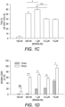
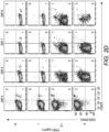
- MOG35-55 concentrations of MOG35-55 were used to activate 2D2-FIG SPL in the presence of 10 nM TGF- ⁇ ( Fig. 1 ).
- 2D2-FIG T cells are specific for MOG35-55 and have an IRES-GFP reporter knock-in immediately downstream of the FOXP3 gene.
- the CD4 + T cell compartment of 2D2-FIG mice typically contained ⁇ 1% FOXP3 + T cells at steady-state.
- the antigen-dependent expression of CD25 was at least two-fold higher on Tregs than on Tcons, and this difference was maximal at intermediate antigen concentrations of MOG35-55 (320 nM - 3.2 ⁇ M) ( Fig. 1 , panels B and D). Similar patterns were noted regarding the concentration dependence of OVA323-339 for CD25 expression on OVA-specific OTII-FIG T cells in the presence of TGF- ⁇ ( Fig. 1 , panels E-F). A lower range of OVA323-339 concentrations was optimal because OT-II T cells have more antigenic potent responsiveness than 2D2 T cells. These data confirm the hypothesis that activated 2D2 and OT-II Tregs exhibit superior CD25 expression compared to Tcon subsets.
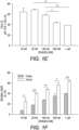
- the anti-CD25 PC61 mAb stabilized short-term Treg cultures.
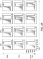
- 2D2-FIG SPL were activated with MOG35-55 ( Fig. 2 , panel A, top row), MOG35-55 and TGF- ⁇ (2 nd row), or MOG35-55, TGF- ⁇ , and the anti-CD25 antibody PC61 (3 rd row) for 3 days and then passaged into IL-2 containing media (without antigen or TGF- ⁇ ) in the absence (rows 1-2) or presence (row 3) of PC61.
- Treg percentages gradually waned during subsequent propagation in IL-2 from a frequency of 47% at day 4 to 19% by day 13.
- Treg percentage values are given in the bottom right of each panel.
- TGF- ⁇ -induced cultures that were continuously supplemented with 10 ⁇ g/ ml (65 nM) of PC61 (bottom row)
- Treg differentiation was inhibited by PC61 during the initial 3-day activation culture.
- the Treg frequency increased from 12% on day 4 to 86% by day 13.
- the elevated Treg frequencies were associated with stronger GFP bright fluorescence which is a correlate of FOXP3 expression (values given at top of each panel).
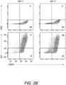
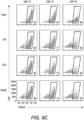
- 2D2-FIG SPL were activated for 3 days with 1 ⁇ M MOG35-55 and 10 nM TGF- ⁇ and then were passaged every 3-4 days in IL-2 and 10 ⁇ g/ ml PC61 ( Fig. 2 , panel B). Again, antigen and TGF- ⁇ were not included in the IL-2 expansion phase of these experiments. Analyses on days 11 and 17 revealed that the line was almost entirely comprised of FOXP3 + Tregs (> 90%).
- a PE-conjugated 3C7 anti-CD25 mAb which recognizes a site on CD25 distinct from the PC61 epitope, revealed high levels of CD25 on these Tregs despite the continuous culture in PC61 and the saturation of CD25 with PC61 ( Fig. 2 , panel B, bottom images).
- an APC-conjugated PC61 did not bind CD25 because PC61-specific epitopes on CD25 were saturated with unlabeled PC61 mAb that had bound CD25 during the culture phase ( Fig. 2 , panel B, top images).
- Treg enrichment required PC61-mediated saturation of CD25 ( Fig. 2 , panel C).
- 2D2-FIG T cells were activated for 3 days with MOG35-55 and TGF- ⁇ in the presence or absence of designated PC61 concentrations. As noted before, high concentrations of PC61 inhibited Treg induction when assessed on day 3. The T cells were then passaged into IL-2 on day 3 with the same PC61 concentrations but without antigen or TGF- ⁇ .
- PC61 On days 5 through 9, concentrations of 1 and 10 ⁇ g/ ml PC61 nearly saturated CD25 as determined by a lack of surface labeling by an APC-conjugated PC61 mAb (y-axis), because pre-existing PC61-CD25 complexes prevented the binding of APC-conjugated PC61. These PC61 concentrations (1 and 10 ⁇ g/ ml) respectively facilitated Treg enrichment to frequencies of 45% and 74%.
- PC61 dose dependently augmented GFP MFI mean fluorescence intensity; 4 digit number on the right of right-most column
- Tregs and Tcons expressed high levels of free CD25, and Treg frequencies were sparse at approximately 20% throughout the 9 days of culture.
- PC61 by itself did not induce FOXP3, as T cells activated without TGF- ⁇ and cultured in the presence of PC61 did not result in Tregs ( Fig. 2 , panel D).
- PC61 stabilized Tregs in IL-2 maintenance cultures
- inclusion of PC61 in the initial 3-day activation culture appeared to delay Treg induction.
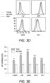
- PC61 was or was not included in the initial 3-day activation culture together with 2D2-FIG SPL, MOG35-55, and TGF- ⁇ ( Fig. 3 , panel A), and then PC61 was included in the IL-2 maintenance cultures of both groups through day 9. Regardless of whether PC61 was added on day 0 (PC61 at activation) or day 3 (PC61 after activation), PC61 facilitated the emergence of highly enriched FOXP3 + Tregs by day 9.
- PC61-cultivated Tregs differed from control (no PC61) cultures via higher Treg expression of FOXP3, LAP, GARP, GITR, and PD-1.
- pre-existing Tregs typically comprise less than 1% of CD4 + T cells, with a frequency range of approximately 0.2-1.5% pre-existing FOXP3 + Tregs.
- Tregs were induced with MOG and TGF- ⁇ from 2D2-FIG Rag1 - / - mice, which lack tTregs ( Fig. 4 ).
- 2D2-FIG Rag1 - / - Tregs were cultured in the presence of IL-2 with or without PC61.
- Tregs cultured in the presence of PC61 showed a stable, high percentage of Tregs (>75%); whereas, Tregs cultured in the absence of PC61 showed a diminishing Treg percentage throughout the duration of the experiment ( Fig. 4 , panels A, B).
- PC61 was the anti-CD25 mAb of choice for blockade of IL-2 signaling and selection of Tregs.
- PC61 Based on its superior IL-2 inhibitory activity ( Fig. 5 , panel A), PC61 was more efficient than the anti-CD25 mAb 7D4 and 3C7 for maintenance of Treg cultures ( Fig. 5 , panel C). Due to the superior inhibitory efficacy and superior Treg selectivity, the anti-CD25 PC61 mAb was used for the remainder of the study.
- PC61 enabled the dominant outgrowth of Tregs in mixed Treg/ Tcon cultures.
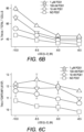
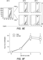
- Treg window was tested with mixed 2D2 (MOG-specific) lines of FOXP3 + and FOXP3 - T cells ( Fig. 6 ). These T cells were propagated in IL-2 for 16 days before use in this assay, so that Tregs represented nearly 96% of the T cells in the FOXP3 + line. Equal numbers of Tcons and Tregs were labeled with CellTrace Violet (CTV) and were cultured for 6 days with designated concentrations of IL-2 and PC61. This analysis distinguished proliferative subsets (left quadrants) from non-proliferative subsets (right quadrants) as well as Tregs (upper quadrants) from Tcons (lower quadrants) ( Fig. 6 , panel A).
- CTV CellTrace Violet
- IL-2 concentrations 100 pM and 316 pM
- proliferative and non-proliferative Tregs were dominant (exhibited higher percentages) compared to Tcons when cultured with 1 ⁇ M PC61.
- 100 pM IL-2 and either 1 ⁇ M PC61 or ⁇ no PC61' 78% or 45% of T cells (sum of upper two quadrants) were Tregs, respectively.
- 316 pM IL-2 proliferative Tregs exhibited higher frequencies than proliferative Tcons in the presence but not absence of 10 nM, 100 nM, and 1 ⁇ M PC61.
- high IL-2 concentrations e.g.
- Tcons were dominant and showed overgrowth regardless of PC61 concentration.
- concentration-dependent ability of PC61 to promote Treg dominance in low IL-2 concentrations Fig. 6 , panel B
- preservation of high GFP fluorescence Fig. 6 , panel C
- Tregs were dominant within a 10-fold range of IL-2 concentrations (320 pM - 3.2 nM, Fig. 6 , panel D) in the presence of 1 ⁇ M PC61 whereas Tcons were dominant at the same IL-2 concentrations in the absence of PC61.
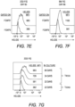
- the anti-CD25 PC61 antibody enabled maintenance of long-term lines of FOXP3 + Tregs.
- FIG SPL FOXP3 + Treg lines from naive, clonotypically-diverse FIG SPL could also be derived by addition of PC61 to IL-2 expansion cultures ( Fig. 7 , panel B).
- Naive FIG SPL were activated for 3 days with Con-A and TGF- ⁇ and then were propagated in IL-2 in the presence of PC61.
- CD4 + T cells were purified to remove CD8 + T cells in polyclonal FIG SPL cultures to prevent CD8 + T cell overgrowth of the line, but CD4 + T cell purification was not necessary for 2D2-FIG Tregs given that 2D2-FIG mice largely lacked CD8 + T cells.
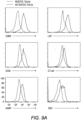
- Neuropilin-1 and Helios which have been associated with the FOXP3 + Treg phenotype ( FIG. 7 , panels C-G).
- Neuropilin-1 was expressed on both 2D2-FIG Tregs ( Fig. 7 , panel C) and polyclonal Tregs ( Fig. 7 , panel D).
- Neuropilin-1 exhibited progressive increases in MFI as a function of time such that Neuropilin-1 expression was positively correlated with the longevity of culture.
- Helios is a transcription factor implicated in Treg function
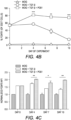
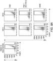
- a 2D2-FIG line (> 90% FOXP3 + Tregs) that had been cultured for 23 days in IL-2 and PC61 was reactivated for 3 or 6 days with irradiated splenic APC and MOG35-55 in the presence of IL-2 with or without PC61 and the neutralizing anti-TGF- ⁇ 1D11 mAb ( Fig. 8 , panel A-C).
- the presence of PC61 during secondary activation stabilized Treg percentages at approximately 90% ( Fig. 8 , panel A top row; Fig. 8 , panel B).
- the presence of PC61 also stabilized FOXP3 expression, as reflected by GFP MFI ( Fig. 8 , panel C).
- 1D11-mediated neutralization of TGF- ⁇ resulted in a loss of FOXP3 + Treg percentages to less than 50% and a decrement in FOXP3 expression on a per cell basis, as reflected by the GFP MFI ( Fig. 8 , panel A middle row; Fig. 8 , panel C).
- GFP MFI Fig. 8 , panel A middle row; Fig. 8 , panel C.
- T cell densities caused a more robust activation and perhaps enhanced the production of pro-inflammatory cytokines (i.e., IL-23 or GM-CSF) that might antagonize the action of TGF- ⁇ .
- pro-inflammatory cytokines i.e., IL-23 or GM-CSF
- the presence of exogenous TGF- ⁇ prevented decrement of Treg frequencies and stabilized the Treg phenotype regardless of the cell density.
- high initial cell densities in the presence of TGF- ⁇ enabled the most robust expansion of Tregs by day 3 of activation (Table 1).
- Tregs exhibit stable expansion in the presence of exogenous TGF- ⁇ , IL-2, and PC61.
- Group a APC Treg # (x 10 6 ) on day 0 Cell # (x 10 6 ) on day 3 Treg # (x 10 6 ) on day 3 Treg yield b FOXP3 + Treg percentages No TGF- ⁇ 20: 2 9.58 3.40 1.7 35 10: 1 3.33 1.44 1.4 42 5: 0.5 2.20 1.48 3.0 66 TGF- ⁇ 20: 2 13.36 13.13 6.6 98 10: 1 5.26 5.18 5.2 98 5: 0.5 2.16 2.13 4.3 99 a T cells were rested for 33 days and were 94% FOXP3 Tregs at the initiation of the experiment.
- T cells were activated with irradiated SPL, 1 ⁇ M MOG35-55, rat IL-2, and 10 ⁇ g/ ml PC61 and in the presence or absence of 100 pM TGF- ⁇ .
- Cultures were setup in 5 ml media containing designated numbers of Tregs and irradiated SPL (column 2).
- b Cells were analyzed on day 3 of activation by gating on V ⁇ 11 and FOXP3 (GFP). Absolute cell counts (trypan blue exclusion), V ⁇ 11 + T cell percentages, and FOXP3 + Treg percentages were used to calculate Treg numbers.
- Treg yield was calculated by dividing the Treg cell count on day 3 by the starting Treg count (2 ⁇ 10 6 , 1 ⁇ 10 6 , or 0.5 ⁇ 10 6 ) on day 0. These data are representative of three independent experiments.
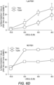
- Treg expansion an established, long-term 2D2-FIG Treg line was activated for 3 days with irradiated bone marrow-derived dendritic cells, 2.5 ⁇ g/ ml Con-A, 1 nM TGF- ⁇ , and IL-2 ( Fig. 8 , panels E, F). T cells were then passaged on days 3 and 6 at a density of 5 ⁇ 10 5 cells/ ml cRPMI containing rat IL-2 with or without PC61. Viable cells were enumerated (Trypan Blue dye exclusion) and analyzed for V ⁇ 11 and FOXP3 expression to calculate expansion of viable FOXP3 + Tregs.
- Tregs During the antigenic activation (days 0-3), Tregs expanded ⁇ 7-fold while retaining high FOXP3 expression in nearly 100% of the population. Tregs expanded another ⁇ 4-fold from days 3-6 for a net 26-28 fold expansion since day 0 ( Fig. 8 , panel F). Importantly, there was no difference in the expansion rates between Tregs cultured in the presence or absence of PC61 immediately following antigenic activation, such that CD25 high Tregs were resistant to PC61 antagonism. From days 6-9, Tregs reverted to a quiescent resting phase and did not expand in numbers either with or without PC61.
- PC61 preserved the FOXP3 phenotype whenever exogenous IL-2 was added to the culture because unmitigated IL-2 signaling resulted in Tcon dominance and overgrowth/ destabilization of the Treg line.
- PC61 was beneficial in secondary re-activation cultures because these cultures were supplemented with exogenous IL-2.
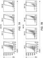
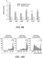
- Tregs Phenotypic analysis of activated Tregs revealed the activation-dependent upregulation of functional Treg markers including LAP, CTLA4, GARP, and TIGIT ( Fig. 9 ). These data reveal that TGF- ⁇ -conditioned activation of Tregs upregulates several markers associated with suppressive Treg function.
- 2D2 Tregs were assayed for in vitro and in vivo suppressive activity.
- CD45.2 2D2 Tregs were cultured for either 40 days or 13 days in IL-2 and PC61 whereas a control CD45.2 2D2 Tcon line was derived by propagation in IL-2 alone for 13 days.
- Treg and control lines were then cultured with CTV-stained naive responders from CD45.1 2D2 splenic leukocytes in the presence of MOG and IL-2 ( Fig. 10 , panel A).
- CTV dilution was measured in the CD45.1 + responders after 5 days as a measure of T cell proliferation.
- Suppressive activity was based on the percentages of hypo-proliferative CD45.1 2D2 T cells ( Fig. 10 , panel B).
- Responder 2D2 T cell proliferation was significantly inhibited by the presence of 2D2 Tregs when compared to cultures containing the 2D2 Tcon line. There was no difference in suppressive activities of Tregs rested for 40 days or 13 days.
- Tregs representing the 2D2 MOG35-55-specific clonotype
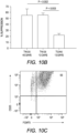
- a continuous line of FOXP3 + Tregs was activated with irradiated splenic APC, 1 ⁇ M MOG35-55, IL-2, and 100 pM TGF- ⁇ for 3 days but without PC61 mAb to avoid coating the T cells with a rat IgG1 mAb that had depleting activity in vivo.
- Cell surface PC61 remained high in the immediate aftermath of the activation culture, and thus the Tregs were cultured overnight in IL-2 to remove more cell-surface CD25/ PC61 complexes.
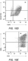
- Tregs on the day of transfer showed that 93% were FOXP3 + CD25 high Tregs ( Fig. 10 , panel C). Most of the transferred Tregs had no or low detectable levels of PC61 on the cell surface as determined by staining with anti-rat IgG secondary antibody ( Fig. 10 , panel D). 65% of Tregs were CD44 high CD62L high, whereas 33% of Tregs were CD44 high CD62L low ( Fig. 10 , panel E).
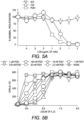
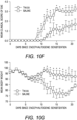
- mice were actively challenged to elicit EAE.
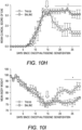
- the adoptive transfer of Tregs ameliorated severity of clinical paralysis and EAE-associated loss of body weight ( Fig. 10 , panels F and G).
- Mice that received Tregs had significantly lower mean daily clinical EAE scores on days 9-13 and 15-21 and significantly less weight loss on days 11-20 as assessed by the Student t test.
- Tregs significantly alleviated the severity of EAE based on a decrease in mean daily clinical scores from days 18-28 and an increase in body weight from days 22-28.
- PC61scFv single-chain fragment variable version of PC61 to assess whether a monovalent non-signaling version of PC61 retained Treg-stabilizing ability.
- This PC61scFv protein has the same binding specificity as PC61 (anti-CD25).
- PC61scFv was monovalent and therefore lacked cross-linking activity necessary for signaling.
- the PC61scFv also lacked constant region heavy chain domains and therefore lacked interactions with complement and FcyR.
- the PC61scFv is believed to have qualitative advantages over intact PC61 in regard to tissue penetrance and absence of in vivo depleting activity.
- PC61scFv was slightly less efficient compared to the intact PC61 mAb, although this minor difference most likely was due to the lower affinity associated with monovalent binding, in contrast to the bivalent interactions of intact PC61 with CD25.
- the specificity of PC61scFv was confirmed by the ability of surface-bound PC61scFv to block the binding of a fluorochrome-conjugated PC61 mAb ( Fig. 11 , panel C). These data indicate that PC61 mediates Treg stabilization via CD25 neutralization rather than CD25 crosslinking.
- Treg lines indefinitely sustained a Treg lineage phenotype when maintained with low concentrations of IL-2 and high concentrations of PC61 in continuous culture.
- the Tregs derived in this study actively expanded and remained stable in the presence of PC61 and TGF- ⁇ in antigen-induced reactivation cultures, such that blastogenic Tregs expressed the prototypic Treg markers together with suppressive activity in vitro and in vivo.
- IL-2 responsiveness of Tregs can be used to selectively promote Treg responses.
- low-dose IL-2 therapies for Type 1 Diabetes selectively expanded existing Treg populations which could suppress islet cell destruction ( Bluestone JA, et al. Sci Transl Med (2015) 7(315):315ra189. doi: 10.1126/scitranslmed.aad4134 . PubMed PMID: 26606968; PubMed Central PMCID: PMC4729454).
- anti-IL-2 mAb/ IL-2 immune complexes may target IL-2 to different T cell subsets depending on the epitope specificity of the anti-IL-2 mAb in the complex ( Boyman O, et al.
- PubMed PMID 16484453 ; Letourneau S, et al. Proc Natl Acad Sci USA (2010) 107(5):2171-6. doi: 10.1073/pnas.0909384107. PubMed PMID: 20133862; PubMed Central PMCID: PMC2836659 ; and Spangler JB, et al. Immunity (2015) 42(5):815-25. doi: 10.1016/j.immuni.2015.04.015. PubMed PMID: 25992858; PubMed Central PMCID: PMC4439582 ).
- the JES6-1 anti-IL-2 mAb/ IL-2 immune complex appeared to target IL-2 to Tregs to favor Treg expansion.
- the S4B6 anti-IL-2 mAb/ IL-2 complex favorably expanded effector T cells by blocking the interaction between IL-2 and CD25.
- Advantages of targeting IL-2 versus CD25 have yet to be directly compared although targeting CD25, the Treg-specific component of the IL-2 receptor, may have qualitative advantages given the wide variations in endogenous IL-2 concentrations that may exist during chronic inflammatory autoimmune disease.
- Blockade of CD25 may constrain IL-2 signaling across a broad IL-2 concentration range, with an upper threshold defined by those levels of IL-2 sufficient for CD25-independent IL2R ⁇ signaling. That is, even with widely varying concentrations of IL-2 and cell surface CD25, mAb-mediated blockade of CD25 may provide a reliable clamp to ensure low-zone IL-2 signaling to promote dominant Treg responses.
- the IL-2 concentration was instrumental in determining T cell subset dominance ( Fig. 6 , panels A and D).
- Low-intensity and high-intensity IL-2 signaling respectively supported Treg or Tcon subset dominance.
- the relation between low-zone IL-2 signaling and Treg responses reflected superior CD25 expression and extraordinar IL-2 sensitivity of Tregs.
- the relation between high-zone IL-2 signaling and Tcon subset dominance may reflect differences in the respective IL-2 signaling pathways.
- IL-2 signaling in Tregs is mediated primarily through the JAK/ STAT pathway whereas IL-2 signaling in Tcon subsets is mediated robustly through both JAK/ STAT and PI(3)K pathways.
- PTEN the PI(3)K inhibitor
- IL-2 has superior potency for Tregs due to higher CD25 expression, which confers Treg dominance at low IL-2 concentrations.
- IL-2 has superior efficacy for Tcons due to robust signaling through both JAK/ STAT and PI(3)K pathways, which confers Tcon dominance at high IL-2 concentrations.
- differential expression of CD25 and PI(3)K signaling pathways provide qualitative distinctions in IL-2 signaling pathways that are foundational for the specialized Treg and Tcon niches.
- Chronic CD25 blockade was the key to exploitation of the Treg window and stabilization of FOXP3 + Tregs.
- anti-CD25 mAbs varied substantially in Treg stabilization activity.
- the mAb of choice for stabilization of mouse Tregs was PC61, which was a stronger inhibitor of IL-2-dependent proliferation than the anti-CD25 mAbs 3C7 and 7D4 ( Fig. 5 , panel A).
- PC61 was a stronger inhibitor of IL-2-dependent proliferation than the anti-CD25 mAbs 3C7 and 7D4 ( Fig. 5 , panel A).
- the use of PC61 in concert with low IL-2 concentrations provided the appropriate qualitative and quantitative signals necessary for stabilization of FOXP3 expression and maintenance of the Treg phenotype.
- PC61 was not useful in initial activation of 2D2-FIG SPL with antigen and TGF- ⁇
- PC61 in concert with IL-2 was included in all subsequent cultures including maintenance cultures and re-activation cultures.
- TGF- ⁇ was beneficial during the initial activation culture and in all subsequent re-activation cultures.
- PC61 worked in concert with IL-2 to maintain Tregs
- TGF- ⁇ worked in concert with MHCII-restricted antigen to stabilize Tregs during blastogenic expansion.
- PC61 was the technical tool that allowed exploitation of a Treg window, which was operationally defined by a low range of IL-2 signaling that enabled dominant outgrowth of stable FOXP3 + Tregs. If IL-2 concentrations in PC61-supplemented cultures exceeded this range, then PC61 lacked Treg stabilization activity, and Tcon subsets dominated the culture.
- CD25 has no known intracellular signaling activity, and IL-2 signaling is mediated through the cytoplasmic domains of CD122 and CD132 ( Boyman O, Sprent J. Nat Rev Immunol (2012) 12(3):180-90. Epub 2012/02/22. doi: 10.1038/nri3156. PubMed PMID: 22343569 ). Nonetheless, CD25 has a cytoplasmic domain, and CD25 crosslinking may indirectly crosslink other associated proteins.
- the IL-2 pathway is critical for self-tolerance in that genetic deficiencies in the major components of the IL-2/ CD25/ IL2R ⁇ / STAT5/ JAK3 pathway result in systemic autoimmunity ( O'Shea JJ, Nat Rev Immunol (2002) 2(1):37-45. Epub 2002/03/22. doi: 10.1038/nri702. PubMed PMID: 11905836 ; Sadlack B, Cell (1993) 75(2):253-61. Epub 1993/10/22. PubMed PMID: 8402910 ; Sharfe N, et al. Proc Natl Acad Sci USA (1997) 94(7):3168-71. Epub 1997/04/01.
- IL-2 is the prototypic T cell growth factor in vitro that drives dominant outgrowth of CD4 + and CD8 + effector lineages.
- IL-2 is required for induction and maintenance of FOXP3 expression in Tregs ( Malek TR, Castro I. Immunity (2010) 33(2):153-65. doi: 10.1016/j.immuni.2010.08.004. PubMed PMID: 20732639; PubMed Central PMCID: PMC2946796 ), but IL-2 also drives proliferation of Tcons and destabilization of Treg populations ( Figs. 2 and 6 ).
- continuous low-zone IL-2 signaling in the context of a Treg window is key for understanding stabilization of FOXP3 + Tregs in vitro, the implication is that continuous low-zone IL-2 signaling may also be key for maintenance and progressive differentiation of Tregs in vivo.
- TGF- ⁇ was also required for Treg stability, but only in the context of MHCII-restricted antigen presentation by splenic APC in activation-dependent expansion cultures. TGF- ⁇ had no effect in maintenance cultures when added during propagation in IL-2 with or without PC61. In re-activation cultures, a TGF- ⁇ -blocking antibody led to accelerated Treg destabilization, whereas addition of exogenous TGF- ⁇ preserved FOXP3 expression and FOXP3 + Treg percentages at optimal levels ( Fig. 8 ). In re-activation cultures that included IL-2 and TGF- ⁇ , Tregs expanded more than 5-fold without loss of FOXP3 as both a percentage of the T cell population and on a per cell basis based on MFI.
- the concentration of TGF- ⁇ in the reactivation culture was tailored to the TCR strength for cognate antigen of the particular clonotype.
- the correct balance of TGF- ⁇ , antigen, and IL-2 was necessary for optimal expansion of Tregs and maintenance of the Treg phenotype.
- 100 pM TGF- ⁇ was sufficient for expansion of stable MOG-specific Tregs, which represents a low affinity clonotype.
- Higher concentrations of TGF- ⁇ (1 nM) were needed for optimal expansion of stable OVA-specific Tregs, which exhibits much stronger antigenic potency.
- TGF- ⁇ was needed in sufficient concentrations to counter-balance antigenic strength but these TGF- ⁇ concentrations had to be below thresholds that completely blocked the antigenic response.
- an appropriate range of TGF- ⁇ concentrations was needed for optimal stabilization of Tregs during antigen-reactivated expansion cultures.
- Treg stabilization reflected the intrinsic stabilization of FOXP3 expression or whether differential growth (or death) rates in Treg and Tcon subsets determined final Treg percentages.
- the most likely scenario is that both mechanisms contributed in part to the derivation of continuous Treg lines.
- Tregs cultured in the presence of PC61 and IL-2 had higher FOXP3 expression on a per cell basis than Tregs cultured in IL-2 alone, and Tregs cultured in PC61 and IL-2 also had higher growth rates than the Tcon subsets.
- PC61 may suppress Tcon proliferation which in turn may suppress secretion of pro-inflammatory cytokines that could antagonize TGF- ⁇ and destabilize FOXP3. These considerations may reflect a fundamental competition between these T cell subsets, wherein Treg subsets inhibit Tcon subsets, and Tcon subsets inhibit Treg subsets via mechanisms of reciprocal inhibition.
- PC61-mediated Treg selection therefore may vary in kinetics based on the inherent antigenic reactivity of the relevant clonotype.
- a second caveat is that PC61/ IL-2 selects CD25 high FOXP3 + subsets at the expense of CD25 low FOXP3 + subsets.
- pre-existing Tregs isolated from secondary lymphoid organs express CD25 across a quantitative continuum, and PC61/ IL-2 selects the CD25 high subset that represents the functionally mature suppressive subset.
- This study defined an operative 'Treg niche' defined by low-zone IL-2 signaling.
- This study also provided a strategy for maintenance and expansion of stable FOXP3 + Treg lines in vitro, which could be propagated indefinitely without phenotypic instability.
- These principles were validated for TGF- ⁇ -inducible Tregs, which may be optimal for derivation of antigen-specific Tregs from peripheral blood. While not wishing to be bound to any particular theory, we believe that these principles would also apply to thymic or natural tTreg/ nTregs. Overall, this strategy could be applied broadly to Tregs of any clonotypic specificity and thereby may have applicability for the advancement of antigen-specific Treg-based immunotherapies.
Landscapes
- Health & Medical Sciences (AREA)
- Life Sciences & Earth Sciences (AREA)
- Immunology (AREA)
- Engineering & Computer Science (AREA)
- General Health & Medical Sciences (AREA)
- Bioinformatics & Cheminformatics (AREA)
- Veterinary Medicine (AREA)
- Animal Behavior & Ethology (AREA)
- Public Health (AREA)
- Chemical & Material Sciences (AREA)
- Biomedical Technology (AREA)
- Organic Chemistry (AREA)
- Epidemiology (AREA)
- Biotechnology (AREA)
- Zoology (AREA)
- Genetics & Genomics (AREA)
- Wood Science & Technology (AREA)
- Cell Biology (AREA)
- Hematology (AREA)
- Medicinal Chemistry (AREA)
- Pharmacology & Pharmacy (AREA)
- Transplantation (AREA)
- Biochemistry (AREA)
- Chemical Kinetics & Catalysis (AREA)
- General Chemical & Material Sciences (AREA)
- Microbiology (AREA)
- Nuclear Medicine, Radiotherapy & Molecular Imaging (AREA)
- General Engineering & Computer Science (AREA)
- Medicines Containing Material From Animals Or Micro-Organisms (AREA)
- Developmental Biology & Embryology (AREA)
- Virology (AREA)
- Medicines Containing Antibodies Or Antigens For Use As Internal Diagnostic Agents (AREA)
Description
- This application claims the benefit of and priority to
U.S. Provisional Application Serial No. 62/430,991, filed December 7, 2016 - The present invention relates to compositions and methods for activating, culturing, expanding, maintaining, and/or stabilizing the growth of regulatory T cell populations, including regulatory FOXP3+ T cells.
- CD4+ CD25+ FOXP3+ regulatory T cells (Tregs) play an integral role in preventing autoimmunity and controlling inflammation. The importance of Tregs is illustrated by the fact that loss-of-function mutations in FOXP3 leads to the rapid, fatal multi-organ autoimmune disorders IPEX and scurfy in humans and mice, respectively. Moreover, dysfunctional Tregs are linked to increased susceptibility to certain autoimmune diseases such as multiple sclerosis (MS) and
type 1 diabetes (T1D). Because of their suppressive capabilities, Tregs offer a unique therapeutic potential for autoimmune diseases, allergic diseases, transplant tolerance, and chronic inflammatory disorders. Treg adoptive transfer therapies, where Tregs are induced or purified ex vivo and then infused into a syngeneic organism, have shown encouraging results in animal models of autoimmune disease such as experimental autoimmune encephalomyelitis (EAE), the animal model of MS, T1D, and colitis. In early phase clinical trials, Treg adoptive transfer therapies for tissue transplantation and T1D have shown favorable outcomes, but no adoptive Treg therapy has yet to be approved as a treatment option. An additional approach to Treg-mediated therapies involves induction or expansion of Tregs in vivo through the use of tolerogenic vaccines or anti-inflammatory drugs, such as interferon-β. Interferon-β is a first-line treatment option for relapsing-remitting MS. Interferon-β has been shown to increase numbers of Tregs in circulation. Tolerogenic vaccines ideally work by inducing or expanding antigen-specific Tregs, which would leave the adaptive immune response intact. - FOXP3 is needed for adaptive self-tolerance, and FOXP3 is expressed in a canonical lineage of suppressive Tregs; however, it cannot be assumed that the role of FOXP3 in self-tolerance is entirely mediated via the expression of FOXP3 in this lineage of CD4+ CD25+ FOXP3+ Tregs. FOXP3 may play inhibitory roles in other cell types, particularly during transient expression in canonical conventional T cell subsets. For example, FOXP3 can be transiently expressed during activation in human T cells and is expressed in quasi-stable non-committed CD25low FOXP3+ T cells in mice. The assessment of FOXP3+ Tregs requires new assessment tools to analyze this subset. One approach to assess these questions is to induce Tregs in vitro and expand/ maintain in pure culture, and show via adoptive transfer experiments that these Tregs suppress EAE.
- One major setback for Treg-mediated therapies is that Tregs are notoriously unstable and plastic. In vivo studies using fate-mapping techniques to track FOXP3 expression showed that under certain environmental circumstances, Tregs can lose FOXP3 expression and actually adopt an effector T cell phenotype. These so called "ex-Tregs" can potentially exacerbate autoimmune disease as FOXP3 is required for Treg suppressive activity. Ex vivo expansion and culturing of Tregs has faced many difficulties due to the fact that Tregs are relatively anergic in vitro and that contaminating FOXP3- conventional cells (Tcons) can quickly outgrow Tregs. Additional evidence shows that Tregs lose FOXP3 expression over time when cultured ex vivo. Propagation and expansion of Tregs in vitro is a step in generating the numbers of Tregs necessary for Treg adoptive transfer therapies. What drives Treg instability, especially in vitro, is not well understood.
- IL-2 is involved in both the induction and maintenance of Tregs. CD25, the IL-2 receptor alpha chain, is constitutively expressed on Tregs and represents a phenotypic marker for Tregs, although CD25 is also expressed at high levels on activated effector T cells. Various therapeutic techniques manipulating IL-2 availability have met with some success in animal models and in the clinic. The rationale is that selectively expanding Tregs will alleviate autoimmune disorders. While most of the focus to date has been on manipulating IL-2 directly, little attention has focused on manipulating CD25.
-
WO 2012/012737 is directed to materials and methods related to culturing stable Treg cells or in vivo expansion of stable Treg cells, the cells made by the methods, treatments for various inflammatory/autoimmune pathologies and transplant/graft rejection, and related materials. Ex vivo induction and expansion of the stable Tregs is described, including use of inducing compositions, such as certain mAbs and other compounds, along with expansion medium comprising IL-2. In vivo expansion of stable Treg cells and treatments for various inflammatory/autoimmune pathologies and transplant/graft rejection are also described, including the use of mAbs and their variants. - Debj ani Ghosh et al, Journal of Leukocyte Biology, , US is discusses whether the resistance of Csf2-deficient mice to experimental autoimmune encephalomyelitis was a result of a requirement for GM-CSF in controlling the functional balance between effector and regulatory T cell subsets during experimental autoimmune encephalomyelitis. The article concludes that experimental autoimmune encephalomyelitis resistance of Csf2-deficient mice, at least in part, reflects a deficient induction of effector T cell function that cannot surmount normal regulatory T cell barriers. Experimental autoimmune encephalomyelitis effector responses, however, are unleashed upon depletion of regulatory CD25+ T cells.
- Dienz O. et al., Cin. Imunol, vol. 130, mo. 1, 2009, pp. 27-33, XP025762127 is a review that highlights the variety of ways in which IL-6 affects CD4 effector functions and how this may contribute to different types of diseases.
- Pellerin L. et al., Immunol Res., vol. 58, no. 0, 2014, pp. 358-368, XP055509649 summarizes the main features of different subtypes of Tregs and focuses on the clinical implications of their altered function in human studies. More specifically, the article discusses abnormalities affecting FOXP3+ Tregs and Tr1 cells that will lead to autoimmune manifestations and/or allergic reactions, and the potential therapeutic use of Tregs.
- Perdigoto A. L. et al., Frontiers in Immunology, vol. 6, 2016, pp. 1-13, XP055509652 discusses that a number of immune therapies, such as rapamycin, IL-2, and anti-T cell antibodies, are able to induce Tregs and are being tested for their efficacy in diverse clinical settings with exciting preliminary results. However, a balance exists with the use of some, such as IL-2, that may have effects on unwanted populations as well as promoting expansion and survival of Tregs requiring careful selection of dose for clinical use. The use of cell surface markers has enabled investigators to isolate and expand ex vivo Tregs more than 500-fold routinely. Clinical trials have begun, administering these expanded Tregs to patients as a means of suppressing autoimmune and alloimmune responses and potentially inducing immune tolerance. Studies in the future are likely to build on these initial technical achievements and use combinations of agents to improve the survival and functional capacity of Tregs.
- Wilkinson D. S. et al., Frontiers in Immunology, vol. 8, 14 December 2017, XP55678640, CH, hypothesises that, since Tregs and Tcons differentially express CD25, anti-CD25 monoclonal antibodies (mAbs) would only partially block IL-2 signaling in CD25high FOXP3+ Tregs while completely blocking IL-2 responses of CD25low-intermediate Tcons to enable preferential outgrowth of Tregs during in vitro propagation. The article concludes that low IL-2 concentrations coupled with high PC61 concentrations constrained IL-2 signaling to a low-intensity range that enabled dominant stable outgrowth of suppressive CD25high FOXP3+ Tregs and states that the ability to indefinitely expand stable Treg lines can provide insight into FOXP3+ Treg physiology and be foundational for Treg-based immunotherapy. FOXP3+ regulatory T cells (Tregs) represent potentially powerful adoptive immunotherapies for autoimmune disease and other chronic inflammatory diseases. However, the field currently lacks reliable technologies to enable long-term in vitro expansion of stable, antigen-specific FOXP3+ Treg lines. Activation of naive T cells in the presence of TGF-β elicits the initial differentiation of the FOXP3+ lineage, but these Treg lines lack phenotypic stability and rapidly convert to a Tcon phenotype during in vitro propagation in IL-2. Thus, there remains a need for improved methods of growing and maintaining Treg populations.
- The present invention is defined by the appended claims and it is related to the development of methods for preparing and/or expanding a regulatory T-cell (Treg) population. Tregs are qualitatively distinguished from Tcons and non-Tregs in that Tregs constitutively express high levels of CD25 (IL-2 receptor alpha, IL-2Rα), whereas Tcons and non-Tregs are CD25- or only transiently express an activation-dependent CD25high phenotype. Due to differential expression of CD25, it was reasoned that anti-CD25 mAbs would only partially block IL-2 signaling in Tregs while completely blocking IL-2 responses of Tcons, and that the differential inhibition of CD25 would enable the preferential and dominant outgrowth of Tregs during in vitro cultivation. TGF-β-induced Treg lines that were maintained in IL-2 in the presence of the anti-CD25 antibody PC61 (non-depleting mAb in vitro) maintained a FOXP3high phenotype during prolonged in vitro propagation (-90% FOXP3+ Tregs) whereas parallel cultures lacking PC61 rapidly lost FOXP3 and reverted to a Tcon phenotype. Stable FOXP3high lines acquired Treg-associated markers Neuropilin-1 and Helios. These lines also exhibited antigen-specific activation and expansion in vitro in the presence of TGF-β and antigen without loss of FOXP3. When activated with myelin oligodendrocyte glycoprotein peptide fragment 35-55 (MOG35-55) and TGF-β, blastogenic FOXP3+ Tregs from MOG35-55-specific 2D2 TCR transgenic mice suppressed EAE in adoptive transfer assays. A "Treg window," marked by low IL-2 concentrations coupled with high PC61 concentrations that restricted IL-2 signaling pathways, enabled dominant stable outgrowth of suppressive FOXP3high Tregs. The ability to maintain and expand stable Treg lines will provide insight into FOXP3+ Treg physiology and can enable feasible strategies of Treg-based immunotherapy.
- Thus, in an aspect of the present invention, provided is a method of preparing a regulatory T-cell (Treg) population including: exposing a T-cell population to a medium including an anti-inflammatory cytokine and an antibody to an anti-inflammatory cytokine receptor; and expanding the T-cell population, to provide a Treg population, wherein the anti-inflammatory cytokine is IL-2 and the anti-inflammatory cytokine receptor inhibitor is an anti-CD25 antibody and/or a fragment thereof.
- In another aspect not according to the present invention, provided is a Treg population, compositions including a Treg population, and pharmaceutical formulations including a Treg population prepared by methods set forth according to the present invention.
- In yet another aspect not according to the invention, provided is a method of treating or modulating an immunological disorder in a subject in need thereof including administering a therapeutic amount of a Treg population, compositions including a Treg population, or pharmaceutical formulations including a Treg population prepared by methods set forth according to the present invention.
- In yet another aspect not according to the invention, provided is a method of eliciting a tolerogenic response in a subject in need thereof including the step of administering a therapeutic amount of a Treg population, or a pharmaceutical formulation including a Treg population prepared by methods set forth according to the present invention.
- In yet another aspect not according to the invention, provided is a method of maintaining a Treg population including: exposing a Treg population to a medium including an anti-inflammatory cytokine and an antibody to an anti-inflammatory cytokine receptor; and maintaining the Treg population in the medium including an anti-inflammatory cytokine and an antibody to an anti-inflammatory cytokine receptor.
- It is noted that aspects of the invention described with respect to one embodiment, may be incorporated in a different embodiment although not specifically described relative thereto. That is, all embodiments and/or features of any embodiment can be combined in any way and/or combination as long as they are covered by the scope of the claims. These and other objects and/or aspects of the present invention are explained in detail in the specification set forth below. Further features, advantages and details of the present invention will be appreciated by those of ordinary skill in the art from a reading of the figures and the detailed description of the example embodiments that follow, such description being merely illustrative of the present invention.
-
-
Fig. 1 . Intermediate concentrations of antigen induced higher percentages of FOXP3+ Tregs. 2D2-FIG (panels A-D) or OTII-FIG (panels E-F) SPL were activated at a density of 2 × 10⁶ cells/ ml cRPMI in the presence of 10 nM TGF-β with the indicated concentration of MOG35-55 or OVA323-339. After 3 days, Vβ11+ (2D2) or Vβ5.1+ (OT-II) T cells were analyzed for expression of FOXP3 (panels A, C, E) and CD25 (panels B, D, F). Numbers at the bottom of each dotplot in (panel A) represent the percentages of FOXP3+ T cells, and the numbers in (panel B) represent the median fluorescence intensity of CD25 expression within the FOXP3negatlve (Tcon) and FOXP3+ (Treg) populations. *p < 0.05, **p < 0.01, ***p < 0.001. These data are representative of three independent experiments. -
Fig. 2 . The anti-CD25 PC61 mAb selectively favored Treg dominance in mixed Treg/ Tcon cultures. (panels A-D) 2D2-FIG SPL were activated at a density of 2 × 10⁶ cells/ ml in cRPMI with MOG35-55 (1 µM) in the presence or absence of TGF-β (10 nM) and/ or designated concentrations of PC61 (10 µg/ ml; 65 nM, unless designated otherwise). Antigen and TGF-β were present only in the first activation culture and not thereafter. After 3-4 days of activation, cells were passaged at a density of 106 cells/ ml in media containing rat IL-2 with or without PC61. Cultures initiated with PC61 were continued with PC61 during passage into the same conditions every 2-4 days. (panel A) Cells were analyzed ondays day days -
Fig. 3 . PC61 allowed for rapid generation of pure Treg lines. 2D2-FIG SPL (panels A-B, D-E) or OTII-FIG SPL (panel C) were activated at a density of 2 × 10⁶ cells/ ml in cRPMI with MOG35-55 (1 µM) or 100 nM OVA323-339 respectively with 10 nM TGF-β in the presence or absence of 10 µg/ ml PC61. Antigen and TGF-β were present only in the initial 3-day activation culture and not thereafter. (panels A-C) PC61 was (PC61 at activation) or was not (PC61 after activation) included in the initial 3-day activation culture but was included in both groups after passage of the cells onday 3. Cells were counted and then analyzed to determine percentages of viable, Vβ11+ (2D2), Vβ5.1+ (OTII), FOXP3+ T cells on the designated days. (panel B) Shown are the 2D2-FIG Treg yields ondays day 10 post-activation. MFI Values were normalized so that values obtained from T cells cultured in the absence of PC61 was equal to 1. These experiments are representative of three independent experiments. -
Fig. 4 . PC61 enabled the preferential outgrowth of TGF-β-induced iTregs from 2D2-FIG Rag1-/- mice. 2D2-FIG Rag1-/- mice SPL were activated for 4 days at a density of 2 × 10⁶ cells/ ml in cRPMI with MOG35-55 in the presence or absence of 1 nM TGF-β to generate Tregs and Tcons, respectively, and were passaged at a density of 5 × 10⁵ cells/ ml cRPMI with rat IL-2 onday 0 of the rest phase. T cells were then passaged with or without PC61 (10 µg/ ml) (in the absence of TGF-β and MOG) as designated every 2-4 days. Vβ11+ T cells were assessed for FOXP3 expression ondays -
Fig. 5 . The PC61 anti-CD25 mAb was a functional antagonist of IL-2 growth at low concentrations of IL-2 and was superior to the 7D4 and 3C7 anti-CD25 mAbs for maintenance of Tregs. (panels A-B) A continuously-propagated line of IL-2 dependent CD4+ T cells (SJL-PLP.1 T cells) was used as IL-2 responders in these assays. These T cells (3,000/ well) were cultured with rat IL-2 and designated concentrations of PC61, 7D4, or 3C7 mAbs (panel A) or were cultured with designated concentrations of purified PC61 and mouse IL-2 (panel B). Cells were pulsed with [3H]thymidine during the last 24 hrs of a 3-day culture. (panel C) 2D2-FIG SPL were activated at a density of 2 × 10⁶ cells/ ml cRPMI with 1 µM MOG35-55 in the presence of 10 nM TGF-β. After 3 days, cells were passaged at a density of 106 cells/ ml with IL-2 plus 10 µg/ ml PC61, 7D4, 3C7, or no anti-CD25 mAb. Cells were passaged every 3-4 days in IL-2 with the same anti-CD25 antibody. Vβ11+ T cells were analyzed for FOXP3 expression ondays -
Fig. 6 . A permissive 'Treg window' that favored dominance of FOXP3+ Tregs was defined by low IL-2 concentrations and high PC61 concentrations. 2D2-FIG T cells were cultured with 1 µM MOG35-55 for 4 days in the presence or absence of 10 nM TGF-β to establish lines of FOXP3+ Tregs and FOXP3negative Tcons, respectively. T cells were passaged every 3 or 4 days for a total 16 days in IL-2, and Treg cultures were also supplemented with 10 µg/ ml PC61 to select and establish high percentages of FOXP3+ Tregs. At the initiation of the experiment, Tregs and Tcons were equally mixed and labelled with CTV and were cultured for 6 days in designated concentrations of IL-2 (100 pM - 10 nM) in the presence or absence of designated concentrations of PC61. Dotplots in (panel A) show proliferating (left quadrants) versus non-proliferating (right quadrants) Tregs (top quadrants) and Tcons (bottom quadrants). Shown are percentages (panel B) and MFI (panel C) of FOXP3+ Tregs in the Vβ11+ population. (panel D) Shown are the percentages of proliferating FOXP3+ Tregs and FOXP3negative Tcons in the presence or absence of 1 µM PC61. (panel E) Normalized proliferation of Tregs and Tcons at designated concentrations of IL-2 and PC61. This experiment is representative of three independent experiments. -
Fig. 7 . The anti-CD25 mAb PC61 enabled long-term, stable propagation of FOXP3+ Tregs. 2D2-FIG SPL or FIG SPL were activated with 1 µM MOG35-55 or 2.5 µg/ ml Con-A, respectively, at a density of 2 × 10⁶ SPL/ ml in the presence of 10 nM TGF-β. After 3 days of activation, cells were passaged at a density of 106 cells/ ml in media containing rat IL-2 and 10 µg/ ml PC61. Cells were passaged every 3-4 days in the same conditions for the indicated durations. In 2D2-FIG cultures, Vβ11+ T cells were analyzed on the indicated days for FOXP3 expression (panel A), Neuropilin-1 expression (panel C), and Helios expression (panels E and G). (panel A) and (panel C) represent analyses of lines established from separate mice. (panels B, D, F) In C57BL/6 (B6)-FIG cultures, CD4+ T cells were purified to remove CD8+ T cells. FIG lines were analyzed for FOXP3 expression (panel B), Neuropilin expression (panel D), and Helios expression (panel F). For (panel E) and (panel F), Helios expression was measured in the FOXP3+ and the FOXP3negative populations from the same line. These experiments are representative of three independent experiments. -
Fig. 8 . TGF-β was required for the maintenance of Treg stability during secondary Treg activation and expansion. (panels A-C) 100,000 Tregs (rested for 23 days, 93% FOXP3+) and 106 irradiated splenic APC were cultured in 200 µl with 1 µM MOG35-55 and IL-2 with or without 10 µg/ ml PC61 and the 1D11 anti-TGF-β mAb (50 µg/ ml) as designated. Cells were analyzed ondays days day 3 and were subsequently passaged again onday 6 at a density of 5 × 10⁵ cells/ ml. Cells were counted by Trypan Blue exclusion and analyzed by flow cytometry for FOXP3 expression on Vβ11+ gated T cells. Cell yields (panel F) were calculated by multiplying cell counts by the percentage of Vβ11+ FOXP3+ T cells ondays -
Fig. 9 . Reactivated Tregs upregulated Treg-associated activation markers. To generated activated Tregs, 100,000 rested Tregs (>90% FOXP3+) and 106 irradiated SPL were cultured for 2 days with 1 µM MOG35-55, rat IL-2, and 10 µg/ ml PC61. A control rested Treg line was passaged in IL-2 and PC61. Activated Tregs versus rested FOXP3+ Tregs were gated and analyzed for the indicated markers. Representative overlapping histograms are shown in (panel A). Relative MFIs for the indicated markers on gated Tregs are shown in (panel B). MFI values were normalized so that rested Treg MFI values were equal to 1. ***p<0.001. These data are representative of three independent experiments. -
Fig. 10 . A 2D2-FIG, MOG-specific FOXP3+ Treg line exhibited in vitro and in vivo suppressive activities. (panels A, B) To prepare Tregs and a control line, CD45.2 2D2-FIG FOXP3+ Tregs were cultured in PC61 and IL-2 for either 13 days or 40 days, and CD45.2 2D2-FIG Tcons were cultured in IL-2 for 13 days. To test the ability of Tregs to suppress naïve T cell activation, 25,000 CD45.2 2D2-FIG Tregs or 25,000 CD45.2 2D2-FIG Tcons were co-cultured with 150,000 CTV-stained CD45.1 2D2-FIG SPL responders for 5 days with 1 µM MOG35-55 and IL-2 (200 µl cRPMI). (panel A) CTV dye dilution and (panel B) percent suppression of CD45.1-gated responder T cells were analyzed by one-way ANOVA (n=3). (panels C-G) A continuous line of FOXP3+ Tregs was activated with irradiated splenic APC, 1 µM MOG35-55, IL-2, and 100 pM TGF-β for 3 days. The PC61 mAb was not added to these cultures to avoid adoptive transfer of antibody-coated T cells. Activated Tregs were cultured one additional day in IL-2 and were analyzed for expression of CD25 (clone 3C7) (panel C), surface-bound PC61 (anti-rat IgG) (panel D), and CD44 and CD62L (panel E). Tregs were extensively washed and injected (3 × 10⁶/ mouse) into naive recipients on day -1. Recipients were then challenged with 100 µg MOG35-55 in CFA onday 0 and were given Pertussis toxin ondays day 0 and Pertussis toxin ondays day 15, indicated by black arrow), activated 2D2 Tregs (3 × 10⁶/ mouse) were administered i.v. by retro-orbital injection. Significant differences (p<0.05) were noted for mean daily EAE scores on days 18-28 (panel H) and for daily weight loss on days 21-28 (panel I). -
Fig. 11 . Like the intact PC61 mAb, the monovalent PC61scFv stabilized Treg lines. 2D2-FIG SPL were activated at a density of 2 × 10⁶ cells/ ml in cRPMI with MOG35-55 (1µM) in the presence of TGF-β (10 nM). After 3 days of activation, T cells were cultured (n =2) at a density of 500,000 cells/ ml in IL-2 together with 65 nM PC61scFv, 65 nM PC61 mAb, or vehicle. Cells were passaged under the same conditions every 3-4 days. (panels A-B) Representative dotplots and timecourse data show FOXP3+ Treg percentages in the Vβ11+ T cell population (values given at the top of each gate). ***p<0.001, **p<0.01, NS: not significant. (panel C) Vβ11+ T cells were analyzed for expression of FOXP3 and the binding of an APC-conjugated PC61 mAb to surface CD25. These data are representative of three independent experiments. - The present invention is now described more fully hereinafter with reference to the accompanying drawings, in which embodiments of the invention are shown.
- The terminology used in the description of the invention herein is for the purpose of describing particular embodiments only and is not intended to be limiting of the invention. As used in the description of the invention and the appended claims, the singular forms "a," "an" and "the" are intended to include the plural forms as well, unless the context clearly indicates otherwise.
- Unless otherwise defined, all terms (including technical and scientific terms) used herein have the same meaning as commonly understood by one of ordinary skill in the art to which this invention belongs. It will be further understood that terms, such as those defined in commonly used dictionaries, should be interpreted as having a meaning that is consistent with their meaning in the context of the present application and relevant art and should not be interpreted in an idealized or overly formal sense unless expressly so defined herein. The terminology used in the description of the invention herein is for the purpose of describing particular embodiments only and is not intended to be limiting of the invention. In case of a conflict in terminology, the present specification is controlling.
- Also as used herein, "and/or" refers to and encompasses any and all possible combinations of one or more of the associated listed items, as well as the lack of combinations when interpreted in the alternative ("or").
- Unless the context indicates otherwise, it is specifically intended that the various features of the invention described herein can be used in any combination. Moreover, the present invention also contemplates that in some embodiments of the invention, any feature or combination of features set forth herein can be excluded or omitted. To illustrate, if the specification states that a complex comprises components A, B and C, it is specifically intended that any of A, B or C, or a combination thereof, can be omitted and disclaimed.
- As used herein, the transitional phrase "consisting essentially of' (and grammatical variants) is to be interpreted as encompassing the recited materials or steps "and those that do not materially affect the basic and novel characteristic(s)" of the claimed invention. See, In re Herz, 537 F.2d 549, 551-52, 190 U.S.P.Q. 461, 463 (CCPA 1976) (emphasis in the original); see also MPEP § 2111.03. Thus, the term "consisting essentially of' as used herein should not be interpreted as equivalent to "comprising."
- The term "about," as used herein when referring to a measurable value such as an amount or concentration and the like, is meant to encompass variations of ± 10%, ± 5%, ± 1%, ± 0.5%, or even ± 0.1% of the specified value as well as the specified value. For example, "about X" where X is the measurable value, is meant to include X as well as variations of ± 10%, ± 5%, ± 1%, ± 0.5%, or even ± 0.1% of X. A range provided herein for a measureable value may include any other range and/or individual value therein.
- It will also be understood that, as used herein, the terms "example," "exemplary," and grammatical variations thereof are intended to refer to non-limiting examples and/or variant embodiments discussed herein, and are not intended to indicate preference for one or more embodiments discussed herein compared to one or more other embodiments.
- The term "modulate," "modulates" or "modulation" refers to enhancement (e.g., an increase) or inhibition (e.g., a reduction) in the specified activity.
- As used herein, the terms "increase," "increases," "increased," "increasing," "improve," "enhance," and similar terms indicate an elevation in the specified parameter and/or activity of at least about 5%, 10%, 15%, 20%, 25%, 30%, 35%, 40%, 45%, 50%, 55%, 60%, 65%, 70%, 75%, 80%, 85%, 90%, 95%, 100%, 150%, 200%, 300%, 400%, 500% or more.
- As used herein, the terms "reduce," "reduces," "reduced," "reduction," "inhibit," and similar terms refer to a decrease in the specified parameter and/or activity of at least about 5%, 10%, 15%, 20%, 25%, 30%, 35%, 40%, 45%, 50%, 55%, 60%, 65%, 70%, 75%, 80%, 85%, 90%, 95%, 97%, or 100%.
- The term "regulate" as used herein refers to the ability to affect a method, process, state of being, disorder or the like. The effect may be that of prevention, treatment or modulation.
- "Treat," "treating" or "treatment of" (and grammatical variations thereof) as used herein refer to any type of treatment that imparts a benefit to a subject and may mean that the severity of the subject's condition is reduced, at least partially improved or ameliorated and/or that some alleviation, mitigation or decrease in at least one clinical symptom associated with a condition (e.g., an immunological and/or metabolic disorder) is achieved and/or there is a delay in the progression of the at least one clinical symptom. In some embodiments, the severity of a symptom associated with an immunological and/or metabolic disorder may be reduced in a subject compared to the severity of the symptom in the absence of a method of the present invention.
- The terms "prevent," "preventing" and "prevention" (and grammatical variations thereof) refer to avoidance, reduction and/or delay of the onset of a symptom associated with a condition (e.g., an immunological and/or metabolic disorder) and/or a reduction in the severity of the onset of symptom associated with a condition (e.g., an immunological and/or metabolic disorder) relative to what would occur in the absence of a method of the present invention. The prevention can be complete, e.g., the total absence of the symptom. The prevention can also be partial, such that the occurrence of the symptom in the subject and/or the severity of onset is less than what would occur in the absence of a method of the present invention. Alternatively stated, the present methods may slow, delay, control, or decrease the likelihood or probability of the condition in the subject, as compared to that which would occur in the absence of the measure taken.
- A "therapeutically effective" or "effective" amount is intended to designate a dose that causes a relief of symptoms of a condition (e.g., a disease or disorder) as noted through clinical testing and evaluation, patient observation, and/or the like. "Effective amount" or "effective" can further designate a dose that causes a detectable change in biological and/or chemical activity. The detectable changes may be detected and/or further quantified by one skilled in the art for the relevant mechanism or process. Moreover, "effective amount" or "effective" can designate an amount that maintains a desired physiological state, i.e., reduces or prevents significant decline and/or promotes improvement in the condition of interest. As is generally understood in the art, the dosage will vary depending on the administration routes, symptoms and body weight of the patient but also depending upon the compound being administered.
- "Immune response" generally refers to innate and acquired immune responses including, but not limited to, both humoral immune responses (mediated by B lymphocytes) and cellular immune responses (mediated by T lymphocytes). An immune response may be beneficial and lead to immunity against infectious pathogens, or an immune response may be pathogenic and lead to autoimmune or hypersensitivity disease. Immune responses against foreign viruses, bacteria, fungi, parasites typically represent beneficial adaptive immune responses. Immune responses against self tissues, innocuous foreign objects (e.g., dust mite or pollen allergens, etc.), or tissue transplants represent examples of adverse maladaptive immune responses.
- The term "antigen" as used herein means a substance or compound that stimulates an immune response. Although usually a protein or polysaccharide, antigens may be any type of molecule, which can include small molecules (haptens) that are coupled to a carrier-protein.
- By the term "immunogenic" it is meant any substance or compound that stimulates an immune response.
- By the term "tolerogen" it is meant any substance that stimulates immunological tolerance. By the terms "tolerogenic" or "tolerogenic activity" it is meant that a response of immunological tolerance is induced by an antigen or antigenic substance or an activity that results in the induction of immunological tolerance toward an antigen or antigenic substance. The term "tolerance" as used herein refers to a decreased level of an immune response, a delay in the onset or progression of an immune response and/or a reduced risk of the onset or progression of an immune response. "Specific" immunological tolerance occurs when immunological tolerance is preferentially invoked against certain antigens in comparison with others. "Active" immunological tolerance refers to a state in which the tolerance effect(s) are the result of an ongoing biological process: for example, down-regulation of specific effector cells by suppressor cells. "Sustained tolerance" is tolerance that measurably persists for an extended period of time.
- Various populations of Tregs have been described and include natural thymic-derived CD4+ FOXP3+ Tregs and peripheral CD4+ FOXP3+ Tregs. These subsets, including both natural thymic Tregs and peripheral Tregs, can suppress immune responses, can play important roles in immunotherapy of autoimmune diseases, and can provide transplantation tolerance. These FOXP3+ Treg subsets represent only about 5-10% of the CD4+ T cells in the peripheral blood and are in a hypoproliferative state which has hampered detailed characterization and the potential use of these cells in a therapeutic setting. It has also been reported that only about 40-50% of CD4+ FOXP3+ Tregs T-cells are CD25high effector Tregs in peripheral blood. In vivo application therefore has relied on in vitro expansion protocols to generate sufficient numbers of CD4+ CD25high FOXP3+ Tregs for in vivo use. The clinical use of Tregs is limited by the lack of suitable maintenance and expansion protocols to generate sufficient numbers for in vivo infusion. Moreover, while activation of naïve T-cells in the presence of TGF-β can elicit an initial differentiation of Tregs, for example, FOXP3+ Tregs, these Treg lines lack phenotypic stability and rapidly convert to a conventional T-cell (Tcons) phenotype during in vitro propagation in IL-2.
- Accordingly, disclosed herein are methods of growing or expanding, propagating and/or maintaining a population of Tregs from a T-cell population. It will be understood by one of skill in the art that T-cells, i.e., thymus-derived cells that participate in a variety of cell-mediated immune reactions, may include Tregs, as well Tcons and non-regulatory T-cells (non-Tregs). The methods described herein are based in part on the discovery that the growth or propagation and maintenance of Tregs under suitable conditions can be directed and favored over the growth and propagation of Tcons and non-Tregs, and that these Tregs can be stably grown or expanded, propagated and/or maintained in vitro. Methods described herein allow for the generation of Tregs in sufficient numbers and purity for research purposes and for clinical use by infusion in patients. In some embodiments, a method of the present invention may preserve the phenotype of a Treg population and/or one or more immunosuppressive activities of a Treg population. In some embodiments, FOXP3 Tregs maintain stability when propagated in a propagation culture (i.e., rest culture) with low concentrations of the anti-inflammatory cytokine IL-2, and high concentrations of the anti-inflammatory cytokine receptor inhibitor anti-CD25 mAb (e.g., PC61).
- It will be appreciated by one of skill in the art that sources of T-cells and methods of isolating particular T-cell populations (e.g., CD4+ cells), which can be used to prepare a Treg population according to the present invention, are well known and described in the literature. For example, T-cells may be conveniently isolated from the blood, e.g., from a peripheral mononuclear cell (PMBC) population isolated from blood, or from other blood-derived preparations such as leukopheresis products or from bone marrow, lymph, thymus, spleen or umbilical cord. T-cells may be derived from any appropriate source, including human or animal sources. In some embodiments, T-cells may be obtained and/or derived from a subject (i.e., source T-cells) to be treated by a Treg population produced and/or derived from the source T-cells.
- Thus, in some embodiments, a method of preparing a Treg population includes providing T-cells and exposing T-cells to a medium suitable for preferentially favoring growth and propagation of Tregs over Tcons, and growing, propagating and/or expanding Tregs from the T-cells to provide a Treg population, or an enriched Treg population, i.e., a T-cell population having a Treg population that percentagewise is higher than it was prior to exposure and/or propagation steps. In some embodiments, the Treg population provided based on a method of the present invention includes at least 35%, 40%, 45%, 50%, 55%, 60%, 65%, 70%, 75%, 80%, 85%, 90%, 91% 92%, 93%, 94%, 95%, 96%, 97%, 98% and/or 99% Tregs based on the total amount of T-cells present. Without a method of the present invention, Tregs cultured with IL-2 but without a stabilizing agent can provide Treg percentages typically less than 30%, 20%, 10%, 7%, 5%, 3%, or 1% of the total T-cell population.
- In some embodiments, the conditions favoring the growth and propagation of Tregs rely on the differential blocking or inhibition of anti-inflammatory cytokine-dependent growth of Tcons and Tregs. Although not particularly limited, the blocking or inhibition of anti-inflammatory cytokine-dependent growth of Tcons may be accomplished by inhibiting the interaction between an anti-inflammatory cytokine and an anti-inflammatory cytokine receptor. This inhibition may be accomplished by, for example, an antibody to the anti-inflammatory cytokine receptor. The interaction blocked or inhibited is an interaction of IL-2 with an IL-2 receptor.
- In some embodiments, the differential inhibition of anti-inflammatory cytokine-dependent growth of Tcons and Tregs may rely on the differential expression of an anti-inflammatory cytokine receptor between Tcons and Tregs, for example, an anti-inflammatory cytokine receptor that is expressed constitutively at high levels in Tregs, but is expressed only transiently at high levels in Tcons. The constitutive expression of IL-2 receptor alpha (CD25) is considered to be a characteristic feature of Tregs. Thus, in some embodiments, IL-2-dependent growth of Tcons is blocked or inhibited by inhibiting the interaction of IL-2 with CD25. Although not necessarily limited thereto, in some embodiments, an inhibitor of the interaction between an anti-inflammatory cytokine and an anti-inflammatory cytokine receptor may be an antibody and/or a fragment thereof (e.g., a single chain FV antibody). In some embodiments, the antibody and/or fragment thereof may be a monoclonal antibody and/or fragment thereof. For example, the antibody and/or fragment thereof may be an anti-CD25 antibody and/or a fragment thereof. The particular antibody is not necessarily limited. Exemplary antibodies include IgA, IgD, IgE, IgG and IgM, and their various subclasses, for example, IgA1, IgA2, IgG1, IgG2, IgG3 and IgG4, and/or fragments thereof. The antibody and/or fragment thereof may have a kappa (κ) light chain or a lambda (λ) light chain, and may be from any organism, human or non-human. In some embodiments, the antibody and/or fragment thereof can be PC61, a rat IgG1, λ monoclonal antibody to CD25. In some embodiments, the antibody and/or fragment thereof can be 7D4, a rat IgM, κ monoclonal antibody. In some embodiments, the Treg stabilizing entity may be an antibody, (e.g., a monoclonal antibody) or a fragment thereof, or a recombinant protein that binds, inhibits, regulates, and/or modifies the activity of human CD25 or a component of the IL-2 receptor signaling complex, including IL-2 receptor-beta (CD122) and/or IL-2 receptor-gamma (CD132) or other downstream signaling pathways to provide preferential growth of human Tregs in mixed cultures of Tregs and non-Treg conventional T cells. In some embodiments, the antibody may have a specificity similar to or overlapping with that of an anti-human CD25 antibody such as, e.g., Daclizumab, Basiliximab, 7G7B6, 2A3, M-A251, BC96, and/or other reagents that modify the activity of the human IL-2 signaling complex. In some embodiments, the inhibitor of the interaction between an anti-inflammatory cytokine and an anti-inflammatory cytokine receptor may be a single chain Fv (scFv) fragment (see, e.g.,
Fig. 11 ). The antibody and/or fragment thereof may directly or indirectly inhibit the binding the anti-inflammatory cytokine to its receptor, i.e. the binding of IL-2 to CD25. The antibody and/or fragment thereof may keep IL-2 signaling in the low-zone intensity range. - In some embodiments, an anti-human CD25 monoclonal antibody or fragment thereof (e.g., an anti-human CD25 single-chain Fv (scFv) recombinant protein) has an amino acid sequence that is at least 70% (e.g., at least 70%, 75%, 80%, 85%, 90%, 95%, 97%, 98%, 99%, or more) identical to the amino acid sequence for PC61 or a fragment thereof such as, e.g., an amino acid sequence including the variable heavy domain and the variable light domain of PC61 (e.g., as described in Huss DJ, et al. Immunology (2016) 148(3):276-86. Epub 2016/03/26. doi: 10.1111/imm.12609. PubMed PMID: 27012310; PubMed Central PMCID: PMCPMC4913290). An anti-human CD25 monoclonal antibody or fragment thereof may induce and/or stabilize Tregs as described herein. In some embodiments, an anti-human CD25 monoclonal antibody or fragment thereof may provide the same or substantially the same (e.g., ± 20% or less) degree of Treg stability as provided by PC61. For example, the anti-human CD25 monoclonal antibody or fragment thereof may provide an amount of Tregs based on the total amount of T-cells present in a culture that this the same or substantially the same (e.g., ± 20% or less) as the amount provided by PC61.
- In some embodiments, a method of preparing a Treg population may include activation of T-cells, for example, activation of naive T-cells in the presence of TGF-β and/or IFN-β, to provide an initial differentiation of T-cells to provide a lineage of Tregs in the T-cell population. In some embodiments, the activated T-cells, including a lineage of Tregs, can be CD4+ T-cells. In some embodiments, the lineage of Tregs includes FOXP3+ Tregs that may constitutively express CD25. Thus, in some embodiments, the lineage of Tregs includes CD4+CD25+FOXP3+ Tregs.
- Activation of T-cells may also take place in the presence of an antigen. In some embodiments, the antigen may be a peptide such as, e.g., a synthetic peptide. Activation of T-cells in the presence of an antigen can provide a lineage of antigen-specific Tregs. In some embodiments, the antigen can be MOG35-55. In some embodiments, a mitogen such as, e.g., Con-A, or a superantigen such as, e.g., Staphylococcus aureus enterotoxin (SEB), provides the activating signal in the presence or absence of a specific antigen. The activation of T-cells may take place in the presence of an anti-inflammatory cytokine receptor inhibitor, such as, e.g., an antibody to an anti-inflammatory cytokine receptor (e.g., an anti-CD25 monoclonal antibody), or the activation of T-cells may take place in the absence of an anti-inflammatory cytokine receptor inhibitor, prior to exposing and propagating T-cells in the presence of an anti-inflammatory cytokine and the anti-inflammatory cytokine receptor inhibitor. In some embodiments, activation of T-cells takes place in the absence of the anti-inflammatory cytokine receptor inhibitor.
- In some embodiments, a Treg population may be maintained in vitro via an activation-rest cycle whereby an approximate 3-4 day activation culture is followed by a series of about 3-4 day propagation cultures over the course of about 1-3 weeks. This cycle can then be repeated such as, e.g., 2, 3, 4, 5, 6, 7, 8, 9, 10 or more times or indefinitely. An activation culture of the present invention may include a stimulus (e.g., an antigen, mitogen, and/or superantigen) and antigen presenting cells (e.g., dendritic cells, macrophages, etc). To maintain Treg stability during activation, the culture may include TGF-β and/or IFN-β, e.g., with 100 pM, 1 nM, 10 nM, or higher concentrations of TGF-β and/or with 1 nM, 10 nM, 100 nM, or 1 µM, or higher concentrations of IFN-β. In some embodiments, TGF-β may be present in a culture of the present invention at a concentration in a range of about 1 pM to about 10 nM and/or IFN-β may be present in a culture of the present invention at a concentration in a range of about 1 nM to about 10 µM. In some embodiments, the culture includes IFN-β and not TGF-β to maintain Treg stability during activation. In some embodiments, the activation culture may comprise an anti-inflammatory cytokine (e.g., IL-2). If the activation culture includes IL-2, then an anti-CD25 reagent may be present in the activation culture to limit IL-2 signaling.
- While not wishing to be bound to any one particular theory, conditions under which anti-inflammatory cytokine-mediated growth of T-cells are favorable for Tregs can be described as being part of or within a "Treg window." Differential IL-2 responsiveness distinguishes Tregs, for example, FOXP3+ Tregs from conventional T-cell subsets. As such, the relative concentrations of an anti-inflammatory cytokine and an anti-inflammatory cytokine receptor inhibitor may be adjusted to favor dominant survival and expansion of Tregs and to achieve long-term stable growth of Tregs. Thus, in some embodiments, such conditions may include the presence of an anti-inflammatory cytokine, for example, IL-2, and an anti-inflammatory cytokine receptor inhibitor, for example, an anti-CD25 antibody, at concentrations in which the anti-inflammatory cytokine-mediated T-cell growth, propagation and/or expansion of Tcons and/or non-Tregs is substantially or completely blocked or inhibited, whereas growth, propagation and/or expansion of Tregs is only partially blocked, thus favoring growth of Tregs from the T-cell population.
- The conditions that are part of or within a "Treg window" may be noted by low concentrations of the anti-inflammatory cytokine in conjunction with high concentrations of the anti-inflammatory cytokine receptor inhibitor. If the concentration of anti-inflammatory cytokine is too high, for example, concentrations of IL-2 greater than about 10 nM, inhibitory actions of the anti-inflammatory cytokine receptor inhibitor on anti-inflammatory cytokine-dependent T-cell growth, propagation and/or proliferation of Tregs may be overwhelmed and conventional T cell subsets may instead dominate the culture. Thus, in some embodiments, the concentration of an anti-inflammatory cytokine, for example, IL-2, may be in a range of about 1 pM to about 100 pM, about 100 pM to about 10 nM, about 100 pM to about 3.2 nM, about 100 pM to about 320 pM, about 320 pM to about 10 nM, or about 320 pM to about 3.2 nM. The concentration of an anti-inflammatory cytokine receptor inhibitor, for example, an anti-CD25 antibody such as, e.g., PC61, may be up to about 1 µM, up to about 100 nM or up to about 10 nM.
- The Treg population grown and provided under the conditions of the present invention can also be stably maintained over a period of time. Following activation of T-cells to provide Tregs, such as FOXP3+ Tregs, T-cell populations passaged over a period of time in the presence of an anti-inflammatory cytokine, such as IL-2, will exhibit a decrease in the percentage of Tregs in the T-cell population during subsequent propagation. Thus, in some embodiments of the invention, exposing and growing the T-cells in the presence of an anti-inflammatory cytokine, such as, e.g., IL-2, and in the presence of an anti-inflammatory cytokine receptor inhibitor, such as a human anti-CD25 antibody under the conditions such as those set forth herein, can result in preferential outgrowth of Tregs, resulting in a T-cell population having at least 35%, 40%, 45%, 50%, 55%, 60%, 65%, 70%, 75%, 80%, 85%, 90%, 91% 92%, 93%, 94%, 95%, 96%, 97%, 98% and/or 99% Tregs based on the total amount of T-cells present.
- In some embodiments, T-cells, such as, activated T-cells, may be exposed to or passaged into a medium including the anti-inflammatory cytokine IL-2 and an inhibitor which is an anti-human CD25 monoclonal antibody with an epitope specificity parallel to that of the anti-murine CD25 monoclonal antibody PC61. The concentrations of the anti-inflammatory cytokine receptor inhibitor for growing and propagating the T-cells that result in an enrichment of Tregs to provide a Treg population, will be dependent upon the inhibitor. For example, concentrations of an anti-inflammatory cytokine receptor (e.g., anti-CD25) in the medium may be about 1 µg/ml to about 32 µg/ml. In some embodiments, the concentration of an anti-inflammatory cytokine receptor (e.g., anti-CD25) may be about 1 µg/ml, about 10 µg/ml or about 32 µg/ml. The concentration of anti-inflammatory cytokine, such as IL-2, in the medium, may be about 0.1% to about 1.0% (v/v) or about 1 unit! ml to about 10, 20, 30, 40, 50, 60, 70, 80, 90, or 100 units per ml. In some embodiments, the concentration of the anti-inflammatory cytokine may be about 0.1% or about 1.0% (v/v). It should be noted that in some embodiments, these concentrations are presented as a dilution (v/v) of an expression media from a recombinant baculovirus/Sf9 insect cell culture that is being used as an abundant source of IL-2.
- Thus, according to some embodiments of the invention, a Treg population can be grown or propagated from activated T-cells in media comprising low concentrations of IL-2 anti-inflammatory cytokine, and high concentrations of an anti-inflammatory cytokine receptor inhibitor which is an anti-CD25 antibody and/or a fragment thereof, such as, e.g., monoclonal anti-CD25 antibody with an epitope specificity parallel to that of the PC61 antibody as set forth hereinabove, without additional physical purification. The growth or propagation and maintenance of the Treg population, for example a T-cell population comprising 90% or more Tregs, under these conditions may be indefinite, or for example, at least about 2, 3, 4, 5, 6, 7, 8, 9, 10, 11, 12, 13, 14, 15, 16, 17, 18, 19, 20, 21, 22, 23, 24, 25, 26 or more days, weeks, or months or, for example, at least about any number of days between 14-105 days, for example, 14 days, 15 days, 20 days, 25 days, 30 days, 35 days, 40 days, 45 days, 50 days, 55 days, 60 days, 65 days or 68 days, 70 days, 75 days, 80 days, 85 days, 90 days, 95 days, 100 days, 105 days or more. In some embodiments, the phenotype of the Treg population and/or one or more immunosuppressive activities of the Treg population may be preserved during the growth or propagation and maintenance of the Treg population (e.g., indefinitely, or 2, 6, 8, 10, 12, 14, 16, 18, 20 22, 24, 26 or more days, weeks, or months).
- According to some embodiments of the present invention, a method of the present invention may comprise co-culturing T-cells (e.g., CD4+ CD45RA+ purified naïve T-cells) with antigen presenting cells (e.g., dendritic cells, macrophages, etc.) in the presence of a stimulus (e.g., an antigen, mitogen, and/or superantigen) for about 2-8 or 3-7 days (e.g., about 2, 3, 4, 5, 6, 7, 8 days) to induce and expand a Treg population. In some embodiments, CD4+ CD45RA+ purified naive T-cells may be co-cultured with autologous immature monocyte-derived dendritic cells in the presence of an antigen and TGF-β and/or IFN-β, optionally with other supplements such as, e.g., Vitamin C, for about 3-7 days to induce and expand antigen-specific FOXP3+ CD25+ Tregs. Then, the T-cells may be rested in media containing an anti-inflammatory cytokine (e.g., IL-2) and an anti-inflammatory cytokine receptor inhibitor (e.g., an anti-CD25 monoclonal antibody, scFv, or other agent that limits IL-2 signaling) to generate high percentages of antigen-specific Tregs ("resting step"). These antigen-specific Tregs may be propagated every about 3 or 4 days for about 1-2 weeks or more in media containing an anti-inflammatory cytokine (e.g., IL-2) and an anti-inflammatory cytokine receptor inhibitor (e.g., anti-CD25 agent) ("propagation step"). The Tregs may then be reactivated by co-culturing with antigen presenting cells (e.g., autologous monocyte-derived DCs), stimulus (e.g., antigen), and TGF-β and/or IFN-β, optionally with growth supplements (e.g., Vitamin C), in the presence of an anti-inflammatory cytokine (e.g., IL-2) and an anti-inflammatory cytokine receptor inhibitor (e.g., anti-CD25 agent) for about 3-4 days ("re-activation step"). The resting, propagation, and re-activation steps may be repeated 1 or more times (e.g., 2, 3, 4, or more times), which may generate high numbers of pure, antigen-specific Tregs (e.g., high numbers of pure, antigen-specific FOXP3+ CD25+ Tregs) that may be suitable for adoptive immunotherapy
- Embodiments not according to the present invention provide methods of treating or modulating an immunological and/or metabolic disorder including administering to a subject an effective amount of a Treg population, a composition comprising a Treg population, and/or a pharmaceutical formulation comprising a Treg population prepared as set forth herein. A composition and/or formulation not according to the present invention may be "pharmaceutically acceptable". "Pharmaceutically acceptable" as used herein means that the composition and/or formulation is suitable for administration to a subject without unduly deleterious side effects in light of the severity of the condition and necessity of the treatment.
- In some embodiments, a composition and/or method may be used in an adoptive immunotherapy method and/or treatment. For example, a Treg population, formulation, and/or composition may be infused into a patient.
- Exemplary immunological disorders include, but are not limited to, autoimmune diseases, allergic or hypersensitivity diseases, transplant rejection, chronic inflammatory and/or tissue disorders.
- Autoimmune diseases include, but are not limited to, those affecting the following organ systems. Nervous system: Acute disseminated encephalomyelitis (demyelinating inflammation following vaccination or infection); Myasthenia Gravis (anti-AchR antibodies, blockade of neuromuscular junction); Multiple sclerosis (inflammation of CNS myelin); Acute inflammatory demyelinating polyneuropathy/ Guillain-Barre syndrome (inflammation of peripheral myelin); Endocrine system: Hashimoto's Thyroiditis (anti-thyroid antibodies, hypothyroidism); Grave's Disease (auto-antibodies stimulate TSH receptors on thyroid follicular cells, hyperthyroidism); Insulin-Dependent Diabetes Mellitus (i.e. juvenile diabetes, inflammation and deletion of β islet cells); Autoimmune adrenal insufficiency (e.g. Addison's disease, inflammation coupled with progressive scarring and atrophy of adrenal glands); Autoimmune oophoritis (inflammation of ovaries, infertility); Autoimmune orchitis (inflammation of testis); Hematopoietic system: Autoimmune hemolytic anemia (anti-erythrocyte antibodies); Paroxysmal cold hemoglobinuria (mediated by IgM cold agglutinins against erythrocytes); Idiopathic thrombocytopenic purpura (anti-platelet antibodies, bleeding); Autoimmune neutropenia (antibodies against neutrophils cause degranulation, neutrophil depletion, and vasculitis); Pernicious anemia (progressive destruction of gastric fundic gland, loss of intrinsic factor, and malabsorption of vitamin B12); Autoimmune coagulopathy (circulating anti-coagulants, anti-phospholipid antibody syndrome, neutralizes phospholipids necessary for clotting activity); Gastrointestinal Tract: Primary biliary cirrhosis (intrahepatic bile duct and portal inflammation leading to fibrosis and cirrhosis); Inflammatory bowel disease (Crohn's disease, ulcerative colitis); Kidney: Glomerulonephritis (antibody against glomerular basement membrane); Immune complex glomerular nephritis (accumulation of deposited immune complexes in basement membrane); Skin: Pemphigus vulgaris (loss of adhesion between epidermal cells, blistering, antibody against stratified squamous epithelium); Systemic autoimmune disease: Systemic Lupus Erythematosus (arthralgias, rash, nephritis, anti-nuclear antibodies); Rheumatoid Arthritis (inflammatory polyarticular arthritis, rheumatoid factor); Sjogren's syndrome (inflammation of lacrymal and parotid glands with arthritis); Polymyositis (inflammation of skeletal muscle); Dermatomyositis (inflammation of skin and skeletal muscle); Scleroderma (progressive systemic sclerosis, sclerosis of skin and internal organs); and Cardiac and vascular diseases: Autoimmune myocarditis (inflammation of cardiac muscle); Immune complex-mediated vasculitis (passive deposition of immune complexes in vessel walls followed by C-mediated lysis and inflammation); Polyarteritis nodosa (type of necrotizing vasculitis that follows certain types of infections). In some embodiments of the present invention, the autoimmune disease is an autoimmune disease affecting the nervous system, endocrine system, hematopoietic system, gastrointestinal tract, renal system, cardiac system, vascular system, musculoskeletal system or a combination thereof. In some embodiments, the autoimmune disease is a systemic autoimmune disease. In some embodiments, the autoimmune disease is multiple sclerosis.
- Allergic or hypersensitivity diseases include, but are not limited to, allergic rhinitis, asthma, atopic dermatitis, allergic gastroenteropathy, contact dermatitis, drug allergy or a combination thereof. In some embodiments, the present invention provides active agents, compositions and methods to induce antigen-specific immunological tolerance to allergens responsible for the allergic diseases described herein.
- Transplant rejection and tissue disorders include, but are not limited to, those affecting the kidney, liver, pancreas, heart, lung, bone, skin and combinations thereof. In some embodiments, the present invention provides compositions and methods to induce antigen-specific immunological tolerance to allogeneic and/or xenogeneic transplantation antigens that may contribute to the rejection of tissue transplants, and thus, facilitate acceptance of kidney transplants, liver transplants, pancreas transplants, skin grafts, heart transplants, and heart-lung transplant. The active agents and methods may also alleviate complications of bone marrow transplantation (i.e., graft versus host disease).
- Exemplary metabolic disorders include, but are not limited to metabolic syndrome, diabetes (e.g.,
Type 1 diabetes and type II diabetes), obesity, cardiovascular disease including arteriosclerosis and atherosclerosis, and other inflammation-associated disorders associated with dysregulation of the body's energy storage systems and/or metabolism. - It is contemplated that diseases and/or disorders treated by the methods of this disclosure can include any disease or disorder that can be treated by mounting an effective tolerogenic response by the Treg population or any disease that can be treated by a composition comprising a Treg population and/or a pharmaceutical formulation comprising a Treg population prepared as set forth herein. Accordingly, embodiments not according to the present invention provide methods of modulating an immune response in a subject including administering a Treg population to the subject in an amount sufficient to elicit a tolerogenic response. In some embodiments, the immune response is antigen-specific. In some embodiments, the administering step is carried out in vivo or ex vivo. In some embodiments, the tolerogenic response is an active tolerance mechanism. In some embodiments, the tolerogenic response is a sustained tolerogenic response.
- It is also contemplated that the Treg population, compositions comprising the Treg population, and/or pharmaceutical formulations comprising the Treg population can be used as a vaccine and/or prophylactic composition and employed in methods of preventing a disease or disorder in a subject, comprising administering to the subject an effective amount of the active agent (e.g., a Treg population) of this invention. The vaccine can be administered to a subject who is identified to be at risk of contracting a particular disease or developing a particular disorder and in whom the ability to elicit an immune response to an antigen may be impaired. Identification of a subject at risk can include, for example, evaluation of such factors as family history, genetic predisposition, age, environmental exposure, occupation, lifestyle and the like, as are well known in the art.
- Having now described the invention, the same will be illustrated with reference to certain examples, which are included herein for illustration purposes only, and which are not intended to be limiting of the invention.
- FOXP3+ regulatory T cells (Tregs) represent a promising platform for effective adoptive immunotherapy of chronic inflammatory disease, including autoimmune diseases such as Multiple Sclerosis. Successful Treg immunotherapy however requires new technologies to enable long-term expansion of stable, antigen-specific FOXP3+ Tregs in cell culture. Antigen-specific activation of naive T cells in the presence of TGF-β elicits the initial differentiation of the FOXP3+ lineage, but these Treg lines lack phenotypic stability and rapidly transition to a conventional T cell (Tcon) phenotype during in vitro propagation. Because Tregs and Tcons differentially express CD25, we hypothesized that anti-CD25 monoclonal antibodies (mAbs) would only partially block IL-2 signaling in CD25high FOXP3+ Tregs while completely blocking IL-2 responses of CD25low-intermediate Tcons to enable preferential outgrowth of Tregs during in vitro propagation. Indeed, murine TGF-β-induced MOG-specific Treg lines from 2D2 transgenic mice that were maintained in IL-2 with the anti-CD25 PC61 mAb rapidly acquired and indefinitely maintained a FOXP3high phenotype during long-term in vitro propagation (>90% FOXP3+ Tregs) whereas parallel cultures lacking PC61 rapidly lost FOXP3. These results pertained to TGF-β-inducible `iTregs' because Tregs from 2D2-FIG Rag1 -/- mice, which lack thymic or natural Tregs, were stabilized by continuous culture in IL-2 and PC61. MOG-specific and polyclonal Tregs upregulated the Treg-associated markers Neuropilin-1 (NRP1) and Helios (IKZF2). Just as PC61 stabilized FOXP3+ Tregs during expansion in IL-2, TGF-β fully stabilized FOXP3+ Tregs during cellular activation in the presence of dendritic cells and antigen/ mitogen. Adoptive transfer of blastogenic CD25high FOXP3+ Tregs from MOG35-55-specific 2D2 TCR transgenic mice suppressed experimental autoimmune encephalomyelitis (EAE) in pretreatment and therapeutic protocols. In conclusion, low IL-2 concentrations coupled with high PC61 concentrations constrained IL-2 signaling to a low-intensity range that enabled dominant stable outgrowth of suppressive CD25high FOXP3+ Tregs. The ability to indefinitely expand stable Treg lines will provide insight into FOXP3+ Treg physiology and will be foundational for Treg-based immunotherapy.
- C57BL/6 mice, MOG35-55 specific TCR transgenic 2D2 mice (B6-Tg(Tcra2D2,Tcrb2D2)1Kuch/J) (Stock Number 006912), OVA323-339 specific TCR transgenic OT-II mice (Stock Number 004194), B6.129S7-Rag1tm1Mom /J (Stock Number 002216), and Foxp3-IRES-GFP knock-in (FIG) mice (B6.Cg-Foxp3 tm2Tch/J, Stock Number 006772) were obtained from Jackson Laboratory (Bar Harbor, ME) and were maintained as a colony in the Department of Comparative Medicine. In the 2D2-FIG and OTII-FIG strains, the FIG GFP reporter was used as a surrogate marker of FOXP3 expression. Routine screening of 2D2 mice was performed by FACS analysis of PBMC by use of antibodies specific for TCR Vβ11 and/ or Vα3.2. Routine screening of OT-II mice was performed by FACS analysis of PBMC by use of antibodies specific for TCR Vβ5.1/5.2 and Vα2. The FIG genotype was screened by use of forward (SEQ ID NO:1; CAC CTA TGC CAC CCT TAT CC) and reverse (SEQ ID NO:2; ATT GTG GGT CAA GGG GAA G) primers. Animal care and use was performed in accordance with approved animal use protocols and guidelines of the East Carolina University Institutional Animal Care and Use Committee.
- Synthetic MOG35-55 (SEQ ID NO:3; MEVGWYRSPFSRVVHLYRNGK) and OVA323-339 (SEQ ID NO:4; ISQAVHAAHAEINEAGR) peptides were obtained from Genscript (Piscataway, NJ). Recombinant rat TGFβ1 was expressed by use of transfected human embryonic kidney (HEK) cells. TGFβ1 was expressed as described (Zou Z, Sun PD. Protein Expr Purif (2004) 37(2):265-72. doi: 10.1016/j.pep.2003.06.001. PubMed PMID: 15358346; and Wang D, et al. J Immunol (2016) 197:2992-3007. Epub 2016/09/14. doi: 10.4049/jimmunol.1500411. PubMed PMID: 27619998).This expression vector encoded a rat serum albumin leader sequence, an 8-histidine purification tag, the latency-associated peptide (LAP), the native RHRR cleavage site, and the C-terminal TGF-β1 sequence. A C32S substitution in the LAP domain enabled high level expression. The protein was expressed in HEK supernatants, purified on Ni-NTA affinity columns, and was activated by 10 minutes of exposure to 70°C. The bioactivity of each TGF-β1 preparation was verified by induction of FOXP3 in cultures of MOG-stimulated 2D2-FIG splenocytes (SPL). Recombinant rat IL-2 (-10-30 Units/ ml) was derived from a baculovirus expression system and was used routinely in bulk T cell culture (Mannie MD, et al. Immunol Cell Biol (2003) 81(1):8-19. Epub 2003/01/22. doi: 10.1046/j.1440-1711.2003.01131.x. PubMed PMID: 12534941). Recombinant murine IL-2 was purified from a stable transfected HEK293F cell line.
- The PC61-5.3 anti-CD25 rat IgG1(λ) hybridoma (Setiady YY, et al. Eur J Immunol (2010) 40(3):780-6. doi: 10.1002/eji.200939613. PubMed PMID: 20039297), the 7D4 anti-CD25 rat IgM(x) hybridoma, and the 1D11.16.8 anti-mouse-TGF-(β1/2/3 mouse IgG1 hybridoma (Dasch JR, et al. J Immunol (1989) 142(5):1536-41. PubMed PMID: 2537357; and Liu VC, et al. J Immunol (2007) 178(5):2883-92. PubMed PMID: 17312132) were obtained from ATCC. The 3C7 anti-CD25 rat IgG2b(κ) hybridoma was a generous gift from Dr. Ethan Shevach (NIH). All hybridomas were subcloned twice to ensure stability. Hybridoma supernatants were clarified at 7,200 × g, precipitated with 50% ammonium sulfate, and dissolved in PBS. MAbs were purified on protein G agarose columns, eluted with 200 mM glycine at pH 3.0, and neutralized by 1M Tris buffer of pH 9.0. The purity of these mAb was verified by SDS-PAGE. Specific activities of PC61 preparations were measured by staining of murine CD25+ T cells with serial ½ log dilutions of the mAb. After washing, PC61-stained T cells were labeled with a PE-conjugated goat anti-rat IgG(H+L) secondary antibodies followed by flow cytometric analysis.
- The PC61scFv gene encoded (from N-terminus to C-terminus) the rat serum albumin signal peptide, a polyhistidine affinity purification tag, the PC61 variable light chain domain, a (Glycine4Serine1)4 linker, and the PC61 variable heavy chain domain. The PC61 VL and VH domain sequences were described previously (Huss DJ, et al. Immunology (2016) 148(3):276-86. Epub 2016/03/26. doi: 10.1111/imm.12609. PubMed PMID: 27012310; PubMed Central PMCID: PMCPMC4913290). The sequence was as follows: M-A-K-W-V-T-F-L-L-L-L-F-I-S-G-S-A-F-S-H-H-H-H-H-H-H-H-H-(Variable light chain domain)-G-G-G-G-S-G-G-G-G-S-G-G-G-G-S-G-G-G-G-S-(Variable heavy chain domain)-A-K-G-G-G-S-E-G-G-G-S-E-G-G-G-S-G (SEQ ID NO:5). The PC61scFv gene sequence was cloned into the pIRES AcGFP1 expression vector (Clontech) and used to stably transfect HEK293F cells. PC61scFv was purified using a column loaded with Ni-NTA resin, and purity was measured using SDS-PAGE. PC61scFv specificity and activity was validated by inhibition of IL-2-dependent proliferation of an IL-2-dependent cell line.
- Cells were washed in HBSS with 2% heat-inactivated FBS and stained for 1 hr at 4°C in the dark with designated cocktails of fluorochrome-conjugated antibodies, including those specific for Vβ11 (KT11), Vβ5.1 (MR9-4), CD25 (PC61), CD25 (3C7), LAP (TW7-16B4), GARP (F011-5), GITR (DTA-1), PD-1 (29F-1A12), Nrp1 (3E12), Helios (22F6), CD69 (H1.2F3), ICOS (C398.4A), CTLA4 (UC10-4B9), TIGIT (1G9), CD44 (IM7), CD62L (MEL-14), CD45.1 (A20), and goat anti-rat IgG. Cells were then washed 3 times with HBSS/ 2% FBS. Data were collected by use of a Becton-Dickinson LSRII flow cytometer (San Jose, CA) and analyzed by use of FlowJo software.
- Naive SPL were harvested from 2D2-FIG mice, 2D2-FIG-Rag1 -/- mice, OTII-FIG mice, or FIG mice. These SPL were activated at a density of 2 × 10⁶/ ml in complete RPMI (cRPMI; 10% heat-inactivated fetal bovine serum, 2 mM glutamine, 100 µg/ ml streptomycin, 100 U/ ml penicillin, 50 µM 2-ME) for 3-4 days with 1 µM MOG35-55, 100 nM OVA323-339, or 2.5 µg/ ml Con-A, as indicated. Naive FOXP3null T cells were isolated from 2D2-FIG SPL by FACS to support the in vitro generation of TGF-P-induced iTregs and thereby exclude the contribution of thymic/ natural tTregs/ nTregs in designated experiments. After the initial activation, T cells were passaged every 3-4 days in rat IL-2 and were periodically reactivated every 2-4 weeks by reactivation with specific antigen or mitogen in a 3-4 day culture with irradiated splenic APC to drive cellular activation and expand T cell numbers. The initial activation also included 10 nM TGF-β to elicit Treg differentiation, but TGF-β was not added during the subsequent maintenance passages of the line in rat IL-2. PC61 or a designated anti-CD25 antibody (10 µg/ ml; 65 nM) was included in the activation and/ or maintenance cultures as designated. Cells were propagated every 3-4 days in maintenance cultures containing cRPMI and rat IL-2 along with 10 µg/ ml PC61 (or designated anti-CD25 antibody). For polyclonal Treg lines derived from FIG mice, CD4+ cells were purified 10 days post-activation using magnetic bead positive selection (Miltenyi Biotec). Unless otherwise noted, subsequent re-activation of Tregs consisted of co-culturing irradiated SPL (2 × 10⁶/ ml) and 2D2-FIG Tregs (2 × 10⁵/ ml) in the presence of 1 µM MOG35-55, TGF-β (as designated), and rat IL-2 with or without PC61 as designated. After 3 days, activated Tregs were passaged into cRPMI containing rat IL-2 and 10 µg/ ml PC61.
- CD45.2 2D2-FIG Tregs were cultured in PC61 and IL-2 for either 13 days or 40 days, and a control line of CD45.2 2D2-FIG Tcons were cultured in IL-2 for 13 days. These Treg and control lines were used to test the ability of Tregs to suppress naive 2D2-FIG T cell activation. CTV-stained CD45.1 2D2-FIG SPL (150,000/ well) were used as responders. CTV-labeled responders were cultured with CD45.2 2D2-FIG Tregs or with CD45.2 2D2-FIG Tcons (25,000/ well) in the presence or absence of 1 µM MOG35-55 and rat IL-2. After 5 days of culture, proliferation of CD45.1 2D2 T cells was analyzed by measuring CTV dilution.
- CFA (Incomplete Freund's Adjuvant plus 4 mg/ ml heat-killed Mycobacterium tuberculosis H37Ra, BD Biosciences, Franklin Lakes, NJ) was mixed 1:1 with MOG35-55 in phosphate-buffered saline. The CFA/ antigen mixture was emulsified by sonication. EAE was elicited in B6 mice by injection of 100 µg or 200 µg MOG35-55 in a total volume of 100 µl emulsion via three subcutaneous injections of 33 µl across the upper back. Each mouse received separate injections (200 nanograms i.p.) of Pertussis toxin on
days - The incidence of EAE reflected the number of mice afflicted with EAE compared to the total group size. Cumulative EAE scores were calculated by summing daily scores for each mouse across the time course of disease. Maximal scores were calculated as the most severe EAE score for each mouse. Mice that did not exhibit EAE had a score of zero for the cumulative and maximal scores, and these scores were included in the group average. Mice that exhibited humane endpoints as assessed by body weight loss, body score, or clinical score of 5.0 were subjected to humane euthanasia and were omitted from scoring thereafter. Time-course graphs portrayed daily mean maximal scores. To calculate percent maximal weight loss, 100% body weight was assigned as the maximal body weight obtained from
day 1 throughday 10, and daily body weights were calculated for each day after normalization to this 100% value. The minimum body weight was defined as the lowest body weight after normalization to the 100% value during the span ofday 11 until the end of the experiment. Maximal weight loss was calculated by subtraction of the normalized minimum value from the 100% value. Average weight loss was calculated as the average of daily body weight measurements fromday 11 until the end of the experiment, subtracted from the 100% maximal body weight. - To determine statistical significance, comparisons among three or more groups were analyzed by use of ANOVA, and comparisons between two groups were analyzed by a Student t test. A P value <0.05 was considered significant. For EAE mean clinical scores and percent initial body weight, error bars represent standard error of the mean (SEM). For all other data, error bars represent standard deviation (SD).
- To optimize antigen-dependent induction of Tregs, designated concentrations of MOG35-55 were used to activate 2D2-FIG SPL in the presence of 10 nM TGF-β (
Fig. 1 ). 2D2-FIG T cells are specific for MOG35-55 and have an IRES-GFP reporter knock-in immediately downstream of the FOXP3 gene. The CD4+ T cell compartment of 2D2-FIG mice typically contained ≤ 1% FOXP3+ T cells at steady-state. After 3 days, 320 nM or 1 µM MOG35-55 elicited percentages of FOXP3+ Tregs (54% and 60% respectively) that were significantly higher than the 40% Tregs induced by the higher concentrations of 3.2 µM and 10 µM MOG35-55 (Fig. 1 , panels A and C). Thus, the antigen-dependent induction of FOXP3 resembled a bell-shaped curve. MOG-induced FOXP3 expression was entirely dependent upon TGF-β (Wang D, et al. J Immunol (2016) 197:2992-3007. Epub 2016/09/14. doi: 10.4049/jimmunol.1500411. PubMed PMID: 27619998). The antigen-dependent expression of CD25 was at least two-fold higher on Tregs than on Tcons, and this difference was maximal at intermediate antigen concentrations of MOG35-55 (320 nM - 3.2 µM) (Fig. 1 , panels B and D). Similar patterns were noted regarding the concentration dependence of OVA323-339 for CD25 expression on OVA-specific OTII-FIG T cells in the presence of TGF-β (Fig. 1 , panels E-F). A lower range of OVA323-339 concentrations was optimal because OT-II T cells have more antigenic potent responsiveness than 2D2 T cells. These data confirm the hypothesis that activated 2D2 and OT-II Tregs exhibit superior CD25 expression compared to Tcon subsets. - To realize the goal of achieving long-term stable growth of FOXP3+ Tregs, we reasoned that an anti-CD25 mAb would differentially block the IL-2 dependent growth and survival of Treg and Tcon subsets. High levels of CD25 were postulated to confer partial resistance to anti-CD25 mAbs among Tregs while completely blocking IL-2 dependent responses of Tcons. Based on this concept, we hypothesized that a 'Treg window' could be operationally defined by adjusting the relative concentrations of IL-2 and anti-CD25 mAb that would favor dominant survival and expansion of Tregs.
- To test whether an anti-CD25 antibody could be used to enrich Treg lines, 2D2-FIG SPL were activated with MOG35-55 (
Fig. 2 , panel A, top row), MOG35-55 and TGF-β (2nd row), or MOG35-55, TGF-β, and the anti-CD25 antibody PC61 (3rd row) for 3 days and then passaged into IL-2 containing media (without antigen or TGF-β) in the absence (rows 1-2) or presence (row 3) of PC61. In cultures of TGF-β-induced FOXP3+ Tregs (2nd row), Treg percentages gradually waned during subsequent propagation in IL-2 from a frequency of 47% atday 4 to 19% byday 13. Treg percentage values are given in the bottom right of each panel. In TGF-β-induced cultures that were continuously supplemented with 10 µg/ ml (65 nM) of PC61 (bottom row), Treg differentiation was inhibited by PC61 during the initial 3-day activation culture. However, during the subsequent culture with PC61 and IL-2, the Treg frequency increased from 12% onday 4 to 86% byday 13. The elevated Treg frequencies were associated with stronger GFPbright fluorescence which is a correlate of FOXP3 expression (values given at top of each panel). These data indicated that PC61 enabled the selective enrichment of Tregs during propagation in IL-2 over 13 days of culture. Cultures seeded in the absence of TGF-β were used as a reference to gauge the GFP+ window. These data revealed that CD25 was important for the initial induction of Tregs, because PC61 inhibited FOXP3 differentiation in the initial 3-day activation. Conversely, partial blockade of the IL-2/ CD25 signaling pathway had the opposite activity in the IL-2 propagation cultures, because PC61 was needed to maintain FOXP3+ Tregs during culture in IL-2. Overall, these data showed that PC61, when used to supplement IL-2 expansion cultures, selected FOXP3+ Tregs as the dominant lineage. - PC61 lacked cytotoxic activity in vitro, although whether PC61 neutralized or down-modulated CD25 was uncertain. To assess this issue, 2D2-FIG SPL were activated for 3 days with 1 µM MOG35-55 and 10 nM TGF-β and then were passaged every 3-4 days in IL-2 and 10 µg/ ml PC61 (
Fig. 2 , panel B). Again, antigen and TGF-β were not included in the IL-2 expansion phase of these experiments. Analyses ondays Fig. 2 , panel B, bottom images). Conversely, an APC-conjugated PC61 did not bind CD25 because PC61-specific epitopes on CD25 were saturated with unlabeled PC61 mAb that had bound CD25 during the culture phase (Fig. 2 , panel B, top images). These data indicate that PC61 facilitates Treg selection, at least in part, by functional neutralization of CD25 in vitro. - An important question was whether Treg enrichment required PC61-mediated saturation of CD25 (
Fig. 2 , panel C). To assess this question, 2D2-FIG T cells were activated for 3 days with MOG35-55 and TGF-β in the presence or absence of designated PC61 concentrations. As noted before, high concentrations of PC61 inhibited Treg induction when assessed onday 3. The T cells were then passaged into IL-2 onday 3 with the same PC61 concentrations but without antigen or TGF-β. Ondays 5 through 9, concentrations of 1 and 10 µg/ ml PC61 nearly saturated CD25 as determined by a lack of surface labeling by an APC-conjugated PC61 mAb (y-axis), because pre-existing PC61-CD25 complexes prevented the binding of APC-conjugated PC61. These PC61 concentrations (1 and 10 µg/ ml) respectively facilitated Treg enrichment to frequencies of 45% and 74%. PC61 dose dependently augmented GFP MFI (mean fluorescence intensity; 4 digit number on the right of right-most column) onday 9 and thereby supported the hypothesis that PC61 preferentially selected mature Tregs. In the presence of 0.1 µg/ ml PC61 or in control cultures without PC61, Tregs and Tcons expressed high levels of free CD25, and Treg frequencies were sparse at approximately 20% throughout the 9 days of culture. Importantly, PC61 by itself did not induce FOXP3, as T cells activated without TGF-β and cultured in the presence of PC61 did not result in Tregs (Fig. 2 , panel D). These data indicate that TGF-β-induced Tregs continuously required saturating PC61 concentrations during propagation in IL-2 to engender high percentages of Tregs. - Although PC61 stabilized Tregs in IL-2 maintenance cultures, inclusion of PC61 in the initial 3-day activation culture appeared to delay Treg induction. To assess this issue, PC61 was or was not included in the initial 3-day activation culture together with 2D2-FIG SPL, MOG35-55, and TGF-β (
Fig. 3 , panel A), and then PC61 was included in the IL-2 maintenance cultures of both groups throughday 9. Regardless of whether PC61 was added on day 0 (PC61 at activation) or day 3 (PC61 after activation), PC61 facilitated the emergence of highly enriched FOXP3+ Tregs byday 9. However, introduction of PC61 after the initial activation culture was advantageous because this regimen resulted in the accelerated enrichment of Tregs as noted by differences at days 3-5 (Fig. 3 , panel A) and increased Treg yields at day 5 (Fig. 3 , panel B). Treg yield onday 5 post-activation was more than 7-fold higher in the "PC61 after activation" group compared to "PC61 at activation" group. Cultures that lacked PC61 in both phases of culture (No PC61) resulted in a dramatic reduction in Treg percentages but similar cell yields compared to the "PC61 after activation" group. Interestingly, PC61 was necessary at activation and during propagation to generate highly pure, stable OT-II Treg lines (Fig. 3 , panel C). This phenomenon is most likely due to the high responsiveness of OT-II T cells to their cognate antigen (OVA323-339). That is, PC61-mediated inhibition was needed earlier in the activation cascade due to the higher antigen reactivity of OT-II T cells. Thus, the requirement for the anti-CD25 mAb during the initial culture for OTII-FIG T cells may reflect the antigenic potency of the cognate antigen. Nevertheless, 2D2 and OT-II FOXP3+ enriched lines were obtained without physical purification or any genetic modification by the end of the experiment. These findings indicated that the presence of PC61 during the IL-2 expansion phase was important for achieving high Treg percentages in stable FOXP3+ Treg lines. - To assess whether PC61 affected expression of selected Treg markers, 2D2-FIG SPL were subjected to a 3-day activation with MOG35-55 and TGF-β (
Fig. 3 , panels D, E). The T cells were then passaged every 3-4 days in IL-2 for an additional 10 days in the presence or absence of PC61, whereby the lines were almost exclusively small, rested T cells. Gated Treg and Tcon subsets were analyzed separately. PC61-cultivated Tregs differed from control (no PC61) cultures via higher Treg expression of FOXP3, LAP, GARP, GITR, and PD-1. These data revealed that PC61-maintained Tregs had markers consistent with the Treg lineage and expressed significantly higher levels of these markers compared to Tregs cultivated in the absence of PC61, signifying the ability of PC61 to select mature Tregs. - An important question was whether iTregs that were induced de novo during activation in the presence of TGF-β were stabilized by PC61, because iTregs may be important starting material for derivation of antigen-specific Tregs. Previous experiments however were initiated with total 2D2-FIG SPL, which may include pre-existing thymically-derived tTregs that may contribute or dominate growth in the presence of PC61. In 2D2-FIG mice, pre-existing Tregs typically comprise less than 1% of CD4+ T cells, with a frequency range of approximately 0.2-1.5% pre-existing FOXP3+ Tregs. To assess this issue, Tregs were induced with MOG and TGF-β from 2D2-FIG Rag1 -/- mice, which lack tTregs (
Fig. 4 ). 2D2-FIG Rag1 -/- Tregs were cultured in the presence of IL-2 with or without PC61. Tregs cultured in the presence of PC61 showed a stable, high percentage of Tregs (>75%); whereas, Tregs cultured in the absence of PC61 showed a diminishing Treg percentage throughout the duration of the experiment (Fig. 4 , panels A, B). The FOXP3 gMFI of Tregs cultured in the presence of PC61 was significantly higher than the gMFI of Tregs cultured in the absence of PC61 (Fig. 4 , panel C). These data provide evidence that PC61 stabilizes outgrowth TGF-β-induced iTregs. - Because anti-CD25 mAbs differ in epitope specificity and inhibitory mechanism, three anti-CD25 mAb were screened for inhibitory efficacy in assays of IL-2 dependent T cell growth. The PC61 mAb (rat IgG1, λ) was more suppressive than the 7D4 mAb (rat IgM, κ) whereas the 3C7 mAb (rat IgG2b, κ) lacked inhibitory activity at this concentration of IL-2 (
Fig. 5 , panel A). PC61 may have superior efficacy because PC61 and IL-2 bind distinct sites on the CD25 IL-2Rα, and PC61 facilitates the dissociation of IL-2, but not vice versa (Lowenthal JW, et al. J Immunol (1985) 135(6):3988-94. PubMed PMID: 3934270). Although PC61 is routinely used for depletion of Tregs in vivo (Setiady YY, et al. Eur J Immunol (2010) 40(3):780-6. doi: 10.1002/eji.200939613. PubMed PMID: 20039297), the PC61 mAb had no cytolytic or depleting activity in vitro, as shown previously inFig. 2 . PC61 mediates Treg depletion in vivo via FcyR-mediated clearance (Setiady YY, et al. Eur J Immunol (2010) 40(3):780-6. doi: 10.1002/eji.200939613. PubMed PMID: 20039297), but this mechanism was not operational in T cell cultures. - As shown in
Fig. 5 , panel B, PC61 and IL-2 exhibited functional competition in cultures of a transformed Tcon line that expressed a CD25high phenotype and potent IL-2 responsiveness. High concentrations of PC61 (up to 1 µM) inhibited IL-2 responses in the presence of limiting IL-2 concentrations (< 10 nM) as shown by right-shifted IL-2 concentration-response curves. In contrast, high concentrations of IL-2 (≥ 10 nM) overwhelmed the inhibitory action of all tested concentrations of PC61. High concentrations of IL-2 may signal through lower affinity IL2Rβ-γ complexes and thereby may drive IL-2 dependent growth independent of CD25. Overall, these data indicate that PC61 effectively inhibited IL-2 responses, but only within a range of relatively low IL-2 concentrations. - Based on its superior IL-2 inhibitory activity (
Fig. 5 , panel A), PC61 was more efficient than the anti-CD25 mAb 7D4 and 3C7 for maintenance of Treg cultures (Fig. 5 , panel C). Due to the superior inhibitory efficacy and superior Treg selectivity, the anti-CD25 PC61 mAb was used for the remainder of the study. - The concept of a Treg window was tested with mixed 2D2 (MOG-specific) lines of FOXP3+ and FOXP3- T cells (
Fig. 6 ). These T cells were propagated in IL-2 for 16 days before use in this assay, so that Tregs represented nearly 96% of the T cells in the FOXP3+ line. Equal numbers of Tcons and Tregs were labeled with CellTrace Violet (CTV) and were cultured for 6 days with designated concentrations of IL-2 and PC61. This analysis distinguished proliferative subsets (left quadrants) from non-proliferative subsets (right quadrants) as well as Tregs (upper quadrants) from Tcons (lower quadrants) (Fig. 6 , panel A). Shown are two relatively low IL-2 concentrations (100 pM and 316 pM) in which proliferative and non-proliferative Tregs were dominant (exhibited higher percentages) compared to Tcons when cultured with 1 µM PC61. At 100 pM IL-2 and either 1 µM PC61 or `no PC61', 78% or 45% of T cells (sum of upper two quadrants) were Tregs, respectively. At 316 pM IL-2, proliferative Tregs exhibited higher frequencies than proliferative Tcons in the presence but not absence of 10 nM, 100 nM, and 1 µM PC61. At high IL-2 concentrations (e.g. 10 nM), Tcons were dominant and showed overgrowth regardless of PC61 concentration. The concentration-dependent ability of PC61 to promote Treg dominance in low IL-2 concentrations (Fig. 6 , panel B) correlated with preservation of high GFP fluorescence (Fig. 6 , panel C), which is an indirect measure of FOXP3 expression on a per cell basis. Tregs were dominant within a 10-fold range of IL-2 concentrations (320 pM - 3.2 nM,Fig. 6 , panel D) in the presence of 1 µM PC61 whereas Tcons were dominant at the same IL-2 concentrations in the absence of PC61. Although PC61 exhibited concentration-dependent inhibition of IL-2 dependent Treg growth, PC61 was a more potent inhibitor of Tcon growth at limiting IL-2 concentrations, such that differential growth and survival favored persistence of Tregs over Tcons (Fig. 6 , panel E). Overall, these data reveal a 'Treg window' defined by ranges of IL-2 and PC61 concentrations that enabled dominant survival and outgrowth of FOXP3+ Tregs. - Given that PC61 was instrumental in selecting enriched FOXP3+ Treg lines over a 2-3 week timespan (
Fig. 2 ), a question was whether PC61 also allowed maintenance of highly enriched Treg lines over the course of months. To assess this question, 2D2-FIG SPL were activated with MOG35-55 and TGF-β in a 3-day activation in the absence of PC61 and were then passaged every 3-4 days in IL-2 with 10 µg/ ml PC61 (Fig. 7 , panel A). Over the course of 68 days, the line was analyzed for percentages of FOXP3+ Tregs. Tregs represented over 90% of the cells byday 15 and persisted at these levels throughout the duration of the culture without showing any signs of attenuation. Equivalent lines propagated in IL-2 but in the absence of PC61 showed progressive loss and extinction of FOXP3+ Tregs across 2-4 weeks of propagation in IL-2 (e.g.,Fig. 2 ). - FOXP3+ Treg lines from naive, clonotypically-diverse FIG SPL could also be derived by addition of PC61 to IL-2 expansion cultures (
Fig. 7 , panel B). Naive FIG SPL were activated for 3 days with Con-A and TGF-β and then were propagated in IL-2 in the presence of PC61. CD4+ T cells were purified to remove CD8+ T cells in polyclonal FIG SPL cultures to prevent CD8+ T cell overgrowth of the line, but CD4+ T cell purification was not necessary for 2D2-FIG Tregs given that 2D2-FIG mice largely lacked CD8+ T cells. Over the course of 105 days, cultures became progressively enriched with polyclonal FOXP3+ Treg cells until over 90% of the line was comprised of FOXP3+ Tregs. In subsequent polyclonal FIG Treg lines, CD4 purification was performed onday 4 post-activation as opposed today 10. This earlier intervention allowed for the generation of Treg lines consisting of>90% Tregs by day 10 (not shown). Culture of Treg lines in IL-2 expansion cultures with PC61 resulted in a slow and steady expansion of cell numbers. These findings reveal the generalized applicability of PC61-mediated Treg selection in mice. The implication is that Treg lines can be generated and maintained indefinitely in the presence of PC61. We have generated at least 15 different 2D2-FIG Treg lines, 5 different OT-II Treg lines, and 4 different polyclonal Treg lines and carried them for more than 50 days with Treg percentages greater than 85% of the T cell population. Overall, these lines showed no loss of Treg percentages and no decrement in GFP fluorescence (i.e., FOXP3 expression) over the entire culture duration. - These long-term lines expressed Neuropilin-1 and Helios, which have been associated with the FOXP3+ Treg phenotype (
Fig. 7 , panels C-G). Neuropilin-1 was expressed on both 2D2-FIG Tregs (Fig. 7 , panel C) and polyclonal Tregs (Fig. 7 , panel D). Neuropilin-1 exhibited progressive increases in MFI as a function of time such that Neuropilin-1 expression was positively correlated with the longevity of culture. Given that Helios is a transcription factor implicated in Treg function, we also analyzed 2D2-FIG Tregs onday 60 of culture (Fig. 7 , panel E) and polyclonal Tregs onday 66 of culture (Fig. 7 , panel F) for expression of Helios. 2D2-FIG Tregs and polyclonal Tregs both expressed Helios whereas Tcons lacked detectable expression. Like Neuropilin-1, Helios expression levels correlated with the duration of culture (Fig. 7 , panel G). These findings provided evidence that expression of both Neuropilin-1 and Helios were amplified by continuous, low-intensity IL-2 signaling in continuity with Treg lineage commitment. - We assumed that re-activation of FOXP3+ Treg lines would be needed for Treg-mediated suppressive activity in adoptive transfer experiments. The issue was whether FOXP3+ Tregs could undergo secondary activation in vitro without loss of FOXP3 and the Treg phenotype. Like the primary activation, we hypothesized that re-activation of established Treg lines would be contingent on TGF-β. To assess this issue, a 2D2-FIG line (> 90% FOXP3+ Tregs) that had been cultured for 23 days in IL-2 and PC61 was reactivated for 3 or 6 days with irradiated splenic APC and MOG35-55 in the presence of IL-2 with or without PC61 and the neutralizing anti-TGF-β 1D11 mAb (
Fig. 8 , panel A-C). The presence of PC61 during secondary activation stabilized Treg percentages at approximately 90% (Fig. 8 , panel A top row;Fig. 8 , panel B). The presence of PC61 also stabilized FOXP3 expression, as reflected by GFP MFI (Fig. 8 , panel C). Notably, 1D11-mediated neutralization of TGF-β resulted in a loss of FOXP3+ Treg percentages to less than 50% and a decrement in FOXP3 expression on a per cell basis, as reflected by the GFP MFI (Fig. 8 , panel A middle row;Fig. 8 , panel C). These data indicate that FOXP3 expression during antigen-driven cellular activation was maintained by TGF-β produced by cells in the culture. - These data provided evidence that exogenous TGF-β might augment Treg stability in subsequent re-activation cultures. To assess this issue, designated cell densities were used at a constant 10:1 APC: Treg ratio at 4 × 10⁵ Tregs/ ml, 2 × 10⁵ Tregs/ ml, or 105 Tregs/ ml (
Fig. 8 , panel D and Table 1). In the absence of exogenous TGF-β, T cells underwent activation and expansion at each density (Table 1), but Treg percentages declined, particularly at higher cell densities. Most likely, high T cell densities caused a more robust activation and perhaps enhanced the production of pro-inflammatory cytokines (i.e., IL-23 or GM-CSF) that might antagonize the action of TGF-β. Notably, the presence of exogenous TGF-β prevented decrement of Treg frequencies and stabilized the Treg phenotype regardless of the cell density. Based on cell counts, high initial cell densities in the presence of TGF-β enabled the most robust expansion of Tregs byday 3 of activation (Table 1). These data indicate that optimal Treg expansion requires the presence of exogenous TGF-β during secondary activation. This observation is key to strategies for expansion of continuous FOXP3+ Treg lines.Table 1: Tregs exhibit stable expansion in the presence of exogenous TGF-β, IL-2, and PC61. Groupa APC: Treg # (x 106) on day 0Cell # (x 106) on day 3Treg # (x 106) on day 3Treg yield b FOXP3+ Treg percentages No TGF-β 20: 2 9.58 3.40 1.7 35 10: 1 3.33 1.44 1.4 42 5: 0.5 2.20 1.48 3.0 66 TGF-β 20: 2 13.36 13.13 6.6 98 10: 1 5.26 5.18 5.2 98 5: 0.5 2.16 2.13 4.3 99 a T cells were rested for 33 days and were 94% FOXP3 Tregs at the initiation of the experiment. T cells were activated with irradiated SPL, 1 µM MOG35-55, rat IL-2, and 10 µg/ ml PC61 and in the presence or absence of 100 pM TGF-β. Cultures were setup in 5 ml media containing designated numbers of Tregs and irradiated SPL (column 2).
b Cells were analyzed onday 3 of activation by gating on Vβ11 and FOXP3 (GFP). Absolute cell counts (trypan blue exclusion), Vβ11+ T cell percentages, and FOXP3+ Treg percentages were used to calculate Treg numbers. Treg yield was calculated by dividing the Treg cell count onday 3 by the starting Treg count (2 × 10⁶, 1 × 10⁶, or 0.5 × 10⁶) onday 0. These data are representative of three independent experiments. - To quantitate Treg expansion, an established, long-term 2D2-FIG Treg line was activated for 3 days with irradiated bone marrow-derived dendritic cells, 2.5 µg/ ml Con-A, 1 nM TGF-β, and IL-2 (
Fig. 8 , panels E, F). T cells were then passaged ondays Fig. 8 , panel F). Importantly, there was no difference in the expansion rates between Tregs cultured in the presence or absence of PC61 immediately following antigenic activation, such that CD25high Tregs were resistant to PC61 antagonism. From days 6-9, Tregs reverted to a quiescent resting phase and did not expand in numbers either with or without PC61. The percentage of FOXP3+ Tregs in each culture remained >98% through the 6 days of IL-2 propagation regardless of the presence or absence of PC61. Overall, in long-term cultures of resting quiescent Tregs (i.e., weeks to months without activation), cell numbers were static or modestly expanded (1-2 fold) over the course of a week. These data provide evidence that 2D2 Tregs can undergo an approximate 26-fold expansion over the course of 9 days following a 3-day cellular activation and 6 days of rest. This activation-rest strategy is based on the classical method for derivation of T cell lines and is also sufficient for expansion of stable FOXP3+ Treg lines. - Although PC61 was deleterious in the initial activation culture (
Fig. 2 , panel A), PC61 was beneficial in the secondary re-activation cultures (Fig. 8 ). However, this difference reflected a difference of strategy regarding the initial selection of the line versus the subsequent re-activation and expansion of the line. That is, exogenous IL-2 was not added to primary activations to reinforce antigen specificity of the initial activation, whereas optimization experiments showed that the inclusion of exogenous IL-2 in secondary re-activation promoted expansion of the established clonotypic line. Basically, PC61 preserved the FOXP3 phenotype whenever exogenous IL-2 was added to the culture because unmitigated IL-2 signaling resulted in Tcon dominance and overgrowth/ destabilization of the Treg line. Thus, PC61 was beneficial in secondary re-activation cultures because these cultures were supplemented with exogenous IL-2. - Phenotypic analysis of activated Tregs revealed the activation-dependent upregulation of functional Treg markers including LAP, CTLA4, GARP, and TIGIT (
Fig. 9 ). These data reveal that TGF-β-conditioned activation of Tregs upregulates several markers associated with suppressive Treg function. To determine if continuously-propagated FOXP3+ Tregs have suppressive capabilities, 2D2 Tregs were assayed for in vitro and in vivo suppressive activity. To assess in vitro suppressive activity, CD45.2 2D2 Tregs were cultured for either 40 days or 13 days in IL-2 and PC61 whereas a control CD45.2 2D2 Tcon line was derived by propagation in IL-2 alone for 13 days. These Treg and control lines were then cultured with CTV-stained naive responders from CD45.1 2D2 splenic leukocytes in the presence of MOG and IL-2 (Fig. 10 , panel A). CTV dilution was measured in the CD45.1+ responders after 5 days as a measure of T cell proliferation. Suppressive activity was based on the percentages of hypo-proliferative CD45.1 2D2 T cells (Fig. 10 , panel B). Responder 2D2 T cell proliferation was significantly inhibited by the presence of 2D2 Tregs when compared to cultures containing the 2D2 Tcon line. There was no difference in suppressive activities of Tregs rested for 40 days or 13 days. These data provide evidence that Treg function is maintained when Tregs are continuously cultured with IL-2 and PC61. - A central question was whether these Tregs, representing the 2D2 MOG35-55-specific clonotype, exhibited suppressive activity in adoptive transfer experiments. To address this question, a continuous line of FOXP3+ Tregs was activated with irradiated splenic APC, 1 µM MOG35-55, IL-2, and 100 pM TGF-β for 3 days but without PC61 mAb to avoid coating the T cells with a rat IgG1 mAb that had depleting activity in vivo. Cell surface PC61 remained high in the immediate aftermath of the activation culture, and thus the Tregs were cultured overnight in IL-2 to remove more cell-surface CD25/ PC61 complexes. Phenotypic analysis of Tregs on the day of transfer showed that 93% were FOXP3+ CD25high Tregs (
Fig. 10 , panel C). Most of the transferred Tregs had no or low detectable levels of PC61 on the cell surface as determined by staining with anti-rat IgG secondary antibody (Fig. 10 , panel D). 65% of Tregs were CD44highCD62Lhigh, whereas 33% of Tregs were CD44high CD62Llow (Fig. 10 , panel E). One day after adoptive transfer, mice were actively challenged to elicit EAE. The adoptive transfer of Tregs ameliorated severity of clinical paralysis and EAE-associated loss of body weight (Fig. 10 , panels F and G). Recipients of Tregs (n = 6) differed from control mice (n = 5) in mean cumulative scores (12.2 ± 4.0 versus 26.8 ± 4.6; p = 0.0198), mean maximal scores (2.0 ± 0.7 versus 3.6 ± 0.2; p = 0.0343), mean maximal weight loss (12.5% ± 4.4% versus 28.7% ± 1.1%; p = 0.00475), and mean average weight loss (5.7% ± 3.3% versus 21.5% ± 2.4%; p = 0.00249). Mice that received Tregs had significantly lower mean daily clinical EAE scores on days 9-13 and 15-21 and significantly less weight loss on days 11-20 as assessed by the Student t test. These data provided evidence that PC61 enables the selection of immunosuppressive FOXP3+ CD25high Tregs that when appropriately activated have the ability to inhibit EAE in adoptive transfer experiments. - To address the Treg adoptive immunotherapy in a relevant pre-clinical model, adoptive transfer of activated FOXP3+ Tregs was performed in mice that were exhibiting severe paralytic EAE (
Fig. 10 , panels H, I). Mice were actively challenged with MOG35-55 in CFA to induce EAE onday 0 and then were separated into two groups that were matched for EAE severity and weight loss onday 15. Blastogenic FOXP3+ Tregs were previously prepared by activation for 3 days with irradiated bone marrow-derived dendritic cells, Con-A, MOG35-55, IL-2, Vitamin C, and TGF-β. These activation cultures were supplemented with Vitamin C due to the emerging awareness that the vitamin acts to promote functionality and stability to effector Tregs (Sasidharan Nair V, et al. J Immunol (2016) 196(5):2119-31. doi: 10.4049/jimmunol.1502352. PubMed PMID: 26826239; Yue X, et al. J Exp Med (2016) 213(3):377-97. doi: 10.1084/jem.20151438. PubMed PMID: 26903244; PubMed Central PMCID: PMC4813667; Kasahara H, et al. Int Immunol (2017). Epub 2017/11/11. doi: 10.1093/intimm/dxx060. PubMed PMID: 29126272; Nikolouli E, et al. Front Immunol (2017) 8:748. Epub 2017/07/14. doi: 10.3389/fimmu.2017.00748. PubMed PMID: 28702031; PubMed Central PMCID: PMCPMC5487376; Someya K, et al. Int Immunol (2017) 29(8):365-75. Epub 2017/10/20. doi: 10.1093/intimm/dxx049. PubMed PMID: 29048538). Approximately 97% of cells expressed high levels of FOXP3, and these blastogenic Tregs had low to no detectable PC61. Adoptive transfer of these Tregs significantly alleviated severity of EAE based on a decrease in mean clinical score and an increase in body weight. When analyzed from days 20-28, mice that received Tregs (n = 4) differed from control mice (n = 5) in mean cumulative scores (13.3 ± 3.3 versus 28.4 ± 1.7; p = 0.003), mean maximal scores (2.1 ± 0.5 versus 3.8 ± 0.2; p = 0.0035), mean maximal weight loss (19.7% ± 0.9% versus 27.3% ± 2.2%; p = 0.0103), and mean average weight loss (12.7% ± 0.9% versus 22.8% ± 1.7%; p = 0.00102). The transfer of Tregs significantly alleviated the severity of EAE based on a decrease in mean daily clinical scores from days 18-28 and an increase in body weight from days 22-28. These data provided evidence that blastogenic 2D2 Tregs can mitigate EAE when administered in a therapeutic treatment regimen. - We derived an expression system for a single-chain fragment variable (scFv) version of PC61 to assess whether a monovalent non-signaling version of PC61 retained Treg-stabilizing ability. This PC61scFv protein has the same binding specificity as PC61 (anti-CD25). However, unlike the intact PC61 mAb, PC61scFv was monovalent and therefore lacked cross-linking activity necessary for signaling. The PC61scFv also lacked constant region heavy chain domains and therefore lacked interactions with complement and FcyR. The PC61scFv is believed to have qualitative advantages over intact PC61 in regard to tissue penetrance and absence of in vivo depleting activity. We tested the ability of this monomeric PC61scFv to stabilize Tregs in short-term cultures in comparison to the PC61 mAb (
Fig. 11 ). 2D2-FIG Tregs were activated with MOG35-55 in the presence of TGF-β for 3 days before being passaged into media containing IL-2 along with 65 nM PC61scFv, 65 nM PC61 mAb, or vehicle. Cells were passaged under the same conditions every 3-4 days. The presence of PC61scFv resulted in high Treg percentages (85% on day 14) indicating that PC61scFv mimicked the effect of PC61 mAb in vitro (Fig. 11 , panels A and B). PC61scFv was slightly less efficient compared to the intact PC61 mAb, although this minor difference most likely was due to the lower affinity associated with monovalent binding, in contrast to the bivalent interactions of intact PC61 with CD25. The specificity of PC61scFv was confirmed by the ability of surface-bound PC61scFv to block the binding of a fluorochrome-conjugated PC61 mAb (Fig. 11 , panel C). These data indicate that PC61 mediates Treg stabilization via CD25 neutralization rather than CD25 crosslinking. - Stable growth of autoreactive, TGF-β-induced FOXP3+ Tregs represents a critical prerequisite for effective Treg adoptive immunotherapies of autoimmunity and chronic inflammatory disease. However, the lack of viable strategies to maintain and expand functional CD25high FOXP3+ Tregs in sustained culture has severely limited progress. Solutions to this problem represent a critical unmet need. The main issue is that FOXP3 expression in some Tregs wanes on a per cell basis over time, and otherwise stable FOXP3+ Tregs are overgrown by conventional T cells when cultured in IL-2, such that the cultured T cell population may acquire immune effector functions rather than suppressive regulation activity. This study provides a solution to this problem. Addition of the anti-mouse CD25 antibody PC61 to a mixed culture of TGF-β-induced Tregs and Tcons in the presence of IL-2 led to the rapid establishment of cultures dominated by Tregs (>90% Tregs). This approach did not depend upon genetic modification or physical purification. These Treg lines indefinitely sustained a Treg lineage phenotype when maintained with low concentrations of IL-2 and high concentrations of PC61 in continuous culture. The Tregs derived in this study actively expanded and remained stable in the presence of PC61 and TGF-β in antigen-induced reactivation cultures, such that blastogenic Tregs expressed the prototypic Treg markers together with suppressive activity in vitro and in vivo.
- Previous studies showed that the potent IL-2 responsiveness of Tregs can be used to selectively promote Treg responses. For example, low-dose IL-2 therapies for
Type 1 Diabetes selectively expanded existing Treg populations which could suppress islet cell destruction (Bluestone JA, et al. Sci Transl Med (2015) 7(315):315ra189. doi: 10.1126/scitranslmed.aad4134. PubMed PMID: 26606968; PubMed Central PMCID: PMC4729454). Additionally, anti-IL-2 mAb/ IL-2 immune complexes may target IL-2 to different T cell subsets depending on the epitope specificity of the anti-IL-2 mAb in the complex (Boyman O, et al. Science (2006) 311(5769): 1924-7. doi: 10.1126/science. 1122927. PubMed PMID: 16484453; Letourneau S, et al. Proc Natl Acad Sci USA (2010) 107(5):2171-6. doi: 10.1073/pnas.0909384107. PubMed PMID: 20133862; PubMed Central PMCID: PMC2836659; and Spangler JB, et al. Immunity (2015) 42(5):815-25. doi: 10.1016/j.immuni.2015.04.015. PubMed PMID: 25992858; PubMed Central PMCID: PMC4439582). The JES6-1 anti-IL-2 mAb/ IL-2 immune complex appeared to target IL-2 to Tregs to favor Treg expansion. Conversely, the S4B6 anti-IL-2 mAb/ IL-2 complex favorably expanded effector T cells by blocking the interaction between IL-2 and CD25. Advantages of targeting IL-2 versus CD25 have yet to be directly compared although targeting CD25, the Treg-specific component of the IL-2 receptor, may have qualitative advantages given the wide variations in endogenous IL-2 concentrations that may exist during chronic inflammatory autoimmune disease. Blockade of CD25 may constrain IL-2 signaling across a broad IL-2 concentration range, with an upper threshold defined by those levels of IL-2 sufficient for CD25-independent IL2Rβγ signaling. That is, even with widely varying concentrations of IL-2 and cell surface CD25, mAb-mediated blockade of CD25 may provide a reliable clamp to ensure low-zone IL-2 signaling to promote dominant Treg responses. - The IL-2 concentration was instrumental in determining T cell subset dominance (
Fig. 6 , panels A and D). Low-intensity and high-intensity IL-2 signaling respectively supported Treg or Tcon subset dominance. The relation between low-zone IL-2 signaling and Treg responses reflected superior CD25 expression and exquisite IL-2 sensitivity of Tregs. The relation between high-zone IL-2 signaling and Tcon subset dominance may reflect differences in the respective IL-2 signaling pathways. IL-2 signaling in Tregs is mediated primarily through the JAK/ STAT pathway whereas IL-2 signaling in Tcon subsets is mediated robustly through both JAK/ STAT and PI(3)K pathways. PTEN, the PI(3)K inhibitor, is selectively expressed in Tregs and is important in stabilizing the Treg phenotype (Huynh A, et al. Nat Immunol (2015) 16(2):188-96. doi: 10.1038/ni.3077. PubMed PMID: 25559257; PubMed Central PMCID: PMC4297515) whereas PTEN is down-regulated in Tcon subsets (Bensinger SJ, et al. J Immunol (2004) 172(9):5287-96. PubMed PMID: 15100267; PubMed Central PMCID: PMC2842445). Thus, IL-2 has superior potency for Tregs due to higher CD25 expression, which confers Treg dominance at low IL-2 concentrations. In contrast, IL-2 has superior efficacy for Tcons due to robust signaling through both JAK/ STAT and PI(3)K pathways, which confers Tcon dominance at high IL-2 concentrations. Thus, differential expression of CD25 and PI(3)K signaling pathways provide qualitative distinctions in IL-2 signaling pathways that are foundational for the specialized Treg and Tcon niches. - Chronic CD25 blockade was the key to exploitation of the Treg window and stabilization of FOXP3+ Tregs. However, anti-CD25 mAbs varied substantially in Treg stabilization activity. The mAb of choice for stabilization of mouse Tregs was PC61, which was a stronger inhibitor of IL-2-dependent proliferation than the anti-CD25 mAbs 3C7 and 7D4 (
Fig. 5 , panel A). The use of PC61 in concert with low IL-2 concentrations provided the appropriate qualitative and quantitative signals necessary for stabilization of FOXP3 expression and maintenance of the Treg phenotype. Although PC61 was not useful in initial activation of 2D2-FIG SPL with antigen and TGF-β, PC61 in concert with IL-2 was included in all subsequent cultures including maintenance cultures and re-activation cultures. TGF-β was beneficial during the initial activation culture and in all subsequent re-activation cultures. Thus, PC61 worked in concert with IL-2 to maintain Tregs, and TGF-β worked in concert with MHCII-restricted antigen to stabilize Tregs during blastogenic expansion. PC61 was the technical tool that allowed exploitation of a Treg window, which was operationally defined by a low range of IL-2 signaling that enabled dominant outgrowth of stable FOXP3+ Tregs. If IL-2 concentrations in PC61-supplemented cultures exceeded this range, then PC61 lacked Treg stabilization activity, and Tcon subsets dominated the culture. - An important consideration was whether PC61 mAb crosslinked CD25 would elicit qualitatively-unique, non-canonical IL-2 signaling pathways to stabilize Tregs. CD25 has no known intracellular signaling activity, and IL-2 signaling is mediated through the cytoplasmic domains of CD122 and CD132 (Boyman O, Sprent J. Nat Rev Immunol (2012) 12(3):180-90. Epub 2012/02/22. doi: 10.1038/nri3156. PubMed PMID: 22343569). Nonetheless, CD25 has a cytoplasmic domain, and CD25 crosslinking may indirectly crosslink other associated proteins. Hence, our studies with intact PC61 mAb could not exclude the possibility that PC61 may qualitatively modulate IL-2 signaling to favor the Treg subset. To address this question, we derived a monomeric PC61scFv that lacked cross-linking activity. This monomeric PC61scFv had essentially the same activity as PC61 mAb in Treg stabilization assays (
Fig. 11 ). These results support the conclusion that PC61 stabilizes Tregs primarily through preferential blockade of IL-2 signaling in Tcon subsets. Notably, the use of PC61 or PC61scFv was fully sufficient for derivation of long-term stable FOXP3+ Treg lines without any physical purification or genetic modification. The use of suitable anti-CD25 reagents could be used to guide formulation of culture conditions suitable for derivation of enriched Treg populations in large culture settings or bioreactors. - The IL-2 pathway is critical for self-tolerance in that genetic deficiencies in the major components of the IL-2/ CD25/ IL2Rβγ/ STAT5/ JAK3 pathway result in systemic autoimmunity (O'Shea JJ, Nat Rev Immunol (2002) 2(1):37-45. Epub 2002/03/22. doi: 10.1038/nri702. PubMed PMID: 11905836; Sadlack B, Cell (1993) 75(2):253-61. Epub 1993/10/22. PubMed PMID: 8402910; Sharfe N, et al. Proc Natl Acad Sci USA (1997) 94(7):3168-71. Epub 1997/04/01. PubMed PMID: 9096364; PubMed Central PMCID: PMCPMC20340; Malek TR, et al. Immunity (2002) 17(2):167-78. Epub 2002/08/28. PubMed PMID: 12196288; Teglund S, et al. Stat5a and Stat5b proteins have essential and nonessential, or redundant, roles in cytokine responses. Cell (1998) 93(5):841-50. Epub 1998/06/18. PubMed PMID: 9630227; Antov A, et al. J Immunol (2003) 171(7):3435-41. Epub 2003/09/23. PubMed PMID: 14500638). The paradox is that IL-2 is the prototypic T cell growth factor in vitro that drives dominant outgrowth of CD4+ and CD8+ effector lineages. Several studies showed that IL-2 is required for induction and maintenance of FOXP3 expression in Tregs (Malek TR, Castro I. Immunity (2010) 33(2):153-65. doi: 10.1016/j.immuni.2010.08.004. PubMed PMID: 20732639; PubMed Central PMCID: PMC2946796), but IL-2 also drives proliferation of Tcons and destabilization of Treg populations (
Figs. 2 and6 ). This study provides insight into the "IL-2 paradox" by showing that continuous culture of Tregs in IL-2 and PC61 resulted in maintenance of high levels of FOXP3 expression and Treg stability for more than 60 days without any phenotypic or functional attenuation (Fig. 7 ). Indeed, continuous culture in IL-2 and PC61 appeared to promote a canonical Treg phenotype as measured by progressive increases in the expression of Helios and Neuropilin-1 (Fig. 7 ). These findings provide suggestive evidence that the duration of a Treg phenotype might be associated with progressive stabilization of a FOXP3+ Treg phenotype, such that Tregs may gravitate to a more differentiated phenotype with time. If continuous low-zone IL-2 signaling in the context of a Treg window is key for understanding stabilization of FOXP3+ Tregs in vitro, the implication is that continuous low-zone IL-2 signaling may also be key for maintenance and progressive differentiation of Tregs in vivo. - TGF-β was also required for Treg stability, but only in the context of MHCII-restricted antigen presentation by splenic APC in activation-dependent expansion cultures. TGF-β had no effect in maintenance cultures when added during propagation in IL-2 with or without PC61. In re-activation cultures, a TGF-β-blocking antibody led to accelerated Treg destabilization, whereas addition of exogenous TGF-β preserved FOXP3 expression and FOXP3+ Treg percentages at optimal levels (
Fig. 8 ). In re-activation cultures that included IL-2 and TGF-β, Tregs expanded more than 5-fold without loss of FOXP3 as both a percentage of the T cell population and on a per cell basis based on MFI. The concentration of TGF-β in the reactivation culture was tailored to the TCR strength for cognate antigen of the particular clonotype. In other words, the correct balance of TGF-β, antigen, and IL-2 was necessary for optimal expansion of Tregs and maintenance of the Treg phenotype. For example, 100 pM TGF-β was sufficient for expansion of stable MOG-specific Tregs, which represents a low affinity clonotype. Higher concentrations of TGF-β (1 nM) were needed for optimal expansion of stable OVA-specific Tregs, which exhibits much stronger antigenic potency. Thus, TGF-β was needed in sufficient concentrations to counter-balance antigenic strength but these TGF-β concentrations had to be below thresholds that completely blocked the antigenic response. Thus, like the Treg window established for PC61/ IL-2 in maintenance cultures, an appropriate range of TGF-β concentrations was needed for optimal stabilization of Tregs during antigen-reactivated expansion cultures. - For both IL-2 and PC61 in maintenance cultures and TGF-β in reactivation cultures, a major question was whether Treg stabilization reflected the intrinsic stabilization of FOXP3 expression or whether differential growth (or death) rates in Treg and Tcon subsets determined final Treg percentages. The most likely scenario is that both mechanisms contributed in part to the derivation of continuous Treg lines. For example, Tregs cultured in the presence of PC61 and IL-2 had higher FOXP3 expression on a per cell basis than Tregs cultured in IL-2 alone, and Tregs cultured in PC61 and IL-2 also had higher growth rates than the Tcon subsets. PC61 may suppress Tcon proliferation which in turn may suppress secretion of pro-inflammatory cytokines that could antagonize TGF-β and destabilize FOXP3. These considerations may reflect a fundamental competition between these T cell subsets, wherein Treg subsets inhibit Tcon subsets, and Tcon subsets inhibit Treg subsets via mechanisms of reciprocal inhibition.
- An important caveat is that this study pertains to the MOG-specific 2D2 clonotype, the OVA-specific OTII clonotype, and mitogen-activated polyclonal C57BL/6 T cells. The kinetics by which PC61 selects for CD25high Tregs will be determined in part by the relative proportion and persistence of CD25high Treg and Tcon subsets in the cell preparation, which may vary in different T cell preparations. Although PC61/ IL-2 selection exploits qualitative distinctions in CD25 expression in Treg and Tcon subsets, stronger antigenic or mitogenic stimuli may cause more persistent CD25 expression in Tcon subsets which may necessitate longer periods of PC61/ IL-2 selection to achieve FOXP3+ Treg dominance. PC61-mediated Treg selection therefore may vary in kinetics based on the inherent antigenic reactivity of the relevant clonotype. A second caveat is that PC61/ IL-2 selects CD25high FOXP3+ subsets at the expense of CD25low FOXP3+ subsets. For example, pre-existing Tregs isolated from secondary lymphoid organs express CD25 across a quantitative continuum, and PC61/ IL-2 selects the CD25high subset that represents the functionally mature suppressive subset.
- This study defined an operative 'Treg niche' defined by low-zone IL-2 signaling. This study also provided a strategy for maintenance and expansion of stable FOXP3+ Treg lines in vitro, which could be propagated indefinitely without phenotypic instability. These principles were validated for TGF-β-inducible Tregs, which may be optimal for derivation of antigen-specific Tregs from peripheral blood. While not wishing to be bound to any particular theory, we believe that these principles would also apply to thymic or natural tTreg/ nTregs. Overall, this strategy could be applied broadly to Tregs of any clonotypic specificity and thereby may have applicability for the advancement of antigen-specific Treg-based immunotherapies.
Claims (10)
- A method of preparing a regulatory T-cell (Treg) population in vitro comprising:exposing T-cells to a medium comprising an anti-inflammatory cytokine and an anti-inflammatory cytokine receptor inhibitor; andpropagating the T-cells in the medium comprising the anti-inflammatory cytokine and the anti-inflammatory cytokine receptor inhibitor to provide a Treg population,wherein the anti-inflammatory cytokine is IL-2 and the anti-inflammatory cytokine receptor inhibitor is an anti-CD25 antibody and/or a fragment thereof.
- The method of claim 1, further comprising, prior to the exposing step, activating the T-cells to provide activated T-cells, and
wherein the exposing step comprises exposing the activated T-cells to the medium comprising the anti-inflammatory cytokine and the anti-inflammatory cytokine receptor inhibitor. - The method of claim 1 or 2, wherein the T-cells are naive T-cells.
- The method of claim 2 or 3, wherein the T-cells are activated in the presence of TGF-β and/or IFN-β.
- The method of any one of claims 2-4, wherein the T-cells are activated in the presence of an antigen, a superantigen, and/or a mitogenic stimulus,
optionally wherein the antigen is myelin oligodendrocyte glycoprotein-35-55 (MOG-35-55) peptide. - The method of any one of claims 1-5, wherein the Treg population is an antigen-specific Treg population.
- The method of any one of claims 1-6, wherein the Treg population comprises FOXP3+ Tregs, and/or wherein the Treg population comprises CD4+CD25+FOXP3+ Tregs, and/or
wherein the Treg population comprises Tregs expressing Nrp1 and/or Helios. - The method of any one of claims 1-7, wherein the concentration of the anti-inflammatory cytokine is in a range of about 1, 10, or 100 pM to about 10 nM
- The method of any one of claims 1-8, wherein the concentration of the anti-inflammatory cytokine receptor inhibitor is in a range of about 1.0 µg/ml to about 32 µg/ml.
- The method of any one of claims 1-9, wherein the anti-CD25 antibody and/or fragment thereof binds to a human CD25 epitope homologous to the mouse CD25 epitope recognized by the anti-mouse CD25 monoclonal antibody PC61.
Applications Claiming Priority (2)
| Application Number | Priority Date | Filing Date | Title |
|---|---|---|---|
| US201662430991P | 2016-12-07 | 2016-12-07 | |
| PCT/US2017/065055 WO2018106885A1 (en) | 2016-12-07 | 2017-12-07 | Compositions and methods for in vitro cultivation and/or expansion of regulatory t cells |
Publications (3)
| Publication Number | Publication Date |
|---|---|
| EP3551749A1 EP3551749A1 (en) | 2019-10-16 |
| EP3551749A4 EP3551749A4 (en) | 2020-05-06 |
| EP3551749B1 true EP3551749B1 (en) | 2023-09-27 |
Family
ID=62491658
Family Applications (1)
| Application Number | Title | Priority Date | Filing Date |
|---|---|---|---|
| EP17878510.1A Active EP3551749B1 (en) | 2016-12-07 | 2017-12-07 | Compositions and methods for in vitro cultivation and/or expansion of regulatory t cells |
Country Status (3)
| Country | Link |
|---|---|
| US (2) | US11384336B2 (en) |
| EP (1) | EP3551749B1 (en) |
| WO (1) | WO2018106885A1 (en) |
Families Citing this family (6)
| Publication number | Priority date | Publication date | Assignee | Title |
|---|---|---|---|---|
| ES3027934T3 (en) * | 2018-11-16 | 2025-06-17 | Rapa Therapeutics Llc | Methods for treating cancer with manufactured t cells |
| US20220184121A1 (en) | 2019-03-18 | 2022-06-16 | The Regents Of The University Of California | Augmentation of t-cell activation by oscillatory forces and engineered antigen-presenting cells |
| PL238336B1 (en) * | 2019-08-22 | 2021-08-09 | Gdanski Univ Medyczny | The method of obtaining in vitro antigen-specific regulatory T lymphocytes |
| CN116897201A (en) * | 2021-02-23 | 2023-10-17 | Ksq治疗公司 | Methods for expanding regulatory T cells |
| KR102803973B1 (en) * | 2021-06-29 | 2025-05-07 | 가톨릭대학교 산학협력단 | A method for mass production of immune inhibitory T cells |
| WO2025165836A1 (en) * | 2024-01-30 | 2025-08-07 | East Carolina University | Compositions and methods for expanding tregs and methods of use thereof |
Family Cites Families (61)
| Publication number | Priority date | Publication date | Assignee | Title |
|---|---|---|---|---|
| US20050101012A1 (en) | 2001-03-12 | 2005-05-12 | Gerold Schuler | CD4+CD25+ regulatory T cells from human blood |
| EP1241249A1 (en) | 2001-03-12 | 2002-09-18 | Gerold Schuler | CD4+CD25+regulatory T cells from human blood |
| WO2002097070A1 (en) | 2001-05-30 | 2002-12-05 | Fondazione Telethon | Ex-vivo isolated cd25+cd4+ t cells with immunosuppressive activity and uses thereof |
| US7651855B2 (en) | 2003-04-17 | 2010-01-26 | The Trustees Of The University Of Pennsylvania | Regulatory T cells and their use in immunotherapy and suppression of autoimmune responses |
| US20070009497A1 (en) | 2004-03-10 | 2007-01-11 | Steinman Ralph M | Dendritic cell expanded T suppressor cells and methods of use thereof |
| AU2005284793B2 (en) | 2004-09-15 | 2011-07-07 | The Trustees Of The University Of Pennsylvania | Methods for the isolation and expansion of cord blood derived T regulatory cells |
| WO2006050138A2 (en) | 2004-10-29 | 2006-05-11 | Benaroya Research Institute At Virginia Mason | Methods of generating antigen-specific cd4+cd25+ regulatory t cells, compositions and methods of use |
| EP1814393A4 (en) | 2004-11-04 | 2010-02-03 | Med College Georgia Res Inst | TOLEROGENIC VACCINE AND METHOD THEREOF |
| WO2006081619A1 (en) | 2005-02-02 | 2006-08-10 | Newsouth Innovations Pty Limited | Improving survival and proliferation of cd4+ and cd25+ t cells |
| GB0503936D0 (en) | 2005-02-25 | 2005-04-06 | San Raffaele Centro Fond | Method |
| WO2006127152A2 (en) | 2005-04-05 | 2006-11-30 | University Of Southern California | Methods for making and using regulatory t cells |
| GB0603081D0 (en) | 2006-02-15 | 2006-03-29 | Dynal Biotech Asa Oslo | Method |
| WO2007117602A2 (en) | 2006-04-07 | 2007-10-18 | Biogen Idec Ma Inc. | Isolation and use of human regulatory t cells |
| AU2007347364A1 (en) | 2007-02-21 | 2008-08-28 | Adelaide Research & Innovation Pty Ltd | Method for obtaining TREG-cells |
| WO2008150853A1 (en) | 2007-05-29 | 2008-12-11 | Trustees Of Dartmouth College | Compositions and methods for producing adaptive regulatory t cells |
| JP2010531138A (en) * | 2007-06-13 | 2010-09-24 | ラ ホヤ インスティテュート フォア アラージー アンド イムノロジー | Regulatory T cells and methods of production and use thereof |
| ITRM20070437A1 (en) | 2007-08-10 | 2009-02-11 | Istituto Naz Per Le Malattie I | METHOD FOR THE GENERATION AND EXPANSION OF CELLS T RANGE / DELTA REGULATORY CELLS SOON OBTAINED AND THEIR APPLICATIONS |
| WO2009047003A1 (en) | 2007-10-12 | 2009-04-16 | Max-Delbrück-Centrum Für Molekulare Medizin (Mdc) | Method and kit for rapid isolation of human foxp3+ treg cells |
| EP2113561A1 (en) | 2008-04-30 | 2009-11-04 | Max-Delbrück-Centrum für Molekulare Medizin (MDC) | Method and kit for rapid isolation of human Foxp3+ Treg cells |
| JP2011503232A (en) | 2007-11-20 | 2011-01-27 | ザ ブリガム アンド ウィメンズ ホスピタル インコーポレイテッド | Modulating the immune response |
| JP5464641B2 (en) | 2008-01-21 | 2014-04-09 | 国立大学法人 東京大学 | Regulatory T cell production method and regulatory T cell amplification apparatus |
| WO2011056972A2 (en) | 2009-11-04 | 2011-05-12 | Case Western Reserve University | Compositions and methods of treating a t cell mediated disorder |
| EP2281031A2 (en) | 2008-04-11 | 2011-02-09 | University Of Southern California | Methods and compositions for accelerating the generation of regulatory tcells ex vivo |
| WO2010129770A1 (en) | 2009-05-07 | 2010-11-11 | The University Of Utah Research Foundation | Methods for expanding human t regulatory cells and uses of same |
| US20100291117A1 (en) | 2009-05-18 | 2010-11-18 | Tinghua Cao | Method for ex-vivo expansion of regulatory t cells with enhanced suppressive function for clinical application in immune mediated diseases |
| CN101603030B (en) | 2009-07-14 | 2011-05-25 | 吕凌 | In vitro amplification method for regulatory T cells |
| ES2363545B2 (en) | 2010-01-26 | 2012-04-18 | Universidade De Santiago De Compostela | METHOD FOR IDENTIFYING AND PURIFYING HUMAN NATURAL REGULATOR T CELLS (NTREG). |
| CN102971416B (en) | 2010-04-22 | 2018-03-30 | 南加州大学 | Methods and compositions for expanding and stabilizing natural regulatory T cells |
| US20110268716A1 (en) | 2010-04-30 | 2011-11-03 | University Of Southern California | Methods for the enrichment of viable foxp3+ cells and uses thereof |
| US9114100B2 (en) | 2010-05-17 | 2015-08-25 | Duke University | Methods of treatment using ex vivo expansion of cord blood T cells |
| WO2012012737A2 (en) * | 2010-07-23 | 2012-01-26 | The University Of Toledo | Stable tregs and related materials and methods |
| WO2012018930A1 (en) | 2010-08-03 | 2012-02-09 | University Of Miami | Methods of isolating and expanding human t regulatory cells and uses thereof for cellular therapy |
| FR2974369B1 (en) | 2011-04-20 | 2014-10-31 | Genzyme Polyclonals S A S | AMPLIFICATION OF REGULATORY T CELLS USING ANTI-THYMOCYTE IMMUNOGLOBULINS |
| KR20140071277A (en) | 2011-05-19 | 2014-06-11 | 타이제닉스, 에스.에이.유. | Cell populations having immunoregulatory activity, methods for the preparation and uses thereof |
| WO2013050596A1 (en) | 2011-10-06 | 2013-04-11 | INSERM (Institut National de la Santé et de la Recherche Médicale) | Method of maintaining foxp3 expression in expanded t regulatory cell |
| US9481866B2 (en) | 2011-12-16 | 2016-11-01 | The United States Of America, As Represented By The Secretary, Department Of Health And Human Services | Methods of producing T cell populations enriched for stable regulatory T-cells |
| WO2013095966A1 (en) | 2011-12-19 | 2013-06-27 | The Rockefeller University | Non-sialylated anti-inflammatory polypeptides |
| CN102517253B (en) | 2011-12-19 | 2014-04-02 | 上海市血液中心 | In vitro amplification and low-temperature storage method for regulatory T cells of umbilical cord blood |
| US20140377240A1 (en) | 2012-01-17 | 2014-12-25 | Northeastern University | Methods and compositions for expanding immunosuppressive t regulatory cells in vitro and uses thereof |
| US9546203B2 (en) | 2013-03-14 | 2017-01-17 | Amgen Inc. | Aglycosylated Fc-containing polypeptides with cysteine substitutions |
| WO2014183056A1 (en) | 2013-05-10 | 2014-11-13 | The Henry M. Jackson Foundation For The Advancement Of Military Medicine, Inc. | Design and use of specific regulatory t-cells to induce immune tolerance |
| CN103436493B (en) | 2013-08-29 | 2016-02-03 | 浙江大学 | The cultural method of rapamycin induction modulability gamma delta T cells |
| KR101757269B1 (en) | 2014-05-02 | 2017-07-12 | 서울대학교산학협력단 | Method for Ex-vivo Expansion of Regulatory T cells Using Autologous Antigen-Presenting Cell and Uses Thereof |
| CN104278012B (en) | 2014-10-11 | 2016-11-23 | 湖南赛诺生物科技股份有限公司 | A kind of adult's regulatory T cells amplification in vitro culture medium and using method thereof |
| CN104357389B (en) | 2014-10-15 | 2017-04-26 | 湖南赛诺生物科技股份有限公司 | Expansion culture medium for regulatory T cells of human cord blood origin and application method of expansion culture medium |
| CN104651309A (en) | 2015-02-04 | 2015-05-27 | 中山大学孙逸仙纪念医院 | Sorting and expanding method of human peripheral blood CD4+CD25+Foxp3+regulatory T cells |
| US11022615B2 (en) | 2015-05-04 | 2021-06-01 | University Of Florida Research Foundation, Inc. | Regulatory T-cells, method for their isolation and uses |
| KR20150126311A (en) | 2015-05-04 | 2015-11-11 | 서울대학교산학협력단 | Method for Ex-vivo Expansion of Regulatory T cells Using Autologous Antigen-Presenting Cell and Uses Thereof |
| WO2016179684A1 (en) | 2015-05-11 | 2016-11-17 | University Health Network | Method for expansion of double negative regulatory t cells |
| CA3012009A1 (en) | 2015-10-09 | 2017-04-13 | Abt Holding Company | Methods for enhancing proliferation of t regulatory cells |
| US20190062706A1 (en) | 2015-10-28 | 2019-02-28 | Life Technologies As | Selective expansion of different subpopulations of t cells by the alteration of cell surfacing signals and signal ratio |
| PL236046B1 (en) | 2015-12-17 | 2020-11-30 | Gdanski Univ Medyczny | Method of in-vitro multiplication of regulatory T cells (Treg) |
| CN105543171A (en) | 2016-01-04 | 2016-05-04 | 广州赛莱拉干细胞科技股份有限公司 | Method for amplifying regulatory T cells |
| US20190032013A1 (en) | 2016-01-29 | 2019-01-31 | Tract Therapeutics, Inc. | Immune cell selection, expansion, and use |
| WO2018024894A1 (en) | 2016-08-05 | 2018-02-08 | INSERM (Institut National de la Santé et de la Recherche Médicale) | Ex vivo generation of mhcii restricted cd4+foxp3+ regulatory t cells and therapeutic uses thereof |
| WO2018024896A1 (en) | 2016-08-05 | 2018-02-08 | INSERM (Institut National de la Santé et de la Recherche Médicale) | EX VIVO GENERATION OF γδ FOXP3+ REGULATORY T CELLS AND THERAPEUTIC USES THEREOF |
| WO2018024893A1 (en) | 2016-08-05 | 2018-02-08 | INSERM (Institut National de la Santé et de la Recherche Médicale) | Invariant foxp3+ regulatory t cells and therapeutic uses thereof |
| CN106244539A (en) | 2016-08-31 | 2016-12-21 | 深圳中山生殖与遗传研究所 | For the test kit from Naive T cells induced amplification regulatory T cells |
| CN107083360B (en) | 2017-05-23 | 2019-07-05 | 华中科技大学同济医学院附属同济医院 | A method for inducing and expanding human antigen non-specific regulatory T cells in vitro |
| CN107164324B (en) | 2017-07-17 | 2020-03-27 | 沃昕生物科技(深圳)有限公司 | In-vitro amplification method of cord blood Treg cells |
| CN107349219B (en) | 2017-07-25 | 2020-07-14 | 中南大学湘雅二医院 | Application of regulatory T cells in the preparation of medicines for the treatment of immune diabetes, culture expansion solution and culture expansion method thereof |
-
2017
- 2017-12-07 EP EP17878510.1A patent/EP3551749B1/en active Active
- 2017-12-07 US US16/465,773 patent/US11384336B2/en active Active
- 2017-12-07 WO PCT/US2017/065055 patent/WO2018106885A1/en not_active Ceased
-
2022
- 2022-06-09 US US17/836,176 patent/US20220380728A1/en not_active Abandoned
Also Published As
| Publication number | Publication date |
|---|---|
| US20220380728A1 (en) | 2022-12-01 |
| WO2018106885A1 (en) | 2018-06-14 |
| EP3551749A4 (en) | 2020-05-06 |
| US20190376031A1 (en) | 2019-12-12 |
| US11384336B2 (en) | 2022-07-12 |
| EP3551749A1 (en) | 2019-10-16 |
Similar Documents
| Publication | Publication Date | Title |
|---|---|---|
| US20220380728A1 (en) | Compositions and methods for in vitro cultivation and/or expansion of regulatory t cells | |
| JP7001575B2 (en) | How to increase and evaluate B cells and how to use increased B cells for disease treatment | |
| Lippens et al. | IDO-orchestrated crosstalk between pDCs and Tregs inhibits autoimmunity | |
| US9018006B2 (en) | Stable Tregs and related materials and methods | |
| Tha-In et al. | Intravenous immunoglobulins suppress T-cell priming by modulating the bidirectional interaction between dendritic cells and natural killer cells | |
| KR20190104521A (en) | Arginase Inhibitor Combination Therapies | |
| KR20200030337A (en) | Pharmaceutical combinations for treating tumor comprising anti-cd19 antibody and natural killer cell | |
| US20160361364A1 (en) | Immunomodulatory compositions | |
| US20250197498A1 (en) | Methods for in vivo expansion of cd8+ t cells and prevention or treatment of gvhd | |
| CN106061999A (en) | IL‑21 Antibody | |
| JP2017527541A (en) | Anti-CD45RC antibody for use as a drug | |
| Wilkinson et al. | Partial CD25 antagonism enables dominance of antigen-inducible CD25high FOXP3+ regulatory T cells as a basis for a regulatory T cell-based adoptive immunotherapy | |
| JP2011505378A (en) | Methods for reducing the effects of graft-versus-host disease using ex vivo expanded CD4 + CD25 + regulatory T cells | |
| D'Addio et al. | A novel clinically relevant approach to tip the balance toward regulation in stringent transplant model | |
| Bartczak et al. | Overexpression of fibrinogen-like protein 2 promotes tolerance in a fully mismatched murine model of heart transplantation | |
| US20070009497A1 (en) | Dendritic cell expanded T suppressor cells and methods of use thereof | |
| Boiko et al. | Chronic graft-versus-host disease: immune insights, therapeutic advances, and parallels for solid organ transplantation | |
| Inoue et al. | Host Foxp3+ CD4+ regulatory T cells act as a negative regulator of dendritic cells in the peritransplantation period | |
| US20220288120A1 (en) | Method for producing engineered tirc7 specific t-regulatory cells (tregs) | |
| US20240350630A1 (en) | Compositions and methods of treating disease with chimeric antigen receptors to b cell maturation antigen (bcma) | |
| JP2022540602A (en) | Methods of Depleting Hematopoietic Stem Cells/Hematopoietic Progenitor Cells (HSC/HP) in Patients Using Bispecific Antibodies | |
| KR101746875B1 (en) | Composition for treating immune disorders comprising regulatory T cells | |
| Schroeder | Transcriptional Regulation in Early Stage CD4+ T Cell Activation and Differentiation | |
| KR20250060146A (en) | Pharmaceutical combinations for treating tumor comprising anti-cd19 antibody and natural killer cell | |
| WO2025165836A1 (en) | Compositions and methods for expanding tregs and methods of use thereof |
Legal Events
| Date | Code | Title | Description |
|---|---|---|---|
| STAA | Information on the status of an ep patent application or granted ep patent |
Free format text: STATUS: THE INTERNATIONAL PUBLICATION HAS BEEN MADE |
|
| PUAI | Public reference made under article 153(3) epc to a published international application that has entered the european phase |
Free format text: ORIGINAL CODE: 0009012 |
|
| STAA | Information on the status of an ep patent application or granted ep patent |
Free format text: STATUS: REQUEST FOR EXAMINATION WAS MADE |
|
| 17P | Request for examination filed |
Effective date: 20190704 |
|
| AK | Designated contracting states |
Kind code of ref document: A1 Designated state(s): AL AT BE BG CH CY CZ DE DK EE ES FI FR GB GR HR HU IE IS IT LI LT LU LV MC MK MT NL NO PL PT RO RS SE SI SK SM TR |
|
| AX | Request for extension of the european patent |
Extension state: BA ME |
|
| DAV | Request for validation of the european patent (deleted) | ||
| DAX | Request for extension of the european patent (deleted) | ||
| A4 | Supplementary search report drawn up and despatched |
Effective date: 20200402 |
|
| RIC1 | Information provided on ipc code assigned before grant |
Ipc: C12N 5/0783 20100101AFI20200328BHEP |
|
| STAA | Information on the status of an ep patent application or granted ep patent |
Free format text: STATUS: EXAMINATION IS IN PROGRESS |
|
| 17Q | First examination report despatched |
Effective date: 20210222 |
|
| GRAP | Despatch of communication of intention to grant a patent |
Free format text: ORIGINAL CODE: EPIDOSNIGR1 |
|
| STAA | Information on the status of an ep patent application or granted ep patent |
Free format text: STATUS: GRANT OF PATENT IS INTENDED |
|
| INTG | Intention to grant announced |
Effective date: 20221222 |
|
| GRAJ | Information related to disapproval of communication of intention to grant by the applicant or resumption of examination proceedings by the epo deleted |
Free format text: ORIGINAL CODE: EPIDOSDIGR1 |
|
| STAA | Information on the status of an ep patent application or granted ep patent |
Free format text: STATUS: EXAMINATION IS IN PROGRESS |
|
| INTC | Intention to grant announced (deleted) | ||
| GRAP | Despatch of communication of intention to grant a patent |
Free format text: ORIGINAL CODE: EPIDOSNIGR1 |
|
| STAA | Information on the status of an ep patent application or granted ep patent |
Free format text: STATUS: GRANT OF PATENT IS INTENDED |
|
| INTG | Intention to grant announced |
Effective date: 20230526 |
|
| GRAS | Grant fee paid |
Free format text: ORIGINAL CODE: EPIDOSNIGR3 |
|
| P01 | Opt-out of the competence of the unified patent court (upc) registered |
Effective date: 20230719 |
|
| GRAA | (expected) grant |
Free format text: ORIGINAL CODE: 0009210 |
|
| STAA | Information on the status of an ep patent application or granted ep patent |
Free format text: STATUS: THE PATENT HAS BEEN GRANTED |
|
| AK | Designated contracting states |
Kind code of ref document: B1 Designated state(s): AL AT BE BG CH CY CZ DE DK EE ES FI FR GB GR HR HU IE IS IT LI LT LU LV MC MK MT NL NO PL PT RO RS SE SI SK SM TR |
|
| REG | Reference to a national code |
Ref country code: GB Ref legal event code: FG4D |
|
| REG | Reference to a national code |
Ref country code: CH Ref legal event code: EP |
|
| REG | Reference to a national code |
Ref country code: DE Ref legal event code: R096 Ref document number: 602017074771 Country of ref document: DE |
|
| REG | Reference to a national code |
Ref country code: IE Ref legal event code: FG4D |
|
| REG | Reference to a national code |
Ref country code: LT Ref legal event code: MG9D |
|
| PG25 | Lapsed in a contracting state [announced via postgrant information from national office to epo] |
Ref country code: GR Free format text: LAPSE BECAUSE OF FAILURE TO SUBMIT A TRANSLATION OF THE DESCRIPTION OR TO PAY THE FEE WITHIN THE PRESCRIBED TIME-LIMIT Effective date: 20231228 |
|
| PG25 | Lapsed in a contracting state [announced via postgrant information from national office to epo] |
Ref country code: SE Free format text: LAPSE BECAUSE OF FAILURE TO SUBMIT A TRANSLATION OF THE DESCRIPTION OR TO PAY THE FEE WITHIN THE PRESCRIBED TIME-LIMIT Effective date: 20230927 Ref country code: RS Free format text: LAPSE BECAUSE OF FAILURE TO SUBMIT A TRANSLATION OF THE DESCRIPTION OR TO PAY THE FEE WITHIN THE PRESCRIBED TIME-LIMIT Effective date: 20230927 Ref country code: NO Free format text: LAPSE BECAUSE OF FAILURE TO SUBMIT A TRANSLATION OF THE DESCRIPTION OR TO PAY THE FEE WITHIN THE PRESCRIBED TIME-LIMIT Effective date: 20231227 Ref country code: LV Free format text: LAPSE BECAUSE OF FAILURE TO SUBMIT A TRANSLATION OF THE DESCRIPTION OR TO PAY THE FEE WITHIN THE PRESCRIBED TIME-LIMIT Effective date: 20230927 Ref country code: LT Free format text: LAPSE BECAUSE OF FAILURE TO SUBMIT A TRANSLATION OF THE DESCRIPTION OR TO PAY THE FEE WITHIN THE PRESCRIBED TIME-LIMIT Effective date: 20230927 Ref country code: HR Free format text: LAPSE BECAUSE OF FAILURE TO SUBMIT A TRANSLATION OF THE DESCRIPTION OR TO PAY THE FEE WITHIN THE PRESCRIBED TIME-LIMIT Effective date: 20230927 Ref country code: GR Free format text: LAPSE BECAUSE OF FAILURE TO SUBMIT A TRANSLATION OF THE DESCRIPTION OR TO PAY THE FEE WITHIN THE PRESCRIBED TIME-LIMIT Effective date: 20231228 Ref country code: FI Free format text: LAPSE BECAUSE OF FAILURE TO SUBMIT A TRANSLATION OF THE DESCRIPTION OR TO PAY THE FEE WITHIN THE PRESCRIBED TIME-LIMIT Effective date: 20230927 |
|
| REG | Reference to a national code |
Ref country code: NL Ref legal event code: MP Effective date: 20230927 |
|
| REG | Reference to a national code |
Ref country code: AT Ref legal event code: MK05 Ref document number: 1615471 Country of ref document: AT Kind code of ref document: T Effective date: 20230927 |
|
| PG25 | Lapsed in a contracting state [announced via postgrant information from national office to epo] |
Ref country code: NL Free format text: LAPSE BECAUSE OF FAILURE TO SUBMIT A TRANSLATION OF THE DESCRIPTION OR TO PAY THE FEE WITHIN THE PRESCRIBED TIME-LIMIT Effective date: 20230927 |
|
| PG25 | Lapsed in a contracting state [announced via postgrant information from national office to epo] |
Ref country code: IS Free format text: LAPSE BECAUSE OF FAILURE TO SUBMIT A TRANSLATION OF THE DESCRIPTION OR TO PAY THE FEE WITHIN THE PRESCRIBED TIME-LIMIT Effective date: 20240127 |
|
| PG25 | Lapsed in a contracting state [announced via postgrant information from national office to epo] |
Ref country code: AT Free format text: LAPSE BECAUSE OF FAILURE TO SUBMIT A TRANSLATION OF THE DESCRIPTION OR TO PAY THE FEE WITHIN THE PRESCRIBED TIME-LIMIT Effective date: 20230927 |
|
| PG25 | Lapsed in a contracting state [announced via postgrant information from national office to epo] |
Ref country code: ES Free format text: LAPSE BECAUSE OF FAILURE TO SUBMIT A TRANSLATION OF THE DESCRIPTION OR TO PAY THE FEE WITHIN THE PRESCRIBED TIME-LIMIT Effective date: 20230927 |
|
| PG25 | Lapsed in a contracting state [announced via postgrant information from national office to epo] |
Ref country code: SM Free format text: LAPSE BECAUSE OF FAILURE TO SUBMIT A TRANSLATION OF THE DESCRIPTION OR TO PAY THE FEE WITHIN THE PRESCRIBED TIME-LIMIT Effective date: 20230927 Ref country code: RO Free format text: LAPSE BECAUSE OF FAILURE TO SUBMIT A TRANSLATION OF THE DESCRIPTION OR TO PAY THE FEE WITHIN THE PRESCRIBED TIME-LIMIT Effective date: 20230927 Ref country code: IS Free format text: LAPSE BECAUSE OF FAILURE TO SUBMIT A TRANSLATION OF THE DESCRIPTION OR TO PAY THE FEE WITHIN THE PRESCRIBED TIME-LIMIT Effective date: 20240127 Ref country code: ES Free format text: LAPSE BECAUSE OF FAILURE TO SUBMIT A TRANSLATION OF THE DESCRIPTION OR TO PAY THE FEE WITHIN THE PRESCRIBED TIME-LIMIT Effective date: 20230927 Ref country code: EE Free format text: LAPSE BECAUSE OF FAILURE TO SUBMIT A TRANSLATION OF THE DESCRIPTION OR TO PAY THE FEE WITHIN THE PRESCRIBED TIME-LIMIT Effective date: 20230927 Ref country code: CZ Free format text: LAPSE BECAUSE OF FAILURE TO SUBMIT A TRANSLATION OF THE DESCRIPTION OR TO PAY THE FEE WITHIN THE PRESCRIBED TIME-LIMIT Effective date: 20230927 Ref country code: AT Free format text: LAPSE BECAUSE OF FAILURE TO SUBMIT A TRANSLATION OF THE DESCRIPTION OR TO PAY THE FEE WITHIN THE PRESCRIBED TIME-LIMIT Effective date: 20230927 Ref country code: PT Free format text: LAPSE BECAUSE OF FAILURE TO SUBMIT A TRANSLATION OF THE DESCRIPTION OR TO PAY THE FEE WITHIN THE PRESCRIBED TIME-LIMIT Effective date: 20240129 Ref country code: SK Free format text: LAPSE BECAUSE OF FAILURE TO SUBMIT A TRANSLATION OF THE DESCRIPTION OR TO PAY THE FEE WITHIN THE PRESCRIBED TIME-LIMIT Effective date: 20230927 |
|
| PG25 | Lapsed in a contracting state [announced via postgrant information from national office to epo] |
Ref country code: PL Free format text: LAPSE BECAUSE OF FAILURE TO SUBMIT A TRANSLATION OF THE DESCRIPTION OR TO PAY THE FEE WITHIN THE PRESCRIBED TIME-LIMIT Effective date: 20230927 Ref country code: IT Free format text: LAPSE BECAUSE OF FAILURE TO SUBMIT A TRANSLATION OF THE DESCRIPTION OR TO PAY THE FEE WITHIN THE PRESCRIBED TIME-LIMIT Effective date: 20230927 |
|
| REG | Reference to a national code |
Ref country code: DE Ref legal event code: R097 Ref document number: 602017074771 Country of ref document: DE |
|
| PG25 | Lapsed in a contracting state [announced via postgrant information from national office to epo] |
Ref country code: DK Free format text: LAPSE BECAUSE OF FAILURE TO SUBMIT A TRANSLATION OF THE DESCRIPTION OR TO PAY THE FEE WITHIN THE PRESCRIBED TIME-LIMIT Effective date: 20230927 |
|
| PG25 | Lapsed in a contracting state [announced via postgrant information from national office to epo] |
Ref country code: DK Free format text: LAPSE BECAUSE OF FAILURE TO SUBMIT A TRANSLATION OF THE DESCRIPTION OR TO PAY THE FEE WITHIN THE PRESCRIBED TIME-LIMIT Effective date: 20230927 |
|
| REG | Reference to a national code |
Ref country code: CH Ref legal event code: PL |
|
| PLBE | No opposition filed within time limit |
Free format text: ORIGINAL CODE: 0009261 |
|
| STAA | Information on the status of an ep patent application or granted ep patent |
Free format text: STATUS: NO OPPOSITION FILED WITHIN TIME LIMIT |
|
| PG25 | Lapsed in a contracting state [announced via postgrant information from national office to epo] |
Ref country code: LU Free format text: LAPSE BECAUSE OF NON-PAYMENT OF DUE FEES Effective date: 20231207 |
|
| PG25 | Lapsed in a contracting state [announced via postgrant information from national office to epo] |
Ref country code: MC Free format text: LAPSE BECAUSE OF FAILURE TO SUBMIT A TRANSLATION OF THE DESCRIPTION OR TO PAY THE FEE WITHIN THE PRESCRIBED TIME-LIMIT Effective date: 20230927 |
|
| REG | Reference to a national code |
Ref country code: BE Ref legal event code: MM Effective date: 20231231 |
|
| PG25 | Lapsed in a contracting state [announced via postgrant information from national office to epo] |
Ref country code: MC Free format text: LAPSE BECAUSE OF FAILURE TO SUBMIT A TRANSLATION OF THE DESCRIPTION OR TO PAY THE FEE WITHIN THE PRESCRIBED TIME-LIMIT Effective date: 20230927 Ref country code: LU Free format text: LAPSE BECAUSE OF NON-PAYMENT OF DUE FEES Effective date: 20231207 |
|
| 26N | No opposition filed |
Effective date: 20240628 |
|
| REG | Reference to a national code |
Ref country code: IE Ref legal event code: MM4A |
|
| PG25 | Lapsed in a contracting state [announced via postgrant information from national office to epo] |
Ref country code: IE Free format text: LAPSE BECAUSE OF NON-PAYMENT OF DUE FEES Effective date: 20231207 |
|
| PG25 | Lapsed in a contracting state [announced via postgrant information from national office to epo] |
Ref country code: BE Free format text: LAPSE BECAUSE OF NON-PAYMENT OF DUE FEES Effective date: 20231231 |
|
| PG25 | Lapsed in a contracting state [announced via postgrant information from national office to epo] |
Ref country code: CH Free format text: LAPSE BECAUSE OF NON-PAYMENT OF DUE FEES Effective date: 20231231 |
|
| PG25 | Lapsed in a contracting state [announced via postgrant information from national office to epo] |
Ref country code: SI Free format text: LAPSE BECAUSE OF FAILURE TO SUBMIT A TRANSLATION OF THE DESCRIPTION OR TO PAY THE FEE WITHIN THE PRESCRIBED TIME-LIMIT Effective date: 20230927 |
|
| PG25 | Lapsed in a contracting state [announced via postgrant information from national office to epo] |
Ref country code: SI Free format text: LAPSE BECAUSE OF FAILURE TO SUBMIT A TRANSLATION OF THE DESCRIPTION OR TO PAY THE FEE WITHIN THE PRESCRIBED TIME-LIMIT Effective date: 20230927 Ref country code: IE Free format text: LAPSE BECAUSE OF NON-PAYMENT OF DUE FEES Effective date: 20231207 Ref country code: CH Free format text: LAPSE BECAUSE OF NON-PAYMENT OF DUE FEES Effective date: 20231231 Ref country code: BE Free format text: LAPSE BECAUSE OF NON-PAYMENT OF DUE FEES Effective date: 20231231 |
|
| PG25 | Lapsed in a contracting state [announced via postgrant information from national office to epo] |
Ref country code: BG Free format text: LAPSE BECAUSE OF FAILURE TO SUBMIT A TRANSLATION OF THE DESCRIPTION OR TO PAY THE FEE WITHIN THE PRESCRIBED TIME-LIMIT Effective date: 20230927 |
|
| PG25 | Lapsed in a contracting state [announced via postgrant information from national office to epo] |
Ref country code: BG Free format text: LAPSE BECAUSE OF FAILURE TO SUBMIT A TRANSLATION OF THE DESCRIPTION OR TO PAY THE FEE WITHIN THE PRESCRIBED TIME-LIMIT Effective date: 20230927 |
|
| PG25 | Lapsed in a contracting state [announced via postgrant information from national office to epo] |
Ref country code: CY Free format text: LAPSE BECAUSE OF FAILURE TO SUBMIT A TRANSLATION OF THE DESCRIPTION OR TO PAY THE FEE WITHIN THE PRESCRIBED TIME-LIMIT; INVALID AB INITIO Effective date: 20171207 |
|
| PG25 | Lapsed in a contracting state [announced via postgrant information from national office to epo] |
Ref country code: HU Free format text: LAPSE BECAUSE OF FAILURE TO SUBMIT A TRANSLATION OF THE DESCRIPTION OR TO PAY THE FEE WITHIN THE PRESCRIBED TIME-LIMIT; INVALID AB INITIO Effective date: 20171207 |
|
| PG25 | Lapsed in a contracting state [announced via postgrant information from national office to epo] |
Ref country code: TR Free format text: LAPSE BECAUSE OF FAILURE TO SUBMIT A TRANSLATION OF THE DESCRIPTION OR TO PAY THE FEE WITHIN THE PRESCRIBED TIME-LIMIT Effective date: 20230927 |
|
| PGFP | Annual fee paid to national office [announced via postgrant information from national office to epo] |
Ref country code: DE Payment date: 20251022 Year of fee payment: 9 |
|
| PGFP | Annual fee paid to national office [announced via postgrant information from national office to epo] |
Ref country code: GB Payment date: 20251016 Year of fee payment: 9 |
|
| PGFP | Annual fee paid to national office [announced via postgrant information from national office to epo] |
Ref country code: FR Payment date: 20251023 Year of fee payment: 9 |