EP3549043B1 - Verfahren und vorrichtung zur bestimmung eines krankheitsparameters - Google Patents
Verfahren und vorrichtung zur bestimmung eines krankheitsparameters Download PDFInfo
- Publication number
- EP3549043B1 EP3549043B1 EP17801734.9A EP17801734A EP3549043B1 EP 3549043 B1 EP3549043 B1 EP 3549043B1 EP 17801734 A EP17801734 A EP 17801734A EP 3549043 B1 EP3549043 B1 EP 3549043B1
- Authority
- EP
- European Patent Office
- Prior art keywords
- velocity
- parameter
- strain
- disease
- srt
- Prior art date
- Legal status (The legal status is an assumption and is not a legal conclusion. Google has not performed a legal analysis and makes no representation as to the accuracy of the status listed.)
- Active
Links
Images



Classifications
-
- G—PHYSICS
- G16—INFORMATION AND COMMUNICATION TECHNOLOGY [ICT] SPECIALLY ADAPTED FOR SPECIFIC APPLICATION FIELDS
- G16H—HEALTHCARE INFORMATICS, i.e. INFORMATION AND COMMUNICATION TECHNOLOGY [ICT] SPECIALLY ADAPTED FOR THE HANDLING OR PROCESSING OF MEDICAL OR HEALTHCARE DATA
- G16H40/00—ICT specially adapted for the management or administration of healthcare resources or facilities; ICT specially adapted for the management or operation of medical equipment or devices
- G16H40/60—ICT specially adapted for the management or administration of healthcare resources or facilities; ICT specially adapted for the management or operation of medical equipment or devices for the operation of medical equipment or devices
- G16H40/63—ICT specially adapted for the management or administration of healthcare resources or facilities; ICT specially adapted for the management or operation of medical equipment or devices for the operation of medical equipment or devices for local operation
-
- A—HUMAN NECESSITIES
- A61—MEDICAL OR VETERINARY SCIENCE; HYGIENE
- A61B—DIAGNOSIS; SURGERY; IDENTIFICATION
- A61B5/00—Measuring for diagnostic purposes; Identification of persons
- A61B5/0033—Features or image-related aspects of imaging apparatus, e.g. for MRI, optical tomography or impedance tomography apparatus; Arrangements of imaging apparatus in a room
- A61B5/004—Features or image-related aspects of imaging apparatus, e.g. for MRI, optical tomography or impedance tomography apparatus; Arrangements of imaging apparatus in a room adapted for image acquisition of a particular organ or body part
- A61B5/0044—Features or image-related aspects of imaging apparatus, e.g. for MRI, optical tomography or impedance tomography apparatus; Arrangements of imaging apparatus in a room adapted for image acquisition of a particular organ or body part for the heart
-
- A—HUMAN NECESSITIES
- A61—MEDICAL OR VETERINARY SCIENCE; HYGIENE
- A61B—DIAGNOSIS; SURGERY; IDENTIFICATION
- A61B5/00—Measuring for diagnostic purposes; Identification of persons
- A61B5/05—Detecting, measuring or recording for diagnosis by means of electric currents or magnetic fields; Measuring using microwaves or radio waves
- A61B5/055—Detecting, measuring or recording for diagnosis by means of electric currents or magnetic fields; Measuring using microwaves or radio waves involving electronic [EMR] or nuclear [NMR] magnetic resonance, e.g. magnetic resonance imaging
-
- G—PHYSICS
- G16—INFORMATION AND COMMUNICATION TECHNOLOGY [ICT] SPECIALLY ADAPTED FOR SPECIFIC APPLICATION FIELDS
- G16H—HEALTHCARE INFORMATICS, i.e. INFORMATION AND COMMUNICATION TECHNOLOGY [ICT] SPECIALLY ADAPTED FOR THE HANDLING OR PROCESSING OF MEDICAL OR HEALTHCARE DATA
- G16H50/00—ICT specially adapted for medical diagnosis, medical simulation or medical data mining; ICT specially adapted for detecting, monitoring or modelling epidemics or pandemics
- G16H50/20—ICT specially adapted for medical diagnosis, medical simulation or medical data mining; ICT specially adapted for detecting, monitoring or modelling epidemics or pandemics for computer-aided diagnosis, e.g. based on medical expert systems
-
- A—HUMAN NECESSITIES
- A61—MEDICAL OR VETERINARY SCIENCE; HYGIENE
- A61B—DIAGNOSIS; SURGERY; IDENTIFICATION
- A61B2576/00—Medical imaging apparatus involving image processing or analysis
- A61B2576/02—Medical imaging apparatus involving image processing or analysis specially adapted for a particular organ or body part
- A61B2576/023—Medical imaging apparatus involving image processing or analysis specially adapted for a particular organ or body part for the heart
Definitions
- Left ventricular cardiac biomechanics is far beyond being fully understood, but it is of crucial importance for the hemodynamical function of the heart in health and in pathologies. As the heart pumps blood by reduction of intraventricular volume, it is desirable to understand this mechanical action from the microscopic level of fibre shortening over cooperation and arrangement of this contraction towards systolic wall thickening and the complementary diastolic relaxation. Since pathologies might affect different steps within this cascade, their assessment by imaging techniques is of great interest.
- the biomechanics of the left ventricular myocardium is of crucial importance for the hemodynamic function of the heart. However, how global systolic function emerges from microscopic muscle fiber contraction is not fully understood. Aspects as the arrangement and orientation of muscle fibers, and fiber sliding, which itself depends on the extracellular matrix composition should play an important role in myocardial wall thickening.
- the present invention provides an apparatus for determining a disease parameter describing or indicating a type of cardiac disease or a presumed type of cardiac disease.
- the apparatus comprises the following units:
- an input parameter can be considered as a velocity vector.
- input parameters are velocity vectors, building a velocity field.
- a voxel is a volume unit at a predefined position within a considered field of view or volume of interest, containing for example the myocardial wall.
- a velocity vector is available, representing the instantaneous velocity.
- a voxel can be considered as a volume unit in a predefined position of the heart or a region of the heart.
- the velocity vector of a corresponding voxel represents the velocity of motion of the wall of the heart or a region of the wall of the heart.
- the velocity vectors are measured for a predefined time point or time interval within the cardiac cycle.
- a processing algorithm is a set of processing rules which have to be applied on the set of input parameters in order to obtain at least the rotation parameter and/or strain-rate-tensor.
- the rotation parameter can be considered as a parameter representing the local voxel wise curl as well as for each time point respectively each time interval.
- the strain-rate-tensor can be considered as a parameter representing a local voxel wise rate of strain for the set of input parameters.
- a disease parameter can be considered as a parameter whose value provides information on a type of cardiac disease. Therefore, this disease parameter can be compared with a reference value in order to provide information about the possible cardiac disease of the patient to a physician.
- the present invention is based on the finding that the disease parameter, which provides information about the type of cardiac disease, can be generated by an analysis of the set of input parameters, which themselves represent the field of velocity vectors. Especially the determination of the rotation parameter in the form of the curl of the set of input parameters andr the strain-rate-tensor provides the basis for calculating a set of disease parameters in order to provide very precise information about the type of cardiac disease to a physician.
- the unit for applying the processing algorithm is configured for a calculation of at least the Jacobian matrix from the set of input parameters, particularly in order to calculate the strain-rate-tensor.
- the Eigensystem decomposition comprises determining the Eigenvalues and/or Eigenvectors.
- the absolute value or magnitude of the rotation parameter can be compared with a product of the number two with a positive Eigenvalue (of the strain-rate-tensor) or with a product of the negative number "-2" with the negative Eigenvalue (of the strain-rate-tensor), especially wherein the third Eigenvalue is zero. If this product on the basis of the Eigenvalues is equal to or close to the magnitude or absolute value of the rotation parameter, it can be concluded that a simple shear deformation is present.
- the present invention provides the advantage that the pure shear deformation of the considered volume element can be easily determined.
- the unit for calculating is configured for calculating the disease parameter on the basis of the rotation parameter and the most positive Eigenvalue of the SRT. More specifically, the disease parameter can be calculated by multiplying the value of 0.5 with the absolute value of the rotation parameter, divided by the positive eigenvalue of the strain-rate-tensor. The smaller a deviation of the disease parameter from the value of 1 is, the larger is the proportion of the simple shear at the measured deformation. I.e. where the disease parameter is equal to the value of 1 and no relevant additional rotation is present a pure simple shear is present. Where the disease parameter is equal to zero there is no relevant simple shear deformation present.
- Such an embodiment of the present invention provides very precise information about the type of the cardiac disease due to the combination of the information from the rotation parameter as well as the information from the strain-rate-tensor.
- the unit for reading in a set of input parameters is read in, which was recorded by means of a magnetic resonance imaging apparatus.
- a magnetic resonance imaging apparatus Such an embodiment of the present invention provides the advantage that the disease parameter can be calculated on a very precise set of input parameters which additionally can be obtained by a technically adult method for measuring vital parameters.
- a special embodiment of the present invention can be used, in which the units for reading in, applying and calculating are configured for consecutively performing sets of input parameters representing the velocity of motion of voxels of different regions of the heart at a predefined time point and/or representing the velocity of motion of a voxel of a region of the heart at different predefined time points.
- Such an embodiment of the present invention provides the advantage of covering greater regions of the heart or longer time intervals in order to closely analyse the type of cardiac disease of a patient.
- the apparatus comprises units for analysing the disease parameter by comparing the disease parameter with a reference value and/or reference value interval in order to output a proposal for a specific cardiac disease.
- a reference value for example taken from a precalculated or previously measured list of reference values
- hint on a special type of cardiac disease can be given to the physician who analyses the patient.
- the apparatus can presently be considered as an electrical device which is configured to process sensor signals and, dependently thereof, provide control and/or data signals.
- the sensor signals can for example be signals of a sensor of a medical device respectively of the sensor which is embedded in a medical device.
- the sensor signals can be considered the sensor signals of a magnetic resonance sensor or an echo sensor or the like.
- the control and/or signals can be considered to be signals which are provided to a control or processing unit which is configured to perform the above-mentioned method in separate instances or subunits.
- Such Subunits can be for example be configured as signal processors or microcontrollers which are capable of performing mathematical algorithms.
- Figure 1 shows a schematic view of a medical device 100 comprising an apparatus 110 according to an embodiment of the approach presented here.
- the medical device 100 comprises a sensor 115 which is configured for measuring a set of input parameters f, which for example represents the movement of voxels 120 of a region of a heart 125 or of the heart 125 itself. It has to be noted, that the proportions of the heart 125 of a patient 130 is shown only schematically in Figure 1 for illustration reasons.
- the sensor 115 provides a field of velocity vectors as set of input parameters f, in which each velocity vector represents the velocity of the respective voxel 120 in spatial dimensions at the time point of measurement.
- the sensor 115 is configured to provide the set of input parameters f such that the velocity vectors for each of the voxels 120 during the measurement are provided for the same time point respectively the same time interval, which is necessary for measuring the velocity for each voxel.
- the time point can also be considered as a (small) time interval for capturing the values for the velocities of the voxels for a set of input parameters.
- the set of input parameters f is then transferred to the apparatus 110 according to an embodiment of the present invention.
- the apparatus comprises an interface 135 for reading in the set of input parameters f.
- the apparatus 110 comprises a unit 140 for applying a processing algorithm on the set of parameters f in order to obtain at least a rotation parameter R and/or at least a strain-right-tensor SRT.
- the rotation parameter R represents the local curl of the set of input parameters f
- the strain-rate-tensor SRT represents the local rate of strain at a certain special point, especially at a voxel, at a certain time point.
- the apparatus 110 furthermore comprises a unit 145 for calculating that disease parameter ⁇ on the basis of the rotation parameter R and/or the strain-rate-tensor SRT.
- the disease parameter ⁇ can then be provided to a display unit 150, which can display one or more disease parameters (for example for different regions of the heart 125 and/or for several, for example consecutive, time points) to a physician 155 in order to provide information from which the type of cardiac disease can be concluded by the physician 155.
- a display unit 150 can display one or more disease parameters (for example for different regions of the heart 125 and/or for several, for example consecutive, time points) to a physician 155 in order to provide information from which the type of cardiac disease can be concluded by the physician 155.
- MRI Magnetic resonance imaging
- the so called keirometry can also be performed on 3D myocardial velocity maps obtained by other techniques for data acquisition as for example echocardiography, other MRI-techniques or other measurement techniques that provide a 3D myocardial velocity map.
- the herein described embodiment of the invention as the so called keirometry uses a special inventive analysis method for 3D myocardial velocity fields.
- FIG. 2 shows a diagram of an example of a set of input parameters as velocity vectors 200.
- This velocity vectors represent a short axis slice of the left ventricle of a person 130 in mid-systole.
- Arrows represent the velocity vectors 200, which depict the voxel wise three-dimensional velocity of the myocardium with different magnitude.
- the data shown in Figure 2 was acquired by means of said custom-made MRI sequence.
- myocardial kinematics and especially myocardial shearing motion requires for example a three-dimensional motion analysis with sufficiently high temporal and spatial resolution with very good data quality.
- This Jacobian matrix Df can be decomposed in parts describing different kinematic features of the local motion.
- These tensors contain the whole information on the local three dimensional rotational-rate and the local three dimensional strain-rate.
- the different components of local shear-rate are defined as the different local spatial derivatives of the velocity components and are as such contained within the SRT.
- This eigensystem allows analysing the principal directions of local strain and their magnitude.
- Figure 3A shows a diagram, depicting that pure shear is a shearing deformation without any rotational component.
- Figure 3B shows a diagram, depicting that simple shear is a shearing deformation, which occurs for example in the case of shearing between two layers.
- Figure 3B thus illustrates the so called simple shear as shearing between two layers. It is a simplified depiction of simple shear by for example cutting planes through a deformed cuboid.
- Figure 3C depicts an illustration for explanation of a decomposition of the so called Simple shear, which contains also a rotational component (rigid body rotation) besides the pure shear component.
- Simple shear can be decomposed in these two components by decomposing Df into the SRT, which represents the pure shear, and the RRT which represents the rotational component.
- a decomposition algorithm is shown in order to further explain the decomposition of the simple shear into the pure shear component SRT and the rotational component RRT in mathematical terms.
- Figure 3C therefore can be considered as an illustration of the decomposition of simple shear: simple shear deformation, which is occurring in the sliding of two layers (and which we expect to occur between intramyocardial layers) can be decomposed into its pure shear component contained within the SRT and its rotational component contained within the RRT.
- eigenvectors oriented in 45 degrees from the x- and y-axis can be found.
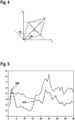
- Figure 4 depicts an illustration of the principal strain axes of an SRT: the axes of principal strain are represented by the eigenvectors of the SRT and are oriented in 45 degrees from the axes of the original cube, more precisely: 45 degrees from the shearing direction (in this case the x-direction) and 45 degrees from the direction perpendicular to the shearing layers.
- a disease parameter ⁇ can be calculated in a respective step of calculating.
- the rate of the local pure shear is dependent on the chosen coordinate system.
- the eigensystem of the local SRT as coordinate system can be chosen. And in this eigensystem there is no more shear-rate. But if a simple shear is present, e.g. shearing between layers, then there is for example an additional rotational component. And this component is invariant under transformation of coordinates. It is contained within the RRT or can simply be calculated as the local Curl and shows the mentioned correlation with factor 2 and factor -2 with the eigenvalues of the SRT (according to equation 1). If there is an additional rigid rotation within the considered field of view thus with a rotational curl due to this rigid rotation directly additive to the Curl due to simple shearing, a correction can be applied.
- Keirometry as an embodiment of the here presented approach, considering not only the SRT but also the RRT, is able to identify and to quantify simple shearing motion without a previously known local shearing direction.
- Performing the so called keirometry means to preferably investigate both in combination: the pattern of the eigensystem of the local SRT and the local Curl.
- the pattern of the eigensystem of the local SRT and the local Curl In a real experiment, there will mostly be a combination of pure deformation (e.g. compression in one direction and dilation in other directions) and simple shear deformation (e.g. shearing between layers).
- the disease parameter ⁇ is introduced.
- the parameter ⁇ reflects the proportion of the two kinds of deformation and the appropriateness of the relation in equation (1) within the measured field of view.
- the disease parameter ⁇ especially reflects the proportion in relation to overall deformation of local layer shearing and local pure deformation within the considered field of view.
- Keirometry means preferably an integrative assessment of both, the local eigensystem of the SRT and the local Curl (contained within the eigensystem of the RRT, by the way).
- Keirometry is for example identifying, analysing and quantifying the local shearing motion within the field of view by investigating these features.
- the local Curl in correlation with the pattern of the local SRT-eigensystem can be used as a measure for the local simple shear rate.
- This shearing causes a strain of the considered volume element.
- This strain effect caused by simple shearing is not only quantitatively related to the rotational curl (see equation (1)) but shows also a certain pattern of the SRT eigensystem (i.e. quantitatively dominating ⁇ 1 and ⁇ 2 with
- Figure 5 shows a diagram of exemplary healthy volunteer data.
- (1unit 20ms) and on the y-axis strain-rate respectively rotational-rate in 1/sec is depicted.
- the beginning of the systole 500 and the beginning of the diastole 510 is also depicted in Figure 5 .
- the line in triangles depicts the mean of the voxel wise maximum positive eigenvalues of endocardilal voxels, i.e. the voxel wise magnitude of maximum extension and the line in rectangles depicts the mean of the half magnitude of the rotational Curl.
- the action depicted in time points 1-14 corresponds to systole.
- Figure 6 shows a diagram of exemplary healthy volunteer data.
- the continuous line represents the disease parameter ⁇ of endocardial voxels and the dashed line represents the disease parameter ⁇ of epicardial voxels.
- the action depicted in time points 1-14 corresponds to systole.
- the action depicted in time points 15-40 corresponds to diastole.
- Figure 7 shows a diagram of exemplary healthy volunteer data, wherein the left ventricular 3D short axis slab in mid diastole at time point 22 is depicted.
- the 3D field of the voxel wise eigenvectors 700 are shown, corresponding to the maximum negative eigenvalue in diastole i.e. local direction and magnitude of maximum instantaneous thinning at the time point 22 in diastole.
- the length of the lines 700 reflect the magnitude i.e. in this case the maximum negative eigenvalues.
- eigenvectors are represented as lines instead of arrows. Clearly visible in this figure is the excellent data quality and realistic depiction of diastolic myocardial deformation.
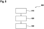
- FIG. 8 shows a flow diagram of a method 800 for Determining a disease parameter describing a type of cardiac disease according to an implementation of the present invention.
- the method 800 comprises a step (810) of reading in a set of input parameters, wherein the input parameters represent a field of velocity vectors measuring 3D myocardial velocity fields with voxel wise 3D velocity information , each velocity vector representing a velocity of motion of a voxel of a region of the heart at a predefined time point or time interval and wherein a velocity vector depicts the voxel wise three-dimensional velocity of the myocardium with different magnitude,
- the method 800 comprises a step 820 of applying a processing algorithm on the set of input parameters in order to obtain at least a rotation parameter and/or at least a strain-rate-tensor, the rotation parameter representing the local curl of the set of input parameters and the strain-rate-tensor representing a local rate of strain at a certain spatial point at a certain time point or time interval, wherein the
- keirometry is a novel method and apparatus for the investigation of myocardial motion in systole and diastole, it opens up a new field of myocardial imaging.
- the approach, Keirometry is able to reveal a hitherto unknown physiological effect of myocardial motion, which can be quantified directly and non-invasively, and thereby provides a strong and fundamentally new tool to investigate myocardial motion in various pathologies including the still poorly understood but clinically highly relevant diastolic heart failure.
- the method is not restricted to MRI data acquisition, but can in principle also be applied to appropriate echocardiographic datasets or datasets from other acquisition techniques.
Landscapes
- Health & Medical Sciences (AREA)
- Engineering & Computer Science (AREA)
- Life Sciences & Earth Sciences (AREA)
- Biomedical Technology (AREA)
- Medical Informatics (AREA)
- Public Health (AREA)
- Nuclear Medicine, Radiotherapy & Molecular Imaging (AREA)
- General Health & Medical Sciences (AREA)
- Physics & Mathematics (AREA)
- Radiology & Medical Imaging (AREA)
- Pathology (AREA)
- Veterinary Medicine (AREA)
- Heart & Thoracic Surgery (AREA)
- Molecular Biology (AREA)
- Surgery (AREA)
- Animal Behavior & Ethology (AREA)
- Biophysics (AREA)
- Epidemiology (AREA)
- Primary Health Care (AREA)
- Cardiology (AREA)
- High Energy & Nuclear Physics (AREA)
- Business, Economics & Management (AREA)
- General Business, Economics & Management (AREA)
- Databases & Information Systems (AREA)
- Data Mining & Analysis (AREA)
- Magnetic Resonance Imaging Apparatus (AREA)
- Theoretical Computer Science (AREA)
- Computer Vision & Pattern Recognition (AREA)
- Computing Systems (AREA)
- Evolutionary Computation (AREA)
- Software Systems (AREA)
- Artificial Intelligence (AREA)
- General Physics & Mathematics (AREA)
- Multimedia (AREA)
Claims (6)
- Vorrichtung (110) zum Bestimmen eines Krankheitsparameters (κ), der eine Art von Herzerkrankung beschreibt, wobei die Vorrichtung (110) die folgenden Einheiten umfasst:- eine Einheit (135) zum Einlesen eines Satzes von Eingangsparametern (f), wobei die Eingangsparameter (f) ein Feld von Geschwindigkeitsvektoren darstellen, die 3D-Myokard-Geschwindigkeitsfelder mit voxelweiser 3D-Geschwindigkeitsinformation messen, wobei jeder Geschwindigkeitsvektor eine Bewegungsgeschwindigkeit eines Voxels (120) eines Bereichs des Herzens (125) zu einem vordefinierten Zeitpunkt oder Zeitintervall repräsentiert und wobei ein Geschwindigkeitsvektor die voxelweise dreidimensionale Geschwindigkeit des Myokards mit unterschiedlicher Größe darstellt;- eine Einheit (140) zum Anwenden eines Verarbeitungsalgorithmus auf den Satz von Eingangsparametern (f), um mindestens einen Rotationsparameter (R) und/oder mindestens einen Dehnungsraten-Tensor (SRT) zu erhalten, wobei der Rotationsparameter (R) die lokale Krümmung des Satzes von Eingangsparametern (f) repräsentiert und der Dehnungsraten-Tensor (SRT) eine lokale Dehnungsrate an einem bestimmten räumlichen Punkt zu einem bestimmten Zeitpunkt oder Zeitintervall repräsentiert, wobei der Dehnungsratentensor (SRT) durch die Jacobimatrix (Df) des betrachteten Geschwindigkeitsfeldes (f) berechnet wird, unter Verwendung der Formel SRT= ½(Df+DfT), wobei DfT eine transponierte Jacobimatrix bezeichnet und wobei der Rotationsparameter (R) aus der Formel curl = ∇ x f abgeleitet wird, wobei ∇ den Nabla-Operator repräsentiert und x das Kreuzprodukt bezeichnet oder wobei der Rotationsparameter (R) unter Verwendung der Formel RRT= ½(Df-DfT) berechnet wird; und- eine Einheit (145) zum Berechnen mindestens des Krankheitsparameters (κ) auf der Basis des Rotationsparameters (R) und des Dehnungsratentensors (SRT), wobei die Einheit (145) zum Berechnen zum Berechnen des Krankheitsparameters (κ) durch Division eines Absolutwertes des Rotationsparameters (R) durch mindestens einen Eigenwert ausgebildet ist, wobei sich der mindestens eine Eigenwert aus der Lösung des Eigenvektorproblems SRT f = λ f ergibt, wobei λ den Eigenwert bezeichnet und wobei der Krankheitsparameter (κ) den Anteil der lokalen Schichtscherung an der Gesamtverformung in Bezug auf die lokale reine Verformung wiedergibt.
- Vorrichtung (110) nach einem der Ansprüche 1, dadurch gekennzeichnet, dass die Einheit (140) zum Anwenden des Verarbeitungsalgorithmus für eine Berechnung zumindest der Jacobimatrix (Df) aus dem Satz von Eingangsparametern (f), insbesondere zur Berechnung des Dehnungsratentensors, ausgebildet ist.
- Vorrichtung (110) nach Anspruch 1 oder 2, dadurch gekennzeichnet, dass die Einheit (140) zum Anwenden des Verarbeitungsalgorithmus für die Berechnung der Jacobimatrix (Df) in Bezug auf jede der drei Raumdimensionen (x1, x2, x3) ausgebildet ist.
- Vorrichtung (110) nach einem der vorhergehenden Ansprüche, dadurch gekennzeichnet, dass in der Einheit (135) zum Einlesen ein Satz von Eingangsparametern (f) eingelesen wird, die mittels eines Magnetresonanztomographen (MRI) aufgenommen wurden.
- Vorrichtung (110) nach einem der vorhergehenden Ansprüche, dadurch gekennzeichnet, dass die Einheiten (135, 1470, 145) zum Einlesen, Anwenden und Berechnen dazu ausgebildet sind, nacheinander Sätze von Eingabeparametern (f) durchzuführen, die die Bewegungsgeschwindigkeit eines Voxels (120) verschiedener Bereiche des Herzens (125) zu einem vorgegebenen Zeitpunkt oder Zeitintervall repräsentieren und/oder die Bewegungsgeschwindigkeit eines Voxels (120) eines Bereichs des Herzens (125) zu verschiedenen vorgegebenen Zeitpunkten oder Zeitintervallen repräsentieren.
- Vorrichtung (110) nach einem der vorhergehenden Ansprüche, gekennzeichnet durch eine Einheit zum Analysieren des Krankheitsparameters durch Vergleichen des Krankheitsparameters mit einem Referenzwert und/oder Referenzwertintervall, um einen Vorschlag für eine bestimmte Herzerkrankung auszugeben.
Applications Claiming Priority (2)
| Application Number | Priority Date | Filing Date | Title |
|---|---|---|---|
| EP16201753.7A EP3330874A1 (de) | 2016-12-01 | 2016-12-01 | Verfahren und vorrichtung zur bestimmung eines krankheitsparameters |
| PCT/EP2017/080471 WO2018099844A1 (en) | 2016-12-01 | 2017-11-27 | Method and apparatus for determining a disease parameter |
Publications (2)
| Publication Number | Publication Date |
|---|---|
| EP3549043A1 EP3549043A1 (de) | 2019-10-09 |
| EP3549043B1 true EP3549043B1 (de) | 2023-05-31 |
Family
ID=57708277
Family Applications (2)
| Application Number | Title | Priority Date | Filing Date |
|---|---|---|---|
| EP16201753.7A Withdrawn EP3330874A1 (de) | 2016-12-01 | 2016-12-01 | Verfahren und vorrichtung zur bestimmung eines krankheitsparameters |
| EP17801734.9A Active EP3549043B1 (de) | 2016-12-01 | 2017-11-27 | Verfahren und vorrichtung zur bestimmung eines krankheitsparameters |
Family Applications Before (1)
| Application Number | Title | Priority Date | Filing Date |
|---|---|---|---|
| EP16201753.7A Withdrawn EP3330874A1 (de) | 2016-12-01 | 2016-12-01 | Verfahren und vorrichtung zur bestimmung eines krankheitsparameters |
Country Status (3)
| Country | Link |
|---|---|
| US (1) | US11457814B2 (de) |
| EP (2) | EP3330874A1 (de) |
| WO (1) | WO2018099844A1 (de) |
Family Cites Families (4)
| Publication number | Priority date | Publication date | Assignee | Title |
|---|---|---|---|---|
| JP5259267B2 (ja) * | 2008-06-19 | 2013-08-07 | 株式会社東芝 | 超音波診断装置、超音波画像処理装置及び超音波画像処理プログラム |
| US10311978B2 (en) * | 2012-01-30 | 2019-06-04 | Siemens Healthcare Gmbh | Method and system for patient specific planning of cardiac therapies on preoperative clinical data and medical images |
| US20140071125A1 (en) * | 2012-09-11 | 2014-03-13 | The Johns Hopkins University | Patient-Specific Segmentation, Analysis, and Modeling from 3-Dimensional Ultrasound Image Data |
| WO2015200552A1 (en) * | 2014-06-25 | 2015-12-30 | William Beaumont Hospital | Simulated cardiac model |
-
2016
- 2016-12-01 EP EP16201753.7A patent/EP3330874A1/de not_active Withdrawn
-
2017
- 2017-11-27 WO PCT/EP2017/080471 patent/WO2018099844A1/en not_active Ceased
- 2017-11-27 EP EP17801734.9A patent/EP3549043B1/de active Active
-
2019
- 2019-05-29 US US16/424,895 patent/US11457814B2/en active Active
Also Published As
| Publication number | Publication date |
|---|---|
| US11457814B2 (en) | 2022-10-04 |
| EP3330874A1 (de) | 2018-06-06 |
| US20190313911A1 (en) | 2019-10-17 |
| EP3549043A1 (de) | 2019-10-09 |
| WO2018099844A1 (en) | 2018-06-07 |
Similar Documents
| Publication | Publication Date | Title |
|---|---|---|
| Jafari Tadi et al. | Gyrocardiography: A new non-invasive monitoring method for the assessment of cardiac mechanics and the estimation of hemodynamic variables | |
| EP2026280B1 (de) | Verfahren und entsprechender Apparat für quantitative Messungen bei Bildsequenzen, v.a. Ultraschallbildsequenzen | |
| US6295464B1 (en) | Apparatus and method for dynamic modeling of an object | |
| Sugeng et al. | Left ventricular assessment using real time three dimensional echocardiography | |
| Gopal et al. | Freehand three-dimensional echocardiography for measurement of left ventricular mass: in vivo anatomic validation using explanted human hearts | |
| Boyle et al. | Regularization-free strain mapping in three dimensions, with application to cardiac ultrasound | |
| US20240108229A1 (en) | Mri-derived strain-based measurements and related image data acquisitions, image data processing, patient evaluations and monitoring methods and systems | |
| Chan et al. | Statistical characterization of human brain deformation during mild angular acceleration measured in vivo by tagged magnetic resonance imaging | |
| Wick et al. | Detection of cardiac quiescence from B-mode echocardiography using a correlation-based frame-to-frame deviation measure | |
| Rahimi et al. | Sensitivity of noninvasive cardiac electrophysiological imaging to variations in personalized anatomical modeling | |
| Kerwin et al. | Image processing and analysis in tagged cardiac MRI | |
| US11457814B2 (en) | Apparatus for determining a disease parameter | |
| Peyrat et al. | Towards a statistical atlas of cardiac fiber structure | |
| Wong et al. | Physiological fusion of functional and structural images for cardiac deformation recovery | |
| JP3715580B2 (ja) | 医用運動解析装置及びその方法 | |
| Liu et al. | Cardiac 3D motion reconstruction using dual-camera defocused speckle imaging with multi-scale amplification | |
| Tyagi et al. | Improving three‐dimensional mechanical imaging of breast lesions with principal component analysis | |
| Colvert et al. | Novel measurement of LV twist using 4DCT: quantifying accuracy as a function of image noise | |
| CN101278828A (zh) | 测量心室壁微面积功的方法 | |
| EP4614509A1 (de) | Schätzung von muskelfasern | |
| Sawada et al. | Three-dimensional stress echocardiography: the promise and limitations of volumetric imaging | |
| JP5311330B2 (ja) | 心機能診断表示システム、表示方法及び表示プログラム | |
| HK40131812A (en) | Muscular fibre estimation | |
| D'hooge et al. | Deformation imaging by ultrasound for the assessment of regional myocardial function | |
| Wang et al. | How much geometrical detail do we need in cardiac electrophysiological imaging? A generic heart-torso representation for fast subject-specific customization |
Legal Events
| Date | Code | Title | Description |
|---|---|---|---|
| STAA | Information on the status of an ep patent application or granted ep patent |
Free format text: STATUS: UNKNOWN |
|
| STAA | Information on the status of an ep patent application or granted ep patent |
Free format text: STATUS: THE INTERNATIONAL PUBLICATION HAS BEEN MADE |
|
| PUAI | Public reference made under article 153(3) epc to a published international application that has entered the european phase |
Free format text: ORIGINAL CODE: 0009012 |
|
| STAA | Information on the status of an ep patent application or granted ep patent |
Free format text: STATUS: REQUEST FOR EXAMINATION WAS MADE |
|
| 17P | Request for examination filed |
Effective date: 20190517 |
|
| AK | Designated contracting states |
Kind code of ref document: A1 Designated state(s): AL AT BE BG CH CY CZ DE DK EE ES FI FR GB GR HR HU IE IS IT LI LT LU LV MC MK MT NL NO PL PT RO RS SE SI SK SM TR |
|
| AX | Request for extension of the european patent |
Extension state: BA ME |
|
| STAA | Information on the status of an ep patent application or granted ep patent |
Free format text: STATUS: EXAMINATION IS IN PROGRESS |
|
| DAV | Request for validation of the european patent (deleted) | ||
| DAX | Request for extension of the european patent (deleted) | ||
| 17Q | First examination report despatched |
Effective date: 20200309 |
|
| REG | Reference to a national code |
Ref country code: DE Ref legal event code: R079 Free format text: PREVIOUS MAIN CLASS: G06F0019000000 Ipc: G16H0040630000 Ref country code: DE Ref legal event code: R079 Ref document number: 602017069247 Country of ref document: DE Free format text: PREVIOUS MAIN CLASS: G06F0019000000 Ipc: G16H0040630000 |
|
| RIC1 | Information provided on ipc code assigned before grant |
Ipc: G16H 40/63 20180101AFI20230107BHEP |
|
| GRAP | Despatch of communication of intention to grant a patent |
Free format text: ORIGINAL CODE: EPIDOSNIGR1 |
|
| STAA | Information on the status of an ep patent application or granted ep patent |
Free format text: STATUS: GRANT OF PATENT IS INTENDED |
|
| INTG | Intention to grant announced |
Effective date: 20230313 |
|
| GRAS | Grant fee paid |
Free format text: ORIGINAL CODE: EPIDOSNIGR3 |
|
| GRAA | (expected) grant |
Free format text: ORIGINAL CODE: 0009210 |
|
| STAA | Information on the status of an ep patent application or granted ep patent |
Free format text: STATUS: THE PATENT HAS BEEN GRANTED |
|
| AK | Designated contracting states |
Kind code of ref document: B1 Designated state(s): AL AT BE BG CH CY CZ DE DK EE ES FI FR GB GR HR HU IE IS IT LI LT LU LV MC MK MT NL NO PL PT RO RS SE SI SK SM TR |
|
| REG | Reference to a national code |
Ref country code: GB Ref legal event code: FG4D Ref country code: CH Ref legal event code: EP |
|
| REG | Reference to a national code |
Ref country code: AT Ref legal event code: REF Ref document number: 1571441 Country of ref document: AT Kind code of ref document: T Effective date: 20230615 Ref country code: DE Ref legal event code: R096 Ref document number: 602017069247 Country of ref document: DE |
|
| REG | Reference to a national code |
Ref country code: IE Ref legal event code: FG4D |
|
| P01 | Opt-out of the competence of the unified patent court (upc) registered |
Effective date: 20230719 |
|
| REG | Reference to a national code |
Ref country code: LT Ref legal event code: MG9D |
|
| REG | Reference to a national code |
Ref country code: NL Ref legal event code: MP Effective date: 20230531 |
|
| REG | Reference to a national code |
Ref country code: AT Ref legal event code: MK05 Ref document number: 1571441 Country of ref document: AT Kind code of ref document: T Effective date: 20230531 |
|
| PG25 | Lapsed in a contracting state [announced via postgrant information from national office to epo] |
Ref country code: SE Free format text: LAPSE BECAUSE OF FAILURE TO SUBMIT A TRANSLATION OF THE DESCRIPTION OR TO PAY THE FEE WITHIN THE PRESCRIBED TIME-LIMIT Effective date: 20230531 Ref country code: NO Free format text: LAPSE BECAUSE OF FAILURE TO SUBMIT A TRANSLATION OF THE DESCRIPTION OR TO PAY THE FEE WITHIN THE PRESCRIBED TIME-LIMIT Effective date: 20230831 Ref country code: ES Free format text: LAPSE BECAUSE OF FAILURE TO SUBMIT A TRANSLATION OF THE DESCRIPTION OR TO PAY THE FEE WITHIN THE PRESCRIBED TIME-LIMIT Effective date: 20230531 Ref country code: AT Free format text: LAPSE BECAUSE OF FAILURE TO SUBMIT A TRANSLATION OF THE DESCRIPTION OR TO PAY THE FEE WITHIN THE PRESCRIBED TIME-LIMIT Effective date: 20230531 |
|
| PG25 | Lapsed in a contracting state [announced via postgrant information from national office to epo] |
Ref country code: RS Free format text: LAPSE BECAUSE OF FAILURE TO SUBMIT A TRANSLATION OF THE DESCRIPTION OR TO PAY THE FEE WITHIN THE PRESCRIBED TIME-LIMIT Effective date: 20230531 Ref country code: PL Free format text: LAPSE BECAUSE OF FAILURE TO SUBMIT A TRANSLATION OF THE DESCRIPTION OR TO PAY THE FEE WITHIN THE PRESCRIBED TIME-LIMIT Effective date: 20230531 Ref country code: NL Free format text: LAPSE BECAUSE OF FAILURE TO SUBMIT A TRANSLATION OF THE DESCRIPTION OR TO PAY THE FEE WITHIN THE PRESCRIBED TIME-LIMIT Effective date: 20230531 Ref country code: LV Free format text: LAPSE BECAUSE OF FAILURE TO SUBMIT A TRANSLATION OF THE DESCRIPTION OR TO PAY THE FEE WITHIN THE PRESCRIBED TIME-LIMIT Effective date: 20230531 Ref country code: LT Free format text: LAPSE BECAUSE OF FAILURE TO SUBMIT A TRANSLATION OF THE DESCRIPTION OR TO PAY THE FEE WITHIN THE PRESCRIBED TIME-LIMIT Effective date: 20230531 Ref country code: IS Free format text: LAPSE BECAUSE OF FAILURE TO SUBMIT A TRANSLATION OF THE DESCRIPTION OR TO PAY THE FEE WITHIN THE PRESCRIBED TIME-LIMIT Effective date: 20230930 Ref country code: HR Free format text: LAPSE BECAUSE OF FAILURE TO SUBMIT A TRANSLATION OF THE DESCRIPTION OR TO PAY THE FEE WITHIN THE PRESCRIBED TIME-LIMIT Effective date: 20230531 Ref country code: GR Free format text: LAPSE BECAUSE OF FAILURE TO SUBMIT A TRANSLATION OF THE DESCRIPTION OR TO PAY THE FEE WITHIN THE PRESCRIBED TIME-LIMIT Effective date: 20230901 |
|
| PG25 | Lapsed in a contracting state [announced via postgrant information from national office to epo] |
Ref country code: FI Free format text: LAPSE BECAUSE OF FAILURE TO SUBMIT A TRANSLATION OF THE DESCRIPTION OR TO PAY THE FEE WITHIN THE PRESCRIBED TIME-LIMIT Effective date: 20230531 |
|
| PG25 | Lapsed in a contracting state [announced via postgrant information from national office to epo] |
Ref country code: SK Free format text: LAPSE BECAUSE OF FAILURE TO SUBMIT A TRANSLATION OF THE DESCRIPTION OR TO PAY THE FEE WITHIN THE PRESCRIBED TIME-LIMIT Effective date: 20230531 |
|
| PG25 | Lapsed in a contracting state [announced via postgrant information from national office to epo] |
Ref country code: SM Free format text: LAPSE BECAUSE OF FAILURE TO SUBMIT A TRANSLATION OF THE DESCRIPTION OR TO PAY THE FEE WITHIN THE PRESCRIBED TIME-LIMIT Effective date: 20230531 Ref country code: SK Free format text: LAPSE BECAUSE OF FAILURE TO SUBMIT A TRANSLATION OF THE DESCRIPTION OR TO PAY THE FEE WITHIN THE PRESCRIBED TIME-LIMIT Effective date: 20230531 Ref country code: RO Free format text: LAPSE BECAUSE OF FAILURE TO SUBMIT A TRANSLATION OF THE DESCRIPTION OR TO PAY THE FEE WITHIN THE PRESCRIBED TIME-LIMIT Effective date: 20230531 Ref country code: PT Free format text: LAPSE BECAUSE OF FAILURE TO SUBMIT A TRANSLATION OF THE DESCRIPTION OR TO PAY THE FEE WITHIN THE PRESCRIBED TIME-LIMIT Effective date: 20231002 Ref country code: EE Free format text: LAPSE BECAUSE OF FAILURE TO SUBMIT A TRANSLATION OF THE DESCRIPTION OR TO PAY THE FEE WITHIN THE PRESCRIBED TIME-LIMIT Effective date: 20230531 Ref country code: DK Free format text: LAPSE BECAUSE OF FAILURE TO SUBMIT A TRANSLATION OF THE DESCRIPTION OR TO PAY THE FEE WITHIN THE PRESCRIBED TIME-LIMIT Effective date: 20230531 Ref country code: CZ Free format text: LAPSE BECAUSE OF FAILURE TO SUBMIT A TRANSLATION OF THE DESCRIPTION OR TO PAY THE FEE WITHIN THE PRESCRIBED TIME-LIMIT Effective date: 20230531 |
|
| REG | Reference to a national code |
Ref country code: DE Ref legal event code: R097 Ref document number: 602017069247 Country of ref document: DE |
|
| PLBE | No opposition filed within time limit |
Free format text: ORIGINAL CODE: 0009261 |
|
| STAA | Information on the status of an ep patent application or granted ep patent |
Free format text: STATUS: NO OPPOSITION FILED WITHIN TIME LIMIT |
|
| PG25 | Lapsed in a contracting state [announced via postgrant information from national office to epo] |
Ref country code: SI Free format text: LAPSE BECAUSE OF FAILURE TO SUBMIT A TRANSLATION OF THE DESCRIPTION OR TO PAY THE FEE WITHIN THE PRESCRIBED TIME-LIMIT Effective date: 20230531 |
|
| 26N | No opposition filed |
Effective date: 20240301 |
|
| PG25 | Lapsed in a contracting state [announced via postgrant information from national office to epo] |
Ref country code: SI Free format text: LAPSE BECAUSE OF FAILURE TO SUBMIT A TRANSLATION OF THE DESCRIPTION OR TO PAY THE FEE WITHIN THE PRESCRIBED TIME-LIMIT Effective date: 20230531 Ref country code: IT Free format text: LAPSE BECAUSE OF FAILURE TO SUBMIT A TRANSLATION OF THE DESCRIPTION OR TO PAY THE FEE WITHIN THE PRESCRIBED TIME-LIMIT Effective date: 20230531 |
|
| REG | Reference to a national code |
Ref country code: CH Ref legal event code: PL |
|
| PG25 | Lapsed in a contracting state [announced via postgrant information from national office to epo] |
Ref country code: MC Free format text: LAPSE BECAUSE OF FAILURE TO SUBMIT A TRANSLATION OF THE DESCRIPTION OR TO PAY THE FEE WITHIN THE PRESCRIBED TIME-LIMIT Effective date: 20230531 |
|
| PG25 | Lapsed in a contracting state [announced via postgrant information from national office to epo] |
Ref country code: LU Free format text: LAPSE BECAUSE OF NON-PAYMENT OF DUE FEES Effective date: 20231127 |
|
| PG25 | Lapsed in a contracting state [announced via postgrant information from national office to epo] |
Ref country code: CH Free format text: LAPSE BECAUSE OF NON-PAYMENT OF DUE FEES Effective date: 20231130 |
|
| GBPC | Gb: european patent ceased through non-payment of renewal fee |
Effective date: 20231127 |
|
| PG25 | Lapsed in a contracting state [announced via postgrant information from national office to epo] |
Ref country code: MC Free format text: LAPSE BECAUSE OF FAILURE TO SUBMIT A TRANSLATION OF THE DESCRIPTION OR TO PAY THE FEE WITHIN THE PRESCRIBED TIME-LIMIT Effective date: 20230531 Ref country code: LU Free format text: LAPSE BECAUSE OF NON-PAYMENT OF DUE FEES Effective date: 20231127 Ref country code: CH Free format text: LAPSE BECAUSE OF NON-PAYMENT OF DUE FEES Effective date: 20231130 |
|
| REG | Reference to a national code |
Ref country code: BE Ref legal event code: MM Effective date: 20231130 |
|
| REG | Reference to a national code |
Ref country code: IE Ref legal event code: MM4A |
|
| PG25 | Lapsed in a contracting state [announced via postgrant information from national office to epo] |
Ref country code: IE Free format text: LAPSE BECAUSE OF NON-PAYMENT OF DUE FEES Effective date: 20231127 |
|
| PG25 | Lapsed in a contracting state [announced via postgrant information from national office to epo] |
Ref country code: GB Free format text: LAPSE BECAUSE OF NON-PAYMENT OF DUE FEES Effective date: 20231127 |
|
| PG25 | Lapsed in a contracting state [announced via postgrant information from national office to epo] |
Ref country code: BE Free format text: LAPSE BECAUSE OF NON-PAYMENT OF DUE FEES Effective date: 20231130 |
|
| PG25 | Lapsed in a contracting state [announced via postgrant information from national office to epo] |
Ref country code: FR Free format text: LAPSE BECAUSE OF NON-PAYMENT OF DUE FEES Effective date: 20231130 |
|
| PG25 | Lapsed in a contracting state [announced via postgrant information from national office to epo] |
Ref country code: IE Free format text: LAPSE BECAUSE OF NON-PAYMENT OF DUE FEES Effective date: 20231127 Ref country code: GB Free format text: LAPSE BECAUSE OF NON-PAYMENT OF DUE FEES Effective date: 20231127 Ref country code: FR Free format text: LAPSE BECAUSE OF NON-PAYMENT OF DUE FEES Effective date: 20231130 Ref country code: BE Free format text: LAPSE BECAUSE OF NON-PAYMENT OF DUE FEES Effective date: 20231130 |
|
| PG25 | Lapsed in a contracting state [announced via postgrant information from national office to epo] |
Ref country code: BG Free format text: LAPSE BECAUSE OF FAILURE TO SUBMIT A TRANSLATION OF THE DESCRIPTION OR TO PAY THE FEE WITHIN THE PRESCRIBED TIME-LIMIT Effective date: 20230531 |
|
| PG25 | Lapsed in a contracting state [announced via postgrant information from national office to epo] |
Ref country code: BG Free format text: LAPSE BECAUSE OF FAILURE TO SUBMIT A TRANSLATION OF THE DESCRIPTION OR TO PAY THE FEE WITHIN THE PRESCRIBED TIME-LIMIT Effective date: 20230531 |
|
| PG25 | Lapsed in a contracting state [announced via postgrant information from national office to epo] |
Ref country code: CY Free format text: LAPSE BECAUSE OF FAILURE TO SUBMIT A TRANSLATION OF THE DESCRIPTION OR TO PAY THE FEE WITHIN THE PRESCRIBED TIME-LIMIT; INVALID AB INITIO Effective date: 20171127 |
|
| PG25 | Lapsed in a contracting state [announced via postgrant information from national office to epo] |
Ref country code: HU Free format text: LAPSE BECAUSE OF FAILURE TO SUBMIT A TRANSLATION OF THE DESCRIPTION OR TO PAY THE FEE WITHIN THE PRESCRIBED TIME-LIMIT; INVALID AB INITIO Effective date: 20171127 |
|
| PG25 | Lapsed in a contracting state [announced via postgrant information from national office to epo] |
Ref country code: TR Free format text: LAPSE BECAUSE OF FAILURE TO SUBMIT A TRANSLATION OF THE DESCRIPTION OR TO PAY THE FEE WITHIN THE PRESCRIBED TIME-LIMIT Effective date: 20230531 |
|
| PGFP | Annual fee paid to national office [announced via postgrant information from national office to epo] |
Ref country code: DE Payment date: 20251118 Year of fee payment: 9 |