EP3544591B1 - Particle-mediated delivery of biologics - Google Patents
Particle-mediated delivery of biologics Download PDFInfo
- Publication number
- EP3544591B1 EP3544591B1 EP17873643.5A EP17873643A EP3544591B1 EP 3544591 B1 EP3544591 B1 EP 3544591B1 EP 17873643 A EP17873643 A EP 17873643A EP 3544591 B1 EP3544591 B1 EP 3544591B1
- Authority
- EP
- European Patent Office
- Prior art keywords
- particle
- mir
- rna
- mrna
- biologics
- Prior art date
- Legal status (The legal status is an assumption and is not a legal conclusion. Google has not performed a legal analysis and makes no representation as to the accuracy of the status listed.)
- Active
Links
- 239000002245 particle Substances 0.000 title claims description 239
- 229960000074 biopharmaceutical Drugs 0.000 title description 88
- 230000001404 mediated effect Effects 0.000 title description 20
- 108020004999 messenger RNA Proteins 0.000 claims description 112
- FHVDTGUDJYJELY-UHFFFAOYSA-N 6-{[2-carboxy-4,5-dihydroxy-6-(phosphanyloxy)oxan-3-yl]oxy}-4,5-dihydroxy-3-phosphanyloxane-2-carboxylic acid Chemical compound O1C(C(O)=O)C(P)C(O)C(O)C1OC1C(C(O)=O)OC(OP)C(O)C1O FHVDTGUDJYJELY-UHFFFAOYSA-N 0.000 claims description 102
- 235000010443 alginic acid Nutrition 0.000 claims description 101
- 229920000615 alginic acid Polymers 0.000 claims description 101
- 229940072056 alginate Drugs 0.000 claims description 100
- 239000000499 gel Substances 0.000 claims description 73
- 102000004196 processed proteins & peptides Human genes 0.000 claims description 53
- 108090000765 processed proteins & peptides Proteins 0.000 claims description 53
- 241000124008 Mammalia Species 0.000 claims description 52
- 229920001184 polypeptide Polymers 0.000 claims description 52
- 230000002051 biphasic effect Effects 0.000 claims description 49
- -1 ErBb3 Proteins 0.000 claims description 46
- 230000004217 heart function Effects 0.000 claims description 35
- 239000000203 mixture Substances 0.000 claims description 25
- 230000001172 regenerating effect Effects 0.000 claims description 20
- 159000000007 calcium salts Chemical class 0.000 claims description 14
- 210000001808 exosome Anatomy 0.000 claims description 13
- 102000008186 Collagen Human genes 0.000 claims description 12
- 108010035532 Collagen Proteins 0.000 claims description 12
- 208000006117 ST-elevation myocardial infarction Diseases 0.000 claims description 12
- 229920001436 collagen Polymers 0.000 claims description 12
- 101000973997 Homo sapiens Nucleosome assembly protein 1-like 4 Proteins 0.000 claims description 10
- 101000947178 Homo sapiens Platelet basic protein Proteins 0.000 claims description 10
- 101800004564 Transforming growth factor alpha Proteins 0.000 claims description 10
- 102400001320 Transforming growth factor alpha Human genes 0.000 claims description 10
- 230000000694 effects Effects 0.000 claims description 10
- 101000914484 Homo sapiens T-lymphocyte activation antigen CD80 Proteins 0.000 claims description 9
- 108010002350 Interleukin-2 Proteins 0.000 claims description 9
- 108060008682 Tumor Necrosis Factor Proteins 0.000 claims description 9
- 102000005789 Vascular Endothelial Growth Factors Human genes 0.000 claims description 9
- 108010019530 Vascular Endothelial Growth Factors Proteins 0.000 claims description 9
- 210000005003 heart tissue Anatomy 0.000 claims description 9
- 102100024222 B-lymphocyte antigen CD19 Human genes 0.000 claims description 8
- 102100022005 B-lymphocyte antigen CD20 Human genes 0.000 claims description 8
- 101000980825 Homo sapiens B-lymphocyte antigen CD19 Proteins 0.000 claims description 8
- 101000897405 Homo sapiens B-lymphocyte antigen CD20 Proteins 0.000 claims description 8
- 102100027222 T-lymphocyte activation antigen CD80 Human genes 0.000 claims description 8
- 230000002438 mitochondrial effect Effects 0.000 claims description 7
- 101710167800 Capsid assembly scaffolding protein Proteins 0.000 claims description 6
- 101710130420 Probable capsid assembly scaffolding protein Proteins 0.000 claims description 6
- 101710204410 Scaffold protein Proteins 0.000 claims description 6
- 238000013146 percutaneous coronary intervention Methods 0.000 claims description 6
- 102000009123 Fibrin Human genes 0.000 claims description 5
- 108010073385 Fibrin Proteins 0.000 claims description 5
- BWGVNKXGVNDBDI-UHFFFAOYSA-N Fibrin monomer Chemical compound CNC(=O)CNC(=O)CN BWGVNKXGVNDBDI-UHFFFAOYSA-N 0.000 claims description 5
- 108010010803 Gelatin Proteins 0.000 claims description 5
- 229950003499 fibrin Drugs 0.000 claims description 5
- 229920000159 gelatin Polymers 0.000 claims description 5
- 239000008273 gelatin Substances 0.000 claims description 5
- 235000019322 gelatine Nutrition 0.000 claims description 5
- 235000011852 gelatine desserts Nutrition 0.000 claims description 5
- 102000012422 Collagen Type I Human genes 0.000 claims description 4
- 108010022452 Collagen Type I Proteins 0.000 claims description 4
- 239000002158 endotoxin Substances 0.000 claims description 4
- 229920006008 lipopolysaccharide Polymers 0.000 claims description 4
- 229940122040 Resolvin D1 agonist Drugs 0.000 claims description 3
- 108090000379 Fibroblast growth factor 2 Proteins 0.000 claims 1
- 102000003974 Fibroblast growth factor 2 Human genes 0.000 claims 1
- 108090000723 Insulin-Like Growth Factor I Proteins 0.000 claims 1
- 102000004218 Insulin-Like Growth Factor I Human genes 0.000 claims 1
- 102000000588 Interleukin-2 Human genes 0.000 claims 1
- 102400000503 Neutrophil-activating peptide 2 Human genes 0.000 claims 1
- 102000003390 tumor necrosis factor Human genes 0.000 claims 1
- 230000014509 gene expression Effects 0.000 description 69
- 230000000747 cardiac effect Effects 0.000 description 67
- 210000001519 tissue Anatomy 0.000 description 49
- 108091032973 (ribonucleotides)n+m Proteins 0.000 description 46
- 230000002411 adverse Effects 0.000 description 41
- 238000000034 method Methods 0.000 description 38
- 210000004027 cell Anatomy 0.000 description 33
- 108091028043 Nucleic acid sequence Proteins 0.000 description 28
- 150000001413 amino acids Chemical group 0.000 description 28
- 150000007523 nucleic acids Chemical group 0.000 description 28
- 239000002105 nanoparticle Substances 0.000 description 27
- 108090000623 proteins and genes Proteins 0.000 description 23
- 239000002502 liposome Substances 0.000 description 22
- 108060001084 Luciferase Proteins 0.000 description 20
- 239000005089 Luciferase Substances 0.000 description 20
- 238000001727 in vivo Methods 0.000 description 20
- 210000004413 cardiac myocyte Anatomy 0.000 description 19
- 238000001890 transfection Methods 0.000 description 19
- 241000699670 Mus sp. Species 0.000 description 17
- 230000002401 inhibitory effect Effects 0.000 description 15
- 239000007924 injection Substances 0.000 description 14
- 238000002347 injection Methods 0.000 description 14
- 101000693076 Homo sapiens Angiopoietin-related protein 4 Proteins 0.000 description 13
- 238000013461 design Methods 0.000 description 13
- 239000007788 liquid Substances 0.000 description 13
- 125000003729 nucleotide group Chemical group 0.000 description 13
- 239000000243 solution Substances 0.000 description 13
- 238000011282 treatment Methods 0.000 description 13
- 108090000613 Cathepsin S Proteins 0.000 description 12
- 108090000331 Firefly luciferases Proteins 0.000 description 12
- 101000897486 Homo sapiens C-C motif chemokine 25 Proteins 0.000 description 12
- 206010000891 acute myocardial infarction Diseases 0.000 description 12
- 229920001223 polyethylene glycol Polymers 0.000 description 12
- 102100025674 Angiopoietin-related protein 4 Human genes 0.000 description 11
- 102100031170 CCN family member 3 Human genes 0.000 description 11
- 101710137351 CCN family member 3 Proteins 0.000 description 11
- 102100030704 Interleukin-21 Human genes 0.000 description 11
- 108010016160 Matrix Metalloproteinase 3 Proteins 0.000 description 11
- 102000000422 Matrix Metalloproteinase 3 Human genes 0.000 description 11
- 108010074108 interleukin-21 Proteins 0.000 description 11
- 239000000463 material Substances 0.000 description 11
- 102000004169 proteins and genes Human genes 0.000 description 11
- 102400000068 Angiostatin Human genes 0.000 description 10
- 108010079709 Angiostatins Proteins 0.000 description 10
- 102100021933 C-C motif chemokine 25 Human genes 0.000 description 10
- OYPRJOBELJOOCE-UHFFFAOYSA-N Calcium Chemical compound [Ca] OYPRJOBELJOOCE-UHFFFAOYSA-N 0.000 description 10
- 102100035654 Cathepsin S Human genes 0.000 description 10
- 102000006440 Chemokine CCL26 Human genes 0.000 description 10
- 108010083698 Chemokine CCL26 Proteins 0.000 description 10
- 102000016970 Follistatin Human genes 0.000 description 10
- 108010014612 Follistatin Proteins 0.000 description 10
- 241000282412 Homo Species 0.000 description 10
- 239000002202 Polyethylene glycol Substances 0.000 description 10
- 102000004338 Transferrin Human genes 0.000 description 10
- 108090000901 Transferrin Proteins 0.000 description 10
- FZCSTZYAHCUGEM-UHFFFAOYSA-N aspergillomarasmine B Natural products OC(=O)CNC(C(O)=O)CNC(C(O)=O)CC(O)=O FZCSTZYAHCUGEM-UHFFFAOYSA-N 0.000 description 10
- 230000017531 blood circulation Effects 0.000 description 10
- 239000011575 calcium Substances 0.000 description 10
- 229910052791 calcium Inorganic materials 0.000 description 10
- 230000015556 catabolic process Effects 0.000 description 10
- 238000006731 degradation reaction Methods 0.000 description 10
- 238000005516 engineering process Methods 0.000 description 10
- 230000004048 modification Effects 0.000 description 10
- 238000012986 modification Methods 0.000 description 10
- 239000002773 nucleotide Substances 0.000 description 10
- 210000002027 skeletal muscle Anatomy 0.000 description 10
- 239000012581 transferrin Substances 0.000 description 10
- 239000013598 vector Substances 0.000 description 10
- 229920001661 Chitosan Polymers 0.000 description 9
- 101000599951 Homo sapiens Insulin-like growth factor I Proteins 0.000 description 9
- 102100036154 Platelet basic protein Human genes 0.000 description 9
- 102100040247 Tumor necrosis factor Human genes 0.000 description 9
- 238000000576 coating method Methods 0.000 description 9
- 229960005188 collagen Drugs 0.000 description 9
- 230000002459 sustained effect Effects 0.000 description 9
- 101001027327 Bos taurus Growth-regulated protein homolog alpha Proteins 0.000 description 8
- 102000016950 Chemokine CXCL1 Human genes 0.000 description 8
- 102000018233 Fibroblast Growth Factor Human genes 0.000 description 8
- 108050007372 Fibroblast Growth Factor Proteins 0.000 description 8
- 206010061216 Infarction Diseases 0.000 description 8
- 102100037852 Insulin-like growth factor I Human genes 0.000 description 8
- 102100020873 Interleukin-2 Human genes 0.000 description 8
- 239000013283 Janus particle Substances 0.000 description 8
- 108010073929 Vascular Endothelial Growth Factor A Proteins 0.000 description 8
- 238000013459 approach Methods 0.000 description 8
- 229940126864 fibroblast growth factor Drugs 0.000 description 8
- 230000001965 increasing effect Effects 0.000 description 8
- 230000007574 infarction Effects 0.000 description 8
- 208000010125 myocardial infarction Diseases 0.000 description 8
- 230000001225 therapeutic effect Effects 0.000 description 8
- 102000040650 (ribonucleotides)n+m Human genes 0.000 description 7
- 108020005345 3' Untranslated Regions Proteins 0.000 description 7
- 210000004204 blood vessel Anatomy 0.000 description 7
- 239000003795 chemical substances by application Substances 0.000 description 7
- 239000011248 coating agent Substances 0.000 description 7
- 238000000386 microscopy Methods 0.000 description 7
- 230000008685 targeting Effects 0.000 description 7
- 108700011259 MicroRNAs Proteins 0.000 description 6
- 108091036407 Polyadenylation Proteins 0.000 description 6
- 108091031326 miR-15b stem-loop Proteins 0.000 description 6
- 108091037006 miR-548h-1 stem-loop Proteins 0.000 description 6
- 108091028705 miR-548h-2 stem-loop Proteins 0.000 description 6
- 108091024683 miR-548h-3 stem-loop Proteins 0.000 description 6
- 108091041881 miR-548h-4 stem-loop Proteins 0.000 description 6
- 108091050095 miR-548h-5 stem-loop Proteins 0.000 description 6
- 108091037014 miR-7-1 stem-loop Proteins 0.000 description 6
- 239000011859 microparticle Substances 0.000 description 6
- 150000003384 small molecules Chemical class 0.000 description 6
- 238000007920 subcutaneous administration Methods 0.000 description 6
- 239000007929 subcutaneous injection Substances 0.000 description 6
- 238000010254 subcutaneous injection Methods 0.000 description 6
- 238000002560 therapeutic procedure Methods 0.000 description 6
- XLYOFNOQVPJJNP-UHFFFAOYSA-N water Substances O XLYOFNOQVPJJNP-UHFFFAOYSA-N 0.000 description 6
- ISAKRJDGNUQOIC-UHFFFAOYSA-N Uracil Chemical group O=C1C=CNC(=O)N1 ISAKRJDGNUQOIC-UHFFFAOYSA-N 0.000 description 5
- 239000000853 adhesive Substances 0.000 description 5
- 230000001070 adhesive effect Effects 0.000 description 5
- 208000037265 diseases, disorders, signs and symptoms Diseases 0.000 description 5
- 239000003814 drug Substances 0.000 description 5
- 230000002500 effect on skin Effects 0.000 description 5
- 238000001476 gene delivery Methods 0.000 description 5
- 230000006698 induction Effects 0.000 description 5
- 230000003993 interaction Effects 0.000 description 5
- 239000008194 pharmaceutical composition Substances 0.000 description 5
- 102000028499 poly(A) binding Human genes 0.000 description 5
- 108091023021 poly(A) binding Proteins 0.000 description 5
- 230000002829 reductive effect Effects 0.000 description 5
- 231100000241 scar Toxicity 0.000 description 5
- 239000012798 spherical particle Substances 0.000 description 5
- MZOFCQQQCNRIBI-VMXHOPILSA-N (3s)-4-[[(2s)-1-[[(2s)-1-[[(1s)-1-carboxy-2-hydroxyethyl]amino]-4-methyl-1-oxopentan-2-yl]amino]-5-(diaminomethylideneamino)-1-oxopentan-2-yl]amino]-3-[[2-[[(2s)-2,6-diaminohexanoyl]amino]acetyl]amino]-4-oxobutanoic acid Chemical compound OC[C@@H](C(O)=O)NC(=O)[C@H](CC(C)C)NC(=O)[C@H](CCCN=C(N)N)NC(=O)[C@H](CC(O)=O)NC(=O)CNC(=O)[C@@H](N)CCCCN MZOFCQQQCNRIBI-VMXHOPILSA-N 0.000 description 4
- XEEYBQQBJWHFJM-UHFFFAOYSA-N Iron Chemical compound [Fe] XEEYBQQBJWHFJM-UHFFFAOYSA-N 0.000 description 4
- 241000700159 Rattus Species 0.000 description 4
- 108700008625 Reporter Genes Proteins 0.000 description 4
- 241000282887 Suidae Species 0.000 description 4
- 229920001222 biopolymer Polymers 0.000 description 4
- 230000015572 biosynthetic process Effects 0.000 description 4
- 210000001054 cardiac fibroblast Anatomy 0.000 description 4
- MWRBNPKJOOWZPW-CLFAGFIQSA-N dioleoyl phosphatidylethanolamine Chemical compound CCCCCCCC\C=C/CCCCCCCC(=O)OCC(COP(O)(=O)OCCN)OC(=O)CCCCCCC\C=C/CCCCCCCC MWRBNPKJOOWZPW-CLFAGFIQSA-N 0.000 description 4
- 201000010099 disease Diseases 0.000 description 4
- 210000002950 fibroblast Anatomy 0.000 description 4
- 229940014259 gelatin Drugs 0.000 description 4
- 230000002209 hydrophobic effect Effects 0.000 description 4
- 239000007925 intracardiac injection Substances 0.000 description 4
- 238000007918 intramuscular administration Methods 0.000 description 4
- 238000013411 master cell bank Methods 0.000 description 4
- 108091089666 miR-1306 stem-loop Proteins 0.000 description 4
- 108091064825 miR-181c stem-loop Proteins 0.000 description 4
- 108091081523 miR-2682 stem-loop Proteins 0.000 description 4
- 108091055954 miR-377 stem-loop Proteins 0.000 description 4
- 108091046103 miR-651 stem-loop Proteins 0.000 description 4
- 108091030520 miR-7-2 stem-loop Proteins 0.000 description 4
- 108091025113 miR-7-3 stem-loop Proteins 0.000 description 4
- 238000001000 micrograph Methods 0.000 description 4
- 230000002107 myocardial effect Effects 0.000 description 4
- 210000004165 myocardium Anatomy 0.000 description 4
- 230000006320 pegylation Effects 0.000 description 4
- 230000004962 physiological condition Effects 0.000 description 4
- 229920000642 polymer Polymers 0.000 description 4
- 238000011002 quantification Methods 0.000 description 4
- OIWTWACQMDFHJG-CCFUIAGSSA-N resolvin D1 Chemical compound CC\C=C/C[C@H](O)\C=C\C=C/C=C/C=C/[C@@H](O)[C@@H](O)C\C=C/CCC(O)=O OIWTWACQMDFHJG-CCFUIAGSSA-N 0.000 description 4
- 210000003705 ribosome Anatomy 0.000 description 4
- 235000002639 sodium chloride Nutrition 0.000 description 4
- 239000000126 substance Substances 0.000 description 4
- 239000000758 substrate Substances 0.000 description 4
- 238000013268 sustained release Methods 0.000 description 4
- 239000012730 sustained-release form Substances 0.000 description 4
- 230000007838 tissue remodeling Effects 0.000 description 4
- 210000003462 vein Anatomy 0.000 description 4
- 239000001993 wax Substances 0.000 description 4
- IXPNQXFRVYWDDI-UHFFFAOYSA-N 1-methyl-2,4-dioxo-1,3-diazinane-5-carboximidamide Chemical compound CN1CC(C(N)=N)C(=O)NC1=O IXPNQXFRVYWDDI-UHFFFAOYSA-N 0.000 description 3
- 108020003589 5' Untranslated Regions Proteins 0.000 description 3
- CSCPPACGZOOCGX-UHFFFAOYSA-N Acetone Chemical compound CC(C)=O CSCPPACGZOOCGX-UHFFFAOYSA-N 0.000 description 3
- YDNKGFDKKRUKPY-JHOUSYSJSA-N C16 ceramide Natural products CCCCCCCCCCCCCCCC(=O)N[C@@H](CO)[C@H](O)C=CCCCCCCCCCCCCC YDNKGFDKKRUKPY-JHOUSYSJSA-N 0.000 description 3
- 108020004414 DNA Proteins 0.000 description 3
- 101001069921 Homo sapiens Growth-regulated alpha protein Proteins 0.000 description 3
- 108010052285 Membrane Proteins Proteins 0.000 description 3
- 108091028076 Mir-127 Proteins 0.000 description 3
- CRJGESKKUOMBCT-VQTJNVASSA-N N-acetylsphinganine Chemical compound CCCCCCCCCCCCCCC[C@@H](O)[C@H](CO)NC(C)=O CRJGESKKUOMBCT-VQTJNVASSA-N 0.000 description 3
- 206010030113 Oedema Diseases 0.000 description 3
- 241000021375 Xenogenes Species 0.000 description 3
- 238000011374 additional therapy Methods 0.000 description 3
- 238000004458 analytical method Methods 0.000 description 3
- 230000027455 binding Effects 0.000 description 3
- 239000002775 capsule Substances 0.000 description 3
- 229940106189 ceramide Drugs 0.000 description 3
- ZVEQCJWYRWKARO-UHFFFAOYSA-N ceramide Natural products CCCCCCCCCCCCCCC(O)C(=O)NC(CO)C(O)C=CCCC=C(C)CCCCCCCCC ZVEQCJWYRWKARO-UHFFFAOYSA-N 0.000 description 3
- 230000003247 decreasing effect Effects 0.000 description 3
- 238000002565 electrocardiography Methods 0.000 description 3
- 239000003925 fat Substances 0.000 description 3
- 238000009472 formulation Methods 0.000 description 3
- 238000003384 imaging method Methods 0.000 description 3
- 230000036512 infertility Effects 0.000 description 3
- 108091047557 let-7a-3 stem-loop Proteins 0.000 description 3
- 108091070946 miR-128 stem-loop Proteins 0.000 description 3
- 108091047498 miR-138-1 stem-loop Proteins 0.000 description 3
- 108091031925 miR-138-2 stem-loop Proteins 0.000 description 3
- 108091049641 miR-181-1 stem-loop Proteins 0.000 description 3
- 108091053227 miR-181a-1 stem-loop Proteins 0.000 description 3
- 108091092591 miR-181a-2 stem-loop Proteins 0.000 description 3
- 108091080263 miR-376a-1 stem-loop Proteins 0.000 description 3
- 108091089006 miR-376a-2 stem-loop Proteins 0.000 description 3
- 108091091563 miR-450a-1 stem-loop Proteins 0.000 description 3
- 108091071218 miR-450a-2 stem-loop Proteins 0.000 description 3
- 108091023818 miR-7 stem-loop Proteins 0.000 description 3
- 108091089851 miR-7-4 stem-loop Proteins 0.000 description 3
- 108091028159 miR-92a-1 stem-loop Proteins 0.000 description 3
- 108091025616 miR-92a-2 stem-loop Proteins 0.000 description 3
- 230000003278 mimic effect Effects 0.000 description 3
- VVGIYYKRAMHVLU-UHFFFAOYSA-N newbouldiamide Natural products CCCCCCCCCCCCCCCCCCCC(O)C(O)C(O)C(CO)NC(=O)CCCCCCCCCCCCCCCCC VVGIYYKRAMHVLU-UHFFFAOYSA-N 0.000 description 3
- 150000003904 phospholipids Chemical class 0.000 description 3
- 230000001681 protective effect Effects 0.000 description 3
- 150000003839 salts Chemical class 0.000 description 3
- 210000002363 skeletal muscle cell Anatomy 0.000 description 3
- 235000010413 sodium alginate Nutrition 0.000 description 3
- 239000000661 sodium alginate Substances 0.000 description 3
- 229940005550 sodium alginate Drugs 0.000 description 3
- 210000000130 stem cell Anatomy 0.000 description 3
- 239000000725 suspension Substances 0.000 description 3
- 239000003826 tablet Substances 0.000 description 3
- 230000002123 temporal effect Effects 0.000 description 3
- 230000002792 vascular Effects 0.000 description 3
- 239000003981 vehicle Substances 0.000 description 3
- 230000003612 virological effect Effects 0.000 description 3
- TZCPCKNHXULUIY-RGULYWFUSA-N 1,2-distearoyl-sn-glycero-3-phosphoserine Chemical compound CCCCCCCCCCCCCCCCCC(=O)OC[C@H](COP(O)(=O)OC[C@H](N)C(O)=O)OC(=O)CCCCCCCCCCCCCCCCC TZCPCKNHXULUIY-RGULYWFUSA-N 0.000 description 2
- WEYNBWVKOYCCQT-UHFFFAOYSA-N 1-(3-chloro-4-methylphenyl)-3-{2-[({5-[(dimethylamino)methyl]-2-furyl}methyl)thio]ethyl}urea Chemical compound O1C(CN(C)C)=CC=C1CSCCNC(=O)NC1=CC=C(C)C(Cl)=C1 WEYNBWVKOYCCQT-UHFFFAOYSA-N 0.000 description 2
- IIZPXYDJLKNOIY-JXPKJXOSSA-N 1-palmitoyl-2-arachidonoyl-sn-glycero-3-phosphocholine Chemical compound CCCCCCCCCCCCCCCC(=O)OC[C@H](COP([O-])(=O)OCC[N+](C)(C)C)OC(=O)CCC\C=C/C\C=C/C\C=C/C\C=C/CCCCC IIZPXYDJLKNOIY-JXPKJXOSSA-N 0.000 description 2
- KWVJHCQQUFDPLU-YEUCEMRASA-N 2,3-bis[[(z)-octadec-9-enoyl]oxy]propyl-trimethylazanium Chemical compound CCCCCCCC\C=C/CCCCCCCC(=O)OCC(C[N+](C)(C)C)OC(=O)CCCCCCC\C=C/CCCCCCCC KWVJHCQQUFDPLU-YEUCEMRASA-N 0.000 description 2
- 101710137115 Adenylyl cyclase-associated protein 1 Proteins 0.000 description 2
- UXVMQQNJUSDDNG-UHFFFAOYSA-L Calcium chloride Chemical compound [Cl-].[Cl-].[Ca+2] UXVMQQNJUSDDNG-UHFFFAOYSA-L 0.000 description 2
- 206010014561 Emphysema Diseases 0.000 description 2
- ZWZWYGMENQVNFU-UHFFFAOYSA-N Glycerophosphorylserin Natural products OC(=O)C(N)COP(O)(=O)OCC(O)CO ZWZWYGMENQVNFU-UHFFFAOYSA-N 0.000 description 2
- DHMQDGOQFOQNFH-UHFFFAOYSA-N Glycine Chemical compound NCC(O)=O DHMQDGOQFOQNFH-UHFFFAOYSA-N 0.000 description 2
- 102000009331 Homeodomain Proteins Human genes 0.000 description 2
- 108010048671 Homeodomain Proteins Proteins 0.000 description 2
- 101500025915 Homo sapiens Angiostatin Proteins 0.000 description 2
- 101000897493 Homo sapiens C-C motif chemokine 26 Proteins 0.000 description 2
- 101000777555 Homo sapiens CCN family member 3 Proteins 0.000 description 2
- 101000931668 Homo sapiens Follistatin Proteins 0.000 description 2
- 101001010621 Homo sapiens Interleukin-21 Proteins 0.000 description 2
- 101000645296 Homo sapiens Metalloproteinase inhibitor 2 Proteins 0.000 description 2
- 101000579423 Homo sapiens Regulator of nonsense transcripts 1 Proteins 0.000 description 2
- 101000766306 Homo sapiens Serotransferrin Proteins 0.000 description 2
- 101000990915 Homo sapiens Stromelysin-1 Proteins 0.000 description 2
- TWRXJAOTZQYOKJ-UHFFFAOYSA-L Magnesium chloride Chemical compound [Mg+2].[Cl-].[Cl-] TWRXJAOTZQYOKJ-UHFFFAOYSA-L 0.000 description 2
- 241001465754 Metazoa Species 0.000 description 2
- 108091093073 MiR-134 Proteins 0.000 description 2
- 108091027966 Mir-137 Proteins 0.000 description 2
- SUHOOTKUPISOBE-UHFFFAOYSA-N O-phosphoethanolamine Chemical compound NCCOP(O)(O)=O SUHOOTKUPISOBE-UHFFFAOYSA-N 0.000 description 2
- 239000004698 Polyethylene Substances 0.000 description 2
- WCUXLLCKKVVCTQ-UHFFFAOYSA-M Potassium chloride Chemical compound [Cl-].[K+] WCUXLLCKKVVCTQ-UHFFFAOYSA-M 0.000 description 2
- 229930185560 Pseudouridine Natural products 0.000 description 2
- PTJWIQPHWPFNBW-UHFFFAOYSA-N Pseudouridine C Natural products OC1C(O)C(CO)OC1C1=CNC(=O)NC1=O PTJWIQPHWPFNBW-UHFFFAOYSA-N 0.000 description 2
- 102000028391 RNA cap binding Human genes 0.000 description 2
- 108091000106 RNA cap binding Proteins 0.000 description 2
- 102100028287 Regulator of nonsense transcripts 1 Human genes 0.000 description 2
- 108091027967 Small hairpin RNA Proteins 0.000 description 2
- 108020004459 Small interfering RNA Proteins 0.000 description 2
- FAPWRFPIFSIZLT-UHFFFAOYSA-M Sodium chloride Chemical compound [Na+].[Cl-] FAPWRFPIFSIZLT-UHFFFAOYSA-M 0.000 description 2
- 102100033082 TNF receptor-associated factor 3 Human genes 0.000 description 2
- 206010000269 abscess Diseases 0.000 description 2
- 230000002378 acidificating effect Effects 0.000 description 2
- 239000000556 agonist Substances 0.000 description 2
- 239000005557 antagonist Substances 0.000 description 2
- 239000000427 antigen Substances 0.000 description 2
- 102000036639 antigens Human genes 0.000 description 2
- 108091007433 antigens Proteins 0.000 description 2
- 208000006673 asthma Diseases 0.000 description 2
- 210000002469 basement membrane Anatomy 0.000 description 2
- 230000008901 benefit Effects 0.000 description 2
- WGDUUQDYDIIBKT-UHFFFAOYSA-N beta-Pseudouridine Natural products OC1OC(CN2C=CC(=O)NC2=O)C(O)C1O WGDUUQDYDIIBKT-UHFFFAOYSA-N 0.000 description 2
- 210000004369 blood Anatomy 0.000 description 2
- 239000008280 blood Substances 0.000 description 2
- 206010006451 bronchitis Diseases 0.000 description 2
- 239000000872 buffer Substances 0.000 description 2
- 239000001110 calcium chloride Substances 0.000 description 2
- 229910001628 calcium chloride Inorganic materials 0.000 description 2
- 229920006317 cationic polymer Polymers 0.000 description 2
- 150000001768 cations Chemical class 0.000 description 2
- 238000010367 cloning Methods 0.000 description 2
- 238000002648 combination therapy Methods 0.000 description 2
- 150000001875 compounds Chemical class 0.000 description 2
- OPTASPLRGRRNAP-UHFFFAOYSA-N cytosine Chemical compound NC=1C=CNC(=O)N=1 OPTASPLRGRRNAP-UHFFFAOYSA-N 0.000 description 2
- 210000000172 cytosol Anatomy 0.000 description 2
- 231100000135 cytotoxicity Toxicity 0.000 description 2
- 230000003013 cytotoxicity Effects 0.000 description 2
- 230000006378 damage Effects 0.000 description 2
- 238000002716 delivery method Methods 0.000 description 2
- 230000001419 dependent effect Effects 0.000 description 2
- 235000014113 dietary fatty acids Nutrition 0.000 description 2
- ZPTBLXKRQACLCR-XVFCMESISA-N dihydrouridine Chemical compound O[C@@H]1[C@H](O)[C@@H](CO)O[C@H]1N1C(=O)NC(=O)CC1 ZPTBLXKRQACLCR-XVFCMESISA-N 0.000 description 2
- 239000003937 drug carrier Substances 0.000 description 2
- 230000008030 elimination Effects 0.000 description 2
- 238000003379 elimination reaction Methods 0.000 description 2
- 108010048367 enhanced green fluorescent protein Proteins 0.000 description 2
- 238000011156 evaluation Methods 0.000 description 2
- 239000000194 fatty acid Substances 0.000 description 2
- 229930195729 fatty acid Natural products 0.000 description 2
- 150000004665 fatty acids Chemical class 0.000 description 2
- 238000000684 flow cytometry Methods 0.000 description 2
- 238000002073 fluorescence micrograph Methods 0.000 description 2
- PCHJSUWPFVWCPO-UHFFFAOYSA-N gold Chemical compound [Au] PCHJSUWPFVWCPO-UHFFFAOYSA-N 0.000 description 2
- 239000008187 granular material Substances 0.000 description 2
- 102000055659 human ANGPTL4 Human genes 0.000 description 2
- 102000046956 human CCL25 Human genes 0.000 description 2
- 102000051934 human CCL26 Human genes 0.000 description 2
- 102000047605 human CCN3 Human genes 0.000 description 2
- 102000055297 human CXCL1 Human genes 0.000 description 2
- 102000050536 human FST Human genes 0.000 description 2
- 238000003364 immunohistochemistry Methods 0.000 description 2
- 238000007917 intracranial administration Methods 0.000 description 2
- 229910052742 iron Inorganic materials 0.000 description 2
- 238000002955 isolation Methods 0.000 description 2
- 210000003734 kidney Anatomy 0.000 description 2
- 235000010445 lecithin Nutrition 0.000 description 2
- 239000000787 lecithin Substances 0.000 description 2
- 229940067606 lecithin Drugs 0.000 description 2
- 108091045440 let-7a-1 stem-loop Proteins 0.000 description 2
- 108091024449 let-7e stem-loop Proteins 0.000 description 2
- 108091029710 let-7f-1 stem-loop Proteins 0.000 description 2
- 150000002632 lipids Chemical class 0.000 description 2
- 229920001427 mPEG Polymers 0.000 description 2
- 238000004519 manufacturing process Methods 0.000 description 2
- 108091028606 miR-1 stem-loop Proteins 0.000 description 2
- 108091064157 miR-106a stem-loop Proteins 0.000 description 2
- 108091051344 miR-1255a stem-loop Proteins 0.000 description 2
- 108091091207 miR-127 stem-loop Proteins 0.000 description 2
- 108091056018 miR-1278 stem-loop Proteins 0.000 description 2
- 108091029137 miR-1287 stem-loop Proteins 0.000 description 2
- 108091074094 miR-1291 stem-loop Proteins 0.000 description 2
- 108091031304 miR-1294 stem-loop Proteins 0.000 description 2
- 108091028466 miR-130b stem-loop Proteins 0.000 description 2
- 108091026375 miR-135b stem-loop Proteins 0.000 description 2
- 108091047467 miR-136 stem-loop Proteins 0.000 description 2
- 108091037859 miR-138-3 stem-loop Proteins 0.000 description 2
- 108091029510 miR-138-4 stem-loop Proteins 0.000 description 2
- 108091024530 miR-146a stem-loop Proteins 0.000 description 2
- 108091043612 miR-146b stem-loop Proteins 0.000 description 2
- 108091047577 miR-149 stem-loop Proteins 0.000 description 2
- 108091064297 miR-151b stem-loop Proteins 0.000 description 2
- 108091056204 miR-16-2 stem-loop Proteins 0.000 description 2
- 108091091751 miR-17 stem-loop Proteins 0.000 description 2
- 108091085286 miR-181a-3 stem-loop Proteins 0.000 description 2
- 108091044400 miR-181c-1 stem-loop Proteins 0.000 description 2
- 108091048818 miR-181c-2 stem-loop Proteins 0.000 description 2
- 108091032779 miR-181c-3 stem-loop Proteins 0.000 description 2
- 108091062221 miR-18a stem-loop Proteins 0.000 description 2
- 108091063348 miR-193 stem-loop Proteins 0.000 description 2
- 108091036762 miR-193a stem-loop Proteins 0.000 description 2
- 108091039097 miR-193b stem-loop Proteins 0.000 description 2
- 108091074450 miR-200c stem-loop Proteins 0.000 description 2
- 108091049679 miR-20a stem-loop Proteins 0.000 description 2
- 108091053494 miR-22 stem-loop Proteins 0.000 description 2
- 108091092722 miR-23b stem-loop Proteins 0.000 description 2
- 108091047483 miR-24-2 stem-loop Proteins 0.000 description 2
- 108091023402 miR-26a-2 stem-loop Proteins 0.000 description 2
- 108091029203 miR-301 stem-loop Proteins 0.000 description 2
- 108091026505 miR-301a stem-loop Proteins 0.000 description 2
- 108091066131 miR-30c-2 stem-loop Proteins 0.000 description 2
- 108091025232 miR-3129 stem-loop Proteins 0.000 description 2
- 108091063801 miR-3173 stem-loop Proteins 0.000 description 2
- 108091024082 miR-32 stem-loop Proteins 0.000 description 2
- 108091046551 miR-324 stem-loop Proteins 0.000 description 2
- 108091042879 miR-326 stem-loop Proteins 0.000 description 2
- 108091031484 miR-335 stem-loop Proteins 0.000 description 2
- 108091090692 miR-337 stem-loop Proteins 0.000 description 2
- 108091049667 miR-340 stem-loop Proteins 0.000 description 2
- 108091090583 miR-34c stem-loop Proteins 0.000 description 2
- 108091041426 miR-365b stem-loop Proteins 0.000 description 2
- 108091063862 miR-3679 stem-loop Proteins 0.000 description 2
- 108091036633 miR-370 stem-loop Proteins 0.000 description 2
- 108091048101 miR-374b stem-loop Proteins 0.000 description 2
- 108091087125 miR-376a stem-loop Proteins 0.000 description 2
- 108091079007 miR-376b stem-loop Proteins 0.000 description 2
- 108091071616 miR-376c stem-loop Proteins 0.000 description 2
- 108091024291 miR-378 stem-loop Proteins 0.000 description 2
- 108091025661 miR-378a stem-loop Proteins 0.000 description 2
- 108091050135 miR-382 stem-loop Proteins 0.000 description 2
- 108091083505 miR-3909 stem-loop Proteins 0.000 description 2
- 108091023797 miR-3928 stem-loop Proteins 0.000 description 2
- 108091057549 miR-3934 stem-loop Proteins 0.000 description 2
- 108091067045 miR-3940 stem-loop Proteins 0.000 description 2
- 108091030938 miR-424 stem-loop Proteins 0.000 description 2
- 108091050001 miR-4443 stem-loop Proteins 0.000 description 2
- 108091036674 miR-450a stem-loop Proteins 0.000 description 2
- 108091083799 miR-4521 stem-loop Proteins 0.000 description 2
- 108091044133 miR-454 stem-loop Proteins 0.000 description 2
- 108091091975 miR-4725 stem-loop Proteins 0.000 description 2
- 108091080700 miR-484 stem-loop Proteins 0.000 description 2
- 108091035982 miR-485 stem-loop Proteins 0.000 description 2
- 108091069917 miR-491 stem-loop Proteins 0.000 description 2
- 108091063900 miR-549-1 stem-loop Proteins 0.000 description 2
- 108091046291 miR-549-2 stem-loop Proteins 0.000 description 2
- 108091055140 miR-574 stem-loop Proteins 0.000 description 2
- 108091045118 miR-576 stem-loop Proteins 0.000 description 2
- 108091072773 miR-585 stem-loop Proteins 0.000 description 2
- 108091024411 miR-590 stem-loop Proteins 0.000 description 2
- 108091056454 miR-625 stem-loop Proteins 0.000 description 2
- 108091049896 miR-629 stem-loop Proteins 0.000 description 2
- 108091052964 miR-654 stem-loop Proteins 0.000 description 2
- 108091092761 miR-671 stem-loop Proteins 0.000 description 2
- 108091045794 miR-766 stem-loop Proteins 0.000 description 2
- 108091056126 miR-769 stem-loop Proteins 0.000 description 2
- 108091034121 miR-92a stem-loop Proteins 0.000 description 2
- 108091049973 miR-92a-4 stem-loop Proteins 0.000 description 2
- 108091030569 miR-940 stem-loop Proteins 0.000 description 2
- 238000010172 mouse model Methods 0.000 description 2
- 210000003205 muscle Anatomy 0.000 description 2
- 201000001119 neuropathy Diseases 0.000 description 2
- 230000007823 neuropathy Effects 0.000 description 2
- 229910001000 nickel titanium Inorganic materials 0.000 description 2
- 231100000252 nontoxic Toxicity 0.000 description 2
- 230000003000 nontoxic effect Effects 0.000 description 2
- 210000000056 organ Anatomy 0.000 description 2
- 244000052769 pathogen Species 0.000 description 2
- 230000002085 persistent effect Effects 0.000 description 2
- 230000000144 pharmacologic effect Effects 0.000 description 2
- 150000003908 phosphatidylinositol bisphosphates Chemical class 0.000 description 2
- 150000003907 phosphatidylinositol monophosphates Chemical class 0.000 description 2
- 150000003905 phosphatidylinositols Chemical class 0.000 description 2
- 239000006187 pill Substances 0.000 description 2
- 229920001606 poly(lactic acid-co-glycolic acid) Polymers 0.000 description 2
- 229920001282 polysaccharide Polymers 0.000 description 2
- 239000005017 polysaccharide Substances 0.000 description 2
- 150000004804 polysaccharides Chemical class 0.000 description 2
- 229920002451 polyvinyl alcohol Polymers 0.000 description 2
- 239000000843 powder Substances 0.000 description 2
- XNSAINXGIQZQOO-SRVKXCTJSA-N protirelin Chemical compound NC(=O)[C@@H]1CCCN1C(=O)[C@@H](NC(=O)[C@H]1NC(=O)CC1)CC1=CN=CN1 XNSAINXGIQZQOO-SRVKXCTJSA-N 0.000 description 2
- PTJWIQPHWPFNBW-GBNDHIKLSA-N pseudouridine Chemical compound O[C@@H]1[C@H](O)[C@@H](CO)O[C@H]1C1=CNC(=O)NC1=O PTJWIQPHWPFNBW-GBNDHIKLSA-N 0.000 description 2
- 238000000275 quality assurance Methods 0.000 description 2
- 239000012266 salt solution Substances 0.000 description 2
- 210000001057 smooth muscle myoblast Anatomy 0.000 description 2
- 239000003381 stabilizer Substances 0.000 description 2
- 231100000419 toxicity Toxicity 0.000 description 2
- 230000001988 toxicity Effects 0.000 description 2
- 230000009466 transformation Effects 0.000 description 2
- 201000008827 tuberculosis Diseases 0.000 description 2
- 238000012800 visualization Methods 0.000 description 2
- PORPENFLTBBHSG-MGBGTMOVSA-N 1,2-dihexadecanoyl-sn-glycerol-3-phosphate Chemical compound CCCCCCCCCCCCCCCC(=O)OC[C@H](COP(O)(O)=O)OC(=O)CCCCCCCCCCCCCCC PORPENFLTBBHSG-MGBGTMOVSA-N 0.000 description 1
- MWRBNPKJOOWZPW-NYVOMTAGSA-N 1,2-dioleoyl-sn-glycero-3-phosphoethanolamine zwitterion Chemical compound CCCCCCCC\C=C/CCCCCCCC(=O)OC[C@H](COP(O)(=O)OCCN)OC(=O)CCCCCCC\C=C/CCCCCCCC MWRBNPKJOOWZPW-NYVOMTAGSA-N 0.000 description 1
- SDWMVMBAKVYNMX-UHFFFAOYSA-N 11-(4-bromophenyl)-9-piperidin-1-ylnaphtho[2,1-b][1,10]phenanthroline-8-carbonitrile Chemical compound BrC1=CC=C(C=C1)C1=C2C=CC3=NC=4C=5N=CC=CC=5C=CC=4C=C3C2=C(C(=C1)N1CCCCC1)C#N SDWMVMBAKVYNMX-UHFFFAOYSA-N 0.000 description 1
- CHHHXKFHOYLYRE-UHFFFAOYSA-M 2,4-Hexadienoic acid, potassium salt (1:1), (2E,4E)- Chemical compound [K+].CC=CC=CC([O-])=O CHHHXKFHOYLYRE-UHFFFAOYSA-M 0.000 description 1
- OGHAROSJZRTIOK-KQYNXXCUSA-N 2-amino-9-[(2r,3r,4s,5r)-3,4-dihydroxy-5-(hydroxymethyl)oxolan-2-yl]-7-methylpurin-9-ium-6-olate Chemical compound C12=NC(N)=NC([O-])=C2N(C)C=[N+]1[C@@H]1O[C@H](CO)[C@@H](O)[C@H]1O OGHAROSJZRTIOK-KQYNXXCUSA-N 0.000 description 1
- CFWRDBDJAOHXSH-SECBINFHSA-N 2-azaniumylethyl [(2r)-2,3-diacetyloxypropyl] phosphate Chemical compound CC(=O)OC[C@@H](OC(C)=O)COP(O)(=O)OCCN CFWRDBDJAOHXSH-SECBINFHSA-N 0.000 description 1
- KPPPLADORXGUFI-KCRXGDJASA-N 4-amino-1-[(2r,3r,4s,5r)-3,4-dihydroxy-5-(1-hydroxyethyl)oxolan-2-yl]pyrimidin-2-one Chemical compound O[C@@H]1[C@H](O)[C@@H](C(O)C)O[C@H]1N1C(=O)N=C(N)C=C1 KPPPLADORXGUFI-KCRXGDJASA-N 0.000 description 1
- 239000005541 ACE inhibitor Substances 0.000 description 1
- 208000002874 Acne Vulgaris Diseases 0.000 description 1
- 102000010825 Actinin Human genes 0.000 description 1
- 108010063503 Actinin Proteins 0.000 description 1
- 208000031261 Acute myeloid leukaemia Diseases 0.000 description 1
- 206010001052 Acute respiratory distress syndrome Diseases 0.000 description 1
- 108020005544 Antisense RNA Proteins 0.000 description 1
- 241000589151 Azotobacter Species 0.000 description 1
- 208000032791 BCR-ABL1 positive chronic myelogenous leukemia Diseases 0.000 description 1
- 208000008035 Back Pain Diseases 0.000 description 1
- 241000894006 Bacteria Species 0.000 description 1
- 241001474374 Blennius Species 0.000 description 1
- 102000004506 Blood Proteins Human genes 0.000 description 1
- 108010017384 Blood Proteins Proteins 0.000 description 1
- 208000019838 Blood disease Diseases 0.000 description 1
- 241000283690 Bos taurus Species 0.000 description 1
- 206010006458 Bronchitis chronic Diseases 0.000 description 1
- 241000282472 Canis lupus familiaris Species 0.000 description 1
- 241000282693 Cercopithecidae Species 0.000 description 1
- 241000196319 Chlorophyceae Species 0.000 description 1
- 206010008909 Chronic Hepatitis Diseases 0.000 description 1
- 208000006545 Chronic Obstructive Pulmonary Disease Diseases 0.000 description 1
- 208000010833 Chronic myeloid leukaemia Diseases 0.000 description 1
- 206010009137 Chronic sinusitis Diseases 0.000 description 1
- 206010009900 Colitis ulcerative Diseases 0.000 description 1
- RYGMFSIKBFXOCR-UHFFFAOYSA-N Copper Chemical compound [Cu] RYGMFSIKBFXOCR-UHFFFAOYSA-N 0.000 description 1
- 208000011231 Crohn disease Diseases 0.000 description 1
- 201000003883 Cystic fibrosis Diseases 0.000 description 1
- AEMOLEFTQBMNLQ-BZINKQHNSA-N D-Guluronic Acid Chemical compound OC1O[C@H](C(O)=O)[C@H](O)[C@@H](O)[C@H]1O AEMOLEFTQBMNLQ-BZINKQHNSA-N 0.000 description 1
- IGXWBGJHJZYPQS-SSDOTTSWSA-N D-Luciferin Chemical compound OC(=O)[C@H]1CSC(C=2SC3=CC=C(O)C=C3N=2)=N1 IGXWBGJHJZYPQS-SSDOTTSWSA-N 0.000 description 1
- AEMOLEFTQBMNLQ-VANFPWTGSA-N D-mannopyranuronic acid Chemical compound OC1O[C@H](C(O)=O)[C@@H](O)[C@H](O)[C@@H]1O AEMOLEFTQBMNLQ-VANFPWTGSA-N 0.000 description 1
- 230000004568 DNA-binding Effects 0.000 description 1
- CYCGRDQQIOGCKX-UHFFFAOYSA-N Dehydro-luciferin Natural products OC(=O)C1=CSC(C=2SC3=CC(O)=CC=C3N=2)=N1 CYCGRDQQIOGCKX-UHFFFAOYSA-N 0.000 description 1
- 102000016911 Deoxyribonucleases Human genes 0.000 description 1
- 108010053770 Deoxyribonucleases Proteins 0.000 description 1
- 201000004624 Dermatitis Diseases 0.000 description 1
- 206010012438 Dermatitis atopic Diseases 0.000 description 1
- 229920002307 Dextran Polymers 0.000 description 1
- 206010012742 Diarrhoea infectious Diseases 0.000 description 1
- 102000016942 Elastin Human genes 0.000 description 1
- 108010014258 Elastin Proteins 0.000 description 1
- 102100031785 Endothelial transcription factor GATA-2 Human genes 0.000 description 1
- 206010014982 Epidermal and dermal conditions Diseases 0.000 description 1
- 241000283086 Equidae Species 0.000 description 1
- 102100031690 Erythroid transcription factor Human genes 0.000 description 1
- 201000005866 Exanthema Subitum Diseases 0.000 description 1
- 101710116650 FAD-dependent monooxygenase Proteins 0.000 description 1
- 241000282326 Felis catus Species 0.000 description 1
- 102000016359 Fibronectins Human genes 0.000 description 1
- 108010067306 Fibronectins Proteins 0.000 description 1
- BJGNCJDXODQBOB-UHFFFAOYSA-N Fivefly Luciferin Natural products OC(=O)C1CSC(C=2SC3=CC(O)=CC=C3N=2)=N1 BJGNCJDXODQBOB-UHFFFAOYSA-N 0.000 description 1
- 102000008412 GATA5 Transcription Factor Human genes 0.000 description 1
- 108010021779 GATA5 Transcription Factor Proteins 0.000 description 1
- JZNWSCPGTDBMEW-UHFFFAOYSA-N Glycerophosphorylethanolamin Natural products NCCOP(O)(=O)OCC(O)CO JZNWSCPGTDBMEW-UHFFFAOYSA-N 0.000 description 1
- 239000004471 Glycine Substances 0.000 description 1
- 108060003393 Granulin Proteins 0.000 description 1
- 102100034221 Growth-regulated alpha protein Human genes 0.000 description 1
- 229940121710 HMGCoA reductase inhibitor Drugs 0.000 description 1
- 206010064140 Hand-arm vibration syndrome Diseases 0.000 description 1
- 206010019280 Heart failures Diseases 0.000 description 1
- 208000002250 Hematologic Neoplasms Diseases 0.000 description 1
- 206010019755 Hepatitis chronic active Diseases 0.000 description 1
- 101100383038 Homo sapiens CD19 gene Proteins 0.000 description 1
- 101001066265 Homo sapiens Endothelial transcription factor GATA-2 Proteins 0.000 description 1
- 101001066268 Homo sapiens Erythroid transcription factor Proteins 0.000 description 1
- 101001052035 Homo sapiens Fibroblast growth factor 2 Proteins 0.000 description 1
- 101001002657 Homo sapiens Interleukin-2 Proteins 0.000 description 1
- 101100456626 Homo sapiens MEF2A gene Proteins 0.000 description 1
- 101100495232 Homo sapiens MS4A1 gene Proteins 0.000 description 1
- 101000629402 Homo sapiens Mesoderm posterior protein 1 Proteins 0.000 description 1
- 101000629405 Homo sapiens Mesoderm posterior protein 2 Proteins 0.000 description 1
- 101000611888 Homo sapiens Platelet-derived growth factor C Proteins 0.000 description 1
- 101000611892 Homo sapiens Platelet-derived growth factor D Proteins 0.000 description 1
- 101000713590 Homo sapiens T-box transcription factor TBX1 Proteins 0.000 description 1
- 101000653640 Homo sapiens T-box transcription factor TBX10 Proteins 0.000 description 1
- 101000653634 Homo sapiens T-box transcription factor TBX15 Proteins 0.000 description 1
- 101000653635 Homo sapiens T-box transcription factor TBX18 Proteins 0.000 description 1
- 101000713596 Homo sapiens T-box transcription factor TBX19 Proteins 0.000 description 1
- 101000713606 Homo sapiens T-box transcription factor TBX20 Proteins 0.000 description 1
- 101000713602 Homo sapiens T-box transcription factor TBX21 Proteins 0.000 description 1
- 101000713600 Homo sapiens T-box transcription factor TBX22 Proteins 0.000 description 1
- 101000666775 Homo sapiens T-box transcription factor TBX3 Proteins 0.000 description 1
- 101000625913 Homo sapiens T-box transcription factor TBX4 Proteins 0.000 description 1
- 101000625859 Homo sapiens T-box transcription factor TBX6 Proteins 0.000 description 1
- 101000946850 Homo sapiens T-lymphocyte activation antigen CD86 Proteins 0.000 description 1
- 101000819111 Homo sapiens Trans-acting T-cell-specific transcription factor GATA-3 Proteins 0.000 description 1
- 101000819074 Homo sapiens Transcription factor GATA-4 Proteins 0.000 description 1
- 101000819088 Homo sapiens Transcription factor GATA-6 Proteins 0.000 description 1
- 101500027527 Homo sapiens Transforming growth factor alpha Proteins 0.000 description 1
- 101000808011 Homo sapiens Vascular endothelial growth factor A Proteins 0.000 description 1
- 102000008100 Human Serum Albumin Human genes 0.000 description 1
- 108091006905 Human Serum Albumin Proteins 0.000 description 1
- 102000006496 Immunoglobulin Heavy Chains Human genes 0.000 description 1
- 108010019476 Immunoglobulin Heavy Chains Proteins 0.000 description 1
- 102000013463 Immunoglobulin Light Chains Human genes 0.000 description 1
- 108010065825 Immunoglobulin Light Chains Proteins 0.000 description 1
- 208000022559 Inflammatory bowel disease Diseases 0.000 description 1
- 208000029523 Interstitial Lung disease Diseases 0.000 description 1
- 206010022680 Intestinal ischaemia Diseases 0.000 description 1
- 241001466453 Laminaria Species 0.000 description 1
- 102000004895 Lipoproteins Human genes 0.000 description 1
- 108090001030 Lipoproteins Proteins 0.000 description 1
- DDWFXDSYGUXRAY-UHFFFAOYSA-N Luciferin Natural products CCc1c(C)c(CC2NC(=O)C(=C2C=C)C)[nH]c1Cc3[nH]c4C(=C5/NC(CC(=O)O)C(C)C5CC(=O)O)CC(=O)c4c3C DDWFXDSYGUXRAY-UHFFFAOYSA-N 0.000 description 1
- 208000028018 Lymphocytic leukaemia Diseases 0.000 description 1
- 206010025323 Lymphomas Diseases 0.000 description 1
- 108091007775 MIR1-1 Proteins 0.000 description 1
- 108091007424 MIR27B Proteins 0.000 description 1
- 108091007771 MIRLET7A1 Proteins 0.000 description 1
- 241001491708 Macrocystis Species 0.000 description 1
- 102000018697 Membrane Proteins Human genes 0.000 description 1
- 201000009906 Meningitis Diseases 0.000 description 1
- 102100026822 Mesoderm posterior protein 1 Human genes 0.000 description 1
- 102100026817 Mesoderm posterior protein 2 Human genes 0.000 description 1
- 108091028080 MiR-132 Proteins 0.000 description 1
- 108091028695 MiR-224 Proteins 0.000 description 1
- 108091027766 Mir-143 Proteins 0.000 description 1
- 108091062170 Mir-22 Proteins 0.000 description 1
- 241001529936 Murinae Species 0.000 description 1
- 241000699666 Mus <mouse, genus> Species 0.000 description 1
- 101100079042 Mus musculus Myef2 gene Proteins 0.000 description 1
- 208000023178 Musculoskeletal disease Diseases 0.000 description 1
- 241000204031 Mycoplasma Species 0.000 description 1
- 201000003793 Myelodysplastic syndrome Diseases 0.000 description 1
- 208000026305 Myelodysplastic-Myeloproliferative disease Diseases 0.000 description 1
- 208000033761 Myelogenous Chronic BCR-ABL Positive Leukemia Diseases 0.000 description 1
- 208000033776 Myeloid Acute Leukemia Diseases 0.000 description 1
- 102100021148 Myocyte-specific enhancer factor 2A Human genes 0.000 description 1
- 206010028980 Neoplasm Diseases 0.000 description 1
- 208000028389 Nerve injury Diseases 0.000 description 1
- 101710163270 Nuclease Proteins 0.000 description 1
- 101710128228 O-methyltransferase Proteins 0.000 description 1
- 108700026244 Open Reading Frames Proteins 0.000 description 1
- 241000283973 Oryctolagus cuniculus Species 0.000 description 1
- 229910019142 PO4 Inorganic materials 0.000 description 1
- 241001494479 Pecora Species 0.000 description 1
- 208000029082 Pelvic Inflammatory Disease Diseases 0.000 description 1
- 208000008469 Peptic Ulcer Diseases 0.000 description 1
- 241000199919 Phaeophyceae Species 0.000 description 1
- 206010035226 Plasma cell myeloma Diseases 0.000 description 1
- 102100040681 Platelet-derived growth factor C Human genes 0.000 description 1
- 102100040682 Platelet-derived growth factor D Human genes 0.000 description 1
- 102100037596 Platelet-derived growth factor subunit A Human genes 0.000 description 1
- 102100040990 Platelet-derived growth factor subunit B Human genes 0.000 description 1
- 206010035664 Pneumonia Diseases 0.000 description 1
- RVGRUAULSDPKGF-UHFFFAOYSA-N Poloxamer Chemical compound C1CO1.CC1CO1 RVGRUAULSDPKGF-UHFFFAOYSA-N 0.000 description 1
- 101710098231 Posterior protein Proteins 0.000 description 1
- 241000288906 Primates Species 0.000 description 1
- 102000007327 Protamines Human genes 0.000 description 1
- 108010007568 Protamines Proteins 0.000 description 1
- 108010019674 Proto-Oncogene Proteins c-sis Proteins 0.000 description 1
- 241000589516 Pseudomonas Species 0.000 description 1
- 206010037423 Pulmonary oedema Diseases 0.000 description 1
- 108091034057 RNA (poly(A)) Proteins 0.000 description 1
- 230000006819 RNA synthesis Effects 0.000 description 1
- 230000004570 RNA-binding Effects 0.000 description 1
- 208000013616 Respiratory Distress Syndrome Diseases 0.000 description 1
- 241000206572 Rhodophyta Species 0.000 description 1
- 102000006382 Ribonucleases Human genes 0.000 description 1
- 108010083644 Ribonucleases Proteins 0.000 description 1
- 208000036485 Roseola Diseases 0.000 description 1
- VYPSYNLAJGMNEJ-UHFFFAOYSA-N Silicium dioxide Chemical compound O=[Si]=O VYPSYNLAJGMNEJ-UHFFFAOYSA-N 0.000 description 1
- BQCADISMDOOEFD-UHFFFAOYSA-N Silver Chemical compound [Ag] BQCADISMDOOEFD-UHFFFAOYSA-N 0.000 description 1
- 229930182558 Sterol Natural products 0.000 description 1
- 101710172711 Structural protein Proteins 0.000 description 1
- 101000897496 Sus scrofa C-C motif chemokine 25 Proteins 0.000 description 1
- 108010029625 T-Box Domain Protein 2 Proteins 0.000 description 1
- 108010014480 T-box transcription factor 5 Proteins 0.000 description 1
- 102100036771 T-box transcription factor TBX1 Human genes 0.000 description 1
- 102100029847 T-box transcription factor TBX10 Human genes 0.000 description 1
- 102100029853 T-box transcription factor TBX15 Human genes 0.000 description 1
- 102100029848 T-box transcription factor TBX18 Human genes 0.000 description 1
- 102100036773 T-box transcription factor TBX19 Human genes 0.000 description 1
- 102100038721 T-box transcription factor TBX2 Human genes 0.000 description 1
- 102100036833 T-box transcription factor TBX20 Human genes 0.000 description 1
- 102100036840 T-box transcription factor TBX21 Human genes 0.000 description 1
- 102100036839 T-box transcription factor TBX22 Human genes 0.000 description 1
- 102100038409 T-box transcription factor TBX3 Human genes 0.000 description 1
- 102100024754 T-box transcription factor TBX4 Human genes 0.000 description 1
- 102100024755 T-box transcription factor TBX5 Human genes 0.000 description 1
- 102100024751 T-box transcription factor TBX6 Human genes 0.000 description 1
- 208000000491 Tendinopathy Diseases 0.000 description 1
- 206010043255 Tendonitis Diseases 0.000 description 1
- 101001023030 Toxoplasma gondii Myosin-D Proteins 0.000 description 1
- 102100021386 Trans-acting T-cell-specific transcription factor GATA-3 Human genes 0.000 description 1
- 108091023040 Transcription factor Proteins 0.000 description 1
- 102000040945 Transcription factor Human genes 0.000 description 1
- 102100021380 Transcription factor GATA-4 Human genes 0.000 description 1
- 102100021382 Transcription factor GATA-6 Human genes 0.000 description 1
- 108090001012 Transforming Growth Factor beta Proteins 0.000 description 1
- 102000004887 Transforming Growth Factor beta Human genes 0.000 description 1
- 102000009618 Transforming Growth Factors Human genes 0.000 description 1
- 108010009583 Transforming Growth Factors Proteins 0.000 description 1
- 102100033732 Tumor necrosis factor receptor superfamily member 1A Human genes 0.000 description 1
- 101710187743 Tumor necrosis factor receptor superfamily member 1A Proteins 0.000 description 1
- 102100033733 Tumor necrosis factor receptor superfamily member 1B Human genes 0.000 description 1
- 101710187830 Tumor necrosis factor receptor superfamily member 1B Proteins 0.000 description 1
- 201000006704 Ulcerative Colitis Diseases 0.000 description 1
- 206010046865 Vaccinia virus infection Diseases 0.000 description 1
- 241000700605 Viruses Species 0.000 description 1
- 208000000260 Warts Diseases 0.000 description 1
- 208000027418 Wounds and injury Diseases 0.000 description 1
- 241000907316 Zika virus Species 0.000 description 1
- HZEWFHLRYVTOIW-UHFFFAOYSA-N [Ti].[Ni] Chemical compound [Ti].[Ni] HZEWFHLRYVTOIW-UHFFFAOYSA-N 0.000 description 1
- 238000010521 absorption reaction Methods 0.000 description 1
- 230000004308 accommodation Effects 0.000 description 1
- DPXJVFZANSGRMM-UHFFFAOYSA-N acetic acid;2,3,4,5,6-pentahydroxyhexanal;sodium Chemical compound [Na].CC(O)=O.OCC(O)C(O)C(O)C(O)C=O DPXJVFZANSGRMM-UHFFFAOYSA-N 0.000 description 1
- 206010000496 acne Diseases 0.000 description 1
- 230000009471 action Effects 0.000 description 1
- 230000004913 activation Effects 0.000 description 1
- 208000021841 acute erythroid leukemia Diseases 0.000 description 1
- 239000000654 additive Substances 0.000 description 1
- 201000000028 adult respiratory distress syndrome Diseases 0.000 description 1
- 239000002170 aldosterone antagonist Substances 0.000 description 1
- 239000000783 alginic acid Substances 0.000 description 1
- 229960001126 alginic acid Drugs 0.000 description 1
- 150000004781 alginic acids Chemical class 0.000 description 1
- PNEYBMLMFCGWSK-UHFFFAOYSA-N aluminium oxide Inorganic materials [O-2].[O-2].[O-2].[Al+3].[Al+3] PNEYBMLMFCGWSK-UHFFFAOYSA-N 0.000 description 1
- CEGOLXSVJUTHNZ-UHFFFAOYSA-K aluminium tristearate Chemical compound [Al+3].CCCCCCCCCCCCCCCCCC([O-])=O.CCCCCCCCCCCCCCCCCC([O-])=O.CCCCCCCCCCCCCCCCCC([O-])=O CEGOLXSVJUTHNZ-UHFFFAOYSA-K 0.000 description 1
- 229940063655 aluminum stearate Drugs 0.000 description 1
- 239000003708 ampul Substances 0.000 description 1
- 229940035676 analgesics Drugs 0.000 description 1
- 229940044094 angiotensin-converting-enzyme inhibitor Drugs 0.000 description 1
- 239000000730 antalgic agent Substances 0.000 description 1
- 239000003242 anti bacterial agent Substances 0.000 description 1
- 229940088710 antibiotic agent Drugs 0.000 description 1
- 230000000890 antigenic effect Effects 0.000 description 1
- 239000003963 antioxidant agent Substances 0.000 description 1
- 235000006708 antioxidants Nutrition 0.000 description 1
- 229940127218 antiplatelet drug Drugs 0.000 description 1
- 238000003556 assay Methods 0.000 description 1
- 201000008937 atopic dermatitis Diseases 0.000 description 1
- 208000010668 atopic eczema Diseases 0.000 description 1
- 230000003416 augmentation Effects 0.000 description 1
- 230000004888 barrier function Effects 0.000 description 1
- OGBUMNBNEWYMNJ-UHFFFAOYSA-N batilol Chemical class CCCCCCCCCCCCCCCCCCOCC(O)CO OGBUMNBNEWYMNJ-UHFFFAOYSA-N 0.000 description 1
- 238000010009 beating Methods 0.000 description 1
- 239000002876 beta blocker Substances 0.000 description 1
- 229940097320 beta blocking agent Drugs 0.000 description 1
- 102000015005 beta-adrenergic receptor activity proteins Human genes 0.000 description 1
- 108040006818 beta-adrenergic receptor activity proteins Proteins 0.000 description 1
- 230000002902 bimodal effect Effects 0.000 description 1
- 230000029918 bioluminescence Effects 0.000 description 1
- 238000005415 bioluminescence Methods 0.000 description 1
- 230000000903 blocking effect Effects 0.000 description 1
- 238000009534 blood test Methods 0.000 description 1
- 210000000988 bone and bone Anatomy 0.000 description 1
- 210000001185 bone marrow Anatomy 0.000 description 1
- 210000004556 brain Anatomy 0.000 description 1
- 235000010410 calcium alginate Nutrition 0.000 description 1
- 239000000648 calcium alginate Substances 0.000 description 1
- 229960002681 calcium alginate Drugs 0.000 description 1
- OKHHGHGGPDJQHR-YMOPUZKJSA-L calcium;(2s,3s,4s,5s,6r)-6-[(2r,3s,4r,5s,6r)-2-carboxy-6-[(2r,3s,4r,5s,6r)-2-carboxylato-4,5,6-trihydroxyoxan-3-yl]oxy-4,5-dihydroxyoxan-3-yl]oxy-3,4,5-trihydroxyoxane-2-carboxylate Chemical compound [Ca+2].O[C@@H]1[C@H](O)[C@H](O)O[C@@H](C([O-])=O)[C@H]1O[C@H]1[C@@H](O)[C@@H](O)[C@H](O[C@H]2[C@H]([C@@H](O)[C@H](O)[C@H](O2)C([O-])=O)O)[C@H](C(O)=O)O1 OKHHGHGGPDJQHR-YMOPUZKJSA-L 0.000 description 1
- 230000008777 canonical pathway Effects 0.000 description 1
- 150000001720 carbohydrates Chemical class 0.000 description 1
- 235000014633 carbohydrates Nutrition 0.000 description 1
- 239000001768 carboxy methyl cellulose Substances 0.000 description 1
- 208000003295 carpal tunnel syndrome Diseases 0.000 description 1
- 125000002091 cationic group Chemical group 0.000 description 1
- 239000001913 cellulose Substances 0.000 description 1
- 229920002678 cellulose Polymers 0.000 description 1
- 235000010980 cellulose Nutrition 0.000 description 1
- 238000007385 chemical modification Methods 0.000 description 1
- 235000012000 cholesterol Nutrition 0.000 description 1
- 150000001841 cholesterols Chemical class 0.000 description 1
- 208000007451 chronic bronchitis Diseases 0.000 description 1
- 230000001684 chronic effect Effects 0.000 description 1
- 208000001277 chronic periodontitis Diseases 0.000 description 1
- 208000027157 chronic rhinosinusitis Diseases 0.000 description 1
- 230000004087 circulation Effects 0.000 description 1
- 230000004186 co-expression Effects 0.000 description 1
- 238000012761 co-transfection Methods 0.000 description 1
- 230000015271 coagulation Effects 0.000 description 1
- 238000005345 coagulation Methods 0.000 description 1
- 239000008119 colloidal silica Substances 0.000 description 1
- 238000012790 confirmation Methods 0.000 description 1
- 210000002808 connective tissue Anatomy 0.000 description 1
- 229910052802 copper Inorganic materials 0.000 description 1
- 239000010949 copper Substances 0.000 description 1
- 238000002586 coronary angiography Methods 0.000 description 1
- 239000003431 cross linking reagent Substances 0.000 description 1
- 229940104302 cytosine Drugs 0.000 description 1
- 230000003210 demyelinating effect Effects 0.000 description 1
- 230000008021 deposition Effects 0.000 description 1
- 238000009795 derivation Methods 0.000 description 1
- 238000011161 development Methods 0.000 description 1
- 238000003745 diagnosis Methods 0.000 description 1
- 230000009699 differential effect Effects 0.000 description 1
- 230000004069 differentiation Effects 0.000 description 1
- 239000003085 diluting agent Substances 0.000 description 1
- GXGAKHNRMVGRPK-UHFFFAOYSA-N dimagnesium;dioxido-bis[[oxido(oxo)silyl]oxy]silane Chemical compound [Mg+2].[Mg+2].[O-][Si](=O)O[Si]([O-])([O-])O[Si]([O-])=O GXGAKHNRMVGRPK-UHFFFAOYSA-N 0.000 description 1
- ZPWVASYFFYYZEW-UHFFFAOYSA-L dipotassium hydrogen phosphate Chemical compound [K+].[K+].OP([O-])([O-])=O ZPWVASYFFYYZEW-UHFFFAOYSA-L 0.000 description 1
- 229910000396 dipotassium phosphate Inorganic materials 0.000 description 1
- 235000019797 dipotassium phosphate Nutrition 0.000 description 1
- BNIILDVGGAEEIG-UHFFFAOYSA-L disodium hydrogen phosphate Chemical compound [Na+].[Na+].OP([O-])([O-])=O BNIILDVGGAEEIG-UHFFFAOYSA-L 0.000 description 1
- 208000035475 disorder Diseases 0.000 description 1
- 239000006185 dispersion Substances 0.000 description 1
- 229940079593 drug Drugs 0.000 description 1
- 238000012377 drug delivery Methods 0.000 description 1
- 229940112141 dry powder inhaler Drugs 0.000 description 1
- 230000009977 dual effect Effects 0.000 description 1
- 208000001848 dysentery Diseases 0.000 description 1
- 229920002549 elastin Polymers 0.000 description 1
- 239000003792 electrolyte Substances 0.000 description 1
- 230000009881 electrostatic interaction Effects 0.000 description 1
- 238000004945 emulsification Methods 0.000 description 1
- 238000005538 encapsulation Methods 0.000 description 1
- 230000002708 enhancing effect Effects 0.000 description 1
- 208000010227 enterocolitis Diseases 0.000 description 1
- 210000002919 epithelial cell Anatomy 0.000 description 1
- BEFDCLMNVWHSGT-UHFFFAOYSA-N ethenylcyclopentane Chemical compound C=CC1CCCC1 BEFDCLMNVWHSGT-UHFFFAOYSA-N 0.000 description 1
- 238000001704 evaporation Methods 0.000 description 1
- 230000008020 evaporation Effects 0.000 description 1
- 238000002474 experimental method Methods 0.000 description 1
- 235000019197 fats Nutrition 0.000 description 1
- 239000000945 filler Substances 0.000 description 1
- 230000037433 frameshift Effects 0.000 description 1
- 230000004927 fusion Effects 0.000 description 1
- 230000002496 gastric effect Effects 0.000 description 1
- 238000011223 gene expression profiling Methods 0.000 description 1
- 230000009395 genetic defect Effects 0.000 description 1
- 230000002068 genetic effect Effects 0.000 description 1
- 230000024924 glomerular filtration Effects 0.000 description 1
- 125000005456 glyceride group Chemical group 0.000 description 1
- 229960002449 glycine Drugs 0.000 description 1
- 229910052737 gold Inorganic materials 0.000 description 1
- 239000010931 gold Substances 0.000 description 1
- 239000003102 growth factor Substances 0.000 description 1
- 230000036541 health Effects 0.000 description 1
- 201000005787 hematologic cancer Diseases 0.000 description 1
- 208000014951 hematologic disease Diseases 0.000 description 1
- 208000024200 hematopoietic and lymphoid system neoplasm Diseases 0.000 description 1
- 230000000004 hemodynamic effect Effects 0.000 description 1
- 102000054189 human CD80 Human genes 0.000 description 1
- 102000049849 human CD86 Human genes 0.000 description 1
- 102000044162 human IGF1 Human genes 0.000 description 1
- 102000058223 human VEGFA Human genes 0.000 description 1
- 230000003301 hydrolyzing effect Effects 0.000 description 1
- 239000002471 hydroxymethylglutaryl coenzyme A reductase inhibitor Substances 0.000 description 1
- 230000008105 immune reaction Effects 0.000 description 1
- 230000002163 immunogen Effects 0.000 description 1
- 230000005847 immunogenicity Effects 0.000 description 1
- 238000000338 in vitro Methods 0.000 description 1
- 238000011503 in vivo imaging Methods 0.000 description 1
- 230000001939 inductive effect Effects 0.000 description 1
- 208000015181 infectious disease Diseases 0.000 description 1
- 230000002458 infectious effect Effects 0.000 description 1
- 230000004968 inflammatory condition Effects 0.000 description 1
- 239000003112 inhibitor Substances 0.000 description 1
- 230000000977 initiatory effect Effects 0.000 description 1
- 208000014674 injury Diseases 0.000 description 1
- 230000015788 innate immune response Effects 0.000 description 1
- 229910052500 inorganic mineral Inorganic materials 0.000 description 1
- 238000003780 insertion Methods 0.000 description 1
- 230000037431 insertion Effects 0.000 description 1
- 230000010354 integration Effects 0.000 description 1
- 230000002452 interceptive effect Effects 0.000 description 1
- 238000001361 intraarterial administration Methods 0.000 description 1
- 238000002608 intravascular ultrasound Methods 0.000 description 1
- 238000001990 intravenous administration Methods 0.000 description 1
- 150000002500 ions Chemical class 0.000 description 1
- 210000005240 left ventricle Anatomy 0.000 description 1
- 108091007423 let-7b Proteins 0.000 description 1
- 108091050724 let-7b stem-loop Proteins 0.000 description 1
- 108091044227 let-7e-1 stem-loop Proteins 0.000 description 1
- 108091071181 let-7e-2 stem-loop Proteins 0.000 description 1
- 210000003041 ligament Anatomy 0.000 description 1
- 230000000670 limiting effect Effects 0.000 description 1
- 210000004185 liver Anatomy 0.000 description 1
- 230000004807 localization Effects 0.000 description 1
- 210000004072 lung Anatomy 0.000 description 1
- 230000001926 lymphatic effect Effects 0.000 description 1
- 208000003747 lymphoid leukemia Diseases 0.000 description 1
- 230000017156 mRNA modification Effects 0.000 description 1
- 229910001629 magnesium chloride Inorganic materials 0.000 description 1
- 239000000391 magnesium silicate Substances 0.000 description 1
- 235000019793 magnesium trisilicate Nutrition 0.000 description 1
- 229940099273 magnesium trisilicate Drugs 0.000 description 1
- 229910000386 magnesium trisilicate Inorganic materials 0.000 description 1
- 230000000873 masking effect Effects 0.000 description 1
- 230000007246 mechanism Effects 0.000 description 1
- 101150014102 mef-2 gene Proteins 0.000 description 1
- 239000000155 melt Substances 0.000 description 1
- 210000002901 mesenchymal stem cell Anatomy 0.000 description 1
- 210000003716 mesoderm Anatomy 0.000 description 1
- 108091044592 miR-1-1 stem-loop Proteins 0.000 description 1
- 108091045542 miR-1-2 stem-loop Proteins 0.000 description 1
- 108091041195 miR-106a-1 stem-loop Proteins 0.000 description 1
- 108091051053 miR-106a-2 stem-loop Proteins 0.000 description 1
- 108091059786 miR-128-1 stem-loop Proteins 0.000 description 1
- 108091084874 miR-128-2 stem-loop Proteins 0.000 description 1
- 108091026034 miR-130b-1 stem-loop Proteins 0.000 description 1
- 108091052728 miR-132 stem-loop Proteins 0.000 description 1
- 108091027019 miR-132-1 stem-loop Proteins 0.000 description 1
- 108091059172 miR-135b-1 stem-loop Proteins 0.000 description 1
- 108091086065 miR-135b-2 stem-loop Proteins 0.000 description 1
- 108091064811 miR-135b-3 stem-loop Proteins 0.000 description 1
- 108091032320 miR-146 stem-loop Proteins 0.000 description 1
- 108091040069 miR-146a-1 stem-loop Proteins 0.000 description 1
- 108091081537 miR-146a-2 stem-loop Proteins 0.000 description 1
- 108091032392 miR-146a-3 stem-loop Proteins 0.000 description 1
- 108091027034 miR-148a stem-loop Proteins 0.000 description 1
- 108091035696 miR-149-1 stem-loop Proteins 0.000 description 1
- 108091031096 miR-149-2 stem-loop Proteins 0.000 description 1
- 108091044046 miR-17-1 stem-loop Proteins 0.000 description 1
- 108091069239 miR-17-2 stem-loop Proteins 0.000 description 1
- 108091065423 miR-17-3 stem-loop Proteins 0.000 description 1
- 108091041042 miR-18 stem-loop Proteins 0.000 description 1
- 108091038080 miR-18a-1 stem-loop Proteins 0.000 description 1
- 108091073055 miR-193a-1 stem-loop Proteins 0.000 description 1
- 108091040345 miR-193a-2 stem-loop Proteins 0.000 description 1
- 108091025686 miR-199a stem-loop Proteins 0.000 description 1
- 108091030817 miR-20a-1 stem-loop Proteins 0.000 description 1
- 108091086627 miR-20a-2 stem-loop Proteins 0.000 description 1
- 108091069790 miR-20a-3 stem-loop Proteins 0.000 description 1
- 108091062762 miR-21 stem-loop Proteins 0.000 description 1
- 108091041631 miR-21-1 stem-loop Proteins 0.000 description 1
- 108091044442 miR-21-2 stem-loop Proteins 0.000 description 1
- 108091078876 miR-224 stem-loop Proteins 0.000 description 1
- 108091035591 miR-23a stem-loop Proteins 0.000 description 1
- 108091047979 miR-23a-2 stem-loop Proteins 0.000 description 1
- 108091031298 miR-23b-1 stem-loop Proteins 0.000 description 1
- 108091082339 miR-23b-2 stem-loop Proteins 0.000 description 1
- 108091079021 miR-27a stem-loop Proteins 0.000 description 1
- 108091043371 miR-27a-1 stem-loop Proteins 0.000 description 1
- 108091070404 miR-27b stem-loop Proteins 0.000 description 1
- 108091088477 miR-29a stem-loop Proteins 0.000 description 1
- 108091029716 miR-29a-1 stem-loop Proteins 0.000 description 1
- 108091092089 miR-29a-2 stem-loop Proteins 0.000 description 1
- 108091066559 miR-29a-3 stem-loop Proteins 0.000 description 1
- 108091024480 miR-301a-1 stem-loop Proteins 0.000 description 1
- 108091062587 miR-32-1 stem-loop Proteins 0.000 description 1
- 108091051540 miR-340-1 stem-loop Proteins 0.000 description 1
- 108091057189 miR-340-2 stem-loop Proteins 0.000 description 1
- 108091082133 miR-34c-1 stem-loop Proteins 0.000 description 1
- 108091084066 miR-34c-2 stem-loop Proteins 0.000 description 1
- 108091032985 miR-382 Proteins 0.000 description 1
- 108091023805 miR-411 stem-loop Proteins 0.000 description 1
- 108091029445 miR-432 stem-loop Proteins 0.000 description 1
- 108091058485 miR-574-1 stem-loop Proteins 0.000 description 1
- 108091055064 miR-574-2 stem-loop Proteins 0.000 description 1
- 108091089534 miR-708 stem-loop Proteins 0.000 description 1
- 239000000693 micelle Substances 0.000 description 1
- 239000003094 microcapsule Substances 0.000 description 1
- 238000012837 microfluidics method Methods 0.000 description 1
- 235000010755 mineral Nutrition 0.000 description 1
- 239000011707 mineral Substances 0.000 description 1
- 208000005264 motor neuron disease Diseases 0.000 description 1
- 230000035772 mutation Effects 0.000 description 1
- 208000025113 myeloid leukemia Diseases 0.000 description 1
- 201000000050 myeloid neoplasm Diseases 0.000 description 1
- 210000003098 myoblast Anatomy 0.000 description 1
- 208000031225 myocardial ischemia Diseases 0.000 description 1
- 210000000107 myocyte Anatomy 0.000 description 1
- 230000001114 myogenic effect Effects 0.000 description 1
- 239000002088 nanocapsule Substances 0.000 description 1
- 239000002077 nanosphere Substances 0.000 description 1
- 239000006199 nebulizer Substances 0.000 description 1
- 230000008764 nerve damage Effects 0.000 description 1
- 201000010193 neural tube defect Diseases 0.000 description 1
- HLXZNVUGXRDIFK-UHFFFAOYSA-N nickel titanium Chemical compound [Ti].[Ti].[Ti].[Ti].[Ti].[Ti].[Ti].[Ti].[Ti].[Ti].[Ti].[Ni].[Ni].[Ni].[Ni].[Ni].[Ni].[Ni].[Ni].[Ni].[Ni].[Ni].[Ni].[Ni].[Ni] HLXZNVUGXRDIFK-UHFFFAOYSA-N 0.000 description 1
- 235000015097 nutrients Nutrition 0.000 description 1
- 231100000590 oncogenic Toxicity 0.000 description 1
- 230000002246 oncogenic effect Effects 0.000 description 1
- 238000005457 optimization Methods 0.000 description 1
- 210000004789 organ system Anatomy 0.000 description 1
- 238000007500 overflow downdraw method Methods 0.000 description 1
- 238000007911 parenteral administration Methods 0.000 description 1
- 230000036961 partial effect Effects 0.000 description 1
- 230000037361 pathway Effects 0.000 description 1
- 201000001245 periodontitis Diseases 0.000 description 1
- 230000002688 persistence Effects 0.000 description 1
- 239000000546 pharmaceutical excipient Substances 0.000 description 1
- 238000001050 pharmacotherapy Methods 0.000 description 1
- 235000021317 phosphate Nutrition 0.000 description 1
- 150000008103 phosphatidic acids Chemical class 0.000 description 1
- WTJKGGKOPKCXLL-RRHRGVEJSA-N phosphatidylcholine Chemical compound CCCCCCCCCCCCCCCC(=O)OC[C@H](COP([O-])(=O)OCC[N+](C)(C)C)OC(=O)CCCCCCCC=CCCCCCCCC WTJKGGKOPKCXLL-RRHRGVEJSA-N 0.000 description 1
- 150000008104 phosphatidylethanolamines Chemical class 0.000 description 1
- 150000003906 phosphoinositides Chemical class 0.000 description 1
- 150000003013 phosphoric acid derivatives Chemical class 0.000 description 1
- 229950004354 phosphorylcholine Drugs 0.000 description 1
- PYJNAPOPMIJKJZ-UHFFFAOYSA-N phosphorylcholine chloride Chemical compound [Cl-].C[N+](C)(C)CCOP(O)(O)=O PYJNAPOPMIJKJZ-UHFFFAOYSA-N 0.000 description 1
- 230000037081 physical activity Effects 0.000 description 1
- 230000000704 physical effect Effects 0.000 description 1
- 210000002381 plasma Anatomy 0.000 description 1
- 239000000106 platelet aggregation inhibitor Substances 0.000 description 1
- 108010017843 platelet-derived growth factor A Proteins 0.000 description 1
- 206010035653 pneumoconiosis Diseases 0.000 description 1
- 230000010287 polarization Effects 0.000 description 1
- 229960000502 poloxamer Drugs 0.000 description 1
- 229920001983 poloxamer Polymers 0.000 description 1
- 229920001987 poloxamine Polymers 0.000 description 1
- 229920000058 polyacrylate Polymers 0.000 description 1
- 229920000867 polyelectrolyte Polymers 0.000 description 1
- 238000006116 polymerization reaction Methods 0.000 description 1
- 229920000575 polymersome Polymers 0.000 description 1
- 229920006327 polystyrene foam Polymers 0.000 description 1
- 239000001267 polyvinylpyrrolidone Substances 0.000 description 1
- 229920000036 polyvinylpyrrolidone Polymers 0.000 description 1
- 235000013855 polyvinylpyrrolidone Nutrition 0.000 description 1
- 230000001124 posttranscriptional effect Effects 0.000 description 1
- 235000010408 potassium alginate Nutrition 0.000 description 1
- 239000000737 potassium alginate Substances 0.000 description 1
- MZYRDLHIWXQJCQ-YZOKENDUSA-L potassium alginate Chemical compound [K+].[K+].O1[C@@H](C([O-])=O)[C@@H](OC)[C@H](O)[C@H](O)[C@@H]1O[C@@H]1[C@@H](C([O-])=O)O[C@@H](O)[C@@H](O)[C@H]1O MZYRDLHIWXQJCQ-YZOKENDUSA-L 0.000 description 1
- 239000001103 potassium chloride Substances 0.000 description 1
- 235000011164 potassium chloride Nutrition 0.000 description 1
- 235000010241 potassium sorbate Nutrition 0.000 description 1
- 239000004302 potassium sorbate Substances 0.000 description 1
- 229940069338 potassium sorbate Drugs 0.000 description 1
- 230000003389 potentiating effect Effects 0.000 description 1
- 150000003141 primary amines Chemical group 0.000 description 1
- 230000008569 process Effects 0.000 description 1
- 229950008679 protamine sulfate Drugs 0.000 description 1
- 208000005333 pulmonary edema Diseases 0.000 description 1
- 230000002685 pulmonary effect Effects 0.000 description 1
- 239000000376 reactant Substances 0.000 description 1
- 230000000306 recurrent effect Effects 0.000 description 1
- 238000009877 rendering Methods 0.000 description 1
- 230000003252 repetitive effect Effects 0.000 description 1
- 230000004044 response Effects 0.000 description 1
- 206010039073 rheumatoid arthritis Diseases 0.000 description 1
- 229920006395 saturated elastomer Polymers 0.000 description 1
- 238000001338 self-assembly Methods 0.000 description 1
- 230000009919 sequestration Effects 0.000 description 1
- 229910052709 silver Inorganic materials 0.000 description 1
- 239000004332 silver Substances 0.000 description 1
- 201000010153 skin papilloma Diseases 0.000 description 1
- 235000019812 sodium carboxymethyl cellulose Nutrition 0.000 description 1
- 229920001027 sodium carboxymethylcellulose Polymers 0.000 description 1
- 239000011780 sodium chloride Substances 0.000 description 1
- 239000007787 solid Substances 0.000 description 1
- 239000002904 solvent Substances 0.000 description 1
- 235000010199 sorbic acid Nutrition 0.000 description 1
- 229940075582 sorbic acid Drugs 0.000 description 1
- 239000004334 sorbic acid Substances 0.000 description 1
- 238000001228 spectrum Methods 0.000 description 1
- 210000005070 sphincter Anatomy 0.000 description 1
- 210000000278 spinal cord Anatomy 0.000 description 1
- 230000000087 stabilizing effect Effects 0.000 description 1
- 239000007858 starting material Substances 0.000 description 1
- 239000008174 sterile solution Substances 0.000 description 1
- 235000003702 sterols Nutrition 0.000 description 1
- 150000003432 sterols Chemical class 0.000 description 1
- 230000035882 stress Effects 0.000 description 1
- 238000009662 stress testing Methods 0.000 description 1
- 239000008261 styrofoam Substances 0.000 description 1
- 230000001629 suppression Effects 0.000 description 1
- 238000011477 surgical intervention Methods 0.000 description 1
- 208000035581 susceptibility to neural tube defects Diseases 0.000 description 1
- 239000000375 suspending agent Substances 0.000 description 1
- 208000024891 symptom Diseases 0.000 description 1
- 230000001360 synchronised effect Effects 0.000 description 1
- 208000011580 syndromic disease Diseases 0.000 description 1
- 238000003786 synthesis reaction Methods 0.000 description 1
- 201000004415 tendinitis Diseases 0.000 description 1
- 238000012360 testing method Methods 0.000 description 1
- ZRKFYGHZFMAOKI-QMGMOQQFSA-N tgfbeta Chemical compound C([C@H](NC(=O)[C@H](C(C)C)NC(=O)CNC(=O)[C@H](CCC(O)=O)NC(=O)[C@H](CCCNC(N)=N)NC(=O)[C@H](CC(N)=O)NC(=O)[C@H](CC(C)C)NC(=O)[C@H]([C@@H](C)O)NC(=O)[C@H](CCC(O)=O)NC(=O)[C@H]([C@@H](C)O)NC(=O)[C@H](CC(C)C)NC(=O)CNC(=O)[C@H](C)NC(=O)[C@H](CO)NC(=O)[C@H](CCC(N)=O)NC(=O)[C@@H](NC(=O)[C@H](C)NC(=O)[C@H](C)NC(=O)[C@@H](NC(=O)[C@H](CC(C)C)NC(=O)[C@@H](N)CCSC)C(C)C)[C@@H](C)CC)C(=O)N[C@@H]([C@@H](C)O)C(=O)N[C@@H](C(C)C)C(=O)N[C@@H](CC=1C=CC=CC=1)C(=O)N[C@@H](C)C(=O)N1[C@@H](CCC1)C(=O)N[C@@H]([C@@H](C)O)C(=O)N[C@@H](CC(N)=O)C(=O)N[C@@H](CCC(O)=O)C(=O)N[C@@H](C)C(=O)N[C@@H](CC=1C=CC=CC=1)C(=O)N[C@@H](CCCNC(N)=N)C(=O)N[C@@H](C)C(=O)N[C@@H](CC(C)C)C(=O)N1[C@@H](CCC1)C(=O)N1[C@@H](CCC1)C(=O)N[C@@H](CCCNC(N)=N)C(=O)N[C@@H](CCC(O)=O)C(=O)N[C@@H](CCCNC(N)=N)C(=O)N[C@@H](CO)C(=O)N[C@@H](CCCNC(N)=N)C(=O)N[C@@H](CC(C)C)C(=O)N[C@@H](CC(C)C)C(O)=O)C1=CC=C(O)C=C1 ZRKFYGHZFMAOKI-QMGMOQQFSA-N 0.000 description 1
- 229940124597 therapeutic agent Drugs 0.000 description 1
- 238000012719 thermal polymerization Methods 0.000 description 1
- 239000002562 thickening agent Substances 0.000 description 1
- 230000000699 topical effect Effects 0.000 description 1
- 238000013518 transcription Methods 0.000 description 1
- 230000035897 transcription Effects 0.000 description 1
- 238000012546 transfer Methods 0.000 description 1
- 230000007704 transition Effects 0.000 description 1
- 150000003626 triacylglycerols Chemical class 0.000 description 1
- 239000001226 triphosphate Substances 0.000 description 1
- 235000011178 triphosphate Nutrition 0.000 description 1
- 239000002691 unilamellar liposome Substances 0.000 description 1
- 229940035893 uracil Drugs 0.000 description 1
- 208000007089 vaccinia Diseases 0.000 description 1
- 235000013311 vegetables Nutrition 0.000 description 1
- 230000002861 ventricular Effects 0.000 description 1
- 244000052613 viral pathogen Species 0.000 description 1
- 229940088594 vitamin Drugs 0.000 description 1
- 239000011782 vitamin Substances 0.000 description 1
- 235000013343 vitamin Nutrition 0.000 description 1
- 229930003231 vitamin Natural products 0.000 description 1
- 239000008215 water for injection Substances 0.000 description 1
- 210000002268 wool Anatomy 0.000 description 1
- 150000003751 zinc Chemical class 0.000 description 1
Images

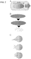

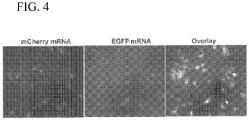
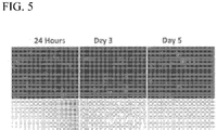
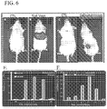


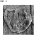
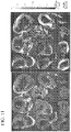


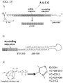

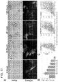
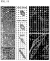
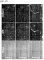
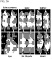
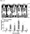


Classifications
-
- A—HUMAN NECESSITIES
- A61—MEDICAL OR VETERINARY SCIENCE; HYGIENE
- A61K—PREPARATIONS FOR MEDICAL, DENTAL OR TOILETRY PURPOSES
- A61K31/00—Medicinal preparations containing organic active ingredients
- A61K31/70—Carbohydrates; Sugars; Derivatives thereof
- A61K31/7088—Compounds having three or more nucleosides or nucleotides
- A61K31/7105—Natural ribonucleic acids, i.e. containing only riboses attached to adenine, guanine, cytosine or uracil and having 3'-5' phosphodiester links
-
- A—HUMAN NECESSITIES
- A61—MEDICAL OR VETERINARY SCIENCE; HYGIENE
- A61K—PREPARATIONS FOR MEDICAL, DENTAL OR TOILETRY PURPOSES
- A61K47/00—Medicinal preparations characterised by the non-active ingredients used, e.g. carriers or inert additives; Targeting or modifying agents chemically bound to the active ingredient
- A61K47/30—Macromolecular organic or inorganic compounds, e.g. inorganic polyphosphates
- A61K47/36—Polysaccharides; Derivatives thereof, e.g. gums, starch, alginate, dextrin, hyaluronic acid, chitosan, inulin, agar or pectin
-
- A—HUMAN NECESSITIES
- A61—MEDICAL OR VETERINARY SCIENCE; HYGIENE
- A61K—PREPARATIONS FOR MEDICAL, DENTAL OR TOILETRY PURPOSES
- A61K48/00—Medicinal preparations containing genetic material which is inserted into cells of the living body to treat genetic diseases; Gene therapy
-
- A—HUMAN NECESSITIES
- A61—MEDICAL OR VETERINARY SCIENCE; HYGIENE
- A61K—PREPARATIONS FOR MEDICAL, DENTAL OR TOILETRY PURPOSES
- A61K48/00—Medicinal preparations containing genetic material which is inserted into cells of the living body to treat genetic diseases; Gene therapy
- A61K48/0008—Medicinal preparations containing genetic material which is inserted into cells of the living body to treat genetic diseases; Gene therapy characterised by an aspect of the 'non-active' part of the composition delivered, e.g. wherein such 'non-active' part is not delivered simultaneously with the 'active' part of the composition
- A61K48/0025—Medicinal preparations containing genetic material which is inserted into cells of the living body to treat genetic diseases; Gene therapy characterised by an aspect of the 'non-active' part of the composition delivered, e.g. wherein such 'non-active' part is not delivered simultaneously with the 'active' part of the composition wherein the non-active part clearly interacts with the delivered nucleic acid
- A61K48/0041—Medicinal preparations containing genetic material which is inserted into cells of the living body to treat genetic diseases; Gene therapy characterised by an aspect of the 'non-active' part of the composition delivered, e.g. wherein such 'non-active' part is not delivered simultaneously with the 'active' part of the composition wherein the non-active part clearly interacts with the delivered nucleic acid the non-active part being polymeric
-
- A—HUMAN NECESSITIES
- A61—MEDICAL OR VETERINARY SCIENCE; HYGIENE
- A61K—PREPARATIONS FOR MEDICAL, DENTAL OR TOILETRY PURPOSES
- A61K48/00—Medicinal preparations containing genetic material which is inserted into cells of the living body to treat genetic diseases; Gene therapy
- A61K48/0075—Medicinal preparations containing genetic material which is inserted into cells of the living body to treat genetic diseases; Gene therapy characterised by an aspect of the delivery route, e.g. oral, subcutaneous
-
- A—HUMAN NECESSITIES
- A61—MEDICAL OR VETERINARY SCIENCE; HYGIENE
- A61K—PREPARATIONS FOR MEDICAL, DENTAL OR TOILETRY PURPOSES
- A61K9/00—Medicinal preparations characterised by special physical form
- A61K9/0012—Galenical forms characterised by the site of application
- A61K9/0019—Injectable compositions; Intramuscular, intravenous, arterial, subcutaneous administration; Compositions to be administered through the skin in an invasive manner
-
- A—HUMAN NECESSITIES
- A61—MEDICAL OR VETERINARY SCIENCE; HYGIENE
- A61K—PREPARATIONS FOR MEDICAL, DENTAL OR TOILETRY PURPOSES
- A61K9/00—Medicinal preparations characterised by special physical form
- A61K9/06—Ointments; Bases therefor; Other semi-solid forms, e.g. creams, sticks, gels
-
- A—HUMAN NECESSITIES
- A61—MEDICAL OR VETERINARY SCIENCE; HYGIENE
- A61K—PREPARATIONS FOR MEDICAL, DENTAL OR TOILETRY PURPOSES
- A61K9/00—Medicinal preparations characterised by special physical form
- A61K9/10—Dispersions; Emulsions
- A61K9/127—Liposomes
-
- A—HUMAN NECESSITIES
- A61—MEDICAL OR VETERINARY SCIENCE; HYGIENE
- A61K—PREPARATIONS FOR MEDICAL, DENTAL OR TOILETRY PURPOSES
- A61K9/00—Medicinal preparations characterised by special physical form
- A61K9/48—Preparations in capsules, e.g. of gelatin, of chocolate
-
- A—HUMAN NECESSITIES
- A61—MEDICAL OR VETERINARY SCIENCE; HYGIENE
- A61K—PREPARATIONS FOR MEDICAL, DENTAL OR TOILETRY PURPOSES
- A61K9/00—Medicinal preparations characterised by special physical form
- A61K9/48—Preparations in capsules, e.g. of gelatin, of chocolate
- A61K9/50—Microcapsules having a gas, liquid or semi-solid filling; Solid microparticles or pellets surrounded by a distinct coating layer, e.g. coated microspheres, coated drug crystals
- A61K9/51—Nanocapsules; Nanoparticles
- A61K9/5107—Excipients; Inactive ingredients
- A61K9/513—Organic macromolecular compounds; Dendrimers
- A61K9/5161—Polysaccharides, e.g. alginate, chitosan, cellulose derivatives; Cyclodextrin
-
- A—HUMAN NECESSITIES
- A61—MEDICAL OR VETERINARY SCIENCE; HYGIENE
- A61P—SPECIFIC THERAPEUTIC ACTIVITY OF CHEMICAL COMPOUNDS OR MEDICINAL PREPARATIONS
- A61P43/00—Drugs for specific purposes, not provided for in groups A61P1/00-A61P41/00
-
- A—HUMAN NECESSITIES
- A61—MEDICAL OR VETERINARY SCIENCE; HYGIENE
- A61P—SPECIFIC THERAPEUTIC ACTIVITY OF CHEMICAL COMPOUNDS OR MEDICINAL PREPARATIONS
- A61P9/00—Drugs for disorders of the cardiovascular system
-
- C—CHEMISTRY; METALLURGY
- C07—ORGANIC CHEMISTRY
- C07H—SUGARS; DERIVATIVES THEREOF; NUCLEOSIDES; NUCLEOTIDES; NUCLEIC ACIDS
- C07H21/00—Compounds containing two or more mononucleotide units having separate phosphate or polyphosphate groups linked by saccharide radicals of nucleoside groups, e.g. nucleic acids
- C07H21/02—Compounds containing two or more mononucleotide units having separate phosphate or polyphosphate groups linked by saccharide radicals of nucleoside groups, e.g. nucleic acids with ribosyl as saccharide radical
-
- C—CHEMISTRY; METALLURGY
- C07—ORGANIC CHEMISTRY
- C07H—SUGARS; DERIVATIVES THEREOF; NUCLEOSIDES; NUCLEOTIDES; NUCLEIC ACIDS
- C07H21/00—Compounds containing two or more mononucleotide units having separate phosphate or polyphosphate groups linked by saccharide radicals of nucleoside groups, e.g. nucleic acids
- C07H21/04—Compounds containing two or more mononucleotide units having separate phosphate or polyphosphate groups linked by saccharide radicals of nucleoside groups, e.g. nucleic acids with deoxyribosyl as saccharide radical
-
- C—CHEMISTRY; METALLURGY
- C12—BIOCHEMISTRY; BEER; SPIRITS; WINE; VINEGAR; MICROBIOLOGY; ENZYMOLOGY; MUTATION OR GENETIC ENGINEERING
- C12N—MICROORGANISMS OR ENZYMES; COMPOSITIONS THEREOF; PROPAGATING, PRESERVING, OR MAINTAINING MICROORGANISMS; MUTATION OR GENETIC ENGINEERING; CULTURE MEDIA
- C12N15/00—Mutation or genetic engineering; DNA or RNA concerning genetic engineering, vectors, e.g. plasmids, or their isolation, preparation or purification; Use of hosts therefor
- C12N15/09—Recombinant DNA-technology
- C12N15/11—DNA or RNA fragments; Modified forms thereof; Non-coding nucleic acids having a biological activity
- C12N15/113—Non-coding nucleic acids modulating the expression of genes, e.g. antisense oligonucleotides; Antisense DNA or RNA; Triplex- forming oligonucleotides; Catalytic nucleic acids, e.g. ribozymes; Nucleic acids used in co-suppression or gene silencing
-
- C—CHEMISTRY; METALLURGY
- C12—BIOCHEMISTRY; BEER; SPIRITS; WINE; VINEGAR; MICROBIOLOGY; ENZYMOLOGY; MUTATION OR GENETIC ENGINEERING
- C12N—MICROORGANISMS OR ENZYMES; COMPOSITIONS THEREOF; PROPAGATING, PRESERVING, OR MAINTAINING MICROORGANISMS; MUTATION OR GENETIC ENGINEERING; CULTURE MEDIA
- C12N15/00—Mutation or genetic engineering; DNA or RNA concerning genetic engineering, vectors, e.g. plasmids, or their isolation, preparation or purification; Use of hosts therefor
- C12N15/09—Recombinant DNA-technology
- C12N15/87—Introduction of foreign genetic material using processes not otherwise provided for, e.g. co-transformation
- C12N15/88—Introduction of foreign genetic material using processes not otherwise provided for, e.g. co-transformation using microencapsulation, e.g. using amphiphile liposome vesicle
-
- C—CHEMISTRY; METALLURGY
- C12—BIOCHEMISTRY; BEER; SPIRITS; WINE; VINEGAR; MICROBIOLOGY; ENZYMOLOGY; MUTATION OR GENETIC ENGINEERING
- C12N—MICROORGANISMS OR ENZYMES; COMPOSITIONS THEREOF; PROPAGATING, PRESERVING, OR MAINTAINING MICROORGANISMS; MUTATION OR GENETIC ENGINEERING; CULTURE MEDIA
- C12N15/00—Mutation or genetic engineering; DNA or RNA concerning genetic engineering, vectors, e.g. plasmids, or their isolation, preparation or purification; Use of hosts therefor
- C12N15/09—Recombinant DNA-technology
- C12N15/11—DNA or RNA fragments; Modified forms thereof; Non-coding nucleic acids having a biological activity
- C12N15/111—General methods applicable to biologically active non-coding nucleic acids
-
- C—CHEMISTRY; METALLURGY
- C12—BIOCHEMISTRY; BEER; SPIRITS; WINE; VINEGAR; MICROBIOLOGY; ENZYMOLOGY; MUTATION OR GENETIC ENGINEERING
- C12N—MICROORGANISMS OR ENZYMES; COMPOSITIONS THEREOF; PROPAGATING, PRESERVING, OR MAINTAINING MICROORGANISMS; MUTATION OR GENETIC ENGINEERING; CULTURE MEDIA
- C12N2320/00—Applications; Uses
- C12N2320/30—Special therapeutic applications
- C12N2320/32—Special delivery means, e.g. tissue-specific
Definitions
- a heart attack also called a myocardial infarction, occurs when a part of the heart muscle does not receive enough blood flow. The more time that passes without treatment to restore blood flow, the greater the damage to the heart muscle. Every year, about 735,000 Americans have a heart attack, including about 210,000 heart attacks that happen to people who have already had a first heart attack ( Mozaffarian et al., Circulation, 131(4):e29-322 (2015 )).
- RNA molecules that increase the length of time that the modified RNA molecules exist in cells.
- This disclosure further provides materials and methods for particle-mediated delivery of one or more biologics (e.g., RNA, modified RNA, and/or microvesicles) to a tissue (e.g., heart tissue).
- a tissue e.g., heart tissue.
- Microvesicles can represent manufactured particles or naturally occurring structures, such as exosomes.
- particles e.g., alginate gels
- RNAs e.g., alginate gels
- an alginate gel can be used to deliver mRNA to a heart tissue where the mRNA can be translated into a functional polypeptide.
- Particle-mediated delivery of RNA can induce robust and sustainable RNA expression.
- particles can be used be designed to control temporal and/or spatial delivery of one or more encapsulated molecules (e.g., biologics).
- composition that generally include a particulate substrate and an mRNA attached to the particulate substrate.
- the mRNA includes at least one modification to inhibit degradation of the mRNA when the mRNA is in cytosol of a cell.
- the mRNA also encodes at least one therapeutic polypeptide.
- the mRNA modification comprises a pseudoknot, an RNA stability element, or an artificial 3' stem loop.
- the particulate substrate can include a chemical modification of its surface.
- the particulate substrate can include a nanoparticle, a plurality of nanoparticles, or a microparticle.
- the therapeutic polypeptide can include an immunoglobulin heavy chain or an immunoglobulin light chain.
- one aspect of this document features a method for improving cardiac function.
- the method includes, or consists essentially of, administering a particle encapsulating an mRNA encoding a polypeptide useful for regenerating cardiac function and/or tissue to a mammal, thereby improving cardiac function of the mammal.
- the polypeptide can be NAP -2, TGF-a, ErBb3, VEGF, IGF-1, FGF-2, PDGF, IL-2, CD19, CD20, and/or CD80/86.
- the mammal can be a human.
- the human can have undergone percutaneous coronary intervention for ST-elevation myocardial infarction.
- the administering can be an arterial administration.
- the particle can include alginate.
- the alginate can be in the form of an alginate gel.
- the alginate gel can include a calcium salt.
- the alginate gel including a calcium salt can have a ratio of alginate to calcium salt that can be from about 2:1 to about 10:1.
- the particle can be from about 5 ⁇ m to about 10 ⁇ m in diameter.
- the particle can be a biphasic particle.
- the biphasic particle can be a polarized particle.
- the biphasic particle can have a tail.
- the method can include administering the composition during a percutaneous coronary intervention.
- the particle can include a scaffold protein (e.g., collagen I, collagen II, collagen III, collagen IV, fibrin, and/or gelatin).
- the particle can encapsulate a polypeptide (e.g., an antibody having the ability to neutralize tumor necrosis factor activity, an antibody having the ability to neutralize mitochondrial complex-1 activity, or a resolvin-D1 agonist).
- the particle can encapsulate a lipopolysaccharide.
- the particle can encapsulate a microvesicle and/or exosome.
- this document features a method for improving cardiac function in a mammal.
- the method includes, or consists essentially of, administering to a mammal a particle encapsulating an inhibitory RNA having the ability to reduce expression of a polypeptide selected from the group consisting of eotaxin-3, cathepsin-S, DK -1, follistatin, ST-2, GRO-a, IL-21, NOV, transferrin, TIMP-2, TNFaRI, TNFaRII, angiostatin, CCL25, ANGPTL4, and MMP-3, thereby improving cardiac function of the mammal.
- the mammal can be a human.
- the human can have undergone percutaneous coronary intervention for ST-elevation myocardial infarction.
- the administering can be an arterial administration.
- the particle can include alginate.
- the alginate can be in the form of an alginate gel.
- the alginate gel can include a calcium salt.
- the alginate gel including a calcium salt can have a ratio of alginate to calcium salt that can be from about 2:1 to about 10:1.
- the particle can be from about 5 ⁇ m to about 10 ⁇ m in diameter.
- the particle can be a biphasic particle.
- the biphasic particle can be a polarized particle.
- the biphasic particle can include a tail.
- the method can include administering the composition during a percutaneous coronary intervention.
- the particle can include a scaffold protein (e.g., collagen I, collagen II, collagen III, collagen IV, fibrin, and/or gelatin).
- the particle can encapsulate a polypeptide (e.g., an antibody having the ability to neutralize tumor necrosis factor activity, an antibody having the ability to neutralize mitochondrial complex-1 activity, or a resolvin-D1 agonist).
- the particle can encapsulate a lipopolysaccharide.
- the particle can encapsulate a microvesicle and/or exosome.
- This disclosure relates to materials and methods for particle-mediated delivery of biologics (e.g., RNA and/or microvesicles) to tissue (e.g., heart tissue).
- biologics e.g., RNA and/or microvesicles
- tissue e.g., heart tissue.
- This disclosure further describes modifications to RNA that increase the length of time that the modified mRNA molecules exist in cells and, therefore, the length of time that the modified mRNA molecules can be translated into produce polypeptides encoded by the modified mRNA.
- RNA and/or microvesicles e.g., RNA and/or microvesicles
- a particle e.g., an alginate gel
- encapsulate and variations thereof is used broadly to refer to any method by which a biologic is housed within a particle or bound, directly or indirectly, to the outer surface of a particle.
- the particle can be nanoparticulate or microparticulate in scale.
- the term "encapsulate” includes, but does not require, that the particle completely surround the material being encapsulated. Rather, a material is encapsulated if it is merely captured to any degree within the dimensions of the particle, including any surface modifications. Surface modifications of the particle can facilitate indirect binding of the encapsulated material to the particle.
- macroencapsulation can be used to deliver a biologic by providing a protective vehicle by which to transmit the biologics either by direct injection or through a blood vessel into a target tissue.
- one or more particles e.g., alginate gels
- one or more biologics e.g., RNAs and/or microvesicles
- a mammal experiencing a major adverse cardiac event e.g., acute myocardial infarction
- a mammal at risk of experiencing a major adverse cardiac event e.g., patients who underwent percutaneous intra intervention (PCI) for ST-elevation myocardial infarction (STEMI)
- PCI percutaneous intra intervention
- ST-elevation myocardial infarction ST-elevation myocardial infarction
- one or more particles e.g., an alginate gels
- one or more biologics e.g., RNAs or microvesicles
- a particle e.g., an alginate gel
- a particle can store mRNA within its interior, and release (e.g., deliver) the mRNA to a tissue (e.g., heart tissue) where the mRNA is expressed as a functional protein.
- one or more particles containing one or more mRNAs can be used to increase expression of polypeptides useful for regenerating cardiac function and/or tissue (e.g., POU homeodomain proteins (such as Oct-4), NK2 homeobox proteins (e.g., NKX2 proteins), myocyte enhancing factors (e.g., MEF2), GATA binding proteins (e.g., GATA1, GATA2, GATA3, GATA4, GATA5, and GATA6), T-box transcription factors (e.g., TBX1, TBX2, TBX3, TBX4, TBX5, TBX6, TBX10, TBX15, TBX18, TBX19, TBX20, TBX21, and TBX22), mesoderm posterior proteins (e.g., MESP1 and MESP2), neutrophil-activating proteins (e.g., NAP-2 and NAP-3), transforming growth factors (e.g., TGF- ⁇ and TGF- ⁇ ), erythroblasts,
- a particle e.g., an alginate gel
- a particle described herein can store inhibitory RNA within its interior, and release (e.g., deliver) the inhibitory RNA to a tissue (e.g., heart tissue) where the inhibitory RNA inhibits or reduces expression of a protein.
- one or more particles containing one or more inhibitory RNAs can be used to decrease expression of one or more of the following polypeptides: eotaxin-3, cathepsin-S, Dickopf-1 (DK -1), follistatin, suppression of tumorigenicity-2 (ST-2), GRO- ⁇ , interleukin-21 (IL-21), nephroblastoma overexpressed (NOV), transferrin, tissue inhibitor of metallopeptidase-2 (TIMP-2), tumor necrosis factor receptor- 1 and -2 (TNFaRI and II), angiostatin, chemokine ligand-25 (CCL25), angiopoietin like-4 (ANGPTL4), and matrix metalloproteinase-3 (MMP-3).
- polypeptides eotaxin-3, cathepsin-S, Dickopf-1 (DK -1), follistatin, suppression of tumorigenicity-2 (ST-2), GRO- ⁇ , interleukin-21 (IL-21),
- the particle is a biphasic particle comprising an alginate gel. To the extent that other particles are described herein they are included solely for reference purposes.
- a particle described herein can be used for particle-mediated delivery of one or more molecules (e.g., biologics including RNA or microvesicles) to a mammal.
- a particle that can be used to encapsulate one or more biologics is non-toxic, biocompatible, non-immunogenic, and/or biodegradable.
- a particle that can be used to encapsulate one or more biologics as described herein can include one or more polysaccharides.
- polysaccharides that can be used in a particle that can be used to encapsulate one or more molecules (e.g., biologics) as described herein include, for example, guluronate, mannuronate, guluronate-mannuronate blocks, and combinations thereof such as alginate.
- a particle that can be used to encapsulate one or more biologics as described herein can include alginate.
- An alginate in a particle that can be used to encapsulate one or more biologics as described herein can be from any appropriate source (e.g., seaweeds such as those in the genera Phaeophyceae , Rhodophyceae, Chlorophyceae, Macrocystis, and Laminaria , or bacteria such as those in the genera Pseudomonas and Azotobacter ) .
- An alginate in a particle that can be used to encapsulate one or more biologics as described herein can be an alginate salt (e.g., sodium alginate, potassium alginate, and/or calcium alginate) or alginic acid.
- an alginate in a particle that can be used to encapsulate one or more biologics as described herein can be sodium alginate.
- An alginate in a particle that can be used to encapsulate one or more biologics as described herein can be in the form of a gel, a liquid, and/or a particle (e.g., a nanoparticle, a microparticle, or a macroparticle).
- an alginate in a particle that can be used to encapsulate one or more biologics as described herein can be an alginate gel.
- a particle described herein can be a liquid at the time of administration and can form a gel (e.g., can polymerize) at the administration site and/or the target site.
- An alginate gel can include a calcium solution (e.g., a calcium salt solution or another appropriate positively charged ionic solution).
- a calcium solution e.g., a calcium salt solution or another appropriate positively charged ionic solution.
- Examples of calcium salts that can be used in a calcium solution to form an alginate gel that can be used to encapsulate one or more biologics as described herein include, without limitation, calcium chloride, magnesium chloride, potassium chloride, and iron-based solutions.
- the ratio of alginate to calcium salt can be used to control the viscosity of the alginate gel that can be used to encapsulate one or more biologics as described herein.
- the amount of alginate can be from about 0.05 percent to about 1.0 percent of the amount of calcium salt.
- the ratio of alginate to calcium salt can be can be from about 2:1 to about 10:1 (e.g., from about 2:1 to about 9:1, from about 2:1 to about 8:1, from about 2:1 to about 7:1, from about 2:1 to about 6:1, from about 2:1 to about 5:1, from about 3:1 to about 10:1, from about 4:1 to about 10:1, from about 5:1 to about 10:1, from about 6:1 to about 10:1, from about 7:1 to about 10:1, or from about 8:1 to about 10:1).
- Examples of appropriate alginate/calcium ratios include, without limitation, those set forth in Table 1.
- An alginate gel encapsulating a biologic (e.g., mRNA) described herein can be made by any appropriate method.
- an alginate gel can be made by hydrolyzing an alginate salt (e.g., sodium alginate) using a soluble calcium salt solution (e.g., calcium chloride) as a crosslinking agent.
- alginate salt e.g., sodium alginate
- a soluble calcium salt solution e.g., calcium chloride
- Table 1 Examples alginate/calcium concentrations. alginate concentration calcium concentration alginate:calcium 0.2% 0.1% 2:1 1% 0.1% 10:1 2% 0.5% 4:1 2% 0.25% 8:1
- a particle that can be used to encapsulate one or more biologics as described herein can be in the form of a liposome, an aggregate (e.g., a nano-aggregate), a capsule (e.g., a nanocapsule), a sphere (e.g., a nanosphere), a polymersome, or a micelle.
- a particle that can be used to encapsulate one or more biologics as described herein that is an alginate gel can be in the form of polymerized sphere.
- a particle that can be used to encapsulate one or more biologics as described herein can be a liposome.
- liposomes examples include, without limitation, a multilamellar vesicle (MLV), a small unilamellar liposome vesicle (SUV), a large unilamellar vesicle (LUV), a giant unilamellar vesicle (GUV), a multivesicular vesicles (MVV), or a cochleate vesicle.
- MLV multilamellar vesicle
- SUV small unilamellar liposome vesicle
- LUV large unilamellar vesicle
- GMV giant unilamellar vesicle
- MVV multivesicular vesicles
- cochleate vesicle a cochleate vesicle.
- a liposome can be composed of phospholipids, cholesterols, lipoproteins, fats, fatty acids, waxes, sterols, monoglycerides, diglycer
- a liposome is composed of phospholipids such as phosphatidic acid (phosphatidate; PA), phosphatidylethanolamine (cephalin; PE), phosphatidylcholine (lecithin; PC), phosphatidylserine (PS), phosphoinositides (e.g., phosphatidylinositol (PI), phosphatidylinositol phosphate (PIP), phosphatidylinositol bisphosphate (PIP2), and phosphatidylinositol triphosphate (PIP3)), ceramide phosphorylcholine (sphingomyelin; SPH), ceramide phosphorylethanolamine (sphingomyelin; Cer-PE), ceramide phosphoryllipid, or any combination thereof.
- phospholipids such as phosphatidic acid (phosphatidate; PA), phosphatidylethanolamine (cephalin; PE), phosphatidylcholine (lecithin
- liposomes examples include, without limitation, those set forth in Table 2.
- a liposome encapsulating a biologic (e.g., mRNA) described herein can be made by any appropriate method.
- a liposome can be made by sonicating a dispersion of amphipatic lipids, such as phospholipids, in water. Table 2. Examples of liposome components.
- liposome component MW (kDa) Concentration Charged Poly(lactide-co -glycolide) (PLGA) about 46 3-300 mg 1,2-Dioleoyl- sn -glycero-3-phosphoethanolamine (DOPE) about 744 20 ⁇ g - 1 mg N-[1-(2,3-dioleoyloxy)propyl]-N,N,N-trimethylammonium methylsulfate (DOTAP) about 774 20 ⁇ g - 3 mg L- ⁇ -Phosphatidylethanolamine, dioleoyl about 744 20 ⁇ g - 3 mg 1, 2-Distearoyl-sn-glycero-3-phosphoethanolamine-Poly(ethylene glycol) (DSPE- PEG) about 770 20 ⁇ g - 3 mg Poly(vinyl-alcohol) (PVA) about 78 1-5% per total volume
- DOPE 1,2-Dioleoyl- sn -gly
- a particle that can be used to encapsulate one or more biologics as described herein can be any size suitable for the selected delivery method.
- a particle that can be used to encapsulate one or more biologics as described herein can be from about 0.3 ⁇ m to about 12 ⁇ m (e.g., from about 0.5 ⁇ m to about 11.5 ⁇ m, from about 1 ⁇ m to about 11 ⁇ m, from about 2 ⁇ m to about 10.5 ⁇ m, or from about 4 ⁇ m to about 10 ⁇ m) in diameter (or as measure across the longest dimension).
- a particle that can be used to encapsulate one or more biologics as described herein can be from about 4.5 ⁇ m to about 7.5 ⁇ m in diameter (or as measure across the longest dimension).
- a particle (e.g., an alginate gel) encapsulating one or more biologics described herein also can include one or more additional molecules in order to achieve a desired property.
- a particle encapsulating one or more biologics described herein can include one or more scaffold proteins.
- scaffold proteins include, for example, matrix proteins (e.g., collagen (e.g., collagen I/II/III/IV)), basement membrane proteins, structural proteins, gelatin, and/or fibrin) can be incorporated into a particle to provide the particle with sustained release properties.
- an alginate gel encapsulating one or more biologics described herein can include alginate and gelatin, alginate and collagen, alginate and fibrin, or any other appropriate combination of alginate with one or more natural basement membrane proteins.
- stimuli-sensitive molecules can be incorporated into a particle to provide the particle with drug release properties under specific stimuli (e.g., pH-sensitive particles and osmolarity/osmolality sensitive particles).
- dioleoylphosphatidylethanolamine can be incorporated into a particle to provide a pH-sensitive particle that maintains stability at physiological pH (about pH 7.4), but destabilize under acidic conditions (e.g., about pH 3.5 to about pH 9) leading to the release of the encapsulated RNA in acidic environments.
- DOPE dioleoylphosphatidylethanolamine
- a particle encapsulating one or more biologics described herein can be an amorphous particle having two or more distinct physical states.
- an amorphous particle can be administered as a liquid and form a gel upon arriving at a target tissue.
- a particle (e.g., an alginate gel) encapsulating one or more biologics described herein can be a biphasic particle (sometimes referred to as a Janus particle) having two or more distinct physical properties (e.g., surface chemistry) occurring on different portions of the particle surface.
- a biphasic particle can have two or more portions that differ in hydrophilic/hydrophobic properties, polarization, solubility properties, volume percent or density of the particle composition, the presence/absence of an additional molecule (e.g., a compound providing sustained release properties), and/or the presence/absence of a tail.
- a biphasic particle can be a spherical particle having hydrophilic portions and hydrophobic portions.
- a biphasic particle can be designed to have a hydrophilic core and a hydrophilic coating.
- a biphasic particle can be designed to have a hydrophilic core and a hydrophilic coating.
- a biphasic particle can be a spherical particle having differential density.
- a biphasic particle can be designed to have increased volume percent of an encapsulated biologic at one end of the particle.
- a biphasic particle can be a spherical particle having a coating.
- a biphasic particle can be designed to have a porous coating.
- a biphasic particle can be a spherical particle having differential solubility.
- a biphasic particle can be designed to have a first side that readily dissolves in certain physiological conditions and a second side that dissolves slowly in the same physiological conditions.
- a biphasic particle can be a spherical particle having a tail extending from one portion of the particle surface.
- a biphasic particle can be designed to guide particles in the blood flow.
- a biphasic particle can be designed to have a first (e.g., leading) side that readily dissolves in certain physiological conditions and a second side that dissolves slowly in the same physiological conditions and includes a tail to guide particles in the blood flow.
- a biphasic particle encapsulating one or more biologics described herein can be made using any appropriate method.
- a biphasic particle can be made using stabilizer technology (e.g., including dry water approaches).
- stabilizer technology e.g., including dry water approaches.
- two or more separate soft liquid cores e.g., two liquids having different hydrophobicity such as a hydrophobic entity and hydrophilic entity, or two liquids having different solubility such as water and an oil
- a biphasic particle can be made using melt combination and fusion technology.
- two or more separate streams can be merged (e.g., injected into a single stream) to form a biphasic particle.
- a biphasic particle can be made using 3D-printer technology.
- two or more coatings e.g., two wax coatings having different properties
- a particulate internal structural element e.g., a particle of polystyrene foam such as STYROFOAM TM
- the particulate internal structural element can be removed (e.g., by acetone elimination) and replaced with one or more biologics.
- a biphasic particle can be made using microfluidics technology.
- two or more liquid flows e.g., two liquids having different properties
- a biphasic particle can be made using protection and de-protection technology.
- a protection particle having a desired surface chemistry can be adsorbed onto a main particle; de-protection can yield the original surface of the main particle, which can be chemically modified.
- a biphasic particle can be made use other appropriate technologies.
- other appropriate methods can include, without limitation, interfacial emulsification, acorn and dumbbell shaped formation, use of magnetic fields to shape and manipulate particles during fabrication, and direct surface coating deposition.
- differential density can be achieved using organogels with varied density.
- differential porosity can be achieved using salts at a particular surface of the particle.
- differential solubility can be achieved using salts and/or using two or more different polymers (e.g., PEGs) having different solubilities.
- various tails can be achieved using waxy components, chemically modifying (e.g., with long-chain reactants), and/or magnetizing a particle.
- Exemplary methods for making a biphasic particle encapsulating one or more biologics described herein are shown in FIG. 1 .
- a particle encapsulating one or more biologics described herein can be used to deliver the encapsulated biologics to a target tissue (e.g., heart tissue).
- Mechanisms of targeting a particle described herein can include using an adhesive particle, a targeting moiety, particle size, capillary leakage in the setting of injury, capillary leakage in the setting of oncogenic neovasculogenesis, and/or direct injection (e.g., via an edema forming needle (e.g., a nitinol (nickel-titanium) helical needle) designed with graded side holes and no end hole (see, e.g., FIG. 14 )).
- an edema forming needle e.g., a nitinol (nickel-titanium) helical needle
- Capillary leakage refers to any physical, chemical, or pharmacological means by which one can enhance capillary porosity or leakage in order to augment biologics delivery. This can be achieved either by administering a factor (e.g., chemical and/or pharmacologic) that enhances capillary porosity. Alternatively, capillary leakage may be achieved by placing a biologics delivery needle that is designed to mimic edema into the target tissue.
- a factor e.g., chemical and/or pharmacologic
- an adhesive moiety can be conjugated to a particle described herein to retain an adhesive particle at the administration site.
- an adhesive particle described herein can be administered (e.g., injected) directly to a target tissue (e.g., cardiac infarct bed).
- a target tissue e.g., cardiac infarct bed
- adhesive moieties include, without limitation, PEG, positive charge, and self-assembly within tissue.
- a targeting moiety can be conjugated to a particle described herein to direct delivery of a particle to a target tissue (e.g., cardiac infarct bed).
- targeting moieties include, without limitation, antigens, tissue targeting peptides, small molecules, and cell surface molecules.
- an antibody can be used to target surface proteins on a cell.
- the size of the particle can be used to direct delivery of a particle to a target tissue (e.g., cardiac infarct bed).
- a target tissue e.g., cardiac infarct bed.
- Human capillaries measure about 5 ⁇ m to 10 ⁇ m in diameter.
- a particle described herein having a diameter of from about 0.3 ⁇ m to about 12 ⁇ m can enter a capillary via the bloodstream, but be limited from exiting the capillary, where the biologics and/or an expressed polypeptide can diffuse into the capillary bed of a tissue (e.g., heart, dermal, lung, solid tumor, brain, bone, ligament, connective tissue structures, kidney, liver, subcutaneous, and vascular tissue).
- Exemplary methods for delivery of one or more biologics using a biphasic particle described herein are shown in FIG. 2 .
- a particle (e.g., an alginate gel) encapsulating one or more biologics described herein can be used to deliver the encapsulated biologic to a target tissue (e.g., cardiac infarct bed) in a temporally and/or spatially specific manner.
- a biphasic particle encapsulating one or more biologics as described herein can be designed to confer a specific orientation of the particle within a blood vessel, to direct the biphasic particle into a capillary, and/or to confer specific release conditions of the encapsulated biologics.
- a biphasic particle has hydrophilic portions and hydrophobic portions on the particle surface
- delivery of one or more encapsulated biologics can be controlled by the solvent present at the delivery site and/or the target site.
- the differential density can orient the particle in the blood flow to direct the particle in an appropriate orientation for delivery of one or more encapsulated biologics.
- a dense side of a biphasic particle can lead the particle in the direction of blood flow within a blood vessel.
- delivery of one or more encapsulated biologics can be controlled by the differential porosity.
- different amounts of porosity can help deliver particles within the blood vessel and alter the pattern of delivery.
- the differential solubility can orient the particle in the blood flow to direct the particle in an appropriate orientation for delivery of one or more encapsulated biologics.
- the tail can orient the particle within a blood vessel to direct the particle into a capillary.
- a biphasic particle can be designed to have a tail to orient the particle within a blood vessel and direct the particle into a blood vessel (e.g., a capillary) where the encapsulated biologic can be delivered.
- the particle encapsulates a biologic that is an mRNA that encodes NAP-2, TGF- ⁇ , ErBb3, VEGF, IGF-1, FGF-2, PDGF, IL-2, CD19, CD20, or CD80/86.
- a biologic that is an mRNA that encodes NAP-2, TGF- ⁇ , ErBb3, VEGF, IGF-1, FGF-2, PDGF, IL-2, CD19, CD20, or CD80/86.
- any appropriate biologic can be encapsulated within a particle described herein for delivery to a tissue.
- biologics that can be encapsulated within a particle described herein include, without limitation, nucleotides, polypeptides, small molecules, microvesicles, exosomes, extracellular vesicles, engineered cells, or combinations thereof.
- a biologic can be a purified biologic (e.g., purified microvesicles or exosomes).
- a particle (e.g., an alginate gel) described herein can be used to encapsulate one or more polypeptides.
- a particle described herein can be used to encapsulate one or more polypeptides useful to treat a mammal experiencing a major adverse cardiac event (e.g., acute myocardial infarction) and/or a mammal at risk of experiencing a major adverse cardiac event (e.g., patients who underwent PCI for STEMI).
- a particle encapsulating a biologic described herein can encapsulate one or more polypeptides useful for regenerating cardiac function and/or tissue or a nucleotide that encodes such a polypeptide.
- polypeptides useful for regenerating cardiac function and/or tissue include, without limitation, antibodies having the ability to neutralize tumor necrosis factor (TNF; e.g., TNF- ⁇ ) activity, antibodies having the ability to neutralize mitochondrial complex-1 activity, and resolvin-D1 agonists.
- TNF tumor necrosis factor
- a particle e.g., an alginate gel
- a particle can be used to encapsulate one or more nucleotides.
- nucleotides that can be encapsulated within a particle include, without limitation, mRNAs, inhibitory RNAs (e.g., antisense RNAs, microRNAs (miRNAs), small interfering RNAs (siRNAs), short hairpin RNAs (shRNAs), and agomiRs), antagomiRs, modified mRNAs, loop-engineered modified mRNAs (see, e.g., FIG. 15 ), or combinations thereof.
- inhibitory RNAs e.g., antisense RNAs, microRNAs (miRNAs), small interfering RNAs (siRNAs), short hairpin RNAs (shRNAs), and agomiRs
- antagomiRs e.g., modified mRNAs, loop-engineered modified mRNAs (see
- a particle e.g., an alginate gel
- a particle can be used to encapsulate one or more mRNAs useful to treat a mammal experiencing a major adverse cardiac event (e.g., acute myocardial infarction) and/or a mammal at risk of experiencing a major adverse cardiac event (e.g., patients who underwent PCI for STEMI).
- a particle encapsulating a biologic described herein can encapsulate one or more mRNAs encoding a polypeptide useful for regenerating cardiac function and/or tissue can be encapsulated within a particle described herein.
- a human Nap-2 polypeptide can have the amino acid sequence set forth in, for example, National Center for Biotechnology Information (NCBI) Accession No. NP_002695.1 (GI No. 5473) and can be encoded by the nucleic acid sequence set forth in NCBI Accession No.
- a human TGF- ⁇ polypeptide can have the amino acid sequence set forth in NCBI Accession No. NP_003227.1 (GI No. 7039) and can be encoded by the nucleic acid sequence set forth in NCBI Accession No. NM_003236 (GI No. 7039).
- a human ErBb3 polypeptide can have the amino acid sequence set forth in NCBI Accession No. NP_001005915.1 or NP_001973.2 (GI No. 2065) and can be encoded by the nucleic acid sequence set forth in NCBI Accession No. NM_001005915.1 or NM_001982.3 (GI No. 2065).
- a human VEGF can have the amino acids set forth in NCBI Accession Nos. AAA35789.1 (GI: 181971), CAA44447.1 (GI: 37659), AAA36804.1 (GI: 340215), or AAK95847.1 (GI: 15422109), and can be encoded by the nucleic acid sequence set forth in NCBI Accession No. AH001553.1 (GI: 340214).
- a human IGF-1 can have the amino acid sequence set forth in NCBI Accession No. CAA01954.1 (GI: 1247519) and can be encoded by the nucleic acid sequence set forth in NCBI Accession No. A29117.1 (GI: 1247518).
- a human FGF-2 can have the amino acid sequence set forth in NCBI Accession No. NP_001997.5 (GI: 153285461) and can be encoded by the nucleic acid sequence set forth in NCBI Accession No. NM_002006.4 (GI: 153285460).
- a human PDGF can have the amino acid sequence set forth in NCBI Accession No. AAA60552.1 (GI: 338209) and can be encoded by the nucleic acid sequence set forth in NCBI Accession No. AH002986.1 (GI: 338208).
- a human IL-2 can have the amino acid sequence set forth in NCBI Accession No.
- AAB46883.1 (GI: 1836111) and can be encoded by the nucleic acid sequence set forth in NCBI Accession No. S77834.1 (GI: 999000).
- a human CD19 can have the amino acid sequence set forth in NCBI Accession No. AAA69966.1 (GI: 901823) and can be encoded by the nucleic acid sequence set forth in NCBI Accession No. M84371.1 (GI: 901822).
- a human CD20 can have the amino acid sequence set forth in NCBI Accession No. CBG76695.1 (GI: 285310157) and can be encoded by the nucleic acid sequence set forth in NCBI Accession No. AH003353.1 (GI: 1199857).
- a human CD80 can have the amino acid sequence set forth in NCBI Accession No. NP_005182.1 (GI: 4885123) and can be encoded by the nucleic acid sequence set forth in NCBI Accession No. NM_005191.3 (GI: 113722122), and a human CD86 can have the amino acid sequence set forth in NCBI Accession No. AAB03814.1 (GI: 439839) and can be encoded by the nucleic acid sequence set forth in NCBI Accession No. CR541844.1 (GI: 49456642).
- a polypeptide that can be useful for regenerating cardiac function and/or tissue can be an antibody directed against TNF- ⁇ , mitochondrial complex-1, or resolvin-D1.
- a particle encapsulating a biologic described herein can encapsulate one or more mRNAs encoding NAP-2 and/or TGF- ⁇ .
- a particle e.g., an alginate gel
- a particle can be used to encapsulate one or more inhibitory RNAs useful to treat a mammal experiencing a major adverse cardiac event (e.g., acute myocardial infarction) and/or a mammal at risk of experiencing a major adverse cardiac event (e.g., patients who underwent PCI for STEMI).
- a major adverse cardiac event e.g., acute myocardial infarction
- a mammal at risk of experiencing a major adverse cardiac event e.g., patients who underwent PCI for STEMI.
- a particle described herein can encapsulate one or more inhibitory RNAs inhibiting and/or reducing expression of one or more of the following polypeptides: eotaxin-3, cathepsin-S, DK -1, follistatin, ST-2, GRO- ⁇ , IL-21, NOV, transferrin, TIMP-2, TNFaRI, TNFaRII, angiostatin, CCL25, ANGPTL4, MMP-3, and polypeptides described in WO 2015/034897 .
- a human eotaxin-3 polypeptide can have an amino acid sequence set forth in, for example, NCBI Accession No: No. NP_006063.1 (GI No.
- a human cathepsin-S can have the amino acid sequence set forth in NCBI Accession No. NP_004070.3 (GI No. 1520) and can be encoded by the nucleic acid sequence set forth in NCBI Accession No. NM_004079.4 (GI No. 1520).
- a human DK -1 can have the amino acid sequence set forth in NCBI Accession No. NP_036374.1 (GI No. 22943) and can be encoded by the nucleic acid sequence set forth in NCBI Accession No. NM_012242 (GI No. 22943).
- a human follistatin can have then amino acid sequence set forth in NCBI Accession No. NP_037541.1 (GI No. 10468) and can be encoded by the nucleic acid sequence set forth in NCBI Accession No. NM_013409.2 (GI No. 10468).
- a human ST-2 can have the amino acid sequence set forth in NCBI Accession No. BAA02233 (GI No. 6761) and can be encoded by the nucleic acid sequence set forth in NCBI Accession No D12763.1 (GI No 6761).
- a human GRO- ⁇ polypeptide can have the amino acid sequence set forth in NCBI Accession No. NP_001502.1 (GI No.
- a human IL-21 can have the amino acid sequence set forth in NCBI Accession No. NP_068575.1 (GI No. 59067) and can be encoded by the nucleic acid sequence set forth in NCBI Accession No. NM_021803 (GI No. 59067).
- a human NOV polypeptide can have the amino acid sequence set forth in NCBI Accession No. NP_002505.1 (GI No. 4856) and can be encoded by the nucleic acid sequence set forth in NCBI Accession No. NM_002514 (GI No. 4856).
- a human transferrin polypeptide can have the amino acid sequence set forth in NCBI Accession No. NP_001054.1 (GI No. 7018) and can be encoded by the nucleic acid sequence set forth in NCBI Accession No. NM_001063.3 (GI No. 7018).
- a human TIMP-2 polypeptide can have the amino acid sequence set forth in NCBI Accession No. NP_003246.1 (GI No. 7077) and can be encoded by the nucleic acid sequence set forth in NCBI Accession No. NM_003255.4 (GI No. 7077).
- a human TNFaRI polypeptide can have the amino acid sequence set forth in NCBI Accession No. NP_001056.1 (GI No.
- a human TNFaRII polypeptide can have the amino acid sequence set forth in NCBI Accession No. NP_001057.1 (GI No. 7133) and can be encoded by the nucleic acid sequence set forth in NCBI Accession No. NM_001066 (GI No. 7133).
- a human angiostatin polypeptide can have the amino acid sequence set forth in NCBI Accession No. NP_000292 (GI No. 5340) and can be encoded by the nucleic acid sequence set forth in NCBI Accession No. NM_000301 (GI No. 5340).
- a human CCL25 polypeptide can have the amino acid sequence set forth in NCBI Accession No. NP_005615.2 (GI No. 6370) and can be encoded by the nucleic acid sequence set forth in NCBI Accession No. NM_005624 (GI No. 6370).
- a human ANGPTL4 polypeptide can have the amino acid sequence set forth in NCBI Accession No. NP_001034756.1 or NP_647475.1 (GI No. 51129) and can be encoded by the nucleic acid sequence set forth in NCBI Accession No. NM_001039667.1 or NM_139314.1 (GI No. 51129).
- a human MMP-3 polypeptide can have the amino acid sequence set forth in NCBI Accession No. NP_002413.1 (GI No. 4314) and can be encoded by the nucleic acid sequence set forth in NCBI Accession No. NM_002422 (GI No. 4314).
- a particle e.g., an alginate gel
- a particle described herein can be used to encapsulate one or more nucleotides that modulate (e.g., mimic or inhibit) microRNAs involved in cardiac regenerative potency.
- a particle described herein can be used to encapsulate one or more agomiRs that mimic one or more miRNAs to augment cardiac regenerative potency.
- a particle described herein can be used to encapsulate one or more antagomiRs that inhibit one or more miRNAs to augment cardiac regenerative potency.
- miRNAs involved in cardiac regenerative potency include, without limitation, miR-127, miR-708, miR-22-3p, miR-411, miR-27a, miR-29a, miR-148a, miR-199a, miR-143, miR-21, miR-23a-5p, miR-23a, miR-146b-5p, miR-146b, miR-146b-3p, miR-2682-3p, miR-2682, miR-4443, miR-4443, miR-4443, miR-4443, miR-4521, miR-4521, miR-2682-5p,miR-2682, miR-137.miR-137, miR-549.miR-549, miR-335-3p, miR-335, miR-181c-5p, miR-181c, miR-224-5p, miR-224, miR-3928, miR-3928, miR-324-5p, miR-324,
- Nucleotides encapsulated within a particle described herein can be modified nucleotides.
- nucleotides can be modified for increased stability.
- one or more uracil residues of an RNA described herein can be replace with a modified uracil residue.
- modified uracil residues include, without limitation, pseudouridine ( ⁇ ), dihydrouridine (D), and dideoxyuracil.
- An mRNA may be modified to form a biofunctionalized microencapsulated modified mRNA (M 3 RNA), which are described in more detail below.
- a particle (e.g., an alginate gel) described herein can be used to encapsulate other molecules in addition to or in place of a biologic.
- a particle (e.g., an alginate gel) described herein can be used to encapsulate one or more small molecules.
- a particle described herein can be used to encapsulate one or more small molecules useful to treat a mammal experiencing a major adverse cardiac event (e.g., acute myocardial infarction) and/or a mammal at risk of experiencing a major adverse cardiac event (e.g., patients who underwent PCI for STEMI).
- a particle described herein can encapsulate one or more small molecules useful for regenerating cardiac function and/or tissue.
- small molecules useful for regenerating cardiac function and/or tissue include, without limitation, lipopolysaccharides, tumor necrosis factor (e.g., TNF- ⁇ ) antagonists, mitochondrial complex-1 antagonists, and resolvin-D1 agonists.
- a particle e.g., an alginate gel
- a particle described herein can be used to encapsulate one or more microvesicles and/or exosomes useful to treat a mammal experiencing a major adverse cardiac event (e.g., acute myocardial infarction) and/or a mammal at risk of experiencing a major adverse cardiac event (e.g., patients who underwent PCI for STEMI).
- a particle described herein can encapsulate one or more microvesicles and/or exosomes useful for regenerating cardiac function and/or tissue.
- microvesicles and exosomes useful for regenerating cardiac function and/or tissue include, without limitation, microvesicles and exosomes isolated from plasma, blood-derived products, and cultured stem cells.
- a particle e.g., an alginate gel
- a detectable label can be incorporated into the particle or encapsulated within the particle.
- detectable molecules include, without limitation, bioluminescent label (e.g., luciferase), fluorescent molecules (e.g., GFP and mCherry), and radionuclide molecules.
- an mRNA expressing a detectable label is encapsulated within a particle such that temporal and/or spatial delivery of the encapsulated RNA can be monitored in a mammal.
- a particle (e.g., an alginate gel) described herein also can include one or more additional therapeutic molecules.
- a therapeutic molecule can be conjugated to a particle, embedded within a particle, encapsulated within a particle, or any combination thereof.
- therapeutic agents include, without limitation, stem cells (e.g., mesenchymal stem cells, cardiac stem cells, and bone marrow), pharmaceuticals (e.g., statins, analgesics, chemotherapeutics, beta blockers, antibiotics, and nutrients (e.g., carbohydrates, fats, vitamins, and minerals).
- a particle e.g., an alginate gel
- encapsulating one or more biologics described herein can be used to encapsulate one or more nucleotides useful for treating other diseases and/or conditions.
- Microencapsulated modified mRNA (M 3 RNA)
- M 3 RNA is a unique platform by which to induce rapid expression of encoded genes into a broad array of tissues.
- M 3 RNA refers to modified microencapsulated mRNA; naked modified RNA (unencapsulated) is referred to as M 2 RNA.
- M 3 RNA is well suited for generating antibodies against new infectious threats. Intrinsic to this technological platform is the ability to rapidly scale within a short timeframe and simultaneously deliver multiple gene constructs. Due to the use of mRNA as the driving biologics within this platform, there is no integrative or mutation risk with therapy. Furthermore, unlike AAV and other viral gene-delivery technologies, the M 3 RNA platform avoids risk of immune reaction to the delivery system allowing its repetitive use with different constructs. M 3 RNA can be readily evolved into an M 3 RNA-Ig delivery system allowing efficient, rapid and sustained expression of antibodies against putative pathogens following delivery.
- M 3 RNA has been tested to deliver several reporter and therapeutic gene constructs in both in vitro and in vivo models. Further, M 3 RNA can accommodate multi-gene therapeutic capability. M 3 RNA can simultaneously deliver, for example, several cardioregenerative genes in the setting of acute myocardial infarction.
- M 3 RNA-Ig platform Specific to the transformation of M 3 RNA towards an M 3 RNA-Ig platform, is the demonstration of simultaneous delivery of reporter genes that emulate the size of IgG heavy and light chains in the form of GFP/mCherry mRNA (720bp) and Firefly Luciferase (FLuc) mRNA (1653bp). The system has been scaled to a three-gene delivery platform in vivo demonstrating similar penetrance for all reporter genes tested.
- microencapsulated modified messenger RNA achieves rapid and robust protein expression within multiple cell lines and primary cells (human dermal fibroblasts, human cardiac fibroblasts, HEK293 cells, HL-1 cardiomyocytes, HUVAC cells, neonatal rat cardiomyocytes, and neonatal rat skeletal muscle cells). Furthermore, microencapsulating M 3 RNA using, for example, PEGylated charged nanoparticles induce rapid (within two hours) in vivo expression following direct injection into multiple organ systems (including both skeletal and cardiac muscle) in murine and porcine models.
- mCherry protein expression resultsed in mCherry protein expression in as early as two hours.
- Daily quantification of expression yielded fluorescence images for HEK293 cells at indicated time periods are shown in FIG. 16 showing a rapid, robust, and sustained protein expression upon mRNA transfection for up to six days. Quantification of fluorescence intensity within these cells (>10 fields of cells/time period) documented increasing fluorescence intensity in the initial 24 hours, sustained up to six days. Using flow cytometry as the gold standard for quantitation of transfection efficiency, analysis of mCherry protein expression levels at four hours and 24 hours post-transfection was performed.
- the M 3 RNA platform is compatible with "hard-to-transfect" primary cell phenotypes such as, for example, neonatal rat cardiomyocytes.
- Neonatal primary cardiomyocytes were isolated and plated yielding synchronous beating pattern of cardiomyocytes in the dishes.
- Cardiomyocytes were transfected with mCherry M 3 RNA and fluorescence images were acquired starting at four hours post-transfection for six days days. Representative images for multiple time periods are shown in FIG. 17A , indicating rapid, robust, and sustained protein expression within primary cardiomyocytes. Quantification of the fluorescence intensity within these primary cardiomyocytes revealed that maximum expression occurred at 24 hours and was sustained, but declining, for up to six days ( FIG. 17B ).
- Transfection efficiency within these primary cardiomyocytes was furthermore assessed using flow cytometry at four hours and 24 hours by transfecting the cells with mCherry mRNA with scatter plot analysis demonstrating a transfection efficiency of 43% at 24 hours ( FIG. 17C ). Transfection of primary cardiomyocytes the M 3 RNA platform did not alter the structural or functional characteristics of cardiomyocytes.
- the M 3 RNA platform also is compatible with intramuscular delivery, providing high transfection efficiency comparable with results obtained with primary cardiomyocyte cultures ( FIG. 19 ).
- the data presented in FIG. 19 employ a primary human skeletal muscle culture system that involves large-scale isolation of satellite cells from surgically disposed human skeletal muscle tissue.
- skeletal muscle donor tissue is used to derive skeletal muscle satellite progenitor populations.
- Cells undergo mycoplasma/sterility profiling over a three-week culture period and are cryopreserved. Prior to derivation of master cell banks, each lot of cryopreserved satellite cells undergoes quality assurance evaluation testing myogenic potency via immunohistochemistry ( FIG.
- Microencapsulation of modified messenger RNA within a PEGylated cation-based micro particle system provides an exemplary platform by which to deliver M 3 RNA in vivo.
- Reporter genes including mCherry (720 bp), GFP (720 bp) mRNA, and Firefly Luciferase (FLuc) mRNA (1653 bp) were used in the in vivo setting.
- FLuc provided an added dimension to the analysis as the kinetics of protein expression in live animals could be prospectively documented.
- Using Luciferin to excite transfection areas provided an exact approach to decipher the localization of the M 3 RNA signal following delivery.
- the M 3 RNA platform was tested in a broad array of tissues including intrahepatic, intrarenal, intraocular, intramuscular and intramyocardial delivery ( FIG. 20 ). In every instance, FLuc expression could be demonstrated within the injected area within two hours following delivery and sustained for more than 72 hours.
- mice were then sacrificed and the heart was excised to assess expression by immunohistochemistry in histological sections confirming overlapping GFP, mCherry, and FLuc protein expression within the combined gene-M 3 RNA injected mice (lower panels) when compared to mock M 3 RNA mice (upper panels) ( FIG. 22 ).
- the M 3 RNA technology can be evolved to achieve a delivery platform for rapid creation of highly potent therapeutics.
- biofunctionalized M 3 RNA can form the foundation of an M 3 RNA-Ig Delivery System (MIDS) capable of rapidly integrating new genetic sequences to target novel pathogens (e.g., viral pathogen such as, for example, Zika virus and H7N9).
- MIMS M 3 RNA-Ig Delivery System
- Design of the M 3 RNA-Ig vector can involve consideration of one or more of the following: (1) whether to join the heavy and light chain genes on a single transcript with a P2A ribosomal skipping sequence versus synthesis as separate transcripts; (2) the length/introduction of the 3'poly(A) tail; (3) the type of 5'm7G cap; (4) selection of modified nucleotides; (5) IRES and pseudoknot modification of the 5' and 3' UTR, respectively, to diminish rate of degradation; and/or (6) nanoparticle-mediated microencapsulation to identify an appropriate stoichiometry for in vivo skeletal muscle delivery.
- the heavy and light chains can be synthesized on two separate transcripts. This approach allows rapid parallel work to incorporate the VL/VH antigen binding domains within a multicloning site ( FIG. 23 ). Specifically, using two transcripts instead of one allows the separate DNA templates to be cloned at the same time instead of sequentially, thereby speeding development of new embodiments. Of course, where desired, one can design the vector so that the heavy chain and light chain are synthesized from a single transcript.
- the vector can include a suitable promoter (e.g., T7), the heavy chain and light chain coding regions, and 300 nucleotide poly(T) tract will serve as the backbone template for mRNA transcription.
- a suitable promoter e.g., T7
- the heavy chain and light chain coding regions e.g., T7
- 300 nucleotide poly(T) tract will serve as the backbone template for mRNA transcription.
- Vectors can be designed as two separate versions to accommodate large variances in the C-regions of new antibodies identified. The initial design can assume an unchanging C-region in prospective antibodies identified. Accordingly, the multi-cloning site in the heavy chain and light chain vectors can allow rapid parallel introduction of the V sequence into this region and initiation of the M 3 RNA-Ig manufacturing process.
- a varied Ig backbone may suggest adjustment also to the C regions of the parallel vector systems.
- additional vectors engineered with multi-cloning sites allowing for insertion of the entirety of the heavy and light chains can be designed.
- M 3 RNA-Ig may be generated using CAP-1 as an approach to avoid any innate immune responses that may be mediated using CAP-0.
- a design including CAP-1 can include post-transcriptional modifications using a Vaccinia Capping System followed by a CAP 2' O-Methyltransferase.
- a CAP-dependent approach can involve interaction of the poly(A) binding protein (PABP), which can add significant length to the construct and thereby limit creativity at the 3' site to further limit RNA degradation ( FIG. 24 , top).
- PABP poly(A) binding protein
- the M 3 RNA-Ig system can include an integral ribosomal entry site (IRES) within the 5'UTR, eliminating the need for PABP interaction ( FIG. 24 , bottom).
- An M 3 RNA system can limit undesirable side effects of mRNA transfection and/or slow degradation of the M 3 RNA by introducing modified nucleotides such as, for example, 5'-methylcytidine in place of cytosine or pseudouridine ( ⁇ ), dihydrouridine (D), or dideoxyuracil in place of uracil.
- modified nucleotides such as, for example, 5'-methylcytidine in place of cytosine or pseudouridine ( ⁇ ), dihydrouridine (D), or dideoxyuracil in place of uracil.
- Modified NTPs are readily abundant as GMP starting material and can be rapidly introduced using standard RNA synthesis techniques, providing significant molecular and translational advantage following delivery.
- the 3' pseudoknot mediates a frameshift read-through by the ribosomal complex, thereby knocking UPF-1 complexes off of the 3'UTR, resulting in mRNA stability ( FIG 25 , Pseudoknot).
- the RNA stability element serves as a decoy, directly blocking UPF-1 interaction with the 3'UTR, and thereby stabilizing mRNA against decay ( FIG. 25 , RNA Stability Element).
- Poly(A) stem loop structures also can increase mRNA stability by inhibiting exosome-mediated mRNA degradation ( FIG. 25 , Stem Loop).
- Nanoparticles represents an exemplary model viral vector-free approach to deliver modified mRNAs. Nanoparticles possess remarkable flexibility for gene delivery including tissue targeting, protect mRNA against nuclease degradation, improve M 2 RNA stability through ionic interactions between the negatively charged mRNA and positively charged nanoparticle surface, and increase transformation efficiency for safety. Nanoparticles are generally accepted for therapeutic applications. First, nanoparticles exist in the same size domains as proteins.
- nanoparticles have large surface areas that can be easily modified using, for example, PEGylation (to increase blood circulation half-life), or a poloxamer, a poloxamine, or chitosan for efficient binding and delivery.
- modified nanoparticles can have controllable absorption and release properties, particle size, and/or surface characteristics.
- Cationic polymer nanoparticles may be used to microencapsulate modified messenger RNA.
- Cationic polymers have positively charged groups in their backbone to interact with negatively charged mRNA-Ig molecules to form neutralized, nanometer-sized complexes.
- Nanoparticles made of iron, silver, gold, or copper can be used, alone or in combination with other nanoparticles and/or other delivery technologies, for delivering mRNA-Ig molecules.
- Nanoparticles may be surface modified to increase the efficiency of modified RNA molecules, whether M 2 RNA (or M 2 RNA-Ig) or M 3 RNA (or M 3 RNA-Ig). Nanoparticles may be modified to introduce, for example, either a biopolymer or PEGylation to increase blood circulation half-lifeFor the delivery of M 3 RNA-Ig to skeletal muscle, the surface of the nanoparticle may be modified to include a biopolymer. Suitable biopolymers include, for example, collagen, elastin, fibronectin, chitosan, dextran etc. The surface of the nanoparticle may be modified with chitosan.
- Chitosan exhibits a cationic polyelectrolyte nature and therefore provides a strong electrostatic interaction with negatively charged DNA or RNA molecules. Moreover, chitosan carries primary amine groups that makes it a biodegradable, biocompatible, and non-toxic biopolymer that provides protection against DNase or RNase degradation.
- the surface of the nanoparticles may be modified by PEGylation.
- PEG polyethylene glycol
- PEGylation involves the polymerization of multiple monomethoxy PEG (mPEG) that are represented as CH 3 O-(CH 2 -CH 2 O) n -CH 2 -CH 2 -OH.
- mPEG monomethoxy PEG
- mPEG monomethoxy PEG
- Introducing PEG molecules significantly increases the half-life of a nanoparticle due to its increased hydrophobicity, reduces glomerular filtration rate, and/or lowers immunogenicity due to masking of antigenic sites by forming protective hydrophilic shield.
- Suitable modifications include modifying the surface of the nanoparticles to possess 3000-4000 PEG molecules, which provides a suitable environment for the physical binding of DNA or RNA molecules.
- the combination of nanoparticles with PEG, chitosan, and M 2 RNA generate the M 3 RNA platform ( FIG. 26 ).
- the modified mRNA (M 2 RNA) is encapsulated by the PEG-and-chitosan-coated microparticle since the RNA is indirectly bound to the microparticle through interaction with the chitosan surface modification.
- composition comprising the biphasic particle comprising an alginate gel encapsulating an mRNA is for use in regenerating cardiac function and/or cardiac tissue in a mammal, thereby improving cardiac function of the mammal.
- the composition comprising the biphasic particle comprising an alginate gel encapsulating an mRNA is for use in regenerating cardiac function and/or cardiac tissue in a mammal, thereby improving cardiac function of the mammal.
- This document also provides methods of using a particle (e.g., an alginate gel) encapsulating one or more molecules (e.g., biologics) described herein.
- a mammal at risk of experiencing a major adverse cardiac event e.g., a mammal identified as being likely to experience a major adverse cardiac event as described herein
- a mammal at risk of experiencing a major adverse cardiac event can be treated by administering a particle encapsulating mRNA encoding NAP-2, TGF- ⁇ , ErBb3, VEGF, IGF-1, FGF-2, PDGF, IL-2, CD19, CD20, and/or CD80/86 to increase the level of NAP-2, TGF- ⁇ , ErBb3, VEGF, IGF-1, FGF-2, PDGF, IL-2, CD19, CD20, and/or CD80/86 polypeptide expression.
- An increase in the level of one or more of these polypeptides can be used to reduce scar size and tissue remodeling and to improve cardiac function.
- a mammal at risk of experiencing a major adverse cardiac event can be treated by administering a particle encapsulating an inhibitory RNA targeting eotaxin-3, cathepsin-S, DK -1, follistatin, ST-2, GRO- ⁇ , IL-21, NOV, transferrin, TIMP-2, TNFaRI, TNF ⁇ RII, angiostatin, CCL25, ANGPTL4, and/or MMP-3 to decrease the level of eotaxin-3, cathepsin-S, DK -1, follistatin, ST-2, GRO- ⁇ , IL-21, NOV, transferrin, TIMP-2, TNFaRI, TNFaRII, angiostatin, CCL25, ANGPTL4, and/or MMP-3 polypeptide expression.
- a decrease in the level of one or more of these polypeptides can be used to reduce scar size and tissue remodeling and to improve cardiac function.
- Any type of mammal experiencing a major adverse cardiac event e.g., acute myocardial infarction
- a mammal at risk of experiencing a major adverse cardiac event e.g., a mammal who underwent PCI for STEMI
- a major adverse cardiac event e.g., a mammal who underwent PCI for STEMI
- humans and other primates such as monkeys experiencing a major adverse cardiac event and/or at risk of experiencing a major adverse cardiac event can be treated with biologics as described herein.
- dogs, cats, horses, cows, pigs, sheep, rabbits, mice, and rats can be treated with biologics as described herein.
- Any appropriate method can be used to identify a mammal experiencing a major adverse cardiac event (e.g., acute myocardial infarction) and/or a mammal at risk of experiencing a major adverse cardiac event (e.g., patients who underwent PCI for STEMI).
- a mammal experiencing a major adverse cardiac event e.g., acute myocardial infarction
- a mammal at risk of experiencing a major adverse cardiac event e.g., patients who underwent PCI for STEMI.
- the methods of identifying a mammal at risk of experiencing a major adverse cardiac event described elsewhere e.g., WO 2015/034897 ) can be used.
- the patient can be administered or instructed to self-administer one or more particles encapsulating a molecule (e.g., a biologic) as described herein.
- a major adverse cardiac event e.g., acute myocardial infarction
- a major adverse cardiac event e.g., acute myocardial infarction
- the patient can be administered or instructed to self-administer one or more particles encapsulating a molecule (e.g., a biologic) as described herein.
- a molecule e.g., a biologic
- the major adverse cardiac event can be any major adverse cardiac event.
- major adverse cardiac events include, without limitation, myocardial infarction (e.g., acute myocardial infarction), heart failure, recurrent myocardial infarction, repeat hospitalization for cardiac-related events, and ischemic heart disease.
- myocardial infarction e.g., acute myocardial infarction
- heart failure e.g., heart failure
- recurrent myocardial infarction e.g., repeat hospitalization for cardiac-related events
- ischemic heart disease ischemic heart disease.
- the major adverse cardiac event treated as described herein can be myocardial infarction, such as acute myocardial infarction.
- particle-mediated delivery of one or more molecules (e.g., biologics) to a mammal as described herein can be used to improve cardiac function.
- improved cardiac function include, without limitation, increased survivorship, reduced hospitalization, symptom-free tolerance of physical activity, improved global physical fitness, improved cardiac ejection fraction, improved cardiac output, improved stroke volume, improved cardiac mass index, and reduced scar size.
- particle-mediated delivery of one or more biologics to a mammal as described herein can be used to increase expression of one or more (e.g., one, two, three, or more) polypeptides useful for regenerating cardiac function and/or tissue.
- polypeptides that can be useful for regenerating cardiac function and/or tissue include NAP-2, TGF- ⁇ , ErBb3, VEGF, IGF-1, FGF-2, PDGF, IL-2, CD19, CD20, CD80/86, and polypeptides described in WO 2015/034897 .
- An increase in the level of one or more of these polypeptides can be used to reduce scar size and tissue remodeling and/or improve cardiac function.
- Methods for increasing expression of a polypeptide useful for regenerating cardiac function and/or tissue in cells can include contacting the cells with one or more particles encapsulating, for example, an mRNA encoding the polypeptide.
- One or more particles encapsulating an mRNA encoding the polypeptide can be contacted with the cells by any appropriate method.
- the term "increased expression" as used herein with respect to the level of a polypeptide is any level that is greater than (e.g., at least about 10, 15, 20, or 25 percent greater than) a reference level for that polypeptide.
- reference level as used herein with respect to a polypeptide is the level of expression of that polypeptide typically observed by healthy humans or humans with a low risk of experiencing a major adverse cardiac event.
- levels of NAP-2, TGF- ⁇ , and ErBb3 expression with respect to healthy humans or humans with a low risk of experiencing a major adverse cardiac event can be as described elsewhere (e.g., WO 2015/034897 ).
- particle-mediated delivery of one or more biologics e.g., mRNA
- a mammal as described herein can be used to increase expression of NAP-2 and/or TGF- ⁇ .
- particle-mediated delivery of one or more biologics to a mammal described herein can be used to decrease expression of one or more (e.g., one, two, three, four, five, six, seven, eight, nine, ten, or more) of the following polypeptides: eotaxin-3, cathepsin-S, DK -1, follistatin, ST-2, GRO- ⁇ , IL-21, NOV, transferrin, TIMP-2, TNFaRI, TNFaRII, angiostatin, CCL25, ANGPTL4, MMP-3, and polypeptides described in WO 2015/034897 .
- one or more biologics e.g., inhibitory RNA
- a decrease in the expression level of one or more of these polypeptides can be used to reduce scar size and tissue remodeling and/or improve cardiac function.
- Methods for decreasing expression of one or more of eotaxin-3, cathepsin-S, DK -1, follistatin, ST-2, GRO- ⁇ , IL-21, NOV, transferrin, TIMP-2, TNFaRI, TNFaRII, angiostatin, CCL25, ANGPTL4, and MMP-3 in cells can include contacting the cells (e.g., cardiomyocytes) with one or more particles encapsulating, for example, an inhibitory RNA.
- One or more particles encapsulating an inhibitory RNA can be contacted with the cells by any appropriate method.
- a particle encapsulating an inhibitory RNA described herein can be used to decrease expression of a human eotaxin-3, a human cathepsin-S, a human DK -1, a human follistatin, a human ST-2, a human GRO- ⁇ , a human IL-21, a human NOV, a human transferrin, a human TIMP-2, a human TNFaRI, a human TNFaRII, a human angiostatin, a human CCL25, a human ANGPTL4, a human MMP-3, or any combination thereof.
- a polypeptide e.g., eotaxin-3, cathepsin-S, DK -1, follistatin, ST-2, GRO- ⁇ , IL-21, NOV, transferrin, TIMP-2, TNFaRI, TNFaRII, angiostatin, CCL25, ANGPTL4, or MMP-3
- a polypeptide e.g., eotaxin-3, cathepsin-S, DK -1, follistatin, ST-2, GRO- ⁇ , IL-21, NOV, transferrin, TIMP-2, TNFaRI, TNFaRII, angiostatin, CCL25, ANGPTL4, or MMP-3
- a reference level for that polypeptide e.g., at least about 10 percent, at least about 15 percent, at least about 20 percent, or at least about 25 percent lower than
- reference level as used herein with respect to a eotaxin-3, cathepsin-S, DK -1, follistatin, ST-2, GRO- ⁇ , IL-21, NOV, transferrin, TIMP-2, TNFaRI, TNFaRII, angiostatin, CCL25, ANGPTL4, or MMP-3 polypeptide is the level of expression of that polypeptide typically observed by healthy humans or humans with a low risk of experiencing a major adverse cardiac event.
- Levels of eotaxin-3, cathepsin-S, DK -1, follistatin, ST-2, GRO-a, IL-21, NOV, transferrin, TIMP-2, TNFaRI, TNF ⁇ RII, angiostatin, CCL25, ANGPTL4, and MMP-3 expression with respect to healthy humans or humans with a low risk of experiencing a major adverse cardiac event can be as described elsewhere (e.g., WO 2015/034897 ).
- a particle e.g., an alginate gel
- one or more molecules e.g., biologics
- a particle encapsulating one or more molecules (e.g., biologics) described herein can be administered to a mammal experiencing a major adverse cardiac event or likely to experience a major adverse cardiac event as a combination therapy with one or more additional agents/therapies used to treat a major adverse cardiac event.
- a combination therapy used to treat a mammal identified as being likely to experience a major adverse cardiac event as described herein can include administering an alginate gel encapsulating one or more biologics described herein and treating with aggressive pharmacotherapy (e.g., beta-adrenoceptor blockade, angiotensin converting enzyme inhibitors, aldosterone antagonism treatments, and/or antiplatelet agents), hemodynamic support (e.g., intra-aortic balloon pump and/or mechanical augmentation of cardiac output), surgical intervention (e.g., coronary bypass grafting or left ventricular assist device placement), and/or device-based intervention (e.g., resychronization therapy or implantable cardiac defibrillators).
- aggressive pharmacotherapy e.g., beta-adrenoceptor blockade, angiotensin converting enzyme inhibitors, aldosterone antagonism treatments, and/or antiplatelet agents
- hemodynamic support e.g., intra-aor
- a particle e.g., an alginate gel
- one or more molecules e.g., biologics
- the one or more additional agents can be administered at the same time or independently.
- an alginate gel encapsulating one or more biologics described herein can be administered first, and the one or more additional agents administered second, or vice versa.
- a particle e.g., an alginate gel
- the one or more additional therapies can be performed at the same time or independently of the administration of one or more particles encapsulating one or more biologics described herein.
- the one or more alginate gels encapsulating one or more biologics described herein can be administered before, during, or after the one or more additional therapies are performed.
- a particle encapsulating one or more molecules (e.g., biologics) described herein can be used to treat numerous other diseases and/or conditions including, without limitation, a hematologic disorder or hematologic malignancy (e.g., lymphoma, lymphocytic leukemia, myeloma, myelogenous leukemia (e.g., acute myelogenous leukemia and chronic myelogenous leukemia), myelodysplastic syndromes, and myeloproliferative diseases), a musculoskeletal disorder (e.g., carpal tunnel syndrome, epidondylitis, tendinitis, back pain, tension neck syndrome, and hand-arm vibration syndrome), a pulmonary condition (e.g., asthma, chronic obstructive pulmonary disease, chronic bronchitis, emphysema, acute bronchitis, cystic fibrosis, pneumonia, tuberculos
- a particle e.g., an alginate gel
- one or more molecules e.g., biologics
- a pharmaceutically acceptable composition for administration to a mammal experiencing a major cardiac event or at risk of experiencing a major cardiac event.
- a therapeutically effective amount of an alginate gel encapsulating a biologic described herein can be formulated together with one or more pharmaceutically acceptable carriers (additives) and/or diluents.
- a pharmaceutical composition can be formulated for administration in solid or liquid form including, without limitation, sterile solutions, suspensions, sustained-release formulations, tablets, capsules, pills, powders, and granules.
- Pharmaceutically acceptable carriers, fillers, and vehicles that may be used in a pharmaceutical composition described herein include, without limitation, ion exchangers, alumina, aluminum stearate, lecithin, serum proteins, such as human serum albumin, buffer substances such as phosphates, glycine, sorbic acid, potassium sorbate, partial glyceride mixtures of saturated vegetable fatty acids, water, salts or electrolytes, such as protamine sulfate, disodium hydrogen phosphate, potassium hydrogen phosphate, sodium chloride, zinc salts, colloidal silica, magnesium trisilicate, polyvinyl pyrrolidone, cellulose-based substances, polyethylene glycol, sodium carboxymethylcellulose, polyacrylates, waxes, polyethylene-polyoxypropylene-block polymers, polyethylene glycol and wool fat.
- ion exchangers alumina, aluminum stearate, lecithin
- serum proteins such as human serum albumin
- buffer substances such as phosphates,
- a pharmaceutical composition containing a particle (e.g., an alginate gel) encapsulating one or more molecules (e.g., biologics) described herein can be designed for oral, parenteral (including subcutaneous, intraarterial, intramuscular, intravenous, intracoronary, intradermal, or topical), or inhaled administration.
- a pharmaceutical composition containing a particle (e.g., an alginate gel) encapsulating one or more biologics described herein can be in the form of a pill, tablet, or capsule.
- compositions suitable for parenteral administration include aqueous and non-aqueous sterile injection solutions that can contain anti-oxidants, buffers, bacteriostats, and solutes which render the formulation isotonic with the blood of the intended recipient; and aqueous and non-aqueous sterile suspensions which may include suspending agents and thickening agents.
- Compositions for inhalation can be delivered using, for example, an inhaler, a nebulizer, and/or a dry powder inhaler.
- the formulations can be presented in unit-dose or multi-dose containers, for example, sealed ampules and vials, and may be stored in a freeze dried (lyophilized) condition requiring only the addition of the sterile liquid carrier, for example water for injections, immediately prior to use.
- sterile liquid carrier for example water for injections, immediately prior to use.
- Extemporaneous injection solutions and suspensions may be prepared from sterile powders, granules, and tablets.
- a pharmaceutically acceptable composition including a particle (e.g., an alginate gel) encapsulating one or more molecules (e.g., biologics) described herein can be administered locally or systemically.
- a composition containing a particle encapsulating one or more biologics described herein can be administered systemically by venous or oral administration to, or inhalation by a mammal (e.g., a human).
- a composition containing a particle encapsulating one or more biologics described herein can be administered locally by percutaneous, subcutaneous, intramuscular, or open surgical administration (e.g., injection) to a target tissue (e.g., cardiac infarct bed) of a mammal (e.g., a human), or by arterial administration to the vascular supply of a target tissue (e.g., cardiac infarct bed) of a mammal (e.g., a human).
- a target tissue e.g., cardiac infarct bed
- a target tissue e.g., cardiac infarct bed
- a mammal e.g., a human
- arterial administration of an alginate gel encapsulating one or more biologics described herein to the vascular supply of the heart can be used to deliver the biologics to the cardiac infarct bed of a human.
- Effective doses can vary depending on the severity of the major cardiac event, the route of administration, the age and general health condition of the subject, excipient usage, the possibility of co-usage with other therapeutic treatments such as use of other agents, and the judgment of the treating physician.
- the frequency of administration can be any frequency that improves cardiac function without producing significant toxicity to the mammal.
- the frequency of administration can be from about once a week to about three times a day, from about twice a month to about six times a day, or from about twice a week to about once a day.
- the frequency of administration can remain constant or can be variable during the duration of treatment.
- a course of treatment with a composition containing a particle (e.g., an alginate gel) encapsulating one or more biologics described herein can include rest periods.
- a composition containing an alginate gel encapsulating one or more biologics described herein can be administered daily over a two-week period followed by a two week rest period, and such a regimen can be repeated multiple times.
- the effective amount various factors can influence the actual frequency of administration used for a particular application. For example, the effective amount, duration of treatment, use of multiple treatment agents, route of administration, and severity of the major cardiac event may require an increase or decrease in administration frequency.
- An effective duration for administering a composition containing a particle (e.g., an alginate gel) encapsulating one or more molecules (e.g., biologics) described herein can be any duration that improves cardiac function without producing significant toxicity to the mammal.
- the effective duration can vary from several days to several weeks, months, or years.
- the effective duration for the treatment of a major cardiac event can range in duration from about one month to about 10 years. Multiple factors can influence the actual effective duration used for a particular treatment.
- an effective duration can vary with the frequency of administration, effective amount, use of multiple treatment agents, route of administration, and severity of the major cardiac event being treated.
- a course of treatment and the cardiac function of the mammal being treated for a major cardiac event can be monitored. Any appropriate method can be used to monitor cardiac function. For example, cardiac function can be assessed using blood tests, electrocardiography (ECG/EKG), cardiac stress testing, coronary catheterization, echocardiogram, and/or intravascular ultrasound at different time points.
- ECG/EKG electrocardiography
- cardiac stress testing cardiac stress testing
- coronary catheterization coronary catheterization
- echocardiogram echocardiogram
- intravascular ultrasound intravascular ultrasound at different time points.
- the steps may be conducted in any feasible order. And, as appropriate, any combination of two or more steps may be conducted simultaneously.
- Example 1 mRNA expression in fibroblasts
- mCherry mRNA was transfected into human dermal fibroblasts and human cardiac fibroblasts. Light phase and fluorescent microscopy was used to evaluate mRNA expression at 4 hours, 2 days, and 5 days post transfection ( FIG. 3 ).
- Example 2 mRNA co-expression in epithelial cells
- mCherry mRNA and EGFP mRNA were transfected into HEK293 cells. Multichannel fluorescent microscopy was used to evaluate mRNA expression at 24 hours post transfection ( FIG. 4 ).
- mCherry mRNA was transfected into HEK293 cells. Light phase and fluorescent microscopy was used to evaluate mRNA expression at 24 hours, 3 days, and 5 days post transfection ( FIG. 5 ).
- Example 4 In vivo expression of subcutaneous liposome-delivered mRNA
- mice were administered a solution of luciferase mRNA via hydrodynamic tail vein injection ( FIG. 6(A) and (B) ) or administered liposomes containing luciferase mRNA via subcutaneous injection ( FIG. 6(C) and (D) ).
- Luciferase expression was imaged using a Xenogen (IVIS) imaging system.
- IVIS Xenogen
- mice administered a solution of luciferase mRNA via hydrodynamic tail vein injection the amount of luciferase expressed was evaluated at the beginning of the experiment and at two hours, four hours, six hours, and 24 hours after administration ( FIG. 6E ).
- mice administered liposomes containing luciferase mRNA via subcutaneous injection the amount of luciferase expressed was evaluated at two hours, four hours, six hours, 24 hours, 48 hours, and 72 hours after administration ( FIG. 6F ).
- mice were administered liposomes containing mCherry mRNA via subcutaneous injection. mCherry expression was evaluated using fluorescent microscopy ( FIG. 7 ).
- liposome delivered mRNA can be sustainably expressed in vivo following subcutaneous administration.
- Example 5 In vivo expression of intracardiac liposome-delivered mRNA
- FIG. 8A Luciferase expression was imaged using a Xenogen (IVIS) imaging system. The amount of luciferase expressed was evaluated at 2, 4, 6, 24, 48, and 72 hours after administration ( FIG. 8B ).
- liposome delivered mRNA can be sustainably expressed in vivo following intracardiac administration.
- Example 6 In vivo expression of open-chest intracardiac liposome-delivered mRNA
- mice were administered liposomes containing luciferase mRNA via open chest intracardiac injection. Luciferase expression was imaged using a Xenogen (IVIS) imaging system ( FIG. 9 ).
- IVIS Xenogen
- liposome delivered mRNA can be sustainably expressed in vivo following intracardiac administration.
- Example 7 In vivo expression of alginate-delivered mRNA
- Pigs were administered alginate gel containing mCherry mRNA using previous reported approaches. The amount of mCherry expression was evaluated after the administration ( FIG. 10 ).
- Example 8 In vivo expression of alginate-delivered mRNA
- Pigs were administered a reduced volume of alginate gel containing mCherry mRNA. mCherry expression was evaluated using fluorescent microscopy.
- Example 9 In vivo expression of alginate-delivered mRNA
- Pigs were administered alginate gels having varied alginate/calcium concentrations containing mCherry mRNA. mCherry expression was evaluated using fluorescent microscopy.
- Example 10 In vivo expression of open-chest intracardiac alginate-delivered mRNA
- mice were administered alginate gels having varied alginate/calcium concentrations containing mCherry mRNA. mCherry expression was evaluated using fluorescent microscopy.
- Alginate gel delivery resulted in sustained release of mCherry mRNA ( FIG. 13 ).
Landscapes
- Health & Medical Sciences (AREA)
- Chemical & Material Sciences (AREA)
- Life Sciences & Earth Sciences (AREA)
- General Health & Medical Sciences (AREA)
- Engineering & Computer Science (AREA)
- Public Health (AREA)
- Veterinary Medicine (AREA)
- Pharmacology & Pharmacy (AREA)
- Medicinal Chemistry (AREA)
- Animal Behavior & Ethology (AREA)
- Epidemiology (AREA)
- Genetics & Genomics (AREA)
- Molecular Biology (AREA)
- Organic Chemistry (AREA)
- Bioinformatics & Cheminformatics (AREA)
- Biochemistry (AREA)
- Biotechnology (AREA)
- Biomedical Technology (AREA)
- Zoology (AREA)
- General Engineering & Computer Science (AREA)
- Wood Science & Technology (AREA)
- Physics & Mathematics (AREA)
- Plant Pathology (AREA)
- Biophysics (AREA)
- Microbiology (AREA)
- Dermatology (AREA)
- Dispersion Chemistry (AREA)
- Optics & Photonics (AREA)
- Nanotechnology (AREA)
- Inorganic Chemistry (AREA)
- Nuclear Medicine, Radiotherapy & Molecular Imaging (AREA)
- General Chemical & Material Sciences (AREA)
- Chemical Kinetics & Catalysis (AREA)
- Heart & Thoracic Surgery (AREA)
- Cardiology (AREA)
- Medicines That Contain Protein Lipid Enzymes And Other Medicines (AREA)
- Medicinal Preparation (AREA)
- Medicines Containing Material From Animals Or Micro-Organisms (AREA)
- Medicines Containing Antibodies Or Antigens For Use As Internal Diagnostic Agents (AREA)
- Pharmaceuticals Containing Other Organic And Inorganic Compounds (AREA)
Description
- This application claims priority to
U.S. Provisional Patent Application No. 62/426,090, filed November 23, 2016 - A heart attack, also called a myocardial infarction, occurs when a part of the heart muscle does not receive enough blood flow. The more time that passes without treatment to restore blood flow, the greater the damage to the heart muscle. Every year, about 735,000 Americans have a heart attack, including about 210,000 heart attacks that happen to people who have already had a first heart attack (Mozaffarian et al., Circulation, 131(4):e29-322 (2015)).
- The invention is as defined in the appended claims. To the extent that other subject matter is disclosed herein, it is included merely for reference purposes. The references to methods of treatment in the subsequent paragraphs of this description are to be interpreted as references to the compounds, pharmaceutical compositions and medicaments of the present invention for use in a method for treatment of the human (or animal) body by therapy (or for diagnosis).
- This disclosure describes modifications to RNA molecules that increase the length of time that the modified RNA molecules exist in cells.
- This disclosure further provides materials and methods for particle-mediated delivery of one or more biologics (e.g., RNA, modified RNA, and/or microvesicles) to a tissue (e.g., heart tissue). Microvesicles can represent manufactured particles or naturally occurring structures, such as exosomes. For example, this document provides particles (e.g., alginate gels) for delivering one or more RNAs to cardiac tissue to improve cardiac function.
- As demonstrated herein, an alginate gel can be used to deliver mRNA to a heart tissue where the mRNA can be translated into a functional polypeptide. Particle-mediated delivery of RNA can induce robust and sustainable RNA expression. In some cases, particles can be used be designed to control temporal and/or spatial delivery of one or more encapsulated molecules (e.g., biologics).
- In one aspect, therefore, this disclosure describes composition that generally include a particulate substrate and an mRNA attached to the particulate substrate. The mRNA includes at least one modification to inhibit degradation of the mRNA when the mRNA is in cytosol of a cell. The mRNA also encodes at least one therapeutic polypeptide. In some embodiments, the mRNA modification comprises a pseudoknot, an RNA stability element, or an artificial 3' stem loop. In some embodiments, the particulate substrate can include a chemical modification of its surface. In various embodiments, the particulate substrate can include a nanoparticle, a plurality of nanoparticles, or a microparticle. In some embodiments, the therapeutic polypeptide can include an immunoglobulin heavy chain or an immunoglobulin light chain.
- In general, one aspect of this document features a method for improving cardiac function. The method includes, or consists essentially of, administering a particle encapsulating an mRNA encoding a polypeptide useful for regenerating cardiac function and/or tissue to a mammal, thereby improving cardiac function of the mammal. The polypeptide can be NAP -2, TGF-a, ErBb3, VEGF, IGF-1, FGF-2, PDGF, IL-2, CD19, CD20, and/or CD80/86. The mammal can be a human. The human can have undergone percutaneous coronary intervention for ST-elevation myocardial infarction. The administering can be an arterial administration. The particle can include alginate. The alginate can be in the form of an alginate gel. The alginate gel can include a calcium salt. The alginate gel including a calcium salt can have a ratio of alginate to calcium salt that can be from about 2:1 to about 10:1. The particle can be from about 5 µm to about 10 µm in diameter. The particle can be a biphasic particle. The biphasic particle can be a polarized particle. The biphasic particle can have a tail. The method can include administering the composition during a percutaneous coronary intervention. The particle can include a scaffold protein (e.g., collagen I, collagen II, collagen III, collagen IV, fibrin, and/or gelatin). The particle can encapsulate a polypeptide (e.g., an antibody having the ability to neutralize tumor necrosis factor activity, an antibody having the ability to neutralize mitochondrial complex-1 activity, or a resolvin-D1 agonist). The particle can encapsulate a lipopolysaccharide. The particle can encapsulate a microvesicle and/or exosome.
- In another aspect, this document features a method for improving cardiac function in a mammal. The method includes, or consists essentially of, administering to a mammal a particle encapsulating an inhibitory RNA having the ability to reduce expression of a polypeptide selected from the group consisting of eotaxin-3, cathepsin-S, DK -1, follistatin, ST-2, GRO-a, IL-21, NOV, transferrin, TIMP-2, TNFaRI, TNFaRII, angiostatin, CCL25, ANGPTL4, and MMP-3, thereby improving cardiac function of the mammal. The mammal can be a human. The human can have undergone percutaneous coronary intervention for ST-elevation myocardial infarction. The administering can be an arterial administration. The particle can include alginate. The alginate can be in the form of an alginate gel. The alginate gel can include a calcium salt. The alginate gel including a calcium salt can have a ratio of alginate to calcium salt that can be from about 2:1 to about 10:1. The particle can be from about 5 µm to about 10 µm in diameter. The particle can be a biphasic particle. The biphasic particle can be a polarized particle. The biphasic particle can include a tail. The method can include administering the composition during a percutaneous coronary intervention. The particle can include a scaffold protein (e.g., collagen I, collagen II, collagen III, collagen IV, fibrin, and/or gelatin). The particle can encapsulate a polypeptide (e.g., an antibody having the ability to neutralize tumor necrosis factor activity, an antibody having the ability to neutralize mitochondrial complex-1 activity, or a resolvin-D1 agonist). The particle can encapsulate a lipopolysaccharide. The particle can encapsulate a microvesicle and/or exosome.
- Unless otherwise defined, all technical and scientific terms used herein have the same meaning as commonly understood by one of ordinary skill in the art to which this disclosure belongs. Methods and materials are described herein for use in the present disclosure; other, suitable methods and materials known in the art can also be used. The materials, methods, and examples are illustrative only and not intended to be limiting. In case of conflict, the present specification, including definitions, will control.
- Other features, objects, and advantages of the invention will be apparent from the description and drawings, and from the claims.
-
-
FIG. 1 Schematics illustrations of exemplary methods for making biphasic particles. (A) A stabilizer technology method where two separate soft liquid cores, such as a water-based component and an oil-based component, are stabilized and then merged into a single Janus particle. (B) A melt combination and fusion method where two polymer streams are merged together forming a single particle and cooled to form two hemispheres of a Janus particle. (C) A microfluidics method that uses controlled liquid flow in microfluidic channels to form droplets that are solidified into formed Janus particles. -
FIG. 2 . Schematic illustrations of exemplary delivery methods using biphasic particles. (A) A Janus particle with different volume percent or density could orient the particle within the blood flow and direct the biologics in an appropriate orientation for delivery. (B) A Janus particle having a coating can control the rate of release and/or augment orienting capability. (C) A Janus particle having a tail (top), having differential solubility (middle), and/or having differential positioning of microcapsules on either end of the Janus particle (bottom) can provide orienting guidance to the particle. -
FIG. 3 . Fluorescent (first and third rows) and light phase (second and fourth rows) microscopy images showing mCherry transfection in human dermal fibroblasts (top two rows) and human cardiac fibroblasts (bottom two rows). -
FIG. 4 . Fluorescent microscopy images showing co-transfection of EGFP and mCherry in HEK 293 cells. -
FIG. 5 . Fluorescent (top row) and light phase (bottom row) microscopy images showing transfection of mCherry in HL-1 cardiomyocytes. -
FIG. 6 . Data showing particle-mediated delivery of mRNA in a mouse model. (A) Tail vein hydrodynamic injection of a control solution (CTL). (B) Tail vein hydrodynamic injection of liposomes containing luciferase mRNA. (C) Subcutaneous injection of a control solution (CTL). (D) Subcutaneous injection of luciferase mRNA. (E) A bar graph showing the amount of luciferase expressed by the mice in (A) and (B). (F) A bar graph showing the amount of luciferase expressed by the mice in (C) and (D). -
FIG. 7 . Microscopy images showing mCherry expression in mice subjected to subcutaneous injection of a control solution (vehicle only; top row) or liposomes containing mCherry RNA (bottom row). -
FIG. 8 . Data showing particle-mediated delivery of mRNA in a mouse model. (A) A photograph of mice injected with a control solution or liposomes containing luciferase mRNA by echo guided intracardiac injection. (B) A bar graph showing the amount of luciferase expressed by the mice in (A). -
FIG. 9 . Photographs of mouse hearts injected with a control solution or liposomes containing luciferase mRNA by open chest intracardiac injection. -
FIG. 10 . A photograph of a cross-section of a porcine heart injected with an alginate gel m-Cherry reporter system. -
FIG. 11 . Fluorescent images of porcine hearts injected with a reduced alginate gel volume. -
FIG. 12 . Fluorescent images of porcine hearts injected with varied alginate/calcium concentrations. -
FIG. 13 . Fluorescent images following administration of alginate spheres containing mCherry mRNA. -
FIG. 14 . A needle design for efficient biologics delivery into tissues, featuring a micro-spiral design and side holes. -
FIG. 15 . Several RNA designs modifying the 5'CAP and the poly(A) tail for sustained gene expression in vivo without DNA integration. (A) Native mRNA. (B) Loop engineered modified mRNA adding a protective loop to the end of the poly(A) tail. (C) Loop poly(A) tail plus modification of the 5' 7-methyl guanosine cap to further limit degradation. -
FIG. 16 . Transfection of HEK cells will M2RNA encoding mCherry results in rapid gene induction that is sustained for a six-day observation period. -
FIG. 17 . Data showing persistent expression of M3RNA. (A) M3RNA induces mCherry in primary cultured cardiomyocytes. Temporal visualization of mCherry expression over a six-day observation period indicates persistence of quantified expression. (C) Flow cytometric evaluation reveals 43% efficiency in inducing cultured cardiomyocytes. -
FIG. 18 . Primary human skeletal muscle culture system. Derived from donor samples, master cell bank of P3 myoblasts are generated within a defined xeno-free propagation medium. Within a differentiation environment MyoD+ cells can be induced to differentiate into actinin-positive myotubes. -
FIG. 19 . M3RNA-mediated dual gene expression within a single encapsulation system. Induction of both GFP and mCherry within four hours and persistent expression over a 24-hour observation period within primary cultures skeletal muscle cells. -
FIG. 20 . In vivo delivery of Flue M3RNA demonstrates compatibility of this platform in a broad array of tissues. Sham controls are provided for each example with the exception of the eye, where the contralateral eye served as sham control. -
FIG. 21 . Quantification of Flue expression following delivery of M3RNA via direct myocardial injection over a 72-hour observation period. -
FIG. 22 . Evidence for simultaneous delivery of three genes using the M3RNA platform with direct myocardial injection. -
FIG. 23 . Vector design strategy for establishment of the M3RNA-Ig platform. -
FIG. 24 . Use of IRES sequence eliminates the need for a CAP/PABP dependent system. -
FIG. 25 . 3' strategies to diminish the rate of mRNA degradation focuses on 3 putative platforms. The Pseudoknot mediates ribosomal read-through knocking off UPF1 molecules. The RNA stability element acts as a decoy to block UPF1 contact with the 3'UTR avoiding activation of the NMD. Poly(A) tail stem loop structures are used to diminish exosome-mediated mRNA degradation in constructs where a CAP/PABP independent IRES platform is used. -
FIG. 26 . Combination of M2RNA with microparticles coated with PEG and chitosan yields the putative M3RNA-Ig platform optimized for gene delivery in skeletal muscle. - This disclosure relates to materials and methods for particle-mediated delivery of biologics (e.g., RNA and/or microvesicles) to tissue (e.g., heart tissue). This disclosure further describes modifications to RNA that increase the length of time that the modified mRNA molecules exist in cells and, therefore, the length of time that the modified mRNA molecules can be translated into produce polypeptides encoded by the modified mRNA.
- This disclosure provides materials and methods for particle-mediated delivery of one or more biologics (e.g., RNA and/or microvesicles) to a mammal. For example, a particle (e.g., an alginate gel) described herein can be used to encapsulate RNA and deliver the encapsulated RNA to a tissue in a temporally and/or spatially specific manner. As used herein, the term "encapsulate" and variations thereof is used broadly to refer to any method by which a biologic is housed within a particle or bound, directly or indirectly, to the outer surface of a particle. As explained in more detail below, the particle can be nanoparticulate or microparticulate in scale. Thus, the term "encapsulate" includes, but does not require, that the particle completely surround the material being encapsulated. Rather, a material is encapsulated if it is merely captured to any degree within the dimensions of the particle, including any surface modifications. Surface modifications of the particle can facilitate indirect binding of the encapsulated material to the particle. In addition, macroencapsulation can be used to deliver a biologic by providing a protective vehicle by which to transmit the biologics either by direct injection or through a blood vessel into a target tissue.
- In some cases, one or more particles (e.g., alginate gels) containing one or more biologics (e.g., RNAs and/or microvesicles) can be used to treat a mammal experiencing a major adverse cardiac event (e.g., acute myocardial infarction) and/or a mammal at risk of experiencing a major adverse cardiac event (e.g., patients who underwent percutaneous intra intervention (PCI) for ST-elevation myocardial infarction (STEMI)).
- In some cases, one or more particles (e.g., an alginate gels) containing one or more biologics (e.g., RNAs or microvesicles) can be used to improve cardiac function.
- In some cases, a particle (e.g., an alginate gel) described herein can store mRNA within its interior, and release (e.g., deliver) the mRNA to a tissue (e.g., heart tissue) where the mRNA is expressed as a functional protein. For example, one or more particles containing one or more mRNAs can be used to increase expression of polypeptides useful for regenerating cardiac function and/or tissue (e.g., POU homeodomain proteins (such as Oct-4), NK2 homeobox proteins (e.g., NKX2 proteins), myocyte enhancing factors (e.g., MEF2), GATA binding proteins (e.g., GATA1, GATA2, GATA3, GATA4, GATA5, and GATA6), T-box transcription factors (e.g., TBX1, TBX2, TBX3, TBX4, TBX5, TBX6, TBX10, TBX15, TBX18, TBX19, TBX20, TBX21, and TBX22), mesoderm posterior proteins (e.g., MESP1 and MESP2), neutrophil-activating proteins (e.g., NAP-2 and NAP-3), transforming growth factors (e.g., TGF-α and TGF-β), erythroblastic leukemia viral oncogene-3 (ErBb3), vascular endothelial growth factor (VEGF), insulin-like growth factor 1 (IGF-1), fibroblast growth factor (FGF-2), platelet-derived growth factors (e.g., PDGFA, PDGFB, PDGFC, and PDGFD), Interleukin-2 (IL-2), CD19, CD20, and CD80/86).
- In some cases, a particle (e.g., an alginate gel) described herein can store inhibitory RNA within its interior, and release (e.g., deliver) the inhibitory RNA to a tissue (e.g., heart tissue) where the inhibitory RNA inhibits or reduces expression of a protein. For example, one or more particles containing one or more inhibitory RNAs can be used to decrease expression of one or more of the following polypeptides: eotaxin-3, cathepsin-S, Dickopf-1 (DK -1), follistatin, suppression of tumorigenicity-2 (ST-2), GRO-α, interleukin-21 (IL-21), nephroblastoma overexpressed (NOV), transferrin, tissue inhibitor of metallopeptidase-2 (TIMP-2), tumor necrosis factor receptor- 1 and -2 (TNFaRI and II), angiostatin, chemokine ligand-25 (CCL25), angiopoietin like-4 (ANGPTL4), and matrix metalloproteinase-3 (MMP-3).
- In the context of the present invention as defined in the appended claims, the particle is a biphasic particle comprising an alginate gel. To the extent that other particles are described herein they are included solely for reference purposes.
- A particle described herein can be used for particle-mediated delivery of one or more molecules (e.g., biologics including RNA or microvesicles) to a mammal. In some cases, a particle that can be used to encapsulate one or more biologics is non-toxic, biocompatible, non-immunogenic, and/or biodegradable.
- A particle that can be used to encapsulate one or more biologics as described herein can include one or more polysaccharides. Examples of polysaccharides that can be used in a particle that can be used to encapsulate one or more molecules (e.g., biologics) as described herein include, for example, guluronate, mannuronate, guluronate-mannuronate blocks, and combinations thereof such as alginate. In some cases, a particle that can be used to encapsulate one or more biologics as described herein can include alginate. An alginate in a particle that can be used to encapsulate one or more biologics as described herein can be from any appropriate source (e.g., seaweeds such as those in the genera Phaeophyceae, Rhodophyceae, Chlorophyceae, Macrocystis, and Laminaria, or bacteria such as those in the genera Pseudomonas and Azotobacter). An alginate in a particle that can be used to encapsulate one or more biologics as described herein can be an alginate salt (e.g., sodium alginate, potassium alginate, and/or calcium alginate) or alginic acid. For example, an alginate in a particle that can be used to encapsulate one or more biologics as described herein can be sodium alginate. An alginate in a particle that can be used to encapsulate one or more biologics as described herein can be in the form of a gel, a liquid, and/or a particle (e.g., a nanoparticle, a microparticle, or a macroparticle). For example, an alginate in a particle that can be used to encapsulate one or more biologics as described herein can be an alginate gel. In some cases, a particle described herein can be a liquid at the time of administration and can form a gel (e.g., can polymerize) at the administration site and/or the target site. An alginate gel can include a calcium solution (e.g., a calcium salt solution or another appropriate positively charged ionic solution). Examples of calcium salts that can be used in a calcium solution to form an alginate gel that can be used to encapsulate one or more biologics as described herein include, without limitation, calcium chloride, magnesium chloride, potassium chloride, and iron-based solutions. The ratio of alginate to calcium salt can be used to control the viscosity of the alginate gel that can be used to encapsulate one or more biologics as described herein. In some cases, the amount of alginate can be from about 0.05 percent to about 1.0 percent of the amount of calcium salt. In some cases, the ratio of alginate to calcium salt can be can be from about 2:1 to about 10:1 (e.g., from about 2:1 to about 9:1, from about 2:1 to about 8:1, from about 2:1 to about 7:1, from about 2:1 to about 6:1, from about 2:1 to about 5:1, from about 3:1 to about 10:1, from about 4:1 to about 10:1, from about 5:1 to about 10:1, from about 6:1 to about 10:1, from about 7:1 to about 10:1, or from about 8:1 to about 10:1). Examples of appropriate alginate/calcium ratios include, without limitation, those set forth in Table 1. An alginate gel encapsulating a biologic (e.g., mRNA) described herein can be made by any appropriate method. For example, an alginate gel can be made by hydrolyzing an alginate salt (e.g., sodium alginate) using a soluble calcium salt solution (e.g., calcium chloride) as a crosslinking agent.
Table 1. Examples alginate/calcium concentrations. alginate concentration calcium concentration alginate:calcium 0.2% 0.1% 2:1 1% 0.1% 10:1 2% 0.5% 4:1 2% 0.25% 8:1 - A particle that can be used to encapsulate one or more biologics as described herein can be in the form of a liposome, an aggregate (e.g., a nano-aggregate), a capsule (e.g., a nanocapsule), a sphere (e.g., a nanosphere), a polymersome, or a micelle. A particle that can be used to encapsulate one or more biologics as described herein that is an alginate gel can be in the form of polymerized sphere. In some cases, a particle that can be used to encapsulate one or more biologics as described herein can be a liposome. Examples of liposomes include, without limitation, a multilamellar vesicle (MLV), a small unilamellar liposome vesicle (SUV), a large unilamellar vesicle (LUV), a giant unilamellar vesicle (GUV), a multivesicular vesicles (MVV), or a cochleate vesicle. A liposome can be composed of phospholipids, cholesterols, lipoproteins, fats, fatty acids, waxes, sterols, monoglycerides, diglycerides, and/or triglycerides. In some cases, a liposome is composed of phospholipids such as phosphatidic acid (phosphatidate; PA), phosphatidylethanolamine (cephalin; PE), phosphatidylcholine (lecithin; PC), phosphatidylserine (PS), phosphoinositides (e.g., phosphatidylinositol (PI), phosphatidylinositol phosphate (PIP), phosphatidylinositol bisphosphate (PIP2), and phosphatidylinositol triphosphate (PIP3)), ceramide phosphorylcholine (sphingomyelin; SPH), ceramide phosphorylethanolamine (sphingomyelin; Cer-PE), ceramide phosphoryllipid, or any combination thereof. Examples of appropriate liposomes include, without limitation, those set forth in Table 2. A liposome encapsulating a biologic (e.g., mRNA) described herein can be made by any appropriate method. For example, a liposome can be made by sonicating a dispersion of amphipatic lipids, such as phospholipids, in water.
Table 2. Examples of liposome components. liposome component MW (kDa) Concentration Charged Poly(lactide-co -glycolide) (PLGA) about 46 3-300 mg 1,2-Dioleoyl-sn-glycero-3-phosphoethanolamine (DOPE) about 744 20 µg - 1 mg N-[1-(2,3-dioleoyloxy)propyl]-N,N,N-trimethylammonium methylsulfate (DOTAP) about 774 20 µg - 3 mg L-α-Phosphatidylethanolamine, dioleoyl about 744 20 µg - 3 mg 1, 2-Distearoyl-sn-glycero-3-phosphoethanolamine-Poly(ethylene glycol) (DSPE- PEG) about 770 20 µg - 3 mg Poly(vinyl-alcohol) (PVA) about 78 1-5% per total volume - A particle that can be used to encapsulate one or more biologics as described herein can be any size suitable for the selected delivery method. A particle that can be used to encapsulate one or more biologics as described herein can be from about 0.3 µm to about 12 µm (e.g., from about 0.5 µm to about 11.5 µm, from about 1 µm to about 11 µm, from about 2 µm to about 10.5 µm, or from about 4 µm to about 10 µm) in diameter (or as measure across the longest dimension). For example, a particle that can be used to encapsulate one or more biologics as described herein can be from about 4.5 µm to about 7.5 µm in diameter (or as measure across the longest dimension).
- A particle (e.g., an alginate gel) encapsulating one or more biologics described herein also can include one or more additional molecules in order to achieve a desired property. In some cases, a particle encapsulating one or more biologics described herein can include one or more scaffold proteins. Examples of scaffold proteins include, for example, matrix proteins (e.g., collagen (e.g., collagen I/II/III/IV)), basement membrane proteins, structural proteins, gelatin, and/or fibrin) can be incorporated into a particle to provide the particle with sustained release properties. For example, an alginate gel encapsulating one or more biologics described herein can include alginate and gelatin, alginate and collagen, alginate and fibrin, or any other appropriate combination of alginate with one or more natural basement membrane proteins. In some cases, stimuli-sensitive molecules can be incorporated into a particle to provide the particle with drug release properties under specific stimuli (e.g., pH-sensitive particles and osmolarity/osmolality sensitive particles). For example, dioleoylphosphatidylethanolamine (DOPE) can be incorporated into a particle to provide a pH-sensitive particle that maintains stability at physiological pH (about pH 7.4), but destabilize under acidic conditions (e.g., about pH 3.5 to about pH 9) leading to the release of the encapsulated RNA in acidic environments.
- A particle (e.g., an alginate gel) encapsulating one or more biologics described herein can be an amorphous particle having two or more distinct physical states. For example, an amorphous particle can be administered as a liquid and form a gel upon arriving at a target tissue.
- A particle (e.g., an alginate gel) encapsulating one or more biologics described herein can be a biphasic particle (sometimes referred to as a Janus particle) having two or more distinct physical properties (e.g., surface chemistry) occurring on different portions of the particle surface. For example, a biphasic particle can have two or more portions that differ in hydrophilic/hydrophobic properties, polarization, solubility properties, volume percent or density of the particle composition, the presence/absence of an additional molecule (e.g., a compound providing sustained release properties), and/or the presence/absence of a tail. In some cases, a biphasic particle can be a spherical particle having hydrophilic portions and hydrophobic portions. For example, a biphasic particle can be designed to have a hydrophilic core and a hydrophilic coating. For example, a biphasic particle can be designed to have a hydrophilic core and a hydrophilic coating. In some cases, a biphasic particle can be a spherical particle having differential density. For example, a biphasic particle can be designed to have increased volume percent of an encapsulated biologic at one end of the particle. In some cases, a biphasic particle can be a spherical particle having a coating. For example, a biphasic particle can be designed to have a porous coating. In some cases, a biphasic particle can be a spherical particle having differential solubility. For example, a biphasic particle can be designed to have a first side that readily dissolves in certain physiological conditions and a second side that dissolves slowly in the same physiological conditions. In some cases, a biphasic particle can be a spherical particle having a tail extending from one portion of the particle surface. For example, a biphasic particle can be designed to guide particles in the blood flow. In some cases, a biphasic particle can be designed to have a first (e.g., leading) side that readily dissolves in certain physiological conditions and a second side that dissolves slowly in the same physiological conditions and includes a tail to guide particles in the blood flow.
- A biphasic particle encapsulating one or more biologics described herein can be made using any appropriate method. In some cases, a biphasic particle can be made using stabilizer technology (e.g., including dry water approaches). For example, two or more separate soft liquid cores (e.g., two liquids having different hydrophobicity such as a hydrophobic entity and hydrophilic entity, or two liquids having different solubility such as water and an oil) can be stabilized and then merged into a biphasic particle. In some cases, a biphasic particle can be made using melt combination and fusion technology. For example, two or more separate streams (e.g., two streams of molten wax having differential properties, or two streams of polymers having different properties) can be merged (e.g., injected into a single stream) to form a biphasic particle. In some cases, a biphasic particle can be made using 3D-printer technology. For example, two or more coatings (e.g., two wax coatings having different properties) can be printed onto a particulate internal structural element (e.g., a particle of polystyrene foam such as STYROFOAM™) to form a biphasic particle. The particulate internal structural element can be removed (e.g., by acetone elimination) and replaced with one or more biologics. In some cases, a biphasic particle can be made using microfluidics technology. For example, two or more liquid flows (e.g., two liquids having different properties) can be flowed into a microfluidic channel to form biphasic droplets, which can be solidified (e.g., by thermal polymerization, elimination of sheer stress on spatially divalent particles, evaporation, coagulation, cation exposure, or pH) into biphasic particles. In some cases, a biphasic particle can be made using protection and de-protection technology. For example, a protection particle having a desired surface chemistry can be adsorbed onto a main particle; de-protection can yield the original surface of the main particle, which can be chemically modified.
- In some cases, a biphasic particle can be made use other appropriate technologies. For example, other appropriate methods can include, without limitation, interfacial emulsification, acorn and dumbbell shaped formation, use of magnetic fields to shape and manipulate particles during fabrication, and direct surface coating deposition. In some embodiments, differential density can be achieved using organogels with varied density. In some embodiments, differential porosity can be achieved using salts at a particular surface of the particle. In some embodiments, differential solubility can be achieved using salts and/or using two or more different polymers (e.g., PEGs) having different solubilities. In some embodiments, various tails can be achieved using waxy components, chemically modifying (e.g., with long-chain reactants), and/or magnetizing a particle. Exemplary methods for making a biphasic particle encapsulating one or more biologics described herein are shown in
FIG. 1 . - A particle (e.g. an alginate gel) encapsulating one or more biologics described herein can be used to deliver the encapsulated biologics to a target tissue (e.g., heart tissue). Mechanisms of targeting a particle described herein can include using an adhesive particle, a targeting moiety, particle size, capillary leakage in the setting of injury, capillary leakage in the setting of oncogenic neovasculogenesis, and/or direct injection (e.g., via an edema forming needle (e.g., a nitinol (nickel-titanium) helical needle) designed with graded side holes and no end hole (see, e.g.,
FIG. 14 )). Capillary leakage refers to any physical, chemical, or pharmacological means by which one can enhance capillary porosity or leakage in order to augment biologics delivery. This can be achieved either by administering a factor (e.g., chemical and/or pharmacologic) that enhances capillary porosity. Alternatively, capillary leakage may be achieved by placing a biologics delivery needle that is designed to mimic edema into the target tissue. - In some cases, an adhesive moiety can be conjugated to a particle described herein to retain an adhesive particle at the administration site. For example, an adhesive particle described herein can be administered (e.g., injected) directly to a target tissue (e.g., cardiac infarct bed). Examples of adhesive moieties include, without limitation, PEG, positive charge, and self-assembly within tissue. In some cases, a targeting moiety can be conjugated to a particle described herein to direct delivery of a particle to a target tissue (e.g., cardiac infarct bed). Examples of targeting moieties include, without limitation, antigens, tissue targeting peptides, small molecules, and cell surface molecules. For example, an antibody can be used to target surface proteins on a cell. In some cases, the size of the particle can be used to direct delivery of a particle to a target tissue (e.g., cardiac infarct bed). Human capillaries measure about 5 µm to 10 µm in diameter. Thus, a particle described herein having a diameter of from about 0.3 µm to about 12 µm can enter a capillary via the bloodstream, but be limited from exiting the capillary, where the biologics and/or an expressed polypeptide can diffuse into the capillary bed of a tissue (e.g., heart, dermal, lung, solid tumor, brain, bone, ligament, connective tissue structures, kidney, liver, subcutaneous, and vascular tissue). Exemplary methods for delivery of one or more biologics using a biphasic particle described herein are shown in
FIG. 2 . - A particle (e.g., an alginate gel) encapsulating one or more biologics described herein can be used to deliver the encapsulated biologic to a target tissue (e.g., cardiac infarct bed) in a temporally and/or spatially specific manner. For example, a biphasic particle encapsulating one or more biologics as described herein can be designed to confer a specific orientation of the particle within a blood vessel, to direct the biphasic particle into a capillary, and/or to confer specific release conditions of the encapsulated biologics. In cases where a biphasic particle has hydrophilic portions and hydrophobic portions on the particle surface, delivery of one or more encapsulated biologics can be controlled by the solvent present at the delivery site and/or the target site. In cases where a biphasic particle has differential density, the differential density can orient the particle in the blood flow to direct the particle in an appropriate orientation for delivery of one or more encapsulated biologics. For example, a dense side of a biphasic particle can lead the particle in the direction of blood flow within a blood vessel. In cases where a biphasic particle has a porous coating, delivery of one or more encapsulated biologics can be controlled by the differential porosity. For example, different amounts of porosity can help deliver particles within the blood vessel and alter the pattern of delivery. In cases where a biphasic particle has differential solubility, the differential solubility can orient the particle in the blood flow to direct the particle in an appropriate orientation for delivery of one or more encapsulated biologics. In cases where a biphasic particle has a tail extending from one portion of the particle surface, the tail can orient the particle within a blood vessel to direct the particle into a capillary. For example, a biphasic particle can be designed to have a tail to orient the particle within a blood vessel and direct the particle into a blood vessel (e.g., a capillary) where the encapsulated biologic can be delivered.
- In the context of the present invention as defined in the appended claims, the particle encapsulates a biologic that is an mRNA that encodes NAP-2, TGF-α, ErBb3, VEGF, IGF-1, FGF-2, PDGF, IL-2, CD19, CD20, or CD80/86. To the extent that other biologics or molecules are described as being encapsulated herein, they are included solely for reference purposes.
- Any appropriate biologic can be encapsulated within a particle described herein for delivery to a tissue. Examples of biologics that can be encapsulated within a particle described herein include, without limitation, nucleotides, polypeptides, small molecules, microvesicles, exosomes, extracellular vesicles, engineered cells, or combinations thereof. In some cases, a biologic can be a purified biologic (e.g., purified microvesicles or exosomes).
- A particle (e.g., an alginate gel) described herein can be used to encapsulate one or more polypeptides. In some cases, a particle described herein can be used to encapsulate one or more polypeptides useful to treat a mammal experiencing a major adverse cardiac event (e.g., acute myocardial infarction) and/or a mammal at risk of experiencing a major adverse cardiac event (e.g., patients who underwent PCI for STEMI). For example, a particle encapsulating a biologic described herein can encapsulate one or more polypeptides useful for regenerating cardiac function and/or tissue or a nucleotide that encodes such a polypeptide. Examples of polypeptides useful for regenerating cardiac function and/or tissue include, without limitation, antibodies having the ability to neutralize tumor necrosis factor (TNF; e.g., TNF-α) activity, antibodies having the ability to neutralize mitochondrial complex-1 activity, and resolvin-D1 agonists.
- A particle (e.g., an alginate gel) described herein can be used to encapsulate one or more nucleotides. Examples of nucleotides that can be encapsulated within a particle include, without limitation, mRNAs, inhibitory RNAs (e.g., antisense RNAs, microRNAs (miRNAs), small interfering RNAs (siRNAs), short hairpin RNAs (shRNAs), and agomiRs), antagomiRs, modified mRNAs, loop-engineered modified mRNAs (see, e.g.,
FIG. 15 ), or combinations thereof. - In some cases, a particle (e.g., an alginate gel) described herein can be used to encapsulate one or more mRNAs useful to treat a mammal experiencing a major adverse cardiac event (e.g., acute myocardial infarction) and/or a mammal at risk of experiencing a major adverse cardiac event (e.g., patients who underwent PCI for STEMI). For example, a particle encapsulating a biologic described herein can encapsulate one or more mRNAs encoding a polypeptide useful for regenerating cardiac function and/or tissue can be encapsulated within a particle described herein. Examples of polypeptides that can be useful for regenerating cardiac function and/or tissue include, without limitation, TNF-α, mitochondrial complex-1, resolvin-D1, NAP-2, TGF-α, ErBb3, VEGF, IGF-1, FGF-2, PDGF, IL-2, CD19, CD20, CD80/86, polypeptides described in
WO 2015/034897 , or an antibody directed against any of the foregoing polypeptides. For example, a human Nap-2 polypeptide can have the amino acid sequence set forth in, for example, National Center for Biotechnology Information (NCBI) Accession No. NP_002695.1 (GI No. 5473) and can be encoded by the nucleic acid sequence set forth in NCBI Accession No. NM_002704 (GI No. 5473). In some cases, a human TGF-α polypeptide can have the amino acid sequence set forth in NCBI Accession No. NP_003227.1 (GI No. 7039) and can be encoded by the nucleic acid sequence set forth in NCBI Accession No. NM_003236 (GI No. 7039). In some cases, a human ErBb3 polypeptide can have the amino acid sequence set forth in NCBI Accession No. NP_001005915.1 or NP_001973.2 (GI No. 2065) and can be encoded by the nucleic acid sequence set forth in NCBI Accession No. NM_001005915.1 or NM_001982.3 (GI No. 2065). For example, a human VEGF can have the amino acids set forth in NCBI Accession Nos. AAA35789.1 (GI: 181971), CAA44447.1 (GI: 37659), AAA36804.1 (GI: 340215), or AAK95847.1 (GI: 15422109), and can be encoded by the nucleic acid sequence set forth in NCBI Accession No. AH001553.1 (GI: 340214). For example, a human IGF-1 can have the amino acid sequence set forth in NCBI Accession No. CAA01954.1 (GI: 1247519) and can be encoded by the nucleic acid sequence set forth in NCBI Accession No. A29117.1 (GI: 1247518). For example, a human FGF-2 can have the amino acid sequence set forth in NCBI Accession No. NP_001997.5 (GI: 153285461) and can be encoded by the nucleic acid sequence set forth in NCBI Accession No. NM_002006.4 (GI: 153285460). For example, a human PDGF can have the amino acid sequence set forth in NCBI Accession No. AAA60552.1 (GI: 338209) and can be encoded by the nucleic acid sequence set forth in NCBI Accession No. AH002986.1 (GI: 338208). For example, a human IL-2 can have the amino acid sequence set forth in NCBI Accession No. AAB46883.1 (GI: 1836111) and can be encoded by the nucleic acid sequence set forth in NCBI Accession No. S77834.1 (GI: 999000). For example, a human CD19 can have the amino acid sequence set forth in NCBI Accession No. AAA69966.1 (GI: 901823) and can be encoded by the nucleic acid sequence set forth in NCBI Accession No. M84371.1 (GI: 901822). For example, a human CD20 can have the amino acid sequence set forth in NCBI Accession No. CBG76695.1 (GI: 285310157) and can be encoded by the nucleic acid sequence set forth in NCBI Accession No. AH003353.1 (GI: 1199857). For example, a human CD80 can have the amino acid sequence set forth in NCBI Accession No. NP_005182.1 (GI: 4885123) and can be encoded by the nucleic acid sequence set forth in NCBI Accession No. NM_005191.3 (GI: 113722122), and a human CD86 can have the amino acid sequence set forth in NCBI Accession No. AAB03814.1 (GI: 439839) and can be encoded by the nucleic acid sequence set forth in NCBI Accession No. CR541844.1 (GI: 49456642). For example, a polypeptide that can be useful for regenerating cardiac function and/or tissue can be an antibody directed against TNF-α, mitochondrial complex-1, or resolvin-D1. In some cases, a particle encapsulating a biologic described herein can encapsulate one or more mRNAs encoding NAP-2 and/or TGF-α. - In some cases, a particle (e.g., an alginate gel) described herein can be used to encapsulate one or more inhibitory RNAs useful to treat a mammal experiencing a major adverse cardiac event (e.g., acute myocardial infarction) and/or a mammal at risk of experiencing a major adverse cardiac event (e.g., patients who underwent PCI for STEMI). For example, a particle described herein can encapsulate one or more inhibitory RNAs inhibiting and/or reducing expression of one or more of the following polypeptides: eotaxin-3, cathepsin-S, DK -1, follistatin, ST-2, GRO-α, IL-21, NOV, transferrin, TIMP-2, TNFaRI, TNFaRII, angiostatin, CCL25, ANGPTL4, MMP-3, and polypeptides described in
WO 2015/034897 . For example, a human eotaxin-3 polypeptide can have an amino acid sequence set forth in, for example, NCBI Accession No: No. NP_006063.1 (GI No. 10344) and can be encoded by the nucleic acid sequence set forth in NCBI Accession No. NM_006072 (GI No. 10344). In some cases, a human cathepsin-S can have the amino acid sequence set forth in NCBI Accession No. NP_004070.3 (GI No. 1520) and can be encoded by the nucleic acid sequence set forth in NCBI Accession No. NM_004079.4 (GI No. 1520). In some cases, a human DK -1can have the amino acid sequence set forth in NCBI Accession No. NP_036374.1 (GI No. 22943) and can be encoded by the nucleic acid sequence set forth in NCBI Accession No. NM_012242 (GI No. 22943). In some cases, a human follistatin can have then amino acid sequence set forth in NCBI Accession No. NP_037541.1 (GI No. 10468) and can be encoded by the nucleic acid sequence set forth in NCBI Accession No. NM_013409.2 (GI No. 10468). In some cases, a human ST-2 can have the amino acid sequence set forth in NCBI Accession No. BAA02233 (GI No. 6761) and can be encoded by the nucleic acid sequence set forth in NCBI Accession No D12763.1 (GI No 6761). In some cases, a human GRO-α polypeptide can have the amino acid sequence set forth in NCBI Accession No. NP_001502.1 (GI No. 2919) and can be encoded by the nucleic acid sequence set forth in NCBI Accession No. NM_001511 (GI No. 2919). In some cases, a human IL-21 can have the amino acid sequence set forth in NCBI Accession No. NP_068575.1 (GI No. 59067) and can be encoded by the nucleic acid sequence set forth in NCBI Accession No. NM_021803 (GI No. 59067). In some cases, a human NOV polypeptide can have the amino acid sequence set forth in NCBI Accession No. NP_002505.1 (GI No. 4856) and can be encoded by the nucleic acid sequence set forth in NCBI Accession No. NM_002514 (GI No. 4856). In some cases, a human transferrin polypeptide can have the amino acid sequence set forth in NCBI Accession No. NP_001054.1 (GI No. 7018) and can be encoded by the nucleic acid sequence set forth in NCBI Accession No. NM_001063.3 (GI No. 7018). In some cases, a human TIMP-2 polypeptide can have the amino acid sequence set forth in NCBI Accession No. NP_003246.1 (GI No. 7077) and can be encoded by the nucleic acid sequence set forth in NCBI Accession No. NM_003255.4 (GI No. 7077). In some cases, a human TNFaRI polypeptide can have the amino acid sequence set forth in NCBI Accession No. NP_001056.1 (GI No. 7132) and can be encoded by the nucleic acid sequence set forth in NCBI Accession No. NM_001065 (GI No. 7132). In some cases, a human TNFaRII polypeptide can have the amino acid sequence set forth in NCBI Accession No. NP_001057.1 (GI No. 7133) and can be encoded by the nucleic acid sequence set forth in NCBI Accession No. NM_001066 (GI No. 7133). In some cases, a human angiostatin polypeptide can have the amino acid sequence set forth in NCBI Accession No. NP_000292 (GI No. 5340) and can be encoded by the nucleic acid sequence set forth in NCBI Accession No. NM_000301 (GI No. 5340). In some cases, a human CCL25 polypeptide can have the amino acid sequence set forth in NCBI Accession No. NP_005615.2 (GI No. 6370) and can be encoded by the nucleic acid sequence set forth in NCBI Accession No. NM_005624 (GI No. 6370). In some cases, a human ANGPTL4 polypeptide can have the amino acid sequence set forth in NCBI Accession No. NP_001034756.1 or NP_647475.1 (GI No. 51129) and can be encoded by the nucleic acid sequence set forth in NCBI Accession No. NM_001039667.1 or NM_139314.1 (GI No. 51129). In some cases, a human MMP-3 polypeptide can have the amino acid sequence set forth in NCBI Accession No. NP_002413.1 (GI No. 4314) and can be encoded by the nucleic acid sequence set forth in NCBI Accession No. NM_002422 (GI No. 4314). - In some cases, a particle (e.g., an alginate gel) described herein can be used to encapsulate one or more nucleotides that modulate (e.g., mimic or inhibit) microRNAs involved in cardiac regenerative potency. For example, a particle described herein can be used to encapsulate one or more agomiRs that mimic one or more miRNAs to augment cardiac regenerative potency. For example, a particle described herein can be used to encapsulate one or more antagomiRs that inhibit one or more miRNAs to augment cardiac regenerative potency. Examples of miRNAs involved in cardiac regenerative potency include, without limitation, miR-127, miR-708, miR-22-3p, miR-411, miR-27a, miR-29a, miR-148a, miR-199a, miR-143, miR-21, miR-23a-5p, miR-23a, miR-146b-5p, miR-146b, miR-146b-3p, miR-2682-3p, miR-2682, miR-4443, miR-4443, miR-4521, miR-4521, miR-2682-5p,miR-2682, miR-137.miR-137, miR-549.miR-549, miR-335-3p, miR-335, miR-181c-5p, miR-181c, miR-224-5p, miR-224, miR-3928, miR-3928, miR-324-5p, miR-324, miR-548h-5p, miR-548h-1, miR-548h-5p, miR-548h-2, miR-548h-5p, miR-548h-3, miR-548h-5p, miR-548h-4, miR-548h-5p, miR-548h-5, miR-4725-3p, miR-4725, miR-92a-3p, miR-92a-1, miR-92a-3p, miR-92a-2, miR-134, miR-134, miR-432-5p, miR-432, miR-651, miR-651, miR-181a-5p, miR-181a-1, miR-181a-5p, miR-181a-2, miR-27a-5p, miR-27a, miR-3940-3p, miR-3940, miR-3129-3p, miR-3129, miR-146b-3p, miR-146b, miR-940, miR-940, miR-484, miR-484, miR-193b-3p, miR-193b, miR-651, miR-651, miR-15b-3p, miR-15b, miR-576-5p, miR-576, miR-377-5p, miR-377, miR-1306-5p, miR-1306, miR-138-5p, miR-138-1, miR-337-5p, miR-337, miR-135b-5p, miR-135b, miR-16-2-3p, miR-16-2, miR-376c.miR-376c, miR-136-5p, miR-136, let-7b-5p, let-7b, miR-377-3p, miR-377, miR-1273g-3p, miR-1273g, miR-34c-3p, miR-34c, miR-485-5p, miR-485, miR-370.miR-370, let-7f-1-3p, let-7f-1, miR-3679-5p, miR-3679, miR-20a-5p, miR-20a, miR-585.miR-585, miR-3934, miR-3934, miR-127-3p, miR-127, miR-424-3p, miR-424, miR-24-2-5p, miR-24-2, miR-130b-5p, miR-130b, miR-138-5p, miR-138-2, miR-769-3p, miR-769, miR-1306-3p, miR-1306, miR-625-3p, miR-625, miR-193a-3p, miR-193a, miR-664-5p, miR-664, miR-5096.miR-5096, let-7a-3p, let-7a-1, let-7a-3p, let-7a-3, miR-15b-5p, miR-15b, miR-18a-5p, miR-18a, let-7e-3p, let-7e, miR-1287.miR-1287, miR-181c-3p, miR-181c, miR-3653, miR-3653, miR-15b-5p, miR-15b, miR-1, miR-1-1, miR-106a-5p, miR-106a, miR-3909.miR-3909, miR-1294.miR-1294, miR-1278, miR-1278, miR-629-3p, miR-629, miR-340-3p, miR-340, miR-200c-3p, miR-200c, miR-22-3p, miR-22, miR-128, miR-128-2, miR-382-5p, miR-382, miR-671-5p, miR-671, miR-27b-5p, miR-27b, miR-335-5p, miR-335, miR-26a-2-3p, miR-26a-2, miR-376b.miR-376b, miR-378a-5p, miR-378a, miR-1255a, miR-1255a, miR-491-5p, miR-491, miR-590-3p, miR-590, miR-32-3p, miR-32, miR-766-3p, miR-766, miR-30c-2-3p, miR-30c-2, miR-128.miR-128-1, miR-365b-5p, miR-365b, miR-132-5p, miR-132, miR-151b.miR-151b, miR-654-5p, miR-654, miR-374b-5p, miR-374b, miR-376a-3p, miR-376a-1, miR-376a-3p, miR-376a-2, miR-149-5p, miR-149, miR-4792.miR-4792, miR-1.miR-1-2, miR-195-3p, miR-195, miR-23b-3p, miR-23b, miR-127-5p, miR-127, miR-574-5p, miR-574, miR-454-3p, miR-454, miR-146a-5p, miR-146a, miR-7-1-3p, miR-7-1, miR-326.miR-326, miR-301a-5p, miR-301a, miR-3173-5p, miR-3173, miR-450a-5p, miR-450a-1, miR-7-5p, miR-7-1, miR-7-5p, miR-7-3, miR-450a-5p, miR-450a-2, miR-1291, miR-1291, miR-7-5p, miR-7-2, and miR-17-5p, miR-17.
- Nucleotides (e.g., RNA) encapsulated within a particle described herein can be modified nucleotides. In some cases, nucleotides can be modified for increased stability. For example, one or more uracil residues of an RNA described herein can be replace with a modified uracil residue. Examples of modified uracil residues include, without limitation, pseudouridine (Ψ), dihydrouridine (D), and dideoxyuracil. An mRNA may be modified to form a biofunctionalized microencapsulated modified mRNA (M3RNA), which are described in more detail below.
- A particle (e.g., an alginate gel) described herein can be used to encapsulate other molecules in addition to or in place of a biologic. In some cases, a particle (e.g., an alginate gel) described herein can be used to encapsulate one or more small molecules. For example, a particle described herein can be used to encapsulate one or more small molecules useful to treat a mammal experiencing a major adverse cardiac event (e.g., acute myocardial infarction) and/or a mammal at risk of experiencing a major adverse cardiac event (e.g., patients who underwent PCI for STEMI). For example, a particle described herein can encapsulate one or more small molecules useful for regenerating cardiac function and/or tissue. Examples of small molecules useful for regenerating cardiac function and/or tissue include, without limitation, lipopolysaccharides, tumor necrosis factor (e.g., TNF-α) antagonists, mitochondrial complex-1 antagonists, and resolvin-D1 agonists. In some cases, a particle (e.g., an alginate gel) described herein can be used to encapsulate one or more microvesicles and/or exosomes. In some cases, a particle described herein can be used to encapsulate one or more microvesicles and/or exosomes useful to treat a mammal experiencing a major adverse cardiac event (e.g., acute myocardial infarction) and/or a mammal at risk of experiencing a major adverse cardiac event (e.g., patients who underwent PCI for STEMI). For example, a particle described herein can encapsulate one or more microvesicles and/or exosomes useful for regenerating cardiac function and/or tissue. Examples of microvesicles and exosomes useful for regenerating cardiac function and/or tissue include, without limitation, microvesicles and exosomes isolated from plasma, blood-derived products, and cultured stem cells.
- A particle (e.g., an alginate gel) described herein also can include one or more detectable labels. A detectable label can be incorporated into the particle or encapsulated within the particle. Examples of detectable molecules include, without limitation, bioluminescent label (e.g., luciferase), fluorescent molecules (e.g., GFP and mCherry), and radionuclide molecules. In some cases, an mRNA expressing a detectable label is encapsulated within a particle such that temporal and/or spatial delivery of the encapsulated RNA can be monitored in a mammal.
- A particle (e.g., an alginate gel) described herein also can include one or more additional therapeutic molecules. A therapeutic molecule can be conjugated to a particle, embedded within a particle, encapsulated within a particle, or any combination thereof. Examples of therapeutic agents include, without limitation, stem cells (e.g., mesenchymal stem cells, cardiac stem cells, and bone marrow), pharmaceuticals (e.g., statins, analgesics, chemotherapeutics, beta blockers, antibiotics, and nutrients (e.g., carbohydrates, fats, vitamins, and minerals).
- In some cases, a particle (e.g., an alginate gel) encapsulating one or more biologics described herein can be used to encapsulate one or more nucleotides useful for treating other diseases and/or conditions.
- M3RNA is a unique platform by which to induce rapid expression of encoded genes into a broad array of tissues. In the context of the M3RNA platform, M3RNA refers to modified microencapsulated mRNA; naked modified RNA (unencapsulated) is referred to as M2RNA. M3RNA is well suited for generating antibodies against new infectious threats. Intrinsic to this technological platform is the ability to rapidly scale within a short timeframe and simultaneously deliver multiple gene constructs. Due to the use of mRNA as the driving biologics within this platform, there is no integrative or mutation risk with therapy. Furthermore, unlike AAV and other viral gene-delivery technologies, the M3RNA platform avoids risk of immune reaction to the delivery system allowing its repetitive use with different constructs. M3RNA can be readily evolved into an M3RNA-Ig delivery system allowing efficient, rapid and sustained expression of antibodies against putative pathogens following delivery.
- M3RNA has been tested to deliver several reporter and therapeutic gene constructs in both in vitro and in vivo models. Further, M3RNA can accommodate multi-gene therapeutic capability. M3RNA can simultaneously deliver, for example, several cardioregenerative genes in the setting of acute myocardial infarction. Specific to the transformation of M3RNA towards an M3RNA-Ig platform, is the demonstration of simultaneous delivery of reporter genes that emulate the size of IgG heavy and light chains in the form of GFP/mCherry mRNA (720bp) and Firefly Luciferase (FLuc) mRNA (1653bp). The system has been scaled to a three-gene delivery platform in vivo demonstrating similar penetrance for all reporter genes tested. The microencapsulated modified messenger RNA achieves rapid and robust protein expression within multiple cell lines and primary cells (human dermal fibroblasts, human cardiac fibroblasts, HEK293 cells, HL-1 cardiomyocytes, HUVAC cells, neonatal rat cardiomyocytes, and neonatal rat skeletal muscle cells). Furthermore, microencapsulating M3RNA using, for example, PEGylated charged nanoparticles induce rapid (within two hours) in vivo expression following direct injection into multiple organ systems (including both skeletal and cardiac muscle) in murine and porcine models.
- Kinetics of M2RNA (modified mCherry mRNA) transfected into HEK293 (human embryonic kidney) cells resulted in mCherry protein expression in as early as two hours. Daily quantification of expression yielded fluorescence images for HEK293 cells at indicated time periods are shown in
FIG. 16 showing a rapid, robust, and sustained protein expression upon mRNA transfection for up to six days. Quantification of fluorescence intensity within these cells (>10 fields of cells/time period) documented increasing fluorescence intensity in the initial 24 hours, sustained up to six days. Using flow cytometry as the gold standard for quantitation of transfection efficiency, analysis of mCherry protein expression levels at four hours and 24 hours post-transfection was performed. Scatter plots, with the fluorescence intensity on the x-axis and sideward scattering signal on the y-axis, revealed the consistent bimodal population upon transfection (FIG. 16 ) with the transition revealing the number of transfected cells as seen at four hours and 24 hours documenting a transfection efficiency of approximately 95%. - The M3RNA platform is compatible with "hard-to-transfect" primary cell phenotypes such as, for example, neonatal rat cardiomyocytes. Neonatal primary cardiomyocytes were isolated and plated yielding synchronous beating pattern of cardiomyocytes in the dishes. Cardiomyocytes were transfected with mCherry M3RNA and fluorescence images were acquired starting at four hours post-transfection for six days days. Representative images for multiple time periods are shown in
FIG. 17A , indicating rapid, robust, and sustained protein expression within primary cardiomyocytes. Quantification of the fluorescence intensity within these primary cardiomyocytes revealed that maximum expression occurred at 24 hours and was sustained, but declining, for up to six days (FIG. 17B ). Transfection efficiency within these primary cardiomyocytes was furthermore assessed using flow cytometry at four hours and 24 hours by transfecting the cells with mCherry mRNA with scatter plot analysis demonstrating a transfection efficiency of 43% at 24 hours (FIG. 17C ). Transfection of primary cardiomyocytes the M3RNA platform did not alter the structural or functional characteristics of cardiomyocytes. - The M3RNA platform also is compatible with intramuscular delivery, providing high transfection efficiency comparable with results obtained with primary cardiomyocyte cultures (
FIG. 19 ). The data presented inFIG. 19 employ a primary human skeletal muscle culture system that involves large-scale isolation of satellite cells from surgically disposed human skeletal muscle tissue. Within a defined xeno-free cultivating culture condition, skeletal muscle donor tissue is used to derive skeletal muscle satellite progenitor populations. Cells undergo mycoplasma/sterility profiling over a three-week culture period and are cryopreserved. Prior to derivation of master cell banks, each lot of cryopreserved satellite cells undergoes quality assurance evaluation testing myogenic potency via immunohistochemistry (FIG. 18 ), gene expression profiling and automated microscopic visualization of myotube formation (FIG. 18 ). With confirmation of potency and sterility, each lot is expanded in three passages (P3) to generate a master cell bank of 200 million cells frozen as 1 million cell aliquots. Master cell bank lots are subjected to repeat quality assurance and sterility assessment prior to experimental use. P3 cells can be induced to generate primary human skeletal muscle cells within 2-3 days through culture within a defined medium (FIG. 18 ). - Microencapsulation of modified messenger RNA within a PEGylated cation-based micro particle system provides an exemplary platform by which to deliver M3RNA in vivo. Reporter genes including mCherry (720 bp), GFP (720 bp) mRNA, and Firefly Luciferase (FLuc) mRNA (1653 bp) were used in the in vivo setting. FLuc provided an added dimension to the analysis as the kinetics of protein expression in live animals could be prospectively documented. Using Luciferin to excite transfection areas provided an exact approach to decipher the localization of the M3RNA signal following delivery. Following optimization using a subcutaneous route, the M3RNA platform was tested in a broad array of tissues including intrahepatic, intrarenal, intraocular, intramuscular and intramyocardial delivery (
FIG. 20 ). In every instance, FLuc expression could be demonstrated within the injected area within two hours following delivery and sustained for more than 72 hours. - Within the heart, rapid FLuc expression following direct myocardial injections of FLuc M3RNA into the anterior left ventricle was quantified at different time points, revealing rapid induction of gene expression sustained over a three-day observation period (
FIG. 21 ). To demonstrate as a proof-of-concept that multiple gene induction is feasible, three different model constructs (mCherry, GFP, and FLuc) were simultaneously microencapsulated and delivered using a single myocardial injection in mice versus scrambled mock M3RNA controls. FLuc expression was confirmed at 24 hours within the heart using the bioluminescence assay in IVIS spectrum in vivo imaging system (FIG. 20 , heart). Mice were then sacrificed and the heart was excised to assess expression by immunohistochemistry in histological sections confirming overlapping GFP, mCherry, and FLuc protein expression within the combined gene-M3RNA injected mice (lower panels) when compared to mock M3RNA mice (upper panels) (FIG. 22 ). - The M3RNA technology can be evolved to achieve a delivery platform for rapid creation of highly potent therapeutics. For example, biofunctionalized M3RNA can form the foundation of an M3RNA-Ig Delivery System (MIDS) capable of rapidly integrating new genetic sequences to target novel pathogens (e.g., viral pathogen such as, for example, Zika virus and H7N9).
- Compatibility of the M3RNA platform for the gene length of IgG heavy and light chain has been confirmed with the concomitant use of FLuc and fluorescent reporter gene, showing simultaneous induction of up to three genes within cardiac muscle tissue in vivo. Rapid and sustainable expression of genes encoded in the M3RNA-Ig vector can be enhanced using specialized design of the 5' and 3' UTR of the RNA molecule. Design of the M3RNA-Ig vector can involve consideration of one or more of the following: (1) whether to join the heavy and light chain genes on a single transcript with a P2A ribosomal skipping sequence versus synthesis as separate transcripts; (2) the length/introduction of the 3'poly(A) tail; (3) the type of 5'm7G cap; (4) selection of modified nucleotides; (5) IRES and pseudoknot modification of the 5' and 3' UTR, respectively, to diminish rate of degradation; and/or (6) nanoparticle-mediated microencapsulation to identify an appropriate stoichiometry for in vivo skeletal muscle delivery.
- The heavy and light chains can be synthesized on two separate transcripts. This approach allows rapid parallel work to incorporate the VL/VH antigen binding domains within a multicloning site (
FIG. 23 ). Specifically, using two transcripts instead of one allows the separate DNA templates to be cloned at the same time instead of sequentially, thereby speeding development of new embodiments. Of course, where desired, one can design the vector so that the heavy chain and light chain are synthesized from a single transcript. - mRNAs synthesized with poly(A) tail of 300 or more nucleotides have significant longer half-life and superior translational properties. Therefore, the vector can include a suitable promoter (e.g., T7), the heavy chain and light chain coding regions, and 300 nucleotide poly(T) tract will serve as the backbone template for mRNA transcription. Vectors can be designed as two separate versions to accommodate large variances in the C-regions of new antibodies identified. The initial design can assume an unchanging C-region in prospective antibodies identified. Accordingly, the multi-cloning site in the heavy chain and light chain vectors can allow rapid parallel introduction of the V sequence into this region and initiation of the M3RNA-Ig manufacturing process. Conversely, in certain cases, a varied Ig backbone may suggest adjustment also to the C regions of the parallel vector systems. As such, to ensure the capability for rapid response, additional vectors engineered with multi-cloning sites allowing for insertion of the entirety of the heavy and light chains can be designed.
- The mRNA CAP mediates efficiency of the expression of the gene product. M3RNA-Ig may be generated using CAP-1 as an approach to avoid any innate immune responses that may be mediated using CAP-0. A design including CAP-1 can include post-transcriptional modifications using a Vaccinia Capping System followed by a CAP 2' O-Methyltransferase. However, a CAP-dependent approach can involve interaction of the poly(A) binding protein (PABP), which can add significant length to the construct and thereby limit creativity at the 3' site to further limit RNA degradation (
FIG. 24 , top). Thus, the M3RNA-Ig system can include an integral ribosomal entry site (IRES) within the 5'UTR, eliminating the need for PABP interaction (FIG. 24 , bottom). - An M3RNA system can limit undesirable side effects of mRNA transfection and/or slow degradation of the M3RNA by introducing modified nucleotides such as, for example, 5'-methylcytidine in place of cytosine or pseudouridine (Ψ), dihydrouridine (D), or dideoxyuracil in place of uracil. Modified NTPs are readily abundant as GMP starting material and can be rapidly introduced using standard RNA synthesis techniques, providing significant molecular and translational advantage following delivery.
- Alternatively, another strategy for extending the life of an mRNA in the cytosol involves interfering with the nonsense-mediated decay pathway. In the canonical pathway, as the ribosome complex hits the gag stop region, the 30s/50s subunits disengage from the mRNA, thus rendering the 3' UTR of the mRNA highly susceptible to a UPF-1 mediated decay (
FIG. 25 , Canonical). Approaches to slow this process have been developed by different viruses including, for example, 3' pseudoknot formation after the gag stop site or introduction of an RNA stability element in the 3'UTR. The 3' pseudoknot mediates a frameshift read-through by the ribosomal complex, thereby knocking UPF-1 complexes off of the 3'UTR, resulting in mRNA stability (FIG 25 , Pseudoknot). The RNA stability element on the other hand, serves as a decoy, directly blocking UPF-1 interaction with the 3'UTR, and thereby stabilizing mRNA against decay (FIG. 25 , RNA Stability Element). Poly(A) stem loop structures also can increase mRNA stability by inhibiting exosome-mediated mRNA degradation (FIG. 25 , Stem Loop). - Design of efficient gene delivery vectors possessing the high transfection efficiencies and low cytotoxicity is one challenge for delivering modified mRNAs-e.g., M2RNA, M3RNA, or M3RNA-Ig) to cells, tissue, or organs. Nanoparticles represents an exemplary model viral vector-free approach to deliver modified mRNAs. Nanoparticles possess remarkable flexibility for gene delivery including tissue targeting, protect mRNA against nuclease degradation, improve M2RNA stability through ionic interactions between the negatively charged mRNA and positively charged nanoparticle surface, and increase transformation efficiency for safety. Nanoparticles are generally accepted for therapeutic applications. First, nanoparticles exist in the same size domains as proteins. Second, nanoparticles have large surface areas that can be easily modified using, for example, PEGylation (to increase blood circulation half-life), or a poloxamer, a poloxamine, or chitosan for efficient binding and delivery. Third, modified nanoparticles can have controllable absorption and release properties, particle size, and/or surface characteristics.
- A wide variety of organic (lipid-based), inorganic, or hybrid materials are used to produce nanoparticles and are discussed in detail above. Cationic polymer nanoparticles may be used to microencapsulate modified messenger RNA. Cationic polymers have positively charged groups in their backbone to interact with negatively charged mRNA-Ig molecules to form neutralized, nanometer-sized complexes.
- Multiple metallic nanoparticles have been suggested to cause minimum cytotoxicity. Nanoparticles made of iron, silver, gold, or copper can be used, alone or in combination with other nanoparticles and/or other delivery technologies, for delivering mRNA-Ig molecules.
- Nanoparticles may be surface modified to increase the efficiency of modified RNA molecules, whether M2RNA (or M2RNA-Ig) or M3RNA (or M3RNA-Ig). Nanoparticles may be modified to introduce, for example, either a biopolymer or PEGylation to increase blood circulation half-lifeFor the delivery of M3RNA-Ig to skeletal muscle, the surface of the nanoparticle may be modified to include a biopolymer. Suitable biopolymers include, for example, collagen, elastin, fibronectin, chitosan, dextran etc. The surface of the nanoparticle may be modified with chitosan. Chitosan exhibits a cationic polyelectrolyte nature and therefore provides a strong electrostatic interaction with negatively charged DNA or RNA molecules. Moreover, chitosan carries primary amine groups that makes it a biodegradable, biocompatible, and non-toxic biopolymer that provides protection against DNase or RNase degradation.
- The surface of the nanoparticles may be modified by PEGylation. The technique of covalently attaching the polyethylene glycol (PEG) to a given molecule, nanoparticle in this case, is a well-established method in targeted drug delivery systems. PEGylation involves the polymerization of multiple monomethoxy PEG (mPEG) that are represented as CH3O-(CH2-CH2O)n-CH2-CH2-OH. Introducing PEG molecules significantly increases the half-life of a nanoparticle due to its increased hydrophobicity, reduces glomerular filtration rate, and/or lowers immunogenicity due to masking of antigenic sites by forming protective hydrophilic shield. Suitable modifications include modifying the surface of the nanoparticles to possess 3000-4000 PEG molecules, which provides a suitable environment for the physical binding of DNA or RNA molecules.
- Taken together, the combination of nanoparticles with PEG, chitosan, and M2RNA generate the M3RNA platform (
FIG. 26 ). InFIG. 26 , the modified mRNA (M2RNA) is encapsulated by the PEG-and-chitosan-coated microparticle since the RNA is indirectly bound to the microparticle through interaction with the chitosan surface modification. - Beyond molecular design, another challenge facing delivery of biologics in muscle tissue is the physical barrier to efficient transfer of biologic payload due, at least in part, to the dense and contractile nature of muscle tissue. Four-dimensional modeling of delivery in tissue using Darcy's Law, a significant limitation has been identified at a level inherent to the design of the needle. By having a straight needle with an end hole, biologics delivery creates a dense pocket of material within skeletal muscle tissue that mimics an abscess. The dense pocket dramatically reduces local tissue uptake of the biologics and preferentially eliminates the biologic via lymphatic and capillary action to alleviate the pressure. Conversely, an adjusted needle design, shown in
FIG. 14 , devoid of an end hole and utilizing a non-sheering conduit pattern with the delivery needle allows the ability to induce an edema effect instead of an abscess, yielding a much higher degree of tissue accommodation and protracted local exposure of product to tissue. In this way, although the dose of the biologic is not increased the penetrance of the therapy increases by an average of 4-5 fold. - In the context of the present invention as defined by the appended claims, the composition comprising the biphasic particle comprising an alginate gel encapsulating an mRNA is for use in regenerating cardiac function and/or cardiac tissue in a mammal, thereby improving cardiac function of the mammal. To the extent that other uses are described herein, they are included solely for reference purposes.
- This document also provides methods of using a particle (e.g., an alginate gel) encapsulating one or more molecules (e.g., biologics) described herein. In some cases, a mammal at risk of experiencing a major adverse cardiac event (e.g., a mammal identified as being likely to experience a major adverse cardiac event as described herein) can be treated by administering a particle encapsulating one or more biologics described herein. For example, a mammal at risk of experiencing a major adverse cardiac event can be treated by administering a particle encapsulating mRNA encoding NAP-2, TGF-α, ErBb3, VEGF, IGF-1, FGF-2, PDGF, IL-2, CD19, CD20, and/or CD80/86 to increase the level of NAP-2, TGF-α, ErBb3, VEGF, IGF-1, FGF-2, PDGF, IL-2, CD19, CD20, and/or CD80/86 polypeptide expression. An increase in the level of one or more of these polypeptides can be used to reduce scar size and tissue remodeling and to improve cardiac function. For example, a mammal at risk of experiencing a major adverse cardiac event can be treated by administering a particle encapsulating an inhibitory RNA targeting eotaxin-3, cathepsin-S, DK -1, follistatin, ST-2, GRO-α, IL-21, NOV, transferrin, TIMP-2, TNFaRI, TNFαRII, angiostatin, CCL25, ANGPTL4, and/or MMP-3 to decrease the level of eotaxin-3, cathepsin-S, DK -1, follistatin, ST-2, GRO-α, IL-21, NOV, transferrin, TIMP-2, TNFaRI, TNFaRII, angiostatin, CCL25, ANGPTL4, and/or MMP-3 polypeptide expression. A decrease in the level of one or more of these polypeptides can be used to reduce scar size and tissue remodeling and to improve cardiac function.
- Any type of mammal experiencing a major adverse cardiac event (e.g., acute myocardial infarction) and/or a mammal at risk of experiencing a major adverse cardiac event (e.g., a mammal who underwent PCI for STEMI) can be treated with using particle-mediated delivery of one or more biologics described herein. For example, humans and other primates such as monkeys experiencing a major adverse cardiac event and/or at risk of experiencing a major adverse cardiac event can be treated with biologics as described herein. In some cases, dogs, cats, horses, cows, pigs, sheep, rabbits, mice, and rats can be treated with biologics as described herein.
- Any appropriate method can be used to identify a mammal experiencing a major adverse cardiac event (e.g., acute myocardial infarction) and/or a mammal at risk of experiencing a major adverse cardiac event (e.g., patients who underwent PCI for STEMI). For example, the methods of identifying a mammal at risk of experiencing a major adverse cardiac event described elsewhere (e.g.,
WO 2015/034897 ) can be used. - Once identified as experiencing a major adverse cardiac event (e.g., acute myocardial infarction) and/or being at risk of experiencing a major adverse cardiac event, the patient can be administered or instructed to self-administer one or more particles encapsulating a molecule (e.g., a biologic) as described herein.
- When treating mammal experiencing a major adverse cardiac event or at risk of experiencing a major adverse cardiac event as described herein, the major adverse cardiac event can be any major adverse cardiac event. Examples of major adverse cardiac events include, without limitation, myocardial infarction (e.g., acute myocardial infarction), heart failure, recurrent myocardial infarction, repeat hospitalization for cardiac-related events, and ischemic heart disease. In some embodiments, the major adverse cardiac event treated as described herein can be myocardial infarction, such as acute myocardial infarction.
- In some cases, particle-mediated delivery of one or more molecules (e.g., biologics) to a mammal as described herein can be used to improve cardiac function. Examples of improved cardiac function include, without limitation, increased survivorship, reduced hospitalization, symptom-free tolerance of physical activity, improved global physical fitness, improved cardiac ejection fraction, improved cardiac output, improved stroke volume, improved cardiac mass index, and reduced scar size.
- In some cases, particle-mediated delivery of one or more biologics (e.g., mRNA) to a mammal as described herein can be used to increase expression of one or more (e.g., one, two, three, or more) polypeptides useful for regenerating cardiac function and/or tissue. Examples of polypeptides that can be useful for regenerating cardiac function and/or tissue include NAP-2, TGF-α, ErBb3, VEGF, IGF-1, FGF-2, PDGF, IL-2, CD19, CD20, CD80/86, and polypeptides described in
WO 2015/034897 . An increase in the level of one or more of these polypeptides can be used to reduce scar size and tissue remodeling and/or improve cardiac function. Methods for increasing expression of a polypeptide useful for regenerating cardiac function and/or tissue in cells (e.g., cardiomyocytes) can include contacting the cells with one or more particles encapsulating, for example, an mRNA encoding the polypeptide. One or more particles encapsulating an mRNA encoding the polypeptide can be contacted with the cells by any appropriate method. The term "increased expression" as used herein with respect to the level of a polypeptide is any level that is greater than (e.g., at least about 10, 15, 20, or 25 percent greater than) a reference level for that polypeptide. The term "reference level" as used herein with respect to a polypeptide is the level of expression of that polypeptide typically observed by healthy humans or humans with a low risk of experiencing a major adverse cardiac event. For example, levels of NAP-2, TGF-α, and ErBb3 expression with respect to healthy humans or humans with a low risk of experiencing a major adverse cardiac event can be as described elsewhere (e.g.,WO 2015/034897 ). In some cases, particle-mediated delivery of one or more biologics (e.g., mRNA) to a mammal as described herein can be used to increase expression of NAP-2 and/or TGF-α. - In some cases, particle-mediated delivery of one or more biologics (e.g., inhibitory RNA) to a mammal described herein can be used to decrease expression of one or more (e.g., one, two, three, four, five, six, seven, eight, nine, ten, or more) of the following polypeptides: eotaxin-3, cathepsin-S, DK -1, follistatin, ST-2, GRO-α, IL-21, NOV, transferrin, TIMP-2, TNFaRI, TNFaRII, angiostatin, CCL25, ANGPTL4, MMP-3, and polypeptides described in
WO 2015/034897 . A decrease in the expression level of one or more of these polypeptides can be used to reduce scar size and tissue remodeling and/or improve cardiac function. Methods for decreasing expression of one or more of eotaxin-3, cathepsin-S, DK -1, follistatin, ST-2, GRO-α, IL-21, NOV, transferrin, TIMP-2, TNFaRI, TNFaRII, angiostatin, CCL25, ANGPTL4, and MMP-3 in cells can include contacting the cells (e.g., cardiomyocytes) with one or more particles encapsulating, for example, an inhibitory RNA. One or more particles encapsulating an inhibitory RNA can be contacted with the cells by any appropriate method. For example, in humans, a particle encapsulating an inhibitory RNA described herein can be used to decrease expression of a human eotaxin-3, a human cathepsin-S, a human DK -1, a human follistatin, a human ST-2, a human GRO-α, a human IL-21, a human NOV, a human transferrin, a human TIMP-2, a human TNFaRI, a human TNFaRII, a human angiostatin, a human CCL25, a human ANGPTL4, a human MMP-3, or any combination thereof. The term "decreased expression" as used herein with respect to the level of a polypeptide (e.g., eotaxin-3, cathepsin-S, DK -1, follistatin, ST-2, GRO-α, IL-21, NOV, transferrin, TIMP-2, TNFaRI, TNFaRII, angiostatin, CCL25, ANGPTL4, or MMP-3) is any level that is lower than (e.g., at least about 10 percent, at least about 15 percent, at least about 20 percent, or at least about 25 percent lower than) a reference level for that polypeptide. The term "reference level" as used herein with respect to a eotaxin-3, cathepsin-S, DK -1, follistatin, ST-2, GRO-α, IL-21, NOV, transferrin, TIMP-2, TNFaRI, TNFaRII, angiostatin, CCL25, ANGPTL4, or MMP-3 polypeptide is the level of expression of that polypeptide typically observed by healthy humans or humans with a low risk of experiencing a major adverse cardiac event. Levels of eotaxin-3, cathepsin-S, DK -1, follistatin, ST-2, GRO-a, IL-21, NOV, transferrin, TIMP-2, TNFaRI, TNFαRII, angiostatin, CCL25, ANGPTL4, and MMP-3 expression with respect to healthy humans or humans with a low risk of experiencing a major adverse cardiac event can be as described elsewhere (e.g.,WO 2015/034897 ). - A particle (e.g., an alginate gel) encapsulating one or more molecules (e.g., biologics) described herein can be administered to a mammal experiencing a major adverse cardiac event or likely to experience a major adverse cardiac event as a combination therapy with one or more additional agents/therapies used to treat a major adverse cardiac event. For example, a combination therapy used to treat a mammal identified as being likely to experience a major adverse cardiac event as described herein can include administering an alginate gel encapsulating one or more biologics described herein and treating with aggressive pharmacotherapy (e.g., beta-adrenoceptor blockade, angiotensin converting enzyme inhibitors, aldosterone antagonism treatments, and/or antiplatelet agents), hemodynamic support (e.g., intra-aortic balloon pump and/or mechanical augmentation of cardiac output), surgical intervention (e.g., coronary bypass grafting or left ventricular assist device placement), and/or device-based intervention (e.g., resychronization therapy or implantable cardiac defibrillators).
- In embodiments where a particle (e.g., an alginate gel) encapsulating one or more molecules (e.g., biologics) described herein is used in combination with additional agents/therapies used to treat a major adverse cardiac event, the one or more additional agents can be administered at the same time or independently. For example, an alginate gel encapsulating one or more biologics described herein can be administered first, and the one or more additional agents administered second, or vice versa. In embodiments where a particle (e.g., an alginate gel) encapsulating one or more biologics described herein is used in combination with one or more additional therapies used to treat a major adverse cardiac event, the one or more additional therapies can be performed at the same time or independently of the administration of one or more particles encapsulating one or more biologics described herein. For example, the one or more alginate gels encapsulating one or more biologics described herein can be administered before, during, or after the one or more additional therapies are performed.
- In some cases, a particle (e.g., an alginate gel) encapsulating one or more molecules (e.g., biologics) described herein can be used to treat numerous other diseases and/or conditions including, without limitation, a hematologic disorder or hematologic malignancy (e.g., lymphoma, lymphocytic leukemia, myeloma, myelogenous leukemia (e.g., acute myelogenous leukemia and chronic myelogenous leukemia), myelodysplastic syndromes, and myeloproliferative diseases), a musculoskeletal disorder (e.g., carpal tunnel syndrome, epidondylitis, tendinitis, back pain, tension neck syndrome, and hand-arm vibration syndrome), a pulmonary condition (e.g., asthma, chronic obstructive pulmonary disease, chronic bronchitis, emphysema, acute bronchitis, cystic fibrosis, pneumonia, tuberculosis, emphysema, pulmonary edema, acute respiratory distress syndrome, pneumoconiosis, and interstitial lung disease), a gastrointestinal, colorectal, anal-sphincter and/or pelvic organ disease (e.g., enterocolitis, infectious diarrhea, mesenteric ischaemia, inflammatory bowel disease, and pelvic inflammatory disease), a neurologic, spinal cord and/or intracranial disease (e.g., neural tube defects, cephalic disorders, raised or decreased intracranial pressure, meningitis, neuropathies, motor neuron diseases, demyelinating neuropathies, and nerve injuries), a dermatologic disorder (e.g., eczema (e.g., atopic dermatitis), warts, acne, and roseola), chronic inflammatory conditions (e.g., asthma, chronic peptic ulcer, tuberculosis, rheumatoid arthritis, chronic periodontitis, ulcerative colitis and crohn's disease, chronic sinusitis, chronic active hepatitis), and/or genetic defects.
- In some cases, a particle (e.g., an alginate gel) encapsulating one or more molecules (e.g., biologics) described herein can be formulated into a pharmaceutically acceptable composition for administration to a mammal experiencing a major cardiac event or at risk of experiencing a major cardiac event. For example, a therapeutically effective amount of an alginate gel encapsulating a biologic described herein can be formulated together with one or more pharmaceutically acceptable carriers (additives) and/or diluents. A pharmaceutical composition can be formulated for administration in solid or liquid form including, without limitation, sterile solutions, suspensions, sustained-release formulations, tablets, capsules, pills, powders, and granules.
- Pharmaceutically acceptable carriers, fillers, and vehicles that may be used in a pharmaceutical composition described herein include, without limitation, ion exchangers, alumina, aluminum stearate, lecithin, serum proteins, such as human serum albumin, buffer substances such as phosphates, glycine, sorbic acid, potassium sorbate, partial glyceride mixtures of saturated vegetable fatty acids, water, salts or electrolytes, such as protamine sulfate, disodium hydrogen phosphate, potassium hydrogen phosphate, sodium chloride, zinc salts, colloidal silica, magnesium trisilicate, polyvinyl pyrrolidone, cellulose-based substances, polyethylene glycol, sodium carboxymethylcellulose, polyacrylates, waxes, polyethylene-polyoxypropylene-block polymers, polyethylene glycol and wool fat.
- A pharmaceutical composition containing a particle (e.g., an alginate gel) encapsulating one or more molecules (e.g., biologics) described herein can be designed for oral, parenteral (including subcutaneous, intraarterial, intramuscular, intravenous, intracoronary, intradermal, or topical), or inhaled administration. When being administered orally, a pharmaceutical composition containing a particle (e.g., an alginate gel) encapsulating one or more biologics described herein can be in the form of a pill, tablet, or capsule. Compositions suitable for parenteral administration include aqueous and non-aqueous sterile injection solutions that can contain anti-oxidants, buffers, bacteriostats, and solutes which render the formulation isotonic with the blood of the intended recipient; and aqueous and non-aqueous sterile suspensions which may include suspending agents and thickening agents. Compositions for inhalation can be delivered using, for example, an inhaler, a nebulizer, and/or a dry powder inhaler. The formulations can be presented in unit-dose or multi-dose containers, for example, sealed ampules and vials, and may be stored in a freeze dried (lyophilized) condition requiring only the addition of the sterile liquid carrier, for example water for injections, immediately prior to use. Extemporaneous injection solutions and suspensions may be prepared from sterile powders, granules, and tablets.
- A pharmaceutically acceptable composition including a particle (e.g., an alginate gel) encapsulating one or more molecules (e.g., biologics) described herein can be administered locally or systemically. In some cases, a composition containing a particle encapsulating one or more biologics described herein can be administered systemically by venous or oral administration to, or inhalation by a mammal (e.g., a human). In some cases, a composition containing a particle encapsulating one or more biologics described herein can be administered locally by percutaneous, subcutaneous, intramuscular, or open surgical administration (e.g., injection) to a target tissue (e.g., cardiac infarct bed) of a mammal (e.g., a human), or by arterial administration to the vascular supply of a target tissue (e.g., cardiac infarct bed) of a mammal (e.g., a human). For example, arterial administration of an alginate gel encapsulating one or more biologics described herein to the vascular supply of the heart can be used to deliver the biologics to the cardiac infarct bed of a human.
- Effective doses can vary depending on the severity of the major cardiac event, the route of administration, the age and general health condition of the subject, excipient usage, the possibility of co-usage with other therapeutic treatments such as use of other agents, and the judgment of the treating physician.
- The frequency of administration can be any frequency that improves cardiac function without producing significant toxicity to the mammal. For example, the frequency of administration can be from about once a week to about three times a day, from about twice a month to about six times a day, or from about twice a week to about once a day. The frequency of administration can remain constant or can be variable during the duration of treatment. A course of treatment with a composition containing a particle (e.g., an alginate gel) encapsulating one or more biologics described herein can include rest periods. For example, a composition containing an alginate gel encapsulating one or more biologics described herein can be administered daily over a two-week period followed by a two week rest period, and such a regimen can be repeated multiple times. As with the effective amount, various factors can influence the actual frequency of administration used for a particular application. For example, the effective amount, duration of treatment, use of multiple treatment agents, route of administration, and severity of the major cardiac event may require an increase or decrease in administration frequency.
- An effective duration for administering a composition containing a particle (e.g., an alginate gel) encapsulating one or more molecules (e.g., biologics) described herein can be any duration that improves cardiac function without producing significant toxicity to the mammal. For example, the effective duration can vary from several days to several weeks, months, or years. In some cases, the effective duration for the treatment of a major cardiac event can range in duration from about one month to about 10 years. Multiple factors can influence the actual effective duration used for a particular treatment. For example, an effective duration can vary with the frequency of administration, effective amount, use of multiple treatment agents, route of administration, and severity of the major cardiac event being treated.
- In certain instances, a course of treatment and the cardiac function of the mammal being treated for a major cardiac event can be monitored. Any appropriate method can be used to monitor cardiac function. For example, cardiac function can be assessed using blood tests, electrocardiography (ECG/EKG), cardiac stress testing, coronary catheterization, echocardiogram, and/or intravascular ultrasound at different time points.
- In the preceding description, particular embodiments may be described in isolation for clarity. Unless otherwise expressly specified that the features of a particular embodiment are incompatible with the features of another embodiment, certain embodiments can include a combination of compatible features described herein in connection with one or more embodiments.
- For any method disclosed herein that includes discrete steps, the steps may be conducted in any feasible order. And, as appropriate, any combination of two or more steps may be conducted simultaneously.
- The present invention is illustrated by the following examples. To the extent that the examples contain subject matter falling outside the scope of the invention, it is included solely for reference purposes.
- mCherry mRNA was transfected into human dermal fibroblasts and human cardiac fibroblasts. Light phase and fluorescent microscopy was used to evaluate mRNA expression at 4 hours, 2 days, and 5 days post transfection (
FIG. 3 ). - These results demonstrated that mRNA can be sustainably expressed in dermal and cardiac fibroblasts.
- mCherry mRNA and EGFP mRNA were transfected into HEK293 cells. Multichannel fluorescent microscopy was used to evaluate mRNA expression at 24 hours post transfection (
FIG. 4 ). - These results demonstrated that multiple mRNAs can be co-expressed.
- mCherry mRNA was transfected into HEK293 cells. Light phase and fluorescent microscopy was used to evaluate mRNA expression at 24 hours, 3 days, and 5 days post transfection (
FIG. 5 ). - These results demonstrated that mRNA can be sustainably expressed in cardiomyocytes.
- Mice were administered a solution of luciferase mRNA via hydrodynamic tail vein injection (
FIG. 6(A) and (B) ) or administered liposomes containing luciferase mRNA via subcutaneous injection (FIG. 6(C) and (D) ). Luciferase expression was imaged using a Xenogen (IVIS) imaging system. For mice administered a solution of luciferase mRNA via hydrodynamic tail vein injection, the amount of luciferase expressed was evaluated at the beginning of the experiment and at two hours, four hours, six hours, and 24 hours after administration (FIG. 6E ). For mice administered liposomes containing luciferase mRNA via subcutaneous injection, the amount of luciferase expressed was evaluated at two hours, four hours, six hours, 24 hours, 48 hours, and 72 hours after administration (FIG. 6F ). - Mice were administered liposomes containing mCherry mRNA via subcutaneous injection. mCherry expression was evaluated using fluorescent microscopy (
FIG. 7 ). - These results demonstrated that liposome delivered mRNA can be sustainably expressed in vivo following subcutaneous administration.
- Mice were administered liposomes containing luciferase mRNA via echo guided intracardiac injection (
FIG. 8A ). Luciferase expression was imaged using a Xenogen (IVIS) imaging system. The amount of luciferase expressed was evaluated at 2, 4, 6, 24, 48, and 72 hours after administration (FIG. 8B ). - These results demonstrated that liposome delivered mRNA can be sustainably expressed in vivo following intracardiac administration.
- Mice were administered liposomes containing luciferase mRNA via open chest intracardiac injection. Luciferase expression was imaged using a Xenogen (IVIS) imaging system (
FIG. 9 ). - These results demonstrated that liposome delivered mRNA can be sustainably expressed in vivo following intracardiac administration.
- Pigs were administered alginate gel containing mCherry mRNA using previous reported approaches. The amount of mCherry expression was evaluated after the administration (
FIG. 10 ). - Previous methods of using alginate delivery of mRNA resulted in sequestration of biologics within the polymerized product with no intramyocardial expression.
- Pigs were administered a reduced volume of alginate gel containing mCherry mRNA. mCherry expression was evaluated using fluorescent microscopy.
- Reduced alginate gel volume resulted in diffuse delivery of biologics and loss of the majority of signal (3.6 radiance efficiency;
FIG. 11 ). - Pigs were administered alginate gels having varied alginate/calcium concentrations containing mCherry mRNA. mCherry expression was evaluated using fluorescent microscopy.
- Targeted delivery into the infarct bed with an alginate/calcium gel remaining liquid in blood and escaping into the infarcted capillary bed where it polymerized and delivered the biologics with high efficiency (localized radiance level >10 versus 3-4 in prior efforts;
FIG. 12 ). - These results demonstrated that alginate gel delivered mRNA can be targeted to infarcted capillary beds.
- Mice were administered alginate gels having varied alginate/calcium concentrations containing mCherry mRNA. mCherry expression was evaluated using fluorescent microscopy.
- Alginate gel delivery resulted in sustained release of mCherry mRNA (
FIG. 13 ). - These results demonstrated that alginate gel delivered mRNA can be sustainably expressed in vivo following delivery.
-
- SEQ ID NO:1

- SEQ ID NO:2
ACGTCTAGGC CCCCCGAACC ACGGGGACGT GGTTTTCCTTT GAAAAA - SEQ ID NO:3

- SEQ ID NO:4

- SEQ ID NO:5

- SEQ ID NO:6

- SEQ ID NO:7

- SEQ ID NO:8

- SEQ ID NO:9

- SEQ ID NO:10

Claims (5)
- A composition comprising a biphasic particle, the biphasic particle comprising an alginate gel encapsulating an mRNA that encodes NAP-2, TGF-α, ErBb3, VEGF, IGF-1, FGF-2, PDGF, IL-2, CD19, CD20, or CD80/86, for use in regenerating cardiac function and/or cardiac tissue in a mammal, thereby improving cardiac function of the mammal.
- The composition for use according to Claim 1, wherein the mammal is a human,
optionally wherein the human has undergone percutaneous coronary intervention for ST-elevation myocardial infarction. - The composition for use according to Claim 1 or 2, wherein the composition is administered to the mammal by arterial administration.
- The composition for use according to any one of Claims 1-3, wherein the alginate gel comprises a calcium salt, optionally wherein the alginate gel has a ratio of alginate to calcium salt from about 2:1 to about 10:1.
- The composition for use according to any one of Claims 1-4, wherein:a) the particle is from about 5 µm to about 10 µm in diameter; and/orb) the particle is a polarized particle or comprises a tail; and/orc) the composition is administered during a percutaneous coronary intervention; and/ord) the particle further comprises a scaffold protein,
optionally wherein the scaffold protein is selected from the group consisting of collagen I, collagen II, collagen III, collagen IV, fibrin, and gelatin; and/ore) the particle further encapsulates a polypeptide,
optionally wherein the polypeptide is an antibody having the ability to neutralize tumor necrosis factor activity, an antibody having the ability to neutralize mitochondrial complex-1 activity, or a resolvin-D1 agonist; and/orf) the particle further encapsulates a lipopolysaccharide; and/org) the particle further encapsulates a microvesicle and/or exosome.
Priority Applications (1)
| Application Number | Priority Date | Filing Date | Title |
|---|---|---|---|
| EP21205388.8A EP3973955A3 (en) | 2016-11-23 | 2017-11-22 | Particle-mediated delivery of inhibitory rna |
Applications Claiming Priority (2)
| Application Number | Priority Date | Filing Date | Title |
|---|---|---|---|
| US201662426090P | 2016-11-23 | 2016-11-23 | |
| PCT/US2017/063060 WO2018098312A2 (en) | 2016-11-23 | 2017-11-22 | Particle-mediated delivery of biologics |
Related Child Applications (2)
| Application Number | Title | Priority Date | Filing Date |
|---|---|---|---|
| EP21205388.8A Division EP3973955A3 (en) | 2016-11-23 | 2017-11-22 | Particle-mediated delivery of inhibitory rna |
| EP21205388.8A Division-Into EP3973955A3 (en) | 2016-11-23 | 2017-11-22 | Particle-mediated delivery of inhibitory rna |
Publications (3)
| Publication Number | Publication Date |
|---|---|
| EP3544591A2 EP3544591A2 (en) | 2019-10-02 |
| EP3544591A4 EP3544591A4 (en) | 2020-10-07 |
| EP3544591B1 true EP3544591B1 (en) | 2022-04-20 |
Family
ID=62195620
Family Applications (2)
| Application Number | Title | Priority Date | Filing Date |
|---|---|---|---|
| EP17873643.5A Active EP3544591B1 (en) | 2016-11-23 | 2017-11-22 | Particle-mediated delivery of biologics |
| EP21205388.8A Pending EP3973955A3 (en) | 2016-11-23 | 2017-11-22 | Particle-mediated delivery of inhibitory rna |
Family Applications After (1)
| Application Number | Title | Priority Date | Filing Date |
|---|---|---|---|
| EP21205388.8A Pending EP3973955A3 (en) | 2016-11-23 | 2017-11-22 | Particle-mediated delivery of inhibitory rna |
Country Status (6)
| Country | Link |
|---|---|
| US (2) | US20190275071A1 (en) |
| EP (2) | EP3544591B1 (en) |
| JP (3) | JP7121005B2 (en) |
| CA (1) | CA3044703A1 (en) |
| ES (1) | ES2912270T3 (en) |
| WO (1) | WO2018098312A2 (en) |
Families Citing this family (4)
| Publication number | Priority date | Publication date | Assignee | Title |
|---|---|---|---|---|
| JP2021524462A (en) * | 2018-05-23 | 2021-09-13 | メイヨ・ファウンデーション・フォー・メディカル・エデュケーション・アンド・リサーチ | Microencapsulated modified polynucleotide compositions and methods |
| CN108753726B (en) * | 2018-06-11 | 2020-10-09 | 西南医科大学 | Exosome containing ECRG4mRNA and preparation method and application thereof |
| WO2021184020A1 (en) * | 2020-03-13 | 2021-09-16 | Codiak Biosciences, Inc. | Methods of treating neuroinflammation |
| CN114984184A (en) * | 2021-03-02 | 2022-09-02 | 上海交通大学医学院附属上海儿童医学中心 | Use of enhanced expression of TBX1 in repair of cardiac tissue damage |
Family Cites Families (22)
| Publication number | Priority date | Publication date | Assignee | Title |
|---|---|---|---|---|
| US7608262B2 (en) | 1996-02-16 | 2009-10-27 | The Kennedy Institute Of Rheumatology | Methods of preventing or treating thrombosis with tumor necrosis factor antagonists |
| AU9319398A (en) * | 1997-09-19 | 1999-04-05 | Sequitur, Inc. | Sense mrna therapy |
| US20090209624A1 (en) * | 2005-10-24 | 2009-08-20 | University Of Massachusetts | Compositions and their uses for gene therapy of bone conditions |
| CN101511264A (en) | 2006-02-21 | 2009-08-19 | 米利开尔文科技有限公司 | Hyperpolarization methods, systems and compositions |
| EP2125030A2 (en) * | 2007-01-29 | 2009-12-02 | Medipol S.A. | Chitosan-based colloidal particles for rna delivery |
| US9822364B2 (en) * | 2008-03-11 | 2017-11-21 | Yale University | Compositions and methods for controlled delivery of inhibitory ribonucleic acids |
| WO2009114614A2 (en) * | 2008-03-11 | 2009-09-17 | Yale University | Compositions and methods for controlled delivery of inhibitory ribonucleic acids |
| DK2506857T3 (en) * | 2009-12-01 | 2018-05-07 | Translate Bio Inc | SUPPLY OF MRNA FOR AMPLIFICATION OF PROTEINS AND ENZYMES IN HUMAN GENETIC DISEASES |
| EP2600901B1 (en) * | 2010-08-06 | 2019-03-27 | ModernaTX, Inc. | A pharmaceutical formulation comprising engineered nucleic acids and medical use thereof |
| KR20140051161A (en) | 2011-03-29 | 2014-04-30 | 베타-셀 엔브이 | Method for encapsulated therapeutic products and uses thereof |
| WO2012138453A1 (en) * | 2011-04-03 | 2012-10-11 | The General Hospital Corporation | Efficient protein expression in vivo using modified rna (mod-rna) |
| JP6184945B2 (en) | 2011-06-08 | 2017-08-23 | シャイアー ヒューマン ジェネティック セラピーズ インコーポレイテッド | Lipid nanoparticle compositions and methods for mRNA delivery |
| PL2791160T3 (en) | 2011-12-16 | 2022-06-20 | Modernatx, Inc. | Modified mrna compositions |
| DE18200782T1 (en) * | 2012-04-02 | 2021-10-21 | Modernatx, Inc. | MODIFIED POLYNUCLEOTIDES FOR THE PRODUCTION OF PROTEINS ASSOCIATED WITH DISEASES IN HUMANS |
| RS63237B1 (en) | 2012-11-26 | 2022-06-30 | Modernatx Inc | Terminally modified rna |
| ES2968649T3 (en) | 2012-12-07 | 2024-05-13 | Translate Bio Inc | Lipid nanoparticles for mRNA delivery to the lungs |
| EP3041514B1 (en) * | 2013-09-03 | 2019-11-13 | Mayo Foundation for Medical Education and Research | Reducing the risk of major adverse cardiac events |
| CA2928188A1 (en) * | 2013-10-22 | 2015-04-30 | Shire Human Genetic Therapies, Inc. | Mrna therapy for argininosuccinate synthetase deficiency |
| CN105813656B (en) * | 2013-10-22 | 2021-01-15 | 夏尔人类遗传性治疗公司 | Lipid formulations for delivery of messenger RNA |
| TR201900649T4 (en) * | 2014-03-24 | 2019-02-21 | Translate Bio Inc | Mrna therapy for the treatment of ocular diseases. |
| AU2015345164B2 (en) | 2014-11-10 | 2021-05-06 | Ethris Gmbh | Induction of osteogenesis by delivering BMP encoding RNA |
| JP2021524462A (en) * | 2018-05-23 | 2021-09-13 | メイヨ・ファウンデーション・フォー・メディカル・エデュケーション・アンド・リサーチ | Microencapsulated modified polynucleotide compositions and methods |
-
2017
- 2017-11-22 WO PCT/US2017/063060 patent/WO2018098312A2/en unknown
- 2017-11-22 CA CA3044703A patent/CA3044703A1/en active Pending
- 2017-11-22 US US16/463,053 patent/US20190275071A1/en not_active Abandoned
- 2017-11-22 JP JP2019527832A patent/JP7121005B2/en active Active
- 2017-11-22 EP EP17873643.5A patent/EP3544591B1/en active Active
- 2017-11-22 ES ES17873643T patent/ES2912270T3/en active Active
- 2017-11-22 EP EP21205388.8A patent/EP3973955A3/en active Pending
-
2022
- 2022-01-03 US US17/567,482 patent/US12036232B2/en active Active
- 2022-08-02 JP JP2022123472A patent/JP7469397B2/en active Active
-
2024
- 2024-04-04 JP JP2024060735A patent/JP2024086785A/en active Pending
Also Published As
| Publication number | Publication date |
|---|---|
| US20190275071A1 (en) | 2019-09-12 |
| WO2018098312A2 (en) | 2018-05-31 |
| EP3544591A2 (en) | 2019-10-02 |
| JP2024086785A (en) | 2024-06-28 |
| JP2020500857A (en) | 2020-01-16 |
| WO2018098312A3 (en) | 2018-07-05 |
| US12036232B2 (en) | 2024-07-16 |
| EP3544591A4 (en) | 2020-10-07 |
| CA3044703A1 (en) | 2018-05-31 |
| EP3973955A3 (en) | 2022-06-15 |
| JP7121005B2 (en) | 2022-08-17 |
| JP7469397B2 (en) | 2024-04-16 |
| JP2022140640A (en) | 2022-09-26 |
| EP3973955A2 (en) | 2022-03-30 |
| ES2912270T3 (en) | 2022-05-25 |
| US20220152084A1 (en) | 2022-05-19 |
Similar Documents
| Publication | Publication Date | Title |
|---|---|---|
| US12036232B2 (en) | Particle-mediated delivery of biologics | |
| CN107427466B (en) | Nanobubsomes derived from cell membranes and uses thereof | |
| JP2022542389A (en) | Nanomaterials containing bound lipids and uses thereof | |
| Fang et al. | Exosome based miRNA delivery strategy for disease treatment | |
| Hahm et al. | Strategies to enhance extracellular vesicle production | |
| CN108451929B (en) | Recombinant lipoprotein with solid-phase kernel entrapped and preparation and application thereof | |
| WO2021031839A1 (en) | Targeted polypeptide-modified drug-loaded lipoprotein nano-drug delivery system, and preparation and application thereof | |
| Hadjizadeh et al. | Polymeric scaffold based gene delivery strategies to improve angiogenesis in tissue engineering: a review | |
| CN107867677B (en) | One-dimensional calcium phosphate nano/micron material and preparation method and application thereof | |
| Song et al. | Efficient Delivery of Lomitapide using Hybrid Membrane‐Coated Tetrahedral DNA Nanostructures for Glioblastoma Therapy | |
| Zhang et al. | Charge‐Reversed Exosomes for Targeted Gene Delivery to Cartilage for Osteoarthritis Treatment | |
| EP3886911A1 (en) | Biomembrane-covered nanoparticles (bionps) for delivering active agents to stem cells | |
| EP3372233B1 (en) | Pharmaceutical composition for treating and/or preventing cancer | |
| US20210205229A1 (en) | Microencapsulated modified polynucleotide compositions and methods | |
| Zhang et al. | Leveraging high-throughput screening technologies in targeted mRNA delivery | |
| US20220031633A1 (en) | Poly(amine-co-ester) polymeric particles for selective pulmonary delivery | |
| CN115029310B (en) | Cell membrane bionic nanoparticle of osteoclast precursor, and preparation method and application thereof | |
| Faria | Internship Reports and Monograph entitled" Stem Cell Membrane-Coated Nanosystems for Biomedical Applications" | |
| Jhunjhunwala | Nucleic Acid Nanotherapeutics | |
| Siddiqui et al. | Application of Nanomaterials in Cancer Diagnosis, Drug Delivery, and Therapy |
Legal Events
| Date | Code | Title | Description |
|---|---|---|---|
| STAA | Information on the status of an ep patent application or granted ep patent |
Free format text: STATUS: THE INTERNATIONAL PUBLICATION HAS BEEN MADE |
|
| PUAI | Public reference made under article 153(3) epc to a published international application that has entered the european phase |
Free format text: ORIGINAL CODE: 0009012 |
|
| STAA | Information on the status of an ep patent application or granted ep patent |
Free format text: STATUS: REQUEST FOR EXAMINATION WAS MADE |
|
| 17P | Request for examination filed |
Effective date: 20190611 |
|
| AK | Designated contracting states |
Kind code of ref document: A2 Designated state(s): AL AT BE BG CH CY CZ DE DK EE ES FI FR GB GR HR HU IE IS IT LI LT LU LV MC MK MT NL NO PL PT RO RS SE SI SK SM TR |
|
| AX | Request for extension of the european patent |
Extension state: BA ME |
|
| DAV | Request for validation of the european patent (deleted) | ||
| DAX | Request for extension of the european patent (deleted) | ||
| RIC1 | Information provided on ipc code assigned before grant |
Ipc: A61K 9/127 20060101AFI20200527BHEP Ipc: A61K 48/00 20060101ALI20200527BHEP Ipc: C07H 21/04 20060101ALI20200527BHEP Ipc: A61P 9/00 20060101ALI20200527BHEP Ipc: C12N 15/11 20060101ALI20200527BHEP Ipc: A61K 9/48 20060101ALI20200527BHEP Ipc: A61K 31/7088 20060101ALI20200527BHEP |
|
| REG | Reference to a national code |
Ref country code: HK Ref legal event code: DE Ref document number: 40015094 Country of ref document: HK |
|
| A4 | Supplementary search report drawn up and despatched |
Effective date: 20200904 |
|
| RIC1 | Information provided on ipc code assigned before grant |
Ipc: A61K 31/7088 20060101ALI20200831BHEP Ipc: A61K 9/127 20060101AFI20200831BHEP Ipc: C12N 15/11 20060101ALI20200831BHEP Ipc: A61K 48/00 20060101ALI20200831BHEP Ipc: A61K 9/48 20060101ALI20200831BHEP Ipc: A61P 9/00 20060101ALI20200831BHEP Ipc: C07H 21/04 20060101ALI20200831BHEP |
|
| STAA | Information on the status of an ep patent application or granted ep patent |
Free format text: STATUS: EXAMINATION IS IN PROGRESS |
|
| 17Q | First examination report despatched |
Effective date: 20210510 |
|
| GRAP | Despatch of communication of intention to grant a patent |
Free format text: ORIGINAL CODE: EPIDOSNIGR1 |
|
| STAA | Information on the status of an ep patent application or granted ep patent |
Free format text: STATUS: GRANT OF PATENT IS INTENDED |
|
| INTG | Intention to grant announced |
Effective date: 20211118 |
|
| GRAS | Grant fee paid |
Free format text: ORIGINAL CODE: EPIDOSNIGR3 |
|
| GRAA | (expected) grant |
Free format text: ORIGINAL CODE: 0009210 |
|
| STAA | Information on the status of an ep patent application or granted ep patent |
Free format text: STATUS: THE PATENT HAS BEEN GRANTED |
|
| AK | Designated contracting states |
Kind code of ref document: B1 Designated state(s): AL AT BE BG CH CY CZ DE DK EE ES FI FR GB GR HR HU IE IS IT LI LT LU LV MC MK MT NL NO PL PT RO RS SE SI SK SM TR |
|
| REG | Reference to a national code |
Ref country code: GB Ref legal event code: FG4D |
|
| REG | Reference to a national code |
Ref country code: CH Ref legal event code: EP |
|
| REG | Reference to a national code |
Ref country code: DE Ref legal event code: R096 Ref document number: 602017056374 Country of ref document: DE |
|
| REG | Reference to a national code |
Ref country code: IE Ref legal event code: FG4D |
|
| REG | Reference to a national code |
Ref country code: AT Ref legal event code: REF Ref document number: 1484633 Country of ref document: AT Kind code of ref document: T Effective date: 20220515 |
|
| REG | Reference to a national code |
Ref country code: NL Ref legal event code: FP Ref country code: ES Ref legal event code: FG2A Ref document number: 2912270 Country of ref document: ES Kind code of ref document: T3 Effective date: 20220525 |
|
| REG | Reference to a national code |
Ref country code: LT Ref legal event code: MG9D |
|
| REG | Reference to a national code |
Ref country code: AT Ref legal event code: MK05 Ref document number: 1484633 Country of ref document: AT Kind code of ref document: T Effective date: 20220420 |
|
| PG25 | Lapsed in a contracting state [announced via postgrant information from national office to epo] |
Ref country code: SE Free format text: LAPSE BECAUSE OF FAILURE TO SUBMIT A TRANSLATION OF THE DESCRIPTION OR TO PAY THE FEE WITHIN THE PRESCRIBED TIME-LIMIT Effective date: 20220420 Ref country code: PT Free format text: LAPSE BECAUSE OF FAILURE TO SUBMIT A TRANSLATION OF THE DESCRIPTION OR TO PAY THE FEE WITHIN THE PRESCRIBED TIME-LIMIT Effective date: 20220822 Ref country code: NO Free format text: LAPSE BECAUSE OF FAILURE TO SUBMIT A TRANSLATION OF THE DESCRIPTION OR TO PAY THE FEE WITHIN THE PRESCRIBED TIME-LIMIT Effective date: 20220720 Ref country code: LT Free format text: LAPSE BECAUSE OF FAILURE TO SUBMIT A TRANSLATION OF THE DESCRIPTION OR TO PAY THE FEE WITHIN THE PRESCRIBED TIME-LIMIT Effective date: 20220420 Ref country code: HR Free format text: LAPSE BECAUSE OF FAILURE TO SUBMIT A TRANSLATION OF THE DESCRIPTION OR TO PAY THE FEE WITHIN THE PRESCRIBED TIME-LIMIT Effective date: 20220420 Ref country code: GR Free format text: LAPSE BECAUSE OF FAILURE TO SUBMIT A TRANSLATION OF THE DESCRIPTION OR TO PAY THE FEE WITHIN THE PRESCRIBED TIME-LIMIT Effective date: 20220721 Ref country code: FI Free format text: LAPSE BECAUSE OF FAILURE TO SUBMIT A TRANSLATION OF THE DESCRIPTION OR TO PAY THE FEE WITHIN THE PRESCRIBED TIME-LIMIT Effective date: 20220420 Ref country code: BG Free format text: LAPSE BECAUSE OF FAILURE TO SUBMIT A TRANSLATION OF THE DESCRIPTION OR TO PAY THE FEE WITHIN THE PRESCRIBED TIME-LIMIT Effective date: 20220720 Ref country code: AT Free format text: LAPSE BECAUSE OF FAILURE TO SUBMIT A TRANSLATION OF THE DESCRIPTION OR TO PAY THE FEE WITHIN THE PRESCRIBED TIME-LIMIT Effective date: 20220420 |
|
| PG25 | Lapsed in a contracting state [announced via postgrant information from national office to epo] |
Ref country code: RS Free format text: LAPSE BECAUSE OF FAILURE TO SUBMIT A TRANSLATION OF THE DESCRIPTION OR TO PAY THE FEE WITHIN THE PRESCRIBED TIME-LIMIT Effective date: 20220420 Ref country code: PL Free format text: LAPSE BECAUSE OF FAILURE TO SUBMIT A TRANSLATION OF THE DESCRIPTION OR TO PAY THE FEE WITHIN THE PRESCRIBED TIME-LIMIT Effective date: 20220420 Ref country code: LV Free format text: LAPSE BECAUSE OF FAILURE TO SUBMIT A TRANSLATION OF THE DESCRIPTION OR TO PAY THE FEE WITHIN THE PRESCRIBED TIME-LIMIT Effective date: 20220420 Ref country code: IS Free format text: LAPSE BECAUSE OF FAILURE TO SUBMIT A TRANSLATION OF THE DESCRIPTION OR TO PAY THE FEE WITHIN THE PRESCRIBED TIME-LIMIT Effective date: 20220820 |
|
| REG | Reference to a national code |
Ref country code: DE Ref legal event code: R097 Ref document number: 602017056374 Country of ref document: DE |
|
| PG25 | Lapsed in a contracting state [announced via postgrant information from national office to epo] |
Ref country code: SM Free format text: LAPSE BECAUSE OF FAILURE TO SUBMIT A TRANSLATION OF THE DESCRIPTION OR TO PAY THE FEE WITHIN THE PRESCRIBED TIME-LIMIT Effective date: 20220420 Ref country code: SK Free format text: LAPSE BECAUSE OF FAILURE TO SUBMIT A TRANSLATION OF THE DESCRIPTION OR TO PAY THE FEE WITHIN THE PRESCRIBED TIME-LIMIT Effective date: 20220420 Ref country code: RO Free format text: LAPSE BECAUSE OF FAILURE TO SUBMIT A TRANSLATION OF THE DESCRIPTION OR TO PAY THE FEE WITHIN THE PRESCRIBED TIME-LIMIT Effective date: 20220420 Ref country code: EE Free format text: LAPSE BECAUSE OF FAILURE TO SUBMIT A TRANSLATION OF THE DESCRIPTION OR TO PAY THE FEE WITHIN THE PRESCRIBED TIME-LIMIT Effective date: 20220420 Ref country code: DK Free format text: LAPSE BECAUSE OF FAILURE TO SUBMIT A TRANSLATION OF THE DESCRIPTION OR TO PAY THE FEE WITHIN THE PRESCRIBED TIME-LIMIT Effective date: 20220420 Ref country code: CZ Free format text: LAPSE BECAUSE OF FAILURE TO SUBMIT A TRANSLATION OF THE DESCRIPTION OR TO PAY THE FEE WITHIN THE PRESCRIBED TIME-LIMIT Effective date: 20220420 |
|
| PLBE | No opposition filed within time limit |
Free format text: ORIGINAL CODE: 0009261 |
|
| STAA | Information on the status of an ep patent application or granted ep patent |
Free format text: STATUS: NO OPPOSITION FILED WITHIN TIME LIMIT |
|
| 26N | No opposition filed |
Effective date: 20230123 |
|
| PG25 | Lapsed in a contracting state [announced via postgrant information from national office to epo] |
Ref country code: AL Free format text: LAPSE BECAUSE OF FAILURE TO SUBMIT A TRANSLATION OF THE DESCRIPTION OR TO PAY THE FEE WITHIN THE PRESCRIBED TIME-LIMIT Effective date: 20220420 |
|
| PG25 | Lapsed in a contracting state [announced via postgrant information from national office to epo] |
Ref country code: SI Free format text: LAPSE BECAUSE OF FAILURE TO SUBMIT A TRANSLATION OF THE DESCRIPTION OR TO PAY THE FEE WITHIN THE PRESCRIBED TIME-LIMIT Effective date: 20220420 |
|
| PG25 | Lapsed in a contracting state [announced via postgrant information from national office to epo] |
Ref country code: MC Free format text: LAPSE BECAUSE OF FAILURE TO SUBMIT A TRANSLATION OF THE DESCRIPTION OR TO PAY THE FEE WITHIN THE PRESCRIBED TIME-LIMIT Effective date: 20220420 |
|
| P01 | Opt-out of the competence of the unified patent court (upc) registered |
Effective date: 20230530 |
|
| PG25 | Lapsed in a contracting state [announced via postgrant information from national office to epo] |
Ref country code: LU Free format text: LAPSE BECAUSE OF NON-PAYMENT OF DUE FEES Effective date: 20221122 |
|
| PGFP | Annual fee paid to national office [announced via postgrant information from national office to epo] |
Ref country code: NL Payment date: 20231126 Year of fee payment: 7 |
|
| PGFP | Annual fee paid to national office [announced via postgrant information from national office to epo] |
Ref country code: GB Payment date: 20231127 Year of fee payment: 7 |
|
| PGFP | Annual fee paid to national office [announced via postgrant information from national office to epo] |
Ref country code: ES Payment date: 20231201 Year of fee payment: 7 |
|
| PGFP | Annual fee paid to national office [announced via postgrant information from national office to epo] |
Ref country code: IT Payment date: 20231122 Year of fee payment: 7 Ref country code: IE Payment date: 20231127 Year of fee payment: 7 Ref country code: FR Payment date: 20231127 Year of fee payment: 7 Ref country code: DE Payment date: 20231129 Year of fee payment: 7 Ref country code: CH Payment date: 20231201 Year of fee payment: 7 |
|
| PGFP | Annual fee paid to national office [announced via postgrant information from national office to epo] |
Ref country code: BE Payment date: 20231127 Year of fee payment: 7 |
|
| PG25 | Lapsed in a contracting state [announced via postgrant information from national office to epo] |
Ref country code: HU Free format text: LAPSE BECAUSE OF FAILURE TO SUBMIT A TRANSLATION OF THE DESCRIPTION OR TO PAY THE FEE WITHIN THE PRESCRIBED TIME-LIMIT; INVALID AB INITIO Effective date: 20171122 |
|
| PG25 | Lapsed in a contracting state [announced via postgrant information from national office to epo] |
Ref country code: CY Free format text: LAPSE BECAUSE OF FAILURE TO SUBMIT A TRANSLATION OF THE DESCRIPTION OR TO PAY THE FEE WITHIN THE PRESCRIBED TIME-LIMIT Effective date: 20220420 |
|
| PG25 | Lapsed in a contracting state [announced via postgrant information from national office to epo] |
Ref country code: MK Free format text: LAPSE BECAUSE OF FAILURE TO SUBMIT A TRANSLATION OF THE DESCRIPTION OR TO PAY THE FEE WITHIN THE PRESCRIBED TIME-LIMIT Effective date: 20220420 |
|
| PG25 | Lapsed in a contracting state [announced via postgrant information from national office to epo] |
Ref country code: TR Free format text: LAPSE BECAUSE OF FAILURE TO SUBMIT A TRANSLATION OF THE DESCRIPTION OR TO PAY THE FEE WITHIN THE PRESCRIBED TIME-LIMIT Effective date: 20220420 |
|
| PG25 | Lapsed in a contracting state [announced via postgrant information from national office to epo] |
Ref country code: MT Free format text: LAPSE BECAUSE OF FAILURE TO SUBMIT A TRANSLATION OF THE DESCRIPTION OR TO PAY THE FEE WITHIN THE PRESCRIBED TIME-LIMIT Effective date: 20220420 |