EP3542164B1 - Method for the preselection of drugs for protein misfolding diseases - Google Patents
Method for the preselection of drugs for protein misfolding diseases Download PDFInfo
- Publication number
- EP3542164B1 EP3542164B1 EP17798259.2A EP17798259A EP3542164B1 EP 3542164 B1 EP3542164 B1 EP 3542164B1 EP 17798259 A EP17798259 A EP 17798259A EP 3542164 B1 EP3542164 B1 EP 3542164B1
- Authority
- EP
- European Patent Office
- Prior art keywords
- target protein
- drug
- protein
- linkers
- potential drug
- Prior art date
- Legal status (The legal status is an assumption and is not a legal conclusion. Google has not performed a legal analysis and makes no representation as to the accuracy of the status listed.)
- Active
Links
Images


Classifications
-
- G—PHYSICS
- G01—MEASURING; TESTING
- G01N—INVESTIGATING OR ANALYSING MATERIALS BY DETERMINING THEIR CHEMICAL OR PHYSICAL PROPERTIES
- G01N33/00—Investigating or analysing materials by specific methods not covered by groups G01N1/00 - G01N31/00
- G01N33/48—Biological material, e.g. blood, urine; Haemocytometers
- G01N33/50—Chemical analysis of biological material, e.g. blood, urine; Testing involving biospecific ligand binding methods; Immunological testing
- G01N33/68—Chemical analysis of biological material, e.g. blood, urine; Testing involving biospecific ligand binding methods; Immunological testing involving proteins, peptides or amino acids
- G01N33/6893—Chemical analysis of biological material, e.g. blood, urine; Testing involving biospecific ligand binding methods; Immunological testing involving proteins, peptides or amino acids related to diseases not provided for elsewhere
- G01N33/6896—Neurological disorders, e.g. Alzheimer's disease
-
- G—PHYSICS
- G01—MEASURING; TESTING
- G01N—INVESTIGATING OR ANALYSING MATERIALS BY DETERMINING THEIR CHEMICAL OR PHYSICAL PROPERTIES
- G01N21/00—Investigating or analysing materials by the use of optical means, i.e. using sub-millimetre waves, infrared, visible or ultraviolet light
- G01N21/17—Systems in which incident light is modified in accordance with the properties of the material investigated
- G01N21/55—Specular reflectivity
- G01N21/552—Attenuated total reflection
-
- G—PHYSICS
- G01—MEASURING; TESTING
- G01N—INVESTIGATING OR ANALYSING MATERIALS BY DETERMINING THEIR CHEMICAL OR PHYSICAL PROPERTIES
- G01N21/00—Investigating or analysing materials by the use of optical means, i.e. using sub-millimetre waves, infrared, visible or ultraviolet light
- G01N21/17—Systems in which incident light is modified in accordance with the properties of the material investigated
- G01N21/25—Colour; Spectral properties, i.e. comparison of effect of material on the light at two or more different wavelengths or wavelength bands
- G01N21/31—Investigating relative effect of material at wavelengths characteristic of specific elements or molecules, e.g. atomic absorption spectrometry
- G01N21/35—Investigating relative effect of material at wavelengths characteristic of specific elements or molecules, e.g. atomic absorption spectrometry using infrared light
- G01N2021/3595—Investigating relative effect of material at wavelengths characteristic of specific elements or molecules, e.g. atomic absorption spectrometry using infrared light using FTIR
-
- G—PHYSICS
- G01—MEASURING; TESTING
- G01N—INVESTIGATING OR ANALYSING MATERIALS BY DETERMINING THEIR CHEMICAL OR PHYSICAL PROPERTIES
- G01N21/00—Investigating or analysing materials by the use of optical means, i.e. using sub-millimetre waves, infrared, visible or ultraviolet light
- G01N21/17—Systems in which incident light is modified in accordance with the properties of the material investigated
- G01N21/25—Colour; Spectral properties, i.e. comparison of effect of material on the light at two or more different wavelengths or wavelength bands
- G01N21/31—Investigating relative effect of material at wavelengths characteristic of specific elements or molecules, e.g. atomic absorption spectrometry
- G01N21/35—Investigating relative effect of material at wavelengths characteristic of specific elements or molecules, e.g. atomic absorption spectrometry using infrared light
-
- G—PHYSICS
- G01—MEASURING; TESTING
- G01N—INVESTIGATING OR ANALYSING MATERIALS BY DETERMINING THEIR CHEMICAL OR PHYSICAL PROPERTIES
- G01N2333/00—Assays involving biological materials from specific organisms or of a specific nature
- G01N2333/435—Assays involving biological materials from specific organisms or of a specific nature from animals; from humans
- G01N2333/46—Assays involving biological materials from specific organisms or of a specific nature from animals; from humans from vertebrates
- G01N2333/47—Assays involving proteins of known structure or function as defined in the subgroups
- G01N2333/4701—Details
- G01N2333/4709—Amyloid plaque core protein
-
- G—PHYSICS
- G01—MEASURING; TESTING
- G01N—INVESTIGATING OR ANALYSING MATERIALS BY DETERMINING THEIR CHEMICAL OR PHYSICAL PROPERTIES
- G01N2500/00—Screening for compounds of potential therapeutic value
- G01N2500/04—Screening involving studying the effect of compounds C directly on molecule A (e.g. C are potential ligands for a receptor A, or potential substrates for an enzyme A)
-
- G—PHYSICS
- G01—MEASURING; TESTING
- G01N—INVESTIGATING OR ANALYSING MATERIALS BY DETERMINING THEIR CHEMICAL OR PHYSICAL PROPERTIES
- G01N2800/00—Detection or diagnosis of diseases
- G01N2800/28—Neurological disorders
- G01N2800/2814—Dementia; Cognitive disorders
- G01N2800/2821—Alzheimer
-
- G—PHYSICS
- G01—MEASURING; TESTING
- G01N—INVESTIGATING OR ANALYSING MATERIALS BY DETERMINING THEIR CHEMICAL OR PHYSICAL PROPERTIES
- G01N2800/00—Detection or diagnosis of diseases
- G01N2800/28—Neurological disorders
- G01N2800/2814—Dementia; Cognitive disorders
- G01N2800/2828—Prion diseases
-
- G—PHYSICS
- G01—MEASURING; TESTING
- G01N—INVESTIGATING OR ANALYSING MATERIALS BY DETERMINING THEIR CHEMICAL OR PHYSICAL PROPERTIES
- G01N2800/00—Detection or diagnosis of diseases
- G01N2800/28—Neurological disorders
- G01N2800/2835—Movement disorders, e.g. Parkinson, Huntington, Tourette
-
- G—PHYSICS
- G01—MEASURING; TESTING
- G01N—INVESTIGATING OR ANALYSING MATERIALS BY DETERMINING THEIR CHEMICAL OR PHYSICAL PROPERTIES
- G01N2800/00—Detection or diagnosis of diseases
- G01N2800/52—Predicting or monitoring the response to treatment, e.g. for selection of therapy based on assay results in personalised medicine; Prognosis
Definitions
- the invention provides a method that gives direct information about the intervention of a potential drug on the secondary structure distribution of a target biomolecule, i.e., for a disease with misfolded proteins, such as many neurodegenerative diseases, by monitoring a secondary structural change by vibrational spectroscopy.
- the method can be applied for prescreening of drug candidates targeting specific biomolecules.
- the intervention of the drug is monitored label-free in real time and provides direct information about the efficacy of the potential drug to prevent pathological folding species.
- AD Alzheimer's disease
- the disease progression is characterized by the formation of plaques and tangles in the brain, which are based on aggregation processes of the A ⁇ peptide and Tau protein.
- the protein aggregation is driven by structural transition into ⁇ -sheet enriched peptides or protein species.
- the drug development against the Alzheimer's disease is challenging. Many promising drug candidates failed in the clinical trials ( Emre, Int. J. Clinical Practice Supplement 127(June):64-72 (2002 ); Imbimbo, J.
- Alzheimer's Disease JAD 17(4):757-60 doi:10.3233/JAD-2009-1092 (2009 ); Cummings, Alzheimer's & Dementia 7(3):e13-44 doi:10.1016/j.jalz.2010.06.004 (2011 ); Greenberg et al., Alzheimer's & Dementia 9(1):39-49 doi:10.1016/j.jalz.2012.02.002 (2013 ); Cedernaes et al., Exp. Geront. 57(Sept.):104-6 doi:10.1016/j.exger.2014.05.002 (2014 )) and until now no drug is available on the market to cure early/mid stages of Alzheimer's disease nor the late stages.
- Such diagnostic tool is patented (previous patent application WO 2015121339 ).
- the patent application here shows that this tool can be used to monitor also the intervention of a drug on the secondary structure distribution and its ability to refold the pathological species into harmless forms and monitor thereby its efficacy.
- Techniques like surface plasmon resonance (SPR), surface acoustic waves (SAW) or quartz crystal microbalance (QCM) are used to analyze protein-ligand or protein drug interactions. Since, these techniques only provide kinetical information, but no spectral or structural resolution; they are not able to monitor secondary structure distributions of proteins.
- HT-CM-SPR high-throughput chemical microarray surface plasmon resonance
- This approach was successfully employed to identify small molecules that bind to monomeric Tau ( Pickhardt et al., Current Alzheimer Res. 12(9):814-28 (2015 )), but an effect on the secondary structure cannot be detected due to the lack of spectral and structural resolution.
- PET Positron emission tomography
- MRT Magnetic resonance tomography
- fluorescence based immuno assays are also an emerging field, especially Enzyme Linked Immunosorbent Assay (ELISA) and surface-based fluorescence intensity distribution analysis (sFIDA).
- ELISA Enzyme Linked Immunosorbent Assay
- sFIDA surface-based fluorescence intensity distribution analysis
- these assays are not label-free as the invention, because the need of fluorescent labelled antibodies which may influence the secondary structure distribution of the target protein ( Hülsemann et al., J. Alzheimer's Disease: JAD, July doi:10.3233/JAD-160253 (2016 ); Kühbach et al., Frontiers in Neuroscience 10:8 doi:10.3389/fnins.2016.00008 (2016 )).
- methylene blue As an example for a drug candidate methylene blue is used. It is a compound that is applied in many different scientific fields ( Ramsay et al., British J. Pharmacol. 152(6):946-51 doi:10.1038/sj.bjp.0707430 (2007 ); Evora, Texas Heart Institute Journal / from the Texas Heart Institute of St. Luke's Episcopal Hospital, Texas Children's Hospital 43(1):103 doi:10.14503/THIJ-15-5629 (2016 ); Rey-Funes et al., Am. J. Physiol., Regulatory, Integrative and Comparative Physiology, March, doi:10.1152/ajpregu.00266.2015 (2016 )).
- AD Alzheimer's disease
- HD Huntigton disease
- PiD Pick disease
- the potential drug berberine is a multiple target drug that originally comes from traditional Chinese medicine ( Yao et al., Science China Life Sciences 58(9):854-59 doi:10.1007/s11427-013-4568-z (2015 )).
- WO 2015/121339 provides a biosensor for conformation and secondary structure analysis, notably for the direct non-invasive qualitative secondary structure analysis of a single selected protein within a complex mixture, as e.g. a body fluid, by vibrational spectroscopic methods. For the analysis it is not required that the selected substance is isolated, concentrated, or pretreated by a special preparative procedure.
- the biosensor is suitable for the determination of a disease, in which a conformational transition of a candidate biomarker protein is associated with disease pathology, wherein a shift of the amide I band maximum of the biomarker protein is indicative for the disease.
- biosensor could potentially also be used to monitor the therapeutic efficacy of drug candidates which support the refolding of A ⁇ back to the less neurotoxic ⁇ -helical form ( Nabers et al., Anal. Chem. Doi: 10.1021/acs.analchem.5b04286 (2016 )).
- ATR-FTIR sensor can be used as an universal in-vitro screening assay in complex bodily fluids to preselect potential drugs for the treatment of neurodegenerative diseases, especially Alzheimer's disease.
- the present invention provides a screening assay utilizing an infrared sensor element for the direct analysis of potential drugs inducing a secondary structural change in the target protein (hereinafter also referred to as "biomarker”, “candidate biomarker” and “candidate biomarker protein”) that correlates with the efficacy of the drug. It is based on a chemically modified (for example silanes or thiols) germanium surface, which is terminated with covalently attached antibodies. The principle is universal, thus any capture antibody against a desired target protein can be applied and furthermore also any potential drug (small molecule, therapeutic antibody) can be in principle screened with the developed assay.
- Target proteins can be either immobilized from purified samples or be extracted out of a complex fluid like human CSF. The analysis of the potential drug is done in real-time, label-free and gives evidence of the efficacy in vitro.
- biomarker for example silanes or thiols germanium surface
- the invention describes a method for the preselection of potential drugs against pathological misfolded protein targets, such as in many neurodegenerative diseases.
- the method comprises the steps:
- the infrared transparent material of the IR cell is selected from gallium arsenide, silicon, germanium, zinc selenide and diamond, and preferably is germanium.
- the candidate biomarker protein alpha-Synuclein undergoes conformational transitions and is an amyloidogenic peptide or a (poly-) peptide of health-status dependent, characteristic secondary structure composition.
- Further comparative examples of candidate biomarker proteins include Amyloid-beta (A ⁇ ) peptides and Tau protein associated with Alzheimer's disease, Prion protein associated with Creutzfeldt-Jakob disease, or Huntingtin protein associated with Huntington's disease.
- sample with candidate biomarker protein may be a purified sample of the biomarker or may be a complex body fluid comprising the biomarker including human CSF.
- suitable complex bodily fluids are human serum, blood plasma, lacrimal fluid and nipple aspirate fluid.
- said infrared sensor element comprises a germanium internal reflection element being of trapezoid or parallelogram shape and being transparent in the infrared with sufficient signal to noise ratio to detect the amide I band beyond large background absorbance, and at least one receptor for the biomarker protein being antibodies capable of specific and conformational independent binding to the biomarker protein, and being directly grafted to at least one surface, preferably to at least two or three surfaces of said internal germanium reflection element, by silanization with short silane linkers or by thiolation with short thiol linkers, reacting freely accessible amine groups of said at least one receptor with amine-reactive groups on the short silane/thiol linkers, and blocking remaining amine-reactive groups on the short silane/thiol linkers with a blocking substance not cross-reacting with the biomarker protein.
- a germanium internal reflection element being of trapezoid or parallelogram shape and being transparent in the infrared with sufficient signal to noise ratio to detect the amide I band beyond large background absorbance
- the internal reflection element is a germanium monocrystal, preferably is a trapezoid cut germanium monocrystal. It is further preferred that the germanium crystal allows for or provides for one, more than one, or more than three reflections of the infrared light through the reflection element, particularly preferred are more than five reflections or even more than twenty reflections (preferred are 25 reflections with 13 actively sensed reflections). Even more, it is particularly preferred that the internal reflection element is suitable for the parallel analysis by another optical method including detection of fluorescence at different wavelengths. Finally, it is crucial that the blocking substance is not cross-reacting with the biomarker protein, which is selected from casein, ethanolamine, L-lysine, polyethylene glycols, albumins and derivatives thereof.
- the biomarker protein which is selected from casein, ethanolamine, L-lysine, polyethylene glycols, albumins and derivatives thereof.
- the silane and thiol linkers for the grafting include homogenous silane and thiol linkers, mixtures of silane linkers and mixtures of thiol linkers, and have an effective linker chain length (combined number carbon and heteroatoms) of not more than 20 atoms or not more than 15 atoms, preferably
- the -(CH 2 ) n - and -(CH 2 ) n' - structural elements in the above formulas may also contain one or more double and/or triple bonds and may be substituted with one or more halogen atoms such as fluorine or with deuterium.
- X is independently selected from C 1-6 alkoxy-groups, preferably from methoxy and ethoxy groups
- Y is -NHCO-
- Z is -CO 2 H or an ester derivative thereof
- n is an integer of 1 to 5 and n' is an integer of 1 to 3, preferably n is 3 and n' is 2.
- W is H
- Y is a chemical bond
- Z is -CO 2 H or an ester derivative thereof
- n is an integer of 1 to 8 and n' is an integer of 1 to 5, preferably n is 8 and n' is 4.
- the biomarker protein is an A ⁇ peptide and the receptor binding to the A ⁇ peptide is an antibody, preferably is an antibody specifically binding to the central epitope of the A ⁇ peptide, including antibody A8978.
- the biomarker protein is a Tau protein and the receptor binding to the Tau protein is an antibody, preferably is an antibody specifically binding to a epitope accessible for all Tau variants (phosphorylated, truncated, 3 to 4 repeat regions etc., isoforms), including antibody Tau-5.
- the concentration of the potential drug in the solution is either below the detection limit of the IR determination or can be easily subtracted by reference spectra of the potential drug.
- the method when the potential drug possesses amide bands, such as antibodies, the method further comprises subtracting a reference spectrum of the potential drug for detecting the shift of the amide I band of the target protein.
- a shift of the amide I band preferably a shift of the amide I band maximum, to any value indicative for the A ⁇ peptide secondary structure is indicative for the efficacy of the potential drug.
- a shift from of 1626 cm -1 to 1655 cm -1 and for the total fraction of the A ⁇ peptide a shift from 1636 cm -1 to 1655 cm -1 is indicative for the efficacy of the potential drug.
- a shift of the amide I band preferably a shift of the amide I band maximum, to any value indicative for the Tau protein secondary structure is indicative for the efficacy of the potential drug.
- a shift from of 1626 cm -1 to 1655 cm -1 and for the total fraction of the Tau protein a shift from 1636 cm -1 to 1655 cm -1 is indicative for the efficacy of the potential drug.
- the present invention is based on the detection of secondary structural changes induced by the potential drug by means of vibrational spectroscopy.
- the invention uses in principle the same experimental set-up as our previous patent application WO 2015121339 .
- a 70V Bruker
- 80V FTIR spectrometer Bruker
- As internal reflection element germanium crystal were chemically modified with NHS-silanes, which function as anchors for the covalent attachment of the desired antibodies. After blocking the surface with casein the surface is ready for capturing of the target protein alpha-Synuclein protein. Further disclosed is the capturing of Tau or A ⁇ 1-42 .
- the Tau protein was directly extracted out of human CSF.
- the target protein was analyzed in the presence of the potential drug and the effect was monitored by the change in the amide I band.
- the effect of the potential drug methylene blue was directly monitored ( Figure 3 ).
- the change of the secondary structural distribution is characterized by the drug induced shift of the amide I band from 1640 cm -1 (untreated) to 1653 cm -1 (treated).
- the method provides label-free direct information about the intervention of the secondary structure distribution of the target protein by a drug candidate as demonstrated for Tau and A ⁇ 1-42 . It resolves the different intervention of the drug on the same target protein.
- This universal approach can in principle be transferred to any protein and small molecule (potential drug) and has therefore a very high potential for pharmaceutical applications.
- CSF was drawn by lumbal puncture and aliquoted at the university hospital Essen, snap-frozen in liquid nitrogen, shipped and stored at -80 °C. Samples were not pretreated before the measurement, only thawed at 37 °C for 30 s and kept on ice until used.
- PBS-buffer 137 mM sodium chloride (NaCl), 2.7 mM potassium chloride (KCI), 12 mM total-phosphate (in the form of Na 2 HPO 4 and NaH 2 PO 4 ), pH 7.4.
- Casein blocking-solution 200 mM sodium hydroxide (NaOH), 1 % (w/v) casein from bovine milk (powder), pH adjusted with H 3 PO 4 to 7.4.
- Antibody For the analysis of A ⁇ 1-42 the antibody A8978 (lot no: 061M4773, Sigma Aldrich) was employed. In case of the Tau protein the antibody Tau-5 (AHB0042, Thermo Fisher Scientific) was used.
- the human A ⁇ -peptide was purchased from Sigma-Aldrich (A9810, Amyloid-beta-Protein fragment 1-42).
- Methylene blue methylthionine hydrochloride, lot no: 66720
- berberine chloride lot no: B3251
- IR-measurements were performed on a Vertex 80V spectrometer (Bruker Optics GmbH, Ettlingen, Germany) with liquid nitrogen cooled mercury-cadmium-telluride (MCT) detector. Double-sided interferograms were recorded in forward-backward interferometer movement at a 80 kHz data rate with a spectral resolution of 2 cm -1 , Blackman-Harris-3-Term-apodisation, Mertz-phase correction and 4 times zero filling. Reference spectra were recorded as an average of 1000, sample spectra of 200 interferograms.
- Tau-protein treated with methylene blue The Tau antibody (Tau-5) from Thermo Fisher Scientific was covalently attached to the germanium surface as described for other antibodies by Nabers et al. ( Nabers et al., J. Biophotonics 9(3):224-34 doi:10.1002/jbio.201400145 (2016 )). After blocking the Tau-antibody terminated surface was incubated with 100 ⁇ l of human CSF till the Tau protein was successfully immobilized (about 60 min). In the next step, 2 ml of a 50 ⁇ M methylene blue solution (PBS, pH 7.4) was flushed over the surface till the system was equilibrated (1 ml) and then circulated for 60 min. The effect on the secondary structure of tau was directly monitored by the band position and shape of the amide I.
- PBS 50 ⁇ M methylene blue solution
- the amide I maximum is stable and only differs about ⁇ 1 wavenumbers without the drug incubation ( Figure 5 , dashed light grey line), whereas with the presence of the drug a clear shift to higher wavenumbers is observed ( Figure 5 , solid black line).
- the second important marker protein for the Alzheimer's disease is A ⁇ 1-42 .
- a ⁇ 1-42 The second important marker protein for the Alzheimer's disease is A ⁇ 1-42 .
- Synthetic A ⁇ 1-42 was monomerized with hexafluoro-2-propanol.
- a solution of monomerized A ⁇ 1-42 was flushed over the sensor and specifically immobilized with antibody A8979 ( Nabers et al., J. Biophotonics 9(3):234-34 doi:10.1002/jbio.201400145 (2016 )).
- the spontaneous fibrilization was monitored over 18 h resulting in an amide I maximum of 1634 cm -1 ( Figure 6 , dashed light grey spectrum).
Landscapes
- Health & Medical Sciences (AREA)
- Life Sciences & Earth Sciences (AREA)
- Engineering & Computer Science (AREA)
- Immunology (AREA)
- Biomedical Technology (AREA)
- Chemical & Material Sciences (AREA)
- Analytical Chemistry (AREA)
- Physics & Mathematics (AREA)
- Pathology (AREA)
- Hematology (AREA)
- General Physics & Mathematics (AREA)
- General Health & Medical Sciences (AREA)
- Molecular Biology (AREA)
- Urology & Nephrology (AREA)
- Biochemistry (AREA)
- Food Science & Technology (AREA)
- Medicinal Chemistry (AREA)
- Biotechnology (AREA)
- Neurology (AREA)
- Neurosurgery (AREA)
- Microbiology (AREA)
- Proteomics, Peptides & Aminoacids (AREA)
- Cell Biology (AREA)
- Peptides Or Proteins (AREA)
- Investigating Or Analysing Materials By The Use Of Chemical Reactions (AREA)
- Investigating Or Analysing Biological Materials (AREA)
- Investigating Or Analysing Materials By Optical Means (AREA)
Description
- The invention provides a method that gives direct information about the intervention of a potential drug on the secondary structure distribution of a target biomolecule, i.e., for a disease with misfolded proteins, such as many neurodegenerative diseases, by monitoring a secondary structural change by vibrational spectroscopy. The method can be applied for prescreening of drug candidates targeting specific biomolecules. The intervention of the drug is monitored label-free in real time and provides direct information about the efficacy of the potential drug to prevent pathological folding species.
- For example, treatment of Alzheimer's disease (AD) is an emerging field, which affects millions of human beings worldwide. The disease progression is characterized by the formation of plaques and tangles in the brain, which are based on aggregation processes of the Aβ peptide and Tau protein. The protein aggregation is driven by structural transition into β-sheet enriched peptides or protein species. The drug development against the Alzheimer's disease is challenging. Many promising drug candidates failed in the clinical trials (Emre, Int. J. Clinical Practice Supplement 127(June):64-72 (2002); Imbimbo, J. Alzheimer's Disease: JAD 17(4):757-60 doi:10.3233/JAD-2009-1092 (2009); Cummings, Alzheimer's & Dementia 7(3):e13-44 doi:10.1016/j.jalz.2010.06.004 (2011); Greenberg et al., Alzheimer's & Dementia 9(1):39-49 doi:10.1016/j.jalz.2012.02.002 (2013); Cedernaes et al., Exp. Geront. 57(Sept.):104-6 doi:10.1016/j.exger.2014.05.002 (2014)) and until now no drug is available on the market to cure early/mid stages of Alzheimer's disease nor the late stages. In these stages at which clinical symptoms appear the brain is already irreversible damaged. Therefore, an early diagnosis before clinical symptoms appear is a prerequisite. Such diagnostic tool is patented (previous patent application
WO 2015121339 ). The patent application here shows that this tool can be used to monitor also the intervention of a drug on the secondary structure distribution and its ability to refold the pathological species into harmless forms and monitor thereby its efficacy. Techniques like surface plasmon resonance (SPR), surface acoustic waves (SAW) or quartz crystal microbalance (QCM) are used to analyze protein-ligand or protein drug interactions. Since, these techniques only provide kinetical information, but no spectral or structural resolution; they are not able to monitor secondary structure distributions of proteins. A related approach is the use of high-throughput chemical microarray surface plasmon resonance (HT-CM-SPR), which is in principle a reversed SPR system, because the potential drugs are immobilized and the target protein is flushed over the surface (Pickhardt et al., Current Alzheimer Res. 12(9):814-28 (2015)). This approach was successfully employed to identify small molecules that bind to monomeric Tau (Pickhardt et al., Current Alzheimer Res. 12(9):814-28 (2015)), but an effect on the secondary structure cannot be detected due to the lack of spectral and structural resolution. Further techniques like surface enhanced Infrared absorption (SEIRA) spectroscopy provide spectral resolution, but the reproducibility of the measurements is very challenging due to the preparation of the rough gold surfaces and thus does not provide a robust platform for a protein-drug analysis. Moreover, secondary structure analysis of proteins by Fourier-transform infrared (FTIR-) spectroscopy has been described for the analysis of recombinant and purified proteins and peptides, i.e. for assessment of the anti-amyloidogenic activity of G. acerosa against the Aβ peptide 25-35 (Syad et al., Neurological Research, 2015 Jan;37(1):14-22). The state of the art in the clinical diagnostics are Positron emission tomography (PET) and Magnetic resonance tomography (MRT) to detect aggregates (accumulated from β-sheet enriched proteins) such as plaques in the human brain. The drug induced dissociation of such aggregates appearing at late stages can be monitored. Nevertheless, PET and MRT are very expensive and time-consuming techniques, which cannot be employed as mass screening method nor for preselection of drug candidates. A further disadvantage is in the case of PET the usage of contrast agents, which also stress the patients. However, these techniques provide the analysis of a drug effect in vivo at later stages of the disease. The presented invention is able to reveal the drug effect by analysing body fluids as shown here for cerebrospinal fluid (CSF). Besides the already mentioned techniques, fluorescence based immuno assays are also an emerging field, especially Enzyme Linked Immunosorbent Assay (ELISA) and surface-based fluorescence intensity distribution analysis (sFIDA). However, in contrast these assays are not label-free as the invention, because the need of fluorescent labelled antibodies which may influence the secondary structure distribution of the target protein (Hülsemann et al., J. Alzheimer's Disease: JAD, July doi:10.3233/JAD-160253 (2016); Kühbach et al., Frontiers in Neuroscience 10:8 doi:10.3389/fnins.2016.00008 (2016)). Most importantly, ELISA and sFIDA do not provide direct information about the secondary structure distribution or changes of the secondary structure distribution by the drug. But this information is crucial for the analysis of a drug effect as for example in neurodegenerative diseases. Common assays like Western blots also require labelled secondary antibodies and provide indirect information on the aggregation state of the protein based on the molecular weight only. Due to the preparation process of the Western blot the native secondary structure of the protein is lost. Thus ELISA, sFIDA, or Western blots are not able to measure directly the intervention of a potential drug on the secondary structure distribution of a target protein. The presented invention overcomes all the mentioned limitations and provides evidence on the drug efficacy in vitro. Detailed information about the surface chemistry and the general set-up is described in the literature by our self (Güldenhaupt et al., FEBS Journal 275 (23):5910-18 doi:10.1111/j.1742-4658.2008.06720.x (2008); Pinkerneil et al., ChemPhysChem 13 (11): 2649-53 doi:10.1002/cphc.201200358 (2012); Schartner et al., JACS 135 (10):4079-87 doi:10.1021/ja400253p (2013); Schartner et al., Chembiochem 15(17):2529-34 doi:10.1002/cbic.201402478 (2014); Nabers et al., J. Biophotonics 9(3):224-34 doi:10.1002/jbio.201400145 (2016); Nabers et al., Anal. Chem. 88(5):2755-62 doi:10.1021/acs.analchem.5b04286 (2016)) and in the patent applicationWO 2015121339 . - As an example for a drug candidate methylene blue is used. It is a compound that is applied in many different scientific fields (Ramsay et al., British J. Pharmacol. 152(6):946-51 doi:10.1038/sj.bjp.0707430 (2007); Evora, Texas Heart Institute Journal / from the Texas Heart Institute of St. Luke's Episcopal Hospital, Texas Children's Hospital 43(1):103 doi:10.14503/THIJ-15-5629 (2016); Rey-Funes et al., Am. J. Physiol., Regulatory, Integrative and Comparative Physiology, March, doi:10.1152/ajpregu.00266.2015 (2016)). The aggregation of the Tau protein is associated with several diseases such as Alzheimer's disease (AD), Huntigton disease (HD), or Pick disease (PiD) (Wang and Mandelkow, Nature Rev. Neuroscience 17(1):5-21 doi:10.1038/nrn.2015.1 (2016); Lee et al., Annual Rev. of Neuroscience 24:1121-59 doi:10.1146/annurev.neuro.24.1.1121 (2001); Alzheimer, Allg. Z. Psychiatrie Psychisch-gerichtl. Med. 64:146-148 (1907)). Claude Wischik showed in 1996 the selective inhibition of the Tau protein aggregation by methylene blue (Wischik et al., PNAS 93(20):11213-18 (1996)). In the last decades methylene blue was then investigated in several studies and is nowadays analyzed in a clinical phase III trial (Baddeley et al., J. Pharm. Exp. Therap. 352(1):110-18 doi:10.1124/jpet.114.219352 (2015); Harrington et al., J. Biol. Chem. 290(17):10862-75 doi:10.1074/ jbc.M114.616029 (2015); M et al., J. Alzheimer's Disease 2:705-720. doi:10.3233/JAD-142874 (2015); Šimić et al., Biomolecules 6(1):6 doi:10.3390/biom6010006 (2016)). In 2013 the oxidation of the Cys residue was found to be the mechanistic reason for the inhibition of Tau aggregation (Akoury et al., Angew. Chem. (Int. Ed. Engl.) 52(12):3511-15 doi:10.1002/anie.201208290 (2013)). We here describe a screening method utilizing an ATR-FTIR sensor that measures directly and label-free the effect of potential drugs on the secondary structure distribution of disease related target proteins as schematically shown in
Figure 1 . A requirement for the assay is the secondary structural change within the protein during the disease progression. Many neurodegenerative diseases ideally fulfil this requirement, because the disease progression is often characterized by aggregation of a specific protein or peptide. Since, the most common cause of dementia is Alzheimer's disease, we investigated the drug interaction with the two major biomarkers Tau and Aβ1-42. In case of the Tau protein, which is involved in the formation of neurofibrillary tangles in the brain of AD patients, methylene blue is a promising drug that is currently evaluated in clinical phase III trial (Wischik et al., PNAS 93(20):11213-18 (1996); Wischik et al., J. Alzheimer's Disease 2:705-720 doi:10.3233/JAD-142874 (2015); Šimić et al., Biomolecules 6(1):6 doi:10.3390/biom6010006. (2016)). We demonstrated the potential of our screening method using two different drug candidates methylene blue and berberine. The intervention of methylene blue on the secondary structure distribution of the human Tau protein extracted from CSF from AD patients was measured (Figure 3 ). The potential drug berberine is a multiple target drug that originally comes from traditional Chinese medicine (Yao et al., Science China Life Sciences 58(9):854-59 doi:10.1007/s11427-013-4568-z (2015)). The broad usage of berberine in medical applications is nicely summarized in the review by Ahmed et al. (Ahmed et al., Pharmacol. Reports 67(5):970-79 doi:10.1016/j.pharep.2015.03.002 (2015)). Especially, in the treatment of AD berberine shows promising effects (Ahmed et al., Pharmacol. Reports 67(5):970-79 doi:10.1016/j.pharep.2015.03.002 (2015); Campisi et al., Phytotherapy Res. 25(6):816-20 doi:10.1002/ptr.3340 (2011); Zhu and Qian, BMC Neuroscience 7:78 doi:10.1186/1471-2202-7-78 (2006); Lee et al., Korean J. Physiol. & Pharmacol. 16(2):79-89 doi:10.4196/kjpp.2012.16.2.79 (2012)). Berberine shows a positive effect on the memory function in rat models (Lee et al., Korean J. Physiol. & Pharmacol. 16(2):79-89 doi:10.4196/kjpp.2012.16.2.79 (2012); Zhu and Qian, BMC Neuroscience 7:78 doi:10.1186/1471-2202-7-78 (2006); Ahmed et al., Pharmacol. Reports 67(5):970-79 doi:10.1016/j.pharep.2015.03.002 (2015)). Due to the broad spectrum of treatment by berberine, there are many pathways and receptors that might play a crucial role (Ahmed et al., Pharmacol. Reports 67(5):970-79 doi:10.1016/j.pharep.2015.03.002. (2015)). The molecular mechanism on the memory effect that could be important in AD treatment is not completely understood so far. - Giehm and Otzen, Anal. Chem., 2010 May 15;400(2):270-81) relate to screening strategies to identify small molecular inhibitors of protein aggregation for possible therapy against deposition diseases.On the other hand, methods for analyzing the secondary structure distribution of a specific protein in bodily fluids are known in the art. In said methods, the protein of interest is selectively bound within the surface layer, which is achieved with an antibody-functionalized internal reflection element (IRE) (Schartner et al., JACS 135(10):4079-87 doi:10.1021/ja400253p (2013)). This method was applied for the extraction and determination of the secondary structure distribution of the soluble Aβ fraction from CSF and blood plasma for moderate AD and disease control differentiation (Nabers et al., J. Biophotonics 9(3):224-34 doi:10.1002/jbio.201400145 (2016); Nabers et al., Anal. Chem. Doi: 10.1021/acs.analchem.5b04286 (2016)).
-
WO 2015/121339 provides a biosensor for conformation and secondary structure analysis, notably for the direct non-invasive qualitative secondary structure analysis of a single selected protein within a complex mixture, as e.g. a body fluid, by vibrational spectroscopic methods. For the analysis it is not required that the selected substance is isolated, concentrated, or pretreated by a special preparative procedure. The biosensor is suitable for the determination of a disease, in which a conformational transition of a candidate biomarker protein is associated with disease pathology, wherein a shift of the amide I band maximum of the biomarker protein is indicative for the disease. It is moreover emphasized that such biosensor could potentially also be used to monitor the therapeutic efficacy of drug candidates which support the refolding of Aβ back to the less neurotoxic α-helical form (Nabers et al., Anal. Chem. Doi: 10.1021/acs.analchem.5b04286 (2016)). - It was now found that - with such sensor and with an appropriate assay setting - an intervention of berberine on the Aβ1-42 secondary structure distribution in a complex body fluid, which closely resembles the Aβ situation in vivo, can in fact be observed. In particular, we found that berberine decelerates the auto-induced fibrilization of Aβ1-42 at high concentrations and may therefore be an interesting drug candidate for further investigations. The two examples show that the ATR-FTIR sensor can be used as an universal in-vitro screening assay in complex bodily fluids to preselect potential drugs for the treatment of neurodegenerative diseases, especially Alzheimer's disease.
- The subject matter of the invention is as defined in the appended claims.
- The present invention provides a screening assay utilizing an infrared sensor element for the direct analysis of potential drugs inducing a secondary structural change in the target protein (hereinafter also referred to as "biomarker", "candidate biomarker" and "candidate biomarker protein") that correlates with the efficacy of the drug. It is based on a chemically modified (for example silanes or thiols) germanium surface, which is terminated with covalently attached antibodies. The principle is universal, thus any capture antibody against a desired target protein can be applied and furthermore also any potential drug (small molecule, therapeutic antibody) can be in principle screened with the developed assay. Target proteins can be either immobilized from purified samples or be extracted out of a complex fluid like human CSF. The analysis of the potential drug is done in real-time, label-free and gives evidence of the efficacy in vitro. The invention thus provides
- (1) a drug-screening assay method for determining the efficacy of a potential drug on a target protein undergoing secondary structural changes associated with a disease with misfolded target proteins in a complex body fluid, comprising the steps:
- (a) conducting, in an IR cell comprising an infrared sensor element having an internal reflection element with a core of an infrared transparent material and at least one receptor for the target protein directly grafted to at least one surface of said core, said at least one receptor for the target protein being antibodies capable of specific and conformationally independent binding to the target protein, a flux of a sample of the complex body fluid with soluble target protein; submitting an IR beam through said IR cell; and obtaining an infrared spectrum therefrom;
- (b) conducting, in the same IR cell of step (a), wherein the receptors for the target protein grafted to the surface of the core are loaded with the target protein, a flux of a solution with potential drug; submitting an IR beam through said IR cell; and obtaining an infrared spectrum therefrom; and
- (c) subtracting a reference spectrum of the potential drug for detecting the shift of the amide I band of the target protein; (d) analyzing the obtained infrared spectra to evaluate the effect of the potential drug by determining the secondary structure distribution of the soluble target protein in the sample and after application of the potential drug, wherein an upshift or disappearance of the amide I band characteristic for β-sheets in the spectrum of (b) relative to the corresponding amide I band in (a) is indicative for the efficacy of the potential drug;
- (2) The use of an infrared sensor element as defined in (1) above for the direct analysis of the interaction between a potential drug and alpha-Synuclein protein undergoing conformational transitions.
-
-
Figure 1 : Graphical abstract showing the general principle of the invented method. -
Figure 2 : (A) Binding of the antibody of Tau-5 via silane-chemistry and final wash with PBS buffer. (B) The antibody specifically captures the Tau protein from human CSF, which remains sufficiently stable after washing the surface with PBS buffer. -
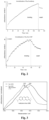
Figure 3 : The Tau protein was immobilized on antibody terminated germanium. The amide I of the Tau protein represents a high amount of β-sheets resulting in an absorbance maximum at 1640 cm-1 (solid black line). Addition of the potential drug methylene blue shifted the amide I band to 1653 cm-1 indicating a major structural change to α-helix dominated secondary structure distribution (dashed line). -
Figure 4 : The difference spectra between the two spectra shown inFigure 3 shows mainly a decrease of β-sheet and an increase of α-helix induced by addition of methylene blue (50 µM). This shows that the drug intervention on the secondary structure distribution can be monitored by the invention very precisely. -
Figure 5 : Structural shift of the amide I band of the Tau protein from CSF with (black solid line) and without the presence of methylene blue (dashed grey line) in dependence of time. -
Figure 6 : Aβ1-42 protein was immobilized on antibody (A8978) terminated germanium surface. The dashed grey spectrum shows the a broad distribution of different secondary structures with an absorbance maximum at 1634 cm-1 characteristic for fibrillized Aβ1-42. Furthermore, the amide I of the intermediate of the folding process (solid grey line) is shown after 160 min. Addition of the potential drug berberine (100 µM) results in a significant different secondary structure distribution dominated now by an α-helical or mainly monomeric isoform of Aβ1-42 at 1659 cm-1 (dashed black spectrum), however with a significant shoulder at 1634 cm-1. The solid black spectrum shows the amide I band as an intermediate of the folding process after 160 min. -
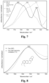
Figure 7 : Comparison of the Aβ1-42 secondary structure distribution (black) after incubation with methylene blue (solid black spectrum), berberine (dashed dark spectrum), and without any drug intervention (dashed light grey spectrum). As seen already inFigure 6 berberine shifts the distribution mainly to α-helix, whereas methylene blue induces β-sheet and fibril formation. This finding is in consistence with the literature (Necula et al., Biochem. 46(30):8850-60 doi:10.1021/bi700411k (2007). This shows that different interventions on the secondary structure distribution by different drugs can be resolved. -
Figure 8 : The potential drug berberine (100 µM) did not change the secondary structure distribution of the Tau protein (extracted from CSF) in contrast to methylene blue as shown inFigure 3 . This finding shows that different interventions of different drugs on the same target protein can be resolved. - The invention describes a method for the preselection of potential drugs against pathological misfolded protein targets, such as in many neurodegenerative diseases. The method comprises the steps:
- (a) conducting, in an IR cell comprising an infrared sensor element having an internal reflection element with a core of an infrared transparent material and at least one receptor for the candidate biomarker directly grafted to at least one surface of said core, said at least one receptor for the target protein being antibodies capable of specific and conformationally independent binding to the target protein, a flux of a sample with soluble candidate biomarker protein, submitting an IR beam through said first IR cell, and obtaining an infrared spectrum therefrom;
- (b) conducting, in the same IR cell of step (a), wherein the receptors for the candidate biomarker, which are grafted to the surface of the core, are loaded with the candidate biomarker protein, a flux of a solution with potential drug, submitting an IR beam through said IR cell, and obtaining an infrared spectrum therefrom; and
- (c) subtracting a reference spectrum of the potential drug for detecting the shift of the amide I band of the target protein; (d) analyzing the obtained infrared spectra to evaluate the effect of the potential drug by determining the secondary structure distribution of the soluble candidate biomarker protein in the sample and after application of the potential drug, wherein an upshift or disappearance of the amide I band in the spectrum of (b) relative to (a) is indicative for the efficacy of the potential drug;
- According to the invention the infrared transparent material of the IR cell is selected from gallium arsenide, silicon, germanium, zinc selenide and diamond, and preferably is germanium. Further, the candidate biomarker protein alpha-Synuclein undergoes conformational transitions and is an amyloidogenic peptide or a (poly-) peptide of health-status dependent, characteristic secondary structure composition. Further comparative examples of candidate biomarker proteins include Amyloid-beta (Aβ) peptides and Tau protein associated with Alzheimer's disease, Prion protein associated with Creutzfeldt-Jakob disease, or Huntingtin protein associated with Huntington's disease. Moreover, the sample with candidate biomarker protein may be a purified sample of the biomarker or may be a complex body fluid comprising the biomarker including human CSF. Other suitable complex bodily fluids are human serum, blood plasma, lacrimal fluid and nipple aspirate fluid.
- It is preferred that said infrared sensor element comprises a germanium internal reflection element being of trapezoid or parallelogram shape and being transparent in the infrared with sufficient signal to noise ratio to detect the amide I band beyond large background absorbance, and at least one receptor for the biomarker protein being antibodies capable of specific and conformational independent binding to the biomarker protein, and being directly grafted to at least one surface, preferably to at least two or three surfaces of said internal germanium reflection element, by silanization with short silane linkers or by thiolation with short thiol linkers, reacting freely accessible amine groups of said at least one receptor with amine-reactive groups on the short silane/thiol linkers, and blocking remaining amine-reactive groups on the short silane/thiol linkers with a blocking substance not cross-reacting with the biomarker protein.
- According to the invention it is particularly preferred that the internal reflection element is a germanium monocrystal, preferably is a trapezoid cut germanium monocrystal. It is further preferred that the germanium crystal allows for or provides for one, more than one, or more than three reflections of the infrared light through the reflection element, particularly preferred are more than five reflections or even more than twenty reflections (preferred are 25 reflections with 13 actively sensed reflections). Even more, it is particularly preferred that the internal reflection element is suitable for the parallel analysis by another optical method including detection of fluorescence at different wavelengths. Finally, it is crucial that the blocking substance is not cross-reacting with the biomarker protein, which is selected from casein, ethanolamine, L-lysine, polyethylene glycols, albumins and derivatives thereof.
- The silane and thiol linkers for the grafting include homogenous silane and thiol linkers, mixtures of silane linkers and mixtures of thiol linkers, and have an effective linker chain length (combined number carbon and heteroatoms) of not more than 20 atoms or not more than 15 atoms, preferably
- the silane linkers have one of the following formulas:
(i) X3Si-(CH2)n-Y-(CH2)n'-Z,
(ii) X2R1Si-(CH2)n-Y-(CH2)n'-Z or
(iii) X(R1)2Si-(CH2)n-Y-(CH2)n'-Z,
- and the thiol linkers have the following formula:
(iv) WS-(CH2)n-Y-(CH2)n'-Z,
- The -(CH2)n- and -(CH2)n'- structural elements in the above formulas may also contain one or more double and/or triple bonds and may be substituted with one or more halogen atoms such as fluorine or with deuterium.
- When the infrared sensor element is obtainable by silanization, it is then preferred that in the linkers of formulas (i) to (iii) above X is independently selected from C1-6 alkoxy-groups, preferably from methoxy and ethoxy groups, Y is -NHCO-, Z is -CO2H or an ester derivative thereof, and n is an integer of 1 to 5 and n' is an integer of 1 to 3, preferably n is 3 and n' is 2.
- When the infrared sensor element is obtainable by thiolation, it is then preferred that in the linker of formula (iv) above W is H, Y is a chemical bond, Z is -CO2H or an ester derivative thereof, and n is an integer of 1 to 8 and n' is an integer of 1 to 5, preferably n is 8 and n' is 4.
- In one disclosure, the biomarker protein is an Aβ peptide and the receptor binding to the Aβ peptide is an antibody, preferably is an antibody specifically binding to the central epitope of the Aβ peptide, including antibody A8978.
- In a further disclosure, the biomarker protein is a Tau protein and the receptor binding to the Tau protein is an antibody, preferably is an antibody specifically binding to a epitope accessible for all Tau variants (phosphorylated, truncated, 3 to 4 repeat regions etc., isoforms), including antibody Tau-5.
- In the method of the invention, the concentration of the potential drug in the solution is either below the detection limit of the IR determination or can be easily subtracted by reference spectra of the potential drug.
- In the method of the invention, when the potential drug possesses amide bands, such as antibodies, the method further comprises subtracting a reference spectrum of the potential drug for detecting the shift of the amide I band of the target protein.
- In one particular disclosure, when the target protein is an Aβ peptide, a shift of the amide I band, preferably a shift of the amide I band maximum, to any value indicative for the Aβ peptide secondary structure is indicative for the efficacy of the potential drug. Notably, for a fibrillary fraction of the Aβ peptide a shift from of 1626 cm-1 to 1655 cm-1 and for the total fraction of the Aβ peptide a shift from 1636 cm-1 to 1655 cm-1 is indicative for the efficacy of the potential drug.
- In a further disclosure, when the target protein is a Tau protein, a shift of the amide I band, preferably a shift of the amide I band maximum, to any value indicative for the Tau protein secondary structure is indicative for the efficacy of the potential drug. Notably, for a fibrillary fraction of the Tau protein a shift from of 1626 cm-1 to 1655 cm-1 and for the total fraction of the Tau protein a shift from 1636 cm-1 to 1655 cm-1 is indicative for the efficacy of the potential drug.
- The present invention is based on the detection of secondary structural changes induced by the potential drug by means of vibrational spectroscopy. The invention uses in principle the same experimental set-up as our previous patent application
WO 2015121339 . Instead of a 70V (Bruker) we employed an 80V FTIR spectrometer (Bruker) to improve the signal to noise ratio of the measurements. As internal reflection element germanium crystal were chemically modified with NHS-silanes, which function as anchors for the covalent attachment of the desired antibodies. After blocking the surface with casein the surface is ready for capturing of the target protein alpha-Synuclein protein. Further disclosed is the capturing of Tau or Aβ1-42. The Tau protein was directly extracted out of human CSF. This is a great advantage since no purified protein samples are required and no pretreatment of CSF is needed, which makes the assay easier accessible for the application in clinics or clinical labs. The target protein was analyzed in the presence of the potential drug and the effect was monitored by the change in the amide I band. As shown for the Tau protein the effect of the potential drug methylene blue was directly monitored (Figure 3 ). The change of the secondary structural distribution is characterized by the drug induced shift of the amide I band from 1640 cm-1 (untreated) to 1653 cm-1 (treated). The effect becomes even more evident in the double difference spectrum (Figure 4 ), which clearly shows a negative band at 1625 cm-1 indicative for the disappearance of β-sheet and a positive band at 1655 cm-1 typical for α-helix. In a control without drug the amide I band absorbance maximum of the Tau protein remains stable (Figure 5 ). Thus, for the pre-selection of drug candidates in the treatment of neurodegenerative diseases the invention provides an ideal platform. Another disclosed example is the study of the second important biomarker Aβ1-42. Since the developed sensor is universal, the antibody A8978 against the epitope 13-28 of Aβ1-42 could be applied for the analysis of the potential drug berberine. To monitor a potential effect of berberine the surface was loaded with synthetic Aβ1-42. Without any incubation the auto-induced fibrilization process at high concentrations leads to a broad secondary structure distribution of Aβ1-42 dominated by β-sheet (Figure 6 , dashed light grey spectrum). Addition of berberine shifts the amide I maximum of the broad secondary structure distribution from 1634 to 1659 cm-1 indicating mainly α-helical or monomeric species (Figure 6 , dashed black spectrum). Thus, berberine seems to decelerate the auto-induced fibrilization process. It may be an interesting target for further investigations. In conclusion, the method provides label-free direct information about the intervention of the secondary structure distribution of the target protein by a drug candidate as demonstrated for Tau and Aβ1-42. It resolves the different intervention of the drug on the same target protein. This universal approach can in principle be transferred to any protein and small molecule (potential drug) and has therefore a very high potential for pharmaceutical applications. - The invention is further disclosed in the following Examples, which are however not to be construed so as to limit the application.
- Materials and Methods: The same experimental set-up is used as in applicant's previous patent application
WO 2015121339 . - Sampling and pretreatment: CSF was drawn by lumbal puncture and aliquoted at the university hospital Essen, snap-frozen in liquid nitrogen, shipped and stored at -80 °C. Samples were not pretreated before the measurement, only thawed at 37 °C for 30 s and kept on ice until used.
- Phosphate buffered saline (PBS-buffer): 137 mM sodium chloride (NaCl), 2.7 mM potassium chloride (KCI), 12 mM total-phosphate (in the form of Na2HPO4 and NaH2PO4), pH 7.4.
- Casein blocking-solution: 200 mM sodium hydroxide (NaOH), 1 % (w/v) casein from bovine milk (powder), pH adjusted with H3PO4 to 7.4.
- Silanization-solution: The used NHS-silane (N-(4,4,4-triethoxysilanebutyl)
succinamic acid 2,5-dioxopyrrolidin-1-yl ester) was synthesized and characterized as described (Schartner et al., JACS 135(10):4079-4087 (2013). - Antibody: For the analysis of Aβ1-42 the antibody A8978 (lot no: 061M4773, Sigma Aldrich) was employed. In case of the Tau protein the antibody Tau-5 (AHB0042, Thermo Fisher Scientific) was used.
- AB1-42 : The human Aβ-peptide was purchased from Sigma-Aldrich (A9810, Amyloid-beta-Protein fragment 1-42).
- Potential drugs: Methylene blue (methylthionine hydrochloride, lot no: 66720) and berberine chloride (lot no: B3251) were purchased by Sigma Aldrich.
- Performing the measurement: The general procedure is identical to the patent application
WO 2015121339 . IR-measurements were performed on a Vertex 80V spectrometer (Bruker Optics GmbH, Ettlingen, Germany) with liquid nitrogen cooled mercury-cadmium-telluride (MCT) detector. Double-sided interferograms were recorded in forward-backward interferometer movement at a 80 kHz data rate with a spectral resolution of 2 cm-1, Blackman-Harris-3-Term-apodisation, Mertz-phase correction and 4 times zero filling. Reference spectra were recorded as an average of 1000, sample spectra of 200 interferograms. Recording reference single channel spectra of the blank sensor, sensor with 2-propanol, the silanized surface, the buffers, antibody or casein coated surface in equilibrium states enabled high sensitivity difference spectroscopy based on Lambert-Beer law (E=-log(I/I0). The absorbance of the state change is the negative decadic logarithm of the intensity relation before and after the change. - Tau-protein treated with methylene blue: The Tau antibody (Tau-5) from Thermo Fisher Scientific was covalently attached to the germanium surface as described for other antibodies by Nabers et al. (Nabers et al., J. Biophotonics 9(3):224-34 doi:10.1002/jbio.201400145 (2016)). After blocking the Tau-antibody terminated surface was incubated with 100 µl of human CSF till the Tau protein was successfully immobilized (about 60 min). In the next step, 2 ml of a 50 µM methylene blue solution (PBS, pH 7.4) was flushed over the surface till the system was equilibrated (1 ml) and then circulated for 60 min. The effect on the secondary structure of tau was directly monitored by the band position and shape of the amide I.
- Aβ-peptide treated with berberine: The antibody A8979 (Sigma-aldrich) was employed for capturing the Aβ-peptide (Nabers et al., Anal. Chem. 88(5):2755-62 doi:10.1021/acs.analchem.5b04286 (2016)). The Aβ-peptide (Aβ1-42, synthetic, Sigma-aldrich, Taufkrichen, Germany) was monomerized by incubation with hexafluoro-2-propanol as described elsewhere (Nabers et al., J. Biophotonics 9(3):224-34 doi:10.1002/jbio.201400145 (2016)). For the
analysis 100 µg of Aβ1-42 were circulated over the antibody terminated sensor for 1 h to ensure that the drug is not interfering with the immobilization process. In the control experiments the immobilization of Aβ1-42 was monitored for further 17 h (total 18 h) in the presence of the potential drug, to follow the auto-induced fibrilization process (Figure 6 , dashed light grey spectrum). To analyze the effect of the potential drug berberine the same protocol as in the control experiment was used. The only difference was the addition of 100 µM berberine after the 1 h immobilization of Aβ1-42 (PBS, pH 7.4) by equilibrating with 1.5 mL of 100 µM berberine solution and subsequently circulating the system in the presence of 100 µM berberine (Figure 6 , dashed black spectrum). The effect on the secondary structure of Aβ1-42 was directly monitored by the band position and shape of the amide I. The corresponding dashed spectra show intermediates of the folding processes for each experiment after a total time of 160 min. - Pretreatment of the spectra: By scaled subtraction of a reference spectrum water vapor was removed. Spectra were baseline corrected, a sliding average was performed as described (Schartner et al., Chembiochem 15(17):2529-34 doi:10.1002/cbic.201402478 (2014)) and normalized to the same amide I signal intensity in the region 1730 till 1590 cm-1 depending on the observed secondary structure.
- To monitor the drug effect of methylene blue the invented method was employed. We previously invented an immuno-ATR sensor, which differentiates AD with an accuracy of 90 % based on CSF and 84 % based on blood plasma analyzes (Nabers et al., Anal. Chem. 88(5):2755-62 doi:10.1021/acs.analchem.5b04286 (2016)). First, we employed silane chemistry to modify the germanium surface (Schartner et al., JACS 135(10):4079-87 doi:10.1021/ja400253p (2013)). Second, the monoclonal IgG1 antibody Tau-5 was covalently immobilized on the germanium surface. The immobilization is completed after 2 hours as presented in
Figure 2A by reaching an absorbance of 5 mOD. After washing the surface withbinding buffer 1 the antibody remains stable (Figure 2A ). To obtain a highly specific surface the saturation with casein is crucial (Nabers et al., J. Biophotonics 9(3):224-34 doi:10.1002/ jbio.201400145 (2016)). Finally, a complex sample such as cerebrospinal fluid (CSF) is flushed over the sensor. The resulting monoexponential binding kinetics of the Tau protein is presented inFigure 2B . With the immobilized Tau fraction it is now possible to analyze the effect of the potential drug methylene blue. A 50 µM solution of methylene blue was flushed over the surface and after equilibration the system was circulated. The above mentioned immuno-ATR-FTIR sensor (WO 2015121339 ) for the diagnosis of Alzheimer's disease uses for the diagnosis a simple threshold classifier with a value at 1643 cm-1 for AD and disease control differentiation, which can also be transferred to the Tau protein (unpublished data, patent application in preparation). The black spectrum inFigure 3 shows an amide I maximum of 1640 cm-1 indicating a higher amount of disease related β-sheet enriched isoforms, which would be diagnosed as diseased by our immuno-IR-sensor (Nabers et al., Anal. Chem. 88(5):2755-62 doi:10.1021/acs.analchem.5b04286 (2016). Upon methylene blue incubation a significant shift to higher wavenumbers was observed within 1 h (Figure 3 , dashed spectrum), thus a secondary structure change to an disordered or α-helical conformation was induced by the potential drug methylene blue. This is in consistence with the in vivo studies of the group of Claude Wischik, which demonstrated the reduction of the Tau associated tangles in the human brain (Šimić et al., Biomolecules 6(1):6 doi:10.3390/biom6010006 (2016); Wischik et al., PNAS 93(20):11213-18 (1996)). The patient would now be diagnosed as healthy by our developed immuno-ATR-FTIR sensor (Figure 3 , dashed spectrum) (Nabers et al., Anal. Chem. 88(5):2755-62 doi:10.1021/acs.analchem.5b04286 (2016)). Thus, the presented approach has a very high potential as prescreening tool for the selection of candidate drugs against the AD and also against other neurodegenerative diseases. By subtraction of the drug treated state minus the untreated state the secondary structural change becomes even more obvious as indicated by the negative band at 1625 cm-1 and the positive band at 1655 cm-1 (Figure 4 ). To prove that the changes are really caused by methylene blue a control without methylene was performed (Figure 5 ). The amide I maximum is stable and only differs about ±1 wavenumbers without the drug incubation (Figure 5 , dashed light grey line), whereas with the presence of the drug a clear shift to higher wavenumbers is observed (Figure 5 , solid black line). - The second important marker protein for the Alzheimer's disease is Aβ1-42. We analyzed the fibrilization process with the described method. Synthetic Aβ1-42 was monomerized with hexafluoro-2-propanol. A solution of monomerized Aβ1-42 was flushed over the sensor and specifically immobilized with antibody A8979 (Nabers et al., J. Biophotonics 9(3):234-34 doi:10.1002/jbio.201400145 (2016)). The spontaneous fibrilization was monitored over 18 h resulting in an amide I maximum of 1634 cm-1 (
Figure 6 , dashed light grey spectrum). The same experiment was done in the presence of 100 µM berberine showing a 25 cm-1 shift of the amide I maximum to 1659 cm-1 (Figure 6 , dashed black spectrum). This indicates that berberine directly decelerates the aggregation process of Aβ1-42. A small amount of β-sheet enriched isoforms is observed as a shoulder at 1634 cm-1, but the dominating conformation is monomeric Aβ1-42. This suggests a direct interaction of berberine and AB1-42 that could be applied as drug to prevent the initial processes of AD and thus might be useful to slow down disease progression. - In addition, the effect of methylene blue on Aβ1-42 was investigated under the same conditions as for berberine. The obtained spectrum clearly shows a fibril (
Figure 7 , dashed light grey spectrum), which is in consistence with the literature (Necula et al., Biochem. 46(30):8850-60 doi:10.1021/bi700411k (2007)). The effect is discussed to prevent the formation of toxic oligomers and therefore might have a potential in treating Alzheimer's disease (Necula et al., Biochem. 46(30):8850-60 doi:10.1021/bi700411k (2007)). This shows that the method works very efficient and gives direct information regarding the molecular mechanism of the drug. - Furthermore, we studied the effect of berberine on the Tau protein (from CSF) under the same conditions as for methylene blue (
Figure 8 ). The amide band and its maximum are not affected by the berberine treatment, which indicates that berberine has no significant effect on the Tau protein in comparison to methylene blue (Figure 3 andFigure 8 ).
Claims (9)
- A drug-screening assay method for determining the efficacy of a potential drug on a target protein undergoing secondary structural changes associated with a disease with misfolded target proteins in a complex body fluid, comprising the steps:(a) conducting, in an IR cell comprising an infrared sensor element having an internal reflection element with a core of an infrared transparent material and at least one receptor for the target protein directly grafted to at least one surface of said core, said at least one receptor for the target protein being antibodies capable of specific and conformationally independent binding to the target protein, a flux of a sample of the complex body fluid with soluble target protein; submitting an IR beam through said IR cell; and obtaining an infrared spectrum therefrom;(b) conducting, in the same IR cell of step (a), wherein the receptors for the target protein grafted to the surface of the core are loaded with the target protein, a flux of a solution with potential drug; submitting an IR beam through said IR cell; and obtaining an infrared spectrum therefrom;(c) subtracting a reference spectrum of the potential drug for detecting the shift of the amide I band of the target protein; and(d) analyzing the obtained infrared spectra to evaluate the effect of the potential drug by determining the secondary structure distribution of the soluble target protein in the sample and after application of the potential drug, wherein an upshift or disappearance of the amide I band characteristic for β-sheets in the spectrum of (b) relative to the corresponding amide I band in (a) is indicative for the efficacy of the potential drug;wherein the target protein undergoing conformational transitions is alpha-Synuclein.
- The method of claim 1, wherein(i) the infrared transparent material of the IR cell is independently selected from gallium arsenide, silicon, germanium, zincselenide and diamond, and preferably is germanium; and/or(ii) the sample of the complex body fluid with target protein is human CSF, serum or blood plasma.
- The method of claim 1, wherein said infrared reflection element is a germanium internal reflection element being of trapezoid or parallelogram shape wherein the antibodies are directly grafted to at least one surface of said internal germanium reflection element by silanization with short silane linkers or by thiolation with short thiol linkers, reacting freely accessible amine groups of said at least one receptor with amine-reactive groups on the short silane/thiol linkers, and blocking remaining amine-reactive groups on the short silane/thiol linkers with a blocking substance not cross-reacting with the target protein.
- The method of claim 1 or 3, wherein the internal reflection element(i) is a germanium monocrystal, preferably is a trapezoid cut germanium monocrystal; and/or(ii) allows for or provides for more than one passages of the infrared light through the reflection element; and/or(iii) is further suitable for the parallel analysis by another optical method including detection of fluorescence at different wavelengths; and/or(iv) the blocking substance not cross-reacting with the target protein is selected from casein, ethanolamine, L-lysine, polyethylene glycols, albumins and derivatives thereof.
- The method of claim 3 or 4, wherein the silane and thiol linkers include homogenous silane and thiol linkers, mixtures of silane linkers and mixtures of thiol linkers, and have an effective linker chain length (combined number carbon and heteroatoms) of not more than 20 atoms or not more than 15 atoms,
preferablythe silane linkers have one of the following formulas:
(i) X3Si-(CH2)n-Y-(CH2)n'-Z,
(ii) X2R1Si-(CH2)n-Y-(CH2)n'-Z or
(iii) X(R1)2Si-(CH2)n-Y-(CH2)n'-Z,
and the thiol linkers have the following formula:wherein W is H or R1S-, X at each occurrence is independently selected from halogen and C1-6 alkoxy, n is an integer of 1 to 10, n' is an integer of 1 to 5; R1 at each occurrence is independently selected from C1-6 alkyl, Y is selected from a chemical bond, O-, -CO-, -SO2-, -NR2-, -S-, -SS-, -NR2CO- , -CONR2-, -NR2SO2-and -SO2NR2- (wherein R2 is H or C1-6 alkyl), and Z is an amine-reactive group including -CO2H, -SO3H and ester derivatives thereof.
(iv) WS-(CH2)n-Y-(CH2)n'-Z,
- The method of claim 5, wherein the infrared sensor element is obtainable by(i) silanization and in the linkers of formulas (i) to (iii) X is independently selected from C1-6 alkoxy-groups, preferably from methoxy and ethoxy groups, Y is -NHCO-, Z is -CO2H or an ester derivative thereof, and n is an integer of 1 to 5 and n' is an integer of 1 to 3, preferably n is 3 and n' is 2; or(ii) thiolation and in the linkers of formula (iv) W is H, Y is a chemical bond, Z is -CO2H or an ester derivative thereof, and n is an integer of 1 to 8 and n' is an integer of 1 to 5, preferably n is 8 and n' is 4.
- Use of an infrared sensor element as defined in any one of claims 1 to 6 for the direct analysis of the interaction between a potential drug and alpha-Synuclein protein undergoing conformational transitions .
- The use of claim 7, wherein the analysis of the secondary structure or structural change of the_target protein upon treatment with the potential drug gives direct information about the efficacy of the drug towards the alpha-Synuclein protein.
- The use of claim 8, which is for the pre-selection of drug candidates for the treatment of Parkinson's disease.
Applications Claiming Priority (2)
| Application Number | Priority Date | Filing Date | Title |
|---|---|---|---|
| EP16199792.9A EP3324186B1 (en) | 2016-11-21 | 2016-11-21 | Method for the preselection of drugs for protein misfolding diseases |
| PCT/EP2017/079924 WO2018091738A1 (en) | 2016-11-21 | 2017-11-21 | Method for the preselection of drugs for protein misfolding diseases |
Publications (2)
| Publication Number | Publication Date |
|---|---|
| EP3542164A1 EP3542164A1 (en) | 2019-09-25 |
| EP3542164B1 true EP3542164B1 (en) | 2025-01-15 |
Family
ID=57406057
Family Applications (2)
| Application Number | Title | Priority Date | Filing Date |
|---|---|---|---|
| EP16199792.9A Active EP3324186B1 (en) | 2016-11-21 | 2016-11-21 | Method for the preselection of drugs for protein misfolding diseases |
| EP17798259.2A Active EP3542164B1 (en) | 2016-11-21 | 2017-11-21 | Method for the preselection of drugs for protein misfolding diseases |
Family Applications Before (1)
| Application Number | Title | Priority Date | Filing Date |
|---|---|---|---|
| EP16199792.9A Active EP3324186B1 (en) | 2016-11-21 | 2016-11-21 | Method for the preselection of drugs for protein misfolding diseases |
Country Status (8)
| Country | Link |
|---|---|
| US (1) | US11592451B2 (en) |
| EP (2) | EP3324186B1 (en) |
| JP (1) | JP7213551B2 (en) |
| AU (1) | AU2017360092B2 (en) |
| CA (1) | CA3044329A1 (en) |
| ES (2) | ES2834484T3 (en) |
| IL (1) | IL266779A (en) |
| WO (1) | WO2018091738A1 (en) |
Families Citing this family (8)
| Publication number | Priority date | Publication date | Assignee | Title |
|---|---|---|---|---|
| EP3943942A1 (en) | 2014-02-14 | 2022-01-26 | betaSENSE GmbH | Biosensor for conformation and secondary structure analysis |
| ES2834484T3 (en) | 2016-11-21 | 2021-06-17 | Univ Ruhr Bochum | Method for preselection of drugs for protein misfolding diseases |
| EP3658172A4 (en) | 2017-07-25 | 2021-05-05 | TrueBinding, Inc. | CANCER TREATMENT BY BLOCKING THE INTERACTION OF TIM-3 AND ITS LIGAND |
| CN113711036B (en) | 2019-01-30 | 2025-03-18 | 真和制药有限公司 | Anti-GAL3 antibodies and uses thereof |
| CN116157151A (en) | 2020-05-26 | 2023-05-23 | 真和制药有限公司 | Methods of treating inflammatory diseases by blocking galectin-3 |
| CN114112980B (en) * | 2022-01-24 | 2022-05-10 | 武汉宏韧生物医药股份有限公司 | Medicine component detection method and system based on data analysis |
| CN115078569B (en) * | 2022-05-26 | 2024-04-12 | 北京中医药大学 | A method for identifying the key quality attributes of cough suppressants using biosensor-integrated UPLC-MS technology |
| CN116773480A (en) * | 2023-06-30 | 2023-09-19 | 国家纳米科学中心 | Graphene plasmon enhanced infrared spectrum sensor loaded with silk nanofibers and preparation method and application thereof |
Family Cites Families (31)
| Publication number | Priority date | Publication date | Assignee | Title |
|---|---|---|---|---|
| US6110446A (en) | 1998-10-05 | 2000-08-29 | Colgate Palmolive Company | Dual component antiplaque and tooth whitening composition |
| ATE335201T1 (en) | 1999-05-14 | 2006-08-15 | Iris Biotechnologies Inc | REVERSIBLE IMMOBILIZATION OF LIGANDS ON METAL SURFACES, THEIR PRODUCTION AND USE AS BIOSENSORS |
| DE10030920C2 (en) | 2000-06-24 | 2003-01-02 | Glukomeditech Ag | Measuring device for the simultaneous refractometric and ATR spectrometric measurement of the concentration of liquid media and use of this device see |
| JP2002040023A (en) | 2000-07-28 | 2002-02-06 | Mitsubishi Chemicals Corp | Methods for detecting Alzheimer's disease |
| DK1354197T3 (en) | 2001-01-16 | 2010-06-07 | Univ Catholique Louvain | Surface chemical modification of optical elements |
| AU2002367716A1 (en) | 2001-09-04 | 2003-10-20 | North Carolina State University | Single pass attenuated total reflection infrared analysis identifying protein secondary structure, surface charge and binding affinity |
| US7132122B2 (en) | 2002-10-30 | 2006-11-07 | The Regents Of The University Of California | Direct micro-patterning of lipid bilayers using UV light and selected uses thereof |
| EP1565726A2 (en) * | 2002-11-22 | 2005-08-24 | Solus Biosystems, Inc. | High throughput screening with parallel vibrational spectroscopy |
| US8481017B2 (en) | 2004-02-23 | 2013-07-09 | Florida State University Research Foundation, Inc. | Thin films for controlled protein interaction |
| EP1806574A1 (en) | 2006-01-05 | 2007-07-11 | Université Catholique de Louvain | Surface modification of optical elements for the spectroscopic detection of molecules and organic components |
| EP1857808A1 (en) | 2006-05-15 | 2007-11-21 | Sika Technology AG | Measuring apparatus |
| JP4147264B2 (en) | 2006-09-08 | 2008-09-10 | 株式会社ステリック再生医科学研究所 | Neurofibrotic degeneration inhibitor |
| AU2008204335B2 (en) | 2007-01-11 | 2013-06-13 | Michael Bacher | Diagnosis and treatment of Alzheimer's and other neurodementing diseases |
| WO2010123534A2 (en) | 2009-01-16 | 2010-10-28 | Rensselaer Polytechnic Institute | Enzyme-based nanoscale decontaminating composites |
| US8883985B2 (en) | 2010-02-16 | 2014-11-11 | National University Corporation Kyoto Institute Of Technology | Antibody-immobilized carrier, method of producing antibody-immobilized carrier, and use of said antibody-immobilized carrier |
| CA2823716C (en) | 2011-01-04 | 2018-02-13 | Exxonmobil Upstream Research Company | Method and apparatus for a mid-infrared (mir) system for real time detection of petroleum in colloidal suspensions of sediments and drilling muds during drilling, logging, and production operations |
| EP2490029A1 (en) | 2011-02-16 | 2012-08-22 | Université Catholique De Louvain | Vancomycin analysis |
| WO2012170993A2 (en) | 2011-06-09 | 2012-12-13 | New York University | Assays and methods pertaining to pre-amyloid intermediates |
| US9845327B2 (en) | 2011-06-22 | 2017-12-19 | The General Hospital Corporation | Treatment of proteinopathies |
| EP2700933A1 (en) | 2012-08-20 | 2014-02-26 | Consejo Superior De Investigaciones Científicas (CSIC) | Raman, infrared, or Raman-Infrared analysis of peripheral blood plasma protein structure and its relation to cognitive development in Alzheimer's disease |
| EP2738558A1 (en) * | 2012-11-28 | 2014-06-04 | ETH Zurich | Method and tools for the determination of conformation and conformational changes of proteins and of derivatives thereof |
| JP2017501848A (en) | 2013-11-19 | 2017-01-19 | プロセナ バイオサイエンシーズ リミテッド | Monitoring immunotherapy of Lewy body disease from constipation symptoms |
| CA2924268C (en) | 2013-11-21 | 2021-05-18 | F. Hoffmann-La Roche Ag | Anti-alpha-synuclein antibodies and methods of use |
| EP3943942A1 (en) * | 2014-02-14 | 2022-01-26 | betaSENSE GmbH | Biosensor for conformation and secondary structure analysis |
| EP3524962A1 (en) | 2015-12-09 | 2019-08-14 | Diamontech GmbH | Device and method for analysing a material |
| ES2791962T3 (en) | 2016-11-21 | 2020-11-06 | Univ Ruhr Bochum | Combined trial for the differential diagnosis of Alzheimer's disease |
| ES2834484T3 (en) | 2016-11-21 | 2021-06-17 | Univ Ruhr Bochum | Method for preselection of drugs for protein misfolding diseases |
| CA3042302A1 (en) | 2016-12-02 | 2018-06-07 | Prothena Biosciences Limited | Infrared assay detecting secondary structure profiles of alpha-synuclein |
| WO2018219969A1 (en) | 2017-05-29 | 2018-12-06 | RUHR-UNIVERSITäT BOCHUM | Biosensor for conformation and secondary structure analysis |
| WO2019000094A1 (en) | 2017-06-30 | 2019-01-03 | The Royal Institution For The Advancement Of Learning/Mcgill University | Identification of microorganisms using reflection infrared spectroscopy |
| US20190369015A1 (en) | 2018-06-01 | 2019-12-05 | Irid Corp. | Identification of microorganisms using disposable dual reflection substrate for measuring infrared spectra of said microorganisms |
-
2016
- 2016-11-21 ES ES16199792T patent/ES2834484T3/en active Active
- 2016-11-21 EP EP16199792.9A patent/EP3324186B1/en active Active
-
2017
- 2017-11-21 US US16/349,194 patent/US11592451B2/en active Active
- 2017-11-21 CA CA3044329A patent/CA3044329A1/en active Pending
- 2017-11-21 AU AU2017360092A patent/AU2017360092B2/en active Active
- 2017-11-21 EP EP17798259.2A patent/EP3542164B1/en active Active
- 2017-11-21 WO PCT/EP2017/079924 patent/WO2018091738A1/en not_active Ceased
- 2017-11-21 ES ES17798259T patent/ES3016783T3/en active Active
- 2017-11-21 JP JP2019527202A patent/JP7213551B2/en active Active
-
2019
- 2019-05-21 IL IL266779A patent/IL266779A/en unknown
Non-Patent Citations (1)
| Title |
|---|
| GIEHM L ET AL: "Strategies to increase the reproducibility of protein fibrillization in plate reader assays", ANALYTICAL BIOCHEMISTRY, vol. 400, no. 2, 15 May 2010 (2010-05-15), pages 270 - 281, XP026971708 * |
Also Published As
| Publication number | Publication date |
|---|---|
| AU2017360092A1 (en) | 2019-05-30 |
| AU2017360092B2 (en) | 2023-12-21 |
| EP3324186B1 (en) | 2020-09-16 |
| ES3016783T3 (en) | 2025-05-09 |
| ES2834484T3 (en) | 2021-06-17 |
| EP3324186A1 (en) | 2018-05-23 |
| EP3542164A1 (en) | 2019-09-25 |
| JP7213551B2 (en) | 2023-01-27 |
| US11592451B2 (en) | 2023-02-28 |
| US20190285650A1 (en) | 2019-09-19 |
| IL266779A (en) | 2019-07-31 |
| JP2020513544A (en) | 2020-05-14 |
| CA3044329A1 (en) | 2018-05-24 |
| WO2018091738A1 (en) | 2018-05-24 |
Similar Documents
| Publication | Publication Date | Title |
|---|---|---|
| US11592451B2 (en) | Method for the preselection of drugs for protein misfolding diseases | |
| US20250052675A1 (en) | Attenuated Total Reflectance-Based Biosensor for Conformation and Secondary Structure Analysis | |
| ES2935060T3 (en) | Procedure for the determination of protein aggregates using the FIDA surface | |
| EP3631446A1 (en) | Biosensor for conformation and secondary structure analysis | |
| Ran et al. | Non-conjugated small molecule FRET for differentiating monomers from higher molecular weight amyloid beta species | |
| Kulenkampff et al. | An antibody scanning method for the detection of α-synuclein oligomers in the serum of Parkinson's disease patients | |
| JP7117476B2 (en) | Combined Assays for Differential Diagnosis of Alzheimer's Disease | |
| JP2016040540A (en) | Specific determination of drebrin a and drebrin e | |
| JP2018535434A (en) | Protein structural type detection | |
| Zhu et al. | A screening platform based on epitope editing for drug discovery | |
| Barton | Origins of amyloid oligomers and novel approaches for their detection | |
| An et al. | Pioneering the Early Diagnosis of Alzheimer’s Disease: Aβ Oligomers-Sensing Probe for Cerebrospinal Fluid Analysis in Patients | |
| WO2025076222A1 (en) | Immunoassay for detecting tau phosphorylated at serine 413 | |
| Thangaraj et al. | How specific are the conformation-specific α-synuclein antibodies? Characterization and validation of 16 α-synuclein conformation-specific antibodies using well-characterized preparations of α-synuclein monomers, fibrils and oligomers with distinct structures and morphology | |
| WO2026033422A1 (en) | Methods to evaluate early-stage pre-tangle tau aggregates and treatment of alzheimer's disease | |
| Maftei | Molecular characterization of neuroprotective β-amyloid interacting peptides and autoantibodies relevant to Alzheimer’s disease | |
| Daghestani | BIOPHYSICAL AND PHARMACOLOGICAL CHARACTERIZATION OF CYTOPLASMIC DYNEIN HEAVY CHAIN 1 |
Legal Events
| Date | Code | Title | Description |
|---|---|---|---|
| STAA | Information on the status of an ep patent application or granted ep patent |
Free format text: STATUS: UNKNOWN |
|
| STAA | Information on the status of an ep patent application or granted ep patent |
Free format text: STATUS: THE INTERNATIONAL PUBLICATION HAS BEEN MADE |
|
| PUAI | Public reference made under article 153(3) epc to a published international application that has entered the european phase |
Free format text: ORIGINAL CODE: 0009012 |
|
| STAA | Information on the status of an ep patent application or granted ep patent |
Free format text: STATUS: REQUEST FOR EXAMINATION WAS MADE |
|
| 17P | Request for examination filed |
Effective date: 20190621 |
|
| AK | Designated contracting states |
Kind code of ref document: A1 Designated state(s): AL AT BE BG CH CY CZ DE DK EE ES FI FR GB GR HR HU IE IS IT LI LT LU LV MC MK MT NL NO PL PT RO RS SE SI SK SM TR |
|
| AX | Request for extension of the european patent |
Extension state: BA ME |
|
| DAV | Request for validation of the european patent (deleted) | ||
| DAX | Request for extension of the european patent (deleted) | ||
| STAA | Information on the status of an ep patent application or granted ep patent |
Free format text: STATUS: EXAMINATION IS IN PROGRESS |
|
| 17Q | First examination report despatched |
Effective date: 20200727 |
|
| RAP1 | Party data changed (applicant data changed or rights of an application transferred) |
Owner name: BETASENSE GMBH |
|
| RAP3 | Party data changed (applicant data changed or rights of an application transferred) |
Owner name: BETASENSE GMBH |
|
| GRAP | Despatch of communication of intention to grant a patent |
Free format text: ORIGINAL CODE: EPIDOSNIGR1 |
|
| STAA | Information on the status of an ep patent application or granted ep patent |
Free format text: STATUS: GRANT OF PATENT IS INTENDED |
|
| INTG | Intention to grant announced |
Effective date: 20240807 |
|
| P01 | Opt-out of the competence of the unified patent court (upc) registered |
Free format text: CASE NUMBER: APP_56320/2024 Effective date: 20241015 |
|
| GRAS | Grant fee paid |
Free format text: ORIGINAL CODE: EPIDOSNIGR3 |
|
| GRAA | (expected) grant |
Free format text: ORIGINAL CODE: 0009210 |
|
| STAA | Information on the status of an ep patent application or granted ep patent |
Free format text: STATUS: THE PATENT HAS BEEN GRANTED |
|
| AK | Designated contracting states |
Kind code of ref document: B1 Designated state(s): AL AT BE BG CH CY CZ DE DK EE ES FI FR GB GR HR HU IE IS IT LI LT LU LV MC MK MT NL NO PL PT RO RS SE SI SK SM TR |
|
| REG | Reference to a national code |
Ref country code: CH Ref legal event code: EP Ref country code: GB Ref legal event code: FG4D |
|
| REG | Reference to a national code |
Ref country code: DE Ref legal event code: R096 Ref document number: 602017087364 Country of ref document: DE |
|
| REG | Reference to a national code |
Ref country code: IE Ref legal event code: FG4D |
|
| REG | Reference to a national code |
Ref country code: NL Ref legal event code: FP |
|
| REG | Reference to a national code |
Ref country code: ES Ref legal event code: FG2A Ref document number: 3016783 Country of ref document: ES Kind code of ref document: T3 Effective date: 20250509 |
|
| PG25 | Lapsed in a contracting state [announced via postgrant information from national office to epo] |
Ref country code: RS Free format text: LAPSE BECAUSE OF FAILURE TO SUBMIT A TRANSLATION OF THE DESCRIPTION OR TO PAY THE FEE WITHIN THE PRESCRIBED TIME-LIMIT Effective date: 20250415 |
|
| PG25 | Lapsed in a contracting state [announced via postgrant information from national office to epo] |
Ref country code: FI Free format text: LAPSE BECAUSE OF FAILURE TO SUBMIT A TRANSLATION OF THE DESCRIPTION OR TO PAY THE FEE WITHIN THE PRESCRIBED TIME-LIMIT Effective date: 20250115 |
|
| PG25 | Lapsed in a contracting state [announced via postgrant information from national office to epo] |
Ref country code: PL Free format text: LAPSE BECAUSE OF FAILURE TO SUBMIT A TRANSLATION OF THE DESCRIPTION OR TO PAY THE FEE WITHIN THE PRESCRIBED TIME-LIMIT Effective date: 20250115 |
|
| REG | Reference to a national code |
Ref country code: LT Ref legal event code: MG9D |
|
| PG25 | Lapsed in a contracting state [announced via postgrant information from national office to epo] |
Ref country code: NO Free format text: LAPSE BECAUSE OF FAILURE TO SUBMIT A TRANSLATION OF THE DESCRIPTION OR TO PAY THE FEE WITHIN THE PRESCRIBED TIME-LIMIT Effective date: 20250415 Ref country code: IS Free format text: LAPSE BECAUSE OF FAILURE TO SUBMIT A TRANSLATION OF THE DESCRIPTION OR TO PAY THE FEE WITHIN THE PRESCRIBED TIME-LIMIT Effective date: 20250515 |
|
| REG | Reference to a national code |
Ref country code: AT Ref legal event code: MK05 Ref document number: 1760153 Country of ref document: AT Kind code of ref document: T Effective date: 20250115 |
|
| PG25 | Lapsed in a contracting state [announced via postgrant information from national office to epo] |
Ref country code: HR Free format text: LAPSE BECAUSE OF FAILURE TO SUBMIT A TRANSLATION OF THE DESCRIPTION OR TO PAY THE FEE WITHIN THE PRESCRIBED TIME-LIMIT Effective date: 20250115 |
|
| PG25 | Lapsed in a contracting state [announced via postgrant information from national office to epo] |
Ref country code: LV Free format text: LAPSE BECAUSE OF FAILURE TO SUBMIT A TRANSLATION OF THE DESCRIPTION OR TO PAY THE FEE WITHIN THE PRESCRIBED TIME-LIMIT Effective date: 20250115 Ref country code: PT Free format text: LAPSE BECAUSE OF FAILURE TO SUBMIT A TRANSLATION OF THE DESCRIPTION OR TO PAY THE FEE WITHIN THE PRESCRIBED TIME-LIMIT Effective date: 20250515 |
|
| PG25 | Lapsed in a contracting state [announced via postgrant information from national office to epo] |
Ref country code: BG Free format text: LAPSE BECAUSE OF FAILURE TO SUBMIT A TRANSLATION OF THE DESCRIPTION OR TO PAY THE FEE WITHIN THE PRESCRIBED TIME-LIMIT Effective date: 20250115 Ref country code: GR Free format text: LAPSE BECAUSE OF FAILURE TO SUBMIT A TRANSLATION OF THE DESCRIPTION OR TO PAY THE FEE WITHIN THE PRESCRIBED TIME-LIMIT Effective date: 20250416 |
|
| PG25 | Lapsed in a contracting state [announced via postgrant information from national office to epo] |
Ref country code: AT Free format text: LAPSE BECAUSE OF FAILURE TO SUBMIT A TRANSLATION OF THE DESCRIPTION OR TO PAY THE FEE WITHIN THE PRESCRIBED TIME-LIMIT Effective date: 20250115 |
|
| PG25 | Lapsed in a contracting state [announced via postgrant information from national office to epo] |
Ref country code: SE Free format text: LAPSE BECAUSE OF FAILURE TO SUBMIT A TRANSLATION OF THE DESCRIPTION OR TO PAY THE FEE WITHIN THE PRESCRIBED TIME-LIMIT Effective date: 20250115 |
|
| PG25 | Lapsed in a contracting state [announced via postgrant information from national office to epo] |
Ref country code: SM Free format text: LAPSE BECAUSE OF FAILURE TO SUBMIT A TRANSLATION OF THE DESCRIPTION OR TO PAY THE FEE WITHIN THE PRESCRIBED TIME-LIMIT Effective date: 20250115 |
|
| PG25 | Lapsed in a contracting state [announced via postgrant information from national office to epo] |
Ref country code: DK Free format text: LAPSE BECAUSE OF FAILURE TO SUBMIT A TRANSLATION OF THE DESCRIPTION OR TO PAY THE FEE WITHIN THE PRESCRIBED TIME-LIMIT Effective date: 20250115 |
|
| REG | Reference to a national code |
Ref country code: DE Ref legal event code: R097 Ref document number: 602017087364 Country of ref document: DE |
|
| PG25 | Lapsed in a contracting state [announced via postgrant information from national office to epo] |
Ref country code: EE Free format text: LAPSE BECAUSE OF FAILURE TO SUBMIT A TRANSLATION OF THE DESCRIPTION OR TO PAY THE FEE WITHIN THE PRESCRIBED TIME-LIMIT Effective date: 20250115 Ref country code: CZ Free format text: LAPSE BECAUSE OF FAILURE TO SUBMIT A TRANSLATION OF THE DESCRIPTION OR TO PAY THE FEE WITHIN THE PRESCRIBED TIME-LIMIT Effective date: 20250115 |
|
| PG25 | Lapsed in a contracting state [announced via postgrant information from national office to epo] |
Ref country code: RO Free format text: LAPSE BECAUSE OF FAILURE TO SUBMIT A TRANSLATION OF THE DESCRIPTION OR TO PAY THE FEE WITHIN THE PRESCRIBED TIME-LIMIT Effective date: 20250115 |
|
| PG25 | Lapsed in a contracting state [announced via postgrant information from national office to epo] |
Ref country code: SK Free format text: LAPSE BECAUSE OF FAILURE TO SUBMIT A TRANSLATION OF THE DESCRIPTION OR TO PAY THE FEE WITHIN THE PRESCRIBED TIME-LIMIT Effective date: 20250115 |
|
| PLBE | No opposition filed within time limit |
Free format text: ORIGINAL CODE: 0009261 |
|
| STAA | Information on the status of an ep patent application or granted ep patent |
Free format text: STATUS: NO OPPOSITION FILED WITHIN TIME LIMIT |
|
| REG | Reference to a national code |
Ref country code: CH Ref legal event code: L10 Free format text: ST27 STATUS EVENT CODE: U-0-0-L10-L00 (AS PROVIDED BY THE NATIONAL OFFICE) Effective date: 20251126 |
|
| REG | Reference to a national code |
Ref country code: CH Ref legal event code: U11 Free format text: ST27 STATUS EVENT CODE: U-0-0-U10-U11 (AS PROVIDED BY THE NATIONAL OFFICE) Effective date: 20251201 |
|
| PGFP | Annual fee paid to national office [announced via postgrant information from national office to epo] |
Ref country code: NL Payment date: 20251119 Year of fee payment: 9 |
|
| 26N | No opposition filed |
Effective date: 20251016 |
|
| PGFP | Annual fee paid to national office [announced via postgrant information from national office to epo] |
Ref country code: DE Payment date: 20251126 Year of fee payment: 9 |
|
| PGFP | Annual fee paid to national office [announced via postgrant information from national office to epo] |
Ref country code: GB Payment date: 20251119 Year of fee payment: 9 |
|
| PGFP | Annual fee paid to national office [announced via postgrant information from national office to epo] |
Ref country code: IT Payment date: 20251121 Year of fee payment: 9 |
|
| PGFP | Annual fee paid to national office [announced via postgrant information from national office to epo] |
Ref country code: FR Payment date: 20251120 Year of fee payment: 9 |
|
| PGFP | Annual fee paid to national office [announced via postgrant information from national office to epo] |
Ref country code: CH Payment date: 20251201 Year of fee payment: 9 |
|
| PGFP | Annual fee paid to national office [announced via postgrant information from national office to epo] |
Ref country code: ES Payment date: 20251216 Year of fee payment: 9 |