EP3535685B1 - Systeme und verfahren zur codierung von bildmerkmalen von hochauflösenden digitalen bildern von biologischen proben - Google Patents
Systeme und verfahren zur codierung von bildmerkmalen von hochauflösenden digitalen bildern von biologischen proben Download PDFInfo
- Publication number
- EP3535685B1 EP3535685B1 EP17797593.5A EP17797593A EP3535685B1 EP 3535685 B1 EP3535685 B1 EP 3535685B1 EP 17797593 A EP17797593 A EP 17797593A EP 3535685 B1 EP3535685 B1 EP 3535685B1
- Authority
- EP
- European Patent Office
- Prior art keywords
- image
- clusters
- superpixels
- superpixel
- graph
- Prior art date
- Legal status (The legal status is an assumption and is not a legal conclusion. Google has not performed a legal analysis and makes no representation as to the accuracy of the status listed.)
- Active
Links
Images


Classifications
-
- G—PHYSICS
- G06—COMPUTING OR CALCULATING; COUNTING
- G06V—IMAGE OR VIDEO RECOGNITION OR UNDERSTANDING
- G06V20/00—Scenes; Scene-specific elements
- G06V20/60—Type of objects
- G06V20/69—Microscopic objects, e.g. biological cells or cellular parts
- G06V20/698—Matching; Classification
-
- G—PHYSICS
- G06—COMPUTING OR CALCULATING; COUNTING
- G06F—ELECTRIC DIGITAL DATA PROCESSING
- G06F18/00—Pattern recognition
- G06F18/20—Analysing
- G06F18/21—Design or setup of recognition systems or techniques; Extraction of features in feature space; Blind source separation
- G06F18/211—Selection of the most significant subset of features
-
- G—PHYSICS
- G06—COMPUTING OR CALCULATING; COUNTING
- G06F—ELECTRIC DIGITAL DATA PROCESSING
- G06F18/00—Pattern recognition
- G06F18/20—Analysing
- G06F18/22—Matching criteria, e.g. proximity measures
-
- G—PHYSICS
- G06—COMPUTING OR CALCULATING; COUNTING
- G06F—ELECTRIC DIGITAL DATA PROCESSING
- G06F18/00—Pattern recognition
- G06F18/20—Analysing
- G06F18/23—Clustering techniques
- G06F18/232—Non-hierarchical techniques
- G06F18/2321—Non-hierarchical techniques using statistics or function optimisation, e.g. modelling of probability density functions
- G06F18/23213—Non-hierarchical techniques using statistics or function optimisation, e.g. modelling of probability density functions with fixed number of clusters, e.g. K-means clustering
-
- G—PHYSICS
- G06—COMPUTING OR CALCULATING; COUNTING
- G06F—ELECTRIC DIGITAL DATA PROCESSING
- G06F18/00—Pattern recognition
- G06F18/40—Software arrangements specially adapted for pattern recognition, e.g. user interfaces or toolboxes therefor
-
- G—PHYSICS
- G06—COMPUTING OR CALCULATING; COUNTING
- G06T—IMAGE DATA PROCESSING OR GENERATION, IN GENERAL
- G06T7/00—Image analysis
- G06T7/0002—Inspection of images, e.g. flaw detection
- G06T7/0012—Biomedical image inspection
-
- G—PHYSICS
- G06—COMPUTING OR CALCULATING; COUNTING
- G06T—IMAGE DATA PROCESSING OR GENERATION, IN GENERAL
- G06T7/00—Image analysis
- G06T7/10—Segmentation; Edge detection
- G06T7/11—Region-based segmentation
-
- G—PHYSICS
- G06—COMPUTING OR CALCULATING; COUNTING
- G06V—IMAGE OR VIDEO RECOGNITION OR UNDERSTANDING
- G06V10/00—Arrangements for image or video recognition or understanding
- G06V10/20—Image preprocessing
- G06V10/26—Segmentation of patterns in the image field; Cutting or merging of image elements to establish the pattern region, e.g. clustering-based techniques; Detection of occlusion
-
- G—PHYSICS
- G06—COMPUTING OR CALCULATING; COUNTING
- G06V—IMAGE OR VIDEO RECOGNITION OR UNDERSTANDING
- G06V10/00—Arrangements for image or video recognition or understanding
- G06V10/40—Extraction of image or video features
- G06V10/42—Global feature extraction by analysis of the whole pattern, e.g. using frequency domain transformations or autocorrelation
- G06V10/422—Global feature extraction by analysis of the whole pattern, e.g. using frequency domain transformations or autocorrelation for representing the structure of the pattern or shape of an object therefor
- G06V10/426—Graphical representations
-
- G—PHYSICS
- G06—COMPUTING OR CALCULATING; COUNTING
- G06V—IMAGE OR VIDEO RECOGNITION OR UNDERSTANDING
- G06V20/00—Scenes; Scene-specific elements
- G06V20/60—Type of objects
- G06V20/69—Microscopic objects, e.g. biological cells or cellular parts
- G06V20/695—Preprocessing, e.g. image segmentation
-
- G—PHYSICS
- G06—COMPUTING OR CALCULATING; COUNTING
- G06T—IMAGE DATA PROCESSING OR GENERATION, IN GENERAL
- G06T2207/00—Indexing scheme for image analysis or image enhancement
- G06T2207/10—Image acquisition modality
- G06T2207/10056—Microscopic image
-
- G—PHYSICS
- G06—COMPUTING OR CALCULATING; COUNTING
- G06T—IMAGE DATA PROCESSING OR GENERATION, IN GENERAL
- G06T2207/00—Indexing scheme for image analysis or image enhancement
- G06T2207/20—Special algorithmic details
- G06T2207/20072—Graph-based image processing
-
- G—PHYSICS
- G06—COMPUTING OR CALCULATING; COUNTING
- G06T—IMAGE DATA PROCESSING OR GENERATION, IN GENERAL
- G06T2207/00—Indexing scheme for image analysis or image enhancement
- G06T2207/30—Subject of image; Context of image processing
- G06T2207/30004—Biomedical image processing
- G06T2207/30024—Cell structures in vitro; Tissue sections in vitro
Definitions
- the present disclosure generally relates to the field of medical imaging. Some examples discussed in the present disclosure relate to methods of efficiently encoding vast amounts of image features of extracted from high-resolution digital images of biological specimens, where the encoded image features can be decoded and used by an image analysis system for performing various image analysis tasks.
- the biological specimens are sometimes stained with one or more different stains or dyes in order to highlight various types of structures within the specimen, such as specific types of tissue, cells, cell organelles, and so forth. This can enable pathologists and other health care professionals to better assess the specimen's morphology and deliver a more accurate diagnosis, prognosis, and treatment plan for the patient.
- H&E staining One method of staining the specimen is hematoxylin and eosin (H&E) staining, in which a dark blue or violet hematoxylin binds to basophilic substances such as DNA and RNA, and a red or pink eosin binds to acidophilic substances such as positively charged amino acid side chains.
- basophilic substances such as DNA and RNA
- red or pink eosin binds to acidophilic substances such as positively charged amino acid side chains.
- cell nuclei on specimen slides stained with H&E are likely to appear in blue or purple, while structures like the cytoplasm, basophils, muscles, erythrocytes, collagen, and mitochondria are likely to appear in different shades of pink or red.
- Immunohistochemistry (IHC) staining is another staining technique that has become popular due to its significant efficiencies and the rich diagnostic information that it generates.
- IHC slide staining can be utilized to identify proteins in cells of a tissue section and hence is widely used in the study of different types of cells, such as cancerous cells and immune cells in biological tissue.
- IHC staining may be used in research and clinical settings to understand the distribution and localization of the differentially expressed biomarkers of immune cells in a cancerous tissue for an immune response study.
- tumors often contain infiltrates of immune cells, which may prevent the development of tumors or favor the outgrowth of tumors.
- multiple stains may be used to target different types of immune cells, and the population distribution of each type of immune cell is used in studying the clinical outcome of the patients.
- ISH staining is a technique that uses a labeled complementary DNA, RNA or modified nucleic acids strand (i.e., probe) to localize a specific DNA or RNA sequence in a portion or section of tissue.
- In situ hybridization can be a powerful technique for identifying specific mRNA species within individual cells in tissue sections, providing insights into physiological processes and disease pathogenesis.
- Digital pathology systems include slide scanners that can scan tissue slides to produce very high resolution (e.g., 0.5 or 0.275 microns per pixel) digital images of the tissue. Digital pathology systems also include hardware and/or software solutions for automatically processing, viewing, segmenting, analyzing (e.g., scoring) and otherwise managing tissue images or other types of biological specimen images.
- image feature extraction In order to automatically analyze a biological specimen image, a pre-processing step of image feature extraction is often required. During feature extraction, various image features such as pixel intensities, pixel intensity gradients (magnitude and direction), and the like can be extracted from the image. The features can then be used by image analysis tasks such as region segmentation, cell segmentation, scoring, image retrieval, and the like.
- image feature extraction can be one of the most computationally expensive step in the image analysis pipeline, because high-dimensional features are often required to characterize the complex image contents.
- digital pathology the computational requirements are even higher because of the immense data density of digitized whole slide images. Therefore, in digital pathology and other applications processing high-resolution images of biological specimens, it is desirable to precompute the image features and store them, thereby avoiding multiple redundant computations.
- a pathologist may be interested in analyzing one region of interest of an image, and later in another region of interest in the same image, which may or may not overlap with the first region.
- the pathologist may want to compare one image with another image acquired for the same patient based on a new stain or morphology metric.
- image feature pre-computation can be indispensable to provide real-time user experience.
- interactive image segmentation task may require the user to mark various image segments, to check the resulting segmentation, and to add additional marking to refine the segmentation. To enable this interactive process, the segmentation results after each marking need to be generated within seconds or fractions of a second. Accordingly, the amount of real-time re-computations needs to be minimized. Examples of prior art documents are:
- the system may include, for example, a superpixel generator configured to obtain a biological specimen image and group pixels of the biological specimen image into a plurality of superpixels; a feature extractor configured to extract, from each superpixel in the plurality of superpixels, a feature vector comprising a plurality of image features; a clustering engine configured to assign the plurality of superpixels to a predefined number of clusters, each cluster being characterized by a centroid vector of feature vectors of superpixels assigned to the cluster; and a storage interface configured to store, for each superpixel in the plurality of superpixels, clustering information identifying the one cluster to which the superpixel is assigned.
- the system may also include a graph engine configured construct a graph based on the stored information, and use the graph to perform a graph-based image processing task.
- the system may also include a graph engine configured to obtain the clustering information stored by the storage interface; based at least on the clustering information, construct a graph comprising a plurality of nodes, wherein adjacent nodes correspond to adjacent superpixels in the biological specimen image and are connected by a weighted edge, wherein the weighted edge has a weight corresponding to a distance between clusters to which the adjacent superpixels belong; and use the graph to perform a graph-based image processing task.
- the graph-based image processing task can be a segmentation operation that groups the plurality of superpixels into a plurality of segments.
- the clustering engine can be further configured to precalculate distances between each two clusters in the predefined number of clusters; the storage interface can be further configured to store the precalculated distances; and the graph engine can be further configured to obtain the precalculated distances stored by the storage interface, and to construct the graph based on the precalculated distances.
- the storage interface can be further configured to store centroid vectors of the predefined number of clusters; and the graph engine can be further configured to obtain the centroid vectors, to calculate distances between each two clusters in the predefined number of clusters based on the centroid vectors, and to construct the graph based on the calculated distances.
- the system may also include a user-interface module configured to collect from a user at least one annotation identifying a plurality of same-segment superpixels in the biological specimen image.
- the clustering engine can be further configured to determine, based on the at least one annotation, a set of feature weights associated with the plurality of image features.
- the clustering engine can be configured to assign the plurality of superpixels to the predefined number of clusters based at least on the determined set of feature weights.
- a method of encoding image features of a biological specimen image obtained by a slide scanner may include: obtaining the biological specimen image; grouping pixels of the biological specimen image into a plurality of superpixels; for each superpixel, extracting a feature vector comprising a plurality of image features characterizing the superpixel; based on the feature vectors extracted for the plurality of superpixels, generating (e.g., using k-means clustering) a predefined number of clusters, each cluster being characterized by a centroid vector, and associating each superpixel with a cluster whose centroid vector is the closest to the feature vector of the superpixel; for each superpixel, storing an identifier of a cluster whose centroid vector is closest to the feature vector of the superpixel; and storing the centroid vector of each cluster in the plurality of clusters and/or distances between each two clusters in the predefined number of clusters.
- the method may further include precalculating the distances between each two clusters within the predefined number of clusters. Furthermore, in some aspects, the method may also include retrieving the centroid vector of each cluster and/or the distances between each two clusters, and using the centroid vector of each cluster and/or the distances between each clusters to construct a graph; and performing a graph-based image processing task based on the graph.
- a non-transitory computer-readable medium storing instructions.
- the instructions when executed by a processing resource of a computing system, can cause the computing system to obtain a biological specimen image; group pixels of the biological specimen image into a plurality of superpixels; for each superpixel, extract a feature vector comprising a plurality of image features characterizing the superpixel; based on the feature vectors extracted for the plurality of superpixels, generate a predefined number of clusters, each cluster being characterized by a centroid vector, and associate each superpixel with a cluster whose centroid vector is the closest to the feature vector of the superpixel; for each superpixel, store an identifier of a cluster whose centroid vector is closest to the feature vector of the superpixel; and store the centroid vector of each cluster in the plurality of clusters and/or distances between each two clusters in the predefined number of clusters.
- Systems and methods described below provide a technical solution to the above described problem of real-time retrieval of precomputed image features of high-density and high-resolution digital images of biological specimen. It is appreciated by a person skilled in the art that the problem arises specifically in the realm of computers, computer networks, and real-time image processing of high-resolution digital images. It is further appreciated that the systems and methods described herein are not limited to solving the particular problem stated above, and can also be utilized in other types of applications facing other types of issues that may or may not be related to performance or storage optimization.
- FIG. 1 is a block diagram of an image analysis system 100, according to an exemplary embodiment of the subject disclosure.
- Image analysis system 100 may include one or more computing devices such as desktop computers, laptop computers, tablets, smartphones, servers, application-specific computing devices, or any other type(s) of electronic device(s) capable of performing the techniques and operations described herein.
- image analysis system 100 may be implemented as a single device.
- image analysis system 100 may be implemented as a combination of two or more devices together achieving the various functionalities discussed herein.
- image analysis system 100 may include one or more server computers and a one or more client computers communicatively coupled to each other via one or more local-area networks and/or wide-area networks such as the Internet.
- image analysis system 100 may include a memory 116, a processor 117, and a display 118.
- Memory 116 may include any combination of any type of volatile or non-volatile memories, such as random-access memories (RAMs), read-only memories such as an Electrically-Erasable Programmable Read-Only Memory (EEPROM), flash memories, hard drives, solid state drives, optical discs, and the like.
- RAMs random-access memories
- EEPROM Electrically-Erasable Programmable Read-Only Memory
- flash memories hard drives, solid state drives, optical discs, and the like.
- FIG. 1 memory 116 is depicted in FIG. 1 as a single device, but it is appreciated that memory 116 can also be distributed across two or more devices.
- Processor 117 may include one or more processors of any type, such as central processing units (CPUs), graphics processing units (GPUs), special-purpose signal or image processors, field-programmable gate arrays (FPGAs), tensor processing units (TPUs), and so forth.
- processors such as central processing units (CPUs), graphics processing units (GPUs), special-purpose signal or image processors, field-programmable gate arrays (FPGAs), tensor processing units (TPUs), and so forth.
- processor 117 is depicted in FIG. 1 as a single device, but it is appreciated that processor 117 can also be distributed across any number of devices.
- Display 118 may be implemented using any suitable technology, such as LCD, LED, OLED, TFT, Plasma, etc. In some implementations, display 118 may be a touch-sensitive display (a touchscreen).
- image analysis system 100 may also include a superpixel generator 110, a feature extractor 111, a clustering engine 112, a storage interface 113, a graph engine 114, and a user-interface module 115. While these modules are depicted in FIG. 1 as standalone modules, it will be evident to persons having ordinary skill in the art that each module may instead be implemented as a number of sub-modules, and that in some embodiments any two or more modules can be combined into a single module. Furthermore, in some embodiments, system 100 may include additional engines and modules (e.g., input devices, networking and communication modules, etc.) not depicted in FIG. 1 for brevity. Furthermore, in some embodiments, some of the blocks depicted in FIG. 1 may be disabled or omitted. As will be discussed in more detail below, the functionality of some or all modules of system 100 can be implemented in hardware, software, firmware, or as any combination thereof.
- image analysis system 100 may be communicatively coupled to an image acquisition system 120.
- Image acquisition system 120 may obtain images of biological specimens and provide those images to image analysis system 100 for analysis and presentation to the user.
- Image acquisition system 120 may include an automated staining platform 123 for staining biological specimen slides using H&E, IHC, ISH, FISH, or any other staining process.
- Commercially available staining platforms include, for example, HE 600, BENCHMARK XT, and BENCHMARK ULTRA products by Ventana Medical Systems, Inc.
- Image acquisition system 120 may also include a scanning platform 125 such as a slide scanner that can scan the stained slides at 20x, 40x, or other magnifications to produce high resolution whole-slide digital images.
- Commercially available slide scanners include, for example, VENTANA iScan HT and iScan Coreo products by Ventana Medical Systems, Inc.
- Images generated by scanning platform 125 may be transferred to image analysis system 100 or to a server or database accessible by image analysis 100. In some embodiments, the images may be transferred automatically via one or more local-area networks and/or wide-area networks. In some embodiments, image analysis system 100 may be integrated with or included in scanning platform 125 and/or other modules of image acquisition system 120, in which case the image may be transferred to image analysis system, e.g., through a memory accessible by both platform 125 an system 120.
- image acquisition system 120 may not be communicatively coupled to image analysis system 100, in which case the images may be stored on a non-volatile storage medium of any type (e.g., a flash drive) and downloaded from the medium to image analysis system 100 or to a server or database communicatively coupled thereto.
- image analysis system 100 may obtain an image of a biological sample, where the sample may have been affixed to a slide and stained by staining platform 123, and where the slide may have been scanned by a slide scanner or another type of scanning platform 125. It is appreciated, however, that in other embodiments, below-described techniques may also be applied to images of biological samples acquired through other means.
- image analysis system 100 may pass the image to superpixel generator 110.
- Superpixel generator 110 may receive the image and divide it (i.e., group its pixels) into a plurality of superpixels. Each superpixel may include a perceptually meaningful atomic region comprising a plurality of pixels. Superpixels can capture local image redundancy and provide a convenient primitive from which the image features can be computed, as discussed below. Processing the image in units of superpixels is generally much more computationally efficient than pixel based processing, especially for very high resolution images such as images of biological specimens.
- Superpixel generator 110 can generate (i.e., group the pixels into) superpixels using any of the available techniques, such as the techniques described in R. Achanta, A.
- the biological sample image obtained by system 100 may have already been divided into superpixels, i.e., superpixel boundaries have been already generated and provided to system 100, in which case superpixel generator 110 may be omitted from or disabled in system 100.
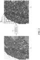
- FIG. 2 shows an exemplary biological specimen image 210 (in this example, an H&E image), and an exemplary plurality of superpixels 220 generated for image 210 by superpixel generator 110.
- each superpixel can include the same number of pixels or a different number of pixels (e.g., within a certain range), but in either case the number of superpixels can be significantly (e.g., one or more orders of magnitude) lower than the number of pixels in image 210.
- the superpixels can be provided to feature extractor 111.
- Feature extractor 111 may extract from (or generate for) each superpixel a plurality of image features characterizing (or representing) the superpixel.
- the extracted image features may include, for example, texture features such Haralick features, bag-of-words features and the like.
- the values of the plurality of image features may be combined into a high-dimensional vector, hereinafter referred to as the "feature vector" characterizing the superpixel. For example, if M features are extracted for each superpixel, each superpixel can be characterized by an M-dimensional feature vector.
- Clustering engine 112 may then cluster the superpixels by assigning each superpixel to a particular cluster of superpixels.
- clustering engine 112 may generally cluster N superpixels into K clusters. The clustering may be performed based on the similarities of the feature vectors associate with each superpixel. For example, each cluster may be associated with a centroid vector, such that feature vector of each superpixel in the cluster is closest to the centroid vector of that cluster than to the centroid of any other cluster.
- the number of clusters K can be predefined for a particular application. For example, for a typical image segmentation problem in which the image needs to be segmented into a predefined maximum number of different regions (e.g., 5), K clusters can be set to a number that is larger but is within the order of magnitude of the predefined maximum number of regions (e.g., 15, 20, or 25). In some examples, the number of clusters K can be dynamically adjusted based on user input.
- clustering engine 112 may cluster the superpixels using a k-means clustering algorithm such as the Lloyd's algorithm, or using any related clustering algorithms such as the k-medians clustering algorithm; the k-medoids or the partitioning around medoids (PAM) algorithm; the Fuzzy C-Means Clustering algorithm; the Gaussian mixture models trained with expectation-maximization algorithm; the k-means++ algorithm; hierarchical variants such as Bisecting k-means, X-means clustering, or G-means clustering; and the like.
- clustering engine 112 may use any other algorithm suitable for clustering the superpixels into a predefined number of clusters based on the similarities of superpixels' feature vectors.
- clustering engine 112 may use a Euclidean distance as the distance metric for performing the clustering. In other embodiments, clustering engine 112 may use other distance metrics such as the sum of absolute differences, correlation and hamming distance, and so forth.
- clustering engine 112 may use a non-weighted Euclidean distance, where each image feature in the feature vector has the same weight. In other embodiments, however, clustering engine 112 may use a weighted Euclidean (or non-Euclidean) distance during clustering, weighing some image features higher than other. For example, in some embodiments, clustering engine 112 may determine and assign different feature weights to different image features. For example, clustering engine 112 may collect (e.g., using user interface module 115) at least one annotation (e.g., a scribble or a line) identifying a plurality of similar superpixels, i.e., superpixels that the user considers to belong to the same segment or category.
- annotation e.g., a scribble or a line
- Clustering engine 112 may then determine, based on the feature vectors of the similar superpixels, which image features in the feature vectors should be assigned higher feature weights than others. Some methods and systems of determining and assigning weights to different image features are described in U.S. Provisional Patent Application No. 62/136381 and in International Patent Publication No. WO/2017150873 , the entireties of which are hereby incorporated by reference.
- clustering engine 112 may precalculate the distances between every two clusters, i.e., the distances between centroid vectors of each cluster and each other cluster. Thus, in some embodiments, clustering engine 112 may precalculate at least K(K-1)/2 distances, which is the number of different combinations of two clusters within K clusters. Clustering engine 112 may calculate the distances between the clusters using the same measure of distance that was used for generating the clusters, for example.
- storage interface 113 may store into a memory (e.g., memory 116 or any other volatile and/or non-volatile memory embedded in or coupled to system 100) either the centroid vectors of all the clusters, or the precalculated distances between the clusters, or both. It will be appreciated that for some image processing tasks, such as the segmentation task discussed in more detail below, only the differences (i.e., distances) between the different superpixels may be required. Thus, in some embodiments, only the precalculated distances between the clusters, without the clusters' centroid vectors can be stored, thereby further reducing the amount of stored data.
- a memory e.g., memory 116 or any other volatile and/or non-volatile memory embedded in or coupled to system 100
- the precalculated distances can be stored, for example, in a table such as a look-up table, or in any other type of data structure.
- the clusters' centroid vectors can be stored, and the distances between the clusters may not be precalculated or stored.
- storage interface 113 may store, for each superpixel, clustering information identifying the cluster to which the superpixel has been assigned. Clustering information can identify the cluster by a number (e.g., 1- K) or by any other type of identifier. Furthermore, in some embodiments, storage interface 113 may also store position information identifying the position of each superpixel within the biological sample image, or at least its relative position to other superpixels.
- All information stored by storage interface 113 can later be retrieved by storage interface 113 and provided to image analysis system 100 that can perform various image processing tasks, examples of which are provided below. It should be evident to a person skilled in the art that by storing only limited amount of data for each superpixel, such as storing its clustering information without storing its feature vector containing its image features, significant reductions in memory consumption and in storage/retrieval times can be achieved.
- N be the number of superpixels generated for a given image
- M be the number of image features extracted for each superpixel
- U be the number of bytes representing each image feature
- K be the number of clusters.
- the original feature vectors for all superpixels occupy NxMxU bytes.
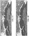
- FIG. 3A shows an exemplary H&E image containing various types of regions, such as the necrosis region, the cancer (tumor) region, the lymphocytes region, and the background region.
- FIG. 3B shows the same image being overlaid with boundaries of exemplary superpixels generated for the image by superpixel generator 110.
- the following image features are generated for each of the three color channels R, G, and B: a histogram of the intensity, a histogram of the gradient magnitude, and a histogram of the gradient direction.
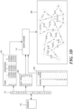
- FIG. 3C further illustrates some embodiments discussed above.
- FIG. 3C shows superpixel generator 110 obtaining an exemplary biological sample image 310 and producing a plurality of superpixels 320 based on biological sample image 310.
- Feature extractor 111 can then obtain the plurality of superpixels 320 and extract feature vectors 330 for all the superpixels.
- Clustering engine 112 can obtain feature vectors 330, and based at least on feature vectors 330, generated a predefined number of (in this example, four) clusters A, B, C, and D arranged, for example, as illustrated in clustering information 350 and visualized by clustering information image 340.
- each cluster can be characterized or represented by a centroid vector, as illustrated in exemplary centroid vector table 370.
- Clustering engine 112 can also precalculate distances between each pair of different clusters, as illustrated in exemplary distance table 360.
- Storage interface 113 can then store clustering information 350 into memory 116 (or any other memory). As discussed above, storage interface 113 can also store into memory distance table 360 and/or centroid vector table 370.
- FIG. 3D shows graph engine 112 retrieving from memory 116 (through storage interface 113) clustering information 350, and distance table 360 and/or centroid vector table 370. Based at least on this information, graph engine 112 generates an exemplary graph 380.
- image analysis system 100 may use storage interface 113 to obtain, for each superpixel, clustering information identifying the cluster to which the superpixel belongs, and then use that that cluster's centroid vector instead of the superpixel's feature vector. Because the clustering algorithm ensures that all superpixels in a given cluster are relatively similar, the centroid vector of the cluster can be a sufficiently good approximation of the feature vector of each superpixel in the cluster, and the greater the number of clusters used, the better the approximation can be.
- some image processing tasks rely solely on the distance between two superpixels, i.e., the distance between the superpixels' feature vectors.
- Such tasks can approximate the distance between superpixels by using the distance between centroid vectors of the two clusters to which the superpixels have been assigned.
- the distance can be calculated in real time based on the centroid vectors, if the centroid vectors have been stored. Alternatively, the distance can be obtained directly (without additional calculations) from a table or another data structure, if the distances have been precalculated and stored, as discussed above.
- graph-based image processing tasks may represent the superpixels in the image in the form of an interconnected graph, and perform various calculations using graph algorithms.
- image analysis system 100 may include a graph engine 114.
- Graph engine 114 may be configured to obtain (e.g., through storage interface) clustering information identifying which superpixel belongs to which cluster, and position information identifying the position of each superpixel within the image, or at least its position relative to other superpixels.
- graph engine 114 can construct a graph that includes a plurality of nodes (vertices) where adjacent nodes correspond to adjacent superpixels within the image.
- nodes in the graph can be connected to each other directly (i.e., by a single edge) if and only if the superpixels those nodes represent are adjacent within the image.
- the edges between every two nodes may correspond to the distance between the superpixels represented by the nodes, where the distance corresponds to the distance between the feature vectors of the superpixels and is approximated by the distance between the clusters to which the superpixels have been assigned.
- the distance between the clusters can be either calculated in real time if clusters' centroid vectors have been stored in the memory, or obtained from a table of precalculated distances if such a table has been stored in the memory.
- graph engine 114 may use the graph to perform a graph-based image processing task.
- One such task is a segmentation operation whose objective is to segment the image (or the superpixels) into a plurality of regions.
- graph engine 114 may collect from the user (e.g., through user-interface module 115) one or more annotations, where each annotation identifies a plurality of same-segment superpixels, i.e., superpixels that the user considers to belong to the same segment.
- the user can draw one or more lines on top of one or more superpixels belonging to a first segment, then draw one or more additional lines (e.g., with a different color) on top of one or more superpixels belonging to a second segment, and so forth.
- Graph engine 114 can then use the graph to determine, for each superpixel that has not been annotated, to which of the annotated superpixels is the superpixel closest and assign it to the same segment as the annotated superpixel.
- graph engine 114 can use the shortest path measure, a smallest maximum path measure, or any other suitable graph distance measure.
- the annotations obtained from the user may be further used to determine different weights associated with different image features within the image, as discussed above.
- the weights can then be used by clustering engine 112, for example, in subsequent clustering operations.
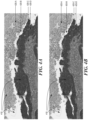
- FIG. 4A shows the results of a segmentation operation operating on non-compressed data, i.e., on actual image features of each superpixel
- FIG. 4B shows the results of a segmentation operation operating on image features that were compressed (encoded) using the techniques described above. Both operations were initialized using the same user annotations 410. It can be appreciated that the segmentation results of the two operations (i.e., segments 420-A, 420-B, 420-C, and 420-D) are very similar, and it is appreciated that higher accuracy can be achieved with additional user annotations.
- Method 500 may begin at block 510 where the method may obtain a biological sample image, where, as discussed above, the biological sample may have been stained with staining platform 123, and the image of the biological sample may have been obtained by scanning platform 125.
- the method may generate a plurality of superpixels for the image, i.e., group the pixels within the image into a plurality of superpixels, using, for example, superpixel generator 110.
- the method may extract, for each superpixel (or substantially for each superpixel as in some embodiments some superpixels can be omitted or disregarded) a feature vector comprising a plurality of image features that characterize the superpixel. As discussed above, this operation can be performed, for example, by feature extractor 111.
- the method may include different or additional steps, such as retrieving the centroid vector of each cluster and/or the distances between each two clusters, and using the centroid vector of each cluster and/or the distances between each clusters to construct a graph, and performing a graph-based image processing task based on the graph.
- the graph-based image processing task may include a segmentation operation that can use the graph and one or more user annotations to segment the biological specimen image into a plurality of segments.
- the method may also include determining a set of different weights for the different image features, where the generation of the predefined number of clusters and the association of each superpixel with the cluster are based at least in part on the set of different weights.
- the hardware may include any type of analog and/or digital circuitry, such as integrated circuits (IC) (e.g., application-specific integrated circuits (ASICs) or field-programmable gate arrays (FPGAs)), or any other type of special- or general-purpose electronic circuitry.
- IC integrated circuits
- ASICs application-specific integrated circuits
- FPGAs field-programmable gate arrays
- the software or firmware may include any type of processor executable instructions that can be stored on any type of tangible non-transitory computer-readable medium, where the instructions can be executed by a processing resource, causing the processing resource to implement the functionality of the respective component.
- the executable instructions can be stored in memory 116, or in any other non-transitory computer-readable storage medium, which can be implemented as any combination of any type of volatile or non-volatile memories, such as random-access memories (RAMs), read-only memories such as an Electrically-Erasable Programmable Read-Only Memory (EEPROM), flash memories, hard drives, solid state drives, optical discs, and the like.
- RAMs random-access memories
- EEPROM Electrically-Erasable Programmable Read-Only Memory
- flash memories hard drives, solid state drives, optical discs, and the like.
- the non-transitory computer-readable medium can be integrated in the same device as the processor resource or it may be separate but accessible to that device and the processor resource.
- the program instructions can be part of an installation package that when installed can be executed by the processor resource to implement the corresponding component.
- the computer-readable medium may be a portable medium such as a CD, DVD, or flash drive or a memory maintained by a server from which the installation package can be downloaded and installed.
- the program instructions may be part of an application or applications already installed, and the computer-readable medium may include integrated memory such as a hard drive, solid state drive, random access memory (RAM), read-only memory (ROM), and the like.
- the processing resource for executing the instructions can be, for example, processor 117, or it can be any other processing resource, such as a CPU, a GPU, an FPGA, a TPU, another type of processor configured to retrieve and execute instructions, or a combination thereof.
- the processing resource can be integrated in a single device or distributed across devices, which may be integrated with image analysis system 100 or communicatively coupled to image analysis system 100.
- FIG. 6 illustrates an example computing device 600, in according to some embodiments.
- Computing system 600 may correspond, for example, to system analysis 100, to a portion thereof, or to any other computing system suitable for performing the functionality described below.
- Computing system 600 may include a processing resource 610, which, as discussed above, may correspond to processor 117 or to any other processing resource suitable for performing the functionality described below.
- Computing system 600 may also include a non-transitory computer-readable storage medium 620, which, as discussed above, may correspond to memory 116 or to any other non-transitory computer-readable storage medium capable of storing instructions executable by processing resource 610.
- medium 620 may store instructions 622, 624, 626, 628, 630, 632, and any other instructions that can be fetched, decoded, and executed by processing resource 610.
- processing resource 610 may include any number of electronic circuits comprising any number of electronic components for performing the functionality of any number of instructions in machine-readable storage medium 620.
- executable instruction representations e.g., boxes
- executable instructions and/or electronic circuits included within one box may, in other examples, be included in a different box shown in the figures or in a different box not shown.
- Instructions 622 when executed by a processor (e.g., 610), may cause a computing system (e.g., 600) to obtain a biological specimen image.
- Instructions 624 when executed by the processor, may cause the computing system to group pixels of the biological specimen image into a plurality of superpixels.
- Instructions 626 when executed by the processor, may cause the computing system to extract, for each superpixel, a feature vector comprising a plurality of image features characterizing the superpixel.
- Instructions 628 when executed by the processor, may cause the computing system to, based on the feature vectors extracted for the plurality of superpixels, generate a predefined number of clusters, each cluster being characterized by a centroid vector, and associate each superpixel with a cluster whose centroid vector is the closest to the feature vector of the superpixel.
- Instructions 630 when executed by the processor, may cause the computing system to store, for each superpixel, an identifier of a cluster whose centroid vector is closest to the feature vector of the superpixel.
- Instructions 632 when executed by the processor, may cause the computing system to store the centroid vector of each cluster in the plurality of clusters and/or distances between each two clusters in the predefined number of clusters.
Landscapes
- Engineering & Computer Science (AREA)
- Theoretical Computer Science (AREA)
- Physics & Mathematics (AREA)
- General Physics & Mathematics (AREA)
- Data Mining & Analysis (AREA)
- Computer Vision & Pattern Recognition (AREA)
- Life Sciences & Earth Sciences (AREA)
- Bioinformatics & Cheminformatics (AREA)
- Bioinformatics & Computational Biology (AREA)
- Evolutionary Biology (AREA)
- Evolutionary Computation (AREA)
- General Engineering & Computer Science (AREA)
- Artificial Intelligence (AREA)
- Multimedia (AREA)
- General Health & Medical Sciences (AREA)
- Health & Medical Sciences (AREA)
- Biomedical Technology (AREA)
- Molecular Biology (AREA)
- Probability & Statistics with Applications (AREA)
- Human Computer Interaction (AREA)
- Medical Informatics (AREA)
- Nuclear Medicine, Radiotherapy & Molecular Imaging (AREA)
- Radiology & Medical Imaging (AREA)
- Quality & Reliability (AREA)
- Image Analysis (AREA)
Claims (11)
- Bildanalysesystem zur Analyse von Bildern biologischer Proben, wobei das Bildanalysesystem Folgendes umfasst:einen Superpixel-Generator, der ausgelegt ist, um ein Bild einer biologischen Probe zu erhalten und Pixel des Bildes der biologischen Probe in eine Vielzahl von Superpixeln zu gruppieren;einen Merkmalsextraktor, der ausgelegt ist, um aus jedem Superpixel in der Vielzahl von Superpixeln einen Merkmalsvektor zu extrahieren, der eine Vielzahl von Bildmerkmalen umfasst;eine Clustering-Engine, die ausgelegt ist, um die Vielzahl von Superpixeln einer vordefinierten Anzahl von Clustern zuzuordnen, wobei jeder Cluster durch einen Schwerpunktvektor von Merkmalsvektoren von Superpixeln, die dem Cluster zugeordnet sind, charakterisiert ist;eine Speicherschnittstelle, die ausgelegt ist, um für jedes Superpixel in der Vielzahl von Superpixeln Clustering-Informationen zu speichern, die einen Cluster identifizieren, dem das Superpixel zugeordnet ist; undeine Graph-Engine, die ausgelegt ist, um:wobei:die Clustering-Informationen zu erhalten, die von der Speicher-Schnittstelle gespeichert werden;zumindest teilweise auf den Clustering-Informationen basierend, einen Graphen zu erstellen, der eine Vielzahl von Knoten umfasst, wobei benachbarte Knoten benachbarten Superpixeln im Bild der biologischen Probe entsprechen und durch eine gewichtete Kante verbunden sind, wobei die gewichtete Kante eine Gewichtung aufweist, die einem Abstand zwischen Clustern entspricht, zu denen die benachbarten Superpixel gehören; undden Graphen zu verwenden, um eine Graph-basierte Bildverarbeitungsaufgabe auszuführen,die Clustering-Engine weiters ausgelegt ist, um Abstände zwischen jeweils zwei Clustern in der vordefinierten Anzahl von Clustern vorzuberechnen;die Speicherschnittstelle ausgelegt ist, um die vorberechneten Abstände zu speichern; unddie Graph-Engine weiters ausgelegt ist, um die vorberechneten Abstände, die von der Speicherschnittstelle gespeichert werden, zu erhalten und den Graphen basierend auf den vorberechneten Abständen zu erstellen.
- Bildanalysesystem nach Anspruch 1, wobei die Graph-basierte Bildverarbeitungsaufgabe eine Segmentierungsoperation umfasst, welche die Vielzahl von Superpixeln in eine Vielzahl von Segmenten gruppiert.
- Bildanalysesystem nach einem der Ansprüche 1 bis 2, wobei:die Speicherschnittstelle weiters ausgelegt ist, um Schwerpunktvektoren der vordefinierten Anzahl von Clustern zu speichern; unddie Graph-Engine weiters ausgelegt ist, um die Schwerpunktvektoren zu erhalten, Abstände jeweils zwischen zwei Clustern in der vordefinierten Anzahl von Clustern basierend auf den Schwerpunktvektoren zu berechnen und den Graphen basierend auf den berechneten Abständen zu erstellen.
- Bildanalysesystem nach einem der Ansprüche 1 bis 2, das weiters ein Benutzerschnittstellenmodul umfasst, das ausgelegt ist, um zumindest eine Anmerkung von einem Benutzer zu erhalten, die eine Vielzahl von Superpixeln aus demselben Segment im Bild der biologischen Probe identifiziert.
- Bildanalysesystem nach Anspruch 4, wobei die Clustering-Engine weiters ausgelegt ist, um basierend auf der zumindest einen Anmerkung einen Satz von Merkmalsgewichtungen zu bestimmen, die der Vielzahl von Bildmerkmalen zugeordnet sind.
- Bildanalysesystem nach Anspruch 5, wobei die Clustering-Engine ausgelegt ist, um die Vielzahl von Superpixeln basierend zumindest auf dem vorbestimmten Satz von Merkmalsgewichtungen der vordefinierten Anzahl von Clustern zuzuordnen.
- Verfahren zum Kodieren von Bildmerkmalen eines Bildes einer biologischen Probe, das mittels eines Objektträgerscanners erhalten wurde, wobei das Verfahren Folgendes umfasst:Erhalten des Bildes der biologischen Probe;Gruppieren von Pixeln des Bildes der biologischen Probe in eine Vielzahl von Superpixeln;für jedes Superpixel, Extrahieren eines Merkmalsvektors, der eine Vielzahl von Bildmerkmalen umfasst, die das Superpixel charakterisieren;basierend auf den Merkmalsvektoren, die für die Vielzahl von Superpixeln extrahiert wurden, Generieren einer vordefinierten Anzahl von Clustern, wobei jeder Cluster durch einen Schwerpunktvektor charakterisiert ist,Zuordnen jedes Superpixels zu einem Cluster, dessen Schwerpunktvektor am nächsten zum Merkmalsvektor des Superpixels ist;für jedes Superpixel, Speichern eines Identifikators eines Clusters, dessen Schwerpunktvektor am nächsten zum Merkmalsvektor des Superpixels ist;Vorberechnen der Abstände zwischen jeweils zwei Clustern innerhalb der vordefinierten Anzahl von Clustern;Speichern der vorberechneten Abstände zwischen jeweils zwei Clustern in der vordefinierten Anzahl von Clustern;Erhalten der gespeicherten Identifikatoren und der gespeicherten vorberechneten Abstände und Erstellen, basierend auf den gespeicherten Identifikatoren und den gespeicherten vorberechneten Abständen, eines Graphen, der eine Vielzahl von Knoten umfasst, wobei benachbarte Knoten benachbarten Superpixeln im Bild der biologischen Probe entsprechen und durch eine gewichtete Kante verbunden sind, und wobei die gewichtete Kante eine Gewichtung aufweist, die einem Abstand zwischen Clustern entspricht, zu denen die benachbarten Superpixel gehören; undAusführen einer Graph-basierten Bildverarbeitungsaufgabe.
- Verfahren nach Anspruch 7, wobei die Graph-basierte Bildverarbeitungsaufgabe eine Segmentierungsoperation umfasst, wobei die Segmentierungsoperation den Graphen und eine oder mehrere Benutzeranmerkungen verwendet, um das Bild der biologischen Probe in eine Vielzahl von Segmenten zu segmentieren.
- Verfahren nach Anspruch 7 oder Anspruch 8, das weiters Folgendes umfasst:
Bestimmen eines Satzes von unterschiedlichen Gewichtungen, die der Vielzahl von Bildmerkmalen im Merkmalsvektor zugeordnet sind, wobei die Generierung der vordefinierten Anzahl von Clustern und die Zuordnung jedes Superpixels zu einem Cluster zumindest teilweise auf dem Satz von unterschiedlichen Gewichtungen basieren. - Verfahren nach einem der Ansprüche 7 bis 9, wobei das Generieren der vordefinierten Anzahl von Clustern das Ausführen eines k-Means-Clustering der Merkmalsvektoren der Vielzahl von Superpixeln in die vordefinierte Anzahl von Clustern umfasst.
- Nichttransitorisches computerlesbares Medium, auf dem Anweisungen gespeichert sind, die bei Ausführung durch eine Verarbeitungsressource eines Rechensystems bewirken, dass das Rechensystem ein Verfahren nach einem der Ansprüche 7 bis 10 ausführt.
Applications Claiming Priority (2)
| Application Number | Priority Date | Filing Date | Title |
|---|---|---|---|
| US201662416660P | 2016-11-02 | 2016-11-02 | |
| PCT/EP2017/077999 WO2018083142A1 (en) | 2016-11-02 | 2017-11-02 | Systems and methods for encoding image features of high-resolution digital images of biological specimens |
Publications (2)
| Publication Number | Publication Date |
|---|---|
| EP3535685A1 EP3535685A1 (de) | 2019-09-11 |
| EP3535685B1 true EP3535685B1 (de) | 2025-02-26 |
Family
ID=60320855
Family Applications (1)
| Application Number | Title | Priority Date | Filing Date |
|---|---|---|---|
| EP17797593.5A Active EP3535685B1 (de) | 2016-11-02 | 2017-11-02 | Systeme und verfahren zur codierung von bildmerkmalen von hochauflösenden digitalen bildern von biologischen proben |
Country Status (3)
| Country | Link |
|---|---|
| US (1) | US11176412B2 (de) |
| EP (1) | EP3535685B1 (de) |
| WO (1) | WO2018083142A1 (de) |
Families Citing this family (10)
| Publication number | Priority date | Publication date | Assignee | Title |
|---|---|---|---|---|
| US11132529B2 (en) * | 2016-11-16 | 2021-09-28 | Ventana Medical Systems, Inc. | Convolutional neural networks for locating objects of interest in images of biological samples |
| KR102864472B1 (ko) * | 2019-05-23 | 2025-09-25 | 삼성에스디에스 주식회사 | 기계 학습을 위한 이미지 처리 장치 및 방법 |
| CN111611954B (zh) * | 2020-05-28 | 2023-11-24 | 云南电网有限责任公司电力科学研究院 | 基于改进K-means算法的高光谱图像分类方法及装置 |
| CN112037167B (zh) * | 2020-07-21 | 2023-11-24 | 苏州动影信息科技有限公司 | 一种基于影像组学和遗传算法的目标区域确定系统 |
| CN113657333B (zh) * | 2021-08-23 | 2024-01-12 | 深圳科卫机器人科技有限公司 | 警戒线识别方法、装置、计算机设备及存储介质 |
| EP4164222A1 (de) * | 2021-10-08 | 2023-04-12 | Robert Bosch GmbH | Verlustbehaftete kompression von videoinhalt in eine graph-darstellung |
| CN114693816B (zh) * | 2022-04-02 | 2023-04-28 | 陕西合友网络科技有限公司 | 一种智能化图像大数据存储方法 |
| CN114567936B (zh) * | 2022-04-11 | 2025-03-04 | 国网浙江省电力有限公司信息通信分公司 | 一种基站网络切片下业务分类方法、终端与流程 |
| CN115359075B (zh) * | 2022-10-24 | 2023-03-24 | 济南霍兹信息科技有限公司 | 基于云计算的软件开发应用数据处理方法 |
| US12231300B2 (en) * | 2023-06-14 | 2025-02-18 | Cisco Technology, Inc. | Heterogeneous graph learning-based unified network representation |
Family Cites Families (3)
| Publication number | Priority date | Publication date | Assignee | Title |
|---|---|---|---|---|
| WO2006054248A2 (en) * | 2004-11-19 | 2006-05-26 | Koninklijke Philips Electronics, N.V. | In-situ data collection architecture for computer-aided diagnosis |
| JP6192271B2 (ja) * | 2012-08-22 | 2017-09-06 | キヤノン株式会社 | 画像処理装置、画像処理方法及びプログラム |
| WO2016150873A1 (en) | 2015-03-20 | 2016-09-29 | Ventana Medical Systems, Inc. | System and method for image segmentation |
-
2017
- 2017-11-02 WO PCT/EP2017/077999 patent/WO2018083142A1/en not_active Ceased
- 2017-11-02 EP EP17797593.5A patent/EP3535685B1/de active Active
- 2017-11-02 US US16/612,170 patent/US11176412B2/en active Active
Non-Patent Citations (2)
| Title |
|---|
| ANONYMOUS: "Vector quantization - Wikipedia", 10 February 2016 (2016-02-10), XP055845364, Retrieved from the Internet <URL:https://en.wikipedia.org/w/index.php?title=Vector_quantization&oldid=704284850> [retrieved on 20210928] * |
| BELEVICH ILYA ET AL: "Microscopy Image Browser: A Platform for Segmentation and Analysis of Multidimensional Datasets", vol. 14, no. 1, 4 January 2016 (2016-01-04), pages e1002340, XP055844583, Retrieved from the Internet <URL:https://storage.googleapis.com/plos-corpus-prod/10.1371/journal.pbio.1002340/1/pbio.1002340.pdf?X-Goog-Algorithm=GOOG4-RSA-SHA256&X-Goog-Credential=wombat-sa@plos-prod.iam.gserviceaccount.com/20210924/auto/storage/goog4_request&X-Goog-Date=20210924T151653Z&X-Goog-Expires=86400&X-Goog-SignedHeaders=h> DOI: 10.1371/journal.pbio.1002340 * |
Also Published As
| Publication number | Publication date |
|---|---|
| US11176412B2 (en) | 2021-11-16 |
| US20200175325A1 (en) | 2020-06-04 |
| WO2018083142A1 (en) | 2018-05-11 |
| EP3535685A1 (de) | 2019-09-11 |
Similar Documents
| Publication | Publication Date | Title |
|---|---|---|
| EP3535685B1 (de) | Systeme und verfahren zur codierung von bildmerkmalen von hochauflösenden digitalen bildern von biologischen proben | |
| US11756318B2 (en) | Convolutional neural networks for locating objects of interest in images of biological samples | |
| US11901077B2 (en) | Multiple instance learner for prognostic tissue pattern identification | |
| Oskal et al. | A U-net based approach to epidermal tissue segmentation in whole slide histopathological images | |
| JP7504116B2 (ja) | 距離ベース類似性ラベルを使用する機械学習 | |
| AU2018394106B2 (en) | Processing of histology images with a convolutional neural network to identify tumors | |
| Stegmaier et al. | Real-time three-dimensional cell segmentation in large-scale microscopy data of developing embryos | |
| Shahzad et al. | Robust Method for Semantic Segmentation of Whole‐Slide Blood Cell Microscopic Images | |
| EP3440629B1 (de) | Erzeugung eines räumlichen index zur ihc-bildanalyse | |
| CN112703531A (zh) | 生成组织图像的注释数据 | |
| CN117859123A (zh) | 全载玻片图像搜索 | |
| US20220092774A1 (en) | Method and apparatus for analysis of histopathology image data | |
| Friebel et al. | Guided interactive image segmentation using machine learning and color-based image set clustering | |
| Cosatto et al. | A multi-scale conditional deep model for tumor cell ratio counting | |
| Chaddad et al. | Real-time abnormal cell detection using a deformable snake model | |
| Lukashevich et al. | An approach to cell nuclei counting in histological image analysis | |
| Villareal et al. | Patch-based convolutional neural networks for TCGA-BRCA breast cancer classification | |
| Gupta et al. | Microscopy cancer cell imaging in b-lineage acute lymphoblastic leukemia | |
| Chai et al. | A Review of Current Cell Annotation Systems for Histopathology Images | |
| Santamaria-Pang et al. | Epithelial cell segmentation via shape ranking | |
| Everitt | Automated histological labeling of mass spectrometry imaging | |
| Blahova et al. | Blood Smear Leukocyte Identification Using an Image Segmentation Approach | |
| HK40051109A (en) | Multiple instance learner for prognostic tissue pattern identification | |
| Rittscher et al. | Mapping for tissue based cytometry | |
| Kiss et al. | High Resolution Digital Tissue Image Processing using Texture Image Databases |
Legal Events
| Date | Code | Title | Description |
|---|---|---|---|
| STAA | Information on the status of an ep patent application or granted ep patent |
Free format text: STATUS: UNKNOWN |
|
| STAA | Information on the status of an ep patent application or granted ep patent |
Free format text: STATUS: THE INTERNATIONAL PUBLICATION HAS BEEN MADE |
|
| PUAI | Public reference made under article 153(3) epc to a published international application that has entered the european phase |
Free format text: ORIGINAL CODE: 0009012 |
|
| STAA | Information on the status of an ep patent application or granted ep patent |
Free format text: STATUS: REQUEST FOR EXAMINATION WAS MADE |
|
| 17P | Request for examination filed |
Effective date: 20190603 |
|
| AK | Designated contracting states |
Kind code of ref document: A1 Designated state(s): AL AT BE BG CH CY CZ DE DK EE ES FI FR GB GR HR HU IE IS IT LI LT LU LV MC MK MT NL NO PL PT RO RS SE SI SK SM TR |
|
| AX | Request for extension of the european patent |
Extension state: BA ME |
|
| DAV | Request for validation of the european patent (deleted) | ||
| DAX | Request for extension of the european patent (deleted) | ||
| STAA | Information on the status of an ep patent application or granted ep patent |
Free format text: STATUS: EXAMINATION IS IN PROGRESS |
|
| 17Q | First examination report despatched |
Effective date: 20200311 |
|
| REG | Reference to a national code |
Ref country code: DE Ref legal event code: R079 Free format text: PREVIOUS MAIN CLASS: G06K0009000000 Ipc: G06V0020690000 Ref document number: 602017088024 Country of ref document: DE |
|
| GRAP | Despatch of communication of intention to grant a patent |
Free format text: ORIGINAL CODE: EPIDOSNIGR1 |
|
| STAA | Information on the status of an ep patent application or granted ep patent |
Free format text: STATUS: GRANT OF PATENT IS INTENDED |
|
| RIC1 | Information provided on ipc code assigned before grant |
Ipc: G06V 10/426 20220101ALI20240911BHEP Ipc: G06V 10/26 20220101ALI20240911BHEP Ipc: G06T 7/00 20170101ALI20240911BHEP Ipc: G06F 18/23213 20230101ALI20240911BHEP Ipc: G06V 20/69 20220101AFI20240911BHEP |
|
| INTG | Intention to grant announced |
Effective date: 20240926 |
|
| GRAS | Grant fee paid |
Free format text: ORIGINAL CODE: EPIDOSNIGR3 |
|
| GRAA | (expected) grant |
Free format text: ORIGINAL CODE: 0009210 |
|
| STAA | Information on the status of an ep patent application or granted ep patent |
Free format text: STATUS: THE PATENT HAS BEEN GRANTED |
|
| AK | Designated contracting states |
Kind code of ref document: B1 Designated state(s): AL AT BE BG CH CY CZ DE DK EE ES FI FR GB GR HR HU IE IS IT LI LT LU LV MC MK MT NL NO PL PT RO RS SE SI SK SM TR |
|
| REG | Reference to a national code |
Ref country code: GB Ref legal event code: FG4D |
|
| REG | Reference to a national code |
Ref country code: CH Ref legal event code: EP |
|
| REG | Reference to a national code |
Ref country code: DE Ref legal event code: R096 Ref document number: 602017088024 Country of ref document: DE |
|
| REG | Reference to a national code |
Ref country code: IE Ref legal event code: FG4D |
|
| REG | Reference to a national code |
Ref country code: NL Ref legal event code: MP Effective date: 20250226 |
|
| PG25 | Lapsed in a contracting state [announced via postgrant information from national office to epo] |
Ref country code: RS Free format text: LAPSE BECAUSE OF FAILURE TO SUBMIT A TRANSLATION OF THE DESCRIPTION OR TO PAY THE FEE WITHIN THE PRESCRIBED TIME-LIMIT Effective date: 20250526 |
|
| PG25 | Lapsed in a contracting state [announced via postgrant information from national office to epo] |
Ref country code: FI Free format text: LAPSE BECAUSE OF FAILURE TO SUBMIT A TRANSLATION OF THE DESCRIPTION OR TO PAY THE FEE WITHIN THE PRESCRIBED TIME-LIMIT Effective date: 20250226 |
|
| PG25 | Lapsed in a contracting state [announced via postgrant information from national office to epo] |
Ref country code: PL Free format text: LAPSE BECAUSE OF FAILURE TO SUBMIT A TRANSLATION OF THE DESCRIPTION OR TO PAY THE FEE WITHIN THE PRESCRIBED TIME-LIMIT Effective date: 20250226 |
|
| PG25 | Lapsed in a contracting state [announced via postgrant information from national office to epo] |
Ref country code: ES Free format text: LAPSE BECAUSE OF FAILURE TO SUBMIT A TRANSLATION OF THE DESCRIPTION OR TO PAY THE FEE WITHIN THE PRESCRIBED TIME-LIMIT Effective date: 20250226 |
|
| REG | Reference to a national code |
Ref country code: LT Ref legal event code: MG9D |
|
| PG25 | Lapsed in a contracting state [announced via postgrant information from national office to epo] |
Ref country code: NO Free format text: LAPSE BECAUSE OF FAILURE TO SUBMIT A TRANSLATION OF THE DESCRIPTION OR TO PAY THE FEE WITHIN THE PRESCRIBED TIME-LIMIT Effective date: 20250526 Ref country code: IS Free format text: LAPSE BECAUSE OF FAILURE TO SUBMIT A TRANSLATION OF THE DESCRIPTION OR TO PAY THE FEE WITHIN THE PRESCRIBED TIME-LIMIT Effective date: 20250626 |
|
| PG25 | Lapsed in a contracting state [announced via postgrant information from national office to epo] |
Ref country code: NL Free format text: LAPSE BECAUSE OF FAILURE TO SUBMIT A TRANSLATION OF THE DESCRIPTION OR TO PAY THE FEE WITHIN THE PRESCRIBED TIME-LIMIT Effective date: 20250226 |
|
| PG25 | Lapsed in a contracting state [announced via postgrant information from national office to epo] |
Ref country code: HR Free format text: LAPSE BECAUSE OF FAILURE TO SUBMIT A TRANSLATION OF THE DESCRIPTION OR TO PAY THE FEE WITHIN THE PRESCRIBED TIME-LIMIT Effective date: 20250226 |
|
| PG25 | Lapsed in a contracting state [announced via postgrant information from national office to epo] |
Ref country code: LV Free format text: LAPSE BECAUSE OF FAILURE TO SUBMIT A TRANSLATION OF THE DESCRIPTION OR TO PAY THE FEE WITHIN THE PRESCRIBED TIME-LIMIT Effective date: 20250226 Ref country code: PT Free format text: LAPSE BECAUSE OF FAILURE TO SUBMIT A TRANSLATION OF THE DESCRIPTION OR TO PAY THE FEE WITHIN THE PRESCRIBED TIME-LIMIT Effective date: 20250626 |
|
| PG25 | Lapsed in a contracting state [announced via postgrant information from national office to epo] |
Ref country code: GR Free format text: LAPSE BECAUSE OF FAILURE TO SUBMIT A TRANSLATION OF THE DESCRIPTION OR TO PAY THE FEE WITHIN THE PRESCRIBED TIME-LIMIT Effective date: 20250527 Ref country code: BG Free format text: LAPSE BECAUSE OF FAILURE TO SUBMIT A TRANSLATION OF THE DESCRIPTION OR TO PAY THE FEE WITHIN THE PRESCRIBED TIME-LIMIT Effective date: 20250226 |
|
| REG | Reference to a national code |
Ref country code: AT Ref legal event code: MK05 Ref document number: 1771449 Country of ref document: AT Kind code of ref document: T Effective date: 20250226 |
|
| PG25 | Lapsed in a contracting state [announced via postgrant information from national office to epo] |
Ref country code: SE Free format text: LAPSE BECAUSE OF FAILURE TO SUBMIT A TRANSLATION OF THE DESCRIPTION OR TO PAY THE FEE WITHIN THE PRESCRIBED TIME-LIMIT Effective date: 20250226 |
|
| PG25 | Lapsed in a contracting state [announced via postgrant information from national office to epo] |
Ref country code: SM Free format text: LAPSE BECAUSE OF FAILURE TO SUBMIT A TRANSLATION OF THE DESCRIPTION OR TO PAY THE FEE WITHIN THE PRESCRIBED TIME-LIMIT Effective date: 20250226 |
|
| PG25 | Lapsed in a contracting state [announced via postgrant information from national office to epo] |
Ref country code: DK Free format text: LAPSE BECAUSE OF FAILURE TO SUBMIT A TRANSLATION OF THE DESCRIPTION OR TO PAY THE FEE WITHIN THE PRESCRIBED TIME-LIMIT Effective date: 20250226 |
|
| PG25 | Lapsed in a contracting state [announced via postgrant information from national office to epo] |
Ref country code: IT Free format text: LAPSE BECAUSE OF FAILURE TO SUBMIT A TRANSLATION OF THE DESCRIPTION OR TO PAY THE FEE WITHIN THE PRESCRIBED TIME-LIMIT Effective date: 20250226 |
|
| PG25 | Lapsed in a contracting state [announced via postgrant information from national office to epo] |
Ref country code: AT Free format text: LAPSE BECAUSE OF FAILURE TO SUBMIT A TRANSLATION OF THE DESCRIPTION OR TO PAY THE FEE WITHIN THE PRESCRIBED TIME-LIMIT Effective date: 20250226 |
|
| PG25 | Lapsed in a contracting state [announced via postgrant information from national office to epo] |
Ref country code: EE Free format text: LAPSE BECAUSE OF FAILURE TO SUBMIT A TRANSLATION OF THE DESCRIPTION OR TO PAY THE FEE WITHIN THE PRESCRIBED TIME-LIMIT Effective date: 20250226 Ref country code: CZ Free format text: LAPSE BECAUSE OF FAILURE TO SUBMIT A TRANSLATION OF THE DESCRIPTION OR TO PAY THE FEE WITHIN THE PRESCRIBED TIME-LIMIT Effective date: 20250226 |
|
| PG25 | Lapsed in a contracting state [announced via postgrant information from national office to epo] |
Ref country code: RO Free format text: LAPSE BECAUSE OF FAILURE TO SUBMIT A TRANSLATION OF THE DESCRIPTION OR TO PAY THE FEE WITHIN THE PRESCRIBED TIME-LIMIT Effective date: 20250226 |
|
| PG25 | Lapsed in a contracting state [announced via postgrant information from national office to epo] |
Ref country code: SK Free format text: LAPSE BECAUSE OF FAILURE TO SUBMIT A TRANSLATION OF THE DESCRIPTION OR TO PAY THE FEE WITHIN THE PRESCRIBED TIME-LIMIT Effective date: 20250226 |
|
| REG | Reference to a national code |
Ref country code: DE Ref legal event code: R097 Ref document number: 602017088024 Country of ref document: DE |
|
| PLBE | No opposition filed within time limit |
Free format text: ORIGINAL CODE: 0009261 |
|
| STAA | Information on the status of an ep patent application or granted ep patent |
Free format text: STATUS: NO OPPOSITION FILED WITHIN TIME LIMIT |
|
| 26N | No opposition filed |
Effective date: 20251127 |