EP3524273B1 - Cysteine modified antibody-drug conjugate and preparation method thereof - Google Patents
Cysteine modified antibody-drug conjugate and preparation method thereof Download PDFInfo
- Publication number
- EP3524273B1 EP3524273B1 EP17857845.6A EP17857845A EP3524273B1 EP 3524273 B1 EP3524273 B1 EP 3524273B1 EP 17857845 A EP17857845 A EP 17857845A EP 3524273 B1 EP3524273 B1 EP 3524273B1
- Authority
- EP
- European Patent Office
- Prior art keywords
- pab
- mmae
- antibody
- tdc
- cysteine
- Prior art date
- Legal status (The legal status is an assumption and is not a legal conclusion. Google has not performed a legal analysis and makes no representation as to the accuracy of the status listed.)
- Active
Links
Images




Classifications
-
- A—HUMAN NECESSITIES
- A61—MEDICAL OR VETERINARY SCIENCE; HYGIENE
- A61K—PREPARATIONS FOR MEDICAL, DENTAL OR TOILETRY PURPOSES
- A61K47/00—Medicinal preparations characterised by the non-active ingredients used, e.g. carriers or inert additives; Targeting or modifying agents chemically bound to the active ingredient
- A61K47/50—Medicinal preparations characterised by the non-active ingredients used, e.g. carriers or inert additives; Targeting or modifying agents chemically bound to the active ingredient the non-active ingredient being chemically bound to the active ingredient, e.g. polymer-drug conjugates
- A61K47/51—Medicinal preparations characterised by the non-active ingredients used, e.g. carriers or inert additives; Targeting or modifying agents chemically bound to the active ingredient the non-active ingredient being chemically bound to the active ingredient, e.g. polymer-drug conjugates the non-active ingredient being a modifying agent
- A61K47/68—Medicinal preparations characterised by the non-active ingredients used, e.g. carriers or inert additives; Targeting or modifying agents chemically bound to the active ingredient the non-active ingredient being chemically bound to the active ingredient, e.g. polymer-drug conjugates the non-active ingredient being a modifying agent the modifying agent being an antibody, an immunoglobulin or a fragment thereof, e.g. an Fc-fragment
-
- A—HUMAN NECESSITIES
- A61—MEDICAL OR VETERINARY SCIENCE; HYGIENE
- A61K—PREPARATIONS FOR MEDICAL, DENTAL OR TOILETRY PURPOSES
- A61K47/00—Medicinal preparations characterised by the non-active ingredients used, e.g. carriers or inert additives; Targeting or modifying agents chemically bound to the active ingredient
- A61K47/50—Medicinal preparations characterised by the non-active ingredients used, e.g. carriers or inert additives; Targeting or modifying agents chemically bound to the active ingredient the non-active ingredient being chemically bound to the active ingredient, e.g. polymer-drug conjugates
- A61K47/51—Medicinal preparations characterised by the non-active ingredients used, e.g. carriers or inert additives; Targeting or modifying agents chemically bound to the active ingredient the non-active ingredient being chemically bound to the active ingredient, e.g. polymer-drug conjugates the non-active ingredient being a modifying agent
- A61K47/68—Medicinal preparations characterised by the non-active ingredients used, e.g. carriers or inert additives; Targeting or modifying agents chemically bound to the active ingredient the non-active ingredient being chemically bound to the active ingredient, e.g. polymer-drug conjugates the non-active ingredient being a modifying agent the modifying agent being an antibody, an immunoglobulin or a fragment thereof, e.g. an Fc-fragment
- A61K47/6801—Drug-antibody or immunoglobulin conjugates defined by the pharmacologically or therapeutically active agent
- A61K47/6803—Drugs conjugated to an antibody or immunoglobulin, e.g. cisplatin-antibody conjugates
- A61K47/68031—Drugs conjugated to an antibody or immunoglobulin, e.g. cisplatin-antibody conjugates the drug being an auristatin
-
- A—HUMAN NECESSITIES
- A61—MEDICAL OR VETERINARY SCIENCE; HYGIENE
- A61K—PREPARATIONS FOR MEDICAL, DENTAL OR TOILETRY PURPOSES
- A61K47/00—Medicinal preparations characterised by the non-active ingredients used, e.g. carriers or inert additives; Targeting or modifying agents chemically bound to the active ingredient
- A61K47/50—Medicinal preparations characterised by the non-active ingredients used, e.g. carriers or inert additives; Targeting or modifying agents chemically bound to the active ingredient the non-active ingredient being chemically bound to the active ingredient, e.g. polymer-drug conjugates
- A61K47/51—Medicinal preparations characterised by the non-active ingredients used, e.g. carriers or inert additives; Targeting or modifying agents chemically bound to the active ingredient the non-active ingredient being chemically bound to the active ingredient, e.g. polymer-drug conjugates the non-active ingredient being a modifying agent
- A61K47/68—Medicinal preparations characterised by the non-active ingredients used, e.g. carriers or inert additives; Targeting or modifying agents chemically bound to the active ingredient the non-active ingredient being chemically bound to the active ingredient, e.g. polymer-drug conjugates the non-active ingredient being a modifying agent the modifying agent being an antibody, an immunoglobulin or a fragment thereof, e.g. an Fc-fragment
- A61K47/6801—Drug-antibody or immunoglobulin conjugates defined by the pharmacologically or therapeutically active agent
- A61K47/6803—Drugs conjugated to an antibody or immunoglobulin, e.g. cisplatin-antibody conjugates
- A61K47/6807—Drugs conjugated to an antibody or immunoglobulin, e.g. cisplatin-antibody conjugates the drug or compound being a sugar, nucleoside, nucleotide, nucleic acid, e.g. RNA antisense
- A61K47/6809—Antibiotics, e.g. antitumor antibiotics anthracyclins, adriamycin, doxorubicin or daunomycin
-
- A—HUMAN NECESSITIES
- A61—MEDICAL OR VETERINARY SCIENCE; HYGIENE
- A61K—PREPARATIONS FOR MEDICAL, DENTAL OR TOILETRY PURPOSES
- A61K47/00—Medicinal preparations characterised by the non-active ingredients used, e.g. carriers or inert additives; Targeting or modifying agents chemically bound to the active ingredient
- A61K47/50—Medicinal preparations characterised by the non-active ingredients used, e.g. carriers or inert additives; Targeting or modifying agents chemically bound to the active ingredient the non-active ingredient being chemically bound to the active ingredient, e.g. polymer-drug conjugates
- A61K47/51—Medicinal preparations characterised by the non-active ingredients used, e.g. carriers or inert additives; Targeting or modifying agents chemically bound to the active ingredient the non-active ingredient being chemically bound to the active ingredient, e.g. polymer-drug conjugates the non-active ingredient being a modifying agent
- A61K47/68—Medicinal preparations characterised by the non-active ingredients used, e.g. carriers or inert additives; Targeting or modifying agents chemically bound to the active ingredient the non-active ingredient being chemically bound to the active ingredient, e.g. polymer-drug conjugates the non-active ingredient being a modifying agent the modifying agent being an antibody, an immunoglobulin or a fragment thereof, e.g. an Fc-fragment
- A61K47/6801—Drug-antibody or immunoglobulin conjugates defined by the pharmacologically or therapeutically active agent
- A61K47/6803—Drugs conjugated to an antibody or immunoglobulin, e.g. cisplatin-antibody conjugates
- A61K47/6811—Drugs conjugated to an antibody or immunoglobulin, e.g. cisplatin-antibody conjugates the drug being a protein or peptide, e.g. transferrin or bleomycin
- A61K47/6817—Toxins
-
- A—HUMAN NECESSITIES
- A61—MEDICAL OR VETERINARY SCIENCE; HYGIENE
- A61K—PREPARATIONS FOR MEDICAL, DENTAL OR TOILETRY PURPOSES
- A61K47/00—Medicinal preparations characterised by the non-active ingredients used, e.g. carriers or inert additives; Targeting or modifying agents chemically bound to the active ingredient
- A61K47/50—Medicinal preparations characterised by the non-active ingredients used, e.g. carriers or inert additives; Targeting or modifying agents chemically bound to the active ingredient the non-active ingredient being chemically bound to the active ingredient, e.g. polymer-drug conjugates
- A61K47/51—Medicinal preparations characterised by the non-active ingredients used, e.g. carriers or inert additives; Targeting or modifying agents chemically bound to the active ingredient the non-active ingredient being chemically bound to the active ingredient, e.g. polymer-drug conjugates the non-active ingredient being a modifying agent
- A61K47/68—Medicinal preparations characterised by the non-active ingredients used, e.g. carriers or inert additives; Targeting or modifying agents chemically bound to the active ingredient the non-active ingredient being chemically bound to the active ingredient, e.g. polymer-drug conjugates the non-active ingredient being a modifying agent the modifying agent being an antibody, an immunoglobulin or a fragment thereof, e.g. an Fc-fragment
- A61K47/6835—Medicinal preparations characterised by the non-active ingredients used, e.g. carriers or inert additives; Targeting or modifying agents chemically bound to the active ingredient the non-active ingredient being chemically bound to the active ingredient, e.g. polymer-drug conjugates the non-active ingredient being a modifying agent the modifying agent being an antibody, an immunoglobulin or a fragment thereof, e.g. an Fc-fragment the modifying agent being an antibody or an immunoglobulin bearing at least one antigen-binding site
- A61K47/6849—Medicinal preparations characterised by the non-active ingredients used, e.g. carriers or inert additives; Targeting or modifying agents chemically bound to the active ingredient the non-active ingredient being chemically bound to the active ingredient, e.g. polymer-drug conjugates the non-active ingredient being a modifying agent the modifying agent being an antibody, an immunoglobulin or a fragment thereof, e.g. an Fc-fragment the modifying agent being an antibody or an immunoglobulin bearing at least one antigen-binding site the antibody targeting a receptor, a cell surface antigen or a cell surface determinant
-
- A—HUMAN NECESSITIES
- A61—MEDICAL OR VETERINARY SCIENCE; HYGIENE
- A61P—SPECIFIC THERAPEUTIC ACTIVITY OF CHEMICAL COMPOUNDS OR MEDICINAL PREPARATIONS
- A61P35/00—Antineoplastic agents
-
- C—CHEMISTRY; METALLURGY
- C07—ORGANIC CHEMISTRY
- C07K—PEPTIDES
- C07K16/00—Immunoglobulins [IG], e.g. monoclonal or polyclonal antibodies
-
- C—CHEMISTRY; METALLURGY
- C07—ORGANIC CHEMISTRY
- C07K—PEPTIDES
- C07K16/00—Immunoglobulins [IG], e.g. monoclonal or polyclonal antibodies
- C07K16/18—Immunoglobulins [IG], e.g. monoclonal or polyclonal antibodies against material from animals or humans
- C07K16/28—Immunoglobulins [IG], e.g. monoclonal or polyclonal antibodies against material from animals or humans against receptors, cell surface antigens or cell surface determinants
-
- C—CHEMISTRY; METALLURGY
- C07—ORGANIC CHEMISTRY
- C07K—PEPTIDES
- C07K16/00—Immunoglobulins [IG], e.g. monoclonal or polyclonal antibodies
- C07K16/18—Immunoglobulins [IG], e.g. monoclonal or polyclonal antibodies against material from animals or humans
- C07K16/28—Immunoglobulins [IG], e.g. monoclonal or polyclonal antibodies against material from animals or humans against receptors, cell surface antigens or cell surface determinants
- C07K16/2863—Immunoglobulins [IG], e.g. monoclonal or polyclonal antibodies against material from animals or humans against receptors, cell surface antigens or cell surface determinants against receptors for growth factors, growth regulators
-
- C—CHEMISTRY; METALLURGY
- C07—ORGANIC CHEMISTRY
- C07K—PEPTIDES
- C07K16/00—Immunoglobulins [IG], e.g. monoclonal or polyclonal antibodies
- C07K16/18—Immunoglobulins [IG], e.g. monoclonal or polyclonal antibodies against material from animals or humans
- C07K16/28—Immunoglobulins [IG], e.g. monoclonal or polyclonal antibodies against material from animals or humans against receptors, cell surface antigens or cell surface determinants
- C07K16/30—Immunoglobulins [IG], e.g. monoclonal or polyclonal antibodies against material from animals or humans against receptors, cell surface antigens or cell surface determinants from tumour cells
-
- A—HUMAN NECESSITIES
- A61—MEDICAL OR VETERINARY SCIENCE; HYGIENE
- A61K—PREPARATIONS FOR MEDICAL, DENTAL OR TOILETRY PURPOSES
- A61K39/00—Medicinal preparations containing antigens or antibodies
- A61K2039/505—Medicinal preparations containing antigens or antibodies comprising antibodies
-
- C—CHEMISTRY; METALLURGY
- C07—ORGANIC CHEMISTRY
- C07K—PEPTIDES
- C07K2317/00—Immunoglobulins specific features
- C07K2317/50—Immunoglobulins specific features characterized by immunoglobulin fragments
- C07K2317/52—Constant or Fc region; Isotype
-
- C—CHEMISTRY; METALLURGY
- C07—ORGANIC CHEMISTRY
- C07K—PEPTIDES
- C07K2317/00—Immunoglobulins specific features
- C07K2317/50—Immunoglobulins specific features characterized by immunoglobulin fragments
- C07K2317/52—Constant or Fc region; Isotype
- C07K2317/522—CH1 domain
-
- C—CHEMISTRY; METALLURGY
- C07—ORGANIC CHEMISTRY
- C07K—PEPTIDES
- C07K2317/00—Immunoglobulins specific features
- C07K2317/70—Immunoglobulins specific features characterized by effect upon binding to a cell or to an antigen
- C07K2317/73—Inducing cell death, e.g. apoptosis, necrosis or inhibition of cell proliferation
-
- C—CHEMISTRY; METALLURGY
- C07—ORGANIC CHEMISTRY
- C07K—PEPTIDES
- C07K2317/00—Immunoglobulins specific features
- C07K2317/90—Immunoglobulins specific features characterized by (pharmaco)kinetic aspects or by stability of the immunoglobulin
- C07K2317/92—Affinity (KD), association rate (Ka), dissociation rate (Kd) or EC50 value
-
- C—CHEMISTRY; METALLURGY
- C07—ORGANIC CHEMISTRY
- C07K—PEPTIDES
- C07K2317/00—Immunoglobulins specific features
- C07K2317/90—Immunoglobulins specific features characterized by (pharmaco)kinetic aspects or by stability of the immunoglobulin
- C07K2317/94—Stability, e.g. half-life, pH, temperature or enzyme-resistance
Definitions
- the present disclosure relates to cysteine modified antibody-cytotoxin conjugates as claimed.
- ADC Antibody-drug conjugate
- US 2016/067351 A1 describes specific sites for modifying antibodies to make immunoconjugates. Dimasi et al (2017), Molecular Pharmaceutics, 14, 5, 1501-1516 describes the preparation of site-specific antibody-drug conjugates using cysteine insertion. US 2017/216452 A1 describes antibodies and antibody fragments for site-specific conjugation. CN 106866822 A describes cysteine modified antibody-toxin conjugates. CN 106467575 A describes cysteine modified antibody drug conjugates. WO 2015/157595 A1 describes conjugated compounds comprising cysteine-engineered antibodies. CN 102770456 A describes multispecific antibodies, antibody analogs, compositions, and methods. WO 2016/131769 A2 describes antibodies having modified constant regions so as to permit conjugation of the antibody to a payload such as a therapeutic agent.
- TDC cysteine modified antibody-cytotoxin conjugates
- the application provides a cysteine modified antibody-cytotoxin conjugate according to claim 1.
- the cysteine insertion site includes one or more of the following three insertion sites or insertion positions in the target antibodies: light chain position 205 (Kabat numbering scheme, wherein the surrounding amino acid sequence comprises GLSSP C VTKSF, with C being the inserted cysteine), light chain 206 (Kabat numbering scheme, wherein the surrounding amino acid sequence comprises GLSSPV C TKSF, with C being the inserted cysteine), and heavy chain position 474 (Kabat numbering scheme, wherein the surrounding amino acid sequence comprises YTQKSLS C LSPGK, with C being the inserted cysteine).
- an antibody comprising one or more of the above cysteine insertion mutations retains the ability to bind the antigen as the parental antibody does (affinity).
- the present disclosure provides the site-directed coupling of antibody-cytotoxin conjugate (TDC) by a cysteine thiol group with a linker-drug (i.e., linker-cytotoxin), wherein the thiol group is from the cysteine inserted into position 205 or/and position 206 of the light chain or/and position 474 of the heavy chain.
- the amino acid sequence surrounding the cysteine insertion site includes one or more of the following three sequences: LC-205ins:GLSSP C VTKS; LC-206ins:GLSSPV C TKSF or HC-474ins: TQKSLS C LSPGK.
- a highly active cytotoxin is conjugated through a linker to a free thiol group from the modified cysteine inserted into specific cysteine insertion sites of the antibody, wherein the antibody light chain comprises an amino acid sequence of GLSSP C VTKSF or GLSSPV C TKSF, and the antibody heavy chain comprises an amino acid sequence of TQKSLS C LSPGK, and wherein the C is the cysteine inserted into the light chain position 205, the light chain position 206, or heavy chain position 474 of the antibody.
- the antibody light chain comprises a kappa (k) or a lambda ( ⁇ ) isotype.
- the antibody heavy chain comprises IgG1, IgG2, IgG3 or IgG4.
- the inserted cysteine comprises a thiol group (-SH).
- a low molecular weight, high activity cytotoxin is site-specifically linked to the thiol group of the inserted cysteine via a linker; the low molecular weight, high activity cytotoxin may include, without limitation, MMAE, MMAF, PBD, SN-38, Dox, and their derivatives thereof.
- the formula of example cytotoxins, MMAE, MMAF, PBD, SN-38, Dox, are shown below:
- the method may include the steps of: reducing the antibody with a reducing reagent (such as DTT, TCEP and the like) to provide a reduced antibody, removing the shielding group from the inserted cysteine of the antibody to provide free thiol group; removing the reducing reagent and the removed shielding group by cation exchange chromatography or ultrafiltration; oxidizing the reduced antibody with an oxidant (such as DHAA, CuSO 4 ) to re-connect interchain disulfide bonds of the antibody; adding a linker-drug (i.e., linker-cytotoxin) to conjugate with the free thiol group from the modified cysteine ; and removing unconjugated linker-drug by cation exchange chromatography or ultrafiltration.
- a reducing reagent such as DTT, TCEP and the like
- TDC cysteine modified antibody-cytotoxin conjugate
- 6-aminocaproic acid (3.9 g, 0.03 mol) and maleic anhydride (3.5 g, 0.036 mol) were added to glacial acetic acid (30 ml). After stirring at 120°C for 4-6 h, the reaction was cooled to room temperature. Most of the acetic acid was removed by concentration in vacuum at 60 °C. The obtained brownish yellow viscous liquid was poured into water, and then extracted with ethyl acetate (20 ml ⁇ 3), and the organic layers were combined.
- the white solid was collected, washed with methyl tert-butyl ether (100 ml), and filtered. The filter cake was washed with methyl tert-butyl ether, and the obtained white solid was dried under reduced pressure at 40 °C to give the white solid 2.2 g, and yield 88%. m/z : 602.6 (M+H)+.
- the organic layer was sequentially washed with citric acid, water, brine, dried with anhydrous sodium sulfate, filtered, and the filtrate was concentrated under reduced pressure to yield light yellowish oily product. Adding methyl tert-butyl ether for crystallization resulted in the white-like solid (86 mg). The yield was 40%.
- the target antibody was expressed using Freestyle TM 293-F (Invitrogen) suspension cells.
- cells were seeded at a density of 6 ⁇ 10 5 cells/mL in a 1 L shake flask containing 300 mL of F17 complete medium (Freestyle TM F17 expression medium, Gibco), grown overnight by shaking at 37 °C, 5% CO 2 , 120 rpm at cell incubator.
- F17 complete medium Freestyle TM F17 expression medium, Gibco
- transfection of the antibody expression plasmid was carried out with PEI, wherein the ratio of plasmid: PEI was 2:1.
- the TN1 feed medium was added at 2.5% (v/v), and the culture was continued for 4 days, and the supernatant was collected by centrifugation.
- the collected cell expression supernatant was eluted by a Protein A affinity chromatography column (Mabselect Sure LX, GE) eluting with 0.1 M citric acid (pH 3.0), and the captured antibody was treated with 1 M Tris-HCl (pH 9.0) and adjusted to pH 7.0 at 1/10 (v/v).
- the target antibody monomer (POI%) obtained by this method was greater than 90% and was stored for subsequent experiments.
- Example 16 Synthesis and preparation of 2A1-HC-Cys474ins-mc-vc-PAB-MMAE TDC by conjugating/coupling 2A1-HC-Cys474ins antibody and mc-vc-PAB-MMAE
- the 2A1-HC-Cys474ins antibody expressed by the cells was purified by Protein A resin such as Mabselect Sure, eluted with low pH solution and neutralized by adding Tris solution immediately after the low pH elution, and the solution was changed to a pH 7.5 Tris-HCl buffer.
- the mc-vc-PAB-MMAE compound being a white powder, was dissolved in DMA for use.
- the antibody was reduced first.
- a 1 M aqueous solution of DTT was added to the 2A1-HC-Cys474ins antibody solution at a molecular ratio of 1:40, and the mixture was mixed evenly and reacted at 20 ° C for 2 hours.
- the pH of the sample was adjusted to 5.0, and the DTT and the masking group in the mixture were removed by cation exchange chromatography such as SP Sepharose F.F. resin.
- a DHAA solution was added to the sample at a molecular ratio of 1:20 and reacted at 25 ° C for 4 hours in the dark to re-connect the interchain disulfide bonds.
- mc-vc-PAB-MMAE solution was added to couple the mc-vc-PAB-MMAE with the inserted or mutant cysteine in the antibody, and the mixture was thoroughly mixed and reacted at 25 ° C for 2 hours.
- mc-vc-PAB-MMAE to which the antibody was not coupled was removed using cation exchange chromatography such as SP Sepharose F.F. to obtain a 2A1-HC-Cys474ins-mc-vc-PAB-MMAE TDC sample.
- Example 17 Synthesis and preparation of 2A1-LC-Cys205ins-mc-vc-PAB-MMAE TDC sample by conjugating/coupling 2A1-LC-Cys205ins antibody and mc-vc-PAB-MMAE
- the 2A1-LC-Cys205ins antibody expressed by the cells was purified by Protein A resin such as Mabselect Sure, eluted with low pH solution and neutralized by adding Tris solution immediately after the low pH elution, and the solution was changed to a pH 7.5 Tris-HCl buffer.
- the mc-vc-PAB-MMAE compound being a white powder, was dissolved in DMA for use.
- the antibody was reduced first.
- a 1 M aqueous solution of DTT was added to the 2A1-LC-Cys205ins antibody solution at a molecular ratio of 1:40, and the mixture was mixed evenly and reacted at 20 ° C for 2 hours.
- the pH of the sample was adjusted to 5.0, and the DTT and the masking group in the mixture were removed by cation exchange chromatography such as SP Sepharose F.F. resin.
- a DHAA solution was added to the sample at a molecular ratio of 1:20 and reacted at 25 ° C for 4 hours in the dark to re-connect the interchain disulfide bonds.
- mc-vc-PAB-MMAE solution was added to couple the mc-vc-PAB-MMAE with the inserted or mutant cysteine in the antibody, and the mixture was thoroughly mixed and reacted at 25 ° C for 2 hours.
- mc-vc-PAB-MMAE to which the antibody was not coupled was removed using cation exchange chromatography such as SP Sepharose F.F. to obtain a 2A1-LC-Cys205ins-mc-vc-PAB-MMAE TDC sample.
- Example 18 Synthesis and preparation of 2A1-LC-Cys206ins-mc-vc-PAB-MMAE TDC sample by conjugating/coupling 2A1-LC-Cys206ins antibody and mc-vc-PAB-MMAE
- the 2A1-LC-Cys206ins antibody expressed by the cells was purified by Protein A resin such as Mabselect Sure, eluted with low pH solution and neutralized by adding Tris solution immediately after the low pH elution, and the solution was changed to a pH 7.5 Tris-HCl buffer.
- the mc-vc-PAB-MMAE compound being a white powder, was dissolved in DMA for use.
- the antibody was reduced first.
- a 1 M aqueous solution of DTT was added to the 2A1-LC-Cys206ins antibody solution at a molecular ratio of 1:40, and the mixture was mixed evenly and reacted at 20 ° C for 2 hours.
- the pH of the sample was adjusted to 5.0, and the DTT and the masking group in the mixture were removed by cation exchange chromatography such as SP Sepharose F.F. resin.
- a DHAA solution was added to the sample at a molecular ratio of 1:20 and reacted at 25 ° C for 4 hours in the dark to re-connect the interchain disulfide bonds.
- mc-vc-PAB-MMAE solution was added to couple the mc-vc-PAB-MMAE with the inserted or mutant cysteine in the antibody, and the mixture was thoroughly mixed and reacted at 25 ° C for 2 hours.
- mc-vc-PAB-MMAE to which the antibody was not coupled was removed using cation exchange chromatography such as SP Sepharose F.F. to obtain a 2A1-LC-Cys206ins-mc-vc-PAB-MMAE TDC sample.
- Example 19 Synthesis and preparation of 4D3-HC-Cys474ins-mc-vc-PAB-MMAE TDC sample by conjugating/coupling 4D3-HC-Cys474ins antibody and mc-vc-PAB-MMAE
- the 4D3-HC-Cys474ins antibody expressed by the cells was purified by Protein A resin such as Mabselect Sure, eluted with low pH solution and neutralized by adding Tris solution immediately after the low pH elution, and the solution was changed to a pH 7.5 Tris-HCl buffer.
- the mc-vc-PAB-MMAE compound being a white powder, was dissolved in DMA for use.
- the antibody was reduced first.
- a 1 M aqueous solution of DTT was added to the 4D3-HC-Cys474ins antibody solution at a molecular ratio of 1:40, and the mixture was mixed evenly and reacted at 20 ° C for 2 hours. After the reaction time was reached, the pH of the sample was adjusted to 5.0, and the DTT and the masking group in the mixture were removed by cation exchange chromatography such as SP Sepharose F.F. resin.
- a DHAA solution was added to the sample at a molecular ratio of 1:20 and reacted at 25 ° C for 4 hours in the dark to re-connect the interchain disulfide bonds.
- mc-vc-PAB-MMAE solution was added to couple the mc-vc-PAB-MMAE with the inserted or mutant cysteine in the antibody, and the mixture was thoroughly mixed and reacted at 25 ° C for 2 hours.
- mc-vc-PAB-MMAE to which the antibody was not coupled was removed using cation exchange chromatography such as SP Sepharose F.F. to obtain a 4D3-HC-Cys474ins-mc-vc-PAB-MMAE TDC sample.
- Example 20 Synthesis and preparation of 4D3-LC-Cys205ins-mc-vc-PAB-MMAE TDC sample by conjugating/coupling 4D3-LC-Cys205ins antibody and mc-vc-PAB-MMAE
- the 4D3-LC-Cys205ins antibody expressed by the cells was purified by Protein A resin such as Mabselect Sure, eluted with low pH solution and neutralized by adding Tris solution immediately after the low pH elution, and the solution was changed to a pH 7.5 Tris-HCl buffer.
- the mc-vc-PAB-MMAE compound being a white powder, was dissolved in DMA for use.
- the antibody was reduced first.
- a 1 M aqueous solution of DTT was added to the 4D3-LC-Cys205ins antibody solution at a molecular ratio of 1:40, and the mixture was mixed evenly and reacted at 20 ° C for 2 hours.
- the pH of the sample was adjusted to 5.0, and the DTT and the masking group in the mixture were removed by cation exchange chromatography such as SP Sepharose F.F. resin.
- a DHAA solution was added to the sample at a molecular ratio of 1:20 and reacted at 25 ° C for 4 hours in the dark to re-connect the interchain disulfide bonds.
- mc-vc-PAB-MMAE solution was added to couple the mc-vc-PAB-MMAE with the inserted or mutant cysteine in the antibody, and the mixture was thoroughly mixed and reacted at 25 ° C for 2 hours.
- mc-vc-PAB-MMAE to which the antibody was not coupled was removed using cation exchange chromatography such as SP Sepharose F.F. to obtain a 4D3-LC-Cys205ins-mc-vc-PAB-MMAE TDC sample.
- Example 21 Synthesis and preparation of 4D3-LC-Cys206ins-mc-vc-PAB-MMAE TDC sample by conjugating/coupling 4D3-LC-Cys206ins antibody and mc-vc-PAB-MMAE
- the 4D3-LC-Cys206ins antibody expressed by the cells was purified by Protein A resin such as Mabselect Sure, eluted with low pH solution and neutralized by adding Tris solution immediately after the low pH elution, and the solution was changed to a pH 7.5 Tris-HCl buffer.
- the mc-vc-PAB-MMAE compound being a white powder, was dissolved in DMA for use.
- the antibody was reduced first.
- a 1 M aqueous solution of DTT was added to the 4D3-LC-Cys206ins antibody solution at a molecular ratio of 1:40, and the mixture was mixed evenly and reacted at 20 ° C for 2 hours.
- the pH of the sample was adjusted to 5.0, and the DTT and the masking group in the mixture were removed by cation exchange chromatography such as SP Sepharose F.F. resin.
- a DHAA solution was added to the sample at a molecular ratio of 1:20 and reacted at 25 ° C for 4 hours in the dark to re-connect the interchain disulfide bonds.
- mc-vc-PAB-MMAE solution was added to couple the mc-vc-PAB-MMAE with the inserted or mutant cysteine in the antibody, and the mixture was thoroughly mixed and reacted at 25 ° C for 2 hours.
- mc-vc-PAB-MMAE to which the antibody was not coupled was removed using cation exchange chromatography such as SP Sepharose F.F. to obtain a 4D3-LC-Cys206ins-mc-vc-PAB-MMAE TDC sample.
- Example 22 Synthesis and preparation of 4E1-HC-Cys474ins-mc-vc-PAB-MMAE TDC sample by conjugating/coupling 4E1-HC-Cys474ins antibody and mc-vc-PAB-MMAE
- the 4E1-HC-Cys474ins antibody expressed by the cells was purified by Protein A resin such as Mabselect Sure, eluted with low pH solution and neutralized by adding Tris solution immediately after the low pH elution, and the solution was changed to a pH 7.5 Tris-HCl buffer.
- the mc-vc-PAB-MMAE compound being a white powder, was dissolved in DMA for use.
- the antibody was reduced first.
- a 1 M aqueous solution of DTT was added to the 4E1-HC-Cys474ins antibody solution at a molecular ratio of 1:40, and the mixture was mixed evenly and reacted at 20 ° C for 2 hours.
- the pH of the sample was adjusted to 5.0, and the DTT and the masking group in the mixture were removed by cation exchange chromatography such as SP Sepharose F.F. resin.
- a DHAA solution was added to the sample at a molecular ratio of 1:20 and reacted at 25 ° C for 4 hours in the dark to re-connect the interchain disulfide bonds.
- mc-vc-PAB-MMAE solution was added to couple the mc-vc-PAB-MMAE with the inserted or mutant cysteine in the antibody, and the mixture was thoroughly mixed and reacted at 25 ° C for 2 hours.
- mc-vc-PAB-MMAE to which the antibody was not coupled was removed using cation exchange chromatography such as SP Sepharose F.F. to obtain a 4E1-HC-Cys474ins-mc-vc-PAB-MMAE TDC sample.
- Example 23 Synthesis and preparation of 4E1-LC-Cys205ins-mc-vc-PAB-MMAE TDC sample by conjugating/coupling 4E1-LC-Cys205ins antibody and mc-vc-PAB-MMAE
- the 4E1-LC-Cys205ins antibody expressed by the cells was purified by Protein A resin such as Mabselect Sure, eluted with low pH solution and neutralized by adding Tris solution immediately after the low pH elution, and the solution was changed to a pH 7.5 Tris-HCl buffer.
- the mc-vc-PAB-MMAE compound being a white powder, was dissolved in DMA for use.
- the antibody was reduced first.
- a 1 M aqueous solution of DTT was added to the 4E1-LC-Cys205ins antibody solution at a molecular ratio of 1:40, and the mixture was mixed evenly and reacted at 20 ° C for 2 hours.
- the pH of the sample was adjusted to 5.0, and the DTT and the masking group in the mixture were removed by cation exchange chromatography such as SP Sepharose F.F. resin.
- a DHAA solution was added to the sample at a molecular ratio of 1:20 and reacted at 25 ° C for 4 hours in the dark to re-connect the interchain disulfide bonds.
- mc-vc-PAB-MMAE solution was added to couple the mc-vc-PAB-MMAE with the inserted or mutant cysteine in the antibody, and the mixture was thoroughly mixed and reacted at 25 ° C for 2 hours.
- mc-vc-PAB-MMAE to which the antibody was not coupled was removed using cation exchange chromatography such as SP Sepharose F.F. to obtain a 4E1-LC-Cys205ins-mc-vc-PAB-MMAE TDC sample.
- Example 24 Synthesis and preparation of 4E1-LC-Cys206ins-mc-vc-PAB-MMAE TDC sample by conjugating/coupling 4E1-LC-Cys206ins antibody and mc-vc-PAB-MMAE
- the 4E1-LC-Cys206ins antibody expressed by the cells was purified by Protein A resin such as Mabselect Sure, eluted with low pH solution and neutralized by adding Tris solution immediately after the low pH elution, and the solution was changed to a pH 7.5 Tris-HCl buffer.
- the mc-vc-PAB-MMAE compound being a white powder, was dissolved in DMA for use.
- the antibody was reduced first.
- a 1 M aqueous solution of DTT was added to the 4E1-LC-Cys206ins antibody solution at a molecular ratio of 1:40, and the mixture was mixed evenly and reacted at 20 ° C for 2 hours.
- the pH of the sample was adjusted to 5.0, and the DTT and the masking group in the mixture were removed by cation exchange chromatography such as SP Sepharose F.F. resin.
- a DHAA solution was added to the sample at a molecular ratio of 1:20 and reacted at 25 ° C for 4 hours in the dark to re-connect the interchain disulfide bonds.
- mc-vc-PAB-MMAE solution was added to couple the mc-vc-PAB-MMAE with the inserted or mutant cysteine in the antibody, and the mixture was thoroughly mixed and reacted at 25 ° C for 2 hours.
- mc-vc-PAB-MMAE to which the antibody was not coupled was removed using cation exchange chromatography such as SP Sepharose F.F. to obtain a 4E1-LC-Cys206ins-mc-vc-PAB-MMAE TDC sample.
- the TDC sample was analyzed by high performance liquid chromatography with hydrophobic chromatography, and drug:antibody ratio (DAR, also known as toxin:antibody ratio) was calculated from the corresponding peak area.
- DAR drug:antibody ratio
- Example 26 Measurement of toxin:antibody ratio (DAR, Drug Antibody ratio) by RP-HPLC
- the ratio of toxin to antibody was measured by RP-HPLC.
- the samples treated with DTT were analyzed by reversed-phase hydrophobic high-performance liquid chromatography, and DAR was calculated from the corresponding peak area.
- DAR was calculated from the corresponding peak area.
- TABLE 1 shows that the coupling efficiency of site-directed TDC compounds by cysteine insertion mutation modification is uniformly high (theoretical maximum is 2.0), with DAR ⁇ 1.6.
- TDC antibody skeleton samples were stored at 37 ° C, and their aggregation was analyzed by SEC-HPLC on days 0, 7, 21, and 29, respectively.
- SEC-HPLC SEC-HPLC
- SEC-HPLC was used to detect and measure the aggregation of TDC antibody skeleton 4D3, 4D3-LC-Cys205ins, 4D3-LC-Cys206ins and 4D3-HC-Cys474ins.
- the samples were stored at 37 ° C for 4 weeks, and the aggregate content remained essentially unchanged.
- the aggregations of the following TDCs are measured: 2A1-LC-V205C-mc-vc-PAB-MMAE TDC, 2A1-LC-Cys205ins-mc-vc-PAB-MMAE TDC, 2A1-LC-Cys206ins--mc-vc-PAB-MMAE TDC, 2A1-HC-Cys474ins-mc-vc-PAB-MMAE, 4E1-LC-Cys205ins-mc-vc-PAB-MMAE, 4E1-LC-Cys206ins-mc-vc-PAB-MMAE 4E1-HC-Cys474ins-mc-vc-PAB-MMAE TDC, 4D3-LC-Cys205ins-mc-vc-PAB-MMAE, 4D3-LC-Cys206ins-mc-vc-PAB-MMAE, 4D3-LC-Cys
- the target monomer content of the TDC compound coupled by the inserted cysteine is above 90%.
- Example 28 Measurement of affinities between skeletal antibodies undergoing cysteine site-directed mutagenesis and insertional mutagenesis and parental antibodies and EGFRvIII, affinities between 4E1 antibodies and c-met, affinities between 4D3 antibodies and Trop2
- the cysteine site-directed mutagenesis or insertion of the mutant antibodies 2A1-LC-V205C, 2A1-LC-Cys205ins, 2A1-LC-Cys206ins and 2A1-HC-Cys474ins retained affinities to EGFRvIII similar to 2A1, as shown by the close EC 50 values; these results indicate that the site-directed mutagenesis of the light chain V205C on 2A1 antibody, the insertional mutation at position 205 of the light chain of the antibody, the insertional mutation at position 206 of the light chain of the antibody, or the insertion mutation at position 474 of the heavy chain of the antibody does not affect their affinity for the EGFRvIII antigen.
- the 2A1-LC-V205C, 2A1-LC-Cys205ins, 2A1-LC-Cys206ins, 2A1-HC-Cys474ins antibodies maintain the affinity of 2A1 to antigen EGFRvIII.
- Example 29 Measurement of affinities of skeletal antibodies undergoing cysteine site-directed mutagenesis and insertional mutagenesis and linked to toxin/drug towards connate antigens, affinities of 4E1 antibodies to c-met, affinities of 4D3 antibodies to Trop2
- TDC 4D3-LC-Cys205ins-MVPM, 4D3-LC-Cys206ins-MVPM, and 4D3-HC-Cys474ins-MVPM retained their binding affinities to Trop2 similar to that of 4D3, as shown by the close EC 50 values; which indicates that the insertional mutation at the position 205 or 206 of 4D3 light chain or at the position 474 of 4D3 heavy chain does not affect the binding affinity of the corresponding TDC to the Trop2 antigen.
- 4E1-LC-Cys205ins-MVPM, 4E1-LC-Cys206ins-MVPM, 4E1-HC-Cys474ins-MVPM antibodies retained the affinity of 4E1 for antigen c-met.
- 4D3-LC-Cys205ins-MVPM, 4D3-LC-Cys206ins-MVPM, 4D3-HC-Cys474ins-MVPM antibodies retained the affinity of 4D3 for the antigen Trop2.
- TDC cytotoxic activity was determined by the following experimental procedures: TDC was separately added to culture media of human tumor cells in which EGFR was overexpressed or EGFRVIII was expressed, and cell viability was measured after 72 hours of cell culture. Cell-based in vitro assays were used to determine cell viability, cytotoxicity, and TDC-induced apoptosis in the present disclosure.
- the in vitro efficacy of the antibody-cytotoxin conjugate was determined by a cell proliferation assay.
- the CellTiter 96 ® Aqueous One Solution Cell Proliferation Assay is commercially available (Promega Corp., Madison, WI).
- the Cell Proliferation Assay (a) is a detection reagent that uses colorimetry to detect the number of viable cells in cell proliferation and cytotoxicity experiments.
- This reagent contains a novel tetrazolium compound [3-(4,5-dimethylthiazol-2-yl)-5-(3-carboxymethoxyphenyl)-2-(4-sulfophenyl)-2H-tetrazolium, inner salt; MTS] and an electronic coupling agent (phenazine ethosulfate; PES).
- PES has enhanced chemical stability, which allows it to be mixed with MTS to form a stable solution.
- This convenient "single solution" mode is based on the first generation CellTiter 96 ® AQueous Assay, in which the electronic coupling agent PMS and MTS solution are supplied separately.
- MTS Oleen's reagent
- a colored formazan product that is directly soluble in the medium ( FIGURE 1 ). This transformation is most likely accomplished by the action of NADPH or NADH produced by dehydrogenase in metabolically active cells.
- NADPH NADPH
- NADH NADH produced by dehydrogenase in metabolically active cells.
- For detection simply add a small amount of CellTiter 96 ® AQueous One Solution Reagent directly to the culture medium well, incubate for 1-4 hours, and then read the absorbance at 490 nm with a microplate reader.
- the amount of formazan product detected at 490 nm is directly proportional to the number of viable cells in the culture. Since the MTS hyperthyroid product is soluble in tissue culture media, the CellTiter 96 ® AQueous One Solution Assay has fewer steps than the MTT or INT method.
- A431 EGFR overexpressing cells
- U87-EGFRVIII EGFR mutant stable cell line
- U87-MG glioblastoma cell line with highly expressed c-Met
- BXPC-3 pancreatic cancer cell line with high expression of Trop2
- the cytotoxic activity of 4D3-LC-Cys205ins-mc-vc-PAB-MMAE TDC, 4D3-LC-Cys206ins-mc-vc-PAB-MMAE TDC and 4D3-HC-Cys474ins-mc-vc-PAB-MMAE TDC in pancreatic cancer cell line BXPC-3 was comparable or similar to each other, and the TDC activity of the 474 inserted mutant was slightly better than those of the 205 and 206 insertion mutant TDCs, and the activity of TDC was significantly better than that of the parental antibody.
- ADC sample Take a certain amount of ADC sample, add it to human plasma from which human IgG has been removed, repeat 2 tubes for each ADC, incubate in a 37 °C water bath, incubate for 0h, 72h, take ADC samples, add 100 ⁇ l ProteinA (MabSelect SuReTM LX Lot: #10221479 GE washed with PBS), shaken for 2 h with a vertical mixer, and subjected to a washing and elution step to obtain an ADC after incubation.
- FIGUREs 21-23 shows the result of the test for in human plasma stability for the 4D3-LC-Cys205ins-mc-vc-PAB-MMAE TDC, 4D3-LC-Cys206ins-mc-vc-PAB-MMAE TDC and 4D3-HC-Cys474ins-mc-vc-PAB-MMAE TDC.
- the detection method for 4D3-HC-Cys474ins-mc-vc-PAB-MMAE was RP-HPLC;
- the detection method for 4D3-LC-Cys205ins-mc-vc-PAB-MMAE TDC and 4D3-LC-Cys206ins-mc-vc is HIC-HPLC.
- FIGUREs 24-26 shows the result of the test for in human plasma stability for the 4E1-LC-Cys205ins-mc-vc-PAB-MMAE TDC, 4E1-LC-Cys206ins-mc-vc-PAB-MMAE TDC and 4E1-HC-Cys474ins-mc-vc-PAB-MMAE TDC.
- the detection method is HIC-HPLC TABLE IV: TDC plasma stability test result (calculated by the change of DAR) Compound DAR 37°C 0h 37°C 72h Site-directed coupling (TDC) 4E1-LC-Cys205ins-mc-vc-PAB-MMAE TDC 1.89 1.77 4E1-LC-Cys206ins-mc-vc-PAB-MMAE TDC 1.81 1.62 4E1-HC-Cys474ins-mc-vc-PAB-MMAE TDC 1.85 1.83 4D3-LC-Cys205ins-mc-vc-PAB-MMAE TDC 1.86 1.71 4D3-LC-Cys206ins-mc-vc-PAB-MMAE TDC 1.76 1.52 4D3-HC-Cys474ins-mc-vc-PAB-MMAE TDC 1.81 1.80
- TDCs were stable after being incubated at 37 ° C for 72 hours in human plasma samples and had good drug-forming properties.
- TDC with 474 insertion mutations had the best stability, followed by TDC with 205 and 206 insertion mutations.
- a BXPC-3 tumor-bearing mouse model was established to evaluate the in vivo efficacy of TDC and parental antibodies.
- 3 ⁇ 10 6 BXPC-3 cells were subcutaneously injected into the back side of 4-8 weeks old BALB/c nude mice, and the average tumor size of the mice was grown to 400-500 mm 3 , the mice were randomly grouped, 5 mice in each group.
- 4D3-LC-Cys205ins-mc-vc-PAB-MMAE TDC, 4D3-LC-Cys206ins-mc-vc-PAB-MMAE TDC and 4D3-HC-Cys474ins-mc-vc-PAB-MMAE TDC were administered in a single intravenous dose at a dose of 5 mg/kg, and the parental antibody 4D3 was administered at a dose of 5 mg/kg.
- Data A shows the mean tumor volume ⁇ SE at the time of measurement
- data B shows the average body weight of the mouse at the time of measurement ⁇ SE.
- FIGURE 27 shows the results of the test on the efficacy in the tumor-bearing mice for 4D3-LC-Cys205ins-mc-vc-PAB-MMAE TDC, 4D3-LC-Cys206ins-mc-vc-PAB-MMAE TDC, 4D3-HC-Cys474ins-mc-vc-PAB-MMAE TDC, and 4D3.
- TDC showed significant anti-tumor effect in vivo compared to the parental antibodies.
- FIGURE 28 shows the results of the test on the efficacy in the tumor-bearing mice for 4D3-LC-Cys205ins-mc-vc-PAB-MMAE TDC, 4D3-LC-Cys206ins-mc-vc-PAB-MMAE TDC, 4D3-HC-Cys474ins-mc-vc-PAB-MMAE TDC and 4D3 parental antibody. There was no significant change in the body weight of the mice, which proved that the TDCs have no or minor toxicity in vivo.
Landscapes
- Health & Medical Sciences (AREA)
- Chemical & Material Sciences (AREA)
- Life Sciences & Earth Sciences (AREA)
- Bioinformatics & Cheminformatics (AREA)
- Engineering & Computer Science (AREA)
- Immunology (AREA)
- General Health & Medical Sciences (AREA)
- Medicinal Chemistry (AREA)
- Organic Chemistry (AREA)
- Animal Behavior & Ethology (AREA)
- Public Health (AREA)
- Pharmacology & Pharmacy (AREA)
- Veterinary Medicine (AREA)
- Molecular Biology (AREA)
- Epidemiology (AREA)
- Biochemistry (AREA)
- Proteomics, Peptides & Aminoacids (AREA)
- Genetics & Genomics (AREA)
- Biophysics (AREA)
- Cell Biology (AREA)
- Nuclear Medicine, Radiotherapy & Molecular Imaging (AREA)
- Chemical Kinetics & Catalysis (AREA)
- General Chemical & Material Sciences (AREA)
- Toxicology (AREA)
- Peptides Or Proteins (AREA)
- Medicines Containing Antibodies Or Antigens For Use As Internal Diagnostic Agents (AREA)
- Medicinal Preparation (AREA)
- Saccharide Compounds (AREA)
- Nitrogen And Oxygen Or Sulfur-Condensed Heterocyclic Ring Systems (AREA)
- Medicines That Contain Protein Lipid Enzymes And Other Medicines (AREA)
- Pharmaceuticals Containing Other Organic And Inorganic Compounds (AREA)
- Nitrogen Condensed Heterocyclic Rings (AREA)
Description
- The present disclosure relates to cysteine modified antibody-cytotoxin conjugates as claimed.
- Antibody-drug conjugate (ADC) is a hotspot for targeted therapy. Two drugs, Adcetris and Kadcyla, have been approved for marketing in the United States, and have shown good clinical efficacy. There are more than 50 ADC drugs in Clinical trial stage.
-
US 2016/067351 A1 describes specific sites for modifying antibodies to make immunoconjugates. Dimasi et al (2017), Molecular Pharmaceutics, 14, 5, 1501-1516 describes the preparation of site-specific antibody-drug conjugates using cysteine insertion.US 2017/216452 A1 describes antibodies and antibody fragments for site-specific conjugation.CN 106866822 A describes cysteine modified antibody-toxin conjugates.CN 106467575 A describes cysteine modified antibody drug conjugates.WO 2015/157595 A1 describes conjugated compounds comprising cysteine-engineered antibodies.CN 102770456 A describes multispecific antibodies, antibody analogs, compositions, and methods.WO 2016/131769 A2 describes antibodies having modified constant regions so as to permit conjugation of the antibody to a payload such as a therapeutic agent. - The invention is defined by the claims.
- The present disclosure provides cysteine modified antibody-cytotoxin conjugates (TDC) as claimed. Also described are the methods of making the antibody-cytotoxin conjugates, and methods of using the same.
- In one aspect, the application provides a cysteine modified antibody-cytotoxin conjugate according to
claim 1. - In one embodiment, the cysteine insertion site includes one or more of the following three insertion sites or insertion positions in the target antibodies: light chain position 205 (Kabat numbering scheme, wherein the surrounding amino acid sequence comprises GLSSPCVTKSF, with C being the inserted cysteine), light chain 206 (Kabat numbering scheme, wherein the surrounding amino acid sequence comprises GLSSPVCTKSF, with C being the inserted cysteine), and heavy chain position 474 (Kabat numbering scheme, wherein the surrounding amino acid sequence comprises YTQKSLSCLSPGK, with C being the inserted cysteine).
- An antibody comprising one or more of the above cysteine insertion mutations retains the ability to bind the antigen as the parental antibody does (affinity). In one embodiment, the present disclosure provides the site-directed coupling of antibody-cytotoxin conjugate (TDC) by a cysteine thiol group with a linker-drug (i.e., linker-cytotoxin), wherein the thiol group is from the cysteine inserted into position 205 or/and position 206 of the light chain or/and position 474 of the heavy chain.
- The amino acid sequence surrounding the cysteine insertion site includes one or more of the following three sequences: LC-205ins:GLSSPCVTKS; LC-206ins:GLSSPVCTKSF or HC-474ins: TQKSLSCLSPGK.
- In one embodiment, a highly active cytotoxin is conjugated through a linker to a free thiol group from the modified cysteine inserted into specific cysteine insertion sites of the antibody, wherein the antibody light chain comprises an amino acid sequence of GLSSPCVTKSF or GLSSPVCTKSF, and the antibody heavy chain comprises an amino acid sequence of TQKSLSCLSPGK, and wherein the C is the cysteine inserted into the light chain position 205, the light chain position 206, or heavy chain position 474 of the antibody.
- In one embodiment, the antibody light chain comprises a kappa (k) or a lambda (λ) isotype. In one embodiment, the antibody heavy chain comprises IgG1, IgG2, IgG3 or IgG4. In one embodiment, the inserted cysteine comprises a thiol group (-SH).
- In one embodiment, a low molecular weight, high activity cytotoxin is site-specifically linked to the thiol group of the inserted cysteine via a linker; the low molecular weight, high activity cytotoxin may include, without limitation, MMAE, MMAF, PBD, SN-38, Dox, and their derivatives thereof. The formula of example cytotoxins, MMAE, MMAF, PBD, SN-38, Dox, are shown below:


- Also described herein are methods of producing cysteine modified antibody-cytotoxin conjugates. The method may include the steps of: reducing the antibody with a reducing reagent (such as DTT, TCEP and the like) to provide a reduced antibody, removing the shielding group from the inserted cysteine of the antibody to provide free thiol group; removing the reducing reagent and the removed shielding group by cation exchange chromatography or ultrafiltration; oxidizing the reduced antibody with an oxidant (such as DHAA, CuSO4) to re-connect interchain disulfide bonds of the antibody; adding a linker-drug (i.e., linker-cytotoxin) to conjugate with the free thiol group from the modified cysteine ; and removing unconjugated linker-drug by cation exchange chromatography or ultrafiltration.
-
Name Symbol and Abbreviation Alanine A and Ala Arginine R and Arg Asparagine N and Asn Aspartic acid D and Asp Cysteine C and Cys Glutamine Q and Gln Glutamic acid E and Glu Glycine G and Gly Histidine H and His Isoleucine I and Ile Leucine L and Leu Lysine K and Lys Methionine M and Met Phenylalanine F and Phe Proline P and Pro Serine S and Ser Threonine T and Thr Tryptophan W and Trp Tyrosine Y and Tyr Valine V and Val - SEQ ID NO:6 LC-Cys205insc light chain constant region (Kappa) amino acid sequence >LC-Cys205ins-Kappa
wherein, the C in the GLSSPCVTKSFN is the site-specific conjugation position. In one embodiment, the cysteine is conjugated with mc-vc-PAB-payload site-specifically.

- SEQ ID NO:8 LC-Cys206insc light chain constant region (Kappa) amino acid sequence >LC-Cys206ins-Kappa
wherein, the C in the GLSSPVCTKSFN is the site-specific conjugation position. In one embodiment, the cysteine is conjugated with mc-vc-PAB-payload site-specifically.

- SEQ ID NO:10 IgG1-Fc-Cys 474insheavy chain constant region (Fc) amino acid sequence >IgG1-Fc-Cys 474ins
wherein, the C in the TQKSLSCLSPGK is the site-specific conjugation position. In one embodiment, the cysteine is conjugated with mc-vc-PAB-payload site-specifically.

- SEQ ID NO:12 LC-V205C light chain constant region (Kappa) amino acid sequence >LC-V205C-Kappa
wherein, the C in the GLSSPCTKSFN is the site-specific conjugation position. In one embodiment, the cysteine is conjugated with mc-vc-PAB-payload site- specifically.

- The present disclosure discloses a novel cysteine modified antibody-cytotoxin conjugate (TDC) as claimed that, when compared to non-site-specific conjugated ADC, provides significant advantages including, without limitation, good homogeneity and low side effect. Preclinical research confirmed that these novel antibody conjugates are significantly superior to non-site-specific conjugated ADC.
- The objectives and advantages of the present application will become apparent from the following detailed description of preferred embodiments thereof in connection with the accompanying drawings.
- The foregoing and other features of this disclosure will become more fully apparent from the following description and appended claims, taken in conjunction with the accompanying drawings. Understanding that these drawings depict only several embodiments arranged in accordance with the disclosure and are, therefore, not to be considered limiting of its scope, the disclosure will be described with additional specificity and detail through use of the accompanying drawings, in which:
-
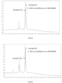
FIGURE 1 is an illustration showing the test result of detecting and measuring 2A1-LC-Cys205ins-mc-vc-PAB-MMAE by HIC-HPLC method, as performed in Example 25; -
FIGURE 2 is an illustration showing the test result of detecting and measuring 2A1-LC-Cys206ins-mc-vc-PAB-MMAE by HIC-HPLC method, as performed in Example 25; -
FIGURE 3 is an illustration showing the result of detecting and measuring 2A1-HC-Cys474ins-mc-vc-PAB-MMAE by HIC-HPLC method, as performed in Example 25; -
FIGURE 4 is an illustration showing the test result of detecting and measuring 4E1-LC-Cys205ins-mc-vc-PAB-MMAE by HIC-HPLC method, as performed in Example 25; -
FIGURE 5 is an illustration showing the test result of detecting and measuring 4E1-LC-Cys206ins-mc-vc-PAB-MMAE by HIC-HPLC method, as performed in Example 25; -
FIGURE 6 is an illustration showing the test result of detecting and measuring 4E1-HC-Cys474ins-mc-vc-PAB-MMAE by HIC-HPLC method, as performed in Example 25; -
FIGURE 7 is an illustration showing the test result of detecting and measuring 4D3-LC-Cys205ins-mc-vc-PAB-MMAE by HIC-HPLC method, as performed in Example 25; -
FIGURE 8 is an illustration showing the result of detecting and measuring 4D3-LC-Cys206ins-mc-vc-PAB-MMAE by HIC-HPLC method, as performed in Example 25; -
FIGURE 9 is an illustration showing the test result of detecting and measuring toxin/antibody ratio of 4D3-HC-Cys474ins-mc-vc-PAB-MMAE by RP-HPLC method, as performed in Example 26. -
FIGURE 10 is an illustration showing the test result of detecting and measuring TDC antibody skeleton 4D3 aggregation by SEC-HPLC method, as performed in Example 27; -
FIGURE 11 is an illustration showing the result of detecting and measuring TDC antibody skeleton 4D3-LC-Cys205ins aggregation by SEC-HPLC method, as performed in Example 27; -
FIGURE 12 is an illustration showing the test result of detecting and measuring TDC antibody skeleton 4D3-LC-Cys206ins aggregation by SEC-HPLC method, as performed in Example 27; -
FIGURE 13 is an illustration showing the test result of detecting and measuring TDC antibody skeleton 4D3-HC-Cys474ins aggregation by SEC-HPLC method, as performed in Example 27; -
FIGURE 14 is an illustration showing the test result of Example 28; -
FIGURE 15 is an illustration showing the test result of Example 29, showing the affinity measurement between the antigen c-met and the antibody 4E1 and TDC 4E1-LC-Cys205ins-MVPM, 4E1-LC-Cys206ins-MVPM, and 4E1-HC-Cys474ins-MVPM; -
FIGURE 16 is an illustration showing the test result of Example 29, showing the affinity measurement between the antigen Trop2 and the antibody 4D3 and TDC 4D3-LC-Cys205ins-MVPM, 4D3-LC-Cys206ins-MVPM, and 4D3-HC-Cys474ins-MVPM; -
FIGURE 17 shows the IC50 of cytotoxicity of the ADCs against cancer cells, wherein the ADCs are 2A1-LC-V205C-mc-vc-PAB-MMAE, 2A1-LC-Cys205ins-mc-vc-PAB-MMAE, 2A1-LC-Cys206ins-mc-vc-PAB-MMAE, and 2A1-HC-Cys474ins-mc-vc-PAB-MMA, and the cancer cells are EGFRwt-overexpressing Human squamous cell carcinoma A431; -
FIGURE 18 shows the IC50 of cytotoxicity of the ADCs against cancer cells, wherein the ADCs are 2A1-LC-V205C-mc-vc-PAB-MMAE, 2A1-LC-Cys205ins-mc-vc-PAB-MMAE, 2A1-LC-Cys206ins-mc-vc-PAB-MMAE, and 2A1-HC-Cys474ins-mc-vc-PAB-MMAE, and the cancer cells are EGFRvlll-overexpressing Human glioma cell line U87-EGFRvIII; -
FIGURE 19 shows the IC50 of cytotoxicity of the ADCs against cancer cells, wherein the ADCs are 4E1-LC-Cys205ins-mc-vc-PAB-MMAE, 4E1-LC-Cys206ins-mc-vc-PAB-MMAE, 4E1-HC-Cys474ins-mc-vc-PAB-MMAE, and 4E1, and the cancer cells are C-met high-expressing malignant glioma cell line U87-MG; -
FIGURE 20 shows the IC50 of cytotoxicity of the ADCs against cancer cells, wherein the ADCs are D3-LC-Cys205ins-mc-vc-PAB-MMAE, 4D3-LC-Cys206ins-mc-vc-PAB-MMAE, 4D3-HC-Cys474ins-mc-vc-PAB-MMAE, and 4D3, and the cancer cells are trop2 high-expressing pancreatic cancer cell line BXPC-3; -
FIGURE 21 is an illustration showing the test result of stability measurement in human plasma for 4D3-LC-Cys205ins-mc-vc-PAB-MMAE; -
FIGURE 22 is an illustration showing the test result of stability measurement in human plasma for 4D3-LC-Cys206ins-mc-vc-PAB-MMAE; -
FIGURE 23 is an illustration showing the test result of stability measurement in human plasma for 4D3-HC-Cys474ins-mc-vc-PAB-MMAE; -
FIGURE 24 is an illustration showing the test result of stability measurement in human plasma for 4E1-LC-Cys205ins-mc-vc-PAB-MMAE; -
FIGURE 25 is an illustration showing the test result of stability measurement in human plasma for 4E1-LC-Cys206ins-mc-vc-PAB-MMAE; -
FIGURE 26 is an illustration showing the test result of stability measurement in human plasma for 4E1-HC-Cys474ins-mc-vc-PAB-MMAE; -
FIGURE 27 is an illustration showing the test result of pharmacodynamic effect measurement for 4D3-LC-Cys205ins-mc-vc-PAB-MMAE, 4D3-LC-Cys206ins-mc-vc-PAB-MMAE, 4D3-HC-Cys474ins-mc-vc-PAB-MMAE, and 4D3 parental antibody in tumor-bearing mice; and -
FIGURE 28 is an illustration showing the test result of pharmacodynamic effect measurement for 4D3-LC-Cys205ins-mc-vc-PAB-MMAE, 4D3-LC-Cys206ins-mc-vc-PAB-MMAE, 4D3-HC-Cys474ins-mc-vc-PAB-MMAE, 4D3-HC-Cys474ins-mc-vc-PAB-MMAE, and 4D3 parental antibody in tumor-bearing mice. - In the following detailed description, reference is made to the accompanying drawings, which form a part hereof. In the drawings, similar symbols typically identify similar components, unless context dictates otherwise. The illustrative embodiments described in the detailed description, drawings, and dependent claims are not meant to be limiting.
- It will be readily understood that the aspects of the present disclosure, as generally described herein, and illustrated in the FIGUREs, can be arranged, substituted, combined, separated, and designed in a wide variety of different configurations, all of which are explicitly contemplated herein.
-

- 6-aminocaproic acid (3.9 g, 0.03 mol) and maleic anhydride (3.5 g, 0.036 mol) were added to glacial acetic acid (30 ml). After stirring at 120°C for 4-6 h, the reaction was cooled to room temperature. Most of the acetic acid was removed by concentration in vacuum at 60 °C. The obtained brownish yellow viscous liquid was poured into water, and then extracted with ethyl acetate (20 ml × 3), and the organic layers were combined. The organic layers were washed with water and brine, dried over anhydrous sodium sulfate, filtered and evaporated in vacuo to yield a brown-yellow oil, which was stirred in 50 ml of water, and white solid materials precipitated out of the solution, the white solid materials is filtered, and the product was dried under reduced pressure at 50 ° C, 5.08 g, yield 80%. Mp: 89-92 °C. m/z: 212.2 [M+H]+. 1H NMR (400Mz, DMSO): 13.21 (br, 1H, COOH), 6.75 (s, 2H, COCH=CHCO), 3.63 (t, 2H, J = 7.2 Hz, NCH2CH2), 2.42 (t, 2H, J = 7.4 Hz, CH2COOH), 1.52 - 1.68 (m, 4H, NCH2CH2CH2CH2), 1.30 - 1.42 (m, 2H, NCH2CH2CH2CH2).
-

- Under nitrogen atmosphere, to a solution of a mixture of MC (4.7 g, 22 mmol) and HOSu (25 g, 22 mmol) in acetonitrile (50 mL) at 0°C was slowly added DCC (4.5 g, 22 mmol) dissolved in 25 ml acetonitrile. The reaction solution was reacted at 0 ° C for 2 hours and then allowed to react at room temperature overnight. After filtering, the filter cake was washed with acetonitrile (10 ml × 3). The filtrate was concentrated to dry under reduced pressure. The obtained oil was dried under reduced pressure at room temperature for 6 h to afford 6.4 g of pale brown solid, and yield 95%. (To be used directly in the next step without purification) m/z : 309.2 [M+H]+∘ 1HNMR (400Mz ,CDCl3) : 1~2 (m,6H,CCH2CH2CH2C), 2.68 (t,2H,CH2CO, 2.95 (s,4H,COCH2CH2CO) , 3.68 (t,2H,CH2N) , 6.81 (s,2H,CH=CH)
-

- To a solution of a mixture of Fmoc-Val (10 g) and HOSu (3.4 g) in THF (100 mL) at 0°C was slowly added DCC (6 g) dissolved in 50 ml acetonitrile. The reaction solution was stirred at room temperature for 24 hours. Filtration was performed, and the filter cake was washed with THF. A transparent oil was obtained by concentrating the filtrates under reduced pressure. The oil was directly used in the next step directly without further purification. m/z : 437.4 [M+H]+.
-

- To a solution of Cit (4.0 g) in THF (20 mL) was added a solution of 60 ml aqueous sodium hydrogencarbonate (containing NaHCO3 2 g, 1.05 eq). A solution of Fmoc-Val-OSu (22.35 mmol) in DME (60 mL) was added to the mixture. After stirring at room temperature for 24 hours, to the reaction was added a solution of 15% aqueous citric acid solution (110 ml), and then extracted with EtOAc twice. The combined organic phases were concentrated in vacuum to get a white solid. 100 ml of methyl tert-butyl ether was added to the white material, the mixture was stirred, filtered, and the filter cake was dried under reduced pressure at 40 ° C for 4 h to obtain the product 4.83 g, and yield 65%. m/z : 497.6 (M+H)+∘ 1HNMR(400Mz,DMSO): 0.92 (6H, m), 1.35 ~ 1.65 (4H, m), 2.10 (1H, m), 3.01(2H, q), 3.99 (1H, t), 4.01 -4.45 (2H, m), 4.45 (2H, t), 5.46 (2H, br), 6.03(1H, t), 7.20-8.02 (8H, m), 8.25 (1H, d).
-

- To a solution of Fmoc-vc (2 g, 4.2 mmol) and PABOH (1.04 g, 2 eq) in DCM/MeOH = 2/1 (60 mL) was added EEDQ (2.0 g, 2 eq) at 0 °C. After stirring for 10 min, a solution of (S)-1-phenylethanamine (17.5 g, 144.2 mmol) in MeOH (200 mL) was added slowly to the mixture after partial dissolution. The reaction system was stirred at room temperature for 2 days in the dark. After completion of the reaction, the mixture was concentrated in vacuum at 40 °C to yield a white solid. The white solid was collected, washed with methyl tert-butyl ether (100 ml), and filtered. The filter cake was washed with methyl tert-butyl ether, and the obtained white solid was dried under reduced pressure at 40 °C to give the white solid 2.2 g, and yield 88%. m/z : 602.6 (M+H)+. 1HNMR (400Mz, DMSO): 0.95 (6H,m), 1.45~1.69 (4H, m), 2.10 (1H, m), 3.11(2H, m), 3.99 (1H, m ), 4.30 (2H, d), 4.05~-4.66 (2H, m), 4.55 (2H, d), 5.21 (1H, t), 5.51 (2H, br), 6.11(1H, t), 7.09 -8.10 (12H, m), 8.21 (1H, d), 10.51(1H, br).
-

- To a solution of Fmoc-vc-PABOH (490 mg, 0.815 mmol) in THF (10 mL) was added diethylamine (2 ml). The reaction mixture was stirred at room temperature for 24 h. 20 ml of DCM was added to the obtained product, the mixture was stirred, and crystalline was precipitated out of reaction solution. The crystalline was filtered and the filter cake was washed with DCM, and the obtained solid was dried under reduced pressure to yield 277 mg. The yield was 90%. m/z : 380.2 (M+H)+∘ 1HNMR (400Mz, DMSO): 0.89 (6H, m), 1.31~1.61 (4H, m), 1.82 (1H, m), 2.86 (1H, m), 2.89(2H, d), 4.38 (2H, d), 4.44 (1H, m), 5.01 (1H, br), 5.35 (2H, br), 5.84 (1H, br), 7.14 (2H, d), 7.42 (2H, d), 8.08 (1H, br), 9.88 (1H, br).
-

- VP-PABOH (205 mg, 0.54 mmol) and MC-OSu (184 mg, 1.1 eq) were added to 10 ml of NMP, and the reaction was stirred at room temperature for 24 h. After completion of the reaction, the mixture was concentrated in vacuo at 40 ° C. Methyl tert-butyl ether (20 ml) was added to the obtained oil and stirred to crystallization. After filtering the crystalline and washing the filter cake with methyl tert-butyl ether, the product was yielded at 310 mg. The yield is 100%. m/z: 573.3 (M+H)+∘ 1HNMR (400Mz, DMSO): 0.89 (6H, m), 1.15-1.99 (10H, m), 2.11(1H, m), 2.31 (2H, t), 3.21(2H, m), 3.53 (2H, t), 4.32 (1H, t), 4.51 (1H, m), 4.59 (2H, br), 5.24 (1H, br), 5.56 (2H, br), 6.20(1H, br), 7.12(2H, s), 7.23(2H, d) ,7.58 (2H, d), 7.94 (1H, d) , 8.1 7 (1H, d), 10.21 (1H, br)
-

- Under nitrogen, to a solution of mc-vc-PABOH (166.0 mg, 0.294 mmol) in anhydrous pyridine (5 ml) was added PNP (179 mg, 3 eq) dissolved in DCM (5 ml) at 0 °C slowly. After stirring at about 0 °C for 10 min, the ice bath was removed, and the reaction was stirred at room temperature for 3 h. After completion of the reaction, EA (70 ml) and a 15% aqueous citric acid solution (100 ml) were added, and the organic layer was separated and recovered. The organic layer was sequentially washed with citric acid, water, brine, dried with anhydrous sodium sulfate, filtered, and the filtrate was concentrated under reduced pressure to yield light yellowish oily product. Adding methyl tert-butyl ether for crystallization resulted in the white-like solid (86 mg). The yield was 40%. m/z: 738(M+H)+∘ 1HNMR (400Mz, CDCl3/CD3OD): 0.84 (6H, m), 1.11-1.84 (10H, m), 2.05 (1H, m), 2.15 (2H, t), 3.09 (2H, m), 3.32 (2H, t), 4.12 (1H, m), 4.38 (1H, m), 5.15 (2H, s), 6.61 (2H, s), 6.84 (1H, d) ,7.61 (1H, d), 7.21 (2H,d), 7.50 (2H,d), 7.61 (2H,d), 8.18 (2H, d), 9.59 (1H, br)
-

- 20 mg of mc-vc-PAB-PNP (1.5 eq) and 3 mg of HOBT were added to 2 ml of DMF. After stirring at room temperature for a moment, 13 mg of MMAE, 0.5 ml of pyridine, and 25 ul of DIEA were added. The reaction solution was stirred at room temperature for 2 d. After the reaction is completed, the reaction solution is directly purified by a preparative column, and the desired components are collected, concentrated, and lyophilized to obtain about 10 mg of a product, and the yield is about 42%. m/z: 1317.1 (M+H)+.
-

- Operate according to the steps of Example 9, about 12.5 mg of mc-vc-PAB-MMAF was obtained, and the yield was 45.2%.
-

- Operate according to the steps of Example 9, about 9.5 mg of mc-vc-PAB-PBD was obtained. The yield was 32.5%. m/z: 1325.4 (M+H)+.
-

- Operate according to the steps of Example 9, about 11.2 mg of mc-vc-PAB-DOX was obtained. The yield was 38.9%. m/z: 1143.2 (M+H)+.
-

- 100 mg of 10-O-Boc-SN-38 was dissolved in 10 ml of dry dichloromethane, 25.6 mg (1 eq) of DMAP was added to the solvent, and a solution of triphosgene in dichloromethane was added dropwise at 0 °C (62 mg of triphosgene was dissolved in 2 ml of Dichloromethane), and the reaction was continued at 0 °C for 12 h. The dichloromethane was removed under reduced pressure. The crude products were dissolved in 10 ml of dry DMF, 144 mg of mc-vc-PABOH was then added, and the mixture was stirred at room temperature for 24 h. 41 mg of mc-vc-PAB-SN-38 was isolated by preparation liquid phase separation, and the total yield in two steps was 19.7%. m/z: 1063.2(M+H)+.
- The target antibody was expressed using Freestyle™ 293-F (Invitrogen) suspension cells. One day before transfection, cells were seeded at a density of 6 × 10⁵ cells/mL in a 1 L shake flask containing 300 mL of F17 complete medium (Freestyle™ F17 expression medium, Gibco), grown overnight by shaking at 37 °C, 5% CO2, 120 rpm at cell incubator. The next day, transfection of the antibody expression plasmid was carried out with PEI, wherein the ratio of plasmid: PEI was 2:1. One day after the transfection, the TN1 feed medium was added at 2.5% (v/v), and the culture was continued for 4 days, and the supernatant was collected by centrifugation.
- The collected cell expression supernatant was eluted by a Protein A affinity chromatography column (Mabselect Sure LX, GE) eluting with 0.1 M citric acid (pH 3.0), and the captured antibody was treated with 1 M Tris-HCl (pH 9.0) and adjusted to pH 7.0 at 1/10 (v/v). Remove impurities such as multimers and endotoxin by gel filtration column SEC (
Superdex 200, GE), and replace the antibody buffer with PBS (pH 7.4) at the same time, a sample of the target peak of UV280 nm was collected and concentrated to 2 mg/ml through an ultrafiltration centrifuge tube (30 KD, Pall Corporation). The target antibody monomer (POI%) obtained by this method was greater than 90% and was stored for subsequent experiments. - The 2A1-HC-Cys474ins antibody expressed by the cells was purified by Protein A resin such as Mabselect Sure, eluted with low pH solution and neutralized by adding Tris solution immediately after the low pH elution, and the solution was changed to a pH 7.5 Tris-HCl buffer. The mc-vc-PAB-MMAE compound, being a white powder, was dissolved in DMA for use. In order to remove the masking group on the mutant cysteine residue, the antibody was reduced first. A 1 M aqueous solution of DTT was added to the 2A1-HC-Cys474ins antibody solution at a molecular ratio of 1:40, and the mixture was mixed evenly and reacted at 20 ° C for 2 hours. After the reaction time was reached, the pH of the sample was adjusted to 5.0, and the DTT and the masking group in the mixture were removed by cation exchange chromatography such as SP Sepharose F.F. resin. Subsequently, a DHAA solution was added to the sample at a molecular ratio of 1:20 and reacted at 25 ° C for 4 hours in the dark to re-connect the interchain disulfide bonds. subsequently, mc-vc-PAB-MMAE solution was added to couple the mc-vc-PAB-MMAE with the inserted or mutant cysteine in the antibody, and the mixture was thoroughly mixed and reacted at 25 ° C for 2 hours. After the end of the reaction, mc-vc-PAB-MMAE to which the antibody was not coupled was removed using cation exchange chromatography such as SP Sepharose F.F. to obtain a 2A1-HC-Cys474ins-mc-vc-PAB-MMAE TDC sample.
- The 2A1-LC-Cys205ins antibody expressed by the cells was purified by Protein A resin such as Mabselect Sure, eluted with low pH solution and neutralized by adding Tris solution immediately after the low pH elution, and the solution was changed to a pH 7.5 Tris-HCl buffer. The mc-vc-PAB-MMAE compound, being a white powder, was dissolved in DMA for use. In order to remove the masking group on the mutant cysteine residue, the antibody was reduced first. A 1 M aqueous solution of DTT was added to the 2A1-LC-Cys205ins antibody solution at a molecular ratio of 1:40, and the mixture was mixed evenly and reacted at 20 ° C for 2 hours. After the reaction time was reached, the pH of the sample was adjusted to 5.0, and the DTT and the masking group in the mixture were removed by cation exchange chromatography such as SP Sepharose F.F. resin. Subsequently, a DHAA solution was added to the sample at a molecular ratio of 1:20 and reacted at 25 ° C for 4 hours in the dark to re-connect the interchain disulfide bonds. subsequently, mc-vc-PAB-MMAE solution was added to couple the mc-vc-PAB-MMAE with the inserted or mutant cysteine in the antibody, and the mixture was thoroughly mixed and reacted at 25 ° C for 2 hours. After the end of the reaction, mc-vc-PAB-MMAE to which the antibody was not coupled was removed using cation exchange chromatography such as SP Sepharose F.F. to obtain a 2A1-LC-Cys205ins-mc-vc-PAB-MMAE TDC sample.
- The 2A1-LC-Cys206ins antibody expressed by the cells was purified by Protein A resin such as Mabselect Sure, eluted with low pH solution and neutralized by adding Tris solution immediately after the low pH elution, and the solution was changed to a pH 7.5 Tris-HCl buffer. The mc-vc-PAB-MMAE compound, being a white powder, was dissolved in DMA for use. In order to remove the masking group on the mutant cysteine residue, the antibody was reduced first. A 1 M aqueous solution of DTT was added to the 2A1-LC-Cys206ins antibody solution at a molecular ratio of 1:40, and the mixture was mixed evenly and reacted at 20 ° C for 2 hours. After the reaction time was reached, the pH of the sample was adjusted to 5.0, and the DTT and the masking group in the mixture were removed by cation exchange chromatography such as SP Sepharose F.F. resin. Subsequently, a DHAA solution was added to the sample at a molecular ratio of 1:20 and reacted at 25 ° C for 4 hours in the dark to re-connect the interchain disulfide bonds. subsequently, mc-vc-PAB-MMAE solution was added to couple the mc-vc-PAB-MMAE with the inserted or mutant cysteine in the antibody, and the mixture was thoroughly mixed and reacted at 25 ° C for 2 hours. After the end of the reaction, mc-vc-PAB-MMAE to which the antibody was not coupled was removed using cation exchange chromatography such as SP Sepharose F.F. to obtain a 2A1-LC-Cys206ins-mc-vc-PAB-MMAE TDC sample.
- The 4D3-HC-Cys474ins antibody expressed by the cells was purified by Protein A resin such as Mabselect Sure, eluted with low pH solution and neutralized by adding Tris solution immediately after the low pH elution, and the solution was changed to a pH 7.5 Tris-HCl buffer. The mc-vc-PAB-MMAE compound, being a white powder, was dissolved in DMA for use. In order to remove the masking group on the mutant cysteine residue, the antibody was reduced first. A 1 M aqueous solution of DTT was added to the 4D3-HC-Cys474ins antibody solution at a molecular ratio of 1:40, and the mixture was mixed evenly and reacted at 20 ° C for 2 hours. After the reaction time was reached, the pH of the sample was adjusted to 5.0, and the DTT and the masking group in the mixture were removed by cation exchange chromatography such as SP Sepharose F.F. resin.
- Subsequently, a DHAA solution was added to the sample at a molecular ratio of 1:20 and reacted at 25 ° C for 4 hours in the dark to re-connect the interchain disulfide bonds. subsequently, mc-vc-PAB-MMAE solution was added to couple the mc-vc-PAB-MMAE with the inserted or mutant cysteine in the antibody, and the mixture was thoroughly mixed and reacted at 25 ° C for 2 hours. After the end of the reaction, mc-vc-PAB-MMAE to which the antibody was not coupled was removed using cation exchange chromatography such as SP Sepharose F.F. to obtain a 4D3-HC-Cys474ins-mc-vc-PAB-MMAE TDC sample.
- The 4D3-LC-Cys205ins antibody expressed by the cells was purified by Protein A resin such as Mabselect Sure, eluted with low pH solution and neutralized by adding Tris solution immediately after the low pH elution, and the solution was changed to a pH 7.5 Tris-HCl buffer. The mc-vc-PAB-MMAE compound, being a white powder, was dissolved in DMA for use. In order to remove the masking group on the mutant cysteine residue, the antibody was reduced first. A 1 M aqueous solution of DTT was added to the 4D3-LC-Cys205ins antibody solution at a molecular ratio of 1:40, and the mixture was mixed evenly and reacted at 20 ° C for 2 hours. After the reaction time was reached, the pH of the sample was adjusted to 5.0, and the DTT and the masking group in the mixture were removed by cation exchange chromatography such as SP Sepharose F.F. resin. Subsequently, a DHAA solution was added to the sample at a molecular ratio of 1:20 and reacted at 25 ° C for 4 hours in the dark to re-connect the interchain disulfide bonds. subsequently, mc-vc-PAB-MMAE solution was added to couple the mc-vc-PAB-MMAE with the inserted or mutant cysteine in the antibody, and the mixture was thoroughly mixed and reacted at 25 ° C for 2 hours. After the end of the reaction, mc-vc-PAB-MMAE to which the antibody was not coupled was removed using cation exchange chromatography such as SP Sepharose F.F. to obtain a 4D3-LC-Cys205ins-mc-vc-PAB-MMAE TDC sample.
- The 4D3-LC-Cys206ins antibody expressed by the cells was purified by Protein A resin such as Mabselect Sure, eluted with low pH solution and neutralized by adding Tris solution immediately after the low pH elution, and the solution was changed to a pH 7.5 Tris-HCl buffer. The mc-vc-PAB-MMAE compound, being a white powder, was dissolved in DMA for use. In order to remove the masking group on the mutant cysteine residue, the antibody was reduced first. A 1 M aqueous solution of DTT was added to the 4D3-LC-Cys206ins antibody solution at a molecular ratio of 1:40, and the mixture was mixed evenly and reacted at 20 ° C for 2 hours. After the reaction time was reached, the pH of the sample was adjusted to 5.0, and the DTT and the masking group in the mixture were removed by cation exchange chromatography such as SP Sepharose F.F. resin. Subsequently, a DHAA solution was added to the sample at a molecular ratio of 1:20 and reacted at 25 ° C for 4 hours in the dark to re-connect the interchain disulfide bonds. subsequently, mc-vc-PAB-MMAE solution was added to couple the mc-vc-PAB-MMAE with the inserted or mutant cysteine in the antibody, and the mixture was thoroughly mixed and reacted at 25 ° C for 2 hours. After the end of the reaction, mc-vc-PAB-MMAE to which the antibody was not coupled was removed using cation exchange chromatography such as SP Sepharose F.F. to obtain a 4D3-LC-Cys206ins-mc-vc-PAB-MMAE TDC sample.
- The 4E1-HC-Cys474ins antibody expressed by the cells was purified by Protein A resin such as Mabselect Sure, eluted with low pH solution and neutralized by adding Tris solution immediately after the low pH elution, and the solution was changed to a pH 7.5 Tris-HCl buffer. The mc-vc-PAB-MMAE compound, being a white powder, was dissolved in DMA for use. In order to remove the masking group on the mutant cysteine residue, the antibody was reduced first. A 1 M aqueous solution of DTT was added to the 4E1-HC-Cys474ins antibody solution at a molecular ratio of 1:40, and the mixture was mixed evenly and reacted at 20 ° C for 2 hours. After the reaction time was reached, the pH of the sample was adjusted to 5.0, and the DTT and the masking group in the mixture were removed by cation exchange chromatography such as SP Sepharose F.F. resin. Subsequently, a DHAA solution was added to the sample at a molecular ratio of 1:20 and reacted at 25 ° C for 4 hours in the dark to re-connect the interchain disulfide bonds. subsequently, mc-vc-PAB-MMAE solution was added to couple the mc-vc-PAB-MMAE with the inserted or mutant cysteine in the antibody, and the mixture was thoroughly mixed and reacted at 25 ° C for 2 hours. After the end of the reaction, mc-vc-PAB-MMAE to which the antibody was not coupled was removed using cation exchange chromatography such as SP Sepharose F.F. to obtain a 4E1-HC-Cys474ins-mc-vc-PAB-MMAE TDC sample.
- The 4E1-LC-Cys205ins antibody expressed by the cells was purified by Protein A resin such as Mabselect Sure, eluted with low pH solution and neutralized by adding Tris solution immediately after the low pH elution, and the solution was changed to a pH 7.5 Tris-HCl buffer. The mc-vc-PAB-MMAE compound, being a white powder, was dissolved in DMA for use. In order to remove the masking group on the mutant cysteine residue, the antibody was reduced first. A 1 M aqueous solution of DTT was added to the 4E1-LC-Cys205ins antibody solution at a molecular ratio of 1:40, and the mixture was mixed evenly and reacted at 20 ° C for 2 hours. After the reaction time was reached, the pH of the sample was adjusted to 5.0, and the DTT and the masking group in the mixture were removed by cation exchange chromatography such as SP Sepharose F.F. resin. Subsequently, a DHAA solution was added to the sample at a molecular ratio of 1:20 and reacted at 25 ° C for 4 hours in the dark to re-connect the interchain disulfide bonds. subsequently, mc-vc-PAB-MMAE solution was added to couple the mc-vc-PAB-MMAE with the inserted or mutant cysteine in the antibody, and the mixture was thoroughly mixed and reacted at 25 ° C for 2 hours. After the end of the reaction, mc-vc-PAB-MMAE to which the antibody was not coupled was removed using cation exchange chromatography such as SP Sepharose F.F. to obtain a 4E1-LC-Cys205ins-mc-vc-PAB-MMAE TDC sample.
- The 4E1-LC-Cys206ins antibody expressed by the cells was purified by Protein A resin such as Mabselect Sure, eluted with low pH solution and neutralized by adding Tris solution immediately after the low pH elution, and the solution was changed to a pH 7.5 Tris-HCl buffer. The mc-vc-PAB-MMAE compound, being a white powder, was dissolved in DMA for use. In order to remove the masking group on the mutant cysteine residue, the antibody was reduced first. A 1 M aqueous solution of DTT was added to the 4E1-LC-Cys206ins antibody solution at a molecular ratio of 1:40, and the mixture was mixed evenly and reacted at 20 ° C for 2 hours. After the reaction time was reached, the pH of the sample was adjusted to 5.0, and the DTT and the masking group in the mixture were removed by cation exchange chromatography such as SP Sepharose F.F. resin. Subsequently, a DHAA solution was added to the sample at a molecular ratio of 1:20 and reacted at 25 ° C for 4 hours in the dark to re-connect the interchain disulfide bonds. subsequently, mc-vc-PAB-MMAE solution was added to couple the mc-vc-PAB-MMAE with the inserted or mutant cysteine in the antibody, and the mixture was thoroughly mixed and reacted at 25 ° C for 2 hours. After the end of the reaction, mc-vc-PAB-MMAE to which the antibody was not coupled was removed using cation exchange chromatography such as SP Sepharose F.F. to obtain a 4E1-LC-Cys206ins-mc-vc-PAB-MMAE TDC sample.
- The TDC sample was analyzed by high performance liquid chromatography with hydrophobic chromatography, and drug:antibody ratio (DAR, also known as toxin:antibody ratio) was calculated from the corresponding peak area. One specific method is described in detail as follows:
- Column: Proteomix® HICBu-NP5 (5 µm, 4.6 x 35 mm);
- Mobile phase: Buffer A: 2M ammonium sulfate, 0.025 M,
pH 7 phosphate buffer; Buffer B: 0.025 M,pH 7 phosphate buffer; Buffer C: 100% isopropanol; - Buffer A was used for equilibration, Buffer B and buffer C were used for gradient elution, detection was performed at 25 ° C, 214 nm and 280 wavelengths. Based on data gathered from
FIGUREs 1-3 , the site-specific coupled DAR is calculated to be between 1.6 and 1.7, showing excellent compound uniformity or homogeneity. Based on data gathered fromFIGUREs 4-6 , the site-specific coupled DAR is calculated to be between 1.6 and 1.95, showing excellent compound uniformity or homogeneity. Based on data gathered fromFIGUREs 7-8 , the site-specific coupled DAR is calculated to be between 1.6 and 1.9, showing excellent compound uniformity or homogeneity. - The ratio of toxin to antibody was measured by RP-HPLC. The samples treated with DTT were analyzed by reversed-phase hydrophobic high-performance liquid chromatography, and DAR was calculated from the corresponding peak area. One specific method is described in detail as follows:
- Column: Proteomix RP-1000 (5µm, 4.6×100mm)
- Mobile phase: Buffer A: 0.1% TFA aqueous solution; Buffer B: 0.1% acetonitrile solution.
- Mobile phase A and mobile phase B were used to elute in a proportional gradient at 80 °C, measurement was performed at 214 nm and 280 wavelengths. Based on data gathered in
FIGURE 9 , the site-specific coupled DAR was calculated to be 1.82, showing excellent compound uniformity or homogeneity. - TABLE 1 shows that the coupling efficiency of site-directed TDC compounds by cysteine insertion mutation modification is uniformly high (theoretical maximum is 2.0), with DAR ≥ 1.6.
- TDC antibody skeleton samples were stored at 37 ° C, and their aggregation was analyzed by SEC-HPLC on
days - Chromatography columns: TSKgel SuperSW mAb HR (7.8mm × 30cm),
- Mobile phase: 0.1 M sodium sulfate, 0.1 M, pH 6.7 phosphate buffer,
- Measurements were performed at 25 ° C, 280 nm.
- As shown in
FIGUREs 10-13 , SEC-HPLC was used to detect and measure the aggregation of TDC antibody skeleton 4D3, 4D3-LC-Cys205ins, 4D3-LC-Cys206ins and 4D3-HC-Cys474ins. The samples were stored at 37 ° C for 4 weeks, and the aggregate content remained essentially unchanged. - Using the same detecting and measurement method, the aggregations of the following TDCs are measured: 2A1-LC-V205C-mc-vc-PAB-MMAE TDC, 2A1-LC-Cys205ins-mc-vc-PAB-MMAE TDC, 2A1-LC-Cys206ins--mc-vc-PAB-MMAE TDC, 2A1-HC-Cys474ins-mc-vc-PAB-MMAE, 4E1-LC-Cys205ins-mc-vc-PAB-MMAE, 4E1-LC-Cys206ins-mc-vc-PAB-MMAE 4E1-HC-Cys474ins-mc-vc-PAB-MMAE TDC, 4D3-LC-Cys205ins-mc-vc-PAB-MMAE, 4D3-LC-Cys206ins-mc-vc-PAB-MMAE, 4D3-HC-Cys474ins-mc-vc-PAB-MMAE TDC. The results are shown in TABLE II
TABLE II: TDS target monomer content list for 2A1-LC-V205C-mc-vc-PAB-MMAE TDC, 2A1-LC-Cys205ins-mc-vc-PAB-MMAE TDC, 2A1-LC-Cys206ins--mc-vc-PAB-MMAE TDC and 2A1-HC-Cys474ins-mc-vc-PAB-MMAE, 4E1-LC-Cys205ins-mc-vc-PAB-MMAE, 4E1-LC-Cys206ins-mc-vc-PAB-MMAE, 4E1-HC-Cys474ins-mc-vc-PAB-MMAE TDC, 4D3-LC-Cys205ins-mc-vc-PAB-MMAE, 4D3-LC-Cys206ins-mc-vc-PAB-MMAE, 4D3-HC-Cys474ins-mc-vc-PAB-MMAE Compound POI% 2A1-LC-V205C-mc-vc-PAB-MMAE TDC 96.0% 2A1-LC-Cys205ins-mc-vc-PAB-MMAE TDC 90.0% 2A1-LC-Cys206ins-mc-vc-PAB-MMAE TDC 90.4% 2A1-HC-Cys474ins-mc-vc-PAB-MMAE TDC 90.0% Site-specific coupling (TDC) 4E1-LC-Cys205ins-mc-vc-PAB-MMAE TDC 98.18% 4E1-LC-Cys206ins-mc-vc-PAB-MMAE TDC 94.34% 4E1-HC-Cys474ins-mc-vc-PAB-MMAE TDC 95.77% 4D3-LC-Cys205ins-mc-vc-PAB-MMAE TDC 97.27% 4D3-LC-Cys206ins-mc-vc-PAB-MMAE TDC 96.06% 4D3-HC-Cys474ins-mc-vc-PAB-MMAE TDC 96.98% - As shown by TABLE II, the target monomer content of the TDC compound coupled by the inserted cysteine is above 90%.
- The relative affinities of 2A1-LC-V205C, 2A1-LC-Cys205ins, 2A1-LC-Cys206ins, 2A1-HC-Cys474ins and 2A1 for EGFRvIII were compared by indirect ELISA. The specific steps are as follows:
Recombinant EGFRvIII-His*6 antigen-coated plate was blocked by fish skin gelatin; Antibodies 2A1, 2A1-LC-V205C, 2A1-LC-Cys205ins, 2A1-LC-Cys206ins and 2A1-HC-Cys474ins were respectively diluted by 4 folds gradient with a total of 11 concentrations with the highest concentration being 10ug/ml; HRP-labeled secondary antibody incubation were performed; after TMB coloration, absorption was detected and measured at 450 nm. The absorption measurement results at A450 were plotted against concentration. The cysteine site-directed mutagenesis or insertion of the mutant antibodies 2A1-LC-V205C, 2A1-LC-Cys205ins, 2A1-LC-Cys206ins and 2A1-HC-Cys474ins retained affinities to EGFRvIII similar to 2A1, as shown by the close EC50 values; these results indicate that the site-directed mutagenesis of the light chain V205C on 2A1 antibody, the insertional mutation at position 205 of the light chain of the antibody, the insertional mutation at position 206 of the light chain of the antibody, or the insertion mutation at position 474 of the heavy chain of the antibody does not affect their affinity for the EGFRvIII antigen. - As shown in the
FIGURE 14 , the 2A1-LC-V205C, 2A1-LC-Cys205ins, 2A1-LC-Cys206ins, 2A1-HC-Cys474ins antibodies maintain the affinity of 2A1 to antigen EGFRvIII. - The relative affinities of 4E1-LC-Cys205ins-MVPM, 4E1-LC-Cys206ins-MVPM, 4E1-HC-Cys474ins-MVPM and 4E1 for C-met were compared by indirect ELISA. The specific steps are as follows:
Recombinant C-met-His*6 antigen-coated plate was blocked by fish skin gelatin; TDC 4E1-LC-Cys205ins-MVPM, 4E1-LC-Cys206ins-MVPM, 4E1-HC-Cys474ins-MVPM and antibody 4E1 were respectively diluted by 4 folds gradient with a total of 11 concentrations with the highest concentration being 10ug/ml; HRP-labeled secondary antibody incubation were performed; after TMB coloration, absorption was detected and measured at 450 nm. The absorption measurements at A450 were plotted against concentration, and the result shows that the antibodies harboring cysteine site-directed insertion mutation, TDC 4E1-LC-Cys205ins-MVPM, 4E1-LC-Cys206ins-MVPM, and 4E1-HC-Cys474ins-MVPM, retained their binding affinities to C-met similar to 4E1, as indicated by the close EC50 values; which indicates that the insertional mutation at the position 205 or 206 of 4E1 light chain or at the position 474 of 4E1 heavy chain does not affect the binding affinity of the corresponding TDC to the c-met antigen. - The relative affinities of 4D3-LC-Cys205ins-MVPM, 4D3-LC-Cys206ins-MVPM, 4D3-HC-Cys474ins-MVPM and 4D3 for Trop2 were compared by indirect ELISA. The specific steps are as follows:
Recombinant Trop2-His*6 antigen-coated plate was blocked by fish skin gelatin; TDC 4D3-LC-Cys205ins-MVPM, 4D3-LC-Cys206ins-MVPM, 4D3-HC-Cys474ins-MVPM and antibody 4D3 were respectively diluted by 4 folds gradient with a total of 11 concentrations with the highest concentration being 10ug/ml; HRP-labeled secondary antibody incubation were performed; after TMB coloration, absorption was detected and measured at 450 nm. The absorption measurements at A450 were plotted against concentration. TDC 4D3-LC-Cys205ins-MVPM, 4D3-LC-Cys206ins-MVPM, and 4D3-HC-Cys474ins-MVPM retained their binding affinities to Trop2 similar to that of 4D3, as shown by the close EC50 values; which indicates that the insertional mutation at the position 205 or 206 of 4D3 light chain or at the position 474 of 4D3 heavy chain does not affect the binding affinity of the corresponding TDC to the Trop2 antigen. - As shown in
FIGURE 15 , 4E1-LC-Cys205ins-MVPM, 4E1-LC-Cys206ins-MVPM, 4E1-HC-Cys474ins-MVPM antibodies retained the affinity of 4E1 for antigen c-met. - As shown in
FIGURE 16 , 4D3-LC-Cys205ins-MVPM, 4D3-LC-Cys206ins-MVPM, 4D3-HC-Cys474ins-MVPM antibodies retained the affinity of 4D3 for the antigen Trop2. - TDC cytotoxic activity was determined by the following experimental procedures: TDC was separately added to culture media of human tumor cells in which EGFR was overexpressed or EGFRVIII was expressed, and cell viability was measured after 72 hours of cell culture. Cell-based in vitro assays were used to determine cell viability, cytotoxicity, and TDC-induced apoptosis in the present disclosure.
- The in vitro efficacy of the antibody-cytotoxin conjugate was determined by a cell proliferation assay. In one embodiment, the CellTiter 96® Aqueous One Solution Cell Proliferation Assay is commercially available (Promega Corp., Madison, WI). The Cell Proliferation Assay (a) is a detection reagent that uses colorimetry to detect the number of viable cells in cell proliferation and cytotoxicity experiments. This reagent contains a novel tetrazolium compound [3-(4,5-dimethylthiazol-2-yl)-5-(3-carboxymethoxyphenyl)-2-(4-sulfophenyl)-2H-tetrazolium, inner salt; MTS] and an electronic coupling agent (phenazine ethosulfate; PES). PES has enhanced chemical stability, which allows it to be mixed with MTS to form a stable solution. This convenient "single solution" mode is based on the first generation CellTiter 96® AQueous Assay, in which the electronic coupling agent PMS and MTS solution are supplied separately. MTS (Owen's reagent) is biologically reduced by cells to a colored formazan product that is directly soluble in the medium (
FIGURE 1 ). This transformation is most likely accomplished by the action of NADPH or NADH produced by dehydrogenase in metabolically active cells. For detection, simply add a small amount of CellTiter 96® AQueous One Solution Reagent directly to the culture medium well, incubate for 1-4 hours, and then read the absorbance at 490 nm with a microplate reader.
- The amount of formazan product detected at 490 nm is directly proportional to the number of viable cells in the culture. Since the MTS hyperthyroid product is soluble in tissue culture media, the CellTiter 96® AQueous One Solution Assay has fewer steps than the MTT or INT method.
- In the present disclosure, A431 (EGFR overexpressing cells), U87-EGFRVIII (EGFR mutant stable cell line), U87-MG (glioblastoma cell line with highly expressed c-Met) and BXPC-3 (pancreatic cancer cell line with high expression of Trop2) are used as research systems for in vitro drug efficacy testing. In a 96-well plate, cell plating was performed at a concentration of 6000/well, and after 24 hours, antibody administration was performed. The initial concentrations of various TDCs corresponding to A431, U87-EGFRVIII cell lines were 10 µM, which were sequentially diluted according to a 5-fold gradient. The initial concentrations of various TDCs corresponding to U87-MG and BXPC-3 cell lines were 1 µM, which were sequentially diluted according to a 5-fold gradient. MTS assay for cell viability were performed after 72 hours of treatment
TABLE III: Cytotoxicity IC50 detection results of TDC, ADC on EGFRwt overexpressing cell line A431 and EGFRvIII expression stable strain U87-EGFRVIII Compounds MTS A431 U87MG-EGFRvIII U87-MG BXPC-3 Antibody 2A1 2A1 >10µM >10µM / / 2A1-LC-V205C >10µM >10µM / / 2A1-LC-Cys205ins >10µM >10µM / / 2A1-LC-Cys206ins >10µM >10µM / / 2A1-HC-Cys474ins >10µM >10µM / / Site-directed coupling (TDC) 2A1-LC-V205C-mc-vc-PAB-MMAE 92.16 nM 296.11 nM / / 2A1-LC-Cys205ins-mc-vc-PAB-MMAE 133.67 nM 492.14 nM / / 2A1-LC-Cys206ins-mc-vc-PAB-MMAE 179.26 nM 457.48 nM / / 2A1-HC-Cys474ins-mc-vc-PAB-MMAE 26.62 nM 118.52 nM / / Antibody 4E1 4E1 / / >1µM / Site-directed coupling (TDC) 4E1-LC-Cys205ins-mc-vc-PAB-MMAE / / 120.50nM / 4E1-LC-Cys206ins-mc-vc-PAB-MMAE / / 79.57nM / 4E1-HC-Cys474ins-mc-vc-PAB-MMAE / / 7.41nM / Antibody 4D3 4D3 / / / >1µM Site-directed coupling (TDC) 4D3-LC-Cys205ins-mc-vc-PAB-MMAE / / / 0.38nM 4D3-LC-Cys206ins-mc-vc-PAB-MMAE / / / 0.45nM 4D3-HC-Cys474ins-mc-vc-PAB-MMAE / / / 0.23nM - The data from the TABLE III shows that, 2A1-LC-V205C-mc-vc-PAB-MMAE TDC, 2A1-LC-Cys205Cins-mc-vc-PAB-MMAE TDC, 2A1-LC-Cys206ins-mc-vc-PAB-MMAE TDC, and 2A1-HC-Cys474ins -mc-vc-PAB-MMAE TDC have comparable cytotoxic activity to EGFRwt overexpressing cell line A431 and EGFRvIII expression stable strain U87-EGFRVIII, and 474 inserted mutant TDC's activity is slightly higher than 205 and 206 insertion mutant TDC.
- There was a certain correlation between cytotoxic activity and coupling position for the 4E1-LC-Cys205ins-mc-vc-PAB-MMAE TDC, 4E1-LC-Cys206ins-mc-vc-PAB-MMAE TDC and 4E1-HC-Cys474ins-mc-vc-PAB-MMAE TDC in U87-MG cells. The TDC activity of the 474 inserted mutant was slightly better than those of the 205 and 206 insertion mutants. The activity of TDC was significantly better than that of the parental antibody.
- The cytotoxic activity of 4D3-LC-Cys205ins-mc-vc-PAB-MMAE TDC, 4D3-LC-Cys206ins-mc-vc-PAB-MMAE TDC and 4D3-HC-Cys474ins-mc-vc-PAB-MMAE TDC in pancreatic cancer cell line BXPC-3 was comparable or similar to each other, and the TDC activity of the 474 inserted mutant was slightly better than those of the 205 and 206 insertion mutant TDCs, and the activity of TDC was significantly better than that of the parental antibody.
- Take a certain amount of ADC sample, add it to human plasma from which human IgG has been removed,
repeat 2 tubes for each ADC, incubate in a 37 °C water bath, incubate for 0h, 72h, take ADC samples, add 100 µl ProteinA (MabSelect SuReTM LX Lot: #10221479 GE washed with PBS), shaken for 2 h with a vertical mixer, and subjected to a washing and elution step to obtain an ADC after incubation. The samples, which had undergone incubation for a certain time, were subjected to HIC-HPLC and RP-HPLC to determine the plasma stability of the samples.
FIGUREs 21-23 shows the result of the test for in human plasma stability for the 4D3-LC-Cys205ins-mc-vc-PAB-MMAE TDC, 4D3-LC-Cys206ins-mc-vc-PAB-MMAE TDC and 4D3-HC-Cys474ins-mc-vc-PAB-MMAE TDC. The detection method for 4D3-HC-Cys474ins-mc-vc-PAB-MMAE was RP-HPLC; The detection method for 4D3-LC-Cys205ins-mc-vc-PAB-MMAE TDC and 4D3-LC-Cys206ins-mc-vc is HIC-HPLC. -
FIGUREs 24-26 shows the result of the test for in human plasma stability for the 4E1-LC-Cys205ins-mc-vc-PAB-MMAE TDC, 4E1-LC-Cys206ins-mc-vc-PAB-MMAE TDC and 4E1-HC-Cys474ins-mc-vc-PAB-MMAE TDC. The detection method is HIC-HPLCTABLE IV: TDC plasma stability test result (calculated by the change of DAR) Compound DAR 37°C 0h 37° C 72hSite-directed coupling (TDC) 4E1-LC-Cys205ins-mc-vc-PAB-MMAE TDC 1.89 1.77 4E1-LC-Cys206ins-mc-vc-PAB-MMAE TDC 1.81 1.62 4E1-HC-Cys474ins-mc-vc-PAB-MMAE TDC 1.85 1.83 4D3-LC-Cys205ins-mc-vc-PAB-MMAE TDC 1.86 1.71 4D3-LC-Cys206ins-mc-vc-PAB-MMAE TDC 1.76 1.52 4D3-HC-Cys474ins-mc-vc-PAB-MMAE TDC 1.81 1.80 - The above TDCs were stable after being incubated at 37 ° C for 72 hours in human plasma samples and had good drug-forming properties. In comparison, TDC with 474 insertion mutations had the best stability, followed by TDC with 205 and 206 insertion mutations.
- In the present disclosure, a BXPC-3 tumor-bearing mouse model was established to evaluate the in vivo efficacy of TDC and parental antibodies. In one embodiment, 3×10⁶ BXPC-3 cells were subcutaneously injected into the back side of 4-8 weeks old BALB/c nude mice, and the average tumor size of the mice was grown to 400-500 mm3, the mice were randomly grouped, 5 mice in each group. On
Day 0 andDay 7, 4D3-LC-Cys205ins-mc-vc-PAB-MMAE TDC, 4D3-LC-Cys206ins-mc-vc-PAB-MMAE TDC and 4D3-HC-Cys474ins-mc-vc-PAB-MMAE TDC were administered in a single intravenous dose at a dose of 5 mg/kg, and the parental antibody 4D3 was administered at a dose of 5 mg/kg. Data A shows the mean tumor volume ± SE at the time of measurement, and data B shows the average body weight of the mouse at the time of measurement ± SE. -
FIGURE 27 shows the results of the test on the efficacy in the tumor-bearing mice for 4D3-LC-Cys205ins-mc-vc-PAB-MMAE TDC, 4D3-LC-Cys206ins-mc-vc-PAB-MMAE TDC, 4D3-HC-Cys474ins-mc-vc-PAB-MMAE TDC, and 4D3. TDC showed significant anti-tumor effect in vivo compared to the parental antibodies. -
FIGURE 28 shows the results of the test on the efficacy in the tumor-bearing mice for 4D3-LC-Cys205ins-mc-vc-PAB-MMAE TDC, 4D3-LC-Cys206ins-mc-vc-PAB-MMAE TDC, 4D3-HC-Cys474ins-mc-vc-PAB-MMAE TDC and 4D3 parental antibody. There was no significant change in the body weight of the mice, which proved that the TDCs have no or minor toxicity in vivo. - The disclosure is not limited to the scope of the specific embodiments disclosed in the embodiments, which are intended to illustrate several aspects of the disclosure .
| Compounds | DAR | |
| 2A1-LC-V205C-mc-vc-PAB-MMAE TDC | 1.81 | |
| 2A1-LC-Cys205ins-mc-vc-PAB-MMAE TDC | 1.72 | |
| 2A1-LC-Cys206ins-mc-vc-PAB-MMAE TDC | 1.65 | |
| 2A1-HC-Cys474ins-mc-vc-PAB-MMAE TDC | 1.74 | |
| Site-specific coupling (TDC) | 4E1-LC-Cys205ins-mc-vc-PAB-MMAE TDC | 1.92 |
| 4E1-LC-Cys206ins-mc-vc-PAB-MMAE TDC | 1.64 | |
| 4E1-HC-Cys474ins-mc-vc-PAB-MMAE TDC | 1.75 | |
| 4D3-LC-Cys205ins-mc-vc-PAB-MMAE TDC | 1.81 | |
| 4D3-LC-Cys206ins-mc-vc-PAB-MMAE TDC | 1.74 | |
| 4D3-HC-Cys474ins-mc-vc-PAB-MMAE TDC | 1.82 |
Claims (5)
- A cysteine modified antibody-cytotoxin conjugate, comprising an antibody and a cytotoxin, wherein the antibody comprises an inserted cysteine at a cysteine insertion site, wherein the cysteine insertion site comprises kappa/λ light chain constant region position 205, light chain constant region position 206, or IgG heavy chain constant region position 474, wherein the position numbers are Kabat numbers, wherein the inserted cysteine comprises a thiol group, wherein the cytotoxin is conjugated to the thiol group through a linker, wherein the cysteine insertion site comprises an amino acid sequence selected from LC-205ins: GLSSPCVTKSF, LC-206ins: GLSSPVCTKSF, and HC-474ins: TQKSLSCLSPGK, and wherein the "C" is the inserted cysteine at the light chain constant region position 205, the light chain constant region position 206, or the heavy chain constant region position 474.
- The cysteine modified antibody-cytotoxin conjugate of Claim 1, wherein the antibody comprises a light chain having an amino acid sequence selected from GLSSPCVTKSF and GLSSPVCTKSF, wherein the antibody comprises a heavy chain having an amino acid sequence of TQKSLSCLSPGK, and wherein the C is the inserted cysteine at the light chain position 205, the light chain position 206, or heavy chain position 474 of the antibody.
- The cysteine modified antibody-cytotoxin conjugate of Claim 1, wherein the antibody comprises a light chain, wherein the light chain comprises a kappa (κ) or lambda (λ) isotype.
- The cysteine modified antibody-cytotoxin conjugate of Claim 1, wherein the antibody comprises a heavy chain, wherein the heavy chain comprises IgG1, IgG2, IgG3 or IgG4.
- The cysteine modified antibody-cytotoxin conjugate of Claim 1, wherein the cytotoxin is selected from MMAE, MMAF, PBD, SN-38, or Dox, wherein the formulae of MMAE, MMAF, PBD, SN-38 and Dox are respectively:


Applications Claiming Priority (2)
| Application Number | Priority Date | Filing Date | Title |
|---|---|---|---|
| CN201610876568 | 2016-10-08 | ||
| PCT/CN2017/104706 WO2018064964A1 (en) | 2016-10-08 | 2017-09-30 | Cysteine modified antibody-drug conjugate and preparation method thereof |
Publications (3)
| Publication Number | Publication Date |
|---|---|
| EP3524273A1 EP3524273A1 (en) | 2019-08-14 |
| EP3524273A4 EP3524273A4 (en) | 2020-07-15 |
| EP3524273B1 true EP3524273B1 (en) | 2025-06-18 |
Family
ID=61533952
Family Applications (1)
| Application Number | Title | Priority Date | Filing Date |
|---|---|---|---|
| EP17857845.6A Active EP3524273B1 (en) | 2016-10-08 | 2017-09-30 | Cysteine modified antibody-drug conjugate and preparation method thereof |
Country Status (12)
| Country | Link |
|---|---|
| US (1) | US11484605B2 (en) |
| EP (1) | EP3524273B1 (en) |
| JP (1) | JP6998386B2 (en) |
| KR (2) | KR102579211B1 (en) |
| CN (3) | CN118846112A (en) |
| AU (1) | AU2017340314B2 (en) |
| CA (1) | CA3039559A1 (en) |
| ES (1) | ES3040239T3 (en) |
| IL (1) | IL265838B2 (en) |
| RU (1) | RU2762594C2 (en) |
| SG (1) | SG11201903084UA (en) |
| WO (1) | WO2018064964A1 (en) |
Families Citing this family (5)
| Publication number | Priority date | Publication date | Assignee | Title |
|---|---|---|---|---|
| CN108743968B (en) * | 2017-06-20 | 2022-04-19 | 成都百利多特生物药业有限责任公司 | Cysteine engineered antibody-toxin conjugate (TDC) site-directed conjugation site screening |
| CN110872339A (en) * | 2018-08-30 | 2020-03-10 | 中国人民解放军军事科学院军事医学研究院 | Linker containing aromatic nitro group, antibody conjugate drug containing linker and application of linker |
| EP3760233B1 (en) | 2019-05-20 | 2023-07-12 | MabPlex International Co., Ltd. | One-pot preparation process for antibody drug conjugate intermediate |
| KR102720758B1 (en) * | 2021-06-25 | 2024-10-22 | 강원대학교산학협력단 | mainstream smoke exposure device |
| AU2023370350A1 (en) * | 2022-10-26 | 2025-05-29 | Curon Biopharmaceutical (Shanghai) Co., Limited | Humanized l1cam antibody-drug conjugate |
Family Cites Families (12)
| Publication number | Priority date | Publication date | Assignee | Title |
|---|---|---|---|---|
| ES2579805T3 (en) * | 2004-09-23 | 2016-08-16 | Genentech, Inc. | Antibodies and conjugates engineered with cysteine |
| TW200902554A (en) * | 2007-05-08 | 2009-01-16 | Genentech Inc | Cysteine engineered anti-MUC16 antibodies and antibody drug conjugates |
| ES2528922T3 (en) * | 2007-07-16 | 2015-02-13 | Genentech, Inc. | Humanized and immunoconjugate anti-CD79b antibodies and methods of use |
| AU2008312457B2 (en) * | 2007-10-19 | 2014-04-17 | Genentech, Inc. | Cysteine engineered anti-TENB2 antibodies and antibody drug conjugates |
| PT2247620T (en) * | 2008-01-31 | 2016-08-23 | Genentech Inc | ANTIBODIES AND IMMUNOCONJUGATES ANTI-CD79B AND METHODS OF USE |
| RU2016105962A (en) * | 2009-12-04 | 2018-11-23 | Дженентек, Инк. | MULTI-SPECIFIC ANTIBODIES, ANTIBODIES ANALOGUES, COMPOSITIONS AND METHODS |
| CA2900755C (en) * | 2013-02-08 | 2021-07-20 | Irm Llc | Specific sites for modifying antibodies to make immunoconjugates |
| SMT201900160T1 (en) * | 2014-04-11 | 2019-05-10 | Medimmune Llc | Conjugated compounds comprising cysteine-engineered antibodies |
| EP3878861B1 (en) * | 2015-02-16 | 2025-05-07 | Lonza Ltd. | Cl and/or ch1 mutated antibodies for drug conjugation |
| CN106467575B (en) * | 2015-08-18 | 2020-07-31 | 四川百利药业有限责任公司 | Cysteine-engineered antibody-toxin conjugates |
| TWI703160B (en) | 2015-11-30 | 2020-09-01 | 美商輝瑞股份有限公司 | Antibodies and antibody fragments for site-specific conjugation |
| CN106866822A (en) * | 2016-12-25 | 2017-06-20 | 四川百利药业有限责任公司 | Cysteine engineered antibody toxin conjugate |
-
2017
- 2017-09-29 CN CN202410922502.3A patent/CN118846112A/en active Pending
- 2017-09-29 CN CN202311413663.1A patent/CN117398475A/en active Pending
- 2017-09-29 CN CN201710910870.6A patent/CN107789630A/en active Pending
- 2017-09-30 IL IL265838A patent/IL265838B2/en unknown
- 2017-09-30 SG SG11201903084UA patent/SG11201903084UA/en unknown
- 2017-09-30 WO PCT/CN2017/104706 patent/WO2018064964A1/en not_active Ceased
- 2017-09-30 US US16/339,719 patent/US11484605B2/en active Active
- 2017-09-30 KR KR1020227044769A patent/KR102579211B1/en active Active
- 2017-09-30 KR KR1020197013122A patent/KR102480873B1/en active Active
- 2017-09-30 EP EP17857845.6A patent/EP3524273B1/en active Active
- 2017-09-30 ES ES17857845T patent/ES3040239T3/en active Active
- 2017-09-30 RU RU2019113758A patent/RU2762594C2/en active
- 2017-09-30 JP JP2019540482A patent/JP6998386B2/en active Active
- 2017-09-30 CA CA3039559A patent/CA3039559A1/en active Pending
- 2017-09-30 AU AU2017340314A patent/AU2017340314B2/en active Active
Also Published As
| Publication number | Publication date |
|---|---|
| RU2019113758A (en) | 2020-11-09 |
| KR20190065374A (en) | 2019-06-11 |
| CN107789630A (en) | 2018-03-13 |
| JP2019533017A (en) | 2019-11-14 |
| NZ752462A (en) | 2024-10-25 |
| US11484605B2 (en) | 2022-11-01 |
| EP3524273A1 (en) | 2019-08-14 |
| AU2017340314A1 (en) | 2019-05-02 |
| KR102480873B1 (en) | 2022-12-26 |
| CN117398475A (en) | 2024-01-16 |
| CN118846112A (en) | 2024-10-29 |
| IL265838B2 (en) | 2024-03-01 |
| AU2017340314B2 (en) | 2021-04-29 |
| US20200129635A1 (en) | 2020-04-30 |
| SG11201903084UA (en) | 2019-05-30 |
| KR102579211B1 (en) | 2023-09-15 |
| JP6998386B2 (en) | 2022-02-10 |
| WO2018064964A1 (en) | 2018-04-12 |
| RU2019113758A3 (en) | 2020-12-25 |
| RU2762594C2 (en) | 2021-12-21 |
| CA3039559A1 (en) | 2018-04-12 |
| IL265838B1 (en) | 2023-11-01 |
| EP3524273A4 (en) | 2020-07-15 |
| KR20230003663A (en) | 2023-01-06 |
| IL265838A (en) | 2019-06-30 |
| ES3040239T3 (en) | 2025-10-29 |
Similar Documents
| Publication | Publication Date | Title |
|---|---|---|
| CN108853514B (en) | Antibody drug conjugates with two different drugs | |
| JP7224365B2 (en) | Antibody drug conjugates with acidic self-stabilizing joints | |
| CN117255790A (en) | A type of chemical coupling linker and its use | |
| US11484605B2 (en) | Cysteine modified antibody-drug conjugate and preparation method thereof | |
| CN106467575A (en) | Cysteine engineered Antibody-toxin conjugate | |
| WO2024012566A2 (en) | Antibody, linkers, payload, conjugates and applications thereof | |
| CN117298291A (en) | Fc binding peptide conjugate, targeted TROP2 site-directed conjugated ADC and preparation method and application thereof | |
| EP4596550A1 (en) | Compound usable in conjugation reaction and conjugate thereof | |
| CN106866822A (en) | Cysteine engineered antibody toxin conjugate | |
| JP7549631B2 (en) | Screening for site-specific binding sites of cysteine engineered antibody-toxin conjugates (TDCs) | |
| WO2025031923A1 (en) | Imidazo[4,5-d]pyridazine compounds and conjugates thereof, their preparation, and their therapeutic applications | |
| WO2024207177A9 (en) | Antibody, linkers, payload, conjugates and applications thereof | |
| HK40005253B (en) | Cysteine modified antibody-drug conjugate and preparation method thereof | |
| HK40005253A (en) | Cysteine modified antibody-drug conjugate and preparation method thereof | |
| CN119031941A (en) | Novel Antibody-Drug Conjugates | |
| TWI903241B (en) | A olistatin drug and its conjugate containing a highly stable hydrophilic linker | |
| HK40035179A (en) | Screening of fixed-point coupling sites of cysteine-modified antibody-toxin conjugate (tdc) | |
| TW202546005A (en) | Antibody-auristatin drug conjugates and methods of making and using thereof | |
| TW202546010A (en) | Antibody-auristatin drug conjugate and pharmaceutical use thereof | |
| WO2025077572A1 (en) | Cleavable linker compound for prodrug delivery system, and preparation and use thereof | |
| WO2025167900A1 (en) | Auristatin antibody-drug conjugate, preparation method therefor, and use thereof | |
| HK40065994A (en) | Targeted delivery of tertiary amine-containing drug substances |
Legal Events
| Date | Code | Title | Description |
|---|---|---|---|
| STAA | Information on the status of an ep patent application or granted ep patent |
Free format text: STATUS: THE INTERNATIONAL PUBLICATION HAS BEEN MADE |
|
| PUAI | Public reference made under article 153(3) epc to a published international application that has entered the european phase |
Free format text: ORIGINAL CODE: 0009012 |
|
| STAA | Information on the status of an ep patent application or granted ep patent |
Free format text: STATUS: REQUEST FOR EXAMINATION WAS MADE |
|
| 17P | Request for examination filed |
Effective date: 20190507 |
|
| AK | Designated contracting states |
Kind code of ref document: A1 Designated state(s): AL AT BE BG CH CY CZ DE DK EE ES FI FR GB GR HR HU IE IS IT LI LT LU LV MC MK MT NL NO PL PT RO RS SE SI SK SM TR |
|
| AX | Request for extension of the european patent |
Extension state: BA ME |
|
| RIN1 | Information on inventor provided before grant (corrected) |
Inventor name: CHEN, LAN Inventor name: ZHU, YI Inventor name: WANG, YIXI Inventor name: WAN, WEILI Inventor name: YU, YONGGUO Inventor name: ZHUO, SHI Inventor name: LI, JIE |
|
| DAV | Request for validation of the european patent (deleted) | ||
| DAX | Request for extension of the european patent (deleted) | ||
| REG | Reference to a national code |
Ref country code: HK Ref legal event code: DE Ref document number: 40005253 Country of ref document: HK |
|
| A4 | Supplementary search report drawn up and despatched |
Effective date: 20200612 |
|
| RIC1 | Information provided on ipc code assigned before grant |
Ipc: C07K 16/00 20060101ALI20200606BHEP Ipc: A61K 47/68 20170101AFI20200606BHEP Ipc: C07K 16/30 20060101ALI20200606BHEP Ipc: A61K 39/00 20060101ALI20200606BHEP Ipc: A61P 35/00 20060101ALI20200606BHEP |
|
| STAA | Information on the status of an ep patent application or granted ep patent |
Free format text: STATUS: EXAMINATION IS IN PROGRESS |
|
| 17Q | First examination report despatched |
Effective date: 20210430 |
|
| RAP1 | Party data changed (applicant data changed or rights of an application transferred) |
Owner name: BAILI-BIO (CHENGDU) PHARMACEUTICAL CO., LTD. |
|
| GRAP | Despatch of communication of intention to grant a patent |
Free format text: ORIGINAL CODE: EPIDOSNIGR1 |
|
| STAA | Information on the status of an ep patent application or granted ep patent |
Free format text: STATUS: GRANT OF PATENT IS INTENDED |
|
| RIC1 | Information provided on ipc code assigned before grant |
Ipc: C07K 16/28 20060101ALI20230511BHEP Ipc: C07K 16/30 20060101ALI20230511BHEP Ipc: C07K 16/00 20060101ALI20230511BHEP Ipc: A61P 35/00 20060101ALI20230511BHEP Ipc: A61K 39/00 20060101ALI20230511BHEP Ipc: A61K 47/68 20170101AFI20230511BHEP |
|
| P01 | Opt-out of the competence of the unified patent court (upc) registered |
Effective date: 20230516 |
|
| INTG | Intention to grant announced |
Effective date: 20230613 |
|
| RAP3 | Party data changed (applicant data changed or rights of an application transferred) |
Owner name: BAILI-BIO (CHENGDU) PHARMACEUTICAL CO., LTD. |
|
| GRAJ | Information related to disapproval of communication of intention to grant by the applicant or resumption of examination proceedings by the epo deleted |
Free format text: ORIGINAL CODE: EPIDOSDIGR1 |
|
| STAA | Information on the status of an ep patent application or granted ep patent |
Free format text: STATUS: EXAMINATION IS IN PROGRESS |
|
| GRAP | Despatch of communication of intention to grant a patent |
Free format text: ORIGINAL CODE: EPIDOSNIGR1 |
|
| STAA | Information on the status of an ep patent application or granted ep patent |
Free format text: STATUS: GRANT OF PATENT IS INTENDED |
|
| INTC | Intention to grant announced (deleted) | ||
| INTG | Intention to grant announced |
Effective date: 20231110 |
|
| GRAJ | Information related to disapproval of communication of intention to grant by the applicant or resumption of examination proceedings by the epo deleted |
Free format text: ORIGINAL CODE: EPIDOSDIGR1 |
|
| STAA | Information on the status of an ep patent application or granted ep patent |
Free format text: STATUS: EXAMINATION IS IN PROGRESS |
|
| INTC | Intention to grant announced (deleted) | ||
| RAP1 | Party data changed (applicant data changed or rights of an application transferred) |
Owner name: SYSTIMMUNE, INC. |
|
| GRAP | Despatch of communication of intention to grant a patent |
Free format text: ORIGINAL CODE: EPIDOSNIGR1 |
|
| STAA | Information on the status of an ep patent application or granted ep patent |
Free format text: STATUS: GRANT OF PATENT IS INTENDED |
|
| INTG | Intention to grant announced |
Effective date: 20240909 |
|
| GRAJ | Information related to disapproval of communication of intention to grant by the applicant or resumption of examination proceedings by the epo deleted |
Free format text: ORIGINAL CODE: EPIDOSDIGR1 |
|
| STAA | Information on the status of an ep patent application or granted ep patent |
Free format text: STATUS: EXAMINATION IS IN PROGRESS |
|
| GRAP | Despatch of communication of intention to grant a patent |
Free format text: ORIGINAL CODE: EPIDOSNIGR1 |
|
| STAA | Information on the status of an ep patent application or granted ep patent |
Free format text: STATUS: GRANT OF PATENT IS INTENDED |
|
| INTC | Intention to grant announced (deleted) | ||
| GRAS | Grant fee paid |
Free format text: ORIGINAL CODE: EPIDOSNIGR3 |
|
| INTG | Intention to grant announced |
Effective date: 20250414 |
|
| GRAA | (expected) grant |
Free format text: ORIGINAL CODE: 0009210 |
|
| STAA | Information on the status of an ep patent application or granted ep patent |
Free format text: STATUS: THE PATENT HAS BEEN GRANTED |
|
| AK | Designated contracting states |
Kind code of ref document: B1 Designated state(s): AL AT BE BG CH CY CZ DE DK EE ES FI FR GB GR HR HU IE IS IT LI LT LU LV MC MK MT NL NO PL PT RO RS SE SI SK SM TR |
|
| REG | Reference to a national code |
Ref country code: GB Ref legal event code: FG4D |
|
| REG | Reference to a national code |
Ref country code: CH Ref legal event code: EP |
|
| REG | Reference to a national code |
Ref country code: DE Ref legal event code: R096 Ref document number: 602017090052 Country of ref document: DE |
|
| REG | Reference to a national code |
Ref country code: CH Ref legal event code: EP |
|
| REG | Reference to a national code |
Ref country code: IE Ref legal event code: FG4D |
|
| REG | Reference to a national code |
Ref country code: NL Ref legal event code: FP |
|
| REG | Reference to a national code |
Ref country code: CH Ref legal event code: U11 Free format text: ST27 STATUS EVENT CODE: U-0-0-U10-U11 (AS PROVIDED BY THE NATIONAL OFFICE) Effective date: 20251001 |
|
| PG25 | Lapsed in a contracting state [announced via postgrant information from national office to epo] |
Ref country code: FI Free format text: LAPSE BECAUSE OF FAILURE TO SUBMIT A TRANSLATION OF THE DESCRIPTION OR TO PAY THE FEE WITHIN THE PRESCRIBED TIME-LIMIT Effective date: 20250618 |
|
| PGFP | Annual fee paid to national office [announced via postgrant information from national office to epo] |
Ref country code: DE Payment date: 20250918 Year of fee payment: 9 |
|
| REG | Reference to a national code |
Ref country code: LT Ref legal event code: MG9D |
|
| PG25 | Lapsed in a contracting state [announced via postgrant information from national office to epo] |
Ref country code: GR Free format text: LAPSE BECAUSE OF FAILURE TO SUBMIT A TRANSLATION OF THE DESCRIPTION OR TO PAY THE FEE WITHIN THE PRESCRIBED TIME-LIMIT Effective date: 20250919 Ref country code: NO Free format text: LAPSE BECAUSE OF FAILURE TO SUBMIT A TRANSLATION OF THE DESCRIPTION OR TO PAY THE FEE WITHIN THE PRESCRIBED TIME-LIMIT Effective date: 20250918 |
|
| PGFP | Annual fee paid to national office [announced via postgrant information from national office to epo] |
Ref country code: NL Payment date: 20250917 Year of fee payment: 9 |
|
| PG25 | Lapsed in a contracting state [announced via postgrant information from national office to epo] |
Ref country code: BG Free format text: LAPSE BECAUSE OF FAILURE TO SUBMIT A TRANSLATION OF THE DESCRIPTION OR TO PAY THE FEE WITHIN THE PRESCRIBED TIME-LIMIT Effective date: 20250618 |
|
| PGFP | Annual fee paid to national office [announced via postgrant information from national office to epo] |
Ref country code: GB Payment date: 20250929 Year of fee payment: 9 |
|
| PG25 | Lapsed in a contracting state [announced via postgrant information from national office to epo] |
Ref country code: HR Free format text: LAPSE BECAUSE OF FAILURE TO SUBMIT A TRANSLATION OF THE DESCRIPTION OR TO PAY THE FEE WITHIN THE PRESCRIBED TIME-LIMIT Effective date: 20250618 |
|
| PGFP | Annual fee paid to national office [announced via postgrant information from national office to epo] |
Ref country code: FR Payment date: 20250925 Year of fee payment: 9 |
|
| PG25 | Lapsed in a contracting state [announced via postgrant information from national office to epo] |
Ref country code: RS Free format text: LAPSE BECAUSE OF FAILURE TO SUBMIT A TRANSLATION OF THE DESCRIPTION OR TO PAY THE FEE WITHIN THE PRESCRIBED TIME-LIMIT Effective date: 20250918 |
|
| PG25 | Lapsed in a contracting state [announced via postgrant information from national office to epo] |
Ref country code: LV Free format text: LAPSE BECAUSE OF FAILURE TO SUBMIT A TRANSLATION OF THE DESCRIPTION OR TO PAY THE FEE WITHIN THE PRESCRIBED TIME-LIMIT Effective date: 20250618 |
|
| REG | Reference to a national code |
Ref country code: ES Ref legal event code: FG2A Ref document number: 3040239 Country of ref document: ES Kind code of ref document: T3 Effective date: 20251029 |
|
| PG25 | Lapsed in a contracting state [announced via postgrant information from national office to epo] |
Ref country code: PT Free format text: LAPSE BECAUSE OF FAILURE TO SUBMIT A TRANSLATION OF THE DESCRIPTION OR TO PAY THE FEE WITHIN THE PRESCRIBED TIME-LIMIT Effective date: 20251020 |
|
| REG | Reference to a national code |
Ref country code: AT Ref legal event code: MK05 Ref document number: 1803594 Country of ref document: AT Kind code of ref document: T Effective date: 20250618 |
|
| PG25 | Lapsed in a contracting state [announced via postgrant information from national office to epo] |
Ref country code: IS Free format text: LAPSE BECAUSE OF FAILURE TO SUBMIT A TRANSLATION OF THE DESCRIPTION OR TO PAY THE FEE WITHIN THE PRESCRIBED TIME-LIMIT Effective date: 20251018 |
|
| PG25 | Lapsed in a contracting state [announced via postgrant information from national office to epo] |
Ref country code: AT Free format text: LAPSE BECAUSE OF FAILURE TO SUBMIT A TRANSLATION OF THE DESCRIPTION OR TO PAY THE FEE WITHIN THE PRESCRIBED TIME-LIMIT Effective date: 20250618 Ref country code: SM Free format text: LAPSE BECAUSE OF FAILURE TO SUBMIT A TRANSLATION OF THE DESCRIPTION OR TO PAY THE FEE WITHIN THE PRESCRIBED TIME-LIMIT Effective date: 20250618 |
|
| PGFP | Annual fee paid to national office [announced via postgrant information from national office to epo] |
Ref country code: IT Payment date: 20251020 Year of fee payment: 9 |
|
| PGFP | Annual fee paid to national office [announced via postgrant information from national office to epo] |
Ref country code: CH Payment date: 20251001 Year of fee payment: 9 |
|
| PG25 | Lapsed in a contracting state [announced via postgrant information from national office to epo] |
Ref country code: CZ Free format text: LAPSE BECAUSE OF FAILURE TO SUBMIT A TRANSLATION OF THE DESCRIPTION OR TO PAY THE FEE WITHIN THE PRESCRIBED TIME-LIMIT Effective date: 20250618 |
|
| PG25 | Lapsed in a contracting state [announced via postgrant information from national office to epo] |
Ref country code: PL Free format text: LAPSE BECAUSE OF FAILURE TO SUBMIT A TRANSLATION OF THE DESCRIPTION OR TO PAY THE FEE WITHIN THE PRESCRIBED TIME-LIMIT Effective date: 20250618 |
|
| PG25 | Lapsed in a contracting state [announced via postgrant information from national office to epo] |
Ref country code: EE Free format text: LAPSE BECAUSE OF FAILURE TO SUBMIT A TRANSLATION OF THE DESCRIPTION OR TO PAY THE FEE WITHIN THE PRESCRIBED TIME-LIMIT Effective date: 20250618 |
|
| PG25 | Lapsed in a contracting state [announced via postgrant information from national office to epo] |
Ref country code: SK Free format text: LAPSE BECAUSE OF FAILURE TO SUBMIT A TRANSLATION OF THE DESCRIPTION OR TO PAY THE FEE WITHIN THE PRESCRIBED TIME-LIMIT Effective date: 20250618 Ref country code: RO Free format text: LAPSE BECAUSE OF FAILURE TO SUBMIT A TRANSLATION OF THE DESCRIPTION OR TO PAY THE FEE WITHIN THE PRESCRIBED TIME-LIMIT Effective date: 20250618 |
|
| PGFP | Annual fee paid to national office [announced via postgrant information from national office to epo] |
Ref country code: ES Payment date: 20251017 Year of fee payment: 9 |
|
| REG | Reference to a national code |
Ref country code: DE Ref legal event code: R097 Ref document number: 602017090052 Country of ref document: DE |
|
| PG25 | Lapsed in a contracting state [announced via postgrant information from national office to epo] |
Ref country code: DK Free format text: LAPSE BECAUSE OF FAILURE TO SUBMIT A TRANSLATION OF THE DESCRIPTION OR TO PAY THE FEE WITHIN THE PRESCRIBED TIME-LIMIT Effective date: 20250618 |
|
| PLBE | No opposition filed within time limit |
Free format text: ORIGINAL CODE: 0009261 |
|
| STAA | Information on the status of an ep patent application or granted ep patent |
Free format text: STATUS: NO OPPOSITION FILED WITHIN TIME LIMIT |
|
| REG | Reference to a national code |
Ref country code: CH Ref legal event code: L10 Free format text: ST27 STATUS EVENT CODE: U-0-0-L10-L00 (AS PROVIDED BY THE NATIONAL OFFICE) Effective date: 20260430 |