EP3520850A1 - System zur erfassung von atemanstrengungen eines patienten - Google Patents
System zur erfassung von atemanstrengungen eines patienten Download PDFInfo
- Publication number
- EP3520850A1 EP3520850A1 EP19401004.7A EP19401004A EP3520850A1 EP 3520850 A1 EP3520850 A1 EP 3520850A1 EP 19401004 A EP19401004 A EP 19401004A EP 3520850 A1 EP3520850 A1 EP 3520850A1
- Authority
- EP
- European Patent Office
- Prior art keywords
- pressure
- patient
- ventilator
- esophageal
- inspiratory
- Prior art date
- Legal status (The legal status is an assumption and is not a legal conclusion. Google has not performed a legal analysis and makes no representation as to the accuracy of the status listed.)
- Granted
Links
Images




Classifications
-
- A—HUMAN NECESSITIES
- A61—MEDICAL OR VETERINARY SCIENCE; HYGIENE
- A61B—DIAGNOSIS; SURGERY; IDENTIFICATION
- A61B5/00—Measuring for diagnostic purposes; Identification of persons
- A61B5/03—Measuring fluid pressure within the body other than blood pressure, e.g. cerebral pressure ; Measuring pressure in body tissues or organs
- A61B5/036—Measuring fluid pressure within the body other than blood pressure, e.g. cerebral pressure ; Measuring pressure in body tissues or organs by means introduced into body tracts
-
- A—HUMAN NECESSITIES
- A61—MEDICAL OR VETERINARY SCIENCE; HYGIENE
- A61B—DIAGNOSIS; SURGERY; IDENTIFICATION
- A61B5/00—Measuring for diagnostic purposes; Identification of persons
- A61B5/03—Measuring fluid pressure within the body other than blood pressure, e.g. cerebral pressure ; Measuring pressure in body tissues or organs
- A61B5/036—Measuring fluid pressure within the body other than blood pressure, e.g. cerebral pressure ; Measuring pressure in body tissues or organs by means introduced into body tracts
- A61B5/037—Measuring oesophageal pressure
-
- A—HUMAN NECESSITIES
- A61—MEDICAL OR VETERINARY SCIENCE; HYGIENE
- A61B—DIAGNOSIS; SURGERY; IDENTIFICATION
- A61B5/00—Measuring for diagnostic purposes; Identification of persons
- A61B5/08—Measuring devices for evaluating the respiratory organs
- A61B5/0803—Recording apparatus specially adapted therefor
-
- A—HUMAN NECESSITIES
- A61—MEDICAL OR VETERINARY SCIENCE; HYGIENE
- A61B—DIAGNOSIS; SURGERY; IDENTIFICATION
- A61B5/00—Measuring for diagnostic purposes; Identification of persons
- A61B5/08—Measuring devices for evaluating the respiratory organs
- A61B5/085—Measuring impedance of respiratory organs or lung elasticity
-
- A—HUMAN NECESSITIES
- A61—MEDICAL OR VETERINARY SCIENCE; HYGIENE
- A61B—DIAGNOSIS; SURGERY; IDENTIFICATION
- A61B5/00—Measuring for diagnostic purposes; Identification of persons
- A61B5/48—Other medical applications
- A61B5/4836—Diagnosis combined with treatment in closed-loop systems or methods
-
- A—HUMAN NECESSITIES
- A61—MEDICAL OR VETERINARY SCIENCE; HYGIENE
- A61B—DIAGNOSIS; SURGERY; IDENTIFICATION
- A61B5/00—Measuring for diagnostic purposes; Identification of persons
- A61B5/68—Arrangements of detecting, measuring or recording means, e.g. sensors, in relation to patient
- A61B5/6846—Arrangements of detecting, measuring or recording means, e.g. sensors, in relation to patient specially adapted to be brought in contact with an internal body part, i.e. invasive
- A61B5/6847—Arrangements of detecting, measuring or recording means, e.g. sensors, in relation to patient specially adapted to be brought in contact with an internal body part, i.e. invasive mounted on an invasive device
- A61B5/6852—Catheters
- A61B5/6853—Catheters with a balloon
-
- A—HUMAN NECESSITIES
- A61—MEDICAL OR VETERINARY SCIENCE; HYGIENE
- A61M—DEVICES FOR INTRODUCING MEDIA INTO, OR ONTO, THE BODY; DEVICES FOR TRANSDUCING BODY MEDIA OR FOR TAKING MEDIA FROM THE BODY; DEVICES FOR PRODUCING OR ENDING SLEEP OR STUPOR
- A61M16/00—Devices for influencing the respiratory system of patients by gas treatment, e.g. ventilators; Tracheal tubes
- A61M16/0003—Accessories therefor, e.g. sensors, vibrators, negative pressure
-
- A—HUMAN NECESSITIES
- A61—MEDICAL OR VETERINARY SCIENCE; HYGIENE
- A61M—DEVICES FOR INTRODUCING MEDIA INTO, OR ONTO, THE BODY; DEVICES FOR TRANSDUCING BODY MEDIA OR FOR TAKING MEDIA FROM THE BODY; DEVICES FOR PRODUCING OR ENDING SLEEP OR STUPOR
- A61M16/00—Devices for influencing the respiratory system of patients by gas treatment, e.g. ventilators; Tracheal tubes
- A61M16/0051—Devices for influencing the respiratory system of patients by gas treatment, e.g. ventilators; Tracheal tubes with alarm devices
-
- A—HUMAN NECESSITIES
- A61—MEDICAL OR VETERINARY SCIENCE; HYGIENE
- A61M—DEVICES FOR INTRODUCING MEDIA INTO, OR ONTO, THE BODY; DEVICES FOR TRANSDUCING BODY MEDIA OR FOR TAKING MEDIA FROM THE BODY; DEVICES FOR PRODUCING OR ENDING SLEEP OR STUPOR
- A61M16/00—Devices for influencing the respiratory system of patients by gas treatment, e.g. ventilators; Tracheal tubes
- A61M16/021—Devices for influencing the respiratory system of patients by gas treatment, e.g. ventilators; Tracheal tubes operated by electrical means
- A61M16/022—Control means therefor
- A61M16/024—Control means therefor including calculation means, e.g. using a processor
-
- A—HUMAN NECESSITIES
- A61—MEDICAL OR VETERINARY SCIENCE; HYGIENE
- A61M—DEVICES FOR INTRODUCING MEDIA INTO, OR ONTO, THE BODY; DEVICES FOR TRANSDUCING BODY MEDIA OR FOR TAKING MEDIA FROM THE BODY; DEVICES FOR PRODUCING OR ENDING SLEEP OR STUPOR
- A61M16/00—Devices for influencing the respiratory system of patients by gas treatment, e.g. ventilators; Tracheal tubes
- A61M16/04—Tracheal tubes
- A61M16/0434—Cuffs
- A61M16/044—External cuff pressure control or supply, e.g. synchronisation with respiration
-
- A—HUMAN NECESSITIES
- A61—MEDICAL OR VETERINARY SCIENCE; HYGIENE
- A61M—DEVICES FOR INTRODUCING MEDIA INTO, OR ONTO, THE BODY; DEVICES FOR TRANSDUCING BODY MEDIA OR FOR TAKING MEDIA FROM THE BODY; DEVICES FOR PRODUCING OR ENDING SLEEP OR STUPOR
- A61M25/00—Catheters; Hollow probes
- A61M25/10—Balloon catheters
-
- A—HUMAN NECESSITIES
- A61—MEDICAL OR VETERINARY SCIENCE; HYGIENE
- A61M—DEVICES FOR INTRODUCING MEDIA INTO, OR ONTO, THE BODY; DEVICES FOR TRANSDUCING BODY MEDIA OR FOR TAKING MEDIA FROM THE BODY; DEVICES FOR PRODUCING OR ENDING SLEEP OR STUPOR
- A61M25/00—Catheters; Hollow probes
- A61M25/10—Balloon catheters
- A61M25/1002—Balloon catheters characterised by balloon shape
-
- A—HUMAN NECESSITIES
- A61—MEDICAL OR VETERINARY SCIENCE; HYGIENE
- A61M—DEVICES FOR INTRODUCING MEDIA INTO, OR ONTO, THE BODY; DEVICES FOR TRANSDUCING BODY MEDIA OR FOR TAKING MEDIA FROM THE BODY; DEVICES FOR PRODUCING OR ENDING SLEEP OR STUPOR
- A61M25/00—Catheters; Hollow probes
- A61M25/10—Balloon catheters
- A61M25/1018—Balloon inflating or inflation-control devices
- A61M25/10184—Means for controlling or monitoring inflation or deflation
-
- G—PHYSICS
- G16—INFORMATION AND COMMUNICATION TECHNOLOGY [ICT] SPECIALLY ADAPTED FOR SPECIFIC APPLICATION FIELDS
- G16H—HEALTHCARE INFORMATICS, i.e. INFORMATION AND COMMUNICATION TECHNOLOGY [ICT] SPECIALLY ADAPTED FOR THE HANDLING OR PROCESSING OF MEDICAL OR HEALTHCARE DATA
- G16H20/00—ICT specially adapted for therapies or health-improving plans, e.g. for handling prescriptions, for steering therapy or for monitoring patient compliance
-
- G—PHYSICS
- G16—INFORMATION AND COMMUNICATION TECHNOLOGY [ICT] SPECIALLY ADAPTED FOR SPECIFIC APPLICATION FIELDS
- G16H—HEALTHCARE INFORMATICS, i.e. INFORMATION AND COMMUNICATION TECHNOLOGY [ICT] SPECIALLY ADAPTED FOR THE HANDLING OR PROCESSING OF MEDICAL OR HEALTHCARE DATA
- G16H40/00—ICT specially adapted for the management or administration of healthcare resources or facilities; ICT specially adapted for the management or operation of medical equipment or devices
- G16H40/60—ICT specially adapted for the management or administration of healthcare resources or facilities; ICT specially adapted for the management or operation of medical equipment or devices for the operation of medical equipment or devices
- G16H40/63—ICT specially adapted for the management or administration of healthcare resources or facilities; ICT specially adapted for the management or operation of medical equipment or devices for the operation of medical equipment or devices for local operation
-
- A—HUMAN NECESSITIES
- A61—MEDICAL OR VETERINARY SCIENCE; HYGIENE
- A61B—DIAGNOSIS; SURGERY; IDENTIFICATION
- A61B2562/00—Details of sensors; Constructional details of sensor housings or probes; Accessories for sensors
- A61B2562/02—Details of sensors specially adapted for in-vivo measurements
- A61B2562/0247—Pressure sensors
-
- A—HUMAN NECESSITIES
- A61—MEDICAL OR VETERINARY SCIENCE; HYGIENE
- A61B—DIAGNOSIS; SURGERY; IDENTIFICATION
- A61B5/00—Measuring for diagnostic purposes; Identification of persons
- A61B5/08—Measuring devices for evaluating the respiratory organs
- A61B5/0816—Measuring devices for examining respiratory frequency
-
- A—HUMAN NECESSITIES
- A61—MEDICAL OR VETERINARY SCIENCE; HYGIENE
- A61B—DIAGNOSIS; SURGERY; IDENTIFICATION
- A61B5/00—Measuring for diagnostic purposes; Identification of persons
- A61B5/08—Measuring devices for evaluating the respiratory organs
- A61B5/087—Measuring breath flow
-
- A—HUMAN NECESSITIES
- A61—MEDICAL OR VETERINARY SCIENCE; HYGIENE
- A61J—CONTAINERS SPECIALLY ADAPTED FOR MEDICAL OR PHARMACEUTICAL PURPOSES; DEVICES OR METHODS SPECIALLY ADAPTED FOR BRINGING PHARMACEUTICAL PRODUCTS INTO PARTICULAR PHYSICAL OR ADMINISTERING FORMS; DEVICES FOR ADMINISTERING FOOD OR MEDICINES ORALLY; BABY COMFORTERS; DEVICES FOR RECEIVING SPITTLE
- A61J15/00—Feeding-tubes for therapeutic purposes
- A61J15/0003—Nasal or oral feeding-tubes, e.g. tube entering body through nose or mouth
-
- A—HUMAN NECESSITIES
- A61—MEDICAL OR VETERINARY SCIENCE; HYGIENE
- A61J—CONTAINERS SPECIALLY ADAPTED FOR MEDICAL OR PHARMACEUTICAL PURPOSES; DEVICES OR METHODS SPECIALLY ADAPTED FOR BRINGING PHARMACEUTICAL PRODUCTS INTO PARTICULAR PHYSICAL OR ADMINISTERING FORMS; DEVICES FOR ADMINISTERING FOOD OR MEDICINES ORALLY; BABY COMFORTERS; DEVICES FOR RECEIVING SPITTLE
- A61J15/00—Feeding-tubes for therapeutic purposes
- A61J15/0026—Parts, details or accessories for feeding-tubes
- A61J15/003—Means for fixing the tube inside the body, e.g. balloons, retaining means
- A61J15/0046—Expandable retainers inside body lumens of the enteral tract, e.g. fixing by radially contacting a lumen wall
- A61J15/0049—Inflatable Balloons
-
- A—HUMAN NECESSITIES
- A61—MEDICAL OR VETERINARY SCIENCE; HYGIENE
- A61J—CONTAINERS SPECIALLY ADAPTED FOR MEDICAL OR PHARMACEUTICAL PURPOSES; DEVICES OR METHODS SPECIALLY ADAPTED FOR BRINGING PHARMACEUTICAL PRODUCTS INTO PARTICULAR PHYSICAL OR ADMINISTERING FORMS; DEVICES FOR ADMINISTERING FOOD OR MEDICINES ORALLY; BABY COMFORTERS; DEVICES FOR RECEIVING SPITTLE
- A61J15/00—Feeding-tubes for therapeutic purposes
- A61J15/0026—Parts, details or accessories for feeding-tubes
- A61J15/0073—Multi-lumen tubes
-
- A—HUMAN NECESSITIES
- A61—MEDICAL OR VETERINARY SCIENCE; HYGIENE
- A61J—CONTAINERS SPECIALLY ADAPTED FOR MEDICAL OR PHARMACEUTICAL PURPOSES; DEVICES OR METHODS SPECIALLY ADAPTED FOR BRINGING PHARMACEUTICAL PRODUCTS INTO PARTICULAR PHYSICAL OR ADMINISTERING FORMS; DEVICES FOR ADMINISTERING FOOD OR MEDICINES ORALLY; BABY COMFORTERS; DEVICES FOR RECEIVING SPITTLE
- A61J15/00—Feeding-tubes for therapeutic purposes
- A61J15/0026—Parts, details or accessories for feeding-tubes
- A61J15/008—Sensor means, e.g. for sensing reflux, acidity or pressure
- A61J15/0084—Sensor means, e.g. for sensing reflux, acidity or pressure for sensing parameters related to the patient
-
- A—HUMAN NECESSITIES
- A61—MEDICAL OR VETERINARY SCIENCE; HYGIENE
- A61M—DEVICES FOR INTRODUCING MEDIA INTO, OR ONTO, THE BODY; DEVICES FOR TRANSDUCING BODY MEDIA OR FOR TAKING MEDIA FROM THE BODY; DEVICES FOR PRODUCING OR ENDING SLEEP OR STUPOR
- A61M16/00—Devices for influencing the respiratory system of patients by gas treatment, e.g. ventilators; Tracheal tubes
- A61M16/04—Tracheal tubes
-
- A—HUMAN NECESSITIES
- A61—MEDICAL OR VETERINARY SCIENCE; HYGIENE
- A61M—DEVICES FOR INTRODUCING MEDIA INTO, OR ONTO, THE BODY; DEVICES FOR TRANSDUCING BODY MEDIA OR FOR TAKING MEDIA FROM THE BODY; DEVICES FOR PRODUCING OR ENDING SLEEP OR STUPOR
- A61M16/00—Devices for influencing the respiratory system of patients by gas treatment, e.g. ventilators; Tracheal tubes
- A61M16/04—Tracheal tubes
- A61M16/0434—Cuffs
-
- A—HUMAN NECESSITIES
- A61—MEDICAL OR VETERINARY SCIENCE; HYGIENE
- A61M—DEVICES FOR INTRODUCING MEDIA INTO, OR ONTO, THE BODY; DEVICES FOR TRANSDUCING BODY MEDIA OR FOR TAKING MEDIA FROM THE BODY; DEVICES FOR PRODUCING OR ENDING SLEEP OR STUPOR
- A61M16/00—Devices for influencing the respiratory system of patients by gas treatment, e.g. ventilators; Tracheal tubes
- A61M16/0003—Accessories therefor, e.g. sensors, vibrators, negative pressure
- A61M2016/0027—Accessories therefor, e.g. sensors, vibrators, negative pressure pressure meter
-
- A—HUMAN NECESSITIES
- A61—MEDICAL OR VETERINARY SCIENCE; HYGIENE
- A61M—DEVICES FOR INTRODUCING MEDIA INTO, OR ONTO, THE BODY; DEVICES FOR TRANSDUCING BODY MEDIA OR FOR TAKING MEDIA FROM THE BODY; DEVICES FOR PRODUCING OR ENDING SLEEP OR STUPOR
- A61M2205/00—General characteristics of the apparatus
- A61M2205/33—Controlling, regulating or measuring
- A61M2205/3331—Pressure; Flow
- A61M2205/3334—Measuring or controlling the flow rate
-
- A—HUMAN NECESSITIES
- A61—MEDICAL OR VETERINARY SCIENCE; HYGIENE
- A61M—DEVICES FOR INTRODUCING MEDIA INTO, OR ONTO, THE BODY; DEVICES FOR TRANSDUCING BODY MEDIA OR FOR TAKING MEDIA FROM THE BODY; DEVICES FOR PRODUCING OR ENDING SLEEP OR STUPOR
- A61M2205/00—General characteristics of the apparatus
- A61M2205/33—Controlling, regulating or measuring
- A61M2205/3331—Pressure; Flow
- A61M2205/3344—Measuring or controlling pressure at the body treatment site
-
- A—HUMAN NECESSITIES
- A61—MEDICAL OR VETERINARY SCIENCE; HYGIENE
- A61M—DEVICES FOR INTRODUCING MEDIA INTO, OR ONTO, THE BODY; DEVICES FOR TRANSDUCING BODY MEDIA OR FOR TAKING MEDIA FROM THE BODY; DEVICES FOR PRODUCING OR ENDING SLEEP OR STUPOR
- A61M2205/00—General characteristics of the apparatus
- A61M2205/35—Communication
- A61M2205/3546—Range
- A61M2205/3553—Range remote, e.g. between patient's home and doctor's office
-
- A—HUMAN NECESSITIES
- A61—MEDICAL OR VETERINARY SCIENCE; HYGIENE
- A61M—DEVICES FOR INTRODUCING MEDIA INTO, OR ONTO, THE BODY; DEVICES FOR TRANSDUCING BODY MEDIA OR FOR TAKING MEDIA FROM THE BODY; DEVICES FOR PRODUCING OR ENDING SLEEP OR STUPOR
- A61M2205/00—General characteristics of the apparatus
- A61M2205/35—Communication
- A61M2205/3546—Range
- A61M2205/3561—Range local, e.g. within room or hospital
-
- A—HUMAN NECESSITIES
- A61—MEDICAL OR VETERINARY SCIENCE; HYGIENE
- A61M—DEVICES FOR INTRODUCING MEDIA INTO, OR ONTO, THE BODY; DEVICES FOR TRANSDUCING BODY MEDIA OR FOR TAKING MEDIA FROM THE BODY; DEVICES FOR PRODUCING OR ENDING SLEEP OR STUPOR
- A61M2210/00—Anatomical parts of the body
- A61M2210/10—Trunk
- A61M2210/101—Pleural cavity
-
- A—HUMAN NECESSITIES
- A61—MEDICAL OR VETERINARY SCIENCE; HYGIENE
- A61M—DEVICES FOR INTRODUCING MEDIA INTO, OR ONTO, THE BODY; DEVICES FOR TRANSDUCING BODY MEDIA OR FOR TAKING MEDIA FROM THE BODY; DEVICES FOR PRODUCING OR ENDING SLEEP OR STUPOR
- A61M2210/00—Anatomical parts of the body
- A61M2210/10—Trunk
- A61M2210/1042—Alimentary tract
- A61M2210/105—Oesophagus
-
- A—HUMAN NECESSITIES
- A61—MEDICAL OR VETERINARY SCIENCE; HYGIENE
- A61M—DEVICES FOR INTRODUCING MEDIA INTO, OR ONTO, THE BODY; DEVICES FOR TRANSDUCING BODY MEDIA OR FOR TAKING MEDIA FROM THE BODY; DEVICES FOR PRODUCING OR ENDING SLEEP OR STUPOR
- A61M2230/00—Measuring parameters of the user
- A61M2230/005—Parameter used as control input for the apparatus
-
- A—HUMAN NECESSITIES
- A61—MEDICAL OR VETERINARY SCIENCE; HYGIENE
- A61M—DEVICES FOR INTRODUCING MEDIA INTO, OR ONTO, THE BODY; DEVICES FOR TRANSDUCING BODY MEDIA OR FOR TAKING MEDIA FROM THE BODY; DEVICES FOR PRODUCING OR ENDING SLEEP OR STUPOR
- A61M2230/00—Measuring parameters of the user
- A61M2230/08—Other bio-electrical signals
-
- A—HUMAN NECESSITIES
- A61—MEDICAL OR VETERINARY SCIENCE; HYGIENE
- A61M—DEVICES FOR INTRODUCING MEDIA INTO, OR ONTO, THE BODY; DEVICES FOR TRANSDUCING BODY MEDIA OR FOR TAKING MEDIA FROM THE BODY; DEVICES FOR PRODUCING OR ENDING SLEEP OR STUPOR
- A61M2230/00—Measuring parameters of the user
- A61M2230/40—Respiratory characteristics
-
- A—HUMAN NECESSITIES
- A61—MEDICAL OR VETERINARY SCIENCE; HYGIENE
- A61M—DEVICES FOR INTRODUCING MEDIA INTO, OR ONTO, THE BODY; DEVICES FOR TRANSDUCING BODY MEDIA OR FOR TAKING MEDIA FROM THE BODY; DEVICES FOR PRODUCING OR ENDING SLEEP OR STUPOR
- A61M2230/00—Measuring parameters of the user
- A61M2230/65—Impedance, e.g. conductivity, capacity
-
- G—PHYSICS
- G16—INFORMATION AND COMMUNICATION TECHNOLOGY [ICT] SPECIALLY ADAPTED FOR SPECIFIC APPLICATION FIELDS
- G16H—HEALTHCARE INFORMATICS, i.e. INFORMATION AND COMMUNICATION TECHNOLOGY [ICT] SPECIALLY ADAPTED FOR THE HANDLING OR PROCESSING OF MEDICAL OR HEALTHCARE DATA
- G16H40/00—ICT specially adapted for the management or administration of healthcare resources or facilities; ICT specially adapted for the management or operation of medical equipment or devices
- G16H40/60—ICT specially adapted for the management or administration of healthcare resources or facilities; ICT specially adapted for the management or operation of medical equipment or devices for the operation of medical equipment or devices
Definitions
- Assisted ventilation is a supportive ventilation.
- the ventilation is triggered by a so-called trigger.
- the patient himself generates a respiratory gas flow or pressure, which the ventilator recognizes as a trigger. If the trigger generated by the patient exceeds the preset threshold, ie the set trigger level, the ventilator delivers breathing gas with a specific pressure and / or volume and thus facilitates the breath.
- the aim of assisted ventilation is to adapt the ventilator's insufflation to the patient's respiratory effort, thereby optimizing patient comfort and minimizing respiratory effort.
- Asynchrony between the patient and the respirator defined as a discrepancy between the patient's natural inspiratory time and ventilator's insufflation time, is a common phenomenon in clinical practice. Nearly a quarter of intubated patients show significant asynchrony during assisted mechanical ventilation, many of whom are clinically unrecognized. The most common pattern of asynchrony is ineffective triggers, where the patient's inspiratory effort does not trigger a ventilation stroke because there is dynamic hyperinflation at the time the trigger is attempted to trigger.
- asynchrony may be an indication of the severity of respiratory status, but on the other hand, it may also be associated with improper ventilator settings that prolong the duration of mechanical ventilation.
- Various settings designed to improve synchrony by reducing dynamic hyperinflation have been suggested, such as the use of external positive end-expiratory pressure (PEEP) and the reduction of insufflation time or the prevention of inadequate high-pressure support.
- PEEP positive end-expiratory pressure
- these approaches have not yet been systematically compared, and their respective effects on respiratory effort and tidal volume thus remain unclear.
- the invention relates to a system for detecting respiratory effort of a patient, comprising a pressure-determining device for determining a pressure at the time of the respiratory effort of the patient.
- the respiratory effort can be an active (muscular) or a passive (eg restoring) respiratory effort.
- the system is also characterized in that the time of respiratory effort is the beginning of the expiration or the beginning of the patient's inspiration.
- a system for sensing respiratory effort of a patient comprising a pressure determining device for determining a pressure at the time of patient respiratory effort.
- the system is also characterized in that the pressure detecting device is designed as an esophageal catheter and has a gas-filled balloon.
- the system is also characterized in that the sensor of the ventilator determines the esophageal pressure detected by the gas-filled balloon of the esophageal catheter.
- the system is designed as an alternative or supplement to the detection of respiratory effort of a patient, comprising a pressure-determining device, for determining a pressure, at the time of the respiratory effort of the patient characterized in that the pressure-determining device is designed as an esophageal catheter and having a gas-filled balloon and comprising Ventilator with a pressure input port for the esophageal catheter and a pressure) sensor, whereby a patient's breathing effort is detected via the gas-filled balloon of the esophageal catheter.
- the system is characterized in that the transpulmonary pressure is determined by the respirator, taking into account the respiration pressure prescribed by the respirator and the sensory esophageal pressure.
- the system is alternatively or additionally characterized in that the gas-filled balloon has a sensor that registers a breath effort and transmitted to a ventilator
- the system is alternatively or additionally characterized in that the determination of the transpulmonary pressure at the end of the exhalation TPP ex and / or at the end of the inhalation TPP in by a specific measurement process in which the ventilator prevents a Atemgastransport to or from the patient.
- the system is alternatively or additionally characterized in that a respiratory effort of the patient is compared with at least one stored threshold value and corresponds to a trigger when the threshold value is exceeded.
- the system is alternatively or additionally characterized in that the time is the beginning of the expiration or the beginning of the inspiration of the patient.
- the system is alternatively or additionally characterized in that the pressure determining device determines the esophageal pressure continuously, for example, at the beginning of inspiration or expiration.
- the system is alternatively or additionally characterized in that the system additionally comprises a device, for example a control unit, which prescribes the respiratory gas pressure provided by the ventilator, taking into account the ascertained esophageal pressure or transpulmonary pressure.
- a device for example a control unit, which prescribes the respiratory gas pressure provided by the ventilator, taking into account the ascertained esophageal pressure or transpulmonary pressure.
- the system is alternatively or additionally characterized in that the device generates a control signal for the ventilator when exceeding or falling below a threshold value for the esophageal pressure, to specify an inspiratory or expiratory breathing gas pressure.
- the system is alternatively or additionally characterized in that the control unit of the ventilator respiratory gas parameters (pressure, flow, volume, frequency) provided for a controlled or assisted ventilation and controls the respiratory gas source to specify the controlled or assisted ventilation.
- the control unit of the ventilator respiratory gas parameters pressure, flow, volume, frequency
- the respiratory gas source controls the respiratory gas source to specify the controlled or assisted ventilation.
- the system is alternatively or additionally characterized in that from the ventilation pressure curve or the control unit, the number of predetermined inspirations per unit time) is detected and recorded.
- the system is alternatively or additionally characterized in that an index of the false triggering is taken from this comparison, which represents the degree of synchronization between the inspiratory efforts of the patient and the inspiratory specification of the ventilator.
- the system is alternatively or additionally characterized in that the means for detecting the synchronicity false trigger, from the time interval between the predetermined Atemgashub and the respiratory effort of the patient recognizes and detects and stores or displays a false trigger rate or an index.
- the system is alternatively or additionally characterized in that the means for detecting the synchronicity false trigger, from the time interval between the predetermined Atemgashub and the respiratory effort of the patient, then recognizes when the time interval is greater than / second, preferably greater / second , more preferably greater than one second.
- the system for detecting respiratory effort of a patient alternatively or additionally, comprises a pressure-determining device for determining a pressure at which time Respiratory effort of the patient, characterized in that the pressure detecting device is formed as an esophageal catheter and having a gas-filled balloon and comprising a ventilator with a pressure input port for the esophageal catheter and a pressure) sensor, wherein a respiratory effort of the patient on the gas-filled balloon of the esophageal Catheter, the system further comprising means for detecting the synchronicity of a respirator stroke (pressure or volume or flow) established by the respirator with the respiratory effort of the patient, wherein an index of false triggering is taken from the comparison Represents the degree of synchronization between the inspiratory effort of the patient and the inspiration of the ventilator.
- a respirator stroke pressure or volume or flow
- the system is characterized in that the device generates a control signal for the respirator when faulty detectors are detected, for specifying a changed inspiration time and / or an altered PEEP and / or an altered trigger sensitivity.
- the system is alternatively or additionally characterized in that the esophageal catheter determines the functional positive end-expiratory pressure, which is referred to as intrinsic PEEP PEEPi).
- the system is alternatively or additionally characterized in that the device generates a control signal for the ventilator when exceeding or falling below a threshold value for the PEEPi, to specify an altered respiratory gas volume or a changed exhalation.
- the system is alternatively or additionally characterized in that it comprises means for detecting the optimal filling volume of the balloon, the device determining the smallest filling volume which makes up the largest pulse pressure variation "swing") of the balloon in one breath from inspiratory to expiratory.
- the device for detecting the optimal filling volume of the balloon may be a flow or volume sensor, which is arranged, for example, in the ventilator and is connected to the control unit.
- the invention also includes a system having means for sensing the optimal inflation volume of the balloon, the device determining the fill volume that makes up the largest pulse swing of the balloon in one breath (from inspiratory to expiratory).
- Fig. 1 shows the basic structure of a ventilator (20).
- a device housing (1) In the area of a device housing (1) an operating element (2) and / or an operating and information system (3) are arranged.
- a coupling hose (5) is connected via a coupling (4).
- an additional pressure measuring hose (6) extend, which is connectable via a pressure input port (7) with the device housing (1).
- the device housing (1) has at least one interface (8, 18).
- a humidifier (21) or a nebulizer (22) can also be adapted.
- the ventilator has a breathing gas source (17)
- An exhalation element (9) is arranged by way of example in the area of an extension of the connecting tube (5) facing away from the device housing (1). Also, an exhalation valve can be used.
- the ventilator (20) can be designed as a sleep therapy device, as a high-flow device, as an anesthetic device, as a clinical or home or emergency respirator.
- Fig. 1 also shows a patient interface designed as a ventilation mask (10).
- a fixation in the region of a head of a patient can be done via a hood (11).
- the patient interface (10) has a coupling element (12).
- the patient interface can also be designed, for example, as a tube.
- the input and / or output of data take place.
- the interfaces can be wired, implemented as an infrared interface, as a Bluetooth interface or USB.
- a card slot is provided.
- the interface (8) can also be designed as a LAN interface or as another interface for connection to the Internet.
- an oxygen switching valve can be adapted to the device for ventilation. It is conceivable to additionally enrich the respiratory gas with oxygen in order to improve the patient care.
- the ventilator (20) is designed so that it can be connected via a hose and a patient interface with a patient to provide ventilation. It comprises a source of breathing gas (17), which is designed, for example, as an electric motor with an impeller or as a compressed gas connection with at least one valve.
- the respirator has a device for determining the pressure and / or flow and / or volume of the respiratory gas.
- a control unit (19) is arranged to determine a respiratory gas parameter, for example, for each respiratory cycle on the basis of a predetermined value and / or on the basis of measurement signals for the parameters pressure and / or flow and / or volume and regulate the source of respiratory gas in such a way in that the respiratory gas parameter is applied.
- the control unit can specify the parameters of the ventilation in a controlled manner and / or at least partially assisted or adaptively taking into account measurement signals.
- the control unit (19) is designed, for example, such that it determines the current pressure and / or flow and / or the volume of respiratory gas and represents the current value via the control and information system or the display (3) with the control unit.
- the control unit (19) is also adapted to determine trend changes in its calculations over time with respect to one or more parameters, wherein the trend changes may be displayed on the display.
- control unit (19) compares those parameter values given by a user, for example upper and lower pressure limits or a maximum tolerable number of apneas per unit time, or a maximum tolerable leakage, with the current values and generates user information about deviations from the Specification.
- the user information is preferably visualized graphically via the operating and information system (3).
- the control unit (19) is for example also designed so that it determines the esophageal pressure at least temporarily or in phases.
- the ventilator (20) has for this purpose a (pneumatic or electronic or optical) pressure measuring input and a pressure sensor (23, 24) to which an esophageal balloon (62) is at least indirectly connected.
- the control unit (19) is configured and designed, for example, to identify a change in the esophageal pressure and then to actuate the respiration device to specify a ventilation parameter.
- the control unit (19) When the upper or lower threshold value for the esophageal pressure (60) is exceeded, the control unit (19) generates, for example, a control signal for the respirator (20) for prescribing an inspiratory or expiratory breathing gas pressure. When exceeding or falling below a threshold value for the esophageal pressure (60), the control unit (19) generates, for example, alternatively a control signal for the ventilator (20), to end the specification of an inspiratory or expiratory breathing gas pressure.
- Fig. 2 schematically shows the arrangement of ventilator (20) in the system.
- the bedside measurement of esophageal pressure (60, peso) is based on the use of an esophageal balloon (62) and reflects the changes in pleural pressure (63) as a surrogate parameter.
- the continuous or phasewise measured or determined transpulmonary pressures (64) allow to assess the mechanical pressure and volume load under ventilation and to adapt the ventilation accordingly lungprotective.
- the transpulmonary pressure (64) is calculated or determined from the measured esophageal pressure (60, peso) and the respiration pressure (27) predetermined or measured by the ventilator (20). Only the measurement of the esophageal pressure (60, peso), allows the determination of the transpulmonary pressure (64).
- the transpulmonary pressure (64) corresponds to the pressure difference between the alveoli and the esophagus.
- the transpulmonary pressure (64) can be measured in endinspiratory or end-tidal occlusion.
- the esophageal catheter (61) has an air-filled balloon (62) serving as a pressure transducer.
- the ventilator (20) has a pressure inlet port (23) for the esophageal catheter (61) and a corresponding pressure sensor (24), which determines the esophageal pressure (60, peso).
- the esophageal catheter (61) can be connected to a separate monitor (51).
- the monitor has for this purpose a (pneumatic or electronic or optical) pressure measuring input and a pressure sensor (23, 24) to which an esophageal balloon (62) is at least indirectly connected via the esophageal catheter (61).
- the monitor (51) and / or the ventilator (3) can display and record the pressure profile of the transpulmonary pressure (64) and / or esophageal pressure (60, peso) and / or the ventilation pressure (27).
- the esophageal catheter (61) has in the simplest case at least one lumen for pressure measurement and an esophageal balloon (62).
- the esophageal catheter (61) may alternatively or additionally be multi-lumen and thus also allow for tube feeding.
- a blood pressure measurement and / or a temperature measurement can additionally be added.
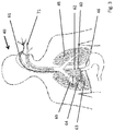
- Fig. 3 shows schematically the arrangement of the system in the patient.
- the system for sensing respiratory effort of a patient (40) includes a pressure sensing device configured as an esophageal catheter (61) for detecting transpulmonary pressure (64). This is similar to alveolar pressure in a good approximation.
- the transpulmonary pressure is determined from the ventilation pressure (27) given by the respirator and the peso (60). The measurement and determination take place, for example, at the end of the exhalation (64 TPP ex) and / or at the end of the inhalation (64 TPP in) by a specific measurement process.
- the esophageal catheter (61) has an air-filled balloon (62) serving as a pressure transducer. The balloon section is adjacent to the lung.
- a respiratory tube (71) can be used in the Trachea be placed to ventilate the patient.
- the two lungs (45) and the diaphragm (46) can be seen.
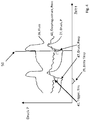
- Fig. 4 schematically shows the course of the signals flow (26), esophageal pressure (60) and ventilation pressure (27). It can be seen from the course of the signals that the pressure-determining device determines the esophageal pressure (60).
- a respiratory effort of the patient (41) is recognized as a rash in the esophageal pressure signal. Diaphragm contractions, for example, produced a negative peso pressure (at 41).
- This patient trigger (41) will, in the left part of the Fig. 4 Not recognized by ventilator as trigger for inspiratory pressure increase (47). This is evident from the time difference (29) between the patient trigger (41) and the actual inspiratory pressure increase (47) by the ventilator.
- this time difference (29) between the patient trigger (41) and the actual inspiratory pressure increase (47) can be detected and evaluated by the ventilator in order to show asynchronicity (30) between the patient and the ventilator, for example as an index futile patient trigger (41).
- a respiratory effort of the patient (41) is recognized as a rash in the esophageal pressure signal.
- Diaphragm contractions for example, produced a negative peso pressure (at 41).
- This patient trigger (41) will, in the right part of the Fig. 4 , at the time (50) detected by the ventilator as trigger for the inspiratory pressure increase (47).
- the device (19, control unit) for specifying the respiratory gas pressure has here detected an exceeding or falling below a threshold value for the esophageal pressure (60) and generates a control signal for the ventilator (20) for setting an inspiratory breathing gas pressure (47).
- the inspiratory breathing gas pressure (47) leads to a breathing gas flow (26) to the patient.
- esophageal pressure measurement is a measurement method available, which can unmask a patient-respirator asynchrony.
- Surveillance of esophageal pressure (60) helps identify and treat the causes of ineffective patient exertion (41).
- Diaphragm contractions generated a negative peso pressure, followed by no inspiratory response in the ventilation pressure curve.
- the combination of high respiratory drive, high flow rate and low tidal volume makes this problem constellation more common.
- Bedside monitoring of respiratory muscle activity in real time by esophageal pressure allows to assess the degree of synchronization (30) between the patient's inspiratory effort and the ventilator's insufflation time and ventilation parameters (eg, optimization of insufflation time or pressure support, or PEEP) or trigger sensitivity adjust accordingly.
- the ventilator's insufflation time and ventilation parameters eg, optimization of insufflation time or pressure support, or PEEP
- Triggered ventilation can avoid diaphragmatic dysfunction by allowing the patient to generate spontaneous inspiratory effort.
- the aim of assisted ventilation is to adapt the insufflation through the ventilator (47) to the respiratory effort of the patient (41), thereby optimizing patient comfort and minimizing the work of breathing.
- Asynchrony (30) between the patient and the ventilator which is defined as a discrepancy between the patient's natural inspiratory time and the ventilator's insufflation time, is a common phenomenon in clinical practice. Nearly a quarter of intubated patients show significant asynchrony during assisted mechanical ventilation, many of whom are clinically unrecognized.
- asynchrony (29) may be an indication of the severity of the respiratory status but, on the other hand, may also be associated with inappropriate settings of the ventilator that prolong the duration of mechanical ventilation.
- Various settings designed to improve synchrony by reducing dynamic hyperinflation have been proposed, such as the use of external positive end-expiratory pressure (PEEP) and the reduction of insufflation time or the prevention of inadequate pressure support.
- PEEP positive end-expiratory pressure
- these approaches are not yet have been systematically compared, and their respective effects on respiratory effort and tidal volume thus remain unclear.
- esophageal pressure measurement is a measurement method available, which can unmask a patient-respirator asynchrony.
- Monitoring esophageal pressure helps identify and treat the causes of ineffective patient effort. Because this asynchronism is associated with a prolonged duration of mechanical ventilation, this could potentially affect the duration of mechanical ventilation. Diaphragm contractions produced a negative peso-pressure, followed by no inspiratory response in the ventilation pressure curve. The combination of high respiratory drive, high flow rate and low tidal volume makes this problem constellation more common.
- Bed-side monitoring of respiratory muscle activity in real time using esophageal pressure allows the degree of synchronization between the patient's inspiratory effort and the ventilator's insufflation time to be assessed and the ventilation parameters (eg, optimization of insufflation time or pressure support, or PEEP) adjusted accordingly, e.g., automated.
- the ventilation parameters e.g, optimization of insufflation time or pressure support, or PEEP
- the number of predetermined inspirations (47) per unit of time is detected and recorded.
- the degree of synchronization (30) between the inspiratory efforts (41) of the patient and the inspiration of the ventilator can be determined.
- an index (30) of the false triggerings can be taken from this comparison, which represents the degree of synchronization (30) between the patient's inspiratory efforts and the inspiratory prescription of the ventilator.
- ⁇ b> ⁇ i> Tab.1 Patient Respirator Asynchrony Unmasking ⁇ / i> ⁇ /b> case scenario Clinical significance Clinical recommendation No peso pressure negatives before ventilator pressure delivery in spontaneously breathing patients autotriggering Control & if necessary adaptation: • leaks • Trigger settings • Condensate in the hoses • Hose systems & ventilation accessories with high resistance • Sedation depth Peso Negative Pressure No Pressure Delivery by the Ventilator in Spontaneously Breathing Patients asynchrony Control & if necessary adaptation: • leaks • Trigger settings • Rise rate (“ramp”) • inspiratory termination ("flow-off criterion”) • PEEP • Condensate in the hoses • Hose systems & ventilation accessories with high resistance Peso inspiratory time longer than inspiratory time of the ventilator Double triggering
- Fig. 5 shows the placement of the peso-catheter.
- pleural pressure (63) was estimated by measuring esophageal pressure using an oesophageal balloon catheter.
- PESO changes during the respiratory cycle reflect the changes in pleural pressure (63) on the lung surface.
- the difference between respiratory and esophageal pressure is a valid estimate of transpulmonary pressure (64, TPP) around the balloon catheter.
- Absolute values of peso can be derived from the respiratory mechanics, the Lung volume, the weight of the mediastinum, the abdomen, the posture, the responsiveness of the smooth muscle wall and the mechanical properties of the balloon are influenced.
- the esophageal balloon In order to obtain a reliable peso measurement, the esophageal balloon must be in a suitable position and filled with a sufficient volume of air. If the balloon is underfilled, the peso will not be transferred correctly. However, a crowded balloon can also overestimate the pressure.
- the volume of air that is "optimal" for the filling depends on the design, dimensions, geometry and material of the esophageal balloon, which in turn affect its mechanical properties.
- the six most common commercial oesophageal catheters have been tested in vitro for external pressures of 0 to 30 cm H2O. It showed that all examined catheters measured the ambient pressure correctly, but there were considerable differences in the optimal filling volume between the catheters.
- the minimum volume required for useful measurements was greater than previously recommended and was also dependent on ambient pressure.
- Another study showed that the filling volume was different for the different catheters studied and that at high pressures a larger filling volume was required.
- simply determine the optimal fill volume by gradually inflating the balloon in the area provided for each catheter to determine the lowest volume at which the peso's largest pulse pressure variation (in a single breath) is delivered (inspiratory) too expiratory).
- Inventive peso-monitoring monitors and intensive fans have special algorithms for optimal filling of the catheter balloon.
- Fig. 6 shows the position control of the peso-catheter.
- the esophageal balloon is secured with a three-way stopcock and a suitable lubricant is applied along the guidewire.
- the peso-catheter is gently advanced to a depth of about 55 cm into the stomach and then filled with the manufacturer's recommended minimum volume.
- the intra-gastric catheter position is confirmed by a slight external manual gastric compression which causes a positive pressure rash. Subsequently, the catheter is gradually withdrawn into the esophagus.
- the pressure changes in the airway ( ⁇ Paw) and in the esophagus ( ⁇ Peso) should be almost identical, because the lung volume does not change and therefore no change in the PL occurs.
- the peso measurement is considered reliable if the ratio ⁇ Peso / ⁇ Paw is between 0.8 and 1.2. Otherwise, the catheter must be repositioned or the balloon volume rechecked.
- the pressure created by the esophageal wall in response to the balloon filling may increase the absolute value of the peso above that of the pleural pressure.
- a calibration method of the invention for eliminating this artifact is that the use of the absolute value of esophageal pressure be improved, particularly when large volume balloons are used.
- the benefit of esophageal measurement as the basis of ventilation therapy in ARDS patients is exemplified in esophageal pressure-controlled ventilation.
- the PEEP values are set with the aim to achieve a transpulmonary pressure (64) between 0 and 10 cmH2O in the end expiratory phase according to a sliding scale based on the ratio PaO2 / FIO2.
- the tidal volume is limited to keep the transpulmonary pressure in the endinspiratory phase below 25 cmH2O.
- PEEP optimization based on peso-monitoring can be used to minimize the onset of atelectasis trauma, to optimize oxygenation, and to improve compliance of the respiratory system.
- the endinspiratory transpulmonary pressure TPP insp (plateau pressure-pleural pressure) may be the alveolar overstretching and thus help to optimize the set tidal volume or the inspiratory pressure.
- the target area is, depending on the clinical findings, a range of up to 20 mbar.
- the work is expressed as force x displacement.
- the work done by the respiratory muscles is equal to the integral of the product of Pmus and volume change. If volume is generated, there is usually a close correlation between WOB and PTP.
- the WOB per respiratory cycle is usually expressed in joules. Work per minute is calculated by multiplying WOB per cycle by the corresponding respiratory rate. Work per liter is calculated by dividing labor per minute by minute volume. A joule is the work required to move 1000ml tidal volume over a pressure difference of 10cm H2O.
- the respective intrinsic PEEP must first be overcome by inhalation of the inspiratory musculature by appropriate pleural pressure-neutralization before an inspiratory flow of respiratory gas is achieved.
- the required breathing work is called "isometric work of breathing" and can account for more than half of the total work of breathing, depending on the severity of COPD.
- ⁇ b> ⁇ i> Tab.4 Spontaneous breathing ⁇ / i> ⁇ /b> abbreviation definition Clinical significance Clinical recommendation Peso min. Lowest pressure within one breath cycle Measure of inadequate spontaneous breathing (eg in the case of ARDS) relatively ⁇ 10 mbar ⁇ PEEP / peso min. Pressure delta between PEEP and peso min in inspiration Measure of inadequate spontaneous breathing (eg in the case of ARDS) ⁇ 10 mbar
- the intrinsic PEEP can be continuously measured with the peso-catheter correctly positioned. It measures the ineffective drop in esophageal pressure during inspiration, which is a surrogate parameter for PEEPi. Based on this, the trapping volume can be calculated.
- the optimal PEEP can also be found by determining the transpulmonary pressure. The transpulmonary pressure is determined as the difference between the alveolar pressure and the pleural pressure, the pleural pressure being measured by measuring the peso-pressure (60). is estimated. According to the invention, an alveolar collapse in expiration is prevented only with positive end-expiratory transpulmonary pressure.
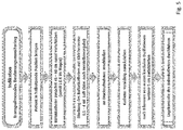
- FIG. 7 illustrates the determination of a false trigger rate.
- the system also includes means for detecting the synchronicity (28) of a respiratory gas delivery (47) (pressure or volume or flow) timed by the ventilator (20) with the respiratory effort (41) of the patient (40).
- the means for detecting the synchronicity (28) may be part of the control unit (19). From the esophageal pressure curve (60) the number of inspiratory efforts (41) of the patient (per unit of time) is detected and recorded. From the respiratory pressure curve (27) or according to the default values of the control unit (with controlled ventilation), the number of predetermined inspirations (47) (per unit time) is detected or removed and recorded.
- the inspiratory efforts of the patient (41) are compared with the given inspirations (47) (per unit of time) and thus the degree of synchronization (30) between the patient's inspiratory efforts (41) and ventilator's inspiratory setting (47) determined. From this comparison, an index (31) of false triggering may be taken, which represents the degree of synchronization (30) between the patient's inspiratory effort and inspiratory prescription of the ventilator.
- the index (31) and / or the degree of synchronization (3) can be output, for example, on the display unit (3) of the ventilator or on a monitor.
- the index (31) and / or the degree of synchronization (30) may alternatively or additionally also be taken into account, at least in part, for the control of the ventilation by the control unit (19).
- the frequency of the breaths (47) are automatically adjusted taking into account index (31) and / or degree of synchronization (30).
- the ventilator or a monitor comprise a device for detecting the synchronicity (28), which may be embodied as part of the control unit (19), the false trigger (31), from the time interval (29) between the predetermined Atemgashub (47) and the respiratory effort (41) of the patient (40) recognizes and determines a false trigger rate or an index (31) and stores or represents.
- the means for detecting the synchronicity (28) determines false trigger (31), from the time interval between the predetermined Atemgashub (47) and the respiratory effort (41) of the patient when the time interval (29) is greater than 1/100 second , preferably greater than 1/10 second, more preferably greater than one second.
Landscapes
- Health & Medical Sciences (AREA)
- Life Sciences & Earth Sciences (AREA)
- Heart & Thoracic Surgery (AREA)
- Engineering & Computer Science (AREA)
- Biomedical Technology (AREA)
- Public Health (AREA)
- General Health & Medical Sciences (AREA)
- Animal Behavior & Ethology (AREA)
- Veterinary Medicine (AREA)
- Pulmonology (AREA)
- Hematology (AREA)
- Biophysics (AREA)
- Anesthesiology (AREA)
- Medical Informatics (AREA)
- Physics & Mathematics (AREA)
- Surgery (AREA)
- Molecular Biology (AREA)
- Pathology (AREA)
- Emergency Medicine (AREA)
- Child & Adolescent Psychology (AREA)
- Physiology (AREA)
- Primary Health Care (AREA)
- Epidemiology (AREA)
- General Business, Economics & Management (AREA)
- Business, Economics & Management (AREA)
- Measurement Of The Respiration, Hearing Ability, Form, And Blood Characteristics Of Living Organisms (AREA)
Abstract
Description
- Die assistierte Beatmung ist eine unterstützende Beatmung. Ausgelöst wird die Beatmung durch einen sogenannten Trigger. Der Patient erzeugt am Beginn der Einatmung selbst einen Atemgas-fluss oder -druck, den das Beatmungsgerät als Trigger erkennt. Überschreitet der vom Patienten erzeugte Trigger die voreingestellte Schwelle, also das eingestellte Triggerniveau, so liefert das Beatmungsgerät Atemgas mit einem bestimmten Druck und/oder Volumen und erleichtert so den Atemzug.
- Ziel einer assistierten Beatmung ist dabei, die Insufflation durch das Beatmungsgerat an die Atemanstrengungen des Patienten anzupassen, um dadurch den Patientenkomfort zu optimieren und die Atemarbeit zu minimieren. Eine Asynchronität zwischen dem Patienten und dem Beatmungsgerat, die als eine Diskrepanz zwischen der natürlichen Inspirationszeit des Patienten und der Insufflationszeit des Beatmungsgeräts definiert wird, ist ein häufiges Phänomen der klinischen Praxis. Fast ein Viertel der intubierten Patienten zeigen während einer assistierten mechanischen Beatmung erhebliche Asynchronitäten, die vielfach klinisch nicht erkannt werden. Das häufigste Asynchronitätsmuster sind ineffektive Triggerungen, bei denen die Inspirationsanstrengungen des Patienten keinen Beatmungshub triggern, da zum Zeitpunkt, zu dem versucht wird, den Trigger auszulosen, eine dynamische Hyperinflation besteht.
- Eine Asynchronität kann einerseits ein Anzeichen für den Schweregrad des respiratorischen Status sein, kann jedoch andererseits auch in Verbindung mit ungeeigneten Einstellungen des Beatmungsgeräts, die die Dauer der mechanischen Beatmung verlängern, stehen. Verschiedene Einstellungen, die die Synchronität durch eine Verringerung der dynamischen Hyperinflation verbessern sollen, wurden bereits vorgeschlagen, wie beispielsweise die Anwendung eines externen positiven endexspiratorischen Drucks (PEEP) und die Verminderung der Insufflationszeit oder die Vermeidung einer inadäquat hohen Druck Unterstützung. Diese Ansätze sind jedoch bisher noch nicht systematisch verglichen worden, und ihre jeweiligen Auswirkungen auf die Atemarbeit und das Tidalvolumen bleiben somit unklar.
- Die Erfindung betrifft ein System zur Erfassung von Atemanstrengungen eines Patienten, umfassend eine Druckermittlungsvorrichtung zum Ermitteln eines Drucks zu dem Zeitpunkt der Atemanstrengung des Patienten.
- Die Atemanstrengung kann dabei eine aktive (muskuläre) oder eine passive (beispielsweise Rückstellkräfte) Atemanstrengung sein.
- Das System ist auch dadurch gekennzeichnet, dass der Zeitpunkt der Atemanstrengung der Beginn der Exspiration oder der Beginn der Inspiration des Patienten ist.
- System zur Erfassung von Atemanstrengungen eines Patienten, umfassend eine Druckermittlungsvorrichtung zum Ermitteln eines Druckes zu dem Zeitpunkt der Atemanstrengung des Patienten.
- Das System ist auch dadurch gekennzeichnet, dass die Druckermittlungsvorrichtung als Ösophagus-Katheter ausgebildet ist und einen gasgefüllten Ballon aufweist.
- Das System ist auch dadurch gekennzeichnet, dass der Sensor des Beatmungsgerätes den Ösophagusdruck bestimmt, der über den gasgefüllten Ballon des Ösophagus-Katheters erfasst wird.
- Das System ist alternativ oder ergänzend zur Erfassung von Atemanstrengungen eines Patienten ausgebildet, umfassend eine Druckermittlungsvorrichtung, zum Ermitteln eines Drucks, zu dem Zeitpunkt der Atemanstrengung des Patienten dadurch gekennzeichnet, dass die Druckermittlungsvorrichtung als Ösophagus-Katheter ausgebildet ist und einen gasgefüllten Ballon aufweist und umfassend ein Beatmungsgerät mit einem Druckeingangsstutzen für den Ösophagus-Katheter und einen Druck)Sensor, wobei eine Atemanstrengung des Patienten über den gasgefüllten Ballon des Ösophagus-Katheters erfasst wird.
- Das System ist alternativ oder ergänzend dadurch gekennzeichnet, dass der transpulmonale Druck von dem Beatmungsgerät bestimmt wird unter Berücksichtigung des, vom Beatmungsgerät vorgegebenen, Beatmungsdrucks und dem sensorisch ermittelten Ösophagusdruck. Das System ist alternativ oder ergänzend dadurch gekennzeichnet, dass der gasgefüllte Ballon einen Sensor aufweist, der eine Atemanstrengung registriert und an ein Beatmungsgerät übermittelt
- Das System ist alternativ oder ergänzend dadurch gekennzeichnet, dass die Bestimmung des transpulmonalen Drucks am Ende der Ausatmung TPP ex und/oder am Ende der Einatmung TPP in durch einen bestimmten Messvorgang erfolgt, bei dem das Beatmungsgerät einen Atemgastransport zu oder von dem Patienten verhindert.
- Das System ist alternativ oder ergänzend dadurch gekennzeichnet, dass eine Atemanstrengung des Patienten mit zumindest einem hinterlegten Schwellwert verglichen wird und bei Überschreitung des Schwellwertes einem Trigger entspricht.
- Das System ist alternativ oder ergänzend dadurch gekennzeichnet, dass der Zeitpunkt der Beginn der Exspiration oder der Beginn der Inspiration des Patienten ist.
- Das System ist alternativ oder ergänzend dadurch gekennzeichnet, dass die Druckermittlungsvorrichtung den Ösophagusdruck fortlaufend, beispielsweise auch zu Beginn der Inspiration oder der Exspiration bestimmt.
- Das System ist alternativ oder ergänzend dadurch gekennzeichnet, dass das System zudem eine Einrichtung, beispielsweise eine Steuereinheit, umfasst die den, von dem Beatmungsgerät bereitgestellten, Atemgasdruck unter Berücksichtigung des ermittelten Ösophagusdrucks oder transpulmonalen Drucks vorgibt.
- Das System ist alternativ oder ergänzend dadurch gekennzeichnet, dass die Einrichtung bei Überschreiten oder Unterschreiten eines Schwellwertes für den Ösophagusdruck ein Steuersignal für das Beatmungsgerät erzeugt, zur Vorgabe eines inspiratorischen oder exspiratorischen Atemgasdrucks.
- Das System ist alternativ oder ergänzend dadurch gekennzeichnet, dass die Steuereinheit des Beatmungsgerätes Atemgasparameter (Druck, Fluss, Volumen, Frequenz) für eine kontrollierte oder assistierte Beatmung bereitgestellt und die Atemgasquelle zur Vorgabe der kontrollierten oder assistierten Beatmung ansteuert. System nach zumindest einem der vorhergehenden Ansprüche dadurch gekennzeichnet, dass aus der Ösophagusdruckkurve die Anzahl der Inspirationsbemühungen des Patienten pro Zeiteinheit) erkannt und aufgezeichnet wird.
- Das System ist alternativ oder ergänzend dadurch gekennzeichnet, dass aus der Beatmungsdruckkurve oder der Steuereinheit die Anzahl der vorgegebenen Inspirationen pro Zeiteinheit) erkannt und aufgezeichnet wird.
- Das System ist alternativ oder ergänzend dadurch gekennzeichnet, dass die Inspirationsbemühungen des Patienten pro Zeiteinheit mit den vorgegebenen Inspirationen pro Zeiteinheit verglichen und so der Grad der Synchronisierung zwischen den Inspirationsbemühungen des Patienten und der Inspiratorischen Vorgabe des Beatmungsgeräts ermittelt wird
- Das System ist alternativ oder ergänzend dadurch gekennzeichnet, dass diesem Vergleich ein Index der Fehltriggerungen entnommen wird, der den Grad der Synchronisierung zwischen den Inspirationsbemühungen des Patienten und der Inspiratorischen Vorgabe des Beatmungsgeräts repräsentiert.
- Das System ist alternativ oder ergänzend dadurch gekennzeichnet, dass die Einrichtung zur Erfassung der Synchronität Fehltrigger, aus dem zeitlichen Abstand zwischen dem vorgegebenen Atemgashub und der Atemanstrengung des Patienten, erkennt und eine Fehltriggerrate oder ein Index ermittelt und speichert oder darstellt.
- Das System ist alternativ oder ergänzend dadurch gekennzeichnet, dass die Einrichtung zur Erfassung der Synchronität Fehltrigger, aus dem zeitlichen Abstand zwischen dem vorgegebenen Atemgashub und der Atemanstrengung des Patienten, dann erkennt, wenn der zeitliche Abstand größer ist als /Sekunde, bevorzugt größer ist /Sekunde, besonders bevorzugt größer ist als eine Sekunde.
- Das System zur Erfassung von Atemanstrengungen eines Patienten, umfasset alternativ oder ergänzend eine Druckermittlungsvorrichtung, zum Ermitteln eines Drucks, zu dem Zeitpunkt der Atemanstrengung des Patienten dadurch gekennzeichnet, dass die Druckermittlungsvorrichtung als Ösophagus-Katheter ausgebildet ist und einen gasgefüllten Ballon aufweist und umfassend ein Beatmungsgerät mit einem Druckeingangsstutzen für den Ösophagus-Katheter und einen Druck)Sensor, wobei eine Atemanstrengung des Patienten über den gasgefüllten Ballon des Ösophagus-Katheters erfasst wird, wobei das System zudem eine Einrichtung zur Erfassung der Synchronität eines durch das Beatmungsgerät (zeitlich) vorgegebenen Atemgashubs (Druck oder Volumen oder Fluss) mit der Atemanstrengung des Patienten, umfasst, wobei diesem Vergleich ein Index der Fehltriggerungen entnommen wird, der den Grad der Synchronisierung zwischen den Inspirationsbemühungen des Patienten und der Inspiratorischen Vorgabe des Beatmungsgeräts repräsentiert.
- Das System ist alternativ oder ergänzend dadurch gekennzeichnet, dass die Einrichtung bei erkannten Fehltriggern ein Steuersignal für das Beatmungsgerät erzeugt, zur Vorgabe einer veränderten Einatemzeit und/oder eines veränderten PEEP und/oder einer veränderten Triggerempfindlichkeit.
- Das System ist alternativ oder ergänzend dadurch gekennzeichnet, dass der Ösophagus-Katheter den funktionellen positiven endexspiratorischen Druck, welcher als intrinsischen PEEP PEEPi) bezeichnet wird, ermittelt.
- Das System ist alternativ oder ergänzend dadurch gekennzeichnet, dass die Einrichtung bei Überschreiten oder Unterschreiten eines Schwellwertes für den PEEPi ein Steuersignal für das Beatmungsgerät erzeugt, zur Vorgabe eines veränderten Atemgasvolumens oder einer veränderten Ausatemzeit.
- Das System ist alternativ oder ergänzend dadurch gekennzeichnet, dass eine Einrichtung zur Erfassung des optimalen Füllvolumens des Ballons umfasst ist, wobei die Einrichtung das geringste Füllvolumen bestimmt, welches die größte Pulsdruckvariation "Swing") des Ballons in einem Atemzug von inspiratorisch zu exspiratorisch) ausmacht. Die Einrichtung zur Erfassung des optimalen Füllvolumens des Ballons kann ein Fluss oder Volumensensor sein, der beispielsweise in dem Beatmungsgerät angeordnet ist und mit der Steuereinheit verbunden ist.
- Die Erfindung umfasst auch ein System mit einer Einrichtung zur Erfassung des optimalen Füllvolumens des Ballons, wobei die Einrichtung das Füllvolumen bestimmt, welches die größte Pulsdruckvariation ("Swing") des Ballons in einem Atemzug (von inspiratorisch zu exspiratorisch) ausmacht.
-
Fig. 1 zeigt den grundsätzlichen Aufbau eines Beatmungsgerätes (20). Im Bereich eines Gerätegehäuses (1) sind ein Bedienelement (2) und/oder ein Bedien-und Informationssystem (3) angeordnet. Über eine Kopplung (4) wird ein Verbindungsschlauch (5) angeschlossen. Entlang des Verbindungsschlauches (5) kann ein zusätzlicher Druckmessschlauch (6) verlaufen, der über einen Druckeingangsstutzen (7) mit dem Gerätegehäuse (1) verbindbar ist. Zur Ermöglichung einer Datenübertragung weist das Gerätegehäuse (1) zumindest eine Schnittstelle (8,18) auf. Ein Anfeuchter (21) oder ein Vernebler (22) kann zudem adaptiert werden. Das Beatmungsgerät weist eine Atemgasquelle (17) auf - Im Bereich einer dem Gerätegehäuse (1) abgewandten Ausdehnung des Verbindungsschlauches (5) ist beispielhaft ein Ausatmungselement (9) angeordnet. Ebenfalls kann ein Ausatemventil verwendet werden.
- Das Beatmungsgerät (20) kann als Schlaftherapiegerät, als High-Flow-Gerät, als Anästhesiegerät, als klinisches- oder Heim- oder Notfall-Beatmungsgerät ausgebildet sein.
-
Fig. 1 zeigt darüber hinaus ein als Beatmungsmaske (10) ausgebildetes Patienten-Interface. Eine Fixierung im Bereich eines Kopfes eines Patienten kann über eine Kopfhaube (11) erfolgen. Im Bereich seiner dem Verbindungsschlauch (5) zugewandten Ausdehnung weist das Patienten-Interface (10) ein Kupplungselement (12) auf. Das Patienten-Interface kann beispielsweise auch als Tubus ausgebildet sein. - Über die Schnittstelle (8,18) kann die Eingabe und / oder Ausgabe von Daten, wie beispielsweise Totraumvolumen, erfolgen. Die Schnittstellen können kabelgebunden, als Infrarot-Schnittstelle, als Bluetooth-Schnittstelle oder USB realisiert sein. Bevorzugt ist auch ein Kartenschacht vorgesehen. Die Schnittstelle (8) kann auch als LAN-Schnittstelle oder als sonstige Schnittstelle zur Anbindung an das Internet ausgeführt sein. Im Bereich eines Gerätegehäuses kann ein Sauerstoffzuschaltventil an die Vorrichtung zur Beatmung adaptiert werden. Es ist denkbar, das Atemgas zusätzlich mit Sauerstoff anzureichern, um die Patientenversorgung zu verbessern.
- Das erfindungsgemäße Beatmungsgerät (20) ist so ausgelegt, dass es über einen Schlauch und ein Patienteninterface mit einem Patienten verbunden werden kann, um eine Beatmung bereitzustellen. Es umfasst eine Quelle für Atemgas (17), die beispielsweise als ein Elektromotor mit Gebläserad oder als Druckgasanschluss mit zumindest einem Ventil ausgebildet ist. Das Beatmungsgerät weist eine Einrichtung zur Ermittlung von Druck und/oder Fluss und/oder Volumen des Atemgases auf. Eine Steuereinheit (19) ist so ausgelegt, dass sie beispielsweise für jeden Atemzyklus auf der Basis eines vorbestimmten Wertes und/oder auf Basis von Messsignalen für die Parameter Druck und/oder Flow und/oder Volumen einen Atemgasparameter bestimmt und die Quelle für Atemgas derart reguliert, dass der Atemgasparameter appliziert wird. Die Steuereinheit kann die Parameter der Beatmung kontrolliert vorgeben und/oder zumindest teilweise assistiert oder adaptiv unter Berücksichtigung von Messignalen.
- Die Steuereinheit (19) ist beispielsweise so ausgelegt, dass sie den aktuellen Druck und/oder Flow und/oder das Volumen von Atemgas bestimmt und den aktuellen Wert über das mit der Steuereinheit Bedien- und Informationssystem oder der Anzeige (3) darstellt. Die Steuereinheit (19) ist außerdem so ausgelegt, dass sie bezogen auf einen oder mehrere Parameter Trendveränderungen ihrer Berechnungen über der Zeit bestimmt, wobei die Trendveränderungen auf der Anzeige angezeigt werden können.
- Weiterhin vergleicht die Steuereinheit (19) solche ParameterWerte, die von einem Benutzer vorgegeben wurden, beispielsweise obere und untere Druckgrenzen oder eine maximal tolerierbare Anzahl von Apnoen pro Zeiteinheit, oder eine maximal tolerierbare Leckage, mit den aktuellen Werten und generiert eine BenutzerInformation zu Abweichungen von der Vorgabe. Die Benutzerinformation wird bevorzugt über das Bedien- und Informationssystem (3) graphisch visualisiert.
- Die Steuereinheit (19) ist beispielsweise auch so ausgelegt, dass sie den Ösophagusdruck zumindest zeitweise oder phasenweise bestimmt. Das Beatmungsgerät (20) weist dazu einen (pneumatischen oder elektronischen oder optischen) Druckmesseingang und einen Drucksensor (23, 24) auf, an den ein Ösophagusballon (62) zumindest mittelbar angeschlossen ist.
- Die Steuereinheit (19) ist beispielsweise eingerichtet und ausgebildet eine Veränderung des Ösophagusdruckes zu identifizieren und daraufhin das Beatmunggerät zur Vorgabe eines Beatmungsparameters anzusteuern.
- Bei Überschreiten oder Unterschreiten eines Schwellwertes für den Ösophagusdruck (60) generiert die Steuereinheit (19) beispielsweise ein Steuersignal für das Beatmungsgerät (20), zur Vorgabe eines inspiratorischen oder exspiratorischen Atemgasdrucks. Bei Überschreiten oder Unterschreiten eines Schwellwertes für den Ösophagusdruck (60) generiert die Steuereinheit (19) beispielsweise alternativ ein Steuersignal für das Beatmungsgerät (20), zur Beendigung der Vorgabe eines inspiratorischen oder exspiratorischen Atemgasdrucks.
-
Fig. 2 zeigt schematisch die Anordnung Beatmungsgerätes (20) in dem System. Die bettseitige Messung des Ösophagusdrucks (60, Peso) basiert auf der Verwendung eines Ösophagusballons (62) und spiegelt dabei als Surrogatparameter die Änderungen des Pleuraldrucks (63) wider. Die fortlaufend oder phasenweise gemessenen bzw. bestimmten transpulmonalen Drücke (64) erlauben, die mechanische Druck- und Volumenbelastung unter Beatmung zu bewerten und die Beatmung entsprechend lungenprotektiv zu adaptieren. Der transpulmonale Druck (64) wird dazu aus dem gemessenen Ösophagusdrucks (60, Peso) und dem vom Beatmungsgerät (20) vorgegebenen oder gemessenen Beatmungsdrucks (27) rechnerisch bestimmt oder ermittelt. Erst die Messung des Ösophagusdrucks (60, Peso), ermöglicht die Bestimmung des transpulmonalen Drucks (64). Dieser ist derjenige Druck, der für die Dehnung der Lunge und der Brustwand erforderlich ist.
Der transpulmonale Druck (64) entspricht dem Druckunterschied zwischen den Alveolen und dem Ösophagus. Beispielsweise kann der transpulmonale Druck (64) bei endinspiratorischer oder endexspiratorischer Okklusion gemessen werden kann.
Das System zur Erfassung von Atemanstrengungen eines Patienten (40), umfasst daher eine Druckermittlungsvorrichtung, die als Ösophagus-Katheter (61) ausgebildet ist, zum Ermitteln eines transpulmonalen Druckes (64) beispielsweise unter Anwendung endinspiratorischer oder endexspiratorischer Okklusionsmanöver. Der Ösophagus-Katheter (61) weist einen luftgefüllten Ballon (62) auf, der als Druckaufnehmer dient. Das Beatmungsgerät (20) weist einen Druckeingangsstutzen (23) für den Ösophagus-Katheter (61) und einen entsprechenden Drucksensor (24) auf, der den Ösophagusdruck (60, Peso) bestimmt.
Alternativ oder ergänzend kann der Ösophagus-Katheter (61) an einen separaten Monitor (51) angeschlossen werden. Der Monitor weist dazu einen (pneumatischen oder elektronischen oder optischen) Druckmesseingang und einen Drucksensor (23, 24) auf, an den ein Ösophagusballon (62) zumindest mittelbar, über den Ösophagus-Katheter (61), angeschlossen ist. Der Monitor (51) und/oder das Beatmungsgerät (3) kann den Druckverlauf des transpulmonalen Druckes (64) und/oder Ösophagusdrucks (60, Peso) und/oder des Beatmungsdruckes (27) darstellen und aufzeichnen. Der Ösophagus-Katheter (61) weist im einfachsten Fall zumindest ein Lumen für die Druckmessung auf und einen Ösophagusballon (62). Der Ösophagus-Katheter (61) kann alternativ oder ergänzend mehrlumig ausgeführt sein und so zudem eine Sondenernährung ermöglichen. Optional kann zusätzlich eine Blutdruckmessung und/oder eine Temperaturmessung ergänzt werden. -
Fig. 3 zeigt schematisch die Anordnung des Systems im Patienten. Das System zur Erfassung von Atemanstrengungen eines Patienten (40), umfasst eine Druckermittlungsvorrichtung, die als Ösophagus-Katheter (61) ausgebildet ist, zum Ermitteln eines transpulmonalen Drucks (64). Dieser gleicht dem alveolären Druck in guter Näherung. Der transpulmonale Druck wird bestimmt aus dem (vom Beatmungsgerät vorgegebenen) Beatmungsdruck (27) und dem Peso (60). Die Messung und Bestimmung erfolgt beispielsweise am Ende der Ausatmung (64 TPP ex) und/oder am Ende der Einatmung (64 TPP in) durch einen bestimmten Messvorgang.
Der Ösophagus-Katheter (61) weist einen luftgefüllten Ballon (62) auf, der als Druckaufnehmer dient. Der Ballonabschnitt liegt dabei benachbart zu der Lunge. Ein Beatmungstubus (71) kann in der Luftröhre platziert werden, um den Patienten zu beatmen. Zu erkennen sind die beiden Lungenflügel (45) und das Zwerchfell (46). -
Fig. 4 zeigt schematisch den Verlauf der Signale Fluss (26), Ösophagus-Druck (60) und Beatmungsdruck (27). Aus dem Verlauf der Signale ist zu erkennen, dass die Druckermittlungsvorrichtung den Ösophagusdruck (60) bestimmt. Eine Atemanstrengung des Patienten (41) wird als Ausschlag in dem Ösophagusdruck-Signal erkannt. Zwerchfellkontraktionen erzeugten so beispielsweise einen negativen Peso-Druck (bei 41). Dieser Patienten-Trigger (41) wird, im linken Teil derFig. 4 , vom Beatmungsgerät nicht als Trigger für die inspiratorische Druckerhöhung (47) erkannt. Dieses wird deutlich durch die zeitliche Differenz (29) zwischen dem Patienten-Trigger (41) und der tatsächlichen inspiratorischen Druckerhöhung (47) durch das Beatmungsgerät. Erfindungsgemäß kann diese die zeitliche Differenz (29) zwischen dem Patienten-Trigger (41) und der tatsächlichen inspiratorischen Druckerhöhung (47) durch das Beatmungsgerät erkannt und ausgewertet werden, um eine Asynchronität (30) zwischen dem Patienten und dem Beatmungsgerät aufzuzeigen, beispielsweise als Index vergeblicher Patienten-Trigger (41). - Erfindungsgemäß ist auch daran gedacht, dass eine Atemanstrengung des Patienten (41) als Ausschlag in dem Ösophagusdruck-Signal erkannt wird. Zwerchfellkontraktionen erzeugten so beispielsweise einen negativen Peso-Druck (bei 41). Dieser Patienten-Trigger (41) wird, im rechten Teil der
Fig. 4 , zum Zeitpunkt (50) vom Beatmungsgerät als Trigger für die inspiratorische Druckerhöhung (47) erkannt. Die Einrichtung (19, Steuereinheit) zur Vorgabe des Atemgasdrucks hat hier ein Überschreiten oder Unterschreiten eines Schwellwertes für den Ösophagusdruck (60) erkannt und generiert ein Steuersignal für das Beatmungsgerät (20) zur Vorgabe eines inspiratorischen Atemgasdrucks (47). Der inspiratorische Atemgasdruck (47) führt zu einem Atemgasfluss (26) zu dem Patienten. - Mit der Ösophagusdruckmessung steht ein Messverfahren zur Verfügung, welches eine Patient-Respirator-Asynchronität demaskieren kann. Die Überwachung des Ösophagusdruckes (60) hilft beim Erkennen und Behandeln der Ursachen der unwirksamen Patientenanstrengungen (41). Zwerchfellkontraktionen erzeugten einen negativen Peso-Druck, auf denen keine inspiratorische Reaktion in der Beatmungsdruckkurve folgt. Durch die Kombination von hohem Atemantrieb, hoher Flussrate und niedrigem Tidalvolumen tritt diese Problemkonstellation häufiger auf. Eine bettseitige Überwachung der Atemmuskelaktivität in Echtzeit mittels Ösophagusdruck erlaubt den Grad der Synchronisierung (30) zwischen den Inspirationsbemühungen des Patienten und der Insufflationszeit des Beatmungsgeräts zu bewerten und die Beatmungsparameter (z.B. Optimierung der Insufflationszeit oder der Druck Unterstützung, bzw. des PEEPs) oder die Triggerempfindlichkeit entsprechend anzupassen.
- Eine getriggerte Beatmung kann eine Zwerchfelldysfunktion vermeiden, indem es dem Patienten ermöglicht wird, spontane Inspirationsanstrengungen zu erzeugen. Ziel einer assistierten Beatmung ist dabei, die Insufflation durch das Beatmungsgerät (47) an die Atemanstrengungen des Patienten (41) anzupassen, um dadurch den Patientenkomfort zu optimieren und die Atemarbeit zu minimieren. Eine Asynchronität (30) zwischen dem Patienten und dem Beatmungsgerät, die als eine Diskrepanz zwischen der natürlichen Inspirationszeit des Patienten und der Insufflationszeit des Beatmungsgeräts definiert wird, ist ein häufiges Phänomen der klinischen Praxis. Fast ein Viertel der intubierten Patienten zeigen während einer assistierten mechanischen Beatmung erhebliche Asynchronitäten, die vielfach klinisch nicht erkannt werden. Das häufigste Asynchronitätsmuster sind ineffektive Triggerungen (41), bei denen die Inspirationsanstrengungen des Patienten keinen Beatmungshub triggern, da zum Zeitpunkt, zu dem versucht wird, den Trigger auszulösen, beispielsweise eine dynamische Hyperinflation besteht.
Eine Asynchronität (29) kann einerseits ein Anzeichen für den Schweregrad des respiratorischen Status sein, kann jedoch andererseits auch in Verbindung mit ungeeigneten Einstellungen des Beatmungsgeräts, die die Dauer der mechanischen Beatmung verlängern, stehen. Verschiedene Einstellungen, die die Synchronität durch eine Verringerung der dynamischen Hyperinflation verbessern sollen, wurden bereits vorgeschlagen, wie beispielsweise die Anwendung eines externen positiven endexspiratorischen Drucks (PEEP) und die Verminderung der Insufflationszeit oder die Vermeidung einer inadäquat hohen Druckunterstützung. Diese Ansätze sind jedoch bisher noch nicht systematisch verglichen worden, und ihre jeweiligen Auswirkungen auf die Atemarbeit und das Tidalvolumen bleiben somit unklar. Mit der Ösophagusdruckmessung steht ein Messverfahren zur Verfügung, welches eine Patient-Respirator-Asynchronität demaskieren kann. Die Überwachung des Ösophagusdruckes hilft beim Erkennen und Behandeln der Ursachen der unwirksamen Patientenanstrengungen. Weil diese Asynchronität mit einer verlängerten Dauer der mechanischen Beatmung verbunden ist, könnte sich dies potenziell auf die Dauer der maschinellen Beatmung auswirken. Zwerchfellkontraktionen erzeugten einen negativen Peso-Druck, auf denen keine inspiratorische Reaktion in der Beatmungsdruckkurve folgt. Durch die Kombination von hohem Atemantrieb, hoher Flussrate und niedrigem Tidalvolumen tritt diese Problemkonstellation häufiger auf. Eine bettseitige Überwachung der Atemmuskelaktivität in Echtzeit mittels Ösophagusdruck erlaubt den Grad der Synchronisierung zwischen den Inspirationsbemühungen des Patienten und der Insufflationszeit des Beatmungsgeräts zu bewerten und die Beatmungsparameter (z.B. Optimierung der Insufflationszeit oder der Druckunterstützung, bzw. des PEEPs) entsprechend, beispielsweise automatisiert, anzupassen. - Aus der Ösophagusdruckkurve (60) wird die Anzahl der Inspirationsbemühungen (41) des Patienten pro Zeiteinheit erkannt und aufgezeichnet.
- Zudem wird aus der Beatmungsdruckkurve (27) oder der Steuereinheit die Anzahl der vorgegebenen Inspirationen (47) pro Zeiteinheit erkannt und aufgezeichnet.
- Werden die Inspirationsbemühungen des Patienten (41) pro Zeiteinheit mit den vorgegebenen Inspirationen (47) pro Zeiteinheit verglichen, kann der Grad der Synchronisierung (30) zwischen den Inspirationsbemühungen (41) des Patienten und der Inspiratorischen Vorgabe des Beatmungsgeräts ermittelt werden.
- Erfindungsgemäß kann diesem Vergleich ein Index (30) der Fehltriggerungen entnommen werden, der den Grad der Synchronisierung (30) zwischen den Inspirationsbemühungen des Patienten und der Inspiratorischen Vorgabe des Beatmungsgeräts repräsentiert.
Tab.1: Demaskierung von Patienten-Respirator-Asynchronie Fallszenarium Klinische Bedeutung Klinische Empfehlung Keine Peso Drucknegativierung vor der Druckabgabe durch das Beatmungsgerät bei spontanatmenden Patienten Autotriggerung Kontrolle & ggfs. Adaption: • Leckagen • Triggereinstellungen • Kondensat in den Schläuchen • Schlauchsysteme & Beatmungszubehör mit hoher Resistance • Sedierungstiefe Peso-Drucknegativierung ohne Druckabgabe durch das Beatmungsgerät bei spontanatmenden Patienten Asynchronität Kontrolle & ggfs. Adaption: • Leckagen • Triggereinstellungen • Druckanstiegsgeschwindigkeit ("Rampe") • Inspiratorische Terminierung ("Flowabschaltkriterium") • PEEP • Kondensat in den Schläuchen • Schlauchsysteme & Beatmungszubehör mit hoher Resistance Peso-Inspirationszeit länger als Inspirationszeit des Beatmungsgeräts Doppel-Triggerung oder vorzeitiges Ende der Inspirationsph ase Kontrolle & ggfs. Adaption: • Inspirationszeit Beatmungsgerät • Höhe und Art der Druckunterstützung • Metabolische Azidose • Enzephalopathie Periodisch auftretende Peso-Negativierung nach der Abgabe von mandatorischen Atemzüge Erkennung von "Reverse Triggering" • Sedierung reduzieren oder ändern (inadäquate Sedierungstiefe) -
Fig. 5 zeigt die Platzierung des Peso-Katheters. Bei aufrecht sitzenden Probanden wurde der Pleuraldruck (63) durch Messen des Ösophagusdrucks unter Verwendung eines ösophagealen Ballonkatheters geschätzt. Die PESO-Änderungen während des Atemzyklus spiegeln die Änderungen des auf die Lungenoberflache einwirkenden Pleuraldrucks (63) wider. Der Unterschied zwischen Beatmungs- und Ösophagusdruck ist eine gültige Schätzung des transpulmonalen Drucks (64, TPP) im Bereich um den Ballonkatheter. Absolute Werte von Peso können von der Atemmechanik, dem Lungenvolumen, dem Gewicht des Mediastinums, dem Abdomen, der Haltung, der Reaktionsfähigkeit der glatten Muskelwand und den mechanischen Eigenschaften des Ballons beeinflusst werden. - Der aktuelle Kenntnisstand über die Auswirkung der Position des Patienten auf den beobachteten Ösophagusdruck und seine respiratorische Variation, bei speziellen Lungen- und Brustwanderkrankungen und Pleuraergüssen, ist noch begrenzt. In vielfaltigen Studien konnte aber aufgezeigt werden, dass selbst unter diesen Bedingungen der Ösophagusdruck ein akzeptabler, zielführender und geeigneter Surrogatparameter zum Pleuraldruck (63) darstellt. Für das zwischenzeitlich am häufigsten eingesetzte Peso-Messverfahren wird ein luft- oder gas-gefüllter Ösophagusballon, quasi eine modifizierte Magensonde verwendet, der über einem langen dünnen Katheter mit einem Druckaufnehmer verbunden ist. Ösophaguskatheter können an moderne Intensivventilatoren (20) mit entsprechenden Anschlüssen (23, 24), an spezielle Monitore (51) zur Ösophagusdruckmessung oder an die invasive RR-Messung von Intensivüberwachungsmonitoren angeschlossen werden.
- Um eine zuverlässige Peso-Messung zu erhalten, muss sich der Ösophagusballon an einer geeigneten Position befinden und mit einem ausreichenden Luftvolumen befüllt sein. Bei Unterfüllung des Ballons wird der Peso nicht korrekt übertragen. Allerdings kann ein überfüllter Ballon auch zur Überschätzung des Drucks fuhren. Welches Luftvolumen für die Füllung "optimal" ist, hangt von Design, Abmessungen, Geometrie und Material des Ösophagusballons ab, die sich wiederum auf dessen mechanische Eigenschaften auswirken. Die sechs am weitesten verbreiteten handelsüblichen Ösophaguskatheter sind in vitro beim externen Drücken von 0 bis 30 cm H2O getestet worden. Dabei zeigte sich, dass alle untersuchten Katheter den Umgebungsdruck korrekt maßen, aber erhebliche Unterschiede im optimalen Füllvolumen zwischen den Kathetern bestanden.
Außerdem war das für brauchbare Messungen erforderliche Mindestvolumen grösser als zuvor empfohlen und hing überdies vom Umgebungsdruck ab. Auch eine weitere Studie zeigte, dass das Füllvolumen für die verschiedenen untersuchten Katheter unterschiedlich war, und dass bei hohen Drücken ein größeres Füllvolumen erforderlich war. In der klinischen Praxis lässt sich das optimale Füllvolumen beispielsweise einfach bestimmen, indem der Ballon nach und nach, im für den jeweiligen Katheter vorgesehenen Bereich, befüllt wird und so das geringste Volumen bestimmt wird, bei dem die gröbste Pulsdruckvariation ("Swing") des Peso in einem Atemzug (von inspiratorisch zu exspiratorisch) auftritt. Erfindungsgemäße Peso- Überwachungsmonitore und Intensivventilatoren verfügen über spezielle Algorithmen zur optimalen Füllung des Katheterballons. -
Fig. 6 zeigt die Lagekontrolle des Peso-Katheters. Nach dem Entleeren wird der Ösophagusballon mit einem Dreiwegehahn gesichert und entlang des Führungsdrahtes ein geeignetes Gleitmittel appliziert.
Vergleichbar einer Magensonde wird der Peso-Katheter vorsichtig bis zu einer Tiefe von etwa 55 cm bis in den Magen vorgeschoben und dann mit dem vom Hersteller empfohlenen Mindestvolumen befüllt. Die intra-gastrische Katheterposition wird durch eine leichte externe manuelle gastrische Kompression bestätigt, die einen positiven Druckausschlag hervorruft. Anschließend wird der Katheter nach und nach in den Ösophagus zurückgezogen.
Eine Positionierung im Ösophagus ist erkennbar am Auftreten kardialer Artefakte in der Druckmessung und durch den Wechsel von intra-abdominalen zu intra-thorakalen Druckverlaufsmustern. Durch die Platzierung des Ösophagusballons innerhalb der unteren zwei Drittel des intra-thorakalen Ösophagus sollen sich mögliche Druckartefakte aufgrund der inhomogenen Kompression des Ösophagus durch externe Strukturen vermeiden lassen. Wenn spontane inspiratorische Bemühungen vorhanden sind, besteht der herkömmliche Test zur Validierung der Peso-Messung darin, die gleichzeitigen negativen Ausschläge des Atemwegs- und Ösophagusdrucks während eines endexspiratorischen Okklusionsmanövers zu vergleichen (sogenannter Baydur-Test). Während der okkludierten Inspiration sollten die Druckänderungen im Atemweg (ΔPaw) und im Ösophagus (ΔPeso) fast identisch sein, weil sich das Lungenvolumen nicht ändert und damit auch keine Änderung im PL auftritt. Die Peso-Messung gilt als verlässlich, wenn das Verhältnis ΔPeso/ΔPaw zwischen 0,8 und 1,2 liegt. Andernfalls muss der Katheter neu positioniert bzw. das Ballonvolumen erneut überprüft werden. - Bei mandatorisch beatmeten Patienten ist eine externe manuelle Thoraxkompression während einer exspiratorischen Pause (Hold-Manöver) anzuwenden, während der, die gleichzeitigen positiven Ausschlage des Atemwegs- und des Ösophagusdrucks zu vergleichen sind (Überdruck- Okklusionstest). Bei gleicher Positionierung des Peso-Katheters liefern der Baydur-Test und der Überdruck-Okklusionstest ähnliche Druckänderungen in den Atemwegen und im Ösophagus. Der absolute Wert kann erheblich hoher sein, wenn der Ösophagusballon im unteren Drittel des Ösophagus anstatt im mittleren Drittel positioniert wird, weil hier der überlagerte Druck durch Herz und Lunge hoher ist. Außerdem kann der Druck, der von der Ösophaguswand als Reaktion auf die Ballonbefüllung erzeugt wird, den absoluten Wert des Peso über den des Pleuraldrucks erhöhen. Ein erfindungsgemäßes Kalibrierverfahren zur Beseitigung dieses Artefakts ist, dass die Verwendung des absoluten Werts des Ösophagusdruckes verbessert ist, insbesondere, wenn Ballons mit großem Volumen eingesetzt werden.
- Der Nutzen der Ösophagusmessung als Basis der Beatmungstherapie beim ARDS-Patienten zeigt sich exemplarisch in einer Ösophagusdruckgesteuerten Beatmung.
Die PEEP-Werte werden mit dem Ziel eingestellt, einen transpulmonalen Druck (64) zwischen 0 und 10 cmH2O in der endexspiratorischen Phase gemäß einer gleitenden Skala basierend auf dem Verhältnis PaO2/FIO2 zu erreichen. Das Tidalvolumen wird beschränkt, um den transpulmonalen Druck in der endinspiratorischen Phase unter 25 cmH2O zu halten. Somit kann die PEEP-Optimierung auf Basis des Peso-Monitorings eingesetzt werden, um das Auftreten eines Atelekttraumas zu minimieren, die Oxygenierung zu optimieren und die Compliance des respiratorischen Systems zu verbessern.
Der endexspiratorische transpulmonale Druck (64, TPP exsp) (= Alveolardruck (65) - Pleuraldruck (63)) kann dann durch die Titration des angewandten PEEP angepasst werden, da der Atemwegsdruck mit dem angewandten PEEP in Zusammenhang steht. Die Titration des angewandten PEEP auf einen endexspiratorischen transpulmonalen Druck zwischen 0 und 10 mbar, aber zumindest im positiven Bereich, kann den zyklischen Alveolarkollaps verringern. Der endinspiratorische transpulmonale Druck TPP insp (Plateaudruck-Pleuraldruck) kann die alveoläre Überdehnung reduzieren und damit die Optimierung des eingestellten Tidalvolumens bzw. des inspiratorischen Drucks unterstützen. Als Zielbereich gilt, je nach klinischem Befund, ein Bereich von bis 20 mbar.Tab. 2: PEEP-Optimierung und Vermeidung der Überdehnung von Lungenarealen Abkürzung Definition Klinische Bedeutung Klinische Empfehlung (64) TPP exp. Endexspiratorischer transpulmonaler Druck (PEEP - Peso) Atemzyklischer Alveolarkollaps im positiven Bereich (64) TPP insp. Endinspiratorischer transpulmonaler Druck (Plateaudruck - Peso) alveoläre Überdehnung < 25 mbar Δ PESO Driving Pressure Ösophagus (ΔPeso = VT / Compliance) •Unabhängiger Mortalitätsfaktor < 12 mbar ΔTPP •Maß für den tidalen Stress des Lungenparenchyms < 12 mbar Transpulmonary Driving Pressure (TPP exp - TPP insp) •Verhältnis von Tidalvolumen zu (statischer) respiratorischer Compliance - Die Messung der Atemarbeit zur Quantifizierung der Atemanstrengung ermöglichen es Peso-Messungen, den Grad der Muskelentlastung unter Beatmung individuell an den Patienten anzupassen. Hierdurch kann eine erhöhte Atemmuskelanstrengung mit der Gefahr der respiratorischen Erschöpfung erkannt und in der Folge die Gefahr eines Weaningversagens minimiert werden. Dabei kann die inspiratorische Atemarbeit sowohl als Flächenintegral über die Zeit (PTP = Druck-Zeit-Produkt) als auch über das Volumen (WOB = Work of Breathing) ermittelt werden. Beide Messverfahren sind wirkungsvolle Methoden zur Einschätzung der Verlustleistung oder des Energieverbrauchs der Atemmuskulatur. Die Arbeit wird als Kraft x Verdrängung ausgedrückt. Bei einem spontan atmenden Patienten ist die von den Atemmuskeln erbrachte Arbeit gleich dem Integral des Produkts aus Pmus und Volumenänderung. Wenn Volumen erzeugt wird, besteht normalerweise eine enge Korrelation zwischen WOB und PTP.
- Die WOB pro Atemzyklus wird normalerweise in Joule ausgedrückt. Arbeit pro Minute wird durch Multiplizieren von WOB pro Zyklus mit der entsprechenden Atemfrequenz berechnet. Arbeit pro Liter wird durch Dividieren von Arbeit pro Minute durch Minutenvolumen berechnet. Ein Joule ist die Arbeit, die erforderlich ist, um 1000ml Tidalvolumen über eine Druckdifferenz von 10 cm H2O zu bewegen. Durch diese Festlegungen kann ein orientierender Normwertbereich festgelegt werden.
Mehrere Studien konnten zeigen, dass sich die Atemanstrengung erhöht, wenn Patienten bei einem Entwöhnungsversuch versagen. Im Verlauf einer Studie zur Spontanatmung blieb das PTP bei der erfolgreichen Entwöhnung von Patienten unverändert. Dagegen entwickelten Patienten mit fehlgeschlagener Entwöhnung als Folge einer Erhöhung der mechanischen Belastung der Atemmuskulatur eine deutliche und progressive Erhöhung von PTP. Am Ende der Studie erhöhte sich der PTP der nicht entwöhnten Patienten um mehr als das Vierfache des normalen Werts. Im Verlauf eines fehlgeschlagenen Entwöhnungsversuchs wiesen Peso-Schwankungen größere Veränderungen auf als der Rapid Shallow Breathing Index (RSBI).
Mit der Unterscheidung von resistiver und elastischer Atemarbeit ergeben sich neue Ansätze die Art der unterstützenden Beatmung selektiv auszurichten, dass die Atemarbeit reduziert wird.Tab. 3: Weaning Abkürzung Definition Klinische Bedeutung Klinische Empfehlung WOB Work of Breathing: Maß für die inspiratorische Atemarbeit pro Atemzug Flächenintegral über das Volumen Gefahr der respiratorischen Erschöpfung 0,3-0,6 Joule/Liter WOB/min. Maß für die inspiratorische Atemarbeit pro Minute (Integral über das Volumen) übertragen auf das Atemminutenvolumen Gefahr der respiratorischen Erschöpfung 2,4 Joule pro Minute PTPes Pressure Time Product: Maß für die Atemanstrengung der Atemmuskulatur Flächenintegral über die Zeit (=Druck-Zeit-Produkt) Gefahr der respiratorischen Erschöpfung > 150 cmH2O*s - Bei chronisch obstruktiven Lungenerkrankungen kann es zu Obstruktionen in den kleinen Atemwegen kommen. Die damit einhergehende inkomplette Ausatmung des eingeatmeten Atemzugvolumens führt zu einer konsekutiver Überblähung der Lungen ("intrinsischer PEEP"). In ähnlicher Wiese kann ein vergleichbares Problem auch durch eine inadäquate Einstellung der Ausatemzeit verursacht werden. Das nicht abgeatmete Lungenvolumen erzeugt einen funktionellen positiven endexspiratorischen Druck, welcher als intrinsische PEEP (PEEPi) bezeichnet wird. Die Höhe des PEEPi ist ein valider Indikator für die dynamische Überblähung der COPD-Lunge.
- Der jeweilige intrinsische PEEP muss bei jeder Einatmung von der Inspirationsmuskulatur durch entsprechende Pleuradrucknegativierung erst überwunden werden, ehe ein inspiratorischer Atemgasfluss zustande kommt. Die hierfür notwendige Atemarbeit wird als "isometrische Atemarbeit" bezeichnet und kann in Abhängigkeit des Schwere-grades der COPD mehr als die Hälfte der gesamten Atemarbeit ausmachen.
Tab.4: Spontanatmung Abkürzung Definition Klinische Bedeutung Klinische Empfehlung Peso min. Geringster Druck innerhalb eines Atemzyklus Maß für inadäquate Spontanatmung (z.B. beim ARDS) relativ <10 mbar Δ PEEP / Peso min. Druckdelta zwischen PEEP und Peso min in der Inspiration Maß für inädaquate Spontanatmung (z.B. beim ARDS) < 10 mbar - Im Gegensatz zur PEEPi-Messung mittels eines speziellen Manöver des Beatmungsgerätes kann unter Spontanatmung der intrinsische PEEP bei korrekt liegendem Peso-Katheter fortlaufend gemessen werden. Dabei wird der flussunwirksame Abfall des Ösophagusdrucks während der Inspiration gemessen, welcher ein Surrogatparameter für den PEEPi ist. Darauf aufbauend kann das Trappingvolumen kalkuliert werden.
Erfindungsgemäß kann der optimale PEEP auch durch Ermittlung des transpulmonalen Druckes gefunden werden. Der transpulmonale Druck wird als Differenz zwischen Alveolardruck und Pleuradruck bestimmt, wobei der Pleuradruck durch Messung des Peso-Drucks (60) abgeschätzt wird. Erfindungsgemäß wird nur bei positivem endexspiratorischem transpulmonalen Druck ein Alveolarkollaps in der Exspiration verhindert. Entsprechend erfolgt die Drucksteuerung derart, dass end-exspiratorisch ein positiver transpulmonaler Druck vorherrscht.Tab. 5: Inadäquate Exspiration Abkürzung Definition Klinische Bedeutung Klinische Empfehlung PEEPi. Intrinsisc her PEEP Maß für den dynamischen PEEP (Auto-PEEP) Kontrolle & ggfs. Adaption: • Inspirationszeit • Exspirationszeit • Frequenz • Druckanstiegsgeschwindigkeit ("Rampe") • Inspiratorische Terminierung ("Flowabschaltkriterium" z.B. "PSV-Endflow") • PEEP •Höhe der Druckunterstützung Vtrap. Trappingvo lumen Indikator für alveoläre Überdehnung ("Überblähung") Kontrolle & ggfs. Adaption: • Inspirationszeit • Exspirationszeit • Frequenz • Druckanstiegsgeschwindigkeit ("Rampe") • Inspiratorische Terminierung ("Flowabschaltkriterium" z.B. "PSV-Endflow") • PEEP •Höhe der Druckunterstützung -
Figur 7 veranschaulicht die Ermittlung einer Fehltriggerrate. Das System weist zudem eine Einrichtung zur Erfassung der Synchronität (28) eines durch das Beatmungsgerät (20) zeitlich vorgegebenen Atemgashubs (47) (Druck oder Volumen oder Fluss) mit der Atemanstrengung (41) des Patienten (40) auf. Die Einrichtung zur Erfassung der Synchronität (28) kann Teil der Steuereinheit (19) sein.
Aus der Ösophagusdruckkurve (60) wird die Anzahl der Inspirationsbemühungen (41) des Patienten (pro Zeiteinheit) erkannt und aufgezeichnet.
Aus der Beatmungsdruckkurve (27) oder entsprechend der Vorgabewerte der Steuereinheit (bei kontrollierter Beatmung) wird die Anzahl der vorgegebenen Inspirationen (47) (pro Zeiteinheit) erkannt oder entnommen und aufgezeichnet. - Die Inspirationsbemühungen des Patienten (41) (pro Zeiteinheit) werden mit den vorgegebenen Inspirationen (47) (pro Zeiteinheit) verglichen und so der Grad der Synchronisierung (30) zwischen den Inspirationsbemühungen (41) des Patienten und der Inspiratorischen Vorgabe (47) des Beatmungsgeräts ermittelt.
Diesem Vergleich kann ein Index (31) der Fehltriggerungen entnommen werden, der den Grad der Synchronisierung (30) zwischen den Inspirationsbemühungen des Patienten und der Inspiratorischen Vorgabe des Beatmungsgeräts repräsentiert. Der Index (31) und/oder der Grad der Synchronisierung (3) können beispielsweise auf der Anzeigeeinheit (3) des Beatmungsgerätes oder auf einem Monitor ausgegeben werden. Der Index (31) und/oder der Grad der Synchronisierung (30) können alternativ oder ergänzend auch für die Steuerung der Beatmung von der Steuereinheit (19) zumindest teilweise berücksichtigt werden. Beispielsweise indem bei der kontrollierten Beatmung die Frequenz der Atemhübe (47) unter Berücksichtigung von Index (31) und/oder Grad der Synchronisierung (30) automatisch angepasst werden.
Das Beatmungsgerät oder ein Monitor weisen eine Einrichtung zur Erfassung der Synchronität (28) auf, die als Teil der Steuereinheit (19) ausgeführt sein kann, die Fehltrigger (31), aus dem zeitlichen Abstand (29) zwischen dem vorgegebenen Atemgashub (47) und der Atemanstrengung (41) des Patienten (40) erkennt und eine Fehltriggerrate oder ein Index (31) ermittelt und speichert oder darstellt.
Die Einrichtung zur Erfassung der Synchronität (28) ermittelt Fehltrigger (31), aus dem zeitlichen Abstand zwischen dem vorgegebenen Atemgashub (47) und der Atemanstrengung (41) des Patienten dann, wenn der zeitliche Abstand (29) größer ist als 1/100 Sekunde, bevorzugt größer ist 1/10 Sekunde, besonders bevorzugt größer ist als eine Sekunde.
Claims (19)
- System zur Erfassung von Atemanstrengungen (41) eines Patienten (40), umfassend eine Druckermittlungsvorrichtung (61, 62) zum Ermitteln eines Druckes (60, 64) zu dem Zeitpunkt (50) der Atemanstrengung (41) des Patienten (40).
- System nach Anspruch 1, dadurch gekennzeichnet, dass die Druckermittlungsvorrichtung als Ösophagus-Katheter (61) ausgebildet ist und einen gasgefüllten Ballon (62) aufweist, wobei der Sensor (24) des Beatmungsgerätes (20) den Ösophagusdruck (60) bestimmt, der über den gasgefüllten Ballon (62) des Ösophagus-Katheters (61) erfasst wird.
- System nach zumindest einem der vorhergehenden Ansprüche, umfassend eine Druckermittlungsvorrichtung (61, 62) zum Ermitteln eines Drucks (60, 64) zu dem Zeitpunkt (50) der Atemanstrengung (41) des Patienten (40) dadurch gekennzeichnet, dass die Druckermittlungsvorrichtung als Ösophagus-Katheter (61) ausgebildet ist und einen gasgefüllten Ballon (62) aufweist und umfassend ein Beatmungsgerät (20) mit einem Druckeingangsstutzen (23) für den Ösophagus-Katheter (61) und einen (Druck)Sensor (24), wobei eine Atemanstrengung (41) des Patienten (40) über den gasgefüllten Ballon (62) des Ösophagus-Katheters (61) erfasst wird.
- System nach zumindest einem der vorhergehenden Ansprüche dadurch gekennzeichnet, dass der transpulmonale Druck (64) von dem Beatmungsgerät bestimmt wird unter Berücksichtigung des, vom Beatmungsgerät vorgegebenen, Beatmungsdrucks (27) und dem sensorisch ermittelten Ösophagusdruck (60).
- System nach zumindest einem der vorhergehenden Ansprüche dadurch gekennzeichnet, dass der gasgefüllte Ballon (62) einen Sensor aufweist, der eine Atemanstrengung (41) registriert und an ein Beatmungsgerät übermittelt.
- System nach zumindest einem der vorhergehenden Ansprüche dadurch gekennzeichnet, dass die Bestimmung des transpulmonalen Drucks (64) am Ende der Ausatmung (64, TPP ex) und/oder am Ende der Einatmung (64, TPP in) durch einen bestimmten Messvorgang erfolgt, bei dem das Beatmungsgerät einen Atemgastransport zu oder von dem Patienten verhindert.
- System nach zumindest einem der vorhergehenden Ansprüche dadurch gekennzeichnet, dass eine Atemanstrengung (41) des Patienten (40) mit zumindest einem hinterlegten Schwellwert verglichen wird und bei Überschreitung des Schwellwertes einem Trigger (42) entspricht.
- System nach zumindest einem der vorhergehenden Ansprüche dadurch gekennzeichnet, dass der Zeitpunkt (50) der Beginn der Exspiration oder der Beginn der Inspiration (41) des Patienten ist.
- System nach zumindest einem der vorhergehenden Ansprüche dadurch gekennzeichnet, dass das System zudem eine Einrichtung (19), beispielsweise eine Steuereinheit, umfasst die den, von dem Beatmungsgerät (20) bereitgestellten, Atemgasdruck unter Berücksichtigung des ermittelten Ösophagusdrucks (60) oder transpulmonalen Drucks (64) vorgibt.
- System nach zumindest einem der vorhergehenden Ansprüche dadurch gekennzeichnet, dass die Einrichtung (19) bei Überschreiten oder Unterschreiten eines Schwellwertes für den Ösophagusdruck (60) ein Steuersignal für das Beatmungsgerät (20) erzeugt, zur Vorgabe eines inspiratorischen oder exspiratorischen Atemgasdrucks (47).
- System nach zumindest einem der vorhergehenden Ansprüche dadurch gekennzeichnet, dass aus der Ösophagusdruckkurve (60) die Anzahl der Inspirationsbemühungen (41) des Patienten (pro Zeiteinheit) erkannt und aufgezeichnet wird und/oder, dass aus der Beatmungsdruckkurve (27) oder der Steuereinheit die Anzahl der vorgegebenen Inspirationen (47) (pro Zeiteinheit) erkannt und aufgezeichnet wird.
- System nach zumindest einem der vorhergehenden Ansprüche dadurch gekennzeichnet, dass die Inspirationsbemühungen des Patienten (41) pro Zeiteinheit mit den vorgegebenen Inspirationen (47) pro Zeiteinheit verglichen und so der Grad der Synchronisierung (30) zwischen den Inspirationsbemühungen (41) des Patienten und der Inspiratorischen Vorgabe des Beatmungsgeräts ermittelt wird.
- System nach zumindest einem der vorhergehenden Ansprüche dadurch gekennzeichnet, dass diesem Vergleich ein Index (31) der Fehltriggerungen entnommen wird, der den Grad der Synchronisierung (30) zwischen den Inspirationsbemühungen des Patienten und der Inspiratorischen Vorgabe des Beatmungsgeräts repräsentiert.
- System nach zumindest einem der vorhergehenden Ansprüche dadurch gekennzeichnet, dass die Einrichtung zur Erfassung der Synchronität (28) Fehltrigger (31), aus dem zeitlichen Abstand (29) zwischen dem vorgegebenen Atemgashub (47) und der Atemanstrengung (41) des Patienten (40), erkennt und eine Fehltriggerrate oder ein Index (31) ermittelt und speichert oder darstellt.
- System nach zumindest einem der vorhergehenden Ansprüche dadurch gekennzeichnet, umfassend eine Druckermittlungsvorrichtung (61, 62) zum Ermitteln eines Drucks (60, 64) zu dem Zeitpunkt (50) der Atemanstrengung (41) des Patienten (40) dadurch gekennzeichnet, dass die Druckermittlungsvorrichtung als Ösophagus-Katheter (61) ausgebildet ist und einen gasgefüllten Ballon (62) aufweist und umfassend ein Beatmungsgerät (20) mit einem Druckeingangsstutzen (23) für den Ösophagus-Katheter (61) und einen (Druck)Sensor (24), wobei eine Atemanstrengung (41) des Patienten (40) über den gasgefüllten Ballon (62) des Ösophagus-Katheters (61) erfasst wird, wobei das System zudem eine Einrichtung zur Erfassung der Synchronität (28) eines durch das Beatmungsgerät (20) (zeitlich) vorgegebenen Atemgashubs (47) (Druck oder Volumen oder Fluss) mit der Atemanstrengung (41) des Patienten (40), umfasst, wobei diesem Vergleich ein Index (31) der Fehltriggerungen entnommen wird, der den Grad der Synchronisierung (30) zwischen den Inspirationsbemühungen des Patienten und der Inspiratorischen Vorgabe des Beatmungsgeräts repräsentiert.
- System nach zumindest einem der vorhergehenden Ansprüche dadurch gekennzeichnet, dass die Einrichtung (19) bei erkannten Fehltriggern (31) ein Steuersignal für das Beatmungsgerät (20) erzeugt, zur Vorgabe einer veränderten Einatemzeit und/oder eines veränderten PEEP und/oder einer veränderten Triggerempfindlichkeit.
- System nach zumindest einem der vorhergehenden Ansprüche dadurch gekennzeichnet, dass der Ösophagus-Katheter (61) den funktionellen positiven endexspiratorischen Druck, welcher als intrinsischen PEEP (PEEPi) bezeichnet wird, ermittelt.
- System nach zumindest einem der vorhergehenden Ansprüche dadurch gekennzeichnet, dass die Einrichtung (19) bei Überschreiten oder Unterschreiten eines Schwellwertes für den PEEPi ein Steuersignal für das Beatmungsgerät (20) erzeugt, zur Vorgabe eines veränderten Atemgasvolumens oder einer veränderten Ausatemzeit.
- System nach zumindest einem der vorhergehenden Ansprüche dadurch gekennzeichnet, dass eine Einrichtung zur Erfassung des optimalen (70) Füllvolumens des Ballons (62) umfasst ist, wobei die Einrichtung das geringste Füllvolumen bestimmt, welches die größte Pulsdruckvariation ("Swing") des Ballons (62) in einem Atemzug (von inspiratorisch zu exspiratorisch) ausmacht
Applications Claiming Priority (1)
| Application Number | Priority Date | Filing Date | Title |
|---|---|---|---|
| DE102018000757 | 2018-01-31 |
Publications (2)
| Publication Number | Publication Date |
|---|---|
| EP3520850A1 true EP3520850A1 (de) | 2019-08-07 |
| EP3520850B1 EP3520850B1 (de) | 2021-03-10 |
Family
ID=65717943
Family Applications (1)
| Application Number | Title | Priority Date | Filing Date |
|---|---|---|---|
| EP19401004.7A Active EP3520850B1 (de) | 2018-01-31 | 2019-01-31 | System zur erfassung von atemanstrengungen eines patienten |
Country Status (3)
| Country | Link |
|---|---|
| US (1) | US12303244B2 (de) |
| EP (1) | EP3520850B1 (de) |
| DE (1) | DE102019102218A1 (de) |
Cited By (2)
| Publication number | Priority date | Publication date | Assignee | Title |
|---|---|---|---|---|
| DE102020001440A1 (de) | 2020-03-06 | 2021-09-09 | Drägerwerk AG & Co. KGaA | Beatmungsvorrichtung |
| EP4154808B1 (de) * | 2021-09-16 | 2026-03-18 | Löwenstein Medical Technology S.A. | System zur befüllung eines ösophagusballons |
Families Citing this family (12)
| Publication number | Priority date | Publication date | Assignee | Title |
|---|---|---|---|---|
| CN110753564B (zh) | 2018-05-02 | 2023-04-11 | 深圳迈瑞生物医疗电子股份有限公司 | 呼吸机和呼吸机的通气控制方法 |
| US11298485B2 (en) * | 2019-07-17 | 2022-04-12 | Convergent Engineering, Inc. | Esophageal pressure clinical decision support system |
| EP3927232A1 (de) * | 2019-02-20 | 2021-12-29 | Maquet Critical Care AB | Automatische auswertung eines füllvolumens eines ösophagus-ballonkatheters |
| CN114126694B (zh) * | 2019-12-03 | 2024-08-23 | 深圳迈瑞生物医疗电子股份有限公司 | 一种患者通气监测装置及其通气监测方法 |
| DE102020114209B3 (de) * | 2020-05-27 | 2021-07-29 | Ventinova Technologies B.V. | Beatmungsvorrichtung und Verfahren zur Bestimmung zumindest der gewebsrelatierten Resistance im Atemweg |
| DE102020120900A1 (de) * | 2020-08-07 | 2022-02-10 | Drägerwerk AG & Co. KGaA | Vorrichtung und Verfahren zur Messung der Lungen-Compliance |
| EP4265291A4 (de) * | 2020-12-21 | 2024-01-17 | Medcaptain Medical Technology Co., Ltd. | Atemanordnung zur unterstützung der sprachfunktion und beatmungsverfahren dafür sowie beatmungsmaschine |
| EP4268721A4 (de) * | 2020-12-25 | 2024-02-28 | Shenzhen Mindray Bio-Medical Electronics Co., Ltd. | Medizinische vorrichtung und positionsaufforderungsverfahren für ösophagusmanometrievorrichtung dafür |
| DE102021104993A1 (de) * | 2021-03-02 | 2022-09-08 | Hamilton Medical Ag | Kalibriersystem für einen Ösophaguskatheter mit Ballonsonde zur Bestimmung eines ösophagealen Drucks |
| DE102021128269A1 (de) * | 2021-10-29 | 2023-05-04 | Hamilton Medical Ag | Vorrichtung zur Einstellung eines durch eine Beatmungsvorrichtung vorgegebenen Beatmungsparameters und/oder zur automatisierten Darstellung von für maschinelle Beatmung relevanter Information auf Grundlage eines Ösophagusballondrucks |
| IT202200001706A1 (it) * | 2022-02-01 | 2023-08-01 | Dimar S R L | Manometro per interfaccia respiratoria non invasiva e relativa interfaccia respiratoria. |
| CN119184668B (zh) * | 2024-09-20 | 2025-10-17 | 浙江工业大学 | 压力控制通气下基于呼吸系统模型的自主吸气努力强度估测方法及系统 |
Citations (7)
| Publication number | Priority date | Publication date | Assignee | Title |
|---|---|---|---|---|
| US5520192A (en) * | 1991-12-23 | 1996-05-28 | Imperial College Of Science, Technology And Medicine | Apparatus for the monitoring and control of respiration |
| DE10213905A1 (de) * | 2001-03-30 | 2002-10-10 | Fred G Goebel | Verfahren zur Steuerung eines Beatmungsgerätes und Anlage hierfür |
| WO2006079152A1 (en) * | 2004-10-20 | 2006-08-03 | Resmed Limited | Method and apparatus for detecting ineffective inspiratory efforts and improving patient-ventilator interaction |
| WO2010121313A1 (en) * | 2009-04-22 | 2010-10-28 | Resmed Ltd | Detection of asynchrony |
| EP2397074A1 (de) * | 2010-06-19 | 2011-12-21 | M Stenqvist AB | System und Verfahren zur Bestimmung des transpulmonalen Drucks in einem Patienten, der an einem Beatmungsgerät angeschlossen ist |
| DE102012215662A1 (de) * | 2012-09-04 | 2014-03-06 | Hamilton Medical Ag | System zur automatisierten Einstellung eines durch eine Beatmungseinrichtung vorgegebenen Drucks |
| US20150040905A1 (en) * | 2013-08-09 | 2015-02-12 | Advanced Cooling Therapy, Llc | Systems and methods for providing ventilation |
Family Cites Families (3)
| Publication number | Priority date | Publication date | Assignee | Title |
|---|---|---|---|---|
| WO2007082384A1 (en) * | 2006-01-19 | 2007-07-26 | Maquet Critical Care Ab | Method and system for determining dynamically respiratory features in spontaneously breathing patients receiving mechanical ventilatory assist |
| WO2014009950A1 (en) * | 2012-07-11 | 2014-01-16 | Lunguard Ltd. | Method for monitoring pressures exerted along the length of a biological channel |
| WO2017085540A1 (de) * | 2015-11-18 | 2017-05-26 | Creative Balloons Gmbh | Vorrichtung zur dynamisch dichtenden okklusion oder raumfüllenden tamponade eines hohlorgans |
-
2019
- 2019-01-29 DE DE102019102218.5A patent/DE102019102218A1/de active Pending
- 2019-01-29 US US16/260,358 patent/US12303244B2/en active Active
- 2019-01-31 EP EP19401004.7A patent/EP3520850B1/de active Active
Patent Citations (7)
| Publication number | Priority date | Publication date | Assignee | Title |
|---|---|---|---|---|
| US5520192A (en) * | 1991-12-23 | 1996-05-28 | Imperial College Of Science, Technology And Medicine | Apparatus for the monitoring and control of respiration |
| DE10213905A1 (de) * | 2001-03-30 | 2002-10-10 | Fred G Goebel | Verfahren zur Steuerung eines Beatmungsgerätes und Anlage hierfür |
| WO2006079152A1 (en) * | 2004-10-20 | 2006-08-03 | Resmed Limited | Method and apparatus for detecting ineffective inspiratory efforts and improving patient-ventilator interaction |
| WO2010121313A1 (en) * | 2009-04-22 | 2010-10-28 | Resmed Ltd | Detection of asynchrony |
| EP2397074A1 (de) * | 2010-06-19 | 2011-12-21 | M Stenqvist AB | System und Verfahren zur Bestimmung des transpulmonalen Drucks in einem Patienten, der an einem Beatmungsgerät angeschlossen ist |
| DE102012215662A1 (de) * | 2012-09-04 | 2014-03-06 | Hamilton Medical Ag | System zur automatisierten Einstellung eines durch eine Beatmungseinrichtung vorgegebenen Drucks |
| US20150040905A1 (en) * | 2013-08-09 | 2015-02-12 | Advanced Cooling Therapy, Llc | Systems and methods for providing ventilation |
Cited By (3)
| Publication number | Priority date | Publication date | Assignee | Title |
|---|---|---|---|---|
| DE102020001440A1 (de) | 2020-03-06 | 2021-09-09 | Drägerwerk AG & Co. KGaA | Beatmungsvorrichtung |
| US11925757B2 (en) | 2020-03-06 | 2024-03-12 | Drägerwerk AG & Co. KGaA | Ventilator |
| EP4154808B1 (de) * | 2021-09-16 | 2026-03-18 | Löwenstein Medical Technology S.A. | System zur befüllung eines ösophagusballons |
Also Published As
| Publication number | Publication date |
|---|---|
| US20190231202A1 (en) | 2019-08-01 |
| EP3520850B1 (de) | 2021-03-10 |
| DE102019102218A1 (de) | 2019-08-01 |
| US12303244B2 (en) | 2025-05-20 |
Similar Documents
| Publication | Publication Date | Title |
|---|---|---|
| EP3520850B1 (de) | System zur erfassung von atemanstrengungen eines patienten | |
| US11666716B2 (en) | System for automated adjustment of a pressure set by a ventilation device | |
| JP3569335B2 (ja) | 肺内への空気流量および肺内の空気圧を調整する換気装置/呼吸器 | |
| US9027552B2 (en) | Ventilator-initiated prompt or setting regarding detection of asynchrony during ventilation | |
| DE60109474T2 (de) | Beatmungsvorrichtung | |
| DE69735428T2 (de) | Gerät zur beatmung während der anästhesie | |
| EP3653248B1 (de) | Beatmungsgeräte | |
| US20150090258A1 (en) | Ventilator-initiated prompt or setting regarding detection of asynchrony during ventilation | |
| US10342942B2 (en) | Respiratory assistance device and method of controlling said device | |
| DE10213905A1 (de) | Verfahren zur Steuerung eines Beatmungsgerätes und Anlage hierfür | |
| JP2017196438A (ja) | 患者の経肺圧を決定可能な呼吸装置 | |
| EP0459284B1 (de) | Vorrichtung zur automatischen Steuerung der Spontanatmungsunterstützung | |
| DE102020123138B3 (de) | Verfahren und Vorrichtung zur automatischen Festlegung der Sollfrequenz eines Beatmungsgeräts | |
| CN111543999B (zh) | 用于感测病人的呼吸努力的系统 | |
| DE102014119374B4 (de) | Beatmungssystem | |
| DE102020001440A1 (de) | Beatmungsvorrichtung | |
| Rittner et al. | Curves and loops in mechanical ventilation | |
| DE102023123866A1 (de) | Beatmungsvorrichtung und Verfahren zur Steuerung einer Beatmungsvorrichtung | |
| EP4154808B1 (de) | System zur befüllung eines ösophagusballons | |
| Rauseo et al. | Proportional modes | |
| Blanch et al. | Is tracheal pressure ventilator control comparable to an equivalent level of pressure support ventilation? | |
| EP4437947A1 (de) | Vorrichtung und verfahren zur nicht invasiven bestimmung eines verschlussdrucks in der lunge eines patienten | |
| WO2023072676A1 (de) | Vorrichtung zur einstellung eines durch eine beatmungsvorrichtung vorgegebenen beatmungsparameters und/oder zur automatisierten darstellung von für maschinelle beatmung relevanter information auf grundlage eines ösophagusballondrucks | |
| Donley | Evaluation of Intensive Care Mechanical Ventilator Response Time During Varying Levels of Inspiratory Effort | |
| WO2015002662A1 (en) | Method and system for controlling breathing |
Legal Events
| Date | Code | Title | Description |
|---|---|---|---|
| PUAI | Public reference made under article 153(3) epc to a published international application that has entered the european phase |
Free format text: ORIGINAL CODE: 0009012 |
|
| STAA | Information on the status of an ep patent application or granted ep patent |
Free format text: STATUS: THE APPLICATION HAS BEEN PUBLISHED |
|
| AK | Designated contracting states |
Kind code of ref document: A1 Designated state(s): AL AT BE BG CH CY CZ DE DK EE ES FI FR GB GR HR HU IE IS IT LI LT LU LV MC MK MT NL NO PL PT RO RS SE SI SK SM TR |
|
| AX | Request for extension of the european patent |
Extension state: BA ME |
|
| STAA | Information on the status of an ep patent application or granted ep patent |
Free format text: STATUS: REQUEST FOR EXAMINATION WAS MADE |
|
| 17P | Request for examination filed |
Effective date: 20200206 |
|
| RBV | Designated contracting states (corrected) |
Designated state(s): AL AT BE BG CH CY CZ DE DK EE ES FI FR GB GR HR HU IE IS IT LI LT LU LV MC MK MT NL NO PL PT RO RS SE SI SK SM TR |
|
| GRAP | Despatch of communication of intention to grant a patent |
Free format text: ORIGINAL CODE: EPIDOSNIGR1 |
|
| STAA | Information on the status of an ep patent application or granted ep patent |
Free format text: STATUS: GRANT OF PATENT IS INTENDED |
|
| RIC1 | Information provided on ipc code assigned before grant |
Ipc: A61B 5/08 20060101ALI20201123BHEP Ipc: A61B 5/113 20060101ALI20201123BHEP Ipc: A61M 16/04 20060101AFI20201123BHEP Ipc: A61B 5/087 20060101ALI20201123BHEP Ipc: A61B 5/00 20060101ALI20201123BHEP Ipc: A61B 5/11 20060101ALI20201123BHEP Ipc: A61M 16/00 20060101ALI20201123BHEP Ipc: A61B 5/03 20060101ALI20201123BHEP Ipc: A61M 25/10 20130101ALN20201123BHEP Ipc: A61B 5/085 20060101ALI20201123BHEP Ipc: A61J 15/00 20060101ALI20201123BHEP |
|
| INTG | Intention to grant announced |
Effective date: 20201211 |
|
| RAP1 | Party data changed (applicant data changed or rights of an application transferred) |
Owner name: LOEWENSTEIN MEDICAL TECHNOLOGY S.A. |
|
| RIC1 | Information provided on ipc code assigned before grant |
Ipc: A61M 25/10 20130101ALN20201130BHEP Ipc: A61B 5/087 20060101ALI20201130BHEP Ipc: A61M 16/00 20060101ALI20201130BHEP Ipc: A61J 15/00 20060101ALI20201130BHEP Ipc: A61B 5/11 20060101ALI20201130BHEP Ipc: A61B 5/03 20060101ALI20201130BHEP Ipc: A61B 5/085 20060101ALI20201130BHEP Ipc: A61B 5/113 20060101ALI20201130BHEP Ipc: A61B 5/00 20060101ALI20201130BHEP Ipc: A61M 16/04 20060101AFI20201130BHEP Ipc: A61B 5/08 20060101ALI20201130BHEP |
|
| GRAS | Grant fee paid |
Free format text: ORIGINAL CODE: EPIDOSNIGR3 |
|
| GRAA | (expected) grant |
Free format text: ORIGINAL CODE: 0009210 |
|
| STAA | Information on the status of an ep patent application or granted ep patent |
Free format text: STATUS: THE PATENT HAS BEEN GRANTED |
|
| AK | Designated contracting states |
Kind code of ref document: B1 Designated state(s): AL AT BE BG CH CY CZ DE DK EE ES FI FR GB GR HR HU IE IS IT LI LT LU LV MC MK MT NL NO PL PT RO RS SE SI SK SM TR |
|
| REG | Reference to a national code |
Ref country code: GB Ref legal event code: FG4D Free format text: NOT ENGLISH |
|
| REG | Reference to a national code |
Ref country code: CH Ref legal event code: EP Ref country code: AT Ref legal event code: REF Ref document number: 1369132 Country of ref document: AT Kind code of ref document: T Effective date: 20210315 |
|
| REG | Reference to a national code |
Ref country code: DE Ref legal event code: R096 Ref document number: 502019000975 Country of ref document: DE |
|
| REG | Reference to a national code |
Ref country code: IE Ref legal event code: FG4D Free format text: LANGUAGE OF EP DOCUMENT: GERMAN |
|
| REG | Reference to a national code |
Ref country code: LT Ref legal event code: MG9D |
|
| PG25 | Lapsed in a contracting state [announced via postgrant information from national office to epo] |
Ref country code: HR Free format text: LAPSE BECAUSE OF FAILURE TO SUBMIT A TRANSLATION OF THE DESCRIPTION OR TO PAY THE FEE WITHIN THE PRESCRIBED TIME-LIMIT Effective date: 20210310 Ref country code: BG Free format text: LAPSE BECAUSE OF FAILURE TO SUBMIT A TRANSLATION OF THE DESCRIPTION OR TO PAY THE FEE WITHIN THE PRESCRIBED TIME-LIMIT Effective date: 20210610 Ref country code: FI Free format text: LAPSE BECAUSE OF FAILURE TO SUBMIT A TRANSLATION OF THE DESCRIPTION OR TO PAY THE FEE WITHIN THE PRESCRIBED TIME-LIMIT Effective date: 20210310 Ref country code: GR Free format text: LAPSE BECAUSE OF FAILURE TO SUBMIT A TRANSLATION OF THE DESCRIPTION OR TO PAY THE FEE WITHIN THE PRESCRIBED TIME-LIMIT Effective date: 20210611 Ref country code: LT Free format text: LAPSE BECAUSE OF FAILURE TO SUBMIT A TRANSLATION OF THE DESCRIPTION OR TO PAY THE FEE WITHIN THE PRESCRIBED TIME-LIMIT Effective date: 20210310 Ref country code: NO Free format text: LAPSE BECAUSE OF FAILURE TO SUBMIT A TRANSLATION OF THE DESCRIPTION OR TO PAY THE FEE WITHIN THE PRESCRIBED TIME-LIMIT Effective date: 20210610 |
|
| REG | Reference to a national code |
Ref country code: NL Ref legal event code: MP Effective date: 20210310 |
|
| PG25 | Lapsed in a contracting state [announced via postgrant information from national office to epo] |
Ref country code: LV Free format text: LAPSE BECAUSE OF FAILURE TO SUBMIT A TRANSLATION OF THE DESCRIPTION OR TO PAY THE FEE WITHIN THE PRESCRIBED TIME-LIMIT Effective date: 20210310 Ref country code: RS Free format text: LAPSE BECAUSE OF FAILURE TO SUBMIT A TRANSLATION OF THE DESCRIPTION OR TO PAY THE FEE WITHIN THE PRESCRIBED TIME-LIMIT Effective date: 20210310 Ref country code: SE Free format text: LAPSE BECAUSE OF FAILURE TO SUBMIT A TRANSLATION OF THE DESCRIPTION OR TO PAY THE FEE WITHIN THE PRESCRIBED TIME-LIMIT Effective date: 20210310 |
|
| PG25 | Lapsed in a contracting state [announced via postgrant information from national office to epo] |
Ref country code: NL Free format text: LAPSE BECAUSE OF FAILURE TO SUBMIT A TRANSLATION OF THE DESCRIPTION OR TO PAY THE FEE WITHIN THE PRESCRIBED TIME-LIMIT Effective date: 20210310 |
|
| PG25 | Lapsed in a contracting state [announced via postgrant information from national office to epo] |
Ref country code: SM Free format text: LAPSE BECAUSE OF FAILURE TO SUBMIT A TRANSLATION OF THE DESCRIPTION OR TO PAY THE FEE WITHIN THE PRESCRIBED TIME-LIMIT Effective date: 20210310 Ref country code: CZ Free format text: LAPSE BECAUSE OF FAILURE TO SUBMIT A TRANSLATION OF THE DESCRIPTION OR TO PAY THE FEE WITHIN THE PRESCRIBED TIME-LIMIT Effective date: 20210310 Ref country code: EE Free format text: LAPSE BECAUSE OF FAILURE TO SUBMIT A TRANSLATION OF THE DESCRIPTION OR TO PAY THE FEE WITHIN THE PRESCRIBED TIME-LIMIT Effective date: 20210310 |
|
| PG25 | Lapsed in a contracting state [announced via postgrant information from national office to epo] |
Ref country code: IS Free format text: LAPSE BECAUSE OF FAILURE TO SUBMIT A TRANSLATION OF THE DESCRIPTION OR TO PAY THE FEE WITHIN THE PRESCRIBED TIME-LIMIT Effective date: 20210710 Ref country code: PL Free format text: LAPSE BECAUSE OF FAILURE TO SUBMIT A TRANSLATION OF THE DESCRIPTION OR TO PAY THE FEE WITHIN THE PRESCRIBED TIME-LIMIT Effective date: 20210310 Ref country code: PT Free format text: LAPSE BECAUSE OF FAILURE TO SUBMIT A TRANSLATION OF THE DESCRIPTION OR TO PAY THE FEE WITHIN THE PRESCRIBED TIME-LIMIT Effective date: 20210712 Ref country code: RO Free format text: LAPSE BECAUSE OF FAILURE TO SUBMIT A TRANSLATION OF THE DESCRIPTION OR TO PAY THE FEE WITHIN THE PRESCRIBED TIME-LIMIT Effective date: 20210310 Ref country code: SK Free format text: LAPSE BECAUSE OF FAILURE TO SUBMIT A TRANSLATION OF THE DESCRIPTION OR TO PAY THE FEE WITHIN THE PRESCRIBED TIME-LIMIT Effective date: 20210310 |
|
| REG | Reference to a national code |
Ref country code: DE Ref legal event code: R097 Ref document number: 502019000975 Country of ref document: DE |
|
| PLBE | No opposition filed within time limit |
Free format text: ORIGINAL CODE: 0009261 |
|
| STAA | Information on the status of an ep patent application or granted ep patent |
Free format text: STATUS: NO OPPOSITION FILED WITHIN TIME LIMIT |
|
| PG25 | Lapsed in a contracting state [announced via postgrant information from national office to epo] |
Ref country code: ES Free format text: LAPSE BECAUSE OF FAILURE TO SUBMIT A TRANSLATION OF THE DESCRIPTION OR TO PAY THE FEE WITHIN THE PRESCRIBED TIME-LIMIT Effective date: 20210310 Ref country code: DK Free format text: LAPSE BECAUSE OF FAILURE TO SUBMIT A TRANSLATION OF THE DESCRIPTION OR TO PAY THE FEE WITHIN THE PRESCRIBED TIME-LIMIT Effective date: 20210310 Ref country code: AL Free format text: LAPSE BECAUSE OF FAILURE TO SUBMIT A TRANSLATION OF THE DESCRIPTION OR TO PAY THE FEE WITHIN THE PRESCRIBED TIME-LIMIT Effective date: 20210310 |
|
| 26N | No opposition filed |
Effective date: 20211213 |
|
| PG25 | Lapsed in a contracting state [announced via postgrant information from national office to epo] |
Ref country code: SI Free format text: LAPSE BECAUSE OF FAILURE TO SUBMIT A TRANSLATION OF THE DESCRIPTION OR TO PAY THE FEE WITHIN THE PRESCRIBED TIME-LIMIT Effective date: 20210310 |
|
| PG25 | Lapsed in a contracting state [announced via postgrant information from national office to epo] |
Ref country code: IT Free format text: LAPSE BECAUSE OF FAILURE TO SUBMIT A TRANSLATION OF THE DESCRIPTION OR TO PAY THE FEE WITHIN THE PRESCRIBED TIME-LIMIT Effective date: 20210310 |
|
| PG25 | Lapsed in a contracting state [announced via postgrant information from national office to epo] |
Ref country code: IS Free format text: LAPSE BECAUSE OF FAILURE TO SUBMIT A TRANSLATION OF THE DESCRIPTION OR TO PAY THE FEE WITHIN THE PRESCRIBED TIME-LIMIT Effective date: 20210710 |
|
| PG25 | Lapsed in a contracting state [announced via postgrant information from national office to epo] |
Ref country code: MC Free format text: LAPSE BECAUSE OF FAILURE TO SUBMIT A TRANSLATION OF THE DESCRIPTION OR TO PAY THE FEE WITHIN THE PRESCRIBED TIME-LIMIT Effective date: 20210310 |
|
| REG | Reference to a national code |
Ref country code: BE Ref legal event code: MM Effective date: 20220131 |
|
| PG25 | Lapsed in a contracting state [announced via postgrant information from national office to epo] |
Ref country code: LU Free format text: LAPSE BECAUSE OF NON-PAYMENT OF DUE FEES Effective date: 20220131 |
|
| PG25 | Lapsed in a contracting state [announced via postgrant information from national office to epo] |
Ref country code: BE Free format text: LAPSE BECAUSE OF NON-PAYMENT OF DUE FEES Effective date: 20220131 |
|
| PG25 | Lapsed in a contracting state [announced via postgrant information from national office to epo] |
Ref country code: IE Free format text: LAPSE BECAUSE OF NON-PAYMENT OF DUE FEES Effective date: 20220131 |
|
| PG25 | Lapsed in a contracting state [announced via postgrant information from national office to epo] |
Ref country code: MK Free format text: LAPSE BECAUSE OF FAILURE TO SUBMIT A TRANSLATION OF THE DESCRIPTION OR TO PAY THE FEE WITHIN THE PRESCRIBED TIME-LIMIT Effective date: 20210310 Ref country code: CY Free format text: LAPSE BECAUSE OF FAILURE TO SUBMIT A TRANSLATION OF THE DESCRIPTION OR TO PAY THE FEE WITHIN THE PRESCRIBED TIME-LIMIT Effective date: 20210310 |
|
| PG25 | Lapsed in a contracting state [announced via postgrant information from national office to epo] |
Ref country code: HU Free format text: LAPSE BECAUSE OF FAILURE TO SUBMIT A TRANSLATION OF THE DESCRIPTION OR TO PAY THE FEE WITHIN THE PRESCRIBED TIME-LIMIT; INVALID AB INITIO Effective date: 20190131 |
|
| PG25 | Lapsed in a contracting state [announced via postgrant information from national office to epo] |
Ref country code: MT Free format text: LAPSE BECAUSE OF FAILURE TO SUBMIT A TRANSLATION OF THE DESCRIPTION OR TO PAY THE FEE WITHIN THE PRESCRIBED TIME-LIMIT Effective date: 20210310 |
|
| P01 | Opt-out of the competence of the unified patent court (upc) registered |
Free format text: CASE NUMBER: UPC_APP_325540/2023 Effective date: 20230523 |
|
| REG | Reference to a national code |
Ref country code: AT Ref legal event code: MM01 Ref document number: 1369132 Country of ref document: AT Kind code of ref document: T Effective date: 20240131 |
|
| PG25 | Lapsed in a contracting state [announced via postgrant information from national office to epo] |
Ref country code: AT Free format text: LAPSE BECAUSE OF NON-PAYMENT OF DUE FEES Effective date: 20240131 |
|
| PG25 | Lapsed in a contracting state [announced via postgrant information from national office to epo] |
Ref country code: TR Free format text: LAPSE BECAUSE OF FAILURE TO SUBMIT A TRANSLATION OF THE DESCRIPTION OR TO PAY THE FEE WITHIN THE PRESCRIBED TIME-LIMIT Effective date: 20210310 |
|
| REG | Reference to a national code |
Ref country code: CH Ref legal event code: U11 Free format text: ST27 STATUS EVENT CODE: U-0-0-U10-U11 (AS PROVIDED BY THE NATIONAL OFFICE) Effective date: 20260201 |
|
| PGFP | Annual fee paid to national office [announced via postgrant information from national office to epo] |
Ref country code: GB Payment date: 20260122 Year of fee payment: 8 |
|
| PGFP | Annual fee paid to national office [announced via postgrant information from national office to epo] |
Ref country code: DE Payment date: 20260120 Year of fee payment: 8 |
|
| PGFP | Annual fee paid to national office [announced via postgrant information from national office to epo] |
Ref country code: AT Payment date: 20260410 Year of fee payment: 5 |
|
| PGFP | Annual fee paid to national office [announced via postgrant information from national office to epo] |
Ref country code: FR Payment date: 20260121 Year of fee payment: 8 |
|
| PGFP | Annual fee paid to national office [announced via postgrant information from national office to epo] |
Ref country code: CH Payment date: 20260201 Year of fee payment: 8 |