EP3515418B1 - Use of c-met inhibitors to treat cancers harbouring met mutations - Google Patents
Use of c-met inhibitors to treat cancers harbouring met mutations Download PDFInfo
- Publication number
- EP3515418B1 EP3515418B1 EP17777211.8A EP17777211A EP3515418B1 EP 3515418 B1 EP3515418 B1 EP 3515418B1 EP 17777211 A EP17777211 A EP 17777211A EP 3515418 B1 EP3515418 B1 EP 3515418B1
- Authority
- EP
- European Patent Office
- Prior art keywords
- met
- cancer
- mutation
- patient
- treatment
- Prior art date
- Legal status (The legal status is an assumption and is not a legal conclusion. Google has not performed a legal analysis and makes no representation as to the accuracy of the status listed.)
- Active
Links
- 206010028980 Neoplasm Diseases 0.000 title claims description 287
- 230000035772 mutation Effects 0.000 title claims description 280
- 239000003112 inhibitor Substances 0.000 title claims description 165
- 201000011510 cancer Diseases 0.000 claims description 241
- XYDNMOZJKOGZLS-NSHDSACASA-N 3-[(1s)-1-imidazo[1,2-a]pyridin-6-ylethyl]-5-(1-methylpyrazol-4-yl)triazolo[4,5-b]pyrazine Chemical group N1=C2N([C@H](C3=CN4C=CN=C4C=C3)C)N=NC2=NC=C1C=1C=NN(C)C=1 XYDNMOZJKOGZLS-NSHDSACASA-N 0.000 claims description 120
- 238000011282 treatment Methods 0.000 claims description 118
- 229950003500 savolitinib Drugs 0.000 claims description 104
- 150000003839 salts Chemical class 0.000 claims description 97
- 208000006265 Renal cell carcinoma Diseases 0.000 claims description 74
- 238000000034 method Methods 0.000 claims description 68
- 201000010279 papillary renal cell carcinoma Diseases 0.000 claims description 64
- 239000008194 pharmaceutical composition Substances 0.000 claims description 23
- 238000000338 in vitro Methods 0.000 claims description 22
- 239000012458 free base Substances 0.000 claims description 18
- 238000012216 screening Methods 0.000 claims description 15
- 238000006467 substitution reaction Methods 0.000 claims description 8
- 206010058467 Lung neoplasm malignant Diseases 0.000 claims description 3
- 208000005718 Stomach Neoplasms Diseases 0.000 claims description 3
- 206010017758 gastric cancer Diseases 0.000 claims description 3
- 201000005202 lung cancer Diseases 0.000 claims description 3
- 208000020816 lung neoplasm Diseases 0.000 claims description 3
- 201000011549 stomach cancer Diseases 0.000 claims description 3
- 125000003275 alpha amino acid group Chemical group 0.000 claims 1
- 210000004027 cell Anatomy 0.000 description 69
- 239000000523 sample Substances 0.000 description 58
- 108090000623 proteins and genes Proteins 0.000 description 46
- 102000004169 proteins and genes Human genes 0.000 description 39
- 238000001514 detection method Methods 0.000 description 36
- 238000003199 nucleic acid amplification method Methods 0.000 description 34
- 150000007523 nucleic acids Chemical group 0.000 description 34
- 230000003321 amplification Effects 0.000 description 33
- 108020004414 DNA Proteins 0.000 description 31
- 238000003752 polymerase chain reaction Methods 0.000 description 24
- 238000004458 analytical method Methods 0.000 description 23
- 108020004707 nucleic acids Proteins 0.000 description 23
- 102000039446 nucleic acids Human genes 0.000 description 23
- 102000000646 Interleukin-3 Human genes 0.000 description 19
- 108010002386 Interleukin-3 Proteins 0.000 description 19
- 230000004044 response Effects 0.000 description 18
- 238000012360 testing method Methods 0.000 description 17
- 229940024606 amino acid Drugs 0.000 description 13
- 150000001413 amino acids Chemical group 0.000 description 13
- 238000009396 hybridization Methods 0.000 description 12
- 102000003745 Hepatocyte Growth Factor Human genes 0.000 description 11
- 108090000100 Hepatocyte Growth Factor Proteins 0.000 description 11
- 241000282414 Homo sapiens Species 0.000 description 11
- 239000003814 drug Substances 0.000 description 11
- 230000005764 inhibitory process Effects 0.000 description 11
- 239000000203 mixture Substances 0.000 description 11
- 101150105382 MET gene Proteins 0.000 description 10
- 230000003902 lesion Effects 0.000 description 10
- 238000012163 sequencing technique Methods 0.000 description 10
- 238000002560 therapeutic procedure Methods 0.000 description 10
- 230000009466 transformation Effects 0.000 description 10
- LIOLIMKSCNQPLV-UHFFFAOYSA-N 2-fluoro-n-methyl-4-[7-(quinolin-6-ylmethyl)imidazo[1,2-b][1,2,4]triazin-2-yl]benzamide Chemical compound C1=C(F)C(C(=O)NC)=CC=C1C1=NN2C(CC=3C=C4C=CC=NC4=CC=3)=CN=C2N=C1 LIOLIMKSCNQPLV-UHFFFAOYSA-N 0.000 description 9
- 238000009007 Diagnostic Kit Methods 0.000 description 9
- 239000002146 L01XE16 - Crizotinib Substances 0.000 description 9
- 108091034117 Oligonucleotide Proteins 0.000 description 9
- 108010089836 Proto-Oncogene Proteins c-met Proteins 0.000 description 9
- 102000008022 Proto-Oncogene Proteins c-met Human genes 0.000 description 9
- 238000003556 assay Methods 0.000 description 9
- 229960005061 crizotinib Drugs 0.000 description 9
- KTEIFNKAUNYNJU-GFCCVEGCSA-N crizotinib Chemical compound O([C@H](C)C=1C(=C(F)C=CC=1Cl)Cl)C(C(=NC=1)N)=CC=1C(=C1)C=NN1C1CCNCC1 KTEIFNKAUNYNJU-GFCCVEGCSA-N 0.000 description 9
- 230000000694 effects Effects 0.000 description 9
- 239000000546 pharmaceutical excipient Substances 0.000 description 9
- 108091028043 Nucleic acid sequence Proteins 0.000 description 8
- 231100000673 dose–response relationship Toxicity 0.000 description 8
- 229940079593 drug Drugs 0.000 description 8
- 239000000243 solution Substances 0.000 description 8
- 108700028369 Alleles Proteins 0.000 description 7
- JLCPHMBAVCMARE-UHFFFAOYSA-N [3-[[3-[[3-[[3-[[3-[[3-[[3-[[3-[[3-[[3-[[3-[[5-(2-amino-6-oxo-1H-purin-9-yl)-3-[[3-[[3-[[3-[[3-[[3-[[5-(2-amino-6-oxo-1H-purin-9-yl)-3-[[5-(2-amino-6-oxo-1H-purin-9-yl)-3-hydroxyoxolan-2-yl]methoxy-hydroxyphosphoryl]oxyoxolan-2-yl]methoxy-hydroxyphosphoryl]oxy-5-(5-methyl-2,4-dioxopyrimidin-1-yl)oxolan-2-yl]methoxy-hydroxyphosphoryl]oxy-5-(6-aminopurin-9-yl)oxolan-2-yl]methoxy-hydroxyphosphoryl]oxy-5-(6-aminopurin-9-yl)oxolan-2-yl]methoxy-hydroxyphosphoryl]oxy-5-(6-aminopurin-9-yl)oxolan-2-yl]methoxy-hydroxyphosphoryl]oxy-5-(6-aminopurin-9-yl)oxolan-2-yl]methoxy-hydroxyphosphoryl]oxyoxolan-2-yl]methoxy-hydroxyphosphoryl]oxy-5-(5-methyl-2,4-dioxopyrimidin-1-yl)oxolan-2-yl]methoxy-hydroxyphosphoryl]oxy-5-(4-amino-2-oxopyrimidin-1-yl)oxolan-2-yl]methoxy-hydroxyphosphoryl]oxy-5-(5-methyl-2,4-dioxopyrimidin-1-yl)oxolan-2-yl]methoxy-hydroxyphosphoryl]oxy-5-(5-methyl-2,4-dioxopyrimidin-1-yl)oxolan-2-yl]methoxy-hydroxyphosphoryl]oxy-5-(6-aminopurin-9-yl)oxolan-2-yl]methoxy-hydroxyphosphoryl]oxy-5-(6-aminopurin-9-yl)oxolan-2-yl]methoxy-hydroxyphosphoryl]oxy-5-(4-amino-2-oxopyrimidin-1-yl)oxolan-2-yl]methoxy-hydroxyphosphoryl]oxy-5-(4-amino-2-oxopyrimidin-1-yl)oxolan-2-yl]methoxy-hydroxyphosphoryl]oxy-5-(4-amino-2-oxopyrimidin-1-yl)oxolan-2-yl]methoxy-hydroxyphosphoryl]oxy-5-(6-aminopurin-9-yl)oxolan-2-yl]methoxy-hydroxyphosphoryl]oxy-5-(4-amino-2-oxopyrimidin-1-yl)oxolan-2-yl]methyl [5-(6-aminopurin-9-yl)-2-(hydroxymethyl)oxolan-3-yl] hydrogen phosphate Polymers Cc1cn(C2CC(OP(O)(=O)OCC3OC(CC3OP(O)(=O)OCC3OC(CC3O)n3cnc4c3nc(N)[nH]c4=O)n3cnc4c3nc(N)[nH]c4=O)C(COP(O)(=O)OC3CC(OC3COP(O)(=O)OC3CC(OC3COP(O)(=O)OC3CC(OC3COP(O)(=O)OC3CC(OC3COP(O)(=O)OC3CC(OC3COP(O)(=O)OC3CC(OC3COP(O)(=O)OC3CC(OC3COP(O)(=O)OC3CC(OC3COP(O)(=O)OC3CC(OC3COP(O)(=O)OC3CC(OC3COP(O)(=O)OC3CC(OC3COP(O)(=O)OC3CC(OC3COP(O)(=O)OC3CC(OC3COP(O)(=O)OC3CC(OC3COP(O)(=O)OC3CC(OC3COP(O)(=O)OC3CC(OC3COP(O)(=O)OC3CC(OC3CO)n3cnc4c(N)ncnc34)n3ccc(N)nc3=O)n3cnc4c(N)ncnc34)n3ccc(N)nc3=O)n3ccc(N)nc3=O)n3ccc(N)nc3=O)n3cnc4c(N)ncnc34)n3cnc4c(N)ncnc34)n3cc(C)c(=O)[nH]c3=O)n3cc(C)c(=O)[nH]c3=O)n3ccc(N)nc3=O)n3cc(C)c(=O)[nH]c3=O)n3cnc4c3nc(N)[nH]c4=O)n3cnc4c(N)ncnc34)n3cnc4c(N)ncnc34)n3cnc4c(N)ncnc34)n3cnc4c(N)ncnc34)O2)c(=O)[nH]c1=O JLCPHMBAVCMARE-UHFFFAOYSA-N 0.000 description 7
- 230000008901 benefit Effects 0.000 description 7
- -1 bevacizumab Chemical compound 0.000 description 7
- 239000000090 biomarker Substances 0.000 description 7
- 229950005852 capmatinib Drugs 0.000 description 7
- 230000007274 generation of a signal involved in cell-cell signaling Effects 0.000 description 7
- 230000012010 growth Effects 0.000 description 7
- 230000037361 pathway Effects 0.000 description 7
- 230000004083 survival effect Effects 0.000 description 7
- FPYJSJDOHRDAMT-KQWNVCNZSA-N 1h-indole-5-sulfonamide, n-(3-chlorophenyl)-3-[[3,5-dimethyl-4-[(4-methyl-1-piperazinyl)carbonyl]-1h-pyrrol-2-yl]methylene]-2,3-dihydro-n-methyl-2-oxo-, (3z)- Chemical compound C=1C=C2NC(=O)\C(=C/C3=C(C(C(=O)N4CCN(C)CC4)=C(C)N3)C)C2=CC=1S(=O)(=O)N(C)C1=CC=CC(Cl)=C1 FPYJSJDOHRDAMT-KQWNVCNZSA-N 0.000 description 6
- IAZDPXIOMUYVGZ-UHFFFAOYSA-N Dimethylsulphoxide Chemical compound CS(C)=O IAZDPXIOMUYVGZ-UHFFFAOYSA-N 0.000 description 6
- 239000002176 L01XE26 - Cabozantinib Substances 0.000 description 6
- 230000003213 activating effect Effects 0.000 description 6
- 230000004913 activation Effects 0.000 description 6
- 239000002585 base Substances 0.000 description 6
- 229960001292 cabozantinib Drugs 0.000 description 6
- ONIQOQHATWINJY-UHFFFAOYSA-N cabozantinib Chemical compound C=12C=C(OC)C(OC)=CC2=NC=CC=1OC(C=C1)=CC=C1NC(=O)C1(C(=O)NC=2C=CC(F)=CC=2)CC1 ONIQOQHATWINJY-UHFFFAOYSA-N 0.000 description 6
- 238000006243 chemical reaction Methods 0.000 description 6
- 239000012139 lysis buffer Substances 0.000 description 6
- 239000000047 product Substances 0.000 description 6
- 230000035945 sensitivity Effects 0.000 description 6
- 102000004190 Enzymes Human genes 0.000 description 5
- 108090000790 Enzymes Proteins 0.000 description 5
- UCEQXRCJXIVODC-PMACEKPBSA-N LSM-1131 Chemical compound C1CCC2=CC=CC3=C2N1C=C3[C@@H]1C(=O)NC(=O)[C@H]1C1=CNC2=CC=CC=C12 UCEQXRCJXIVODC-PMACEKPBSA-N 0.000 description 5
- CXQHYVUVSFXTMY-UHFFFAOYSA-N N1'-[3-fluoro-4-[[6-methoxy-7-[3-(4-morpholinyl)propoxy]-4-quinolinyl]oxy]phenyl]-N1-(4-fluorophenyl)cyclopropane-1,1-dicarboxamide Chemical compound C1=CN=C2C=C(OCCCN3CCOCC3)C(OC)=CC2=C1OC(C(=C1)F)=CC=C1NC(=O)C1(C(=O)NC=2C=CC(F)=CC=2)CC1 CXQHYVUVSFXTMY-UHFFFAOYSA-N 0.000 description 5
- 201000010099 disease Diseases 0.000 description 5
- 208000037265 diseases, disorders, signs and symptoms Diseases 0.000 description 5
- 229950002846 ficlatuzumab Drugs 0.000 description 5
- 229950008692 foretinib Drugs 0.000 description 5
- 230000006870 function Effects 0.000 description 5
- 239000012528 membrane Substances 0.000 description 5
- 238000007481 next generation sequencing Methods 0.000 description 5
- 229950000846 onartuzumab Drugs 0.000 description 5
- 230000007170 pathology Effects 0.000 description 5
- 230000026731 phosphorylation Effects 0.000 description 5
- 238000006366 phosphorylation reaction Methods 0.000 description 5
- 238000002360 preparation method Methods 0.000 description 5
- 208000019465 refractory cytopenia of childhood Diseases 0.000 description 5
- 229950003238 rilotumumab Drugs 0.000 description 5
- 229950005976 tivantinib Drugs 0.000 description 5
- 108091032973 (ribonucleotides)n+m Proteins 0.000 description 4
- 241000283973 Oryctolagus cuniculus Species 0.000 description 4
- 206010038389 Renal cancer Diseases 0.000 description 4
- 230000000259 anti-tumor effect Effects 0.000 description 4
- 230000010261 cell growth Effects 0.000 description 4
- 230000008859 change Effects 0.000 description 4
- 238000003745 diagnosis Methods 0.000 description 4
- 238000002565 electrocardiography Methods 0.000 description 4
- 239000003446 ligand Substances 0.000 description 4
- 238000004949 mass spectrometry Methods 0.000 description 4
- 230000003389 potentiating effect Effects 0.000 description 4
- 208000037821 progressive disease Diseases 0.000 description 4
- RXWNCPJZOCPEPQ-NVWDDTSBSA-N puromycin Chemical compound C1=CC(OC)=CC=C1C[C@H](N)C(=O)N[C@H]1[C@@H](O)[C@H](N2C3=NC=NC(=C3N=C2)N(C)C)O[C@@H]1CO RXWNCPJZOCPEPQ-NVWDDTSBSA-N 0.000 description 4
- 238000012552 review Methods 0.000 description 4
- 239000000725 suspension Substances 0.000 description 4
- 239000003826 tablet Substances 0.000 description 4
- 210000001519 tissue Anatomy 0.000 description 4
- 206010069754 Acquired gene mutation Diseases 0.000 description 3
- 108020004635 Complementary DNA Proteins 0.000 description 3
- 206010051055 Deep vein thrombosis Diseases 0.000 description 3
- HTTJABKRGRZYRN-UHFFFAOYSA-N Heparin Chemical compound OC1C(NC(=O)C)C(O)OC(COS(O)(=O)=O)C1OC1C(OS(O)(=O)=O)C(O)C(OC2C(C(OS(O)(=O)=O)C(OC3C(C(O)C(O)C(O3)C(O)=O)OS(O)(=O)=O)C(CO)O2)NS(O)(=O)=O)C(C(O)=O)O1 HTTJABKRGRZYRN-UHFFFAOYSA-N 0.000 description 3
- 229920000209 Hexadimethrine bromide Polymers 0.000 description 3
- ROHFNLRQFUQHCH-UHFFFAOYSA-N Leucine Natural products CC(C)CC(N)C(O)=O ROHFNLRQFUQHCH-UHFFFAOYSA-N 0.000 description 3
- 102000019149 MAP kinase activity proteins Human genes 0.000 description 3
- 108040008097 MAP kinase activity proteins Proteins 0.000 description 3
- 206010027476 Metastases Diseases 0.000 description 3
- 108010021466 Mutant Proteins Proteins 0.000 description 3
- 102000008300 Mutant Proteins Human genes 0.000 description 3
- 239000007983 Tris buffer Substances 0.000 description 3
- 239000000853 adhesive Substances 0.000 description 3
- 230000001070 adhesive effect Effects 0.000 description 3
- 150000001875 compounds Chemical class 0.000 description 3
- 230000000875 corresponding effect Effects 0.000 description 3
- 230000034994 death Effects 0.000 description 3
- 230000001419 dependent effect Effects 0.000 description 3
- 230000002255 enzymatic effect Effects 0.000 description 3
- 239000000499 gel Substances 0.000 description 3
- 238000001502 gel electrophoresis Methods 0.000 description 3
- 210000004072 lung Anatomy 0.000 description 3
- 206010061289 metastatic neoplasm Diseases 0.000 description 3
- 238000010369 molecular cloning Methods 0.000 description 3
- 239000012071 phase Substances 0.000 description 3
- 239000013612 plasmid Substances 0.000 description 3
- 239000000843 powder Substances 0.000 description 3
- 150000003384 small molecules Chemical class 0.000 description 3
- 230000037439 somatic mutation Effects 0.000 description 3
- 238000002626 targeted therapy Methods 0.000 description 3
- LENZDBCJOHFCAS-UHFFFAOYSA-N tris Chemical compound OCC(N)(CO)CO LENZDBCJOHFCAS-UHFFFAOYSA-N 0.000 description 3
- 238000010200 validation analysis Methods 0.000 description 3
- 230000003612 virological effect Effects 0.000 description 3
- 239000011534 wash buffer Substances 0.000 description 3
- GLBZSOQDAOLMGC-UHFFFAOYSA-N 1-(2-hydroxy-2-methylpropyl)-n-[5-(7-methoxyquinolin-4-yl)oxypyridin-2-yl]-5-methyl-3-oxo-2-phenylpyrazole-4-carboxamide Chemical compound C=1C=NC2=CC(OC)=CC=C2C=1OC(C=N1)=CC=C1NC(=O)C(C1=O)=C(C)N(CC(C)(C)O)N1C1=CC=CC=C1 GLBZSOQDAOLMGC-UHFFFAOYSA-N 0.000 description 2
- RGAZVGZUBCFHRJ-UHFFFAOYSA-N 1-(4-fluorophenyl)-n-[3-fluoro-4-[(3-phenyl-1h-pyrrolo[2,3-b]pyridin-4-yl)oxy]phenyl]-2,3-dimethyl-5-oxopyrazole-4-carboxamide Chemical compound O=C1N(C=2C=CC(F)=CC=2)N(C)C(C)=C1C(=O)NC(C=C1F)=CC=C1OC(C1=2)=CC=NC=2NC=C1C1=CC=CC=C1 RGAZVGZUBCFHRJ-UHFFFAOYSA-N 0.000 description 2
- PDMUGYOXRHVNMO-UHFFFAOYSA-N 2-[4-[3-(6-quinolinylmethyl)-5-triazolo[4,5-b]pyrazinyl]-1-pyrazolyl]ethanol Chemical compound C1=NN(CCO)C=C1C1=CN=C(N=NN2CC=3C=C4C=CC=NC4=CC=3)C2=N1 PDMUGYOXRHVNMO-UHFFFAOYSA-N 0.000 description 2
- RNCNPRCUHHDYPC-UHFFFAOYSA-N 6-[[6-(1-methylpyrazol-4-yl)imidazo[1,2-b]pyridazin-3-yl]methyl]quinoline Chemical compound C1=NN(C)C=C1C1=NN2C(CC=3C=C4C=CC=NC4=CC=3)=CN=C2C=C1 RNCNPRCUHHDYPC-UHFFFAOYSA-N 0.000 description 2
- JRWCBEOAFGHNNU-UHFFFAOYSA-N 6-[difluoro-[6-(1-methyl-4-pyrazolyl)-[1,2,4]triazolo[4,3-b]pyridazin-3-yl]methyl]quinoline Chemical compound C1=NN(C)C=C1C1=NN2C(C(F)(F)C=3C=C4C=CC=NC4=CC=3)=NN=C2C=C1 JRWCBEOAFGHNNU-UHFFFAOYSA-N 0.000 description 2
- JGEBLDKNWBUGRZ-HXUWFJFHSA-N 9-[[[(2r)-1,4-dioxan-2-yl]methyl-methylsulfamoyl]amino]-2-(1-methylpyrazol-4-yl)-11-oxobenzo[1,2]cyclohepta[2,4-b]pyridine Chemical compound C=1C=C2C=CC3=NC=C(C4=CN(C)N=C4)C=C3C(=O)C2=CC=1NS(=O)(=O)N(C)C[C@@H]1COCCO1 JGEBLDKNWBUGRZ-HXUWFJFHSA-N 0.000 description 2
- HEAIZQNMNCHNFD-UHFFFAOYSA-N AMG-208 Chemical compound C=1C=NC2=CC(OC)=CC=C2C=1OCC(N1N=2)=NN=C1C=CC=2C1=CC=CC=C1 HEAIZQNMNCHNFD-UHFFFAOYSA-N 0.000 description 2
- MLDQJTXFUGDVEO-UHFFFAOYSA-N BAY-43-9006 Chemical compound C1=NC(C(=O)NC)=CC(OC=2C=CC(NC(=O)NC=3C=C(C(Cl)=CC=3)C(F)(F)F)=CC=2)=C1 MLDQJTXFUGDVEO-UHFFFAOYSA-N 0.000 description 2
- LQVXSNNAFNGRAH-QHCPKHFHSA-N BMS-754807 Chemical compound C([C@@]1(C)C(=O)NC=2C=NC(F)=CC=2)CCN1C(=NN1C=CC=C11)N=C1NC(=NN1)C=C1C1CC1 LQVXSNNAFNGRAH-QHCPKHFHSA-N 0.000 description 2
- 208000003174 Brain Neoplasms Diseases 0.000 description 2
- 206010006187 Breast cancer Diseases 0.000 description 2
- 208000026310 Breast neoplasm Diseases 0.000 description 2
- 208000019487 Clear cell papillary renal cell carcinoma Diseases 0.000 description 2
- 208000030808 Clear cell renal carcinoma Diseases 0.000 description 2
- 108010081668 Cytochrome P-450 CYP3A Proteins 0.000 description 2
- 102100039205 Cytochrome P450 3A4 Human genes 0.000 description 2
- 230000004544 DNA amplification Effects 0.000 description 2
- 238000000018 DNA microarray Methods 0.000 description 2
- 102000016928 DNA-directed DNA polymerase Human genes 0.000 description 2
- 108010014303 DNA-directed DNA polymerase Proteins 0.000 description 2
- KCXVZYZYPLLWCC-UHFFFAOYSA-N EDTA Chemical compound OC(=O)CN(CC(O)=O)CCN(CC(O)=O)CC(O)=O KCXVZYZYPLLWCC-UHFFFAOYSA-N 0.000 description 2
- 102100031968 Ephrin type-B receptor 2 Human genes 0.000 description 2
- HKVAMNSJSFKALM-GKUWKFKPSA-N Everolimus Chemical compound C1C[C@@H](OCCO)[C@H](OC)C[C@@H]1C[C@@H](C)[C@H]1OC(=O)[C@@H]2CCCCN2C(=O)C(=O)[C@](O)(O2)[C@H](C)CC[C@H]2C[C@H](OC)/C(C)=C/C=C/C=C/[C@@H](C)C[C@@H](C)C(=O)[C@H](OC)[C@H](O)/C(C)=C/[C@@H](C)C(=O)C1 HKVAMNSJSFKALM-GKUWKFKPSA-N 0.000 description 2
- UQRCJCNVNUFYDX-UHFFFAOYSA-N Golvatinib Chemical compound C1CN(C)CCN1C1CCN(C(=O)NC=2N=CC=C(OC=3C=C(F)C(NC(=O)C4(CC4)C(=O)NC=4C=CC(F)=CC=4)=CC=3)C=2)CC1 UQRCJCNVNUFYDX-UHFFFAOYSA-N 0.000 description 2
- 206010019280 Heart failures Diseases 0.000 description 2
- 101000851007 Homo sapiens Vascular endothelial growth factor receptor 2 Proteins 0.000 description 2
- 241000725303 Human immunodeficiency virus Species 0.000 description 2
- 208000008839 Kidney Neoplasms Diseases 0.000 description 2
- 239000002147 L01XE04 - Sunitinib Substances 0.000 description 2
- 239000005511 L01XE05 - Sorafenib Substances 0.000 description 2
- 239000003798 L01XE11 - Pazopanib Substances 0.000 description 2
- 102000003960 Ligases Human genes 0.000 description 2
- 108090000364 Ligases Proteins 0.000 description 2
- 229940125895 MET kinase inhibitor Drugs 0.000 description 2
- VNBRGSXVFBYQNN-UHFFFAOYSA-N N-[4-[(2-amino-3-chloro-4-pyridinyl)oxy]-3-fluorophenyl]-4-ethoxy-1-(4-fluorophenyl)-2-oxo-3-pyridinecarboxamide Chemical compound O=C1C(C(=O)NC=2C=C(F)C(OC=3C(=C(N)N=CC=3)Cl)=CC=2)=C(OCC)C=CN1C1=CC=C(F)C=C1 VNBRGSXVFBYQNN-UHFFFAOYSA-N 0.000 description 2
- UFICVEHDQUKCEA-UHFFFAOYSA-N N-[[3-fluoro-4-[[2-(1-methyl-4-imidazolyl)-7-thieno[3,2-b]pyridinyl]oxy]anilino]-sulfanylidenemethyl]-2-phenylacetamide Chemical compound CN1C=NC(C=2SC3=C(OC=4C(=CC(NC(=S)NC(=O)CC=5C=CC=CC=5)=CC=4)F)C=CN=C3C=2)=C1 UFICVEHDQUKCEA-UHFFFAOYSA-N 0.000 description 2
- 108020004711 Nucleic Acid Probes Proteins 0.000 description 2
- 238000012408 PCR amplification Methods 0.000 description 2
- OYONTEXKYJZFHA-SSHUPFPWSA-N PHA-665752 Chemical compound CC=1C(C(=O)N2[C@H](CCC2)CN2CCCC2)=C(C)NC=1\C=C(C1=C2)/C(=O)NC1=CC=C2S(=O)(=O)CC1=C(Cl)C=CC=C1Cl OYONTEXKYJZFHA-SSHUPFPWSA-N 0.000 description 2
- 102000007982 Phosphoproteins Human genes 0.000 description 2
- 108010089430 Phosphoproteins Proteins 0.000 description 2
- 108091000080 Phosphotransferase Proteins 0.000 description 2
- 102100028286 Proto-oncogene tyrosine-protein kinase receptor Ret Human genes 0.000 description 2
- BCZUAADEACICHN-UHFFFAOYSA-N SGX-523 Chemical compound C1=NN(C)C=C1C1=NN2C(SC=3C=C4C=CC=NC4=CC=3)=NN=C2C=C1 BCZUAADEACICHN-UHFFFAOYSA-N 0.000 description 2
- 239000006180 TBST buffer Substances 0.000 description 2
- 102000013530 TOR Serine-Threonine Kinases Human genes 0.000 description 2
- 108010065917 TOR Serine-Threonine Kinases Proteins 0.000 description 2
- CBPNZQVSJQDFBE-FUXHJELOSA-N Temsirolimus Chemical compound C1C[C@@H](OC(=O)C(C)(CO)CO)[C@H](OC)C[C@@H]1C[C@@H](C)[C@H]1OC(=O)[C@@H]2CCCCN2C(=O)C(=O)[C@](O)(O2)[C@H](C)CC[C@H]2C[C@H](OC)/C(C)=C/C=C/C=C/[C@@H](C)C[C@@H](C)C(=O)[C@H](OC)[C@H](O)/C(C)=C/[C@@H](C)C(=O)C1 CBPNZQVSJQDFBE-FUXHJELOSA-N 0.000 description 2
- 108091023045 Untranslated Region Proteins 0.000 description 2
- KZSNJWFQEVHDMF-UHFFFAOYSA-N Valine Natural products CC(C)C(N)C(O)=O KZSNJWFQEVHDMF-UHFFFAOYSA-N 0.000 description 2
- 102000005789 Vascular Endothelial Growth Factors Human genes 0.000 description 2
- 108010019530 Vascular Endothelial Growth Factors Proteins 0.000 description 2
- 102100033177 Vascular endothelial growth factor receptor 2 Human genes 0.000 description 2
- 230000004075 alteration Effects 0.000 description 2
- 229960000397 bevacizumab Drugs 0.000 description 2
- 230000027455 binding Effects 0.000 description 2
- 239000012472 biological sample Substances 0.000 description 2
- 238000001574 biopsy Methods 0.000 description 2
- 230000015572 biosynthetic process Effects 0.000 description 2
- 210000004369 blood Anatomy 0.000 description 2
- 239000008280 blood Substances 0.000 description 2
- 210000000481 breast Anatomy 0.000 description 2
- 239000002775 capsule Substances 0.000 description 2
- 229910052799 carbon Inorganic materials 0.000 description 2
- 125000004432 carbon atom Chemical group C* 0.000 description 2
- 238000012054 celltiter-glo Methods 0.000 description 2
- 230000001413 cellular effect Effects 0.000 description 2
- 238000012512 characterization method Methods 0.000 description 2
- 239000003153 chemical reaction reagent Substances 0.000 description 2
- 239000003795 chemical substances by application Substances 0.000 description 2
- 238000011342 chemoimmunotherapy Methods 0.000 description 2
- 238000002512 chemotherapy Methods 0.000 description 2
- 210000000349 chromosome Anatomy 0.000 description 2
- 206010073251 clear cell renal cell carcinoma Diseases 0.000 description 2
- 230000002860 competitive effect Effects 0.000 description 2
- 210000000805 cytoplasm Anatomy 0.000 description 2
- OPTASPLRGRRNAP-UHFFFAOYSA-N cytosine Chemical group NC=1C=CNC(=O)N=1 OPTASPLRGRRNAP-UHFFFAOYSA-N 0.000 description 2
- 238000012217 deletion Methods 0.000 description 2
- 230000037430 deletion Effects 0.000 description 2
- 238000001378 electrochemiluminescence detection Methods 0.000 description 2
- 238000001962 electrophoresis Methods 0.000 description 2
- 229960005167 everolimus Drugs 0.000 description 2
- 238000002474 experimental method Methods 0.000 description 2
- 208000015700 familial long QT syndrome Diseases 0.000 description 2
- 239000000796 flavoring agent Substances 0.000 description 2
- 238000002866 fluorescence resonance energy transfer Methods 0.000 description 2
- 210000004602 germ cell Anatomy 0.000 description 2
- 229950007540 glesatinib Drugs 0.000 description 2
- 229950010662 golvatinib Drugs 0.000 description 2
- HNDVDQJCIGZPNO-UHFFFAOYSA-N histidine Natural products OC(=O)C(N)CC1=CN=CN1 HNDVDQJCIGZPNO-UHFFFAOYSA-N 0.000 description 2
- 238000009169 immunotherapy Methods 0.000 description 2
- 238000011065 in-situ storage Methods 0.000 description 2
- 238000003780 insertion Methods 0.000 description 2
- 230000037431 insertion Effects 0.000 description 2
- 238000007918 intramuscular administration Methods 0.000 description 2
- 201000010982 kidney cancer Diseases 0.000 description 2
- 210000003292 kidney cell Anatomy 0.000 description 2
- 238000005259 measurement Methods 0.000 description 2
- 239000002207 metabolite Substances 0.000 description 2
- 230000001394 metastastic effect Effects 0.000 description 2
- 208000037819 metastatic cancer Diseases 0.000 description 2
- 208000011575 metastatic malignant neoplasm Diseases 0.000 description 2
- PDYXPCKITKHFOZ-UHFFFAOYSA-N n-[4-(2-amino-3-chloropyridin-4-yl)oxy-3-fluorophenyl]-5-(4-fluorophenyl)-4-oxo-1h-pyridine-3-carboxamide Chemical compound NC1=NC=CC(OC=2C(=CC(NC(=O)C=3C(C(C=4C=CC(F)=CC=4)=CNC=3)=O)=CC=2)F)=C1Cl PDYXPCKITKHFOZ-UHFFFAOYSA-N 0.000 description 2
- 239000013642 negative control Substances 0.000 description 2
- 208000002154 non-small cell lung carcinoma Diseases 0.000 description 2
- 239000002853 nucleic acid probe Substances 0.000 description 2
- 230000003287 optical effect Effects 0.000 description 2
- 230000002018 overexpression Effects 0.000 description 2
- 230000036961 partial effect Effects 0.000 description 2
- 229960000639 pazopanib Drugs 0.000 description 2
- CUIHSIWYWATEQL-UHFFFAOYSA-N pazopanib Chemical compound C1=CC2=C(C)N(C)N=C2C=C1N(C)C(N=1)=CC=NC=1NC1=CC=C(C)C(S(N)(=O)=O)=C1 CUIHSIWYWATEQL-UHFFFAOYSA-N 0.000 description 2
- 239000002953 phosphate buffered saline Substances 0.000 description 2
- 102000020233 phosphotransferase Human genes 0.000 description 2
- 229950010131 puromycin Drugs 0.000 description 2
- 102000005962 receptors Human genes 0.000 description 2
- 108020003175 receptors Proteins 0.000 description 2
- 201000010174 renal carcinoma Diseases 0.000 description 2
- 230000010076 replication Effects 0.000 description 2
- 238000002271 resection Methods 0.000 description 2
- 230000000284 resting effect Effects 0.000 description 2
- 238000007894 restriction fragment length polymorphism technique Methods 0.000 description 2
- 229960003787 sorafenib Drugs 0.000 description 2
- 239000000758 substrate Substances 0.000 description 2
- 229960001796 sunitinib Drugs 0.000 description 2
- WINHZLLDWRZWRT-ATVHPVEESA-N sunitinib Chemical compound CCN(CC)CCNC(=O)C1=C(C)NC(\C=C/2C3=CC(F)=CC=C3NC\2=O)=C1C WINHZLLDWRZWRT-ATVHPVEESA-N 0.000 description 2
- 238000001356 surgical procedure Methods 0.000 description 2
- 238000003786 synthesis reaction Methods 0.000 description 2
- 229960000235 temsirolimus Drugs 0.000 description 2
- QFJCIRLUMZQUOT-UHFFFAOYSA-N temsirolimus Natural products C1CC(O)C(OC)CC1CC(C)C1OC(=O)C2CCCCN2C(=O)C(=O)C(O)(O2)C(C)CCC2CC(OC)C(C)=CC=CC=CC(C)CC(C)C(=O)C(OC)C(O)C(C)=CC(C)C(=O)C1 QFJCIRLUMZQUOT-UHFFFAOYSA-N 0.000 description 2
- 229950009455 tepotinib Drugs 0.000 description 2
- AHYMHWXQRWRBKT-UHFFFAOYSA-N tepotinib Chemical compound C1CN(C)CCC1COC1=CN=C(C=2C=C(CN3C(C=CC(=N3)C=3C=C(C=CC=3)C#N)=O)C=CC=2)N=C1 AHYMHWXQRWRBKT-UHFFFAOYSA-N 0.000 description 2
- 230000001225 therapeutic effect Effects 0.000 description 2
- RWQNBRDOKXIBIV-UHFFFAOYSA-N thymine Chemical class CC1=CNC(=O)NC1=O RWQNBRDOKXIBIV-UHFFFAOYSA-N 0.000 description 2
- 238000003146 transient transfection Methods 0.000 description 2
- 239000003656 tris buffered saline Substances 0.000 description 2
- 210000004881 tumor cell Anatomy 0.000 description 2
- 208000029729 tumor suppressor gene on chromosome 11 Diseases 0.000 description 2
- 239000004474 valine Substances 0.000 description 2
- 230000035899 viability Effects 0.000 description 2
- 238000001262 western blot Methods 0.000 description 2
- 239000012130 whole-cell lysate Substances 0.000 description 2
- PGOHTUIFYSHAQG-LJSDBVFPSA-N (2S)-6-amino-2-[[(2S)-5-amino-2-[[(2S)-2-[[(2S)-2-[[(2S)-2-[[(2S)-4-amino-2-[[(2S)-2-[[(2S)-2-[[(2S)-2-[[(2S)-2-[[(2S)-5-amino-2-[[(2S)-5-amino-2-[[(2S)-2-[[(2S)-2-[[(2S)-2-[[(2S,3R)-2-[[(2S)-5-amino-2-[[(2S)-2-[[(2S)-2-[[(2S,3R)-2-[[(2S)-2-[[(2S)-2-[[(2S)-2-[[(2S)-2-[[(2S)-5-amino-2-[[(2S)-1-[(2S,3R)-2-[[(2S)-2-[[(2S)-2-[[(2R)-2-[[(2S)-2-[[(2S)-2-[[2-[[(2S)-2-[[(2S)-2-[[(2S)-2-[[(2S)-1-[(2S)-2-[[(2S)-2-[[(2S)-2-[[(2S)-2-amino-4-methylsulfanylbutanoyl]amino]-3-(1H-indol-3-yl)propanoyl]amino]-5-carbamimidamidopentanoyl]amino]propanoyl]pyrrolidine-2-carbonyl]amino]-3-methylbutanoyl]amino]-4-methylpentanoyl]amino]-4-methylpentanoyl]amino]acetyl]amino]-3-hydroxypropanoyl]amino]-4-methylpentanoyl]amino]-3-sulfanylpropanoyl]amino]-4-methylsulfanylbutanoyl]amino]-5-carbamimidamidopentanoyl]amino]-3-hydroxybutanoyl]pyrrolidine-2-carbonyl]amino]-5-oxopentanoyl]amino]-3-hydroxypropanoyl]amino]-3-hydroxypropanoyl]amino]-3-(1H-imidazol-5-yl)propanoyl]amino]-4-methylpentanoyl]amino]-3-hydroxybutanoyl]amino]-3-(1H-indol-3-yl)propanoyl]amino]-5-carbamimidamidopentanoyl]amino]-5-oxopentanoyl]amino]-3-hydroxybutanoyl]amino]-3-hydroxypropanoyl]amino]-3-carboxypropanoyl]amino]-3-hydroxypropanoyl]amino]-5-oxopentanoyl]amino]-5-oxopentanoyl]amino]-3-phenylpropanoyl]amino]-5-carbamimidamidopentanoyl]amino]-3-methylbutanoyl]amino]-4-methylpentanoyl]amino]-4-oxobutanoyl]amino]-5-carbamimidamidopentanoyl]amino]-3-(1H-indol-3-yl)propanoyl]amino]-4-carboxybutanoyl]amino]-5-oxopentanoyl]amino]hexanoic acid Chemical compound CSCC[C@H](N)C(=O)N[C@@H](Cc1c[nH]c2ccccc12)C(=O)N[C@@H](CCCNC(N)=N)C(=O)N[C@@H](C)C(=O)N1CCC[C@H]1C(=O)N[C@@H](C(C)C)C(=O)N[C@@H](CC(C)C)C(=O)N[C@@H](CC(C)C)C(=O)NCC(=O)N[C@@H](CO)C(=O)N[C@@H](CC(C)C)C(=O)N[C@@H](CS)C(=O)N[C@@H](CCSC)C(=O)N[C@@H](CCCNC(N)=N)C(=O)N[C@@H]([C@@H](C)O)C(=O)N1CCC[C@H]1C(=O)N[C@@H](CCC(N)=O)C(=O)N[C@@H](CO)C(=O)N[C@@H](CO)C(=O)N[C@@H](Cc1cnc[nH]1)C(=O)N[C@@H](CC(C)C)C(=O)N[C@@H]([C@@H](C)O)C(=O)N[C@@H](Cc1c[nH]c2ccccc12)C(=O)N[C@@H](CCCNC(N)=N)C(=O)N[C@@H](CCC(N)=O)C(=O)N[C@@H]([C@@H](C)O)C(=O)N[C@@H](CO)C(=O)N[C@@H](CC(O)=O)C(=O)N[C@@H](CO)C(=O)N[C@@H](CCC(N)=O)C(=O)N[C@@H](CCC(N)=O)C(=O)N[C@@H](Cc1ccccc1)C(=O)N[C@@H](CCCNC(N)=N)C(=O)N[C@@H](C(C)C)C(=O)N[C@@H](CC(C)C)C(=O)N[C@@H](CC(N)=O)C(=O)N[C@@H](CCCNC(N)=N)C(=O)N[C@@H](Cc1c[nH]c2ccccc12)C(=O)N[C@@H](CCC(O)=O)C(=O)N[C@@H](CCC(N)=O)C(=O)N[C@@H](CCCCN)C(O)=O PGOHTUIFYSHAQG-LJSDBVFPSA-N 0.000 description 1
- FWMNVWWHGCHHJJ-SKKKGAJSSA-N 4-amino-1-[(2r)-6-amino-2-[[(2r)-2-[[(2r)-2-[[(2r)-2-amino-3-phenylpropanoyl]amino]-3-phenylpropanoyl]amino]-4-methylpentanoyl]amino]hexanoyl]piperidine-4-carboxylic acid Chemical compound C([C@H](C(=O)N[C@H](CC(C)C)C(=O)N[C@H](CCCCN)C(=O)N1CCC(N)(CC1)C(O)=O)NC(=O)[C@H](N)CC=1C=CC=CC=1)C1=CC=CC=C1 FWMNVWWHGCHHJJ-SKKKGAJSSA-N 0.000 description 1
- DWHXUGDWKAIASB-CQSZACIVSA-N 6-[(1r)-1-[8-fluoro-6-(1-methylpyrazol-4-yl)-[1,2,4]triazolo[4,3-a]pyridin-3-yl]ethyl]-3-(2-methoxyethoxy)-1,6-naphthyridin-5-one Chemical compound C=1N2C([C@@H](C)N3C=CC4=NC=C(C=C4C3=O)OCCOC)=NN=C2C(F)=CC=1C=1C=NN(C)C=1 DWHXUGDWKAIASB-CQSZACIVSA-N 0.000 description 1
- 201000004384 Alopecia Diseases 0.000 description 1
- 241000239290 Araneae Species 0.000 description 1
- 206010003445 Ascites Diseases 0.000 description 1
- 206010003671 Atrioventricular Block Diseases 0.000 description 1
- 241000894006 Bacteria Species 0.000 description 1
- 206010006580 Bundle branch block left Diseases 0.000 description 1
- CCXBEKIJVSYXPN-LBPRGKRZSA-N C[C@@H](c(cc1)c[n]2c1ncc2)[n]1nnc(nc2)c1nc2-c1c[n](C)cc1 Chemical compound C[C@@H](c(cc1)c[n]2c1ncc2)[n]1nnc(nc2)c1nc2-c1c[n](C)cc1 CCXBEKIJVSYXPN-LBPRGKRZSA-N 0.000 description 1
- CURLTUGMZLYLDI-UHFFFAOYSA-N Carbon dioxide Chemical compound O=C=O CURLTUGMZLYLDI-UHFFFAOYSA-N 0.000 description 1
- 206010007559 Cardiac failure congestive Diseases 0.000 description 1
- 102000000844 Cell Surface Receptors Human genes 0.000 description 1
- 108010001857 Cell Surface Receptors Proteins 0.000 description 1
- 238000003734 CellTiter-Glo Luminescent Cell Viability Assay Methods 0.000 description 1
- 108091026890 Coding region Proteins 0.000 description 1
- 108091033380 Coding strand Proteins 0.000 description 1
- 206010009944 Colon cancer Diseases 0.000 description 1
- 208000001333 Colorectal Neoplasms Diseases 0.000 description 1
- 108010074922 Cytochrome P-450 CYP1A2 Proteins 0.000 description 1
- 102100026533 Cytochrome P450 1A2 Human genes 0.000 description 1
- 102000004127 Cytokines Human genes 0.000 description 1
- 108090000695 Cytokines Proteins 0.000 description 1
- 230000009946 DNA mutation Effects 0.000 description 1
- 238000001712 DNA sequencing Methods 0.000 description 1
- 206010061818 Disease progression Diseases 0.000 description 1
- 206010059866 Drug resistance Diseases 0.000 description 1
- 239000004150 EU approved colour Substances 0.000 description 1
- 241001534160 Escherichia virus Qbeta Species 0.000 description 1
- 208000018522 Gastrointestinal disease Diseases 0.000 description 1
- 206010064571 Gene mutation Diseases 0.000 description 1
- 101150022655 HGF gene Proteins 0.000 description 1
- 208000010271 Heart Block Diseases 0.000 description 1
- 208000005176 Hepatitis C Diseases 0.000 description 1
- 208000027927 Hereditary papillary renal cell carcinoma Diseases 0.000 description 1
- 108091027305 Heteroduplex Proteins 0.000 description 1
- 101000932478 Homo sapiens Receptor-type tyrosine-protein kinase FLT3 Proteins 0.000 description 1
- 206010020772 Hypertension Diseases 0.000 description 1
- 208000019025 Hypokalemia Diseases 0.000 description 1
- 102000000588 Interleukin-2 Human genes 0.000 description 1
- 108010002350 Interleukin-2 Proteins 0.000 description 1
- 239000012097 Lipofectamine 2000 Substances 0.000 description 1
- 208000009018 Medullary thyroid cancer Diseases 0.000 description 1
- 206010059282 Metastases to central nervous system Diseases 0.000 description 1
- 206010051696 Metastases to meninges Diseases 0.000 description 1
- 241001529936 Murinae Species 0.000 description 1
- 239000000020 Nitrocellulose Substances 0.000 description 1
- 108091005461 Nucleic proteins Proteins 0.000 description 1
- 239000004677 Nylon Substances 0.000 description 1
- 206010033128 Ovarian cancer Diseases 0.000 description 1
- 206010061535 Ovarian neoplasm Diseases 0.000 description 1
- 208000016222 Pancreatic disease Diseases 0.000 description 1
- 229940122907 Phosphatase inhibitor Drugs 0.000 description 1
- 229920001213 Polysorbate 20 Polymers 0.000 description 1
- 206010036790 Productive cough Diseases 0.000 description 1
- 229940124158 Protease/peptidase inhibitor Drugs 0.000 description 1
- 108010029485 Protein Isoforms Proteins 0.000 description 1
- 102000001708 Protein Isoforms Human genes 0.000 description 1
- 239000012980 RPMI-1640 medium Substances 0.000 description 1
- 102100020718 Receptor-type tyrosine-protein kinase FLT3 Human genes 0.000 description 1
- 102000003800 Selectins Human genes 0.000 description 1
- 108090000184 Selectins Proteins 0.000 description 1
- 206010041067 Small cell lung cancer Diseases 0.000 description 1
- 206010041549 Spinal cord compression Diseases 0.000 description 1
- 206010042434 Sudden death Diseases 0.000 description 1
- 108700005078 Synthetic Genes Proteins 0.000 description 1
- 108010000499 Thromboplastin Proteins 0.000 description 1
- 208000007536 Thrombosis Diseases 0.000 description 1
- 102100030859 Tissue factor Human genes 0.000 description 1
- 238000008050 Total Bilirubin Reagent Methods 0.000 description 1
- 208000032109 Transient ischaemic attack Diseases 0.000 description 1
- 208000003721 Triple Negative Breast Neoplasms Diseases 0.000 description 1
- 208000007814 Unstable Angina Diseases 0.000 description 1
- 206010047249 Venous thrombosis Diseases 0.000 description 1
- 241000700605 Viruses Species 0.000 description 1
- 235000010724 Wisteria floribunda Nutrition 0.000 description 1
- 230000003187 abdominal effect Effects 0.000 description 1
- 230000001594 aberrant effect Effects 0.000 description 1
- 230000002159 abnormal effect Effects 0.000 description 1
- 230000005856 abnormality Effects 0.000 description 1
- 238000010521 absorption reaction Methods 0.000 description 1
- 239000002253 acid Substances 0.000 description 1
- 150000007513 acids Chemical class 0.000 description 1
- 239000011149 active material Substances 0.000 description 1
- 230000001154 acute effect Effects 0.000 description 1
- 206010000891 acute myocardial infarction Diseases 0.000 description 1
- 239000002671 adjuvant Substances 0.000 description 1
- 230000002411 adverse Effects 0.000 description 1
- 239000011543 agarose gel Substances 0.000 description 1
- 238000000246 agarose gel electrophoresis Methods 0.000 description 1
- 231100000360 alopecia Toxicity 0.000 description 1
- 230000033115 angiogenesis Effects 0.000 description 1
- 230000000692 anti-sense effect Effects 0.000 description 1
- 238000011319 anticancer therapy Methods 0.000 description 1
- 238000013459 approach Methods 0.000 description 1
- 230000002763 arrhythmic effect Effects 0.000 description 1
- 229960003005 axitinib Drugs 0.000 description 1
- RITAVMQDGBJQJZ-FMIVXFBMSA-N axitinib Chemical compound CNC(=O)C1=CC=CC=C1SC1=CC=C(C(\C=C\C=2N=CC=CC=2)=NN2)C2=C1 RITAVMQDGBJQJZ-FMIVXFBMSA-N 0.000 description 1
- 210000003719 b-lymphocyte Anatomy 0.000 description 1
- 230000004888 barrier function Effects 0.000 description 1
- AFYNADDZULBEJA-UHFFFAOYSA-N bicinchoninic acid Chemical compound C1=CC=CC2=NC(C=3C=C(C4=CC=CC=C4N=3)C(=O)O)=CC(C(O)=O)=C21 AFYNADDZULBEJA-UHFFFAOYSA-N 0.000 description 1
- 239000011230 binding agent Substances 0.000 description 1
- 230000004071 biological effect Effects 0.000 description 1
- OWMVSZAMULFTJU-UHFFFAOYSA-N bis-tris Chemical compound OCCN(CCO)C(CO)(CO)CO OWMVSZAMULFTJU-UHFFFAOYSA-N 0.000 description 1
- 230000000903 blocking effect Effects 0.000 description 1
- 230000036772 blood pressure Effects 0.000 description 1
- 210000001124 body fluid Anatomy 0.000 description 1
- 239000010839 body fluid Substances 0.000 description 1
- 238000009835 boiling Methods 0.000 description 1
- 210000004556 brain Anatomy 0.000 description 1
- 239000000872 buffer Substances 0.000 description 1
- 235000011089 carbon dioxide Nutrition 0.000 description 1
- 239000000969 carrier Substances 0.000 description 1
- 238000004113 cell culture Methods 0.000 description 1
- 239000013592 cell lysate Substances 0.000 description 1
- 239000003638 chemical reducing agent Substances 0.000 description 1
- 210000005266 circulating tumour cell Anatomy 0.000 description 1
- 238000003776 cleavage reaction Methods 0.000 description 1
- 238000010367 cloning Methods 0.000 description 1
- 230000015271 coagulation Effects 0.000 description 1
- 238000005345 coagulation Methods 0.000 description 1
- 239000011248 coating agent Substances 0.000 description 1
- 239000003086 colorant Substances 0.000 description 1
- 238000004040 coloring Methods 0.000 description 1
- 239000002299 complementary DNA Substances 0.000 description 1
- 238000012790 confirmation Methods 0.000 description 1
- 238000010276 construction Methods 0.000 description 1
- 239000003433 contraceptive agent Substances 0.000 description 1
- 230000002254 contraceptive effect Effects 0.000 description 1
- 230000002596 correlated effect Effects 0.000 description 1
- 239000006071 cream Substances 0.000 description 1
- 230000002074 deregulated effect Effects 0.000 description 1
- 206010012601 diabetes mellitus Diseases 0.000 description 1
- 238000002405 diagnostic procedure Methods 0.000 description 1
- 230000035487 diastolic blood pressure Effects 0.000 description 1
- 208000010643 digestive system disease Diseases 0.000 description 1
- 239000003085 diluting agent Substances 0.000 description 1
- LOKCTEFSRHRXRJ-UHFFFAOYSA-I dipotassium trisodium dihydrogen phosphate hydrogen phosphate dichloride Chemical compound P(=O)(O)(O)[O-].[K+].P(=O)(O)([O-])[O-].[Na+].[Na+].[Cl-].[K+].[Cl-].[Na+] LOKCTEFSRHRXRJ-UHFFFAOYSA-I 0.000 description 1
- 230000005750 disease progression Effects 0.000 description 1
- 239000007884 disintegrant Substances 0.000 description 1
- 229940000406 drug candidate Drugs 0.000 description 1
- 238000007876 drug discovery Methods 0.000 description 1
- 230000013020 embryo development Effects 0.000 description 1
- 239000000839 emulsion Substances 0.000 description 1
- 230000002327 eosinophilic effect Effects 0.000 description 1
- 229960005542 ethidium bromide Drugs 0.000 description 1
- ZMMJGEGLRURXTF-UHFFFAOYSA-N ethidium bromide Chemical compound [Br-].C12=CC(N)=CC=C2C2=CC=C(N)C=C2[N+](CC)=C1C1=CC=CC=C1 ZMMJGEGLRURXTF-UHFFFAOYSA-N 0.000 description 1
- 230000007717 exclusion Effects 0.000 description 1
- 230000029142 excretion Effects 0.000 description 1
- 235000013861 fat-free Nutrition 0.000 description 1
- 239000000945 filler Substances 0.000 description 1
- 239000012530 fluid Substances 0.000 description 1
- 239000012909 foetal bovine serum Substances 0.000 description 1
- 239000012634 fragment Substances 0.000 description 1
- 238000007710 freezing Methods 0.000 description 1
- 230000008014 freezing Effects 0.000 description 1
- 230000004927 fusion Effects 0.000 description 1
- 230000002496 gastric effect Effects 0.000 description 1
- 208000018685 gastrointestinal system disease Diseases 0.000 description 1
- 238000010353 genetic engineering Methods 0.000 description 1
- 238000003205 genotyping method Methods 0.000 description 1
- 239000011521 glass Substances 0.000 description 1
- 208000005017 glioblastoma Diseases 0.000 description 1
- 230000024924 glomerular filtration Effects 0.000 description 1
- 239000008187 granular material Substances 0.000 description 1
- 239000003102 growth factor Substances 0.000 description 1
- 210000003780 hair follicle Anatomy 0.000 description 1
- 239000007902 hard capsule Substances 0.000 description 1
- 208000019622 heart disease Diseases 0.000 description 1
- 238000003505 heat denaturation Methods 0.000 description 1
- 230000002489 hematologic effect Effects 0.000 description 1
- 238000007490 hematoxylin and eosin (H&E) staining Methods 0.000 description 1
- 208000002672 hepatitis B Diseases 0.000 description 1
- 238000003384 imaging method Methods 0.000 description 1
- 201000004933 in situ carcinoma Diseases 0.000 description 1
- 238000011534 incubation Methods 0.000 description 1
- 239000000411 inducer Substances 0.000 description 1
- 208000015181 infectious disease Diseases 0.000 description 1
- 230000002401 inhibitory effect Effects 0.000 description 1
- 238000011221 initial treatment Methods 0.000 description 1
- 238000007689 inspection Methods 0.000 description 1
- 230000003993 interaction Effects 0.000 description 1
- 229940076264 interleukin-3 Drugs 0.000 description 1
- 238000001990 intravenous administration Methods 0.000 description 1
- 238000002955 isolation Methods 0.000 description 1
- 229960000310 isoleucine Drugs 0.000 description 1
- AGPKZVBTJJNPAG-UHFFFAOYSA-N isoleucine Natural products CCC(C)C(N)C(O)=O AGPKZVBTJJNPAG-UHFFFAOYSA-N 0.000 description 1
- 210000003734 kidney Anatomy 0.000 description 1
- 230000003907 kidney function Effects 0.000 description 1
- 238000002372 labelling Methods 0.000 description 1
- 238000007834 ligase chain reaction Methods 0.000 description 1
- 230000000670 limiting effect Effects 0.000 description 1
- 238000009092 lines of therapy Methods 0.000 description 1
- 239000008263 liquid aerosol Substances 0.000 description 1
- 210000004185 liver Anatomy 0.000 description 1
- 208000019423 liver disease Diseases 0.000 description 1
- 230000003908 liver function Effects 0.000 description 1
- 238000011068 loading method Methods 0.000 description 1
- 230000004777 loss-of-function mutation Effects 0.000 description 1
- 239000003055 low molecular weight heparin Substances 0.000 description 1
- 229940127215 low-molecular weight heparin Drugs 0.000 description 1
- 239000007937 lozenge Substances 0.000 description 1
- 239000000314 lubricant Substances 0.000 description 1
- 208000037841 lung tumor Diseases 0.000 description 1
- 210000001165 lymph node Anatomy 0.000 description 1
- 108010026228 mRNA guanylyltransferase Proteins 0.000 description 1
- 230000036210 malignancy Effects 0.000 description 1
- 239000000463 material Substances 0.000 description 1
- 238000002483 medication Methods 0.000 description 1
- 239000002609 medium Substances 0.000 description 1
- 208000023356 medullary thyroid gland carcinoma Diseases 0.000 description 1
- 230000004060 metabolic process Effects 0.000 description 1
- 230000009401 metastasis Effects 0.000 description 1
- 239000008267 milk Substances 0.000 description 1
- 210000004080 milk Anatomy 0.000 description 1
- 235000013336 milk Nutrition 0.000 description 1
- 229920001220 nitrocellulos Polymers 0.000 description 1
- 239000012740 non-selective inhibitor Substances 0.000 description 1
- 230000000683 nonmetastatic effect Effects 0.000 description 1
- 230000037434 nonsense mutation Effects 0.000 description 1
- 230000009871 nonspecific binding Effects 0.000 description 1
- 238000010606 normalization Methods 0.000 description 1
- 239000002773 nucleotide Substances 0.000 description 1
- 125000003729 nucleotide group Chemical group 0.000 description 1
- 229920001778 nylon Polymers 0.000 description 1
- 239000002674 ointment Substances 0.000 description 1
- 229940126701 oral medication Drugs 0.000 description 1
- 239000012188 paraffin wax Substances 0.000 description 1
- 238000007911 parenteral administration Methods 0.000 description 1
- 239000008188 pellet Substances 0.000 description 1
- 239000000137 peptide hydrolase inhibitor Substances 0.000 description 1
- 210000005259 peripheral blood Anatomy 0.000 description 1
- 239000011886 peripheral blood Substances 0.000 description 1
- 229940124531 pharmaceutical excipient Drugs 0.000 description 1
- COLNVLDHVKWLRT-UHFFFAOYSA-N phenylalanine Natural products OC(=O)C(N)CC1=CC=CC=C1 COLNVLDHVKWLRT-UHFFFAOYSA-N 0.000 description 1
- 230000036417 physical growth Effects 0.000 description 1
- 210000002381 plasma Anatomy 0.000 description 1
- 235000010486 polyoxyethylene sorbitan monolaurate Nutrition 0.000 description 1
- 239000000256 polyoxyethylene sorbitan monolaurate Substances 0.000 description 1
- 238000010837 poor prognosis Methods 0.000 description 1
- 239000013641 positive control Substances 0.000 description 1
- 238000009597 pregnancy test Methods 0.000 description 1
- 239000003755 preservative agent Substances 0.000 description 1
- 238000002203 pretreatment Methods 0.000 description 1
- 230000037452 priming Effects 0.000 description 1
- 238000004393 prognosis Methods 0.000 description 1
- 230000002062 proliferating effect Effects 0.000 description 1
- AAEVYOVXGOFMJO-UHFFFAOYSA-N prometryn Chemical compound CSC1=NC(NC(C)C)=NC(NC(C)C)=N1 AAEVYOVXGOFMJO-UHFFFAOYSA-N 0.000 description 1
- 238000000746 purification Methods 0.000 description 1
- 230000005855 radiation Effects 0.000 description 1
- 230000002285 radioactive effect Effects 0.000 description 1
- 238000001959 radiotherapy Methods 0.000 description 1
- 239000011541 reaction mixture Substances 0.000 description 1
- 230000001105 regulatory effect Effects 0.000 description 1
- 230000033764 rhythmic process Effects 0.000 description 1
- 239000012723 sample buffer Substances 0.000 description 1
- 238000005070 sampling Methods 0.000 description 1
- 230000007017 scission Effects 0.000 description 1
- 238000007790 scraping Methods 0.000 description 1
- 201000002932 second-degree atrioventricular block Diseases 0.000 description 1
- 210000002966 serum Anatomy 0.000 description 1
- 230000019491 signal transduction Effects 0.000 description 1
- 230000011664 signaling Effects 0.000 description 1
- 229910052710 silicon Inorganic materials 0.000 description 1
- 239000010703 silicon Substances 0.000 description 1
- 238000009097 single-agent therapy Methods 0.000 description 1
- 210000003491 skin Anatomy 0.000 description 1
- 201000008261 skin carcinoma Diseases 0.000 description 1
- 208000000587 small cell lung carcinoma Diseases 0.000 description 1
- 238000002415 sodium dodecyl sulfate polyacrylamide gel electrophoresis Methods 0.000 description 1
- KYITYFHKDODNCQ-UHFFFAOYSA-M sodium;2-oxo-3-(3-oxo-1-phenylbutyl)chromen-4-olate Chemical compound [Na+].[O-]C=1C2=CC=CC=C2OC(=O)C=1C(CC(=O)C)C1=CC=CC=C1 KYITYFHKDODNCQ-UHFFFAOYSA-M 0.000 description 1
- 239000007901 soft capsule Substances 0.000 description 1
- 239000007787 solid Substances 0.000 description 1
- 239000007790 solid phase Substances 0.000 description 1
- 241000894007 species Species 0.000 description 1
- 210000003802 sputum Anatomy 0.000 description 1
- 208000024794 sputum Diseases 0.000 description 1
- 239000003381 stabilizer Substances 0.000 description 1
- 230000010473 stable expression Effects 0.000 description 1
- 238000010561 standard procedure Methods 0.000 description 1
- 230000000638 stimulation Effects 0.000 description 1
- 210000002784 stomach Anatomy 0.000 description 1
- CIOAGBVUUVVLOB-OUBTZVSYSA-N strontium-89 Chemical compound [89Sr] CIOAGBVUUVVLOB-OUBTZVSYSA-N 0.000 description 1
- 229940006509 strontium-89 Drugs 0.000 description 1
- 238000007920 subcutaneous administration Methods 0.000 description 1
- 239000000126 substance Substances 0.000 description 1
- 239000006228 supernatant Substances 0.000 description 1
- 239000000829 suppository Substances 0.000 description 1
- 238000004114 suspension culture Methods 0.000 description 1
- 239000003765 sweetening agent Substances 0.000 description 1
- 239000006188 syrup Substances 0.000 description 1
- 235000020357 syrup Nutrition 0.000 description 1
- 238000009121 systemic therapy Methods 0.000 description 1
- 230000035488 systolic blood pressure Effects 0.000 description 1
- 230000008685 targeting Effects 0.000 description 1
- 238000011287 therapeutic dose Methods 0.000 description 1
- 239000002562 thickening agent Substances 0.000 description 1
- 230000000699 topical effect Effects 0.000 description 1
- 231100000419 toxicity Toxicity 0.000 description 1
- 230000001988 toxicity Effects 0.000 description 1
- 238000013518 transcription Methods 0.000 description 1
- 230000035897 transcription Effects 0.000 description 1
- 238000001890 transfection Methods 0.000 description 1
- 238000012546 transfer Methods 0.000 description 1
- 230000001131 transforming effect Effects 0.000 description 1
- 201000010875 transient cerebral ischemia Diseases 0.000 description 1
- 230000010474 transient expression Effects 0.000 description 1
- 102000027257 transmembrane receptors Human genes 0.000 description 1
- 108091008578 transmembrane receptors Proteins 0.000 description 1
- 208000022679 triple-negative breast carcinoma Diseases 0.000 description 1
- 230000004614 tumor growth Effects 0.000 description 1
- 238000000870 ultraviolet spectroscopy Methods 0.000 description 1
- 230000004222 uncontrolled growth Effects 0.000 description 1
- 210000002700 urine Anatomy 0.000 description 1
- 229960002647 warfarin sodium Drugs 0.000 description 1
- 230000029663 wound healing Effects 0.000 description 1
Images


Classifications
-
- A—HUMAN NECESSITIES
- A61—MEDICAL OR VETERINARY SCIENCE; HYGIENE
- A61K—PREPARATIONS FOR MEDICAL, DENTAL OR TOILETRY PURPOSES
- A61K31/00—Medicinal preparations containing organic active ingredients
- A61K31/33—Heterocyclic compounds
- A61K31/395—Heterocyclic compounds having nitrogen as a ring hetero atom, e.g. guanethidine or rifamycins
- A61K31/495—Heterocyclic compounds having nitrogen as a ring hetero atom, e.g. guanethidine or rifamycins having six-membered rings with two or more nitrogen atoms as the only ring heteroatoms, e.g. piperazine or tetrazines
- A61K31/4985—Pyrazines or piperazines ortho- or peri-condensed with heterocyclic ring systems
-
- A—HUMAN NECESSITIES
- A61—MEDICAL OR VETERINARY SCIENCE; HYGIENE
- A61K—PREPARATIONS FOR MEDICAL, DENTAL OR TOILETRY PURPOSES
- A61K31/00—Medicinal preparations containing organic active ingredients
-
- A—HUMAN NECESSITIES
- A61—MEDICAL OR VETERINARY SCIENCE; HYGIENE
- A61K—PREPARATIONS FOR MEDICAL, DENTAL OR TOILETRY PURPOSES
- A61K31/00—Medicinal preparations containing organic active ingredients
- A61K31/33—Heterocyclic compounds
- A61K31/395—Heterocyclic compounds having nitrogen as a ring hetero atom, e.g. guanethidine or rifamycins
- A61K31/435—Heterocyclic compounds having nitrogen as a ring hetero atom, e.g. guanethidine or rifamycins having six-membered rings with one nitrogen as the only ring hetero atom
- A61K31/44—Non condensed pyridines; Hydrogenated derivatives thereof
- A61K31/445—Non condensed piperidines, e.g. piperocaine
- A61K31/4523—Non condensed piperidines, e.g. piperocaine containing further heterocyclic ring systems
- A61K31/4545—Non condensed piperidines, e.g. piperocaine containing further heterocyclic ring systems containing a six-membered ring with nitrogen as a ring hetero atom, e.g. pipamperone, anabasine
-
- A—HUMAN NECESSITIES
- A61—MEDICAL OR VETERINARY SCIENCE; HYGIENE
- A61K—PREPARATIONS FOR MEDICAL, DENTAL OR TOILETRY PURPOSES
- A61K31/00—Medicinal preparations containing organic active ingredients
- A61K31/33—Heterocyclic compounds
- A61K31/395—Heterocyclic compounds having nitrogen as a ring hetero atom, e.g. guanethidine or rifamycins
- A61K31/435—Heterocyclic compounds having nitrogen as a ring hetero atom, e.g. guanethidine or rifamycins having six-membered rings with one nitrogen as the only ring hetero atom
- A61K31/47—Quinolines; Isoquinolines
- A61K31/4738—Quinolines; Isoquinolines ortho- or peri-condensed with heterocyclic ring systems
- A61K31/4745—Quinolines; Isoquinolines ortho- or peri-condensed with heterocyclic ring systems condensed with ring systems having nitrogen as a ring hetero atom, e.g. phenantrolines
-
- A—HUMAN NECESSITIES
- A61—MEDICAL OR VETERINARY SCIENCE; HYGIENE
- A61K—PREPARATIONS FOR MEDICAL, DENTAL OR TOILETRY PURPOSES
- A61K31/00—Medicinal preparations containing organic active ingredients
- A61K31/33—Heterocyclic compounds
- A61K31/395—Heterocyclic compounds having nitrogen as a ring hetero atom, e.g. guanethidine or rifamycins
- A61K31/53—Heterocyclic compounds having nitrogen as a ring hetero atom, e.g. guanethidine or rifamycins having six-membered rings with three nitrogens as the only ring hetero atoms, e.g. chlorazanil, melamine
-
- A—HUMAN NECESSITIES
- A61—MEDICAL OR VETERINARY SCIENCE; HYGIENE
- A61K—PREPARATIONS FOR MEDICAL, DENTAL OR TOILETRY PURPOSES
- A61K31/00—Medicinal preparations containing organic active ingredients
- A61K31/33—Heterocyclic compounds
- A61K31/395—Heterocyclic compounds having nitrogen as a ring hetero atom, e.g. guanethidine or rifamycins
- A61K31/535—Heterocyclic compounds having nitrogen as a ring hetero atom, e.g. guanethidine or rifamycins having six-membered rings with at least one nitrogen and one oxygen as the ring hetero atoms, e.g. 1,2-oxazines
- A61K31/5375—1,4-Oxazines, e.g. morpholine
- A61K31/5377—1,4-Oxazines, e.g. morpholine not condensed and containing further heterocyclic rings, e.g. timolol
-
- A—HUMAN NECESSITIES
- A61—MEDICAL OR VETERINARY SCIENCE; HYGIENE
- A61K—PREPARATIONS FOR MEDICAL, DENTAL OR TOILETRY PURPOSES
- A61K9/00—Medicinal preparations characterised by special physical form
- A61K9/0012—Galenical forms characterised by the site of application
- A61K9/0053—Mouth and digestive tract, i.e. intraoral and peroral administration
-
- A—HUMAN NECESSITIES
- A61—MEDICAL OR VETERINARY SCIENCE; HYGIENE
- A61P—SPECIFIC THERAPEUTIC ACTIVITY OF CHEMICAL COMPOUNDS OR MEDICINAL PREPARATIONS
- A61P35/00—Antineoplastic agents
-
- A—HUMAN NECESSITIES
- A61—MEDICAL OR VETERINARY SCIENCE; HYGIENE
- A61P—SPECIFIC THERAPEUTIC ACTIVITY OF CHEMICAL COMPOUNDS OR MEDICINAL PREPARATIONS
- A61P43/00—Drugs for specific purposes, not provided for in groups A61P1/00-A61P41/00
-
- C—CHEMISTRY; METALLURGY
- C07—ORGANIC CHEMISTRY
- C07K—PEPTIDES
- C07K16/00—Immunoglobulins [IGs], e.g. monoclonal or polyclonal antibodies
- C07K16/18—Immunoglobulins [IGs], e.g. monoclonal or polyclonal antibodies against material from animals or humans
- C07K16/28—Immunoglobulins [IGs], e.g. monoclonal or polyclonal antibodies against material from animals or humans against receptors, cell surface antigens or cell surface determinants
-
- C—CHEMISTRY; METALLURGY
- C12—BIOCHEMISTRY; BEER; SPIRITS; WINE; VINEGAR; MICROBIOLOGY; ENZYMOLOGY; MUTATION OR GENETIC ENGINEERING
- C12Q—MEASURING OR TESTING PROCESSES INVOLVING ENZYMES, NUCLEIC ACIDS OR MICROORGANISMS; COMPOSITIONS OR TEST PAPERS THEREFOR; PROCESSES OF PREPARING SUCH COMPOSITIONS; CONDITION-RESPONSIVE CONTROL IN MICROBIOLOGICAL OR ENZYMOLOGICAL PROCESSES
- C12Q1/00—Measuring or testing processes involving enzymes, nucleic acids or microorganisms; Compositions therefor; Processes of preparing such compositions
- C12Q1/68—Measuring or testing processes involving enzymes, nucleic acids or microorganisms; Compositions therefor; Processes of preparing such compositions involving nucleic acids
- C12Q1/6876—Nucleic acid products used in the analysis of nucleic acids, e.g. primers or probes
- C12Q1/6883—Nucleic acid products used in the analysis of nucleic acids, e.g. primers or probes for diseases caused by alterations of genetic material
- C12Q1/6886—Nucleic acid products used in the analysis of nucleic acids, e.g. primers or probes for diseases caused by alterations of genetic material for cancer
-
- G—PHYSICS
- G01—MEASURING; TESTING
- G01N—INVESTIGATING OR ANALYSING MATERIALS BY DETERMINING THEIR CHEMICAL OR PHYSICAL PROPERTIES
- G01N33/00—Investigating or analysing materials by specific methods not covered by groups G01N1/00 - G01N31/00
- G01N33/48—Biological material, e.g. blood, urine; Haemocytometers
- G01N33/50—Chemical analysis of biological material, e.g. blood, urine; Testing involving biospecific ligand binding methods; Immunological testing
- G01N33/53—Immunoassay; Biospecific binding assay; Materials therefor
- G01N33/574—Immunoassay; Biospecific binding assay; Materials therefor for cancer
- G01N33/57407—Specifically defined cancers
- G01N33/57423—Specifically defined cancers of lung
-
- G—PHYSICS
- G01—MEASURING; TESTING
- G01N—INVESTIGATING OR ANALYSING MATERIALS BY DETERMINING THEIR CHEMICAL OR PHYSICAL PROPERTIES
- G01N33/00—Investigating or analysing materials by specific methods not covered by groups G01N1/00 - G01N31/00
- G01N33/48—Biological material, e.g. blood, urine; Haemocytometers
- G01N33/50—Chemical analysis of biological material, e.g. blood, urine; Testing involving biospecific ligand binding methods; Immunological testing
- G01N33/53—Immunoassay; Biospecific binding assay; Materials therefor
- G01N33/574—Immunoassay; Biospecific binding assay; Materials therefor for cancer
- G01N33/57407—Specifically defined cancers
- G01N33/57438—Specifically defined cancers of liver, pancreas or kidney
-
- C—CHEMISTRY; METALLURGY
- C12—BIOCHEMISTRY; BEER; SPIRITS; WINE; VINEGAR; MICROBIOLOGY; ENZYMOLOGY; MUTATION OR GENETIC ENGINEERING
- C12Q—MEASURING OR TESTING PROCESSES INVOLVING ENZYMES, NUCLEIC ACIDS OR MICROORGANISMS; COMPOSITIONS OR TEST PAPERS THEREFOR; PROCESSES OF PREPARING SUCH COMPOSITIONS; CONDITION-RESPONSIVE CONTROL IN MICROBIOLOGICAL OR ENZYMOLOGICAL PROCESSES
- C12Q2600/00—Oligonucleotides characterized by their use
- C12Q2600/106—Pharmacogenomics, i.e. genetic variability in individual responses to drugs and drug metabolism
-
- G—PHYSICS
- G01—MEASURING; TESTING
- G01N—INVESTIGATING OR ANALYSING MATERIALS BY DETERMINING THEIR CHEMICAL OR PHYSICAL PROPERTIES
- G01N2800/00—Detection or diagnosis of diseases
- G01N2800/52—Predicting or monitoring the response to treatment, e.g. for selection of therapy based on assay results in personalised medicine; Prognosis
Definitions
- c-Met c-Met receptor tyrosine kinase
- MET mutations e.g. cancers comprising cells that express a MET protein having a MET V1092I, MET H1094L or MET L1195F mutation.
- This specification further relates to the use of MET mutation status to select patients suitable for treatment with a c-Met inhibitor, and to methods of treating cancers characterised by certain MET mutations with a c-Met inhibitor.
- the MET protein also known as c-Met receptor tyrosine kinase, is a transmembrane receptor essential for embryonic development and wound healing.
- the MET receptor is normally activated through interaction with its specific ligand, hepatocyte growth factor (HGF), and is the only high-affinity cell surface receptor for HGF (Bottaro et al. 1991).
- HGF hepatocyte growth factor
- the MET receptor is deregulated in many types of human malignancies, including cancers of the kidney, liver, stomach, lung, breast, and brain. Aberrant activation of the HGF/c-Met axis in tumours triggers tumour growth, promotes tumour angiogenesis, and induces tumour metastasis. In addition, abnormal MET activation is associated with drug resistance and is correlated with poor prognosis. Recently, inhibition of the c-Met signalling pathway has become an area of interest in the search for potential new therapies for cancers driven by c-Met activation.
- Germline mutations of the MET gene locus 7q31 have been detected in patients with hereditary papillary renal cell carcinoma (PRCC) and in sporadic PRCC (Salvi et al. 2008, Schmidt et al. 1997, Schmidt et al. 1999). It is commonly believed that Type I tumours are generally associated with a MET mutation, whether sporadic or hereditary. Although MET mutations have been associated with Type I PRCC in cohort analyses, MET mutations have also been reported in the non-Type I histological subtype of PRCC (Albiges et al. 2014, Linehan et al. 2016).
- MET somatic mutations in sporadic PRCC have been reported, while analysis of only Type I PRCC cases show a 21.6% (11/51) frequency of MET kinase domain mutations (Albiges et al. 2014), and a report from The Cancer Genome Atlas (TCGA) indicates 13/75 (17.3%) Type I PRCC and 1/26 (3.8%) unclassified PRCC to harbour somatic mutation in the MET kinase domain (Linehan et al. 2015). All reported MET mutations have been missense mutations, and no nonsense or loss of function mutations have been found. Furthermore, MET mutations have been reported by TCGA as the most frequently mutated gene in PRCC, 17/157 (10.8%).
- RCC renal cell carcinoma
- Approved agents clear cell RCC target the VEGF pathway and include sunitinib, sorafenib, bevacizumab, pazopanib, and axitinib.
- Agents targeting the mTOR pathway that are approved include temsirolimus and everolimus. Best response rates for PRCC patients with any VEGF or mTOR pathway inhibitors is 11% ORR.
- Savolitinib is a potent and selective small molecule c-Met kinase inhibitor ( Jia H. et al., J. Med. Chem. 2014; 7577 ). Savolitinib was found to inhibit c-Met kinase at the enzyme and cell levels with IC 50 s of 4nM for both enzyme and Met phosphorylation in the cell. Consistent with its potent enzyme and cell activity, savolitinib was found to inhibit cell growth in vitro against tumours with MET gene amplification in the absence of HGF stimulation with IC 50 s generally below 10nM. In human xenograft models in mouse, savolitinib demonstrated excellent anti-tumour activity against MET gene amplified gastric and lung tumours with ED50s below 5 mg/kg following once-daily oral treatment.
- MET is not yet a clinically validated target.
- non-selective inhibitors which incidentally target MET have been approved for use in therapy.
- cabozantinib RET, FLT3, KIT, MET, VEGFR2
- RET cabozantinib
- ALK crizotinib
- these drugs have MET kinase inhibitory activity, patients with MET driven disease are not yet served by targeted therapies.
- savolitinib a potent and selective MET inhibitor may provide the clinical validation needed to provide anti-tumour benefit to patients with MET gene amplifications, MET mutations and other MET pathway biomarkers that remain to be validated.
- the present specification concerns the characterization of certain MET mutations which may be useful as biomarkers for c-Met therapies, including METL1195F, MET V1092I and MET H1094L mutations.
- the MET L1195F mutation has been previously reported in papillary renal carcinoma patients ( Schmidt et al. Nature Genetics 1997 , Albiges et al. CCR 2014 ), but has not been functionally investigated before. Data reported in this specification shows for the first time that this MET L1195F mutant is actionable.
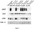
- MET L1195F is phosphorylated when overexpressed demonstrating its propensity to activate the MET pathway including ERK1/2 activation.
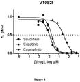
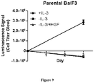
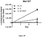
- the MET L1195F mutant is demonstrated to be sensitive to savolitinib inhibition and can confer a growth advantage in stable cell lines to a similar extent to a MET mutation that has been pre-clinically functionally characterized M1250T ( Bardelli et al. PNAS 1998 ).
- the present specification also provides clinical validation that MET L1195F mutants are actionable, showing that a PRCC patient harbouring a MET L1195F mutation in the absence of any other MET alteration derives benefit from monotherapy with savolitinib.
- the pre-clinical functional characterization of Met L1195F and the clinical signal provide the basis for classifying this kinase domain mutation as the first reported to be functionally understood at the bench and demonstrate clinical anti-tumour validation.
- the MET L1195F mutant is found in the context of Type II histological subtype of PRCC patients which further provides a case for a molecular classification of PRCC rather than the classical Type I association with MET gene mutations.
- MET inhibitors are thought to be ideally developed in MET gene copy number amplification settings ( Garber, Nature Reviews Drug Discovery 2014 ), this specification provides evidence for the additional use of MET mutants as actionable biomarkers.
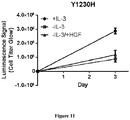
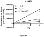
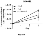
- the present specification demonstrates that certain MET mutations including METL1195F, MET V1092I and MET H1094L are actionable as biomarkers for c-Met inhibition. Cancer cell MET mutation status may therefore be useful to identify cancer patients (for example papillary renal cell carcinoma patients) more likely to respond to treatment with a c-Met inhibitor (for example savolitinib or a pharmaceutically acceptable salt thereof).
- a c-Met inhibitor for example savolitinib or a pharmaceutically acceptable salt thereof.
- the present invention relates to a c-Met inhibitor for use in the treatment of cancer, where the cancer is characterised by a MET 1195 mutation and where the c-Met inhibitor is savolitinib or a pharmaceutically acceptable salt thereof.
- the invention also relates to a method of screening cancer patients to establish their suitability for treatment with a c-Met inhibitor, comprising analysing a representative sample of a patient's cancer in-vitro to determine whether the cancer is characterised by a MET 1195 mutation, where if the patient's cancer is found to be characterised by a MET 1195 mutation, the patient is suitable for treatment with the c-Met inhibitor, and where the c-Met inhibitor is savolitinib or a pharmaceutically acceptable salt thereof.
- This specification also describes, in part, a c-Met inhibitor for use in the treatment of cancer, where the cancer is characterised by a MET 1092, MET 1094 or MET 1195 mutation.
- This specification also describes, in part, a method of screening cancer patients to establish their suitability for treatment with a c-Met inhibitor, comprising analysing a representative sample of a patient's cancer in-vitro to determine whether the cancer is characterised by a MET 1092, MET 1094 or MET 1195 mutation, where if the patient's cancer is found to be characterised by a MET 1092, MET 1094 or MET 1195 mutation, the patient is suitable for treatment with a c-Met inhibitor.
- This specification also describes, in part, use of a c-Met inhibitor in the preparation of a medicament for the treatment of cancer, where the cancer is characterised by a MET 1092, MET 1094 or MET 1195 mutation.
- This specification also describes, in part, a method for treating cancer comprising administering a therapeutically effective amount of a c-Met inhibitor to a patient in need of such treatment, wherein said cancer is characterised by a MET 1092, MET 1094 or MET 1195 mutation.
- This specification also describes, in part, a method for treating cancer in a patient in need of such treatment, comprising the steps of: a) requesting a test whose results can determine whether the patient's cancer is characterised by a MET 1092, MET 1094 or MET 1195 mutation; and b) administering a therapeutically effective amount of a c-Met inhibitor to the patient if the patient's cancer is found to be characterised by a MET 1092, MET 1094 or MET 1195 mutation.
- a c-Met inhibitor for use in the treatment of cancer where the cancer is characterised by a MET 1092, MET 1094 or MET 1195 mutation.
- c-Met inhibitor is a molecule that reduces the activity of c-Met receptor tyrosine kinase.
- c-Met inhibitors include both small molecules (whether selective or unselective) and biomolecules (for example antibodies, both natural and engineered), c-Met inhibitors may inhibit c-Met receptor tyrosine kinase either directly (for example by binding directly to the enzyme) or indirectly (for example by binding to hepatocyte growth factor, the natural ligand of c-Met receptor tyrosine kinase).
- Example c-Met inhibitors include AMG-208, AMG-337, AMG-458, PHA-665752, SU11274, NPS-1034, SGX-523, BMS-777607, tepotinib, BMS-794833, NVP-BVU972, MK-2461, MGCD-265, golvatinib, JNJ-38877605, BMS-754807, PF-04217903, savolitinib, crizotinib, tivantinib, cabozantinib, foretinib, capmatinib (INC280), onartuzumab, ficlatuzumab or rilotumumab.
- the c-Met inhibitor may be AMG-208 or a pharmaceutically acceptable salt thereof, AMG-458 or a pharmaceutically acceptable salt thereof, PHA-665752 or a pharmaceutically acceptable salt thereof, SU11274 or a pharmaceutically acceptable salt thereof, NPS-1034 or a pharmaceutically acceptable salt thereof, SGX-523 or a pharmaceutically acceptable salt thereof, BMS-777607 or a pharmaceutically acceptable salt thereof, tepotinib or a pharmaceutically acceptable salt thereof, BMS-794833 or a pharmaceutically acceptable salt thereof, NVP-BVU972 or a pharmaceutically acceptable salt thereof, MK-2461 or a pharmaceutically acceptable salt thereof, MGCD-265 or a pharmaceutically acceptable salt thereof, golvatinib or a pharmaceutically acceptable salt thereof, JNJ-38877605 or a pharmaceutically acceptable salt thereof, BMS-754807 or a pharmaceutically acceptable salt thereof, PF-04217903 or a pharmaceutically acceptable salt thereof,
- the c-Met inhibitor may be savolitinib or a pharmaceutically acceptable salt thereof, crizotinib or a pharmaceutically acceptable salt thereof, tivantinib or a pharmaceutically acceptable salt thereof, cabozantinib or a pharmaceutically acceptable salt thereof, foretinib or a pharmaceutically acceptable salt thereof, capmatinib or a pharmaceutically acceptable salt thereof, onartuzumab, ficlatuzumab or rilotumumab.
- the c-Met inhibitor may be savolitinib or a pharmaceutically acceptable salt thereof, crizotinib or a pharmaceutically acceptable salt thereof, tivantinib or a pharmaceutically acceptable salt thereof, cabozantinib or a pharmaceutically acceptable salt thereof, foretinib or a pharmaceutically acceptable salt thereof, or capmatinib or a pharmaceutically acceptable salt thereof.
- the c-Met inhibitor may be savolitinib or a pharmaceutically acceptable salt thereof, crizotinib or a pharmaceutically acceptable salt thereof or capmatinib or a pharmaceutically acceptable salt thereof.
- the c-Met inhibitor may be capmatinib or a pharmaceutically acceptable salt thereof.
- the c-Met inhibitor may be crizotinib or a pharmaceutically acceptable salt thereof.
- the c-Met inhibitor may be savolitinib or a pharmaceutically acceptable salt thereof.
- the c-Met inhibitor may be onartuzumab, ficlatuzumab or rilotumumab According to the present invention, the c-Met inhibitor is savolitinib or a pharmaceutically acceptable salt thereof.
- Savolitinib is described in WO2011079804 (as compound 270 on page 106) Savolitinib as the free base has the following structure:
- pharmaceutically acceptable is used to specify that an object (for example a salt or an excipient) is suitable for use in patients.
- An example list of pharmaceutically acceptable salts can be found in the Handbook of Pharmaceutical Salts: Properties, Selection and Use, 2nd Revised Edition; editors P. H. Stahl and C. G. Wermuth; Wiley 2011; ISBN: 978-3-90639-051-2 ;
- the pharmaceutically acceptable salt may be any such salt found in the Handbook of Pharmaceutical Salts: Properties, Selection and Use, 2nd Revised Edition; editors P. H. Stahl and C. G. Wermuth; Wiley 2011; ISBN: 978-3-90639-051-2 .
- c-Met inhibitors may exist in optically active or racemic forms by virtue of an asymmetric carbon atom.
- savolitinib and its pharmaceutically acceptable salts possess such an asymmetric carbon atom.
- the synthesis of optically active forms may be carried out by standard techniques of organic chemistry well known in the art, for example by synthesis using optically active materials or by resolution of a racemic form.
- the c-Met inhibitor may comprise a pharmaceutical composition where a single optical isomer is present in an enantiomeric or diastereomeric excess (%ee) of ⁇ 95%, ⁇ 98% or ⁇ 99%.
- the c-Met inhibitor may comprise a pharmaceutical composition where a single optical isomer is present in an enantiomeric or diastereomeric excess (%ee) of ⁇ 99%.
- the savolitinib or a pharmaceutically acceptable salt thereof may be an (S)-optical isomer being in an enantiomeric excess (%ee) of ⁇ 95%, ⁇ 98% or ⁇ 99%.
- the (S)-optical isomer may be present in an enantiomeric excess (%ee) of > 99%.
- c-Met inhibitors may be administered as pharmaceutical compositions, comprising one or more pharmaceutically acceptable excipients.
- the excipient(s) selected for inclusion in a particular composition will depend on factors such as the mode of administration and the form of the composition provided. Suitable pharmaceutically acceptable excipients are well known to persons skilled in the art and are described, for example, in the Handbook of Pharmaceutical Excipients, 6th Edition; editors R. C. Rowe, P.J. Sheskey and M. Quinn; Pharmaceutical Press .
- Pharmaceutically acceptable excipients may function as, for example, adjuvants, diluents, carriers, stabilisers, flavourings, colorants, fillers, binders, disintegrants, lubricants, glidants, thickening agents and coating agents.
- Certain pharmaceutically acceptable excipients may serve more than one function and may serve alternative functions depending on how much of the excipient is present in the composition and what other excipients are present in the composition.
- compositions comprising c-Met inhibitors may be in a form suitable for oral use (for example as tablets, lozenges, hard or soft capsules, aqueous or oily suspensions, emulsions, dispersible powders or granules, syrups or elixirs), for topical use (for example as creams, ointments, gels, or aqueous or oily solutions or suspensions), for administration by inhalation (for example as a finely divided powder or a liquid aerosol), for administration by insufflation (for example as a finely divided powder) or for parenteral administration (for example as a sterile aqueous or oily solution for intravenous, subcutaneous, intramuscular or intramuscular dosing), or as a suppository for rectal dosing.
- the compositions may be obtained by conventional procedures well known in the art.
- Compositions intended for oral use may contain additional components, for example, one or more colouring, sweetening, flavouring and/or
- the c-Met inhibitor may be administered orally.
- the c-Met inhibitor may be administered orally as an tablet or capsule.
- c-Met inhibitors may be administered in a unit dose form to achieve a therapeutically-effective dose.
- a unit dose form such as a tablet or capsule will usually contain, for example, 0.1-5000 mg of c-Met inhibitor.
- the overall dose and dose schedule will necessarily be varied depending upon the host treated, the particular route of administration, any therapies being co-administered, and the severity of the illness being treated. Accordingly the practitioner who is treating any particular patient may determine the optimum dosage with reference to any regulatory label associated with the c-Met inhibitor.
- the c-Met inhibitor or a pharmaceutically acceptable salt thereof may be administered in a pharmaceutical composition comprising between 0.1-1mg, 1-10 mg, 10-50mg, 50-100mg, 100-500mg, or 500mg to 5000mg of c-Met inhibitor or a pharmaceutically acceptable salt thereof.
- the savolitinib or a pharmaceutically acceptable salt thereof may be administered in a pharmaceutical composition comprising between 0.1-1mg, 1-10 mg, 10-50mg, 50-100mg, 100-500mg, or 500mg to 5000mg of savolitinib or a pharmaceutically acceptable salt thereof.
- the savolitinib or a pharmaceutically acceptable salt thereof may be administered in a pharmaceutical composition comprising 100-1000 mg of savolitinib in its free base form.
- the savolitinib or a pharmaceutically acceptable salt thereof may be administered in a pharmaceutical composition comprising 500-1000 mg of savolitinib in its free base form.
- the savolitinib or a pharmaceutically acceptable salt thereof may be administered in a pharmaceutical composition comprising 600 mg of savolitinib in its free base form.
- the savolitinib or a pharmaceutically acceptable salt thereof may be administered in a pharmaceutical composition comprising 600 mg of savolitinib in its free base form, dosed once daily.
- a cancer that "is characterised by a MET 1092, MET 1094 or MET 1195 mutation” comprises cells which express MET protein comprising a MET 1092, MET 1094 or MET 1195 mutation.
- a "MET protein comprising a MET 1092, MET 1094 or MET 1195 mutation” is a MET protein differing from wild-type MET by a mutation or variance (for example a substitution or deletion) occurring at one or more of the specified amino acid positions.
- RefSeq NCBI Reference Sequence Database (RefSeq Accession NM_00245), which is provided herein (SEQ ID NO: 1).
- This sequence is used as the reference sequence to describe the protein mutations described in the present specification.
- references to a cancer that is characterised by a MET 1092, MET 1094 or MET 1195 mutation describe a cancer comprising cells which express MET protein comprising at least one mutation or variance at one or more of positions 1092, 1094 or 1195 of the sequence recited herein.
- MET mutations in the long isoform of MET with 18 additional amino acids would result in the same biological consequence, i.e. sensitivity to c-Met inhibitors.
- a cancer is characterised by a MET 1092, MET 1094 or MET 1195 mutation
- the cancer may also be characterised by other mutations (including those not relating to MET).
- a MET protein comprising a MET 1092, MET 1094 or MET 1195 mutation may also have other mutations at other amino acid positions.
- a cancer that is characterised by a MET 1092, MET 1094 or MET 1195 mutation may be characterised by one or more of the specified mutations, e.g. a MET 1092 and a MET 1094 mutation, a MET 1092 and a MET 1195 mutation, a MET 1094 and a MET 1195 mutation, or a MET 1092, MET 1094 and MET 1195 mutation.
- a MET protein comprising a MET 1092, MET 1094 or MET 1195 mutation may comprise one or more of the specified mutations.
- the cancer may be characterised by a MET 1092 and a MET 1094 mutation.
- the cancer may be characterised by a MET 1092 and a MET 1195 mutation.
- the cancer may be characterised by a MET 1094 and a MET 1195 mutation.
- the cancer may be characterised by a MET 1092, a MET 1094 and a MET 1195 mutation.
- the mutation may be an amino acid substitution.
- amino acid substitution may include in frame indels.
- An “in frame indel” is is an insertion and/or deletion of an amino acid that is in the reading frame and therefore resulting in either an extra amino acid or a lost amino acid at a given position.
- the mutation may be an indel.
- the mutation may be an in frame indel.
- the mutation may be a somatic mutation.
- the MET mutation may be a germ-line mutation.
- the MET mutation may be a MET V1092 mutation.
- a MET V1092 mutation occurs when the valine amino acid at position 1092 of the wild-type protein is changed.
- the MET mutation may be a MET V1092I mutation.
- a MET V1092I mutation occurs when the valine amino acid at position 1092 of the wild-type protein is substituted by an isoleucine amino acid.
- the MET mutation may be a MET H1094 mutation.
- a MET H1094 mutation occurs when the histidine amino acid at position 1094 of the wild-type protein is changed.
- the MET mutation may be a MET H1094L mutation.
- a MET H1094L mutation occurs when the histidine amino acid at position 1094 of the wild-type protein is substituted by a leucine amino acid.
- the MET mutation may be a MET L1195 mutation.
- a MET L1195 mutation occurs when the leucine amino acid at position 1195 of the wild-type protein is changed.
- the MET mutation may be a MET L1195F mutation.
- a MET L1195F mutation occurs when the leucine amino acid at position 1195 of the wild-type protein is substituted by a phenylalanine amino acid.
- a c-Met inhibitor for use in the treatment of cancer where the cancer is characterised by a MET VI0921, MET H1094L or MET L1195F mutation.
- a c-Met inhibitor for use in the treatment of cancer where the cancer is characterised by a V1092I mutation.
- a c-Met inhibitor for use in the treatment of cancer where the cancer is characterised by a MET H1094L mutation.
- a c-Met inhibitor for use in the treatment of cancer where the cancer is characterised by a MET L1195F mutation.
- cancer cells characterised by a MET 1092, MET 1094 or MET 1195 mutation express MET protein comprising at least one of the stated variances.
- the relevant cancer cells carry corresponding mutations in the nucleic acid sequences coding for the relevant mutant proteins.
- Such mutant nucleic acids can also be used as biomarkers for c-Met inhibition and therefore also form embodiments of the specification.
- a c-Met inhibitor for use in the treatment of cancer where the cancer is characterised by a MET_c.3583C>T mutation.
- a "MET_c.3583C>T mutation” occurs when a cytosine nucleotide at position 3583 of the nucleic acid sequence encoding the wild-type MET protein is substituted by a thymine nucleotide.
- a MET_c.3583C>T mutation encodes for a MET protein comprising a MET L1195F mutation.
- the nucleic acid sequence encoding the wild-type MET protein is described in RefSeq: NCBI Reference Sequence Database (RefSeq Accession NM_00245), which is provided herein (SEQ ID NO: 2).
- the provided RefSeq includes noncoding sequences ("untranslated regions" or "UTRs"), including a UTR sequence 202 nucleotides in length prior to the start of coding sequence.
- RefSeq NCBI Reference Sequence Database (RefSeq Accession NM_00245) is used as the reference sequence when describing the genetic mutations discussed in the present specification.
- references to a nucleic acid with a MET_c.3583C>T mutation describe a nucleic acid where the cytosine nucleotide at position 3785 of the sequence recited herein has been replaced by a thymine nucleotide.
- Cancer refers to the uncontrolled growth of cells resulting in tumours or growths in a patient's body.
- Cancer and tumor are used interchangeably to describe the physical growths present in a cancer patient's body.
- Cancer includes both non-metastatic cancer and metastatic cancer, such that treating cancer may comprise treatment of both primary tumours and also local or distant metastases.
- Cancer tumours are heterogeneous in nature, and each tumour in a cancer patient's body may contain separate populations of cells which are proliferating in an uncontrolled fashion for different reasons.
- a given tumour may contain different populations of cells whose growth is being driven by different aberrations in their cellular machinery, for example different DNA mutations.
- a cancer is characterised by a MET 1092, MET 1094 or MET 1195 mutation, and therefore comprises cells that express MET protein comprising a MET 1092, MET 1094 or MET 1195 mutation, not all of the cancer cells may express such mutant MET proteins.
- the cancer may be lung cancer (for example small cell lung cancer or non-small cell lung cancer), gastric cancer, brain cancer (for example glioblastoma), colorectal cancer, breast cancer (for example triple negative breast cancer) or renal cancer (for example renal clear cell carcinoma or papillary renal cell carcinoma).
- lung cancer for example small cell lung cancer or non-small cell lung cancer
- gastric cancer for example glioblastoma
- brain cancer for example glioblastoma
- colorectal cancer for example triple negative breast cancer
- breast cancer for example triple negative breast cancer
- renal cancer for example renal clear cell carcinoma or papillary renal cell carcinoma
- the cancer may be renal cancer (for example renal clear cell carcinoma or papillary renal cell carcinoma).
- the cancer may be papillary renal cell carcinoma.
- the papillary renal cell carcinoma may be Type I papillary renal cell carcinoma.
- Type I papillary renal cell carcinoma is a histological subtype characterised by a lining of small cells with clear to basophilic cytoplasm.
- the papillary renal cell carcinoma may be Type II papillary renal cell carcinoma.
- Type II papillary renal cell carcinoma is a histological subtype characterised by a lining of by large cells with abundant eosinophilic cytoplasm.
- the skilled person is able to distinguish between histological subtypes of papillary renal cell carcinoma using the above and other histological criteria.
- a c-Met inhibitor for use in the treatment of cancer, where the cancer is characterised by a 1195 mutation, and the c-Met inhibitor is savolitinib or a pharmaceutically acceptable salt thereof, which is administered in a pharmaceutical composition comprising 500 - 1000 mg of savolitinib in its free base form, dosed once daily.
- a c-Met inhibitor for use in the treatment of cancer where the cancer is characterised by a MET 1195 mutation, and the c-Met inhibitor is savolitinib or a pharmaceutically acceptable salt thereof, which is administered in a pharmaceutical composition comprising 600 mg of savolitinib in its free base form, dosed once daily.
- a c-Met inhibitor for use in the treatment of cancer, where the cancer is characterised by a MET L1195F mutation, and the c-Met inhibitor is savolitinib or a pharmaceutically acceptable salt thereof, which is administered in a pharmaceutical composition comprising 600 mg of savolitinib in its free base form, dosed once daily.
- a c-Met inhibitor for use in the treatment of cancer where the cancer is papillary renal cell carcinoma that is characterised by a MET V1092I, MET H1094L or MET L1195F mutation.
- a c-Met inhibitor for use in the treatment of cancer, where the cancer is papillary renal cell carcinoma that is characterised by a MET L1195F mutation, and the c-Met inhibitor is savolitinib or a pharmaceutically acceptable salt thereof.
- a c-Met inhibitor for use in the treatment of cancer, where the cancer is papillary renal cell carcinoma that is characterised by a MET L1195F mutation, and the c-Met inhibitor is savolitinib or a pharmaceutically acceptable salt thereof, which is administered in a pharmaceutical composition comprising 600 mg of savolitinib in its free base form, dosed once daily.
- a c-Met inhibitor for use in the treatment of cancer where the cancer is Type II papillary renal cell carcinoma that is characterised by a MET L1195F mutation, and the c-Met inhibitor is savolitinib or a pharmaceutically acceptable salt thereof, which is administered in a pharmaceutical composition comprising 600 mg of savolitinib in its free base form, dosed once daily.
- savolitinib for use in the treatment of Type II papillary renal cell carcinoma that is characterised by a MET L1195F mutation, where the savolitinib is administered in a pharmaceutical composition comprising 600 mg of savolitinib in its free base form, dosed once daily.
- a method of screening cancer patients to establish their suitability for treatment with a c-Met inhibitor comprising analysing a representative sample of a patient's cancer in-vitro to determine whether the patient's cancer is characterised by a MET 1092, MET 1094 or MET 1195 mutation, where if the patient's cancer is found to be characterised by a MET 1092, MET 1094 or MET 1195 mutation, the patient is suitable for treatment with a c-Met inhibitor.
- a method of screening cancer patients to establish their suitability for treatment with a c-Met inhibitor comprising obtaining a representative sample of a patient's cancer, analysing the sample in-vitro to determine whether the patient's cancer is characterised by a MET 1092, MET 1094 or MET 1195 mutation, where if the patient's cancer is found to be characterised by a MET 1092, MET 1094 or MET 1195 mutation, the patient is suitable for treatment with a c-Met inhibitor.
- a method of determining whether a cancer patient is likely to benefit from treatment with a c-Met inhibitor comprising in-vitro testing a representative sample of a patient's cancer for the presence of a MET 1092, MET 1094 or MET 1195 mutation to determine whether the patient's cancer is characterised by a MET 1092, MET 1094 or MET 1195 mutation, where if the patient's cancer is found to be characterised by a MET 1092, MET 1094 or MET 1195 mutation, the patient is more likely to benefit from treatment with a c-Met inhibitor.
- a method for selecting a suitable treatment regime for a cancer patient comprising the steps of:
- a method for selecting a suitable treatment regime for a cancer patient comprising the steps of:
- a method of screening cancer patients to establish their suitability for treatment with a c-Met inhibitor comprising analysing a representative sample of a patient's cancer in-vitro to determine whether the patient's cancer is characterised by a 1195 mutation, where if the patient's cancer is found to be characterised by a MET 1195 mutation, the patient is suitable for treatment with a c-Met inhibitor, where the c-Met inhibitor is savolitinib or a pharmaceutically acceptable salt thereof, which is administered in a pharmaceutical composition comprising 500-1000 mg of savolitinib in its free base form, dosed once daily.
- a method of screening cancer patients to establish their suitability for treatment with a c-Met inhibitor comprising analysing a representative sample of a patient's cancer in-vitro to determine whether the patient's cancer is characterised by a MET 1195 mutation, where if the patient's cancer is found to be characterised by a MET 1195 mutation, the patient is suitable for treatment with a c-Met inhibitor, where the c-Met inhibitor is savolitinib or a pharmaceutically acceptable salt thereof, which is administered in a pharmaceutical composition comprising 600 mg of savolitinib in its free base form, dosed once daily.
- a method of screening cancer patients to establish their suitability for treatment with a c-Met inhibitor comprising analysing a representative sample of a patient's cancer in-vitro to determine whether the patient's cancer is characterised by a MET L1195F mutation, where if the patient's cancer is found to be characterised by a MET L1195F, the patient is suitable for treatment with a c-Met inhibitor, where the c-Met inhibitor is savolitinib or a pharmaceutically acceptable salt thereof, which is administered in a pharmaceutical composition comprising 600 mg of savolitinib in its free base form, dosed once daily.
- a method of screening cancer patients to establish their suitability for treatment with a c-Met inhibitor comprising analysing a representative sample of a patient's cancer in-vitro to determine whether the patient's cancer is characterised by a MET VI0921, MET H1094L or MET L1195F mutation, where if the patient's cancer is found to be characterised by a MET V1092I, MET H1094L or MET L1195F, the patient is suitable for treatment with a c-Met inhibitor, where the cancer is papillary renal cell carcinoma.
- a method of screening cancer patients to establish their suitability for treatment with a c-Met inhibitor comprising analysing a representative sample of a patient's cancer in-vitro to determine whether the patient's cancer is characterised by a MET L1195F mutation, where if the patient's cancer is found to be characterised by a MET L1195F, the patient is suitable for treatment with a c-Met inhibitor, where the cancer is papillary renal cell carcinoma, and the c-Met inhibitor is savolitinib or a pharmaceutically acceptable salt thereof.
- a method of screening cancer patients to establish their suitability for treatment with a c-Met inhibitor comprising analysing a representative sample of a patient's cancer in-vitro to determine whether the patient's cancer is characterised by a MET L1195F mutation, where if the patient's cancer is found to be characterised by a MET L1195F, the patient is suitable for treatment with a c-Met inhibitor, where the cancer is papillary renal cell carcinoma, and the c-Met inhibitor is savolitinib or a pharmaceutically acceptable salt thereof, which is administered in a pharmaceutical composition comprising 600 mg of savolitinib in its free base form, dosed once daily.
- a method of screening cancer patients to establish their suitability for treatment with a c-Met inhibitor comprising analysing a representative sample of a patient's cancer in-vitro to determine whether the patient's cancer is characterised by a MET L1195F mutation, where if the patient's cancer is found to be characterised by a MET L1195F, the patient is suitable for treatment with a c-Met inhibitor, where the cancer is type II papillary renal cell carcinoma, which is administered in a pharmaceutical composition comprising 600 mg of savolitinib in its free base form, dosed once daily.
- a method of screening papillary renal cell carcinoma patients to establish their suitability for treatment with savolitinib or a pharmaceutically acceptable salt thereof comprising analysing a representative sample of a patient's cancer in-vitro to determine whether the patient's cancer is characterised by a mutation, where if the patient's cancer is found to be characterised by a MET L1195F mutation, the patient is suitable for treatment with with savolitinib or a pharmaceutically acceptable salt thereof.
- a c-Met inhibitor in the preparation of a medicament for the treatment of cancer, where the cancer is characterised by a MET 1092, MET 1094 or MET 1195 mutation.
- a method for treating cancer in a patient whose cancer is characterised by a MET 1092, MET 1094 or MET 1195 mutation which comprises administering to said patient a therapeutically effective amount of a c-Met inhibitor.
- a method for treating cancer in a patient in need of such treatment comprising the steps of:
- a method for treating cancer in a patient in need of such treatment comprising the steps of:
- a method for treating cancer in a patient in need of such treatment comprising the steps of:
- a diagnostic kit suitable for the determination of a MET mutation (for example a MET 1092, MET 1094 or MET 1195 mutation), comprising degenerate primers suitable to amplify a target nucleic acid in the MET gene and optional instructions comprising amplification protocol and result analysis.
- a MET mutation for example a MET 1092, MET 1094 or MET 1195 mutation
- the kit may comprise instructions comprising amplification protocol and result analysis.
- the kit may comprise buffers, enzymes, and containers for performing the amplification and analysis of the amplification products.
- the kit may comprise one or more DNA microarrays.
- the kit may comprise one or more control templates.
- Suitable control templates include nucleic acids isolated from normal tissue sample, and samples representing different variances in the reference genes.
- the kit may comprise two or more primer pairs, each pair capable of amplifying a different region of the MET gene (each region a site of potential variance) thereby providing a kit for analysis of the presence of several gene variances in a biological sample in one reaction or several parallel reactions.
- Suitable primers may be labelled (for example fluorescently) to facilitate detection of the amplification products and consequent analysis of the nucleic acid variances.
- the kit may allow for more than one variance to be detected in one analysis.
- a combination kit will therefore comprise primers capable of amplifying different segments of the reference gene.
- the primers may be differentially labelled, for example using different fluorescent labels, so as to differentiate between the variances.
- kits suitable for the determination of a MET mutation may allow for mutant forms of MET to be determined, wherein the mutation is present at any position across the gene.
- kits suitable for the determination of a MET mutation allows for a MET 1092, MET 1094 or MET 1195 mutation to be determined.
- kits suitable for the determination of a MET mutation allows for a MET V1092I, MET H1094L or MET L1195F mutation to be determined.
- Determining whether or not a cancer is characterised by a certain mutation may be based on the analysis of the cell's genomic DNA, or by analysis of DNA transcripts or proteins encoded by the DNA. It may be desirable to confirm the presence of a given mutation in a cancer cell's genomic DNA by parallel analysis of transcripts and/or proteins in order to ensure that the mutation is indeed expressed in the patient's cancer cells.
- the determination may comprise a mutation detection technique.
- the determination may comprise a mutation detection technique and an amplification method.
- the determination may comprise a mutation detection technique and a capture or enrichment method.
- the determination may comprise a mutation detection technique and a signal generation system.
- the determination may comprise a mutation detection technique, an amplification method, a capture or enrichment method and a signal generation system.
- a “mutation detection technique” can be performed by analysing DNA or RNA collected from biological samples by a variety of methods including PCR, hybridization with allele-specific probes, enzymatic mutation detection, chemical cleavage of mismatches, mass spectrometry or DNA sequencing (including minisequencing).
- Example mutation detection techniques include amplification refractory mutation system (ARMSTM), amplification refractory mutation system linear extension (ALEXTM), competitive oligonucleotide priming system (COPS), Taqman, Molecular Beacons, restriction fragment length polymorphism (RFLP), restriction site based PCR and fluorescence resonance energy transfer (FRET) techniques and oligonucleotide ligation assay (OLA).
- Hybridization with allele specific probes can be conducted using: (1) allele specific oligonucleotides bound to a solid phase (e.g. glass, silicon, nylon membranes) which is contacted with the labelled sample in solution, for example as in many DNA chip applications; or, (2) bound sample (often cloned DNA or PCR amplified DNA) and labelled oligonucleotides in solution (either allele specific or short so as to allow sequencing by hybridization). Diagnostic tests may comprise a panel of mutations, often on a solid support, which enables the simultaneous determination of more than one mutation.
- a solid phase e.g. glass, silicon, nylon membranes
- Diagnostic tests may comprise a panel of mutations, often on a solid support, which enables the simultaneous determination of more than one mutation.
- hybridization probes are well known in the art (see e.g., Molecular Cloning: A Laboratory Manual, 4th Edition, Volumes 1-3; editors Green and Sambrook; Cold Spring Harbor, N.Y 2012, ISBN: 9781936113422 ) and may span two or more variance sites.
- the mutation detection technique may comprise contacting MET nucleic acid containing a putative mutation site with at least one nucleic acid probe.
- the mutation detection technique may comprise contacting MET nucleic acid containing a potential mutation site with at least one nucleic acid probe, where the probe hybridizes with a nucleic acid sequence including a mutation site containing complementary nucleotide bases at the mutation site under selective hybridization conditions.
- Hybridization can be detected using labels known to one skilled in the art.
- labels include radioactive, fluorescent, dye, and enzymatic labels.
- the mutation detection technique may comprise contacting MET nucleic acid containing a potential mutation site with at least one nucleic acid primer.
- the mutation detection technique may comprise contacting MET nucleic acid containing a potential mutation site with at least one nucleic acid primer, where the primer preferentially hybridizes with a nucleic acid sequence including a mutation site containing complementary nucleotide bases at the variance site under selective hybridization conditions.
- Oligonucleotides used as primers for specific amplification may carry the complementary nucleotide base to the mutation of interest in the centre of the molecule, so that amplification depends on differential hybridization (see e.g., Gibbs, et al. Nucl. Acids Res. 1989, 17, 2437 ) or at the extreme 3'-terminus of one primer where, under appropriate conditions, mismatch can prevent, or reduce polymerase extension (see, e.g., Prossner 1993, Tibtech, 11 238 ).
- the mutation detection technique may comprise sequencing at least one nucleic acid sequence and comparing the obtained sequence with the known wild type nucleic acid sequence.
- the mutation detection technique may comprise mass spectrometric determination of at least one nucleic acid sequence.
- the mutation detection technique may comprise performing a PCR.
- Mutations in genomic nucleic acid may also be advantageously detected by techniques based on mobility shift in amplified nucleic acid fragments. For instance, Chen et al., Anal Biochem. 1996, 239, 61 , describe the detection of single-base mutations by a competitive mobility shift assay. Moreover, assays based on the technique of Marcelino et al., BioTechniques 1999, 26, 1134-1148 are available commercially.
- the mutation detection technique may comprise using capillary heteroduplex analysis to detect the presence of mutations based on mobility shift of duplex nucleic acids in capillary systems as a result of the presence of mismatches.
- An "amplification method” may be used to generate further amounts of nucleic acids detected during a mutation detection technique, making the subsequent analysis steps easier.
- Most amplification methods rely on an enzymatic chain reaction (such as a PCR, a ligase chain reaction, or a self-sustained sequence replication) or from the replication of all or part of the vector into which it has been cloned.
- amplification methods have been described in the literature, for example, general reviews of these methods in Landegren, U. et al., Science 1988, 242, 229-237 and Lewis, R., Genetic Engineering News 1990, 10, 54-55 .
- These amplification methods can be used in the methods described herein, and include PCR, PCR in situ, ligase amplification method (LAR), ligase hybridisation, Q ⁇ bacteriophage replicase, transcription-based amplification system (TAS), genomic amplification with transcript sequencing (GAWTS), nucleic acid sequence-based amplification (NASBA) and in-situ hybridisation.
- Primers suitable for use in various amplification techniques can be prepared according to methods known in the art.
- PCR is a nucleic acid amplification method described inter alia in U.S. Pat. Nos. 4,683,195 and 4,683,202 .
- PCR consists of repeated cycles of DNA polymerase generated primer extension reactions.
- the target DNA is heat denatured and two oligonucleotides, which bracket the target sequence on opposite strands of the DNA to be amplified, are hybridised. These oligonucleotides become primers for use with DNA polymerase.
- the DNA is copied by primer extension to make a second copy of both strands. By repeating the cycle of heat denaturation, primer hybridisation and extension, the target DNA can be amplified a million fold or more in about two to four hours.
- PCR is a molecular biology tool, which must be used in conjunction with a detection technique to determine the results of amplification.
- An advantage of PCR is that it increases sensitivity by amplifying the amount of target DNA by 1 million to 1 billion fold in approximately 4 hours.
- PCR can be used to amplify any known nucleic acid in a diagnostic context ( Mok et al., Gynaecologic Oncology, 1994, 52: 247-252 ,).
- An allele specific amplification technique such as Amplification Refractory Mutation System (ARMSTM) ( Newton et al., Nucleic Acids Res., 1989, 17, 2503-2516 ) can also be used to detect single base mutations. Under the appropriate PCR amplification conditions a single base mismatch located at the 3'-end of the primer is sufficient for preferential amplification of the perfectly matched allele (Newton et al., 1989, supra), allowing the discrimination of closely related species.
- the basis of an amplification system using the primers described above is that oligonucleotides with a mismatched 3'-residue will not function as primers in the PCR under appropriate conditions. This amplification system allows genotyping solely by inspection of reaction mixtures after agarose gel electrophoresis.
- the amplification method may comprise a PCR.
- Signal generation systems can be used to analyse amplification products.
- Signal generation systems can be performed using any method capable of separating the amplification products according to their size, including automated and manual gel electrophoresis, mass spectrometry, and the like.
- the detection of a MET mutation may comprise a signal generation systems which is gel electrophoresis.
- the detection of a MET mutation may comprise a signal generation systems which is mass spectrometry.
- Methods that may be used to detect the protein mutations described herein include mass spectrometry or antibody based approaches that are designed to detect the amino acid substitution at the indicated positions.
- a "representative sample of a patient's cancer” can be any tumour tissue or tumour-cell containing sample obtained or obtainable from a cancer patient, including any sample containing tumour nucleic acid, such as circulating free DNA (ctDNA).
- Samples of a patient's cancer may include a blood, mouth swab, biopsy, or other body fluid or tissue obtained from a patient.
- Particular examples include circulating tumour cells or circulating tumour DNA present in a patient's plasma or serum, cells isolated from the ascites fluid of ovarian cancer patients, lung sputum obtained from patients with tumours within the lung, a fine needle aspirate obtained from a breast cancer patient, urine, peripheral blood, a cell scraping, a hair follicle, a skin punch or a buccal sample.
- Representative samples of a patient's cancer for mutation status testing may equally be nucleic acid sequences corresponding to the sequence obtained in a representative sample of a patient's cancer, that is to say that all or a part of the region in the sample nucleic acid may firstly be amplified using any convenient technique e.g. polymerase chain reaction (PCR), before analysis.
- the nucleic acid may be genomic DNA or fractionated or whole cell RNA.
- the RNA is whole cell RNA and is used directly as the template for labelling a first strand cDNA using random primers or poly A primers.
- the nucleic acid or protein in a sample of a patient's cancer may be extracted from the sample according to standard methodologies, e.g. those referenced in Molecular Cloning: A Laboratory Manual, 4th Edition, Volumes 1-3; editors Green and Sambrook; Cold Spring Harbor, N.Y 2012, ISBN: 9781936113422 ,
- a representative sample of a patient's cancer may previously have been taken from the patient. Such samples may be preserved by freezing or fixed and embedded in formalin-paraffin or other media. Alternatively, a fresh sample may be obtained and used.
- the representative sample of a patient's cancer may comprise a tumour cell sample.
- the representative sample of a patient's cancer may comprise a sample containing tumour nucleic acid.
- the representative sample of a patient's cancer may comprise a sample containing tumour DNA.
- the representative sample of a patient's cancer may comprise a sample containing tumour circulating free DNA.
- a c-Met inhibitor for use in the treatment of cancer where the cancer is characterised by a MET 1092, MET 1094 or MET 1195 mutation.
- a c-Met inhibitor for use in the treatment of cancer as described in embodiment [A] or embodiment [B], where the cancer is characterised by a MET V1092I, MET H1094L or MET L1195F mutation.
- a c-Met inhibitor for use in the treatment of cancer as described in any one of embodiments [A] to [I], where the c-Met inhibitor is savolitinib or a pharmaceutically acceptable salt thereof, crizotinib or a pharmaceutically acceptable salt thereof, tivantinib or a pharmaceutically acceptable salt thereof, cabozantinib or a pharmaceutically acceptable salt thereof, foretinib or a pharmaceutically acceptable salt thereof, capmatinib or a pharmaceutically acceptable salt thereof, onartuzumab, ficlatuzumab or rilotumumab.
- a c-Met inhibitor for use in the treatment of cancer as described in any one of embodiments [A] to [K], where the treatment of cancer comprises analysing a representative sample of a patient's cancer in-vitro to determine whether the cancer is characterised by a MET 1092, MET 1094 or MET 1195 mutation, where if the patient's cancer is found to be characterised by a MET 1092, MET 1094 or MET 1195 mutation, the patient is suitable for treatment with a c-Met inhibitor.
- a method of screening cancer patients to establish their suitability for treatment with a c-Met inhibitor comprising analysing a representative sample of a patient's cancer in-vitro to determine whether the cancer is characterised by a MET 1092, MET 1094 or MET 1195 mutation, where if the patient's cancer is found to be characterised by a MET 1092, MET 1094 or MET 1195 mutation, the patient is suitable for treatment with a c-Met inhibitor.
- embodiment [O] there is provided the method of embodiment [N], comprising analysing a representative sample of a patient's cancer in-vitro to determine whether the cancer is characterised by a MET V1092I mutation, where if the patient's cancer is found to be characterised by a MET V1092I mutation, the patient is suitable for treatment with a c-Met inhibitor.
- embodiment [P] there is provided the method of embodiment [N], comprising analysing a representative sample of a patient's cancer in-vitro to determine whether the cancer is characterised by a MET H1 094L mutation, where if the patient's cancer is found to be characterised by a MET H1094L mutation, the patient is suitable for treatment with a c-Met inhibitor.
- embodiment [Q] there is provided the method of embodiment [N], comprising analysing a representative sample of a patient's cancer in-vitro to determine whether the cancer is characterised by a MET L1195F mutation, where if the patient's cancer is found to be characterised by a MET L1195F mutation, the patient is suitable for treatment with a c-Met inhibitor.
- a c-Met inhibitor in the preparation of a medicament for the treatment of cancer, where the cancer is characterised by a MET 1092, MET 1094 or MET 1195 mutation.
- embodiment [V] there is provided the method of embodiment [U], where the cancer is papillary renal cell carcinoma.
- embodiment [W] there is provided the method of embodiment [U] or embodiment [V], where the c-Met inhibitor is savolitinib or a pharmaceutically acceptable salt thereof.
- the c-Met inhibitor is savolitinib, or a pharmaceutically acceptable salt thereof, and the cancer is characterised by a MET 1195 mutation.
- the following tests support the embodiments described herein: a) phosphorylation of the MET receptor tyrosine kinase and effect of point mutations on sensitivity to MET inhibition with savolitinib; b) determination of the dose response of MET WT and MET point mutants to savolitinib and other MET inhibitors; c) transformation capacity of MET WT, MET mutant or parental Ba/F3 stable cell lines; d) MET L1195F mutant PRCC patient tumour response.
- MET WT and all MET mutants are activating when transiently over-expressed as demonstrated by DMSO treated condition and detection of phosphorylated MET (Y1234/Y1235).
- Novel MET mutant L1195F can activate MET and ERK1/2 and is sensitive to savolitinib inhibition to a similar extent as MET WT.
- Novel MET mutants V1092I and H1094L are also activated by over-expression and lead to downstream ERK pathway activation.
- M1250T a known robust activating MET mutant
- Y1230H is completely resistant to savolitinib.
- the results of this test are shown in Figure 1 and demonstrate that MET L1195F, V1092I and H1094L are activating mutants of MET, and that downstream ERK1/2 signalling and can be inhibited with savolitinib treatment.
- Test b Determination of the dose response of MET WT and MET point mutants to savolitinib and other MET inhibitors
- the IC 50 indicates the sensitivity of each MET WT and mutant protein toward inhibition with the relevant c-Met inhibitor.
- Savolitinib has an IC 50 of 13nM against WT MET protein, and 6nM against the known activating MET mutant M1250T IC 50 .
- savolitinib has comparable IC 50 values toward both proteins.
- cells that transiently express MET L1195F have a savolitinib IC 50 of 35nM also indicating that this MET mutant can be inhibited with savolitinib to a similar extent as for MET WT and the known activating M1250T mutant.
- MET V1092I and H1094L demonstrate that these mutant proteins are phosphorylated and that this phosphorylation can be inhibited with a MET inhibitor, such that the IC 50 of savolitinib is 12nM and 4nM respectively.
- a MET inhibitor such that the IC 50 of savolitinib is 12nM and 4nM respectively.
- the MET Y1230H mutant is not sensitive to savolitinib inhibition since an IC 50 could not be determined due to the inability to inhibit at least 50% of the phospho-MET MSD signal.
- Phospho-MET MSD signal is normalized to total MET signal.
- Test c Transformation capacity of MET WT, MET mutant or parental Ba / F3 stable cell lines
- MET WT overexpression also did not confer a growth advantage in an IL-3 independent manner.
- MET M1250T cells grow in the absence of IL-3 as expected and novel MET mutant L1195F is demonstrated to have the capacity to confer growth to a similar extent to MET M1250T.
- Other novel MET mutants (MET V1092I and MET H1094L, but not MET Y1230H) also have relatively greater capacity to confer IL-3 independent cell growth.
- the patient started treatment with savolitinib on 9/2/14.
- NGS Sequencing of the diagnostic tumour sample (Foundation Medicine Inc, FMI) with a median exon coverage of 500x demonstrated the existence of a MET mutation MET_c.3583C>T with an allele fraction of 24%.
- the amino acid substitution of L1195F is located in exon 18 within the kinase domain of the MET receptor tyrosine kinase. This is reported by FMI as a variant of unknown significance.
- This tumour specimen harboured no co-occurring MET gene copy number variation, no other MET mutations and also did not co-occur in the background of Chromosome 7 gain which is a hallmark of PRCC and where both MET gene and it's ligand HGF gene loci reside.
- Chromosome 7 copy number was detected using an integrated copy number analysis of the 400 genes and 4200 SNPs measured in the FMI sequencing assay.
- Subject 9203-002 had stable disease according to RECIST 1.1 criteria. However, this subject's best tumour response occurring at 36 weeks of savolitinib treatment was 29.7% decrease in the sum of the longest diameter of 4 target lesions ( Figure 9 ). Sum of the target lesions are indicated with data labels in mm on the spider plot.
- the synthetic gene pLVXIP_MET_FLAG was assembled from synthetic oligonucleotides and/or PCR products. The fragment was inserted into the vector backbone pLVXIRES-Puro_P839 cloning sites BamHI / EcoRI . The plasmid DNA was purified from transformed bacteria and concentration determined by UV spectroscopy. The final construct was verified by sequencing. The sequence (ABI) congruence within the insertion sites was 100%. Point mutations at indicated amino acid position (NM_000245) M1250T, Y1230H, L1195F, H1094L and V1092I were generated in the same manner as MET WT (Invitrogen by Thermo Fischer Scientific).
- HEK293T Human Embryonic Kidney cell line
- HEK293T cells were transiently transfected with plasmids as outlined in MET WT and mutant clones section of M&M above.
- HEK293T cells were plated so 95% confluent on day of transfection (1 plate for each viral construct).
- the MET construct (24 ⁇ g) and Optimem (1.5 mL) were combined with Lipofectamine 2000 (60 ⁇ L per reaction, Invitrogen) in Optimem (1.5mL per reaction) for a MasterMix and incubated for 5 minutes at room temperature.
- Ad1.5 mL of Master Mix was added to each tube containing a construct for a final volume of ⁇ 3 mL.
- Lysis Buffer TBS + 1% NP40
- PhosSTOP phosphatase inhibitor PhosSTOP phosphatase inhibitor and Complete Protease Inhibitor cocktails (Roche), 1 tablet each for every 10mL of lysis buffer. The mixture was kept cold. Cells were removed from wells by pipetting lysis buffer media (6-well dish) until semi-adherent 293T cells were suspended. These were then spun down at 1500 rpm for 5 min. Media was aspirated and the cell-pellet resuspended in 100 ⁇ L of lysis buffer on ice. The suspension was immediately freeze/thawed twice in dry ice. Samples were then centrifuged at -14000 rpm for 10 minutes.
- MSD MULTI-SPOT Phospho(Tyr1349)/Total Met Assay
- the SECTOR® Imager applies a voltage to the plate electrodes causes the labels bound to the electrode surface to emit light.
- the instrument measures intensity of emitted light to provide a quantitative measure of phosphorylated Met (Tyr1349) and total Met present in the sample.
- the percent phospho-Met is can be calculated using independent MSD phospho-Met and total Met protein with the MSD phospho-/total multiplex phosphoprotein assays.
- IL-3 dependent murine pro B cell line Ba/F3 were plated at 5.2 x 10⁵ cells/mL density in 6-well plate with mutant viral constructs, polybrene at final concentration of 8 ⁇ g/mL, and filled to a 2 mL volume with complete media (RPMI-1640 (Gibco), 10% FBS, 1% L-glut, IL-3 1 ng/mL). Suspension culture was sealed with parafilm and centrifuged for 45 minutes at 1500-2000 rpm at 20°C then incubated overnight and replaced with complete media. At confluency (2.0 x 10⁶ cells/mL, 2-4 days later, replaced media with adding selective pressure where media contains a final concentration of 0.5 ⁇ g/mL Puromycin.
- Sequences were confirmed with Sequencer 5.4.
- RFP Red Fluorescence Protein
- PCR Primers used include MET sense 143090519 and antisense 143090520. Sequencing Primers used include F1 143090521, F2 143090522, F3 143090523, R1 143090524, R2 143090525, R3 143090526. Sequences for the primers used to confirm mutant constructs at position 1195, 1092 and 1094 are represented below and in the sequence listings herein.
- Non-coding strand primers R1 TCTGGTCATCAGCTCCCA (SEQ ID NO: 6) R2 AGCAGGCTCAGCACGTTGG (SEQ ID NO: 7) R3 AACTGGTCCTCGGGGAAGG (SEQ ID NO: 8)
- the cells were treated with complete media (+IL-3, a Ba/F3 dependant growth factor), media without IL-3 (-IL-3), and media without IL-3 but containing the ligand for MET, HGF (-IL-3/+HGF). Viability was measured with CellTiter-Glo® Cell Viability Assay (Promega) and 21 day live cell count was taken with Cellometer (Nexcelom). Parental Ba/F3 cell line was assayed as a negative control. All MET stable lines were assayed in duplicate in 6 well format.
- cells were plated at 1.0 x10⁶ cells/ml, culture under indicated conditions where either one of 3 conditions: + IL-3 (10 ng/mL), - IL-3, -IL-3/+HGF(100 ng/mL).
- MET L1195F mutant PRCC patient tumour response was determined during an open-label, single-arm, multicentre, global phase II trial to evaluate the efficacy of savolitinib (AZD6094 / HMPL-504) in patients with papillary renal cell carcinoma (PRCC) who are treatment naive or previously treated (ClinicalTrials.gov identifier: NCT02127710; web address: https://clinicaltrials.gov/ct2/show/NCT02127710). Key trial and dosing details that would allow the skilled clinical team to replicate the results are as follows:
- Safety profiles will be assessed in terms of AEs and laboratory data, vital signs and ECGs that will be collected for all patients.
- PK of AZD6094 and major metabolites M2 and M3 for AUC, AUC(0-24), AUC(0-t), AUCss, Cmax, Css max, and Css min (Time Frame: Cycle 1 Day 8 and 15, Cycle 2 Day 1, Cycle 3, Day 1, and Cycle 4, Day 1). Characterise the PK of AZD6094 and the major metabolites M2 and M3 following administration to steady state after multiple dosing when given orally.
- Target Lesion is percentage change from baseline in tumour size at 12 weeks. This is based on RECIST target lesion measurements taken at baseline and at week 12. Target lesions are measureable tumour lesions.
- Duration of Response is defined as the time from the date of first documented response until the date of documented progression or any cause death. In the case where a subject does not progress following response, the duration of response will be the same as the PFS censoring time.
- OS Overall Survival
- AZD6094 is a potent and selective small molecule c-Met kinase inhibitor. Other Name: HMPL-504.
- Eligibility Ages Eligible for Study: 18 Years to 99 Years (Adult, Senior); Genders Eligible for Study: Both; Accepts Healthy Volunteers: No.
- Inclusion criteria Provision of informed consent prior to any study specific procedures, sampling and analyses.
- Previous treatments may include: targeted therapy (i.e. sunitinib, sorafenib, bevacizumab, pazopanib, temsirolimus, and everolimus), traditional immunotherapy (i.e. interferon-a, Interleukin-2), chemotherapy or a combination of chemoimmunotherapy.
- targeted therapy i.e. sunitinib, sorafenib, bevacizumab, pazopanib, temsirolimus, and everolimus
- traditional immunotherapy i.e. interferon-a, Interleukin-2
- chemotherapy i.e. interferon-a, Interleukin-2
- At least one lesion, not previously irradiated, and not chosen for a biopsy if performed during the screening period that can be accurately measured at baseline and which is suitable for accurate repeated measurements.
- Adequate renal function defined as GFR ⁇ 40 mL/min
- Adequate coagulation parameters defined as International Normalisation Ratio(INR) ⁇ 1.5 x ULN or aPTT ⁇ 1.5 x ULN.
- Exclusion criteria Most recent chemotherapy, immunotherapy, chemoimmunotherapy, or investigational agents ⁇ 21 days of the first dose of study treatment. Most recent targeted therapy ⁇ 14 days of the first dose of study treatment.
- Wide field radiotherapy including therapeutic radioisotopes such as strontium-89 administered ⁇ 28 days or limited field radiation for palliation ⁇ 7 days prior to starting study drug or has not recovered from side effects of such therapy.
- Any clinically important abnormalities in rhythm, conduction or morphology of resting ECGs e.g. complete left bundle branch block, third degree heart block, second degree heart block, PR interval >250 msec.
- HIV human immunodeficiency virus
- hepatitis B hepatitis B
- hepatitis C hepatitis C
Description
- This specification relates to c-Met receptor tyrosine kinase ("c-Met") inhibitors and their use in the treatment of cancers characterised by certain MET mutations (e.g. cancers comprising cells that express a MET protein having a MET V1092I, MET H1094L or MET L1195F mutation). This specification further relates to the use of MET mutation status to select patients suitable for treatment with a c-Met inhibitor, and to methods of treating cancers characterised by certain MET mutations with a c-Met inhibitor.
- The MET protein, also known as c-Met receptor tyrosine kinase, is a transmembrane receptor essential for embryonic development and wound healing. The MET receptor is normally activated through interaction with its specific ligand, hepatocyte growth factor (HGF), and is the only high-affinity cell surface receptor for HGF (Bottaro et al. 1991). The MET receptor is deregulated in many types of human malignancies, including cancers of the kidney, liver, stomach, lung, breast, and brain. Aberrant activation of the HGF/c-Met axis in tumours triggers tumour growth, promotes tumour angiogenesis, and induces tumour metastasis. In addition, abnormal MET activation is associated with drug resistance and is correlated with poor prognosis. Recently, inhibition of the c-Met signalling pathway has become an area of interest in the search for potential new therapies for cancers driven by c-Met activation.
- Germline mutations of the MET gene locus 7q31 have been detected in patients with hereditary papillary renal cell carcinoma (PRCC) and in sporadic PRCC (Salvi et al. 2008, Schmidt et al. 1997, Schmidt et al. 1999). It is commonly believed that Type I tumours are generally associated with a MET mutation, whether sporadic or hereditary. Although MET mutations have been associated with Type I PRCC in cohort analyses, MET mutations have also been reported in the non-Type I histological subtype of PRCC (Albiges et al. 2014, Linehan et al. 2016). MET somatic mutations in sporadic PRCC have been reported, while analysis of only Type I PRCC cases show a 21.6% (11/51) frequency of MET kinase domain mutations (Albiges et al. 2014), and a report from The Cancer Genome Atlas (TCGA) indicates 13/75 (17.3%) Type I PRCC and 1/26 (3.8%) unclassified PRCC to harbour somatic mutation in the MET kinase domain (Linehan et al. 2015). All reported MET mutations have been missense mutations, and no nonsense or loss of function mutations have been found. Furthermore, MET mutations have been reported by TCGA as the most frequently mutated gene in PRCC, 17/157 (10.8%).
- At present, there is no approved therapy indicated specifically for the treatment of PRCC, and therefore patients with PRCC are treated in a similar manner as clear-cell (known also as conventional) RCC patients (RCC = renal cell carcinoma). Primary treatment of patients with early stage RCC involves radical surgical resection which cures more than 40% to 60% of patients, although many patients with localized disease will relapse (Linehan et al. 2001). Approximately 25% of patients will present with locally advanced or metastatic disease at diagnosis. The prognosis for patients with distant metastases is poor, with a 5-year survival rate of 10% in patients with stage IV disease. Approved agents clear cell RCC target the VEGF pathway and include sunitinib, sorafenib, bevacizumab, pazopanib, and axitinib. Agents targeting the mTOR pathway that are approved include temsirolimus and everolimus. Best response rates for PRCC patients with any VEGF or mTOR pathway inhibitors is 11% ORR.
- Savolitinib is a potent and selective small molecule c-Met kinase inhibitor (Jia H. et al., J. Med. Chem. 2014; 7577). Savolitinib was found to inhibit c-Met kinase at the enzyme and cell levels with IC50s of 4nM for both enzyme and Met phosphorylation in the cell. Consistent with its potent enzyme and cell activity, savolitinib was found to inhibit cell growth in vitro against tumours with MET gene amplification in the absence of HGF stimulation with IC50s generally below 10nM. In human xenograft models in mouse, savolitinib demonstrated excellent anti-tumour activity against MET gene amplified gastric and lung tumours with ED50s below 5 mg/kg following once-daily oral treatment.
- MET is not yet a clinically validated target. However, non-selective inhibitors which incidentally target MET have been approved for use in therapy. These include cabozantinib (RET, FLT3, KIT, MET, VEGFR2), which is approved in the treatment of Medullary Thyroid Cancer (as a result of its RET activity) and clear cell RCC (as a result of its VEGFR2 activity); and crizotinib (ALK, MET) which is approved to treat Non-Small Cell Lung Cancer with ALK fusions. Although these drugs have MET kinase inhibitory activity, patients with MET driven disease are not yet served by targeted therapies. The use of savolitinib, a potent and selective MET inhibitor may provide the clinical validation needed to provide anti-tumour benefit to patients with MET gene amplifications, MET mutations and other MET pathway biomarkers that remain to be validated.
- The present specification concerns the characterization of certain MET mutations which may be useful as biomarkers for c-Met therapies, including METL1195F, MET V1092I and MET H1094L mutations. The MET L1195F mutation has been previously reported in papillary renal carcinoma patients (Schmidt et al. Nature Genetics 1997, Albiges et al. CCR 2014), but has not been functionally investigated before. Data reported in this specification shows for the first time that this MET L1195F mutant is actionable. MET L1195F is phosphorylated when overexpressed demonstrating its propensity to activate the MET pathway including ERK1/2 activation. The MET L1195F mutant is demonstrated to be sensitive to savolitinib inhibition and can confer a growth advantage in stable cell lines to a similar extent to a MET mutation that has been pre-clinically functionally characterized M1250T (Bardelli et al. PNAS 1998).
- The present specification also provides clinical validation that MET L1195F mutants are actionable, showing that a PRCC patient harbouring a MET L1195F mutation in the absence of any other MET alteration derives benefit from monotherapy with savolitinib. Taken together, the pre-clinical functional characterization of Met L1195F and the clinical signal provide the basis for classifying this kinase domain mutation as the first reported to be functionally understood at the bench and demonstrate clinical anti-tumour validation. Of note, the MET L1195F mutant is found in the context of Type II histological subtype of PRCC patients which further provides a case for a molecular classification of PRCC rather than the classical Type I association with MET gene mutations.
- Although MET inhibitors are thought to be ideally developed in MET gene copy number amplification settings (Garber, Nature Reviews Drug Discovery 2014 ), this specification provides evidence for the additional use of MET mutants as actionable biomarkers. As will be seen, the present specification demonstrates that certain MET mutations including METL1195F, MET V1092I and MET H1094L are actionable as biomarkers for c-Met inhibition. Cancer cell MET mutation status may therefore be useful to identify cancer patients (for example papillary renal cell carcinoma patients) more likely to respond to treatment with a c-Met inhibitor (for example savolitinib or a pharmaceutically acceptable salt thereof). This will not be true for all possible MET mutations: the finding that MET L1195F, MET V1092I and MET H1094L are actionable biomarkers for c-Met therapy is particularly surprising given that when examined in the same in-vitro tests described herein, cells comprising a MET Y1230H mutation (another c-Met kinase domain mutation previously reported in papillary renal carcinoma patients and predicted to be activating) were not sensitive to treatment with a c-Met inhibitor.
- The present invention relates to a c-Met inhibitor for use in the treatment of cancer, where the cancer is characterised by a MET 1195 mutation and where the c-Met inhibitor is savolitinib or a pharmaceutically acceptable salt thereof.
- The invention also relates to a method of screening cancer patients to establish their suitability for treatment with a c-Met inhibitor, comprising analysing a representative sample of a patient's cancer in-vitro to determine whether the cancer is characterised by a MET 1195 mutation, where if the patient's cancer is found to be characterised by a MET 1195 mutation, the patient is suitable for treatment with the c-Met inhibitor, and where the c-Met inhibitor is savolitinib or a pharmaceutically acceptable salt thereof.
- This specification also describes, in part, a c-Met inhibitor for use in the treatment of cancer, where the cancer is characterised by a MET 1092, MET 1094 or MET 1195 mutation.
- This specification also describes, in part, a method of screening cancer patients to establish their suitability for treatment with a c-Met inhibitor, comprising analysing a representative sample of a patient's cancer in-vitro to determine whether the cancer is characterised by a MET 1092, MET 1094 or MET 1195 mutation, where if the patient's cancer is found to be characterised by a MET 1092, MET 1094 or MET 1195 mutation, the patient is suitable for treatment with a c-Met inhibitor.
- This specification also describes, in part, use of a c-Met inhibitor in the preparation of a medicament for the treatment of cancer, where the cancer is characterised by a MET 1092, MET 1094 or MET 1195 mutation.
- This specification also describes, in part, a method for treating cancer comprising administering a therapeutically effective amount of a c-Met inhibitor to a patient in need of such treatment, wherein said cancer is characterised by a MET 1092, MET 1094 or MET 1195 mutation.
- This specification also describes, in part, a method for treating cancer in a patient in need of such treatment, comprising the steps of: a) requesting a test whose results can determine whether the patient's cancer is characterised by a MET 1092, MET 1094 or MET 1195 mutation; and b) administering a therapeutically effective amount of a c-Met inhibitor to the patient if the patient's cancer is found to be characterised by a MET 1092, MET 1094 or MET 1195 mutation.
-
-
Figure 1 : Phosphorylation of the MET Receptor Tyrosine Kinase and Effect of Point Mutations on Sensitivity to c-Met Inhibition with Savolitinib -
Figure 2 : Dose Response of MET Wild-type (WT) to Savolitinib and Other c-Met Inhibitors -
Figure 3 : Dose Response of a MET M1250T Point Mutant to Savolitinib and Other c-Met Inhibitors -
Figure 4 : Dose Response of a MET V1092I Point Mutant to Savolitinib and Other c-Met Inhibitors -
Figure 5 : Dose Response of a MET H1094L Point Mutant to Savolitinib and Other c-Met Inhibitors -
Figure 6 : Dose Response of a MET L1195F Point Mutant to Savolitinib and Other c-Met Inhibitors -
Figure 7 : Dose Response of a MET Y1230H Point Mutant to Savolitinib and Other c-Met Inhibitors -
Figure 8 : Stable Expression of MET Mutant Clones in MET Mutant Cell Lines -
Figure 9 : Transformation Capacity of Parental Ba/F3 Stable Cell Lines -
Figure 10 : Transformation Capacity of MET WT Stable Cell Lines -
Figure 11 : Transformation Capacity of MET Y1230H Point Mutant Stable Cell Lines -
Figure 12 : Transformation Capacity of MET M1250T Point Mutant Stable Cell Lines -
Figure 13 : Transformation Capacity of MET V1092I Point Mutant Stable Cell Lines -
Figure 14 : Transformation Capacity of MET H1 094L Point Mutant Stable Cell Lines -
Figure 15 : Transformation Capacity of MET L1195F Point Mutant Stable Cell Lines -
Figure 16 : MET L1195F Mutant Papillary Renal Cell Carcinoma Patient Tumour Response - Many embodiments are detailed in this specification and will be apparent to a reader skilled in the art. The embodiments are not to be considered limiting.
- In the first embodiment there is provided a c-Met inhibitor for use in the treatment of cancer, where the cancer is characterised by a MET 1092, MET 1094 or MET 1195 mutation.
- A "c-Met inhibitor" is a molecule that reduces the activity of c-Met receptor tyrosine kinase. c-Met inhibitors include both small molecules (whether selective or unselective) and biomolecules (for example antibodies, both natural and engineered), c-Met inhibitors may inhibit c-Met receptor tyrosine kinase either directly (for example by binding directly to the enzyme) or indirectly (for example by binding to hepatocyte growth factor, the natural ligand of c-Met receptor tyrosine kinase).
- Example c-Met inhibitors include AMG-208, AMG-337, AMG-458, PHA-665752, SU11274, NPS-1034, SGX-523, BMS-777607, tepotinib, BMS-794833, NVP-BVU972, MK-2461, MGCD-265, golvatinib, JNJ-38877605, BMS-754807, PF-04217903, savolitinib, crizotinib, tivantinib, cabozantinib, foretinib, capmatinib (INC280), onartuzumab, ficlatuzumab or rilotumumab.
- In any embodiment where a c-Met inhibitor is mentioned, the c-Met inhibitor may be AMG-208 or a pharmaceutically acceptable salt thereof, AMG-458 or a pharmaceutically acceptable salt thereof, PHA-665752 or a pharmaceutically acceptable salt thereof, SU11274 or a pharmaceutically acceptable salt thereof, NPS-1034 or a pharmaceutically acceptable salt thereof, SGX-523 or a pharmaceutically acceptable salt thereof, BMS-777607 or a pharmaceutically acceptable salt thereof, tepotinib or a pharmaceutically acceptable salt thereof, BMS-794833 or a pharmaceutically acceptable salt thereof, NVP-BVU972 or a pharmaceutically acceptable salt thereof, MK-2461 or a pharmaceutically acceptable salt thereof, MGCD-265 or a pharmaceutically acceptable salt thereof, golvatinib or a pharmaceutically acceptable salt thereof, JNJ-38877605 or a pharmaceutically acceptable salt thereof, BMS-754807 or a pharmaceutically acceptable salt thereof, PF-04217903 or a pharmaceutically acceptable salt thereof, savolitinib or a pharmaceutically acceptable salt thereof, crizotinib or a pharmaceutically acceptable salt thereof, tivantinib or a pharmaceutically acceptable salt thereof, cabozantinib or a pharmaceutically acceptable salt thereof, foretinib or a pharmaceutically acceptable salt thereof, capmatinib or a pharmaceutically acceptable salt thereof, onartuzumab, ficlatuzumab or rilotumumab.
- In any embodiment where a c-Met inhibitor is mentioned, the c-Met inhibitor may be savolitinib or a pharmaceutically acceptable salt thereof, crizotinib or a pharmaceutically acceptable salt thereof, tivantinib or a pharmaceutically acceptable salt thereof, cabozantinib or a pharmaceutically acceptable salt thereof, foretinib or a pharmaceutically acceptable salt thereof, capmatinib or a pharmaceutically acceptable salt thereof, onartuzumab, ficlatuzumab or rilotumumab.
- In any embodiment where a c-Met inhibitor is mentioned, the c-Met inhibitor may be savolitinib or a pharmaceutically acceptable salt thereof, crizotinib or a pharmaceutically acceptable salt thereof, tivantinib or a pharmaceutically acceptable salt thereof, cabozantinib or a pharmaceutically acceptable salt thereof, foretinib or a pharmaceutically acceptable salt thereof, or capmatinib or a pharmaceutically acceptable salt thereof.
- In any embodiment where a c-Met inhibitor is mentioned, the c-Met inhibitor may be savolitinib or a pharmaceutically acceptable salt thereof, crizotinib or a pharmaceutically acceptable salt thereof or capmatinib or a pharmaceutically acceptable salt thereof.
- In any embodiment where a c-Met inhibitor is mentioned, the c-Met inhibitor may be capmatinib or a pharmaceutically acceptable salt thereof.
- In any embodiment where a c-Met inhibitor is mentioned, the c-Met inhibitor may be crizotinib or a pharmaceutically acceptable salt thereof.
- In any embodiment where a c-Met inhibitor is mentioned, the c-Met inhibitor may be savolitinib or a pharmaceutically acceptable salt thereof.
- In any embodiment where a c-Met inhibitor is mentioned, the c-Met inhibitor may be onartuzumab, ficlatuzumab or rilotumumab According to the present invention, the c-Met inhibitor is savolitinib or a pharmaceutically acceptable salt thereof.
- "Savolitinib" is described in
WO2011079804 (as compound 270 on page 106) Savolitinib as the free base has the following structure:
- The term "pharmaceutically acceptable" is used to specify that an object (for example a salt or an excipient) is suitable for use in patients. An example list of pharmaceutically acceptable salts can be found in the Handbook of Pharmaceutical Salts: Properties, Selection and Use, 2nd Revised Edition; editors P. H. Stahl and C. G. Wermuth; Wiley 2011; ISBN: 978-3-90639-051-2;
- In any embodiment where a pharmaceutically acceptable salt is mentioned, the pharmaceutically acceptable salt may be any such salt found in the Handbook of Pharmaceutical Salts: Properties, Selection and Use, 2nd Revised Edition; editors P. H. Stahl and C. G. Wermuth; Wiley 2011; ISBN: 978-3-90639-051-2.
- c-Met inhibitors may exist in optically active or racemic forms by virtue of an asymmetric carbon atom. For example, savolitinib and its pharmaceutically acceptable salts possess such an asymmetric carbon atom. The synthesis of optically active forms may be carried out by standard techniques of organic chemistry well known in the art, for example by synthesis using optically active materials or by resolution of a racemic form.
- In any embodiment where a c-Met inhibitor is mentioned, the c-Met inhibitor may comprise a pharmaceutical composition where a single optical isomer is present in an enantiomeric or diastereomeric excess (%ee) of ≥ 95%, ≥ 98% or ≥ 99%.
- In any embodiment where a c-Met inhibitor is mentioned, the c-Met inhibitor may comprise a pharmaceutical composition where a single optical isomer is present in an enantiomeric or diastereomeric excess (%ee) of ≥ 99%.
- In any embodiment where savolitinib or a pharmaceutically acceptable salt thereof is mentioned, the savolitinib or a pharmaceutically acceptable salt thereof may be an (S)-optical isomer being in an enantiomeric excess (%ee) of ≥ 95%, ≥ 98% or ≥ 99%. In any embodiment where an (S)-isomer of savolitinib or a pharmaceutically acceptable salt thereof is mentioned, the (S)-optical isomer may be present in an enantiomeric excess (%ee) of > 99%.
- c-Met inhibitors may be administered as pharmaceutical compositions, comprising one or more pharmaceutically acceptable excipients. The excipient(s) selected for inclusion in a particular composition will depend on factors such as the mode of administration and the form of the composition provided. Suitable pharmaceutically acceptable excipients are well known to persons skilled in the art and are described, for example, in the Handbook of Pharmaceutical Excipients, 6th Edition; editors R. C. Rowe, P.J. Sheskey and M. Quinn; Pharmaceutical Press. Pharmaceutically acceptable excipients may function as, for example, adjuvants, diluents, carriers, stabilisers, flavourings, colorants, fillers, binders, disintegrants, lubricants, glidants, thickening agents and coating agents. Certain pharmaceutically acceptable excipients may serve more than one function and may serve alternative functions depending on how much of the excipient is present in the composition and what other excipients are present in the composition.
- Pharmaceutical compositions comprising c-Met inhibitors may be in a form suitable for oral use (for example as tablets, lozenges, hard or soft capsules, aqueous or oily suspensions, emulsions, dispersible powders or granules, syrups or elixirs), for topical use (for example as creams, ointments, gels, or aqueous or oily solutions or suspensions), for administration by inhalation (for example as a finely divided powder or a liquid aerosol), for administration by insufflation (for example as a finely divided powder) or for parenteral administration (for example as a sterile aqueous or oily solution for intravenous, subcutaneous, intramuscular or intramuscular dosing), or as a suppository for rectal dosing. The compositions may be obtained by conventional procedures well known in the art. Compositions intended for oral use may contain additional components, for example, one or more colouring, sweetening, flavouring and/or preservative agents.
- In any embodiment where a c-Met inhibitor is mentioned, the c-Met inhibitor may be administered orally.
- In any embodiment where a c-Met inhibitor is mentioned, the c-Met inhibitor may be administered orally as an tablet or capsule.
- c-Met inhibitors may be administered in a unit dose form to achieve a therapeutically-effective dose. A unit dose form such as a tablet or capsule will usually contain, for example, 0.1-5000 mg of c-Met inhibitor. The overall dose and dose schedule will necessarily be varied depending upon the host treated, the particular route of administration, any therapies being co-administered, and the severity of the illness being treated. Accordingly the practitioner who is treating any particular patient may determine the optimum dosage with reference to any regulatory label associated with the c-Met inhibitor.
- In any embodiment where a c-Met inhibitor or a pharmaceutically acceptable salt thereof is mentioned, the c-Met inhibitor or a pharmaceutically acceptable salt thereof may be administered in a pharmaceutical composition comprising between 0.1-1mg, 1-10 mg, 10-50mg, 50-100mg, 100-500mg, or 500mg to 5000mg of c-Met inhibitor or a pharmaceutically acceptable salt thereof.
- In any embodiment where savolitinib or a pharmaceutically acceptable salt thereof is mentioned, the savolitinib or a pharmaceutically acceptable salt thereof may be administered in a pharmaceutical composition comprising between 0.1-1mg, 1-10 mg, 10-50mg, 50-100mg, 100-500mg, or 500mg to 5000mg of savolitinib or a pharmaceutically acceptable salt thereof.
- In any embodiment where savolitinib or a pharmaceutically acceptable salt thereof is mentioned, the savolitinib or a pharmaceutically acceptable salt thereof may be administered in a pharmaceutical composition comprising 100-1000 mg of savolitinib in its free base form.
- In any embodiment where savolitinib or a pharmaceutically acceptable salt thereof is mentioned, the savolitinib or a pharmaceutically acceptable salt thereof may be administered in a pharmaceutical composition comprising 500-1000 mg of savolitinib in its free base form.
- In any embodiment where savolitinib or a pharmaceutically acceptable salt thereof is mentioned, the savolitinib or a pharmaceutically acceptable salt thereof may be administered in a pharmaceutical composition comprising 600 mg of savolitinib in its free base form.
- In any embodiment where savolitinib or a pharmaceutically acceptable salt thereof is mentioned, the savolitinib or a pharmaceutically acceptable salt thereof may be administered in a pharmaceutical composition comprising 600 mg of savolitinib in its free base form, dosed once daily.
- A cancer that "is characterised by a MET 1092, MET 1094 or MET 1195 mutation" comprises cells which express MET protein comprising a MET 1092, MET 1094 or MET 1195 mutation.
- A "MET protein comprising a MET 1092, MET 1094 or MET 1195 mutation" is a MET protein differing from wild-type MET by a mutation or variance (for example a substitution or deletion) occurring at one or more of the specified amino acid positions.
- The sequence of the wild-type MET protein is described in RefSeq: NCBI Reference Sequence Database (RefSeq Accession NM_00245), which is provided herein (SEQ ID NO: 1). This sequence is used as the reference sequence to describe the protein mutations described in the present specification. For example, references to a cancer that is characterised by a MET 1092, MET 1094 or MET 1195 mutation describe a cancer comprising cells which express MET protein comprising at least one mutation or variance at one or more of positions 1092, 1094 or 1195 of the sequence recited herein.
- MET mutations in the long isoform of MET with 18 additional amino acids would result in the same biological consequence, i.e. sensitivity to c-Met inhibitors.
- When it is mentioned that a cancer is characterised by a MET 1092, MET 1094 or MET 1195 mutation, it is to be understood that the cancer may also be characterised by other mutations (including those not relating to MET). Similarly, a MET protein comprising a MET 1092, MET 1094 or MET 1195 mutation may also have other mutations at other amino acid positions.
- It is also to be understood that a cancer that is characterised by a MET 1092, MET 1094 or MET 1195 mutation may be characterised by one or more of the specified mutations, e.g. a MET 1092 and a MET 1094 mutation, a MET 1092 and a MET 1195 mutation, a MET 1094 and a MET 1195 mutation, or a MET 1092, MET 1094 and MET 1195 mutation. Similarly, a MET protein comprising a MET 1092, MET 1094 or MET 1195 mutation may comprise one or more of the specified mutations.
- In any embodiment where a cancer that is characterised by a MET 1092, MET 1094 or MET 1195 mutation is mentioned, the cancer may be characterised by a MET 1092 and a MET 1094 mutation.
- In any embodiment where a cancer that is characterised by a MET 1092, MET 1094 or MET 1195 mutation is mentioned, the cancer may be characterised by a MET 1092 and a MET 1195 mutation.
- In any embodiment where a cancer that is characterised by a MET 1092, MET 1094 or MET 1195 mutation is mentioned, the cancer may be characterised by a MET 1094 and a MET 1195 mutation.
- In any embodiment where a cancer that is characterised by a MET 1092, MET 1094 or MET 1195 mutation is mentioned, the cancer may be characterised by a MET 1092, a MET 1094 and a MET 1195 mutation.
- In any embodiment where a MET mutation (for example a MET 1092, MET 1094 or MET 1195 mutation) is mentioned, the mutation may be an amino acid substitution.
- An "amino acid substitution" may include in frame indels. An "in frame indel" is is an insertion and/or deletion of an amino acid that is in the reading frame and therefore resulting in either an extra amino acid or a lost amino acid at a given position.
- In any embodiment where a MET mutation (for example a MET 1092, MET 1094 or MET 1195 mutation) is mentioned, the mutation may be an indel.
- In any embodiment where a MET mutation (for example a MET 1092, MET 1094 or MET 1195 mutation) is mentioned, the mutation may be an in frame indel.
- In any embodiment where a MET mutation (for example a MET 1092, MET 1094 or MET 1195 mutation) is mentioned, the mutation may be a somatic mutation.
- In any embodiment where a MET mutation (for example a MET 1092, MET 1094 or MET 1195 mutation) is mentioned, the MET mutation may be a germ-line mutation.
- In any embodiment where a MET mutation (for example a MET 1092, MET 1094 or MET 1195 mutation) is mentioned, the MET mutation may be a MET V1092 mutation. A MET V1092 mutation occurs when the valine amino acid at position 1092 of the wild-type protein is changed.
- In any embodiment where a MET mutation (for example a MET 1092, MET 1094 or MET 1195 mutation)is mentioned, the MET mutation may be a MET V1092I mutation. A MET V1092I mutation occurs when the valine amino acid at position 1092 of the wild-type protein is substituted by an isoleucine amino acid.
- In any embodiment where a MET mutation (for example a MET 1092, MET 1094 or MET 1195 mutation)is mentioned, the MET mutation may be a MET H1094 mutation. A MET H1094 mutation occurs when the histidine amino acid at position 1094 of the wild-type protein is changed.
- In any embodiment where a MET mutation (for example a MET 1092, MET 1094 or MET 1195 mutation)is mentioned, the MET mutation may be a MET H1094L mutation. A MET H1094L mutation occurs when the histidine amino acid at position 1094 of the wild-type protein is substituted by a leucine amino acid.
- In any embodiment where a MET mutation (for example a MET 1092, MET 1094 or MET 1195 mutation)is mentioned, the MET mutation may be a MET L1195 mutation. A MET L1195 mutation occurs when the leucine amino acid at position 1195 of the wild-type protein is changed.
- In any embodiment where a MET mutation (for example a MET 1092, MET 1094 or MET 1195 mutation) is mentioned, the MET mutation may be a MET L1195F mutation. A MET L1195F mutation occurs when the leucine amino acid at position 1195 of the wild-type protein is substituted by a phenylalanine amino acid.
- In one embodiment there is provided a c-Met inhibitor for use in the treatment of cancer, where the cancer is characterised by a MET VI0921, MET H1094L or MET L1195F mutation.
- In one
embodiment 2. of the disclosure there is provided a c-Met inhibitor for use in the treatment of cancer, where the cancer is characterised by a V1092I mutation. - In one embodiment of the disclosure there is provided a c-Met inhibitor for use in the treatment of cancer, where the cancer is characterised by a MET H1094L mutation.
- In one embodiment of the invention there is provided a c-Met inhibitor for use in the treatment of cancer, where the cancer is characterised by a MET L1195F mutation.
- As already mentioned, cancer cells characterised by a MET 1092, MET 1094 or MET 1195 mutation express MET protein comprising at least one of the stated variances. As such, the relevant cancer cells carry corresponding mutations in the nucleic acid sequences coding for the relevant mutant proteins. Such mutant nucleic acids can also be used as biomarkers for c-Met inhibition and therefore also form embodiments of the specification.
- In one embodiment there is provided a c-Met inhibitor for use in the treatment of cancer, where the cancer is characterised by a MET_c.3583C>T mutation.
- A "MET_c.3583C>T mutation" occurs when a cytosine nucleotide at position 3583 of the nucleic acid sequence encoding the wild-type MET protein is substituted by a thymine nucleotide. A MET_c.3583C>T mutation encodes for a MET protein comprising a MET L1195F mutation.
- The nucleic acid sequence encoding the wild-type MET protein is described in RefSeq: NCBI Reference Sequence Database (RefSeq Accession NM_00245), which is provided herein (SEQ ID NO: 2). The provided RefSeq includes noncoding sequences ("untranslated regions" or "UTRs"), including a UTR sequence 202 nucleotides in length prior to the start of coding sequence.
- RefSeq: NCBI Reference Sequence Database (RefSeq Accession NM_00245) is used as the reference sequence when describing the genetic mutations discussed in the present specification. For example, references to a nucleic acid with a MET_c.3583C>T mutation describe a nucleic acid where the cytosine nucleotide at position 3785 of the sequence recited herein has been replaced by a thymine nucleotide.
- "Cancer" refers to the uncontrolled growth of cells resulting in tumours or growths in a patient's body. "Cancer" and "tumour" are used interchangeably to describe the physical growths present in a cancer patient's body. "Cancer" includes both non-metastatic cancer and metastatic cancer, such that treating cancer may comprise treatment of both primary tumours and also local or distant metastases.
- Cancer tumours are heterogeneous in nature, and each tumour in a cancer patient's body may contain separate populations of cells which are proliferating in an uncontrolled fashion for different reasons. For example, a given tumour may contain different populations of cells whose growth is being driven by different aberrations in their cellular machinery, for example different DNA mutations. As such, when it is mentioned that a cancer is characterised by a MET 1092, MET 1094 or MET 1195 mutation, and therefore comprises cells that express MET protein comprising a MET 1092, MET 1094 or MET 1195 mutation, not all of the cancer cells may express such mutant MET proteins.
- In any embodiment where cancer is mentioned, the cancer may be lung cancer (for example small cell lung cancer or non-small cell lung cancer), gastric cancer, brain cancer (for example glioblastoma), colorectal cancer, breast cancer (for example triple negative breast cancer) or renal cancer (for example renal clear cell carcinoma or papillary renal cell carcinoma).
- In any embodiment where cancer is mentioned, the cancer may be renal cancer (for example renal clear cell carcinoma or papillary renal cell carcinoma).
- In any embodiment where cancer is mentioned, the cancer may be papillary renal cell carcinoma.
- In any embodiment where papillary renal cell carcinoma is mentioned, the papillary renal cell carcinoma may be Type I papillary renal cell carcinoma.
- "Type I papillary renal cell carcinoma" is a histological subtype characterised by a lining of small cells with clear to basophilic cytoplasm.
- In any embodiment where papillary renal cell carcinoma is mentioned, the papillary renal cell carcinoma may be Type II papillary renal cell carcinoma.
- "Type II papillary renal cell carcinoma" is a histological subtype characterised by a lining of by large cells with abundant eosinophilic cytoplasm.
- The skilled person is able to distinguish between histological subtypes of papillary renal cell carcinoma using the above and other histological criteria.
- In one embodiment of the invention there is provided a c-Met inhibitor for use in the treatment of cancer, where the cancer is characterised by a 1195 mutation, and the c-Met inhibitor is savolitinib or a pharmaceutically acceptable salt thereof, which is administered in a pharmaceutical composition comprising 500 - 1000 mg of savolitinib in its free base form, dosed once daily.
- In one embodiment of the invention there is provided a c-Met inhibitor for use in the treatment of cancer, where the cancer is characterised by a MET 1195 mutation, and the c-Met inhibitor is savolitinib or a pharmaceutically acceptable salt thereof, which is administered in a pharmaceutical composition comprising 600 mg of savolitinib in its free base form, dosed once daily.
- In one embodiment of the invention there is provided a c-Met inhibitor for use in the treatment of cancer, where the cancer is characterised by a MET L1195F mutation, and the c-Met inhibitor is savolitinib or a pharmaceutically acceptable salt thereof, which is administered in a pharmaceutical composition comprising 600 mg of savolitinib in its free base form, dosed once daily.
- In one embodiment there is provided a c-Met inhibitor for use in the treatment of cancer, where the cancer is papillary renal cell carcinoma that is characterised by a MET V1092I, MET H1094L or MET L1195F mutation.
- In one embodiment of the invention there is provided a c-Met inhibitor for use in the treatment of cancer, where the cancer is papillary renal cell carcinoma that is characterised by a MET L1195F mutation, and the c-Met inhibitor is savolitinib or a pharmaceutically acceptable salt thereof.
- In one embodiment of the invention there is provided a c-Met inhibitor for use in the treatment of cancer, where the cancer is papillary renal cell carcinoma that is characterised by a MET L1195F mutation, and the c-Met inhibitor is savolitinib or a pharmaceutically acceptable salt thereof, which is administered in a pharmaceutical composition comprising 600 mg of savolitinib in its free base form, dosed once daily.
- In one embodiment of the invention there is provided a c-Met inhibitor for use in the treatment of cancer, where the cancer is Type II papillary renal cell carcinoma that is characterised by a MET L1195F mutation, and the c-Met inhibitor is savolitinib or a pharmaceutically acceptable salt thereof, which is administered in a pharmaceutical composition comprising 600 mg of savolitinib in its free base form, dosed once daily.
- In one embodiment of the invention there is provided savolitinib for use in the treatment of Type II papillary renal cell carcinoma that is characterised by a MET L1195F mutation, where the savolitinib is administered in a pharmaceutical composition comprising 600 mg of savolitinib in its free base form, dosed once daily.
- In one embodiment there is provided a method of screening cancer patients to establish their suitability for treatment with a c-Met inhibitor, comprising analysing a representative sample of a patient's cancer in-vitro to determine whether the patient's cancer is characterised by a MET 1092, MET 1094 or MET 1195 mutation, where if the patient's cancer is found to be characterised by a MET 1092, MET 1094 or MET 1195 mutation, the patient is suitable for treatment with a c-Met inhibitor.
- In one embodiment there is provided a method of screening cancer patients to establish their suitability for treatment with a c-Met inhibitor, comprising obtaining a representative sample of a patient's cancer, analysing the sample in-vitro to determine whether the patient's cancer is characterised by a MET 1092, MET 1094 or MET 1195 mutation, where if the patient's cancer is found to be characterised by a MET 1092, MET 1094 or MET 1195 mutation, the patient is suitable for treatment with a c-Met inhibitor.
- In one embodiment there is provided a method of determining whether a cancer patient is likely to benefit from treatment with a c-Met inhibitor, comprising in-vitro testing a representative sample of a patient's cancer for the presence of a MET 1092, MET 1094 or MET 1195 mutation to determine whether the patient's cancer is characterised by a MET 1092, MET 1094 or MET 1195 mutation, where if the patient's cancer is found to be characterised by a MET 1092, MET 1094 or MET 1195 mutation, the patient is more likely to benefit from treatment with a c-Met inhibitor.
- In one embodiment there is provided a method for selecting a suitable treatment regime for a cancer patient, comprising the steps of:
- a) determining the presence of a MET 1092, MET 1094 or MET 1195 mutation in a patient's cancer cells by analysing in a representative sample of a patient's cancer; and
- b) selecting a treatment regime comprising the administration of a c-Met inhibitor if a MET L1195F mutation is present in a patient's cancer cells.
- In one embodiment there is provided a method for selecting a suitable treatment regime for a cancer patient, comprising the steps of:
- a) obtaining a representative sample of a patient's cancer;
- b) determining the presence of a MET 1092, MET 1094 or MET 1195 mutation in a patient's cancer cells by analysing the sample; and
- b) selecting a treatment regime comprising the administration of a c-Met inhibitor if a MET 1092, MET 1094 or MET 1195 mutation is present in a patient's cancer cells.
- In one embodiment of the invention there is provided a method of screening cancer patients to establish their suitability for treatment with a c-Met inhibitor, comprising analysing a representative sample of a patient's cancer in-vitro to determine whether the patient's cancer is characterised by a 1195 mutation, where if the patient's cancer is found to be characterised by a MET 1195 mutation, the patient is suitable for treatment with a c-Met inhibitor, where the c-Met inhibitor is savolitinib or a pharmaceutically acceptable salt thereof, which is administered in a pharmaceutical composition comprising 500-1000 mg of savolitinib in its free base form, dosed once daily.
- In one embodiment of the invention there is provided a method of screening cancer patients to establish their suitability for treatment with a c-Met inhibitor, comprising analysing a representative sample of a patient's cancer in-vitro to determine whether the patient's cancer is characterised by a MET 1195 mutation, where if the patient's cancer is found to be characterised by a MET 1195 mutation, the patient is suitable for treatment with a c-Met inhibitor, where the c-Met inhibitor is savolitinib or a pharmaceutically acceptable salt thereof, which is administered in a pharmaceutical composition comprising 600 mg of savolitinib in its free base form, dosed once daily.
- In one embodiment of the invention there is provided a method of screening cancer patients to establish their suitability for treatment with a c-Met inhibitor, comprising analysing a representative sample of a patient's cancer in-vitro to determine whether the patient's cancer is characterised by a MET L1195F mutation, where if the patient's cancer is found to be characterised by a MET L1195F, the patient is suitable for treatment with a c-Met inhibitor, where the c-Met inhibitor is savolitinib or a pharmaceutically acceptable salt thereof, which is administered in a pharmaceutical composition comprising 600 mg of savolitinib in its free base form, dosed once daily.
- In one embodiment there is provided a method of screening cancer patients to establish their suitability for treatment with a c-Met inhibitor, comprising analysing a representative sample of a patient's cancer in-vitro to determine whether the patient's cancer is characterised by a MET VI0921, MET H1094L or MET L1195F mutation, where if the patient's cancer is found to be characterised by a MET V1092I, MET H1094L or MET L1195F, the patient is suitable for treatment with a c-Met inhibitor, where the cancer is papillary renal cell carcinoma.
- In one embodiment of the invention there is provided a method of screening cancer patients to establish their suitability for treatment with a c-Met inhibitor, comprising analysing a representative sample of a patient's cancer in-vitro to determine whether the patient's cancer is characterised by a MET L1195F mutation, where if the patient's cancer is found to be characterised by a MET L1195F, the patient is suitable for treatment with a c-Met inhibitor, where the cancer is papillary renal cell carcinoma, and the c-Met inhibitor is savolitinib or a pharmaceutically acceptable salt thereof.
- In one embodiment of the invention there is provided a method of screening cancer patients to establish their suitability for treatment with a c-Met inhibitor, comprising analysing a representative sample of a patient's cancer in-vitro to determine whether the patient's cancer is characterised by a MET L1195F mutation, where if the patient's cancer is found to be characterised by a MET L1195F, the patient is suitable for treatment with a c-Met inhibitor, where the cancer is papillary renal cell carcinoma, and the c-Met inhibitor is savolitinib or a pharmaceutically acceptable salt thereof, which is administered in a pharmaceutical composition comprising 600 mg of savolitinib in its free base form, dosed once daily.
- In one embodiment of the invention there is provided a method of screening cancer patients to establish their suitability for treatment with a c-Met inhibitor, comprising analysing a representative sample of a patient's cancer in-vitro to determine whether the patient's cancer is characterised by a MET L1195F mutation, where if the patient's cancer is found to be characterised by a MET L1195F, the patient is suitable for treatment with a c-Met inhibitor, where the cancer is type II papillary renal cell carcinoma, which is administered in a pharmaceutical composition comprising 600 mg of savolitinib in its free base form, dosed once daily.
- In one embodiment of the invention there is provided a method of screening papillary renal cell carcinoma patients to establish their suitability for treatment with savolitinib or a pharmaceutically acceptable salt thereof, comprising analysing a representative sample of a patient's cancer in-vitro to determine whether the patient's cancer is characterised by a mutation, where if the patient's cancer is found to be characterised by a MET L1195F mutation, the patient is suitable for treatment with with savolitinib or a pharmaceutically acceptable salt thereof.
- In one embodiment there is provided the use of a c-Met inhibitor in the preparation of a medicament for the treatment of cancer, where the cancer is characterised by a MET 1092, MET 1094 or MET 1195 mutation.
- In one embodiment there is provided a method for treating cancer in a patient whose cancer is characterised by a MET 1092, MET 1094 or MET 1195 mutation, which comprises administering to said patient a therapeutically effective amount of a c-Met inhibitor.
- In one embodiment there is provided a method for treating cancer in a patient in need of such treatment, comprising the steps of:
- a) determining whether the patient's cancer is characterised by a MET 1092, MET 1094 or MET 1195 mutation; and
- b) administering a therapeutically effective amount of a c-Met inhibitor to the patient if the patient's cancer is found to be characterised by a MET 1092, MET 1094 or MET 1195 mutation.
- In one embodiment there is provided a method for treating cancer in a patient in need of such treatment, comprising the steps of:
- a) requesting a test whose results can determine whether the patient's cancer is characterised by a MET 1092, MET 1094 or MET 1195 mutation; and
- b) prescribing a therapeutically effective amount of a c-Met inhibitor to the patient if the patient's cancer is found to be characterised by a MET 1092, MET 1094 or MET 1195 mutation.
- In one embodiment there is provided a diagnostic kit suitable for the determination of a MET mutation (for example a MET 1092, MET 1094 or MET 1195 mutation), comprising degenerate primers suitable to amplify a target nucleic acid in the MET gene and optional instructions comprising amplification protocol and result analysis.
- In any embodiment where a diagnostic kit suitable for the determination of a MET mutation is mentioned, the kit may comprise instructions comprising amplification protocol and result analysis.
- In any embodiment where a diagnostic kit suitable for the determination of a MET mutation is mentioned, the kit may comprise buffers, enzymes, and containers for performing the amplification and analysis of the amplification products.
- In any embodiment where a diagnostic kit suitable for the determination of a MET mutation is mentioned, the kit may comprise one or more DNA microarrays.
- In any embodiment where a diagnostic kit suitable for the determination of a MET mutation is mentioned, the kit may comprise one or more control templates. Suitable control templates include nucleic acids isolated from normal tissue sample, and samples representing different variances in the reference genes.
- In any embodiment where a diagnostic kit suitable for the determination of a MET mutation is mentioned, the kit may comprise two or more primer pairs, each pair capable of amplifying a different region of the MET gene (each region a site of potential variance) thereby providing a kit for analysis of the presence of several gene variances in a biological sample in one reaction or several parallel reactions.
- Suitable primers may be labelled (for example fluorescently) to facilitate detection of the amplification products and consequent analysis of the nucleic acid variances. The kit may allow for more than one variance to be detected in one analysis. A combination kit will therefore comprise primers capable of amplifying different segments of the reference gene. The primers may be differentially labelled, for example using different fluorescent labels, so as to differentiate between the variances.
- In any embodiment where a diagnostic kit suitable for the determination of a MET mutation is mentioned, the kit may allow for mutant forms of MET to be determined, wherein the mutation is present at any position across the gene.
- In any embodiment where a diagnostic kit suitable for the determination of a MET mutation is mentioned, the kit allows for a MET 1092, MET 1094 or MET 1195 mutation to be determined.
- In any embodiment where a diagnostic kit suitable for the determination of a MET mutation is mentioned, the kit allows for a MET V1092I, MET H1094L or MET L1195F mutation to be determined.
- Determining whether or not a cancer is characterised by a certain mutation (for example whether or not the cancer comprises cells expressing MET protein comprising a mutation at positions 1092, 1094 or 1195 relative to wild-type MET protein) may be based on the analysis of the cell's genomic DNA, or by analysis of DNA transcripts or proteins encoded by the DNA. It may be desirable to confirm the presence of a given mutation in a cancer cell's genomic DNA by parallel analysis of transcripts and/or proteins in order to ensure that the mutation is indeed expressed in the patient's cancer cells.
- There are a large number of analytical procedures well known in the art which may be used to detect mutations at one or more positions in a gene, which then code for corresponding mutations in a protein, such as the MET protein. In general, such detection methods require a mutation detection technique, optionally an amplification method (for example a polymerase chain reaction, or "PCR"), optionally a capture or enrichment method (for example hybridization capture in next generation sequencing methods) and optionally a signal generation system. Example methods for the detection of genetic mutations are discussed in Nollau et al. Clin. Chem. 1997, 43, 1114-1120; Anderson, S.M; Expert Rev. Mol. Diagn. 2011, 11, 635-642; Meyerson M. et al., Nat. Rev. Genet. 2010, 11, 685-696; and in standard textbooks, for example Laboratory Protocols for Mutation Detection, 2nd Edition; Edited by U. Landegren; Oxford University Press 1996 and PCR by Newton & Graham, BIOS Scientific Publishers Limited 1997.
- In any embodiment where it is necessary to determine whether a cancer is characterised by a MET mutation (for example a MET 1092, MET 1094 or MET 1195 mutation), the determination may comprise a mutation detection technique.
- In any embodiment where it is necessary to determine whether a cancer is characterised by a mutation (for example a MET 1092, MET 1094 or MET 1195 mutation), the determination may comprise a mutation detection technique and an amplification method.
- In any embodiment where it is necessary to determine whether a cancer is characterised by a mutation (for example a MET 1092, MET 1094 or MET 1195 mutation), the determination may comprise a mutation detection technique and a capture or enrichment method.
- In any embodiment where it is necessary to determine whether a cancer is characterised by a mutation (for example a MET 1092, MET 1094 or MET 1195 mutation), the determination may comprise a mutation detection technique and a signal generation system.
- In any embodiment where it is necessary to determine whether a cancer is characterised by a mutation (for example a MET 1092, MET 1094 or MET 1195 mutation), the determination may comprise a mutation detection technique, an amplification method, a capture or enrichment method and a signal generation system.
- A "mutation detection technique" can be performed by analysing DNA or RNA collected from biological samples by a variety of methods including PCR, hybridization with allele-specific probes, enzymatic mutation detection, chemical cleavage of mismatches, mass spectrometry or DNA sequencing (including minisequencing). Example mutation detection techniques include amplification refractory mutation system (ARMS™), amplification refractory mutation system linear extension (ALEX™), competitive oligonucleotide priming system (COPS), Taqman, Molecular Beacons, restriction fragment length polymorphism (RFLP), restriction site based PCR and fluorescence resonance energy transfer (FRET) techniques and oligonucleotide ligation assay (OLA).
- Hybridization with allele specific probes can be conducted using: (1) allele specific oligonucleotides bound to a solid phase (e.g. glass, silicon, nylon membranes) which is contacted with the labelled sample in solution, for example as in many DNA chip applications; or, (2) bound sample (often cloned DNA or PCR amplified DNA) and labelled oligonucleotides in solution (either allele specific or short so as to allow sequencing by hybridization). Diagnostic tests may comprise a panel of mutations, often on a solid support, which enables the simultaneous determination of more than one mutation. Such hybridization probes are well known in the art (see e.g., Molecular Cloning: A Laboratory Manual, 4th Edition, Volumes 1-3; editors Green and Sambrook; Cold Spring Harbor, N.Y 2012, ISBN: 9781936113422) and may span two or more variance sites.
- In any embodiment where a mutation detection technique is mentioned, the mutation detection technique may comprise contacting MET nucleic acid containing a putative mutation site with at least one nucleic acid probe.
- In any embodiment where a mutation detection technique is mentioned, the mutation detection technique may comprise contacting MET nucleic acid containing a potential mutation site with at least one nucleic acid probe, where the probe hybridizes with a nucleic acid sequence including a mutation site containing complementary nucleotide bases at the mutation site under selective hybridization conditions.
- Hybridization can be detected using labels known to one skilled in the art. Such labels include radioactive, fluorescent, dye, and enzymatic labels.
- In any embodiment where a mutation detection technique is mentioned, the mutation detection technique may comprise contacting MET nucleic acid containing a potential mutation site with at least one nucleic acid primer.
- In any embodiment where a mutation detection technique is mentioned, the mutation detection technique may comprise contacting MET nucleic acid containing a potential mutation site with at least one nucleic acid primer, where the primer preferentially hybridizes with a nucleic acid sequence including a mutation site containing complementary nucleotide bases at the variance site under selective hybridization conditions.
- Oligonucleotides used as primers for specific amplification may carry the complementary nucleotide base to the mutation of interest in the centre of the molecule, so that amplification depends on differential hybridization (see e.g., Gibbs, et al. Nucl. Acids Res. 1989, 17, 2437) or at the extreme 3'-terminus of one primer where, under appropriate conditions, mismatch can prevent, or reduce polymerase extension (see, e.g., Prossner 1993, Tibtech, 11 238).
- In any embodiment where a mutation detection technique is mentioned, the mutation detection technique may comprise sequencing at least one nucleic acid sequence and comparing the obtained sequence with the known wild type nucleic acid sequence.
- In any embodiment where a mutation detection technique is mentioned, the mutation detection technique may comprise mass spectrometric determination of at least one nucleic acid sequence.
- In any embodiment where a mutation detection technique is mentioned, the mutation detection technique may comprise performing a PCR.
- Mutations in genomic nucleic acid may also be advantageously detected by techniques based on mobility shift in amplified nucleic acid fragments. For instance, Chen et al., Anal Biochem. 1996, 239, 61, describe the detection of single-base mutations by a competitive mobility shift assay. Moreover, assays based on the technique of Marcelino et al., BioTechniques 1999, 26, 1134-1148 are available commercially.
- In any embodiment where a mutation detection technique is mentioned, the mutation detection technique may comprise using capillary heteroduplex analysis to detect the presence of mutations based on mobility shift of duplex nucleic acids in capillary systems as a result of the presence of mismatches.
- An "amplification method" may be used to generate further amounts of nucleic acids detected during a mutation detection technique, making the subsequent analysis steps easier. Most amplification methods rely on an enzymatic chain reaction (such as a PCR, a ligase chain reaction, or a self-sustained sequence replication) or from the replication of all or part of the vector into which it has been cloned.
- Many target and signal amplification methods have been described in the literature, for example, general reviews of these methods in Landegren, U. et al., Science 1988, 242, 229-237 and Lewis, R., . These amplification methods can be used in the methods described herein, and include PCR, PCR in situ, ligase amplification method (LAR), ligase hybridisation, Qβ bacteriophage replicase, transcription-based amplification system (TAS), genomic amplification with transcript sequencing (GAWTS), nucleic acid sequence-based amplification (NASBA) and in-situ hybridisation. Primers suitable for use in various amplification techniques can be prepared according to methods known in the art.
- PCR is a nucleic acid amplification method described inter alia in
U.S. Pat. Nos. 4,683,195 and4,683,202 . PCR consists of repeated cycles of DNA polymerase generated primer extension reactions. The target DNA is heat denatured and two oligonucleotides, which bracket the target sequence on opposite strands of the DNA to be amplified, are hybridised. These oligonucleotides become primers for use with DNA polymerase. The DNA is copied by primer extension to make a second copy of both strands. By repeating the cycle of heat denaturation, primer hybridisation and extension, the target DNA can be amplified a million fold or more in about two to four hours. PCR is a molecular biology tool, which must be used in conjunction with a detection technique to determine the results of amplification. An advantage of PCR is that it increases sensitivity by amplifying the amount of target DNA by 1 million to 1 billion fold in approximately 4 hours. PCR can be used to amplify any known nucleic acid in a diagnostic context (Mok et al., Gynaecologic Oncology, 1994, 52: 247-252,). - An allele specific amplification technique such as Amplification Refractory Mutation System (ARMS™) (Newton et al., Nucleic Acids Res., 1989, 17, 2503-2516) can also be used to detect single base mutations. Under the appropriate PCR amplification conditions a single base mismatch located at the 3'-end of the primer is sufficient for preferential amplification of the perfectly matched allele (Newton et al., 1989, supra), allowing the discrimination of closely related species. The basis of an amplification system using the primers described above is that oligonucleotides with a mismatched 3'-residue will not function as primers in the PCR under appropriate conditions. This amplification system allows genotyping solely by inspection of reaction mixtures after agarose gel electrophoresis.
- In any embodiment where an amplification method is mentioned, the amplification method may comprise a PCR.
- Various "signal generation systems" can be used to analyse amplification products. Signal generation systems can be performed using any method capable of separating the amplification products according to their size, including automated and manual gel electrophoresis, mass spectrometry, and the like.
- The methods of nucleic acid isolation, amplification and analysis are routine for one skilled in the art and examples of protocols can be found, for example, Molecular Cloning: A Laboratory Manual, 4th Edition, Volumes 1-3; editors Green and Sambrook; Cold Spring Harbor, N.Y 2012, ISBN: 9781936113422). A particularly useful protocol source for methods used in PCR amplification is PCR (Basics: From Background to Bench) by M. J. McPherson, S. G. Mailer, R. Beynon, C. Howe, Springer Verlag; 1st edition (October 15, 2000), ISBN: 0387916008,
- In any embodiment where a MET mutation (for example a MET 1092, MET 1094 or MET 1195 mutation) is mentioned, the detection of a MET mutation may comprise a signal generation systems which is gel electrophoresis.
- In any embodiment where a MET mutation (for example a MET 1092, MET 1094 or MET 1195 mutation) is mentioned, the detection of a MET mutation may comprise a signal generation systems which is mass spectrometry.
- Methods that may be used to detect the protein mutations described herein include mass spectrometry or antibody based approaches that are designed to detect the amino acid substitution at the indicated positions.
- A "representative sample of a patient's cancer" can be any tumour tissue or tumour-cell containing sample obtained or obtainable from a cancer patient, including any sample containing tumour nucleic acid, such as circulating free DNA (ctDNA). Samples of a patient's cancer may include a blood, mouth swab, biopsy, or other body fluid or tissue obtained from a patient. Particular examples include circulating tumour cells or circulating tumour DNA present in a patient's plasma or serum, cells isolated from the ascites fluid of ovarian cancer patients, lung sputum obtained from patients with tumours within the lung, a fine needle aspirate obtained from a breast cancer patient, urine, peripheral blood, a cell scraping, a hair follicle, a skin punch or a buccal sample.
- Representative samples of a patient's cancer for mutation status testing may equally be nucleic acid sequences corresponding to the sequence obtained in a representative sample of a patient's cancer, that is to say that all or a part of the region in the sample nucleic acid may firstly be amplified using any convenient technique e.g. polymerase chain reaction (PCR), before analysis. The nucleic acid may be genomic DNA or fractionated or whole cell RNA. In some embodiments the RNA is whole cell RNA and is used directly as the template for labelling a first strand cDNA using random primers or poly A primers. The nucleic acid or protein in a sample of a patient's cancer may be extracted from the sample according to standard methodologies, e.g. those referenced in Molecular Cloning: A Laboratory Manual, 4th Edition, Volumes 1-3; editors Green and Sambrook; Cold Spring Harbor, N.Y 2012, ISBN: 9781936113422,
- A representative sample of a patient's cancer may previously have been taken from the patient. Such samples may be preserved by freezing or fixed and embedded in formalin-paraffin or other media. Alternatively, a fresh sample may be obtained and used.
- In any embodiment where a representative sample of a patient's cancer is mentioned, the representative sample of a patient's cancer may comprise a tumour cell sample.
- In any embodiment where a representative sample of a patient's cancer is mentioned, the representative sample of a patient's cancer may comprise a sample containing tumour nucleic acid.
- In any embodiment where a representative sample of a patient's cancer is mentioned, the representative sample of a patient's cancer may comprise a sample containing tumour DNA.
- In any embodiment where a representative sample of a patient's cancer is mentioned, the representative sample of a patient's cancer may comprise a sample containing tumour circulating free DNA.
- In embodiment [A] there is provided a c-Met inhibitor for use in the treatment of cancer, where the cancer is characterised by a MET 1092, MET 1094 or MET 1195 mutation.
- In embodiment [B] there is provided a c-Met inhibitor for use in the treatment of cancer as described in embodiment [A], where the MET 1092, MET 1094 or MET 1195 mutation is an amino acid substitution.
- In embodiment [C] there is provided a c-Met inhibitor for use in the treatment of cancer as described in embodiment [A] or embodiment [B], where the cancer is characterised by a MET V1092I, MET H1094L or MET L1195F mutation.
- In embodiment [D] there is provided a c-Met inhibitor for use in the treatment of cancer as described in embodiment [C], where the cancer is characterised by a MET V1092I mutation.
- In embodiment [E] there is provided a c-Met inhibitor for use in the treatment of cancer as described in embodiment [C], where the cancer is characterised by a MET H1094L mutation.
- In embodiment [F] there is provided a c-Met inhibitor for use in the treatment of cancer as described in embodiment [A], where the cancer is characterised by a MET L1195F mutation.
- In embodiment [G] there is provided a c-Met inhibitor for use in the treatment of cancer as described in any one of embodiments [A] to [F], where the cancer is lung cancer, gastric cancer or papillary renal cell carcinoma.
- In embodiment [H] there is provided a c-Met inhibitor for use in the treatment of cancer as described in embodiment [G], where the cancer is papillary renal cell carcinoma.
- In embodiment [I] there is provided a c-Met inhibitor for use in the treatment of cancer as described in embodiment [H], where the cancer is Type IIpapillary renal cell carcinoma.
- In embodiment [J] there is provided a c-Met inhibitor for use in the treatment of cancer as described in any one of embodiments [A] to [I], where the c-Met inhibitor is savolitinib or a pharmaceutically acceptable salt thereof, crizotinib or a pharmaceutically acceptable salt thereof, tivantinib or a pharmaceutically acceptable salt thereof, cabozantinib or a pharmaceutically acceptable salt thereof, foretinib or a pharmaceutically acceptable salt thereof, capmatinib or a pharmaceutically acceptable salt thereof, onartuzumab, ficlatuzumab or rilotumumab.
- In embodiment [K] there is provided a c-Met inhibitor for use in the treatment of cancer as described in embodiment [J], where the c-Met inhibitor is savolitinib or a pharmaceutically acceptable salt thereof.
- In embodiment [L] there is provided a c-Met inhibitor for use in the treatment of cancer as described in any one of embodiments [A] to [K], where the treatment of cancer comprises analysing a representative sample of a patient's cancer in-vitro to determine whether the cancer is characterised by a MET 1092, MET 1094 or MET 1195 mutation, where if the patient's cancer is found to be characterised by a MET 1092, MET 1094 or MET 1195 mutation, the patient is suitable for treatment with a c-Met inhibitor.
- In embodiment [M] there is provided a c-Met inhibitor for use in the treatment of cancer as described in embodiment [A], where the cancer is Type IIpapillary renal cell carcinoma that is characterised by a MET V1092I, MET H1094L or MET L1195F mutation, and the c-Met inhibitor is savolitinib or a pharmaceutically acceptable salt thereof, which is administered in a pharmaceutical composition comprising 600 mg of savolitinib in its free base form, dosed once daily.
- In embodiment [N] there is provided a method of screening cancer patients to establish their suitability for treatment with a c-Met inhibitor, comprising analysing a representative sample of a patient's cancer in-vitro to determine whether the cancer is characterised by a MET 1092, MET 1094 or MET 1195 mutation, where if the patient's cancer is found to be characterised by a MET 1092, MET 1094 or MET 1195 mutation, the patient is suitable for treatment with a c-Met inhibitor.
- In embodiment [O] there is provided the method of embodiment [N], comprising analysing a representative sample of a patient's cancer in-vitro to determine whether the cancer is characterised by a MET V1092I mutation, where if the patient's cancer is found to be characterised by a MET V1092I mutation, the patient is suitable for treatment with a c-Met inhibitor.
- In embodiment [P] there is provided the method of embodiment [N], comprising analysing a representative sample of a patient's cancer in-vitro to determine whether the cancer is characterised by a MET H1 094L mutation, where if the patient's cancer is found to be characterised by a MET H1094L mutation, the patient is suitable for treatment with a c-Met inhibitor.
- In embodiment [Q] there is provided the method of embodiment [N], comprising analysing a representative sample of a patient's cancer in-vitro to determine whether the cancer is characterised by a MET L1195F mutation, where if the patient's cancer is found to be characterised by a MET L1195F mutation, the patient is suitable for treatment with a c-Met inhibitor.
- In embodiment [R] there is provided the method of any one of embodiments [N] to [Q], where the cancer is papillary renal cell carcinoma.
- In embodiment [S] there is provided the method of any one of embodiments [N] to [R], where the c-Met inhibitor is savolitinib or a pharmaceutically acceptable salt thereof.
- In embodiment [T] there is provided the use of a c-Met inhibitor in the preparation of a medicament for the treatment of cancer, where the cancer is characterised by a MET 1092, MET 1094 or MET 1195 mutation.
- In embodiment [U] there is provided a method for treating cancer in a patient in need of such treatment, comprising the steps of:
- a) requesting a test whose results can determine whether the patient's cancer is characterised by a MET 1092, MET 1094 or MET 1195 mutation; and
- b) administering a therapeutically effective amount of a c-Met inhibitor to the patient if the patient's cancer is found to be characterised by a MET 1092, MET 1094 or MET 1195 mutation.
- In embodiment [V] there is provided the method of embodiment [U], where the cancer is papillary renal cell carcinoma.
- In embodiment [W] there is provided the method of embodiment [U] or embodiment [V], where the c-Met inhibitor is savolitinib or a pharmaceutically acceptable salt thereof.
- According to the present invention, the c-Met inhibitor is savolitinib, or a pharmaceutically acceptable salt thereof, and the cancer is characterised by a MET 1195 mutation.
- The following tests support the embodiments described herein: a) phosphorylation of the MET receptor tyrosine kinase and effect of point mutations on sensitivity to MET inhibition with savolitinib; b) determination of the dose response of MET WT and MET point mutants to savolitinib and other MET inhibitors; c) transformation capacity of MET WT, MET mutant or parental Ba/F3 stable cell lines; d) MET L1195F mutant PRCC patient tumour response. During the description of the tests, generally:
- i. The IC50 value is the concentration of test compound that inhibits 50% of biological activity.
- ii. Abbreviations used are explained in the main text or in the following paragraph, or would otherwise be well known to the skilled reader. aPTT = activated partial thromboplastin time; BCA = bicinchoninic acid; EDTA = ethylenediaminetetraacetic Acid; CTCAE = common terminology criteria for adverse events; DBP = diastolic blood pressure; DoR = duration of response; DVT = deep vein thrombosis; ECOG = Eastern Cooperative Oncology Group; ECG = electrocardiogram; FBS = Foetal Bovine Serum; h = Hour(s); GFR = glomerular filtration rate; HEK = Himan Embryonic Kidney cell line; IL-3 = interleukin-3; LDS = Loading Dye Solution; LMWH = low molecular weight heparin; NGS = next-generation sequencing; NYHA = New York heart association; ORR = overall response rate; OS = overall survival; PD = progressive disease; PBS = Phosphate buffered saline; PFS = progression-free survival; PK = pharmacokinetic; PO = by mouth; QD = once daily; RFP = red fluorescence protein; SC = sub-cutaneously; SBP = systolic blood pressure; TBST = tris-buffered saline -
tween 20; ULN = upper limit of normal; WB =Western Blot. - Transient transfections in HEK293T cells of MET WT and specific mutants with amino acid substitutions were investigated for their ability to activate the MET kinase using phosphorylated MET WB analysis, and downstream pathway activation with pERK1/2 WB analysis. MET WT and all MET mutants are activating when transiently over-expressed as demonstrated by DMSO treated condition and detection of phosphorylated MET (Y1234/Y1235). Novel MET mutant L1195F can activate MET and ERK1/2 and is sensitive to savolitinib inhibition to a similar extent as MET WT. Novel MET mutants V1092I and H1094L are also activated by over-expression and lead to downstream ERK pathway activation. In contrast, M1250T, a known robust activating MET mutant, is only partially inhibited at 100nM and Y1230H is completely resistant to savolitinib. The results of this test are shown in
Figure 1 and demonstrate that MET L1195F, V1092I and H1094L are activating mutants of MET, and that downstream ERK1/2 signalling and can be inhibited with savolitinib treatment. - Transiently transfected HEK293T cells with MET WT and MET mutant constructs as indicated were treated with savolitinib, INC280 or crizotinib at indicated doses (range 2.7nM to 6.0µM) for 2.5 h to detect the extent of phospho-MET inhibition. The results are shown in
Figures 2 - 7 and Table 1 below.
- In Table 1 the IC50 indicates the sensitivity of each MET WT and mutant protein toward inhibition with the relevant c-Met inhibitor. Savolitinib has an IC50 of 13nM against WT MET protein, and 6nM against the known activating MET mutant M1250T IC50. As such savolitinib has comparable IC50 values toward both proteins. Similarly, cells that transiently express MET L1195F have a savolitinib IC50 of 35nM also indicating that this MET mutant can be inhibited with savolitinib to a similar extent as for MET WT and the known activating M1250T mutant. Transient expression of MET V1092I and H1094L demonstrate that these mutant proteins are phosphorylated and that this phosphorylation can be inhibited with a MET inhibitor, such that the IC50 of savolitinib is 12nM and 4nM respectively. In contrast, the MET Y1230H mutant is not sensitive to savolitinib inhibition since an IC50 could not be determined due to the inability to inhibit at least 50% of the phospho-MET MSD signal. Phospho-MET MSD signal is normalized to total MET signal.
- Stable cell line populations whose growth is dependent on IL-3 were assayed for their relative viability in the presence of IL-3 to establish the propensity for cell growth. IL-3 was removed to determine the transforming capability of each MET construct. Furthermore, HGF treatment conditions demonstrate the full contribution to MET dependant cell growth. The results of this test are shown in
Figure 8 . - Where parental Ba/F3 cells that are dependent on IL-3 for growth were not responsive to vector or HGF treatment, MET WT overexpression also did not confer a growth advantage in an IL-3 independent manner. In contrast, MET M1250T cells grow in the absence of IL-3 as expected and novel MET mutant L1195F is demonstrated to have the capacity to confer growth to a similar extent to MET M1250T. Other novel MET mutants (MET V1092I and MET H1094L, but not MET Y1230H) also have relatively greater capacity to confer IL-3 independent cell growth.
- A 56 year old woman was enrolled on a phase II trial to evaluate the efficacy of savolitinib (AZD6094/HMPL-504) in patients with PRCC (NCT02127710) in September 2014. A diagnostic sample was collected from the patient in December 2012 during resections of several metastatic lesions to the abdominal lymph nodes. This archival tumour sample was analysed by central pathology demonstrating high grade, poorly differentiated PRCC Type II. The patient started treatment with savolitinib on 9/2/14. Prior systemic therapies included suntinib and cytokines with best response being progressive disease (PD) for both prior lines of therapies. Savolitinib cycles used were 3 weeks.
- Sequencing (NGS) of the diagnostic tumour sample (Foundation Medicine Inc, FMI) with a median exon coverage of 500x demonstrated the existence of a MET mutation MET_c.3583C>T with an allele fraction of 24%. The amino acid substitution of L1195F is located in exon 18 within the kinase domain of the MET receptor tyrosine kinase. This is reported by FMI as a variant of unknown significance. This tumour specimen harboured no co-occurring MET gene copy number variation, no other MET mutations and also did not co-occur in the background of Chromosome 7 gain which is a hallmark of PRCC and where both MET gene and it's ligand HGF gene loci reside. Chromosome 7 copy number was detected using an integrated copy number analysis of the 400 genes and 4200 SNPs measured in the FMI sequencing assay.
- Subject 9203-002 had stable disease according to RECIST 1.1 criteria. However, this subject's best tumour response occurring at 36 weeks of savolitinib treatment was 29.7% decrease in the sum of the longest diameter of 4 target lesions (
Figure 9 ). Sum of the target lesions are indicated with data labels in mm on the spider plot. - The following general experimental techniques were used during the above tests:
- The synthetic gene pLVXIP_MET_FLAG was assembled from synthetic oligonucleotides and/or PCR products. The fragment was inserted into the vector backbone pLVXIRES-Puro_P839 cloning sites BamHI / EcoRI . The plasmid DNA was purified from transformed bacteria and concentration determined by UV spectroscopy. The final construct was verified by sequencing. The sequence (ABI) congruence within the insertion sites was 100%. Point mutations at indicated amino acid position (NM_000245) M1250T, Y1230H, L1195F, H1094L and V1092I were generated in the same manner as MET WT (Invitrogen by Thermo Fischer Scientific).
- Human Embryonic Kidney cell line (HEK293T) were transiently transfected with plasmids as outlined in MET WT and mutant clones section of M&M above. HEK293T cells were plated so 95% confluent on day of transfection (1 plate for each viral construct). The MET construct (24 µg) and Optimem (1.5 mL) were combined with Lipofectamine 2000 (60 µL per reaction, Invitrogen) in Optimem (1.5mL per reaction) for a MasterMix and incubated for 5 minutes at room temperature. Ad1.5 mL of Master Mix was added to each tube containing a construct for a final volume of ~3 mL. Each mixture was pipetted up and down to mix and let to sit for 20 minutes at room temperature. The plasmid mix (3 mL) was gently added to each 10 cm plate containing 12 mL of media and the plate incubated overnight. After 24 h, the media was removed and steps taken to gently pipette off 293T cells using sheer forces to re-plate at 1:10 in desired dosing plate (6 well or 96 well), which were let adhere overnight. Savolitinib (AZD6094) was diluted in DMSO at indicated concentrations and subsequently diluted into cell culture conditions for 2.5 h of treatment. Media was aspirated from cells and replaced with dosing media. The resultant plates were incubated for 2.5 h and immediately lysed in ice cold lysis buffer before being stored at -20°C.
- Lysis Buffer (TBS + 1% NP40) was supplemented with PhosSTOP phosphatase inhibitor and Complete Protease Inhibitor cocktails (Roche), 1 tablet each for every 10mL of lysis buffer. The mixture was kept cold. Cells were removed from wells by pipetting lysis buffer media (6-well dish) until semi-adherent 293T cells were suspended. These were then spun down at 1500 rpm for 5 min. Media was aspirated and the cell-pellet resuspended in 100µL of lysis buffer on ice. The suspension was immediately freeze/thawed twice in dry ice. Samples were then centrifuged at -14000 rpm for 10 minutes. Supernatant was transferred to a new tube (∼85µL), and kept on ice or frozen at -20°C until ready to use. A BCA assay was performed to determine protein concentration. Samples were normalized to 3.0 mg/ml in 90 µL using lysis buffer and 30µL LDS sample buffer and reducing agent solution added. The resultant suspension was centrifuged at 1500 RPM for 10 seconds, the samples heated to boiling (Thermo Cycler-200), centrifuged again, and allowed to cool.
- 40µg proteins from whole cell lysate were separated via gel electrophoresis using SDS-
PAGE 10% Bis-Tris MidiGel (LifeTech). Following electrophoresis, separated proteins were transferred onto a nitrocellulose (LifeTech) membrane. Membranes were blocked to prevent nonspecific binding of antibodies with 5% non-fat dry milk in TBST. The membrane was then incubated with a primary antibody raised against the protein of interest and then a subsequent secondary antibody conjugated to HRP raised against the primary antibody. Finally, the membrane was incubated with a substrate detectable via enhanced chemiluminescence (ECL, Thermo Fisher Scientific) 3 min incubation at room temperature, and developed with imaging system (Fuji, ImageQuant LAS-4000). - Antibodies used included: MET (CST, 8198S, rabbit), pMET (CST, 3077S, rabbit),
ERK 1/2 (CST, 4695S, rabbit), pERK (CST, 4370S, rabbit), secondary anti-Rabbit HRP conjugated (CST, 7074P2). - Whole cell lysates were prepared as above for use with the MULTI-SPOT Phospho(Tyr1349)/Total Met Assay (MesoScale Discovery, MSD). MSD provides a plate that has been pre-coated with capture antibodies for phosphorylated Met (Tyr1349) and total Met on spatially distinct spots. Added 150 µL of blocking solution into each well. Sealed the plate with an adhesive plate seal, and incubated for 1 h with vigorous shaking (300-1000 rpm) at room temperature. Washed the
plate 3 times with 300 µL/well of Tris Wash Buffer. Added 25 µL of samples per well. Sealed the plate with an adhesive plate seal, and incubated for 1 h with vigorous shaking (300-1000 rpm) at room temperature. Washed theplate 3 times with 300 µL/well of Tris Wash Buffer. Added 25 µL of detection antibody solution to each well of the MSD plate - anti-total Met conjugated with an electrochemiluminescent compound, MSD SULFO-TAG label. Sealed the plate with an adhesive plate seal, and incubated for 1 h with vigorous shaking (300-1000 rpm) at room temperature. Washed theplate 3 times with 300 µL/well of Tris Wash Buffer. Added 150 µL of IX Read Buffer T to each well of the MSD plate. Loaded the plate into an MSD SECTOR® Imager for analysis. The SECTOR® Imager applies a voltage to the plate electrodes causes the labels bound to the electrode surface to emit light. The instrument measures intensity of emitted light to provide a quantitative measure of phosphorylated Met (Tyr1349) and total Met present in the sample. The percent phospho-Met is can be calculated using independent MSD phospho-Met and total Met protein with the MSD phospho-/total multiplex phosphoprotein assays. - IL-3 dependent murine pro B cell line, Ba/F3 were plated at 5.2 x 10⁵ cells/mL density in 6-well plate with mutant viral constructs, polybrene at final concentration of 8 µg/mL, and filled to a 2 mL volume with complete media (RPMI-1640 (Gibco), 10% FBS, 1% L-glut, IL-3 1 ng/mL). Suspension culture was sealed with parafilm and centrifuged for 45 minutes at 1500-2000 rpm at 20°C then incubated overnight and replaced with complete media. At confluency (2.0 x 10⁶ cells/mL, 2-4 days later, replaced media with adding selective pressure where media contains a final concentration of 0.5 µg/mL Puromycin. Maintained cells under selectin with Puromycin for 3 weeks. Cell populations were split and one well processed to verify infected clone in stable cell line. DNA was extracted using Qiagen Blood and Tissue Kit and DNA concentration verified with NanoDrop. DNA was amplified by PCR using Q5 Hot Start High Fidelity 2X Master Mix (New England BioLabs). PCR products were purified by electrophoresis on a 1% Agarose Gel with Ethidium Bromide. Band of the correct molecular weight was cut from the gel and purified using Wizard SV Gel Purification Kit (Promega). DNA concentration was verified with NanoDrop and 10nM purified DNA and 10µM Primers sequenced to confirm MET mutant clones stable cell line (Eton Bioscience). Sequences were confirmed with Sequencer 5.4. A negative control group of cells infected with polybrene and viral packages with no construct, and a positive control group of cells was infected with polybrene and a Red Fluorescence Protein (RFP) virus.
- PCR Primers used include MET sense 143090519 and antisense 143090520. Sequencing Primers used include F1 143090521, F2 143090522, F3 143090523, R1 143090524, R2 143090525, R3 143090526. Sequences for the primers used to confirm mutant constructs at position 1195, 1092 and 1094 are represented below and in the sequence listings herein.
- Met sequencing primers:
F1 AGATACGACGCCCGGGTGC (SEQ ID NO: 3) F2 AGAAAATCCACTGCGCCG (SEQ ID NO: 4) F3 CGTGCACAACAAGACAGG (SEQ ID NO: 5) - Non-coding strand primers:
R1 TCTGGTCATCAGCTCCCA (SEQ ID NO: 6) R2 AGCAGGCTCAGCACGTTGG (SEQ ID NO: 7) R3 AACTGGTCCTCGGGGAAGG (SEQ ID NO: 8) - For 1195 mutant constructs F2 and R3 primers were used. For 1092 mutant constructs F1 and R2 primers were used. For 1094 mutant constructs F1 and R2 primers were used.
- The cells were treated with complete media (+IL-3, a Ba/F3 dependant growth factor), media without IL-3 (-IL-3), and media without IL-3 but containing the ligand for MET, HGF (-IL-3/+HGF). Viability was measured with CellTiter-Glo® Cell Viability Assay (Promega) and 21 day live cell count was taken with Cellometer (Nexcelom). Parental Ba/F3 cell line was assayed as a negative control. All MET stable lines were assayed in duplicate in 6 well format. Briefly, cells were plated at 1.0 x10⁶ cells/ml, culture under indicated conditions where either one of 3 conditions: + IL-3 (10 ng/mL), - IL-3, -IL-3/+HGF(100 ng/mL). CellTiter-Glo® Reagent directly to cells cultured in serum-supplemented medium at
day - One 5µM thick section of the archival tumour specimen was used for H&E staining and central pathology review (Labcorp). Two pathologists were read the stained section to confirm the PRCC diagnosis and assign a histological subtype (Type I or Type2). Should oe pathologist be unsure of their analysis an external third pathologist with expertise in PRCC pathology read the case (Brigham and Woman's Hospital).
- Methods and Materials are described elsewhere (Frampton et al, Nature Biotechnology 2013) Briefly, targeted NGS of a 400 gene panel (version T7) was conducted after DNA preparation and library construction in the Foundation Medicine Inc. CLIA laboratory. Proprietary analytics are used to call variants resulting from the FMI pipeline. Raw sequencing BAM files were re-analysed by AZ bioinformatic pipelines and confirm findings reported by FMI.
- MET L1195F mutant PRCC patient tumour response was determined during an open-label, single-arm, multicentre, global phase II trial to evaluate the efficacy of savolitinib (AZD6094 / HMPL-504) in patients with papillary renal cell carcinoma (PRCC) who are treatment naive or previously treated (ClinicalTrials.gov identifier: NCT02127710; web address: https://clinicaltrials.gov/ct2/show/NCT02127710). Key trial and dosing details that would allow the skilled clinical team to replicate the results are as follows:
- Primary Outcome Measures: Assess the antitumor activity of AZD6094 in patients with PRCC and in the subgroup of MET-positive patients as measured by investigator-assessment of Overall Response Rate. The response rate is defined as the number (%) of subjects with at least one visit response of complete or partial response that is confirmed at least 4 weeks later.
- Secondary Outcome Measures: PFS, change in target lesion tumour size from baseline, DoR and OS in patients with PRCC and in the subgroup of MET-positive patients. Progression-free survival is defined as the time from randomisation until the date of objective disease progression or death (by any cause in the absence of progression) regardless of whether the subject withdraws from randomised therapy or receives another anti-cancer therapy prior to progression.
- Assess the safety and tolerability of
AZD 6094. Safety profiles will be assessed in terms of AEs and laboratory data, vital signs and ECGs that will be collected for all patients. - PK of AZD6094 and major metabolites M2 and M3 for AUC, AUC(0-24), AUC(0-t), AUCss, Cmax, Css max, and Css min (Time Frame:
Cycle 1Day 8 and 15,Cycle 2Day 1,Cycle 3,Day 1, andCycle 4, Day 1). Characterise the PK of AZD6094 and the major metabolites M2 and M3 following administration to steady state after multiple dosing when given orally. - Assess change in target lesion tumour size from baseline. Change in Target Lesion is percentage change from baseline in tumour size at 12 weeks. This is based on RECIST target lesion measurements taken at baseline and at week 12. Target lesions are measureable tumour lesions.
- Assess duration of response according to RECIST v1.1. Duration of Response is defined as the time from the date of first documented response until the date of documented progression or any cause death. In the case where a subject does not progress following response, the duration of response will be the same as the PFS censoring time.
- Assess duration of overall survival according to RECIST v1.1. Overall Survival (OS) - Overall survival is defined as the time from the date of randomization until death due to any cause.
- Experimental: AZD6094 600 mg daily continuously. All patients entering the study will take AZD6094 600 mg PO QD. Treatment will be given continuously. Drug: AZD6094. AZD6094 is a potent and selective small molecule c-Met kinase inhibitor. Other Name: HMPL-504.
- Detailed Description: This is an open-label, single-arm, multicentre, global, Phase II, study designed to evaluate the efficacy and safety of AZD6094 in patients with PRCC who are treatment naive or previously treated.
- An independent central pathology review of tumour samples will be used to confirm the diagnosis of PRCC of all patients enrolling. However, locally available pathology results confirming PRCC will be allowed for timely study entry.
- All patients entering the study will take AZD6094 600 mg PO QD. Treatment will be given continuously.
- Eligibility: Ages Eligible for Study: 18 Years to 99 Years (Adult, Senior); Genders Eligible for Study: Both; Accepts Healthy Volunteers: No.
- Inclusion criteria: Provision of informed consent prior to any study specific procedures, sampling and analyses.
- Histologically confirmed papillary renal cell carcinoma, which is locally advanced or metastatic.
- Availability of an archival tumour sample or a pre-treatment fresh tumour sample for confirmation of PRCC by a central laboratory and other biomarker.
- Treatment naive or have failed on previous treatment for PRCC. Previous treatments may include: targeted therapy (i.e. sunitinib, sorafenib, bevacizumab, pazopanib, temsirolimus, and everolimus), traditional immunotherapy (i.e. interferon-a, Interleukin-2), chemotherapy or a combination of chemoimmunotherapy.
- ECOG performance status of 0 or 1.
- At least one lesion, not previously irradiated, and not chosen for a biopsy if performed during the screening period that can be accurately measured at baseline and which is suitable for accurate repeated measurements.
- Adequate haematological function defined as:
- (ANC) ≥1500µL.
- (Hgb) ≥9 g/dL.
- Platelets ≥100,000/µL.
- Adequate liver function defined as:
- ALT and AST ≤2.5 x ULN.
- Total bilirubin ≤1.5 x ULN.
- Adequate renal function defined as GFR ≥ 40 mL/min,
- Adequate coagulation parameters, defined as International Normalisation Ratio(INR) <1.5 x ULN or aPTT <1.5 x ULN.
- Patients with known tumour thrombus or DVT are eligible if stable on LMWH for ≥4 weeks.
- Females should be using adequate contraceptive measures should not be breast feeding, and must have a negative pregnancy test prior to start of dosing if of childbearing potential or must have evidence of non-childbearing potential.
- Male patients should be willing to use barrier contraception, i.e. condoms.
- Ability to swallow and retain oral medications.
- Predicted life expectancy ≥12 weeks.
- Aged at least 18 years.
- Willingness and ability to comply with study and follow-up procedures.
- Ability to understand the nature of this study and give written informed consent.
- Exclusion criteria: Most recent chemotherapy, immunotherapy, chemoimmunotherapy, or investigational agents <21 days of the first dose of study treatment. Most recent targeted therapy <14 days of the first dose of study treatment.
- Unresolved toxicities from any prior therapy greater than
CTCAE Grade 1 at the time of starting study treatment with the exception of alopecia. - Prior or current treatment with a c-Met inhibitor.
- Strong inducers or inhibitors of CYP3A4, strong inhibitors of CYP1A2, or CYP3A4 substrates with a narrow therapeutic range within 2 weeks before the first dose of study treatment (3 weeks for St John's Wort).
- Wide field radiotherapy (including therapeutic radioisotopes such as strontium-89) administered ≤28 days or limited field radiation for palliation ≤7 days prior to starting study drug or has not recovered from side effects of such therapy.
- Major surgical procedures ≤28 days of beginning study drug or minor surgical procedures ≤7 days. No waiting is required following port-a-cath placement.
- Previously untreated brain metastases.
- Current leptomeningeal metastases or spinal cord compression due to disease.
- Acute or chronic liver or pancreatic disease.
- Uncontrolled diabetes mellitus.
- Gastrointestinal disease or other condition that will interfere significantly with the absorption, distribution, metabolism, or excretion of oral therapy.
- Any of the following cardiac diseases currently or within the last 6 months:
- Unstable angina pectoris.
- Congestive heart failure (NYHA ≥
Grade 2. - Acute myocardial infarction.
- Stroke or transient ischemic attack.
- Inadequately controlled hypertension (i.e., SBP >160 mmHg or DBP >100 mmHg) (patients with values above these levels must have their blood pressure controlled with medication prior to starting treatment).
- Mean resting correct QT interval (QTc) >470 msec obtained from triplicate ECGs.
- Any clinically important abnormalities in rhythm, conduction or morphology of resting ECGs, e.g. complete left bundle branch block, third degree heart block, second degree heart block, PR interval >250 msec.
- Any factors that increase the risk of QTc prolongation or risk of arrhythmic events such as heart failure, hypokalaemia, congenital or familial long QT syndrome or family history of unexplained sudden death under 40 years of age or any concomitant medications known to prolong QT interval.
- Currently receiving treatment with therapeutic doses of warfarin sodium. LMWH is allowed.
- Serious active infection at the time of treatment, or another serious underlying medical condition that would impair the ability of the patient to receive protocol treatment.
- Known diagnosis of human immunodeficiency virus (HIV), hepatitis B, or hepatitis C.
- Presence of other active cancers, or history of treatment for invasive cancer ≤5years. Patients with Stage I cancer who have received definitive local treatment at least 3 years previously, and are considered unlikely to recur are eligible. All patients with previously treated in-situ carcinoma (i.e., non-invasive) are eligible, as are patients with history of non-melanoma skin cancer.
- Psychological, familial, sociological, or geographical conditions that do not permit compliance with the protocol.
- <110> AstraZeneca AB
- <120> Use of c-Met Inhibitors to Treat Cancers Harbouring MET Mutations
- <130> 200560-WO-PCT
- <160> 8
- <170> PatentIn version 3.5
- <210> 1
<211> 1390
<212> PRT
<213> Homo Sapiens - <400> 1


- <210> 2
<211> 6656
<212> DNA
<213> Homo Sapiens - <400> 2


- <210> 3
<211> 19
<212> DNA
<213> Homo sapiens - <400> 3
agatacgacg cccgggtgc 19 - <210> 4
<211> 18
<212> DNA
<213> Homo Sapiens - <400> 4
agaaaatcca ctgcgccg 18 - <210> 5
<211> 18
<212> DNA
<213> Homo Sapiens - <400> 5
cgtgcacaac aagacagg 18 - <210> 6
<211> 18
<212> DNA
<213> Homo sapiens - <400> 6
tctggtcatc agctccca 18 - <210> 7
<211> 19
<212> DNA
<213> Homo sapiens - <400> 7
agcaggctca gcacgttgg 19 - <210> 8
<211> 19
<212> DNA
<213> Homo sapiens - <400> 8
aactggtcct cggggaagg 19
Claims (10)
- A c-Met inhibitor for use in the treatment of cancer, where the cancer is characterised by a MET 1195 mutation and where the c-Met inhibitor is savolitinib or a pharmaceutically acceptable salt thereof.
- A c-Met inhibitor for use in the treatment of cancer as claimed in claim 1, where the MET 1195 mutation is an amino acid substitution.
- A c-Met inhibitor for use in the treatment of cancer as claimed in claim 2, where the cancer is characterised by a MET L1195F mutation.
- A c-Met inhibitor for use in the treatment of cancer as claimed in any one of claims 1 to 3, where the cancer is lung cancer, gastric cancer or papillary renal cell carcinoma.
- A c-Met inhibitor for use in the treatment of cancer as claimed in claim 4, where the cancer is papillary renal cell carcinoma.
- A c-Met inhibitor for use in the treatment of cancer as claimed in claim 5, where the cancer is Type II papillary renal cell carcinoma.
- A c-Met inhibitor for use in the treatment of cancer as claimed in claim 1, where the cancer is Type II papillary renal cell carcinoma that is characterised by a MET L1195F mutation, and the savolitinib, or a pharmaceutically acceptable salt thereof, is administered in a pharmaceutical composition comprising 600 mg of savolitinib in its free base form, dosed once daily.
- A method of screening cancer patients to establish their suitability for treatment with a c-Met inhibitor, comprising analysing a representative sample of a patient's cancer in-vitro to determine whether the cancer is characterised by a MET 1195 mutation, where if the patient's cancer is found to be characterised by a MET 1195 mutation, the patient is suitable for treatment with the c-Met inhibitor, and where the c-Met inhibitor is savolitinib or a pharmaceutically acceptable salt thereof.
- The method claimed in claim 8, comprising analysing a representative sample of a patient's cancer in-vitro to determine whether the cancer is characterised by a MET L1195F mutation, where if the patient's cancer is found to be characterised by a MET L1195F mutation, the patient is suitable for treatment with the c-Met inhibitor, and where the c-Met inhibitor is savolitinib or a pharmaceutically acceptable salt thereof.
- The method claimed in any one of claims 8 or 9, where the cancer is papillary renal cell carcinoma.
Priority Applications (4)
| Application Number | Priority Date | Filing Date | Title |
|---|---|---|---|
| RS20210854A RS62084B1 (en) | 2016-09-22 | 2017-09-21 | Use of c-met inhibitors to treat cancers harbouring met mutations |
| PL17777211T PL3515418T3 (en) | 2016-09-22 | 2017-09-21 | Use of c-met inhibitors to treat cancers harbouring met mutations |
| SI201730818T SI3515418T1 (en) | 2016-09-22 | 2017-09-21 | Use of c-met inhibitors to treat cancers harbouring met mutations |
| HRP20210998TT HRP20210998T1 (en) | 2016-09-22 | 2021-06-24 | Use of c-met inhibitors to treat cancers harbouring met mutations |
Applications Claiming Priority (2)
| Application Number | Priority Date | Filing Date | Title |
|---|---|---|---|
| GBGB1616116.8A GB201616116D0 (en) | 2016-09-22 | 2016-09-22 | Use of c-Met inhibitors to treat cancers harbouring MET mutations |
| PCT/EP2017/073894 WO2018055029A1 (en) | 2016-09-22 | 2017-09-21 | Use of c-met inhibitors to treat cancers harbouring met mutations |
Publications (2)
| Publication Number | Publication Date |
|---|---|
| EP3515418A1 EP3515418A1 (en) | 2019-07-31 |
| EP3515418B1 true EP3515418B1 (en) | 2021-04-14 |
Family
ID=57539720
Family Applications (1)
| Application Number | Title | Priority Date | Filing Date |
|---|---|---|---|
| EP17777211.8A Active EP3515418B1 (en) | 2016-09-22 | 2017-09-21 | Use of c-met inhibitors to treat cancers harbouring met mutations |
Country Status (17)
| Country | Link |
|---|---|
| US (1) | US20190282570A1 (en) |
| EP (1) | EP3515418B1 (en) |
| JP (1) | JP7065858B2 (en) |
| CN (1) | CN109789108A (en) |
| CY (1) | CY1124668T1 (en) |
| DK (1) | DK3515418T3 (en) |
| ES (1) | ES2880085T3 (en) |
| GB (1) | GB201616116D0 (en) |
| HR (1) | HRP20210998T1 (en) |
| HU (1) | HUE054765T2 (en) |
| LT (1) | LT3515418T (en) |
| PL (1) | PL3515418T3 (en) |
| PT (1) | PT3515418T (en) |
| RS (1) | RS62084B1 (en) |
| SI (1) | SI3515418T1 (en) |
| TW (1) | TWI784969B (en) |
| WO (1) | WO2018055029A1 (en) |
Families Citing this family (1)
| Publication number | Priority date | Publication date | Assignee | Title |
|---|---|---|---|---|
| US20220008419A1 (en) * | 2018-11-14 | 2022-01-13 | Kanazawa Medical University | Pharmaceutical composition for treating diffuse-type gastric cancer |
Family Cites Families (4)
| Publication number | Priority date | Publication date | Assignee | Title |
|---|---|---|---|---|
| US4683195A (en) | 1986-01-30 | 1987-07-28 | Cetus Corporation | Process for amplifying, detecting, and/or-cloning nucleic acid sequences |
| EP1959955B1 (en) * | 2005-12-05 | 2010-11-17 | Pfizer Products Inc. | Method of treating abnormal cell growth |
| CA2646048A1 (en) * | 2006-03-30 | 2007-11-08 | Novartis Ag | Compositions and methods of use for antibodies of c-met |
| WO2011079804A1 (en) | 2009-12-31 | 2011-07-07 | Hutchison Medipharma Limited | Certain triazolopyridines and triazolopyrazines, compositions thereof and methods of use therefor |
-
2016
- 2016-09-22 GB GBGB1616116.8A patent/GB201616116D0/en not_active Ceased
-
2017
- 2017-09-21 LT LTEP17777211.8T patent/LT3515418T/en unknown
- 2017-09-21 DK DK17777211.8T patent/DK3515418T3/en active
- 2017-09-21 JP JP2019536698A patent/JP7065858B2/en active Active
- 2017-09-21 TW TW106132358A patent/TWI784969B/en active
- 2017-09-21 EP EP17777211.8A patent/EP3515418B1/en active Active
- 2017-09-21 RS RS20210854A patent/RS62084B1/en unknown
- 2017-09-21 HU HUE17777211A patent/HUE054765T2/en unknown
- 2017-09-21 SI SI201730818T patent/SI3515418T1/en unknown
- 2017-09-21 ES ES17777211T patent/ES2880085T3/en active Active
- 2017-09-21 WO PCT/EP2017/073894 patent/WO2018055029A1/en unknown
- 2017-09-21 PL PL17777211T patent/PL3515418T3/en unknown
- 2017-09-21 CN CN201780057297.7A patent/CN109789108A/en active Pending
- 2017-09-21 US US16/335,697 patent/US20190282570A1/en not_active Abandoned
- 2017-09-21 PT PT177772118T patent/PT3515418T/en unknown
-
2021
- 2021-06-24 HR HRP20210998TT patent/HRP20210998T1/en unknown
- 2021-07-13 CY CY20211100624T patent/CY1124668T1/en unknown
Non-Patent Citations (1)
| Title |
|---|
| None * |
Also Published As
| Publication number | Publication date |
|---|---|
| HUE054765T2 (en) | 2021-09-28 |
| JP2019529554A (en) | 2019-10-17 |
| TW201909916A (en) | 2019-03-16 |
| SI3515418T1 (en) | 2021-08-31 |
| JP7065858B2 (en) | 2022-05-12 |
| GB201616116D0 (en) | 2016-11-09 |
| US20190282570A1 (en) | 2019-09-19 |
| CN109789108A (en) | 2019-05-21 |
| LT3515418T (en) | 2021-07-26 |
| CY1124668T1 (en) | 2022-07-22 |
| HRP20210998T1 (en) | 2021-09-17 |
| ES2880085T3 (en) | 2021-11-23 |
| PL3515418T3 (en) | 2021-12-13 |
| EP3515418A1 (en) | 2019-07-31 |
| RS62084B1 (en) | 2021-08-31 |
| DK3515418T3 (en) | 2021-07-12 |
| TWI784969B (en) | 2022-12-01 |
| WO2018055029A1 (en) | 2018-03-29 |
| PT3515418T (en) | 2021-07-13 |
Similar Documents
| Publication | Publication Date | Title |
|---|---|---|
| JP7219791B2 (en) | Compositions and methods for treating patients with RTK mutated cells | |
| Doebele et al. | Mechanisms of resistance to crizotinib in patients with ALK gene rearranged non–small cell lung cancer | |
| EP2740742B1 (en) | Fusion gene of kif5b gene and ret gene, and method for determining effectiveness of cancer treatment targeting fusion gene | |
| Vanni et al. | The current state of molecular testing in the BRAF-mutated melanoma landscape | |
| JP2009523410A (en) | Effect of inhibitors of FGFR3 on gene transcription | |
| EP2681330B1 (en) | Use of the olfactomedin-4 protein (olfm4) in colorectal cancer diagnosis | |
| WO2012177925A1 (en) | Akt inhibitors for treating cancer expressing a magi3 - akt3 fusion gene | |
| US20220313700A1 (en) | Methods for treating map3k8 positive cancers | |
| EP3327144A1 (en) | Novel androgen receptor mutation | |
| ES2622401T3 (en) | Method of predicting the efficacy of an angiogenesis inhibitor | |
| JP6387001B2 (en) | Biomarkers associated with CDK inhibitors | |
| Fu et al. | Mutation status of somatic EGFR and KRAS genes in Chinese patients with prostate cancer (PCa) | |
| EP3515418B1 (en) | Use of c-met inhibitors to treat cancers harbouring met mutations | |
| US20110020365A1 (en) | Methods and Compositions for Identifying, Diagnosing, and Treating Neuroblastoma | |
| JP6858563B2 (en) | Prediction of EGFR inhibitor effect by BRAF mutation detection | |
| US20220305008A1 (en) | Treatment of cancer having gnaq or gna11 genetic mutations with protein kinase c inhibitors | |
| US20220184029A1 (en) | Compositions and methods for treating neuroblastoma | |
| US10213449B2 (en) | Compositions and methods for treating medulloblastoma | |
| JP7254020B2 (en) | Methods of treating cancers containing fusion genes | |
| WO2021108927A1 (en) | Methods and compositions for treating cancers having f-box and wd-repeat protein 7 (fbxw7) alterations and/or cyclin l1 (ccnl1) gain or amplification | |
| WO2011146405A1 (en) | Egf +61g/a and ts 5'utr 2r/3r polymorphisms predict clinical outcomes in cancer patients undergoing anti-egfr therapy |
Legal Events
| Date | Code | Title | Description |
|---|---|---|---|
| STAA | Information on the status of an ep patent application or granted ep patent |
Free format text: STATUS: UNKNOWN |
|
| STAA | Information on the status of an ep patent application or granted ep patent |
Free format text: STATUS: THE INTERNATIONAL PUBLICATION HAS BEEN MADE |
|
| PUAI | Public reference made under article 153(3) epc to a published international application that has entered the european phase |
Free format text: ORIGINAL CODE: 0009012 |
|
| STAA | Information on the status of an ep patent application or granted ep patent |
Free format text: STATUS: REQUEST FOR EXAMINATION WAS MADE |
|
| 17P | Request for examination filed |
Effective date: 20190423 |
|
| AK | Designated contracting states |
Kind code of ref document: A1 Designated state(s): AL AT BE BG CH CY CZ DE DK EE ES FI FR GB GR HR HU IE IS IT LI LT LU LV MC MK MT NL NO PL PT RO RS SE SI SK SM TR |
|
| AX | Request for extension of the european patent |
Extension state: BA ME |
|
| DAV | Request for validation of the european patent (deleted) | ||
| DAX | Request for extension of the european patent (deleted) | ||
| REG | Reference to a national code |
Ref country code: HK Ref legal event code: DE Ref document number: 40009189 Country of ref document: HK |
|
| REG | Reference to a national code |
Ref country code: DE Ref legal event code: R079 Ref document number: 602017036739 Country of ref document: DE Free format text: PREVIOUS MAIN CLASS: A61K0031000000 Ipc: A61K0031498500 |
|
| GRAP | Despatch of communication of intention to grant a patent |
Free format text: ORIGINAL CODE: EPIDOSNIGR1 |
|
| STAA | Information on the status of an ep patent application or granted ep patent |
Free format text: STATUS: GRANT OF PATENT IS INTENDED |
|
| RIC1 | Information provided on ipc code assigned before grant |
Ipc: C12Q 1/6886 20180101ALI20200623BHEP Ipc: A61K 31/4985 20060101AFI20200623BHEP Ipc: A61K 31/4745 20060101ALI20200623BHEP Ipc: G01N 33/48 20060101ALI20200623BHEP Ipc: A61K 31/5377 20060101ALI20200623BHEP Ipc: A61K 31/4545 20060101ALI20200623BHEP Ipc: A61P 35/00 20060101ALI20200623BHEP Ipc: A61K 31/53 20060101ALI20200623BHEP Ipc: G01N 33/574 20060101ALI20200623BHEP Ipc: A61K 31/47 20060101ALI20200623BHEP Ipc: C07K 16/28 20060101ALI20200623BHEP Ipc: A61K 31/00 20060101ALI20200623BHEP |
|
| INTG | Intention to grant announced |
Effective date: 20200709 |
|
| RIN1 | Information on inventor provided before grant (corrected) |
Inventor name: FRIGAULT, MELANIE MAE |
|
| GRAJ | Information related to disapproval of communication of intention to grant by the applicant or resumption of examination proceedings by the epo deleted |
Free format text: ORIGINAL CODE: EPIDOSDIGR1 |
|
| STAA | Information on the status of an ep patent application or granted ep patent |
Free format text: STATUS: REQUEST FOR EXAMINATION WAS MADE |
|
| GRAP | Despatch of communication of intention to grant a patent |
Free format text: ORIGINAL CODE: EPIDOSNIGR1 |
|
| STAA | Information on the status of an ep patent application or granted ep patent |
Free format text: STATUS: GRANT OF PATENT IS INTENDED |
|
| INTC | Intention to grant announced (deleted) | ||
| INTG | Intention to grant announced |
Effective date: 20201109 |
|
| GRAS | Grant fee paid |
Free format text: ORIGINAL CODE: EPIDOSNIGR3 |
|
| GRAA | (expected) grant |
Free format text: ORIGINAL CODE: 0009210 |
|
| STAA | Information on the status of an ep patent application or granted ep patent |
Free format text: STATUS: THE PATENT HAS BEEN GRANTED |
|
| AK | Designated contracting states |
Kind code of ref document: B1 Designated state(s): AL AT BE BG CH CY CZ DE DK EE ES FI FR GB GR HR HU IE IS IT LI LT LU LV MC MK MT NL NO PL PT RO RS SE SI SK SM TR |
|
| REG | Reference to a national code |
Ref country code: GB Ref legal event code: FG4D |
|
| REG | Reference to a national code |
Ref country code: CH Ref legal event code: EP |
|
| REG | Reference to a national code |
Ref country code: DE Ref legal event code: R096 Ref document number: 602017036739 Country of ref document: DE |
|
| REG | Reference to a national code |
Ref country code: IE Ref legal event code: FG4D |
|
| REG | Reference to a national code |
Ref country code: AT Ref legal event code: REF Ref document number: 1381674 Country of ref document: AT Kind code of ref document: T Effective date: 20210515 |
|
| REG | Reference to a national code |
Ref country code: HR Ref legal event code: TUEP Ref document number: P20210998 Country of ref document: HR |
|
| REG | Reference to a national code |
Ref country code: FI Ref legal event code: FGE |
|
| REG | Reference to a national code |
Ref country code: RO Ref legal event code: EPE |
|
| REG | Reference to a national code |
Ref country code: DK Ref legal event code: T3 Effective date: 20210707 |
|
| REG | Reference to a national code |
Ref country code: PT Ref legal event code: SC4A Ref document number: 3515418 Country of ref document: PT Date of ref document: 20210713 Kind code of ref document: T Free format text: AVAILABILITY OF NATIONAL TRANSLATION Effective date: 20210707 |
|
| REG | Reference to a national code |
Ref country code: NL Ref legal event code: FP |
|
| REG | Reference to a national code |
Ref country code: SE Ref legal event code: TRGR |
|
| REG | Reference to a national code |
Ref country code: GR Ref legal event code: EP Ref document number: 20210401797 Country of ref document: GR Effective date: 20210813 |
|
| REG | Reference to a national code |
Ref country code: EE Ref legal event code: FG4A Ref document number: E021094 Country of ref document: EE Effective date: 20210707 |
|
| REG | Reference to a national code |
Ref country code: SK Ref legal event code: T3 Ref document number: E 37676 Country of ref document: SK |
|
| REG | Reference to a national code |
Ref country code: HR Ref legal event code: T1PR Ref document number: P20210998 Country of ref document: HR |
|
| REG | Reference to a national code |
Ref country code: NO Ref legal event code: T2 Effective date: 20210414 |
|
| REG | Reference to a national code |
Ref country code: HU Ref legal event code: AG4A Ref document number: E054765 Country of ref document: HU |
|
| REG | Reference to a national code |
Ref country code: HR Ref legal event code: ODRP Ref document number: P20210998 Country of ref document: HR Payment date: 20210902 Year of fee payment: 5 |
|
| REG | Reference to a national code |
Ref country code: ES Ref legal event code: FG2A Ref document number: 2880085 Country of ref document: ES Kind code of ref document: T3 Effective date: 20211123 |
|
| REG | Reference to a national code |
Ref country code: AT Ref legal event code: UEP Ref document number: 1381674 Country of ref document: AT Kind code of ref document: T Effective date: 20210414 |
|
| REG | Reference to a national code |
Ref country code: DE Ref legal event code: R097 Ref document number: 602017036739 Country of ref document: DE |
|
| PLBE | No opposition filed within time limit |
Free format text: ORIGINAL CODE: 0009261 |
|
| STAA | Information on the status of an ep patent application or granted ep patent |
Free format text: STATUS: NO OPPOSITION FILED WITHIN TIME LIMIT |
|
| 26N | No opposition filed |
Effective date: 20220117 |
|
| REG | Reference to a national code |
Ref country code: HR Ref legal event code: ODRP Ref document number: P20210998 Country of ref document: HR Payment date: 20220920 Year of fee payment: 6 |
|
| PGFP | Annual fee paid to national office [announced via postgrant information from national office to epo] |
Ref country code: CY Payment date: 20220919 Year of fee payment: 6 |
|
| PGFP | Annual fee paid to national office [announced via postgrant information from national office to epo] |
Ref country code: MK Payment date: 20220801 Year of fee payment: 6 Ref country code: AL Payment date: 20220913 Year of fee payment: 6 |
|
| P01 | Opt-out of the competence of the unified patent court (upc) registered |
Effective date: 20230329 |
|
| PGFP | Annual fee paid to national office [announced via postgrant information from national office to epo] |
Ref country code: PT Payment date: 20230630 Year of fee payment: 7 Ref country code: DK Payment date: 20230627 Year of fee payment: 7 |
|
| PGFP | Annual fee paid to national office [announced via postgrant information from national office to epo] |
Ref country code: PL Payment date: 20230628 Year of fee payment: 7 |
|
| PGFP | Annual fee paid to national office [announced via postgrant information from national office to epo] |
Ref country code: NL Payment date: 20230816 Year of fee payment: 7 |
|
| REG | Reference to a national code |
Ref country code: HR Ref legal event code: ODRP Ref document number: P20210998 Country of ref document: HR Payment date: 20230731 Year of fee payment: 7 |
|
| PGFP | Annual fee paid to national office [announced via postgrant information from national office to epo] |
Ref country code: TR Payment date: 20230920 Year of fee payment: 7 Ref country code: SM Payment date: 20230928 Year of fee payment: 7 Ref country code: RO Payment date: 20230824 Year of fee payment: 7 Ref country code: NO Payment date: 20230911 Year of fee payment: 7 Ref country code: MC Payment date: 20230828 Year of fee payment: 7 Ref country code: LU Payment date: 20230914 Year of fee payment: 7 Ref country code: IT Payment date: 20230810 Year of fee payment: 7 Ref country code: IE Payment date: 20230710 Year of fee payment: 7 Ref country code: GB Payment date: 20230803 Year of fee payment: 7 Ref country code: FI Payment date: 20230912 Year of fee payment: 7 Ref country code: EE Payment date: 20230803 Year of fee payment: 7 Ref country code: CZ Payment date: 20230831 Year of fee payment: 7 Ref country code: BG Payment date: 20230714 Year of fee payment: 7 Ref country code: AT Payment date: 20230825 Year of fee payment: 7 |
|
| PGFP | Annual fee paid to national office [announced via postgrant information from national office to epo] |
Ref country code: SK Payment date: 20230814 Year of fee payment: 7 Ref country code: SI Payment date: 20230725 Year of fee payment: 7 Ref country code: SE Payment date: 20230810 Year of fee payment: 7 Ref country code: RS Payment date: 20230815 Year of fee payment: 7 Ref country code: IS Payment date: 20230905 Year of fee payment: 7 Ref country code: HU Payment date: 20230815 Year of fee payment: 7 Ref country code: HR Payment date: 20230731 Year of fee payment: 7 Ref country code: GR Payment date: 20230811 Year of fee payment: 7 Ref country code: FR Payment date: 20230808 Year of fee payment: 7 Ref country code: DE Payment date: 20230726 Year of fee payment: 7 Ref country code: BE Payment date: 20230818 Year of fee payment: 7 |
|
| PGFP | Annual fee paid to national office [announced via postgrant information from national office to epo] |
Ref country code: MT Payment date: 20230918 Year of fee payment: 7 Ref country code: LV Payment date: 20230719 Year of fee payment: 7 Ref country code: LT Payment date: 20230908 Year of fee payment: 7 |
|
| PGFP | Annual fee paid to national office [announced via postgrant information from national office to epo] |
Ref country code: ES Payment date: 20231004 Year of fee payment: 7 |
|
| PGFP | Annual fee paid to national office [announced via postgrant information from national office to epo] |
Ref country code: CH Payment date: 20231001 Year of fee payment: 7 |
|
| PGFP | Annual fee paid to national office [announced via postgrant information from national office to epo] |
Ref country code: AL Payment date: 20230908 Year of fee payment: 7 |
|
| PGFP | Annual fee paid to national office [announced via postgrant information from national office to epo] |
Ref country code: MK Payment date: 20230728 Year of fee payment: 7 |